EP4219765B1 - Prostatakrebsprognose unter verwendung von biomarkern - Google Patents
Prostatakrebsprognose unter verwendung von biomarkern Download PDFInfo
- Publication number
- EP4219765B1 EP4219765B1 EP23156159.8A EP23156159A EP4219765B1 EP 4219765 B1 EP4219765 B1 EP 4219765B1 EP 23156159 A EP23156159 A EP 23156159A EP 4219765 B1 EP4219765 B1 EP 4219765B1
- Authority
- EP
- European Patent Office
- Prior art keywords
- targets
- coding
- cancer
- instances
- patients
- Prior art date
- Legal status (The legal status is an assumption and is not a legal conclusion. Google has not performed a legal analysis and makes no representation as to the accuracy of the status listed.)
- Active
Links
Images


Classifications
-
- C—CHEMISTRY; METALLURGY
- C12—BIOCHEMISTRY; BEER; SPIRITS; WINE; VINEGAR; MICROBIOLOGY; ENZYMOLOGY; MUTATION OR GENETIC ENGINEERING
- C12Q—MEASURING OR TESTING PROCESSES INVOLVING ENZYMES, NUCLEIC ACIDS OR MICROORGANISMS; COMPOSITIONS OR TEST PAPERS THEREFOR; PROCESSES OF PREPARING SUCH COMPOSITIONS; CONDITION-RESPONSIVE CONTROL IN MICROBIOLOGICAL OR ENZYMOLOGICAL PROCESSES
- C12Q1/00—Measuring or testing processes involving enzymes, nucleic acids or microorganisms; Compositions therefor; Processes of preparing such compositions
- C12Q1/68—Measuring or testing processes involving enzymes, nucleic acids or microorganisms; Compositions therefor; Processes of preparing such compositions involving nucleic acids
- C12Q1/6876—Nucleic acid products used in the analysis of nucleic acids, e.g. primers or probes
- C12Q1/6883—Nucleic acid products used in the analysis of nucleic acids, e.g. primers or probes for diseases caused by alterations of genetic material
- C12Q1/6886—Nucleic acid products used in the analysis of nucleic acids, e.g. primers or probes for diseases caused by alterations of genetic material for cancer
-
- G—PHYSICS
- G06—COMPUTING OR CALCULATING; COUNTING
- G06N—COMPUTING ARRANGEMENTS BASED ON SPECIFIC COMPUTATIONAL MODELS
- G06N5/00—Computing arrangements using knowledge-based models
- G06N5/01—Dynamic search techniques; Heuristics; Dynamic trees; Branch-and-bound
-
- C—CHEMISTRY; METALLURGY
- C12—BIOCHEMISTRY; BEER; SPIRITS; WINE; VINEGAR; MICROBIOLOGY; ENZYMOLOGY; MUTATION OR GENETIC ENGINEERING
- C12Q—MEASURING OR TESTING PROCESSES INVOLVING ENZYMES, NUCLEIC ACIDS OR MICROORGANISMS; COMPOSITIONS OR TEST PAPERS THEREFOR; PROCESSES OF PREPARING SUCH COMPOSITIONS; CONDITION-RESPONSIVE CONTROL IN MICROBIOLOGICAL OR ENZYMOLOGICAL PROCESSES
- C12Q2600/00—Oligonucleotides characterized by their use
- C12Q2600/118—Prognosis of disease development
-
- C—CHEMISTRY; METALLURGY
- C12—BIOCHEMISTRY; BEER; SPIRITS; WINE; VINEGAR; MICROBIOLOGY; ENZYMOLOGY; MUTATION OR GENETIC ENGINEERING
- C12Q—MEASURING OR TESTING PROCESSES INVOLVING ENZYMES, NUCLEIC ACIDS OR MICROORGANISMS; COMPOSITIONS OR TEST PAPERS THEREFOR; PROCESSES OF PREPARING SUCH COMPOSITIONS; CONDITION-RESPONSIVE CONTROL IN MICROBIOLOGICAL OR ENZYMOLOGICAL PROCESSES
- C12Q2600/00—Oligonucleotides characterized by their use
- C12Q2600/158—Expression markers
Definitions
- Cancer is the uncontrolled growth of abnormal cells anywhere in a body.
- the abnormal cells are termed cancer cells, malignant cells, or tumor cells.
- Many cancers and the abnormal cells that compose the cancer tissue are further identified by the name of the tissue that the abnormal cells originated from (for example, breast cancer, lung cancer, colon cancer, prostate cancer, pancreatic cancer, thyroid cancer). Cancer is not confined to humans; animals and other living organisms can get cancer. Cancer cells can proliferate uncontrollably and form a mass of cancer cells. Cancer cells can break away from this original mass of cells, travel through the blood and lymph systems, and lodge in other organs where they can again repeat the uncontrolled growth cycle. This process of cancer cells leaving an area and growing in another body area is often termed metastatic spread or metastatic disease. For example, if breast cancer cells spread to a bone (or anywhere else), it can mean that the individual has metastatic breast cancer.
- Standard clinical parameters such as tumor size, grade, lymph node involvement and tumor-node-metastasis (TIVM) staging (American Joint Committee on Cancer http://www.cancerstaging.org) may correlate with outcome and serve to stratify patients with respect to (neo)adjuvant chemotherapy, immunotherapy, antibody therapy and/or radiotherapy regimens.
- Incorporation of molecular markers in clinical practice may define tumor subtypes that are more likely to respond to targeted therapy. However, stage-matched tumors grouped by histological or molecular subtypes may respond differently to the same treatment regimen. Additional key genetic and epigenetic alterations may exist with important etiological contributions.
- compositions and systems for the analysis of coding and non-coding targets for the prognosis of prostate cancer are further methods, compositions and systems for the analysis of coding and non-coding targets for the prognosis of prostate cancer.
- the invention is as recited in the appended claims and consists in a method of prognosing prostate cancer in a subject having said cancer comprising: (a) assaying an expression level of a plurality of targets in a sample from the subject, wherein the plurality of targets comprises at least 6 targets selected from IQGAP3, AN07, ZWII,CH, CAMK2N1, RABGAP1, EPPK1, GLYATL1P4/PCAT-80, LASP1, MYBPC1, NFIB, NUSAP1, PBX1, PCAT-32, PCDH7, S1PR4, THBS2, TNFRSF19, C6orf10, UBE2C, SENP7, ANLN, ClOorfl 16, PPP6R3, PDS5B, TOP2A, IL1RAP, CENPE, GALNT8, KCNQ1, C6orf155, LUZP2, HEATR3, TMEM1, CFBXXbac-BPG116M5.17, ECHDC2, ARN
- a method of prognosing prostate cancer in a subject comprising (a) assaying an expression level in a sample from the subject for a plurality of targets, wherein the plurality of targets comprises more than one target selected from Tables 2, 4, 11 or 55; and (b) prognosing the cancer in a subject based on the expression levels of the plurality of targets.
- the plurality of targets comprises a coding target. In some embodiments, the coding target is a coding antisense sequence an exonic sequence. In some embodiments, the plurality of targets comprises a non-coding target. In some embodiments, the non-coding target comprises an intronic sequence or partially overlaps an intronic sequence. In some embodiments, the non-coding target comprises an intronic sequence or partially overlaps an intronic sequence. In some embodiments, the non-coding target comprises a sequence within the UTR or partially overlaps with a UTR sequence. In some embodiments, the non-coding target comprises an antisense sequence or partially overlaps with an antisense sequence. In some embodiments, the non-coding target comprises an intergenic sequence.
- the target comprises a nucleic acid sequence.
- the nucleic acid sequence is a DNA sequence.
- the nucleic acid sequence is an RNA sequence.
- the plurality of targets comprises at least 5 targets selected from Tables 2, 4, 11 or 55.
- the plurality of targets comprises at least 10 targets selected from Tables 2, 4, 11 or 55.
- the plurality of targets comprises at least 15 targets selected from Tables 2, 4, 11 or 55.
- the plurality of targets comprises at least 20 targets selected from Tables 2, 4, 11 or 55.
- the plurality of targets comprises at least 22 targets selected from Tables 2, 11 or 55.
- the plurality of targets comprises at least 30 targets selected from Tables 2, 11 or 55. In some embodiments, the plurality of targets comprises at least 35 targets selected from Tables 2, 11 or 55. In some embodiments, the plurality of targets comprises at least 40 targets selected from Tables 2, 11 or 55. In some embodiments, the plurality of targets comprises at least 5 targets selected from Tables 2, 4, 11 or 55. In some embodiments, the plurality of targets comprises 2, 3, 4, 5, 6, 7, 8, 9, 10, 11, 12, 13, 14, 15, 16, 17, 18, 19, 20, 21, 22, 23, 24, 25, 26, 27, 28, 29, 30, 31, 32, 33, 34, 35, 36, 37, 38, 39, 40, 41, 42, 43, 44, 45, 46, 47, 48, 49, or 50 targets selected from Tables 2, 4, 11 or 55.
- the plurality of targets is selected from SEQ ID NOs:1-43. In some embodiments, the plurality of targets is selected from SEQ ID NOs:1-22. In certain embodiments, the plurality of targets is selected from the group consisting of: PBX1, MYBPC1, LASP1, CAMK2N1, RABGAP1, UBE2C, PCAT-32, NF1B, TNFRSF19, PCDH7, ANO7, GLYATL1P4/PCAT-80, C6orf10, IQGAP3, THBS2, EPPK1, NUSAP1, ZWILCH, S1P4, SENP7, ANLN, C10orf116, PPP6R3, PDS5B, TOP2A, IL1RAP, CENPE, GALNT8, KCNQ1, C6orf155, LUZP2, HEATR3, TMEM108, CFBXXbac-BPG116M5.17, ECHDC2, and ARNTL.
- the plurality of targets is selected from the group consisting of: PBX1, MYBPC1, LASP1, CAMK2N1, RABGAP1, UBE2C, PCAT-32, NF1B, TNFRSF19, PCDH7, ANO7, GLYATL1P4/PCAT-80, C6orf10, IQGAP3, THBS2, EPPK1, NUSAP1, ZWILCH, and S1P4.
- the prognosis includes determining the malignancy of the cancer. In some embodiments, the prognosis includes determining the stage of the cancer. In some embodiments, the prognosis includes assessing the risk of cancer recurrence.
- the prognosis includes assessing the grade of the cancer. In some embodiments, determining the treatment for the cancer includes determining the efficacy of treatment. In some embodiments, the method further comprises sequencing the plurality of targets. In some embodiments, the method further comprises hybridizing the plurality of targets to a solid support. In some embodiments, the solid support is a bead or array. In some embodiments, assaying the expression level of a plurality of targets may comprise the use of a probe set. In some embodiments, assaying the expression level may comprise the use of a classifier. The classifier may comprise a probe selection region (PSR). In some embodiments, the classifier may comprise the use of an algorithm. The algorithm may comprise a machine learning algorithm. In some embodiments, assaying the expression level may also comprise sequencing the plurality of targets.
- PSR probe selection region
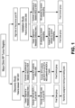
- the present specification discloses methods for prognosing prostate cancer in a subject using expression-based analysis of a plurality of targets.
- the method comprises (a) optionally providing a sample from a subject suffering from prostate cancer; (b) assaying the expression level for a plurality of targets in the sample; and (c) prognosing the cancer based on the expression level of the plurality of targets.
- Assaying the expression level for a plurality of targets in the sample may comprise applying the sample to a microarray. In some instances, assaying the expression level may comprise the use of an algorithm. The algorithm may be used to produce a classifier. Alternatively, the classifier may comprise a probe selection region. In some instances, assaying the expression level for a plurality of targets comprises detecting and/or quantifying the plurality of targets. In some embodiments, assaying the expression level for a plurality of targets comprises sequencing the plurality of targets. In some embodiments, assaying the expression level for a plurality of targets comprises amplifying the plurality of targets. In some embodiments, assaying the expression level for a plurality of targets comprises quantifying the plurality of targets. In some embodiments, assaying the expression level for a plurality of targets comprises conducting a multiplexed reaction on the plurality of targets.
- the plurality of targets comprises more than one target selected from Tables 2, 4, 11 or 55. In some instances, the plurality of targets comprises at least about 2, at least about 3, at least about 4, at least about 5, at least about 6, at least about 7, at least about 8, at least about 9, or at least about 10 targets selected from Table 2, 4, and 11. In other instances, the plurality of targets comprises at least about12, at least about 15, at least about 17, at least about 20, at least about 22, at least about 25, at least about 27, at least about 30, at least about 32, at least about 35, at least about 37, or at least about 40 targets selected from Tables 2, 4, 11 or 55. In some instances, the targets are selected from Table 2.
- the plurality of targets comprises 2, 3, 4, 5, 6, 7, 8, 9, 10, 11, 12, 13, 14, 15, 16, 17, 18, 19, 20, 21, 22, 23, 24, 25, 26, 27, 28, 29, 30, 31, 32, 33, 34, 35, 36, 37, 38, 39, 40, 41, 42, 43, 44, 45, 46, 47, 48, 49, or 50 targets selected from Tables 2, 4, 11 or 55.
- the plurality of targets is selected from SEQ ID NOs:1-43.
- the plurality of targets is selected from SEQ ID NOs:1-22.
- the targets are selected from Table 4.
- the targets are selected from Table 11.
- the plurality of targets comprises a coding target, non-coding target, or any combination thereof.
- the coding target comprises an exonic sequence.
- the non-coding target comprises a non-exonic sequence.
- the non-exonic sequence comprises an untranslated region (e.g., UTR), intronic region, intergenic region, or any combination thereof.
- the plurality of targets comprises an anti-sense sequence.
- the plurality of targets comprises a non-coding RNA transcript.
- the probe set comprises a plurality of probes capable of detecting an expression level of more than one target selected from Tables 2, 4, 11 or 55, wherein the expression level determines the cancer status of the subject with at least about 45% specificity.
- detecting an expression level comprise detecting gene expression, protein expression, or any combination thereof.
- the plurality of targets comprises more than one target selected from Tables 2, 4, 11 or 55.
- the plurality of targets comprises at least about 2, at least about 3, at least about 4, at least about 5, at least about 6, at least about 7, at least about 8, at least about 9, or at least about 10 targets selected from Table 2, 4, and 11.
- the plurality of targets comprises at least about12, at least about 15, at least about 17, at least about 20, at least about 22, at least about 25, at least about 27, at least about 30, at least about 32, at least about 35, at least about 37, or at least about 40 targets selected from Tables 2, 4, 11 or 55.
- the targets are selected from Table 2.
- the targets are selected from Table 4.
- the targets are selected from Table 11..
- the plurality of targets comprises 2, 3, 4, 5, 6, 7, 8, 9, 10, 11, 12, 13, 14, 15, 16, 17, 18, 19, 20, 21, 22, 23, 24, 25, 26, 27, 28, 29, 30, 31, 32, 33, 34, 35, 36, 37, 38, 39, 40, 41, 42, 43, 44, 45, 46, 47, 48, 49, or 50 targets selected from Tables 2, 4, 11 or 55.
- the plurality of targets is selected from SEQ ID NOs:1-43.
- the plurality of targets is selected from SEQ ID NOs:1-22.
- the plurality of targets comprises a coding target, non-coding target, or any combination thereof.
- the coding target comprises an exonic sequence.
- the non-coding target comprises a non-exonic sequence.
- the non-exonic sequence comprises an untranslated region (e.g., UTR), intronic region, intergenic region, or any combination thereof.
- the plurality of targets comprises an anti-sense sequence.
- the plurality of targets comprises a non-coding RNA transcript.
- the method comprises: (a) providing a sample from a subject; (b) assaying the expression level for a plurality of targets in the sample; and (c) characterizing the subject based on the expression level of the plurality of targets.
- the plurality of targets comprises more than one target selected from Tables 2, 4, 11 or 55.
- the plurality of targets comprises at least about 2, at least about 3, at least about 4, at least about 5, at least about 6, at least about 7, at least about 8, at least about 9, or at least about 10 targets selected from Tables 2, 4, 11 or 55.
- the plurality of targets comprises at least about12, at least about 15, at least about 17, at least about 20, at least about 22, at least about 25, at least about 27, at least about 30, at least about 32, at least about 35, at least about 37, or at least about 40 targets selected from Tables 2, 4, 11 or 55.
- the targets are selected from Table 2.
- the targets are selected from Table 4.
- the targets are selected from Table 11..
- the plurality of targets comprises 2, 3, 4, 5, 6, 7, 8, 9, 10, 11, 12, 13, 14, 15, 16, 17, 18, 19, 20, 21, 22, 23, 24, 25, 26, 27, 28, 29, 30, 31, 32, 33, 34, 35, 36, 37, 38, 39, 40, 41, 42, 43, 44, 45, 46, 47, 48, 49, or 50 targets selected from Tables 2, 4, 11 or 55.
- the plurality of targets is selected from SEQ ID NOs:1-43.
- the plurality of targets is selected from SEQ ID NOs:1-22.
- the plurality of targets comprises a coding target, non-coding target, or any combination thereof.
- the coding target comprises an exonic sequence.
- the non-coding target comprises a non-exonic sequence.
- the non-exonic sequence comprises an untranslated region (e.g., UTR), intronic region, intergenic region, or any combination thereof.
- the plurality of targets comprises an anti-sense sequence.
- the plurality of targets comprises a non-coding RNA transcript.
- polynucleotide refers to a polymer of greater than one nucleotide in length of ribonucleic acid (RNA), deoxyribonucleic acid (DNA), hybrid RNA/DNA, modified RNA or DNA, or RNA or DNA mimetics, including peptide nucleic acids (PNAs).
- the polynucleotides may be single- or double-stranded.
- the term includes polynucleotides composed of naturally-occurring nucleobases, sugars and covalent internucleoside (backbone) linkages as well as polynucleotides having non-naturally-occurring portions which function similarly.
- backbone backbone linkages
- Such modified or substituted polynucleotides are well known in the art and for the purposes of the present invention, are referred to as "analogues.”
- Complementary or “substantially complementary” refers to the ability to hybridize or base pair between nucleotides or nucleic acids, such as, for instance, between a sensor peptide nucleic acid or polynucleotide and a target polynucleotide.
- Complementary nucleotides are, generally, A and T (or A and U), or C and G.
- Two single-stranded polynucleotides or PNAs are said to be substantially complementary when the bases of one strand, optimally aligned and compared and with appropriate insertions or deletions, pair with at least about 80% of the bases of the other strand, usually at least about 90% to 95%, and more preferably from about 98 to 100%.
- substantial complementarity exists when a polynucleotide may hybridize under selective hybridization conditions to its complement.
- selective hybridization may occur when there is at least about 65% complementarity over a stretch of at least 14 to 25 bases, for example at least about 75%, or at least about 90% complementarity.
- Preferential binding or “preferential hybridization” refers to the increased propensity of one polynucleotide to bind to its complement in a sample as compared to a noncomplementary polymer in the sample.
- Hybridization conditions may typically include salt concentrations of less than about 1M, more usually less than about 500 mM, for example less than about 200 mM.
- the hybridization can be done in solutions containing little or no salt.
- Hybridization temperatures can be as low as 5° C, but are typically greater than 22° C, and more typically greater than about 30° C, for example in excess of about 37° C. Longer fragments may require higher hybridization temperatures for specific hybridization as is known in the art.
- hybridization conditions which may be controlled include buffer type and concentration, solution pH, presence and concentration of blocking reagents to decrease background binding such as repeat sequences or blocking protein solutions, detergent type(s) and concentrations, molecules such as polymers which increase the relative concentration of the polynucleotides, metal ion(s) and their concentration(s), chelator(s) and their concentrations, and other conditions known in the art.
- Multiplexing herein refers to an assay or other analytical method in which multiple analytes are assayed. In some instances, the multiple analytes are from the same sample. In some instances, the multiple analytes are assayed simultaneously. Alternatively, the multiple analytes are assayed sequentially. In some instances, assaying the multiple analytes occurs in the same reaction volume. Alternatively, assaying the multiple analytes occurs in separate or multiple reaction volumes.
- a “target sequence” as used herein refers to a region of the genome against which one or more probes can be designed.
- a “target sequence” may be a coding target or a non-coding target.
- a “target sequence” may comprise exonic and/or non-exonic sequences.
- a “target sequence” may comprise an ultraconserved region.
- An ultraconserved region is generally a sequence that is at least 200 base pairs and is conserved across multiple specieis.
- An ultraconserved region may be exonic or non-exonic. Exonic sequences may comprise regions on a protein-coding gene, such as an exon, LTTR, or a portion thereof.
- Non-exonic sequences may comprise regions on a protein-coding, non protein-coding gene, or a portion thereof.
- non-exonic sequences may comprise intronic regions, promoter regions, intergenic regions, a non-coding transcript, an exon anti-sense region, an intronic anti-sense region, UTR anti-sense region, non-coding transcript anti-sense region, or a portion thereof.
- a probe is any polynucleotide capable of selectively hybridizing to a target sequence or its complement, or to an RNA version of either.
- a probe may comprise ribonucleotides, deoxyribonucleotides, peptide nucleic acids, and combinations thereof.
- a probe may optionally comprise one or more labels.
- a probe may be used to amplify one or both strands of a target sequence or an RNA form thereof, acting as a sole primer in an amplification reaction or as a member of a set of primers.
- a non-coding target may comprise a nucleotide sequence.
- the nucleotide sequence is a DNA or RNA sequence.
- a non-coding target may include a UTR sequence, an intronic sequence, or a non-coding RNA transcript.
- a non-coding target also includes sequences which partially overlap with a UTR sequence or an intronic sequence.
- a non-coding target also includes non-exonic transcripts.
- a coding target includes nucleotide sequences that encode for a protein and peptide sequences.
- the nucleotide sequence is a DNA or RNA sequence.
- the coding target includes protein-coding sequence.
- Protein-coding sequences include exon-coding sequences (e.g., exonic sequences).
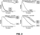
- diagnosis of cancer may include the identification of cancer in a subject, determining the malignancy of the cancer, or determining the stage of the cancer.
- prognosis of cancer may include predicting the clinical outcome of the patient, assessing the risk of cancer recurrence, determining treatment modality, or determining treatment efficacy.
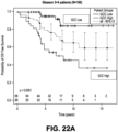
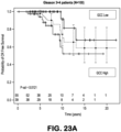
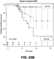
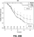
- ⁇ NED' describes a clinically distinct disease state in which patients show no evidence of disease (NED') at least 5 years after surgery
- ⁇ PSA' describes a clinically distinct disease state in which patients show biochemical relapse only (two successive increases in prostate-specific antigen levels but no other symptoms of disease with at least 5 years follow up after surgery
- ⁇ PSA' and 'SYS' describes a clinically distinct disease state in which patients develop biochemical relapse and present with systemic cancer disease or metastases ('SYS') within five years after the initial treatment with radical prostatectomy.
- METS cardiovascular disease
- SYS biological reccurrence
- RP radial prostectomy
- metastases within 5 years of BCR.
- METS may experience BCR after 5 years of RP.
- the term "about” refers to approximately a +/-10% variation from a given value. It is to be understood that such a variation is always included in any given value provided herein, whether or not it is specifically referred to.
- the methods disclosed herein often comprise assaying the expression level of a plurality of targets.
- the plurality of targets may comprise coding targets and/or non-coding targets of a protein-coding gene or a non protein-coding gene.
- a protein-coding gene structure may comprise an exon and an intron.
- the exon may further comprise a coding sequence (CDS) and an untranslated region (UTR).
- CDS coding sequence
- UTR untranslated region
- the protein-coding gene may be transcribed to produce a pre-mRNA and the pre-mRNA may be processed to produce a mature mRNA.
- the mature mRNA may be translated to produce a protein.
- a non protein-coding gene structure may comprise an exon and intron.
- the exon region of a non protein-coding gene primarily contains a UTR.
- the non protein-coding gene may be transcribed to produce a pre-mRNA and the pre-mRNA may be processed to produce a non-coding RNA (ncRNA).
- a coding target may comprise a coding sequence of an exon.
- a non-coding target may comprise a UTR sequence of an exon, intron sequence, intergenic sequence, promoter sequence, non-coding transcript, CDS antisense, intronic antisense, UTR antisense, or non-coding transcript antisense.
- a non-coding transcript may comprise a non-coding RNA (ncRNA).
- the plurality of targets may be differentially expressed.
- a plurality of probe selection regions (PSRs) is differentially expressed.
- the plurality of targets comprises more than one target selected from Tables 2, 4, 11 or 55. In some instances, the plurality of targets comprises at least about 2, at least about 3, at least about 4, at least about 5, at least about 6, at least about 7, at least about 8, at least about 9, or at least about 10 targets selected from Tables 2, 4, 11 or 55. In other instances, the plurality of targets comprises at least about12, at least about 15, at least about 17, at least about 20, at least about 22, at least about 25, at least about 27, at least about 30, at least about 32, at least about 35, at least about 37, or at least about 40 targets selected from Tables 2, 4, 11 or 55. In some instances, the targets are selected from Table 2. In some instances, the targets are selected from Table 4. In some instances, the targets are selected from Table 11..
- the plurality of targets comprises 2, 3, 4, 5, 6, 7, 8, 9, 10, 11, 12, 13, 14, 15, 16, 17, 18, 19, 20, 21, 22, 23, 24, 25, 26, 27, 28, 29, 30, 31, 32, 33, 34, 35, 36, 37, 38, 39, 40, 41, 42, 43, 44, 45, 46, 47, 48, 49, or 50 targets selected from Tables 2, 4, 11 or 55.
- the plurality of targets is selected from SEQ ID NOs:1-43.
- the plurality of targets is selected from SEQ ID NOs:1-22.
- the plurality of targets comprises a coding target, non-coding target, or any combination thereof.
- the coding target comprises an exonic sequence.
- the non-coding target comprises a non-exonic sequence.
- a non-coding target comprises a UTR sequence, an intronic sequence, or a non-coding RNA transcript.
- a non-coding target comprises sequences which partially overlap with a UTR sequence or an intronic sequence.
- a non-coding target also includes non-exonic transcripts. Exonic sequences may comprise regions on a protein-coding gene, such as an exon, UTR, or a portion thereof. Non-exonic sequences may comprise regions on a protein-coding, non protein-coding gene, or a portion thereof.
- non-exonic sequences may comprise intronic regions, promoter regions, intergenic regions, a non-coding transcript, an exon anti-sense region, an intronic anti-sense region, UTR anti-sense region, non-coding transcript anti-sense region, or a portion thereof.
- the plurality of targets comprises a non-coding RNA transcript.
- the plurality of targets is at least about 70% identical to a sequence selected from SEQ ID NOs 1-43. Alternatively, the plurality of targets is at least about 80% identical to a sequence selected from SEQ ID NOs 1-43. In some instances, the plurality of targets is at least about 85% identical to a sequence selected from SEQ ID NOs 1-43. In some instances, the plurality of targets is at least about 90% identical to a sequence selected from SEQ ID NOs 1-43. Alternatively, the plurality of targets is at least about 95% identical to a sequence selected from SEQ ID NOs 1-43.
- a probe set for prognosing prostate cancer in a subject comprising a plurality of probes, wherein (i) the probes in the set are capable of detecting an expression level of more than one non-coding target; and (ii) the expression level determines the cancer status of the subject with at least about 40% specificity.
- the probe set may comprise more than one polynucleotide probe.
- Individual polynucleotide probes comprise a nucleotide sequence derived from the nucleotide sequence of the target sequences or complementary sequences thereof.
- the nucleotide sequence of the polynucleotide probe is designed such that it corresponds to, or is complementary to the target sequences.
- the polynucleotide probe can specifically hybridize under either stringent or lowered stringency hybridization conditions to a region of the target sequences, to the complement thereof, or to a nucleic acid sequence (such as a cDNA) derived therefrom.
- polynucleotide probe sequences and determination of their uniqueness may be carried out in silica using techniques known in the art, for example, based on a BLASTN search of the polynucleotide sequence in question against gene sequence databases, such as the Human Genome Sequence, UniGene, dbEST or the non-redundant database at NCBI.
- the polynucleotide probe is complementary to a region of a target mRNA derived from a target sequence in the probe set.
- Computer programs can also be employed to select probe sequences that may not cross hybridize or may not hybridize non-specifically.
- microarray hybridization of RNA, extracted from prostate cancer tissue samples and amplified may yield a dataset that is then summarized and normalized by the fRMA technique.
- the 5,362,207 raw expression probes are summarized and normalized into 1,411,399 probe selection regions ("PSRs").
- PSRs probe selection regions
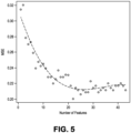
- the data can be decomposed into its principal components and an analysis of variance model is used to determine the extent to which a batch effect remains present in the first 10 principal components.
- nucleotide sequence of the polynucleotide probe need not be identical to its target sequence in order to specifically hybridize thereto.
- the polynucleotide probes therefore, comprise a nucleotide sequence that is at least about 65% identical to a region of the coding target or non-coding target selected from Tables 2, 4, 11 or 55.
- nucleotide sequence of the polynucleotide probe is at least about 70% identical a region of the coding target or non-coding target from Tables 2, 4, 11 or 55.
- nucleotide sequence of the polynucleotide probe is at least about 75% identical a region of the coding target or non-coding target from Tables 2, 4, 11 or 55. In another embodiment, the nucleotide sequence of the polynucleotide probe is at least about 80% identical a region of the coding target or non-coding target from Tables 2, 4, 11 or 55. In another embodiment, the nucleotide sequence of the polynucleotide probe is at least about 85% identical a region of the coding target or non-coding target from Tables 2, 4, 11 or 55.
- the nucleotide sequence of the polynucleotide probe is at least about 90% identical a region of the coding target or non-coding target from Tables 2, 4, 11 or 55. In a further embodiment, the nucleotide sequence of the polynucleotide probe is at least about 95% identical to a region of the coding target or non-coding target from Tables 2, 4, 11 or 55.
- the targets are selected from Table 2. In some instances, the targets are selected from Table 4. In some instances, the targets are selected from Table 11.
- nucleotide sequence of the polynucleotide probes may exhibit variability by differing ( e . g . by nucleotide substitution, including transition or transversion) at one, two, three, four or more nucleotides from the sequence of the coding target or non-coding target.
- the probes can be designed to have ⁇ 50% G content and/or between about 25% and about 70% G+C content.
- Strategies to optimize probe hybridization to the target nucleic acid sequence can also be included in the process of probe selection.
- Hybridization under particular pH, salt, and temperature conditions can be optimized by taking into account melting temperatures and by using empirical rules that correlate with desired hybridization behaviors.
- Computer models may be used for predicting the intensity and concentration-dependence of probe hybridization.
- the polynucleotide probes may range in length from about 15 nucleotides to the full length of the coding target or non-coding target. In one embodiment the polynucleotide probes are at least about 15 nucleotides in length. In another embodiment, the polynucleotide probes are at least about 20 nucleotides in length. In a further embodiment, the polynucleotide probes are at least about 25 nucleotides in length. In another embodiment, the polynucleotide probes are between about 15 nucleotides and about 500 nucleotides in length.
- the polynucleotide probes are between about 15 nucleotides and about 450 nucleotides, about 15 nucleotides and about 400 nucleotides, about 15 nucleotides and about 350 nucleotides, about 15 nucleotides and about 300 nucleotides, about 15 nucleotides and about 250 nucleotides, about 15 nucleotides and about 200 nucleotides in length.
- the probes are at least 15 nucleotides in length. In some embodiments, the probes are at least 15 nucleotides in length.
- the probes are at least 20 nucleotides, at least 25 nucleotides, at least 50 nucleotides, at least 75 nucleotides, at least 100 nucleotides, at least 125 nucleotides, at least 150 nucleotides, at least 200 nucleotides, at least 225 nucleotides, at least 250 nucleotides, at least 275 nucleotides, at least 300 nucleotides, at least 325 nucleotides, at least 350 nucleotides, at least 375 nucleotides in length.
- the polynucleotide probes of a probe set can comprise RNA, DNA, RNA or DNA mimetics, or combinations thereof, and can be single-stranded or double-stranded.
- the polynucleotide probes can be composed of naturally-occurring nucleobases, sugars and covalent internucleoside (backbone) linkages as well as polynucleotide probes having non-naturally-occurring portions which function similarly.
- Such modified or substituted polynucleotide probes may provide desirable properties such as, for example, enhanced affinity for a target gene and increased stability.
- the probe set may comprise a coding target and/or a non-coding target.
- the probe set comprises a combination of a coding target and non-coding target.
- the probe set comprise a plurality of target sequences that hybridize to at least about 5 coding targets and/or non-coding targets selected from Tables 2, 4, 11 or 55.
- the probe set comprise a plurality of target sequences that hybridize to at least about 10 coding targets and/or non-coding targets selected from Tables 2, 4, 11 or 55.
- the probe set comprise a plurality of target sequences that hybridize to at least about 15 coding targets and/or non-coding targets selected from Tables 2, 4, 11 or 55.
- the probe set comprise a plurality of target sequences that hybridize to at least about 20 coding targets and/or non-coding targets selected from Tables 2, 4, 11 or 55.
- the probe set comprise a plurality of target sequences that hybridize to at least about 30 coding targets and/or non-coding targets selected from Tables 2, 4, 11 or 55.
- the targets are selected from Table 2.
- the targets are selected from Table 4.
- the targets are selected from Table 11.
- the probe set comprises a plurality of target sequences that hybridize to a plurality of targets, wherein the at least about 20% of the plurality of targets are targets selected from Tables 2, 4, 11 or 55. In some embodiments, the probe set comprises a plurality of target sequences that hybridize to a plurality of targets, wherein the at least about 25% of the plurality of targets are targets selected from Tables 2, 4, 11 or 55. In some embodiments, the probe set comprise a plurality of target sequences that hybridize to a plurality of targets, wherein the at least about 30% of the plurality of targets are targets selected from Tables 2, 4, 11 or 55.
- the probe set comprise a plurality of target sequences that hybridize to a plurality of targets, wherein the at least about 35% of the plurality of targets are targets selected from Tables 2, 4, 11 or 55. In some embodiments, the probe set comprise a plurality of target sequences that hybridize to a plurality of targets, wherein the at least about 40% of the plurality of targets are targets selected from Tables 2, 4, 11 or 55. In some embodiments, the probe set comprise a plurality of target sequences that hybridize to a plurality of targets, wherein the at least about 45% of the plurality of targets are targets selected from Tables 2, 4, 11 or 55.
- the probe set comprise a plurality of target sequences that hybridize to a plurality of targets, wherein the at least about 50% of the plurality of targets are targets selected from Tables 2, 4, 11 or 55. In some embodiments, the probe set comprise a plurality of target sequences that hybridize to a plurality of targets, wherein the at least about 60% of the plurality of targets are targets selected from Tables 2, 4, 11 or 55. In some embodiments, the probe set comprise a plurality of target sequences that hybridize to a plurality of targets, wherein the at least about 70% of the plurality of targets are targets selected from Tables 2, 4, 11 or 55. In some instances, the targets are selected from Table 2. In some instances, the targets are selected from Table 4. In some instances, the targets are selected from Table 11.
- the system of the present disclosure further provides for primers and primer pairs capable of amplifying target sequences defined by the probe set, or fragments or subsequences or complements thereof.
- the nucleotide sequences of the probe set may be provided in computer-readable media for in silica applications and as a basis for the design of appropriate primers for amplification of one or more target sequences of the probe set.
- Primers based on the nucleotide sequences of target sequences can be designed for use in amplification of the target sequences.
- a pair of primers can be used.
- the exact composition of the primer sequences is not critical to the invention, but for most applications the primers may hybridize to specific sequences of the probe set under stringent conditions, particularly under conditions of high stringency, as known in the art.
- the pairs of primers are usually chosen so as to generate an amplification product of at least about 50 nucleotides, more usually at least about 100 nucleotides. Algorithms for the selection of primer sequences are generally known, and are available in commercial software packages.
- primers may be used in standard quantitative or qualitative PCR-based assays to assess transcript expression levels of RNAs defined by the probe set.
- these primers may be used in combination with probes, such as molecular beacons in amplifications using real-time PCR.
- the primers or primer pairs when used in an amplification reaction, specifically amplify at least a portion of a nucleic acid sequence of a target selected from any of Tables 2, 4, 11 or 55 (or subgroups thereof as set forth herein), an RNA form thereof, or a complement to either thereof.
- the targets are selected from Table 2.
- the targets are selected from Table 4.
- the targets are selected from Table 11.
- a nucleoside is a base-sugar combination and a nucleotide is a nucleoside that further includes a phosphate group covalently linked to the sugar portion of the nucleoside.
- the phosphate groups covalently link adjacent nucleosides to one another to form a linear polymeric compound, with the normal linkage or backbone of RNA and DNA being a 3' to 5' phosphodiester linkage.
- polynucleotide probes or primers useful in this invention include oligonucleotides containing modified backbones or non-natural internucleoside linkages.
- oligonucleotides having modified backbones include both those that retain a phosphorus atom in the backbone and those that lack a phosphorus atom in the backbone.
- modified oligonucleotides that do not have a phosphorus atom in their internucleoside backbone can also be considered to be oligonucleotides.
- the present disclosure also contemplates oligonucleotide mimetics in which both the sugar and the internucleoside linkage of the nucleotide units are replaced with novel groups.
- the base units are maintained for hybridization with an appropriate nucleic acid target compound.
- PNA peptide nucleic acid
- the sugar-backbone of an oligonucleotide is replaced with an amide containing backbone, in particular an aminoethylglycine backbone.
- the nucleobases are retained and are bound directly or indirectly to aza-nitrogen atoms of the amide portion of the backbone.
- LNAs locked nucleic acids
- LNA and LNA analogues may display very high duplex thermal stabilities with complementary DNA and RNA, stability towards 3'-exonuclease degradation, and good solubility properties. Synthesis of the LNA analogues of adenine, cytosine, guanine, 5-methylcytosine, thymine and uracil, their oligomerization, and nucleic acid recognition properties have been described. Studies of mismatched sequences show that LNA obey the Watson-Crick base pairing rules with generally improved selectivity compared to the corresponding unmodified reference strands.
- the oligonucleotides may comprise one of the following substituents at the 2' position: C 1 to C 10 lower alkyl, substituted lower alkyl, alkaryl, aralkyl, O-alkaryl or O-aralkyl, SH, SCH 3 , OCN, Cl, Br, CN, CF 3 , OCF 3 , SOCH 3 , SO 2 CH 3 , ONO 2 , NO 2 , N 3 , NH 2 , heterocycloalkyl, heterocycloalkaryl, aminoalkylamino, polyalkylamino, substituted silyl, an RNA cleaving group, a reporter group, an intercalator, a group for improving the pharmacokinetic properties of an oligonucleotide, or a group for improving the pharmacodynamic properties of an oligonucleotide, and other substituents having similar properties.
- 2'-methoxyethoxy (2'-O--CH 2 CH 2 OCH 3 , also known as 2'-O-(2-methoxyethyl) or 2'-MOE), 2'-dimethylaminooxyethoxy (O(CH2)2 ON(CH 3 ) 2 group, also known as 2'- DMA0E), 2'-methoxy (2'-O--CH 3 ), 2'-aminopropoxy (2'-OCH 2 CH 2 CH 2 NH 2 ) and 2'-fluoro (2'-F).
- polynucleotide probes or primers may also have sugar mimetics such as cyclobutyl moieties in place of the pentofuranosyl sugar.
- Polynucleotide probes or primers may also include modifications or substitutions to the nucleobase.
- "unmodified” or “natural” nucleobases include the purine bases adenine (A) and guanine (G), and the pyrimidine bases thymine (T), cytosine (C) and uracil (U).
- Modified nucleobases include other synthetic and natural nucleobases such as 5-methylcytosine (5-me-C), 5- hydroxymethyl cytosine, xanthine, hypoxanthine, 2-aminoadenine, 6-methyl and other alkyl derivatives of adenine and guanine, 2-propyl and other alkyl derivatives of adenine and guanine, 2-thiouracil, 2-thiothymine and 2-thiocytosine, 5-halouracil and cytosine, 5- propynyl uracil and cytosine, 6-azo uracil, cytosine and thymine, 5-uracil (pseudouracil), 4-thiouracil, 8-halo, 8-amino, 8-thiol, 8-thioalkyl, 8-hydroxyl and other 8-substituted adenines and guanines, 5-halo particularly 5-bromo, 5-trifluoromethyl and other 5-substit
- nucleobases include those disclosed in U.S. Pat. No. 3,687,808 ; The Concise Encyclopedia Of Polymer Science And Engineering, (1990) pp 858-859, Kroschwitz, J. I., ed. John Wiley & Sons ; Englisch et al., Angewandte Chemie, Int. Ed., 30:613 (1991 ); and Sanghvi, Y. S., (1993) Antisense Research and Applications, pp 289-302, Crooke, S. T. and Lebleu, B., ed., CRC Press . Certain of these nucleobases are particularly useful for increasing the binding affinity of the polynucleotide probes.
- 5-substituted pyrimidines 6-azapyrimidines and N-2, N-6 and O-6 substituted purines, including 2-aminopropyladenine, 5- propynyluracil and 5-propynylcytosine.
- 5-methylcytosine substitutions have been shown to increase nucleic acid duplex stability by 0.6-1.2°C.
- nucleotide sequence of the entire length of the polynucleotide probe or primer does not need to be derived from the target sequence.
- the polynucleotide probe may comprise nucleotide sequences at the 5' and/or 3' termini that are not derived from the target sequences.
- Nucleotide sequences which are not derived from the nucleotide sequence of the target sequence may provide additional functionality to the polynucleotide probe. For example, they may provide a restriction enzyme recognition sequence or a "tag" that facilitates detection, isolation, purification or immobilization onto a solid support.
- the additional nucleotides may provide a self-complementary sequence that allows the primer/probe to adopt a hairpin configuration.
- Such configurations are necessary for certain probes, for example, molecular beacon and Scorpion probes, which can be used in solution hybridization techniques.
- the polynucleotide probes or primers can incorporate moieties useful in detection, isolation, purification, or immobilization, if desired.
- moieties are well-known in the art (see, for example, Ausubel et al., (1997 & updates) Current Protocols in Molecular Biology, Wiley & Sons, New York ) and are chosen such that the ability of the probe to hybridize with its target sequence is not affected.
- Suitable moieties are detectable labels, such as radioisotopes, fluorophores, chemiluminophores, enzymes, colloidal particles, and fluorescent microparticles, as well as antigens, antibodies, haptens, avidin/streptavidin, biotin, haptens, enzyme cofactors / substrates, enzymes, and the like.
- a label can optionally be attached to or incorporated into a probe or primer polynucleotide to allow detection and/or quantitation of a target polynucleotide representing the target sequence of interest.
- the target polynucleotide may be the expressed target sequence RNA itself, a cDNA copy thereof, or an amplification product derived therefrom, and may be the positive or negative strand, so long as it can be specifically detected in the assay being used.
- an antibody may be labeled.
- labels used for detecting different targets may be distinguishable.
- the label can be attached directly (e.g., via covalent linkage) or indirectly, e.g., via a bridging molecule or series of molecules (e.g., a molecule or complex that can bind to an assay component, or via members of a binding pair that can be incorporated into assay components, e.g. biotin-avidin or streptavidin).
- a bridging molecule or series of molecules e.g., a molecule or complex that can bind to an assay component, or via members of a binding pair that can be incorporated into assay components, e.g. biotin-avidin or streptavidin.
- Many labels are commercially available in activated forms which can readily be used for such conjugation (for example through amine acylation), or labels may be attached through known or determinable conjugation schemes, many of which are known in the art.
- Labels useful in the invention described herein include any substance which can be detected when bound to or incorporated into the biomolecule of interest. Any effective detection method can be used, including optical, spectroscopic, electrical, piezoelectrical, magnetic, Raman scattering, surface plasmon resonance, colorimetric, calorimetric, etc.
- a label is typically selected from a chromophore, a lumiphore, a fluorophore, one member of a quenching system, a chromogen, a hapten, an antigen, a magnetic particle, a material exhibiting nonlinear optics, a semiconductor nanocrystal, a metal nanoparticle, an enzyme, an antibody or binding portion or equivalent thereof, an aptamer, and one member of a binding pair, and combinations thereof.
- Quenching schemes may be used, wherein a quencher and a fluorophore as members of a quenching pair may be used on a probe, such that a change in optical parameters occurs upon binding to the target introduce or quench the signal from the fluorophore.
- a molecular beacon Suitable quencher/fluorophore systems are known in the art.
- the label may be bound through a variety of intermediate linkages.
- a polynucleotide may comprise a biotin-binding species, and an optically detectable label may be conjugated to biotin and then bound to the labeled polynucleotide.
- a polynucleotide sensor may comprise an immunological species such as an antibody or fragment, and a secondary antibody containing an optically detectable label may be added.
- Chromophores useful in the methods described herein include any substance which can absorb energy and emit light.
- a plurality of different signaling chromophores can be used with detectably different emission spectra.
- the chromophore can be a lumophore or a fluorophore.
- Typical fluorophores include fluorescent dyes, semiconductor nanocrystals, lanthanide chelates, polynucleotide-specific dyes and green fluorescent protein.
- Coding schemes may optionally be used, comprising encoded particles and/or encoded tags associated with different polynucleotides.
- a variety of different coding schemes are known in the art, including fluorophores, including SCNCs, deposited metals, and RF tags.
- Polynucleotides from the described target sequences may be employed as probes for detecting target sequences expression, for ligation amplification schemes, or may be used as primers for amplification schemes of all or a portion of a target sequences.
- amplified either strand produced by amplification may be provided in purified and/or isolated form.
- polynucleotides include (a) a nucleic acid depicted in Tables 2, 4, 11 or 55; (b) an RNA form of any one of the nucleic acids depicted in Tables 2, 4, 11 or 55; (c) a peptide nucleic acid form of any of the nucleic acids depicted in Tables 2, 4, 11 or 55; (d) a nucleic acid comprising at least 20 consecutive bases of any of (a-c); (e) a nucleic acid comprising at least 25 bases having at least 90% sequenced identity to any of (a-c); and (f) a complement to any of (a-e).
- Complements may take any polymeric form capable of base pairing to the species recited in (a)-(e), including nucleic acid such as RNA or DNA, or may be a neutral polymer such as a peptide nucleic acid.
- Polynucleotides can be selected from the subsets of the recited nucleic acids described herein, as well as their complements.
- polynucleotides comprise at least 20 consecutive bases of the nucleic acid sequence of a target selected from any of Tables 2, 4, 11 or 55 or a complement thereto.
- the polynucleotides may comprise at least 21, 22, 23, 24, 25, 27, 30, 32, 35 or more consecutive bases of the nucleic acids sequence of a target selected from any of Tables 2, 4, 11 or 55, as applicable.
- the targets are selected from Table 2.
- the targets are selected from Table 4.
- the targets are selected from Table 11.
- the polynucleotides may be provided in a variety of formats, including as solids, in solution, or in an array.
- the polynucleotides may optionally comprise one or more labels, which may be chemically and/or enzymatically incorporated into the polynucleotide.
- solutions comprising polynucleotide and a solvent are also provided.
- the solvent may be water or may be predominantly aqueous.
- the solution may comprise at least two, three, four, five, six, seven, eight, nine, ten, twelve, fifteen, seventeen, twenty or more different polynucleotides, including primers and primer pairs. Additional substances may be included in the solution, alone or in combination, including one or more labels, additional solvents, buffers, biomolecules, polynucleotides, and one or more enzymes useful for performing methods described herein, including polymerases and ligases.
- the solution may further comprise a primer or primer pair capable of amplifying a polynucleotide present in the solution.
- one or more polynucleotides provided herein can be provided on a substrate.
- the substrate can comprise a wide range of material, either biological, nonbiological, organic, inorganic, or a combination of any of these.
- the substrate may be a polymerized Langmuir Blodgett film, functionalized glass, Si, Ge, GaAs, GaP, SiO 2 , SiN 4 , modified silicon, or any one of a wide variety of gels or polymers such as (poly)tetrafluoroethylene, (poly)vinylidenedifluoride, polystyrene, cross-linked polystyrene, polyacrylic, polylactic acid, polyglycolic acid, poly(lactide coglycolide), polyanhydrides, poly(methyl methacrylate), poly(ethylene-co-vinyl acetate), polysiloxanes, polymeric silica, latexes, dextran polymers, epoxies, polycarbonates,
- Substrates can be planar crystalline substrates such as silica based substrates (e.g. glass, quartz, or the like), or crystalline substrates used in, e.g., the semiconductor and microprocessor industries, such as silicon, gallium arsenide, indium doped GaN and the like, and include semiconductor nanocrystals.
- silica based substrates e.g. glass, quartz, or the like
- crystalline substrates used in, e.g., the semiconductor and microprocessor industries such as silicon, gallium arsenide, indium doped GaN and the like, and include semiconductor nanocrystals.
- the substrate can take the form of an array, a photodiode, an optoelectronic sensor such as an optoelectronic semiconductor chip or optoelectronic thin-film semiconductor, or a biochip.
- the location(s) of probe(s) on the substrate can be addressable; this can be done in highly dense formats, and the location(s) can be microaddressable or nanoaddressable.
- Silica aerogels can also be used as substrates, and can be prepared by methods known in the art. Aerogel substrates may be used as free standing substrates or as a surface coating for another substrate material.
- the substrate can take any form and typically is a plate, slide, bead, pellet, disk, particle, microparticle, nanoparticle, strand, precipitate, optionally porous gel, sheets, tube, sphere, container, capillary, pad, slice, film, chip, multiwell plate or dish, optical fiber, etc.
- the substrate can be any form that is rigid or semi-rigid.
- the substrate may contain raised or depressed regions on which an assay component is located.
- the surface of the substrate can be etched using known techniques to provide for desired surface features, for example trenches, v-grooves, mesa structures, or the like.
- Surfaces on the substrate can be composed of the same material as the substrate or can be made from a different material, and can be coupled to the substrate by chemical or physical means.
- Such coupled surfaces may be composed of any of a wide variety of materials, for example, polymers, plastics, resins, polysaccharides, silica or silica-based materials, carbon, metals, inorganic glasses, membranes, or any of the above-listed substrate materials.
- the surface can be optically transparent and can have surface Si-OH functionalities, such as those found on silica surfaces.
- the substrate and/or its optional surface can be chosen to provide appropriate characteristics for the synthetic and/or detection methods used.
- the substrate and/or surface can be transparent to allow the exposure of the substrate by light applied from multiple directions.
- the substrate and/or surface may be provided with reflective "mirror" structures to increase the recovery of light.
- the substrate and/or its surface is generally resistant to, or is treated to resist, the conditions to which it is to be exposed in use, and can be optionally treated to remove any resistant material after exposure to such conditions.
- the polynucleotide probes or primers can be prepared by conventional techniques well-known to those skilled in the art.
- the polynucleotide probes can be prepared using solid-phase synthesis using commercially available equipment.
- modified oligonucleotides can also be readily prepared by similar methods.
- the polynucleotide probes can also be synthesized directly on a solid support according to methods standard in the art. This method of synthesizing polynucleotides is particularly useful when the polynucleotide probes are part of a nucleic acid array.
- Prognostic samples for use with the systems and in the methods of the present invention comprise nucleic acids suitable for providing RNAs expression information.
- the biological sample from which the expressed RNA is obtained and analyzed for target sequence expression can be any material suspected of comprising cancer tissue or cells.
- the prognostic sample can be a biological sample used directly in a method of the invention.
- the prognostic sample can be a sample prepared from a biological sample.
- One or more alcohols may be used to fix tissue, alone or in combination with other fixatives.
- exemplary alcohols used for fixation include methanol, ethanol and isopropanol.
- Formalin fixation is frequently used in medical laboratories.
- Formalin comprises both an alcohol, typically methanol, and formaldehyde, both of which can act to fix a biological sample.
- the biological sample may optionally be embedded in an embedding medium.
- embedding media used in histology including paraffin, Tissue-Tek ® V.I.P.TM, Paramat, Paramat Extra, Paraplast, Paraplast X-tra, Paraplast Plus, Peel Away Paraffin Embedding Wax, Polyester Wax, Carbowax Polyethylene Glycol, PolyfinTM, Tissue Freezing Medium TFMFM, Cryo-GefTM, and OCT Compound (Electron Microscopy Sciences, Hatfield, PA).
- the embedding material may be removed via any suitable techniques, as known in the art.
- the sample is a fixed, wax-embedded biological sample.
- samples from medical laboratories are provided as fixed, wax-embedded samples, most commonly as formalin-fixed, paraffin embedded (FFPE) tissues.
- FFPE formalin-fixed, paraffin embedded
- the target polynucleotide that is ultimately assayed can be prepared synthetically (in the case of control sequences), but typically is purified from the biological source and subjected to one or more preparative steps.
- the RNA may be purified to remove or diminish one or more undesired components from the biological sample or to concentrate it. Conversely, where the RNA is too concentrated for the particular assay, it may be diluted.
- RNA can be extracted and purified from biological samples using any suitable technique.
- a number of techniques are known in the art, and several are commercially available (e.g., FormaPure nucleic acid extraction kit, Agencourt Biosciences, Beverly MA, High Pure FFPE RNA Micro Kit, Roche Applied Science, Indianapolis, IN).
- RNA can be extracted from frozen tissue sections using TRIzol (Invitrogen, Carlsbad, CA) and purified using RNeasy Protect kit (Qiagen, Valencia, CA). RNA can be further purified using DNAse I treatment (Ambion, Austin, TX) to eliminate any contaminating DNA.
- RNA concentrations can be made using a Nanodrop ND-1000 spectrophotometer (Nanodrop Technologies, Rockland, DE).
- RNA can be further purified to eliminate contaminants that interfere with cDNA synthesis by cold sodium acetate precipitation.
- RNA integrity can be evaluated by running electropherograms, and RNA integrity number (RIN, a correlative measure that indicates intactness of mRNA) can be determined using the RNA 6000 PicoAssay for the Bioanalyzer 2100 (Agilent Technologies, Santa Clara, CA).
- the nucleic acid portion of the sample comprising RNA that is or can be used to prepare the target polynucleotide(s) of interest can be subjected to one or more preparative reactions.
- These preparative reactions can include in vitro transcription (IVT), labeling, fragmentation, amplification and other reactions.
- mRNA can first be treated with reverse transcriptase and a primer to create cDNA prior to detection, quantitation and/or amplification; this can be done in vitro with purified mRNA or in situ, e.g., in cells or tissues affixed to a slide.
- amplification is meant any process of producing at least one copy of a nucleic acid, in this case an expressed RNA, and in many cases produces multiple copies.
- An amplification product can be RNA or DNA, and may include a complementary strand to the expressed target sequence.
- DNA amplification products can be produced initially through reverse translation and then optionally from further amplification reactions.
- the amplification product may include all or a portion of a target sequence, and may optionally be labeled.
- a variety of amplification methods are suitable for use, including polymerase-based methods and ligation-based methods.
- Exemplary amplification techniques include the polymerase chain reaction method (PCR), the lipase chain reaction (LCR), ribozyme-based methods, self sustained sequence replication (3SR), nucleic acid sequence-based amplification (NASBA), the use of Q Beta replicase, reverse transcription, nick translation, and the like.
- Asymmetric amplification reactions may be used to preferentially amplify one strand representing the target sequence that is used for detection as the target polynucleotide.
- the presence and/or amount of the amplification product itself may be used to determine the expression level of a given target sequence.
- the amplification product may be used to hybridize to an array or other substrate comprising sensor polynucleotides which are used to detect and/or quantitate target sequence expression.
- the first cycle of amplification in polymerase-based methods typically forms a primer extension product complementary to the template strand.
- the template is single-stranded RNA
- a polymerase with reverse transcriptase activity is used in the first amplification to reverse transcribe the RNA to DNA, and additional amplification cycles can be performed to copy the primer extension products.
- the primers for a PCR must, of course, be designed to hybridize to regions in their corresponding template that can produce an amplifiable segment; thus, each primer must hybridize so that its 3' nucleotide is paired to a nucleotide in its complementary template strand that is located 3' from the 3' nucleotide of the primer used to replicate that complementary template strand in the PCR.
- the target polynucleotide can be amplified by contacting one or more strands of the target polynucleotide with a primer and a polymerase having suitable activity to extend the primer and copy the target polynucleotide to produce a full-length complementary polynucleotide or a smaller portion thereof.
- Any enzyme having a polymerase activity that can copy the target polynucleotide can be used, including DNA polymerases, RNA polymerases, reverse transcriptases, enzymes having more than one type of polymerase or enzyme activity.
- the enzyme can be thermolabile or thermostable. Mixtures of enzymes can also be used.
- Exemplary enzymes include: DNA polymerases such as DNA Polymerase I ("Pol I"), the Klenow fragment of Pol I, T4, T7, Sequenase ® T7, Sequenase ® Version 2.0 T7, Tub, Taq, Tth, Pfic, Pfu, Tsp, Tfl, Tli and Pyrococcus sp GB-D DNA polymerases; RNA polymerases such as E. coil, SP6, T3 and T7 RNA polymerases; and reverse transcriptases such as AMV, M-MuLV, MMLV, RNAse H MMLV (SuperScript ® ), SuperScript ® II, ThermoScript ® , HIV-1, and RAV2 reverse transcriptases.
- DNA polymerases such as DNA Polymerase I ("Pol I"), the Klenow fragment of Pol I, T4, T7, Sequenase ® T7, Sequenase ® Version 2.0 T7, Tub, Taq, Tth,
- Exemplary polymerases with multiple specificities include RAV2 and Tli (exo-) polymerases.
- Exemplary thermostable polymerases include Tub, Taq, Tth, Pfic, Pfu, Tsp, Tf1, Tli and Pyrococcus sp.
- GB-D DNA polymerases are commercially available.
- Suitable reaction conditions are chosen to permit amplification of the target polynucleotide, including pH, buffer, ionic strength, presence and concentration of one or more salts, presence and concentration of reactants and cofactors such as nucleotides and magnesium and/or other metal ions (e.g., manganese), optional cosolvents, temperature, thermal cycling profile for amplification schemes comprising a polymerase chain reaction, and may depend in part on the polymerase being used as well as the nature of the sample.
- Cosolvents include formamide (typically at from about 2 to about 10 %), glycerol (typically at from about 5 to about 10 %), and DMSO (typically at from about 0.9 to about 10 %).
- Techniques may be used in the amplification scheme in order to minimize the production of false positives or artifacts produced during amplification. These include "touchdown" PCR, hot-start techniques, use of nested primers, or designing PCR primers so that they form stem-loop structures in the event of primer-dimer formation and thus are not amplified.
- Techniques to accelerate PCR can be used, for example centrifugal PCR, which allows for greater convection within the sample, and comprising infrared heating steps for rapid heating and cooling of the sample.
- One or more cycles of amplification can be performed.
- An excess of one primer can be used to produce an excess of one primer extension product during PCR; preferably, the primer extension product produced in excess is the amplification product to be detected.
- a plurality of different primers may be used to amplify different target polynucleotides or different regions of a particular target polynucleotide within the sample.
- An amplification reaction can be performed under conditions which allow an optionally labeled sensor polynucleotide to hybridize to the amplification product during at least part of an amplification cycle.
- an assay is performed in this manner, real-time detection of this hybridization event can take place by monitoring for light emission or fluorescence during amplification, as known in the art.
- amplification product is to be used for hybridization to an array or microarray
- suitable commercially available amplification products include amplification kits available from NuGEN, Inc. (San Carlos, CA), including the WT-OvationTm System, WT-OvationTm System v2, WT-OvationTm Pico System, WT-Ovation'm FFPE Exon Module, WT-OvationTm FFPE Exon Module RiboAmp and RiboAmp Plus RNA Amplification Kits (MDS Analytical Technologies (formerly Arcturus) (Mountain View, CA), Genisphere, Inc.
- NuGEN, Inc. San Carlos, CA
- WT-OvationTm System WT-OvationTm System v2
- WT-OvationTm Pico System WT-Ovation'm FFPE Exon Module
- Amplified nucleic acids may be subjected to one or more purification reactions after amplification and labeling, for example using magnetic beads (e.g., RNAClean magnetic beads, Agencourt Biosciences).
- magnetic beads e.g., RNAClean magnetic beads, Agencourt Biosciences.
- RNA biomarkers can be analyzed using real-time quantitative multiplex RT-PCR platforms and other multiplexing technologies such as GenomeLab GeXP Genetic Analysis System (Beckman Coulter, Foster City, CA), SmartCycler ® 9600 or GeneXpert(R) Systems (Cepheid, Sunnyvale, CA), ABI 7900 HT Fast Real Time PCR system (Applied Biosystems, Foster City, CA), LightCycler ® 480 System (Roche Molecular Systems, Pleasanton, CA), xMAP 100 System (Luminex, Austin, TX) Solexa Genome Analysis System (Illumina, Hayward, CA), OpenArray Real Time qPCR (BioTrove, Woburn, MA) and BeadXpress System (Illumina, Hayward, CA).
- GenomeLab GeXP Genetic Analysis System Beckman Coulter, Foster City, CA
- SmartCycler ® 9600 or GeneXpert(R) Systems (Cepheid, Sunnyvale, CA)
- any method of detecting and/or quantitating the expression of the encoded target sequences can in principle be used in the invention.
- the expressed target sequences can be directly detected and/or quantitated, or may be copied and/or amplified to allow detection of amplified copies of the expressed target sequences or its complement.
- Methods for detecting and/or quantifying a target can include Northern blotting, sequencing, array or microarray hybridization, by enzymatic cleavage of specific structures (e.g., an Invader ® assay, Third Wave Technologies, e.g. as described in U.S. Pat. Nos. 5,846,717 , 6,090,543 ; 6,001,567 ; 5,985,557 ; and 5,994,069 ) and amplification methods, e.g. RT-PCR, including in a TaqMan ® assay (PE Biosystems, Foster City, Calif., e.g. as described in U.S. Pat. Nos.
- specific structures e.g., an Invader ® assay, Third Wave Technologies, e.g. as described in U.S. Pat. Nos. 5,846,717 , 6,090,543 ; 6,001,567 ; 5,985,557 ; and 5,994,069
- amplification methods
- nucleic acids may be amplified, labeled and subjected to microarray analysis.
- target sequences may be detected by sequencing.
- Sequencing methods may comprise whole genome sequencing or exome sequencing. Sequencing methods such as Maxim-Gilbert, chain-termination, or high-throughput systems may also be used. Additional, suitable sequencing techniques include classic dideoxy sequencing reactions (Sanger method) using labeled terminators or primers and gel separation in slab or capillary, sequencing by synthesis using reversibly terminated labeled nucleotides, pyrosequencing, 454 sequencing, allele specific hybridization to a library of labeled oligonucleotide probes, sequencing by synthesis using allele specific hybridization to a library of labeled clones that is followed by ligation, real time monitoring of the incorporation of labeled nucleotides during a polymerization step, and SOLiD sequencing.
- Additional methods for detecting and/or quantifying a target include single-molecule sequencing (e.g., Helicos, PacBio), sequencing by synthesis (e.g., Illumina, Ion Torrent), sequencing by ligation (e.g., ABI SOLID), sequencing by hybridization (e.g., Complete Genomics), in situ hybridization, bead-array technologies (e.g., Luminex xMAP, Illumina BeadChips), branched DNA technology (e.g., Panomics, Genisphere). Sequencing methods may use fluorescent (e.g., Illumina) or electronic (e.g., Ion Torrent, Oxford Nanopore) methods of detecting nucleotides.
- single-molecule sequencing e.g., Helicos, PacBio
- sequencing by synthesis e.g., Illumina, Ion Torrent
- sequencing by ligation e.g., ABI SOLID
- sequencing by hybridization e.g., Complete Genomics
- in situ hybridization e.g
- Reverse transcription can be performed by any method known in the art. For example, reverse transcription may be performed using the Omniscript kit (Qiagen, Valencia, CA), Superscript III kit (Invitrogen, Carlsbad, CA), for RT-PCR. Target-specific priming can be performed in order to increase the sensitivity of detection of target sequences and generate target-specific cDNA.
- Omniscript kit Qiagen, Valencia, CA
- Superscript III kit Invitrogen, Carlsbad, CA
- Target-specific priming can be performed in order to increase the sensitivity of detection of target sequences and generate target-specific cDNA.
- TaqMan ® RT-PCR can be performed using Applied Biosystems Prism (ABI) 7900 HT instruments in a 5 1.11 volume with target sequence-specific cDNA equivalent to 1 ng total RNA.
- Primers and probes concentrations for TaqMan analysis are added to amplify fluorescent amplicons using PCR cycling conditions such as 95°C for 10 minutes for one cycle, 95°C for 20 seconds, and 60°C for 45 seconds for 40 cycles.
- a reference sample can be assayed to ensure reagent and process stability.
- Negative controls e.g., no template should be assayed to monitor any exogenous nucleic acid contamination.
- Examples include, but are not limited to, polymers, such as (polytetrafluoroethylene, (poly)vinylidenedifluoride, polystyrene, polycarbonate, polypropylene and polystyrene; ceramic; silicon; silicon dioxide; modified silicon; (fused) silica, quartz or glass; functionalized glass; paper, such as filter paper; diazotized cellulose; nitrocellulose filter; nylon membrane; and polyacrylamide gel pad. Substrates that are transparent to light are useful for arrays that may be used in an assay that involves optical detection.
- Supervised learning may comprise ordinal classification such as regression analysis and Information fuzzy networks (IFN).
- supervised learning methods may comprise statistical classification, such as AODE, Linear classifiers (e.g., Fisher's linear discriminant, Logistic regression, Naive Bayes classifier, Perceptron, and Support vector machine), quadratic classifiers, k-nearest neighbor, Boosting, Decision trees (e.g., C4.5, Random forests), Bayesian networks, and Hidden Markov models.
- the machine learning algorithms may also comprise an unsupervised learning algorithm.
- unsupervised learning algorithms may include artificial neural network, Data clustering, Expectation-maximization algorithm, Self-organizing map, Radial basis function network, Vector Quantization, Generative topographic map, Information bottleneck method, and IBSEAD.
- Unsupervised learning may also comprise association rule learning algorithms such as Apriori algorithm, Eclat algorithm and FP-growth algorithm.
- Hierarchical clustering such as Single-linkage clustering and Conceptual clustering, may also be used.
- unsupervised learning may comprise partitional clustering such as K-means algorithm and Fuzzy clustering.
- the machine learning algorithms comprise a reinforcement learning algorithm.
- reinforcement learning algorithms include, but are not limited to, temporal difference learning, Q-learning and Learning Automata.
- the machine learning algorithm may comprise Data Pre-processing.
- the methods disclosed herein may include additional techniques such as cytology, histology, ultrasound analysis, MRI results, CT scan results, and measurements of PSA levels.
- Certified tests for classifying disease status and/or designating treatment modalities may also be used in prognosing prostate cancer in a subject.
- a certified test may comprise a means for characterizing the expression levels of one or more of the target sequences of interest, and a certification from a government regulatory agency endorsing use of the test for classifying the disease status of a biological sample.
- the certified test may comprise reagents for amplification reactions used to detect and/or quantitate expression of the target sequences to be characterized in the test.
- An array of probe nucleic acids can be used, with or without prior target amplification, for use in measuring target sequence expression.
- test is submitted to an agency having authority to certify the test for use in distinguishing disease status and/or outcome.
- Results of detection of expression levels of the target sequences used in the test and correlation with disease status and/or outcome are submitted to the agency.
- a certification authorizing the prognostic use of the test is obtained.
- portfolios of expression levels comprising a plurality of normalized expression levels of the target selected from any of Tables 2, 4, 11 or 55.
- the target is selected from Table 2.
- the target is selected from Table 4.
- the target is selected from Table 11.
- Such portfolios may be provided by performing the methods described herein to obtain expression levels from an individual patient or from a group of patients.
- the expression levels can be normalized by any method known in the art; exemplary normalization methods that can be used in various embodiments include Robust Multichip Average (RMA), probe logarithmic intensity error estimation (PLIER), non-linear fit (NLFIT) quantile-based and nonlinear normalization, and combinations thereof.
- Background correction can also be performed on the expression data; exemplary techniques useful for background correction include mode of intensities, normalized using median polish probe modeling and sketch-normalization.
- portfolios are established such that the combination of genes in the portfolio exhibit improved sensitivity and specificity relative to known methods.
- a small standard deviation in expression measurements correlates with greater specificity.
- Other measurements of variation such as correlation coefficients can also be used in this capacity.
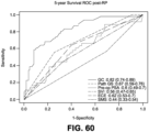
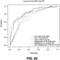
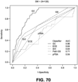
- the invention also encompasses the above methods where the expression level determines the status or outcome of a cancer in the subject with at least about 45% specificity. In some embodiments, the expression level determines the status or outcome of a cancer in the subject with at least about 50% specificity. In some embodiments, the expression level determines the status or outcome of a cancer in the subject with at least about 55% specificity.
- the expression level determines the status or outcome of a cancer in the subject with at least about 60% specificity. In some embodiments, the expression level determines the status or outcome of a cancer in the subject with at least about 65% specificity. In some embodiments, the expression level determines the status or outcome of a cancer in the subject with at least about 70% specificity. In some embodiments, the expression level determines the status or outcome of a cancer in the subject with at least about 75% specificity. In some embodiments, the expression level determines the status or outcome of a cancer in the subject with at least about 80% specificity. In some embodiments, t the expression level determines the status or outcome of a cancer in the subject with at least about 85% specificity. In some embodiments, the expression level determines the status or outcome of a cancer in the subject with at least about 90% specificity. In some embodiments, the expression level determines the status or outcome of a cancer in the subject with at least about 95% specificity.
- the 95% CI of a classifier or biomarker may be less than about 1.75, 1.74, 1.73, 1.72, 1.71, 1.70, 1.69, 1.68, 1.67, 1.66, 1.65, 1.64, 1.63, 1.62, 1.61, 1.60, 1.59, 1.58, 1.57, 1.56, 1.55, 1.54, 1.53, 1.52, 1.51, 1.50 or less.
- the 95% CI of a classifier or biomarker may be less than about 1.61, 1.60, 1.59, 1.58, 1.56, 1.55, or 1.53.
- the 95% CI of a classifier or biomarker may be between about 1.10 to 1.70, between about 1.12 to about 1.68, between about 1.14 to about 1.62, between about 1.15 to about 1.61, between about 1.15 to about 1.59, between about 1.16 to about 1.160, between about 1.19 to about 1.55, between about 1.20 to about 1.54, between about 1.21 to about 1.53, between about 1.26 to about 1.63, between about 1.27 to about 1.61, or between about 1.28 to about 1.60.
- the accuracy of a biomarker or classifier is dependent on the difference in range of the 95% CI (e.g., difference in the high value and low value of the 95% CI interval).
- biomarkers or classifiers with large differences in the range of the 95% CI interval have greater variability and are considered less accurate than biomarkers or classifiers with small differences in the range of the 95% CI intervals.
- a biomarker or classifier is considered more accurate if the difference in the range of the 95% CI is less than about 0.60, 0.55, 0.50, 0.49, 0.48, 0.47, 0.46, 0.45, 0.44, 0.43, 0.42, 0.41, 0.40, 0.39, 0.38, 0.37, 0.36, 0.35, 0.34, 0.33, 0.32, 0.31, 0.30, 0.29, 0.28, 0.27, 0.26, 0.25 or less.
- the difference in the range of the 95% CI of a biomarker or classifier may be less than about 0.48, 0.45, 0.44, 0.42, 0.40, 0.37, 0.35, 0.33, or 0.32.
- the difference in the range of the 95% CI for a biomarker or classifier is between about 0.25 to about 0.50, between about 0.27 to about 0.47, or between about 0.30 to about 0.45.
- the invention also encompasses the any of the methods disclosed herein where the sensitivity is at least about 45%. In some embodiments, the sensitivity is at least about 50%. In some embodiments, the sensitivity is at least about 55%. In some embodiments, the sensitivity is at least about 60%. In some embodiments, the sensitivity is at least about 65%. In some embodiments, the sensitivity is at least about 70%. In some embodiments, the sensitivity is at least about 75%. In some embodiments, the sensitivity is at least about 80%. In some embodiments, the sensitivity is at least about 85%. In some embodiments, the sensitivity is at least about 90%. In some embodiments, the sensitivity is at least about 95%.
- the classifiers or biomarkers disclosed herein are clinically significant.
- the clinical significance of the classifiers or biomarkers is determined by the AUC value.
- the AUC value is at least about 0.5, 0.55, 0.6, 0.65, 0.7, 0.75, 0.8, 0.85, 0.9, or 0.95.
- the clinical significance of the classifiers or biomarkers can be determined by the percent accuracy.
- a classifier or biomarker is determined to be clinically significant if the accuracy of the classifier or biomarker is at least about 50%, 55%, 60%, 65%, 70%, 72%, 75%, 77%, 80%, 82%, 84%, 86%, 88%, 90%, 92%, 94%, 96%, or 98%.
- the clinical significance of the classifiers or biomarkers is determined by the median fold difference (MDF) value.
- MDF value is at least about 0.8, 0.9, 1.0, 1.1, 1.2, 1.3, 1.4, 1.5, 1.6, 1.7, 1.9, or 2.0.
- the MDF value is greater than or equal to 1.1.
- the MDF value is greater than or equal to 1.2.
- the clinical significance of the classifiers or biomarkers is determined by the t-test P-value.
- the t-test P-value is less than about 0.070, 0.065, 0.060, 0.055, 0.050, 0.045, 0.040, 0.035, 0.030, 0.025, 0.020, 0.015, 0.010, 0.005, 0.004, or 0.003.
- the t-test P-value can be less than about 0.050.
- the t-test P-value is less than about 0.010.
- the clinical significance of the classifiers or biomarkers is determined by the clinical outcome. For example, different clinical outcomes can have different minimum or maximum thresholds for AUC values, MDF values, t-test P-values, and accuracy values that would determine whether the classifier or biomarker is clinically significant. In another example, a classifier or biomarker is considered clinically significant if the P-value of the t-test was less than about 0.08, 0.07, 0.06, 0.05, 0.04, 0.03, 0.02, 0.01, 0.005, 0.004, 0.003, 0.002, or 0.001.
- the P-value may be based on any of the following comparisons: BCR vs non-BCR, CP vs non-CP, PCSM vs non-PCSM.
- a classifier or biomarker is determined to be clinically significant if the P-values of the differences between the KM curves for BCR vs non-BCR, CP vs non-CP, PCSM vs non-PCSM is lower than about 0.08, 0.07, 0.06, 0.05, 0.04, 0.03, 0.02, 0.01, 0.005, 0.004, 0.003, 0.002, or 0.001.
- the performance of the classifier or biomarker is based on the odds ratio.
- a classifier or biomarker may be considered to have good performance if the odds ratio is at least about 1.30, 1.31, 1.32, 1.33, 1.34, 1.35, 1.36, 1.37, 1.38, 1.39, 1.40, 1.41, 1.42, 1.43, 1.44, 1.45, 1.46, 1.47, 1.48, 1.49, 1.50, 1.52, 1.55, 1.57, 1.60, 1.62, 1.65, 1.67, 1.70 or more.
- the odds ratio of a classifier or biomarker is at least about 1.33.
- the clinical significance of the classifiers and/or biomarkers may be based on Univariable Analysis Odds Ratio P-value (uvaORPval ).
- the Univariable Analysis Odds Ratio P-value (uvaORPval ) of the classifier and/or biomarker may be between about 0-0.4.
- the Univariable Analysis Odds Ratio P-value (uvaORPval ) of the classifier and/or biomarker may be between about 0-0.3.
- the Univariable Analysis Odds Ratio P-value (uvaORPval ) of the classifier and/or biomarker may be between about 0-0.2.
- the Univariable Analysis Odds Ratio P-value (uvaORPval ) of the classifier and/or biomarker may be less than or equal to 0.25, 0.22, 0.21, 0.20, 0.19, 0.18, 0.17, 0.16, 0.15, 0.14, 0.13, 0.12, 0.11.
- the Univariable Analysis Odds Ratio P-value (uvaORPval ) of the classifier and/or biomarker may be less than or equal to 0.10, 0.09, 0.08, 0.07, 0.06, 0.05, 0.04, 0.03, 0.02, 0.01.
- the Univariable Analysis Odds Ratio P-value (uvaORPval ) of the classifier and/or biomarker may be less than or equal to 0.009, 0.008, 0.007, 0.006, 0.005, 0.004, 0.003, 0.002, 0.001.
- the clinical significance of the classifiers and/or biomarkers may be based on multivariable analysis Odds Ratio P-value (mvaORPval ).
- the multivariable analysis Odds Ratio P-value (mvaORPval ) of the classifier and/or biomarker may be between about 0-1.
- the multivariable analysis Odds Ratio P-value (mvaORPval ) of the classifier and/or biomarker may be between about 0-0.9.
- the multivariable analysis Odds Ratio P-value (mvaORPval ) of the classifier and/or biomarker may be between about 0-0.8.
- the multivariable analysis Odds Ratio P-value (mvaORPval ) of the classifier and/or biomarker may be less than or equal to 0.90, 0.88, 0.86, 0.84, 0.82, 0.80.
- the multivariable analysis Odds Ratio P-value (mvaORPval ) of the classifier and/or biomarker may be less than or equal to 0.78, 0.76, 0.74, 0.72, 0.70, 0.68, 0.66, 0.64, 0.62, 0.60, 0.58, 0.56, 0.54, 0.52, 0.50.
- the multivariable analysis Odds Ratio P-value (mvaORPval) of the classifier and/or biomarker may be less than or equal to 0.48, 0.46, 0.44, 0.42, 0.40, 0.38, 0.36, 0.34, 0.32, 0.30, 0.28, 0.26, 0.25, 0.22, 0.21, 0.20, 0.19, 0.18, 0.17, 0.16, 0.15, 0.14, 0.13, 0.12, 0.11.
- the multivariable analysis Odds Ratio P-value (mvaORPval ) of the classifier and/or biomarker may be less than or equal to 0.10, 0.09, 0.08, 0.07, 0.06, 0.05, 0.04, 0.03, 0.02, 0.01.
- the multivariable analysis Odds Ratio P-value (mvaORPval ) of the classifier and/or biomarker may be less than or equal to 0.009, 0.008, 0.007, 0.006, 0.005, 0.004, 0.003, 0.002, 0.001.
- the clinical significance of the classifiers and/or biomarkers may be based on the Kaplan Meier P-value (KM P-value).
- the Kaplan Meier P-value (KM P-value) of the classifier and/or biomarker may be between about 0-0.8.
- the Kaplan Meier P-value (KM P-value) of the classifier and/or biomarker may be between about 0-0.7.
- the Kaplan Meier P-value (KM P-value) of the classifier and/or biomarker may be less than or equal to 0.80, 0.78, 0.76, 0.74, 0.72, 0.70, 0.68, 0.66, 0.64, 0.62, 0.60, 0.58, 0.56, 0.54, 0.52, 0.50.
- the Kaplan Meier P-value (KM P-value) of the classifier and/or biomarker may be less than or equal to 0.48, 0.46, 0.44, 0.42, 0.40, 0.38, 0.36, 0.34, 0.32, 0.30, 0.28, 0.26, 0.25, 0.22, 0.21, 0.20, 0.19, 0.18, 0.17, 0.16, 0.15, 0.14, 0.13, 0.12, 0.11.
- the Kaplan Meier P-value (KM P-value) of the classifier and/or biomarker may be less than or equal to 0.10, 0.09, 0.08, 0.07, 0.06, 0.05, 0.04, 0.03, 0.02, 0.01.
- the Kaplan Meier P-value (KM P-value) of the classifier and/or biomarker may be less than or equal to 0.009, 0.008, 0.007, 0.006, 0.005, 0.004, 0.003, 0.002, 0.001.
- the clinical significance of the classifiers and/or biomarkers may be based on the survival AUC value (survAUC).
- the survival AUC value (survAUC) of the classifier and/or biomarker may be between about 0-1.
- the survival AUC value (survAUC) of the classifier and/or biomarker may be between about 0-0.9.
- the survival AUC value (survAUC) of the classifier and/or biomarker may be less than or equal to 1, 0.98, 0.96, 0.94, 0.92, 0.90, 0.88, 0.86, 0.84, 0.82, 0.80.
- the survival AUC value (survAUC) of the classifier and/or biomarker may be less than or equal to 0.80, 0.78, 0.76, 0.74, 0.72, 0.70, 0.68, 0.66, 0.64, 0.62, 0.60, 0.58, 0.56, 0.54, 0.52, 0.50.
- the survival AUC value (survAUC) of the classifier and/or biomarker may be less than or equal to 0.48, 0.46, 0.44, 0.42, 0.40, 0.38, 0.36, 0.34, 0.32, 0.30, 0.28, 0.26, 0.25, 0.22, 0.21, 0.20, 0.19, 0.18, 0.17, 0.16, 0.15, 0.14, 0.13, 0.12, 0.11.
- the survival AUC value (survAUC) of the classifier and/or biomarker may be less than or equal to 0.10, 0.09, 0.08, 0.07, 0.06, 0.05, 0.04, 0.03, 0.02, 0.01.
- the survival AUC value (survAUC) of the classifier and/or biomarker may be less than or equal to 0.009, 0.008, 0.007, 0.006, 0.005, 0.004, 0.003, 0.002, 0.001.
- the clinical significance of the classifiers and/or biomarkers may be based on the Univariable Analysis Hazard Ratio P-value (uvaHRPval).
- the Univariable Analysis Hazard Ratio P-value (uvaHRPval) of the classifier and/or biomarker may be between about 0-0.4.
- the Univariable Analysis Hazard Ratio P-value (uvaHRPval) of the classifier and/or biomarker may be between about 0-0.3.
- the Univariable Analysis Hazard Ratio P-value (uvaHRPval) of the classifier and/or biomarker may be less than or equal to 0.40, 0.38, 0.36, 0.34, 0.32.
- the Univariable Analysis Hazard Ratio P-value (uvaHRPval) of the classifier and/or biomarker may be less than or equal to 0.30, 0.29, 0.28, 0.27, 0.26, 0.25, 0.24, 0.23, 0.22, 0.21, 0.20.
- the Univariable Analysis Hazard Ratio P-value (uvaHRPval) of the classifier and/or biomarker may be less than or equal to 0.19, 0.18, 0.17, 0.16, 0.15, 0.14, 0.13, 0.12, 0.11.
- the Univariable Analysis Hazard Ratio P-value (uvaHRPval) of the classifier and/or biomarker may be less than or equal to 0.10, 0.09, 0.08, 0.07, 0.06, 0.05, 0.04, 0.03, 0.02, 0.01.
- the Univariable Analysis Hazard Ratio P-value (uvaHRPval) of the classifier and/or biomarker may be less than or equal to 0.009, 0.008, 0.007, 0.006, 0.005, 0.004, 0.003, 0.002, 0.001.
- the clinical significance of the classifiers and/or biomarkers may be based on the Multivariable Analysis Hazard Ratio P-value (mvaHRPval)mva HRPval.
- the Multivariable Analysis Hazard Ratio P-value (mvaHRPval)mva HRPval of the classifier and/or biomarker may be between about 0-1.
- the Multivariable Analysis Hazard Ratio P-value (mvaHRPval)mva HRPval of the classifier and/or biomarker may be between about 0-0.9.
- the Multivariable Analysis Hazard Ratio P-value (mvaHRPval)mva HRPval of the classifier and/or biomarker may be less than or equal to 1, 0.98, 0.96, 0.94, 0.92, 0.90, 0.88, 0.86, 0.84, 0.82, 0.80.
- the Multivariable Analysis Hazard Ratio P-value (mvaHRPval)mva HRPval of the classifier and/or biomarker may be less than or equal to 0.80, 0.78, 0.76, 0.74, 0.72, 0.70, 0.68, 0.66, 0.64, 0.62, 0.60, 0.58, 0.56, 0.54, 0.52, 0.50.
- the Multivariable Analysis Hazard Ratio P-value (mvaHRPval)mva HRPval of the classifier and/or biomarker may be less than or equal to 0.48, 0.46, 0.44, 0.42, 0.40, 0.38, 0.36, 0.34, 0.32, 0.30, 0.28, 0.26, 0.25, 0.22, 0.21, 0.20, 0.19, 0.18, 0.17, 0.16, 0.15, 0.14, 0.13, 0.12, 0.11.
- the Multivariable Analysis Hazard Ratio P-value (mvaHRPval)mva HRPval of the classifier and/or biomarker may be less than or equal to 0.10, 0.09, 0.08, 0.07, 0.06, 0.05, 0.04, 0.03, 0.02, 0.01.
- the Multivariable Analysis Hazard Ratio P-value (mvaHRPval)mva HRPval of the classifier and/or biomarker may be less than or equal to 0.009, 0.008, 0.007, 0.006, 0.005, 0.004, 0.003, 0.002, 0.001.
- the clinical significance of the classifiers and/or biomarkers may be based on the Multivariable Analysis Hazard Ratio P-value (mvaHRPval).
- the Multivariable Analysis Hazard Ratio P-value (mvaHRPval) of the classifier and/or biomarker may be between about 0 to about 0.60. significance of the classifier and/or biomarker may be based on the Multivariable Analysis Hazard Ratio P-value (mvaHRPval).
- the Multivariable Analysis Hazard Ratio P-value (mvaHRPval) of the classifier and/or biomarker may be between about 0 to about 0.50. significance of the classifier and/or biomarker may be based on the Multivariable Analysis Hazard Ratio P-value (mvaHRPval).
- the Multivariable Analysis Hazard Ratio P-value (mvaHRPval) of the classifier and/or biomarker may be less than or equal to 0.50, 0.47, 0.45, 0.43, 0.40, 0.38, 0.35, 0.33, 0.30, 0.28, 0.25, 0.22, 0.20, 0.18, 0.16, 0.15, 0.14, 0.13, 0.12, 0.11, 0.10.
- the Multivariable Analysis Hazard Ratio P-value (mvaHRPval) of the classifier and/or biomarker may be less than or equal to 0.10, 0.09, 0.08, 0.07, 0.06, 0.05, 0.04, 0.03, 0.02, 0.01.
- the methods disclosed herein may comprise the use of a genomic classifier (GC) model.
- a general method for developing a GC model may comprise (a) providing a sample from a subject suffering from prostate cancer; (b) assaying the expression level for a plurality of targets; (c) generating a model by using a machine learning algorithm.
- the machine learning algorithm comprises Random Forests.
- a GC model may developed by using a machine learning algorithm to analyze and rank genomic features. Analyzing the genomic features may comprise classifying one or more genomic features. The method may further comprise validating the classifier and/or refining the classifier by using a machine learning algorithm.
- Modifiers may also be appended to some stages.
- a or B may indicate the absence of constitutional (B-type) symptoms is denoted by adding an "A” to the stage; the presence is denoted by adding a "B” to the stage.
- E can be used if the disease is "extranodal” (not in the lymph nodes) or has spread from lymph nodes to adjacent tissue.
- X is often used if the largest deposit is >10 cm large (“bulky disease”), or whether the mediastinum is wider than 1/3 of the chest on a chest X-ray.
- S may be used if the disease has spread to the spleen.
- CS may denote that the clinical stage as obtained by doctor's examinations and tests.
- PS may denote that the pathological stage as obtained by exploratory laparotomy (surgery performed through an abdominal incision) with splenectomy (surgical removal of the spleen).
- Pathology grading systems classify the microscopic cell appearance abnormality and deviations in their rate of growth with the goal of predicting developments at tissue level (see also the 4 major histological changes in dysplasia).
- Cancer is a disorder of cell life cycle alteration that leads (non-trivially) to excessive cell proliferation rates, typically longer cell lifespans and poor differentiation.
- the grade score (numerical: G1 up to G4) increases with the lack of cellular differentiation - it reflects how much the tumor cells differ from the cells of the normal tissue they have originated from (see 'Categories' below). Tumors may be graded on four-tier, three-tier, or two-tier scales, depending on the institution and the tumor type.
- the histologic tumor grade score along with the metastatic (whole-body-level cancer-spread) staging are used to evaluate each specific cancer patient, develop their individual treatment strategy and to predict their prognosis.
- the most commonly used system of grading is as per the guidelines of the American Joint Commission on Cancer. As per their standards, the following are the grading categories:GX Grade cannot be assessed; G1 Well differentiated (Low grade); G2 Moderately differentiated (Intermediate grade); G3 Poorly differentiated (High grade) and G4 Undifferentiated (High grade).
- the method further comprises assaying an expression level of a non-coding target.
- the non-coding target is selected from the group consisting of targets identified in Tables 2, 4, 11 or 55.
- the non-coding target is a non-coding transcript.
- the non-coding target is an intronic sequence.
- the non-coding target is an intergenic sequence.
- the non-coding target is a UTR sequence.
- the non-coding target is a non-coding RNA transcript.
- the target comprises a nucleic acid sequence.
- the nucleic acid sequence is a DNA sequence.
- the nucleic acid sequence is an RNA sequence.
- the target comprises a polypeptide sequence.
- the plurality of targets comprises 2 or more targets selected from the group of targets identified in Tables 2, 4, 11 or 55. In some instances, the plurality of targets comprises 5 or more targets selected from the group of targets identified in Tables 2, 4, 11 or 55. In some instances, the plurality of targets comprises 10 or more targets selected from the group of targets identified in Tables 2, 4, 11 or 55. In some instances, the plurality of targets comprises 15 or more targets selected from the group of targets identified in Tables 2, 4, 11 or 55. In some instances, the plurality of targets comprises 20 or more targets selected from the group of targets identified in Tables 2, 4, 11 or 55.
- the plurality of targets comprises 25 or more targets selected from the group of targets identified in Tables 2, 4, 11 or 55. In some instances, the plurality of targets comprises 30 or more targets selected from the group of targets identified in Tables 2, 4, 11 or 55. In some instances, the plurality of targets comprises 35 or more targets selected from the group of targets identified in Tables 2, 4, 11 or 55. In some instances, the plurality of targets comprises 40 or more targets selected from the group of targets identified in Tables 2, 4, 11 or 55. In some instances, the target is selected from Table 2. In other instances, the target is selected from Table 4. In some embodiments, the target is selected from Table 11. In some embodiments, assaying the expression level comprises detecting and/or quantifying a nucleotide sequence of the plurality of targets.
- assaying the expression level comprises detecting and/or quantifying a polypeptide sequence of the plurality of targets. In some embodiments, assaying the expression level comprises detecting and/or quantifying the DNA levels of the plurality of targets. In some embodiments, assaying the expression level comprises detecting and/or quantifying the RNA or mRNA levels of the plurality of targets. In some embodiments, assaying the expression level comprises detecting and/or quantifying the protein level of the plurality of targets. In some embodiments, the prognosing comprises determining the malignancy of the cancer.
- the methods use the probe sets, probes and primers described herein to provide expression signatures or profiles from a test sample derived from a subject having or suspected of having cancer.
- such methods involve contacting a test sample with a probe set comprising a plurality of probes under conditions that permit hybridization of the probe(s) to any target nucleic acid(s) present in the test sample and then detecting any probe:target duplexes formed as an indication of the presence of the target nucleic acid in the sample.
- Expression patterns thus determined are then compared to one or more reference profiles or signatures.
- the expression pattern can be normalized.
- the methods use the probe sets, probes and primers described herein to provide expression signatures or profiles from a test sample derived from a subject to classify the cancer as recurrent or non-recurrent.
- the methods generate expression profiles or signatures detailing the expression of the target sequences having altered relative expression with different cancer outcomes.
- the methods detect combinations of expression levels of sequences exhibiting positive and negative correlation with a disease status. In one embodiment, the methods detect a minimal expression signature.
- the gene expression profiles of each of the target sequences comprising the portfolio can fixed in a medium such as a computer readable medium.
- a medium such as a computer readable medium.
- This can take a number of forms. For example, a table can be established into which the range of signals (e.g., intensity measurements) indicative of disease or outcome is input. Actual patient data can then be compared to the values in the table to determine the patient samples diagnosis or prognosis.
- patterns of the expression signals e.g., fluorescent intensity
- the expression profiles of the samples can be compared to a control portfolio.
- the expression profiles can be used to diagnose, predict, or monitor a status or outcome of a cancer.
- diagnosing, predicting, or monitoring a status or outcome of a cancer may comprise diagnosing or detecting a cancer, cancer metastasis, or stage of a cancer.
- diagnosing, predicting, or monitoring a status or outcome of a cancer may comprise predicting the risk of cancer recurrence.
- diagnosing, predicting, or monitoring a status or outcome of a cancer may comprise predicting mortality or morbidity.
- the method comprises: (a) providing a sample from a subject; (b) assaying the expression level for a plurality of targets in the sample; and (c) characterizing the subject based on the expression level of the plurality of targets.
- the method further comprises assaying an expression level of a coding target.
- the coding target is selected from the group consisting of targets identified in Tables 2, 4, 11 or 55.
- the coding target is an exon-coding transcript.
- the exon-coding transcript is an exonic sequence.
- the method further comprises assaying an expression level of a non-coding target.
- the non-coding target is selected from the group consisting of targets identified in Tables 2, 4, 11 or 55.
- the non-coding target is a non-coding transcript.
- the non-coding target is an intronic sequence.
- the non-coding target is an intergenic sequence.
- the non-coding target is a UTR sequence.
- the non-coding target is a non-coding RNA transcript.
- the target comprises a nucleic acid sequence.
- the nucleic acid sequence is a DNA sequence.
- the nucleic acid sequence is an RNA sequence.
- the target comprises a polypeptide sequence.
- the plurality of targets comprises 2 or more targets selected from the group of targets identified in Tables 2, 4, 11 or 55. In some instances, the plurality of targets comprises 5 or more targets selected from the group of targets identified in Tables 2, 4, 11 or 55. In some instances, the plurality of targets comprises 10 or more targets selected from the group of targets identified in Tables 2, 4, 11 or 55. In some instances, the plurality of targets comprises 15 or more targets selected from the group of targets identified in Tables 2, 4, 11 or 55. In some instances, the plurality of targets comprises 20 or more targets selected from the group of targets identified in Tables 2, 4, 11 or 55. In some instances, the plurality of targets comprises 25 or more targets selected from the group of targets identified in Tables 2, 4, 11 or 55.
- the plurality of targets comprises 30 or more targets selected from the group of targets identified in Tables 2, 4, 11 or 55. In some instances, the plurality of targets comprises 35 or more targets selected from the group of targets identified in Tables 2, 4, 11 or 55. In some instances, the plurality of targets comprises 40 or more targets selected from the group of targets identified in Tables 2, 4, 11 or 55. In some instances, the target is selected from Table 2. In other instances, the target is selected from Table 4. In some embodiments, the target is selected from Table 11.
- the plurality of targets comprises 2, 3, 4, 5, 6, 7, 8, 9, 10, 11, 12, 13, 14, 15, 16, 17, 18, 19, 20, 21, 22, 23, 24, 25, 26, 27, 28, 29, 30, 31, 32, 33, 34, 35, 36, 37, 38, 39, 40, 41, 42, 43, 44, 45, 46, 47, 48, 49, or 50 targets selected from Tables 2, 4, 11 or 55.
- the plurality of targets is selected from SEQ ID NOs: 1-43. In some embodiments, the plurality of targets is selected from SEQ ID NOs: 1-22.
- the method comprises: (a) providing a sample from a subject; (b) assaying the expression level for a plurality of targets in the sample; and (c) characterizing the subject based on the expression level of the plurality of targets.
- the method further comprises assaying an expression level of a coding target.
- the coding target is selected from the group consisting of targets identified in Tables 2, 4, 11 or 55.
- the coding target is an exon-coding transcript.
- the exon-coding transcript is an exonic sequence.
- the method further comprises assaying an expression level of a non-coding target.
- the non-coding target is selected from the group consisting of targets identified in Tables 2, 4, 11 or 55.
- the non-coding target is a non-coding transcript.
- the non-coding target is an intronic sequence.
- the non-coding target is an intergenic sequence.
- the non-coding target is a UTR sequence.
- the non-coding target is a non-coding RNA transcript.
- the target comprises a nucleic acid sequence.
- the nucleic acid sequence is a DNA sequence.
- the nucleic acid sequence is an RNA sequence.
- the target comprises a polypeptide sequence.
- the plurality of targets comprises 2 or more targets selected from the group of targets identified in Tables 2, 4, 11 or 55. In some instances, the plurality of targets comprises 5 or more targets selected from the group of targets identified in Tables 2, 4, 11 or 55. In some instances, the plurality of targets comprises 10 or more targets selected from the group of targets identified in Tables 2, 4, 11 or 55. In some instances, the plurality of targets comprises 15 or more targets selected from the group of targets identified in Tables 2, 4, 11 or 55. In some instances, the plurality of targets comprises 20 or more targets selected from the group of targets identified in Tables 2, 4, 11 or 55. In some instances, the plurality of targets comprises 25 or more targets selected from the group of targets identified in Tables 2, 4, 11 or 55.
- the plurality of targets comprises 30 or more targets selected from the group of targets identified in Tables 2, 4, 11 or 55. In some instances, the plurality of targets comprises 35 or more targets selected from the group of targets identified in Tables 2, 4, 11 or 55. In some instances, the plurality of targets comprises 40 or more targets selected from the group of targets identified in Tables 2, 4, 11 or 55. In some instances, the target is selected from Table 2. In other instances, the target is selected from Table 4. In some embodiments, the target is selected from Table 11.
- the plurality of targets comprises 2, 3, 4, 5, 6, 7, 8, 9, 10, 11, 12, 13, 14, 15, 16, 17, 18, 19, 20, 21, 22, 23, 24, 25, 26, 27, 28, 29, 30, 31, 32, 33, 34, 35, 36, 37, 38, 39, 40, 41, 42, 43, 44, 45, 46, 47, 48, 49, or 50 targets selected from Tables 2, 4, 11 or 55.
- the plurality of targets is selected from SEQ ID NOs:1-43.
- the plurality of targets is selected from SEQ ID NOs:1-22.
- assaying the expression level comprises detecting and/or quantifying a nucleotide sequence of the plurality of targets.
- FFPE formalin-fixed paraffin embedded
- NED No evidence of disease for those patients with no biochemical or other clinical signs of disease progression (at least 10 years follow-up); PSA: prostate-specific antigen biochemical recurrence for those patients with two successive increases in PSA measurements above an established cut-point of>0.2 ng/ (with the subsequent measure 0.05 ng/mL above the first measurement) within 5 years of RP and no detectable metases up to 10 years after RP; METS: for those patients experience BCR within 5 years of RP and that developed metastases (confirmed by bone or CT scan) within 5 years of BCR.
- Patient selection for nested case-control design is outlined in Nakagawa.
- Subjects for independent validation were identified from a population of 1,010 men prospectively enrolled in the Mayo Clinic tumor registry who underwent RP for prostatic adenocarcinoma from 2000-2006 and were at high risk for disease recurrence.
- High-risk for recurrence was defined by pre-operative PSA >20ng/mL, or pathological Gleason score ⁇ 8, or seminal vesicle invasion (SVI) or GPSM (Gleason, PSA, seminal vesicle and margin status) score ⁇ 10.
- Data was collected using a case-cohort design over the follow-up period (median, 8.06 years), 71 patients developed metastatic disease ( mets ) as evidenced by positive bone and/or CT scans.
- RNA was prepared as described herein.
- total RNA was extracted and purified using a modified protocol for the commercially available RNeasy FFPE nucleic acid extraction kit (Qiagen Inc., Valencia, CA). RNA concentrations were determined using a Nanodrop ND-1000 spectrophotometer (Nanodrop Technologies, Rockland, DE). Purified total RNA was subjected to whole-transcriptome amplification using the WT-Ovation FFPE system according to the manufacturer's recommendation with minor modifications (NuGen, San Carlos, CA). For the discovery study the WT-Ovation FFPE V2 kit was used together with the Exon Module while for the validation only the Ovation ® FFPE WTA System was used.
- Probeset summarization and normalization was performed by fRMA, which is available through Bioconductor.
- the fRMA algorithm relates to RMA with the exception that it specifically attempts to consider batch effect during probeset summarization and is capable of storing the model parameters in so called ⁇ frozen vectors'.
- We generated a custom set of frozen vectors by randomly selecting 15 arrays from each of the 19 batches in the discovery study.
- the frozen vectors can be applied to novel data without having to renormalize the entire dataset.
- We furthermore filtered out unreliable PSRs by removing cross-hybridizing probes as well as high PSRs variability of expression values in a prostate cancer cell line and those with fewer than 4 probes.
- Sequence information from each probe in a PSR can be aligned and annotated against the human reference genome by xmapcore (Yates, et al., X:Map: Annotation and visualization of genome structure for Affymetrix exon array analysis, Nucleic Acids Res. (2008), Epub.)
- xmapcore Yates, et al., X:Map: Annotation and visualization of genome structure for Affymetrix exon array analysis, Nucleic Acids Res. (2008), Epub.
- annotation data from the human genome version hg19/GRCh37 Ensembl annotation release 62
- Non-coding one or more probes don't align perfectly to the genome, or one or more probes align perfectly to multiple regions of the genome.
- Non-coding (UTR_Antisense) PSR corresponds to a segment in the opposite strand of the UTR sequence (5' or 3') of a transcript.
- Non-coding if the probes were not categorized under any of the groups above and it is annotated as 'intergenic' by xmapcore.
- FIG. 3 Overview of the entire microarray analysis pipeline is provided in FIG. 3 .
- RF22, RF43 2 242138 538 242138 661 NON_CODING (ncTRANSCRI PT) 1 ANO7 ENSG0000 0146205 13.
- the set of probe sets reported here is rich in novel information for prostate cancer prognosis, as most of the probe sets fall in non-coding regions suggesting that non-coding RNAs may constitute a set of highly informative markers for prostate cancer.
- Example 3 A 22-Biomarker Signature for Prostate Cancer and comparison to clinical and integrated models.
- FIG. 6 calculated the variable importance by ranking the features according to their mean decrease in accuracy (MDA) and mean decrease in gini (MDG). Some features have low MDA and MDG, which may warrant their removal from the marker set, however FIG. 5 shows that the inclusion of even some of the less differentiating features still contributed to a lower MSE..
- MDA mean decrease in accuracy
- MDG mean decrease in gini
- GC genomic classifier
- CC clinical classifier
- GCC integrated genomic clinical classifier
- a total of 22 features were used for model building.
- the expression values for the 22 features were percentile ranked for each patient.
- the tuning parameters were selected to optimize classification accuracy in the training set.
- the model built from the training data is frozen and stored for future application to novel data.
- the model for classification built from the 22-biomarker feature set is henceforth referred to as the genomic classifier (GC) when applied to novel data.
- GC genomic classifier
- the final feature set (Table 4) is such that most of the 22 features in GC were ncRNA and only three were from protein-encoding mRNA (Table 4).
- the GC outputs a score between [0,1], where 1 indicates higher metastatic potential.
- the score is derived as an output of the random forest, and rather than representing a probability it represents the total percentage of the trees in the forest that classified a new case as METS.
- each decision tree within the random forest decides whether the expression levels of the 22 features in a given tumor sample is more representative of MET disease or not.
- a clinical classifier (CC) for predicting the METS end point was developed using the following clinicopathologic variables: Lymph node invasion status (LNI); Surgical Margin Status (SMS); Seminal Vesicle Invasion (SVI); Extra Capsular Extension (ECE); Pathological Gleason Score; and the pre-operative PSA.
- the first four clinical variables (LNI, SMS, SVI and ECE) are used as binary variables, indicating present or not; the pre-operative PSA values are taken to log10.
- the clinical variables were assembled in a logistic regression and the trained model was used to predict METS in the testing set and validation study.
- CC produces a probability between 0 and 1, where 0 indicates low metastatic potential and 1 indicates a high metastatic potential. Table 4. 22-Biomarker Set.
- Chromosomal coordinates correspond to the hg19 version of the human genome.
- the motivation for developing the CC was to compare the GC and GCC (described below) against a prognostic model that was developed for a similar endpoint. As the majority of post-operative nomograms prognosticate BCR in an "all-comers" population, we felt it was necessary to compare GC and GCC against a nomogram based on a high-risk population with the METS endpoint.
- the CC served as an intermediate benchmark between the GC and GCC and made it possible to quantify the difference additional genomic information provided in risk prediction.
- GCC Genomic Clinical Classifier
- GCC Genomic Clinical Classifier
- the CC, GC and GCC prediction models were evaluated in the independent validation cohort and compared to the GPSM scores for predicting the primary endpoint of mets.
- researchers at GenomeDx were blinded to the outcomes and the initial analysis was conducted by Mayo Clinic statisticians (RC and EJB).
- C discrimination index (c-index) an extension of the area under the ROC curve for the case of censored survival data was used to initially compare the performance of each model to predict mets.
- the 95% confidence intervals for the c-index were approximated through bootstrapping.
- Cox proportional hazards regression analysis was used to test for associations between models and the mets endpoint. Proportional hazards assumptions of the Cox model were confirmed by evaluating the scaled Schoenfeld residuals. Due to the case-cohort design of the validation study, survival analysis utilized the Lin-Ying method, weighting the controls to reflect mets incidence in the cohort at large. Cumulative incidence curves were constructed using competing risks analysis to accommodate censoring due to death, and other events, which bias Kaplan-Meier estimates of incidence. CC, GC and GCC models were subdivided into tertiles as an objective demarcation of low, intermediate and high risk groups. The GPSM score risk groups were defined previously.
- discrimination box-plots and calibration curves show the improved performance for classifiers that used the genomic variables over clinicopathologic models (CC and GPSM) ( FIG.s 9-11 ).
- a decision curve comparing the models is shown in FIG. 12 .
- 'decision-to-treat' threshold probabilities e.g., probability of metastatic disease at 5 years after RP
- the net benefit of the genomic-based models exceeded that of both clinical-only models.
- GPSM, and GC groups defined by their pre-determined cut-offs, and GPSM and GCC groups defined by tertiles were also compared when patients who received adjuvant androgen deprivation therapy were excluded from the analysis, and GC and GCC groups were significantly different ( FIG.s 14-15 ).
- Table 6 shows the comparison of GPSM and GC categorization of subjects to risk groups.
- the study consists of mainly high-risk patients; as such there are no GPSM low risk patients.
- GC can adequately identify those patients that are truly high-risk from those that are not, we confirm this using a McNemar's test.
- GPSM and GC gave consistent results in 88 (40%) of subjects, but GC reclassified 124 out of 196 (63%) GPSM high-risk (GPSM >10) patients into lower risk groups.
- GC is adjusted for clinicopathologic components of GCC (seminal vesicle invasion, pathological Gleason sum, pre-operative PSA, extra-capsular extension,lymph node involvement and positive margin as well as administration of adjuvant hormones.
- the Lin-Ying method was used to account for the case-cohort design when determining the hazard ratios.
- the GC hazard ratio is for a 0.1 unit change in the GC risk score.
- GC was also adjusted for CC and GPSM in separate multivariable regression analyses and yet remained the only significant risk factor (Table 10).
- Table 10 Multivariable Analysis of GC compared to GPSM and CC (for mets) MVA with GC and GPSM Hazard ratio (95% CI) p-value GC 1.51 (1.31-1.75) ⁇ 0.001 GPSM 1.16 (0.96-1.39) 0.17 MVA with GC and CC Hazard ratio (95% CI) p-value GC 1.51 (1.29-1.75) ⁇ 0.001 GPSM 1.12 (0.96-1.30) 0.16
- a genomic classifier identified by analyzing 764 high-risk radical prostatectomy patients (545 in discovery set and 219 in an independent validation set) with long-term follow-up.
- the GC was designed to predict rapid metastatic disease progression, an endpoint based on radiographic imaging that is clinically much more relevant for aggressive prostate cancer than most previous biomarker reports using the BCR endpoint.
- All tumor specimens were profiled using high-density microarray analysis of RNA from archived patient FFPE specimens.
- the transcriptome-wide approach allowed interrogation of a much richer genomic dataset, including many thousands of ncRNAs, compared to previous efforts which were primarily protein-coding 'gene-centric'.
- the GC model was validated in an independent blinded study of a contemporary cohort (2000-2006) of prostatectomy patients with adverse pathology, reflecting the population where clinical variables and nomogram models fail to decipher the small percentage of men who will develop lethal prostate cancer.
- FIG. 20 clearly shows the superiority of the GCC in segregating these patients with a mets endpoint (also referred to as Clinical Recurrence, or CR).
- a mets endpoint also referred to as Clinical Recurrence, or CR.
- the GCC is also able to segregate patients when the endpoint was changed to PCSM; the conventional 4+3 vs. 3+4 methods has a limited capacity to separate patients as shown in FIG. 21 .
- N+ patients are uniformly treated in clinical settings with Adjuvant Hormones (ADTHx+) as standard of care; the ability of the GCC to further segregate the N+ patients into good and poor outcomes even after ADT might indicate an important clinical utility that could warrant the treatment of high risk (GCC>0.5) patients with additional therapies.
- ADTHx+ Adjuvant Hormones
- Example 4 Method of Diagnosing a Leukemia in a Subject
- a subject arrives at a doctor's office and complains of symptoms including bone and joint pain, easy bruising, and fatigue.
- the doctor examines the subject and also notices that the subject's lymph nodes are also swollen.
- Bone marrow and blood samples are obtained from the subject.
- Microarray analysis of the samples obtained from the subject reveal aberrant expression of one or more transcripts selected from Tables 2, 4, 11 or 55 and the subject is diagnosed with acute lymphoblastic leukemia.
- Example 5 Method of Determining a Treatment for Breast Cancer in a Subject
- a subject is diagnosed with breast cancer.
- a tissue sample is obtained from the subject.
- Nucleic acids are isolated from the tissue sample and a probe set comprising at least ten probes capable of detecting the expression of at least one non-coding RNA transcript and at least one protein-coding transcript.
- Analysis of the expression level of one or more transcripts selected from Tables 2, 4, 11 or 55 reveals the subject has a tamoxifen-resistant breast cancer and gefitinib is recommended as an alternative therapy.
- Example 6 Method of Determining the Prognosis a Pancreatic Cancer in a Subject
- a subject is diagnosed with pancreatic cancer.
- a tissue sample is obtained from the subject.
- the tissue sample is assayed for the expression level of biomarkers comprising one or more transcripts selected from Tables 2, 4, 11 or 55. Based on the expression level of the one or more transcripts selected from Tables 2, 4, 11 or 55, it is determined that the pancreatic cancer has a high risk of recurrence.
- Example 7 A 22-marker genomic classifier (GC) outperformed previously reported genomic signatures and individual gene biomarkers
- a final set of 22 markers was selected for building a random forest classifier.
- the high-density array used in this study permits measurement of the expression patterns of RNAs associated with multiple biological processes in prostate cancer progression. Also, this transcriptome-wide approach allowed interrogation of a much richer genomic dataset, including thousands of ncRNA. Furthermore, the genomic markers measure the biological potential of the tumor to metastasize.
- the biological processes represented in the 22 markers include cell cycle progression, cell adhesion, tumor cell motility, migration and immune system modulation Multidimensional scaling analysis depicts clustering of cases and controls based on the expression of these 22 markers ( FIG. 26 ).
- Controls correspond to pooled NED and PSA patients since, at a fold-change threshold of 1.5 (after correcting for false-discovery), only 2 (out of ⁇ 1.4 million) features were found to be differentially expressed between these two groups groups, compared to 1187 and 887 in metastasis outcomes compared to NED and PSA groups.
- a random forest machine-learning algorithm was used to generate GC scores on the training and testing set after assembling the 22 markers with forest parameters to optimize for highest accuracy in the training set.
- GC was compared to that of previously published gene signatures: Agell et al 2012, Bibikova et al 2007, Bismar et al 2006, Cuzick et al 2011, Glinsky et al 2005, LaPointe et al 2004, Larkin et al 2012, Nakagawa et al 2008, Olmos et al 2012, Penney et al 2011, Ramaswamy et al 2003, Ross et al 2012, Talantov et al 2010, Varambally et al 2005 and Yu et al 2007 and individual genomic markers associated with prostate cancer progression including CHGA, DAB2IP, GOLPH2, PAP, ETVl and ERG, KI-67, PSA, PSCA, PSMA, AMACR, GSTP1, PCA3, B7-H3, TOP2A and CAV1.
- Each genomic marker and gene in the signatures were mapped to its associated Affymetrix core transcript cluster (http://www.affymetrix.com/analysis/index.affx)where available,otherwise the extended transcript cluster was used.
- the signatures were modeled in the training set using a random forest and tuned with the tune.randomForest function from the e1071 R package. Tuning involved performing a 20 by 20 grid search to find the optimal "mtry” and "nodesize" model parameters evaluated via 5-fold cross validation in order to maximize accuracy.
- Example 8 A 22-marker genomic classifier (GC) outperformed individual clinicopathologic variables and was prognostic within different Gleason Scores groups
- Pathological Gleason Score (GS, or pathGS) was dichotomized into groups with the threshold of ⁇ 8; although convention is to segregate GS into three groups ( ⁇ _6, 7, >_8) the relative lack of patients with GS ⁇ 6 prompted the dichotomization of GS.
- the pre-operative PSA (pPSA), measured immediately prior to RP, was log 2 -transformed.
- the following variables were binary: Extra-capsular Extension (ECE); Seminal Vesicle Invasion (SVI); Surgical Margins (SM+, or SMS) and Lymph Node Involvement (N+, or LNI).
- Hormone and radiation therapy were included as separate binary covariates if administered in an adjuvant ( ⁇ 90 days post RP) or salvage (following PSA rise) setting. Treatments administered subsequent to clinical metastasis were not included.
- ROC area-under the curve (AUC) values for GC, CC and GCC were 0.90, 0.76 and 0.91 respectively, outperforming all individual clinical variables: GSm N+, ECE, SVI, SM+, N+, pPSA and Tumor Volume ( FIG. 29 ).
- GC and GCC had the highest AUC of 0.75, and 0.74, respectively for predicting cases.
- the clinical-only CC had an AUC of 0.69, which was only marginally better than pathological Gleason score alone (0.65).
- the shape of the ROC curves for GC and GCC showed that these models had the highest specificity and sensitivity compared to clinical models above a threshold of ⁇ 50% specificity ( FIG. 30 ).
- GC In multivariable analysis, after adjustment for post RP treatment, GC remained the only significant prognostic variable (p ⁇ 0.001) with an OR of 1.36 for every 10% increase in GC score.
- the independent significance of GC suggested that a more direct measure of tumor biology (e.g., 22-marker expression signature) added significant prognostic information for prediction of early metastasis after rising PSA, which was not captured by the clinical variables available from pathological analysis.
- Example 9 Combined value of genomic biomarkers and CAPRA-S in predicting prostate cancer death in a high-risk surgical cohort
- Biomarkers potentially improve prediction of progression risk after radical prostatectomy (RP).
- RP radical prostatectomy
- GC genomic classifier
- CAPRA-S based on standard clinicopathologic parameters
- Subjects were identified from a population of 1,010 men prospectively enrolled in the Mayo Clinic tumor registry who underwent RP for prostatic adenocarcinoma from 2000-2006 (see Example 1). This population was clinically high-risk for metastasis, as defined by pre-operative prostate-specific antigen (PSA) levels >20ng/mL, pathological Gleason score >_8, Seminal Vesicle Invasion (SVI), or GPSM (Gleason score; pre-operative PSA; SVI; surgical margin status, SMS) score ⁇ 10. Data was collected using a case-cohort design; of the 1,010 men, 73 (7.2%) patients developed metastatic disease as evidenced by bone and/or CT scans. These 73 men were defined as cases.
- PSA prostate-specific antigen
- SVI Seminal Vesicle Invasion
- GPSM GlobalM
- GeneChips Affymetrix, Santa Clara, CA
- GC post-RP classifier
- CAPRA-S is a nomogram that is based on standard clinical parameters, developed using the CAPSURE registry and biochemical recurrence (BCR) as the primary endpoint ( Cooperberg, M.
- CAPRA-S score A straightforward tool for improved prediction of outcomes after radical prostatectomy. Cancer, 117(22), 5039-46 .).
- CAPRA-S scores >_ 6 were considered at higher risk of BCR. Out of the 219 patients, 212 had sufficient data with which to calculate the CAPRA-S score. Of these, 28 had CSM events.
- GC and CAPRA-S were integrated using a cox-proportional hazard model with prostate cancer specific mortality (CSM) as the primary endpoint.
- CSM prostate cancer specific mortality
- GC and CAPRA-S classifiers were developed for different endpoints (metastases and BCR, respectively), high scores in these models could translate to greater risk of CSM.
- Neither GC nor CAPRA-S were trained or further refined on this patient population and the raw classifier scores were used for an integrated genomic and clinical classifier.
- This integrated genomic-clinical classifier characterized by the equation 0.20 * CAPRA-S + 5.68*GC, was validated using the optimism estimate of the c-index (calculated by bootstrapped validation), and its performance was further evaluated in an independent low risk patient population.
- the area under the receiver operating characteristic (ROC) curve was used to initially compare classifier performance to predict metastasis.
- Calibration plots, ROC curves and decision curves were used to assess overall discrimination.
- Survival decision curve analysis was used to compare the net benefit (e.g., gain in sensitivity weighted by loss in specificity) over a range of "decision-to-treat" threshold probabilities using the GC and CAPRA-S classifiers.
- the decision curve was evaluated for prediction of CSM within 5 years post-RP.
- Cox proportional hazards analysis was used to test for associations between classifiers and adverse pathologic features (APFs) for the CSM endpoint.
- the proportional hazard analysis used a Barlow weighting scheme to account for the case-cohort design of the study, the Lin-Ying method was used to refine estimates of the variance. Cumulative incidence curves were constructed using Fine-Gray competing risks analysis to accommodate censoring due to death. Analyses were performed using R v2.14.1.
- Table 21 shows the clinical characteristics of the cohort used for this study. The high number of metastasis and CSM events demonstrated the high risk of this cohort.
- CAPRA-S and GC were the most prognostic indicators of CSM by survival ROC analysis (Table 22). GC had a survival AUC of 0.78 (0.65-0.89 95% CI) whereas CAPRA-S had a survival AUC of 0.76 (0.65-0.88 95% CI). Survival decision curve analysis ( FIG. 37 ) showed that GC had a higher Net Benefit over a range of "decision-to-treat" threshold probabilities. Table 21.
- UVA Univariable
- MVA Multivariable analysis
- Example 10 Clinical Utility of a genomic-based prognostic test for metastasis in high-risk post-prostatectomy patients.
- Prostate cancer presents a significant population health burden in the United States. As the most frequently diagnosed cancer among men, almost 240,000 new cases are projected for 2013 (ACS, 2013). About half of these men will be treated with radical prostatectomy (RP) (Marciscano et al., 2012) and while many will achieve a durable cure, up to 50% will present with one or more adverse pathology features such as, seminal vesicle invasion (SVI), extracapsular extension (ECE) or positive surgical margins (Swanson et al., 2007, NCCN, 2013). Although these patients are considered by guidelines to be at an increased risk for disease progression, only a minority will develop metastatic disease and ultimately die of prostate cancer (Pound et al.).
- RP radical prostatectomy
- NCCN National Comprehensive Cancer Network
- NCCN adjuvant therapy
- GC postoperative genomic classifier
- This clinical utility study used a prospective, pre-post design, consisting of two independent sub-studies to assess patient cases at different points in patient management; both are collectively referred to herein as the DECIDE study (DECision-Impact DEcipher).
- DECIDE study DECision-Impact DEcipher
- urologist treatment recommendations were assessed in the adjuvant setting, following RP without any evidence of PSA rise or BCR.
- treatment recommendations were assessed for a different cohort of cases in the salvage setting, following RP with evidence of PSA rise or BCR.
- Urologists were invited to review a set of twelve cases and provide treatment recommendations for cases at each of the adjuvant and salvage time points. Urologists were presented de-identified clinical results from real patients involved in a previously conducted clinical validation study (Davicioni et al.
- Protocol-defined eligibility criteria for participation in the study required US board certified urologists practicing for at least 3 years and performing a high volume of RPs annually (Table 25). All urologists participating in the study were fellowship trained, urologic oncologists. Potential participants were identified through conference delegate lists and through established networks of key opinion leaders and were assessed for eligibility using an available database.
- High-risk post-RP patient cases (12 adjuvant and 12 salvage) were selected for urologist review from the previously conducted clinical validation study.
- the number of patient cases was selected to provide enough cases to sufficiently evaluate urologist decision making across a range of high-risk patient types and was limited to twelve cases in each treatment setting so as to minimize study participant fatigue in reviewing patient cases.
- All cases were high-risk post-RP as defined by the presence of one or more adverse pathological features including (1) pathological Gleason score 8+ or Gleason score 7 with primary pattern 4; (2) pathological stage T3a (extracapsular extension) or T3b (seminal vesicle invasion); (3) positive surgical margins; or (4) Gleason grade upgrade from biopsy to surgery. Cases that did not experience a PSA nadir after RP were excluded from the study.
- Cases were selected on the basis of their clinical risk factors and the GC predicted probability of developing metastatic disease at 5 and 3 years post-RP for the adjuvant and salvage treatment settings, respectively.
- the adjuvant setting six cases with concordant clinical risk features and GC risk and six cases with discordant predicted risk were selected.
- the salvage setting these numbers were 5 and 7, respectively.
- Clinical risk was determined based on the following clinicopathological variables: age at surgery, pre-operative PSA levels, pathologic stage, biopsy and pathologic Gleason score, presence or absence of SVI, presence or absence of ECE, surgical margin status and lymph node involvement (Table 26). Additionally, PSA doubling time (PSAdt) and time to BCR were provided for cases evaluated in the salvage setting.
- High (low) GC risk was defined as a 5- or 3-year predicted probability of metastasis greater (less) than 6% for the adjuvant setting and greater (less) than 18% for the salvage setting.
- the predicted probability was obtained from a prediction curve that uses Cox regression modeling to convert the oligonucleotide microarray 22-marker GC score into a patient probability of clinical metastasis at 5 years post RP.
- a function was created that translated GC scores into 5-year clinical metastasis event probabilities, and the resulting line of best fit was used for future predictions for novel patients.
- the curve allowed for the translation of a GC score (x-axis) into a patient's probability of clinical metastasis (y-axis) by visual inspection or by simple calculation.
- the threshold cut-off for the GC test of ⁇ 6% was used to identify a patient at elevated risk for clinical metastasis above the average risk for other similar high-risk (e.g., patients with one or more adverse pathology features) or conversely at lower risk than the average risk of such patients for patients with Decipher test results ⁇ 6%.
- Results showed that for case evaluations where a treatment recommendation was made, urologist confidence in treatment recommendations increased by 25% and 23% in the adjuvant and salvage settings, respectively. Additionally, urologists reported that the GC test result influenced their treatment recommendation in 83.5% (adjuvant) and 87.4% (salvage) of case evaluations (Table 31). As shown in FIG. 45 , urologists report increased confidence in treatment recommendations made post GC test results. Table 32 shows five de-identified patients from the cohort used in this study, their clinical characteristics, the predicted probability at five years based on GC test and the actual clinical outcome observed. As seen there, Low predicted probabilities by GC test correspond with no evidence of disease, whereas high predicted probability corresponds with metastatic disease. Table 28.
- results from this study also confirmed that urologist decision-making in the adjuvant setting was mainly focused on whether or not to recommend postoperative radiation therapy.
- urologists Prior to presentation of the GC test results, urologists recommended treatment in 52% of case evaluations with 99% of those recommendations including radiation therapy and only 20% of recommendations including hormone therapy.
- Accurate direction of radiation therapy to patients who are at highest biological risk for developing metastasis is critical as the morbidities and costs associated with treating patients with radiation modalities such as IMRT run high.
- the GC risk significantly influenced the treatment recommendations irrespective of the presence or absence of specific clinicopathogic features. Additionally, these findings suggested that in the salvage setting, the sensitivity of PSA rise may motivate urologists to recommend treatment despite its poor specificity.
- the DECIDE study assessed the effect of the GC test on urologist treatment recommendations for high-risk case evaluations in the adjuvant and salvage treatment settings. Findings demonstrated that knowledge of the GC test result frequently impacted urologists' treatment recommendations in both the adjuvant and salvage settings. Furthermore, the GC test appeared to better direct urologist treatment recommendations irrespective of the presence or absence of conventional pathology and clinical variables that are currently used to assess risk in these patients.
- Example 11 Validation of a genomic classifier that predicts metastatic disease progression in men with biochemical recurrence post radical prostatectomy
- the aim of this study was to determine whether molecular features of primary prostate tumor specimens could aid in the prediction of outcomes at the time of BCR. Accordingly, we selected 110 Caucasian patients from a high risk cohort of over 1,000 men who experienced BCR following radical prostatectomy and for whom tissue was available (see Example 1). Only men with adenocarcinoma at the time of radical prostatectomy were included. Following prostatectomy, men were typically followed by a PSA measurement every 3 months for the first year, every 6 months for the second year and then annually thereafter. Biochemical recurrence was defined as a PSA > 0.2 ng/ml with a subsequent confirmatory value.
- Adjuvant setting was defined as any treatment within 90 days after surgery. Salvage therapy was defined as any treatment after 90 days. Patient tumor and treatment characteristics are detailed in Table 33. Table 33. PatientsCharacteristics Total Mets Mets-Free P-value* n n (row %) n (row %) n (row %) Study Cohort 85 51(60) 34(40) Age 0.822 46-60 35 21(60) 14(40) 61-74 50 30(60) 20(40) Pathological Stage 0.026 pT2N0M0 22 8(36) 14(64) pT3/4N0M0 46 30(65) 16(35) pTanyN+M0 17 13(76) 4(24) Pathological Gleason Score (path GS) 0.034 ⁇ 6 4 0 (0.0) 4(100.0) 7 44 23(52) 21(48) ⁇ 8 27 18(67) 9(33) Pre-operative Prostate-specific Antigen (pre-op PSA) 0.362 ⁇ 10 ng/m
- FFPE formalin-fixed paraffin embedded prostatic adenocarcinoma tissues from the primary tumor at the time of prostatectomy were macrodissected.
- Amplified products were fragmented, labeled, and hybridized to Human Exon 1.0 ST GeneChips (Affymetrix, Santa Clara, CA) that profile coding and non-coding regions of the transcriptome using approximately 1.4 million probe selection regions, each representing a genomic biomarker or feature.
- probeset summarization and normalization was performed by frozen robust multi-array analysis, which is available through Bioconductor. Human Exon GeneChip files corresponding to these cases are available from the National Center for Biotechnology Information's Gene Expression Omnibus database.
- GC 22-marker genomic classifier
- Cumulative incidence curves were constructed using Fine-Gray competing risks analysis to estimate the risk of failure due to prostate cancer only, after removing other type of failures (e.g. other reason for death). Time-dependent analyses were performed by weighting patients without the event as suggested by Barlow.
- the GC low group has a 0.08 incidence rate and the GC high group has a 0.4 incidence rate.
- the GC low group has an incidence rate of 0.1 and the GC high group has an incidence rate of 0.54 ( FIG. 51 ).
- Statistical significance was also achieved when partitioning the set of patients into low and high risk when using a cut off of 0.5 (majority-based criteria) ( FIG. 52 ).
- the KM method not only takes into consideration the number of patients at risk but also censored data (e.g., patients for which there was a loss of follow up at some point in time) to compute the proportions, the number of patients at risk for each time point in FIG.
- Tables 34-35 are shown in Tables 34-35, respectively.
- Table 34 Time to PCSM after BCR (years) 0 2 4 6 GC Low 139 106 76 41 10 # Patients at risk GC High 82 39 19 10 5 Table 35.
- the KM method not only takes into consideration the number of patients at risk but also censored data (e.g., patients for which there was a loss of follow up at some point in time) to compute the proportions, the number of patients at risk for each time point in FIG. 53 is shown in Table 36.
- Table 36 Time to PCS after BCR (years) 0 2 4 6 8 GC Low 104 79 50 25 10 # Patients at risk GC High 30 27 18 10 5
- UVA Univariable
- MCA Multivariable analysis
- GC score predicted metastasis following BCR, as did clinicopathologic variables and clinical-only models (Table 37 and Table 38).
- GC score and pathological Gleason Score remained the only significant predictors of metastatic disease in a multivariable model after adjusting for clinicopathologic information (Table 37).
- GC remained significant while the clinical-only classifiers were not significant (Table 38).
- Example 12 Prognostic value of univariable and pairwise combination of prognostic features from a 43 Biomarker Panel for Prostate Cancer Progression across different endpoints
- Example 2 The 43 biomarkers discovered in Example 2 (Table 2) were assessed for their performance across a range of different metrics and endpoints.
- the associated p-value provided for the metrics gives a measure of the statistical significance of the corresponding metric.
- the AUC lower and AUC upper, as well as the Accuracy lower and Accuracy upper, represent the lower and upper bound of the 95% Confidence Interval for those metrics.
- Table 47 biomarkers from the 43 biomarker panel with significance for Wilcoxon P-value (auc.pvalue) and other metrics for the psaDT endpoint.
- Auc.pvalue Wilcoxon Test P-value.
- MFD Median Fold Difference.
- uvaORPval Univariable Analysis Odds Ratio P-value.
- pairwise classifiers can result in an improved performance for a given endpoint compared to their univariable counterparts, with all the classifiers listed presenting statistical significance based on, at least, Wilcoxon P-value.
- each classifier name is described by the machine learning algorithm that combines the biomarkers as well as the SEQ ID NO of the corresponding biomarkers (Table 2, Table 11).
- the machine learning algorithms included in this analysis are Naive Bayes (NB), recursive Partitioning (Rpart), Support Vector Machines (SVMs), Random Forest (RF) and K Nearest Neighbors (KNN). These machine learning algorithms were executed with default parameters using packages rpart 4.1-0, HDclassif 1.2.2, randomForest 4.6-7, caret 5.15-61, cluster 1.14.3, e1071 1.6-1, class 7.3-5 in R. Tables 49 to 52 contain metrics and endpoints described above for tables 39 to 48.
- Example 1 In order to define, from the entire set of 1.4 million features, a comprehensive library of biomarkers that presents prognostic ability, the Discovery Set (Training and Testing) presented in Example 1 was processed as follows. First, all features with a probe count lower than 4 or with cross-hybridizing probes as defined by affymetrix (http://www.affymetrix.com) were removed. Second, a background filter was applied in order to ensure signal in the expression capture for each feature.
- MDA Mean Decrease Gini
- MDG Mean Decrease Gini
- MDG Mean Decrease in Accuracy
- Example 14 Validation of the 22-marker Prostate cancer Genomic Classifier in multiple cohorts of varying clinical risk.
- GC 22-marker genomic classifier
- RP metastatic disease progression post Radical Prostatectomy
- the associated expression values for the 22 markers in GC were extracted from the Affymetrix Human Exon Arrays available for each patient and the GC score was computed.
- a prediction curve using Cox regression modeling to convert the oligonucleotide microarray 22-marker GC score into a patient probability of clinical metastasis at 5 years post RP was used (hereinafter referred to us GC probability).
- the curve was modeled using a population of 1,010 high-risk men treated for prostate cancer with RP (Example 1), for the endpoint of clinical metastasis at 5-year post RP.
- a function is created that translates GC scores into 5-year clinical metastasis event probabilities, and the resulting line of best fit is used for future predictions for novel patients.
- This probability is reported as a percentage probability at 5 years post RP.
- the risk probability of metastasis at five years was computed for each of the patients. Risk probabilities were further dichotomized around the 6% average risk of metastasis at 5 years that was observed in the prospectively-designed blinded validation study of a high risk population treated at Mayo Clinic (Example 1). Negative predictive value (NPV) and positive predictive value (PPV) were calculated to reflect a population prevalence of 4.5% ( Solo et al, ASCO Annual Meeting, vol. 29. Journal of Clinical Oncology, p. abstr 4637 ).
- the 1008 samples assessed in this study correspond to formalin-fixed paraffin-embedded (FFPE) or fresh frozen tissue obtained post Radical Prostatectomy (RP) and present varying risk levels as determined by clinical guidelines and based on patient clinical and pathological characteristics.
- FFPE formalin-fixed paraffin-embedded
- RP fresh frozen tissue obtained post Radical Prostatectomy
- ROC Receiver Operating Characteristic
- GC risk probabilities were dichotomized around the 6% average risk of metastasis at 5 years that was observed in the prospectively-designed blinded validation study of a high risk population treated at Mayo Clinic (Example 1).
- the overall accuracy varies between 70% (Erasmus Medical Center cohort) to 89% (MSKCC cohort) and the average sensitivity and specificity is 69% and 80% respectively, with an average negative predictive value (NPV) of 98.2%. This demonstrates strong performance by GC across these five cohorts based on the above-mentioned metrics.
- Other routine clinical and pathological factors had a much lower ability to prognosticate the development of metastasis in these cohorts (data not shown).
- GC provided independent prognostic information and is a better predictor of metastasis than clinical or pathological variables among men at risk of recurrence due to its significantly increased specificity.
- Gleason Score An established pathological feature that is used among clinicians for treatment decisions (alone or in combination with other clinical variables) is the Gleason Score (GS). While this is a subjective metric that is based on interpretation of histology, when conducted by an expert genitourinary pathologist it has been shown to be the top performing prognostic feature in prostate cancer. However, the majority of prostate cancer patients are classified as GS7, which as a group that has proved to be highly heterogeneous in terms of clinical outcomes. Overall, higher GS values are indicative of altered tissue architecture and correlates with poor outcome. Hence, the distribution of predicted risk probabilities of metastasis at five years by GC should be such that it increases across higher GS.
- BCR has low specificity for the development of metastasis ( Pound et al, JAMA, vol. 281, no. 17, pp. 1591-7, 1999 ) and hence assessment of correlation of GC probabilities with BCR might not be informative, for the set of patients for which BCR was not observed post RP the expected result of a test that has high specificity is that most patients would have a low risk probability of metastasis at five years. As expected, when focusing on patients that did not present with BCR post RP within these 7 cohorts, the median probability of metastasis is consistently found below the 6% threshold, corresponding to a low risk GC test result ( Figure 59 ).
- the risk probability falls above the 6% threshold, which may be explained by several factors discussed above for the metastatic endpoint.
- the proportion of patients treated within the low risk probability group is lower than the proportion of patients treated within the high risk probability group (Table 57).
- many of the patients with predicted probabilities above the 6% threshold were treated (likely as a consequence of other adverse pathological factors such as positive surgical margins), suggesting that for patients with high GC probabilities, intensification with secondary adjuvant therapy may delay or suppress BCR and subsequent metastatic disease for primary adenocarcinomas that presented initially with aggressive tumor biology.
- GC has been evaluated in 7 independent cohorts totaling 1,008 prostate cancer patients who underwent RP for their primary tumor.
- the performance results of GC were consistent across all validation cohorts and did not overestimate metastasis in lower risk patients, showing an expected distribution of probabilities across diverse patient populations based on their biological risk for metastatic disease progression.
- Overall, GC is an independent and better predictor of metastasis than clinical or pathological variables among men at risk of recurrence, and can also identify patients that are not at true risk of metastasizing, thereby sparing them additional morbid therapies.
- the robust specificity and negative predictive value of GC demonstrated in both retrospective and prospective studies establishes the clinical validity of the GC test.
- GC was also compared to other standardized clinical nomograms, which included the Stephenson nomogram ( Stephenson et al, Postoperative Nomogram Predicting the 10-Year Probability of Prostate Cancer Recurrence After Radical Prostatectomy. 2008;23(28):7005-7012 ) and CAPRA-S ( Cooperberg et al, Cancer. 2011;117(22):5039-46 .) ( Figure 62 ).
- Stephenson nomogram Stephenson et al, Postoperative Nomogram Predicting the 10-Year Probability of Prostate Cancer Recurrence After Radical Prostatectomy. 2008;23(28):7005-7012
- CAPRA-S Cooperberg et al, Cancer. 2011;117(22):5039-46 .
- a clinical-only (CC; including pGS, pre-op PSA, ECE, SVI, SM) classifier was constructed for this cohort of patients, and was then combined with GC to produce a genomic-clinical classifier (GCC). While the AUC of CC (0.73) was comparable to the other clinical-only nomograms, its combination with GC (as GCC) increased its AUC to 0.86. However, the overlap in the 95% Cis for GCC and GC indicated that their performances were statistically comparable. The combination of GC with the Stephenson nomogram, the best performing clinical-only classifier in this cohort, increased the AUC to 0.84, although the 95% Cis still indicated that its performance was comparable to that of GC alone.
- CC genomic-clinical classifier
- the GC scores associated with patients in this high-risk cohort ranged from 0.0086 to 0.9906 with a median of 0.35 (IQR: 0.21,0.604).
- the patients associated with lower scores may represent (i) false negative cases, or (ii) those with a lower risk than that estimated by AUA and NCCN guidelines.
- an optimal threshold that maximizes sensitivity and specificity for the GC score was first derived at 0.4. Patients were then categorized to GC-low risk (GC ⁇ 0.4) and GC-high risk (GC ⁇ 0.4) groups based on their GC scores.
- a multivariable GC composed of 22 markers has independent prognostic value for detecting metastatic prostate cancer compared to established clinicopathologic variables in the post RP setting. While GC is prognostic for high-risk disease after prostatectomy, clinical decisions regarding treatment intensification with adjuvant radiation are currently guided by presence of high-risk features including T3 disease (pT3N0M0) or T2 disease with positive margins (pT2N0M0, SM+). The results presented in this study detail the ability of GC to predict metastatic disease in such patients post RP.
- Example 16 Evaluation of a 22-marker genomic classifier for improved risk stratification in prostate cancer patients with specific adverse pathologies after surgery.
- APFs adverse pathological features
- SVI seminal vesicle invasion
- ECE extra capsular extension
- SM surgical margins
- GS Gleason score
- RP radical prostatectomy
- Example 14 Patient cohort of previously described genomic prostate cancer studies from Mayo Clinic (high risk, Example 1) and Memorial Sloan Kettering Cancer Center (a.k.a. MSKCC, low-intermediate risk, Example 14) were combined. Details on these two cohorts can be found in Example 1 and Example 14, respectively.
- Subsets of the patients were selected from the final combined cohort according to the corresponding adverse pathology feature (SM+, ECE+, SVI+ or GS ⁇ 7).
- GC scores were dichotomized into high (GC ⁇ 0.4) and low risk. Reclassification of APF subgroups by GC risk categories, was assessed.
- the c-index of GC was 0.79 and significantly higher than any individual APF (range 0.54-0.72) for predicting metastatic disease post-RP ( Figure 67 ).
- the c-indices of GC in the APF subset analysis for SVI+, ECE+, SM+ and GS ⁇ 7 subsets were 0.83, 0.79, 0.86 and 0.77, respectively ( Figures 68-71 ).
Landscapes
- Chemical & Material Sciences (AREA)
- Life Sciences & Earth Sciences (AREA)
- Health & Medical Sciences (AREA)
- Engineering & Computer Science (AREA)
- Organic Chemistry (AREA)
- Proteomics, Peptides & Aminoacids (AREA)
- Analytical Chemistry (AREA)
- Wood Science & Technology (AREA)
- Pathology (AREA)
- Immunology (AREA)
- Zoology (AREA)
- Genetics & Genomics (AREA)
- Physics & Mathematics (AREA)
- General Engineering & Computer Science (AREA)
- Theoretical Computer Science (AREA)
- Microbiology (AREA)
- Hospice & Palliative Care (AREA)
- Molecular Biology (AREA)
- Biotechnology (AREA)
- Biochemistry (AREA)
- Bioinformatics & Cheminformatics (AREA)
- Biophysics (AREA)
- General Health & Medical Sciences (AREA)
- Oncology (AREA)
- Computational Linguistics (AREA)
- Data Mining & Analysis (AREA)
- Evolutionary Computation (AREA)
- Computing Systems (AREA)
- General Physics & Mathematics (AREA)
- Mathematical Physics (AREA)
- Software Systems (AREA)
- Artificial Intelligence (AREA)
- Measuring Or Testing Involving Enzymes Or Micro-Organisms (AREA)
- Investigating Or Analysing Biological Materials (AREA)
- Medicines That Contain Protein Lipid Enzymes And Other Medicines (AREA)
Claims (12)
- Ein Verfahren für die Prognose von Prostatakrebs in einem Subjekt, das Krebs hat, umfassend:(a) Bestimmen eines Expressionsniveaus einer Vielzahl von Zielen in einer Probe von dem Subjekt, wobei die Vielzahl der Ziele mindestens 6 Ziele ausgewählt aus IQGAP3, ANO7, ZWILCH, CAMK2N1, RABGAP1, EPPK1, GLYATL1P4/PCAT-80, LASP1, MYBPC1, NFIB, NUSAP1, PBX1, PCAT-32, PCDH7, S1PR4, THBS2, TNFRSF19, C6orf10, UBE2C,SENP7, ANLN, C10orf116, PPP6R3, PDS5B, TOP2A, II,1RAP, CENPE, GALNT8, KCNQ1, C6orf155, LUZP2, HEATR3, TMEM1, CFBXXbac-BPG116M5.17, ECHDC2, ARNTL oder SEQ ID NO: 1-43 umfasst, und(b) Prognostizieren des Krebses basierend auf dem Expressionsniveaus der Vielzahl der Ziele.
- Das Verfahren nach Anspruch 1, wobei die Vielzahl der Ziele ein kodierendes Ziel oder ein nicht-kodierendes Ziel umfasst.
- Das Verfahren nach Anspruch 2, wobei das kodierende Ziel eine kodierende Antisense-Sequenz oder eine exonische Sequenz ist.
- Das Verfahren nach Anspruch 2, wobei das nicht-kodierende Ziel eine intronische Sequenz, teilweise eine intronische Sequenz überlappt, eine intergene Sequenz, eine UTR oder teilweise mit einer UTR Sequenz überlappt, umfasst.
- Das Verfahren nach Anspruch 1, wobei die Vielzahl der Ziele eine Nukleinsäuresequenz umfasst.
- Das Verfahren nach Anspruch 5, wobei die Nukleinsäuresequenz ausgewählt ist aus der Gruppe bestehend aus einer DNA-Sequenz und einer RNA-Sequenz.
- Das Verfahren nach Anspruch 1, wobei die Vielzahl der Ziele mindestens 7, 8, 9 Ziele wie in Anspruch 1 definiert, umfasst.
- Das Verfahren nach Anspruch 1, wobei die Vielzahl der Ziele ausgewählt ist aus IQGAP3, ANO7, ZWILCH, CAMK2N1, RABGAP1, EPPK1, GLYATL1P4/PCAT-80, LASP1, MYBPC1, NFIB, NUSAP1, PBX1, PCAT-32, PCDH7, S1PR4, THBS2, TNFRSF19, C6orf10, UBE2C, oder SEQ ID NO: 1-22.
- Das Verfahren nach Anspruch 1, ferner einschließend Bestimmen der Malignität des Krebses, des Stadiums des Krebses, des Grads des Krebses, des Risikos des Wiederauftretens des Krebses oder einer Kombination davon.
- Das Verfahren nach Anspruch 1, ferner umfassend Sequenzieren der Vielzahl der Ziele.
- Das Verfahren nach Anspruch 1, ferner umfassend Hybridisieren der Vielzahl der Ziele an einen festen Träger.
- Das Verfahren nach Anspruch 11, wobei der feste Träger ein Bead oder Array ist.
Applications Claiming Priority (6)
| Application Number | Priority Date | Filing Date | Title |
|---|---|---|---|
| US201261684066P | 2012-08-16 | 2012-08-16 | |
| US201361764365P | 2013-02-13 | 2013-02-13 | |
| US201361783124P | 2013-03-14 | 2013-03-14 | |
| EP13829137.2A EP2885640B1 (de) | 2012-08-16 | 2013-08-16 | Prostatakrebsprognostik mittels biomarkern |
| PCT/US2013/055429 WO2014028884A2 (en) | 2012-08-16 | 2013-08-16 | Cancer diagnostics using biomarkers |
| EP18179289.6A EP3435084B1 (de) | 2012-08-16 | 2013-08-16 | Prostatakrebsprognose unter verwendung von biomarkern |
Related Parent Applications (2)
| Application Number | Title | Priority Date | Filing Date |
|---|---|---|---|
| EP18179289.6A Division EP3435084B1 (de) | 2012-08-16 | 2013-08-16 | Prostatakrebsprognose unter verwendung von biomarkern |
| EP13829137.2A Division EP2885640B1 (de) | 2012-08-16 | 2013-08-16 | Prostatakrebsprognostik mittels biomarkern |
Publications (4)
| Publication Number | Publication Date |
|---|---|
| EP4219765A2 EP4219765A2 (de) | 2023-08-02 |
| EP4219765A3 EP4219765A3 (de) | 2023-09-20 |
| EP4219765B1 true EP4219765B1 (de) | 2024-12-18 |
| EP4219765C0 EP4219765C0 (de) | 2024-12-18 |
Family
ID=50101621
Family Applications (3)
| Application Number | Title | Priority Date | Filing Date |
|---|---|---|---|
| EP13829137.2A Active EP2885640B1 (de) | 2012-08-16 | 2013-08-16 | Prostatakrebsprognostik mittels biomarkern |
| EP23156159.8A Active EP4219765B1 (de) | 2012-08-16 | 2013-08-16 | Prostatakrebsprognose unter verwendung von biomarkern |
| EP18179289.6A Active EP3435084B1 (de) | 2012-08-16 | 2013-08-16 | Prostatakrebsprognose unter verwendung von biomarkern |
Family Applications Before (1)
| Application Number | Title | Priority Date | Filing Date |
|---|---|---|---|
| EP13829137.2A Active EP2885640B1 (de) | 2012-08-16 | 2013-08-16 | Prostatakrebsprognostik mittels biomarkern |
Family Applications After (1)
| Application Number | Title | Priority Date | Filing Date |
|---|---|---|---|
| EP18179289.6A Active EP3435084B1 (de) | 2012-08-16 | 2013-08-16 | Prostatakrebsprognose unter verwendung von biomarkern |
Country Status (7)
| Country | Link |
|---|---|
| US (4) | US11035005B2 (de) |
| EP (3) | EP2885640B1 (de) |
| AU (4) | AU2013302365B2 (de) |
| CA (1) | CA2881627C (de) |
| DK (1) | DK3435084T3 (de) |
| ES (2) | ES3015226T3 (de) |
| WO (1) | WO2014028884A2 (de) |
Families Citing this family (37)
| Publication number | Priority date | Publication date | Assignee | Title |
|---|---|---|---|---|
| US8338109B2 (en) | 2006-11-02 | 2012-12-25 | Mayo Foundation For Medical Education And Research | Predicting cancer outcome |
| BRPI0913578A2 (pt) | 2008-05-14 | 2017-06-06 | Dermtech Int | diagnose de melanoma e lentigo solar por análise de ácido nucléico |
| EP2806054A1 (de) | 2008-05-28 | 2014-11-26 | Genomedx Biosciences Inc. | Systeme und Verfahren zur expressionsbasierten Unterscheidung verschiedener klinischer Krankheitszustände bei Prostatakrebs |
| US10407731B2 (en) | 2008-05-30 | 2019-09-10 | Mayo Foundation For Medical Education And Research | Biomarker panels for predicting prostate cancer outcomes |
| US20130267443A1 (en) | 2010-11-19 | 2013-10-10 | The Regents Of The University Of Michigan | ncRNA AND USES THEREOF |
| EP2791359B1 (de) | 2011-12-13 | 2020-01-15 | Decipher Biosciences, Inc. | Krebsdiagnostik mit nichtcodierenden transkripten |
| DK3435084T3 (da) | 2012-08-16 | 2023-05-30 | Mayo Found Medical Education & Res | Prostatakræftprognose under anvendelse af biomarkører |
| WO2019161126A1 (en) | 2018-02-14 | 2019-08-22 | Dermtech, Inc. | Novel gene classifiers and uses thereof in non-melanoma skin cancers |
| US10709428B2 (en) | 2015-05-01 | 2020-07-14 | Dermtech, Inc. | Non-invasive skin collection system |
| WO2016183414A1 (en) * | 2015-05-14 | 2016-11-17 | The Arizona Board Of Regents On Behalf Of The University Of Arizona | Systems and methods for characterizing granulomatous diseases |
| EP3359689B1 (de) * | 2015-10-08 | 2021-04-14 | Decipher Biosciences, Inc. | Diagnostische verwendung eines einer genetischen signatur zur bewertung von behandlungsstrategien für prostatakrebs |
| EP3365682B1 (de) | 2015-10-23 | 2020-10-07 | Novartis AG | System zur ableitung der räumlichen nähe zwischen zellen |
| EP3504348B1 (de) | 2016-08-24 | 2022-12-14 | Decipher Biosciences, Inc. | Verwendung von genomischen signaturen zur vorhersage des ansprechens von patienten mit prostatakrebs auf postoperative strahlentherapie |
| GB201616912D0 (en) | 2016-10-05 | 2016-11-16 | University Of East Anglia | Classification of cancer |
| EP3571322B9 (de) | 2017-01-20 | 2023-10-04 | VERACYTE SD, Inc. | Molekulare untertypisierung, prognose und behandlung von blasenkrebs |
| EP3593140A4 (de) | 2017-03-09 | 2021-01-06 | Decipher Biosciences, Inc. | Subtypisierung von prostatakrebs zur vorhersage der reaktion auf eine hormontherapie |
| CA3062716A1 (en) * | 2017-05-12 | 2018-11-15 | Decipher Biosciences, Inc. | Genetic signatures to predict prostate cancer metastasis and identify tumor agressiveness |
| AU2018269103A1 (en) * | 2017-05-16 | 2019-10-31 | The Chinese University Of Hong Kong | Integrative single-cell and cell-free plasma RNA analysis |
| EP3879535B1 (de) * | 2017-06-13 | 2024-12-11 | BostonGene Corporation | Systeme und verfahren zur identifizierung von krebsbehandlungen aus normierten biomarker-scores |
| WO2019028285A2 (en) | 2017-08-04 | 2019-02-07 | Genomedx, Inc. | USE OF SPECIFIC GENE EXPRESSION OF IMMUNE CELLS FOR THE PROGNOSIS OF PROSTATE CANCER AND THE PREDICTION OF SENSITIVITY TO RADIOTHERAPY |
| CN108830039A (zh) * | 2018-03-19 | 2018-11-16 | 青岛理工大学 | 一种pcr控制系统控制节点计算方法 |
| CA3115922A1 (en) | 2018-10-09 | 2020-04-16 | Genecentric Therapeutics, Inc. | Detecting cancer cell of origin |
| EP3636779A1 (de) * | 2018-10-11 | 2020-04-15 | Koninklijke Philips N.V. | Präoperative risikostratifizierung auf basis von pde4d7- und dhx9-expression |
| CN111139298B (zh) * | 2018-11-05 | 2023-04-11 | 中山大学肿瘤防治中心(中山大学附属肿瘤医院、中山大学肿瘤研究所) | 4-LncRNA分子标签在肺癌预后评估中的应用 |
| US11894139B1 (en) * | 2018-12-03 | 2024-02-06 | Patientslikeme Llc | Disease spectrum classification |
| WO2020198229A1 (en) | 2019-03-26 | 2020-10-01 | Dermtech, Inc. | Novel gene classifiers and uses thereof in skin cancers |
| GB201915464D0 (en) * | 2019-10-24 | 2019-12-11 | Uea Enterprises Ltd | Novel biomarkers and diagnostic profiles for prostate cancer |
| WO2021055956A1 (en) * | 2019-09-20 | 2021-03-25 | The Uab Research Foundation | Methods of treatment of neurofibromatosis type 1 (nf1) and nf1-mediated conditions and compositions for use in such methods |
| WO2021154705A1 (en) * | 2020-01-27 | 2021-08-05 | The United States Of America, As Represented By The Secretary, Department Of Health And Human Services | Rab13 and net1 antisense oligonucleotides to treat metastatic cancer |
| CN112086145B (zh) * | 2020-09-02 | 2024-04-16 | 腾讯科技(深圳)有限公司 | 一种化合物活性预测方法、装置、电子设备和存储介质 |
| CA3199114A1 (en) * | 2020-11-20 | 2022-05-27 | Edward Matthew SCHAEFFER | Methods and genomic classifiers for identifying homologous recombination deficiency prostate cancer |
| EP4026918A1 (de) * | 2021-01-12 | 2022-07-13 | Koninklijke Philips N.V. | Vorhersage eines ergebnisses eines patienten mit kolorektalkrebs |
| WO2024026103A1 (en) * | 2022-07-28 | 2024-02-01 | Univeristy Of Pittsburgh - Of The Commonwealth System Of Higher Education | Systems and methods for predicting prostate cancer recurrence |
| WO2024108156A2 (en) | 2022-11-17 | 2024-05-23 | Bostongene Corporation | Comprehensive immunoprofiling of peripheral blood |
| WO2024216069A1 (en) * | 2023-04-13 | 2024-10-17 | Bostongene Corporation | Pan-cancer tumor microenvironment classification based on immune escape mechanisms and immune infiltration |
| WO2026054617A1 (ko) * | 2024-09-09 | 2026-03-12 | 재단법인 아산사회복지재단 | 신장암 환자에서 방사선 치료 반응성을 예측하기 위한 유전자 패널 |
| CN120473159B (zh) * | 2025-07-14 | 2025-09-26 | 泛肽生物科技(浙江)有限公司 | 一种鼻咽癌远处转移预测模型的构建方法 |
Family Cites Families (323)
| Publication number | Priority date | Publication date | Assignee | Title |
|---|---|---|---|---|
| AT277936B (de) | 1968-02-27 | 1970-01-12 | Guenter Dipl Ing Knapp | Verfahren zur quantitativen Bestimmung von Jod und von Schilddrüsenhormonen sowie Vorrichtung zur Durchführung des Verfahrens |
| US3687808A (en) | 1969-08-14 | 1972-08-29 | Univ Leland Stanford Junior | Synthetic polynucleotides |
| US4323546A (en) | 1978-05-22 | 1982-04-06 | Nuc Med Inc. | Method and composition for cancer detection in humans |
| US4683202A (en) | 1985-03-28 | 1987-07-28 | Cetus Corporation | Process for amplifying nucleic acid sequences |
| US4683195A (en) | 1986-01-30 | 1987-07-28 | Cetus Corporation | Process for amplifying, detecting, and/or-cloning nucleic acid sequences |
| US4965188A (en) | 1986-08-22 | 1990-10-23 | Cetus Corporation | Process for amplifying, detecting, and/or cloning nucleic acid sequences using a thermostable enzyme |
| EP0232967B1 (de) | 1986-01-10 | 1993-04-28 | Amoco Corporation | Kompetitiver homogener Test |
| US4800159A (en) | 1986-02-07 | 1989-01-24 | Cetus Corporation | Process for amplifying, detecting, and/or cloning nucleic acid sequences |
| US5283174A (en) | 1987-09-21 | 1994-02-01 | Gen-Probe, Incorporated | Homogenous protection assay |
| US5130238A (en) | 1988-06-24 | 1992-07-14 | Cangene Corporation | Enhanced nucleic acid amplification process |
| US5225326A (en) | 1988-08-31 | 1993-07-06 | Research Development Foundation | One step in situ hybridization assay |
| US5744101A (en) | 1989-06-07 | 1998-04-28 | Affymax Technologies N.V. | Photolabile nucleoside protecting groups |
| US5143854A (en) | 1989-06-07 | 1992-09-01 | Affymax Technologies N.V. | Large scale photolithographic solid phase synthesis of polypeptides and receptor binding screening thereof |
| CA2020958C (en) | 1989-07-11 | 2005-01-11 | Daniel L. Kacian | Nucleic acid sequence amplification methods |
| FI911152A7 (fi) | 1989-07-11 | 1991-03-07 | Gen Probe Inc | Nukleiinihapposekvenssin amplifikaatiomenetelmät |
| US5494810A (en) | 1990-05-03 | 1996-02-27 | Cornell Research Foundation, Inc. | Thermostable ligase-mediated DNA amplifications system for the detection of genetic disease |
| US5798257A (en) | 1990-07-09 | 1998-08-25 | Research Corporation Technologies, Inc. | Nucleic acid encoding human MTS-1 protein |
| DE69132531T2 (de) | 1990-12-06 | 2001-09-13 | Affymetrix, Inc. (N.D.Ges.D.Staates Delaware) | Verbindungen und ihre Verwendung in einer binären Synthesestrategie |
| US5455166A (en) | 1991-01-31 | 1995-10-03 | Becton, Dickinson And Company | Strand displacement amplification |
| US5846717A (en) | 1996-01-24 | 1998-12-08 | Third Wave Technologies, Inc. | Detection of nucleic acid sequences by invader-directed cleavage |
| US5994069A (en) | 1996-01-24 | 1999-11-30 | Third Wave Technologies, Inc. | Detection of nucleic acids by multiple sequential invasive cleavages |
| US5270184A (en) | 1991-11-19 | 1993-12-14 | Becton, Dickinson And Company | Nucleic acid target generation |
| DE69233501T2 (de) | 1991-11-22 | 2006-02-23 | Affymetrix, Inc. (n.d.Ges.d.Staates Delaware), Santa Clara | Kombinatorische Strategien für die Polymersynthese |
| US5384261A (en) | 1991-11-22 | 1995-01-24 | Affymax Technologies N.V. | Very large scale immobilized polymer synthesis using mechanically directed flow paths |
| US5545524A (en) | 1991-12-04 | 1996-08-13 | The Regents Of The University Of Michigan | Compositions and methods for chromosome region-specific probes |
| US6204370B1 (en) | 1992-03-11 | 2001-03-20 | Institute Of Virology, Slovak Academy Of Sciences | MN gene and protein |
| US6027887A (en) | 1992-03-11 | 2000-02-22 | Institute Of Virology, Solvak Academy Of Sciences | MN gene and protein |
| US5541061A (en) | 1992-04-29 | 1996-07-30 | Affymax Technologies N.V. | Methods for screening factorial chemical libraries |
| US5288514A (en) | 1992-09-14 | 1994-02-22 | The Regents Of The University Of California | Solid phase and combinatorial synthesis of benzodiazepine compounds on a solid support |
| US5925517A (en) | 1993-11-12 | 1999-07-20 | The Public Health Research Institute Of The City Of New York, Inc. | Detectably labeled dual conformation oligonucleotide probes, assays and kits |
| US5538848A (en) | 1994-11-16 | 1996-07-23 | Applied Biosystems Division, Perkin-Elmer Corp. | Method for detecting nucleic acid amplification using self-quenching fluorescence probe |
| US5989815A (en) | 1994-03-18 | 1999-11-23 | University Of Utah Research Foundation | Methods for detecting predisposition to cancer at the MTS gene |
| US5648211A (en) | 1994-04-18 | 1997-07-15 | Becton, Dickinson And Company | Strand displacement amplification using thermophilic enzymes |
| US7625697B2 (en) | 1994-06-17 | 2009-12-01 | The Board Of Trustees Of The Leland Stanford Junior University | Methods for constructing subarrays and subarrays made thereby |
| US5604839A (en) * | 1994-07-29 | 1997-02-18 | Microsoft Corporation | Method and system for improving speech recognition through front-end normalization of feature vectors |
| US5830753A (en) | 1994-09-30 | 1998-11-03 | Ludwig Institute For Cancer Research | Isolated nucleic acid molecules coding for tumor rejection antigen precursor dage and uses thereof. |
| JP3189000B2 (ja) | 1994-12-01 | 2001-07-16 | 東ソー株式会社 | 特定核酸配列の検出方法 |
| US5710029A (en) | 1995-06-07 | 1998-01-20 | Gen-Probe Incorporated | Methods for determining pre-amplification levels of a nucleic acid target sequence from post-amplification levels of product |
| US5705365A (en) | 1995-06-07 | 1998-01-06 | Gen-Probe Incorporated | Kits for determining pre-amplification levels of a nucleic acid target sequence from post-amplification levels of product |
| US5854206A (en) | 1995-08-25 | 1998-12-29 | Corixa Corporation | Compounds and methods for treatment and diagnosis of prostate cancer |
| US5854033A (en) | 1995-11-21 | 1998-12-29 | Yale University | Rolling circle replication reporter systems |
| US5985557A (en) | 1996-01-24 | 1999-11-16 | Third Wave Technologies, Inc. | Invasive cleavage of nucleic acids |
| US6121489A (en) | 1996-03-05 | 2000-09-19 | Trega Biosciences, Inc. | Selectively N-alkylated peptidomimetic combinatorial libraries and compounds therein |
| NZ331866A (en) | 1996-03-15 | 2000-05-26 | Corixa Corp | Compounds for immunotherapy and immunodiagnosis of prostate cancer |
| EP0892808B1 (de) | 1996-04-12 | 2008-05-14 | PHRI Properties, Inc. | Sonden, kits und assays |
| ATE298887T1 (de) | 1996-04-16 | 2005-07-15 | Kishimoto Tadamitsu | Verfahren zum nachweis solider krebszellen und histologischer heterotypien und verfahren zur prüfung von gewebe für die knochenmarktransplantation und die transplantation von peripheren blutstammzellen |
| US6136182A (en) | 1996-06-07 | 2000-10-24 | Immunivest Corporation | Magnetic devices and sample chambers for examination and manipulation of cells |
| US5711029A (en) | 1996-06-21 | 1998-01-27 | Visco; Raymond D. | Protective apparatus for dispersing pressure applied at a joint |
| TR199902053T2 (xx) | 1997-02-25 | 2000-04-21 | Corixa Corporation | Prostat kanserinin immunoterapisine y�nelik bile�ikler ve bunlar�n kullan�lmas�na matuf metodlar. |
| US20030185830A1 (en) | 1997-02-25 | 2003-10-02 | Corixa Corporation | Compositions and methods for the therapy and diagnosis of prostate cancer |
| DE69837839T2 (de) | 1997-03-07 | 2007-12-13 | Clare Chemical Research LLC, Denver | Fluorometrischer Nachweis mit sichtbarem Light |
| DE69841038D1 (de) | 1997-03-07 | 2009-09-17 | Siemens Healthcare Diagnostics | |
| JP3981416B2 (ja) | 1997-04-10 | 2007-09-26 | ユニバーシティ メディカル センター ナイメーヘン | Pca3タンパク質、pca3遺伝子、及びこれらの用途 |
| GB9711040D0 (en) | 1997-05-29 | 1997-07-23 | Duff Gordon W | Prediction of inflammatory disease |
| US8101349B2 (en) | 1997-12-23 | 2012-01-24 | Novartis Vaccines And Diagnostics, Inc. | Gene products differentially expressed in cancerous cells and their methods of use II |
| US20020076735A1 (en) | 1998-09-25 | 2002-06-20 | Williams Lewis T. | Diagnostic and therapeutic methods using molecules differentially expressed in cancer cells |
| GB9805918D0 (en) * | 1998-03-19 | 1998-05-13 | Nycomed Amersham Plc | Sequencing by hybridisation |
| CA2329776C (en) | 1998-06-08 | 2009-02-10 | Fuso Pharmaceutical Industries, Ltd. | Antibody against lar phosphatase subunit |
| ES2330812T3 (es) | 1998-07-02 | 2009-12-15 | Gen-Probe Incorporated | Antorchas moleculares. |
| US6406921B1 (en) | 1998-07-14 | 2002-06-18 | Zyomyx, Incorporated | Protein arrays for high-throughput screening |
| US20060204989A1 (en) | 1998-09-22 | 2006-09-14 | Kopreski Michael S | Comparative analysis of extracellular RNA species |
| US6573043B1 (en) | 1998-10-07 | 2003-06-03 | Genentech, Inc. | Tissue analysis and kits therefor |
| US6468476B1 (en) | 1998-10-27 | 2002-10-22 | Rosetta Inpharmatics, Inc. | Methods for using-co-regulated genesets to enhance detection and classification of gene expression patterns |
| ATE374833T1 (de) | 1998-11-09 | 2007-10-15 | Eiken Chemical | Verfahren zur synthese von nukleinsäuren |
| US6828429B1 (en) | 1999-03-26 | 2004-12-07 | Henry M. Jackson Foundation For The Advancement Of Military Medicine | Prostate-specific gene, PCGEM1, and the methods of using PCGEM1 to detect, treat, and prevent prostate cancer |
| US6303305B1 (en) | 1999-03-30 | 2001-10-16 | Roche Diagnostics, Gmbh | Method for quantification of an analyte |
| US6436642B1 (en) | 1999-04-20 | 2002-08-20 | Curagen Corporation | Method of classifying a thyroid carcinoma using differential gene expression |
| US6541205B1 (en) | 1999-05-24 | 2003-04-01 | Tosoh Corporation | Method for assaying nucleic acid |
| MXPA02002656A (es) | 1999-09-13 | 2003-10-14 | Nugen Technologies Inc | Metodos y composiciones para la ampliacion lineal isotermica de secuencias de polinucleotidos. |
| US7244559B2 (en) | 1999-09-16 | 2007-07-17 | 454 Life Sciences Corporation | Method of sequencing a nucleic acid |
| US7211390B2 (en) | 1999-09-16 | 2007-05-01 | 454 Life Sciences Corporation | Method of sequencing a nucleic acid |
| US6723506B2 (en) | 2000-01-20 | 2004-04-20 | Brigham And Women's Hospital | Method of identifying PAX8-PPAR gamma-nucleic acid molecules |
| US20020168638A1 (en) | 2000-01-24 | 2002-11-14 | Robert Schlegel | Compositions, kits, and methods for identification, assessment, prevention, and therapy of human prostate cancer |
| US7700341B2 (en) | 2000-02-03 | 2010-04-20 | Dendreon Corporation | Nucleic acid molecules encoding transmembrane serine proteases, the encoded proteins and methods based thereon |
| US7361488B2 (en) | 2000-02-07 | 2008-04-22 | Illumina, Inc. | Nucleic acid detection methods using universal priming |
| WO2001060860A2 (en) | 2000-02-17 | 2001-08-23 | Millennium Predictive Medicine, Inc. | Genes differentially expressed in human prostate cancer and their use |
| JP2004502406A (ja) | 2000-03-09 | 2004-01-29 | カイロン コーポレイション | ヒト遺伝子およびヒト遺伝子発現産物 |
| US7700359B2 (en) | 2000-06-02 | 2010-04-20 | Novartis Vaccines And Diagnostics, Inc. | Gene products differentially expressed in cancerous cells |
| FR2810677B1 (fr) | 2000-06-27 | 2004-10-01 | Urogene | Procede de diagnostic in vitro du cancer de la prostate et kit de mise en oeuvre |
| US6673545B2 (en) | 2000-07-28 | 2004-01-06 | Incyte Corporation | Prostate cancer markers |
| US20070037165A1 (en) | 2000-09-08 | 2007-02-15 | Applera Corporation | Polymorphisms in known genes associated with human disease, methods of detection and uses thereof |
| US7892730B2 (en) | 2000-12-22 | 2011-02-22 | Sagres Discovery, Inc. | Compositions and methods for cancer |
| US6988040B2 (en) | 2001-01-11 | 2006-01-17 | Affymetrix, Inc. | System, method, and computer software for genotyping analysis and identification of allelic imbalance |
| US20040029114A1 (en) | 2001-01-24 | 2004-02-12 | Eos Technology, Inc. | Methods of diagnosis of breast cancer, compositions and methods of screening for modulators of breast cancer |
| US20020169137A1 (en) | 2001-02-09 | 2002-11-14 | Active Pass Pharmaceuticals, Inc. | Regulation of amyloid precursor protein expression by modification of ABC transporter expression or activity |
| US7026163B1 (en) | 2001-02-23 | 2006-04-11 | Mayo Foundation For Medical Education And Research | Sulfotransferase sequence variants |
| CA2477043A1 (en) | 2001-02-27 | 2002-09-06 | Eos Biotechnology, Inc. | Novel methods of diagnosis of metastatic colorectal cancer, compositions and methods of screening for modulators of metastatic colorectal cancer |
| US20030190602A1 (en) | 2001-03-12 | 2003-10-09 | Monogen, Inc. | Cell-based detection and differentiation of disease states |
| US7736654B2 (en) | 2001-04-10 | 2010-06-15 | Agensys, Inc. | Nucleic acids and corresponding proteins useful in the detection and treatment of various cancers |
| US20040009481A1 (en) | 2001-06-11 | 2004-01-15 | Millennium Pharmaceuticals, Inc. | Compositions, kits, and methods for identification, assessment, prevention, and therapy of human prostate cancer |
| US7229774B2 (en) | 2001-08-02 | 2007-06-12 | Regents Of The University Of Michigan | Expression profile of prostate cancer |
| ATE420351T1 (de) | 2001-08-31 | 2009-01-15 | Gen Probe Inc | Sonden mit affinitätsverschiebung zur quantifizierung von analytenpolynukleotiden |
| ATE309393T1 (de) | 2001-09-06 | 2005-11-15 | Adnagen Ag | Verfahren zum qualitativen und/oder quantitativen nachweis von zellen |
| US20040142325A1 (en) | 2001-09-14 | 2004-07-22 | Liat Mintz | Methods and systems for annotating biomolecular sequences |
| US20050202442A1 (en) | 2003-12-15 | 2005-09-15 | Morris David W. | Novel therapeutic targets in cancer |
| JP4646315B2 (ja) | 2001-12-07 | 2011-03-09 | ノバルティス バクシンズ アンド ダイアグノスティックス,インコーポレーテッド | 前立腺癌においてアップレギュレートされた内因性レトロウイルス |
| US6949342B2 (en) | 2001-12-21 | 2005-09-27 | Whitehead Institute For Biomedical Research | Prostate cancer diagnosis and outcome prediction by expression analysis |
| US7704700B2 (en) | 2002-02-12 | 2010-04-27 | Burnham Institute For Medical Research | Methods for determining the prognosis for patients with a prostate neoplastic condition using inhibitor of apoptosis polypeptides |
| US20030186248A1 (en) | 2002-03-29 | 2003-10-02 | Erlander Mark G. | Interpreting cytological specimens via molecular histological signatures |
| US20030194734A1 (en) | 2002-03-29 | 2003-10-16 | Tim Jatkoe | Selection of markers |
| US20050143933A1 (en) | 2002-04-23 | 2005-06-30 | James Minor | Analyzing and correcting biological assay data using a signal allocation model |
| US20050032065A1 (en) | 2002-06-24 | 2005-02-10 | Afar Daniel E. H. | Methods of prognosis of prostate cancer |
| US20040018493A1 (en) | 2002-07-12 | 2004-01-29 | Anastasio Alison E. | Haplotypes of the CD3E gene |
| JP2005534686A (ja) | 2002-07-31 | 2005-11-17 | ユニバーシティ オブ サザン カリフォルニア | 疾患および処置結果の予見のための多型性 |
| JP4632788B2 (ja) | 2002-09-20 | 2011-02-16 | ニュー・イングランド・バイオラブズ・インコーポレイティッド | 核酸のヘリカーゼ依存性増幅 |
| US7300788B2 (en) | 2002-10-08 | 2007-11-27 | Affymetrix, Inc. | Method for genotyping polymorphisms in humans |
| US20050266443A1 (en) | 2002-10-11 | 2005-12-01 | Thomas Jefferson University | Novel tumor suppressor gene and compositions and methods for making and using the same |
| JP4425142B2 (ja) | 2002-11-07 | 2010-03-03 | 洋一 松原 | 遺伝子変異検出法 |
| WO2005003375A2 (en) | 2003-01-29 | 2005-01-13 | 454 Corporation | Methods of amplifying and sequencing nucleic acids |
| CA2516553C (en) | 2003-02-20 | 2013-04-16 | Genomic Health, Inc. | Use of intronic rna to measure gene expression |
| US7378233B2 (en) | 2003-04-12 | 2008-05-27 | The Johns Hopkins University | BRAF mutation T1796A in thyroid cancers |
| WO2004092397A2 (en) | 2003-04-15 | 2004-10-28 | Novartis Ag | Tmprss2 regulatory sequences and uses thereof |
| AU2004235747B2 (en) | 2003-05-01 | 2009-05-28 | Gen-Probe Incorporated | Oligonucleotides comprising a molecular switch |
| WO2004111603A2 (en) | 2003-05-28 | 2004-12-23 | Genomic Health, Inc. | Gene expression markers for predicting response to chemotherapy |
| AU2004251256B2 (en) | 2003-05-30 | 2009-05-28 | The Board Of Trustees Of The University Of Illinois | Gene expression profiles that identify genetically elite ungulate mammals |
| EP1639090A4 (de) | 2003-06-09 | 2008-04-16 | Univ Michigan | Zusammensetzungen und verfahren zur behandlung und diagnose von krebs |
| US7526387B2 (en) | 2003-07-10 | 2009-04-28 | Genomic Health, Inc. | Expression profile algorithm and test for cancer prognosis |
| WO2005007830A2 (en) | 2003-07-14 | 2005-01-27 | Mayo Foundation For Medical Education And Research | Methods and compositions for diagnosis, staging and prognosis of prostate cancer |
| WO2005010492A2 (en) * | 2003-07-17 | 2005-02-03 | Yale University | Classification of disease states using mass spectrometry data |
| US20050118625A1 (en) | 2003-10-02 | 2005-06-02 | Mounts William M. | Nucleic acid arrays for detecting gene expression associated with human osteoarthritis and human proteases |
| CA2542656A1 (en) | 2003-10-16 | 2005-05-06 | Genomic Health, Inc. | Qrt-pcr assay system for gene expression profiling |
| US20050130177A1 (en) | 2003-12-12 | 2005-06-16 | 3M Innovative Properties Company | Variable valve apparatus and methods |
| JP2007513635A (ja) * | 2003-12-12 | 2007-05-31 | バイエル・フアーマシユーチカルズ・コーポレーシヨン | 遺伝子発現プロファイルおよび使用方法 |
| US20050250125A1 (en) | 2003-12-19 | 2005-11-10 | Novakoff James L | Method for conducting pharmacogenomics-based studies |
| US20090020433A1 (en) | 2003-12-31 | 2009-01-22 | Microfabrica Inc. | Electrochemical Fabrication Methods for Producing Multilayer Structures Including the use of Diamond Machining in the Planarization of Deposits of Material |
| US7407755B2 (en) | 2004-01-15 | 2008-08-05 | Lubinski Jan | Determining a predisposition to cancer |
| EP2295604B1 (de) | 2004-02-09 | 2015-04-08 | Thomas Jefferson University | Krebsdiagnose und Behandlung durch microRNA, die sich in der Nähe von Krebs-verbindenen Chromosom-Merkmale befinden |
| WO2005085471A2 (de) | 2004-03-03 | 2005-09-15 | Universität Leipzig | Verfahren und mittel zur differentiellen diagnose von schilddrüsentumoren |
| US8426126B2 (en) | 2004-03-18 | 2013-04-23 | Applied Biosystems, Llc | Modified surfaces as solid supports for nucleic acid purification |
| WO2005095654A1 (en) | 2004-03-25 | 2005-10-13 | Biohelix Corporation | Helicase-dependent amplification of circular nucleic acids |
| US7319011B2 (en) | 2004-04-08 | 2008-01-15 | Duke University | Method for distinguishing follicular thyroid adenoma (FTA) from follicular thyroid carcinoma (FTC) |
| CA2563074C (en) | 2004-04-09 | 2014-05-20 | Genomic Health, Inc. | Gene expression markers for predicting response to chemotherapy |
| US7901881B2 (en) | 2004-04-09 | 2011-03-08 | The United States Of America As Represented By The Department Of Health And Human Services | Diagnostic tool for diagnosing benign versus malignant thyroid lesions |
| US20050240357A1 (en) | 2004-04-26 | 2005-10-27 | Minor James M | Methods and systems for differential clustering |
| US7306916B2 (en) | 2004-05-04 | 2007-12-11 | Dako Denmark A/S | Methods for detecting chromosome aberrations |
| EP2290071B1 (de) | 2004-05-28 | 2014-12-31 | Asuragen, Inc. | Verfahren und Zusammensetzungen mit MicroRNA |
| CN101040056A (zh) | 2004-06-04 | 2007-09-19 | 阿威亚拉德克斯股份有限公司 | Hoxb13基因对癌症的重要性 |
| US20060019615A1 (en) | 2004-07-24 | 2006-01-26 | Ditmer Larry R | Wireless keying for a continuous wave (CW) amateur radio transmitter |
| JP5076058B2 (ja) | 2004-08-04 | 2012-11-21 | 独立行政法人理化学研究所 | 骨・関節疾患感受性遺伝子およびその用途 |
| US20070065833A1 (en) | 2004-08-11 | 2007-03-22 | The Cleveland Clinic Foundation | Method of detecting thyroid cancer |
| CA2898814C (en) | 2004-08-27 | 2015-12-29 | Gen-Probe Incorporated | Single-primer nucleic acid amplification methods |
| US20060046253A1 (en) | 2004-09-02 | 2006-03-02 | Suntory Limited | Method for analyzing genes of industrial yeasts |
| US7642348B2 (en) | 2004-10-04 | 2010-01-05 | Rosetta Genomics Ltd | Prostate cancer-related nucleic acids |
| ITBZ20040048A1 (it) | 2004-10-06 | 2005-01-06 | Genprofiler | Metodo per l'identificazione della trasformazione neoplastica, con particolare riferimento al cancro prostatico |
| ES2671893T3 (es) | 2004-10-06 | 2018-06-11 | Mayo Foundation For Medical Education And Research | B7-H1 y PD-1 en el tratamiento del carcinoma de células renales |
| US7629325B2 (en) | 2004-10-11 | 2009-12-08 | Technion Research & Development Foundation Ltd. | Human Sef isoforms and methods of using same for cancer diagnosis and gene therapy |
| US7485468B2 (en) | 2004-10-15 | 2009-02-03 | Galapagos Bv | Molecular targets and compounds, and methods to identify the same, useful in the treatment of joint degenerative and inflammatory diseases |
| WO2006047483A2 (en) | 2004-10-22 | 2006-05-04 | Redpath Integrated Pathology, Inc. | Dynamic genomic deletion expansion and formulation of molecular marker panels for integrated molecular pathology diagnosis and characterization of tissue, cellular fluid, and pure fluid specimens |
| EP1815021A2 (de) | 2004-11-03 | 2007-08-08 | Almac Diagnostics Limited | Transkriptom-mikroarray-technologie und verfahren zu ihrer verwendung |
| US7666595B2 (en) | 2005-02-25 | 2010-02-23 | The Brigham And Women's Hospital, Inc. | Biomarkers for predicting prostate cancer progression |
| US20120122718A1 (en) | 2005-03-01 | 2012-05-17 | Reisman David N | BRM Expression and Related Diagnostics |
| US8349555B2 (en) | 2005-03-16 | 2013-01-08 | Gennadi V. Glinsky | Methods and compositions for predicting death from cancer and prostate cancer survival using gene expression signatures |
| US20070020657A1 (en) | 2005-05-20 | 2007-01-25 | Grebe Stefan K | Methods for detecting circulating tumor cells |
| US20070037186A1 (en) | 2005-05-20 | 2007-02-15 | Yuqiu Jiang | Thyroid fine needle aspiration molecular assay |
| US20060292610A1 (en) | 2005-06-06 | 2006-12-28 | Regents Of The University Of Michigan | Prognostic meta signatures and uses thereof |
| EP1907858A4 (de) | 2005-06-13 | 2009-04-08 | Univ Michigan | Zusammensetzungen und verfahren zur behandlung und diagnose von krebs |
| WO2006135886A2 (en) | 2005-06-13 | 2006-12-21 | The Regents Of The University Of Michigan | Compositions and methods for treating and diagnosing cancer |
| IL177006A0 (en) | 2005-08-02 | 2006-12-10 | Veridex Llc | Predicting bone relapse of breast cancer |
| US7931902B2 (en) | 2005-08-15 | 2011-04-26 | Genentech, Inc. | Gene disruptions, compositions and methods relating thereto |
| US9957569B2 (en) | 2005-09-12 | 2018-05-01 | The Regents Of The University Of Michigan | Recurrent gene fusions in prostate cancer |
| CA2814598A1 (en) | 2005-09-12 | 2007-03-22 | The Regents Of The University Of Michigan | Recurrent gene fusions in prostate cancer |
| TW200729050A (en) | 2005-09-22 | 2007-08-01 | China Synthetic Rubber Corp | Gene expression profiling for identification of prognostic subclasses in nasopharyngeal carcinomas |
| CA2624086A1 (en) | 2005-09-28 | 2007-04-05 | H. Lee Moffitt Cancer Center | Individualized cancer treatments |
| US7962291B2 (en) | 2005-09-30 | 2011-06-14 | Affymetrix, Inc. | Methods and computer software for detecting splice variants |
| US20080254470A1 (en) | 2005-10-03 | 2008-10-16 | Epigenomics Ag | Methods and Nucleic Acids For the Analysis of Gene Expression Associated With the Prognosis of Cell Proliferative Disorders |
| US7598052B2 (en) | 2005-10-11 | 2009-10-06 | The Regents Of The University Of Michigan | Expression profile of thyroid cancer |
| EP1777523A1 (de) | 2005-10-19 | 2007-04-25 | INSERM (Institut National de la Santé et de la Recherche Médicale) | In-vitro-Verfahren zur Prognose einer Tumorprogression und des Ergebnisses bei einem Patienten sowie Mittel zur Durchführung dieses Verfahrens |
| US20070220621A1 (en) | 2005-10-31 | 2007-09-20 | Clarke Michael F | Genetic characterization and prognostic significance of cancer stem cells in cancer |
| CA2628390A1 (en) * | 2005-11-02 | 2007-05-18 | The Regents Of The University Of Michigan | Molecular profiling of cancer |
| WO2007070621A2 (en) | 2005-12-13 | 2007-06-21 | Children's Medical Center Corporation | Prognosis indicators for solid human tumors |
| CN102943108B (zh) | 2006-01-05 | 2014-05-21 | 俄亥俄州立大学研究基金会 | 用于肺癌的诊断、预后和治疗的基于微小rna的方法和组合物 |
| EP2487258B1 (de) | 2006-01-05 | 2014-10-01 | The Ohio State University Research Foundation | Verfahren auf Mikro-RNA-Basis zur Diagnose von Darmkrebs, Bauchspeicheldrüsenkrebs und Magenkrebs |
| US20070172841A1 (en) | 2006-01-25 | 2007-07-26 | Hui Wang | Probe/target stabilization with add-in oligo |
| FR2896881B1 (fr) | 2006-01-31 | 2008-04-18 | Biomerieux Sa | Procede de dosage du prongf pour le diagnostic in vitro du cancer du sein et utilisation du prongf en therapie |
| US7670775B2 (en) | 2006-02-15 | 2010-03-02 | The Ohio State University Research Foundation | Method for differentiating malignant from benign thyroid tissue |
| JP4867018B2 (ja) | 2006-03-22 | 2012-02-01 | 富士フイルム株式会社 | 癌の検出方法および抑制方法 |
| US7914988B1 (en) * | 2006-03-31 | 2011-03-29 | Illumina, Inc. | Gene expression profiles to predict relapse of prostate cancer |
| US20100279327A1 (en) | 2006-06-12 | 2010-11-04 | Bipar Sciences, Inc. | Method of treating diseases with parp inhibitors |
| US20080076674A1 (en) | 2006-07-06 | 2008-03-27 | Thomas Litman | Novel oligonucleotide compositions and probe sequences useful for detection and analysis of non coding RNAs associated with cancer |
| US20080028302A1 (en) | 2006-07-31 | 2008-01-31 | Steffen Meschkat | Method and apparatus for incrementally updating a web page |
| FI20060751A0 (fi) | 2006-08-23 | 2006-08-23 | Valtion Teknillinen | Menetelmä prostatasyövän hoitoon ja mainitusta menetelmästä hyödyntävien potilaiden seulonta |
| WO2008039774A1 (en) | 2006-09-25 | 2008-04-03 | Mayo Foundation For Medical Education And Research | Extracellular and membrane-associated prostate cancer markers |
| EP2837697B1 (de) | 2006-10-10 | 2016-12-07 | The Henry M. Jackson Foundation for the Advancement of Military Medicine, Inc. | Prostatakrebsspezifische Veränderungen bei der ERG-Genexpression sowie Nachweis auf Basis dieser Veränderungen |
| WO2008046911A2 (en) | 2006-10-20 | 2008-04-24 | Exiqon A/S | Novel human micrornas associated with cancer |
| US8338109B2 (en) | 2006-11-02 | 2012-12-25 | Mayo Foundation For Medical Education And Research | Predicting cancer outcome |
| WO2008069906A2 (en) | 2006-11-14 | 2008-06-12 | The Regents Of The University Of California | Digital expression of gene analysis |
| US8470534B2 (en) | 2006-12-01 | 2013-06-25 | Erik S. Knudsen | Methods of predicting resistance or sensitivity to therapies for cancer |
| US20110045464A1 (en) | 2007-01-10 | 2011-02-24 | Henry Ford Health System | Methods and compositions for identification of prostate cancer markers |
| WO2008112283A2 (en) | 2007-03-12 | 2008-09-18 | Government Of The United States Of America, As Represented By The Secretary, Department Of Health And Human Services | Microrna profiling of androgen responsiveness for predicting the appropriate prostate cancer treatment |
| US20100273172A1 (en) | 2007-03-27 | 2010-10-28 | Rosetta Genomics Ltd. | Micrornas expression signature for determination of tumors origin |
| EP2132327A2 (de) | 2007-03-27 | 2009-12-16 | Rosetta Genomics Ltd | Genexpressionssignatur zur tumorklassifizierung |
| US8802599B2 (en) | 2007-03-27 | 2014-08-12 | Rosetta Genomics, Ltd. | Gene expression signature for classification of tissue of origin of tumor samples |
| US9096906B2 (en) | 2007-03-27 | 2015-08-04 | Rosetta Genomics Ltd. | Gene expression signature for classification of tissue of origin of tumor samples |
| EP1975252A1 (de) | 2007-03-29 | 2008-10-01 | INSERM (Institut National de la Santé et de la Recherche Medicale) | Verfahren zur Vorhersage oder Diagnose einer Schilddrüsenkrankheit |
| US20090062144A1 (en) | 2007-04-03 | 2009-03-05 | Nancy Lan Guo | Gene signature for prognosis and diagnosis of lung cancer |
| US7901888B2 (en) | 2007-05-09 | 2011-03-08 | The Regents Of The University Of California | Multigene diagnostic assay for malignant thyroid neoplasm |
| WO2009009432A2 (en) | 2007-07-06 | 2009-01-15 | The Regents Of The University Of Michigan | Recurrent gene fusions in prostate cancer |
| WO2009020521A2 (en) | 2007-08-03 | 2009-02-12 | The Brigham And Women's Hospital, Inc. | Identification and treatment of estrogen responsive prostate tumors |
| JP2010535473A (ja) | 2007-08-03 | 2010-11-25 | ズィ、オハイオウ、ステイト、ユーニヴァーサティ、リサーチ、ファウンデイシャン | ncRNAをコードする超保存領域 |
| US9234244B2 (en) | 2007-08-27 | 2016-01-12 | The United States Of America, As Represented By The Secretary, Department Of Health And Human Services | Diagnostic tool for diagnosing benign versus malignant thyroid lesions |
| AT505726A2 (de) | 2007-08-30 | 2009-03-15 | Arc Austrian Res Centers Gmbh | Set von tumor-markern |
| NZ562237A (en) | 2007-10-05 | 2011-02-25 | Pacific Edge Biotechnology Ltd | Proliferation signature and prognosis for gastrointestinal cancer |
| WO2009074968A2 (en) | 2007-12-12 | 2009-06-18 | Ecole Polytechnique Federale De Lausanne (Epfl) | Method for predicting the efficacy of cancer therapy |
| US20090191535A1 (en) | 2007-12-22 | 2009-07-30 | Mark Carle Connelly | Method of assessing metastatic carcinomas from circulating endothelial cells and disseminated tumor cells |
| WO2009087139A1 (en) | 2008-01-04 | 2009-07-16 | Centre National De La Recherche Scientifique | Molecular in vitro diagnosis of breast cancer |
| US20110054009A1 (en) | 2008-02-28 | 2011-03-03 | The Ohio State University Research Foundation | MicroRNA-Based Methods and Compositions for the Diagnosis, Prognosis and Treatment of Prostate Related Disorders |
| US20100021538A1 (en) | 2008-02-29 | 2010-01-28 | Youngro Byun | Pharmaceutical compositions containing heparin derivatives |
| WO2009120800A2 (en) | 2008-03-25 | 2009-10-01 | University Of Southern California | Prognostic panel for urinary bladder cancer |
| AU2009246256A1 (en) | 2008-05-14 | 2009-11-19 | Dnar, Inc. | Biomarkers for the identification, monitoring, and treatment of head and neck cancer |
| EP2806054A1 (de) * | 2008-05-28 | 2014-11-26 | Genomedx Biosciences Inc. | Systeme und Verfahren zur expressionsbasierten Unterscheidung verschiedener klinischer Krankheitszustände bei Prostatakrebs |
| US20090298082A1 (en) * | 2008-05-30 | 2009-12-03 | Klee George G | Biomarker panels for predicting prostate cancer outcomes |
| DK2297359T3 (en) | 2008-05-30 | 2014-02-24 | Univ Utah Res Found | Gene expression profiles to predict the outcome of breast cancer |
| US10407731B2 (en) | 2008-05-30 | 2019-09-10 | Mayo Foundation For Medical Education And Research | Biomarker panels for predicting prostate cancer outcomes |
| RU2011101378A (ru) | 2008-06-16 | 2012-07-27 | Сайвидон Дайагностикс Гмбх (De) | Алгоритмы предсказания исхода у пациентов с узловой формой рака молочной железы после химиотерапии |
| US20110077168A1 (en) | 2008-06-17 | 2011-03-31 | Nitzan Rosenfeld | Methods for distinguishing between specific types of lung cancers |
| CA2729931A1 (en) | 2008-07-07 | 2010-01-14 | Decode Genetics Ehf | Genetic variants predictive of cancer risk in humans |
| US9779213B2 (en) * | 2008-07-25 | 2017-10-03 | Fundacao D. Anna Sommer Champalimaud E Dr. Carlos Montez Champalimaud | System for evaluating a pathological stage of prostate cancer |
| CA2734123A1 (en) | 2008-08-15 | 2010-02-18 | Decode Genetics Ehf | Genetic variants predictive of cancer risk |
| WO2010030365A2 (en) | 2008-09-12 | 2010-03-18 | Cornell Research Foundation, Inc. Wmc | Thyroid tumors identified |
| US20120108453A1 (en) | 2008-10-01 | 2012-05-03 | Noviogendix Research B.V. | Molecular markers in prostate cancer |
| CN102232117A (zh) | 2008-10-14 | 2011-11-02 | 卡里斯Mpi公司 | 描绘肿瘤类型生物标志模式和特征集的基因靶和基因表达的蛋白靶 |
| WO2010056931A1 (en) | 2008-11-14 | 2010-05-20 | Intelligent Oncotherapeutics, Inc. | Methods for identification of tumor phenotype and treatment |
| US20110230372A1 (en) | 2008-11-14 | 2011-09-22 | Stc Unm | Gene expression classifiers for relapse free survival and minimal residual disease improve risk classification and outcome prediction in pediatric b-precursor acute lymphoblastic leukemia |
| US9495515B1 (en) | 2009-12-09 | 2016-11-15 | Veracyte, Inc. | Algorithms for disease diagnostics |
| US10236078B2 (en) | 2008-11-17 | 2019-03-19 | Veracyte, Inc. | Methods for processing or analyzing a sample of thyroid tissue |
| WO2010056374A2 (en) | 2008-11-17 | 2010-05-20 | Veracyte, Inc. | Methods and compositions of molecular profiling for disease diagnostics |
| EP2367939A4 (de) * | 2008-11-20 | 2012-07-04 | Oncotherapy Science Inc | Verfahren zur diagnose oder behandlung von prostatakrebs |
| CA2777638A1 (en) | 2008-11-26 | 2010-06-03 | Decode Genetics Ehf. | Genetic variants useful for risk assessment of thyroid cancer |
| US9090943B2 (en) | 2008-11-30 | 2015-07-28 | Rosetta Genomics Ltd. | Methods for detecting an increased susceptibility to cancer |
| CA2745961A1 (en) | 2008-12-04 | 2010-06-10 | The Regents Of The University Of California | Materials and methods for determining diagnosis and prognosis of prostate cancer |
| CN102333888B (zh) | 2008-12-24 | 2013-07-10 | 姜桥 | 用于肿瘤样本起源组织分类的基因表达签名 |
| US20120041274A1 (en) | 2010-01-07 | 2012-02-16 | Myriad Genetics, Incorporated | Cancer biomarkers |
| EP2382328A2 (de) | 2009-01-09 | 2011-11-02 | The Regents of the University of Michigan | Rezidivierende genfusionen bei krebs |
| US9040286B2 (en) | 2009-02-03 | 2015-05-26 | Children's Medical Center Corporation | Diagnosis and treatment of cancer |
| US9074258B2 (en) | 2009-03-04 | 2015-07-07 | Genomedx Biosciences Inc. | Compositions and methods for classifying thyroid nodule disease |
| US8697275B2 (en) | 2009-03-04 | 2014-04-15 | Samsung Sdi Co., Ltd. | Rechargeable battery having an extendable case region |
| WO2010123625A1 (en) | 2009-04-24 | 2010-10-28 | University Of Southern California | Cd133 polymorphisms predict clinical outcome in patients with cancer |
| CA2753916C (en) | 2009-04-29 | 2020-08-25 | Genomedx Biosciences Inc. | Systems and methods for expression-based classification of thyroid tissue |
| US8669057B2 (en) | 2009-05-07 | 2014-03-11 | Veracyte, Inc. | Methods and compositions for diagnosis of thyroid conditions |
| JP5795579B2 (ja) * | 2009-06-26 | 2015-10-14 | ザ リージェンツ オブ ザ ユニバーシティ オブ カリフォルニア | 系統発生分析のための方法およびシステム |
| EP2473628A1 (de) | 2009-08-31 | 2012-07-11 | Alcedo Biotech GmbH | Mirna-basierte verfahren und zusammensetzungen zur diagnose, prognose und behandlung von tumor mit chromosomaler neuanordnung |
| US9110065B2 (en) | 2009-09-21 | 2015-08-18 | Paul Walfish | Methods and compositions for the diagnosis and treatment of thyroid cancer |
| JP2013505730A (ja) | 2009-10-01 | 2013-02-21 | チップディーエックス エルエルシー | 患者を分類するためのシステムおよび方法 |
| US8975019B2 (en) | 2009-10-19 | 2015-03-10 | University Of Massachusetts | Deducing exon connectivity by RNA-templated DNA ligation/sequencing |
| US10446272B2 (en) | 2009-12-09 | 2019-10-15 | Veracyte, Inc. | Methods and compositions for classification of samples |
| GB0922085D0 (en) | 2009-12-17 | 2010-02-03 | Cambridge Entpr Ltd | Cancer diagnosis and treatment |
| US9642821B2 (en) | 2010-01-14 | 2017-05-09 | Dana-Farber Cancer Institute, Inc. | MiR-182 in the diagnosis and treatment of cancer |
| US8994946B2 (en) * | 2010-02-19 | 2015-03-31 | Pacific Biosciences Of California, Inc. | Integrated analytical system and method |
| EP2366800A1 (de) | 2010-03-01 | 2011-09-21 | Centrum Onkologii-Instytut im M. Sklodowskiej-Curie Oddzial w Gliwicach | Kit, Methode und Verwendung eines Genexpressionsprofils zur Diagnose von papillären Schilddrüsenkrebs |
| WO2011116380A2 (en) | 2010-03-19 | 2011-09-22 | H. Lee Moffitt Cancer Center And Research Institute, Inc. | Hybrid model for the classification of carcinoma subtypes |
| US9157123B2 (en) | 2010-04-20 | 2015-10-13 | The Johns Hopkins University | Genetic amplification of IQGAP1 in cancer |
| US20110312520A1 (en) | 2010-05-11 | 2011-12-22 | Veracyte, Inc. | Methods and compositions for diagnosing conditions |
| WO2011150453A1 (en) | 2010-06-01 | 2011-12-08 | The University Of Queensland | Diagnostic, prognostic and therapeutic use of a long non-coding rna |
| WO2012012225A2 (en) | 2010-07-22 | 2012-01-26 | Mayo Foundation For Medical Education And Research | Predicting cancer outcome |
| US20140045915A1 (en) | 2010-08-31 | 2014-02-13 | The General Hospital Corporation | Cancer-related biological materials in microvesicles |
| EP2772550B1 (de) | 2010-11-17 | 2017-03-29 | Interpace Diagnostics, LLC | MiRNA als Biomarker zur Unterscheidung zwischen benignen und malignen Thyroid-Neoplasien |
| EP2640854B1 (de) | 2010-11-19 | 2018-02-21 | The Regents Of The University Of Michigan | Pcat1 nc-rna und verwendung davon |
| US20130267443A1 (en) | 2010-11-19 | 2013-10-10 | The Regents Of The University Of Michigan | ncRNA AND USES THEREOF |
| US20130273543A1 (en) | 2010-12-21 | 2013-10-17 | Decode Genetics Ehf. | Genetic variants useful for risk assessment of thyroid cancer |
| WO2012094744A1 (en) | 2011-01-11 | 2012-07-19 | University Health Network | Prognostic signature for oral squamous cell carcinoma |
| US20140087961A1 (en) | 2011-03-17 | 2014-03-27 | Illumina Inc. | Genetic variants useful for risk assessment of thyroid cancer |
| US8945829B2 (en) | 2011-03-22 | 2015-02-03 | Cornell University | Distinguishing benign and malignant indeterminate thyroid lesions |
| EP2691547A4 (de) | 2011-03-26 | 2015-01-14 | Univ Oregon Health & Science | Genexpressions-prädiktoren für krebsprognosen |
| EP2505664B1 (de) | 2011-03-30 | 2014-12-03 | Universität Leipzig | Verfahren und Mittel zur Unterscheidung von gut- und bösartigen Tumorproben, insbesondere bei der FNAB |
| WO2013006495A2 (en) | 2011-07-01 | 2013-01-10 | Dana-Farber Cancer Institute, Inc. | Methods of predicting prognosis in cancer |
| US9435812B2 (en) | 2011-08-31 | 2016-09-06 | Ventana Medical Systems, Inc. | Expression of ETS related gene (ERG) and phosphatase and tensin homolog (PTEN) correlates with prostate cancer capsular penetration |
| WO2013066678A1 (en) | 2011-10-26 | 2013-05-10 | Georgetown University | Microrna expression profiling of thyroid cancer |
| WO2013063544A1 (en) | 2011-10-27 | 2013-05-02 | Asuragen, Inc. | Mirnas as diagnostic biomarkers to distinguish benign from malignant thyroid tumors |
| WO2013086524A1 (en) | 2011-12-09 | 2013-06-13 | The Johns Hopkins University | Compositions and methods for characterizing thyroid neoplasia |
| WO2013086429A2 (en) | 2011-12-09 | 2013-06-13 | Veracyte, Inc. | Methods and compositions for classification of samples |
| CA2858383A1 (en) | 2011-12-09 | 2013-06-13 | British Columbia Cancer Agency Branch | Predicting prognosis in classic hodgkin lymphoma |
| GB2513732B (en) | 2011-12-10 | 2020-12-02 | Veracyte Inc | Methods and compositions for sample identification |
| WO2013088457A1 (en) | 2011-12-13 | 2013-06-20 | Decode Genetics Ehf | Genetic variants useful for risk assessment of thyroid cancer |
| EP2791359B1 (de) | 2011-12-13 | 2020-01-15 | Decipher Biosciences, Inc. | Krebsdiagnostik mit nichtcodierenden transkripten |
| US20130184999A1 (en) | 2012-01-05 | 2013-07-18 | Yan Ding | Systems and methods for cancer-specific drug targets and biomarkers discovery |
| WO2013116472A1 (en) | 2012-01-31 | 2013-08-08 | Massachusetts Institute Of Technology | Imaging probe including nanoparticle |
| US8725426B2 (en) | 2012-01-31 | 2014-05-13 | Genomic Health, Inc. | Gene expression profile algorithm and test for determining prognosis of prostate cancer |
| WO2013116742A1 (en) | 2012-02-01 | 2013-08-08 | Mayo Foundation For Medical Education And Research | Predicting responses to androgen deprivation therapy and methods for treating prostate cancer |
| WO2013138726A1 (en) | 2012-03-15 | 2013-09-19 | Sabiosciences Corp. | Thyroid cancer biomarker |
| US20150141470A1 (en) | 2012-05-08 | 2015-05-21 | The Broad Institute, Inc. | Diagnostic and treatment methods in patients having or at risk of developing resistance to cancer therapy |
| CA2918608A1 (en) * | 2012-08-03 | 2014-02-06 | Icahn School Of Medicine At Mount Sinai | Biomarker associated with risk of melanoma reoccurrence |
| DK3435084T3 (da) | 2012-08-16 | 2023-05-30 | Mayo Found Medical Education & Res | Prostatakræftprognose under anvendelse af biomarkører |
| CA2885202A1 (en) | 2012-09-20 | 2014-03-27 | Genomedx Biosciences, Inc. | Thyroid cancer diagnostics |
| WO2014058317A1 (en) | 2012-10-10 | 2014-04-17 | Stichting Het Nederlands Kanker Instituut-Antoni van Leeuwenhoek Ziekenhuis | Methods and means for predicting resistance to anti-cancer treatment |
| US10585102B2 (en) | 2012-10-11 | 2020-03-10 | Vanderbilt University | Characterization of cancer using detection of activated leukocyte cell adhesion molecule (ALCAM) shedding |
| US20140121126A1 (en) | 2012-10-25 | 2014-05-01 | Memorial Sloan-Kettering Cancer Center | Methods of detecting axl and gas6 in cancer patients |
| US20140143188A1 (en) | 2012-11-16 | 2014-05-22 | Genformatic, Llc | Method of machine learning, employing bayesian latent class inference: combining multiple genomic feature detection algorithms to produce an integrated genomic feature set with specificity, sensitivity and accuracy |
| WO2014085666A1 (en) | 2012-11-27 | 2014-06-05 | Board Of Regents, The University Of Texas System | Methods of characterizing and treating molecular subset of muscle-invasive bladder cancer |
| US10260103B2 (en) | 2012-11-27 | 2019-04-16 | Pontificia Universidad Catolica De Chile | Compositions and methods for diagnosing thyroid tumors |
| EP2929350A4 (de) | 2012-12-04 | 2016-11-16 | Caris Mpi Inc | Molekulare profilierung von krebs |
| US9850542B2 (en) | 2013-03-04 | 2017-12-26 | Board Of Regents, The University Of Texas System | Gene signature to predict homologous recombination (HR) deficient cancer |
| WO2014164480A1 (en) | 2013-03-12 | 2014-10-09 | Cepheid | Methods of detecting cancer |
| EP2971174A4 (de) | 2013-03-14 | 2017-06-14 | Genomedx Biosciences Inc. | Krebsbiomarker und klassifizierer sowie verwendungen davon |
| WO2014151764A2 (en) | 2013-03-15 | 2014-09-25 | Veracyte, Inc. | Methods and compositions for classification of samples |
| CA2906394A1 (en) | 2013-03-15 | 2014-09-18 | Fundacio Institut De Recerca Biomedica (Irb Barcelona) | Method for the prognosis and treatment of cancer metastasis |
| US20140349856A1 (en) | 2013-03-15 | 2014-11-27 | Biotheranostics, Inc. | Neuroendocrine Tumors |
| CN105378110B (zh) | 2013-04-17 | 2024-06-25 | 生命技术公司 | 与癌症相关的基因融合体和基因变异体 |
| JP6533519B2 (ja) | 2013-05-10 | 2019-06-19 | メモリアル スローン ケタリング キャンサー センター | Rasがんにおける脂質捕捉 |
| CN110468205A (zh) | 2013-08-19 | 2019-11-19 | 拜恩科技诊断有限责任公司 | 用于肿瘤分子亚型分型的方法和试剂盒 |
| PL406033A1 (pl) | 2013-11-14 | 2015-05-25 | Warszawski Uniwersytet Medyczny | Sposób diagnozowania raka brodawkowatego tarczycy, zastosowanie markera mikroRNA do diagnozowania nowotworu tarczycy, oceny stopnia zaawansowania choroby oraz oceny podatności pacjenta i/lub choroby na zaproponowane leczenie oraz zawierający takie markery zestaw diagnostyczny |
| WO2015073949A1 (en) | 2013-11-15 | 2015-05-21 | The University Of North Carolina At Chapel Hill | Method of subtyping high-grade bladder cancer and uses thereof |
| GB201322034D0 (en) | 2013-12-12 | 2014-01-29 | Almac Diagnostics Ltd | Prostate cancer classification |
| US9708667B2 (en) | 2014-05-13 | 2017-07-18 | Rosetta Genomics, Ltd. | MiRNA expression signature in the classification of thyroid tumors |
| EP3825411A1 (de) | 2014-06-18 | 2021-05-26 | Clear Gene, Inc. | Verfahren, zusammensetzungen und vorrichtungen zur schnellanalyse von biologischen markern |
| JP2016031772A (ja) | 2014-07-30 | 2016-03-07 | 株式会社日立ハイテクファインシステムズ | 熱アシスト磁気ヘッド素子の検査方法及びその装置 |
| CA2978442A1 (en) | 2015-03-04 | 2016-09-09 | Veracyte, Inc. | Methods for assessing the risk of disease occurrence or recurrence using expression level and sequence variant information |
| WO2017042625A2 (en) | 2015-09-09 | 2017-03-16 | Genomedx Biosciences, Inc. | Molecular subtyping, prognosis and treatment of prostate cancer |
| US11174518B2 (en) | 2015-10-05 | 2021-11-16 | Cedars-Sinai Medical Center | Method of classifying and diagnosing cancer |
| EP3359689B1 (de) | 2015-10-08 | 2021-04-14 | Decipher Biosciences, Inc. | Diagnostische verwendung eines einer genetischen signatur zur bewertung von behandlungsstrategien für prostatakrebs |
| WO2017106790A1 (en) | 2015-12-18 | 2017-06-22 | Clear Gene, Inc. | Methods, compositions, kits and devices for rapid analysis of biological markers |
| WO2018038900A1 (en) | 2016-08-22 | 2018-03-01 | Ostrow Michael | Laser led hybrid light source for projection display |
| EP3504348B1 (de) | 2016-08-24 | 2022-12-14 | Decipher Biosciences, Inc. | Verwendung von genomischen signaturen zur vorhersage des ansprechens von patienten mit prostatakrebs auf postoperative strahlentherapie |
| EP3571322B9 (de) | 2017-01-20 | 2023-10-04 | VERACYTE SD, Inc. | Molekulare untertypisierung, prognose und behandlung von blasenkrebs |
| WO2018161081A1 (en) | 2017-03-03 | 2018-09-07 | Board Of Regents, The University Of Texas System | Gene signatures to predict drug response in cancer |
| EP3593140A4 (de) | 2017-03-09 | 2021-01-06 | Decipher Biosciences, Inc. | Subtypisierung von prostatakrebs zur vorhersage der reaktion auf eine hormontherapie |
| CA3062716A1 (en) | 2017-05-12 | 2018-11-15 | Decipher Biosciences, Inc. | Genetic signatures to predict prostate cancer metastasis and identify tumor agressiveness |
| GB2581584A (en) | 2017-07-27 | 2020-08-26 | Veracyte Inc | Genomic sequencing classifier |
| WO2019028285A2 (en) | 2017-08-04 | 2019-02-07 | Genomedx, Inc. | USE OF SPECIFIC GENE EXPRESSION OF IMMUNE CELLS FOR THE PROGNOSIS OF PROSTATE CANCER AND THE PREDICTION OF SENSITIVITY TO RADIOTHERAPY |
| CN110998320B (zh) | 2017-08-21 | 2024-04-09 | 凸版印刷株式会社 | 抗癌效果的评价方法及癌症免疫疗法的有效性预测方法 |
| SG11202006147SA (en) | 2017-12-27 | 2020-07-29 | Tesaro Inc | Methods of treating cancer |
| WO2020231997A1 (en) | 2019-05-16 | 2020-11-19 | Decipher Biosciences, Inc. | Non-cording rna for treatment of bladder cancer |
| GB2612199A (en) | 2020-04-03 | 2023-04-26 | Decipher Biosciences Inc | Genomic classifiers for prognosing and treating clinically aggressive luminal bladder cancer |
| CA3199114A1 (en) | 2020-11-20 | 2022-05-27 | Edward Matthew SCHAEFFER | Methods and genomic classifiers for identifying homologous recombination deficiency prostate cancer |
-
2013
- 2013-08-16 DK DK18179289.6T patent/DK3435084T3/da active
- 2013-08-16 AU AU2013302365A patent/AU2013302365B2/en active Active
- 2013-08-16 EP EP13829137.2A patent/EP2885640B1/de active Active
- 2013-08-16 EP EP23156159.8A patent/EP4219765B1/de active Active
- 2013-08-16 US US13/968,838 patent/US11035005B2/en active Active
- 2013-08-16 WO PCT/US2013/055429 patent/WO2014028884A2/en not_active Ceased
- 2013-08-16 ES ES23156159T patent/ES3015226T3/es active Active
- 2013-08-16 EP EP18179289.6A patent/EP3435084B1/de active Active
- 2013-08-16 ES ES18179289T patent/ES2945036T3/es active Active
- 2013-08-16 CA CA2881627A patent/CA2881627C/en active Active
-
2019
- 2019-03-07 AU AU2019201577A patent/AU2019201577B2/en active Active
-
2021
- 2021-06-11 US US17/346,106 patent/US20220349011A1/en not_active Abandoned
- 2021-11-23 AU AU2021273545A patent/AU2021273545A1/en not_active Abandoned
-
2023
- 2023-11-10 US US18/506,690 patent/US12378610B2/en active Active
-
2024
- 2024-09-17 AU AU2024219781A patent/AU2024219781A1/en active Pending
-
2025
- 2025-06-30 US US19/255,992 patent/US20250327136A1/en active Pending
Also Published As
| Publication number | Publication date |
|---|---|
| AU2021273545A1 (en) | 2021-12-16 |
| EP4219765A2 (de) | 2023-08-02 |
| EP2885640A2 (de) | 2015-06-24 |
| US20140066323A1 (en) | 2014-03-06 |
| US12378610B2 (en) | 2025-08-05 |
| US20250156731A1 (en) | 2025-05-15 |
| WO2014028884A2 (en) | 2014-02-20 |
| EP2885640B1 (de) | 2018-07-18 |
| AU2019201577A1 (en) | 2019-03-28 |
| ES2945036T3 (es) | 2023-06-28 |
| CA2881627C (en) | 2024-09-10 |
| AU2019201577B2 (en) | 2021-08-26 |
| AU2013302365A1 (en) | 2015-02-26 |
| EP3435084A1 (de) | 2019-01-30 |
| US20220349011A1 (en) | 2022-11-03 |
| US11035005B2 (en) | 2021-06-15 |
| DK3435084T3 (da) | 2023-05-30 |
| EP4219765A3 (de) | 2023-09-20 |
| US20250327136A1 (en) | 2025-10-23 |
| AU2024219781A1 (en) | 2024-10-10 |
| EP4219765C0 (de) | 2024-12-18 |
| AU2013302365B2 (en) | 2019-03-21 |
| EP2885640A4 (de) | 2016-05-04 |
| CA2881627A1 (en) | 2014-02-20 |
| WO2014028884A3 (en) | 2014-05-08 |
| EP3435084B1 (de) | 2023-02-22 |
| HK1210832A1 (en) | 2016-05-06 |
| ES3015226T3 (en) | 2025-04-30 |
Similar Documents
| Publication | Publication Date | Title |
|---|---|---|
| EP4219765B1 (de) | Prostatakrebsprognose unter verwendung von biomarkern | |
| US20230220485A1 (en) | Cancer biomarkers and classifiers and uses thereof | |
| US10533225B2 (en) | Methods and systems for monitoring, diagnosing, and treating chronic obstructive pulmonary disease | |
| US20240229148A1 (en) | Compositions and methods for characterizing bladder cancer | |
| AU2022203428A1 (en) | Gene expression panel for prognosis of prostate cancer recurrence | |
| EP2288741B1 (de) | Prognostische und prädikative gensignatur für nichtkleinzelligen lungenkrebs und adjuvanschemotherapie | |
| EP2898125A1 (de) | Diagnostizierung von schilddrüsenkrebs | |
| CA2858581A1 (en) | Cancer diagnostics using non-coding transcripts | |
| SG192108A1 (en) | Colon cancer gene expression signatures and methods of use | |
| US20160258026A1 (en) | Cancer biomarkers and classifiers and uses thereof | |
| US10519505B2 (en) | Genomic signatures of metastasis in prostate cancer | |
| EP2582848A2 (de) | Prognostische und prädiktive gensignatur für nicht-kleinzelligen lungenkrebs und adjuvante chemotherapie | |
| HK1210832B (en) | Prostate cancer prognostics using biomarkers | |
| WO2026050182A1 (en) | Prognosis and treatment of metastatic prostate cancer | |
| HK1231515B (en) | Gene expression panel for prognosis of prostate cancer recurrence |
Legal Events
| Date | Code | Title | Description |
|---|---|---|---|
| PUAI | Public reference made under article 153(3) epc to a published international application that has entered the european phase |
Free format text: ORIGINAL CODE: 0009012 |
|
| STAA | Information on the status of an ep patent application or granted ep patent |
Free format text: STATUS: THE APPLICATION HAS BEEN PUBLISHED |
|
| AC | Divisional application: reference to earlier application |
Ref document number: 2885640 Country of ref document: EP Kind code of ref document: P Ref document number: 3435084 Country of ref document: EP Kind code of ref document: P |
|
| AK | Designated contracting states |
Kind code of ref document: A2 Designated state(s): AL AT BE BG CH CY CZ DE DK EE ES FI FR GB GR HR HU IE IS IT LI LT LU LV MC MK MT NL NO PL PT RO RS SE SI SK SM TR |
|
| REG | Reference to a national code |
Ref country code: DE Ref legal event code: R079 Free format text: PREVIOUS MAIN CLASS: C12Q0001688600 Ipc: G01N0033574000 Ref country code: DE Ref legal event code: R079 Ref document number: 602013086391 Country of ref document: DE Free format text: PREVIOUS MAIN CLASS: C12Q0001688600 Ipc: G01N0033574000 |
|
| PUAL | Search report despatched |
Free format text: ORIGINAL CODE: 0009013 |
|
| AK | Designated contracting states |
Kind code of ref document: A3 Designated state(s): AL AT BE BG CH CY CZ DE DK EE ES FI FR GB GR HR HU IE IS IT LI LT LU LV MC MK MT NL NO PL PT RO RS SE SI SK SM TR |
|
| RIC1 | Information provided on ipc code assigned before grant |
Ipc: C40B 30/04 20060101ALI20230816BHEP Ipc: C12Q 1/68 20180101ALI20230816BHEP Ipc: G01N 33/574 20060101AFI20230816BHEP |
|
| P01 | Opt-out of the competence of the unified patent court (upc) registered |
Effective date: 20230822 |
|
| RAP3 | Party data changed (applicant data changed or rights of an application transferred) |
Owner name: MAYO FOUNDATION FOR MEDICAL EDUCATION AND RESEARCH Owner name: VERACYTE SD, INC. |
|
| STAA | Information on the status of an ep patent application or granted ep patent |
Free format text: STATUS: REQUEST FOR EXAMINATION WAS MADE |
|
| 17P | Request for examination filed |
Effective date: 20240311 |
|
| RBV | Designated contracting states (corrected) |
Designated state(s): AL AT BE BG CH CY CZ DE DK EE ES FI FR GB GR HR HU IE IS IT LI LT LU LV MC MK MT NL NO PL PT RO RS SE SI SK SM TR |
|
| GRAP | Despatch of communication of intention to grant a patent |
Free format text: ORIGINAL CODE: EPIDOSNIGR1 |
|
| STAA | Information on the status of an ep patent application or granted ep patent |
Free format text: STATUS: GRANT OF PATENT IS INTENDED |
|
| INTG | Intention to grant announced |
Effective date: 20240730 |
|
| RIC1 | Information provided on ipc code assigned before grant |
Ipc: C40B 30/04 20060101ALI20240719BHEP Ipc: C12Q 1/68 20180101ALI20240719BHEP Ipc: G01N 33/574 20060101AFI20240719BHEP |
|
| GRAS | Grant fee paid |
Free format text: ORIGINAL CODE: EPIDOSNIGR3 |
|
| GRAA | (expected) grant |
Free format text: ORIGINAL CODE: 0009210 |
|
| STAA | Information on the status of an ep patent application or granted ep patent |
Free format text: STATUS: THE PATENT HAS BEEN GRANTED |
|
| AC | Divisional application: reference to earlier application |
Ref document number: 2885640 Country of ref document: EP Kind code of ref document: P Ref document number: 3435084 Country of ref document: EP Kind code of ref document: P |
|
| AK | Designated contracting states |
Kind code of ref document: B1 Designated state(s): AL AT BE BG CH CY CZ DE DK EE ES FI FR GB GR HR HU IE IS IT LI LT LU LV MC MK MT NL NO PL PT RO RS SE SI SK SM TR |
|
| REG | Reference to a national code |
Ref country code: CH Ref legal event code: EP |
|
| REG | Reference to a national code |
Ref country code: DE Ref legal event code: R096 Ref document number: 602013086391 Country of ref document: DE |
|
| REG | Reference to a national code |
Ref country code: IE Ref legal event code: FG4D |
|
| U01 | Request for unitary effect filed |
Effective date: 20250109 |
|
| P04 | Withdrawal of opt-out of the competence of the unified patent court (upc) registered |
Free format text: CASE NUMBER: APP_2176/2025 Effective date: 20250114 |
|
| U07 | Unitary effect registered |
Designated state(s): AT BE BG DE DK EE FI FR IT LT LU LV MT NL PT RO SE SI Effective date: 20250116 |
|
| PG25 | Lapsed in a contracting state [announced via postgrant information from national office to epo] |
Ref country code: HR Free format text: LAPSE BECAUSE OF FAILURE TO SUBMIT A TRANSLATION OF THE DESCRIPTION OR TO PAY THE FEE WITHIN THE PRESCRIBED TIME-LIMIT Effective date: 20241218 |
|
| PG25 | Lapsed in a contracting state [announced via postgrant information from national office to epo] |
Ref country code: GR Free format text: LAPSE BECAUSE OF FAILURE TO SUBMIT A TRANSLATION OF THE DESCRIPTION OR TO PAY THE FEE WITHIN THE PRESCRIBED TIME-LIMIT Effective date: 20250319 |
|
| PG25 | Lapsed in a contracting state [announced via postgrant information from national office to epo] |
Ref country code: RS Free format text: LAPSE BECAUSE OF FAILURE TO SUBMIT A TRANSLATION OF THE DESCRIPTION OR TO PAY THE FEE WITHIN THE PRESCRIBED TIME-LIMIT Effective date: 20250318 |
|
| REG | Reference to a national code |
Ref country code: ES Ref legal event code: FG2A Ref document number: 3015226 Country of ref document: ES Kind code of ref document: T3 Effective date: 20250430 |
|
| PG25 | Lapsed in a contracting state [announced via postgrant information from national office to epo] |
Ref country code: SM Free format text: LAPSE BECAUSE OF FAILURE TO SUBMIT A TRANSLATION OF THE DESCRIPTION OR TO PAY THE FEE WITHIN THE PRESCRIBED TIME-LIMIT Effective date: 20241218 |
|
| PG25 | Lapsed in a contracting state [announced via postgrant information from national office to epo] |
Ref country code: PL Free format text: LAPSE BECAUSE OF FAILURE TO SUBMIT A TRANSLATION OF THE DESCRIPTION OR TO PAY THE FEE WITHIN THE PRESCRIBED TIME-LIMIT Effective date: 20241218 |
|
| PG25 | Lapsed in a contracting state [announced via postgrant information from national office to epo] |
Ref country code: IS Free format text: LAPSE BECAUSE OF FAILURE TO SUBMIT A TRANSLATION OF THE DESCRIPTION OR TO PAY THE FEE WITHIN THE PRESCRIBED TIME-LIMIT Effective date: 20250418 |
|
| PG25 | Lapsed in a contracting state [announced via postgrant information from national office to epo] |
Ref country code: SK Free format text: LAPSE BECAUSE OF FAILURE TO SUBMIT A TRANSLATION OF THE DESCRIPTION OR TO PAY THE FEE WITHIN THE PRESCRIBED TIME-LIMIT Effective date: 20241218 |
|
| PG25 | Lapsed in a contracting state [announced via postgrant information from national office to epo] |
Ref country code: CZ Free format text: LAPSE BECAUSE OF FAILURE TO SUBMIT A TRANSLATION OF THE DESCRIPTION OR TO PAY THE FEE WITHIN THE PRESCRIBED TIME-LIMIT Effective date: 20241218 |
|
| PGFP | Annual fee paid to national office [announced via postgrant information from national office to epo] |
Ref country code: ES Payment date: 20250909 Year of fee payment: 13 |
|
| PGFP | Annual fee paid to national office [announced via postgrant information from national office to epo] |
Ref country code: NO Payment date: 20250808 Year of fee payment: 13 |
|
| PLBE | No opposition filed within time limit |
Free format text: ORIGINAL CODE: 0009261 |
|
| STAA | Information on the status of an ep patent application or granted ep patent |
Free format text: STATUS: NO OPPOSITION FILED WITHIN TIME LIMIT |
|
| REG | Reference to a national code |
Ref country code: CH Ref legal event code: L10 Free format text: ST27 STATUS EVENT CODE: U-0-0-L10-L00 (AS PROVIDED BY THE NATIONAL OFFICE) Effective date: 20251029 |
|
| 26N | No opposition filed |
Effective date: 20250919 |
|
| U21 | Renewal fee for the european patent with unitary effect paid with additional fee |
Year of fee payment: 13 Effective date: 20260119 |
|
| REG | Reference to a national code |
Ref country code: CH Ref legal event code: H13 Free format text: ST27 STATUS EVENT CODE: U-0-0-H10-H13 (AS PROVIDED BY THE NATIONAL OFFICE) Effective date: 20260324 |
|
| PG25 | Lapsed in a contracting state [announced via postgrant information from national office to epo] |
Ref country code: MC Free format text: LAPSE BECAUSE OF FAILURE TO SUBMIT A TRANSLATION OF THE DESCRIPTION OR TO PAY THE FEE WITHIN THE PRESCRIBED TIME-LIMIT Effective date: 20241218 |