EP4491740A2 - Analyse de chromatine à l'aide d'une enzyme de coupure - Google Patents
Analyse de chromatine à l'aide d'une enzyme de coupure Download PDFInfo
- Publication number
- EP4491740A2 EP4491740A2 EP24213575.4A EP24213575A EP4491740A2 EP 4491740 A2 EP4491740 A2 EP 4491740A2 EP 24213575 A EP24213575 A EP 24213575A EP 4491740 A2 EP4491740 A2 EP 4491740A2
- Authority
- EP
- European Patent Office
- Prior art keywords
- chromatin
- cells
- labeled
- dna
- sample
- Prior art date
- Legal status (The legal status is an assumption and is not a legal conclusion. Google has not performed a legal analysis and makes no representation as to the accuracy of the status listed.)
- Granted
Links
Images









Classifications
-
- C—CHEMISTRY; METALLURGY
- C12—BIOCHEMISTRY; BEER; SPIRITS; WINE; VINEGAR; MICROBIOLOGY; ENZYMOLOGY; MUTATION OR GENETIC ENGINEERING
- C12Q—MEASURING OR TESTING PROCESSES INVOLVING ENZYMES, NUCLEIC ACIDS OR MICROORGANISMS; COMPOSITIONS OR TEST PAPERS THEREFOR; PROCESSES OF PREPARING SUCH COMPOSITIONS; CONDITION-RESPONSIVE CONTROL IN MICROBIOLOGICAL OR ENZYMOLOGICAL PROCESSES
- C12Q1/00—Measuring or testing processes involving enzymes, nucleic acids or microorganisms; Compositions therefor; Processes of preparing such compositions
- C12Q1/68—Measuring or testing processes involving enzymes, nucleic acids or microorganisms; Compositions therefor; Processes of preparing such compositions involving nucleic acids
- C12Q1/6876—Nucleic acid products used in the analysis of nucleic acids, e.g. primers or probes
- C12Q1/6883—Nucleic acid products used in the analysis of nucleic acids, e.g. primers or probes for diseases caused by alterations of genetic material
- C12Q1/6886—Nucleic acid products used in the analysis of nucleic acids, e.g. primers or probes for diseases caused by alterations of genetic material for cancer
-
- C—CHEMISTRY; METALLURGY
- C12—BIOCHEMISTRY; BEER; SPIRITS; WINE; VINEGAR; MICROBIOLOGY; ENZYMOLOGY; MUTATION OR GENETIC ENGINEERING
- C12Q—MEASURING OR TESTING PROCESSES INVOLVING ENZYMES, NUCLEIC ACIDS OR MICROORGANISMS; COMPOSITIONS OR TEST PAPERS THEREFOR; PROCESSES OF PREPARING SUCH COMPOSITIONS; CONDITION-RESPONSIVE CONTROL IN MICROBIOLOGICAL OR ENZYMOLOGICAL PROCESSES
- C12Q1/00—Measuring or testing processes involving enzymes, nucleic acids or microorganisms; Compositions therefor; Processes of preparing such compositions
- C12Q1/68—Measuring or testing processes involving enzymes, nucleic acids or microorganisms; Compositions therefor; Processes of preparing such compositions involving nucleic acids
- C12Q1/6806—Preparing nucleic acids for analysis, e.g. for polymerase chain reaction [PCR] assay
-
- C—CHEMISTRY; METALLURGY
- C12—BIOCHEMISTRY; BEER; SPIRITS; WINE; VINEGAR; MICROBIOLOGY; ENZYMOLOGY; MUTATION OR GENETIC ENGINEERING
- C12Q—MEASURING OR TESTING PROCESSES INVOLVING ENZYMES, NUCLEIC ACIDS OR MICROORGANISMS; COMPOSITIONS OR TEST PAPERS THEREFOR; PROCESSES OF PREPARING SUCH COMPOSITIONS; CONDITION-RESPONSIVE CONTROL IN MICROBIOLOGICAL OR ENZYMOLOGICAL PROCESSES
- C12Q1/00—Measuring or testing processes involving enzymes, nucleic acids or microorganisms; Compositions therefor; Processes of preparing such compositions
- C12Q1/68—Measuring or testing processes involving enzymes, nucleic acids or microorganisms; Compositions therefor; Processes of preparing such compositions involving nucleic acids
- C12Q1/6813—Hybridisation assays
- C12Q1/6827—Hybridisation assays for detection of mutation or polymorphism
-
- C—CHEMISTRY; METALLURGY
- C12—BIOCHEMISTRY; BEER; SPIRITS; WINE; VINEGAR; MICROBIOLOGY; ENZYMOLOGY; MUTATION OR GENETIC ENGINEERING
- C12Q—MEASURING OR TESTING PROCESSES INVOLVING ENZYMES, NUCLEIC ACIDS OR MICROORGANISMS; COMPOSITIONS OR TEST PAPERS THEREFOR; PROCESSES OF PREPARING SUCH COMPOSITIONS; CONDITION-RESPONSIVE CONTROL IN MICROBIOLOGICAL OR ENZYMOLOGICAL PROCESSES
- C12Q1/00—Measuring or testing processes involving enzymes, nucleic acids or microorganisms; Compositions therefor; Processes of preparing such compositions
- C12Q1/68—Measuring or testing processes involving enzymes, nucleic acids or microorganisms; Compositions therefor; Processes of preparing such compositions involving nucleic acids
- C12Q1/6813—Hybridisation assays
- C12Q1/6841—In situ hybridisation
-
- C—CHEMISTRY; METALLURGY
- C12—BIOCHEMISTRY; BEER; SPIRITS; WINE; VINEGAR; MICROBIOLOGY; ENZYMOLOGY; MUTATION OR GENETIC ENGINEERING
- C12Q—MEASURING OR TESTING PROCESSES INVOLVING ENZYMES, NUCLEIC ACIDS OR MICROORGANISMS; COMPOSITIONS OR TEST PAPERS THEREFOR; PROCESSES OF PREPARING SUCH COMPOSITIONS; CONDITION-RESPONSIVE CONTROL IN MICROBIOLOGICAL OR ENZYMOLOGICAL PROCESSES
- C12Q1/00—Measuring or testing processes involving enzymes, nucleic acids or microorganisms; Compositions therefor; Processes of preparing such compositions
- C12Q1/68—Measuring or testing processes involving enzymes, nucleic acids or microorganisms; Compositions therefor; Processes of preparing such compositions involving nucleic acids
- C12Q1/6869—Methods for sequencing
-
- C—CHEMISTRY; METALLURGY
- C12—BIOCHEMISTRY; BEER; SPIRITS; WINE; VINEGAR; MICROBIOLOGY; ENZYMOLOGY; MUTATION OR GENETIC ENGINEERING
- C12Q—MEASURING OR TESTING PROCESSES INVOLVING ENZYMES, NUCLEIC ACIDS OR MICROORGANISMS; COMPOSITIONS OR TEST PAPERS THEREFOR; PROCESSES OF PREPARING SUCH COMPOSITIONS; CONDITION-RESPONSIVE CONTROL IN MICROBIOLOGICAL OR ENZYMOLOGICAL PROCESSES
- C12Q2521/00—Reaction characterised by the enzymatic activity
- C12Q2521/30—Phosphoric diester hydrolysing, i.e. nuclease
- C12Q2521/301—Endonuclease
-
- C—CHEMISTRY; METALLURGY
- C12—BIOCHEMISTRY; BEER; SPIRITS; WINE; VINEGAR; MICROBIOLOGY; ENZYMOLOGY; MUTATION OR GENETIC ENGINEERING
- C12Q—MEASURING OR TESTING PROCESSES INVOLVING ENZYMES, NUCLEIC ACIDS OR MICROORGANISMS; COMPOSITIONS OR TEST PAPERS THEREFOR; PROCESSES OF PREPARING SUCH COMPOSITIONS; CONDITION-RESPONSIVE CONTROL IN MICROBIOLOGICAL OR ENZYMOLOGICAL PROCESSES
- C12Q2521/00—Reaction characterised by the enzymatic activity
- C12Q2521/30—Phosphoric diester hydrolysing, i.e. nuclease
- C12Q2521/331—Methylation site specific nuclease
-
- C—CHEMISTRY; METALLURGY
- C12—BIOCHEMISTRY; BEER; SPIRITS; WINE; VINEGAR; MICROBIOLOGY; ENZYMOLOGY; MUTATION OR GENETIC ENGINEERING
- C12Q—MEASURING OR TESTING PROCESSES INVOLVING ENZYMES, NUCLEIC ACIDS OR MICROORGANISMS; COMPOSITIONS OR TEST PAPERS THEREFOR; PROCESSES OF PREPARING SUCH COMPOSITIONS; CONDITION-RESPONSIVE CONTROL IN MICROBIOLOGICAL OR ENZYMOLOGICAL PROCESSES
- C12Q2525/00—Reactions involving modified oligonucleotides, nucleic acids, or nucleotides
- C12Q2525/10—Modifications characterised by
- C12Q2525/117—Modifications characterised by incorporating modified base
-
- C—CHEMISTRY; METALLURGY
- C12—BIOCHEMISTRY; BEER; SPIRITS; WINE; VINEGAR; MICROBIOLOGY; ENZYMOLOGY; MUTATION OR GENETIC ENGINEERING
- C12Q—MEASURING OR TESTING PROCESSES INVOLVING ENZYMES, NUCLEIC ACIDS OR MICROORGANISMS; COMPOSITIONS OR TEST PAPERS THEREFOR; PROCESSES OF PREPARING SUCH COMPOSITIONS; CONDITION-RESPONSIVE CONTROL IN MICROBIOLOGICAL OR ENZYMOLOGICAL PROCESSES
- C12Q2527/00—Reactions demanding special reaction conditions
- C12Q2527/156—Permeability
-
- C—CHEMISTRY; METALLURGY
- C12—BIOCHEMISTRY; BEER; SPIRITS; WINE; VINEGAR; MICROBIOLOGY; ENZYMOLOGY; MUTATION OR GENETIC ENGINEERING
- C12Q—MEASURING OR TESTING PROCESSES INVOLVING ENZYMES, NUCLEIC ACIDS OR MICROORGANISMS; COMPOSITIONS OR TEST PAPERS THEREFOR; PROCESSES OF PREPARING SUCH COMPOSITIONS; CONDITION-RESPONSIVE CONTROL IN MICROBIOLOGICAL OR ENZYMOLOGICAL PROCESSES
- C12Q2531/00—Reactions of nucleic acids characterised by
- C12Q2531/10—Reactions of nucleic acids characterised by the purpose being amplify/increase the copy number of target nucleic acid
- C12Q2531/119—Strand displacement amplification [SDA]
-
- C—CHEMISTRY; METALLURGY
- C12—BIOCHEMISTRY; BEER; SPIRITS; WINE; VINEGAR; MICROBIOLOGY; ENZYMOLOGY; MUTATION OR GENETIC ENGINEERING
- C12Q—MEASURING OR TESTING PROCESSES INVOLVING ENZYMES, NUCLEIC ACIDS OR MICROORGANISMS; COMPOSITIONS OR TEST PAPERS THEREFOR; PROCESSES OF PREPARING SUCH COMPOSITIONS; CONDITION-RESPONSIVE CONTROL IN MICROBIOLOGICAL OR ENZYMOLOGICAL PROCESSES
- C12Q2535/00—Reactions characterised by the assay type for determining the identity of a nucleotide base or a sequence of oligonucleotides
- C12Q2535/122—Massive parallel sequencing
-
- C—CHEMISTRY; METALLURGY
- C12—BIOCHEMISTRY; BEER; SPIRITS; WINE; VINEGAR; MICROBIOLOGY; ENZYMOLOGY; MUTATION OR GENETIC ENGINEERING
- C12Q—MEASURING OR TESTING PROCESSES INVOLVING ENZYMES, NUCLEIC ACIDS OR MICROORGANISMS; COMPOSITIONS OR TEST PAPERS THEREFOR; PROCESSES OF PREPARING SUCH COMPOSITIONS; CONDITION-RESPONSIVE CONTROL IN MICROBIOLOGICAL OR ENZYMOLOGICAL PROCESSES
- C12Q2543/00—Reactions characterised by the reaction site, e.g. cell or chromosome
- C12Q2543/10—Reactions characterised by the reaction site, e.g. cell or chromosome the purpose being "in situ" analysis
-
- C—CHEMISTRY; METALLURGY
- C12—BIOCHEMISTRY; BEER; SPIRITS; WINE; VINEGAR; MICROBIOLOGY; ENZYMOLOGY; MUTATION OR GENETIC ENGINEERING
- C12Q—MEASURING OR TESTING PROCESSES INVOLVING ENZYMES, NUCLEIC ACIDS OR MICROORGANISMS; COMPOSITIONS OR TEST PAPERS THEREFOR; PROCESSES OF PREPARING SUCH COMPOSITIONS; CONDITION-RESPONSIVE CONTROL IN MICROBIOLOGICAL OR ENZYMOLOGICAL PROCESSES
- C12Q2563/00—Nucleic acid detection characterized by the use of physical, structural and functional properties
- C12Q2563/131—Nucleic acid detection characterized by the use of physical, structural and functional properties the label being a member of a cognate binding pair, i.e. extends to antibodies, haptens, avidin
-
- C—CHEMISTRY; METALLURGY
- C12—BIOCHEMISTRY; BEER; SPIRITS; WINE; VINEGAR; MICROBIOLOGY; ENZYMOLOGY; MUTATION OR GENETIC ENGINEERING
- C12Q—MEASURING OR TESTING PROCESSES INVOLVING ENZYMES, NUCLEIC ACIDS OR MICROORGANISMS; COMPOSITIONS OR TEST PAPERS THEREFOR; PROCESSES OF PREPARING SUCH COMPOSITIONS; CONDITION-RESPONSIVE CONTROL IN MICROBIOLOGICAL OR ENZYMOLOGICAL PROCESSES
- C12Q2565/00—Nucleic acid analysis characterised by mode or means of detection
- C12Q2565/10—Detection mode being characterised by the assay principle
- C12Q2565/133—Detection mode being characterised by the assay principle conformational analysis
-
- C—CHEMISTRY; METALLURGY
- C12—BIOCHEMISTRY; BEER; SPIRITS; WINE; VINEGAR; MICROBIOLOGY; ENZYMOLOGY; MUTATION OR GENETIC ENGINEERING
- C12Q—MEASURING OR TESTING PROCESSES INVOLVING ENZYMES, NUCLEIC ACIDS OR MICROORGANISMS; COMPOSITIONS OR TEST PAPERS THEREFOR; PROCESSES OF PREPARING SUCH COMPOSITIONS; CONDITION-RESPONSIVE CONTROL IN MICROBIOLOGICAL OR ENZYMOLOGICAL PROCESSES
- C12Q2600/00—Oligonucleotides characterized by their use
- C12Q2600/154—Methylation markers
Definitions
- the mammalian genome is largely packaged into chromatin consisting primarily of DNA, proteins and RNA.
- This macromolecular structure is further condensed into larger folded structures such as chromosomes during cell division.
- Cell cycle and the transcriptional status of the cell influence the state of the chromatin. It often undergoes remodeling events thus allowing switching between closed and open conformations, providing accessibility to DNA binding proteins including transcription factors ( Zaret, et al. Curr Opin Genet Dev. 2016, 37:76-81 ; Weipoltshammer, et al. Histochem Cell Biol 2016, 145:343-358 ; Tsompana,et al., Epigenetics Chromatin 2014, 7:33 ).
- chromatin In addition to core histones, chromatin is composed of a wide variety of non-histone chromosomal proteins, which are involved in various activities, including DNA replication and gene expression ( Radman-Livaja, et al., Dev Biol 2010, 339:258-26 ; Boyle, et al. Genome Res 2011, 21:456-464 ).
- a series of genome wide methods and studies for mapping chromatin accessibility (open chromatin), nucleosome positioning and transcription factor occupancy have been established to decipher the epigenetic information encoded in chromatin ( Schones, et al. Cell 2008, 132:887-898 ; Shivaswamy, et al. PLoS Biol 2008, 6:e65 ; Lee, et al.
- nucleosome depleted regions are hypersensitive to DNase I, and those regions lack proteins and are associated with gene activation in eukaryotic organisms ( Wu, et al. Cell 1979, 16:807-814 ; Wu, Nature 1980, 286:854-860 ; Keene, et al. Cell 1981, 27:57-64 ; Levy, et al. Nature 1981, 289:198-203 ). Although all these methods are powerful on their own, they all require specific reagents and relatively large amounts of cells. For example, mapping of open chromatin by DNase-seq requires between one million to ten million cells and often involves titration of enzyme and multiple steps before the library is made for sequencing.
- transcription factor binding sites can be interrogated using chromatin-immunoprecipitation (ChIP) sequencing technology ( Park, Nat Rev Genet 2009, 10:669-680 ).
- ChIP chromatin-immunoprecipitation
- the major drawback of this method is the availability and specificity of appropriate antibodies.
- DNase I hypersensitive site mapping has been performed with an improved protocol by adding circular carrier DNA, referred to as single cell DNase I seq (scDNasel-seq). This technology employs between 1 and 1,000 cells. In single cells, highly expressed genic regions with multiple active histone marks displayed constitutive DNase I hypersensitive sites. However coverage was limited with the mappability of 1000 cells to the reference genome at 40% and single cells at 2% ( Jin, et al., Nature 2015, 528:142-146 ).
- DNase-seq DNase I hypersensitivity site sequencing
- FAIRE-seq Formmaldehyde Assisted Isolation of Regulatory Element sequencing
- ATAC-seq Nagy, et al. Proc Natl Acad Sci USA 2003, 100:6364-6369 ; Crawford, et al. Genome Res 2006, 16:123-131 ; Buenrostro, et al. Nat Methods 2013, 10:1213-1218 ).
- DNase-seq relies on the preferential digestion of nucleosome and transcription factor depleted regions of chromatin using DNase I followed by sequencing of the digested DNA.
- FAIRE-seq enriches the nucleosome depleted DNA using formaldehyde fixation, sonication and phenol/chloroform extraction of the non protein-bound DNA followed by sequencing.
- ATAC-seq uses a hyperactive Tn5 transposes to integrate its adaptor payload preferentially into accessible open chromatin regions.
- a problem with this approach is the generation of non-specific adapter integration and amplification of non-nuclear DNA such as mitochondrial genome. The mitochondrial genome is represented in up to half of all reads.
- Both DNase-seq and FAIRE-seq can be implemented on fixed cells, whereas ATAC-seq works on unfixed cell nuclei. Furthermore, there is no common protocol for chromatin occupancy mapping in both unfixed and fixed cells.
- compositions and methods for analyzing open and/or closed chromatin using a nicking enzyme may nick the DNA after recognizing a sequence that contains only unmethylated cytosine or contains a mixture of methylated and unmethylated cytosines.
- An example is Nt.CviPII (New England Biolabs, Ipswich, MA) that nicks DNA at CCD sequence or at C m CD, but not at m C m CD. Examples of other nicking enzymes are described herein that target open chromatin.
- the nicking enzyme may nick the DNA at recognition sequences that contain methylated cytosine (for example, m CpG) and does not nick unmethylated sequences.
- methylated cytosine for example, m CpG
- examples may be provided in which open chromatin only is specified. However, the same methods are applicable to closed chromatin depending on the nicking enzyme that is selected according to the above.
- the method may be used to detect open and/or closed chromatin in fixed or unfixed cell nuclei.
- the method may use enzymes that are methylation dependent, methylation independent and/or methylation sensitive, thereby allowing the analysis of methylated sequences or unmethylated sequences including enrichment or visualization.
- the method may comprise obtaining a sample containing permeabilized cell nuclei, wherein the cell nuclei comprise chromatin; reacting the cell nuclei with a composition comprising a nicking enzyme, four dNTPs, at least one labeled dNTP such as two or more labeled dNTPs, and a polymerase, to selectively label the chromatin; and analyzing the labeled nuclei, wherein the analyzing is done by: detecting an optically-detectable signal from the cell nuclei, if the at least one labeled dNTP comprises an optically detectable label; or enriching for and then sequencing labeled DNA fragments that comprise the labeled nucleotide, if the labeled nucleotide comprises an affinity tag.
- the method comprises obtaining a sample comprising chromatin (e.g., isolated chromatin, isolated permeabilized nuclei, or permeabilized cells, which may be fixed or unfixed) and reacting the sample with a nicking enzyme, a polymerase, four dNTPs and at least one or more labeled dNTPs (or two or more labeled dNTPs) to selectively label the chromatin in the sample.
- chromatin e.g., isolated chromatin, isolated permeabilized nuclei, or permeabilized cells, which may be fixed or unfixed
- the nicking enzyme produces nicks in accessible regions of the DNA in the sample (i.e., in the open chromatin), and the polymerase adds the at least one (or two or more) labeled nucleotides to the nick sites (e.g., to the 3' hydroxyl at those sites).
- the nicking enzyme may only recognize methylated sites, e.g., methylated CpGs, in which case only sequences adjacent or approximate to the methylated nucleotides will be nicked and labeled. Addition of the labeled nucleotide(s) facilitates analysis of the chromatin.
- the labeled nucleotide(s) comprises an optically detectable label
- the labeled sample may be analyzed by detecting an optically-detectable signal.
- the analysis may be performed by microscopy (e.g., fluorescence microscopy) to produce an image of at least part of the sample.
- the labeled nucleotide(s) comprises an affinity tag
- the labeled sample may be analyzed by enriching for fragments that comprise the labeled nucleotide(s).
- the enriched fragments may be sequenced and optionally mapped to a referenced genome.
- the sequence reads should correspond to open chromatin.
- the sequence reads can be used to determine whether a particular sequence is in open or closed chromatin, or to make a chromatin accessibility map.
- Embodiments of the methods described herein may have certain advantages over methods used in the prior art. For example, the amount of nicking enzyme does not need to be titrated prior to use. Further, as will be shown below, embodiments directed to labeling open chromatin may be employed to rapidly identify cells with altered characteristics of growth and division such as cancerous cells in a tissue section. Specifically, cells may be rapidly "stained" using present embodiments either directly using, for example, a dye modified nucleotide, or indirectly, using, for example, a secondary reagent that reacts with a modified nucleotide such as an antibody or stain. Embodiments of the method provide a means to identify cells that have altered chromatin within a few minutes rather than hours or days.
- the plasma membranes, cell wall and nuclei in a fixed or unfixed tissue section may be permeabilized and then treated with a nicking enzyme, a polymerase and a nucleotide comprising an optically detectable label, thereby selectively adding the optically detectable label to the open chromatin in the cells.
- a nicking enzyme e.g., as low as 25, 50, 100, or 250 cells.
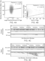
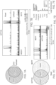
- the random nicking activity of DNase I (which is not considered a nicking enzyme herein) can be compared with a sequence-specific nicking enzyme such as Nt.CviPII and its impact on open chromatin identification.
- Nt.CviPII recognizes the CCD trinucleotide sequence and these sites are densely dispersed throughout the human genome.
- the open chromatin is readily accessible by the nicking enzyme.
- DNase I sequencing bookmarks the ends of the open chromatin
- the nicking enzyme based methods described herein actually capture the open chromatin sites between nucleosomes.
- a composition comprising: a nicking enzyme, a polymerase, four dNTPs and a labeled dNTP is provided.
- the composition may further comprise chromatin.
- the chromatin may comprise open chromatin and closed chromatin, and at least some of the open chromatin may be labeled by the labeled nucleotide.
- the composition may comprise an isolated nucleus, and the chromatin is contained within an isolated nucleus.
- the composition may comprise a permeabilized cell, wherein the chromatin is contained in the permeabilized cell.
- the cell may be a fixed cell. In other embodiments, the cell may be an unfixed cell.
- the composition may comprise a clinical sample, e.g., a tumor biopsy.
- the labeled nucleotide may comprise a detectable label, e.g., a fluorophore.
- the labeled nucleotide may comprise an affinity tag such as a biotin moiety.
- the composition may comprise: an enzyme selected from a nicking enzyme and DNase I.
- the nicking enzyme is methylation-dependent.
- the nicking enzyme may nick at methylated CpGs.
- the nicking enzyme is methylation-sensitive. Methylation-dependent and methylation-sensitive nicking enzymes are known in the art, and examples are provided herein.
- the method may comprise: (a) obtaining a sample comprising chromatin; (b) reacting the sample with a nicking enzyme, a polymerase and a labeled nucleotide to selectively label the open chromatin in the sample; and (c) analyzing the labeled sample of (b).
- the analyzing is done by: (i) detecting an optically-detectable signal from the sample, if the labeled nucleotide comprises an optically detectable label; and/or (ii) enriching for and then sequencing fragments that comprise the labeled nucleotide, if the labeled nucleotide comprises an affinity tag.
- the labeled nucleotide comprises an optically detectable label and the analyzing step of (c) is done by microscopy to produce an image of at least part of the sample.
- the labeled nucleotide may comprise an affinity tag and the analyzing step of (c) includes: i. fragmenting the DNA in the sample, ii. enriching for fragments that contain the labeled nucleotide and iii. sequencing the enriched fragments.
- the nicking enzyme is methylation-dependent.
- the nicking enzyme may nick at methylated CpNs including 5m CpG.
- the nicking enzyme is methylation-sensitive. Methylation-dependent and methylation-sensitive nicking enzymes are known in the art, and examples are provided herein.
- the sample may comprise an isolated nucleus, and the chromatin is contained within an isolated nucleus.
- the sample may comprise a permeabilized cell, and the chromatin is contained in the permeabilized cell.
- the cell is a fixed cell. In other embodiments, the cell is an unfixed cell.
- the composition may comprise a clinical sample, where the cell is in the clinical sample. The clinical sample is a tumor biopsy.
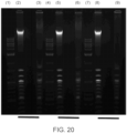
- Input DNA (lanes 2, 5 and 8) is a mixture of human genomic DNA and unmethylated synthetic DNA of various sizes.
- the bright band on the gel is human DNA and the smaller bands are the synthetic DNA of varying sizes.
- Unbound DNA (unlabeled DNA) was obtained after biotin labeled human genomic DNA was removed by streptavidin beads. Lanes 3, 6 and 9 show that using any of the 3 tested nicking enzymes, the human genomic DNA has been removed from the input preparation so that only the synthetic unmethylated DNA remains.
- Lanes 1, 4 and 7 are DNA size ladders.
- nucleic acids are written left to right in 5' to 3' orientation; amino acid sequences are written left to right in amino to carboxy orientation, respectively.
- dNTP mixture and "four dNTPs” is intended to refer to mixture of deoxyribonucleotides that correspond to G, A, T and C that can be incorporated by a polymerase into a growing polynucleotide strand.
- a dNTP mix may contain dGTP, dATP, dTTP and dCTP as well as other deoxyribonucleotides, e.g., a labeled dNTP.
- the composition of the invention comprises all four dNTPs dGTP, dATP, dTTP and dCTP, and also a labelled dNTP.
- the method of the invention uses all four dNTPs dGTP, dATP, dTTP and dCTP, and also a labeled dNTP.
- the dNTPs may each be at a working concentration of 50 ⁇ M to 1 mM (e.g., 100 ⁇ M to 500 ⁇ M, or 150 ⁇ M to 300 ⁇ M).
- nucleotide includes dNTPs (also referred to as nucleoside triphosphates) as well as nucleic acid residues that are in a polynucleotide.
- Nucleotides include those moieties that contain not only the known purine and pyrimidine bases, but also other heterocyclic bases that have been modified. Such modifications include methylated purines or pyrimidines, acetylated purines or pyrimidines, alkylated riboses or other heterocycles.
- nucleotide includes those moieties that contain hapten or fluorescent labels and may contain not only conventional ribose and deoxyribose sugars, but other sugars as well.
- Modified nucleosides or nucleotides also include modifications on the sugar moiety, e.g., wherein one or more of the hydroxyl groups are replaced with halogen atoms or aliphatic groups, or are functionalized as ethers, amines, or the like.
- methylation-dependent is intended to refer to an enzyme that only cleaves at, adjacent or proximate to a recognition site in DNA that contains at least one methylated nucleotide, e.g., methylcytosine. These enzymes cleave single strand or double strand DNA depending on whether a subset or all nucleotides (e.g. cytosines) in the recognition site are methylated, and do not cleave DNA if the recognition sequence is unmethylated. Some methylation-dependent nicking enzymes recognize methylated CpGs.
- methylation-sensitive is intended to refer to an enzyme that only nicks at or adjacent to a recognition site that contains one or more unmethylated nucleotides, e.g., one or more unmethylated cytosines. These enzymes nick DNA if one or more nucleotides in the recognition site are unmethylated and do not nick DNA if all the nucleotides in the recognition site are methylated.
- methylation-insensitive is intended to refer to an enzyme that nicks at or adjacent to a recognition site that contains methylated or unmethylated nucleotides (e.g. cytosine or methylcytosine). These enzymes nick DNA regardless of whether any nucleotides in the recognition site are methylated.
- a "plurality” contains at least 2 members.
- a plurality of labeled nucleotides means 2 or more labeled nucleotides.
- a plurality may have at least 2, at least 5, at least 10, at least 100, at least 1000, at least 10,000, at least 100,000, at least 10 6 , at least 10 7 , at least 10 8 or at least 10 9 or more members.
- strand refers to a nucleic acid made up of nucleotides covalently linked together by covalent bonds, e.g., phosphodiester bonds.
- DNA usually exists in a double-stranded form, and as such, has two complementary strands of nucleic acid referred to herein as the "top” and “bottom” strands.
- complementary strands of a chromosomal region may be referred to as “plus” and “minus” strands, the “first” and “second” strands, the “coding” and “noncoding” strands, the “Watson” and “Crick” strands or the “sense” and “antisense” strands.
- the assignment of a strand as being a top or bottom strand is arbitrary and does not imply any particular orientation, function or structure.
- sequence refers to a method by which the identity of at least 10 consecutive nucleotides (e.g., the identity of at least 20, at least 50, at least 100 or at least 200 or more consecutive nucleotides) of a polynucleotide are obtained.
- next-generation sequencing refers to the so-called parallel sequencing-by-synthesis or sequencing-by-ligation platforms currently employed by Illumina, Life Technologies, Pacific Biosciences and Roche etc.
- Next-generation sequencing methods may also include nanopore sequencing methods or electronic-detection based methods such as Ion Torrent technology commercialized by Life Technologies.
- extending refers to the extension of a nucleic acid strand by the addition of one or more nucleotides using a polymerase.
- a polymerase may generate an oligonucleotide flap at a nick site in a double stranded DNA where all of one or two types of nucleotides in the flap are labeled.
- the flap may be a plurality of nucleotides, having a length ranging from 2 nucleotides to several hundred nucleotides. If a primer that is annealed to a nucleic acid is extended, the nucleic acid acts as a template for extension reaction.
- in vitro refers to a reaction that occurs in a vessel with isolated components, not in live cells.
- ex vivo refers to a reaction or method that is not performed on the living human or animal body.
- an ex vivo method may be performed outside the living human or animal body on a sample (e.g. a cell or tissue sample, such as a clinical sample) that has previously been obtained from the human or animal body.
- non-naturally occurring refers to a composition that does not exist in nature. Any protein described herein may be non-naturally occurring, where the term “non-naturally occurring” refers to a protein that has an amino acid sequence and/or a post-translational modification pattern that is different to the protein in its natural state. For example, a non-naturally occurring protein may have one or more amino acid substitutions, deletions or insertions at the N-terminus, the C-terminus and/or between the N- and C-termini of the protein.
- a “non-naturally occurring" protein may have an amino acid sequence that is different to a naturally occurring amino acid sequence (i.e., having less than 100% sequence identity to the amino acid sequence of a naturally occurring protein) but that is at least 80%, at least 85%, at least 90%, at least 95%, at least 97%, at least 98% or at least 99% identical to the naturally occurring amino acid sequence.
- a non-naturally occurring protein may contain an N-terminal methionine or may lack one or more post-translational modifications (e.g., glycosylation, phosphorylation, etc.) if it is produced by a different (e.g., bacterial) cell.
- a “mutant” or “variant” protein may have one or more amino acid substitutions relative to a wild-type protein and may include a “fusion” protein.
- the term “fusion protein” refers to a protein composed of a plurality of polypeptide components that are unjoined in their native state. Fusion proteins may be a combination of two, three or even four or more different proteins.
- polypeptide includes fusion proteins, including, but not limited to, a fusion of two or more heterologous amino acid sequences, a fusion of a polypeptide with: a heterologous targeting sequence, a linker, an immunologically tag, a detectable fusion partner, such as a fluorescent protein, ⁇ -galactosidase, luciferase, etc., and the like.
- a fusion protein may have one or more heterologous domains added to the N-terminus, C-terminus, and or the middle portion of the protein. If two parts of a fusion protein are "heterologous", they are not part of the same protein in its natural state.
- non-naturally occurring refers to a nucleic acid that contains: a) a sequence of nucleotides that is different to a nucleic acid in its natural state (i.e. having less than 100% sequence identity to a naturally occurring nucleic acid sequence), b) one or more non-naturally occurring nucleotide monomers (which may result in a non-natural backbone or sugar that is not G, A, T or C) and/or c) may contain one or more other modifications (e.g., an added label or other moiety) to the 5'- end, the 3' end, and/or between the 5'- and 3'-ends of the nucleic acid.
- modifications e.g., an added label or other moiety
- non-naturally occurring refers to: a) a combination of components that are not combined by nature, e.g., because they are at different locations, in different cells or different cell compartments; b) a combination of components that have relative concentrations that are not found in nature; c) a combination that lacks something that is usually associated with one of the components in nature; d) a combination that is in a form that is not found in nature, e.g., dried, freeze dried, crystalline, aqueous; and/or e) a combination that contains a component that is not found in nature.
- a preparation may contain a "non-naturally occurring" buffering agent (e.g., Tris, HEPES, TAPS, MOPS, tricine or MES), a detergent, a dye, a reaction enhancer or inhibitor, an oxidizing agent, a reducing agent, a solvent or a preservative that is not found in nature.
- a buffering agent e.g., Tris, HEPES, TAPS, MOPS, tricine or MES
- a detergent e.g., Tris, HEPES, TAPS, MOPS, tricine or MES
- a dye e.g., Tris, HEPES, TAPS, MOPS, tricine or MES
- a reaction enhancer or inhibitor e.g., an oxidizing agent, a reducing agent, a solvent or a preservative that is not found in nature.
- nicking refers to a reaction that breaks the phosphodiester bond between two nucleotides in one strand of a double-stranded DNA molecule to produce a 3' hydroxyl group and a 5' phosphate group.
- nick site refers to the site at which a double-stranded DNA molecule has been nicked.
- nicking enzyme refers to a site specific enzyme that cleaves (e.g. nicks) one strand (either the top or bottom strands, but not both strands) of a double-stranded nucleic acid at a nonrandom position in the DNA. In some cases a nicking enzyme will nick the bottom or top strand at a specific sequence on the nucleic acid.
- nicking enzymes useful in the compositions and methods of the invention which may be methylation-dependent, methylation-sensitive, or methylation-insensitive, are known in the art and various examples are provided herein.
- Nb.Bsml, Nb.BbvCl, Nb.BsrDI, Nb.Btsl, Nt.BbvCl, Nt.Alwl, Nt. CviPII, Nt. BsmAl, Nt. Alwl and Nt.BstNBI are examples of naturally occurring nicking enzymes that are not 5-methylcytosine dependent.
- Nicking enzymes that have been engineered from Typells restriction enzymes (e.g., Alwl, Bpu10I, BbvCl, Bsal, BsmBI, BsmAl, Bsml, BspQl, Mlyl, Mva1269I and Sapl, etc.) and methods of making nicking enzymes can be found in references for example, US 7,081,358 ; US 7,011,966 ; US 7,943,303 ; US 7,820,424 .
- Typells restriction enzymes e.g., Alwl, Bpu10I, BbvCl, Bsal, BsmBI, BsmAl, Bsml, BspQl, Mlyl, Mva1269I and Sapl, etc.
- Labeling of closed chromatin may occur using methylation dependent nicking enzymes that preferably favor 5m CpG sites.
- Selective labeling of open chromatin preferentially uses methylation independent or methylation sensitive nicking enzymes, such as exemplified herein.
- Nicking enzymes that are methylation-dependent include, but are not limited to: Nhol (GSmCNGSmC); Bisl (GSmCNGSmC) ( Chmuzh, et al., Biotekhnologiya 3: 22-26 (2005 ); Pam 7902I (GSmCNNGSmC); N.BceSVIII; and Nb.LpnPI (C5mCDG(N) 10 /GGHmC(N) 14 , nicking of the bottom strand) ( Cohen Karni, et al., Proc. Natl. Acad. Sci. U. S. A. 108: 11040-11045 (2011 ); Xu et al, Sci. Rep. 6:28579 (2016 )).
- Methylation-dependent nicking enzymes can be produced using the methods described in, Gutjahr, et al., Nucleic Acids Res. 42:e77 (2014 ) and Xu, et al., Sci. Rep. 6:28579 (2016 ).
- N. Gamma is a strand-specific and site-specific DNA nicking enzyme that cleaves at (YCG ⁇ GT or AC ⁇ CGR).
- Nb.LpnPI can be made by making an R335A mutation in the sequence LpnPI sequence defined by Genbank accession number AAU27318.1.
- Other nicking enzymes can be made by making an Arg to Ala substitution at the position corresponding to position 335 in LpnPl. Such enzymes may cleave at a methylated CpG.
- the amino acid sequences of N. LpnPI as well as other methylation-specific nicking enzymes is shown below:
- nicking enzymes can be found in a variety of publications (e.g., Bellamy, et al. J. Mol. Biol. 2005 345, 641-653 ; Heiter, et al., J. Mol. Biol. 2005 348, 631-640 ; Xu, et al., Proc. Natl. Acad. Sci. USA 2001 98, 12990-12995 ; Samuelson, et al., Nucl. Acids Res. 2004 32, 3661-3671 ; Zhu, et al., J. Mol. Biol. 2004 337, 573-583 ; Morgan, et al., Biol. Chem. 2000 381, 1123-1125 ; Chan, Nucl.
- DNase I is not a nicking enzyme because DNase I cleaves DNA at random positions.
- the term nicking enzyme specifically excludes DNase I.
- a nicking enzyme can also be made by inactivating one of the catalytic domains.
- a programmable endonuclease e.g., Cas9 or a functional equivalent thereof (such as Argonaute or Cpf1).
- Cas9 contains two catalytic domains, RuvC and HNH. Inactivating one of those domains will generate a nicking enzyme.
- the RuvC domain can be inactivated by an amino acid substitution at position D10 (e.g., D10A) and the HNH domain can be inactivated by an amino acid substitution at position H840 (e.g., H840A), or at a position corresponding to those amino acids in other proteins.
- Such endonucleases may be Argonaute or Type II CRISPR/Cas endonucleases that are composed of two components: a nuclease (e.g., a Cas9 or Cpf1 endonuclease or variant or ortholog thereof) that cleaves the target DNA and a guide nucleic acid e.g., a guide DNA or RNA that targets the nuclease to a specific site in the target DNA (see, e.g., Hsu, et al., Nature Biotechnology 2013 31: 827-832 ).
- a nuclease e.g., a Cas9 or Cpf1 endonuclease or variant or ortholog thereof
- a guide nucleic acid e.g., a guide DNA or RNA that targets the nuclease to a specific site in the target DNA
- a nicking enzyme can also be made by fusing a site specific DNA binding domain such as the DNA binding domain of a DNA binding protein (e.g., a restriction endonuclease, a transcription factor, or another domain that binds to DNA at non-random positions) with a nuclease or deaminase so that it acts on a non-random site.
- a DNA binding protein e.g., a restriction endonuclease, a transcription factor, or another domain that binds to DNA at non-random positions
- the deaminase can introduce a uracil
- a nick can be created by removing the uracil using a deglycosylase and treating the abasic site with an AP endonuclease.
- non-random cleavage by a nicking enzyme results from recognition sites within the nicking enzyme or from guide molecules that direct the nicking enzyme to a non-random site or optionally by inherent defined bias of the enzyme for a plurality of nucleotides that may be preferentially As and Ts or Gc and Cs.
- chromatin refers to a complex of molecules including proteins and genomic DNA as found in a nucleus of a eukaryotic cell. Chromatin is composed in part of histone proteins that form nucleosomes, genomic DNA, and other DNA binding proteins (e.g., transcription factors) that are bound to the genomic DNA. Chromatin is therefore distinct from purified genomic DNA. Chromatin is available in permeabilized cells, in isolated nuclei, and as well as in isolated chromatin.
- open chromatin and “closed chromatin” refer to the level of the accessibility of genomic DNA in a sample that contains chromatin.
- Open chromatin or “euchromatin”
- closed chromatin or “heterochromatin”
- Open and closed chromatins are schematically illustrated in FIG. 1 .
- isolated nucleus refers to a nucleus that has been isolated from other components of a cell, e.g., from the cytoplasm and plasma membrane, by centrifugation or another technique.
- permeabilized cell refers to a cell that has a cell plasma membrane and, in some cases a nuclear membrane, that have been permeabilized, e.g., by a detergent.
- fixed cell refers to a cell that has been treated with a crosslinking or non-crosslinking fixative, e.g., formaldehyde or paraformaldehyde, acetone, or methanol or the like.
- a fixed cell may be "formalin fixed", in which case it may be fixed in formaldehyde (e.g., 3%-5% formaldehyde in phosphate buffered saline) or Bouin solution.
- An "unfixed” cell refers to a cell that has not been treated by such an agent.
- labeled nucleotide refers to a modified nucleotide that has an optically detectable label or an affinity tag attached thereto.
- optically detectible label refers to a light-emitting or fluorescent label that can be detected using a light detector, e.g., a microscope.
- Light emitting labels include fluorophores, although others are known.
- affinity tag refers to a tag that can be used to separate a molecule to which the affinity tag is attached from other molecules that do not contain the affinity tag.
- an affinity tag is a member of a specific binding pair, i.e. two molecules where one of the molecules through chemical or physical means specifically binds to the other molecule.
- the complementary member of the specific binding pair which can be referred to herein as a “capture agent” may be immobilized (e.g., to a chromatography support, a bead or a planar surface) to produce an affinity chromatography support that specifically binds the affinity tag.
- Affinity tags include a biotin moiety (e.g., biotin, desthibiotin, oxybiotin, 2-iminobiotin, diaminobiotin, biotin sulfoxide, biocytin, etc.) which can bind to streptavidin.
- Affinity tags also include chemoselective groups such as azido and alkynyl groups, which can participate in a copper-free cycloadition reaction (see, e.g., Kolb, et al., Drug Discov Today 2003 8: 1128-113 and Baskin, et al., Proc. Natl. Acad. Sci. 2007 104: 16793-16797 ).
- enriching refers to a method step in which some components of a sample (e.g., components that are tagged) are separated from other components in the sample (e.g., components that are not tagged).
- barcode sequence refers to a unique sequence of nucleotides used to (a) identify and/or track the source of a polynucleotide in a reaction and/or (b) count how many times an initial molecule is sequenced (e.g., in cases where substantially every molecule in a sample is tagged with a different sequence, and then the sample is amplified).
- a barcode sequence may be at the 5'-end, the 3'-end or in the middle of an oligonucleotide, or both the 5' end and the 3' end.
- Barcode sequences may vary widely in size and composition; the following references provide guidance for selecting sets of barcode sequences appropriate for particular embodiments: Brenner, US 5,635,400 ; Brenner et al, Proc. Natl. Acad. Sci., 97: 1665-1670 (2000 ); Shoemaker, et al, Nature Genetics, 14: 450-456 (1996 ); Morris et al, European patent publication 0799897A1 ; Wallace, US 5,981,179 ; and the like.
- a barcode sequence may have a length in range of from 4 to 36 nucleotides, or from 6 to 30 nucleotides, or from 8 to 20 nucleotides.
- reacting refers to combining under conditions (e.g., a suitable temperature, time and conditions) that result in a reaction, e.g., nicking and/or strand extension by a polymerase.
- NE-seq refers to embodiments in which nuclei are examined for open chromatin by adding a nicking enzyme to the nuclei, permitting open chromatin to be nicked, and with a strand-displacing polymerase having activity, causing a plurality of labeled nucleotides to be incorporated at the nick site by newly synthesized strand, enrichment of the labeled polynucleotides, followed by library construction, amplification, and sequencing.
- a composition comprising a nicking enzyme, a polymerase, four dNTPs and at least one labeled dNTP is provided for sequencing of open chromatin.
- the composition may alternatively comprise a mixture of a nicking enzyme, four dNTPs, and at least one labeled dNTP.
- the polymerase is provided separately from the nicking enzyme, dNTPs, and at least one labelled dNTP (e.g. in a separate composition).
- a nicking enzyme may be provided for visualizing open chromatin in a histological preparation of a cell biopsy. In one embodiment, therefore, the composition is for visualizing open chromatin in a histological preparation of a tissue biopsy.
- the composition is for detecting open chromatin. In one embodiment, the composition is for analyzing chromatin.
- the components of the composition may be dissolved in an aqueous solution that may comprise a buffering agent, such as a non-naturally occurring buffering agent, and other essential compounds required for activity of the enzymes in the composition.
- the composition may contain other components, e.g., glycerol.
- the concentration of the one or more labeled dNTP in the composition may be in the range of 3 ⁇ M or 5 ⁇ M to 200 ⁇ M.
- the nicking enzyme may be methylation-sensitive or methylation-dependent.
- the molar ratio of the labeled dNTP to the unlabeled version of the same dNTP (e.g. biotin-dCTP to dCTP) in the composition may be in the range of 1:1000 to 1000:1, e.g., 1:100 to 100:1 or 1:10 to 10:1.
- the ratio the molar ratio of the labeled dNTP to the corresponding unlabeled dNTP (e.g. biotin-dCTP to dCTP) in the nucleotide mix may be in the range of 1:1000 to 1:100, 1:100 to 1:10, 1:10 to 1:1, 1:1 to 1:10, 1:10 to 1:100, or 1:100 to 100:1000.
- the nicking enzyme used in some embodiments of the method may be from any source and, in some embodiments, a plurality of nicking enzymes may be used. Examples are provided above for analyzing open chromatin or closed chromatin.
- the polymerase employed in the method may be a nick translation polymerase.
- Nick translation occurs when the polymerase associated flap endonuclease chews up one strand of the DNA as the polymerase progresses in the 5'- 3' direction, essentially replacing the existing DNA strand. Strand displacement displaces the strand ahead of the replication fork without destroying it. Either activity could be employed here.
- DNA Polymerase I, Taq, and the wild type Bst DNA polymerase have a 5'-3' flap endonuclease activity and will do nick translation. Phi29 or Klenow fragment or Bst large fragment would do strand displacement.
- T4 and T7 DNA polymerases have neither strand displacement or nick translation abilities, but can be used in the chewback/fill-in approach described here.
- the polymerase and nick translation move in the 5' to 3' direction starting from a nick site.
- the polymerase could be a proofreading polymerase that removes one or more nucleotides in the 3' to 5' direction starting from a nick site, and then fills in the overhang it has just generated.
- the polymerase may or may not have strand displacement activity.
- the polymerase is a strand-displacing polymerase. Examples of strand-displacing polymerases are known in the art.
- DNA polymerases useful in the invention include E. coli DNA polymerase I, E. coli DNA polymerase I Large Fragment (Klenow fragment), phage T4 DNA polymerase, or phage T7 DNA polymerase.
- the polymerase can be a thermophilic polymerase such as Thermus aquaticus (Taq) DNA polymerase, Thermus flavus (Tfl) DNA polymerase, Thermus Thermophilus (Tth) DNA polymerase, Thermococcus aggregans (Tag) DNA polymerase, Thermococcus litoralis (Tli) DNA polymerase, Pyrococcus furiosus (Pfu) DNA polymerase, Vent DNA polymerase, or Bacillus stearothermophilus (Bst) DNA polymerase.
- Taq Thermus aquaticus
- Tfl Thermus flavus
- Tth Thermus Thermophilus
- Tth Thermococcus aggregans
- Tli Thermococcus litoralis
- Pfu Pyrococcus furiosus
- Vent DNA polymerase or Bacillus stearothermophilus
- the one or more labeled dNTPs in the composition may vary.
- the composition may comprise any one or combination of labeled dGTP, labeled dATP, labeled dTTP, and labeled dCTP.
- the composition comprises a combination of labeled dATP and labeled dCTP.
- the labeled nucleotides could be chain terminator nucleotides.
- the labeled dNTP comprises an affinity tag.
- the affinity tag may comprise an azide/alkyne group (which are suitable for conjugation to another moiety, e.g., a solid support via click chemistry), or a biotin group so that the labeled nucleic acid can be enriched on a suitable support and sequenced.
- the labeled dNTP comprises an optically detectable label. If the labeled dNTP comprises an optically detectable label, the label may comprise a fluorophore (or any other type of optically detectable moiety). Fluorophores of interest include xanthene dyes, e.g.
- fluorescein and rhodamine dyes such as fluorescein isothiocyanate (FITC), 6-carboxyfluorescein (commonly known by the abbreviations FAM and F),6-carboxy-2',4',7',4,7-hexachlorofluorescein (HEX), 6-carboxy-4', 5'-dichloro-2', 7'-dimethoxyfluorescein (JOE or J), N,N,N',N'-tetramethyl-6-carboxyrhodamine (TAMRA or T), 6-carboxy-X-rhodamine (ROX or R), 5-carboxyrhodamine-6G (R6G5 or G5), 6-carboxyrhodamine-6G (R6G6 or G6), and rhodamine 110; cyanine dyes, e.g.
- Cy3, Cy5 and Cy7 dyes include coumarins, e.g umbelliferone; benzimide dyes, e.g. Hoechst 33258; phenanthridine dyes, e.g. Texas Red; ethidium dyes; acridine dyes; carbazole dyes; phenoxazine dyes; porphyrin dyes; polymethine dyes, e.g. cyanine dyes such as Cy3, Cy5, etc.; BODIPY dyes and quinoline dyes.
- fluorophores of interest that are commonly used in subject applications include: Pyrene, Coumarin, Diethylaminocoumarin, FAM, Fluorescein Chlorotriazinyl, Fluorescein, R110, Eosin, JOE, R6G, Tetramethylrhodamine, TAMRA, Lissamine, ROX, Napthofluorescein, Texas Red, Napthofluorescein, Cy3, and Cy5, etc. (Amersham Inc., Piscataway, NJ). Suitable fluorescent labels may be listed in Kricka, et al., Ann Clin Biochem. 39:114-29, 2002 . Such nucleotides are commercially available from a variety of vendors.
- the cell may be permeabilized to allow access of the other components of the composition to the chromatin.
- the permeabilization can be performed in a way to minimally perturb the nuclei in the cell sample.
- the cells can be permeabilized using a permeabilization agent.
- permeabilization agents include, but are not limited to, NP40, digitonin, tween, streptolysin, and cationic lipids.
- the cell sample can be permeabilized using hypotonic shock and/or ultrasonication.
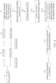
- FIG. 1A-1C An overview of an embodiment of the method is shown in FIG. 1A-1C .
- the nicking enzyme Nt.CviPII, DNA polymerase I, and combination of biotin-dATP/dCTP illustrated in FIG. 1A-1C are not intended to be limiting, and any nicking enzyme, polymerase, and labelled dNTP(s) described herein may be used in the method of the invention.
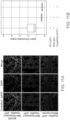
- FIG. 2A-2B shows that the method works with nuclei obtained both fixed and unfixed cells.
- the minimum number of cells that can be used in the method is 10, e.g., in some embodiments the number of cells that can be used in the method is as few as 25 cells or more, as few as 50 cells or more, at least 50, at least 100, or at least 250 cells.
- the sample comprises fewer than 500 cells, fewer than 250 cells, fewer than 200 cells, fewer than 100 cells, fewer than 50 cells, or fewer than 25 cells. In some embodiments, therefore, the method is performed on chromatin obtained from as few as 1, 5, 10, 25, 50, 100 or 250 cells.
- the chromatin in the sample is from fewer than 500 cells, fewer than 250 cells, fewer than 200 cells, fewer than 100 cells, fewer than 50 cells, or fewer than 25 cells. In certain embodiments, chromatin from a single cell may be analyzed.
- the sample may be optionally stained with other antibodies/stains before imaging.
- the stain may enhance contrast or imaging of intracellular or extracellular structures.
- the sample may be stained with DAPI, ethidium bromide, Hoechst 33258, Hoechst 33342, eosin, hematoxylin, methylene blue, or rhodamine.
- the sample may be stained with DAPI.
- the sample may be stained with haematoxylin and eosin (H&E).
- the tissue sections may be immunohistochemically stained using standard protocols and optimized as necessary for each primary antibody, using standard processes.
- markers that can be investigated in these embodiments are well known.
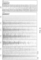
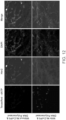
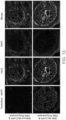
- An example of one implementation of such a method is shown in FIG. 9 (wherein the use of Texas Red-5-dATP/dCTP is not intended to be limiting, and can be replaced by any other optically-detectably labeled nucleotides).
- the optically detectable label can be detected using any suitable means. For example, fluorescence can be viewed with a fluorescence microscope and an appropriate filter for each fluorophore, or by using dual or triple band-pass filter sets to observe multiple fluorophore ( See, e.g., US 5,776,688 ).
- the images may be overlaid or compared and analyzed to identify the boundaries of individual cells, and/or subcellular features in individual cells, in the image.
- the stained cells could be analyzed by flow cytometry (e.g., FACS).
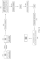
- the sample may be processed using the method shown in FIG. 3 (wherein the use of biotin-dATP/dCTP and streptavidin beads is not intended to be limiting, and can be replaced by any other affinity-tagged nucleotides and corresponding capture moiety, optionally linked to a solid phase, not limited to a bead).
- the method comprises reverse cross-linking and isolating labelled genomic DNA, and then fragmenting the isolated labelled genomic DNA.
- the reverse crosslinking may be done using heat or by chemical treatment, and genomic DNA may be fragmented to any convenient size (e.g., a median size in the range of 100bp to 1 kb, e.g., 150bp to 500bp).
- the method may further comprise processing the labelled genomic DNA fragments by performing end repair, dA-tailing, and/or adapter ligation, which may be done using conventional methods. Any of the adaptors and/or primers may carry a barcode to facilitate multiplexing and/or molecular counting.
- the method then comprises enriching the labeled genomic DNA using any convenient method - e.g. by capturing the labelled, processed genomic DNA fragments.
- the labelled, processed genomic DNA fragments may be enriched by capturing using a streptavidin-coated solid phase such as a streptavidin-coated bead.
- the enriched DNA may then be amplified using any convenient method.
- the enriched DNA is then analyzed and/or sequenced.
- the enriched sequences may be sequenced using any convenient method.
- the fragments may be sequenced using Illumina's reversible terminator method, Roche's pyrosequencing method (454), Life Technologies' sequencing by ligation (the SOLiD platform), Life Technologies' Ion Torrent platform or Pacific Biosciences' fluorescent base-cleavage method, etc.
- the sequence read obtained from the sequencing step may be mapped onto a genomic sequence to obtain a map of the open and closed chromatin.
- an abundant sequence indicates that the region corresponding to that sequence is in open chromatin.
- Such a map can show one or more of the following: a profile of chromatin accessibility along a region; DNA binding protein (e.g., transcription factor) occupancy for a site in a region; nucleosome-free DNA in a region; positioning of nucleosomes along a region; or a profile of chromatin states along a region, for example.
- the map can also be annotated with sequence information, and information about the sequence (e.g., the positions of promoters, introns, exons, known enhancers, transcriptional start sites, untranslated regions, terminators, etc.) so that the chromatin accessibility map can be viewed in context with the annotation.
- sequence information e.g., the positions of promoters, introns, exons, known enhancers, transcriptional start sites, untranslated regions, terminators, etc.
- the nuclei containing chromatin can be from any source.
- the nuclei may be obtained from a culture of cells, e.g., a cell line.
- the cells may be isolated from an individual (e.g., a patient).
- the cells may be isolated from a soft tissue or from a bodily fluid, or from a cell culture that is grown in vitro.
- the nuclei may be isolated from a soft tissue such as brain, adrenal gland, skin, lung, spleen, kidney, liver, spleen, lymph node, bone marrow, bladder, stomach, small intestine, large intestine or muscle, etc.
- Bodily fluids include blood, plasma, saliva, mucous, phlegm, cerebral spinal fluid, pleural fluid, tears, lacteal duct fluid, lymph, sputum, cerebrospinal fluid, synovial fluid, urine, amniotic fluid, and semen, etc.
- the chromatin may from a tissue biopsy obtained from a patient.
- Biopsies of interest include both tumor and non-neoplastic biopsies of skin (melanomas, carcinomas, etc.), soft tissue, bone, breast, colon, liver, kidney, adrenal, gastrointestinal, pancreatic, gall bladder, salivary gland, cervical, ovary, uterus, testis, prostate, lung, thymus, thyroid, parathyroid, pituitary (adenomas, etc.), brain, spinal cord, ocular, nerve, and skeletal muscle, etc.
- the chromatin analyzed in the method may be from blood cells, wherein "blood cells” refers to a sample of whole blood or a sub-population of cells in whole blood.
- Sub-populations of cells in whole blood include platelets, red blood cells (erythrocytes), platelets and white blood cells (i.e., peripheral blood leukocytes, which are made up of neutrophils, lymphocytes, eosinophils, basophils and monocytes).
- white blood cells can be further divided into two groups, granulocytes (which are also known as polymorphonuclear leukocytes and include neutrophils, eosinophils and basophils) and mononuclear leukocytes (which include monocytes and lymphocytes). Lymphocytes can be further divided into T cells, B cells and NK cells. Peripheral blood cells are found in the circulating pool of blood and not sequestered within the lymphatic system, spleen, liver, or bone marrow. Other cells are present in blood that can be isolated. If blood is first contacted with an agent and then a sample of the blood is used in an assay, then a portion or all of the contacted blood may be used in the assay.
- granulocytes which are also known as polymorphonuclear leukocytes and include neutrophils, eosinophils and basophils
- mononuclear leukocytes which include monocytes and lymphocytes
- Lymphocytes can be further divided into T
- the cell sample can be isolated directly from a primary source.
- the cell sample can be isolated directly from fresh tissues.
- the cell sample can be isolated directly from frozen tissues.
- the cell sample can be isolated directly from fixed tissues.
- the disease state in a subject can be analyzed.
- the method can be used to compare two samples to identify a change in chromatin structure.
- the method may comprise analyzing a first population of cells using the above-described method to produce a data set and optionally a first epigenetic map; and analyzing a second population of cells using the above-described method to produce a second data set and optionally a second epigenetic map; and comparing the first data set or epigenetic map to the second data set or epigenetic map, e.g., to see if there are any changes in chromatin openness or transcription factor occupancy, for example.
- the first population of cells and the second population of cells may be collected from the same individual at different times. In other embodiments, the first population of cells and the second population of cells are different populations of cells collected from tissues or different individuals.
- Exemplary cell types that can be used in the method include, for example, cells isolated from a tissue biopsy (e.g., from a tissue having a disease such as colon, breast, prostate, lung, skin cancer, or infected with a pathogen etc.) and normal cells from the same tissue, e.g., from the same patient; cells grown in tissue culture that are immortal (e.g., cells with a proliferative mutation or an immortalizing transgene), infected with a pathogen, or treated (e.g., with environmental or chemical agents such as peptides, hormones, altered temperature, growth condition, physical stress, cellular transformation, etc.), and normal cells (e.g., cells that are otherwise identical to the experimental cells except that they are not immortalized, infected, or treated, etc.); cells isolated from a mammal with a cancer, a disease, a geriatric mammal, or a mammal exposed to a condition, and cells from a mammal of the same species, e.g.,
- cells of different types e.g., neuronal and non-neuronal cells, or cells of different status (e.g., before and after a stimulus on the cells) may be compared.
- the experimental material is cells susceptible to infection by a pathogen such as a virus, e.g., human immunodeficiency virus (HIV), etc.
- the control material is cells resistant to infection by the pathogen.
- the sample pair is represented by undifferentiated cells, e.g., stem cells, and differentiated cells. Cells from yeast, plants and animals, such as fish, birds, reptiles, amphibians and mammals may be used in the subject methods.
- mammalian cells i.e., cells from mice, rabbits, primates, or humans, or cultured derivatives thereof, may be used.
- the method may be used to identify the effect of a test agent, e.g., a drug, or to determine if there are differences in the effect of two or more different test agents.
- a test agent e.g., a drug
- two or more identical populations of cells may be prepared and, depending on how the experiment is to be performed, one or more of the populations of cells may be incubated with the test agent for a defined period of time. After incubation with the test agent, the chromatin of the populations of cells can be analyzed using the methods set forth above, and the results can be compared.
- the cells may be blood cells, and the cells can be incubated with the test agent ex vivo. These methods can be used to determine the mode of action of a test agent, to identify changes in chromatin structure or transcription factor occupancy in response to the drug, for example.
- the method described above may also be used as a diagnostic (which term is intended to include methods that provide a diagnosis as well as methods that provide a prognosis). Diagnostic and prognostic methods may be performed ex vivo, on a sample of chromatin obtained from a subject or patient. These methods may comprise, e.g., analyzing chromatin from a patient using the method described above to produce results; and providing a diagnosis or prognosis based on the results.
- the method set forth herein may be used to provide a reliable diagnostic to any condition associated with altered chromatin.
- the method can be applied to the characterization, classification, differentiation, grading, staging, diagnosis, or prognosis of a condition characterized by a chromatin alteration.
- the method can be used to determine whether the chromatin in a sample from an individual suspected of being affected by a disease or condition is the same as or different to a sample that is considered "normal" with respect to the disease or condition.
- the method can be directed to diagnosing an individual with a condition that is characterized by altered chromatin at a particular locus in a test sample, where the pattern is correlated with the condition.
- the methods can also be used for predicting the susceptibility of an individual to a condition.
- Exemplary conditions that are suitable for analysis using the methods set forth herein can be, for example, cell proliferative disorder or predisposition to cell proliferative disorder; metabolic malfunction or disorder; immune malfunction, damage or disorder; CNS malfunction, damage or disease; symptoms of aggression or behavioral disturbance; clinical, psychological and social consequences of brain damage; psychotic disturbance and personality disorder; dementia or associated syndrome; cardiovascular disease, malfunction and damage; malfunction, damage or disease of the gastrointestinal tract; malfunction, damage or disease of the respiratory system; lesion, inflammation, infection, immunity and/or convalescence; malfunction, damage or disease of the body as an abnormality in the development process; malfunction, damage or disease of the skin, the muscles, the connective tissue or the bones; endocrine and metabolic malfunction, damage or disease; headache or sexual malfunction, and combinations thereof.
- the method can provide a prognosis, e.g., to determine if a patient is at risk for recurrence.
- Cancer recurrence is a concern relating to a variety of types of cancer.
- the prognostic method can be used to identify surgically treated patients likely to experience cancer recurrence so that they can be offered additional therapeutic options, including preoperative or postoperative adjuncts such as chemotherapy, radiation, biological modifiers and other suitable therapies.
- the methods are especially effective for determining the risk of metastasis in patients who demonstrate no measurable metastasis at the time of examination or surgery.
- the method can also be used to determining a proper course of treatment for a patient having a disease or condition, e.g., a patient that has cancer.
- a course of treatment refers to the therapeutic measures taken for a patient after diagnosis or after treatment.
- a determination of the likelihood for recurrence, spread, or patient survival can assist in determining whether a more conservative or more radical approach to therapy should be taken, or whether treatment modalities should be combined.
- cancer recurrence it can be advantageous to precede or follow surgical treatment with chemotherapy, radiation, immunotherapy, biological modifier therapy, gene therapy, vaccines, and the like, or adjust the span of time during which the patient is treated.
- a lab will receive a sample (e.g., blood) from a remote location (e.g., a physician's office or hospital), the lab will analyze cells in the sample as described above to produce data, and the data may be forwarded to the remote location for analysis.
- a sample e.g., blood
- a remote location e.g., a physician's office or hospital
- Nicking enzymes used in the methods described above for epigenome analysis may also be used to differentiate eukaryotic DNA from prokaryotic DNA including mitochondria and chloroplast DNA. This can be achieved by relying on the substantially greater density of methylated nucleotides in eukaryotic DNA than in prokaryotic DNA.
- a nicking enzyme that has a specificity for a recognition site that contains 5m CpG such as Pam 7902 I, Lpnl, MspJl (New England Biolabs, Ipswich, MA), Nhol and Bisl can be used on DNA in a body fluid or cells from an invertebrate (such as a mosquito) or vertebrate (such as a human) for introducing labeled nucleotides into the methylated DNA so that it can be enriched by binding to an affinity matrix if the label is an affinity tag such as biotin.
- Unmethylated prokaryotic DNA will be substantially unlabeled and thus will not bind the affinity column and can be separated from the eukaryotic DNA in the eluent.
- human DNA can be separated from microbial sequences in body fluid and tissue samples (such as saliva, feces, etc.) for mutation, epimutation and genomics analysis.
- tissue samples such as saliva, feces, etc.
- Obtaining saliva for genomic analyses is a popular collection method with less invasiveness compared to blood draws or biopsies.
- a major drawback of saliva DNA is the non-human DNA contamination particularly of oral microbiome.
- a 5m C specific nicking enzyme e.g. Pam 7902 I, Nhol, N. Lpnpl, Bisl and MspJl
- human genome for example, with biotin
- FIG. 20 is then enriched (for example, using streptavidin beads) for sequence analysis.
- the analysis of chromatin is performed on isolated nuclei. Therefore, amounts of mitochondrial DNA are minimal at the starting point of the method. If the frequency of nicking enzyme recognition sites (e.g. CCD sites) in the mitochondrial DNA is very high and the DNA is devoid of histones, any residual mitochondrial DNA would be substantially fragmented.
- nicking enzyme recognition sites e.g. CCD sites
- Embodiment 1 A composition comprising: a nicking enzyme, a polymerase and a labeled nucleotide.
- Embodiment 2 The composition of embodiment 1, further comprising chromatin.
- Embodiment 3 The composition of embodiment 2, wherein the chromatin comprises open chromatin and closed chromatin, and at least some of the open chromatin is labeled by the labeled nucleotide.
- Embodiment 4 The composition of any of embodiments 2-3, wherein the composition comprises an isolated nucleus, and the chromatin is contained within an isolated nucleus.
- Embodiment 5 The composition of any of embodiments 2-3, wherein the composition comprises a permeabilized cell, wherein the chromatin is contained in the permeabilized cell.
- Embodiment 6 The composition of embodiment 5, wherein the cell is a fixed cell.
- Embodiment 7 The composition of embodiment 5, wherein the cell is an unfixed cell.
- Embodiment 8 The composition of any of embodiments 5-7, wherein the composition comprises a clinical sample, wherein the cell is in the clinical sample.
- Embodiment 9 The composition of embodiment 8, wherein the clinical sample is a tumor biopsy.
- Embodiment 10 The composition of any prior embodiment, wherein the labeled nucleotide comprises a detectable label.
- Embodiment 11 The composition of any prior embodiment, wherein the detectable label is a fluorophore.
- Embodiment 12 The composition of any of embodiments 1-9, wherein the labeled nucleotide comprises an affinity tag.
- Embodiment 13 The composition of embodiment 12, wherein the labeled nucleotide comprises a biotin moiety.
- Embodiment 13.1 The composition of any prior embodiment, wherein the nicking enzyme is methylation-dependent.
- Embodiment 13.2 The composition of any prior embodiment, wherein the nicking enzyme is methylation-sensitive.
- Embodiment 14 A method for detecting open chromatin, comprising: (a) obtaining a sample comprising chromatin; (b) reacting the sample with a nicking enzyme, a polymerase and a labeled nucleotide to selectively label the open chromatin in the sample; and (c) analyzing the labeled sample of (b), wherein the analyzing is done by: (i) detecting an optically-detectable signal from the sample, if the labeled nucleotide comprises an optically detectable label; or (ii) enriching for and then sequencing fragments that comprise the labeled nucleotide, if the labeled nucleotide comprises an affinity tag.
- Embodiment 15 The method of embodiment 14, wherein the labeled nucleotide comprises an optically detectable label and the analyzing step of (c) is performed by microscopy to produce an image of at least part of the sample.
- Embodiment 16 The method of embodiment 14, wherein the labeled nucleotide comprises an affinity tag and the analyzing step of (c) is performed by: i. fragmenting the DNA in the sample, ii. enriching for fragments that contain the labeled nucleotide and iii. sequencing the enriched fragments.
- Embodiment 17 The method of any of embodiments 14-17, wherein the sample comprises an isolated nucleus, and the chromatin is contained within an isolated nucleus.
- Embodiment 18 The method of any of embodiments 14-17, wherein the composition comprises a permeabilized cell, wherein the chromatin is contained in the permeabilized cell.
- Embodiment 19 The method of embodiment 18, wherein the cell is a fixed cell.
- Embodiment 20 The method of embodiment 18, wherein the cell is an unfixed cell.
- Embodiment 21 The method of any of embodiments 14-20, wherein the composition comprises a clinical sample, wherein the cell is in the clinical sample.
- Embodiment 22 The method of embodiment 21, wherein the clinical sample is a tumor biopsy.
- Embodiment 23 The method of any prior method embodiment, wherein the nicking enzyme is methylation-sensitive.
- Embodiment 24 The method of any prior method embodiment, wherein the nicking enzyme is methylation-dependent.
- Embodiment 25 A composition comprising: a nicking enzyme, a polymerase and a labeled nucleotide, further comprising chromatin.
- Embodiment 26 The composition of embodiment 25, wherein the chromatin comprises open chromatin and closed chromatin, and at least some of the open chromatin is labeled by the labeled nucleotide.
- Embodiment 27 The composition of any of embodiments 25-26, wherein the composition comprises an isolated nucleus, and the chromatin is contained within an isolated nucleus.
- Embodiment 28 The composition of any of embodiments 25-26, wherein the composition comprises a permeabilized cell, wherein the chromatin is contained in the permeabilized cell.
- Embodiment 29 The composition of embodiment 28, wherein the cell is a fixed cell or an unfixed cell.
- Embodiment 30 The composition of any of embodiments 28-29, wherein the composition comprises a clinical sample, wherein the cell is in the clinical sample.
- Embodiment 31 The composition of any of embodiments 25-30, wherein the labeled nucleotide comprises a detectable label.
- Embodiment 32 The composition of any of embodiments 25-30, wherein the labeled nucleotide comprises an affinity tag.
- Embodiment 33 The composition of any of embodiments 25-32, wherein the nicking enzyme is methylation-dependent.
- Embodiment 34 The composition of any prior embodiment, wherein the nicking enzyme is methylation-sensitive.
- Embodiment 35 A method for detecting open chromatin, further comprising reacting a sample comprising chromatin, wherein the chromatin comprises open chromatin, with a nicking enzyme, a polymerase, and a labeled nucleotide, to selectively label the open chromatin; and analyzing the labeled sample, wherein the analyzing is done by: (i) detecting an optically-detectable signal from the sample, if the labeled nucleotide comprises an optically detectable label; or (ii) enriching for and then sequencing fragments that comprise the labeled nucleotide, if the labeled nucleotide comprises an affinity tag.
- Embodiment 36 A method for analyzing chromatin, comprising: reacting a sample comprising chromatin, wherein the chromatin comprises closed chromatin, with a methylation-dependent nicking enzyme, a polymerase, and a labeled nucleotide, to selectively label the closed chromatin; and analyzing the labeled sample, wherein the analyzing is done by: (i) detecting an optically-detectable signal from the sample, if the labeled nucleotide comprises an optically detectable label; or (ii) enriching for and then sequencing fragments that comprise the labeled nucleotide, if the labeled nucleotide comprises an affinity tag.
- Embodiment 37 The method of embodiment 36, wherein the nicking enzyme nicks at methylated CpGs.
- Embodiment 38 The method of any of embodiments 35-37, wherein the labeled nucleotide comprises an optically detectable label and the analyzing step of (c) is performed by microscopy to produce an image of at least part of the sample.
- Embodiment 39 The method of any of embodiments 35-37, wherein the labeled nucleotide comprises an affinity tag and the analyzing step of (c) is performed by: i. fragmenting the DNA in the sample, ii. enriching for fragments that contain the labeled nucleotide and iii. sequencing the enriched fragments.
- Embodiment 40 The method of any of embodiments 35-39, wherein the sample comprises an isolated nucleus, and the chromatin is contained within an isolated nucleus or wherein the composition comprises a permeabilized cell, wherein the chromatin is contained in the permeabilized cell.
- Embodiment 41 A kit, comprising: a nicking enzyme, four dNTPs, and at least one labeled dNTP and a polymerase in a single reaction vessel or in a plurality of reaction vessels.
- HCT116 cells were cultured in McCoy's 5A media supplemented with 10% fetal bovine serum.
- HCT116 cells Open chromatin labeling offixed cells: One million HCT116 cells were used for routine library construction. Cells were cross-linked using 1% formaldehyde for 10 minutes at room temperature and quenched by using 125 mM glycine. Nuclei were isolated by incubating the cross-linked cells in cytosolic buffer (15 mM Tris-HCl pH 7.5, 5 mM MgCl 2 , 60 mM KCl, 0.5 mM DTT, 15 mM NaCl, 300 mM sucrose and 1% NP40) for 10 minutes on ice with occasional agitation. Nuclei were precipitated by spinning at 1000 ⁇ g, 4°C for 5 minutes and supernatant were discarded.
- cytosolic buffer 15 mM Tris-HCl pH 7.5, 5 mM MgCl 2 , 60 mM KCl, 0.5 mM DTT, 15 mM NaCl, 300 mM sucrose and 1% NP40
- Open chromatin DNA was labeled with biotin by incubating the nuclei in presence of 2.5 U of Nt.CviPII, 50 U of DNA polymerase I and 30 ⁇ M of each dNTP including 6 ⁇ M of biotin-14-dATP (Invitrogen, Carlsbad, CA) and 6 ⁇ M of biotin-16-dCTP (ChemCyte, San Diego, CA) in 200 ⁇ L of 1 ⁇ NEBuffer 2 (New England Biolabs, Ipswich, MA).
- the labeling reaction was carried out at 37°C in a thermo-mixer for 2 hours. 20 ⁇ L of 0.5 M EDTA and 2 ⁇ g of RNase A was added to the labeling reaction and incubated at 37°C for 0.5 hour to stop the labeling reaction and digest RNA.
- Open chromatin labeling of unfixed cells is exemplified here using freshly harvested HCT116 cells. Nuclei were isolated by incubating the cells in cytosolic buffer (15 mM Tris-HCl pH 7.5, 5 mM MgCl2, 60 mM KCl, 0.5 mM DTT, 15 mM NaCl, 300 mM sucrose, and 1% NP40) for 10 minutes on ice. Nuclei were precipitated by spinning at 1000 ⁇ g, 4°C for 5 minutes and the supernatant was discarded.
- cytosolic buffer 15 mM Tris-HCl pH 7.5, 5 mM MgCl2, 60 mM KCl, 0.5 mM DTT, 15 mM NaCl, 300 mM sucrose, and 1% NP40
- Open chromatin DNA was labeled with biotin by incubating the nuclei in the presence of 2.5 U of Nt.CviPII, 10 U of DNA polymerase I and 30 ⁇ M of each dNTP including 6 ⁇ M of biotin-14-dATP and 6 ⁇ M of biotin-16-dCTP in 200 ⁇ L of 1 ⁇ NEBuffer 2.
- the labeling reaction was carried out at 37°C in a thermo-mixer at 800 RPM for 2 hours. Further, 20 ⁇ L of 0.5 M EDTA and 2 ⁇ g of RNase A to the labeling reaction and incubated it at 37°C for 0.5 hours to stop the reaction and digest RNA.
- Quantification of labeling efficiency-dot blot Open chromatin labeling efficiency was analyzed by dot blot on genomic DNA. Genomic DNA purified from labeled chromatin was denatured by heating at 95°C for 3 minutes followed by incubating in ice-water bath for 3 minutes. A serial dilution of genomic DNA was spotted onto positively charged nylon membrane (Roche, Basel, Switzerland) and cross-linked by UV. Membrane was blocked by 5% non-fat milk and blotted using an HRP-conjugated goat anti-biotin antibody (1:2000 dilution, Cell Signaling Technology, Beverly, MA). Biotin signal was revealed using the LumiGLO ® reagent (Cell Signaling Technology, Beverly MA). The above steps are embodiments of the method shown in FIG. 2 .
- the genomic DNA was sonicated into 150 bp fragments (Covaris, Woburn, MA) and 1 ⁇ g of DNA was end-repaired, dA-tailed and ligated with NEBNext ® Illumina adaptor (New England Biolabs, Ipswich, MA,). Without further purification, the ligation product was mixed with 50 ⁇ L of Streptavidin magnetic beads ((Invitrogen, Carlsbad, CA), blocked using 0.1% cold fish gelatin in 1 ⁇ PBS overnight at 4°C) in 1 mL of B&W buffer (10 mM Tris-HCl pH 8.0, 1 mM EDTA, 2 M NaCl).
- Biotin-labeled open chromatin DNA was captured by streptavidin at 4°C for 2 hours with end-over-end rotation.
- the beads were washed four times with B&W buffer plus 0.005% of Triton X-100 followed by one time wash with TE plus Triton X-100.
- the beads were resuspended in 40 ⁇ L of nuclease free water and 4 ⁇ L was used for library amplification using PCR. 10 PCR cycles were usually sufficient to generate enough amount of library DNA for sequencing.
- correlation analysis was performed on CCD site density and open chromatin tag density in 100-bp genomic tiles or open chromatin peak regions called by MACS2. Hg19 was scanned for Nt.CviPII sites and the genomic coordinates of Nt.CviPII sites were recorded. Nt.CviPII site density was expressed as sites per kb genomic sequence.
- Pearson linear correlation was implemented on the number of Nt.CviPII sites and the number of sequencing tags or Nt.CviPII site density and log2 fold enrichment of open chromatin tags in MACS2 peak regions.
- the Pearson product-moment correlation coefficient was used to measure the degree of correlation (see FIG. 4A-4D ).
- Biotin-labeled genomic DNA from 25, 250, 2500, 25,000 and 250,000 cells were analyzed using the methods described above and the results are shown in FIG. 5A-5C and Table 3.
- Biotin-labeled genomic DNA from fixed and unfixed cells were analyzed using the methods described above.
- the mapped reads was normalized to 14.4 million mapped read pairs. Peaks were called using the same parameter with MACS2 as mentioned above (see FIG. 5A-5C and Table 3).
- Open chromatin peaks identified in NE-seq were correlated with H3K4me1, H3K4me3, H3K27ac, RNA pol II, and YY1.
- ChlP-seq sequencing reads of the HCT116 cell line were downloaded from the ENCODE project (wgEncodeEH002874, wgEncodeEH000949, wgEncodeEH002873, wgEncodeEH001627, and wgEncodeEH001671). Sequencing tag density of chromatin and transcription factor marks on open chromatin peaks was counted using HOMER ( Heinz, et al.
- Genomic DNA from fixed (HCT116 and MCF7) and unfixed (HCT116) reactions were analyzed. The results are shown in FIG. 7A-7D .
- DNase-seq DNase I hypersensitivity sequencing
- Open chromatin labeling efficiency was analyzed by imaging labeled cells: Open chromatin labeling for the purpose of microscopy was performed by, culturing HeLa cells in an 8 well Lab-Tek II chambered coverglass system (Nalge Nunc International, Penfield, NY). The cells were fixed using 1% paraformaldehyde for 10 minutes and then washed thrice with 1X PBS for 5 minutes. Following this the fixed cells were incubated with cytosolic buffer for 10 minutes. Nicking reaction mix as described earlier was added to the cells with one change. Biotinylated dATP and dCTP were replaced by Texas Red-5-dATP and Texas Red-5-dCTP.
- Frozen tissue sections of various tumor biopsies were fixed using ice-cold acetone/methanol at -20°C for 8 minutes and air-dried for 20 minutes. Dried sections were hydrated using 1X PBS for 5 minutes followed by immersion in cytosol extraction buffer (15 mM Tris, pH 7.5, 5 mM MgCl2, 60 mM KCl, 0.5 mM DTT, 15 mM NaCL, 300 mM sucrose, 1% NP40) for 10 minutes at room temperature. Labeling mix was added to the sections and incubated at 37°C for 45 minutes.
- Labeling reaction was stopped by incubating the section in stop solution for 5 minutes and followed by 3 washes of 1X PBS with 0.05% TritonX-100 for 10 minutes each. Further sections were dehydrated in 70% and 100% ethanol washes for 2 minutes each; air-dried and mounted using DAPI containing mounting media. Finally, sections were visualized under a microscope for determining labeling efficiency. The visualization results for breast cancer tissue and adjacent no-cancerous tissue is shown in FIGs. 11A-13 .
- OCI open chromatin index
- Open chromatin labeling for NGS library preparation Open chromatin labeling of fresh frozen tissue sections for matched adjacent normal tissue, tumor tissue, and tumor microarray was performed using 10 ⁇ m tissue sections mounted on glass slide with OCT. The specimen was fixed using cold acetone at - 20°C for 8 minutes in a coplin jar and air dried under a fume hood for 20 minutes. Following this the specimen was hydrated in 1X PBS for 5 minutes and incubated in cytosol extraction buffer for 10 minutes using a coplin jar. The boundary of the specimen was marked using a hydrophobic pen and chromatin labeling mix as described above under the section entitled "Open chromatin labeling of fixed cells”.
- NE-seq library from biotin-labeled genomic DNA was prepared using standard techniques for library preparation (New England Biolabs, Ipswich, MA)
- HeLa cells were cultured in an 8 well Lab-Tek II chambered coverglass system. The cells were fixed using 1% paraformaldehyde for 10 minutes and then washed thrice with 1X PBS for 5 minutes. Following this the fixed cells were incubated with cytosolic buffer for 10 minutes. Nicking reaction mix as described earlier was added to the cells with one change. Biotinylated dATP and dCTP were replaced by Texas Red-5-dATP and Texas Red-5-dCTP. The reaction was carried out for 1 hour and then the reaction was stopped by washing the cells with 1X PBS supplemented with 50 mM EDTA and 0.1% TritonX-100 (wash buffer) for 5 minutes.
- Nuclear staining was performed by incubating the cells in 1X PBS with 0.01% Hoechst stain for 5 minutes. Finally, cells were washed thrice with wash buffer for 10 minutes and visualized using a Zeiss LSM 880 confocal microscope with 20X objective (Zeiss, Oberkochen, Germany).
- streptavidin capture 5 ⁇ l of streptavidin-magnetic beads were added and incubated for 2 hours at room temperature to capture methylated human gDNA (bound fraction). The remaining DNA in the supernatant was the unbound DNA/fraction. Once the supernatant was collected, the beads were washed using wash buffer (10 mM tris pH 8.0, 2 M NaCl, 1 mM EDTA). The DNA was resolved on 0.8% TBE agarose gel (see FIG. 20 )
- the genomic DNA from the labeling reaction was purified, fragmented and captured using Streptavidin beads for library construction using the method illustrated in FIG. 3 .
- Streptavidin-captured DNA from putative open chromatin regions was used for high throughput sequencing.
- the lower limit for open chromatin mapping using NE-seq was determined to be ⁇ 25 cells.
- the OCS between 25-25000 cells was compared using selected genic regions and observed consistent tag density over 419 to 14 kb regions ( FIG. 5C and 5D), confirming the versatility of NE-seq.
- Heat maps for sequence reads obtained from NE-seq revealed the distribution of open chromatin in the genome.
- ENCODE datasets for various active chromatin marks and DNA-binding protein factors were confirmed using OCSs identified by NE-seq.
- the distribution of tag densities for various Ch I P-seq experiments (H3K4me1, H3K4me3, H3K27ac, RNA pol II and YY1) in a ⁇ 3 kb window around the OCSs identified in NE-seq generated heat maps ( FIG. 6 ).
- Heat map of NE-seq matched well with RNA pol II suggesting that most of the OCSs were bound with RNA pol II and are transcriptionally active.
- signature transcriptional activation marks H3K27ac and H3K4me3 also displayed strong enrichment around OCSs in the heat map.
- H3K4me1 which is mainly enriched in the enhancer regions, were more depleted in the center and showed a bimodal distribution around OCS ( FIG. 6 ).
- the heat map for CpG methylation status for OCSs inversely mirrored the NE-seq open chromatin configuration.
- NE-seq identifies unique and divergent peaks on unfixed or fixed chromatin
- Open chromatin configurations are preserved between unfixed and formaldehyde fixed cells.
- NE-seq of both HCT116 fixed and unfixed cells revealed that most of the open chromatin peaks in unfixed cells were a subset of the fixed cells where unfixed cell chromatin structure is likely to be dynamic and less efficient at incorporation of biotinylated dNTP ( FIG. 7A ).
- Sequence read peaks confirmed that some peaks or OCS were relatively static and the others were dynamic ( FIG. 7B ).
- NE-seq and DNase-seq identify an overlapping as well as unique set of open chromatin sites in cancer cells
- DNase-seq and NE-seq identify regions of chromatin devoid of nucleosomes and other DNA binding proteins. To quantify the level of overlap between these assays, we identified the peaks common to both techniques. ENCODE data for DNase-seq analysis of HCT116 cells was used to call peaks as described in materials and methods. Among 85K and 90K peaks identified by both NE-seq and DNase-seq respectively, 75% of them overlapped and ⁇ 25% were unique ( FIG. 8A ). Comparison of genome browser track profiles for OCS and DHS from both NE-seq and DNase-seq also displayed common peaks ( FIG. 8B ). Annotation of peaks unique to NE-seq, DNase-seq and common to both NE-seq and DNase-seq showed similar enrichment across different genomic regions ( FIG. 8C ).
- Embodiments of the method described herein to identify tumor cells in a biopsy and to differentiate these cells from non-tumor cells can be used in the context of any method capable of introducing a foreign nucleotide into open chromatin.
- any of the prior art methods described in Table 1 including ATAC-seq that relies on the use of transposons may be used in the present method for identifying tumor cells.
- Texas Red-5-dATP can be used to fill in the cross-linked HeLa cells after the nicking enzyme incubation to demonstrate the labeling of OCS.
- the cells without Nt.CviPII and E . coli DNA polymerase I (control) displayed no Texas Red signal ( FIG. 10A ).
- Strong labeling was observed when Nt.CviPII, E. coli DNA polymerase I and Texas Red-5-dATP and Texas Red-5-dCTP were added to cells ( FIG. 10A ).
- the cells were scored for Texas Red-5-dATP/dCTP incorporation and compared to DAPI for OCI measurement ( FIG. 10B ).
- cross-linked cells were efficiently labeled and the labeling reaction could be visualized in the nucleus.
- Fresh frozen acetone fixed breast cancer tissue sections were used to access open chromatin labeling and OCI calculation.
- Texas Red-5-dATP was used to fill in the cross-linked tissue sections after the nicking enzyme incubation to demonstrate the labeling of OCS.
- the cells were scored for Texas Red-5-dATP/dCTP incorporation and compared to DAPI for OCI measurement.
- the sections without Nt.CviPII and E . coli DNA polymerase I (control) or E. coli DNA polymerase I plus Texas Red-5-dATP displayed no fluorescence signal ( FIG. 11A ). Only in the presence of Nt.CviPII, E.
- FIG. 11A Box plot of OCI values showing the quantitation of open chromatin was plotted ( FIG. 11B ).
- Open chromatin labeling in fresh frozen acetone fixed Her2+ normal adjacent breast tissue section was performed using dNTPs supplemented with Texas Red-5-dATP/dCTP, Nt.CviPII, and E. coli DNA polymerase I.
- Labeling reactions were performed in the absence of Nt.CviPII and DNA polymerase I showed background levels of fluorescence incorporation ( FIG. 12 , top panel). Further, labeling reaction performed in the presence of Nt.CviPII and DNA polymerase I showed limited levels of OCS labeling ( FIG. 12 , bottom panel). Herceptin levels were determined using anti-her2 antibody. Her2 levels were close to background as expected for a normal breast tissue section.
- FIG. 15 provides an OCS index and available epidemiological data. In all the cancer types tested a significantly higher OCS index was observed for tumor sections when compared to normal adjacent tissue sections. Further, the increase in OCI for tumor sections was highly significant with p values ranging from e -4 to e -9 for all tested tissue sections.
- DHS DNase I hypersensitive sites
- OCS reads were compared for HCT116 cells, about a third of them from either method remain unique. Further transcriptional and histone marks analysis and comparison of these unique sequence tags revealed that the OCS peaks are more enriched for modified histones- H3K4me3 and H3K27ac. This demonstrates that non-random nicking enzymes provide higher specificity than random nicking enzymes resulting in more accurate open-chromatin region determination. Furthermore, OCS between two different cancer cell lines demonstrated cell type specific and common open chromatin regions suggesting conservation of OCS in mammalian cells.
- Formaldehyde fixed cells were incubated with a nicking enzyme: Nhol, Bisl or Pam 7902I (all of which nick genomic duplex DNA when one or two m CpGs are present in their DNA recognition sequences) DNA Poll and Texas Red conjugated dATP using methods described above.
- the nuclei of the human HeLa cell line were labeled as illustrated in FIG. 19 , column 1.
- a merge between nuclear DAPI staining and Texas Red-5-dATP stained cells are shown in column 3. All three nicking enzymes were able to recognize and nick the methylated chromatin as shown with Texas Red in FIG. 19 .
- use of biotin labeled dNTPs would enable capture of methylated chromatin for sequencing.
- human genomic DNA In a mixture of human genomic DNA that is naturally CpG methylated ( 5m CpG) and unmethylated synthetic DNA, the human genomic DNA was shown to be selectively nicked and labeled with Biotinylated-dATP and Biotinylated-dCTP by a 5m C recognizing nicking enzymes and a suitable DNA polymerase, as described herein. This reaction was incubated with streptavidin magnetic beads for human genomic DNA binding. The bound fraction was observed to retain all detectable human DNA whereas the unbound fraction contained the unmethylated DNA as shown on the gel in FIG. 20 .
- Table 1 Comparison of NE-seq with other methods Mnase-seq Dnase-seq FAIRE-seq ATAC-seq NE-seq No. of Cells 1 to 10 Million 1 to 10 Million 100,00 to 10 Million 500 - 50,000 25 - 250,000 Sequencing type Paired-end or Single-end Paired-end or Single-end Paired-end or Single-end Paired-end Paired-end or Single-end Genomic target MNase digested unprotected genomic DNA DNase I cut unprotected genomic DNA phenolchloroform separation of nucleosome-bound and free sonicated areas of a genome Unfixed nuclei tagged in vitro with adapters for NGS by purified Tn5 transposase Native or Fixed nuclei Enzyme accessible genomic DNA Read out Total nucleosome population in a qualitative and quantitative manner Maps open chromatin Maps open chromatin Maps open chromatin, TF and nucleosome occupancy Maps open chromatin,
- NE-seq is a straightforward method that can be performed on potentially any cell type from any species with a sequenced genome. It has similar but better resolution to DNase-seq and no prior knowledge is required with regards to histone modifications, transcription factor binding sites, gene annotation, or relative degree of sequence conservation between species. It can identify the location of most active gene regulatory elements with no false positive reads. This will aid in understanding chromatin landscape during mammalian development and epigenetic drug discovery.
Landscapes
- Chemical & Material Sciences (AREA)
- Life Sciences & Earth Sciences (AREA)
- Organic Chemistry (AREA)
- Health & Medical Sciences (AREA)
- Proteomics, Peptides & Aminoacids (AREA)
- Engineering & Computer Science (AREA)
- Zoology (AREA)
- Wood Science & Technology (AREA)
- Analytical Chemistry (AREA)
- Immunology (AREA)
- Genetics & Genomics (AREA)
- Molecular Biology (AREA)
- Bioinformatics & Cheminformatics (AREA)
- General Engineering & Computer Science (AREA)
- Biochemistry (AREA)
- Microbiology (AREA)
- General Health & Medical Sciences (AREA)
- Biotechnology (AREA)
- Physics & Mathematics (AREA)
- Biophysics (AREA)
- Chemical Kinetics & Catalysis (AREA)
- Pathology (AREA)
- Hospice & Palliative Care (AREA)
- Oncology (AREA)
- Measuring Or Testing Involving Enzymes Or Micro-Organisms (AREA)
- Cell Biology (AREA)
- Bioinformatics & Computational Biology (AREA)
- Enzymes And Modification Thereof (AREA)
Applications Claiming Priority (5)
| Application Number | Priority Date | Filing Date | Title |
|---|---|---|---|
| US201662383151P | 2016-09-02 | 2016-09-02 | |
| EP21174494.1A EP3885448B1 (fr) | 2016-09-02 | 2017-08-31 | Analyse de la chromatine au moyen d'une enzyme de coupure |
| EP17765007.4A EP3507382B1 (fr) | 2016-09-02 | 2017-08-31 | Analyse de la chromatine au moyen d'une enzyme de coupure |
| PCT/US2017/049556 WO2018045141A1 (fr) | 2016-09-02 | 2017-08-31 | Analyse de la chromatine au moyen d'une enzyme de coupure |
| EP23156800.7A EP4219746B1 (fr) | 2016-09-02 | 2017-08-31 | Analyse de chromatine à l'aide d'une enzyme de coupure |
Related Parent Applications (3)
| Application Number | Title | Priority Date | Filing Date |
|---|---|---|---|
| EP23156800.7A Division EP4219746B1 (fr) | 2016-09-02 | 2017-08-31 | Analyse de chromatine à l'aide d'une enzyme de coupure |
| EP21174494.1A Division EP3885448B1 (fr) | 2016-09-02 | 2017-08-31 | Analyse de la chromatine au moyen d'une enzyme de coupure |
| EP17765007.4A Division EP3507382B1 (fr) | 2016-09-02 | 2017-08-31 | Analyse de la chromatine au moyen d'une enzyme de coupure |
Publications (3)
| Publication Number | Publication Date |
|---|---|
| EP4491740A2 true EP4491740A2 (fr) | 2025-01-15 |
| EP4491740A3 EP4491740A3 (fr) | 2025-02-26 |
| EP4491740B1 EP4491740B1 (fr) | 2026-04-15 |
Family
ID=59846704
Family Applications (4)
| Application Number | Title | Priority Date | Filing Date |
|---|---|---|---|
| EP24213575.4A Active EP4491740B1 (fr) | 2016-09-02 | 2017-08-31 | Analyse de chromatine à l'aide d'une enzyme de coupure |
| EP23156800.7A Active EP4219746B1 (fr) | 2016-09-02 | 2017-08-31 | Analyse de chromatine à l'aide d'une enzyme de coupure |
| EP17765007.4A Active EP3507382B1 (fr) | 2016-09-02 | 2017-08-31 | Analyse de la chromatine au moyen d'une enzyme de coupure |
| EP21174494.1A Active EP3885448B1 (fr) | 2016-09-02 | 2017-08-31 | Analyse de la chromatine au moyen d'une enzyme de coupure |
Family Applications After (3)
| Application Number | Title | Priority Date | Filing Date |
|---|---|---|---|
| EP23156800.7A Active EP4219746B1 (fr) | 2016-09-02 | 2017-08-31 | Analyse de chromatine à l'aide d'une enzyme de coupure |
| EP17765007.4A Active EP3507382B1 (fr) | 2016-09-02 | 2017-08-31 | Analyse de la chromatine au moyen d'une enzyme de coupure |
| EP21174494.1A Active EP3885448B1 (fr) | 2016-09-02 | 2017-08-31 | Analyse de la chromatine au moyen d'une enzyme de coupure |
Country Status (3)
| Country | Link |
|---|---|
| US (3) | US11198910B2 (fr) |
| EP (4) | EP4491740B1 (fr) |
| WO (1) | WO2018045141A1 (fr) |
Families Citing this family (15)
| Publication number | Priority date | Publication date | Assignee | Title |
|---|---|---|---|---|
| US11021701B2 (en) * | 2017-11-22 | 2021-06-01 | New England Biolabs, Inc. | Method for fragmenting DNA by nick translation |
| CN110572692A (zh) * | 2018-06-06 | 2019-12-13 | 中国电信股份有限公司 | 直播视频美颜方法和装置、计算机可读存储介质 |
| CA3106677A1 (fr) * | 2018-08-17 | 2020-02-20 | Dana-Farber Cancer Institue, Inc. | Procede d'amplification d'adn pour la production de sonde |
| CA3120359A1 (fr) * | 2018-11-19 | 2020-05-28 | The Regents Of The University Of California | Procedes de detection et de sequencage d'un acide nucleique cible |
| US11634766B2 (en) * | 2019-02-04 | 2023-04-25 | The Broad Institute, Inc. | Methods and compositions for analyzing nucleic acids |
| CN111073953A (zh) * | 2019-12-19 | 2020-04-28 | 中国农业科学院棉花研究所 | 一种适合植物的Hi-C高通量测序建库方法 |
| CN111235249B (zh) * | 2020-03-26 | 2022-10-11 | 同济大学 | 一种检测线粒体基因组开放状态的方法 |
| US11492667B2 (en) | 2020-06-12 | 2022-11-08 | New England Biolabs, Inc. | Compositions and methods for detecting molecular targets on chromosomal DNA |
| WO2022109269A2 (fr) * | 2020-11-20 | 2022-05-27 | The General Hospital Corporation | Procédés d'analyse de méthylation de l'adn |
| GB202018476D0 (en) * | 2020-11-24 | 2021-01-06 | Genome Res Ltd | Novel method |
| EP4271835A1 (fr) * | 2020-12-30 | 2023-11-08 | Guardant Health, Inc. | Détection d'état épigénétique à l'aide d'une dégradation spécifique à une séquence |
| CN114381497B (zh) * | 2021-12-31 | 2024-01-26 | 湖南中医药大学 | Dna甲基化修饰的检测方法 |
| WO2025066245A1 (fr) * | 2023-09-28 | 2025-04-03 | 上海市同济医院 | Procédé de détection d'accessibilité à la chromatine ou d'empreintes protéiques de liaison à l'adn dans des cellules |
| WO2025081111A1 (fr) * | 2023-10-12 | 2025-04-17 | The University Of Chicago | Procédés et compositions pour caractériser des sites de liaison de protéine de liaison à l'arn par séquençage basé sur la transcription inverse in situ |
| WO2025104680A1 (fr) | 2023-11-17 | 2025-05-22 | Friedrich Miescher Institute For Biomedical Research | Accessibilité de l'adn pour l'évaluation de la fertilité masculine |
Citations (11)
| Publication number | Priority date | Publication date | Assignee | Title |
|---|---|---|---|---|
| US5635400A (en) | 1994-10-13 | 1997-06-03 | Spectragen, Inc. | Minimally cross-hybridizing sets of oligonucleotide tags |
| EP0799897A1 (fr) | 1996-04-04 | 1997-10-08 | Affymetrix, Inc. (a California Corporation) | Méthodes et compositions pour sélectionner tag acides nucléiques et épreuves correspondantes |
| US5776688A (en) | 1990-09-20 | 1998-07-07 | Vysis, Inc. | Methods for detection by in situ hybridization of multiple chromosomes or regions thereof |
| US5981179A (en) | 1991-11-14 | 1999-11-09 | Digene Diagnostics, Inc. | Continuous amplification reaction |
| US6191267B1 (en) | 2000-06-02 | 2001-02-20 | New England Biolabs, Inc. | Cloning and producing the N.BstNBI nicking endonuclease |
| US20050136462A1 (en) | 2003-12-19 | 2005-06-23 | New England Biolabs, Inc. | Method for engineering nicking enzymes |
| US7011966B2 (en) | 2003-04-16 | 2006-03-14 | New England Biolabs, Inc. | Method for cloning and expression of AcuI restriction endonuclease and AcuI methylase in E. coli |
| WO2006047183A2 (fr) | 2004-10-21 | 2006-05-04 | New England Biolabs, Inc. | Endonuclease de coupure d'adn recombinee et utilisations associees |
| US7081358B2 (en) | 2001-08-23 | 2006-07-25 | New England Biolabs, Inc. | Method for engineering strand-specific, sequence-specific, DNA-nicking enzymes |
| US7820424B2 (en) | 2004-07-22 | 2010-10-26 | New England Biolabs, Inc. | Nicking endonuclease methods and compositions |
| US7943303B2 (en) | 2003-12-18 | 2011-05-17 | New England Biolabs, Inc. | Method for engineering strand-specific nicking endonucleases from restriction endonucleases |
Family Cites Families (19)
| Publication number | Priority date | Publication date | Assignee | Title |
|---|---|---|---|---|
| US5264343A (en) | 1990-08-31 | 1993-11-23 | Eleanor Roosevelt Institute | Method for distinguishing normal and cancer cells |
| WO2005085477A1 (fr) * | 2004-03-02 | 2005-09-15 | Orion Genomics Llc | Fragmentation enzymatique differentielle par l'amplification de genome entier |
| US7790379B2 (en) * | 2005-05-19 | 2010-09-07 | Universite De Geneve | Mapping of proteins along chromatin by chromatin cleavage |
| TW200829566A (en) | 2006-12-08 | 2008-07-16 | Astrazeneca Ab | Chemical compounds |
| US9689031B2 (en) * | 2007-07-14 | 2017-06-27 | Ionian Technologies, Inc. | Nicking and extension amplification reaction for the exponential amplification of nucleic acids |
| US8221973B2 (en) * | 2007-10-17 | 2012-07-17 | Wisconsin Alumni Research Foundation | Methods of whole genome analysis |
| US9063156B2 (en) * | 2009-06-12 | 2015-06-23 | Pacific Biosciences Of California, Inc. | Real-time analytical methods and systems |
| EP2491138A1 (fr) * | 2009-10-21 | 2012-08-29 | Bionano Genomics, Inc. | Procédés et dispositifs correspondants pour une analyse génomique complète de molécule simple |
| US20110306042A1 (en) * | 2010-06-04 | 2011-12-15 | Bio-Rad Laboratories, Inc. | Determination of chromatin conformation |
| EP2577275A1 (fr) * | 2010-06-04 | 2013-04-10 | Katholieke Universiteit Leuven K.U. Leuven R&D | Cartographie optique de l'adn génomique |
| US8980553B2 (en) * | 2011-04-02 | 2015-03-17 | New England Biolabs, Inc. | Methods and compositions for enriching either target polynucleotides or non-target polynucleotides from a mixture of target and non-target polynucleotides |
| WO2014200926A2 (fr) * | 2013-06-10 | 2014-12-18 | Bionano Genomics, Inc. | Analyse de polynucléotides |
| WO2015035108A1 (fr) * | 2013-09-05 | 2015-03-12 | The Jackson Laboratory | Composition pour analyse de l'interaction entre un arn et la chromatine et utilisations de celle-ci |
| WO2016034405A1 (fr) * | 2014-09-03 | 2016-03-10 | Siemens Aktiengesellschaft | Procédé pour la séparation d'adn procaryote d'adn eucaryote dans un échantillon de sang total |
| JP6769969B2 (ja) * | 2015-01-12 | 2020-10-14 | 10エックス ジェノミクス, インコーポレイテッド | 核酸配列決定ライブラリーを作製するためのプロセス及びシステム、並びにこれらを使用して作製したライブラリー |
| RU2755495C2 (ru) * | 2015-01-30 | 2021-09-16 | Инвайролоджикс Инк. | Молекула субстрата |
| EP3250709B1 (fr) * | 2015-01-30 | 2019-12-25 | Envirologix Inc. | Compositions et procédés de détection rapide de salmonelle |
| US10538758B2 (en) * | 2015-08-19 | 2020-01-21 | Arc Bio, Llc | Capture of nucleic acids using a nucleic acid-guided nuclease-based system |
| DK3455372T3 (da) * | 2016-05-11 | 2020-01-06 | Illumina Inc | Polynukleotidberigelse og -amplifikation ved anvendelse af argonautsystemer |
-
2017
- 2017-08-31 US US16/327,698 patent/US11198910B2/en active Active
- 2017-08-31 EP EP24213575.4A patent/EP4491740B1/fr active Active
- 2017-08-31 WO PCT/US2017/049556 patent/WO2018045141A1/fr not_active Ceased
- 2017-08-31 EP EP23156800.7A patent/EP4219746B1/fr active Active
- 2017-08-31 EP EP17765007.4A patent/EP3507382B1/fr active Active
- 2017-08-31 EP EP21174494.1A patent/EP3885448B1/fr active Active
-
2021
- 2021-11-09 US US17/454,082 patent/US11840741B2/en active Active
-
2023
- 2023-11-15 US US18/509,884 patent/US20240158870A1/en active Pending
Patent Citations (12)
| Publication number | Priority date | Publication date | Assignee | Title |
|---|---|---|---|---|
| US5776688A (en) | 1990-09-20 | 1998-07-07 | Vysis, Inc. | Methods for detection by in situ hybridization of multiple chromosomes or regions thereof |
| US5981179A (en) | 1991-11-14 | 1999-11-09 | Digene Diagnostics, Inc. | Continuous amplification reaction |
| US5635400A (en) | 1994-10-13 | 1997-06-03 | Spectragen, Inc. | Minimally cross-hybridizing sets of oligonucleotide tags |
| EP0799897A1 (fr) | 1996-04-04 | 1997-10-08 | Affymetrix, Inc. (a California Corporation) | Méthodes et compositions pour sélectionner tag acides nucléiques et épreuves correspondantes |
| US6191267B1 (en) | 2000-06-02 | 2001-02-20 | New England Biolabs, Inc. | Cloning and producing the N.BstNBI nicking endonuclease |
| US7081358B2 (en) | 2001-08-23 | 2006-07-25 | New England Biolabs, Inc. | Method for engineering strand-specific, sequence-specific, DNA-nicking enzymes |
| US7011966B2 (en) | 2003-04-16 | 2006-03-14 | New England Biolabs, Inc. | Method for cloning and expression of AcuI restriction endonuclease and AcuI methylase in E. coli |
| US7943303B2 (en) | 2003-12-18 | 2011-05-17 | New England Biolabs, Inc. | Method for engineering strand-specific nicking endonucleases from restriction endonucleases |
| US20050136462A1 (en) | 2003-12-19 | 2005-06-23 | New England Biolabs, Inc. | Method for engineering nicking enzymes |
| US7820424B2 (en) | 2004-07-22 | 2010-10-26 | New England Biolabs, Inc. | Nicking endonuclease methods and compositions |
| US8163529B2 (en) | 2004-07-22 | 2012-04-24 | New England Biolabs, Inc. | Nicking endonuclease methods and compositions |
| WO2006047183A2 (fr) | 2004-10-21 | 2006-05-04 | New England Biolabs, Inc. | Endonuclease de coupure d'adn recombinee et utilisations associees |
Non-Patent Citations (57)
| Title |
|---|
| "Genbank", Database accession no. AAU27318.1 |
| APPLEBY ET AL., METHODS MOL BIOL., vol. 513, 2009, pages 19 - 108 |
| BASKIN ET AL., PROC. NATL. ACAD. SCI., vol. 104, 2007, pages 16793 - 16797 |
| BELLAMY ET AL., J. MOL. BIOL., vol. 348, 2005, pages 631 - 640 |
| BOEGER ET AL., MOL CELL, vol. 11, 2003, pages 1587 - 1598 |
| BOYLE ET AL., GENOME RES, vol. 21, 2011, pages 456 - 464 |
| BRENNER ET AL., PROC. NATL. ACAD. SCI., vol. 97, 2000, pages 1665 - 1670 |
| BUCK ET AL., NAT GENET, vol. 38, 2006, pages 1446 - 1451 |
| BUENROSTRO ET AL., NAT METHODS, vol. 10, 2013, pages 1213 - 1218 |
| CHMUZH ET AL., BIOTEKHNOLOGIYA, vol. 3, 2005, pages 22 - 26 |
| COHEN KARNI ET AL., PROC. NATL. ACAD. SCI. U. S., vol. 108, 2011, pages 11040 - 11045 |
| CRAWFORD ET AL., GENOME RES, vol. 16, 2006, pages 123 - 131 |
| ENGLISH, PLOS ONE., vol. 7, 2012, pages 47768 |
| GASPAR-MAIA ET AL., NAT REV MOL CELL BIOL., vol. 12, 2011, pages 36 - 47 |
| GUTJAHR ET AL., NUCLEIC ACIDS RES., vol. 42, 2014, pages 77 |
| HALEMARKHAM, THE HARPER COLLINS DICTIONARY OF BIOLOGY, 1991 |
| HEINZ ET AL., MOL CELL, vol. 38, 2010, pages 576 - 589 |
| HOGAN ET AL., PLOS GENET, vol. 2, 2006, pages 158 |
| HSU ET AL., NATURE BIOTECHNOLOGY, vol. 31, 2013, pages 827 - 832 |
| IMELFORT ET AL., BRIEF BIOINFORM., vol. 10, 2009, pages 609 - 18 |
| JIN ET AL., NATURE, vol. 528, 2015, pages 142 - 146 |
| JO ET AL., PNAS, vol. 104, 2007, pages 2673 - 2678 |
| KEENE ET AL., CELL, vol. 27, 1981, pages 57 - 64 |
| KOLB ET AL., DRUG DISCOV TODAY, vol. 8, 2003, pages 1128 - 113 |
| KORBER ET AL., MOL CELL BIOL, vol. 24, 2004, pages 10965 - 10974 |
| KRICKA ET AL., ANN CLIN BIOCHEM., vol. 39, 2002, pages 114 - 29 |
| LANGMEAD ET AL., NAT METHODS, vol. 9, 2012, pages 357 - 359 |
| LEE ET AL., NAT GENET, vol. 36, 2004, pages 900 - 905 |
| LEVY ET AL., NATURE, vol. 289, 1981, pages 198 - 203 |
| MARGULIES ET AL., NATURE, vol. 437, 2005, pages 376 - 80 |
| MEYER ET AL., NAT REV GENET, vol. 15, 2014, pages 709 - 721 |
| MORGAN ET AL., BIOL. CHEM., vol. 381, 2000, pages 1123 - 1125 |
| MOROZOVA, GENOMICS, vol. 92, 2008, pages 255 - 64 |
| NAGY ET AL., PROC NATL ACAD SCI USA, vol. 100, 2003, pages 6364 - 6369 |
| NATURE BIOTECHNOLOGY, vol. 29, 2011, pages 24 - 26 |
| PARK, NAT REV GENET, vol. 10, 2009, pages 669 - 680 |
| RADMAN-LIVAJA ET AL., DEV BIOL, vol. 339, 2010, pages 258 - 26 |
| RONAGHI ET AL., ANALYTICAL BIOCHEMISTRY, vol. 242, 1996, pages 84 - 9 |
| ROSS-INNES ET AL., NATURE, vol. 481, 2012, pages 389 - 393 |
| SAMUELSON ET AL., NUCL. ACIDS RES., vol. 32, 2004, pages 6187 - 6199 |
| SASNAUSKAS, PROC. NATL. ACAD. SCI. USA, vol. 100, 2003, pages 6410 - 6415 |
| SCHONES ET AL., CELL, vol. 132, 2008, pages 887 - 898 |
| SHENDURE, SCIENCE, vol. 309, 2005, pages 1728 |
| SHIVASWAMY ET AL., PLOS BIOL, vol. 6, 2008, pages 65 |
| SHOEMAKER ET AL., NATURE GENETICS, vol. 14, 1996, pages 450 - 456 |
| SHU ET AL., NUCLEIC ACIDS RES, vol. 39, 2011, pages 7428 - 7443 |
| SINGLETON ET AL.: "DICTIONARY OF MICROBIOLOGY AND MOLECULAR BIOLOGY", 1994, JOHN WILEY AND SONS |
| WALLRATH ET AL., BIOESSAYS, vol. 16, 1994, pages 165 - 170 |
| WEIPOLTSHAMMER ET AL., HISTOCHEM CELL BIOL, vol. 145, 2016, pages 343 - 358 |
| WU ET AL., CELL, vol. 16, 1979, pages 807 - 814 |
| WU, NATURE, vol. 286, 1980, pages 854 - 860 |
| XIAO ET AL., NUCLEIC ACIDS RES., vol. 35, 2007, pages 16 |
| XU ET AL., PROC. NATL. ACAD. SCI. USA, vol. 98, 2001, pages 12990 - 12995 |
| XU ET AL., SCI. REP., vol. 6, 2016, pages 28579 |
| ZARET ET AL., CURR OPIN GENET DEV., vol. 37, 2016, pages 76 - 81 |
| ZHANG ET AL., GENOME BIOL, vol. 9, 2008, pages 137 |
| ZHU ET AL., J. MOL. BIOL., vol. 337, 2004, pages 573 - 583 |
Also Published As
| Publication number | Publication date |
|---|---|
| US11840741B2 (en) | 2023-12-12 |
| US20220056537A1 (en) | 2022-02-24 |
| WO2018045141A1 (fr) | 2018-03-08 |
| EP4219746A3 (fr) | 2023-10-18 |
| EP4219746A2 (fr) | 2023-08-02 |
| EP3507382A1 (fr) | 2019-07-10 |
| US20190211404A1 (en) | 2019-07-11 |
| EP4491740B1 (fr) | 2026-04-15 |
| US20240158870A1 (en) | 2024-05-16 |
| EP3507382B1 (fr) | 2021-06-23 |
| EP3885448B1 (fr) | 2023-03-22 |
| EP4491740A3 (fr) | 2025-02-26 |
| EP4219746B1 (fr) | 2024-12-18 |
| US11198910B2 (en) | 2021-12-14 |
| EP3885448A1 (fr) | 2021-09-29 |
Similar Documents
| Publication | Publication Date | Title |
|---|---|---|
| US11840741B2 (en) | Analysis of chromatin using a nicking enzyme | |
| JP7504854B2 (ja) | 個別的エピゲノミクスのための天然クロマチンへの転移 | |
| AU2021232750B2 (en) | Methods for labeling DNA fragments to reconstruct physical linkage and phase | |
| US10894980B2 (en) | Methods of amplifying nucleic acid sequences mediated by transposase/transposon DNA complexes | |
| US20230123635A1 (en) | Compositions and Methods for Detecting Molecular Targets on Chromosomal DNA | |
| US11898200B2 (en) | Method for detecting single strand breaks in DNA | |
| CN121399276A (zh) | 核酸单链断裂的检测方法 |
Legal Events
| Date | Code | Title | Description |
|---|---|---|---|
| PUAI | Public reference made under article 153(3) epc to a published international application that has entered the european phase |
Free format text: ORIGINAL CODE: 0009012 |
|
| STAA | Information on the status of an ep patent application or granted ep patent |
Free format text: STATUS: THE APPLICATION HAS BEEN PUBLISHED |
|
| AC | Divisional application: reference to earlier application |
Ref document number: 3507382 Country of ref document: EP Kind code of ref document: P Ref document number: 3885448 Country of ref document: EP Kind code of ref document: P Ref document number: 4219746 Country of ref document: EP Kind code of ref document: P |
|
| AK | Designated contracting states |
Kind code of ref document: A2 Designated state(s): AL AT BE BG CH CY CZ DE DK EE ES FI FR GB GR HR HU IE IS IT LI LT LU LV MC MK MT NL NO PL PT RO RS SE SI SK SM TR |
|
| PUAL | Search report despatched |
Free format text: ORIGINAL CODE: 0009013 |
|
| AK | Designated contracting states |
Kind code of ref document: A3 Designated state(s): AL AT BE BG CH CY CZ DE DK EE ES FI FR GB GR HR HU IE IS IT LI LT LU LV MC MK MT NL NO PL PT RO RS SE SI SK SM TR |
|
| RIC1 | Information provided on ipc code assigned before grant |
Ipc: C12Q 1/68 20180101AFI20250121BHEP |
|
| STAA | Information on the status of an ep patent application or granted ep patent |
Free format text: STATUS: REQUEST FOR EXAMINATION WAS MADE |
|
| 17P | Request for examination filed |
Effective date: 20250805 |
|
| GRAP | Despatch of communication of intention to grant a patent |
Free format text: ORIGINAL CODE: EPIDOSNIGR1 |
|
| STAA | Information on the status of an ep patent application or granted ep patent |
Free format text: STATUS: GRANT OF PATENT IS INTENDED |
|
| INTG | Intention to grant announced |
Effective date: 20251114 |
|
| GRAS | Grant fee paid |
Free format text: ORIGINAL CODE: EPIDOSNIGR3 |
|
| GRAA | (expected) grant |
Free format text: ORIGINAL CODE: 0009210 |
|
| STAA | Information on the status of an ep patent application or granted ep patent |
Free format text: STATUS: THE PATENT HAS BEEN GRANTED |
|
| P01 | Opt-out of the competence of the unified patent court (upc) registered |
Free format text: CASE NUMBER: UPC_APP_0008085_4491740/2026 Effective date: 20260226 |
|
| AC | Divisional application: reference to earlier application |
Ref document number: 4219746 Country of ref document: EP Kind code of ref document: P Ref document number: 3885448 Country of ref document: EP Kind code of ref document: P Ref document number: 3507382 Country of ref document: EP Kind code of ref document: P |
|
| AK | Designated contracting states |
Kind code of ref document: B1 Designated state(s): AL AT BE BG CH CY CZ DE DK EE ES FI FR GB GR HR HU IE IS IT LI LT LU LV MC MK MT NL NO PL PT RO RS SE SI SK SM TR |
|
| REG | Reference to a national code |
Ref country code: CH Ref legal event code: F10 Free format text: ST27 STATUS EVENT CODE: U-0-0-F10-F00 (AS PROVIDED BY THE NATIONAL OFFICE) Effective date: 20260415 |