EP4494699A2 - Kardiosphären-zellen und durch diese zellen sekretierte exosomen bei der behandlung von muskeldystrophie - Google Patents
Kardiosphären-zellen und durch diese zellen sekretierte exosomen bei der behandlung von muskeldystrophie Download PDFInfo
- Publication number
- EP4494699A2 EP4494699A2 EP24189285.0A EP24189285A EP4494699A2 EP 4494699 A2 EP4494699 A2 EP 4494699A2 EP 24189285 A EP24189285 A EP 24189285A EP 4494699 A2 EP4494699 A2 EP 4494699A2
- Authority
- EP
- European Patent Office
- Prior art keywords
- mir
- exosomes
- cdc
- cells
- cdcs
- Prior art date
- Legal status (The legal status is an assumption and is not a legal conclusion. Google has not performed a legal analysis and makes no representation as to the accuracy of the status listed.)
- Pending
Links
Images

































Classifications
-
- A—HUMAN NECESSITIES
- A61—MEDICAL OR VETERINARY SCIENCE; HYGIENE
- A61K—PREPARATIONS FOR MEDICAL, DENTAL OR TOILETRY PURPOSES
- A61K35/00—Medicinal preparations containing materials or reaction products thereof with undetermined constitution
- A61K35/12—Materials from mammals; Compositions comprising non-specified tissues or cells; Compositions comprising non-embryonic stem cells; Genetically modified cells
- A61K35/34—Muscles; Smooth muscle cells; Heart; Cardiac stem cells; Myoblasts; Myocytes; Cardiomyocytes
-
- A—HUMAN NECESSITIES
- A61—MEDICAL OR VETERINARY SCIENCE; HYGIENE
- A61K—PREPARATIONS FOR MEDICAL, DENTAL OR TOILETRY PURPOSES
- A61K31/00—Medicinal preparations containing organic active ingredients
- A61K31/70—Carbohydrates; Sugars; Derivatives thereof
- A61K31/7088—Compounds having three or more nucleosides or nucleotides
- A61K31/713—Double-stranded nucleic acids or oligonucleotides
-
- A—HUMAN NECESSITIES
- A61—MEDICAL OR VETERINARY SCIENCE; HYGIENE
- A61K—PREPARATIONS FOR MEDICAL, DENTAL OR TOILETRY PURPOSES
- A61K38/00—Medicinal preparations containing peptides
- A61K38/16—Peptides having more than 20 amino acids; Gastrins; Somatostatins; Melanotropins; Derivatives thereof
- A61K38/17—Peptides having more than 20 amino acids; Gastrins; Somatostatins; Melanotropins; Derivatives thereof from animals; from humans
- A61K38/1703—Peptides having more than 20 amino acids; Gastrins; Somatostatins; Melanotropins; Derivatives thereof from animals; from humans from vertebrates
- A61K38/1709—Peptides having more than 20 amino acids; Gastrins; Somatostatins; Melanotropins; Derivatives thereof from animals; from humans from vertebrates from mammals
-
- A—HUMAN NECESSITIES
- A61—MEDICAL OR VETERINARY SCIENCE; HYGIENE
- A61K—PREPARATIONS FOR MEDICAL, DENTAL OR TOILETRY PURPOSES
- A61K9/00—Medicinal preparations characterised by special physical form
- A61K9/0012—Galenical forms characterised by the site of application
- A61K9/0019—Injectable compositions; Intramuscular, intravenous, arterial, subcutaneous administration; Compositions to be administered through the skin in an invasive manner
-
- A—HUMAN NECESSITIES
- A61—MEDICAL OR VETERINARY SCIENCE; HYGIENE
- A61P—SPECIFIC THERAPEUTIC ACTIVITY OF CHEMICAL COMPOUNDS OR MEDICINAL PREPARATIONS
- A61P21/00—Drugs for disorders of the muscular or neuromuscular system
-
- A—HUMAN NECESSITIES
- A61—MEDICAL OR VETERINARY SCIENCE; HYGIENE
- A61P—SPECIFIC THERAPEUTIC ACTIVITY OF CHEMICAL COMPOUNDS OR MEDICINAL PREPARATIONS
- A61P9/00—Drugs for disorders of the cardiovascular system
- A61P9/10—Drugs for disorders of the cardiovascular system for treating ischaemic or atherosclerotic diseases, e.g. antianginal drugs, coronary vasodilators, drugs for myocardial infarction, retinopathy, cerebrovascula insufficiency, renal arteriosclerosis
-
- C—CHEMISTRY; METALLURGY
- C12—BIOCHEMISTRY; BEER; SPIRITS; WINE; VINEGAR; MICROBIOLOGY; ENZYMOLOGY; MUTATION OR GENETIC ENGINEERING
- C12N—MICROORGANISMS OR ENZYMES; COMPOSITIONS THEREOF; PROPAGATING, PRESERVING, OR MAINTAINING MICROORGANISMS; MUTATION OR GENETIC ENGINEERING; CULTURE MEDIA
- C12N15/00—Mutation or genetic engineering; DNA or RNA concerning genetic engineering, vectors, e.g. plasmids, or their isolation, preparation or purification; Use of hosts therefor
- C12N15/09—Recombinant DNA-technology
- C12N15/11—DNA or RNA fragments; Modified forms thereof; Non-coding nucleic acids having a biological activity
- C12N15/113—Non-coding nucleic acids modulating the expression of genes, e.g. antisense oligonucleotides; Antisense DNA or RNA; Triplex- forming oligonucleotides; Catalytic nucleic acids, e.g. ribozymes; Nucleic acids used in co-suppression or gene silencing
-
- C—CHEMISTRY; METALLURGY
- C12—BIOCHEMISTRY; BEER; SPIRITS; WINE; VINEGAR; MICROBIOLOGY; ENZYMOLOGY; MUTATION OR GENETIC ENGINEERING
- C12N—MICROORGANISMS OR ENZYMES; COMPOSITIONS THEREOF; PROPAGATING, PRESERVING, OR MAINTAINING MICROORGANISMS; MUTATION OR GENETIC ENGINEERING; CULTURE MEDIA
- C12N2310/00—Structure or type of the nucleic acid
- C12N2310/10—Type of nucleic acid
- C12N2310/14—Type of nucleic acid interfering nucleic acids [NA]
- C12N2310/141—MicroRNAs, miRNAs
-
- C—CHEMISTRY; METALLURGY
- C12—BIOCHEMISTRY; BEER; SPIRITS; WINE; VINEGAR; MICROBIOLOGY; ENZYMOLOGY; MUTATION OR GENETIC ENGINEERING
- C12N—MICROORGANISMS OR ENZYMES; COMPOSITIONS THEREOF; PROPAGATING, PRESERVING, OR MAINTAINING MICROORGANISMS; MUTATION OR GENETIC ENGINEERING; CULTURE MEDIA
- C12N2320/00—Applications; Uses
- C12N2320/30—Special therapeutic applications
- C12N2320/31—Combination therapy
-
- C—CHEMISTRY; METALLURGY
- C12—BIOCHEMISTRY; BEER; SPIRITS; WINE; VINEGAR; MICROBIOLOGY; ENZYMOLOGY; MUTATION OR GENETIC ENGINEERING
- C12N—MICROORGANISMS OR ENZYMES; COMPOSITIONS THEREOF; PROPAGATING, PRESERVING, OR MAINTAINING MICROORGANISMS; MUTATION OR GENETIC ENGINEERING; CULTURE MEDIA
- C12N2320/00—Applications; Uses
- C12N2320/30—Special therapeutic applications
- C12N2320/32—Special delivery means, e.g. tissue-specific
Definitions
- This invention relates to the use of cells and their extracts, specifically cellular exosomes, for therapeutic use, including treatment of heart disease.
- Duchenne muscular dystrophy afflicts -20,000 boys and young men in the USA.
- the central cause is a genetic abnormality in the dystrophin complex, with secondary damage to skeletal muscle and heart tissue. Although virtually all patients are treated with corticosteroids, no treatment has been proven effective.
- Heart failure (HF) secondary to DMD afflicts virtually all DMD patients aged >15 years, and is often the cause of death.
- DMD-associated HF aggressively progresses from the initial insult (a genetic abnormality in the dystrophin complex), to asymptomatic abnormalities in cardiac structure and function (stage B), to overt symptomatic HF (stage C), to advanced HF (stage D) and death.
- HF Progression of HF is associated with high risk of hospitalization and intense overall health care resource utilization. Modes of death during the course of DMD-associated HF include sudden cardiac death (which increases as HF worsens), or progressive HF culminating in circulatory collapse. Moreover, much of the disability in the later years of DMD is due to HF rather than to skeletal muscle disease. Thus, DMD HF represents an important, neglected target for innovative therapy.
- a highly promising avenue of therapy for cardiac-related diseases and conditions includes cardiosphere-derived cells (CDCs) that are capable of stimulating regeneration, angiogenesis, and functional improvement in infarcted myocardium.
- CDCs cardiosphere-derived cells
- Previous or ongoing trials involving CDCs target adult patients in stage B; wherein heart function is depressed, but symptoms of HF have yet to appear.
- DMD-associated HF therapeutic approaches may be most dramatic for stages C and D. These later stages of disease are associated with high mortality (>20% per year) despite optimal medical therapy, which have also never been shown to actually slow the progression of disease in DMD patients.
- heart transplantation is not an option for DMD patients. These patients are also not candidates for mechanical circulatory support devices.
- no treatment modality currently available addresses the underlying pathophysiology of DMD-associated HF, which is a loss of functional heart muscle and conversion of living heart muscle to scar.
- CDCs are rich biological factories that secrete many growth factors and cytokines, the beneficial therapeutic effects of CDCs manage to persist long after the injected cells have been cleared.
- these positive factors may exist in cellular exosomes produced by CDCs, the lipid bilayer nanovesicles secreted by cells when multivesicular endosomes fuse with the plasma membrane. Confirming a role for secreted exosomes in these processes has yet to be considered, and understanding these processes governing CDC-initiated regeneration may open new avenues therapeutic approaches.
- compositions and techniques related to generation and therapeutic application of CDC-derived exosomes contain a unique milieu of biological factors, including cytokines, growth factors, transcription factors, nucleic acids including non-coding nucleic acids such as microRNAs that serve to initiate and promote many of the therapeutic effects of CDCs.
- cytokines include cytokines, growth factors, transcription factors, nucleic acids including non-coding nucleic acids such as microRNAs that serve to initiate and promote many of the therapeutic effects of CDCs.
- nucleic acids including non-coding nucleic acids such as microRNAs that serve to initiate and promote many of the therapeutic effects of CDCs.
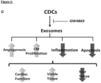
- the Inventors' work demonstrates that exosomes and their constituent microRNAs favorably modulate apoptosis, inflammation and fibrosis in the injured heart, leading to functional recovery and increase tissue viability.
- CDC-derived exosomes represent a novel "cell-free" therapeutic candidate for tissue repair.
- Described herein is a method of treatment, including selecting a subject in need of treatment for heart failure secondary to a chronic degenerative muscular disease and administering a composition including a plurality of exosomes to the subject, wherein the plurality of the exosomes are isolated from cardiosphere-derived cells (CDCs) grown in serum-free media, include exosomes with a diameter of about 90 nm to about 200 nm and are CD81+, CD63+, or both, and further wherein administration of the composition treats the subject.
- the chronic degenerative muscular disease is Duchenne muscular dystrophy.
- administering a composition includes about 1 to about 100 mg exosome protein in a single dose.
- administering a composition includes injection.
- the injection includes percutaneous injection.
- the injection is directly into heart muscle.
- administering a composition includes myocardial infusion.
- myocardial infusion is intra-arterial or intravenous.
- treatment of the subject results in decreased fibrosis, decreased inflammation, increased mitochondrial function and/or increased cardiomyogenesis.
- decreased fibrosis includes a reduction in collagen accumulation.
- collagen includes collagen I and/or collagen III.
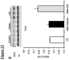
- decreased inflammation includes an increase in cytoplasmic nuclear factor (erythroid-derived 2)-like 2 (Nrf2), reduction in fatty acid peroxidation end products, reduced numbers of inflammatory cells, and/or upregulated expression of antioxidants.
- antioxidants include heme oxygenase-1 (HO-1), catalase, superoxide dismutase-2 (SOD-2), and glutamate-cystein ligase catalytic (GCLC) subunit.
- inflammatory cells include CD68+ macrophages and CD3+ T-cells.
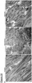
- increased mitochondrial function includes increased mitochondrial ultrastructure and/or increased mitochondrial biogenesis.
- increased mitochondrial function includes increased nuclear PPAR- ⁇ co-activator-1 (PGC-1) expression.
- the exosomes include one or more microRNAs selected from the group consisting of: microRNAs miR-146a, miR148a, miR22, miR-24, miR-210, miR-150, miR-140-3p, miR-19a, miR-27b, miR-19b, miR-27a, miR-376c, miR-128, miR-320a, miR-143, miR-21, miR-130a, miR-9, miR-185, and miR-23a.
- treatment of the subject results in decreased fibrosis, decreased inflammation, increased mitochondrial function and/or increased cardiomyogenesis.
- decreased fibrosis includes a reduction in collagen accumulation.
- collagen includes collagen I and/or collagen III.
- decreased inflammation includes an increase in cytoplasmic nuclear factor (erythroid-derived 2)-like 2 (Nrf2), reduction in fatty acid peroxidation end products, reduced numbers of inflammatory cells, and/or upregulated expression of antioxidants.
- antioxidants include heme oxygenase-1 (HO-1), catalase, superoxide dismutase-2 (SOD-2), and glutamate-cystein ligase catalytic (GCLC) subunit.
- inflammatory cells include CD68+ macrophages and CD3+ T-cells.
- increased mitochondrial function includes increased mitochondrial ultrastructure and/or increased mitochondrial biogenesis.
- increased mitochondrial function includes increased nuclear PPAR- ⁇ co-activator-1 (PGC-1) expression.
- Duchenne muscular dystrophy a crippling genetic disease leading to premature death, affects the heart as well as skeletal muscle. Indeed, cardiomyopathy is the leading cause of death in Duchenne patients. There are no approved treatments for the cardiomyopathy, and novel Duchenne-specific experimental approaches such as exon skipping do not benefit the heart.
- cardiosphere-derived cells CDCs
- Exosomes secreted by human CDCs reproduce the benefits of CDCs in mdx mice, and reverse abnormalities of calcium cycling and mitochondrial respiration in human Duchenne cardiomyocytes.
- DMD Duchenne muscular dystrophy
- CDCs Cardiosphere-derived cells
- CDCs are not only regenerative, but also anti-inflammatory and anti-fibrotic; they work indirectly via the secretion of exosomes laden with noncoding RNA including microRNAs (miRs).
- miRs microRNAs
- CDC-exosomes mimic the functional and structural benefits of CDCs, while blockade of exosome biosynthesis renders CDCs ineffective.
- the Inventors reasoned that CDCs might be useful in treating Duchenne cardiomyopathy. The goal is not to replace dystrophin, but rather to offset the pathophysiological consequences of dystrophin deletion, by recruiting regeneration, reversing fibrosis and targeting inflammation.
- Exosomes secreted lipid vesicles containing a rich milieu of biological factors, provide powerful paracrine signals by which stem cells potentiate their biological effects to neighboring cells, including diseased or injured cells.
- bio-active lipid and nucleic acid "cargo"
- these natural delivery devices are capable of inducing significant phenotypic and functional changes in recipient cells that lead to activation of regenerative programs.
- the role of such indirect mechanisms to in stem cell initiated regeneration is strongly suggested by growing evidence that after stem cell administration and clearance from delivery sites in tissue and organs, regeneration processes nevertheless persist and arise from endogenous tissues.
- Stem cell-derived exosomes have been identified and isolated from supernatants of several cell types with demonstrated therapeutic potential, including mesenchymal stromal (MSC), (bone marrow stem cells) mononuclear (MNC) cells, immune cells (dendritic and CD34+), human neural stem cells (hNSCs), among others.
- MSC mesenchymal stromal
- MNC mononuclear
- hNSCs human neural stem cells
- human cardiosphere derived cells CDCs
- Stem cell-derived exosomes including those produced by CDCs, may provide a potent and rich source for developing "cell-free" therapies.
- exosome-based, "cell-free” therapies in contrast to cell therapy, provide distinct advantages in regenerative medicine.
- these features significantly obviate safety issues.
- stem cell-derived exosomes are less immunogenic than parental cells, as a result of a lower content of membrane-bound proteins, including MHC complex molecules. Replacing the administration of live cells with their secreted exosomes, mitigates many of the safety concerns and limitations associated with the transplantation of viable replicating cells.
- exosome encapsulation of bioactive components in lipid vesicles allows protection of contents from degradation in vivo, thereby potentially negating obstacles often associated with delivery of soluble molecules such as cytokines, growth factors, transcription factors and RNAs. This comparative ease of administration may ultimately allow for repeated and sustained delivery to patients, thereby maximizing the potential for regeneration and repair of diseased and/or dysfunctional tissue.
- exosome production under defined conditions allows for easier manufacture and scale-up opportunity.
- Manufacture of exosomes is akin to conventional biopharmacological product manufacture, allowing for standardization in production and quality control for dosage and biological activity testing.
- the durability of exosomes in culture allows for the acquisition of large quantities of exosomes through their collection from a culture medium in which the exosomes are secreted over periods of time.
- exosomes are involved in intercellular communication between different cell types, much remains to be discovered in regard to the mechanisms of their production within parental cells of origin and effects on target recipient cells. Exosomes have been reported to be involved in numerous cellular, tissue and physiological processes, including immune modulating processes, angiogenesis, migration of endothelial cells in connection with tumor growth, or reducing damage in ischemia reperfusion injury. Of critical scientific interest in establishing whether exosomes secreted by cells, such as cardiosphere-derived cells (CDCs), are capable of reproducing the therapeutic benefits of their parental cells, or possibly, are indispensable in effectuating such therapeutic benefits.
- CDCs cardiosphere-derived cells
- exosomes secreted by a wide range of cell types, exosomes are lipid bilayer vesicles that are enriched in a variety of biological factors, including cytokines, growth factors, transcription factors, lipids, and coding and non-coding nucleic acids. Exosomes are found in blood, urine, amniotic fluid, interstitial and extracellular spaces. These exocytosed vesicles of endosomal origin can range in size between 30-200 nm, including sizes of 40-100 nm, and possess a cup-shaped morphology, as revealed by electron microscopy.
- MVB multivesicular bodies
- exosomes reflect their parental cellular origin, as containing distinct subsets of biological factors in connection with their parent cellular origin, including the cell regulatory state of the parental cells when formed.
- the rich biological milieu of different proteins, including cytokines and growth factors, lipids, coding and noncoding RNA molecules, within exosomes are all necessarily derived from their parental cells.
- exosomes further express the extracellular domain of membrane-bound receptors at the surface of the membrane.
- endosome-associated proteins e.g., Rab GTPase, SNAREs, Annexins, and flotillin
- proteins that are known to cluster into microdomains at the plasma membrane or at endosomes four transmembrane domain tetraspanins, e.g., CD63, CD81, CD82, CD53, and CD37
- lipid raft associated proteins e.g., glycosylphosphatidylinositol- anchored proteins and flotillin
- cholesterol sphingomyelin
- hexosylceramides as examples.
- exosomes In addition to these core components reflecting their vesicle origin, a critical property of exosomes is a demonstrated capability to contain both mRNA and microRNA associated with signaling processes, with both cargo mRNA being capable to translation in recipient cells, or microRNA functionally degrading target mRNA in recipient cells. Other noncoding RNAs, capable for influencing gene expression, may also be present in exosomes. While the processes governing the selective incorporation of mRNA or microRNA populations into exosomes is not entirely understood, it is clear that RNA molecules are selectively, not randomly incorporated into exosomes, as revealed by studies reporting enrichment of exosome cargo RNAs when compared to the RNA profiles of the originating cells. Given the growing understanding of how such RNA molecules play a role in disease pathogenesis and regenerative processes, the presence of RNA molecules in exosomes and apparent potency in affecting target recipient cells suggests critical features that can be deployed in therapeutic approaches.
- the natural bilayer membrane encapsulation of exosomes provides a protected and controlled internal microenvironment that allows cargo contents to persist or migrate in the bloodstream or within tissues without degradation.
- Their release into the extracellular environment allows for interaction with recipient cells via adhesion to the cell surface mediated by lipid-ligand receptor interactions, internalization via endocytic uptake, or by direct fusion of the vesicles and cell membrane. These processes lead to the release of exosome cargo content into the target cell.
- exosome-cell interactions modulation of genetic pathways in the target recipient cell, as induced through any of several different mechanisms including antigen presentation, the transfer of transcription factors, cytokines, growth factors, nucleic acid such as mRNA and microRNAs.
- ESC embryonic stem cell
- mRNA and proteins to hematopoietic progenitors.
- Other studies have shown that adult stem cell-derived exosomes also shuttle selected patterns of mRNA, microRNA and pre-microRNA associated with several cellular functions involved in the control of transcription, proliferation and cell immune regulation.
- Exosome isolation relies on exploiting their generic biochemical and biophysical features for separation and analysis. For example, differential ultracentrifugation has become a leading technique wherein secreted exosomes are isolated from the supernatants of cultured cells. This approach allows for separation of exosomes from nonmembranous particles, by exploiting their relatively low buoyant density. Size exclusion allows for their separation from biochemically similar, but biophysically different microvesicles, which possess larger diameters of up to 1,000 nm. Differences in flotation velocity further allows for separation of differentially sized exosomes. In general, exosome sizes will possess a diameter ranging from 30-200 nm, including sizes of 40-100 nm. Further purification may rely on specific properties of the particular exosomes of interest. This includes, for example, use of immunoadsorption with a protein of interest to select specific vesicles with exoplasmic or outward orientations.
- differential ultracentrifugation is the most commonly used for exosome isolation. This technique utilizes increasing centrifugal force from 2000xg to 10,000xg to separate the medium- and larger-sized particles and cell debris from the exosome pellet at 100,000xg. Centrifugation alone allows for significant separation/collection of exosomes from a conditioned medium, although it is insufficient to remove various protein aggregates, genetic materials, particulates from media and cell debris that are common contaminants.
- Enhanced specificity of exosome purification may deploy sequential centrifugation in combination with ultrafiltration, or equilibrium density gradient centrifugation in a sucrose density gradient, to provide for the greater purity of the exosome preparation (flotation density 1.1-1.2g/ml) or application of a discrete sugar cushion in preparation.
- ultrafiltration can be used to purify exosomes without compromising their biological activity.
- Membranes with different pore sizes - such as 100 kDa molecular weight cut-off (MWCO) and gel filtration to eliminate smaller particles - have been used to avoid the use of a nonneutral pH or non-physiological salt concentration.
- MWCO molecular weight cut-off
- THF tangential flow filtration
- HPLC can also be used to purify exosomes to homogeneouslysized particles and preserve their biological activity as the preparation is maintained at a physiological pH and salt concentration.
- exosomes for precipitation techniques, addition to volume-excluding polymers (e.g., polyethylene glycols (PEGs)), possibly combined additional rounds of centrifugation or filtration.
- a precipitation reagent e.g., ExoQuick ®
- ExoQuick ® can be added to conditioned cell media to quickly and rapidly precipitate a population of exosomes, although re-suspension of pellets prepared via this technique may be difficult.
- Flow field-flow fractionation is an elution-based technique that is used to separate and characterize macromolecules (e.g., proteins) and nano-to micro-sized particles (e.g., organelles and cells) and which has been successfully applied to fractionate exosomes from culture media.
- macromolecules e.g., proteins
- nano-to micro-sized particles e.g., organelles and cells
- exosomes may be applied to isolated specific exosomes of interest. This includes relying on antibody immunoaffinity to recognizing certain exosome-associated antigens. As described, exosomes further express the extracellular domain of membrane-bound receptors at the surface of the membrane. This presents a ripe opportunity for isolating and segregating exosomes in connections with their parental cellular origin, based on a shared antigenic profile. Conjugation to magnetic beads, chromatography matrices, plates or microfluidic devices allows isolating of specific exosome populations of interest as may be related to their production from a parent cell of interest or associated cellular regulatory state. Other affinity-capture methods use lectins which bind to specific saccharide residues on the exosome surface.
- Exosome-Based Therapies A chief goal of developing exosome-based therapy is the creation of "cell-free" therapies, wherein the benefits of cellular therapeutics can be provided with reduced risks or in scenarios in which cell therapy would be unavailable.
- DMD Duchenne muscular dystrophy
- HF heart failure
- the therapeutic benefits of cell-based therapies such as cardiosphere-derived cells (CDCs) appear to occur through indirect mechanisms involving regenerated myocardium and vasculature arising from endogenous origin.
- CDCs cardiosphere-derived cells
- CDC-derived exosomes may effectively address a major unmet medical need, by recruiting synergistic mechanisms to attract endogenous stem cells to sites of myocardial injury, promote differentiation into heart muscle and vessels, thereby reversing the pathophysiology of HF.
- DMD is an X-linked recessive disorder characterized by myopathy (cell membrane damage in muscle fiber) as exemplified by a variety of pathological features. This includes skeletal muscle weakness starting 3-5 years from onset, progressive weakness, wheelchair dependency at approximately 13 years from onset. Importantly, cardiomyopathy is observed to take hold in 1/3 of patients from less than 13 years from onset, increasing to 1/2 of patients less than 18 years from onset, and in all patients after 18 years. Dilated cardiomyopathy includes left ventricle posterobasal fibrosis; conduction abnormalities are mainly intra-atrial: SVT with abnormal AV nodal conduction. Patients may further suffer from smooth muscle myopathy including vascular dysfunction, further including GI and urinary tract systems involvement.
- dystrophin gene mutation deletion
- loss of dystrophin results in cellular membrane damage and leakage of extracellular Ca 2+ into the cell.
- Elevated intracellular levels ultimately result in increased oxidative and/or nitrosative stress and inflammation, and activation of calpain.
- the combination of these effects results in muscle proteolysis and apoptosis, leading to the degradative features described above.
- CDCs have been demonstrated as promoting antioxidative, anti-inflammatory, anti-apoptotic, anti-remodeling effects, in addition to enhancing regenerative capacity.
- CDC administration is beneficial in retarding/reversing DMD, and exosome populations derived from CDCs may allow for these benefits to be delivered, while avoiding obstacles associated with cell-based therapy.
- stem cell-derived exosomes are likely to be less immunogenic than parental cells.
- exosome encapsulation of bioactive components in lipid vesicles allows protection of contents from degradation in vivo, thereby potentially negating obstacles often associated with delivery of soluble molecules such as cytokines, growth factors, transcription factors and RNAs, while potentially allowing for increased concentrations to be provided.
- soluble molecules such as cytokines, growth factors, transcription factors and RNAs
- compositions and methods and compositions providing significant benefits in the repair or regeneration of damaged or diseased tissues via "cell-free” methods involving exosomes.
- human cardiosphere-derived cells (CDC)-derived exosomes are demonstrated as effective in reducing scar size and regenerating viable myocardium.
- CDC cardiosphere-derived cells
- Described herein is a method of treatment, including selecting a subject in need of treatment for heart failure secondary to a chronic degenerative muscular disease and administering a composition including a plurality of exosomes to the subject, wherein the plurality of the exosomes are isolated from cardiosphere-derived cells (CDCs) grown in serum-free media, include exosomes with a diameter of about 90 nm to about 200 nm and are CD81+, CD63+, or both, and further wherein administration of the composition treats the subject.
- the chronic degenerative muscular disease is Duchenne muscular dystrophy.
- administering a composition includes about 1 to about 100 mg exosome protein in a single dose.
- administering a composition includes injection.
- the injection includes percutaneous injection.
- the injection is directly into heart muscle.
- administering a composition includes myocardial infusion.
- myocardial infusion is intra-arterial or intravenous.
- treatment of the subject results in decreased fibrosis, decreased inflammation, increased mitochondrial function and/or increased cardiomyogenesis.
- decreased fibrosis includes a reduction in collagen accumulation.
- collagen includes collagen I and/or collagen III.
- decreased inflammation includes an increase in cytoplasmic nuclear factor (erythroid-derived 2)-like 2 (Nrf2), reduction in fatty acid peroxidation end products, reduced numbers of inflammatory cells, and/or upregulated expression of antioxidants.
- antioxidants include heme oxygenase-1 (HO-1), catalase, superoxide dismutase-2 (SOD-2), and glutamate-cystein ligase catalytic (GCLC) subunit.
- inflammatory cells include CD68+ macrophages and CD3+ T-cells.
- increased mitochondrial function includes increased mitochondrial ultrastructure and/or increased mitochondrial biogenesis.
- increased mitochondrial function includes increased nuclear PPAR- ⁇ co-activator-1 (PGC-1) expression.
- the exosomes include one or more microRNAs selected from the group consisting of: microRNAs miR-146a, miR148a, miR22, miR-24, miR-210, miR-150, miR-140-3p, miR-19a, miR-27b, miR-19b, miR-27a, miR-376c, miR-128, miR-320a, miR-143, miR-21, miR-130a, miR-9, miR-185, and miR-23a.
- a composition including cardiosphere-derived cells including cardiosphere-derived cells (CDCs), wherein administration of the composition treats the subject.
- the chronic muscular disease is Duchenne muscular dystrophy.
- administering a composition includes about 1 ⁇ 10 5 to about 1 ⁇ 10 8 or more CDCs in a single dose.
- the number of administered CDCs includes intracoronary 25 million CDCs per coronary artery (i.e., 75 million CDCs total) as another baseline for exosome dosage quantity.
- the numbers of CDCs includes 1 ⁇ 10 5 , 1 ⁇ 10 6 , 1 ⁇ 10 7 , 1 ⁇ 10 8 , 1 ⁇ 10 9 CDCs in a single dose as another baseline for exosome dosage quantity. In certain instances, this may be prorated to body weight (range 100,000-1M CDCs/kg body weight total CDC dose).
- administering a composition includes myocardial infusion.
- myocardial infusion is intracoronary.
- myocardial infusion is intra-arterial or intravenous.
- treatment of the subject results in decreased fibrosis, decreased inflammation, increased mitochondrial function and/or increased cardiomyogenesis.
- decreased fibrosis includes a reduction in collagen accumulation.
- collagen includes collagen I and/or collagen III.
- decreased inflammation includes an increase in cytoplasmic nuclear factor (erythroid-derived 2)-like 2 (Nrf2), reduction in fatty acid peroxidation end products, reduced numbers of inflammatory cells, and/or upregulated expression of antioxidants.
- antioxidants include heme oxygenase-1 (HO-1), catalase, superoxide dismutase-2 (SOD-2), and glutamate-cystein ligase catalytic (GCLC) subunit.
- inflammatory cells include CD68+ macrophages and CD3+ T-cells.
- increased mitochondrial function includes increased mitochondrial ultrastructure and/or increased mitochondrial biogenesis.
- increased mitochondrial function includes increased nuclear PPAR- ⁇ co-activator-1 (PGC-1) expression. Further examples are found in U.S. App. No. 11/666,685 , 12/622,143 , and 12/622,106 , which are herein incorporated by reference.
- compositions including a plurality of exosomes.
- the plurality of exosomes are generated by a method including providing a population of cells, and isolating a plurality of exosomes from the population of cells.
- the cells are stem cells, progenitors and/or precursors.
- the stem cells, progenitors and/or precursors are cardiosphere-derived cells (CDCs).
- the stem cells, progenitors and/or precursors are pluripotent stem cells (pSCs), such as embryonic stem cells (ESCs) and induced pluripotent stem cells (iPSCs) derived from any one of various somatic sources in the body such as fibroblasts, blood and hematopoietic stem cells (hSCs), immune cells, bone and bone marrow, neural tissue, among others.
- pSCs pluripotent stem cells
- ESCs embryonic stem cells
- iPSCs induced pluripotent stem cells derived from any one of various somatic sources in the body
- fibroblasts fibroblasts
- hSCs hematopoietic stem cells
- immune cells bone and bone marrow, neural tissue, among others.
- the stem cells, progenitors and/or precursors include hSCs, mesenchymal stem cells (MSCs), or endothelial precursor cells (EPCs).
- the cells are stem cells, progenitors and/or precursors derived from human biopsy tissue.
- the cells are stem cells, progenitors and/or precursors are a primary culture.
- the cells are stem cells, progenitors and/or precursors which may constitute a cell line capable of serial passaging.
- the plurality of exosomes is isolated from the supernatants of the population of cells. This includes, for example, exosomes secreted into media as conditioned by a population of cells in culture, further including cell lines capable of serial passaging.
- the cells are cultured in serum-free media.
- the cells in culture are grown to 10, 20, 30, 40, 50, 60, 70, 80, 90, or 90% or more confluency when exosomes are isolated.
- the population of cells has been genetically manipulated. This includes, for example, knockout (KO) or transgenic (TG) cell lines, wherein an endogenous gene has been removed and/or an exogenous introduced in a stable, persistent manner.
- the population of cells has been altered by exposure to environmental conditions (e.g., hypoxia), small molecule addition, presence/absence of exogenous factors (e.g., growth factors, cytokines) at the time, or substantially contemporaneous with, isolating the plurality of exosomes in a manner altering the regulatory state of the cell.
- environmental conditions e.g., hypoxia
- exogenous factors e.g., growth factors, cytokines
- altering the regulatory state of the cell changes composition of one or more exosomes in the plurality of exosomes.
- the plurality of exosomes include one or more exosomes that are about 10 nm to about 250 nm in diameter, including those about 10 nm to about 15 nm, about 15 nm to about 20 nm, about 20 nm to about 25 nm, about 25 nm to about 30 nm, about 30 nm to about 35 nm, about 35 nm to about 40 nm, about 40 nm to about 50 nm, about 50 nm to about 60 nm3 about 60 nm to about 70 nm, about 70 nm to about 80 nm, about 80 nm to about 90 nm, about 90 nm to about 95 nm, about 95 nm to about 100 nm, about 100 nm to about 105 nm, about 105 nm to about 110 nm, about 110 nm to about 115 nm, about 115 nm to about 120 nm, about 120 nm to about
- the plurality of exosomes includes one or more exosomes expressing a biomarker.
- the biomarkers are tetraspanins.
- the tetraspanins are one or more selected from the group including CD63, CD81, CD82, CD53, and CD37.
- the exosomes express one or more lipid raft associated protiens (e.g., glycosylphosphatidylinositol-anchored proteins and flotillin), cholesterol, sphingomyelin, and/or hexosylceramides.
- the plurality of exosomes includes one or more exosomes containing a biological protein.
- the biological protein includes transcription factors, cytokines, growth factors, and similar proteins capable of modulating signaling pathways in a target cell.
- the biological protein is capable of facilitating regeneration and/or improved function of a tissue.
- the biological protein is capable of modulating pathways related to Irak1, Traf6, toll-like receptor (TLR) signaling pathway, NOX-4, SMAD-4, and/or TGF- ⁇ .
- TLR Irak1, Traf6, toll-like receptor
- the biological protein related to exosome formation and packaging of cytosolic proteins such as Hsp70, Hsp90, 14-3-3 epsilon, PKM2, GW182 and AGO2.
- the plurality of exosomes includes one or more exosomes containing a signaling lipid. This includes ceramide and derivatives. In other embodiments, the plurality of exosomes includes one or more exosomes containing a coding and/or non-coding nucleic acid.
- the plurality of exosomes includes one or more exosomes containing microRNAs.
- these microRNAs can include miR-146a, miR22, miR-24, miR-210, miR-150, miR-140-3p, miR-19a, miR-27b, miR-19b, miR-27a, miR-376c, miR-128, miR-320a, miR-143, miR-21, miR-130a, miR-9, miR-185, and/or miR-23a.
- the plurality of exosomes include one or more exosomes enriched in at least one of miR-146a, miR22, miR-24, miR-210, miR-150, miR-140-3p, miR-19a, miR-27b, miR-19b, miR-27a, miR-376c, miR-128, miR-320a, miR-143, miR-21, miR-130a, miR-9, miR-185, and/or miR-23a.
- the plurality of exosomes includes one or more exosomes enriched in at least one of miR-146a, miR-22, miR-24.
- Enrichment can be measured by, for example, comparing the amount of one or more of the described microRNAs when derived from cells providing salutary benefit in a therapeutic setting (e.g., cardiosphere-derived cells (CDCs) compared to cells that do not provide such a salutary benefit (e.g., fibroblasts). Enrichment may also be measured in absolute or relative quantities, such as when compared to a standardized dilution series.
- a therapeutic setting e.g., cardiosphere-derived cells (CDCs)
- fibroblasts e.g., fibroblasts
- the plurality of exosomes can include one or more exosomes containing microRNAs.
- the plurality of exosomes can include one or more exosomes containing microRNAs.
- microRNAs known in the art, such as miR-17, miR-21 , miR-92, miR92a, miR-29, miR-29a, miR-29b, miR-29c, miR-34, mi-R34a, miR-150, miR-451, miR-145, miR-143, miR-144, miR-193a-3p, miR-133a, miR-155, miR-181a, miR-214, miR-199b, miR-199a, miR-210, miR-126, miR-378, miR-363 and miR-30b, and miR-499.
- microRNAs known in the art include miR-92, miR-17, miR-21, miR-92, miR92a, miR-29, miR- 29a, miR-29b, miR-29c, miR-34, mi-R34a, miR-150, miR-451, miR-145, miR-143, miR- 144, miR-193a-3p, miR-133a, miR-155, miR-181a, miR-214, miR-199b, miR-199a, miR- 126, miR-378, miR-363 and miR-30b, and/or miR-499.
- isolating a plurality of exosomes from the population of cells includes centrifugation of the cells and/or media conditioned by the cells. In several embodiments, ultracentrifugation is used. In several embodiments, isolating a plurality of exosomes from the population of cells is via size-exclusion filtration. In other embodiments, isolating a plurality of exosomes from the population of cells includes use of discontinuous density gradients, immunoaffinity, ultrafiltration and/or high performance liquid chromatography (HPLC).
- HPLC high performance liquid chromatography
- differential ultracentrifugation includes using centrifugal force from 1000-2000xg, 2000-3000xg, 3000-4000xg, 4000-5000xg, 5000xg-6000xg, 6000-7000xg, 7000-8000xg, 8000-9000xg, 9000-10,000xg, to 10,000xg or more to separate larger-sized particles from a plurality of exosome derived from the cells.
- isolating a plurality of exosomes from the population of cells includes use of filtration or ultrafiltration.
- a size exclusion membrane with different pore sizes is used.
- a size exclusion membrane can include use of a filter with a pore size of 0.1-0.5 ⁇ M, 0.5-1.0 ⁇ M, 1-2.5 ⁇ M, 2.5-5 ⁇ M, 5 or more ⁇ M. In certain embodiments, the pore size is about 0.2 ⁇ M.
- a molecular weight cut-off (MWCO) gel filtration capable of isolating particles ranging from 100-500 daltons (Da), 500-1 kDa, 1-2 kDa, 2-5 kDa, 5-10 kDa, 10-25 kDa, 25-50 kDa, 50-100 kDa, 100-250 kDa, 250-500 kDa, 500 or more kDa.
- the size exclusion is for about 2-5 kDa. In certain embodiments, the size exclusion is for about 3 kDa. In various embodiments, such systems are used in combination with variable fluid flow systems.
- isolating a plurality of exosomes from the population of cells includes use of tangential flow filtration (TFF) systems are used purify and/or concentrate the exosome fractions.
- isolating a plurality of exosomes from the population of cells includes use of (HPLC) can also be used to purify exosomes to homogeneously sized particles.
- density gradients as used such as centrifugation in a sucrose density gradient or application of a discrete sugar cushion in preparation.
- isolating a plurality of exosomes from the population of cells includes use of a precipitation reagent.
- a precipitation reagent can be added to conditioned cell media to quickly and rapidly precipitate a population of exosomes.
- isolating a plurality of exosomes from the population of cells includes use of volume-excluding polymers (e.g., polyethylene glycols (PEGs)) are used.
- isolating a plurality of exosomes from the population of cells includes use of flow field-flow fractionation (FlFFF), an elution-based technique.
- FlFFF flow field-flow fractionation
- isolating a plurality of exosomes from the population of cells includes use of one or more capture agents to isolate one or more exosomes possessing specific biomarkers or containing particular biological molecules.
- one or more capture agents include at least one antibody.
- antibody immunoaffinity recognizing exosome-associated antigens is used to capture specific exosomes.
- the at least one antibody are conjugated to a fixed surface, such as magnetic beads, chromatography matrices, plates or microfluidic devices, thereby allowing isolation of the specific exosome populations of interest.
- isolating a plurality of exosomes from the population of cells includes use of one or more capture agents that is not an antibody.
- the plurality of exosomes range in size from 30 to 300 nm. In various embodiments, the plurality of exosomes range in size from 40 to 100 nm. In certain embodiments, the plurality of exosomes is cardiosphere-derived cell (CDC) exosomes. In certain embodiments, the plurality of exosomes includes exosomes that are CD63+.
- CDC cardiosphere-derived cell
- the exosomes include microRNAs miR-146a, miR22, miR-24, miR-210, miR-150, miR-140-3p, miR-19a, miR-27b, miR-19b, miR-27a, miR-376c, miR-128, miR-320a, miR-143, miR-21, miR-130a, miR-9, miR-185, and/or miR-23a.
- the exosomes are 2-5 kDa, such as 3 kDa.
- Other examples or embodiments relating to the composition and techniques involving exosomes are presented, in PCT Pub. No. WO 2014/028,493 , which is fully incorporated herein by reference.
- Described herein is a method for treatment including, selecting a subject in need of treatment, administering a composition including a plurality of exosomes to the individual, wherein the administration of the composition treats the subject.
- the subject is in need to treatment for a disease and/or condition involving tissue damage or dysfunction.
- the disease and/or condition involving tissue damage or dysfunction is heart disease.
- the plurality of exosomes includes exosomes including one or more microRNAs.
- the plurality of exosomes are generated by a method including providing a population of cells, and isolating a plurality of exosomes from the population of cells.
- the cells are stem cells, progenitors and/or precursors.
- the stem cells, progenitors and/or precursors are cardiosphere-derived cells (CDCs).
- the stem cells, progenitors and/or precursors are pluripotent stem cells (pSCs), such as embryonic stem cells (ESCs) and induced pluripotent stem cells (iPSCs) derived from any one of various somatic sources in the body such as fibroblasts, blood and hematopoietic stem cells (hSCs), immune cells, bone and bone marrow, neural tissue, among others.
- pSCs pluripotent stem cells
- ESCs embryonic stem cells
- iPSCs induced pluripotent stem cells
- the stem cells, progenitors and/or precursors includes hSCs, mesenchymal stem cells (MSCs), or endothelial precursor cells (EPCs).
- the cells are stem cells, progenitors and/or precursors derived from human biopsy tissue.
- the cells are stem cells, progenitors and/or precursors are a primary culture. In various embodiments, the cells are stem cells, progenitors and/or precursors are a cell line capable of serial passaging. In certain embodiments, the exosomes are synthetic.
- the plurality of exosomes is derived from cardiosphere-derived cells (CDCs). In other embodiments, the plurality of exosomes includes exosomes including one or more biological molecules. In other embodiments, the plurality of exosomes includes exosomes enriched for one or more biological molecules when derived from CDCs compared to exosome derived from non-CDC sources. In various embodiments, the one or more biological molecules are proteins, growth factors, cytokines, transcription factors and/or morphogenic factors. In other embodiments, the plurality of exosomes includes exosomes enriched for one or more biological molecules includes microRNAs, further including microRNAs that are enriched when derived from CDCs compared to exosome derived from non-CDC sources.
- these microRNAs can include miR-146a, miR22, miR-24, miR-210, miR-150, miR-140-3p, miR-19a, miR-27b, miR-19b, miR-27a, miR-376c, miR-128, miR-320a, miR-143, miR-21, miR-130a, miR-9, miR-185, and/or miR-23a.
- the plurality of exosomes includes one or more exosomes enriched in at least one of miR-146a, miR-22, miR-24.
- the CDCs are mammalian. In other embodiments, the CDCs are human.
- the exosomes are synthetic. In certain embodiments, the synthetic exosomes possess substantially similar content (e.g., microRNAs, biological molecules) as exosomes derived from CDCs.
- the numbers of exosomes is relative to the number of cells used in a clinically relevant dose for a cell-therapy method.
- 3mL / 3 x 10 5 CDCs is capable of providing therapeutic benefit in intracoronary administration, and therefore, a plurality of exosomes as derived from that number of cells in a clinically relevant dose for a cell-therapy method.
- administration can be in repeated doses.
- the number of administered CDCs includes intracoronary 25 million CDCs per coronary artery (i.e., 75 million CDCs total) as another baseline for exosome dosage quantity.
- the numbers of CDCs includes 1 ⁇ 10 5 , 1 ⁇ 10 6 , 1 ⁇ 10 7 , 1 ⁇ 10 8 , 1 ⁇ 10 9 CDCs in a single dose as another baseline for exosome dosage quantity. In certain instances, this may be prorated to body weight (range 100,000-1M CDCs/kg body weight total CDC dose).
- exosome quantity may be defined by protein quantity, such as dosages including 1-10, 10-25, 25-50, 50-75, 75-100, or 100 or more mg exosome protein.
- Defining an effective dose range, dosing regimen and route of administration may be guided by studies using fluorescently labeled exosomes, and measuring target tissue retention, which can be >10X, >50X, or >100X background, as measured 5, 10, 15, 30, or 30 or more min as a screening criterion.
- >100X background measured at 30 mins is a baseline measurement for a low and high dose that is then assess for safety and bioactivity (e.g., using MRI endpoints: scar size, global and regional function).
- single doses are compared to two, three, four, four or more sequentially-applied doses.
- the repeated or sequentially-applied doses are provided for treatment of an acute disease and/or condition.
- the repeated or sequentially-applied doses are provided for treatment of a chronic disease and/or condition.
- administration of exosomes to the subject occurs through any of known techniques in the art. In some embodiments, this includes percutaneous delivery. In other embodiments, myocardial infusion is used, for example, the use of intracoronary catheters. In various embodiments, delivery can be intra-arterial or intravenous. Additional delivery sites include any one or more compartments of the heart, such as arterial, venous, and/or ventricular locations. In certain embodiments, administration can include delivery to a tissue or organ site that is different from the site or diseased and/or dysfunctional tissue. In certain embodiments, the delivery is via inhalation or oral administration.
- functional improvement may include increased cardiac output, contractility, ventricular function and/or reduction in arrhythmia (among other functional improvements).
- improved function may be realized as well, such as enhanced cognition in response to treatment of neural damage, improved blood-oxygen transfer in response to treatment of lung damage, improved immune function in response to treatment of damaged immunological-related tissues.
- administration of the plurality of exosomes alters gene expression in the damaged or dysfunctional tissue, improves viability of the damaged tissue, and/or enhances regeneration or production of new tissue in the individual. In various embodiments, administration of the exosomes results in functional improvement in the tissue.
- the damaged or dysfunctional tissue includes skeletal muscle tissue.
- functional improvement may include increased contractile strength, improved ability to walk (for example, and increase in the six-minute walk test results), improved ability to stand from a seated position, improved ability to sit from a recumbent or supine position, or improved manual dexterity such as pointing and/or clicking a mouse.
- the damaged or dysfunctional tissue is in need of repair, regeneration, or improved function due to an acute event.
- Acute events include, but are not limited to, trauma such as laceration, crush or impact injury, shock, loss of blood or oxygen flow, infection, chemical or heat exposure, poison or venom exposure, drug overuse or overexposure, and the like.
- the damaged tissue is cardiac tissue and the acute event includes a myocardial infarction.
- administration of the exosomes results in an increase in cardiac wall thickness in the area subjected to the infarction.
- tissue is also subject to damage due to chronic disease, such as for example congestive heart failure, including as conditions secondary to diseases such as Duchenne muscular dystrophy, ischemic heart disease, hypertension, valvular heart disease, dilated cardiomyopathy, infection, diabetes, and the like.
- the administration can be in repeated doses, such as two, three, four, four or more sequentially-applied doses.
- the repeated or sequentially-applied doses are provided for treatment of an acute disease and/or condition.
- the repeated or sequentially-applied doses are provided for treatment of a chronic disease and/or condition.
- the regenerative cells are from the same tissue type as is in need of repair or regeneration. In several other embodiments, the regenerative cells are from a tissue type other than the tissue in need of repair or regeneration.
- the method of treatment includes, selecting a subject in need of treatment for a heart related disease and/or condition, administering a composition including a plurality of exosomes to the individual, wherein the administration of the composition treats thesubject.
- the heart related disease and/or condition includes heart failure, further including Duchenne muscular dystrophy related heart failure.
- the plurality of exosomes range in size from 30 to 300 nm. In various embodiments, the plurality of exosomes range in size from 40 to 100 nm.
- the plurality of exosomes are cardiosphere-derived cell (CDC) exosomes.
- the plurality of exosomes include exosomes that are CD63+.
- the exosomes include microRNAs miR-146a, miR22, miR-24, miR-210, miR-150, miR-140-3p, miR-19a, miR-27b, miR-19b, miR-27a, miR-376c, miR-128, miR-320a, miR-143, miR-21, miR-130a, miR-9, miR-185, and/or miR-23a.
- the exosomes are 2-5 kDa, such as 3 kDa.
- administering a composition includes a dosage of 1 ⁇ 10 8 , 1 ⁇ 10 8 to 1 ⁇ 10 9 , 1 ⁇ 10 9 to 1 ⁇ 10 10 , 1 ⁇ 10 10 to 1 ⁇ 10 11 , 1 ⁇ 10 11 to 1 ⁇ 10 12 , 1 ⁇ 10 12 or more exosomes.
- 3mL / 3 ⁇ 10 5 CDCs is capable of providing therapeutic benefit in intracoronary administration, and therefore, a plurality of exosomes as derived from that number of cells in a clinically relevant dose for a cell-therapy method.
- administration can be in repeated doses.
- the number of administered CDCs includes intracoronary 25 million CDCs per coronary artery (i.e., 75 million CDCs total) as another baseline for exosome dosage quantity.
- the numbers of CDCs includes 1 ⁇ 10 5 , 1 ⁇ 10 6 , 1 ⁇ 10 7 , 1 ⁇ 10 8 , 1 ⁇ 10 9 CDCs in a single dose. In certain instances, this may be prorated to body weight (range 100,000-1M CDCs/kg body weight total CDC dose).
- exosome quantity may be defined by protein quantity, such as dosages including 1-10, 10-25, 25-50, 50-75, 75-100, or 100 ore more mg exosome protein.
- administering a composition includes multiple dosages of the exosomes.
- the repeated or sequentially-applied doses are provided for treatment of an acute disease and/or condition.
- the repeated or sequentially-applied doses are provided for treatment of a chronic disease and/or condition.
- administering a composition includes percutaneous injection.
- administering a composition includes myocardial infusion.
- administering a composition includes use of a intracoronary catheter.
- administration a composition includes intra-arterial or intravenous delivery.
- improving cardiac performance in a subject including, selecting a subject, administering a composition including a plurality of exosomes to the individual, wherein the administration of the composition improves cardiac performance in the subject.
- improving cardiac performance can be demonstrated, by for example, improvements in baseline ejection volume.
- improving cardiac performance relates to increases in viable tissue, reduction in scar mass, improvements in wall thickness, regenerative remodeling of injury sites, enhanced angiogenesis, improvements in cardiomyogenic effects, reduction in apoptosis, and/or decrease in levels of pro-inflammatory cytokines.
- the method of improving cardiac performance includes, selecting a subject in need of treatment for a heart related disease and/or condition, administering a composition including a plurality of exosomes to the individual, wherein the administration of the composition treats thesubject.
- the heart related disease and/or condition includes heart failure, further including Duchenne muscular dystrophy related heart failure.
- the plurality of exosomes range in size from 30 to 300 nm. In various embodiments, the plurality of exosomes range in size from 40 to 100 nm.
- the plurality of exosomes are cardiosphere-derived cell (CDC) exosomes.
- the plurality of exosomes include exosomes that are CD63+.
- the exosomes include microRNAs miR-146a, miR22, miR-24, miR-210, miR-150, miR-140-3p, miR-19a, miR-27b, miR-19b, miR-27a, miR-376c, miR-128, miR-320a, miR-143, miR-21, miR-130a, miR-9, miR-185, and/or miR-23a.
- the exosomes are 2-5 kDa, such as 3 kDa.
- administering a composition includes a dosage of 1 ⁇ 10 8 , 1 ⁇ 10 8 to 1 ⁇ 10 9 , 1 ⁇ 10 9 to 1 ⁇ 10 10 , 1 ⁇ 10 10 to 1 ⁇ 10 11 , 1 ⁇ 10 11 to 1 ⁇ 10 12 , 1 ⁇ 10 12 or more exosomes.
- 3mL / 3 ⁇ 10 5 CDCs is capable of providing therapeutic benefit in intracoronary administration, and therefore, a plurality of exosomes as derived from that number of cells in a clinically relevant dose for a cell-therapy method.
- administration can be in repeated doses.
- the number of administered CDCs includes intracoronary 25 million CDCs per coronary artery (i.e., 75 million CDCs total) as another baseline for exosome dosage quantity.
- the numbers of CDCs includes 1 ⁇ 10 5 , 1 ⁇ 10 6 , 1 ⁇ 10 7 , 1 ⁇ 10 8 , 1 ⁇ 10 9 CDCs in a single dose. In certain instances, this may be prorated to body weight (range 100,000-1M CDCs/kg body weight total CDC dose).
- exosome quantity may be defined by protein quantity, such as dosages including 1-10, 10-25, 25-50, 50-75, 75-100, or 100 or more mg exosome protein.
- administering a composition includes multiple dosages of the exosomes.
- the repeated or sequentially-applied doses are provided for treatment of an acute disease and/or condition.
- the repeated or sequentially-applied doses are provided for treatment of a chronic disease and/or condition.
- administering a composition includes percutaneous injection.
- administering a composition includes myocardial infusion.
- administering a composition includes use of a intracoronary catheter.
- administration a composition includes intra-arterial or intravenous delivery.
- CDCs cardiosphere-derived cells
- Exosomes secreted by human CDCs reproduce the benefits of CDCs in mdx mice, and reverse abnormalities of calcium cycling and mitochondrial respiration in human Duchenne cardiomyocytes.
- Both CDCs and their exosomes improve heart function in mdx mice; a single injection of CDCs suffices to increase maximal exercise capacity and improve survival.
- CDCs ameliorate Duchenne cardiomyopathy via exosome-mediated transfer of signaling molecules including miR-148a.
- the present findings motivate clinical testing of CDCs in patients with Duchenne cardiomyopathy.
- Endomyocardial biopsies from the right ventricular aspect of the interventricular septum are obtained from healthy hearts of deceased tissue donors. Cardiosphere-derived cells were derived as described previously. See Makkar et al., (2012). "Intracoronary cardiosphere-derived cells for heart regeneration after myocardial infarction (CADUCEUS): a prospective, randomized phase 1 trial.” Lancet 379, 895-904 (2012 ), which is fully incorporated by reference herein.
- heart biopsies are minced into small fragments and briefly digested with collagenase. Explants were then cultured on 20 mg/ml fibronectin-coated dishes. Stromal-like flat cells and phase-bright round cells grow out spontaneously from tissue fragments and reach confluence by 2-3 weeks. These cells are harvested using 0.25% trypsin and cultured in suspension on 20 mg/ml poly d-lysine to form self-aggregating cardiospheres.
- cardiosphere-derived cells CDCs
- All cultures are maintained at 5% CO2 at 37°C, using IMDM basic medium supplemented with 20% FBS, 1% penicillin/streptomycin, and 0.1 ml 2-mercaptoethanol.
- Exosomes are harvested from CDCs at passage 4.
- NHDF normal human dermal fibroblasts
- CDCs and NHDFs are conditioned in serum-free media for 15 days at 100% confluence. Aspirated media is then centrifuged at 3,000xg for 15 min to remove cellular debris. Exosomes were then isolated using Exoquick Exosome Precipitation Solution.
- Exosome pellets are resuspended in the appropriate media and used for assays. Expression of the conserved exosome marker CD63 is verified using ELISA. RNA content of exosome pellets can also be quantified using a Nanodrop spectrophotometer. For generation of miR-146a-deficient exosomes, CDC are transfected in suspension with miRIDIAN miR-146a hairpin inhibitor or a miRIDIAN hairpin control and seeded on to fibronectin-coated flasks. Exosomes are isolated from serum-free conditioned media (48 hr conditioning).
- Exosomal RNA degradation is performed by suspending exosome pellets in 2 ml of PBS. To one sample, 100 ml of Triton X-100 (Sigma Aldrich) is added to achieve 5% triton concentration. Exosomes are treated with 0.4 mg/ml RNase A treatment for 10 min at 37°C. Samples are further treated with 0.1 mg/ml Proteinase K for 20 min at 37°C. RNA is purified from samples using an microRNA isolation kit. RNA levels are measured using Nanodrop.
- Proteins are prepared for digestion using the filter-assisted sample preparation (FASP) method. Concentrations re measured using a Qubitfluorometer. Trypsin is added at a 1:40 enzyme-to-substrate ratio and the sample incubated overnight on a heat block at 37°C. The device was centrifuged and the filtrate collected. Digested peptides are desalted using C18 stop-and-go extraction (STAGE) tips. Peptides are fractionated by strong anion exchange STAGE tip chromatography. Peptides re eluted from the C18 STAGE tip and dried. Each fraction is analyzed with liquid chromatography-tandem mass spectrometry. Samples are loaded to a 2 cm ⁇ 100 mm I.D. trap column.
- FASP filter-assisted sample preparation
- the analytical column is 13 cm ⁇ 75 mm I.D. fused silica with a pulled tip emitter.
- the mass spectrometer is programmed to acquire, by data-dependent acquisition, tandem mass spectra from the top 15 ions in the full scan from 400 to 1,400 m/z.
- Mass spectrometer RAW data files re converted to MGF format using msconvert.
- MGF files re searched using X!Hunter against the latest spectral library available on the GPM at the time.
- MGF files are also searched using X! !Tandem using both the native and k-score scoring algorithms and by OMSSA.
- Proteins re required to have one or more unique peptides with peptide E-value scores of 0.01 or less from X!Tandem, 0.01 or less from OMSSA, 0.001 or less and theta values of 0.5 or greater from X!Hunter searches, and protein E-value scores of 0.0001 or less from X! !Tandem and X!Hunter.
- NRCMs neonatal rat cardiomyoctes
- NRCMs neonatal rat cardiomyoctes
- Another useful source includes human vein umbilical vein endothelial cells plated on growth factor-deprived Matrigel (BD Biosciences) to assay angiogenesis.
- Cells are then incubated with 7 ⁇ 10 8 and 4.0 ⁇ 10 8 CDC exosomes or NHDF exosomes, respectively. Difference in doses reflects the different exosome output from cells during conditioning. Cells were allowed to produce exosomes under similar conditions such that the relative doses might be representative of the relative exosome production in vivo. Four hours later, tube formation was measured.
- the Inventors plated 1.5 ⁇ 10 4 NRCMs in fibronectin-coated eight-chamber slides. After 5 days, media is replaced with new fresh media containing 3.5 ⁇ 10 8 or 2 ⁇ 10 8 CDC or NHDF exosomes, respectively. Cells are then fixed with 4% paraformaldehyde for 30 min at 4°C. Chambers are washed three times with cold PBS then blocked and permeabilized with Dako/0.1% Saponin (Invitrogen) for 1 hr at 37°C. Cells are incubated (overnight, 4°C) with rabbit anti-Ki-67 (1:100) primary antibody and mouse anti-a-sarcomericactinin (Abcam).
- One injury model can include use of NRCMs plated in a monolayer on fibronectin-coated 12-well plates and treated with either 40 nM of miR-146a or mimic for 24 hr. Media is then changed and cells were washed three times with PBS. Cells are then stressed using hydrogen peroxide (100 mM H2O2 in serum free media for 2 hr) or cobalt chloride (5mMCoCl2 in serum-free media for 2 hr). Viability is measured by washing cells with PBS and treating with 20 mM Calcein PBS solution for 20 min at 37°C in dark conditions. Fluorescence is read using a Soft Max Pro 5 Plate Reader (Molecular Devices). Data per well is the average of nine consecutive measurements.
- a second model includes plating cardiomyocytes on 25 mm precoated glass coverslips (Fischer Scientific) in six-well plates. Cells are stressed using 50 mM H2O2 for 15min followed incubation with transwell membrane inserts containing CDCs or incubation with CDC exosomes for 4 hr. Cells are then washed, fixed with paraformaldehyde, and stained for analysis as explained above.
- CDCs are grown to confluence in T175 flasks.
- CDCs were conditioned for 15 days in 20 mM GW4869 (Sigma Aldrich), serum-free media, or serum-free media containing an equivalent volume of DMSO.
- 20 mM GW4869 Sigma Aldrich
- serum-free media serum-free media containing an equivalent volume of DMSO.
- CDCs are washed three times in PBS and supplanted with serum-free media. Inserts containing treated CDCs are added into six-well plates containing cardiomyocytes.
- CDCs are treated with 20 mM GW4869 or an equivalent volume of DMSO for 12 hr. Prior to injection, CDC flasks are washed twice with PBS, trypsinized, and counted; 10 5 CDCs were injected per animal.
- SCID severe combined immunodeficient mice
- isoflurane Three-month-old male severe combined immunodeficient (SCID)- beige mice are anesthetized with isoflurane. Following surgical preparation, a 2 cm vertical incision is introduced in the midclavicular line for a lateral thoracotomy. The left anterior descending was ligated using 7-0 silk. Animals are injected with exosomes, microRNAs, CDCs, or media control at two peri-infarct sites with a volume of 40 ml per injection.
- mice are infarcted as described above without any treatment administration. Three weeks later, the animals are given the treatment in the same manner as above.
- pellets are resuspended in Iscove's Modified Dulbecco's (IMDM) basal media. Animals are injected with 2.8 ⁇ 10 9 and 1.56 ⁇ 10 9 of CDC and NHDF exosomes, respectively.
- microRNA-treated animals are injected with 80 ng of miR-146a or microRNA mimic control.
- miRIDIAN miR-146a or miRIDIAN negative control is vortexed in Dharmafect (Thermo Scientific) transfection reagent and IMDM basal media and incubated for 10 min at room temperature to allow complexes to form. microRNA complexes are resuspended in IMDM for injection. For CDC treatments, animals are injected with 10 5 CDCs as described.
- Echocardiography SCID beige mice are evaluated via echocardiography 24 hr (baseline), 14 days, and 4 weeks after surgery using Vevo 770 Imaging System (Visual Sonics). After induction of light general anesthesia, hearts are 3D imaged in the long axis view at the level of maximum left ventricular diameter. Left ventricular ejection fraction can be measured with Visual Sonics version 1.3.8 software from 2D views of LV end-diastolic and LV end-systolic area. Each animal/time point is measured multiple times and the average used for statistical analysis.
- Infarct size can be established by measuring area and intensity of blue in each section to calculate infarct size. Percent viable and infarct mass were calculated by averaging percent infarct across four sections analyzed per heart. Infarct and viable masses were calculated as the product of the infarct or viable tissue, the height of the average mouse heart (3 mm) and the specific gravity of heart tissue (1.05 g/ml). Infarct wall thickness is calculated by measuring the thinnest area of the infarct. In a chronic model of MI where significant hypertrophy and adverse remodeling took place, one can adjust the viable mass of each heart based on the derived mass of cardiomyocytes in the tissue.
- Myocyte mass is obtained by measuring the cross-sectional area of perpendicularly sectioned cardiomyocytes (defined as round cells with red cytoplasm and a visible nucleus in the center). The Inventors measured at least 25 myocytes per heart. Myocyte volume is quantified using the simplifying assumption of a cylindrical shape; mass was derived by multiplying volumes by the specific gravity of a cardiomyocyte (1.15 mg/ml). The viable mass of each mouse heart was divided by the mass of the cardiomyocytes in that heart.
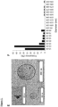
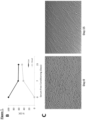
- Exosomes are isolated from serum-free media conditioned over 15 days by cultured human CDCs (or normal human dermal fibroblasts (NHDFs) as a therapeutically inert control) ( Figure 7 available online). By the end of the conditioning period, most of the CDCs remained alive despite the absence of regular media changes ( Figures 7B and 7C ). Purified exosome pellets were enriched in RNA ( Figure 1A ). The Inventors confirmed that the RNA resides within exosomes by exposing the pellet to RNase A in the presence of 5% triton ( Figure 1B ), with proteinase K added to dissociate protein complexes that may shield RNA.
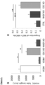
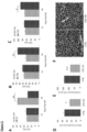
- CDC exosomes but not NHDF exosomes, promoted tube formation in human umbilical cord endothelial cells, indicative of enhanced angiogenesis ( Figure 1G ).
- CDC-exosome-treated neonatal cardiomyocytes proliferated more than those exposed to NHDF exosomes or media only, as evidenced by higher proportions of Ki67-positive nuclei ( Figure 1H ).
- CDC-exosome-treated cardiomyocytes exhibited fewer terminal deoxynucleotidyltransferase nick end labeling (TUNEL)-positive nuclei ( Figure 1I ).
- TUNEL terminal deoxynucleotidyltransferase nick end labeling
- CDCs stimulate functional improvement and regeneration in the infarcted myocardium in both animals and humans, but of central important to the present technology is whether exosomes derived from CDCs can reproduce or are indispensable to these processes.
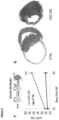
- the Inventors induced acute MI in immunodeficient mice then injected CDC exosomes, NHDF exosomes, or serum-free media into the MI border zone.
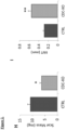
- microRNAs were differentially present in the two groups; among these, miR-146a was the most highly enriched in CDC exosomes (262-fold higher than in NHDF exosomes; Figures 4A , 4B , and 11 ).
- miR-146a tissue levels were increased in post-MI hearts from animals injected with CDC exosomes relative to those injected with NHDF exosomes ( Figure 4C ), rendering plausible the idea that CDC exosomes might act via miR-146a transfer.
- Exposure of neonatal rat cardiomyocytes to a miR-146a mimic increased cardiomyocyte viability and protected against oxidant stress ( Figures 4D and 12A ).
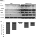
- Whole-transcriptome microarrays revealed downregulation of Irak1 and Traf6, two signaling mediators of the TLR-NFkB axis that are known targets of miR-146a ( Figure 4E ).
- miR-146a leads to thicker infarct wall thickness and increased viable tissue in a mouse model of myocardial infarct.
- the Inventors developed miR-146a-deficient exosomes by transfecting CDCs with a miR-146a hairpin inhibitor (or a control hairpin) followed by media conditioning and exosome isolation. Successful knockdown of miR-146a was confirmed by qPCR on resultant exosomes and on NRVMs exposed to either control or miR-146adepleted exosomes ( Figures 12B and 12C ).
- Exogenous miR-146a is known to suppress ischemia/reperfusion injury via targeting of Irak-1 and Traf6, both involved in the toll-like receptor (TLR) signaling pathway.
- TLR signaling underlying innate immunity plays a major role in the pathology of sterile inflammation, including MI.
- CDC-exosome-mediated reductions of proinflammatory cytokines ( Figure 8 ) and suppression of Irak1 and Traf6 by miR-146a (which is augmented in hearts injected with CDC exosomes; Figure 4C ) are consistent with blunted TLR signaling.
- miR-146a suppresses NOX-4, which has been shown to impart oxidative stress and to potentiate cardiac injury, and SMAD4, a member of the transforming growth factor b (TGF-b) profibrotic pathway.
- TGF-b transforming growth factor b
- Cardiosphere-derived cells have been shown to induce therapeutic regeneration of the infarcted human heart.
- CDCs led to shrinkage of scar and growth of new, functional myocardium. Similar effects have been corroborated in animal models.
- the Inventors show that exosomes reproduce CDC-induced therapeutic regeneration, and that inhibition of exosome production undermines the benefits of CDCs.
- Exosomes contain microRNAs, which have the ability to alter cell behavior through paracrine mechanisms ( Figure 6B ).
- the Inventors have identified miR-146a as being particularly enriched in CDC exosomes.
- miR-146a When administered alone, miR-146a reproduces some, but not all, of the salient benefits of CDCs and of CDC exosomes ( Figure 3 ). Likewise, miR-146a-depleted exosomes were still able to suppress cardiomyocyte apoptosis ( Figure 5A ), albeit more weakly than when miR-146a is present. Treating hearts with miR-146a in a chronic model of MI (after the scar is permanent) does reproduce the increase in viable mass that is the signature of therapeutic regeneration, but fails to mimic two key beneficial effects of CDC exosomes: decreased scar mass and improved global function. The increase in viable myocardium does not suffice to increase function, possibly because of inadequate angiogenesis elicited by miR-146a.
- miR-146a plays an important part in mediating the effects of CDC exosomes, but alone does not suffice to confer comprehensive therapeutic benefit.
- Other microRNAs in the repertoire may exert synonymous or perhaps synergistic effects with miR-146a.
- miR-22 another microRNA highly enriched in CDC exosomes
- miR-24 also identified in CDC exosomes
- overexpression of miR-24 in a model of MI decreased myocardial scar formation.
- CDC exosomes are naturally cell permeant, and their lipid bilayer coat protects their payloads from degradation as particles shuttle from cell to cell, so that the intact particles themselves may be well suited for disease applications.
- Injection of CDC-derived exosomes into the injured heart mimics the structural and functional benefits of CDC transplantation; conversely, inhibition of exosome secretion by CDC s abrogates the therapeutic benefits of transplanted CDCs. Not all exosomes are salutary: Injection of exosomes from dermal fibroblasts, control cells which are therapeutically inert, had no benefit. DC -exosomes decreased acute cardiomyocyte death and inflammatory cytokine release, while attenuating left ventricular (LV) remodeling and fibrosis in the injured heart. MicroRNA arrays reveal several "signature microRNAs" that are highly up-regulated in CDC-exosomes. In contrast, mass spectrometry indicates that the protein composition of CDC-exosomes is conventional and comparable to that of fibroblast exosomes.
- CDCs exert diverse but coordinated effects: they recruit endogenous progenitor cells and coax surviving heart cells to proliferate; on the other hand, injected CDCs suppress maladaptive LV remodeling, apoptosis, inflammation, and tissue fibrosis after MI. While it is possible that CDCs secrete a medley of individual growth factors and cytokines that collectively produce diverse benefits, the involvement of master-regulator microRNAs within exosomes would help tie together the various effects without postulating complex mixtures of numerous secreted protein factors.
- CDC exosomes contain rich signaling information conferred by a cell type that is the first shown to be capable of producing regeneration in a setting of "permanent" injury, and confer the same benefits as CDCs without transplantation of living cells. For all these reasons, CDC exosomes merit further development as cell-free therapeutic candidates.
- CDC-exosomes are demonstrated as capable of treating heart-related conditions, such as treat heart failure (HF) associated with Duchenne muscular dystrophy (DMD).
- HF heart failure
- DMD Duchenne muscular dystrophy
- Exosomes secreted by cells are capable of reproducing therapeutic benefits of their parental cells and based on the described knockdown studies, appear to be indispensable in effectuating such therapeutic benefits.
- these results have further identified that within their rich biological cargo of various proteins and RNA, microRNAs play a central role in activating regenerative processes, suggesting compelling applications in clinical therapeutics.
- Exosomes have significant advantages over traditional cell-based therapies including manufacturing advantages, relative ease of definition and characterization, lack of tumorgenicity and immunogenicity, and possibility of administration in therapeutic scenarios for which cell, tissue, organ or mechanical transplant is not available.
- CDC-exosomes represent a significant advance in biologic therapy.
- results are presented as mean ⁇ SEM; results for alternans are presented as mean ⁇ SD.
- Normality and equality of variances of data sets were first tested using Kolmogorov-Smirnov and Levene's tests, respectively. If both were confirmed, t-test or analysis of variance followed by Bonferroni 's post hoc test were used for determination of statistical significance; if either normality or equality of variances was not assured, nonparametric tests (Wilcoxon test or Kruskal-Wallis test followed by Dunn's post-test) were applied (SPSS II, SPSS Inc., Chicago, Illinois). No preliminary data were available for a power analysis. Experiments were planned with a sample size of 4 animals per group as an initial pilot project. Results from the pilot project allowed us to power subsequent studies. The study followed preclinical reporting standards, as described.
- Echocardiographic studies were performed two days before (Baseline) and 3 weeks, 2 and 3 months after first CDC/CDC exosome (CDC-XO) injection and 3 weeks, 2 and 3 months after second CDC/CDC-XO injection using the Vevo 770 Imaging System (VisualSonics, Toronto, Canada). The same imaging system was used to perform echocardiographic studies at baseline (2 day before) and 3 weeks after miR148a mimic injection. After induction of light general anesthesia, the heart was imaged at the level of the greatest LV diameter. LV ejection fraction (LVEF) was measured with VisualSonics version 1.3.8 software from 2-dimensional long-axis views.
- LVEF LV ejection fraction
- Exercise capacity was assessed weekly with Exer-3/6 open treadmill (Columbus Instruments, Columbus, OH), beginning 3 weeks after CDC/vehicle injection (exercise capacity measured in a subset of mdx mice 1 week pre-operatively was equivalent to that measured 3 weeks post-operatively in the Mdx+Vehicle group; data not shown). After an acclimation period (10 m/min for 20 min) stepwise increases in average speed (1 m/min) were applied every two minutes during treadmill exercise until the mouse became exhausted (spending >10 seconds on shocker; continuous nudging was used during treadmill to help mice stay on the track). Subsequently, the mouse was returned to the cage and the total distance recorded. After 3 months of weekly exercise, CDC/vehicle mdx mice along with wild-type age-matched mice were followed for assessment of mortality. The treadmill protocol conformed to guidelines from the American Physiological Society 3 .
- Mouse CDCs were expanded from wild-type strain-matched mouse hearts (C57BL/10ScSnJ wild type mouse heart) as described 2 . Briefly, ventricular tissues were minced into ⁇ 1 mm explants, partially digested enzymatically and plated on adherent (fibronectin-coated) culture dishes. These explants spontaneously yield outgrowth cells (explant-derived cells) which were harvested once confluent and plated in suspension culture (10 5 cells/mL on poly-D-lysine-coated dishes) to enable self-assembly of three-dimensional cardiospheres. Subsequent replating of cardiospheres on adherent culture dishes yielded CDCs which were used in all experiments at passage one.
- PCR Quantitative polymerase chain reaction
- Paraffin-embedded sections from apical, middle and basal parts of each heart were used for Masson's trichrome staining and immunostaining with antibodies against Ki67 and aurora B.
- Myocardial abundance of collagen I A1 and collagen III A1 was measured by Western blot analysis (Fig. 29).
- Exosomes were isolated from serum-free media conditioned overnight (24 hr) by cultured human CDCs (CDC-XO) [or normal human dermal fibroblasts (NHDF) as a control] in hypoxia (2% O 2 ; default condition) or normoxia (20% O 2 , solely for studies comparing RNA content of exosomes).
- Ultracentrifugation 100,000g for 1 hr was used to isolate exosomes from conditioned media after sequential centrifugations at 300g (10min) and 10,000g (30min) and filtration with 0.22 micron filters. Isolated exosomes underwent RNA extraction and subsequently RNA sequencing (Fig.
- preliminary dose-response experiments were performed, which identified 1 ⁇ 10 5 cells in first injection and 1 ⁇ 10 4 cells in second injection (3 months after first injection) as effective doses, consistent with prior dose-ranging experiments in ischemic and nonischemic mouse models.
- a total of 1 ⁇ 10 5 cells/40 ⁇ L phosphate-buffered saline (PBS; first injection) or 1 ⁇ 10 4 cells/40 ⁇ L PBS (second injection) or PBS alone were injected into left ventricular (LV) myocardium divided equally among 4 points as described.
- the LV was visually divided into three zones: basal, middle, and apical, with one injection in the basal, two in the middle and one in the apical zone.
- a miR-148a mimic (hsa-miR-148a-3p, 2 ⁇ g; Sigma-Aldrich, St. Louis, MO) was mixed with RNAiMAX transfection reagent (life technologies, Grand Island, NY) for 30 min at room temperature at a total volume of 40 ⁇ l and injected into 4 points per heart as described above.
- Paraffin-embedded sections from apical, middle and basal parts of each heart were used for histology.
- Masson's trichrome staining HT15 Trichrome Stain [Masson] Kit; Sigma-Aldrich, St.
- T cells, B cells and macrophages were assessed by immunostaining with antibodies against mouse CD3, CD20 and CD68, respectively, and the average number of cells in each heart was calculated from counting cells in 10 fields (20x magnification) from each of 10 sections selected randomly from the apical (3 sections; 50 ⁇ m interval), middle (4sections; 50 ⁇ m interval) and basal (3 sections; 50 ⁇ m interval) regions of each heart.
- Immunofluorescence staining was conducted using antibodies against mouse Ki-67 (SP6; 1:50; Thermo Fisher Scientific, Fremont, CA), WGA (Wheat germ agglutinin; 1:200; Life Technologies, Grand Island, NY), Nrf2 (C20; 1:50; Santa Cruz Biotechnology, Santa Cruz, CA), aurora B (1:250; BD Biosciences, San Jose, CA).
- Immunoperoxidase staining Immunohistochemical detection of CD3, CD20 and CD68 was performed on 5- ⁇ m sections using prediluted rabbit monoclonal antibodies from Ventana Medical System (Tuscon, AZ; CD68) and Cell Marque (Rocklin, CA; CD3, CD20).
- Staining was conducted on the Leica Bond-Max Ventana automated slide stainer (Chicago, IL) using onboard heat-induced epitope retrieval method in high pH ER2 buffer (Leica Biosystems, Buffalo Grove, IL). The staining was visualized using the Dako Envision + rabbit detection System and Dako DAB (Carpinteria, CA). The slides were subsequently counterstained with mayer's hematoxylin for 1 minute and coverslipped.
- Nrf2 signaling [Nrf2, phospho-Nrf2 (Nrf2-p s40 ) and Nrf2 downstream gene products: heme oxygenase-1 (HO-1), catalase, superoxide dismutase-2 (SOD-2), and catalytic subunit of glutamate-cysteine ligase (GCLC)], Nrf2 phosphorylation [phospho-Akt(Akt-p 308 )], oxidative phosphorylation [CI (NDUFB8 subunit), CII (SDHB subunit), CIV (MTCO1 subunit), CIII (UQCRC2 subunit) and CV (ATPSA subunit)], mitochondrial biogenesis (PGC-1), mitophagy (PINK1), inflammation (NF- ⁇ B and MCP-1) and fibrosis (Collagen IA1 and collagen IIIA1).
- Nrf2 signaling [Nrf2, phospho-Nrf2 (Nrf2-p s40 ) and Nrf2 downstream gene products: heme oxygenase
- Myocardial density of malondialdehyde protein adducts was also measured by Western blotting (WB).
- WB Western blotting
- Samples from apical, middle and basal parts of each heart were mixed and homogenized, and nuclear and cytoplasmic fractions were extracted per manufacturer's instructions (CelLytic NuCLEAR Extraction Kit, Sigma-Aldrich, St. Louis, MO).
- Cytoplasmic, nuclear and mitochondrial extracts for WB analysis were stored at -80C°.
- the protein concentrations in extracts were determined by the Micro BCA Protein Assay Kit (Life technologies, Grand Island, NY).
- Target proteins in the cytoplasmic, nuclear and mitochondrial fractions were measured by Western blot analysis using the following antibodies: antibodies against mouse Nrf2, HO-1, catalase, SOD-2, GCLC, collagen IA1, and collagen IIIA1, and PGC-1 were purchased from Santa Cruz Biotechnology (Santa Cruz, CA), phospho-Nrf2 (Nrf2-p s40 ; Biorbyt, San Francisco, CA), respiratory chain subunits (Total OXPHOS Rodent WB Antibody Cocktail antibody), malondialdehyde, citrate synthase and TBP (Abcam, Cambridge, MA), Akt and Akt-p T308 , I ⁇ B- ⁇ , p-I ⁇ B- ⁇ , (Cell Signaling Technology, Denver, CO), PINK1, MCP-1 and
- TBP TATA binding protein
- citrate synthase cytosolic and mitochondrial target proteins.
- Western blot methods Briefly, aliquots containing 20 ⁇ g proteins were fractionated on 8, 10 and 4-12% Bis-Tris gel (Life technologies, Grand Island, NY) at 120 V for 2 h and transferred to a PVDF membrane (Life technologies, Grand Island, NY). The membrane was incubated for 1 h in blocking buffer (1 ⁇ TBS, 0.05% Tween-20 and 5% nonfat milk) and then overnight in the same buffer containing the given antibodies at optimal dilutions listed in Table 1. Table 1.
- Antibodies and the optimal dilutions applied in Western blot analyses Antibody p-Akt Akt Malondialdehyde NF- ⁇ B p65 Nrf2 HO-1 p-Nrf2 Dilution 1:500 1:1000 1:1000 1:1000 1:500 1:500 1:250 Antibody TBP Collagen I Collagen III Citrate syn. p-I ⁇ B- ⁇ I ⁇ B- ⁇ PGC-1 Dilution 1:2000 1:500 1:500 1:2000 1:500 1:1000 1:500 Antibody Catalase SOD-2 GCLC MCP OXPHOS PINK1 Dilution 1:500 1:500 1:500 1:100 1:500 2 ⁇ g/ml
- the membrane was washed 3 times for 5 min in 1 ⁇ TBS, 0.05% Tween-20 before a 2-h incubation in a buffer (1 ⁇ TBS, 0.05% Tween-20 and 3% nonfat milk) containing horseradish peroxidase-linked anti-rabbit IgG, anti-mouse IgG (Cell Signaling Technology, Denver, CO) and anti-goat IgG (Sigma-Aldrich, St. Louis, MO) at 1:1000-3000 dilution.
- the membrane was washed 3 times for 5 min in 1 ⁇ TBS, 0.05% Tween-20 and developed by autoluminography using the ECL chemiluminescent agents (Super Signal West Pico Chemiluminescent Substrate; Life Technologies, Grand Island, NY). Citrate synthase and TBP were used as housekeeping proteins against which expressions of the proteins of interest were normalized. Phosphorylated Akt, Nrf2 and I ⁇ B- ⁇ were normalized to total Akt, Nrf2 and I ⁇ B- ⁇ . Western blot analyses of collagen I and collagen III were conducted under nonreducing, non-denaturing condition.
- Extracted DNAs (QIAamp DNA Mini Kit, QIAGEN, Germantown, MD) from whole heart tissue were used to measure mitochondrial to nuclear DNA ratio using PCR format per manufacturer's instructions (NovaQUANT TM Mouse Mitochondrial to Nuclear Ratio kit, EMD Millipore, Billerica, MA).
- mice were sacrificed via cervical dislocation after isofluorane anesthesia.
- Hearts were immediately excised, rinsed in PBS and homogenized via polytron in 1mL ice cold HES buffer (250mM sucrose, 1mM EDTA, 10mM HEPES, pH 7.4). Lysates were spun down at 1000g for 5min at 4°C to remove unbroken cells and large debris. Supernatant was then spun down at 7000g for 10min at 4°C to separate mitochondria-enriched fraction from crude cytosol. Pellet was resuspended in 1mL HES buffer (A subportion in lysis buffer for WB).
- Cal 520 was excited with a 488 nm laser and its emission (>505 nm) was collected with a 10X objective (Leica: N PLAN 10x/0.25) at scan speeds ranging from 36 to 7 ms per frame depending on the field size.
- the fluorescence intensity (F) proportional to Ca 2+ concentration was normalized to baseline fluorescence, F0 (F/F0).
- Time to peak and Ca 2+ transient amplitude (F/F0) were analyzed with the software Clampfit (ver. 10.2, Molecular Devices, Inc.). Beat-to-beat alternans in each group calculated over the 5-10 sec interval of pacing at 1 Hz.
- the amplitude of each transient from each cell was measured during pacing and mean and standard deviation were calculated and compared among groups.
- CDC injection enhanced ambulatory function ( Fig. 23B ).
- CDC-treated mdx mice showed a substantial increase in maximal exercise capacity, relative to vehicle-treated mdx mice, over the 3 mos that it was measured; survival also differed in the two groups ( Fig. 23C ).
- Fig. 23C By ⁇ 23 mos of age all vehicle-treated mdx mice had died, whereas >50% of CDC-treated mdx mice remained alive ( Fig. 23C ).
- Nrf2 is normally sequestered in the cytoplasm via binding to its repressor molecule, Keap1.
- Oxidative stress (as well as Nrf2 phosphorylation by protein kinases such as Akt) causes dissociation of the Nrf2-Keap1 complex, which culminates in nuclear translocation of Nrf2 and transcriptional activation of antioxidant enzymes.
- Akt and cytoplasmic and nuclear Nrf2 were high (as expected in response to oxidative stress); CDC treatment further increased their protein levels ( Fig. 23E ).
- downstream effectors heme oxygenase-1 (HO-1), catalase, superoxide dismutase-2 (SOD-2), and the catalytic subunit of glutamate-cysteine ligase (GCLC) were upregulated ( Fig. 23E ), leading to a profound reduction of malondialdehyde adducts (fatty acid peroxidation end products; Fig. 23F ) in CDC-treated mdx heart.
- Mitochondrial structure and function are abnormal in muscular dystrophy-associated heart failure.
- mitochondrial integrity improved 3 weeks after CDC injection: CDCs restored mitochondrial ultrastructure ( Fig. 24A ), increased mitochondrial DNA copy numbers (but not mitochondrial number; Fig. 24B & C ), augmented levels of respiratory chain subunits ( Fig. 24D ) and normalized the deficient respiratory capacity of isolated mdx mitochondria ( Fig. 24E ).
- NF ⁇ B the master regulator of pro-inflammatory cytokines and chemokines
- MCP1 monocyte chemoattractant protein1
- CD68 + macrophages and CD3 + T cells Increases in phosphorylated I ⁇ B and nuclear p65 contents were accompanied by marked upregulation of MCP1 (monocyte chemoattractant protein1) and accumulation of CD68 + macrophages and CD3 + T cells.
- CDC treatment reversed activation of NF ⁇ B and decreased the number of inflammatory cells in mdx hearts 3 weeks after CDC injection ( Fig. 24G & H ). The Inventors also probed the effects of CDCs on cardiomyogenesis.
- CDCs are known to increase endogenous cardiomyogenesis in ischemic and non-ischemic models. Likewise, CDC treatment promoted cardiomyogenesis in the mdx heart, as evidenced by a marked increase in Ki67 + and aurora B + cardiomyocytes (Fig. 29).
- Exosomes secreted by CDCs mimic the functional and structural benefits of CDCs in a murine model of myocardial infarction.
- CDC-exosomes In the mdx mouse model of DMD, likewise, functional, anti-fibrotic, and cardiomyogenic benefits of CDCs are reproduced by administration of exosomes isolated from media conditioned by hypoxic CDCs.
- Intramyocardial injection of two repeat doses of human CDC-exosomes led to sustained improvement in EF in mdx mice, relative to vehicle-treated mice ( Fig. 25A & Fig. 27).
- the amounts of collagen I and III decreased in CDC-exosome-injected mdx hearts ( Fig. 25B ), along with marked increases in the numbers of cycling (Ki67 + , Fig. 25C1 ) and proliferating (aurora B + , Fig. 25C2 ) cardiomyocytes.
- Duchenne human iPS-derived cardiomyocytes exhibit a number of phenotypic deficits also seen in mdx mouse hearts. Decreased oxygen consumption rate (OCR), reminiscent of that observed in mdx heart mitochondria ( Fig. 24E ), and abnormal calcium cycling are among the reported deficits 21 .
- OCR oxygen consumption rate
- CDCs attenuate fibrosis and inflammation in the mdx heart, while improving pump function, increasing exercise capacity and enhancing survival.
- the salient benefits of CDCs were reproduced by CDC-exosomes.
- the Inventors' findings support the hypothesis that CDCs act by secreting exosomes laden with genetic signals, including (but not limited to) miR-148a. These exosomes are taken up by the surrounding myocardium, where they antagonize multiple pathophysiological pathways that underlie DMD cardiomyopathy.
- the constellation of effects is synergistic: oxidative stress, inflammation and fibrosis are blunted, while cardiomyogenesis and mitochondrial function are augmented.
- cardiosphere derived cells are sources of cardiosphere derived cells, the use of alternative sources such as cells derived directly from heart biopsies (explant-derived cells), or from self-assembling clusters of heart-derived cells (cardiospheres), exosomes produced by such cells, method of isolating, characterizing or altering exosomes produced by such cells, and the particular use of the products created through the teachings of the invention.
- Various embodiments of the invention can specifically include or exclude any of these variations or elements.
- the numbers expressing quantities of ingredients, properties such as concentration, reaction conditions, and so forth, used to describe and claim certain embodiments of the invention are to be understood as being modified in some instances by the term "about.” Accordingly, in some embodiments, the numerical parameters set forth in the written description and attached claims are approximations that can vary depending upon the desired properties sought to be obtained by a particular embodiment. In some embodiments, the numerical parameters should be construed in light of the number of reported significant digits and by applying ordinary rounding techniques. Notwithstanding that the numerical ranges and parameters setting forth the broad scope of some embodiments of the invention are approximations, the numerical values set forth in the specific examples are reported as precisely as practicable. The numerical values presented in some embodiments of the invention may contain certain errors necessarily resulting from the standard deviation found in their respective testing measurements.
Landscapes
- Health & Medical Sciences (AREA)
- Life Sciences & Earth Sciences (AREA)
- Chemical & Material Sciences (AREA)
- Engineering & Computer Science (AREA)
- General Health & Medical Sciences (AREA)
- Veterinary Medicine (AREA)
- Public Health (AREA)
- Animal Behavior & Ethology (AREA)
- Pharmacology & Pharmacy (AREA)
- Medicinal Chemistry (AREA)
- Bioinformatics & Cheminformatics (AREA)
- Epidemiology (AREA)
- Zoology (AREA)
- Organic Chemistry (AREA)
- Biomedical Technology (AREA)
- Genetics & Genomics (AREA)
- Molecular Biology (AREA)
- Biotechnology (AREA)
- Cell Biology (AREA)
- Developmental Biology & Embryology (AREA)
- Immunology (AREA)
- Vascular Medicine (AREA)
- Cardiology (AREA)
- Chemical Kinetics & Catalysis (AREA)
- General Chemical & Material Sciences (AREA)
- Nuclear Medicine, Radiotherapy & Molecular Imaging (AREA)
- Biochemistry (AREA)
- General Engineering & Computer Science (AREA)
- Wood Science & Technology (AREA)
- Marine Sciences & Fisheries (AREA)
- Virology (AREA)
- Dermatology (AREA)
- Proteomics, Peptides & Aminoacids (AREA)
- Gastroenterology & Hepatology (AREA)
- Heart & Thoracic Surgery (AREA)
- Neurology (AREA)
- Urology & Nephrology (AREA)
- Orthopedic Medicine & Surgery (AREA)
- Physical Education & Sports Medicine (AREA)
- Physics & Mathematics (AREA)
Applications Claiming Priority (3)
| Application Number | Priority Date | Filing Date | Title |
|---|---|---|---|
| US201462059308P | 2014-10-03 | 2014-10-03 | |
| PCT/US2015/053853 WO2016054591A1 (en) | 2014-10-03 | 2015-10-02 | Cardiosphere-derived cells and exosomes secreted by such cells in the treatment of muscular dystrophy |
| EP15846113.7A EP3200808B1 (de) | 2014-10-03 | 2015-10-02 | Kardiosphären-zellen und durch diese zellen sekretierte exosomen bei der behandlung von muskeldystrophie |
Related Parent Applications (1)
| Application Number | Title | Priority Date | Filing Date |
|---|---|---|---|
| EP15846113.7A Division EP3200808B1 (de) | 2014-10-03 | 2015-10-02 | Kardiosphären-zellen und durch diese zellen sekretierte exosomen bei der behandlung von muskeldystrophie |
Publications (2)
| Publication Number | Publication Date |
|---|---|
| EP4494699A2 true EP4494699A2 (de) | 2025-01-22 |
| EP4494699A3 EP4494699A3 (de) | 2025-04-30 |
Family
ID=55631661
Family Applications (2)
| Application Number | Title | Priority Date | Filing Date |
|---|---|---|---|
| EP24189285.0A Pending EP4494699A3 (de) | 2014-10-03 | 2015-10-02 | Kardiosphären-zellen und durch diese zellen sekretierte exosomen bei der behandlung von muskeldystrophie |
| EP15846113.7A Active EP3200808B1 (de) | 2014-10-03 | 2015-10-02 | Kardiosphären-zellen und durch diese zellen sekretierte exosomen bei der behandlung von muskeldystrophie |
Family Applications After (1)
| Application Number | Title | Priority Date | Filing Date |
|---|---|---|---|
| EP15846113.7A Active EP3200808B1 (de) | 2014-10-03 | 2015-10-02 | Kardiosphären-zellen und durch diese zellen sekretierte exosomen bei der behandlung von muskeldystrophie |
Country Status (6)
| Country | Link |
|---|---|
| US (2) | US11357799B2 (de) |
| EP (2) | EP4494699A3 (de) |
| JP (4) | JP6878274B2 (de) |
| AU (2) | AU2015327812B2 (de) |
| CA (1) | CA2962444C (de) |
| WO (1) | WO2016054591A1 (de) |
Families Citing this family (28)
| Publication number | Priority date | Publication date | Assignee | Title |
|---|---|---|---|---|
| US11660317B2 (en) | 2004-11-08 | 2023-05-30 | The Johns Hopkins University | Compositions comprising cardiosphere-derived cells for use in cell therapy |
| US11286463B2 (en) | 2012-03-08 | 2022-03-29 | Advanced ReGen Medical Technologies, LLC | Reprogramming of aged adult stem cells |
| EP3563859B1 (de) | 2012-08-13 | 2021-10-13 | Cedars-Sinai Medical Center | Von kardiosphären abgeleitete exosomen zur geweberegeneration |
| US10772911B2 (en) | 2013-12-20 | 2020-09-15 | Advanced ReGen Medical Technologies, LLC | Cell free compositions for cellular restoration and methods of making and using same |
| US20150174166A1 (en) | 2013-12-20 | 2015-06-25 | Advanced ReGen Medical Technologies, LLC | Compositions for Cellular Restoration and Methods of Making and Using Same |
| EP4494699A3 (de) | 2014-10-03 | 2025-04-30 | Cedars-Sinai Medical Center | Kardiosphären-zellen und durch diese zellen sekretierte exosomen bei der behandlung von muskeldystrophie |
| US20170304368A1 (en) * | 2014-10-06 | 2017-10-26 | Cedars-Sinai Medical Center | Polarization of macrophages to a healing phenotype by cardiosphere-derived cells and by the exosomes secreted by such cells |
| WO2017123662A1 (en) | 2016-01-11 | 2017-07-20 | Cedars-Sinai Medical Center | Cardiosphere-derived cells and exosomes secreted by such cells in the treatment of heart failure with preserved ejection fraction |
| US11203754B2 (en) | 2016-04-29 | 2021-12-21 | Advanced ReGen Medical Technologies, LLC | Microrna compositions and methods of making and using same |
| WO2017210652A1 (en) | 2016-06-03 | 2017-12-07 | Cedars-Sinai Medical Center | Cdc-derived exosomes for treatment of ventricular tachyarrythmias |
| US11541078B2 (en) | 2016-09-20 | 2023-01-03 | Cedars-Sinai Medical Center | Cardiosphere-derived cells and their extracellular vesicles to retard or reverse aging and age-related disorders |
| CN110072534A (zh) * | 2016-10-13 | 2019-07-30 | Vbc控股有限责任公司 | 外泌体的医学应用 |
| US20190382850A1 (en) * | 2017-01-27 | 2019-12-19 | Memorial Sloan-Kettering Cancer Center | Method for identifying mitochondrial dna in extracellular vesicles and treatment of mtdna-related disorders and cancer |
| JP7336769B2 (ja) * | 2017-04-19 | 2023-09-01 | シーダーズ―シナイ メディカル センター | 骨格筋ジストロフィーを治療する方法及び組成物 |
| EP4218775A1 (de) * | 2017-08-04 | 2023-08-02 | Cedars-Sinai Medical Center | Aus kardiosphären abgeleitete zellen und deren extrazelluläre vesikel zur behandlung und prävention von krebs |
| US11446323B2 (en) | 2017-08-31 | 2022-09-20 | Tel Hashomer Medical Research Infrastructure And Services Ltd. | Compositions and methods for the treatment of fibrotic diseases |
| WO2019126068A1 (en) | 2017-12-20 | 2019-06-27 | Cedars-Sinai Medical Center | Engineered extracellular vesicles for enhanced tissue delivery |
| EP3740576A4 (de) | 2018-01-18 | 2021-10-20 | Advanced Regen Medical Technologies, LLC | Therapeutische zusammensetzungen und verfahren zur herstellung und verwendung davon |
| EP3749344A4 (de) | 2018-02-05 | 2022-01-26 | Cedars-Sinai Medical Center | Verfahren zur therapeutischen verwendung von exosomen und y-rnas |
| EP3598978B1 (de) | 2018-07-26 | 2024-05-29 | EXOFIX S.r.l. | Aus fibroadipogenem vorläufer stammende exosomen zur regenerierung von dystrophischen muskeln |
| EP3674420A1 (de) * | 2018-12-28 | 2020-07-01 | Fundación Para la Investigación del Hospital Universitario y Politécnico La Fe de la Comunidad Valenciana | Verfahren zur vorhersage des kardiotoxizitätsrisikos bei krebspatienten, die eine chemotherapeutische mit anthracyclinen erhalten |
| US20220241325A1 (en) * | 2019-07-26 | 2022-08-04 | Mayo Foundation For Medical Education And Research | Antioxidant and antiviral compositions and methods |
| WO2021127003A1 (en) * | 2019-12-16 | 2021-06-24 | Children's Medical Center Corporation | Treating heart disease in muscular dystrophy patients |
| JP2023550453A (ja) * | 2020-11-18 | 2023-12-01 | 富士フイルム株式会社 | セクレトーム含有組成物の生成、並びにその使用及び分析方法 |
| CA3217654A1 (en) * | 2021-04-23 | 2022-10-27 | Spot Biosystems Ltd. | Extracellular vesicles loaded with biomolecules |
| AU2023314136A1 (en) * | 2022-07-26 | 2025-02-13 | Mayo Foundation For Medical Education And Research | Compositions and methods for treating peripheral vascular disease |
| CN116473512B (zh) * | 2023-03-22 | 2024-05-03 | 上海交通大学 | 一种动物循环系统中外泌体的监测装置及监测方法 |
| WO2025166297A1 (en) * | 2024-02-01 | 2025-08-07 | Immorta Bio, Inc. | Therapeutic use of autologous pluripotent stem cell and differentiated cell exosomes |
Citations (2)
| Publication number | Priority date | Publication date | Assignee | Title |
|---|---|---|---|---|
| US5585089A (en) | 1988-12-28 | 1996-12-17 | Protein Design Labs, Inc. | Humanized immunoglobulins |
| WO2014028493A2 (en) | 2012-08-13 | 2014-02-20 | Cedars-Sinai Medical Center | Exosomes and micro-ribonucleic acids for tissue regeneration |
Family Cites Families (385)
| Publication number | Priority date | Publication date | Assignee | Title |
|---|---|---|---|---|
| US3470876A (en) | 1966-09-28 | 1969-10-07 | John Barchilon | Dirigible catheter |
| US4106488A (en) | 1974-08-20 | 1978-08-15 | Robert Thomas Gordon | Cancer treatment method |
| US3964468A (en) | 1975-05-30 | 1976-06-22 | The Board Of Trustees Of Leland Stanford Junior University | Bioptome |
| US4659839A (en) | 1984-10-10 | 1987-04-21 | Mallinckrodt, Inc. | Coupling agents for radiolabeled antibody fragments |
| GB8711614D0 (en) | 1987-05-16 | 1987-06-24 | Medical Res Council | Proteins |
| US4960134A (en) | 1988-11-18 | 1990-10-02 | Webster Wilton W Jr | Steerable catheter |
| US5175004A (en) | 1988-12-27 | 1992-12-29 | Matsumura Kenneth N | Propagatable, new combinant cells for cellular replacement therapy |
| US4921482A (en) | 1989-01-09 | 1990-05-01 | Hammerslag Julius G | Steerable angioplasty device |
| US5052402A (en) | 1989-01-31 | 1991-10-01 | C.R. Bard, Inc. | Disposable biopsy forceps |
| US5104787A (en) | 1990-03-05 | 1992-04-14 | Lindstrom Richard L | Method for apparatus for a defined serumfree medical solution useful for corneal preservation |
| US6326198B1 (en) | 1990-06-14 | 2001-12-04 | Regents Of The University Of Michigan | Methods and compositions for the ex vivo replication of stem cells, for the optimization of hematopoietic progenitor cell cultures, and for increasing the metabolism, GM-CSF secretion and/or IL-6 secretion of human stromal cells |
| US5401629A (en) | 1990-08-07 | 1995-03-28 | The Salk Institute Biotechnology/Industrial Associates, Inc. | Assay methods and compositions useful for measuring the transduction of an intracellular signal |
| ES2077007T3 (es) | 1990-12-07 | 1995-11-16 | Ruesch Willy Ag | Instrumento medico con cabeza orientable. |
| US5329923A (en) | 1991-02-15 | 1994-07-19 | Lundquist Ingemar H | Torquable catheter |
| US5228441A (en) | 1991-02-15 | 1993-07-20 | Lundquist Ingemar H | Torquable catheter and method |
| US5454787A (en) | 1991-02-15 | 1995-10-03 | Lundquist; Ingemar H. | Torquable tubular assembly and torquable catheter utilizing the same |
| AU660444B2 (en) | 1991-02-15 | 1995-06-29 | Ingemar H. Lundquist | Torquable catheter and method |
| US5315996A (en) | 1991-02-15 | 1994-05-31 | Lundquist Ingemar H | Torquable catheter and method |
| US5981165A (en) | 1991-07-08 | 1999-11-09 | Neurospheres Holdings Ltd. | In vitro induction of dopaminergic cells |
| US5287857A (en) | 1992-06-22 | 1994-02-22 | David Mann | Apparatus and method for obtaining an arterial biopsy |
| US5243167A (en) | 1992-09-16 | 1993-09-07 | Ingemar H. Lundquist | Apparatus and method for manufacturing a slotted torque tube |
| US5334145A (en) | 1992-09-16 | 1994-08-02 | Lundquist Ingemar H | Torquable catheter |
| US5383852A (en) | 1992-12-04 | 1995-01-24 | C. R. Bard, Inc. | Catheter with independent proximal and distal control |
| US5368564A (en) | 1992-12-23 | 1994-11-29 | Angeion Corporation | Steerable catheter |
| ATE335091T1 (de) | 1993-05-28 | 2006-08-15 | Sinai School Medicine | Bc200 rna, daraus abgeleitete proben und entsprechende anwendungen |
| US5492825A (en) | 1993-08-06 | 1996-02-20 | The Regents Of The University Of California | Mammalian inward rectifier potassium channel cDNA, IRK1, corresponding vectors, and transformed cells |
| US5616568A (en) | 1993-11-30 | 1997-04-01 | The Research Foundation Of State University Of New York | Functionalized derivatives of hyaluronic acid |
| US5454827A (en) | 1994-05-24 | 1995-10-03 | Aust; Gilbert M. | Surgical instrument |
| US5840502A (en) | 1994-08-31 | 1998-11-24 | Activated Cell Therapy, Inc. | Methods for enriching specific cell-types by density gradient centrifugation |
| US5702905A (en) | 1994-09-28 | 1997-12-30 | Spectral Diagnostics | Monoclonal antibody to human ventricular myosin light chains |
| DE69621528T2 (de) | 1995-02-07 | 2003-01-16 | Shiseido Co. Ltd., Tokio/Tokyo | Anti-inflamatorische agenzien |
| US5551427A (en) | 1995-02-13 | 1996-09-03 | Altman; Peter A. | Implantable device for the effective elimination of cardiac arrhythmogenic sites |
| US5715832A (en) | 1995-02-28 | 1998-02-10 | Boston Scientific Corporation | Deflectable biopsy catheter |
| US5925567A (en) | 1995-05-19 | 1999-07-20 | T. Breeders, Inc. | Selective expansion of target cell populations |
| US5702433A (en) | 1995-06-27 | 1997-12-30 | Arrow International Investment Corp. | Kink-resistant steerable catheter assembly for microwave ablation |
| ATE365808T1 (de) | 1995-07-28 | 2007-07-15 | Marie Curie Cancer Care | Transportproteine und deren verwendungen |
| FR2739621B1 (fr) | 1995-10-05 | 1997-12-05 | Centre Nat Rech Scient | Peptides utilisables comme vecteurs pour l'adressage intracellulaire de molecules actives |
| US6337387B1 (en) | 1995-11-17 | 2002-01-08 | Asahi Kasei Kabushiki Kaisha | Differentiation-suppressive polypeptide |
| US5762069A (en) | 1995-12-29 | 1998-06-09 | Akos Biomedical, Inc. | Multiple sample biopsy forceps |
| US5856155A (en) | 1996-02-23 | 1999-01-05 | The Johns Hopkins University School Of Medicine | Compounds and related methods for modulating potassium ion channels and assays for such compounds |
| US5824031A (en) | 1996-02-28 | 1998-10-20 | Cardio Source | Apparatus and method for deflecting a tip of a lead or catheter |
| US6132390A (en) | 1996-02-28 | 2000-10-17 | Eupalamus Llc | Handle for manipulation of a stylet used for deflecting a tip of a lead or catheter |
| US5782743A (en) | 1996-05-06 | 1998-07-21 | Russell; John J. | Magnetic medical treatment device |
| US5782748A (en) | 1996-07-10 | 1998-07-21 | Symbiosis Corporation | Endoscopic surgical instruments having detachable proximal and distal portions |
| GB9615726D0 (en) | 1996-07-26 | 1996-09-04 | Medical Res Council | Anti-viral agent 11 |
| US5955275A (en) | 1997-02-14 | 1999-09-21 | Arcaris, Inc. | Methods for identifying nucleic acid sequences encoding agents that affect cellular phenotypes |
| US6017735A (en) | 1997-01-23 | 2000-01-25 | Marie Curie Cancer Care | Materials and methods for intracellular transport and their uses |
| US6086582A (en) | 1997-03-13 | 2000-07-11 | Altman; Peter A. | Cardiac drug delivery system |
| US6511477B2 (en) | 1997-03-13 | 2003-01-28 | Biocardia, Inc. | Method of drug delivery to interstitial regions of the myocardium |
| US6547787B1 (en) | 1997-03-13 | 2003-04-15 | Biocardia, Inc. | Drug delivery catheters that attach to tissue and methods for their use |
| US6443949B2 (en) | 1997-03-13 | 2002-09-03 | Biocardia, Inc. | Method of drug delivery to interstitial regions of the myocardium |
| US6416510B1 (en) | 1997-03-13 | 2002-07-09 | Biocardia, Inc. | Drug delivery catheters that attach to tissue and methods for their use |
| AU733310C (en) | 1997-05-14 | 2001-11-29 | University Of British Columbia, The | High efficiency encapsulation of charged therapeutic agents in lipid vesicles |
| US6099832A (en) | 1997-05-28 | 2000-08-08 | Genzyme Corporation | Transplants for myocardial scars |
| US5899914A (en) | 1997-06-11 | 1999-05-04 | Endius Incorporated | Surgical instrument |
| US5851212A (en) | 1997-06-11 | 1998-12-22 | Endius Incorporated | Surgical instrument |
| CA2241615A1 (en) | 1997-06-26 | 1998-12-26 | An-Go-Gen Inc. | Catheters |
| US6387369B1 (en) | 1997-07-14 | 2002-05-14 | Osiris Therapeutics, Inc. | Cardiac muscle regeneration using mesenchymal stem cells |
| US7514074B2 (en) | 1997-07-14 | 2009-04-07 | Osiris Therapeutics, Inc. | Cardiac muscle regeneration using mesenchymal stem cells |
| GB9718609D0 (en) | 1997-09-02 | 1997-11-05 | Imp College Innovations Ltd | Fusion protein |
| US6123699A (en) | 1997-09-05 | 2000-09-26 | Cordis Webster, Inc. | Omni-directional steerable catheter |
| US7037648B1 (en) | 1997-11-07 | 2006-05-02 | John Hopkins University | Somatic transfer of modified genes to predict drug effects |
| US5938603A (en) | 1997-12-01 | 1999-08-17 | Cordis Webster, Inc. | Steerable catheter with electromagnetic sensor |
| US6203487B1 (en) | 1997-12-31 | 2001-03-20 | Thomas Jefferson University | Use of magnetic particles in the focal delivery of cells |
| JP4535468B2 (ja) | 1998-02-05 | 2010-09-01 | バイオセンス・ウエブスター・インコーポレーテツド | 心臓内薬物送達 |
| AU3112499A (en) | 1998-03-23 | 1999-10-18 | Zymogenetics Inc. | Cardiac-derived stem cells |
| DE29825182U1 (de) | 1998-04-02 | 2005-12-22 | Vossen, Franz | Vorrichtung zum Entfernen von Ausbrechteilen aus einem Werkstoffbogen o.dgl. |
| US6296630B1 (en) | 1998-04-08 | 2001-10-02 | Biocardia, Inc. | Device and method to slow or stop the heart temporarily |
| US20010044619A1 (en) | 1998-04-08 | 2001-11-22 | Peter A. Altman | Cardiac drug delivery system and method for use |
| US6171610B1 (en) | 1998-04-24 | 2001-01-09 | University Of Massachusetts | Guided development and support of hydrogel-cell compositions |
| US6165164A (en) | 1999-03-29 | 2000-12-26 | Cordis Corporation | Catheter for injecting therapeutic and diagnostic agents |
| US6540725B1 (en) | 1998-06-04 | 2003-04-01 | Biosense Webster, Inc. | Injection catheter with controllably extendable injection needle |
| DE19833476B4 (de) | 1998-07-24 | 2005-08-25 | Huss, Ralf, Dr. | Genetisch modifizierte CD34-Negative, adhärent wachsende hämatopoetische Stammzellen und deren Verwendung in der Gentherapie |
| US6102887A (en) | 1998-08-11 | 2000-08-15 | Biocardia, Inc. | Catheter drug delivery system and method for use |
| US6074408A (en) | 1998-10-13 | 2000-06-13 | Freeman; Kenneth V. | Modular medical instrument and method of using same |
| US6153582A (en) | 1998-11-05 | 2000-11-28 | Bausch & Lomb Surgical, Inc. | Defined serumfree medical solution for ophthalmology |
| US6572611B1 (en) | 1998-11-23 | 2003-06-03 | C. R. Bard, Inc. | Intracardiac grasp catheter |
| US6642049B1 (en) | 1998-12-04 | 2003-11-04 | The United States Of America As Represented By The Secretary Of The Navy | Human brain endothelial cells and growth medium and method for expansion of primitive CD34+CD38-bone marrow stem cells |
| US6193763B1 (en) | 1998-12-17 | 2001-02-27 | Robert A. Mackin | Apparatus and method for contemporaneous treatment and fluoroscopic mapping of body tissue |
| AUPP785098A0 (en) | 1998-12-21 | 1999-01-21 | Victor Chang Cardiac Research Institute, The | Treatment of heart disease |
| WO2007019398A1 (en) | 2005-08-03 | 2007-02-15 | Advanced Cell Technology, Inc. | Improved methods of reprogramming animal somatic cells |
| US6783510B1 (en) | 1999-07-08 | 2004-08-31 | C.R. Bard, Inc. | Steerable catheter |
| US7015037B1 (en) | 1999-08-05 | 2006-03-21 | Regents Of The University Of Minnesota | Multiponent adult stem cells and methods for isolation |
| WO2001010482A1 (en) | 1999-08-05 | 2001-02-15 | Biocardia, Inc. | A system and method for delivering thermally sensitive and reverse-thermal gelation matrials |
| SE9903185D0 (sv) | 1999-09-08 | 1999-09-08 | Europ I Of Science Ab | Terapeutisk metod och anordning baserad på magnetism |
| JP2003509374A (ja) | 1999-09-14 | 2003-03-11 | チルドレンズ メディカル センター コーポレーション | 骨髄細胞を用いる筋ジストロフィーの治療方法 |
| US20030161817A1 (en) | 2001-03-28 | 2003-08-28 | Young Henry E. | Pluripotent embryonic-like stem cells, compositions, methods and uses thereof |
| WO2001021767A2 (en) | 1999-09-24 | 2001-03-29 | Morphogen Pharmaceuticals, Inc. | Pluripotent embryonic-like stem cells, compositions, methods and uses thereof |
| WO2001026727A1 (en) | 1999-10-13 | 2001-04-19 | Biocardia, Inc. | Pulmonary vein arrhythmia diagnostic device and method for use |
| US6716242B1 (en) | 1999-10-13 | 2004-04-06 | Peter A. Altman | Pulmonary vein stent and method for use |
| WO2001037721A2 (en) | 1999-11-22 | 2001-05-31 | The Research Foundation Of State University Of New York | Magnetic nanoparticles for selective therapy |
| US6224587B1 (en) | 1999-11-22 | 2001-05-01 | C.R. Bard, Inc. | Steerable catheter |
| CA2392615A1 (en) | 1999-12-06 | 2001-06-07 | The General Hospital Corporation | Pancreatic stem cells and their use in transplantation |
| EP1254952A4 (de) | 1999-12-28 | 2004-09-22 | Kyowa Hakko Kogyo Kk | Zellen die in der lage sind in herzmuskelzellen zu differenzieren |
| JP2003530086A (ja) | 2000-01-14 | 2003-10-14 | ベス イスラエル ディーコネス メディカル センター | 心臓細胞特異的エンハンサー因子とその使用 |
| US6530944B2 (en) | 2000-02-08 | 2003-03-11 | Rice University | Optically-active nanoparticles for use in therapeutic and diagnostic methods |
| US6585716B2 (en) | 2000-04-05 | 2003-07-01 | Biocardia, Inc. | Method of treating the heart |
| US6478776B1 (en) | 2000-04-05 | 2002-11-12 | Biocardia, Inc. | Implant delivery catheter system and methods for its use |
| US20040087016A1 (en) | 2000-05-12 | 2004-05-06 | University Of Utah Research Foundation | Compositions and methods for cell dedifferentiation and tissue regeneration |
| AU2001273421A1 (en) | 2000-07-13 | 2002-01-30 | Bioheart, Inc. | Deployment system for myocardial cellular material |
| WO2002013760A2 (en) | 2000-07-31 | 2002-02-21 | New York Medical College | Methods and compositions for the repair and/or regeneration of damaged myocardium |
| US20110091428A1 (en) | 2000-07-31 | 2011-04-21 | New York Medical College | Compositions of adult organ stem cells and uses thereof |
| US7862810B2 (en) | 2000-07-31 | 2011-01-04 | New York Medical College | Methods and compositions for the repair and/or regeneration of damaged myocardium |
| US20020061587A1 (en) | 2000-07-31 | 2002-05-23 | Piero Anversa | Methods and compositions for the repair and/or regeneration of damaged myocardium |
| US7547674B2 (en) | 2001-06-06 | 2009-06-16 | New York Medical College | Methods and compositions for the repair and/or regeneration of damaged myocardium |
| WO2002087419A2 (en) | 2001-04-27 | 2002-11-07 | Johns Hopkins University | Biological pacemaker |
| WO2002019966A2 (en) | 2000-09-06 | 2002-03-14 | Johns Hopkins University | Cardiac arrhythmia treatment methods |
| US6569105B1 (en) | 2000-09-14 | 2003-05-27 | Syntheon, Llc | Rotatable and deflectable biopsy forceps |
| FR2814752A1 (fr) | 2000-10-02 | 2002-04-05 | Chru Lille | Procede d'obtention in vitro de cellules insulino- secretrices de mammifere et leurs utilisations |
| US6511471B2 (en) | 2000-12-22 | 2003-01-28 | Biocardia, Inc. | Drug delivery catheters that attach to tissue and methods for their use |
| EP2316919B1 (de) | 2001-02-14 | 2015-10-07 | Anthrogenesis Corporation | Post-partum Säugetier-Plazenta, deren Verwendung und daraus gewonnene Stammzellen |
| EP1372916A2 (de) | 2001-03-21 | 2004-01-02 | Vitrox APS | Verfahren und gehäuse zum bearbeiten von materialien |
| AU2002306865A1 (en) | 2001-03-23 | 2003-03-03 | Warren J. Alilain | Generation of multipotent central nervous system stem cells |
| US7026121B1 (en) | 2001-06-08 | 2006-04-11 | Expression Diagnostics, Inc. | Methods and compositions for diagnosing and monitoring transplant rejection |
| US6905827B2 (en) | 2001-06-08 | 2005-06-14 | Expression Diagnostics, Inc. | Methods and compositions for diagnosing or monitoring auto immune and chronic inflammatory diseases |
| WO2003052925A1 (en) | 2001-06-29 | 2003-06-26 | Xanoptix, Inc. | Bicmos ac filter circuit |
| CA2351156A1 (en) | 2001-07-04 | 2003-01-04 | Peter W. Zandstra | A bioprocess for the generation of pluripotent cell derived cells and tissues |
| US6796963B2 (en) | 2001-07-10 | 2004-09-28 | Myocardial Therapeutics, Inc. | Flexible tissue injection catheters with controlled depth penetration |
| WO2003006950A2 (en) | 2001-07-12 | 2003-01-23 | Geron Corporation | Cells of the cardiomyocyte lineage produced from human pluripotent stem cells |
| WO2003008535A2 (en) | 2001-07-20 | 2003-01-30 | Technion Research And Development Foundation Ltd. | Methods of generating human cardiac cells and tissues and uses thereof |
| JP2006508014A (ja) | 2001-07-24 | 2006-03-09 | イエール・ユニバーシテイ | キチナーゼおよびキチナーゼ様分子、ならびに炎症性疾患に関する方法、組成物およびキット |
| US7074175B2 (en) | 2001-07-25 | 2006-07-11 | Erik Schroeder Handy | Thermotherapy via targeted delivery of nanoscale magnetic particles |
| US7731648B2 (en) | 2001-07-25 | 2010-06-08 | Aduro Biotech | Magnetic nanoscale particle compositions, and therapeutic methods related thereto |
| US6997863B2 (en) | 2001-07-25 | 2006-02-14 | Triton Biosystems, Inc. | Thermotherapy via targeted delivery of nanoscale magnetic particles |
| US20030044976A1 (en) | 2001-08-27 | 2003-03-06 | Advanced Cell Technology | De-differentiation and re-differentiation of somatic cells and production of cells for cell therapies |
| US7452532B2 (en) | 2001-09-30 | 2008-11-18 | Scicotec Gmbh | Transluminal application of adult stem cells for body organ tissue repair |
| US6805860B1 (en) | 2001-09-30 | 2004-10-19 | Eckhard Alt | Method of transluminal application of myogenic cells for repair or replacement of heart tissue |
| US20030082153A1 (en) | 2001-10-22 | 2003-05-01 | The Government Of The United States Of America | Stem cells that transform to beating cardiomyocytes |
| JP4447916B2 (ja) | 2001-11-05 | 2010-04-07 | メドジニックス・インコーポレイテッド | 微小器官処理システム及びそれを用いた微小器官の製造方法 |
| DE10162353A1 (de) | 2001-12-18 | 2003-07-03 | Roehm Gmbh | Extrusionsdüse für Folien oder Platten mit seitlicher Schmelzeeinspeisung |
| US20040014209A1 (en) | 2002-01-23 | 2004-01-22 | Lassar Andrew B. | Compositions and methods for modulating cell differentiation |
| GB0202149D0 (en) | 2002-01-30 | 2002-03-20 | Univ Edinburgh | Pluripotency determining factors and uses thereof |
| TWI288779B (en) | 2002-03-28 | 2007-10-21 | Blasticon Biotech Forschung | Dedifferentiated, programmable stem cells of monocytic origin, and their production and use |
| US6866117B2 (en) | 2002-04-05 | 2005-03-15 | Wing Enterprises, Inc. | Light weight ladder systems and methods |
| US7840261B2 (en) | 2002-06-05 | 2010-11-23 | Biocardia, Inc. | Catheter systems and methods for placing bi-ventricular pacing leads |
| EP1539799B1 (de) | 2002-06-21 | 2013-12-11 | The University of Utah Research Foundation | Quervernetzte verbindungen und verfahren zu deren herstellung und anwendung |
| AU2003259152A1 (en) | 2002-07-23 | 2004-02-09 | Boston Scientific Limited | Cell therapy for regeneration |
| JP2005534345A (ja) | 2002-07-29 | 2005-11-17 | エス セル インターナショナル ピーティーイー リミテッド | インスリン陽性、グルコース応答性細胞の分化のための多段階方法 |
| AU2003270051A1 (en) | 2002-08-29 | 2004-03-19 | Baylor College Of Medicine | Heart derived cells for cardiac repair |
| CA2505251A1 (en) | 2002-11-05 | 2004-05-27 | Brigham And Women's Hospital, Inc. | Mesenchymal stem cells and methods of use thereof |
| WO2004058939A2 (en) | 2002-11-12 | 2004-07-15 | The Research Foundation Of The State University Of New York | TRANSLATIONAL CONTROL BY SMALL, NON-TRANSLATABLE RNAs |
| US7794702B2 (en) | 2003-01-15 | 2010-09-14 | The Trustees Of Columbia University In The City Of New York | Mesenchymal stem cells as a vehicle for ion channel transfer in syncytial structures |
| CN1768133A (zh) | 2003-01-31 | 2006-05-03 | 加利福尼亚大学董事会 | 使用islet1作为标志分离或产生干细胞 |
| DE10331439B3 (de) | 2003-07-10 | 2005-02-03 | Micromod Partikeltechnologie Gmbh | Magnetische Nanopartikel mit verbesserten Magneteigenschaften |
| EP2287315A1 (de) | 2003-03-04 | 2011-02-23 | Intercell AG | Streptococcus-pyogenes-Antigene |
| US7837631B2 (en) | 2003-03-14 | 2010-11-23 | Boston Scientific Scimed Inc. | Biopsy forceps with removable jaw segments |
| US20060041182A1 (en) | 2003-04-16 | 2006-02-23 | Forbes Zachary G | Magnetically-controllable delivery system for therapeutic agents |
| US20040214182A1 (en) | 2003-04-25 | 2004-10-28 | Vinod Sharma | Genetic modification of targeted regions of the cardiac conduction system |
| US7329638B2 (en) | 2003-04-30 | 2008-02-12 | The Regents Of The University Of Michigan | Drug delivery compositions |
| US20060025713A1 (en) | 2003-05-12 | 2006-02-02 | Alex Rosengart | Magnetic particle-based therapy |
| CA2431425A1 (en) | 2003-06-05 | 2004-12-05 | Angiogene, Inc. | Epas1 gene transfer to improve cell therapy |
| GB0313259D0 (en) | 2003-06-09 | 2003-07-16 | Consejo Superior Investigacion | Magnetic nanoparticles |
| WO2004110270A1 (en) | 2003-06-12 | 2004-12-23 | Regents Of The University Of Minnesota | Directing cells to target tissues or organs |
| ES2265199B1 (es) | 2003-06-12 | 2008-02-01 | Cellerix, S.L. | Celulas madre adultas multipotentes procedentes de condrocitos desdiferenciados y sus aplicaciones. |
| WO2005001080A2 (en) | 2003-06-27 | 2005-01-06 | Ethicon, Incorporated | Postpartum-derived cells for use in treatment of disease of the heart and circulatory system |
| KR100799060B1 (ko) | 2003-06-30 | 2008-01-29 | 에자이 가부시키가이샤 | 자성 세포 및 그의 사용 방법 |
| WO2005007799A2 (en) | 2003-07-17 | 2005-01-27 | Gamida-Cell Ltd. | Methods for ex-vivo expanding stem/progenitor cells |
| ITRM20030376A1 (it) | 2003-07-31 | 2005-02-01 | Univ Roma | Procedimento per l'isolamento e l'espansione di cellule staminali cardiache da biopsia. |
| US20080213812A1 (en) | 2003-09-29 | 2008-09-04 | Andrews William H | Methods and compositions for modulating telomerase reverse transcriptase (TERT) expression |
| JP2005110565A (ja) | 2003-10-07 | 2005-04-28 | Nobuya Yamanaka | 分化多能性維持剤 |
| JP2007518426A (ja) | 2003-10-10 | 2007-07-12 | マルチセル テクノロジーズ、インコーポレイテッド | 不死化肝細胞 |
| US7280863B2 (en) | 2003-10-20 | 2007-10-09 | Magnetecs, Inc. | System and method for radar-assisted catheter guidance and control |
| CN1537646A (zh) | 2003-10-22 | 2004-10-20 | 高春平 | 肿瘤局部综合治疗方法和装置 |
| US20050090732A1 (en) | 2003-10-28 | 2005-04-28 | Triton Biosystems, Inc. | Therapy via targeted delivery of nanoscale particles |
| WO2005047524A2 (en) | 2003-11-10 | 2005-05-26 | The Scripps Research Institute | Compositions and methods for inducing cell dedifferentiation |
| GB0329310D0 (en) | 2003-12-18 | 2004-01-21 | Univ Keele | Method |
| US7625581B2 (en) | 2003-12-19 | 2009-12-01 | Ethicon, Inc. | Tissue scaffolds for use in muscoloskeletal repairs |
| US20070196281A1 (en) | 2003-12-31 | 2007-08-23 | Sungho Jin | Method and articles for remote magnetically induced treatment of cancer and other diseases, and method for operating such article |
| EP1716165A4 (de) | 2004-01-23 | 2008-06-18 | Virxsys Corp | Expression von apoa-1 und varianten davon unter verwendung des spleissosom-vermittelten rna-trans-spleissens |
| WO2005076743A2 (en) | 2004-02-17 | 2005-08-25 | Yeda Research And Development Co. Ltd. | Disaccharide molecules and derivatives thereof and methods of using same |
| US7780873B2 (en) | 2004-02-23 | 2010-08-24 | Texas A&M University System | Bioactive complexes compositions and methods of use thereof |
| WO2005085422A1 (en) | 2004-02-27 | 2005-09-15 | Michigan State University | Adult stem cells and uses thereof |
| US20050214938A1 (en) | 2004-03-26 | 2005-09-29 | Gold Joseph D | Cardiac bodies: clusters of spontaneously contracting cells for regenerating cardiac function |
| US20090074669A1 (en) | 2004-03-31 | 2009-03-19 | Ebert Steven N | PNMT as a novel marker for progenitor cells |
| WO2005110395A1 (en) | 2004-05-19 | 2005-11-24 | University Of South Carolina | System and device for magnetic drug targeting with magnetic drug carrier particles |
| WO2005113754A1 (en) | 2004-05-20 | 2005-12-01 | New York Medical College | Pluripotent adult stem cells |
| JP2006006125A (ja) | 2004-06-22 | 2006-01-12 | Nipro Corp | 骨髄単核細胞の調製方法及び細胞調製装置 |
| WO2006004910A2 (en) | 2004-06-28 | 2006-01-12 | Transtarget Inc. | Improved bispecific antibodies |
| US20060116321A1 (en) | 2004-07-01 | 2006-06-01 | Robbins Paul D | Immunosuppressive exosomes |
| JP4731556B2 (ja) | 2004-07-02 | 2011-07-27 | サイトリ セラピューティクス インコーポレイテッド | 臨床的に安全な脂肪組織由来再生細胞を単離し、使用するためのシステムおよび方法 |
| US20070196918A1 (en) | 2004-07-15 | 2007-08-23 | Sayre Chauncey B | Reprogramming of adult human testicular stem cells to pluripotent germ-line stem cells |
| JP5053089B2 (ja) | 2004-08-03 | 2012-10-17 | ベクトン・ディキンソン・アンド・カンパニー | 化合物の直接単離および多成分サンプルの分別のための磁性材料の使用 |
| ES2313805B1 (es) | 2004-10-04 | 2009-12-23 | Cellerix, S.L. | Identificacion y aislamiento de celulas multipotentes de tejido mesenquimal no osteocondral. |
| US8431397B2 (en) | 2004-09-14 | 2013-04-30 | The Trustees Of Columbia University In The City Of New York | Differentiation of human mesenchymal stem cells to cardiac progenitor cells that promote cardiac repair |
| US20060234375A1 (en) | 2004-09-30 | 2006-10-19 | Doronin Sergey V | Use of human stem cells and/or factors they produce to promote adult mammalian cardiac repair through cardiomyocyte cell division |
| WO2006044204A2 (en) | 2004-10-05 | 2006-04-27 | University Of Georgia Research Foundation, Inc. | Neuronal progenitors from feeder-free human embryonic stem cell culture |
| FR2877571B1 (fr) | 2004-11-05 | 2007-04-13 | Nanobiotix Sarl | Nanoparticules pourvues d'un element de ciblage intracellulaire, preparation et utilisations |
| US11660317B2 (en) | 2004-11-08 | 2023-05-30 | The Johns Hopkins University | Compositions comprising cardiosphere-derived cells for use in cell therapy |
| CN101087563A (zh) | 2004-11-08 | 2007-12-12 | 约翰霍普金斯大学 | 活检钳 |
| US7402151B2 (en) | 2004-12-17 | 2008-07-22 | Biocardia, Inc. | Steerable guide catheters and methods for their use |
| EP1674128A1 (de) | 2004-12-22 | 2006-06-28 | Steinbeis-Transferzentrum für Herz-Kreislaufforschung | Magnetische Polmatrix zur Verwendung für Gewebeherstellung und die Behandlung von Krankheiten |
| US7850960B2 (en) | 2004-12-30 | 2010-12-14 | University Of Washington | Methods for regulation of stem cells |
| DE602006017071D1 (de) | 2005-01-25 | 2010-11-04 | Five Prime Therapeutics Inc | Zusammensetzungen und verfahren zur behandlung von herzkrankheiten |
| JP4783909B2 (ja) | 2005-03-04 | 2011-09-28 | 国立大学法人京都大学 | 心臓組織由来の多能性幹細胞 |
| CN101248171B (zh) | 2005-04-12 | 2015-11-25 | 迈索布拉斯特股份有限公司 | 通过组织非特异性碱性磷酸酶分离成人多潜能细胞 |
| DE102005016873A1 (de) | 2005-04-12 | 2006-10-19 | Magforce Nanotechnologies Ag | Nanopartikel-Wirstoff-Konjugate |
| US7846393B2 (en) | 2005-04-21 | 2010-12-07 | California Institute Of Technology | Membrane filter for capturing circulating tumor cells |
| GB0508110D0 (en) | 2005-04-22 | 2005-06-01 | Univ Keele | Gene delivery |
| WO2006118914A2 (en) | 2005-04-29 | 2006-11-09 | Children's Medical Center Corporation | Methods of increasing proliferation of adult mammalian cardiomyocytes through p38 map kinase inhibition |
| US20070003528A1 (en) | 2005-06-29 | 2007-01-04 | Paul Consigny | Intracoronary device and method of use thereof |
| US20070014869A1 (en) | 2005-07-15 | 2007-01-18 | Cormatrix Cardiovascular, Inc. | Compositions for reconstruction, replacement or repair of intracardiac tissue |
| US20070048383A1 (en) | 2005-08-25 | 2007-03-01 | Helmus Michael N | Self-assembled endovascular structures |
| US20070054397A1 (en) | 2005-08-26 | 2007-03-08 | Harald Ott | Adult cardiac uncommitted progenitor cells |
| EP1931783A1 (de) | 2005-09-09 | 2008-06-18 | The Trustees of Columbia University in the City of New York | Chimäre hcn-kanäle |
| CN101326281A (zh) | 2005-10-13 | 2008-12-17 | 人类起源公司 | 用源自胎盘的干细胞制备少突胶质细胞 |
| US7736346B2 (en) | 2005-10-18 | 2010-06-15 | Biocardia, Inc. | Bio-interventional therapeutic treatments for cardiovascular diseases |
| AR058135A1 (es) | 2005-10-21 | 2008-01-23 | Chugai Pharmaceutical Co Ltd | Agentes para el tratamiento de cardiopatias |
| JP5153639B2 (ja) | 2005-10-25 | 2013-02-27 | ウィミンズ アンド チルドレンズ ヘルス リサーチ インスティテュート | 創傷修復を調節するための方法及び組成物 |
| CN100509059C (zh) | 2005-10-25 | 2009-07-08 | 南京工业大学 | 磁热疗用纳米磁粉-抗cea抗体靶向药物 |
| CN100355454C (zh) | 2005-10-25 | 2007-12-19 | 南京工业大学 | 磁热疗用纳米磁粉-抗人肝癌单抗HAb18靶向药物 |
| US8048999B2 (en) | 2005-12-13 | 2011-11-01 | Kyoto University | Nuclear reprogramming factor |
| WO2007133812A2 (en) | 2005-12-30 | 2007-11-22 | Philadelphia Health & Education Corporation, D/B/A Drexel University College Of Medicine | Improved carriers for delivery of nucleic acid agents to cells and tissues |
| US7875451B2 (en) | 2006-01-19 | 2011-01-25 | The University Of Washington | Formulation to improve survival of transplanted cells |
| US7869854B2 (en) | 2006-02-23 | 2011-01-11 | Magnetecs, Inc. | Apparatus for magnetically deployable catheter with MOSFET sensor and method for mapping and ablation |
| AU2007232356A1 (en) | 2006-04-03 | 2007-10-11 | Keele University | Targeted therapy |
| WO2007146319A2 (en) | 2006-06-13 | 2007-12-21 | Symphony Medical, Inc. | Methods and apparatus for using polymer-based beads and hydrogels for cardiac applications |
| US8568286B2 (en) | 2006-06-14 | 2013-10-29 | Cardiac Pacemakers, Inc. | Methods to position therapeutic agents using a magnetic field |
| US9149564B2 (en) | 2006-06-23 | 2015-10-06 | The Regents Of The University Of California | Articles comprising large-surface-area bio-compatible materials and methods for making and using them |
| CN101511875A (zh) | 2006-07-11 | 2009-08-19 | 犹他大学研究基金会 | 硫醇化的大分子及其制备方法和应用 |
| JP2010502992A (ja) | 2006-09-05 | 2010-01-28 | コロンバス ナノワークス,インコーポレーテッド | 磁性粒子、並びにその作製及び使用方法 |
| WO2008036776A2 (en) | 2006-09-19 | 2008-03-27 | Asuragen, Inc. | Mir-15, mir-26, mir -31,mir -145, mir-147, mir-188, mir-215, mir-216 mir-331, mmu-mir-292-3p regulated genes and pathways as targets for therapeutic intervention |
| WO2008040027A2 (en) | 2006-09-28 | 2008-04-03 | The Regents Of The University Of California | Directed differentiation and maturation of stem cell-derived cardiomyocytes |
| EP2208798B1 (de) | 2006-10-09 | 2013-08-07 | Medizinische Hochschule Hannover | MicroRNA (miRNA) zur Diagnose und Therapie von Herzerkrankungen |
| EP3824885A1 (de) | 2006-10-20 | 2021-05-26 | Children's Medical Center Corporation | Verfahren zur verbesserung der geweberegeneration |
| ZA200902233B (en) | 2006-10-23 | 2010-07-28 | Anthrogenesis Corp | Methods and compositions for treatment of bone defects with placental cell populations |
| US7787949B2 (en) | 2006-10-30 | 2010-08-31 | Medtronic, Inc. | Biological pacemaker compositions and systems incorporating interstitial cells of Cajal |
| WO2008058216A2 (en) | 2006-11-07 | 2008-05-15 | Keck Graduate Institute | Enriched stem cell and progenitor cell populations, and methods of producing and using such populations |
| EP2087098A4 (de) | 2006-11-09 | 2010-03-31 | Univ Johns Hopkins | Entdifferenzierung der kardiomyozyten erwachsener säugetiere in herzstammzellen |
| US8076305B2 (en) | 2006-11-30 | 2011-12-13 | Medtronic, Inc. | Biological pacemakers including mutated hyperpolarization-activated cyclic nucleotide-gated (HCN) channels |
| KR100830889B1 (ko) | 2006-12-18 | 2008-05-21 | 주식회사 케이티 | 세툭시맵이 결합된 나노 입자 및 이의 제조 방법 |
| DE102007008650B4 (de) | 2007-02-20 | 2012-06-06 | Charité - Universitätsmedizin Berlin | Zellen zur Therapie des Herzens |
| JP5813321B2 (ja) | 2007-03-23 | 2015-11-17 | ウィスコンシン アラムニ リサーチ ファンデーション | 体細胞の再プログラム化 |
| CN105861443A (zh) | 2007-04-07 | 2016-08-17 | 怀特黑德生物医学研究所 | 体细胞重编程 |
| US8496926B2 (en) | 2007-04-16 | 2013-07-30 | Biocardia, Inc. | Treatment for chronic myocardial infarction |
| US9205112B2 (en) | 2007-04-23 | 2015-12-08 | Creative Medical Health, Inc. | Combination treatment of cardiovascular disease |
| US20080297287A1 (en) | 2007-05-30 | 2008-12-04 | Magnetecs, Inc. | Magnetic linear actuator for deployable catheter tools |
| FI20075417A0 (fi) | 2007-06-05 | 2007-06-05 | Marjo-Riitta Suuronen | Koostumuksia ja menetelmiä alkion kantasolujen kasvatukseen |
| WO2009032456A2 (en) | 2007-08-01 | 2009-03-12 | Primegen Biotech Llc | Non-viral delivery of transcription factors that reprogram human somatic cells into a stem cell-like state |
| US20090136582A1 (en) | 2007-08-03 | 2009-05-28 | Albrecht Ralph M | Colloidal magnetic nanobioparticles for cytotoxicity and drug delivery |
| WO2009023510A1 (en) | 2007-08-09 | 2009-02-19 | Boston Scientific Scimed, Inc. | Catheter devices for myocardial injections or other uses |
| US20090081276A1 (en) | 2007-08-13 | 2009-03-26 | Eben Alsberg | Bioresorbable implant composition |
| US20090081170A1 (en) | 2007-09-13 | 2009-03-26 | Paul Riley | Cardiac progenitor cells |
| EP2203746B1 (de) | 2007-09-24 | 2013-03-06 | Technion Research & Development Foundation Ltd. | T-zellen-subpopulationen mit fähigkeit zur krebsbehandlung |
| EP2210622B1 (de) | 2007-10-10 | 2015-07-08 | Kyoto University | Therapeutisches mittel gegen herzerkrankungen zur verwendung in der zelltransplantationstherapie |
| US20100233216A1 (en) | 2007-10-15 | 2010-09-16 | Vincenzo Cantaluppi | Use of microvesicles (mvs) for preparing a medicament having adjuvant activity on endothelial cell transplantation, particularly in the treatment of diabetes by pancreatic islet transplantation, and related method |
| WO2009058818A2 (en) | 2007-10-29 | 2009-05-07 | The Board Of Regents Of The University Of Texas System | Compositions comprising a micro-rna and methods of their use in regulating cardiac remodeling |
| DE102007052114B4 (de) | 2007-10-30 | 2011-01-05 | T2Cure Gmbh | Verfahren zur Modulation der Funktion, des Wachstums oder der Differenzierung einer Zelle |
| US8617877B2 (en) | 2007-11-02 | 2013-12-31 | The Johns Hopkins University | Cardiac stem cell and myocyte secreted paracrine factors |
| KR20100099158A (ko) | 2007-11-09 | 2010-09-10 | 보드 오브 리전츠 더 유니버시티 오브 텍사스 시스템 | 심근세포 생존과 심장 회복을 제어하는 MIR-15 집단의 마이크로RNAs |
| WO2009062143A2 (en) | 2007-11-09 | 2009-05-14 | New York Medical College | Methods and compositions for the repair and/or regeneration of damaged myocardium using cytokines and variants thereof |
| WO2009064964A2 (en) | 2007-11-15 | 2009-05-22 | The University Of California | Switchable nano-vehicle delivery systems, and methods for making and using them |
| WO2009067644A1 (en) | 2007-11-21 | 2009-05-28 | University Of Miami | Compositions, systems and methods for obtaining and expanding insulin-producing cells |
| US9186317B2 (en) | 2007-11-26 | 2015-11-17 | Stc.Unm | Active nanoparticles and method of using |
| AU2008334036B2 (en) | 2007-11-30 | 2014-05-29 | New York Medical College | Methods of reducing transplant rejection and cardiac allograft vasculopathy by implanting autologous stem cells |
| CA2743697A1 (en) | 2007-11-30 | 2009-06-11 | New York Medical College | Methods of isolating non-senescent cardiac stem cells and uses thereof |
| EP2245140A2 (de) | 2007-11-30 | 2010-11-03 | New York Medical College | Zusammensetzungen mit hdac-inhibitoren und verfahren zu deren verwendung bei der wiederherstellung der stammzellenfunktion und vorbeugung von herzversagen |
| US8512696B2 (en) | 2007-11-30 | 2013-08-20 | Autologous, Llc | Methods of isolating non-senescent cardiac stem cells and uses thereof |
| EP2227238A2 (de) | 2007-11-30 | 2010-09-15 | New York Medical College | Zusammensetzungen mit vaskulären und myozyten-vorläuferzellen und anwendungsverfahren davon |
| WO2009079592A2 (en) | 2007-12-17 | 2009-06-25 | California Institute Of Technology | Modulating immune system development and function through microrna mir-146 |
| WO2009089476A1 (en) | 2008-01-11 | 2009-07-16 | Kardia Therapeutics, Inc. | Adult human cardiac-derived progenitor cells |
| US7750027B2 (en) | 2008-01-18 | 2010-07-06 | Oxagen Limited | Compounds having CRTH2 antagonist activity |
| US7999025B2 (en) | 2008-01-28 | 2011-08-16 | University Of Utah Research Foundation | Asymmetrically-functionalized nanoparticles organized on one-dimensional chains |
| EP2249800B1 (de) | 2008-02-04 | 2024-04-17 | Jeffrey L. Goldberg | Magnetische eisenzellen zur lokalisierung der abgabe und zur reparatur von augengewebe |
| WO2009103818A1 (en) | 2008-02-22 | 2009-08-27 | INSERM (Institut National de la Santé et de la Recherche Médicale) | Methods for obtaining progenitor cells and uses thereof in the treatment of tissue or organ damage |
| EP2254586B1 (de) | 2008-02-22 | 2015-04-08 | Agency For Science, Technology And Research (A*star) | Mesenchym-stammzellenteilchen |
| WO2009120702A2 (en) | 2008-03-25 | 2009-10-01 | Emory University | Elemental iron nanoparticles |
| EP2294185A2 (de) | 2008-05-08 | 2011-03-16 | Coretherapix Slu | Multipotente adulte stammzellenpopulation |
| WO2009147519A1 (en) | 2008-06-06 | 2009-12-10 | Centre National De La Recherche Scientifique - Cnrs- | Use of endo-lysosomal system and secreted vesicles (exosome-like) in treatments and diagnostics based on small rna and experimental study of small rna |
| EP2303292A4 (de) | 2008-06-09 | 2013-01-02 | New York Medical College | Zusammensetzungen mit kardialen stammzellen mit überexpression von spezifischen mikrornas und verfahren zu ihrer verwendung bei der reparatur des geschädigten myokards |
| CN102056591B (zh) | 2008-06-11 | 2013-12-11 | 刘彦仿 | 脂质体药剂及其制备方法和用途 |
| WO2009149956A2 (en) | 2008-06-13 | 2009-12-17 | Life & Brain Gmbh | Fusion protein and use thereof |
| EP2300607A2 (de) | 2008-06-26 | 2011-03-30 | Spectrum Health Innovations, LLC | Verfahren zur behandlung und verhinderung von strahlungsschäden mithilfe genetisch modifizierter mesenchymaler stammzellen |
| GB0814302D0 (en) | 2008-08-05 | 2008-10-01 | Coretherapix Slu | Compounds and methods |
| US20130309304A1 (en) | 2008-08-05 | 2013-11-21 | Bernardo Nadal-Ginard | Compounds and methods |
| US20110070154A1 (en) | 2008-08-13 | 2011-03-24 | Hyde Roderick A | Artificial cells |
| US8172831B2 (en) | 2008-09-02 | 2012-05-08 | Abbott Cardiovascular Systems Inc. | Catheter configured for incremental rotation |
| WO2010059806A2 (en) | 2008-11-20 | 2010-05-27 | Cedars-Sinai Medical Center | Generation of induced pluripotent stem cells without the use of viral vectors |
| US20120046242A1 (en) | 2008-12-24 | 2012-02-23 | Massachusetts Institute Of Technology | Molecular activators of the wnt/beta-catenin pathway |
| WO2010083466A1 (en) | 2009-01-16 | 2010-07-22 | Cedars-Sinai Medical Center | Methods and compositions for cardiac tissue regeneration |
| EP2228444A1 (de) | 2009-03-09 | 2010-09-15 | Julius-Maximilians-Universität Würzburg | microRNA zu Diagnose- und Therapiezwecken bei kardiovaskulären Krankheiten |
| WO2010118059A1 (en) | 2009-04-06 | 2010-10-14 | Capricor, Inc. | Systems and methods for cardiac tissue repair |
| MX2011011556A (es) | 2009-04-29 | 2012-04-30 | Nutrition Physiology Company Llc | Inhibición del crecimiento patogénico en materias vegetales usando microorganismos que producen ácido láctico. |
| AU2010249482A1 (en) | 2009-05-20 | 2012-01-12 | Board Of Regents, The University Of Texas System | Identification of micro-RNAs involved in post-myocardial infarction remodeling and heart failure |
| CN102596177B (zh) | 2009-07-01 | 2014-05-28 | 阿昂梅迪克斯公司 | 来源于有核哺乳动物细胞的微囊泡及其应用 |
| ES2657737T3 (es) | 2009-07-15 | 2018-03-06 | Calimmune Inc. | Vector dual para la inhibición del virus de la inmunodeficiencia humana |
| EP2467159A1 (de) | 2009-08-20 | 2012-06-27 | INSERM (Institut National de la Santé et de la Recherche Médicale) | Vla-4 als biomarker zur prognose und target zur behandlung der duchenne-muskeldystrophie |
| US8609617B2 (en) | 2009-09-04 | 2013-12-17 | University Of Miami | KLF family members regulate intrinsic axon regeneration ability |
| AU2010294197C9 (en) | 2009-09-10 | 2018-09-13 | Velin-Pharma A/S | Method for the preparation of micro-RNA and its therapeutic application |
| US20120301463A1 (en) | 2009-09-30 | 2012-11-29 | President And Fellows Of Harvard College | Methods for Modulation of Autophagy Through the Modulation of Autophagy-Enhancing Gene Products |
| US20150010640A1 (en) | 2009-10-27 | 2015-01-08 | Cedars-Sinai Medical Center | Bi-functional compositions for targeting cells to diseased tissues and methods of using same |
| EP2493547A4 (de) | 2009-10-27 | 2013-06-19 | Cedars Sinai Medical Center | Externe magnetkraft für gezielte zellenfreisetzung mit verstärkter zellenrückhaltung |
| US9095629B2 (en) | 2009-10-30 | 2015-08-04 | Northwestern University | Magnetic nanostructures as theranostic agents |
| WO2011053257A2 (en) | 2009-11-02 | 2011-05-05 | Agency For Science, Technology And Research (A*Star) | Methods of monitoring cellular states |
| JP2011101162A (ja) | 2009-11-05 | 2011-05-19 | Renesas Electronics Corp | データ処理装置及び通信システム |
| DK2498796T3 (en) | 2009-11-09 | 2018-03-05 | Aal Scient Inc | HEART DISEASE TREATMENT |
| WO2011057251A2 (en) | 2009-11-09 | 2011-05-12 | The Brigham And Women's Hospital, Inc. | Treatment of heart disease |
| WO2011062244A1 (ja) | 2009-11-18 | 2011-05-26 | Kuroda Masahiko | キャリア、その製造方法およびその用途 |
| EP2327781A1 (de) | 2009-11-27 | 2011-06-01 | RWTH Aachen | Mikro-RNA und Gewebereparatur |
| US20110135577A1 (en) | 2009-12-03 | 2011-06-09 | National Taiwan University | Superparamagnetic nanoparticles IN MEDICAL THERAPEUTICS and manufacturing method THEREOF |
| AU2010340062A1 (en) | 2009-12-15 | 2012-07-12 | Board Of Regents, The University Of Texas System | Micro-RNA regulation in ischemia and ischemia-reperfusion injury |
| US20130189780A1 (en) | 2009-12-31 | 2013-07-25 | Fate Therapeutics, Inc. | Reprogramming compositions |
| EP2371370A1 (de) | 2010-04-01 | 2011-10-05 | Johann Wolfgang Goethe-Universität Frankfurt am Main | Antagonisten der miRNA-29-Expression und deren Verwendung bei der Prävention und Behandlung von Aortenaneurysmen und Destabilisierung atherosklerotischer Plaques |
| EP2559442B1 (de) | 2010-04-13 | 2017-12-06 | Jiangsu Mingma Biotech Co., Ltd | Verfahren zur regulierung des microrna-gehalts bei organismen und seine verwendung |
| JP5988961B2 (ja) | 2010-04-28 | 2016-09-07 | ザ ジェイ. デヴィッド グラッドストーン インスティテューツ | 心筋細胞を発生させるための方法 |
| US9845457B2 (en) | 2010-04-30 | 2017-12-19 | Cedars-Sinai Medical Center | Maintenance of genomic stability in cultured stem cells |
| US9249392B2 (en) | 2010-04-30 | 2016-02-02 | Cedars-Sinai Medical Center | Methods and compositions for maintaining genomic stability in cultured stem cells |
| EP2385120A1 (de) | 2010-05-04 | 2011-11-09 | Justus-Liebig- Universitat Giessen | Verwendung von anti-miRNA antisense Oligonucleotiden zur Behandlung von pulmonaler Hypertonie |
| CN103154236B (zh) | 2010-05-12 | 2016-08-31 | 再生医学Tx有限责任公司 | 生物活性肾细胞 |
| AU2011263546B2 (en) | 2010-06-10 | 2015-01-22 | Midatech Limited | Peptide-carrying nanoparticles |
| US8980279B2 (en) | 2010-07-26 | 2015-03-17 | Qu Biologics | Personalized site-specific immunomodulation |
| CN108504539B (zh) | 2010-08-06 | 2021-11-02 | 通用医院公司以麻省总医院名义经营 | 用于细胞处理相关应用的系统和设备 |
| WO2012020308A2 (en) | 2010-08-13 | 2012-02-16 | The University Court Of The University Of Glasgow | Cellular and molecular therapies |
| US20120093885A1 (en) | 2010-10-18 | 2012-04-19 | Northwestern University | Therapeutic vesicles |
| EP2446929A1 (de) | 2010-10-27 | 2012-05-02 | Johann Wolfgang Goethe-Universität Frankfurt am Main | Aus atheroprotektiven Endothelzellen abgeleitete Mikrovesikel zur Behandlung und Prävention von atherosklerotischen Leiden |
| US9248144B2 (en) | 2010-11-11 | 2016-02-02 | University Of Miami | Compositions, kits and methods for treatment of cardiovascular, immunological and inflammatory diseases |
| SG183579A1 (en) | 2011-02-11 | 2012-09-27 | Agency Science Tech & Res | Methods of detecting therapeutic exosomes |
| US9901600B2 (en) | 2011-03-11 | 2018-02-27 | Children's Medical Center Corporation | Methods and compositions relating to mesenchymal stem cell exosomes |
| US8871731B2 (en) | 2011-03-16 | 2014-10-28 | Migagen Therapeutics, Inc. | Micro-RNA for the regulation of cardiac apoptosis and contractile function |
| WO2012135253A1 (en) | 2011-03-29 | 2012-10-04 | Geron Corporation | Enriched populations of cardiomyocyte lineage cells from pluripotent stem cells |
| US9241950B2 (en) | 2011-04-28 | 2016-01-26 | New York University | MiR-33 inhibitors and uses thereof to decrease inflammation |
| WO2012162741A1 (en) | 2011-06-01 | 2012-12-06 | Monash University | Enrichment of cardiomyocytes |
| EP2535412A1 (de) | 2011-06-17 | 2012-12-19 | Universitat Pompeu-Fabra | Neue Behandlung für Muskeldystrophien |
| EP2549399A1 (de) | 2011-07-19 | 2013-01-23 | Koninklijke Philips Electronics N.V. | Beurteilung der Wnt-Weg-Aktivität mit probabilistischer Modellierung der Zielgenexpression |
| WO2013028968A1 (en) | 2011-08-25 | 2013-02-28 | Wisconsin Alumni Research Foundation | 3-dimensional cardiac fibroblast derived extracellular matrix |
| WO2013048734A1 (en) | 2011-09-28 | 2013-04-04 | Tufts Medical Center, Inc. | Treatment and prevention of cardiovascular disease with cell derived lipid vesicles, microvesicles and exosomes |
| US20130280205A1 (en) | 2012-01-13 | 2013-10-24 | Georgia Regents University | Activators of SGK-1 for Use as Cardioprotective Agents |
| US9777334B2 (en) | 2012-04-30 | 2017-10-03 | The Research Foundation for State University of New York | Cancer blood test using BC200 RNA isolated from peripheral blood for diagnosis and treatment of invasive breast cancer |
| KR101389850B1 (ko) | 2012-05-04 | 2014-04-29 | 이화여자대학교 산학협력단 | 심장전구세포의 배양방법 및 그 용도 |
| WO2013170170A2 (en) | 2012-05-10 | 2013-11-14 | Board Of Regents Of The University Of Nebraska | Compositions and methods for gene therapy |
| US9884076B2 (en) | 2012-06-05 | 2018-02-06 | Capricor, Inc. | Optimized methods for generation of cardiac stem cells from cardiac tissue and their use in cardiac therapy |
| EP2687219A1 (de) | 2012-07-18 | 2014-01-22 | Universität Duisburg-Essen | Verwendung von Zubereitungen, umfassend Exosome, die aus mesenchymalen Stammzellen (MSC) zur Prävention und Therapie von entzündlichen Zuständen abgeleitet wurden |
| SG11201500329WA (en) | 2012-07-19 | 2015-02-27 | Reneuron Ltd | Stem cell microparticles |
| US9492484B2 (en) | 2012-09-27 | 2016-11-15 | The Regents Of The University Of California | Cardiosphere derived cell population and methods of use |
| EP2914273B1 (de) | 2012-10-26 | 2018-07-04 | Cedars-Sinai Medical Center | An spezifischen zellsubpopulationen abgereicherte therapeutische zellen zur gewebereparatur oder -regeneration |
| EP2948543A1 (de) | 2013-01-24 | 2015-12-02 | Bernardo Nadal-Ginard | Modulation von kardialen stamm- und vorläuferzelldifferenzierung, tests und verwendungen davon |
| AU2014244053B2 (en) | 2013-03-13 | 2018-08-30 | University Of Cincinnati | Treatment of a diastolic cardiac dysfunction with a TRPV2 receptor agonist |
| WO2014152211A1 (en) | 2013-03-14 | 2014-09-25 | Moderna Therapeutics, Inc. | Formulation and delivery of modified nucleoside, nucleotide, and nucleic acid compositions |
| US20140275976A1 (en) | 2013-03-15 | 2014-09-18 | Adventist Health System/Sunbelt, Inc. | Global Ventricular Cardiac Diastolic Function Evaluation System and Associated Methods |
| US20160193252A1 (en) | 2013-08-14 | 2016-07-07 | Reneuron Limited | STEM CELL MICROPARTICLES AND miRNA |
| ES2724549T3 (es) | 2013-10-18 | 2019-09-12 | Inst Nat Sante Rech Med | ARN pequeños derivados de RNY como biomarcadores para trastornos relacionados con la aterosclerosis |
| US11377639B2 (en) | 2013-11-15 | 2022-07-05 | Wisconsin Alumni Research Foundation | Lineage reprogramming to induced cardiac progenitor cells (iCPC) by defined factors |
| US9921223B2 (en) | 2013-12-04 | 2018-03-20 | Board Of Regents, The University Of Texas System | Analysis of genomic DNA, RNA, and proteins in exosomes for diagnosis and theranosis |
| PT3083997T (pt) | 2013-12-20 | 2020-11-03 | Univ Lausanne | Utilizações diagnósticas, prognósticas e terapêuticas de rnas longos não codificadores para doenças cardíacas e medicina regenerativa |
| US20160346334A1 (en) | 2014-02-05 | 2016-12-01 | Stc.Unm | Exosomes as a therapeutic for cancer |
| US20150328263A1 (en) | 2014-05-14 | 2015-11-19 | University Of Maryland, Baltimore | Cardiac stem cells for cardiac repair |
| CN106062559B (zh) | 2014-06-27 | 2018-12-14 | 北京新源长青生物科技有限公司 | 用于富集cns来源的外泌体的方法 |
| EP4494699A3 (de) | 2014-10-03 | 2025-04-30 | Cedars-Sinai Medical Center | Kardiosphären-zellen und durch diese zellen sekretierte exosomen bei der behandlung von muskeldystrophie |
| WO2016054569A1 (en) | 2014-10-03 | 2016-04-07 | Cedars-Sinai Medical Center | Cardiosphere-derived cells (cdcs) as therapeutic agents for pulmonary hypertension |
| US20170304368A1 (en) | 2014-10-06 | 2017-10-26 | Cedars-Sinai Medical Center | Polarization of macrophages to a healing phenotype by cardiosphere-derived cells and by the exosomes secreted by such cells |
| WO2016065349A2 (en) | 2014-10-24 | 2016-04-28 | University Of Maryland, Baltimore | Short non-coding protein regulatory rnas (sprrnas) and methods of use |
| WO2016090183A1 (en) | 2014-12-03 | 2016-06-09 | Capricor Therapeutics, Inc. | Processes for producing stable exosome formulations |
| EP3247716A4 (de) | 2015-01-20 | 2018-10-17 | Miragen Therapeutics, Inc. | Mir-92-inhibitoren und verwendungen davon |
| WO2016152786A1 (ja) | 2015-03-20 | 2016-09-29 | 国立大学法人三重大学 | 細胞傷害性t細胞放出エキソソームによる癌間質間葉系細胞を標的とした腫瘍増殖及び転移抑制に係る治療薬 |
| US10131878B2 (en) | 2015-04-06 | 2018-11-20 | Wisconsin Alumni Research Foundation | Methods for epicardial differentiation of human pluripotent stem cells |
| KR101818560B1 (ko) | 2015-05-18 | 2018-01-16 | 가톨릭관동대학교기술지주 주식회사 | 줄기세포 유래 미세소포체를 포함하는 부정맥 예방 또는 치료용 약제학적 조성물 |
| US10624849B2 (en) | 2015-09-28 | 2020-04-21 | Northwestern University | Targeted extracellular vesicles comprising membrane proteins with engineered glycosylation sites |
| WO2017123662A1 (en) | 2016-01-11 | 2017-07-20 | Cedars-Sinai Medical Center | Cardiosphere-derived cells and exosomes secreted by such cells in the treatment of heart failure with preserved ejection fraction |
| US20190365913A1 (en) | 2016-02-04 | 2019-12-05 | Tarveda Therapeutics, Inc. | Stapled peptide conjugates and particles |
| WO2017147594A1 (en) | 2016-02-26 | 2017-08-31 | Yale University | COMPOSITIONS AND METHODS OF USING piRNAS IN CANCER DIAGNOSTICS AND THERAPEUTICS |
| US11801268B2 (en) | 2016-03-14 | 2023-10-31 | Capricor, Inc. | Methods of treating ocular inflammation and chemical injuries of the eye with extracellular vesicles |
| US11458097B2 (en) | 2016-03-30 | 2022-10-04 | The University Of North Carolina At Chapel Hill | Biological agent-exosome compositions and uses thereof |
| EP4711465A2 (de) | 2016-04-14 | 2026-03-18 | Fred Hutchinson Cancer Center | Zusammensetzungen und verfahren zur programmierung therapeutischer zellen unter verwendung gezielter nukleinsäurenanoträger |
| US11859234B2 (en) | 2016-05-06 | 2024-01-02 | The Trustees Of Princeton University | Method of monitoring RNase L activity |
| WO2017210652A1 (en) | 2016-06-03 | 2017-12-07 | Cedars-Sinai Medical Center | Cdc-derived exosomes for treatment of ventricular tachyarrythmias |
| US11541078B2 (en) | 2016-09-20 | 2023-01-03 | Cedars-Sinai Medical Center | Cardiosphere-derived cells and their extracellular vesicles to retard or reverse aging and age-related disorders |
| US11147249B2 (en) | 2016-12-08 | 2021-10-19 | Alector Llc | Siglec transgenic mice and methods of use thereof |
| JP7336769B2 (ja) | 2017-04-19 | 2023-09-01 | シーダーズ―シナイ メディカル センター | 骨格筋ジストロフィーを治療する方法及び組成物 |
| HUE048369T2 (hu) | 2017-07-17 | 2020-07-28 | Univ Masarykova | Diagnosztikai eljárás végbélrák kimutatására |
| EP4218775A1 (de) | 2017-08-04 | 2023-08-02 | Cedars-Sinai Medical Center | Aus kardiosphären abgeleitete zellen und deren extrazelluläre vesikel zur behandlung und prävention von krebs |
| US11667916B2 (en) | 2017-09-08 | 2023-06-06 | Korea University Research And Business Foundation | Composition for preventing or treating liver fibrosis, containing exosome or exosome-derived ribonucleic acid |
| WO2019067425A1 (en) | 2017-09-26 | 2019-04-04 | The Trustees Of The University Of Pennsylvania | COMPOSITIONS AND METHODS FOR TREATING CARDIAC DISEASE BY REDIRECTED T-CELL IMMUNOTHERAPIES |
| WO2019117270A1 (ja) | 2017-12-13 | 2019-06-20 | 国立大学法人広島大学 | 頭頸部がんの検出を補助する方法 |
| WO2019126068A1 (en) | 2017-12-20 | 2019-06-27 | Cedars-Sinai Medical Center | Engineered extracellular vesicles for enhanced tissue delivery |
| JP2021512595A (ja) | 2018-01-30 | 2021-05-20 | カプリコール,インコーポレイテッド | 細胞療法に適した活性化誘導組織エフェクター細胞およびそれに由来する細胞外小胞 |
| EP3749344A4 (de) | 2018-02-05 | 2022-01-26 | Cedars-Sinai Medical Center | Verfahren zur therapeutischen verwendung von exosomen und y-rnas |
| US12258594B2 (en) | 2018-12-05 | 2025-03-25 | The Broad Institute, Inc. | Cas proteins with reduced immunogenicity and methods of screening thereof |
| WO2020131986A1 (en) | 2018-12-21 | 2020-06-25 | Pioneer Hi-Bred International, Inc. | Multiplex genome targeting |
| WO2020227489A1 (en) | 2019-05-08 | 2020-11-12 | Cedars-Sinai Medical Center | Therapeutically active cells and exosomes |
| WO2021178514A1 (en) | 2020-03-04 | 2021-09-10 | Cedars-Sinai Medical Center | Cardiosphere-derived cells, exosomes derived therefrom, and methods of using same to treat volumetric muscle loss |
| US20230141499A1 (en) | 2020-03-20 | 2023-05-11 | Cedars-Sinai Medical Center | Cardiosphere-derived cell (cdc) therapy for the treatment of viral infections |
| JP2023527766A (ja) | 2020-05-19 | 2023-06-30 | シーダーズ―シナイ メディカル センター | エキソソーム由来のPIWI-interacting RNAおよびその使用方法 |
| WO2023278802A1 (en) | 2021-07-01 | 2023-01-05 | Cedars-Sinai Medical Center | Formulations for oral delivery of nucleic acids |
| CA3259099A1 (en) | 2022-06-15 | 2023-12-21 | Cedars-Sinai Medical Center | Non-coding RNA agents (BCYRN1 and their derivatives) for treating immune disorders |
| JP2025534332A (ja) | 2022-09-30 | 2025-10-15 | セダーズ-シナイ メディカル センター | 治療用核酸及びその使用方法 |
| WO2025171122A1 (en) | 2024-02-08 | 2025-08-14 | Cedars-Sinai Medical Center | Enhanced regulatory t cells and methods of use thereof |
-
2015
- 2015-10-02 EP EP24189285.0A patent/EP4494699A3/de active Pending
- 2015-10-02 CA CA2962444A patent/CA2962444C/en active Active
- 2015-10-02 WO PCT/US2015/053853 patent/WO2016054591A1/en not_active Ceased
- 2015-10-02 JP JP2017518059A patent/JP6878274B2/ja active Active
- 2015-10-02 AU AU2015327812A patent/AU2015327812B2/en active Active
- 2015-10-02 US US15/516,658 patent/US11357799B2/en active Active
- 2015-10-02 EP EP15846113.7A patent/EP3200808B1/de active Active
-
2021
- 2021-04-28 JP JP2021076780A patent/JP7275193B2/ja active Active
- 2021-06-02 AU AU2021203597A patent/AU2021203597B2/en active Active
- 2021-11-17 US US17/528,822 patent/US12544409B2/en active Active
-
2023
- 2023-03-13 JP JP2023038837A patent/JP7746319B2/ja active Active
-
2025
- 2025-09-17 JP JP2025154495A patent/JP2026001023A/ja active Pending
Patent Citations (2)
| Publication number | Priority date | Publication date | Assignee | Title |
|---|---|---|---|---|
| US5585089A (en) | 1988-12-28 | 1996-12-17 | Protein Design Labs, Inc. | Humanized immunoglobulins |
| WO2014028493A2 (en) | 2012-08-13 | 2014-02-20 | Cedars-Sinai Medical Center | Exosomes and micro-ribonucleic acids for tissue regeneration |
Non-Patent Citations (8)
| Title |
|---|
| ALLEN ET AL.: "Remington: The Science and Practice of Pharmacy", 15 September 2012, PHARMACEUTICAL PRESS |
| GREENFIELD: "March's Advanced Organic Chemistry Reactions, Mechanisms and Structure", 2013, COLD SPRING HARBOR PRESS |
| HORNYAK ET AL.: "Introduction to Nanoscience and Nanotechnology", 2008, CRC PRESS |
| KOHLERMILSTEIN: "Derivation of specific antibody-producing tissue culture and tumor lines by cell fusion", EUR. J. IMMUNOL., vol. 6, no. 7, July 1976 (1976-07-01), pages 511 - 9, XP000654957, DOI: 10.1002/eji.1830060713 |
| MAKKAR ET AL.: "Intracoronary cardiosphere-derived cells for heart regeneration after myocardial infarction (CADUCEUS): a prospective, randomized phase 1 trial", LANCET, vol. 379, 2012, pages 895 - 904, XP055245447, DOI: 10.1016/S0140-6736(12)60195-0 |
| RIECHMANN ET AL.: "Reshaping human antibodies for therapy", NATURE, vol. 332, no. 6162, 24 March 1988 (1988-03-24), pages 323 - 7, XP002007067, DOI: 10.1038/332323a0 |
| SINGLETON: "Dictionary of DNA and Genome Technology", 28 November 2012, WILEY-BLACKWELL |
| SINGLETONSAINSBURY: "Dictionary of Microbiology and Molecular Biology", 2006, J. WILEY & SONS |
Also Published As
| Publication number | Publication date |
|---|---|
| JP7746319B2 (ja) | 2025-09-30 |
| AU2021203597B2 (en) | 2024-05-02 |
| AU2015327812A1 (en) | 2017-04-27 |
| AU2021203597A1 (en) | 2021-07-01 |
| US20170290860A1 (en) | 2017-10-12 |
| JP7275193B2 (ja) | 2023-05-17 |
| JP2023075258A (ja) | 2023-05-30 |
| EP4494699A3 (de) | 2025-04-30 |
| US20220072062A1 (en) | 2022-03-10 |
| WO2016054591A1 (en) | 2016-04-07 |
| EP3200808A1 (de) | 2017-08-09 |
| JP2022017178A (ja) | 2022-01-25 |
| EP3200808A4 (de) | 2018-05-16 |
| CA2962444A1 (en) | 2016-04-07 |
| AU2015327812B2 (en) | 2021-04-15 |
| JP6878274B2 (ja) | 2021-05-26 |
| EP3200808B1 (de) | 2024-07-31 |
| JP2017530158A (ja) | 2017-10-12 |
| JP2026001023A (ja) | 2026-01-06 |
| CA2962444C (en) | 2023-09-05 |
| US11357799B2 (en) | 2022-06-14 |
| US12544409B2 (en) | 2026-02-10 |
Similar Documents
| Publication | Publication Date | Title |
|---|---|---|
| AU2021203597B2 (en) | Cardiosphere-derived cells and exosomes secreted by such cells in the treatment of muscular dystrophy | |
| Hassanzadeh et al. | Mesenchymal stem/stromal cell-derived exosomes in regenerative medicine and cancer; overview of development, challenges, and opportunities | |
| US20230381243A1 (en) | Methods and compositions for treating skeletal muscular dystrophy | |
| EP3432897B1 (de) | Verwendung von aus nabelschnurblut gewonnenen exosomen zur gewebereparatur | |
| US20170304368A1 (en) | Polarization of macrophages to a healing phenotype by cardiosphere-derived cells and by the exosomes secreted by such cells | |
| US11053476B2 (en) | Generation of cancer stem cells and use thereof | |
| WO2021195154A1 (en) | Isolation and purification of exosomes for regenerative medicine | |
| WO2016196822A1 (en) | Urodele exosomes as therapeutic agents | |
| AU2015327882A1 (en) | Cardiosphere-derived cells (CDCS) as therapeutic agents for pulmonary hypertension | |
| HK40120941A (en) | Cardiosphere-derived cells and exosomes secreted by such cells in the treatment of muscular dystrophy | |
| Lim | The Effects of Aging on Mesenchymal Stem Cell Extracellular Vesicle Treatment Following Myocardial Ischemia/Reperfusion | |
| Kjems et al. | Extracellular vesicular miRNAs in osteoblastogenesis |
Legal Events
| Date | Code | Title | Description |
|---|---|---|---|
| PUAI | Public reference made under article 153(3) epc to a published international application that has entered the european phase |
Free format text: ORIGINAL CODE: 0009012 |
|
| STAA | Information on the status of an ep patent application or granted ep patent |
Free format text: STATUS: THE APPLICATION HAS BEEN PUBLISHED |
|
| AC | Divisional application: reference to earlier application |
Ref document number: 3200808 Country of ref document: EP Kind code of ref document: P |
|
| AK | Designated contracting states |
Kind code of ref document: A2 Designated state(s): AL AT BE BG CH CY CZ DE DK EE ES FI FR GB GR HR HU IE IS IT LI LT LU LV MC MK MT NL NO PL PT RO RS SE SI SK SM TR |
|
| REG | Reference to a national code |
Ref country code: DE Ref legal event code: R079 Free format text: PREVIOUS MAIN CLASS: A61P0009100000 Ipc: A61K0035340000 |
|
| PUAL | Search report despatched |
Free format text: ORIGINAL CODE: 0009013 |
|
| AK | Designated contracting states |
Kind code of ref document: A3 Designated state(s): AL AT BE BG CH CY CZ DE DK EE ES FI FR GB GR HR HU IE IS IT LI LT LU LV MC MK MT NL NO PL PT RO RS SE SI SK SM TR |
|
| RIC1 | Information provided on ipc code assigned before grant |
Ipc: A61P 9/10 20060101ALI20250326BHEP Ipc: A61P 21/00 20060101ALI20250326BHEP Ipc: A61K 31/713 20060101ALI20250326BHEP Ipc: A61K 38/17 20060101ALI20250326BHEP Ipc: A61K 9/00 20060101ALI20250326BHEP Ipc: C12N 15/113 20100101ALI20250326BHEP Ipc: A61K 35/34 20150101AFI20250326BHEP |
|
| REG | Reference to a national code |
Ref country code: HK Ref legal event code: DE Ref document number: 40120941 Country of ref document: HK |
|
| STAA | Information on the status of an ep patent application or granted ep patent |
Free format text: STATUS: REQUEST FOR EXAMINATION WAS MADE |
|
| 17P | Request for examination filed |
Effective date: 20251029 |
|
| GRAP | Despatch of communication of intention to grant a patent |
Free format text: ORIGINAL CODE: EPIDOSNIGR1 |
|
| STAA | Information on the status of an ep patent application or granted ep patent |
Free format text: STATUS: GRANT OF PATENT IS INTENDED |
|
| RIC1 | Information provided on ipc code assigned before grant |
Ipc: A61K 35/34 20150101AFI20260202BHEP Ipc: C12N 15/113 20100101ALI20260202BHEP Ipc: A61K 9/00 20060101ALI20260202BHEP Ipc: A61K 38/17 20060101ALI20260202BHEP Ipc: A61K 31/713 20060101ALI20260202BHEP Ipc: A61P 21/00 20060101ALI20260202BHEP Ipc: A61P 9/10 20060101ALI20260202BHEP |
|
| INTG | Intention to grant announced |
Effective date: 20260218 |