EP4570171A2 - Medizinisches behandlungssystem mit messdaten von mehreren sensoren - Google Patents
Medizinisches behandlungssystem mit messdaten von mehreren sensoren Download PDFInfo
- Publication number
- EP4570171A2 EP4570171A2 EP25174558.4A EP25174558A EP4570171A2 EP 4570171 A2 EP4570171 A2 EP 4570171A2 EP 25174558 A EP25174558 A EP 25174558A EP 4570171 A2 EP4570171 A2 EP 4570171A2
- Authority
- EP
- European Patent Office
- Prior art keywords
- patient
- heart
- measurement
- measurements
- medical treatment
- Prior art date
- Legal status (The legal status is an assumption and is not a legal conclusion. Google has not performed a legal analysis and makes no representation as to the accuracy of the status listed.)
- Pending
Links
Images


Classifications
-
- A—HUMAN NECESSITIES
- A61—MEDICAL OR VETERINARY SCIENCE; HYGIENE
- A61B—DIAGNOSIS; SURGERY; IDENTIFICATION
- A61B5/00—Measuring for diagnostic purposes; Identification of persons
- A61B5/02—Detecting, measuring or recording for evaluating the cardiovascular system, e.g. pulse, heart rate, blood pressure or blood flow
- A61B5/0205—Simultaneously evaluating both cardiovascular conditions and different types of body conditions, e.g. heart and respiratory condition
- A61B5/02055—Simultaneously evaluating both cardiovascular condition and temperature
-
- A—HUMAN NECESSITIES
- A61—MEDICAL OR VETERINARY SCIENCE; HYGIENE
- A61B—DIAGNOSIS; SURGERY; IDENTIFICATION
- A61B5/00—Measuring for diagnostic purposes; Identification of persons
- A61B5/0002—Remote monitoring of patients using telemetry, e.g. transmission of vital signals via a communication network
- A61B5/0031—Implanted circuitry
-
- A—HUMAN NECESSITIES
- A61—MEDICAL OR VETERINARY SCIENCE; HYGIENE
- A61B—DIAGNOSIS; SURGERY; IDENTIFICATION
- A61B5/00—Measuring for diagnostic purposes; Identification of persons
- A61B5/02—Detecting, measuring or recording for evaluating the cardiovascular system, e.g. pulse, heart rate, blood pressure or blood flow
- A61B5/0205—Simultaneously evaluating both cardiovascular conditions and different types of body conditions, e.g. heart and respiratory condition
-
- A—HUMAN NECESSITIES
- A61—MEDICAL OR VETERINARY SCIENCE; HYGIENE
- A61B—DIAGNOSIS; SURGERY; IDENTIFICATION
- A61B5/00—Measuring for diagnostic purposes; Identification of persons
- A61B5/02—Detecting, measuring or recording for evaluating the cardiovascular system, e.g. pulse, heart rate, blood pressure or blood flow
- A61B5/021—Measuring pressure in heart or blood vessels
-
- A—HUMAN NECESSITIES
- A61—MEDICAL OR VETERINARY SCIENCE; HYGIENE
- A61B—DIAGNOSIS; SURGERY; IDENTIFICATION
- A61B5/00—Measuring for diagnostic purposes; Identification of persons
- A61B5/07—Endoradiosondes
- A61B5/076—Permanent implantation
-
- A—HUMAN NECESSITIES
- A61—MEDICAL OR VETERINARY SCIENCE; HYGIENE
- A61B—DIAGNOSIS; SURGERY; IDENTIFICATION
- A61B5/00—Measuring for diagnostic purposes; Identification of persons
- A61B5/145—Measuring characteristics of blood in vivo, e.g. gas concentration or pH-value ; Measuring characteristics of body fluids or tissues, e.g. interstitial fluid or cerebral tissue
- A61B5/14542—Measuring characteristics of blood in vivo, e.g. gas concentration or pH-value ; Measuring characteristics of body fluids or tissues, e.g. interstitial fluid or cerebral tissue for measuring blood gases
-
- A—HUMAN NECESSITIES
- A61—MEDICAL OR VETERINARY SCIENCE; HYGIENE
- A61B—DIAGNOSIS; SURGERY; IDENTIFICATION
- A61B5/00—Measuring for diagnostic purposes; Identification of persons
- A61B5/145—Measuring characteristics of blood in vivo, e.g. gas concentration or pH-value ; Measuring characteristics of body fluids or tissues, e.g. interstitial fluid or cerebral tissue
- A61B5/1455—Measuring characteristics of blood in vivo, e.g. gas concentration or pH-value ; Measuring characteristics of body fluids or tissues, e.g. interstitial fluid or cerebral tissue using optical sensors, e.g. spectral photometrical oximeters
- A61B5/14551—Measuring characteristics of blood in vivo, e.g. gas concentration or pH-value ; Measuring characteristics of body fluids or tissues, e.g. interstitial fluid or cerebral tissue using optical sensors, e.g. spectral photometrical oximeters for measuring blood gases
- A61B5/14552—Details of sensors specially adapted therefor
-
- A—HUMAN NECESSITIES
- A61—MEDICAL OR VETERINARY SCIENCE; HYGIENE
- A61B—DIAGNOSIS; SURGERY; IDENTIFICATION
- A61B5/00—Measuring for diagnostic purposes; Identification of persons
- A61B5/20—Measuring for diagnostic purposes; Identification of persons for measuring urological functions restricted to the evaluation of the urinary system
- A61B5/201—Assessing renal or kidney functions
-
- A—HUMAN NECESSITIES
- A61—MEDICAL OR VETERINARY SCIENCE; HYGIENE
- A61B—DIAGNOSIS; SURGERY; IDENTIFICATION
- A61B5/00—Measuring for diagnostic purposes; Identification of persons
- A61B5/48—Other medical applications
- A61B5/4836—Diagnosis combined with treatment in closed-loop systems or methods
- A61B5/4839—Diagnosis combined with treatment in closed-loop systems or methods combined with drug delivery
-
- A—HUMAN NECESSITIES
- A61—MEDICAL OR VETERINARY SCIENCE; HYGIENE
- A61B—DIAGNOSIS; SURGERY; IDENTIFICATION
- A61B5/00—Measuring for diagnostic purposes; Identification of persons
- A61B5/48—Other medical applications
- A61B5/486—Biofeedback
-
- A—HUMAN NECESSITIES
- A61—MEDICAL OR VETERINARY SCIENCE; HYGIENE
- A61B—DIAGNOSIS; SURGERY; IDENTIFICATION
- A61B5/00—Measuring for diagnostic purposes; Identification of persons
- A61B5/68—Arrangements of detecting, measuring or recording means, e.g. sensors, in relation to patient
- A61B5/6846—Arrangements of detecting, measuring or recording means, e.g. sensors, in relation to patient specially adapted to be brought in contact with an internal body part, i.e. invasive
- A61B5/6847—Arrangements of detecting, measuring or recording means, e.g. sensors, in relation to patient specially adapted to be brought in contact with an internal body part, i.e. invasive mounted on an invasive device
-
- A—HUMAN NECESSITIES
- A61—MEDICAL OR VETERINARY SCIENCE; HYGIENE
- A61B—DIAGNOSIS; SURGERY; IDENTIFICATION
- A61B5/00—Measuring for diagnostic purposes; Identification of persons
- A61B5/68—Arrangements of detecting, measuring or recording means, e.g. sensors, in relation to patient
- A61B5/6846—Arrangements of detecting, measuring or recording means, e.g. sensors, in relation to patient specially adapted to be brought in contact with an internal body part, i.e. invasive
- A61B5/6867—Arrangements of detecting, measuring or recording means, e.g. sensors, in relation to patient specially adapted to be brought in contact with an internal body part, i.e. invasive specially adapted to be attached or implanted in a specific body part
- A61B5/6869—Heart
-
- A—HUMAN NECESSITIES
- A61—MEDICAL OR VETERINARY SCIENCE; HYGIENE
- A61B—DIAGNOSIS; SURGERY; IDENTIFICATION
- A61B5/00—Measuring for diagnostic purposes; Identification of persons
- A61B5/68—Arrangements of detecting, measuring or recording means, e.g. sensors, in relation to patient
- A61B5/6846—Arrangements of detecting, measuring or recording means, e.g. sensors, in relation to patient specially adapted to be brought in contact with an internal body part, i.e. invasive
- A61B5/6879—Means for maintaining contact with the body
- A61B5/6882—Anchoring means
-
- A—HUMAN NECESSITIES
- A61—MEDICAL OR VETERINARY SCIENCE; HYGIENE
- A61B—DIAGNOSIS; SURGERY; IDENTIFICATION
- A61B5/00—Measuring for diagnostic purposes; Identification of persons
- A61B5/72—Signal processing specially adapted for physiological signals or for diagnostic purposes
- A61B5/7271—Specific aspects of physiological measurement analysis
- A61B5/7275—Determining trends in physiological measurement data; Predicting development of a medical condition based on physiological measurements, e.g. determining a risk factor
-
- A—HUMAN NECESSITIES
- A61—MEDICAL OR VETERINARY SCIENCE; HYGIENE
- A61B—DIAGNOSIS; SURGERY; IDENTIFICATION
- A61B5/00—Measuring for diagnostic purposes; Identification of persons
- A61B5/74—Details of notification to user or communication with user or patient; User input means
- A61B5/742—Details of notification to user or communication with user or patient; User input means using visual displays
-
- A—HUMAN NECESSITIES
- A61—MEDICAL OR VETERINARY SCIENCE; HYGIENE
- A61F—FILTERS IMPLANTABLE INTO BLOOD VESSELS; PROSTHESES; DEVICES PROVIDING PATENCY TO, OR PREVENTING COLLAPSING OF, TUBULAR STRUCTURES OF THE BODY, e.g. STENTS; ORTHOPAEDIC, NURSING OR CONTRACEPTIVE DEVICES; FOMENTATION; TREATMENT OR PROTECTION OF EYES OR EARS; BANDAGES, DRESSINGS OR ABSORBENT PADS; FIRST-AID KITS
- A61F2/00—Filters implantable into blood vessels; Prostheses, i.e. artificial substitutes or replacements for parts of the body; Appliances for connecting them with the body; Devices providing patency to, or preventing collapsing of, tubular structures of the body, e.g. stents
- A61F2/02—Prostheses implantable into the body
- A61F2/24—Heart valves ; Vascular valves, e.g. venous valves; Heart implants, e.g. passive devices for improving the function of the native valve or the heart muscle; Transmyocardial revascularisation [TMR] devices; Valves implantable in the body
- A61F2/2478—Passive devices for improving the function of the heart muscle, i.e. devices for reshaping the external surface of the heart, e.g. bags, strips or bands
- A61F2/2487—Devices within the heart chamber, e.g. splints
-
- A—HUMAN NECESSITIES
- A61—MEDICAL OR VETERINARY SCIENCE; HYGIENE
- A61L—METHODS OR APPARATUS FOR STERILISING MATERIALS OR OBJECTS IN GENERAL; DISINFECTION, STERILISATION OR DEODORISATION OF AIR; CHEMICAL ASPECTS OF BANDAGES, DRESSINGS, ABSORBENT PADS OR SURGICAL ARTICLES; MATERIALS FOR BANDAGES, DRESSINGS, ABSORBENT PADS OR SURGICAL ARTICLES
- A61L31/00—Materials for other surgical articles, e.g. stents, stent-grafts, shunts, surgical drapes, guide wires, materials for adhesion prevention, occluding devices, surgical gloves, tissue fixation devices
- A61L31/08—Materials for coatings
- A61L31/10—Macromolecular materials
-
- A—HUMAN NECESSITIES
- A61—MEDICAL OR VETERINARY SCIENCE; HYGIENE
- A61M—DEVICES FOR INTRODUCING MEDIA INTO, OR ONTO, THE BODY; DEVICES FOR TRANSDUCING BODY MEDIA OR FOR TAKING MEDIA FROM THE BODY; DEVICES FOR PRODUCING OR ENDING SLEEP OR STUPOR
- A61M27/00—Drainage appliance for wounds or the like, i.e. wound drains, implanted drains
- A61M27/002—Implant devices for drainage of body fluids from one part of the body to another
-
- A—HUMAN NECESSITIES
- A61—MEDICAL OR VETERINARY SCIENCE; HYGIENE
- A61B—DIAGNOSIS; SURGERY; IDENTIFICATION
- A61B17/00—Surgical instruments, devices or methods
- A61B17/12—Surgical instruments, devices or methods for ligaturing or otherwise compressing tubular parts of the body, e.g. blood vessels or umbilical cord
- A61B17/12022—Occluding by internal devices, e.g. balloons or releasable wires
-
- A—HUMAN NECESSITIES
- A61—MEDICAL OR VETERINARY SCIENCE; HYGIENE
- A61B—DIAGNOSIS; SURGERY; IDENTIFICATION
- A61B2560/00—Constructional details of operational features of apparatus; Accessories for medical measuring apparatus
- A61B2560/02—Operational features
- A61B2560/0204—Operational features of power management
- A61B2560/0214—Operational features of power management of power generation or supply
- A61B2560/0219—Operational features of power management of power generation or supply of externally powered implanted units
-
- A—HUMAN NECESSITIES
- A61—MEDICAL OR VETERINARY SCIENCE; HYGIENE
- A61B—DIAGNOSIS; SURGERY; IDENTIFICATION
- A61B5/00—Measuring for diagnostic purposes; Identification of persons
- A61B5/02—Detecting, measuring or recording for evaluating the cardiovascular system, e.g. pulse, heart rate, blood pressure or blood flow
- A61B5/021—Measuring pressure in heart or blood vessels
- A61B5/0215—Measuring pressure in heart or blood vessels by means inserted into the body
- A61B5/02158—Measuring pressure in heart or blood vessels by means inserted into the body provided with two or more sensor elements
-
- A—HUMAN NECESSITIES
- A61—MEDICAL OR VETERINARY SCIENCE; HYGIENE
- A61F—FILTERS IMPLANTABLE INTO BLOOD VESSELS; PROSTHESES; DEVICES PROVIDING PATENCY TO, OR PREVENTING COLLAPSING OF, TUBULAR STRUCTURES OF THE BODY, e.g. STENTS; ORTHOPAEDIC, NURSING OR CONTRACEPTIVE DEVICES; FOMENTATION; TREATMENT OR PROTECTION OF EYES OR EARS; BANDAGES, DRESSINGS OR ABSORBENT PADS; FIRST-AID KITS
- A61F2250/00—Special features of prostheses classified in groups A61F2/00 - A61F2/26 or A61F2/82 or A61F9/00 or A61F11/00 or subgroups thereof
- A61F2250/0001—Means for transferring electromagnetic energy to implants
- A61F2250/0002—Means for transferring electromagnetic energy to implants for data transfer
-
- A—HUMAN NECESSITIES
- A61—MEDICAL OR VETERINARY SCIENCE; HYGIENE
- A61F—FILTERS IMPLANTABLE INTO BLOOD VESSELS; PROSTHESES; DEVICES PROVIDING PATENCY TO, OR PREVENTING COLLAPSING OF, TUBULAR STRUCTURES OF THE BODY, e.g. STENTS; ORTHOPAEDIC, NURSING OR CONTRACEPTIVE DEVICES; FOMENTATION; TREATMENT OR PROTECTION OF EYES OR EARS; BANDAGES, DRESSINGS OR ABSORBENT PADS; FIRST-AID KITS
- A61F2250/00—Special features of prostheses classified in groups A61F2/00 - A61F2/26 or A61F2/82 or A61F9/00 or A61F11/00 or subgroups thereof
- A61F2250/0058—Additional features; Implant or prostheses properties not otherwise provided for
- A61F2250/0069—Sealing means
-
- A—HUMAN NECESSITIES
- A61—MEDICAL OR VETERINARY SCIENCE; HYGIENE
- A61F—FILTERS IMPLANTABLE INTO BLOOD VESSELS; PROSTHESES; DEVICES PROVIDING PATENCY TO, OR PREVENTING COLLAPSING OF, TUBULAR STRUCTURES OF THE BODY, e.g. STENTS; ORTHOPAEDIC, NURSING OR CONTRACEPTIVE DEVICES; FOMENTATION; TREATMENT OR PROTECTION OF EYES OR EARS; BANDAGES, DRESSINGS OR ABSORBENT PADS; FIRST-AID KITS
- A61F2250/00—Special features of prostheses classified in groups A61F2/00 - A61F2/26 or A61F2/82 or A61F9/00 or A61F11/00 or subgroups thereof
- A61F2250/0058—Additional features; Implant or prostheses properties not otherwise provided for
- A61F2250/0093—Ultrasound system, e.g. for inducing coagulation during eye surgery
-
- A—HUMAN NECESSITIES
- A61—MEDICAL OR VETERINARY SCIENCE; HYGIENE
- A61L—METHODS OR APPARATUS FOR STERILISING MATERIALS OR OBJECTS IN GENERAL; DISINFECTION, STERILISATION OR DEODORISATION OF AIR; CHEMICAL ASPECTS OF BANDAGES, DRESSINGS, ABSORBENT PADS OR SURGICAL ARTICLES; MATERIALS FOR BANDAGES, DRESSINGS, ABSORBENT PADS OR SURGICAL ARTICLES
- A61L2300/00—Biologically active materials used in bandages, wound dressings, absorbent pads or medical devices
- A61L2300/20—Biologically active materials used in bandages, wound dressings, absorbent pads or medical devices containing or releasing organic materials
- A61L2300/23—Carbohydrates
- A61L2300/236—Glycosaminoglycans, e.g. heparin, hyaluronic acid, chondroitin
-
- A—HUMAN NECESSITIES
- A61—MEDICAL OR VETERINARY SCIENCE; HYGIENE
- A61L—METHODS OR APPARATUS FOR STERILISING MATERIALS OR OBJECTS IN GENERAL; DISINFECTION, STERILISATION OR DEODORISATION OF AIR; CHEMICAL ASPECTS OF BANDAGES, DRESSINGS, ABSORBENT PADS OR SURGICAL ARTICLES; MATERIALS FOR BANDAGES, DRESSINGS, ABSORBENT PADS OR SURGICAL ARTICLES
- A61L2300/00—Biologically active materials used in bandages, wound dressings, absorbent pads or medical devices
- A61L2300/40—Biologically active materials used in bandages, wound dressings, absorbent pads or medical devices characterised by a specific therapeutic activity or mode of action
- A61L2300/42—Anti-thrombotic agents, anticoagulants, anti-platelet agents
-
- A—HUMAN NECESSITIES
- A61—MEDICAL OR VETERINARY SCIENCE; HYGIENE
- A61L—METHODS OR APPARATUS FOR STERILISING MATERIALS OR OBJECTS IN GENERAL; DISINFECTION, STERILISATION OR DEODORISATION OF AIR; CHEMICAL ASPECTS OF BANDAGES, DRESSINGS, ABSORBENT PADS OR SURGICAL ARTICLES; MATERIALS FOR BANDAGES, DRESSINGS, ABSORBENT PADS OR SURGICAL ARTICLES
- A61L31/00—Materials for other surgical articles, e.g. stents, stent-grafts, shunts, surgical drapes, guide wires, materials for adhesion prevention, occluding devices, surgical gloves, tissue fixation devices
- A61L31/08—Materials for coatings
-
- A—HUMAN NECESSITIES
- A61—MEDICAL OR VETERINARY SCIENCE; HYGIENE
- A61M—DEVICES FOR INTRODUCING MEDIA INTO, OR ONTO, THE BODY; DEVICES FOR TRANSDUCING BODY MEDIA OR FOR TAKING MEDIA FROM THE BODY; DEVICES FOR PRODUCING OR ENDING SLEEP OR STUPOR
- A61M2205/00—General characteristics of the apparatus
- A61M2205/04—General characteristics of the apparatus implanted
-
- A—HUMAN NECESSITIES
- A61—MEDICAL OR VETERINARY SCIENCE; HYGIENE
- A61M—DEVICES FOR INTRODUCING MEDIA INTO, OR ONTO, THE BODY; DEVICES FOR TRANSDUCING BODY MEDIA OR FOR TAKING MEDIA FROM THE BODY; DEVICES FOR PRODUCING OR ENDING SLEEP OR STUPOR
- A61M2205/00—General characteristics of the apparatus
- A61M2205/33—Controlling, regulating or measuring
- A61M2205/3303—Using a biosensor
-
- A—HUMAN NECESSITIES
- A61—MEDICAL OR VETERINARY SCIENCE; HYGIENE
- A61M—DEVICES FOR INTRODUCING MEDIA INTO, OR ONTO, THE BODY; DEVICES FOR TRANSDUCING BODY MEDIA OR FOR TAKING MEDIA FROM THE BODY; DEVICES FOR PRODUCING OR ENDING SLEEP OR STUPOR
- A61M2205/00—General characteristics of the apparatus
- A61M2205/33—Controlling, regulating or measuring
- A61M2205/3331—Pressure; Flow
-
- A—HUMAN NECESSITIES
- A61—MEDICAL OR VETERINARY SCIENCE; HYGIENE
- A61M—DEVICES FOR INTRODUCING MEDIA INTO, OR ONTO, THE BODY; DEVICES FOR TRANSDUCING BODY MEDIA OR FOR TAKING MEDIA FROM THE BODY; DEVICES FOR PRODUCING OR ENDING SLEEP OR STUPOR
- A61M2205/00—General characteristics of the apparatus
- A61M2205/35—Communication
- A61M2205/3507—Communication with implanted devices, e.g. external control
Definitions
- the present disclosure relates systems for treating patients, and more specifically to systems and methods for determining administration of medications using physiologic measurements.
- An acute decompensation episode is when fluid levels rise and/or vascular blood distribution declines to a state that causes the patient to experience fatigue and dyspnea (trouble breathing), thus presenting to the hospital. If left untreated, this may result to serious complications and death.
- heart failure primarily initiates as a result of left-side heart issues.
- oxygenated blood is first carried from the pulmonary veins, through the left atrium, into the left ventricle, and into the aorta, after which the blood is carried throughout the body.
- deoxygenated blood is carried from the two vena cavae into the right atrium, through the right ventricle, and into the pulmonary arteries, which then carry the blood into the lungs for oxygenation.
- the pumping performance of the left ventricle can be affected by the thickening/thinning of the left ventricular wall or by the aortic/mitral valve damage, causing less blood to be pumped to the rest of the body.
- HFrEF heart failure with reduced ejection fraction
- HFpEF heart failure with preserved ejection fraction
- HFrEF the left ventricle fills with enough blood, but cannot pump enough blood out due to poor contraction of the heart muscle. This is also called systolic heart failure.
- HFpEF the heart can pump blood out normally, but the left ventricle fills with less blood due to poor relaxation of the heart muscle creating less blood volume in the ventricle. This is also called diastolic heart failure. In either case, there generally is not enough blood being pumped to the body. Less commonly, biventricular failure can occur, which is when the left heart cannot pump enough blood out to the body and the right heart cannot pump enough blood to the lungs.
- Pharmacological treatments are commonly employed to reduce heart pressure and prevent acute decompensation episodes. Remotely, the particular drug used is often determined by a trial and error approach using sign/symptoms such as weight gain, or by a singular intra-cardiac blood pressure measurement.
- Medications that are used today to reduce heart pressure and prevent acute decompensation episodes primarily include diuretics and vasodilators (nitrates, hydralazine, ace inhibitors, etc.) while other medications can be beta-blockers, inotropes, and more.
- Diuretics primarily target excess fluid buildup (fluid retention) and work by making the kidney release more sodium into the urine. The sodium then takes water with it from the bloodstream, thereby decreasing the amount of fluid flowing through the blood vessels and ultimately reducing blood pressure.
- Loop diuretics which are common in chronic heart failure, are also known to have a vasodilator effect on the venous vasculature, causing an increase in venous capacitance. Therefore, diuretics primarily help lower the preload on the heart by reducing blood volume from circulation.
- Vasodilators are medications that open or dilate blood vessels, which can include nitrates, hydralazine, ace-inhibitors, and angiotensin receptor blockers, to name a few.
- nitrates for example, are venous dilators at very low initial doses, but primarily increasingly affect arterial dilation in moderate to high doses (typical dosage of heart failure).
- vasodilator therapy is primarily used to help reduce vascular resistance and afterload on the heart, which enhances stroke volume and cardiac output and leads to secondary decreases in left ventricular preload and venous pressures resulting in lower left sided filling pressure.
- Beta-blockers work to make the heart pump slower, i.e. induces lower heart rate, and with less force, thereby reducing blood pressure.
- Inotropes work to increase the strength of ventricular contraction and therefore increase the heart rate. This medication is used in severe cases where extremely poor perfusion exists and a ventricular assist device (VAD) or heart transplant is needed.
- VAD ventricular assist device
- the rise signals the need for one of the aforesaid medications to be added or the dosage of said medications to be increased, in order to reduce the intracardiac blood filling pressure.
- Increasing the dosage of the medications to keep down the intracardiac filling pressure before it gets too high is thought to be the key to reduce the number of hospitalizations.
- the disease is already at a later stage that is dangerously close to a decompensation of vital organs in the body, which in many cases include the kidneys. Therefore, it is best to treat the earliest symptoms because when the patient experiences the later symptoms that occur prior to a decompensation episode, it may be too late and permanent damages may have already been done to the organs.
- Noninvasive measurements include: echocardiogram, which is used to diagnose the disease, monitor blood flow, and visualize changes in physiology; weight gain, which determines changes in fluid retention; visual inspection of the jugular vein, which determines fluid retention status; blood pressure readings, which estimate the blood flow of the body; heart rate; electrocardiography (ECG); and oxygen saturation.
- Invasive measurements include: right heart catheterization and left heart catheterization.
- Right heart catheterization which is performed using Swan-Ganz catheterization, can measure the central venous pressure, right atrial pressure (RAP), right ventricular diastolic and systolic pressures, pulmonary arterial diastolic and systolic pressures, and pulmonary artery wedge pressure (PAWP). Also, this method can measure the oxygen status, temperature, and heart rate of the patient, as well as calculate the cardiac output, systemic vascular resistance, and pulmonary vascular resistance.
- the right heart catheterization is primarily used to check pressures, cardiac output, resistance, and fluid status in the heart.
- Left heart catheterization can measure the left atrial pressure as well as the left ventricular diastolic and systolic pressures.
- the right heart catheterization can be left in a patient for a few days while the medical service providers attempt to reduce the patient's intracardiac blood filling pressure back to acceptable levels using medications. This is an effective practice in an acute setting.
- the use of pressure measurements was determined as a viable means to improve a patient's overall status in the acute setting, for example by targeting a RAP of ⁇ 8 mm Hg and a PAWP of ⁇ 15 mm Hg.
- the acute setting is where a patient is in a hospital and assessed using various methods (invasive or noninvasive) as discussed above, in a one-time event, in which the patient is sitting down or lying down in a stationary manner while these tests and measurements are performed.
- the remote setting is where the patient's pressure readings are done outside the hospital, such as at home, and medications are adjusted in this setting.
- a right heart catherization gives the medical service providers the information they need to choose the right medications for the patient to reduce symptoms. Primarily, it is very useful in separating the effects of fluid retention and fluid distribution. This is done by observing both the PAWP and the right atrial pressures together. Medical service providers will look at the absolute values and ratios to distinguish between the two issues, particularly in the left heart failure, such that they know when fluid is offloaded and are then able to determine the status of the blood distribution.
- the acute setting allows for the most accurate measurement of the heart's health because pressure readings from different locations within the heart are taken into consideration simultaneously.
- the remote cardiac pressure sensors which are currently commercially available only report a single pressure measurement.
- FIG. 1B is illustrative of how a right heart catheterization is implemented.
- the measurement device 40 is attached to the end of a pulmonary artery catheter 18 which passes through the right atrium 1, the tricuspid valve 7, the right ventricle 2, through the pulmonary valve 58, and into the pulmonary artery 16 where the device 40 takes measurement of the blood pressure as deoxygenated blood is carried into the lung 22. Then, fresh air is carried into the lung 22 from the trachea 23 after which oxygenated blood is carried through the pulmonary vein 17, the left atrium 3, the mitral valve 6, the left ventricle 4, and the aortic valve 57.
- the catheter 18 also has a proximal injection port which injects cold saline bolus 20 into the right atrium, and a thermistor 21 located at a distal end of the catheter to measure the temperature of the blood in the pulmonary artery 16.
- This method of measurement is known as thermodilution, which measures the blood flow based on the premise that when the cold saline bolus is added to the circulating blood, the rate of blood flow is inversely proportional to the rate of change in blood temperature resulting from the cold saline bolus over time. This provides a measure of cardiac output.
- Pulmonary artery wedge pressure and pulmonary artery diastolic is a surrogate measurement for the pressure within the left ventricle, which is a typical area of concern in heart failure. It has been shown that the pulmonary artery and left ventricular filling pressures correlate on most occasions except for certain comorbidities such as primary pulmonary arterial hypertension. Such pressures change because of circulating volume increase (fluid retention) or declining pumping efficiency of the left ventricle (e.g., thickening, dilation, or vasoconstriction of the peripheral resistance vessels).
- the measurement device resides in the right ventricle and reports an estimated pulmonary artery diastolic pressure (ePAD) to a receiving device. It has been stated that the measurements showed a correlation between right ventricular diastolic pressure, right ventricular systolic pressure, and ePAD, with the increase in all these pressure readings acting as indicators of an impending hospitalization.
- ePAD estimated pulmonary artery diastolic pressure
- HeartPOD TM uses a lead-and-can design with delivery of a measurement device by septal puncture method, with the measurement device remaining in the atrial septum and measuring left atrial pressure.
- the medical service providers will first try diuretics to reduce the pressure, if they assume that the pressure increase is due to a fluid retention issue. If this does not work, they may increase the dosage of diuretics again. If this still does not work, the medical service providers may then decide that the problem is not in the fluid retention, but in the fluid distribution, after which they will attempt to use medications such as vasodilators and eventually inotropes. In other words, this method relies heavily on the medical service providers' intuitions which may eventually lead to a correct diagnosis, but at the cost of spending a longer time to do so.
- the abovementioned trial-and-error method has a number of drawbacks. Because the heart failure decompensation is a time-based event, which means that blood pressure will rise over time leading to a hospitalization event, every attempt with medication changes has a few days of turnaround time from resulting pressure changes, leaving a relatively small window for error. Also, applying diuretics when there is not enough fluid in the system (in the case of a wrong guess made by the medical service providers) leads to overdiuresis, which can create a hypovolemic state and harm the kidneys.
- Medical service providers often send the patient to the lab for a creatinine test to assess the health of the kidneys, and if the creatinine levels are outside the normal range for a healthy kidney, the medical service providers then know that damage may have been implicated on the kidneys and that they have performed too much diuresis. More importantly, the longer the pressures remain high, the more likely it is that further damages can occur to the patient's body. For example, the right heart failure may accelerate, the left heart failure may accelerate, the valves within the heart may be damaged, and the kidney failure may accelerate.
- a medical treatment system for determining administration of medications to a patient comprises: a plurality of sensors configured to perform a first set of physiologic measurements in a right side of the heart and a second set of physiologic measurements in a left side of the heart; and a receiver configured to receive measurement data regarding the first and second sets of physiologic measurements and output to a display device the received measurement data.
- the medical treatment system of Example 1 further comprising: a memory unit configured to store the received measurement data; and a controller configured to determine, based on the received measurement data, whether each of the first and second sets of measurements is: trending lower or higher.
- Example 3 the medical treatment system of Example 2, wherein the controller is further configured to determine whether to modify a pharmacologic treatment regimen based upon the received measurement data.
- Example 4 the medical treatment system of either Example 2 or 3, wherein the controller is associated with an implantable measurement device.
- Example 5 the medical treatment system of any one of Example 2-4, wherein the controller is associated with a monitoring system configured to be located external to the patient.
- Example 6 the medical treatment system of any one of Examples 1-5, wherein the physiologic measurements include blood pressure measurements.
- Example 7 the medical treatment system of any one of Examples 1-6, wherein the physiologic measurements include at least one of blood temperature and oxygen saturation measurements.
- a method of assessing a heart failure status of a patient comprising: receiving first measurement data based on a first set of physiologic measurements performed in a right side of a heart of the patient, wherein the first measurement data is transmitted from an implanted measurement system; receiving second measurement data based on a second set of physiologic measurements performed in a left side of the heart, wherein the second measurement data is transmitted from the implanted measurement system; and outputting, to a display device, the received first and second measurement data.
- Example 9 the method of Example 8, further comprising determining, based on the first and second measurement data, whether each of the first and second sets of physiologic measurements is: trending lower or higher.
- Example 10 the method of any one of Examples 8-9, further comprising determining whether to modify a pharmacologic treatment regimen based upon the received first and second measurement data.
- Example 11 the method of any one of Examples 8-10, wherein the implanted measurement system comprises a plurality of sensors, and the first and second sets of physiologic measurements are performed using the plurality of sensors.
- Example 12 the method of any one of Examples 8-11, wherein the physiologic measurements include blood pressure measurements.
- Example 13 the method of any one of Examples 8-12, wherein the physiologic measurements include at least one of blood temperature and oxygen saturation measurements.
- Example 14 the method of any one of Examples 12-13, wherein the sensors perform the blood pressure measurements in the left and right atria.
- Example 15 the method of any of Examples 8-14, further comprising: displaying, based on the determination of how the first and second sets of physiologic measurements are trending, an instruction on what recommended medication(s) to administer and the dosage thereof.
- a method of informing changes to the pharmacologic management of a patient comprising: obtaining first measurement data based on a first set of hemodynamic measurements representing the right side filling pressure of the heart of the patient, wherein the first measurement data is transmitted from an implanted measurement system; obtaining second measurement data based on a second set of hemodynamic measurements representing the left side filling pressure of the heart of the patient, wherein the second measurement data is transmitted from an implanted measurement system; and determining, based on the first and second measurement data, whether each of the first and second sets of physiologic measurements is: trending lower or higher.
- Example 17 the method of Example 16, wherein the patient is suffering from heart failure, kidney failure, or both.
- aspects of the present disclosure are directed toward implantable medical devices such as device for performing physiologic measurements in the left and right sides of the heart.
- the various aspects of the present disclosure relate to methods and devices for performing pressure measurements.
- the present disclosure also include a medical treatment system for determining administration of medications to a patient based on the measurements performed.
- surrogate measurement is chosen instead of directly measuring the left ventricle because of the risks associated with implanting measurement sensors within the left side of the heart.
- the left side of the heart takes oxygenated blood from the lungs and distributes it to the rest of the body, while the right side of the heart carries deoxygenated blood from the body to the lungs.
- a blood clot may form on the surface of the implanted sensor.
- the blood clot formed inside a blood vessel is called a thrombus. Occasionally, the thrombus may break off and travel to a different part of the body.
- the lodged thrombus When the thrombus lodges in a blood vessel that is too small to let it pass and blocks the blood flow therein, the lodged thrombus is called an embolus.
- the damage done by the embolus varies depending on its location. If the sensor is in the right side of the heart, the embolus would likely travel to the lungs, but if the sensor is in the left side, the embolus could travel to any part of the body.
- the worst-case scenarios in this situation is when the embolus lodges in an artery leading to the brain and blocks the blood flow, causing atherothrombotic stroke.
- none of the prior-art devices described above appear capable of simultaneously measuring pressure in two different portions of the heart, instead only measuring the pressure in the single region in which the measurement device is implemented. As such, there has been a need for a safer but more accurate measurement device.
- FIG. 2 shows an embodiment of a measurement device 41 according to the present disclosure.
- the measurement device has a right side sensing element 10 and a left side sensing element 11, both of which sense and measure the pressure level within the respective side in which they are integrated.
- the right side sensing element 10 measures the pressure level in the right atrium 1
- the left side sensing element 11 measures the pressure level in the left atrium 3 of the patient's heart.
- the pressure sensing elements 10,11 may incorporate MEMS technology or other pressure measurement means, as suitable, to measure intracardiac pressure levels.
- the measurement device 41 has a right anchoring disc 8 and a left anchoring disc 9 which work together to help hold the measurement device 41 in place.
- the two discs 8,9 are designed to sandwich the atrial septum 5 between the two atria 1,3 (e.g., either actively engaging or contacting each side in an opposing manner).
- the placement of the measurement device 41 can be achieved with a catheter procedure and septal puncture.
- the sensing elements 10, 11 may be utilized along with a variety of devices that anchor to and extend across the atrial septum.
- the measurement device 41 leaves no hole after surgery because of the anchoring discs 8,9 acting as occluders.
- the measurement device 41 can be configured to promote tissue ingrowth (e.g., into the anchoring discs 8,9) for any of a variety of reasons, including better tissue integration, reduced erosion, reduced thrombosis, or other beneficial features. Reduction of thrombosis can be particularly important on the left side of the heart.
- the measurement device 41 is configured such that the left side sensing element 11 is relatively low profile (e.g., relatively flat in profile). A relatively low profile may assist with reducing the potential for thrombosis.
- some level of tissue overgrowth over the sensor may also be permitted while still permitting proper functioning of the left side sensing element 11.
- the pressure on the left side e.g., left atrium
- the left side may be read through a relatively thin layer of tissue, if necessary.
- FIG. 3 shows another embodiment of a measurement device 42 according to the present disclosure.
- a right ventricle sensing tether 12 extends from the right side of the measurement device 42 (e.g., from the right side sensing element 10), into the right ventricle.
- a remote right ventricle sensing element 13 may be attached to the right ventricle wall (e.g., using soft tissue anchors and / or tissue ingrowth features). The remote right ventricle sensing element 13 is configured to measure the pressure inside the right ventricle.
- a left ventricle sensing tether 14 extends from the left side of the measurement device 42 (e.g., from the left side sensing element 11) into the left ventricle.
- a remote left ventricle sensing element 15 may be attached to the left ventricle wall (e.g., using soft tissue anchors and / or tissue ingrowth features). The remote left ventricle sensing element 15 is configured to measure the pressure inside the left ventricle.
- the remote sensing elements 13,15 are configured to measure pressures in different portions of the heart than the sensing elements 10,11.
- the measurement device 42 is configured to provide additional measurement data (e.g., left ventricular and right ventricular pressure data) for analysis.
- the sensing tethers 12, 14 may be arranged or otherwise positioned to extend through the commissures of the valves that reside between the right atrium and the right ventricle (tricuspid valve) and the left atrium and left ventricle (mitral valve).
- FIGs. 4 and 5 show optional positions for the sensing tethers 12, 14, which include positions adjacent the commissures 59 located between the leaflets of the tricuspid valve 7 ( FIG. 4 ) or the mitral valve 6 ( FIG.
- additional sensors may be incorporated into the sensing tethers 12,14.
- the additional sensors may be implemented into other elements of the measurement device 42 at the points of tether attachment to measure a force, i.e. tensile stress, on the tethers 12,14.
- the force on the tethers 12,14 may be used as an indication of local blood flow velocity within the heart, and this measurement data may be used by itself or in combination with other measurement parameters to assess cardiac function of the patient.
- Advantages of measuring such force on the tethers 12,14 including the ability to obtain data which serves as indicators relating to the cardiac function such as mitral inflow velocity, tricuspid flow, and severity of a potential regurgitation, for example, which may be difficult to detect using other means of measurement.
- the tethers 12,14 and remote sensing elements 13,15 in this example are at least partially free-floating (i.e. not attached to the walls of an atrium or ventricle).
- the various sensing elements may be configured to measure temperature (e.g., by including one or more thermistor elements).
- a cold bolus e.g., fluid
- the rate of temperature equalization may be used to determine cardiac output at various locations in the heart.
- various examples include use of a cold air bolus in the lungs to measure the rate at which temperature equalizes from blood returning from the lungs.
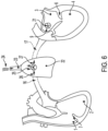
- FIG. 6 shows how a cardiac output is affected by and therefore can be measured via thermodilution with a cold air bolus and a left atrium sensor, or thermistor 21, according to some embodiments.
- cold air bolus 24 is injected into the lung 22 (e.g., by introducing inhalation of a bolus of cold air into the lungs through the patient's trachea 23).
- heat exchange 25 takes place when warmer deoxygenated blood 26 enters the lung through the pulmonary artery 16 and is then cooled by the cold air bolus 24 during oxygenation.
- cooler oxygenated blood 27 leaves the lung through the pulmonary vein 17 into the left atrium 3 where the thermistor 21, immobilized using the left anchoring disc 9, measures the temperature in the left atrium 3. If more blood is flowing, the temperature within the left atrium 3 would return to normal temperature sooner than when less blood is flowing. As such, the inhalation of cold air in this case can be used to determine the initial drop in the blood temperature in the left atrium 3, which is used to correlate the time for the temperature to return to normal. The rate at which the temperature returns to normal correlates with the cardiac output.
- one or more O2 sensors may be included at one or more of the first pair of sensing elements 10,11 and the remote second pair of sensing elements 13,15.
- FIG. 7 shows a diagram of blood flow from the right to left side of the heart through a lung and can be used to describe how cardiac output can be measured via O2 blood saturation sensors 28,29 (e.g., using Fick's Law).
- a right O2 saturation sensor 28 is placed in the right atrium 1 (e.g., associated with the sensing element 10 immobilized using the right anchoring disc 8), and a left O2 saturation sensor 29 is placed in the left atrium 3 (e.g., associated with the sensing element 11 immobilized using the left anchoring disc 9), to measure the SvO2 and SaO2 levels in their respective areas of measurement.
- the blood flow to the patient's heart can be calculated using a marker substance, which in this case is oxygen (O2).
- O2 oxygen
- the necessary data for making such calculations include the amount of oxygen taken up by the heart per unit time, the O2 blood saturation in the pulmonary artery, and the O2 blood saturation in the pulmonary vein.
- the O2 blood saturation of the pulmonary artery 16 is measured at the right atrium 1
- the O2 blood saturation of the pulmonary vein 17 is measured at the left atrium 3.
- Other data for the calculation can include maximal oxygen uptake (VO2 max), which is the maximum rate of oxygen consumption measured during incremental exercise, and hemoglobin test, which in combination with the arterial and venous percentages will determine oxygen concentration.
- VO2 max maximal oxygen uptake
- sensors can be implemented in the embodiments as disclosed herein (e.g., pressure, flow, temperature, and/or O2), with each measurement contributing vital data regarding the health of the patient's heart.
- the sensors themselves can be of various shapes and sizes, as deemed suitable by a person of ordinary skills in the art, to be implemented inside a patient's heart.
- FIGs. 8-10 show additional details of possible sensor element configurations.
- a measurement device 43 has a right atrium sensing element 30 included in right atrium electronics 37 and a left atrium sensing element 31 included in left atrium electronics 38.
- the electronics 37,38 are attached to their respective anchoring discs 8,9 as shown in FIG. 8.
- FIG. 9 shows that the electronics 37,38 both include a control module 33 (e.g., printed circuit board) in a proximal part 34 of a case 36 with regard to the anchoring discs 8,9.
- the case 36 may be made of titanium, stainless steel, or other suitable materials.
- the right atrium electronics 37 further include the right atrium sensing element 30 and an antenna coil 35.
- the left atrium electronics 38 further include the left atrium sensing element 31 and a power source 32 (e.g., battery and/or inductive power source).
- a power source 32 e.g., battery and/or inductive power source.
- an external reader device such as the external charger and communications relay 70 in FIG. 18 , charges the power source 32 as well as communicating with the electronics 37,38 to obtain the measurement data stored in a memory implemented in the control modules 33.
- the control modules 33 can be designed such that they are configured to perform a sequence of steps for taking measurements within different portions of the heart (e.g., each of the chambers), whether the measurements are in blood pressure, temperature, and/or oxygen saturation), as well as to store the data until the external reader device can access the data, usually wirelessly.
- the power source 32 can be any suitable power source that can be used in this implementation.
- the power source can be coupled to a charging coil which enables inductive charging of the power source such that the external reader device can remotely charge the power source from outside the patient's body, which reduces the need to exchange the power source once it runs out of power.
- the antenna coil 35 can be used as a charging coil in addition to performing relay/communication functions.
- FIGs. 11-13 show various configurations that may be used in various examples.
- the left atrium electronics 38 of a measurement device 44 only include the left atrium sensing element 31 (e.g., sensing, temperature and/or O2). Minimizing the number of components / elements on the left side of the device may help minimize the overall size of the device and the associated amount of foreign material in the left atrium. As described above, this may reduce the potential for thrombosis and foreign body response.
- the right atrium electronics 37 include the power source 32 to power the sensing elements 31, any associated data storage elements, and more generally the control module 33.
- the right atrium electronics 37 do not include an onboard power source.
- measurement data may only be taken when an external reader device is engaged to power the measurement device 44 (e.g., using inductive power to activate the sensing elements 30,31 and control module 33).
- one or more of the measurement devices 41,42,43,44 (e.g., one or both of the anchoring discs 8,9) is configured to be fenestrated, or crossed by a surgical implement (e.g., trans-septal needle) following implantation.
- FIGs. 14 and 15 show a puncture needle 51 attached to the end of a catheter sheath 50 penetrating the surfaces of the anchoring discs 8,9 to indicate that the measurement device 45 is re-crossable (e.g., may be penetrable by a needle and followed by a sheath).
- FIG. 14 shows the needle 51 and catheter sheath 50 entering the right atrium 1 from the inferior vena cava 53, but in some instances may enter the right atrium 1 from the superior vena cava 52 as necessary.
- the surface of the anchoring discs 8,9 includes a material than can be safely penetrated, such as an expanded polytetrafluoroethylene (ePTFE) membrane 54, although other suitable materials can be implemented to provide re-crossing capability.
- ePTFE expanded polytetrafluoroethylene
- the outer edge of the anchoring disc 9 is defined by a nitinol frame 55 which also extends radially from the left side sensing element 11 to the outer edge, although other suitable materials may be used for the frame as well.
- the needle 51 forms a puncture hole 56 in the membrane 54 to allow for the catheter sheath 50 to pass through.
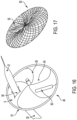
- FIG. 16 shows another embodiment as disclosed herein where the left side sensing element 60 of a measurement device 46 is located on a portion of the left anchoring disc 9.
- the left side sensing element 60 may be located on membrane material (e.g., ePTFE membrane 54) of the left anchoring disc 9, as opposed to the frame of the left anchoring disc 9 (e.g., center eyelet or outer frame).
- the left side sensing element 60 is coupled to the membrane 54 to help prevent the sensing element 60 from protruding outward from the septal wall, or reduce the amount the left sensing element 60 protrudes from the septal wall from the measurement device 46.
- an antenna coil 61 is implemented in the anchoring disc 9.
- the sensing element 60 is attached to the antenna coil 61, and the antenna coil 61 is wrapped around the ePTFE membrane 54 to form the outer edge of the left anchoring disc 9, thereby defining the periphery of the left anchoring disc 9. Additional embodiments may further reduce the amount the left sensing element 60 protrudes from the septal wall.
- the left anchoring disc 9 may be replaced with a small tissue anchoring structure which help align the outer surface of the left sensing element 60 to be substantially flush with the surface of the surrounding septal wall, such that the sensing element 60 would not visibly protrude from the septal wall.
- small hooks or other suitable structures may be implemented to hold the left sensing element 60 in place.
- a cover or other similar component may be employed over the left sensing element 60 to prevent the sensing element from substantially protruding into the left atrium.
- the cover may be made of a material that is chemically inert such as low temperature isotropic (LTI) carbon and diamond like carbon (DLC), or polymers such as polytetrafluoroethylene (PTFE), expanded PTFE, or polyethylene terephthalate (PET).
- LTI low temperature isotropic
- DLC diamond like carbon
- polymers such as polytetrafluoroethylene (PTFE), expanded PTFE, or polyethylene terephthalate (PET).
- the cover may be a thin film placed over the left sensing element 60 to promote tissue ingrowth over the sensing element.
- FIG. 17 shows another potential configuration for the medical device 46 where the left anchoring disc 62 uses a frame (e.g., nitinol) configured into a mesh design that defines the left and/or right disc(s).
- the configuration of FIG. 17 may or may not employ a cover or membrane 54 (e.g., ePTFE).
- the re-crossable surface is designed such that catheters with a French gauge suitable to fit between the frame elements (e.g., up to 24 Fr) may fit through the anchoring disc(s) without interfering with the electronics.
- anchoring discs illustrated in the figures are relatively flat and circular in structure, any of the anchoring disc configurations described herein can use other shapes (e.g., rectangular, triangular, etc.) as well.
- the anchoring discs may be configured with a curved side profile (e.g., concave and / or convex) to accommodate for the different contours defining the surface within the heart to which the anchoring discs are to be engaged.
- the measurement device 45,46 can also act as a therapeutic device, such as an intra-atrial shunt, a controllable intra-atrial shunt, an occluder for atrial septal defects (ADS), and so on.
- the measurement device 45,46 can act as such a controllable shunt because it is located at an interatrial septum between the left and right atria, and the membrane 54 can be opened via interventional or noninvasive procedures.
- the membrane 54 may be expanded, contracted, opened, closed, fenestrated, sealed, punctured, resealed, traversed, or crossed using appropriate tools during different procedures to actuate the controllable shunt.
- the use of a needle to make a puncture hole 56 in the membrane 54 as discussed above is one example of the interventional procedure.
- Other interventional procedures include mechanical, thermal, laser, ultrasound, and inductive methods.
- the opening of the hole can be triggered via wireless, extracorporeal energization, including inductive energy transfer and ultrasound energy transfer.
- the membrane 54 can be melted to form an opening after exposing the membrane 54 to thermal or ultrasound energy, i.e. via thermal activation.
- An advantage in having an opening in the membrane 54 includes, when the measurement device 45,46 is located between the left and right atria, reducing the left atrial pressure when it rises to a life-threatening level.
- the measurement device 45,46 can continue taking measurements within the two atria.
- the size of the shunt can be adjusted based on the required degree of pressure relief. For example, if the pressure is significantly higher than the normal level such that the pressure must be lowered immediately, the shunt can be opened wider. When the shunt is opened via mechanical piercing or thermal ablation, it can prevent embolization as well.
- a pressure-sensitive valve may be implemented in the measurement device 45,46 such that the membrane 54 opens to form the shunt above a threshold pressure level. In another example, the valve may also track and transmit its status (i.e.
- a remote monitoring system (for example a remote device 72 in FIG. 18 ) which receives data from the valve can use the status data of the valve to determine a difference in pressure between the left and right atria.
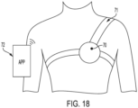
- FIG. 18 shows an example of external charging and communications relay according to some examples.
- the external charger and communications relay 70 is a device which can charge or power a power source of the measurement device (for example, the battery 32 in the measurement devices 43,44) via electromagnetic induction, as well as to communicate with the measurement device 43 or 44 to obtain the measurement data.
- the external charger and communications relay 70 may be worn (e.g., using a harness 71) such that the location of the charger and relay 70 is placed at an operable location for the charger and relay 70 to charge and obtain data from the measurement device.
- a monitoring system 72 which can be a smart device such as a smartphone, can be used by the patient or other party (e.g., medical service provider or remote monitoring facility) to receive information regarding the measurement data via an application software in the monitoring system.
- the remote device 72 can visually show the blood pressure, temperature, and/or oxygen saturation in a simple, user-friendly interface. If the patient is visually impaired or prefers audio notifications, the remote device 72 can provide audio output to alert the patient if the sensor measurements indicate that the patient's heart may be at risk of acute decompensation episodes, so that the patient can go to a hospital for a further examination.
- the remote device 72 can also upload the measurement data onto a server (not shown) to be collected by medical service providers or a database to remotely monitor the conditions of the patient's heart.
- a tethered sensor can be sent off the left anchoring disc, between the mitral valve leaflets, and into the left ventricle, where tissue ingrowth can implement the sensor into the wall of the left ventricle.
- the sensor directly measures the left ventricular systolic and diastolic pressure, which also gives a direct indication of systolic systemic blood pressure
- the tethered sensor can be sent off the left anchoring disc, between the mitral valve leaflets, through the aortic valve, and into the aorta, where the sensor is secured to the wall of the aorta. This placement allows for direct measurement of the aortic pressure which gives a direct indication of systolic and diastolic blood pressures.
- the tethered sensor can be sent off the right anchoring disc, between the tricuspid valve leaflets, and into the right ventricle, where tissue ingrowth can implement the sensor into the wall of the right ventricle.
- the sensor directly measures the right ventricular pressure which gives a direct indication of systolic and diastolic right ventricular pressures.
- the tethered sensor can be sent off the right anchoring disc, between the tricuspid valve leaflets, through the pulmonary valve, and into the pulmonary artery, where it is secured. This placement allows for direct measurement of the pulmonary pressure which gives a direct indication of pulmonary status via pulmonary systolic and diastolic pressures.
- the implanted device that measures the left and right atrial pressures can be used in combination with other medical devices.
- medical devices include, but are not limited to, blood pressure cuffs, pulse-oximeters, scales, creatinine testing devices, smart devices, and wearable medical tracking devices, to name a few.
- the measurement device 41 can also be combined with other implantable devices, such as a ventricular assist device (VAD), drug delivery shunt or system, or other device.
- VAD ventricular assist device
- the measurement device 41 may provide feedback to the other implantable device(s), as part of a closed loop or open loop feedback system.
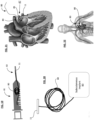
- FIGs. 19 to 22 show additional sensor element examples as well as the associated delivery systems and methods.
- the left side sensing element 11 may be coupled with a tether 80, with the sensor element 11 and tether 80 configured to delivered through a needle delivery system 81 shown schematically in FIG. 19 .
- the delivery system 81 may include a catheter and associated delivery needle 82 at a distal end of the catheter that is used to access a target area in the body (e.g., via ultrasound, radiographic, optical or other guidance).
- the needle may be used to puncture a wall of the heart (e.g., left atrial wall) and the left side sensing element 11 may be advanced through the needle 82 into the target space (e.g., the left atrium).
- the needle 82 may then be retracted and the sensing element 11 may be pulled or tensioned against an inner wall of the heart (e.g., the inner wall defining the left atrium).
- a pledget or other anchor 84 may be advanced (e.g., over the tether 80) to a position on an opposite side of the heart wall from the sensing element 11 (e.g., a location on an outer wall of the heart proximate to the location of the sensing element 11) to help secure the sensing element 11 in place (e.g., as shown in FIG. 21 ).
- the tether 80 may be connected to a subcutaneous implant 83 that handles power, signal processing, and data transmission functions as shown in FIG. 20 .
- the subcutaneous implant 83 can include a battery, an antenna, and a control module (e.g., a microchip) to help control data collection and communication functions.
- the measurement device 47 may include a plug 85 that is placed between the sensing element 11 and the pledget 84 to help fill the fenestration left by the needle 82.
- FIG. 22 shows a subcutaneous implant 83 on the other end of the tether 80 opposite from the sensing element 11.
- Multiple sensor elements similar to the left side sensing element 11 can be placed in any different parts of the heart as deemed suitable by the medical service providers. For example, after the left side sensing element 11 is placed in the left atrium, another sensing element such as the right side sensing element 10 can be implemented into the right atrium using the same technique that is used to place the sensing element 11 in the left atrium. Another similar sensing element can be implanted in the left ventricle, right ventricle, or any other location in the heart or vasculature as desired. As such, any number of sensor elements can be implemented by penetrating a wall of the heart to take measurements (pressure, temperature and/or oxygen saturation) in any of the chambers of the heart or associated vasculature.
- the tether(s) associated with the sensor elements can be coupled to the same subcutaneous implant or different subcutaneous implants as desired. Whether a single subcutaneous implant with data receiving and communication capabilities or different subcutaneous implants, it should be understood that any of the combination of measurements (pressure, temperature and/or oxygen saturation) at any combination of locations (e.g., left atrium, right atrium, left ventricle, and/or right ventricle) may be realized using the tethered sensor elements described in association with FIGS. 19 to 22 .
- the pressure measurement data obtained using the sensing elements 10,11,13,15 as described herein can be used to perform pulse-contour method, which is another method that is used to measure the cardiac output of the patient.
- This method uses the continuous pressure measurement data to plot a pressure-versus-time graph for the patient's heart, after which the pressure integral, i.e. the area beneath the plotted line on the pressure-versus-time graph, is used to determine the stroke volume (SV) of the portion of the heart that is being measured.
- the value of SV multiplied by the heart rate is the cardiac output.
- FIG. 23 is a flow chart showing a remote medical treatment monitoring method 99 that can be implemented using one or more electronic devices, such as the monitoring system 72, using measurement data received from the measurement device 41, for example, or any of the sensor elements described herein.
- the method 99 is used for patients with a history of left heart failure, to determine treatment protocols guided by measured right and left heart physiologic metrics (e.g., pressure, temperature, and/or oxygen saturation).
- the service provider determines if the patient receiving treatment has a history of either left heart (LH) or right heart (RH)/biventricular failure.
- the method 99 may be used for patients with a risk of LH or RH/biventricular failure as determined by the medical service providers, regardless of history.
- the medical service provider set a baseline "normal" level for applicable physiologic metrices (e.g., the left and right atrial pressures) in the acute setting by performing various tests on the patient to determine, based on the current condition of the patient, what normal levels (pressure, cardiac output, and/or oxygen saturation) would be. Baseline values can then be entered into the system which transfers the data to the monitoring system 72.
- the pressures being measured are the left atrial pressure (LAP) and the right atrial pressure (RAP).
- Other embodiments may include other measurements of other parts of the heart, as deemed appropriate by the medical service provider.
- the monitoring system 72 receives RAP and LAP measurements from the sensors in step 92, such as the right side sensing element 10 and the left side sensing element 11.
- the measurements include whether the pressure values of the right atrium and the left atrium are trending below, at, or above the normal level.
- the method may also consider whether the pressure values are increasing, decreasing, or staying steady as an additional input into the overall assessment.
- the monitoring system 72 confirms whether the patient has a history of LH or RH/biventricular failure.
- the monitoring system 72 optionally uses a medication administration reference table 100 in FIG. 24 to determine and indicate if dosage of certain medications needs to be increased or reduced, in step 94.
- a medical service provider e.g., physician
- uses the data directly to assess what treatment regimen e.g., pharmacological
- the table 100 has three columns and three rows, where the columns pertain to "RAP trending below normal” 101, “RAP trending normal” 102, and “RAP trending above normal” 103, and the columns pertain to "LAP trending below normal” 104, "LAP trending normal” 105, and “LAP trending above normal” 106.
- the method would include the step of "Increase Vasodilators” according to the table 100. If automated, a consistent “message” or communication could be relayed to a user of the monitoring system. On the other hand, if the RAP is also trending above normal, the method would include the step of "Increase Diuretics".
- the method 99 includes verifying to see if the RAP is still trending above normal and if the RAP value is unaffected by diuretics, in step 95. This may occur in the second example shown above, where the LAP and RAP are both trending above normal so the amount of diuretics administered to the patient is increased, but a subsequent measurement of the RAP shows that this pressure is still above normal.
- the monitoring system 72 could display an indication in step 96 instructing the medical service provider to bring the patient in for a potential diagnosis of RH failure (or the medical service provider could carry out the step 96 based upon the data).
- the monitoring system 72 could display an indication in step 96 instructing the medical service provider to bring the patient in for a potential diagnosis of RH failure (or the medical service provider could carry out the step 96 based upon the data).
- Among other possible causes of high RAP is primary pulmonary arterial hypertension.
- the medical service provider When the medical service provider tests the patient for possible diagnosis of these conditions, the medical service provider can set a new baseline value range for the "RAP normal" level and update the patient's status as having a history of RH/biventricular failure so that moving forward, the method will proceed to step 97 instead of step 95 in the future. Otherwise, if the RAP decreases to the normal level, the monitoring system 72 optionally goes back to step 92 to take subsequent RAP and LAP measurements.
- step 97 the method 99 includes determining if the medication administered in step 94 is effective.
- the method 99 may include comparing the previous LAP and RAP values with the new LAP and RAP values taken after the medication is administered.
- the medical service provider may bring the patient in for adjusted treatment and/or the monitoring system 72 may provide a message or other communication indicating that further diagnosis / treatment is warranted in step 98.
- the possible lack of efficacy of the medications may be a sign of increased exigency or that immediate medical attention is otherwise warranted. Otherwise, if the administered medication is showing apparent efficacy in moving LAP and RAP toward nominal or desired levels, the method returns to step 92 and the monitoring system 72 continues to receive and evaluate new measurements for assessing patient health.
- the method 99 may be programmed so that instead of using the actual measured LAP and RAP values, a ratio of LAP to RAP (or a ratio of RAP to LAP) may be used to determine which medications to administer and how much.
- a ratio of LAP to RAP (or a ratio of RAP to LAP) may be used to determine which medications to administer and how much.
- This methodology may be based on the understanding that the pressures within the left and right atria should correspond to a desired ratio (e.g., 2:1 LAP:RAP) in a healthy heart, therefore the ideal ratio of LAP to RAP can be determined (e.g., an ideal ratio of 2:1 pressures are desired), and any ratio that is significantly smaller or larger than the desired ratio (e.g., 2:1) would pose a threat to the patient's health.
- the method may include a determination that the amount of vasodilators being administered should be increased.
- the threshold ratio value of LAP to RAP which triggers such a determination may be determined and updated periodically by the medical service provider (e.g., after examination performed on the patient).
- various methods include one or more medical service providers determining the range of "normal" baseline ratios, which will then be used in the medication administration reference table.
- a generalized set of guidelines may be provided to medical service providers regarding an appropriate baseline.
- the method 99 can be adjusted to be more specific in terms of how much a pharmacological, or medication regimen needs to be increased or reduced, which can be varied based on how much the LAP and RAP are trending above or below the normal level. This may be done by implementing another table or set of guidelines within the table 100 that indicates the amount of medication to be administered (e.g., so that a treatment dosage may be adjusted for a patient without requiring direct medical service provider intervention).
- the table 100 can include any of a variety of medical recommendations / indications, such as beta-blockers and inotropes, for example, as indicated by a particular set of physiologic measurements and associated guidance of the table 100.
- the type of medication e.g. diuretic or vasodilator
- the dosage thereof can be displayed on, for example, the screen of a computer or a display of a smart device used by the patient.
- the measurement data and associated monitoring and treatment methodology is not necessarily limited to LAP and RAP measurements.
- additional or alternative locations e.g., pulmonary arteries, ventricles, pulmonary veins, aorta, and others
- additional or alternative metrics e.g., temperature and/or oxygen saturation
- the method 99 may be performed manually or may be partially or completely automated using any device capable of receiving and processing the measurement data from the measurement device 41.
- the method 99 may be implemented entirely in the monitoring system 72 (e.g., such as a smart device), which performs all the comparisons, calculations, and determinations after receiving the LAP and RAP measurement data from the measurement system 41.
- the method may be implemented partially in the monitoring system 72 and partially in the communications relay 70 which may include a processing unit to receive the LAP and RAP measurement data from the sensors, determine whether the LAP and RAP are above/at/below normal level and decreasing/steady/increasing, then relay this information to the remote device 72 to perform the rest of the method.
- the subcutaneous implant 83 may be programmed to perform a portion or the entirety of the method.
- the method 99 may be implemented in a device with a user interface allowing the patient to administer medications according to the results of the method.
- the method may also be implemented in the medical service providers' electronic health record (EHR) or electronic medical record (EMR) systems which keep track of the necessary records of each patient.
- EHR or EMR systems may use local or remote database to access, among other things, the patient's history of LH or RH/biventricular failure and whether the medical service providers have deemed the patient to be at a risk of such failure.
- the resulting data from the method may be displayed on a dashboard of the user interface with multiple options for the user (e.g.
- the dashboard may also be configured such that the user can first pull up the most meaningful information, such as the averages and trends, then dig in further for a more detailed analysis, such as the waveforms. This may be implemented by organizing the multiple options in a hierarchical manner based on the importance of each option. In one example, this hierarchical order of the options is customizable according to the user's preference, such that the most preferred information can be pulled up first.
Landscapes
- Health & Medical Sciences (AREA)
- Life Sciences & Earth Sciences (AREA)
- Engineering & Computer Science (AREA)
- Physics & Mathematics (AREA)
- Veterinary Medicine (AREA)
- Public Health (AREA)
- Heart & Thoracic Surgery (AREA)
- General Health & Medical Sciences (AREA)
- Animal Behavior & Ethology (AREA)
- Biomedical Technology (AREA)
- Surgery (AREA)
- Pathology (AREA)
- Biophysics (AREA)
- Medical Informatics (AREA)
- Molecular Biology (AREA)
- Cardiology (AREA)
- Physiology (AREA)
- Vascular Medicine (AREA)
- Pulmonology (AREA)
- Optics & Photonics (AREA)
- Computer Networks & Wireless Communication (AREA)
- Pharmacology & Pharmacy (AREA)
- Medicinal Chemistry (AREA)
- Chemical & Material Sciences (AREA)
- Bioinformatics & Cheminformatics (AREA)
- Epidemiology (AREA)
- Spectroscopy & Molecular Physics (AREA)
- Oral & Maxillofacial Surgery (AREA)
- Transplantation (AREA)
- Urology & Nephrology (AREA)
- Biodiversity & Conservation Biology (AREA)
- Artificial Intelligence (AREA)
- Computer Vision & Pattern Recognition (AREA)
- Psychiatry (AREA)
- Signal Processing (AREA)
- Otolaryngology (AREA)
- Ophthalmology & Optometry (AREA)
- Anesthesiology (AREA)
- Hematology (AREA)
- Measuring Pulse, Heart Rate, Blood Pressure Or Blood Flow (AREA)
Applications Claiming Priority (7)
| Application Number | Priority Date | Filing Date | Title |
|---|---|---|---|
| US201862783935P | 2018-12-21 | 2018-12-21 | |
| US201862783902P | 2018-12-21 | 2018-12-21 | |
| US201962845386P | 2019-05-09 | 2019-05-09 | |
| US201962894260P | 2019-08-30 | 2019-08-30 | |
| US201962901105P | 2019-09-16 | 2019-09-16 | |
| EP19842527.4A EP3897370B1 (de) | 2018-12-21 | 2019-12-23 | Medizinisches behandlungssystem mit messdaten von mehreren sensoren |
| PCT/US2019/068277 WO2020132669A1 (en) | 2018-12-21 | 2019-12-23 | Medical treatment system using measurement data from multiple sensors |
Related Parent Applications (2)
| Application Number | Title | Priority Date | Filing Date |
|---|---|---|---|
| EP19842527.4A Division EP3897370B1 (de) | 2018-12-21 | 2019-12-23 | Medizinisches behandlungssystem mit messdaten von mehreren sensoren |
| EP19842527.4A Division-Into EP3897370B1 (de) | 2018-12-21 | 2019-12-23 | Medizinisches behandlungssystem mit messdaten von mehreren sensoren |
Publications (2)
| Publication Number | Publication Date |
|---|---|
| EP4570171A2 true EP4570171A2 (de) | 2025-06-18 |
| EP4570171A3 EP4570171A3 (de) | 2025-08-06 |
Family
ID=69185746
Family Applications (7)
| Application Number | Title | Priority Date | Filing Date |
|---|---|---|---|
| EP25174558.4A Pending EP4570171A3 (de) | 2018-12-21 | 2019-12-23 | Medizinisches behandlungssystem mit messdaten von mehreren sensoren |
| EP19842527.4A Active EP3897370B1 (de) | 2018-12-21 | 2019-12-23 | Medizinisches behandlungssystem mit messdaten von mehreren sensoren |
| EP25191682.1A Pending EP4613184A3 (de) | 2018-12-21 | 2019-12-23 | Implantierbare herzsensoren |
| EP25175056.8A Pending EP4574028A3 (de) | 2018-12-21 | 2019-12-23 | Implantierbare medizinische vorrichtung mit verstellbarem blutfluss |
| EP19839774.7A Active EP3897369B1 (de) | 2018-12-21 | 2019-12-23 | Implantierbare herzsensoren |
| EP19842528.2A Active EP3897371B1 (de) | 2018-12-21 | 2019-12-23 | Implantierbare medizinische vorrichtung mit verstellbarem blutfluss |
| EP19848954.4A Active EP3897372B1 (de) | 2018-12-21 | 2019-12-23 | Implantierbare herzsensoren |
Family Applications After (6)
| Application Number | Title | Priority Date | Filing Date |
|---|---|---|---|
| EP19842527.4A Active EP3897370B1 (de) | 2018-12-21 | 2019-12-23 | Medizinisches behandlungssystem mit messdaten von mehreren sensoren |
| EP25191682.1A Pending EP4613184A3 (de) | 2018-12-21 | 2019-12-23 | Implantierbare herzsensoren |
| EP25175056.8A Pending EP4574028A3 (de) | 2018-12-21 | 2019-12-23 | Implantierbare medizinische vorrichtung mit verstellbarem blutfluss |
| EP19839774.7A Active EP3897369B1 (de) | 2018-12-21 | 2019-12-23 | Implantierbare herzsensoren |
| EP19842528.2A Active EP3897371B1 (de) | 2018-12-21 | 2019-12-23 | Implantierbare medizinische vorrichtung mit verstellbarem blutfluss |
| EP19848954.4A Active EP3897372B1 (de) | 2018-12-21 | 2019-12-23 | Implantierbare herzsensoren |
Country Status (8)
| Country | Link |
|---|---|
| US (5) | US11911135B2 (de) |
| EP (7) | EP4570171A3 (de) |
| JP (5) | JP7377269B2 (de) |
| CN (4) | CN113473903B (de) |
| AU (5) | AU2019406218B2 (de) |
| CA (4) | CA3123616A1 (de) |
| ES (4) | ES2981314T3 (de) |
| WO (4) | WO2020132669A1 (de) |
Families Citing this family (68)
| Publication number | Priority date | Publication date | Assignee | Title |
|---|---|---|---|---|
| WO2005074367A2 (en) | 2004-02-03 | 2005-08-18 | Atria Medical Inc. | Device and method for controlling in-vivo pressure |
| US9681948B2 (en) | 2006-01-23 | 2017-06-20 | V-Wave Ltd. | Heart anchor device |
| EP2427143B1 (de) | 2009-05-04 | 2017-08-02 | V-Wave Ltd. | Vorrichtung zur druckregelung in einer herzkammer |
| US12453626B2 (en) | 2009-05-04 | 2025-10-28 | V-Wave Ltd. | Shunt for redistributing atrial blood volume |
| US9034034B2 (en) | 2010-12-22 | 2015-05-19 | V-Wave Ltd. | Devices for reducing left atrial pressure, and methods of making and using same |
| US12186176B2 (en) | 2009-05-04 | 2025-01-07 | V-Wave Ltd. | Shunt for redistributing atrial blood volume |
| US11135054B2 (en) | 2011-07-28 | 2021-10-05 | V-Wave Ltd. | Interatrial shunts having biodegradable material, and methods of making and using same |
| US9713696B2 (en) | 2013-05-21 | 2017-07-25 | V-Wave Ltd. | Apparatus and methods for delivering devices for reducing left atrial pressure |
| WO2018031714A1 (en) | 2016-08-11 | 2018-02-15 | Foundry Innovation & Research 1, Ltd. | Systems and methods for patient fluid management |
| US12465324B2 (en) | 2015-02-12 | 2025-11-11 | Foundry Innovation & Research 1, Ltd. | Patient fluid management systems and methods employing integrated fluid status sensing |
| WO2016178171A1 (en) | 2015-05-07 | 2016-11-10 | The Medical Research Infrastructure And Health Services Fund Of The Tel-Aviv Medical Center | Temporary interatrial shunts |
| WO2017024051A1 (en) | 2015-08-03 | 2017-02-09 | Foundry Innovation & Research 1, Ltd. | Devices and methods for measurement of vena cava dimensions, pressure, and oxygen saturation |
| US20170340460A1 (en) | 2016-05-31 | 2017-11-30 | V-Wave Ltd. | Systems and methods for making encapsulated hourglass shaped stents |
| US10835394B2 (en) | 2016-05-31 | 2020-11-17 | V-Wave, Ltd. | Systems and methods for making encapsulated hourglass shaped stents |
| US11701018B2 (en) | 2016-08-11 | 2023-07-18 | Foundry Innovation & Research 1, Ltd. | Wireless resonant circuit and variable inductance vascular monitoring implants and anchoring structures therefore |
| US11206992B2 (en) | 2016-08-11 | 2021-12-28 | Foundry Innovation & Research 1, Ltd. | Wireless resonant circuit and variable inductance vascular monitoring implants and anchoring structures therefore |
| US11291807B2 (en) | 2017-03-03 | 2022-04-05 | V-Wave Ltd. | Asymmetric shunt for redistributing atrial blood volume |
| WO2018220146A1 (en) | 2017-05-31 | 2018-12-06 | Foundry Innovation & Research 1, Ltd. | Implantable sensors for vascular monitoring |
| EP3629937B1 (de) | 2017-05-31 | 2026-01-14 | Foundry Innovation & Research 1, Ltd. | Implantierbarer ultraschallgefässsensor |
| EP3740163A1 (de) | 2018-01-20 | 2020-11-25 | V-Wave Ltd. | Vorrichtungen und verfahren zur bereitstellung eines durchgangs zwischen herzkammern |
| US11458287B2 (en) | 2018-01-20 | 2022-10-04 | V-Wave Ltd. | Devices with dimensions that can be reduced and increased in vivo, and methods of making and using the same |
| US10898698B1 (en) | 2020-05-04 | 2021-01-26 | V-Wave Ltd. | Devices with dimensions that can be reduced and increased in vivo, and methods of making and using the same |
| CA3105224A1 (en) | 2018-07-18 | 2020-01-23 | W. L. Gore & Associates, Inc. | Implantable medical device deployment system |
| JP7377269B2 (ja) | 2018-12-21 | 2023-11-09 | ダブリュ.エル.ゴア アンド アソシエイツ,インコーポレイティド | インプラント可能な心臓センサ |
| EP3906085A4 (de) | 2019-01-04 | 2022-09-28 | Shifamed Holdings, LLC | Interne wiederaufladesysteme und verwendungsverfahren |
| US12226602B2 (en) | 2019-04-03 | 2025-02-18 | V-Wave Ltd. | Systems for delivering implantable devices across an atrial septum |
| US11612385B2 (en) | 2019-04-03 | 2023-03-28 | V-Wave Ltd. | Systems and methods for delivering implantable devices across an atrial septum |
| WO2020234751A1 (en) | 2019-05-20 | 2020-11-26 | V-Wave Ltd. | Systems and methods for creating an interatrial shunt |
| US12533500B2 (en) | 2019-06-18 | 2026-01-27 | Shifamed Holdings, Llc | Adjustable interatrial shunts and associated systems and methods |
| WO2021050589A1 (en) | 2019-09-09 | 2021-03-18 | Shifamed Holdings, Llc | Adjustable shunts and associated systems and methods |
| EP4090251A1 (de) | 2020-01-17 | 2022-11-23 | W. L. Gore & Associates, Inc. | Medizinisches implantat mit zwei rahmenkomponenten und ösenpaaren |
| EP4099898B1 (de) * | 2020-02-05 | 2026-01-28 | Shifamed Holdings, LLC | Intrakardialer drucksensor mit clip-struktur |
| EP4138981A4 (de) | 2020-04-23 | 2024-05-22 | Shifamed Holdings, LLC | Leistungsverwaltung für interatriale shunts sowie zugehörige systeme und verfahren |
| EP4138649B1 (de) | 2020-04-23 | 2025-09-24 | Shifamed Holdings, LLC | Intrakardiale sensoren mit schaltbaren konfigurationen und zugehörige systeme und verfahren |
| EP4138978A4 (de) | 2020-04-23 | 2024-05-01 | Shifamed Holdings, LLC | Systeme und verfahren zur radiographischen überwachung von shunts |
| US12599756B2 (en) | 2020-05-04 | 2026-04-14 | V-Wave Ltd. | Devices with dimensions that can be reduced and increased In Vivo |
| US11801369B2 (en) | 2020-08-25 | 2023-10-31 | Shifamed Holdings, Llc | Adjustable interatrial shunts and associated systems and methods |
| CN116648282A (zh) | 2020-10-16 | 2023-08-25 | 施菲姆德控股有限责任公司 | 用于植入式医疗装置的双用途电感器及相关联的系统和方法 |
| US12544010B2 (en) | 2020-10-28 | 2026-02-10 | Shifamed Holdings, Llc | Systems and methods for electrical monitoring of implantable devices |
| US20240016486A1 (en) * | 2020-11-02 | 2024-01-18 | Recross Cardio Inc. | Interseptal occluder device |
| US11857197B2 (en) | 2020-11-12 | 2024-01-02 | Shifamed Holdings, Llc | Adjustable implantable devices and associated methods |
| US11234702B1 (en) * | 2020-11-13 | 2022-02-01 | V-Wave Ltd. | Interatrial shunt having physiologic sensor |
| JP2023552725A (ja) * | 2020-11-24 | 2023-12-19 | ジョージア テック リサーチ コーポレイション | ウェアラブル心臓血管センサにより血行動態パラメータを測定するためのシステム及び方法 |
| US20220180991A1 (en) * | 2020-12-06 | 2022-06-09 | Acorai Ab | Systems and methods for creating personalized medicines for treating heart failure and heart disease |
| JP2024506875A (ja) * | 2021-02-05 | 2024-02-15 | エドワーズ ライフサイエンシーズ コーポレイション | シャントセンサ埋め込み装置 |
| WO2022192280A1 (en) | 2021-03-09 | 2022-09-15 | Shifamed Holdings, Llc | Shape memory actuators for adjustable shunting systems, and associated systems and methods |
| JP2024513703A (ja) * | 2021-03-15 | 2024-03-27 | エドワーズ ライフサイエンシーズ コーポレイション | オフセットセンサアーム付きシャント埋め込み装置 |
| EP4280940B1 (de) * | 2021-03-15 | 2025-02-19 | Edwards Lifesciences Corporation | Shunt-implantatvorrichtungen mit überkanalsensorarmen |
| NL2028193B1 (en) * | 2021-05-11 | 2022-12-02 | Amazec Photonics Ip B V | Obtaining cardiovascular and/or respiratory information from the mammal body |
| EP4340777A4 (de) * | 2021-05-21 | 2025-04-30 | Shifamed Holdings, LLC | Implantierbare medizinische vorrichtungen mit kanistern in ankern mit hohlräumen sowie zugehörige systeme und verfahren |
| US20230149694A1 (en) * | 2021-11-15 | 2023-05-18 | Medtronic, Inc. | Mechanical circulatory support device |
| CN113951854A (zh) * | 2021-11-22 | 2022-01-21 | 山东瑞安泰医疗技术有限公司 | 一种植入式肺动脉末端压力监控装置 |
| WO2023123428A1 (zh) * | 2021-12-31 | 2023-07-06 | 合源医疗器械(上海)有限公司 | 心房分流装置及其安装方法、心房植入设备、医疗系统、心脏活动信息采集方法、医疗设备 |
| CN116407771A (zh) * | 2021-12-31 | 2023-07-11 | 合源医疗器械(上海)有限公司 | 心房植入设备、医疗系统以及心脏信息采集方法 |
| EP4468949A4 (de) * | 2022-01-28 | 2025-08-27 | Veris Health Inc | Gefässmessvorrichtungen, -systeme und -verfahren |
| AU2023252664A1 (en) | 2022-04-14 | 2024-10-17 | V-Wave Ltd. | Interatrial shunt with expanded neck region |
| CA3255933A1 (en) * | 2022-04-18 | 2023-10-26 | Edwards Lifesciences Corporation | SENSOR IMPLANT DEVICE WITH STABILIZATION APPENDAGE |
| AU2023262477A1 (en) | 2022-04-29 | 2024-12-05 | Relief Cardiovascular, Inc. | Systems, devices, and methods for controllably and selectively occluding, restricting, and diverting flow within a patient's vasculature |
| US11883030B2 (en) | 2022-04-29 | 2024-01-30 | inQB8 Medical Technologies, LLC | Systems, devices, and methods for controllably and selectively occluding, restricting, and diverting flow within a patient's vasculature |
| WO2024023642A1 (en) * | 2022-07-27 | 2024-02-01 | Medtronic, Inc. | Tracking patient condition symptoms with temperature and impedance data collected with implanted sensor |
| EP4561673A1 (de) | 2022-07-29 | 2025-06-04 | Foundry Innovation & Research 1, Ltd. | An dynamische in-vivo-umgebungen angepasste mehrstrangleiter |
| CN115581542A (zh) * | 2022-09-23 | 2023-01-10 | 浙江智柔科技有限公司 | 用于瓣膜压差测量的压力传感器及可植入心脏瓣膜 |
| CN117982258B (zh) * | 2022-10-27 | 2025-07-25 | 合源医疗器械(上海)有限公司 | 医疗装置 |
| CN118680726A (zh) * | 2023-03-24 | 2024-09-24 | 合源医疗器械(上海)有限公司 | 心脏植入装置 |
| WO2024233931A2 (en) * | 2023-05-10 | 2024-11-14 | Qura, Inc. | Implantable pressure sensor system for measurement and wireless transmission of internal pressures |
| AU2024363044A1 (en) * | 2023-10-18 | 2026-04-16 | V-Wave Ltd. | Hybrid devices with dimensions that can be adjusted in vivo and methods of manufacturing thereof |
| WO2026000135A1 (zh) * | 2024-06-24 | 2026-01-02 | 中国医学科学院阜外医院 | 起搏装置 |
| WO2026050638A1 (en) * | 2024-08-30 | 2026-03-05 | Edwards Lifesciences Corporation | Drug dosage management method and system |
Citations (5)
| Publication number | Priority date | Publication date | Assignee | Title |
|---|---|---|---|---|
| US20170042705A1 (en) | 2002-05-31 | 2017-02-16 | W. L. Gore & Associates, Inc. | Implantable product with improved aqueous interface characteristics and method for making and using same |
| US20170105711A1 (en) | 2009-06-22 | 2017-04-20 | W. L. Gore & Associates, Inc. | Sealing device and delivery system |
| US9636094B2 (en) | 2009-06-22 | 2017-05-02 | W. L. Gore & Associates, Inc. | Sealing device and delivery system |
| US9861346B2 (en) | 2003-07-14 | 2018-01-09 | W. L. Gore & Associates, Inc. | Patent foramen ovale (PFO) closure device with linearly elongating petals |
| US9949728B2 (en) | 2007-04-05 | 2018-04-24 | W.L. Gore & Associates, Inc. | Septal closure device with centering mechanism |
Family Cites Families (140)
| Publication number | Priority date | Publication date | Assignee | Title |
|---|---|---|---|---|
| CA962021A (en) | 1970-05-21 | 1975-02-04 | Robert W. Gore | Porous products and process therefor |
| US3874388A (en) | 1973-02-12 | 1975-04-01 | Ochsner Med Found Alton | Shunt defect closure system |
| AR205110A1 (es) | 1974-04-02 | 1976-04-05 | Gore & Ass | Protesis vascular artificial |
| US4057857A (en) * | 1975-09-08 | 1977-11-15 | Shiley Laboratories, Inc. | Heart valve with arcuate occluder |
| US5276276A (en) | 1988-07-18 | 1994-01-04 | Gunn Dennis R | Coil transducer |
| WO1993013712A1 (en) | 1992-01-21 | 1993-07-22 | Regents Of The University Of Minnesota | Septal defect closure device |
| US5683411A (en) | 1994-04-06 | 1997-11-04 | William Cook Europe A/S | Medical article for implantation into the vascular system of a patient |
| US5879366A (en) | 1996-12-20 | 1999-03-09 | W.L. Gore & Associates, Inc. | Self-expanding defect closure device and method of making and using |
| US6042605A (en) | 1995-12-14 | 2000-03-28 | Gore Enterprose Holdings, Inc. | Kink resistant stent-graft |
| US6042602A (en) | 1997-06-04 | 2000-03-28 | Wells; Charles A. | Apparatus for effecting traction on the spine and method of using the same |
| US20020120200A1 (en) * | 1997-10-14 | 2002-08-29 | Brian Brockway | Devices, systems and methods for endocardial pressure measurement |
| US5944738A (en) | 1998-02-06 | 1999-08-31 | Aga Medical Corporation | Percutaneous catheter directed constricting occlusion device |
| EP2138137A3 (de) * | 1998-09-08 | 2013-06-12 | Kabushikikaisha Igaki Iryo Sekkei | Stent für Gefäß |
| NO984143L (no) | 1998-09-09 | 2000-03-10 | Norsk Hydro As | Ny prosess for å fremstille overflatemodifiserende stoffer |
| US7128073B1 (en) * | 1998-11-06 | 2006-10-31 | Ev3 Endovascular, Inc. | Method and device for left atrial appendage occlusion |
| US7049380B1 (en) | 1999-01-19 | 2006-05-23 | Gore Enterprise Holdings, Inc. | Thermoplastic copolymer of tetrafluoroethylene and perfluoromethyl vinyl ether and medical devices employing the copolymer |
| AU5812299A (en) | 1999-09-07 | 2001-04-10 | Microvena Corporation | Retrievable septal defect closure device |
| US6328699B1 (en) | 2000-01-11 | 2001-12-11 | Cedars-Sinai Medical Center | Permanently implantable system and method for detecting, diagnosing and treating congestive heart failure |
| GB0005867D0 (en) * | 2000-03-10 | 2000-05-03 | Medinnova Sf | Method |
| US8690910B2 (en) | 2000-12-07 | 2014-04-08 | Integrated Vascular Systems, Inc. | Closure device and methods for making and using them |
| US6783499B2 (en) | 2000-12-18 | 2004-08-31 | Biosense, Inc. | Anchoring mechanism for implantable telemetric medical sensor |
| US6746404B2 (en) | 2000-12-18 | 2004-06-08 | Biosense, Inc. | Method for anchoring a medical device between tissue |
| US6824521B2 (en) | 2001-01-22 | 2004-11-30 | Integrated Sensing Systems, Inc. | Sensing catheter system and method of fabrication |
| US8091556B2 (en) | 2001-04-20 | 2012-01-10 | V-Wave Ltd. | Methods and apparatus for reducing localized circulatory system pressure |
| WO2005074367A2 (en) * | 2004-02-03 | 2005-08-18 | Atria Medical Inc. | Device and method for controlling in-vivo pressure |
| US7288105B2 (en) | 2001-08-01 | 2007-10-30 | Ev3 Endovascular, Inc. | Tissue opening occluder |
| US20030139819A1 (en) | 2002-01-18 | 2003-07-24 | Beer Nicholas De | Method and apparatus for closing septal defects |
| US7236821B2 (en) * | 2002-02-19 | 2007-06-26 | Cardiac Pacemakers, Inc. | Chronically-implanted device for sensing and therapy |
| US6638257B2 (en) | 2002-03-01 | 2003-10-28 | Aga Medical Corporation | Intravascular flow restrictor |
| WO2003082076A2 (en) | 2002-03-25 | 2003-10-09 | Nmt Medical, Inc. | Patent foramen ovale (pfo) closure clips |
| AU2003247526A1 (en) * | 2002-06-12 | 2003-12-31 | Mitral Interventions, Inc. | Method and apparatus for tissue connection |
| US7105175B2 (en) | 2002-06-19 | 2006-09-12 | Boston Scientific Scimed, Inc. | Implantable or insertable medical devices for controlled delivery of a therapeutic agent |
| US20040024448A1 (en) | 2002-08-05 | 2004-02-05 | Chang James W. | Thermoplastic fluoropolymer-coated medical devices |
| US20040063805A1 (en) | 2002-09-19 | 2004-04-01 | Pacetti Stephen D. | Coatings for implantable medical devices and methods for fabrication thereof |
| US7615010B1 (en) | 2002-10-03 | 2009-11-10 | Integrated Sensing Systems, Inc. | System for monitoring the physiologic parameters of patients with congestive heart failure |
| JP4528766B2 (ja) * | 2003-01-24 | 2010-08-18 | プロテウス バイオメディカル インコーポレイテッド | 遠隔血行力学的モニタリングのためのシステム |
| US7200439B2 (en) * | 2003-01-24 | 2007-04-03 | Proteus Biomedical, Inc. | Method and apparatus for enhancing cardiac pacing |
| US20040267306A1 (en) | 2003-04-11 | 2004-12-30 | Velocimed, L.L.C. | Closure devices, related delivery methods, and related methods of use |
| CN1819855A (zh) * | 2003-05-13 | 2006-08-16 | 萨瓦柯公司 | 检测、诊断和治疗心血管疾病的系统和方法 |
| US20050038351A1 (en) | 2003-07-23 | 2005-02-17 | Starobin Joseph M. | Method and system for evaluating cardiac ischemia based on heart rate fluctuations |
| US8021331B2 (en) | 2003-09-15 | 2011-09-20 | Atrium Medical Corporation | Method of coating a folded medical device |
| US7789891B2 (en) * | 2003-09-23 | 2010-09-07 | Boston Scientific Scimed, Inc. | External activation of vaso-occlusive implants |
| ES2289478T3 (es) | 2004-02-04 | 2008-02-01 | Carag Ag | Implante para cerrar un paso corporal de un cuerpo humano. |
| US7713298B2 (en) * | 2004-06-29 | 2010-05-11 | Micardia Corporation | Methods for treating cardiac valves with adjustable implants |
| US8696564B2 (en) * | 2004-07-09 | 2014-04-15 | Cardiac Pacemakers, Inc. | Implantable sensor with biocompatible coating for controlling or inhibiting tissue growth |
| US20060079736A1 (en) * | 2004-10-13 | 2006-04-13 | Sing-Fatt Chin | Method and device for percutaneous left ventricular reconstruction |
| WO2006054343A1 (ja) | 2004-11-18 | 2006-05-26 | Japan Health Sciences Foundation | 心疾患治療システム |
| US20060116590A1 (en) | 2004-11-30 | 2006-06-01 | Pacesetter, Inc. | Endocardial pressure differential sensing systems and methods |
| US9545300B2 (en) | 2004-12-22 | 2017-01-17 | W. L. Gore & Associates, Inc. | Filament-wound implantable devices |
| EP1861007A2 (de) * | 2005-01-18 | 2007-12-05 | Koninklijke Philips Electronics N.V. | Elektronisch steuerbare einnahmekapsel zur probenentnahme von flüssigkeiten im verdauungstrakt |
| CN101107038A (zh) * | 2005-01-18 | 2008-01-16 | 皇家飞利浦电子股份有限公司 | 电控胶囊 |
| US8900154B2 (en) | 2005-05-24 | 2014-12-02 | Cardiac Pacemakers, Inc. | Prediction of thoracic fluid accumulation |
| EP1933756B1 (de) * | 2005-08-19 | 2016-07-20 | CHF Technologies Inc. | Lenkbare läsionsausschliessende herzimplantate bei kongestiver herzinsuffizienz |
| CA2872116C (en) * | 2005-09-07 | 2017-02-28 | Medtentia International Ltd Oy | A device for improving the function of a heart valve |
| US7632308B2 (en) * | 2005-11-23 | 2009-12-15 | Didier Loulmet | Methods, devices, and kits for treating mitral valve prolapse |
| US7682313B2 (en) | 2005-11-23 | 2010-03-23 | Vital Sensors Holding Company, Inc. | Implantable pressure monitor |
| US9681948B2 (en) * | 2006-01-23 | 2017-06-20 | V-Wave Ltd. | Heart anchor device |
| JP4581050B2 (ja) | 2006-02-16 | 2010-11-17 | 財団法人ヒューマンサイエンス振興財団 | 心臓酸素消費量自動最小化システムおよびこれを用いた心疾患治療システム |
| US9114194B2 (en) | 2006-05-12 | 2015-08-25 | W. L. Gore & Associates, Inc. | Immobilized biologically active entities having high biological activity following mechanical manipulation |
| DE102006050385A1 (de) | 2006-10-05 | 2008-04-10 | pfm Produkte für die Medizin AG | Implantierbare Einrichtung |
| US8740962B2 (en) | 2006-11-07 | 2014-06-03 | Dc Devices, Inc. | Prosthesis for retrieval and deployment |
| CA2664557C (en) | 2006-11-07 | 2015-05-26 | Corvia Medical, Inc. | Devices and methods for the treatment of heart failure |
| US10413284B2 (en) * | 2006-11-07 | 2019-09-17 | Corvia Medical, Inc. | Atrial pressure regulation with control, sensing, monitoring and therapy delivery |
| US9232997B2 (en) | 2006-11-07 | 2016-01-12 | Corvia Medical, Inc. | Devices and methods for retrievable intra-atrial implants |
| WO2008061043A2 (en) * | 2006-11-10 | 2008-05-22 | Glaukos Corporation | Uveoscleral shunt and methods for implanting same |
| WO2008094691A2 (en) | 2007-02-01 | 2008-08-07 | Cook Incorporated | Closure device and method for occluding a bodily passageway |
| WO2008112563A1 (en) | 2007-03-09 | 2008-09-18 | Flowmedica, Inc. | Acute kidney injury treatment systems and methods |
| WO2008115456A1 (en) * | 2007-03-15 | 2008-09-25 | Nunez Anthony I | Transseptal monitoring device |
| US20090024042A1 (en) * | 2007-07-03 | 2009-01-22 | Endotronix, Inc. | Method and system for monitoring ventricular function of a heart |
| US7890187B2 (en) * | 2007-07-24 | 2011-02-15 | Cardiac Pacemakers, Inc. | Lead assembly having a tethered sensor and methods related thereto |
| US20130165967A1 (en) | 2008-03-07 | 2013-06-27 | W.L. Gore & Associates, Inc. | Heart occlusion devices |
| US20110184439A1 (en) | 2008-05-09 | 2011-07-28 | University Of Pittsburgh-Of The Commonwealth System Of Higher Education | Biological Matrix for Cardiac Repair |
| US8652202B2 (en) | 2008-08-22 | 2014-02-18 | Edwards Lifesciences Corporation | Prosthetic heart valve and delivery apparatus |
| US20100056978A1 (en) * | 2008-08-27 | 2010-03-04 | Lindsay Machan | Externally adjustable blood flow valve |
| US8032212B2 (en) | 2008-09-15 | 2011-10-04 | Pacesetter, Inc. | System and method for monitoring thoracic fluid levels based on impedance using an implantable medical device |
| US7998189B2 (en) | 2008-10-10 | 2011-08-16 | Cook Medical Technologies Llc | Curvable stent-graft and apparatus and fitting method |
| US20100150920A1 (en) * | 2008-12-04 | 2010-06-17 | Bert Glaser | System and Method for Identifying Biomarkers in Ocular Fluid That Are Indicative of Ocular Disease |
| EG26158A (en) * | 2009-01-08 | 2013-03-31 | داود حنا ايهاب | Complete artificial heart (fixed inside the heart) for patients with myocardial infarction and weakness |
| WO2010111225A2 (en) * | 2009-03-23 | 2010-09-30 | Ut-Battelle, Llc | Remote actuated valve implant |
| CA2778129C (en) * | 2009-04-28 | 2014-09-02 | Dc Devices, Inc. | Devices, systems and methods to treat heart failure |
| EP2445418B1 (de) | 2009-06-26 | 2015-03-18 | Cook Medical Technologies LLC | Lineare klammern für anastomose |
| US9757107B2 (en) | 2009-09-04 | 2017-09-12 | Corvia Medical, Inc. | Methods and devices for intra-atrial shunts having adjustable sizes |
| EP2496148B1 (de) | 2009-11-03 | 2013-11-20 | Cook Medical Technologies LLC | Flache klammern für anastomose |
| KR101070122B1 (ko) * | 2009-11-06 | 2011-10-05 | 서울대학교산학협력단 | 심박변이도 측정 지표를 이용한 정신분열병 진단 장치 및 방법 |
| WO2011069154A2 (en) * | 2009-12-05 | 2011-06-09 | Integrated Sensing Systems, Inc. | Delivery system, method, and anchor for medical implant placement |
| US8715300B2 (en) | 2009-12-05 | 2014-05-06 | Integrated Sensing Systems, Inc. | Delivery system, method, and anchor for medical implant placement |
| US8597225B2 (en) * | 2010-07-26 | 2013-12-03 | The Cleveland Clinic Foundation | Method for increasing blood flow in or about a cardiac or other vascular or prosthetic structure to prevent thrombosis |
| EP2618777A1 (de) * | 2010-09-21 | 2013-07-31 | The Regents of the University of Colorado, a body corporate | Kammerwasser-mikrobypass-shunt |
| US9186152B2 (en) | 2010-11-12 | 2015-11-17 | W. L. Gore & Associates, Inc. | Left atrial appendage occlusive devices |
| US9545323B2 (en) | 2010-11-16 | 2017-01-17 | W. L. Gore & Associates, Inc. | Fenestration devices, systems, and methods |
| JP6067008B2 (ja) * | 2011-06-30 | 2017-01-25 | ユニヴァーシティ オヴ ピッツバーグ オヴ ザ コモンウェルス システム オヴ ハイアー エデュケーション | 心肺機能不全に対する易罹患性(susceptibility)を判断するシステム及び方法 |
| US9629715B2 (en) * | 2011-07-28 | 2017-04-25 | V-Wave Ltd. | Devices for reducing left atrial pressure having biodegradable constriction, and methods of making and using same |
| US8951223B2 (en) * | 2011-12-22 | 2015-02-10 | Dc Devices, Inc. | Methods and devices for intra-atrial shunts having adjustable sizes |
| EP3156006B1 (de) | 2012-03-16 | 2022-05-18 | Terumo Corporation | Stent und stentfreisetzungssystem |
| US10588611B2 (en) | 2012-04-19 | 2020-03-17 | Corvia Medical Inc. | Implant retention attachment and method of use |
| US10675012B2 (en) | 2012-11-16 | 2020-06-09 | W. L. Gore & Associates, Inc. | Joint assembly for medical devices |
| US9008769B2 (en) | 2012-12-21 | 2015-04-14 | Backbeat Medical, Inc. | Methods and systems for lowering blood pressure through reduction of ventricle filling |
| US20140200662A1 (en) * | 2013-01-16 | 2014-07-17 | Mvalve Technologies Ltd. | Anchoring elements for intracardiac devices |
| WO2014116502A1 (en) | 2013-01-23 | 2014-07-31 | Rox Medical, Inc. | Methods, systems and devices for treating cardiac arrhythmias |
| US20140222040A1 (en) | 2013-02-01 | 2014-08-07 | Jin S. Park | Method and Device for Connecting a Conduit to a Hollow Organ |
| JP6189043B2 (ja) | 2013-02-12 | 2017-08-30 | セイコーエプソン株式会社 | 左心房圧計測装置の作動方法および左心房圧計測装置 |
| US9649481B2 (en) | 2013-03-14 | 2017-05-16 | Siddharth Sadanand | Shunt flow monitor |
| CA2905283C (en) | 2013-03-15 | 2021-03-02 | Corvia Medical, Inc. | Devices and methods for retrievable intra-atrial implants |
| ES2908132T3 (es) | 2013-05-20 | 2022-04-27 | Edwards Lifesciences Corp | Aparato de suministro de válvula cardiaca protésica |
| US9713696B2 (en) | 2013-05-21 | 2017-07-25 | V-Wave Ltd. | Apparatus and methods for delivering devices for reducing left atrial pressure |
| CA2915073A1 (en) * | 2013-06-14 | 2014-12-18 | Cardiosolutions, Inc. | Mitral valve spacer and system and method for implanting the same |
| CN104414692A (zh) | 2013-08-21 | 2015-03-18 | 赵菁 | 异形心脏室间隔封堵器 |
| CN104511060A (zh) | 2013-10-03 | 2015-04-15 | 北京精密机电控制设备研究所 | 血泵控制系统及血泵系统 |
| US9775591B2 (en) | 2013-11-21 | 2017-10-03 | Edwards Lifesciences Corporation | Sealing devices and related delivery apparatuses |
| US20180008830A1 (en) | 2014-01-14 | 2018-01-11 | CardioFlow Technologies, LLC | Apparatus and methods for optimizing intra-cardiac pressures for improved exercise capacity |
| US10617543B2 (en) | 2014-01-14 | 2020-04-14 | The Regents Of The University Of California | Delivery system for transcatheter detachment of a stent from the delivery device |
| GB2527075A (en) | 2014-03-17 | 2015-12-16 | Daassist As | Percutaneous system, devices and methods |
| US11712230B2 (en) | 2014-05-02 | 2023-08-01 | W. L. Gore & Associates, Inc. | Occluder and anastomosis devices |
| WO2016028583A1 (en) * | 2014-08-18 | 2016-02-25 | St. Jude Medical, Cardiology Division, Inc. | Sensors for prosthetic heart devices |
| JP6754354B2 (ja) | 2014-09-28 | 2020-09-16 | ポトレロ メディカル,インコーポレイテッド | 生理学的データを感知し、体液を排出しかつ分析するためのシステム、デバイス、および方法 |
| EP3253297A1 (de) * | 2015-02-04 | 2017-12-13 | Boston Scientific Scimed, Inc. | Neuartiges konzept zur reduzierung des linksatrialen drucks bei patienten mit systolischer und diastolischer herzinsuffizienz zur behandlung von lungenödem und senkung der hospitalisierungsrate |
| WO2016179425A1 (en) * | 2015-05-05 | 2016-11-10 | The Johns Hopkins University | A device and method for non-invasive left ventricular end diastolic pressure (lvedp) measurement |
| WO2016178197A1 (en) | 2015-05-07 | 2016-11-10 | Vectorious Medical Technologies Ltd | Deploying and fixating an implant across an organ wall |
| WO2016178171A1 (en) * | 2015-05-07 | 2016-11-10 | The Medical Research Infrastructure And Health Services Fund Of The Tel-Aviv Medical Center | Temporary interatrial shunts |
| AU2016262564B2 (en) | 2015-05-14 | 2020-11-05 | Cephea Valve Technologies, Inc. | Replacement mitral valves |
| US10350416B2 (en) * | 2015-07-28 | 2019-07-16 | Medtronic, Inc. | Intracardiac pacemaker with sensing extension in pulmonary artery |
| US20170035433A1 (en) * | 2015-08-06 | 2017-02-09 | Thomas J. Forbes | Left atrial appendage occluder device anchoring system, anchor, and method of attachment |
| US9789294B2 (en) | 2015-10-07 | 2017-10-17 | Edwards Lifesciences Corporation | Expandable cardiac shunt |
| US10555814B2 (en) * | 2015-11-17 | 2020-02-11 | Edwards Lifesciences Corporation | Ultrasound probe for cardiac treatment |
| WO2017087424A1 (en) * | 2015-11-17 | 2017-05-26 | Millipede, Inc. | Implantable device and delivery system for reshaping a heart valve annulus |
| US20190015104A1 (en) | 2016-01-08 | 2019-01-17 | Nuheart As | Connector and method for coupling anatomical walls |
| US20170196673A1 (en) | 2016-01-12 | 2017-07-13 | W. L. Gore & Associates, Inc. | Implantable devices capable of selective degradation |
| US20170358942A1 (en) | 2016-06-13 | 2017-12-14 | Johnson & Johnson Vision Care, Inc. | Methods and apparatus for wireless biomedical device charging |
| EP3487551A4 (de) * | 2016-07-20 | 2020-02-26 | Cardioflow Technologies, LLC | Vorrichtung und verfahren zur optimierung von intrakardialen drücken für verbesserte trainingskapazität |
| EP3311859B1 (de) | 2016-10-19 | 2019-12-04 | Abiomed Europe GmbH | Ventrikelunterstützungsvorrichtungssteuerung |
| WO2018089311A1 (en) | 2016-11-08 | 2018-05-17 | Cardiac Pacemakers, Inc | Implantable medical device for atrial deployment |
| WO2018145055A1 (en) * | 2017-02-06 | 2018-08-09 | Caisson Interventional Llc | Systems and methods for heart valve therapy |
| EP3366224A1 (de) * | 2017-02-23 | 2018-08-29 | Berlin Heart GmbH | Verschlusseinrichtung für eine öffnung im herzen eines patienten |
| US11410757B2 (en) * | 2017-10-30 | 2022-08-09 | International Business Machines Corporation | Facilitating health intervention suggestion for disease mitigation and/or prevention |
| EP3764886B1 (de) * | 2018-03-16 | 2024-01-10 | ZOLL Medical Corporation | Überwachung des physiologischen zustands auf basis einer biovibrations- und funkfrequenzdatenanalyse |
| CA3203550A1 (en) | 2018-07-18 | 2020-01-23 | W. L. Gore & Associates, Inc. | Medical devices for shunts, occluders, fenestrations and related systems and methods |
| CA3105224A1 (en) | 2018-07-18 | 2020-01-23 | W. L. Gore & Associates, Inc. | Implantable medical device deployment system |
| US12390332B2 (en) * | 2018-08-28 | 2025-08-19 | Edwards Lifesciences Corporation | Methods and devices for ventricular reshaping and heart valve reshaping |
| US20200179663A1 (en) | 2018-12-11 | 2020-06-11 | W. L. Gore & Associates, Inc. | Medical devices for shunts, occluders, fenestrations and related systems and methods |
| JP7377269B2 (ja) | 2018-12-21 | 2023-11-09 | ダブリュ.エル.ゴア アンド アソシエイツ,インコーポレイティド | インプラント可能な心臓センサ |
-
2019
- 2019-12-23 JP JP2021536172A patent/JP7377269B2/ja active Active
- 2019-12-23 US US16/724,788 patent/US11911135B2/en active Active
- 2019-12-23 ES ES19839774T patent/ES2981314T3/es active Active
- 2019-12-23 JP JP2021536049A patent/JP7231742B2/ja active Active
- 2019-12-23 WO PCT/US2019/068277 patent/WO2020132669A1/en not_active Ceased
- 2019-12-23 WO PCT/US2019/068282 patent/WO2020132671A1/en not_active Ceased
- 2019-12-23 EP EP25174558.4A patent/EP4570171A3/de active Pending
- 2019-12-23 CA CA3123616A patent/CA3123616A1/en active Pending
- 2019-12-23 JP JP2021536176A patent/JP7248800B2/ja active Active
- 2019-12-23 ES ES19842528T patent/ES3042808T3/es active Active
- 2019-12-23 CA CA3123620A patent/CA3123620A1/en active Pending
- 2019-12-23 CA CA3123614A patent/CA3123614C/en active Active
- 2019-12-23 WO PCT/US2019/068280 patent/WO2020132670A1/en not_active Ceased
- 2019-12-23 CN CN201980092841.0A patent/CN113473903B/zh active Active
- 2019-12-23 EP EP19842527.4A patent/EP3897370B1/de active Active
- 2019-12-23 AU AU2019406218A patent/AU2019406218B2/en active Active
- 2019-12-23 EP EP25191682.1A patent/EP4613184A3/de active Pending
- 2019-12-23 JP JP2021536171A patent/JP7499770B2/ja active Active
- 2019-12-23 CN CN201980092874.5A patent/CN113473905B/zh active Active
- 2019-12-23 WO PCT/US2019/068275 patent/WO2020132668A2/en not_active Ceased
- 2019-12-23 CN CN201980092866.0A patent/CN113473904A/zh active Pending
- 2019-12-23 ES ES19848954T patent/ES3049402T3/es active Active
- 2019-12-23 CA CA3123613A patent/CA3123613A1/en active Pending
- 2019-12-23 EP EP25175056.8A patent/EP4574028A3/de active Pending
- 2019-12-23 US US16/724,797 patent/US20200196944A1/en not_active Abandoned
- 2019-12-23 US US16/724,791 patent/US20200196876A1/en active Pending
- 2019-12-23 AU AU2019404586A patent/AU2019404586B2/en active Active
- 2019-12-23 CN CN201980092882.XA patent/CN113490452A/zh not_active Withdrawn
- 2019-12-23 EP EP19839774.7A patent/EP3897369B1/de active Active
- 2019-12-23 US US16/724,794 patent/US11540731B2/en active Active
- 2019-12-23 EP EP19842528.2A patent/EP3897371B1/de active Active
- 2019-12-23 ES ES19842527T patent/ES3036274T3/es active Active
- 2019-12-23 AU AU2019403560A patent/AU2019403560B2/en active Active
- 2019-12-23 AU AU2019404588A patent/AU2019404588B2/en active Active
- 2019-12-23 EP EP19848954.4A patent/EP3897372B1/de active Active
-
2023
- 2023-04-27 AU AU2023202557A patent/AU2023202557A1/en not_active Withdrawn
- 2023-06-14 JP JP2023097695A patent/JP2023126223A/ja active Pending
- 2023-12-28 US US18/398,962 patent/US20240130624A1/en active Pending
Patent Citations (5)
| Publication number | Priority date | Publication date | Assignee | Title |
|---|---|---|---|---|
| US20170042705A1 (en) | 2002-05-31 | 2017-02-16 | W. L. Gore & Associates, Inc. | Implantable product with improved aqueous interface characteristics and method for making and using same |
| US9861346B2 (en) | 2003-07-14 | 2018-01-09 | W. L. Gore & Associates, Inc. | Patent foramen ovale (PFO) closure device with linearly elongating petals |
| US9949728B2 (en) | 2007-04-05 | 2018-04-24 | W.L. Gore & Associates, Inc. | Septal closure device with centering mechanism |
| US20170105711A1 (en) | 2009-06-22 | 2017-04-20 | W. L. Gore & Associates, Inc. | Sealing device and delivery system |
| US9636094B2 (en) | 2009-06-22 | 2017-05-02 | W. L. Gore & Associates, Inc. | Sealing device and delivery system |
Also Published As
Similar Documents
| Publication | Publication Date | Title |
|---|---|---|
| US11540731B2 (en) | Medical treatment system using measurement data from multiple sensors | |
| EP4114255B1 (de) | Drahtloses herzdrucksensorsystem | |
| CA3167547C (en) | Wireless heart pressure sensor system and method |
Legal Events
| Date | Code | Title | Description |
|---|---|---|---|
| PUAI | Public reference made under article 153(3) epc to a published international application that has entered the european phase |
Free format text: ORIGINAL CODE: 0009012 |
|
| STAA | Information on the status of an ep patent application or granted ep patent |
Free format text: STATUS: REQUEST FOR EXAMINATION WAS MADE |
|
| 17P | Request for examination filed |
Effective date: 20250506 |
|
| AC | Divisional application: reference to earlier application |
Ref document number: 3897370 Country of ref document: EP Kind code of ref document: P |
|
| AK | Designated contracting states |
Kind code of ref document: A2 Designated state(s): AL AT BE BG CH CY CZ DE DK EE ES FI FR GB GR HR HU IE IS IT LI LT LU LV MC MK MT NL NO PL PT RO RS SE SI SK SM TR |
|
| PUAL | Search report despatched |
Free format text: ORIGINAL CODE: 0009013 |
|
| AK | Designated contracting states |
Kind code of ref document: A3 Designated state(s): AL AT BE BG CH CY CZ DE DK EE ES FI FR GB GR HR HU IE IS IT LI LT LU LV MC MK MT NL NO PL PT RO RS SE SI SK SM TR |
|
| RIC1 | Information provided on ipc code assigned before grant |
Ipc: A61B 5/0215 20060101AFI20250630BHEP |