EP4582555A2 - Verfahren, zusammensetzungen, kits und systeme zur verbesserung der analyterfassung zur räumlichen analyse - Google Patents
Verfahren, zusammensetzungen, kits und systeme zur verbesserung der analyterfassung zur räumlichen analyse Download PDFInfo
- Publication number
- EP4582555A2 EP4582555A2 EP25157797.9A EP25157797A EP4582555A2 EP 4582555 A2 EP4582555 A2 EP 4582555A2 EP 25157797 A EP25157797 A EP 25157797A EP 4582555 A2 EP4582555 A2 EP 4582555A2
- Authority
- EP
- European Patent Office
- Prior art keywords
- substrate
- capture
- biological sample
- analyte
- sample
- Prior art date
- Legal status (The legal status is an assumption and is not a legal conclusion. Google has not performed a legal analysis and makes no representation as to the accuracy of the status listed.)
- Pending
Links
Images











Classifications
-
- C—CHEMISTRY; METALLURGY
- C12—BIOCHEMISTRY; BEER; SPIRITS; WINE; VINEGAR; MICROBIOLOGY; ENZYMOLOGY; MUTATION OR GENETIC ENGINEERING
- C12Q—MEASURING OR TESTING PROCESSES INVOLVING ENZYMES, NUCLEIC ACIDS OR MICROORGANISMS; COMPOSITIONS OR TEST PAPERS THEREFOR; PROCESSES OF PREPARING SUCH COMPOSITIONS; CONDITION-RESPONSIVE CONTROL IN MICROBIOLOGICAL OR ENZYMOLOGICAL PROCESSES
- C12Q1/00—Measuring or testing processes involving enzymes, nucleic acids or microorganisms; Compositions therefor; Processes of preparing such compositions
- C12Q1/68—Measuring or testing processes involving enzymes, nucleic acids or microorganisms; Compositions therefor; Processes of preparing such compositions involving nucleic acids
- C12Q1/6813—Hybridisation assays
- C12Q1/6841—In situ hybridisation
-
- C—CHEMISTRY; METALLURGY
- C12—BIOCHEMISTRY; BEER; SPIRITS; WINE; VINEGAR; MICROBIOLOGY; ENZYMOLOGY; MUTATION OR GENETIC ENGINEERING
- C12N—MICROORGANISMS OR ENZYMES; COMPOSITIONS THEREOF; PROPAGATING, PRESERVING, OR MAINTAINING MICROORGANISMS; MUTATION OR GENETIC ENGINEERING; CULTURE MEDIA
- C12N15/00—Mutation or genetic engineering; DNA or RNA concerning genetic engineering, vectors, e.g. plasmids, or their isolation, preparation or purification; Use of hosts therefor
- C12N15/09—Recombinant DNA-technology
- C12N15/10—Processes for the isolation, preparation or purification of DNA or RNA
- C12N15/1034—Isolating an individual clone by screening libraries
- C12N15/1065—Preparation or screening of tagged libraries, e.g. tagged microorganisms by STM-mutagenesis, tagged polynucleotides, gene tags
Definitions
- Cells within a tissue of a subject have differences in cell morphology and/or function due to varied analyte levels (e.g., gene and/or protein expression) within the different cells.
- the specific position of a cell within a tissue e.g., the cell's position relative to neighboring cells or the cell's position relative to the tissue microenvironment
- Preserving the original spatial distribution of gene expression is critical for spatial transcriptomics gene expression assays.
- One option to analyze gene expression is by using multiple substrates.
- one substrate e.g., slide
- another includes a biological specimen such as a tissue section.
- This set up provides the ability to capture analytes on one substrate.
- capturing analytes on one substrate provides spatial information, there remains a need to increase resolution and/or sensitivity in spatial methods that incorporate a system that utilizes capture probes on multiple substrates.
- the methods and systems disclosed herein increase the resolution and/or sensitivity of analyte detection.
- the methods and systems here utilize multiple substrate arrays, which include at least two slides on either side of a biological sample.
- methods and systems that include a first slide that includes a plurality of probes comprising a first capture probe and a first spatial barcode.
- the methods here also feature a second slide which also includes an array of capture probes that include at least a capture domain sequence and spatial barcode.
- the biological sample is provided on the first slide; and after permeabilizing the biological sample, analytes are free to disperse from the biological sample. Because the analytes passively diffuse, they will be captured by the probes on both the first and second slides, thus increasing the total number of analytes captured at a particular spot (e.g., or i.e., compared to a system that utilizes one substrate).
- the method comprises: (a) providing a first substrate and a biological sample mounted thereon, wherein the first substrate comprises a plurality of first capture probes, wherein a first capture probe of the plurality of first capture probes comprises (i) a first spatial barcode and (ii) a first capture domain; (b) aligning a second substrate on the opposite side of the first substrate relative to the biological sample, thereby sandwiching the first substrate, the biological sample, and the second substrate, wherein the second substrate comprises a plurality of second capture probes, wherein a second capture probe of the plurality of second capture probes comprises (i) a second spatial barcode and (ii) a second capture domain; and (c) hybridizing a first analyte of the multiple analytes to the first capture domain and hybridizing a second analyte of the multiple analytes to the second capture domain.
- the method further comprises (d) (A) determining (i) all or a portion of the sequence of the first spatial barcode, or a complement thereof, and (ii) all or a portion of the sequence of the first analyte captured on the first capture domain, or a complement thereof, and using the sequences of (i) and (ii) to determine the abundance and the location of the first analyte in the biological sample, and/or (B) determining (i) all or a portion of the sequence of the second spatial barcode, or a complement thereof, and (ii) all or a portion of the sequence of the second analyte captured on the second capture domain, or a complement thereof, and using the sequences of (i) and (ii) to determine the abundance and the location of the second analyte in the biological sample.
- the methods further comprise, prior to (a) mounting the biological sample onto the plurality of first capture probes of the first substrate.
- total number of the multiple analytes determined is increased by about 10%, about 20%, about 30%, about 40%, about 50%, about 60%, about 70%, about 80%, about 90%, about 100%, or more compared to a method using one substrate.
- the methods further include adding a permeabilization buffer to the biological sample, thereby promoting migration of the first analyte and the second analyte from the biological sample.
- the permeabilization buffer comprises pepsin or proteinase K.
- step (b) of the above methods is performed with the aid of a sample holder comprising: (i) a first member comprising a first retaining mechanism configured to receive the first substrate, (ii) a second member configured to receive the second substrate, and (iii) an alignment mechanism that is connected to at least one of the first member and second member and configured to align the first substrate and the second substrate.
- step (b) of the above methods comprises (i) retaining the first substrate in the first retaining mechanism of the first substrate, (ii) retaining the second substrate in the second retaining mechanism of the second substrate, and (iii) using the alignment mechanism to align the second substrate on the opposite side of the first substrate relative to the biological sample, thereby sandwiching the first substrate, the biological sample, and the second substrate.
- the first capture domain comprises (i) a poly-thymine (poly(T)) sequence, (ii) a sequence complementary to a capture handle sequence present in an analyte capture agent, or (iii) a sequence complementary to a portion of a connected probe generated by templated ligation.
- the plurality of first capture probes is arranged on a plurality of first beads.
- the first capture probe further comprises one or more first functional domains, a first unique molecular identifier, a first cleavage domain, and combinations thereof.
- the methods further include generating a first extended capture probe using the first analyte as a template.
- the first capture probe is extended at its 3' end.
- the generating the first extended capture probe utilizes a reverse transcriptase.
- the generating the first extended capture probe utilizes fluorescently labeled nucleotides.
- the methods further include amplifying the extended capture probe to produce a plurality of extended capture probes.
- the amplifying utilizes a DNA polymerase, a plurality of primers, and a plurality of nucleotides.
- the determining step in step (d)(A) in the above methods comprises sequencing. In some instances, the determining step in step (d)(A) comprises sequencing (i) all or a portion of the sequence of the first spatial barcode or the complement thereof, and (ii) all or a portion of the sequence of the first analyte.
- the second capture domain comprises (i) a poly-thymine (poly(T)) sequence, (ii) a sequence complementary to a capture handle sequence present in an analyte capture agent, or (iii) a sequence complementary to a portion of a connected probe generated by templated ligation.
- the plurality of second capture probes is arranged on a plurality of second beads.
- the second capture probe further comprises one or more second functional domains, a second unique molecular identifier, a second cleavage domain, and combinations thereof.
- the methods disclosed herein further include generating a second extended capture probe using the second analyte as a template.
- the second capture probe is extended at its 3' end.
- the generating the second extended capture probe utilizes a reverse transcriptase.
- the generating the second extended capture probe utilizes fluorescently labeled nucleotides.
- the biological sample is a tissue section sample. In some instances, the biological sample is a tissue section. In some instances, the biological sample is a formalin-fixed, paraffin-embedded (FFPE) sample, a frozen sample, a fresh frozen sample, or a fresh sample. In some instances, the biological sample is an FFPE sample.
- FFPE formalin-fixed, paraffin-embedded
- Spatial analysis methodologies and compositions described herein can provide a vast amount of analyte and/or expression data for a variety of analytes within a biological sample at high spatial resolution, while retaining native spatial context.
- Spatial analysis methods and compositions can include, e.g., the use of a capture probe including a spatial barcode (e.g., a nucleic acid sequence that provides information as to the location or position of an analyte within a cell or a tissue sample (e.g., mammalian cell or a mammalian tissue sample) and a capture domain that is capable of binding to an analyte (e.g., a protein and/or a nucleic acid) produced by and/or present in a cell.
- a spatial barcode e.g., a nucleic acid sequence that provides information as to the location or position of an analyte within a cell or a tissue sample
- a capture domain that is capable of binding to an analyte (
- Analytes can be broadly classified into one of two groups: nucleic acid analytes, and non-nucleic acid analytes.
- non-nucleic acid analytes include, but are not limited to, lipids, carbohydrates, peptides, proteins, glycoproteins (N-linked or O-linked), lipoproteins, phosphoproteins, specific phosphorylated or acetylated variants of proteins, amidation variants of proteins, hydroxylation variants of proteins, methylation variants of proteins, ubiquitylation variants of proteins, sulfation variants of proteins, viral proteins (e.g., viral capsid, viral envelope, viral coat, viral accessory, viral glycoproteins, viral spike, etc.), extracellular and intracellular proteins, antibodies, and antigen binding fragments.
- viral proteins e.g., viral capsid, viral envelope, viral coat, viral accessory, viral glycoproteins, viral spike, etc.
- the analyte(s) can be localized to subcellular location(s), including, for example, organelles, e.g., mitochondria, Golgi apparatus, endoplasmic reticulum, chloroplasts, endocytic vesicles, exocytic vesicles, vacuoles, lysosomes, etc.
- organelles e.g., mitochondria, Golgi apparatus, endoplasmic reticulum, chloroplasts, endocytic vesicles, exocytic vesicles, vacuoles, lysosomes, etc.
- analyte(s) can be peptides or proteins, including without limitation antibodies and enzymes. Additional examples of analytes can be found in Section (I)(c) of WO 2020/176788 and/or U.S. Patent Application Publication No. 2020/0277663 .
- a “capture probe” refers to any molecule capable of capturing (directly or indirectly) and/or labelling an analyte (e.g., an analyte of interest) in a biological sample.
- the capture probe is a nucleic acid or a polypeptide.
- the capture probe includes a barcode (e.g., a spatial barcode and/or a unique molecular identifier (UMI)) and a capture domain).
- UMI unique molecular identifier
- a capture probe can include a cleavage domain and/or a functional domain (e.g., a primer-binding site, such as for next-generation sequencing (NGS)).
- NGS next-generation sequencing
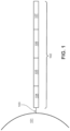
- FIG. 1 is a schematic diagram showing an exemplary capture probe, as described herein.
- the capture probe 102 is optionally coupled to a feature 101 by a cleavage domain 103, such as a disulfide linker.
- the capture probe can include a_functional sequence 104 that is useful for subsequent processing.
- the functional sequence 104 can include all or a part of sequencer specific flow cell attachment sequence (e.g., a P5 or P7 sequence), all or a part of a sequencing primer sequence, (e.g., a R1 primer binding site, a R2 primer binding site), or combinations thereof.
- the capture probe can also include a spatial barcode 105.
- the capture probe can also include a unique molecular identifier (UMI) sequence 106. While FIG.
- UMI unique molecular identifier
- Such splint oligonucleotide in addition to having a sequence complementary to a capture domain of a capture probe, can have a sequence of a nucleic acid analyte, a sequence complementary to a portion of a connected probe described herein, and/or a capture handle sequence described herein.
- the functional sequences can generally be selected for compatibility with any of a variety of different sequencing systems, e.g., Ion Torrent Proton or PGM, Illumina sequencing instruments, PacBio, Oxford Nanopore, etc., and the requirements thereof.
- functional sequences can be selected for compatibility with non-commercialized sequencing systems. Examples of such sequencing systems and techniques, for which suitable functional sequences can be used, include (but are not limited to) Ion Torrent Proton or PGM sequencing, Illumina sequencing, PacBio SMRT sequencing, and Oxford Nanopore sequencing.
- functional sequences can be selected for compatibility with other sequencing systems, including non-commercialized sequencing systems.
- the spatial barcode 105 and functional sequences 104 are common to all of the probes attached to a given feature.
- the UMI sequence 106 of a capture probe attached to a given feature is different from the UMI sequence of a different capture probe attached to the given feature.
- FIG. 2 is a schematic illustrating a cleavable capture probe, wherein the cleaved capture probe can enter into a non-permeabilized cell and bind to analytes within the sample.
- the capture probe 201 contains a cleavage domain 202, a cell penetrating peptide 203, a reporter molecule 204, and a disulfide bond (-S-S-).
- 205 represents all other parts of a capture probe, for example a spatial barcode and a capture domain.
- a second type of capture probe associated with the feature includes the spatial barcode 302 in combination with a random N-mer capture domain 304 for gDNA analysis.
- a third type of capture probe associated with the feature includes the spatial barcode 302 in combination with a capture domain complementary to a capture handle sequence of an analyte capture agent of interest 305.
- a fourth type of capture probe associated with the feature includes the spatial barcode 302 in combination with a capture domain that can specifically bind a nucleic acid molecule 306 that can function in a CRISPR assay (e.g., CRISPR/Cas9). While only four different capture probe-barcoded constructs are shown in FIG.
- capture-probe barcoded constructs can be tailored for analyses of any given analyte associated with a nucleic acid and capable of binding with such a construct.
- the schemes shown in FIG. 3 can also be used for concurrent analysis of other analytes disclosed herein, including, but not limited to: (a) mRNA, a lineage tracing construct, cell surface or intracellular proteins and metabolites, and gDNA; (b) mRNA, accessible chromatin (e.g., ATAC-seq, DNase-seq, and/or MNase-seq) cell surface or intracellular proteins and metabolites, and a perturbation agent (e.g., a CRISPR crRNA/sgRNA, TALEN, zinc finger nuclease, and/or antisense oligonucleotide as described herein); (c) mRNA, cell surface or intracellular proteins and/or metabolites, a barcoded labelling agent (e.g., the MHC multimers
- a perturbation agent can be a small molecule, an antibody, a drug, an aptamer, a miRNA, a physical environmental (e.g., temperature change), or any other known perturbation agents. See, e.g., Section (II)(b) (e.g., subsections (i)-(vi)) of WO 2020/176788 and/or U.S. Patent Application Publication No. 2020/0277663 . Generation of capture probes can be achieved by any appropriate method, including those described in Section (II)(d)(ii) of WO 2020/176788 and/or U.S. Patent Application Publication No. 2020/0277663 .
- more than one analyte type e.g., nucleic acids and proteins
- a biological sample can be detected (e.g., simultaneously or sequentially) using any appropriate multiplexing technique, such as those described in Section (IV) of WO 2020/176788 and/or U.S. Patent Application Publication No. 2020/0277663 .
- an analyte capture agent refers to an agent that interacts with an analyte (e.g., an analyte in a biological sample) and with a capture probe (e.g., a capture probe attached to a substrate or a feature) to identify the analyte.
- the analyte capture agent includes: (i) an analyte binding moiety (e.g., that binds to an analyte), for example, an antibody or antigen-binding fragment thereof; (ii) analyte binding moiety barcode; and (iii) a capture handle sequence.
- an analyte binding moiety barcode refers to a barcode that is associated with or otherwise identifies the analyte binding moiety.
- the term “analyte capture sequence” or “capture handle sequence” refers to a region or moiety configured to hybridize to, bind to, couple to, or otherwise interact with a capture domain of a capture probe.
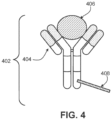
- the analyte capture agent can include an analyte -binding moiety barcode domain 408, a nucleotide sequence (e.g., an oligonucleotide), which can hybridize to at least a portion or an entirety of a capture domain of a capture probe.
- the analyte-binding moiety barcode domain 408 can comprise an analyte binding moiety barcode and a capture handle sequence described herein.
- the analyte-binding moiety 404 can include a polypeptide and/or an aptamer.
- the analyte-binding moiety 404 can include an antibody or antibody fragment (e.g., an antigen-binding fragment).
- FIG. 5 is a schematic diagram depicting an exemplary interaction between a feature-immobilized capture probe 524 and an analyte capture agent 526.
- the feature-immobilized capture probe 524 can include a spatial barcode 508 as well as functional sequences 506 and UMI 510, as described elsewhere herein.
- the capture probe can also include a capture domain 512 that is capable of binding to an analyte capture agent 526.
- the analyte capture agent 526 can include a functional sequence 518, analyte binding moiety barcode 516, and a capture handle sequence 514 that is capable of binding to the capture domain 512 of the capture probe 524.
- the analyte capture agent can also include a linker 520 that allows the capture agent barcode domain 516 to couple to the analyte binding moiety 522.
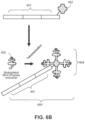
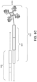
- one example oligonucleotide is capture probe 606 that comprises a complementary sequence (e.g., rGrGrG corresponding to C C C), a barcode sequence and other functional sequences, such as, for example, a UMI, an adapter sequence (e.g., comprising a sequencing primer sequence (e.g., R1 or a partial R1 ("pR1"), R2), a flow cell attachment sequence (e.g., P5 or P7 or partial sequences thereof)), etc.
- capture probe 606 may at first be associated with a feature (e.g., a gel bead) and released from the feature.
- capture probe 606 can hybridize with a capture agent barcode domain 601 of the MHC-oligonucleotide complex 605.
- the hybridized oligonucleotides (Spacer C C C and Spacer rGrGrG) can then be extended in primer extension reactions such that constructs comprising sequences that correspond to each of the two spatial barcode sequences (the spatial barcode associated with the capture probe, and the barcode associated with the MHC-oligonucleotide complex) are generated.
- one or both of the corresponding sequences may be a complement of the original sequence in capture probe 606 or capture agent barcode domain 601.
- the capture probe and the capture agent barcode domain are ligated together.
- the resulting constructs can be optionally further processed (e.g., to add any additional sequences and/or for clean-up) and subjected to sequencing.
- a sequence derived from the capture probe 606 spatial barcode sequence may be used to identify a feature and the sequence derived from spatial barcode sequence on the capture agent barcode domain 601 may be used to identify the particular peptide MHC complex 604 bound on the surface of the cell (e.g., when using MHC-peptide libraries for screening immune cells or immune cell populations).
- capture probes may be configured to prime, replicate, and consequently yield optionally barcoded extension products from a template (e.g., a DNA or RNA template, such as an analyte or an intermediate agent (e.g., a connected probe (e.g., a ligation product) or an analyte capture agent), or a portion thereof), or derivatives thereof (see, e.g., Section (II)(b)(vii) of WO 2020/176788 and/or U.S. Patent Application Publication No. 2020/0277663 regarding extended capture probes).
- a template e.g., a DNA or RNA template, such as an analyte or an intermediate agent (e.g., a connected probe (e.g., a ligation product) or an analyte capture agent), or a portion thereof
- a template e.g., a DNA or RNA template, such as an analyte or an intermediate agent (e.g.,
- Spatial information can provide information of biological and/or medical importance.
- the methods and compositions described herein can allow for: identification of one or more biomarkers (e.g., diagnostic, prognostic, and/or for determination of efficacy of a treatment) of a disease or disorder; identification of a candidate drug target for treatment of a disease or disorder; identification (e.g., diagnosis) of a subject as having a disease or disorder; identification of stage and/or prognosis of a disease or disorder in a subject; identification of a subject as having an increased likelihood of developing a disease or disorder; monitoring of progression of a disease or disorder in a subject; determination of efficacy of a treatment of a disease or disorder in a subject; identification of a patient subpopulation for which a treatment is effective for a disease or disorder; modification of a treatment of a subject with a disease or disorder; selection of a subject for participation in a clinical trial; and/or selection of a treatment for a subject with a disease or disorder.
- analytes and/or intermediate agents can be captured when contacting a biological sample with a substrate including capture probes (e.g., a substrate with capture probes embedded, spotted, printed, fabricated on the substrate, or a substrate with features (e.g., beads, wells) comprising capture probes).
- capture probes e.g., a substrate with capture probes embedded, spotted, printed, fabricated on the substrate, or a substrate with features (e.g., beads, wells) comprising capture probes.
- contact contacted
- contacting a biological sample with a substrate refers to any contact (e.g., direct or indirect) such that capture probes can interact (e.g., bind covalently or non-covalently (e.g., hybridize)) with analytes from the biological sample.
- each array feature location represents a position relative to a coordinate reference point (e.g., an array location, a fiducial marker) for the array. Accordingly, each feature location has an "address" or location in the coordinate space of the array.
- Capture probes of a feature of the array may comprise a particular spatial barcode (e.g., a feature-specific spatial barcode), whereas capture probes of a different feature of the array may comprise a different spatial barcode (e.g., a different feature-specific spatial barcode).
- a spatially barcoded substrate may be referred to herein as a spatially barcoded array or a substrate comprising an array of capture probes.
- Spatially barcoded substrates with capture domains configured to hybridize to mRNA analytes may be referred to herein as "gene expression substrates," “gene expression slides,” “GEx slides,” or “gene expression arrays”. Unless explicitly indicated, the terms are interchangeable.
- a capture probe on the first substrate includes a capture domain (e.g., a poly(T) sequence), a unique molecular identifier, a functional sequence such as a primer, a spatial barcode, or combinations thereof.
- the capture domain on the first substrate includes a poly-thymine (e.g., also called poly(T), poly-d(T), or oligo d(T) throughout) sequence that is complementary to a polyadenylation sequence.
- the capture probes are distributed on the first substrate inferior to the biological sample.
- a capture probe in the plurality is about 20, about 25, about 30, about 35, about 40, about 45, about 50, about 55, about 60, about 65, about 70, about 75, about 80, about 85, about 90, about 95, about 100, or more single-stranded nucleotides in length.
- a capture probe includes one or more non-naturally occurring nucleotides.
- the capture probes are distributed equally on the array of the first substrate.
- the capture probes on the first substrate are adhered directly (e.g., as described herein).
- the capture probes are placed using printed spots (e.g., as described herein).
- a second substrate e.g., a second spatially barcoded substrate (e.g., a second gene expression substrate)
- a second substrate is placed superior to the biological sample opposite to the first substrate, creating a sandwich configuration wherein the biological sample and the permeabilization buffer are located in between the two substrates.
- the second substrate is placed below the biological sample, opposite to the first substrate creating a sandwich configuration wherein the biological sample and the permeabilization buffer are located in between the two substrates.
- the second substrate is a glass substrate.
- the second substrate includes various glasses, substrate s formed from various polymers, hydrogels, layers and/or films, membranes (e.g., porous membranes), flow cells, wafers, plates, or combinations thereof.
- the second substrate includes a plurality of probes (e.g., as described herein).
- one or more probes in the plurality includes a spatial barcode and a capture domain.
- a probe on the second substrate includes a capture domain (e.g., a poly(T) sequence), a unique molecular identifier, a functional sequence such as a primer, a spatial barcode, or combinations thereof.
- a probe on the second substrate can be a probe as described in FIG. 1 .
- the probes on the second substrate are adhered directly to the substrate (e.g., as described herein).
- the probes on the second substrate are placed using printed spots (e.g., as described herein).
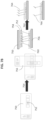
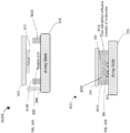
- the first substrate 720 includes an array of probes 722 that include inter alia a capture domain (e.g., a poly-thymine sequence) and a spatial barcode.
- the array (e.g., plurality) of capture probes can be associated with a whole substrate, parts of a substrate, or defined regions on a substrate.
- the system includes a permeabilization buffer that is added to the biological sample 724, allowing analytes 726 to diffuse 728 from the biological sample 724. Analytes 726 diffuse from the cell passively in any direction, for example, vertically, horizontally, or laterally, (e.g., arrows represented as 728 ).
- analytes diffuse from the cell to the array of probes 726 on the first substrate 720.
- Analytes also migrate to a second substrate 730, which includes a second array (e.g., or i.e., plurality) of capture probes 732.
- a capture probe or bead (which comprises probes) in the second array includes a capture domain sequence (e.g., a poly-thymine sequence) and a spatial barcode.
- the analyte migrates to the second substrate in a vector, for example, as shown by 728. After migrating to the second substrate, the analyte can be extended, amplified, and sequence using methods disclosed herein.
- the first substrate 720 and the second substrate 730 are sandwiched together and form substantially parallel planes.
- the angle of migration of the analyte is measured as an angle from the first substrate 720. In some instances, the angle is 90 degrees relative to the first substrate 720. In some instances, the angle is about 85, about 80, about 75, about 70, about 65, about 60, about 55, about 50, or about 45 degrees relative to the first substrate 720.
- the first substrate 720 and the second substrate 730 are sandwiched together in a sandwiching process.
- the first substrate is aligned with the second substrate, such that at least a portion of the biological sample is aligned with at least a portion of the array (e.g., aligned in a sandwich configuration).
- the first substrate is aligned with the second substrate such that at least a portion of the biological sample is vertically aligned with at least a portion of the array.
- the second substrate is in a superior position to the first substrate.
- the first substrate may be positioned superior to the second substrate (as depicted in FIG. 7 , bottom left image).
- the first and second substrates are aligned to maintain a gap or separation distance between the two substrates.
- one or more analytes are released from the biological sample and actively or passively migrate to the array for capture.
- the migration occurs while the aligned portions of the biological sample and the array are contacted with a reagent medium (e.g., permeabilization buffer).
- the released one or more analytes may actively or passively migrate across the gap via the reagent medium toward the capture probes or beads on second substrate 730, and be captured by the capture probes 732.
- the separation distance between first and second substrates is maintained between 2 microns and 1 mm (e.g., between 2 microns and 800 microns, between 2 microns and 700 microns, between 2 microns and 600 microns, between 2 microns and 500 microns, between 2 microns and 400 microns, between 2 microns and 300 microns, between 2 microns and 200 microns, between 2 microns and 100 microns, between 2 microns and 25 microns, between 2 microns and 10 microns), measured in a direction orthogonal to the surface of first substrate that supports sample.
- the distance is 2 microns. In some instances, the distance is 2.5 microns.
- the distance is about 2, 3, 4, 5, 6, 7, 8, 9, 10, 11, 12, 13, 14, 15, 16, 17, 18, 19, 20, 21, 22, 23, 24, or 25 microns.
- second substrate is placed in direct contact with the sample on the first substrate ensuring no diffusive spatial resolution losses.
- the separation distance is measured in a direction orthogonal to a surface of the first substrate that supports the biological sample.
- the first and second substrates are placed in a substrate holder (e.g., an array alignment device) configured to align the biological sample and the array.
- the device comprises a sample holder.
- the sample holder includes first and second members that comprise first and second retaining mechanisms configured to retain the first and second substrates, respectively.
- the device can include an alignment mechanism that is connected to at least one of the members and aligns the first and second members.
- a sample holder is provided as part of the sandwiching mechanism (e.g., sandwiching apparatus) used in the methods disclosed herein.
- the sample holder includes a first member that includes a first retaining mechanism that retains substrate with sample.
- the sample holder also includes a second member that includes a second retaining mechanism that retains second substrate with a feature array.
- An alignment mechanism is connected to at least one of first and second members or to both first and second members. During an alignment and contacting procedure, an alignment mechanism functions to align the first and second members, thereby ensuring that sample and feature array are also aligned and brought into contact to facilitate analysis of sample.
- the alignment mechanism can be implemented as a rotating actuator connected to the first and second members.
- a rotating actuator is a hinge.
- rotation of one of the members about the hinge axis aligns members and, and also aligns sample and feature array.
- the members can be rotated about the hinge axis until the sample and feature array are aligned and in contact.
- the rotating actuator is implemented as a folding member Folding member can be formed from a variety of materials, including compliant materials such as rubber and vinyl, metals and metal alloys, and plastics.
- the rotating actuator can include at least one arm. In some instances, the rotating actuator can include multiple arms (e.g., 2 or more, 3 or more, 4 or more, or even more).
- sample holder is implemented as a unitary (e.g., i.e., one-piece) device. In some instances, Sample holder can also be implemented as a two-piece device, with first and second members being separate but reproducibly connectable via the alignment mechanism. When the first and second members are brought into proximity, connectors engage with receivers, aligning first and second members, and also aligning sample with the feature array. It should be noted that while connectors are positioned on second member and receivers are positioned on first member, the reverse could also be true. Moreover, first and second members could each have one or more connectors and one or more receivers.
- first retaining mechanism can be implemented in various ways.
- first retaining mechanism can correspond to a recess dimensioned to receive first substrate.
- a gasket can optionally be positioned within the recess to maintain an interference fit between the edges of the recess and first substrate.
- first retaining mechanism can correspond to one or more members positioned to apply a force to first substrate, in particular, to maintain contact between first substrate and first member. Examples of such members include, but are not limited to, clips, screws and other threaded retaining fasteners, and members that snap-fasten or otherwise engage with first member. The members can apply a force to the sample bearing surface of first substrate and/or to one or more lateral surfaces first substrate.
- second retaining mechanism can correspond to any of the different types of retaining mechanisms discussed above in connection with first retaining mechanism.
- First and second retaining mechanisms and can be different or the same.
- a reagent medium can be positioned on a first or second substrate. More generally, however, the first or second substrate may further comprise a reagent medium placed thereon.
- the reagent medium includes a permeabilization reagent (e.g., a solid, liquid, gel, or dried permeabilization reagent).
- the reagent medium includes one or more additional components.
- the additional components can include a hydrogel compound or layer with an embedded permeabilization reagent.
- the second member includes at least one aperture. More generally, the second member can include one or more (e.g., 2 or more, 3 or more, 4 or more, 5 or more, 6 or more, 8 or more, 10 or more, 15 or more, 20 or more, 30 or more, 40 or more, 50 or more, or even more) second apertures.
- the second aperture is aligned with at least a portion of the sample region on substrate when the first and second members and are aligned.
- a second aperture can used for various purposes.
- the feature array and/or the sample can be viewed or imaged through second aperture. Viewing/imaging can be used to adjust the relative positions of the feature array and the sample to improve alignment, for example.
- one or more bounding surfaces of the second aperture and a back surface of the second substrate cooperate to form a reagent well.
- a reagent solution e.g., comprising a permeabilization reagent
- the reagent solution can permeate (e.g., by diffusion) through the back surface of the second substrate and contact the sample.
- the sample holder includes a first adjustment mechanism connected to the first member.
- the first adjustment mechanism translates the first substrate in at least one direction parallel to the surface of the first substrate that supports the sample. In some embodiments, the first adjustment mechanism translates the first substrate in two directions parallel to the surface of the first substrate.
- the first adjustment mechanism can be implemented in various ways.
- the first adjustment mechanism includes one or more thumbscrews or linear actuators that can be used to translate the first substrate.
- the alignment mechanism is also configured to maintain a separation between the first and second substrates (and the first and second members) when the substrates (and members) are aligned.
- the separation can be maintained such that at least a portion of the sample contacts the reagent medium (e.g., the feature array of the reagent medium).
- the alignment mechanism maintains the first and second substrates in an approximately parallel relationship when the substrates (and the first and second members) are aligned.
- An included angle between the first and second substrates in such circumstances can be 2 degrees or less (e.g., 1 degree or less, 0.5 degrees or less, 0.25 degrees or less).
- the sample holder includes a second adjustment mechanism.
- the second adjustment mechanism adjusts a distance of the separation between the first and second substrates (e.g., or i.e., in a direction orthogonal to the surface of the first substrate that supports the sample).
- the adjustment mechanism is connected to both members.
- the second adjustment mechanism can be implemented in various ways.
- the second adjustment mechanism includes one or more thumbscrews or adjustable pins or posts.
- the second adjustment mechanism includes one or more linear actuators.
- the second adjustment mechanism includes a swellable or expandable membrane, gasket, or layer positioned between the first and second members.
- the sample holder can be introduced into a thermocycler to promote capture of analytes from the sample by the feature array.
- the sample holder can be inserted directly into a suitable thermocycler for this purpose.
- the sample holder can be coupled to a thermocycler adapter and the coupled holder and adapter inserted into a thermocycler. Exemplary devices and exemplary sample holders are described in PCT Patent Application Publication No. WO 2020/123320 , which is incorporated by reference in its entirety.
- the probes on the first and/or second substrate are adhered to beads (e.g., as described herein).
- the probes are placed on the first and/or second substrate using microspheres (e.g., as described herein).
- the beads or microspheres that include probes are associated with, or affixed to, the first and/or second substrate. For example, in some instances capture probe containing beads or microsphere are affixed directly or indirectly to a substrate via surface chemistries, hydrogel, and the like.
- the diameter of a bead the is adhered to the probe on the second substrate is about 1 ⁇ m, about 2 ⁇ m, about 3 ⁇ m, about 4 ⁇ m, about 5 ⁇ m, about 6 ⁇ m, about 7 ⁇ m, about 8 ⁇ m, about 9 ⁇ m, about 10 ⁇ m, about 15 ⁇ m, about 20 ⁇ m, or more.
- the aligned portions of the biological sample and the array are in contact with the reagent medium for about 1 minute. In some instances, the aligned portions of the biological sample and the array are in contact with the reagent medium for about 5 minutes. In some instances, the aligned portions of the biological sample and the array are in contact with the reagent medium in the gap for about 1 minute, about 5 minutes, about 10 minutes, about 12 minutes, about 15 minutes, about 18 minutes, about 20 minutes, about 25 minutes, about 30 minutes, about 36 minutes, about 45 minutes, or about an hour. In some instances, the aligned portions of the biological sample and the array are in contact with the reagent medium for about 1-60 minutes. In some instances, the aligned portions of the biological sample and the array are in contact with the reagent medium for about 30 minutes.
- the biological sample on the first substrate is stained using any of the methods described herein.
- the biological sample is imaged, capturing the stain pattern created during the stain step.
- the biological sample then is destained prior to the sandwiching process.
- the first substrate and the second substrate are separated (e.g., such that they are no longer aligned in a sandwich configuration, also referred to herein as opening the sandwich).
- subsequent analysis e.g., reverse transcription, cDNA synthesis, library preparation, and sequences
- the permeabilization buffer includes proteinase K, pepsin, collagenase, a detergent, one or more ribonuclease inhibitor, or combinations thereof.
- the detergent is selected from sodium dodecyl sulfate (SDS), polyethylene glycol tert-octylphenyl ether, polysorbate 80, polysorbate 20, N-lauroylsarcosine (or a sodium salt thereof), or combinations thereof.
- the permeabilization buffer comprises a hydrogel.
- biological samples Prior to analyte capture, in some instances, biological samples can be stained using a wide variety of stains and staining techniques.
- the biological sample is a tissue section on a substrate (e.g., a slide; e.g., a 10 ⁇ m biological section section).
- the tissue section is about 5, 6, 7, 8, 9, 10, 11, 12, 13, 14, 15, 16, 17, 18, 19, 20, 21, 22, 23, 24, or 25 ⁇ m in thickness.
- the biological sample is dried after placement onto the first substrate. In some instances, the biological sample is dried at 42°C. In some instances, drying occurs for about 1 hour, about 2, hours, about 3 hours, or until the sections become transparent. In some instances, the biological sample can be dried overnight (e.g., in a desiccator at room temperature).
- a sample can be stained using any number of biological stains, including but not limited to, acridine orange, Bismarck brown, carmine, coomassie blue, cresyl violet, DAPI, eosin, ethidium bromide, acid fuchsine, hematoxylin, Hoechst stains, iodine, methyl green, methylene blue, neutral red, Nile blue, Nile red, osmium tetroxide, propidium iodide, rhodamine, or safranin.
- the methods disclosed herein include imaging the biological sample. In some instances, imaging the sample occurs prior to deaminating the biological sample.
- the sample can be stained using known staining techniques, including Can-Grunwald, Giemsa, hematoxylin and eosin (H&E), Jenner's, Leishman, Masson's trichrome, Papanicolaou, Romanowsky, silver, Sudan, Wright's, and/or Periodic Acid Schiff (PAS) staining techniques.
- PAS staining is typically performed after formalin or acetone fixation. In some instances, the stain is an H&E stain.
- a biological sample can be prepared by staining and imaging using one technique (e.g., H&E staining and brightfield imaging), followed by staining and imaging using another technique (e.g., IHC/IF staining and fluorescence microscopy) for the same biological sample.
- one technique e.g., H&E staining and brightfield imaging
- another technique e.g., IHC/IF staining and fluorescence microscopy
- biological samples can be destained.
- Methods of destaining or discoloring a biological sample are known in the art, and generally depend on the nature of the stain(s) applied to the sample.
- H&E staining can be destained by washing the sample in HCl, or any other acid (e.g., selenic acid, sulfuric acid, hydroiodic acid, benzoic acid, carbonic acid, malic acid, phosphoric acid, oxalic acid, succinic acid, salicylic acid, tartaric acid, sulfurous acid, trichloroacetic acid, hydrobromic acid, hydrochloric acid, nitric acid, orthophosphoric acid, arsenic acid, selenous acid, chromic acid, citric acid, hydrofluoric acid, nitrous acid, isocyanic acid, formic acid, hydrogen selenide, molybdic acid, lactic acid, acetic acid, carbonic acid, hydrogen sulfide, or combinations thereof).
- destaining can include 1, 2, 3, 4, 5, or more washes in an acid (e.g., HCl).
- destaining can include adding HCl to a downstream solution (e.g., permeabilization solution).
- destaining can include dissolving an enzyme used in the disclosed methods (e.g., pepsin) in an acid (e.g., HCl) solution.
- other reagents can be added to the destaining solution to raise the pH for use in other applications. For example, SDS can be added to an acid destaining solution in order to raise the pH as compared to the acid destaining solution alone.
- immunofluorescence or immunohistochemistry protocols can be performed as a part of, or in addition to, the exemplary spatial workflows presented herein.
- tissue sections can be fixed according to methods described herein.
- the biological sample can be transferred to an array (e.g., capture probe array), wherein analytes (e.g., proteins) are detected using immunofluorescence protocols.
- analytes e.g., proteins
- the sample can be rehydrated, blocked, and permeabilized (3X SSC, 2% BSA, 0.1% Triton X, 1 U/ ⁇ l RNAse inhibitor for 10 minutes at 4°C) before being stained with fluorescent primary antibodies (1:100 in 3XSSC, 2% BSA, 0.1% Triton X, 1 U/ ⁇ l RNAse inhibitor for 30 minutes at 4°C).
- the biological sample can be washed, coverslipped (in glycerol + 1 U/ ⁇ l RNAse inhibitor), imaged (e.g., using a confocal microscope or other apparatus capable of fluorescent detection), washed, and processed according to analyte capture or spatial workflows described herein.
- a glycerol solution and a cover slip can be added to the sample.
- the glycerol solution can include a counterstain (e.g., DAPI).
- an antigen retrieval buffer can improve antibody capture in IF/IHC protocols.
- An exemplary protocol for antigen retrieval can be preheating the antigen retrieval buffer (e.g., to 95°C), immersing the biological sample in the heated antigen retrieval buffer for a predetermined time, and then removing the biological sample from the antigen retrieval buffer and washing the biological sample.
- optimizing permeabilization can be useful for identifying intracellular analytes.
- Permeabilization optimization can include selection of permeabilization agents, concentration of permeabilization agents, and permeabilization duration. Tissue permeabilization is discussed elsewhere herein.
- blocking an array and/or a biological sample in preparation of labeling the biological sample decreases nonspecific binding of the antibodies to the array and/or biological sample (decreases background).
- Some embodiments provide for blocking buffers/blocking solutions that can be applied before and/or during application of the label, wherein the blocking buffer can include a blocking agent, and optionally a surfactant and/or a salt solution.
- a blocking agent can be bovine serum albumin (BSA), serum, gelatin (e.g., fish gelatin), milk (e.g., non-fat dry milk), casein, polyethylene glycol (PEG), polyvinyl alcohol (PVA), or polyvinylpyrrolidone (PVP), biotin blocking reagent, a peroxidase blocking reagent, levamisole, Carnoy's solution, glycine, lysine, sodium borohydride, pontamine sky blue, Sudan Black, trypan blue, FITC blocking agent, and/or acetic acid.
- the blocking buffer/blocking solution can be applied to the array and/or biological sample prior to and/or during labeling (e.g., application of fluorophore-conjugated antibodies) to the biological sample.
- one or more analytes from the biological sample are released from the biological sample and migrate to a substrate comprising an array of capture probes for attachment to the capture probes of the array.
- the release and migration of the analytes to the substrate comprising the array of capture probes occurs in a manner that preserves the original spatial context of the analytes in the biological sample.
- the biological sample is mounted on a first substrate and the substrate comprising the array of capture probes is a second substrate.
- the method is facilitated by a sandwiching process. Sandwiching processes are described in, e.g., US. Patent Application Pub. No.
- the sandwiching process may be facilitated by a device, sample holder, sample handling apparatus, or system described in, e.g., US. Patent Application Pub. No. 20210189475 , PCT/US2021/036788 , or PCT/US2021/050931 .
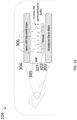
- FIG. 14 is a schematic diagram depicting an exemplary sandwiching process 104 between a first substrate comprising a biological sample (e.g., a tissue section 302 on a slide 303) and a second substrate comprising a spatially barcoded array, e.g., a slide 304 that is populated with spatially-barcoded capture probes 306.
- the first substrate is aligned with the second substrate, such that at least a portion of the biological sample is aligned with at least a portion of the array (e.g., aligned in a sandwich configuration).
- the second substrate e.g., slide 304 is in a superior position to the first substrate (e.g., slide 303).
- the first substrate may be positioned superior to the second substrate (e.g., slide 304).
- a reagent medium 305 e.g., permeabilization solution
- a reagent medium 305 e.g., permeabilization solution
- a permeabilization buffer which permeabilizes or digests the sample 302 and the analytes (e.g., the different analytes described herein, such as, protein, nucleic acid (e.g., RNA or DNA), intermediate agent (e.g., connected probe (e.g., RTL probe) or analyte capture agent (e.g., oligo-conjugated antibody)) or portion thereof, targeted capture, and/or whole transcriptome) 308 of the biological sample 302 may release, actively or passively migrate (e.g., diffuse) across the gap 307 toward the capture probes 306, and bind on the capture probes 306.
- analytes e.g., the different analytes described herein, such as, protein,
- an extension reaction may occur, thereby generating a spatially barcoded library.
- an extension reaction may occur, thereby generating a spatially barcoded library.
- reverse transcription may be used to generate a cDNA library associated with a particular spatial barcode.
- Barcoded cDNA libraries may be mapped back to a specific spot on a capture area of the capture probes 306. This data may be subsequently layered over a high-resolution microscope image of the biological sample, making it possible to visualize the data within the morphology of the tissue in a spatially-resolved manner.
- the extension reaction can be performed separately from the sample handling apparatus described herein that is configured to perform the exemplary sandwiching process 104.
- the sandwich configuration of the sample 302, the first substrate (e.g., slide 303) and the second substrate (e.g., slide 304) may provide advantages over other methods of spatial analysis and/or analyte capture.
- the sandwich configuration may reduce a burden of users to develop in house tissue sectioning and/or tissue mounting expertise.
- the sandwich configuration may decouple sample preparation/tissue imaging from the barcoded array (e.g., spatially-barcoded capture probes 306) and enable selection of a particular region of interest of analysis (e.g., for a tissue section larger than the barcoded array).
- the sandwich configuration also beneficially enables spatial analysis without having to place a biological sample (e.g., tissue section) 302 directly on the second substrate (e.g., slide 304).
- the sandwiching process comprises: mounting the first substrate on a first member of a support device, the first member configured to retain the first substrate; mounting the second substrate on a second member of the support device, the second member configured to retain the second substrate, applying a reagent medium to the first substrate and/or the second substrate, the reagent medium comprising a permeabilization agent, operating an alignment mechanism (also referred to herein as an adjustment mechanism) of the support device to move the first member and/or the second member such that a portion of the biological sample is aligned (e.g., vertically aligned) with a portion of the array of capture probes and within a threshold distance of the array of capture probes, and such that the portion of the biological sample and the capture probe contact the reagent medium, wherein the permeabilization agent releases the analyte from the biological sample.
- an alignment mechanism also referred to herein as an adjustment mechanism
- the sandwiching process methods described above can be implemented using a variety of hardware components.
- the sandwiching process methods can be implemented using a sample holder (also referred to herein as a support device, a sample handling apparatus, and an array alignment device).
- the sample holder can include a first member including a first retaining mechanism configured to retain a first substrate comprising a sample.
- the first retaining mechanism can be configured to retain the first substrate disposed in a first plane.
- the sample holder can further include a second member including a second retaining mechanism configured to retain a second substrate disposed in a second plane.
- the sample holder can further includes an alignment mechanism connected to one or both of the first member and the second member.
- the alignment mechanism can be configured to align the first and second members along the first plane and/or the second plane such that the sample contacts at least a portion of the reagent medium when the first and second members are aligned and within a threshold distance along an axis orthogonal to the second plane.
- the adjustment mechanism may be configured to move the second member along the axis orthogonal to the second plane and/or move the first member along an axis orthogonal to the first plane.
- the adjustment mechanism includes a linear actuator.
- the linear actuator is configured to move the second member along an axis orthogonal to the plane or the first member and/or the second member.
- the linear actuator is configured to move the first member along an axis orthogonal to the plane of the first member and/or the second member.
- the linear actuator is configured to move the first member, the second member, or both the first member and the second member at a velocity of at least 0.1 mm/sec.
- the linear actuator is configured to move the first member, the second member, or both the first member and the second member with an amount of force of at least 0.1 lbs.
- FIG. 15A is a perspective view of an example sample handling apparatus 1400 in a closed position in accordance with some example implementations.
- the sample handling apparatus 1400 includes a first member 1404, a second member 1410, optionally an image capture device 1420, a first substrate 1406, optionally a hinge 1415, and optionally a mirror 1416.
- the hinge 1415 may be configured to allow the first member 1404 to be positioned in an open or closed configuration by opening and/or closing the first member 1404 in a clamshell manner along the hinge 1415.
- FIG. 15B is a perspective view of the example sample handling apparatus 1400 in an open position in accordance with some example implementations.
- the sample handling apparatus 1400 includes one or more first retaining mechanisms 1408 configured to retain one or more first substrates 1406.
- the first member 1404 is configured to retain two first substrates 1406, however the first member 1404 may be configured to retain more or fewer first substrates 1406.
- the first substrate 1406 and/or the second substrate 1412 may be loaded and positioned within the sample handling apparatus 1400 such as within the first member 1404 and the second member 1410, respectively.
- the hinge 1415 may allow the first member 1404 to close over the second member 1410 and form a sandwich configuration (e.g., the sandwich configuration shown in FIG. 14 ).
- an adjustment mechanism (not shown) of the sample handling apparatus 1400 may actuate the first member 1404 and/or the second member 1410 to form the sandwich configuration for the permeabilization step (e.g., bringing the first substrate 1406 and the second substrate 1412 closer to each other and within a threshold distance for the sandwich configuration).
- the adjustment mechanism may be configured to control a speed, an angle, a force, or the like of the sandwich configuration.
- the biological sample (e.g., sample 302) may be aligned within the first member 1404 (e.g., via the first retaining mechanism 1408) prior to closing the first member 1404 such that a desired region of interest of the sample 302 is aligned with the barcoded array of the second substrate (e.g., the slide 304), e.g., when the first and second substrates are aligned in the sandwich configuration.
- Such alignment may be accomplished manually (e.g., by a user) or automatically (e.g., via an automated alignment mechanism).
- spacers may be applied to the first substrate 1406 and/or the second substrate 1412 to maintain a minimum spacing between the first substrate 1406 and the second substrate 1412 during sandwiching.
- the permeabilization solution (e.g., permeabilization solution 305) may be applied to the first substrate 1406 and/or the second substrate 1412.
- the first member 1404 may then close over the second member 1410 and form the sandwich configuration.
- Analytes e.g., the different analytes described herein, such as, mRNA transcripts
- 308 may be captured by the capture probes 306 and may be processed for spatial analysis.
- the image capture device 1420 may capture images of the overlap area (e.g., overlap area 710) between the tissue 302 and the capture probes 306. If more than one first substrates 1406 and/or second substrates 1412 are present within the sample handling apparatus 1400, the image capture device 1420 may be configured to capture one or more images of one or more overlap areas 710. Further details on support devices, sample holders, sample handling apparatuses, or systems for implementing a sandwiching process are described in, e.g., US. Patent Application Pub. No. 20210189475 , and PCT/US2021/050931 , each of which are incorporated by reference in their entirety.
- Analytes within a biological sample may be released through disruption (e.g., permeabilization, digestion, etc.) of the biological sample or may be released without disruption.
- permeabilizing e.g., any of the permeabilization reagents and/or conditions described herein
- a biological sample including for example including the use of various detergents, buffers, proteases, and/or nucleases for different periods of time and at various temperatures.
- various methods of delivering fluids e.g., a buffer, a permeabilization solution
- a substrate holder e.g., for sandwich assembly, sandwich configuration, as described herein.
- the sandwich configuration described herein between a first substrate comprising a biological sample (e.g., slide 303) and a second substrate comprising a spatially barcoded array (e.g., slide 304 with barcoded capture probes 306) may include a reagent medium (e.g., a liquid reagent medium, e.g., a permeabilization solution 305 or other target molecule release and capture solution) to fill a gap (e.g., gap 307). It may be desirable that the reagent medium be free from air bubbles between the slides to facilitate transfer of target molecules with spatial information. Additionally, air bubbles present between the slides may obscure at least a portion of an image capture of a desired region of interest. Accordingly, it may be desirable to ensure or encourage suppression and/or elimination of air bubbles between the two substrates (e.g., slide 303 and slide 304) during a permeabilization step (e.g., step 104).
- a reagent medium e.g., a liquid reagent medium,
- Workflows described herein may include contacting a drop of the reagent medium (e.g., liquid reagent medium, e.g., a permeabilization solution 305) disposed on a first substrate or a second substrate with at least a portion of the second substrate or first substrate, respectively.
- the contacting comprises bringing the two substrates into proximity such that the sample on the first substrate is aligned with the barcode array of capture probes on the second substrate.
- the drop includes permeabilization reagents (e.g., any of the permeabilization reagents described herein).
- the rate of permeabilization of the biological sample is modulated by delivering the permeabilization reagents (e.g., a fluid containing permeabilization reagents) at various temperatures.
- FIG. 16B shows a fully formed sandwich configuration creating a chamber 3650 formed from the one or more spacers 3610, the first substrate (e.g., the slide 303), and the second substrate (e.g., the slide 304 including spatially barcoded capture probes 306) in accordance with some example implementations.
- the liquid reagent e.g., the permeabilization solution 305 fills the volume of the chamber 3650 and may create a permeabilization buffer that allows analytes (e.g., mRNA transcripts and/or other molecules) to diffuse from the biological sample 302 toward the capture probes 306 of the second substrate (e.g., slide 304).
- analytes e.g., mRNA transcripts and/or other molecules
- FIG. 17B shows that as the first substrate lowers, and/or as the second substrate rises, the dropped side of the first substrate (e.g., a side of the slide 303 angled toward the second substrate) may contact the drop of the reagent medium 305.
- the dropped side of the first substrate may urge the reagent medium 305 toward the opposite direction (e.g., towards an opposite side of the spacer 3610, towards an opposite side of the first substrate relative to the dropped side).
- the reagent medium 305 may be urged from right to left as the sandwich is formed.
- the first substrate and/or the second substrate are further moved to achieve an approximately parallel arrangement of the first substrate and the second substrate.
- FIGs. 17A-17C depict the first substrate (e.g., the slide 303 including biological sample 302) angled over (superior to) the second substrate (e.g., slide 304) and the second substrate comprising the spacer 3610, it should be understood that an exemplary angled closure workflow can include the second substrate angled over (superior to) the first substrate and the first substrate comprising the spacer 3610.
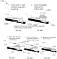
- FIGs. 18A-18E depict an example workflow 1700 for an angled sandwich assembly in accordance with some example implementations.
- a substrate 1712 e.g., a first substrate such as slide 303 or a second substrate such as slide 304 comprising spatially barcoded capture probes 306
- a base 1704 e.g., a first member or a second member of a sample holder disclosed herein
- the spring 1715 may extend from the base 1704 in a superior direction and may be configured to dispose the substrate 1712 along a plane angled differently than the base 1704.
- the angle of the substrate 1712 may be such that a drop of reagent medium 1705 (e.g., drop of liquid reagent medium) placed on the surface of the substrate 1712 (e.g., a surface of a spacer attached to the substrate) will not fall off the surface (e.g., due to gravity).
- the angle may be determined based on a gravitational force versus any surface force to move the drop away from and off the substrate 1712.
- FIG. 18B depicts a drop 1705 of reagent medium placed on the substrate 1712. As shown, the drop 1705 is located on the side of the substrate 1712 contacting the spring 1715 and is located in proximity and above (superior to) the spring 1715.
- another substrate 1706 may be positioned above (superior to) the substrate 1712 and at an angle substantially parallel with the base 1704.
- substrate 1706 may be a first substrate disclosed herein (e.g., slide 303).
- substrate 1706 may be a second substrate (e.g., slide 304 comprising spatially barcoded capture probes).
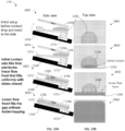
- FIG. 19A is a side view of the angled closure workflow 1700 in accordance with some example implementations.
- FIG. 19B is a top view of the angled closure workflow 1700 in accordance with some example implementations.
- the drop of reagent medium 1705 is positioned to the side of the substrate 1712 contacting the spring 1715.
- the dropped side of the angled substrate 1706 contacts the drop of reagent medium 1705 first.
- the contact of the substrate 1706 with the drop of reagent medium 1705 may form a linear or low curvature flow front that fills uniformly with the slides closed.
- the drop of reagent medium 1705 fills the gap (e.g., the gap 307) between the substrate 1706 and the substrate 1712.
- the linear flow front of the liquid reagent may form by squeezing the drop 1705 volume along the contact side of the substrate 1712 and/or the substrate 1706. Additionally, capillary flow may also contribute to filling the gap area.
- the spring 1715 may be fully compressed such that the substrate 1706, the substrate 1712, and the base 1704 are substantially parallel to each other.
- an angled closure workflow disclosed herein may be performed by a sample handling apparatus (e.g., as described in PCT/US2021/050931 , which is hereby incorporated by reference in its entirety.
- the disclosure provides methods, kits, substrates, and apparatuses that examine spatial analyte expression in multiple tissue types.
- the methods provided herein are performed on an FFPE sample.
- the methods provided herein are performed on a fresh frozen sample.
- the biological sample is deparaffinized.
- Deparaffinization can be achieved using any method known in the art.
- the biological samples is treated with a series of washes that include xylene and various concentrations of ethanol.
- methods of deparaffinization include treatment of xylene (e.g., three washes at 5 minutes each).
- the methods further include treatment with ethanol (e.g., 100% ethanol, two washes 10 minutes each; 95% ethanol, two washes 10 minutes each; 70% ethanol, two washes 10 minutes each; 50% ethanol, two washes 10 minutes each).
- the biological sample can be washed with deionized water (e.g., two washes for 5 minutes each). It is appreciated that one skilled in the art can adjust these methods to optimize deparaffinization.
- the biological sample is decrosslinked.
- the biological sample is decrosslinked in a solution containing TE buffer (comprising Tris and EDTA).
- the TE buffer is basic (e.g., at a pH of about 9).
- decrosslinking occurs at about 50°C to about 80°C.
- decrosslinking occurs at about 70°C.
- decrosslinking occurs for about 1 hour at 70°C.
- the biological sample can be treated with an acid (e.g., 0.1M HCl for about 1 minute). After the decrosslinking step, the biological sample can be washed (e.g., with 1x PBST).
- the methods of preparing a biological sample for analyte capture include steps of equilibrating and blocking the biological sample.
- equilibrating is performed using a pre-hybridization (pre-Hyb) buffer.
- pre-Hyb buffer is RNase-free.
- pre-Hyb buffer contains no bovine serum albumin (BSA), solutions like Denhardt's, or other potentially nuclease-contaminated biological materials.
- BSA bovine serum albumin
- the equilibrating step is performed multiple times (e.g., 2 times at 5 minutes each; 3 times at 5 minutes each).
- the biological sample is blocked with a blocking buffer.
- the blocking buffer includes a carrier such as tRNA, for example yeast tRNA such as from brewer's yeast (e.g., at a final concentration of 10-20 ⁇ g/mL). In some instances, blocking can be performed for 5, 10, 15, 20, 25, or 30 minutes.
- any of the foregoing steps can be optimized for performance. For example, one can vary the temperature.
- the pre-hybridization methods are performed at room temperature. In some instances, the pre-hybridization methods are performed at 4°C (in some instances, varying the timeframes provided herein).
- the methods of preparing a biological sample for analyte capture include permeabilizing the sample.
- the biological sample is permeabilized using a phosphate buffer.
- the phosphate buffer is PBS (e.g., 1x PBS).
- the phosphate buffer is PBST (e.g., 1x PBST).
- the permeabilization step is performed multiple times (e.g., 3 times at 5 minutes each).
- a permeabilization buffer (e.g., any permeabilization buffer described herein) is added to the biological sample.
- Permeabilization solutions can include, by way of example only, enzymes (e.g., proteinase K, pepsin, and collagenase), detergents (e.g., N-lauroylsarcosine or a sodium salt thereof, sodium dodecyl sulfate (SDS), polyethylene glycol tert-octylphenyl ether, polysorbate 80, and polysorbate 20), ribonuclease inhibitors, buffers optimized for electrophoresis, buffers optimized for permeabilization, buffers optimized for hybridization, or combinations thereof.
- the permeabilization buffer releases the analyte from the sample, allowing it to diffuse from the sample.
- the biological sample can be treated with a proteinase.
- the proteinase is proteinase K.
- permeabilization occurs using a protease.
- the protease is an endopeptidase.
- Endopeptidases that can be used include but are not limited to trypsin, chymotrypsin, elastase, thermolysin, pepsin, clostripan, glutamyl endopeptidase (GluC), ArgC, peptidyl-asp endopeptidase (ApsN), endopeptidase LysC and endopeptidase LysN.
- the endopeptidase is pepsin.
- the biological sample is permeabilized prior to capture of the analytes on either the first substrate or the second substrate (or both).
- the permeabilization step includes application of a permeabilization buffer to the biological sample.
- the permeabilization buffer includes a buffer (e.g., Tris pH 7.5), MgCl2, sarkosyl detergent (e.g., sodium lauroyl sarcosinate), enzyme (e.g., proteinase K, and nuclease free water.
- the permeabilization step is performed at 37°C.
- the permeabilization step is performed for about 20 minutes to 2 hours (e.g., about 20 minutes, about 30 minutes, about 40 minutes, about 50 minutes, about 1 hour, about 1.5 hours, or about 2 hours).
- the releasing step is performed for about 40 minutes.
- the methods provided herein include a permeabilizing step in order to release the analyte.
- permeabilization occurs using a protease.
- the protease is an endopeptidase.
- Endopeptidases that can be used include but are not limited to trypsin, chymotrypsin, elastase, thermolysin, pepsin, clostripan, glutamyl endopeptidase (GluC), ArgC, peptidyl-asp endopeptidase (ApsN), endopeptidase LysC and endopeptidase LysN.
- the endopeptidase is pepsin.
- methods provided herein include permeabilization of the biological sample such that the capture probe can more easily bind to the analyte (e.g., or i.e., compared to no permeabilization).
- the permeabilization step includes application of a permeabilization buffer to the biological sample.
- the permeabilization buffer includes a buffer (e.g., Tris pH 7.5), MgCl 2 , sarkosyl detergent (e.g., sodium lauroyl sarcosinate), enzyme (e.g., proteinase K), and nuclease free water.
- the permeabilization step is performed at 37°C.
- the permeabilization step is performed for about 20 minutes to 2 hours (e.g., about 20 minutes, about 30 minutes, about 40 minutes, about 50 minutes, about 1 hour, about 1.5 hours, or about 2 hours).
- the releasing step is performed for about 40 minutes.
- the analyte is released using an endoribonuclease.
- the endoribonuclease is an RNase.
- the RNase can be RNase H, RNase A, RNase C, or RNase I.
- the RNase H is RNase H1, RNase H2, or RNase H1, or RNase H2.
- the releasing step is performed using a releasing buffer.
- the release buffer includes one or more of a buffer (e.g., Tris pH 7.5), enzyme (e.g., RNAse H) and nuclease-free water.
- the releasing step is performed at 37°C.
- the releasing step is performed for about 20 minutes to 2 hours (e.g., about 20 minutes, about 30 minutes, about 40 minutes, about 50 minutes, about 1 hour, about 1.5 hours, or about 2 hours). In some instances, the releasing step is performed for about 30 minutes.
- the releasing step occurs after the permeabilization step. In some instances, the releasing step occurs at the same time as the permeabilization step (e.g., in the same buffer).
- the reagent medium e.g., the permeabilization buffer
- the reagent medium comprises one or more of sodium dodecyl sulfate (SDS), proteinase K, pepsin, N-lauroylsarcosine or a sodium salt thereof, RNAse, and a sodium salt thereof.
- SDS sodium dodecyl sulfate
- proteinase K proteinase K
- pepsin proteinase K
- N-lauroylsarcosine or a sodium salt thereof RNAse, and a sodium salt thereof.
- a hydrogel is used to enhance spatial resolution.
- a biological sample e.g., tissue section
- hydrogel subunits are infused into the biological sample, and polymerization of the hydrogel is initiated by an external or internal stimulus.
- a "hydrogel” as described herein can include a cross-linked 3D network of hydrophilic polymer chains.
- a “hydrogel subunit” can be a hydrophilic monomer, a molecular precursor, or a polymer that can be polymerized (e.g., cross-linked) to form a three-dimensional (3D) hydrogel network. Additional disclosure of hydrogels is found in WO 2020/176788 and U.S. Patent Application Publication No. 2020/0277663 , each of which is incorporated by reference in its entirety.
- analytes migrate through the biological sample. In some instances, analytes migrate from the biological sample to the first and/or second probe. In some instances, migration disclosed herein is passive migration. As some migration is passive, analytes (e.g., mRNA) can migrate in any direction. In some instances, probes on the first substrate capture analytes that migrate passively. In some instances, probes on the second substrate capture analytes that migrate passively.
- analytes e.g., mRNA
- a certain period of time e.g., about 5 minutes to about 10 hours, about 5 minutes to about 5 hours, about 5 minutes to about 1 hour, about 5 minutes to about 45 minutes, about 5 minutes to about 30 minutes, about 5 minutes to about 15 minutes, about 15 minutes to about 10 hours, about 15 minutes to about 5 hours, about 15 minutes to about 1 hour, about 15 minutes to about 45 minutes, about 15 minutes to about 30 minutes, about 30 minutes to about 10 hours, about 30 minutes to about 5 hours, about 30 minutes to about 1 hour, about 30 minutes to about 45 minutes, about 45 minutes to about 10 hours, about 45 minutes to about 5 hours, about 45 minutes to about 1 hour, about 1 hour to about 10 hours, about 1 hour to about 5 hours, about 1 hour to about 1.5 hours, about 1.5 hours to about 10 hours, about 1.5 hours to about 5 hours, about 1.5 hours to about 2 hours, about 2 hours to about 10 hours, about 2 hours to about 5 hours, about 2 hours to about 3 hours, about 2.5 hours to about 10 hours, about 2.5 hours to about 5 hours, about 2.5 hours to about 10 hours, about 2.5
- one or more analytes after a certain period of time as previously listed, one or more analytes have passively migrated and been captured by the capture probe(s) on the second substrate. In some instances, at least about 80%, or at least about 90% of all analytes are captured by probes on the second substrate. It is contemplated that while a portion of the analytes, for example those in close proximity to the first substrate capture probes, will passively migrate and be captured on the first substrate, whereas the majority of the analytes, those that are not in close proximity to the first substrate, will passively migrate to the second substrate and be captured by the barcoded capture probes.
- the one or more solutions between the first substrate /biological sample and the second substrate can include a permeabilization buffer (e.g., any of the permeabilization buffers described herein).
- the one or more solutions can include a buffer that can maintain the pH at a relatively constant value.
- the gap between the biological sample and the second substrate with the second set of capture probes is about 0.5 ⁇ m, about 1.0 ⁇ m, about 1.5 ⁇ m, about 2.0 ⁇ m, about 2.5 ⁇ m, about 3.0 ⁇ m, about 3.5 ⁇ m, about 4.0 ⁇ m, about 4.5 ⁇ m, about 5.0 ⁇ m, or more. In some instances, the distance between the biological sample and the second set of capture probes is about 2.5 ⁇ m.
- the method described herein further comprises separating the first substrate and the second substrates after the capture of the analytes. In some embodiments, the first substrate and the second substrate are de-aligned.
- the spatial resolution of analytes from a biological sample is increased.
- the resolution of detection of analytes is increased by about 5%, about 10%, about 15%, about 20%, about 25%, about 30%, about 35%, about 40%, about 45%, about 50%, about 55%, about 60%, about 65%, about 70%, about 75%, about 80%, about 85%, about 90%, about 95%, about 100%, about 1.5 fold, about 2-fold, about 3-fold, about 4-fold, about 5-fold, about 10-fold, about 15-fold, about 20-fold, or more compared to a sandwich configuration that does not include probes on the first substrate.
- the resolution of detection of an analyte can be determined by measuring the width of the captured analyte zone. In some instances, increased resolution of detection of the analyte corresponds to decreased width of the captured analyte zone. In some instances, the width of the captured analyte zone is decreased by about 5%, about 10%, about 15%, about 20%, about 25%, about 30%, about 35%, about 40%, about 45%, about 50%, about 55%, about 60%, about 65%, about 70%, about 75%, about 80%, about 85%, about 90%, about 95%, about 100% or more compared to a sandwich configuration that does not include probes on the first substrate.
- the analytes from the biological sample hybridize to the capture probe on both the first substrate and the second substrate.
- one or more analytes from the biological sample may be captured by capture probes 722 of the first substrate 720, and one or more analytes from the biological sample may be captured by second capture probes 732 of the second substrate 730.
- the methods provided herein include determining the spatial expression (e.g., abundance and location) of the analytes in the biological sample using the captured analytes on the first substrate and/or the second substrate.
- the analytes may be captured by the first capture probes or second capture probes in a manner that retains spatial context of the analytes in the biological sample.
- the capture probe is extended at the 3' end and a copy of the additional capture probe components (e.g., the spatial barcode) of the capture probe is synthesized.
- reverse transcription (RT) reagents can be added to the permeabilized biological samples. Incubation with the RT reagents can produce spatially-barcoded full-length cDNA from the captured analytes (e.g., polyadenylated mRNA).
- Second strand reagents e.g., second strand primers, enzymes
- Second strand reagents can additionally be added to initiate second strand synthesis.
- one or more analytes migrate through a solution and specifically bind (e.g., hybridize) to the capture domains on the capture probes of the first substrate and/or the second substrate.
- one or more mRNAs migrate through a solution that includes a permeabilization buffer and bind (e.g., hybridize) to the capture domains on the capture probes on the first substrate and/or the second substrate.
- the sequence of all or a portion of the capture probes e.g., the spatial barcode or a portion thereof) or a complement thereof, on the first substrate and/or the second substrate and all or a portion of the sequence of the corresponding captured analyte (or a complement thereof) are determined.
- the location of the one or more analytes in the biological sample are determined based on all or a portion of the sequence of the capture probes (e.g., the spatial barcode or a portion thereof) on the array, or a complement thereof, and all or a portion of the sequence of the corresponding captured one or more analytes, or a complement thereof.
- the sequence of the capture probes e.g., the spatial barcode or a portion thereof
- determining the location of the one or more analytes in the biological sample includes determining all or a portion of the sequence of the capture probes (e.g., the spatial barcode, or a portion thereof) on the array, or a complement thereof, and all or a portion of the sequence of the corresponding captured one or more analytes, or complement thereof, and correlating such sequence information to an image of the biological sample. Some embodiments of these methods further include obtaining an image of the biological sample.
- the barcoded constructs that result from hybridization/association are analyzed.
- a removal step can optionally be performed to remove all or a portion of the biological sample from the substrate.
- the removal step includes enzymatic and/or chemical degradation of cells of the biological sample.
- the removal step can include treating the biological sample with an enzyme (e.g., a proteinase, e.g., proteinase K) to remove at least a portion of the biological sample from the substrate.
- the removal step can include ablation of the tissue (e.g., laser ablation).
- analyte e.g., detecting the location of an analyte, e.g., a biological analyte
- a biological sample e.g., present in a biological sample
- the method comprising: (a) optionally staining and/or imaging a biological sample on a substrate; (b) permeabilizing (e.g., providing a solution comprising a permeabilization reagent to) the biological sample on the substrate; (c) contacting the biological sample with the first substrate and/or the second substrate comprising a plurality of capture probes, wherein a capture probe of the plurality captures the biological analyte; and (d) analyzing the captured biological analyte, thereby spatially detecting the biological analyte; wherein the biological sample is fully or partially removed from the substrate.
- a biological sample is not removed from the substrate.
- the biological sample is not removed from the substrate prior to releasing a capture probe (e.g., a capture probe bound to an analyte) from the substrate.
- a capture probe e.g., a capture probe bound to an analyte
- such releasing comprises cleavage of the capture probe from the substrate (e.g., via a cleavage domain).
- such releasing does not comprise releasing the capture probe from the substrate (e.g., a copy of the capture probe bound to an analyte can be made and the copy can be released from the substrate, e.g., via denaturation).
- the biological sample is not removed from the substrate prior to analysis of an analyte bound to a capture probe after it is released from the substrate. In some embodiments, the biological sample remains on the substrate during removal of a capture probe from the substrate and/or analysis of an analyte bound to the capture probe after it is released from the substrate. In some embodiments, the biological sample remains on the substrate during removal (e.g., via denaturation) of a copy of the capture probe (e.g., complement).
- a copy of the capture probe e.g., complement
- analysis of an analyte bound to capture probe from the substrate can be performed without subjecting the biological sample to enzymatic and/or chemical degradation of the cells (e.g., permeabilized cells) or ablation of the tissue (e.g., laser ablation).
- the biological sample e.g., permeabilized cells
- ablation of the tissue e.g., laser ablation
- At least a portion of the biological sample is not removed from the substrate.
- a portion of the biological sample can remain on the substrate prior to releasing a capture probe (e.g., a capture prove bound to an analyte) from the substrate and/or analyzing an analyte bound to a capture probe released from the substrate.
- at least a portion of the biological sample is not subjected to enzymatic and/or chemical degradation of the cells (e.g., permeabilized cells) or ablation of the tissue (e.g., laser ablation) prior to analysis of an analyte bound to a capture probe from the substrate.
- analyte e.g., detecting the location of an analyte, e.g., a biological analyte
- a biological sample e.g., present in a biological sample
- permeabilizing e.g., providing a solution comprising a permeabilization reagent to
- the biological sample on the substrate e.g., a biological sample on the substrate
- permeabilizing e.g., providing a solution comprising a permeabilization reagent to
- methods for spatially detecting a biological analyte of interest from a biological sample that include: (a) staining and imaging a biological sample on a substrate; (b) providing a solution comprising a permeabilization reagent to the biological sample on the substrate; (c) contacting the biological sample with an array on a substrate, wherein the array comprises one or more capture probe pluralities thereby allowing the one or more pluralities of capture probes to capture the biological analyte of interest; and (d) analyzing the captured biological analyte, thereby spatially detecting the biological analyte of interest; where the biological sample is not removed from the substrate.
- the method further includes subjecting at least a portion of the biological sample to spatial omics analysis (e.g., spatial transcriptomic analysis). In some embodiments, the method further includes subjecting a region of interest in the biological sample to spatial omics analysis (e.g., spatial transcriptomic analysis).
- one or more of the capture probes includes a capture domain. In some embodiments, one or more of the capture probes comprises a unique molecular identifier (UMI). In some embodiments, one or more of the capture probes comprises a cleavage domain.
- UMI unique molecular identifier
- the cleavage domain comprises a sequence recognized and cleaved by a uracil-DNA glycosylase, apurinic/apyrimidinic (AP) endonuclease (APE1), U uracil-specific excision reagent (USER), and/or an endonuclease VIII.
- one or more capture probes do not comprise a cleavage domain and is not cleaved from the array.
- a capture probe can be extended (an "extended capture probe," e.g., as described herein).
- extending a capture probe can include generating cDNA from a captured (hybridized) RNA. This process involves synthesis of a complementary strand of the hybridized nucleic acid, e.g., generating cDNA based on the captured RNA template (the RNA hybridized to the capture domain of the capture probe).
- the captured (hybridized) nucleic acid e.g., RNA
- acts as a template for the extension e.g., reverse transcription, step.
- the capture probe is extended using reverse transcription.
- reverse transcription includes synthesizing cDNA (complementary or copy DNA) from RNA, e.g., (messenger RNA), using a reverse transcriptase.
- reverse transcription is performed while the tissue is still in place, generating an analyte library, where the analyte library includes the spatial barcodes from the adjacent capture probes.
- the capture probe is extended using one or more DNA polymerases.
- a capture domain of a capture probe includes a primer for producing the complementary strand of a nucleic acid hybridized to the capture probe, e.g., a primer for DNA polymerase and/or reverse transcription.
- the nucleic acid, e.g., DNA and/or cDNA, molecules generated by the extension reaction incorporate the sequence of the capture probe.
- the extension of the capture probe e.g., a DNA polymerase and/or reverse transcription reaction, can be performed using a variety of suitable enzymes and protocols.
- a full-length DNA (e.g., cDNA) molecule is generated.
- a "full-length" DNA molecule refers to the whole of the captured nucleic acid molecule. However, if a nucleic acid (e.g., RNA) was partially degraded in the tissue sample, then the captured nucleic acid molecules will not be the same length as the initial RNA in the tissue sample.
- the 3' end of the extended probes e.g., first strand cDNA molecules, is modified. For example, a linker or adaptor can be ligated to the 3' end of the extended probes.
- RNA ligase a single stranded ligation enzyme
- Circligase TM available from Lucigen, Middleton, WI.
- template switching oligonucleotides are used to extend cDNA in order to generate a full-length cDNA (or as close to a full-length cDNA as possible).
- a second strand synthesis helper probe (a partially double stranded DNA molecule capable of hybridizing to the 3' end of the extended capture probe), can be ligated to the 3' end of the extended probe, e.g., first strand cDNA, molecule using a double stranded ligation enzyme such as T4 DNA ligase.
- a polynucleotide tail e.g., a poly(A) tail, is incorporated at the 3' end of the extended probe molecules. In some embodiments, the polynucleotide tail is incorporated using a terminal transferase active enzyme.
- double-stranded extended capture probes are treated to remove any unextended capture probes prior to amplification and/or analysis, e.g., sequence analysis. This can be achieved by a variety of methods, e.g., using an enzyme to degrade the unextended probes, such as an exonuclease enzyme, or purification columns.
- extended capture probes are amplified to yield quantities that are sufficient for analysis, e.g., via DNA sequencing.
- the first strand of the extended capture probes e.g., DNA and/or cDNA molecules
- acts as a template for the amplification reaction e.g., a polymerase chain reaction.
- the amplification reaction incorporates an affinity group onto the extended capture probe (e.g., RNA-cDNA hybrid) using a primer including the affinity group.
- the primer includes an affinity group and the extended capture probes includes the affinity group.
- the affinity group can correspond to any of the affinity groups described previously.
- the extended capture probes including the affinity group can be coupled to a substrate specific for the affinity group.
- the substrate can include an antibody or antibody fragment.
- the substrate includes avidin or streptavidin and the affinity group includes biotin.
- the substrate includes maltose and the affinity group includes maltose-binding protein.
- the substrate includes maltose-binding protein and the affinity group includes maltose.
- amplifying the extended capture probes can function to release the extended probes from the surface of the substrate, insofar as copies of the extended probes are not immobilized on the substrate.
- the extended capture probe or complement or amplicon thereof is released.
- the step of releasing the extended capture probe or complement or amplicon thereof from the surface of the substrate can be achieved in a number of ways.
- an extended capture probe or a complement thereof is released from the array by nucleic acid cleavage and/or by denaturation (e.g., by heating to denature a double-stranded molecule).
- the extended capture probe or complement or amplicon thereof is released from the surface of the substrate (e.g., array) by physical means.
- the extended capture probe is indirectly immobilized on the array substrate, e.g., via hybridization to a surface probe, it can be sufficient to disrupt the interaction between the extended capture probe and the surface probe.
- Methods for disrupting the interaction between nucleic acid molecules include denaturing double stranded nucleic acid molecules are known in the art.
- a straightforward method for releasing the DNA molecules is to use a solution that interferes with the hydrogen bonds of the double stranded molecules.
- the extended capture probe is released by an applying heated solution, such as water or buffer, of at least 85°C, e.g., at least 90, 91, 92, 93, 94, 95, 96, 97, 98, or 99°C.
- a solution including salts, surfactants, etc. that can further destabilize the interaction between the nucleic acid molecules is added to release the extended capture probe from the substrate.
- the extended capture probe is released from the surface of the substrate by cleavage.
- the cleavage domain of the extended capture probe can be cleaved by any of the methods described herein.
- the extended capture probe is released from the surface of the substrate, e.g., via cleavage of a cleavage domain in the extended capture probe, prior to the step of amplifying the extended capture probe.
- probes complementary to the extended capture probe can be contacted with the substrate.
- the biological sample can be in contact with the substrate when the probes are contacted with the substrate.
- the biological sample can be removed from the substrate prior to contacting the substrate with probes.
- the probes can be labeled with a detectable label (e.g., any of the detectable labels described herein).
- probes that do not specially bind (e.g., hybridize) to an extended capture probe can be washed away.
- probes complementary to the extended capture probe can be detected on the substrate (e.g., imaging, any of the detection methods described herein).
- probes complementary to an extended capture probe can be about 4 nucleotides to about 100 nucleotides long. In some embodiments, probes (e.g., detectable probes) complementary to an extended capture probe can be about 10 nucleotides to about 90 nucleotides long. In some embodiments, probes (e.g., detectable probes) complementary to an extended capture probe can be about 20 nucleotides to about 80 nucleotides long. In some embodiments, probes (e.g., detectable probes) complementary to an extended capture probe can be about 30 nucleotides to about 60 nucleotides long.
- probes (e.g., detectable probes) complementary to an extended capture probe can be about 40 nucleotides to about 50 nucleotides long. In some embodiments, probes (e.g., detectable probes) complementary to an extended capture probe can be about 5, about 6, about 7, about 8, about 9, about 10, about 11, about 12, about 13, about 14, about 15, about 16, about 17, about 18, about 19, about 20, about 21, about 22, about 23, about 24, about 25, about 26, about 27, about 28, about 29, about 30, about 31, about 32, about 33, about 34, about 35, about 36, about 37, about 38, about 39, about 40, about 41, about 42, about 43, about 44, about 45, about 46, about 47, about 48, about 49, about 50, about 51, about 52, about 53, about 54, about 55, about 56, about 57, about 58, about 59, about 60, about 61, about 62, about 63, about 64, about 65, about 66, about 67, about 68, about 69, about 70, about 71, about
- about 1 to about 100 probes can be contacted to the substrate and specifically bind (e.g., hybridize) to an extended capture probe.
- about 1 to about 10 probes can be contacted to the substrate and specifically bind (e.g., hybridize) to an extended capture probe.
- about 10 to about 100 probes can be contacted to the substrate and specifically bind (e.g., hybridize) to an extended capture probe.
- about 20 to about 90 probes can be contacted to the substrate and specifically bind (e.g., hybridize) to an extended capture probe.
- about 30 to about 80 probes can be contacted to the substrate and specifically bind (e.g., hybridize) to an extended capture probe.
- about 40 to about 70 probes can be contacted to the substrate and specifically bind (e.g., hybridize) to an extended capture probe.
- about 50 to about 60 probes can be contacted to the substrate and specifically bind (e.g., hybridize) to an extended capture probe.
- the probes can be complementary to a single analyte (e.g., a single gene). In some embodiments, the probes can be complementary to one or more analytes (e.g., analytes in a family of genes). In some embodiments, the probes (e.g., detectable probes) can be for a panel of genes associated with a disease (e.g., cancer, Alzheimer's disease, Parkinson's disease).
- a disease e.g., cancer, Alzheimer's disease, Parkinson's disease.
- the analyte and capture probe can be amplified or copied, creating a plurality of cDNA molecules.
- cDNA can be denatured from the capture probe template and transferred (e.g., to a clean tube) for amplification, and/or library construction.
- the spatially-barcoded cDNA can be amplified via PCR prior to library construction.
- the cDNA can then be enzymatically fragmented and size-selected in order to optimize for cDNA amplicon size.
- P5 and P7 sequences directed to capturing the amplicons on a sequencing flowcell can be appended to the amplicons, i7, and i5 can be used as sample indexes, and TruSeq Read 2 can be added via End Repair, A-tailing, Adaptor Ligation, and PCR.
- the cDNA fragments can then be sequenced using paired-end sequencing using TruSeq Read 1 and TruSeq Read 2 as sequencing primer sites.
- the additional sequences are directed toward Illumina sequencing instruments or sequencing instruments that utilize those sequences; however a skilled artisan will understand that additional or alternative sequences used by other sequencing instruments or technologies are also equally applicable for use in the aforementioned methods.
- sequencing can be performed on the intact sample.
- sequenced polynucleotides can be, for example, nucleic acid molecules such as deoxyribonucleic acid (DNA) or ribonucleic acid (RNA), including variants or derivatives thereof (e.g., single stranded DNA or DNA/RNA hybrids, and nucleic acid molecules with a nucleotide analog).
- DNA deoxyribonucleic acid
- RNA ribonucleic acid
- variants or derivatives thereof e.g., single stranded DNA or DNA/RNA hybrids, and nucleic acid molecules with a nucleotide analog.
- Sequencing of polynucleotides can be performed by various systems. More generally, sequencing can be performed using nucleic acid amplification, polymerase chain reaction (PCR) (e.g., digital PCR and droplet digital PCR (ddPCR), quantitative PCR, real time PCR, multiplex PCR, PCR-based single plex methods, emulsion PCR), and/or isothermal amplification.
- PCR polymerase chain reaction
- ddPCR digital PCR and droplet digital PCR
- quantitative PCR quantitative PCR
- real time PCR real time PCR
- multiplex PCR multiplex PCR
- PCR-based single plex methods emulsion PCR
- a derivative of an analyte is captured.
- the derivative of the analyte is a ligation product that comprises two or more nucleic acid sequences that hybridize to adjacent sequences of the analyte.
- the biological sample can be permeabilized and both subtrates can capture the ligation product (similar to capture of an analyte). Capture of the ligation product is similar to capture of the analyte because the ligation product comprises a poly(A) tail similar to an mRNA analyte. Additional features of templated ligation are now provided.
- Templated ligation or RNA-templated ligation is a process that includes multiple oligonucleotides (also called “oligonucleotide probes” or simply “probes,” and a pair of probes can be called interchangeably “first probes” and “second probes,” or “first probe oligonucleotides” and “second probe oligonucleotides,”) that hybridize to adjacent complementary analyte (e.g., mRNA) sequences.
- first probe oligonucleotides e.g., mRNA sequences.
- At least one of the oligonucleotides includes a sequence (e.g., a poly-adenylation sequence) that can be hybridized to a probe on an array described herein (e.g., the probe comprises a poly-thymine sequence in some instances).
- a sequence e.g., a poly-adenylation sequence
- the probe comprises a poly-thymine sequence in some instances.
- an endonuclease digests the analyte that is hybridized to the ligation product. This step frees the newly formed ligation product to hybridize to a capture probe on a spatial array. In this way, templated ligation provides a method to perform targeted RNA capture on a spatial array.
- Targeted RNA capture allows for examination of a subset of RNA analytes from the entire transcriptome.
- the subset of analytes includes an individual target RNA.
- the presence of the ligation product that is created as a result of the templated ligation methods described herein indicates that the individual target RNA is present.
- the absence of the ligation product that is created as a result of the templated ligation methods described herein indicates that the individual target RNA is not present.
- an absence of the ligation product is because one of the oligonucleotide probes did not hybridize to the analyte.
- an absence of the ligation product is because both (e.g., two) of the oligonucleotide probes did not hybridize to the analyte.
- the subset of analytes detected using methods disclosed herein includes two or more (e.g., 2, 3, 4, 5, 6, 7, 8, 9, 10, or more) targeted RNAs.
- the subset of analytes includes one or more mRNAs transcribed by a single gene.
- the subset of analytes includes one or more mRNAs transcribed by more than one targeted genes.
- the subset of analytes includes one or more mRNA splice variants of one or more targeted genes.
- the subset of analytes includes non-polyadenylated RNAs in a biological sample.
- the subset of analytes includes detection of mRNAs having one or more (e.g., 2, 3, 4, 5, or more) single nucleotide polymorphisms (SNPs) in a biological sample.
- SNPs single nucleotide polymorphisms
- the subset of analytes includes mRNAs that mediate expression of a set of genes of interest.
- the subset of analytes detected using the templated ligation methods disclosed herein include analytes that are translated into transcription factors that control one or more cellular pathways.
- the subset of analytes includes mRNAs that share identical or substantially similar sequences, which mRNAs are translated into polypeptides having similar functional groups or protein domains.
- the subset of analytes includes mRNAs that do not share identical or substantially similar sequences, which mRNAs are translated into proteins that do not share similar functional groups or protein domains.
- the subset of analytes includes mRNAs that are translated into proteins that function in the same or similar biological pathways.
- the biological pathways are associated with a pathologic disease.
- targeted RNA capture can detect genes that are overexpressed or underexpressed in a cancer sample.
- the subset of analytes includes 2, 3, 4, 5, 6, 7, 8, 9, 10, 11, 12, 13, 14, 15, 16, 17, 18, 19, 20, 21, 22, 23, 24, 25, 26, 27, 28, 29, 30, 31, 32, 33, 34, 35, 36, 37, 38, 39, 40, 41, 42, 43, 44, 45, 46, 47, 48, 49, 50, about 55, about 60, about 65, about 70, about 75, about 80, about 85, about 90, about 95, about 100, about 110, about 120, about 130, about 140, about 150, about 160, about 170, about 180, about 190, about 200, about 225, about 250, about 275, about 300, about 325, about 350, about 375, about 400, about 425, about 450, about 475, about 500, about 600, about 700, about 800, about 900, about 1000, or more analytes.
- the subset of analytes detected by targeted RNA capture methods provided herein includes a large proportion of the transcriptome of one or more cells.
- the subset of analytes detected by targeted RNA capture methods provided herein can include at least about 5%, at least about 10%, at least about 15%, at least about 20%, at least about 25%, at least about 30%, at least about 35%, at least about 40%, at least about 45%, at least about 50%, at least about 55%, at least about 60%, at least about 65%, at least about 70%, at least about 75%, at least about 80%, at least about 85%, at least about 90%, at least about 95%, or more of the mRNAs present in the transcriptome of one or more cells.
- the sample is a fresh tissue.
- the sample is a frozen sample.
- the sample was previously frozen.
- the sample is a formalin-fixed, paraffin embedded (FFPE) sample.
- FFPE samples generally are heavily cross-linked and fragmented, and therefore this type of sample allows for limited RNA recovery using conventional detection techniques.
- methods of targeted RNA capture provided herein are less affected by RNA degradation associated with FFPE fixation than other methods (e.g., methods that take advantage of oligo-dT capture and reverse transcription of mRNA).
- methods provided herein enable sensitive measurement of specific genes of interest that otherwise might be missed with a whole transcriptomic approach.
- a biological sample e.g., tissue section
- methanol stained with hematoxylin and eosin
- fixing, staining, and imaging occurs before one or more oligonucleotide probes are hybridized to the sample.
- a destaining step e.g., a hematoxylin and eosin destaining step
- destaining can be performed by performing one or more (e.g., one, two, three, four, or five) washing steps (e.g., one or more (e.g., one, two, three, four, or five) washing steps performed using a buffer including HCl).
- the images can be used to map spatial gene expression patterns back to the biological sample.
- a permeabilization enzyme can be used to permeabilize the biological sample directly on the slide.
- the methods of targeted RNA capture as disclosed herein include hybridization of multiple probe oligonucleotides. In some embodiments, the methods include 2, 3, 4, or more probe oligonucleotides that hybridize to one or more analytes of interest. In some embodiments, the methods include two probe oligonucleotides. In some embodiments, the probe oligonucleotide includes sequences complementary that are complementary or substantially complementary to an analyte. For example, in some embodiments, the probe oligonucleotide includes a sequence that is complementary or substantially complementary to an analyte (e.g., an mRNA of interest (e.g., to a portion of the sequence of an mRNA of interest)).
- analyte e.g., an mRNA of interest (e.g., to a portion of the sequence of an mRNA of interest)
- a method of analyzing a sample comprising a nucleic acid molecule may comprise providing a plurality of nucleic acid molecules (e.g., RNA molecules), where each nucleic acid molecule comprises a first target region (e.g., a sequence that is 3' of a target sequence or a sequence that is 5' of a target sequence) and a second target region (e.g., a sequence that is 5' of a target sequence or a sequence that is 3' of a target sequence), a plurality of first probe oligonucleotides, and a plurality of second probe oligonucleotides.
- RNA molecules e.g., RNA molecules
- the templated ligation methods that allow for targeted RNA capture as provided herein include a first probe oligonucleotide and a second probe oligonucleotide.
- the first and second probe oligonucleotides each include sequences that are substantially complementary to the sequence of an analyte of interest.
- substantially complementary it is meant that the first and/or second probe oligonucleotide is at least 70%, at least 75%, at least 80%, at least 85%, at least 90%, at least 91%, at least 92%, at least 93%, at least 94%, at least 95%, at least 96%, at least 97%, at least 98%, at least 99%, or 100% complementary to a sequence in an analyte.
- the first probe oligonucleotide and the second probe oligonucleotide hybridize to adjacent sequences on an analyte.
- the first and/or second probe as disclosed herein includes one of at least two ribonucleic acid bases at the 3' end; a functional sequence; a phosphorylated nucleotide at the 5' end; and/or a capture probe binding domain.
- the functional sequence is a primer sequence.
- the capture probe binding domain is a sequence that is complementary to a particular capture domain present in a capture probe.
- the capture probe binding domain includes a poly(A) sequence.
- the capture probe binding domain includes a poly-uridine sequence, a poly-thymidine sequence, or both.
- the capture probe binding domain includes a random sequence (e.g., a random hexamer or octamer). In some embodiments, the capture probe binding domain is complementary to a capture domain in a capture probe that detects a particular target(s) of interest.
- a capture probe binding domain blocking moiety that interacts with the capture probe binding domain.
- the capture probe binding domain blocking moiety includes a nucleic acid sequence.
- the capture probe binding domain blocking moiety is a DNA oligonucleotide.
- the capture probe binding domain blocking moiety is an RNA oligonucleotide.
- a capture probe binding domain blocking moiety includes a sequence that is complementary or substantially complementary to a capture probe binding domain.
- a capture probe binding domain blocking moiety prevents the capture probe binding domain from binding the capture probe when present.
- a capture probe binding domain blocking moiety is removed prior to binding the capture probe binding domain (e.g., present in a ligated probe) to a capture probe.
- a capture probe binding domain blocking moiety comprises a poly-uridine sequence, a poly-thymidine sequence, or both.
- the first probe oligonucleotide hybridizes to an analyte.
- the second probe oligonucleotide hybridizes to an analyte.
- both the first probe oligonucleotide and the second probe oligonucleotide hybridize to an analyte.
- Hybridization can occur at a target having a sequence that is 100% complementary to the probe oligonucleotide(s). In some embodiments, hybridization can occur at a target having a sequence that is at least (e.g., at least about) 80%, at least (e.g. at least about) 85%, at least (e.g.
- probe oligonucleotides e.g., first and the second probe oligonucleotides
- the probe oligonucleotides are ligated together, creating a single ligated probe that is complementary to the analyte. Ligation can be performed enzymatically or chemically, as described herein.
- the sample is a solid tissue sample.
- the sample is a fresh tissue.
- the sample is a frozen sample.
- the sample was previously frozen, e.g., is a fresh frozen sample.
- the sample is a formalin-fixed, paraffin embedded (FFPE) sample.
- FFPE formalin-fixed, paraffin embedded
- a “biological sample” can be obtained from the subject for analysis using any of a variety of techniques including, but not limited to, biopsy, surgery, and laser capture microscopy (LCM), and generally includes cells and/or other biological material from the subject.
- a biological sample can be obtained from non-mammalian organisms (e.g., a plants, an insect, an arachnid, a nematode (e.g., Caenorhabditis elegans ) , a fungi, an amphibian, or a fish (e.g., zebrafish)).
- a biological sample can be obtained from a prokaryote such as a bacterium, e.g., Escherichia coli, Staphylococci or Mycoplasma pneumoniae; an archaea; a virus such as Hepatitis C virus or human immunodeficiency virus; or a viroid.
- a biological sample can be obtained from a eukaryote, such as a patient derived organoid (PDO) or patient derived xenograft (PDX).
- PDO patient derived organoid
- PDX patient derived xenograft
- the biological sample can include organoids, a miniaturized and simplified version of an organ produced in vitro in three dimensions that shows realistic micro-anatomy.
- Organoids can be generated from one or more cells from a tissue, embryonic stem cells, and/or induced pluripotent stem cells, which can self-organize in three-dimensional culture owing to their self-renewal and differentiation capacities.
- an organoid is a cerebral organoid, an intestinal organoid, a stomach organoid, a lingual organoid, a thyroid organoid, a thymic organoid, a testicular organoid, a hepatic organoid, a pancreatic organoid, an epithelial organoid, a lung organoid, a kidney organoid, a gastruloid, a cardiac organoid, or a retinal organoid.
- Subjects from which biological samples can be obtained can be healthy or asymptomatic individuals, individuals that have or are suspected of having a disease (e.g., cancer) or a pre-disposition to a disease, and/or individuals that are in need of therapy or suspected of needing therapy.
- a disease e.g., cancer
- pre-disposition to a disease e.g., cancer
- Bio samples can be derived from a homogeneous culture or population of the subjects or organisms mentioned herein or alternatively from a collection of several different organisms, for example, in a community or ecosystem.
- Biological samples can also include fetal cells.
- a procedure such as amniocentesis can be performed to obtain a fetal cell sample from maternal circulation.
- Sequencing of fetal cells can be used to identify any of a number of genetic disorders, including, e.g., aneuploidy such as Down's syndrome, Edwards syndrome, and Patau syndrome.
- cell surface features of fetal cells can be used to identify any of a number of disorders or diseases.
- Biological samples can also include immune cells. Sequence analysis of the immune repertoire of such cells, including genomic, proteomic, and cell surface features, can provide a wealth of information to facilitate an understanding the status and function of the immune system. By way of example, determining the status (e.g., negative or positive) of minimal residue disease (MRD) in a multiple myeloma (MM) patient following autologous stem cell transplantation is considered a predictor of MRD in the MM patient (see, e.g., U.S. Patent Application Publication No. 2018/0156784 , the entire contents of which are incorporated herein by reference).
- MRD minimal residue disease
- immune cells in a biological sample include, but are not limited to, B cells, T cells (e.g., cytotoxic T cells, natural killer T cells, regulatory T cells, and T helper cells), natural killer cells, cytokine induced killer (CIK) cells, myeloid cells, such as granulocytes (basophil granulocytes, eosinophil granulocytes, neutrophil granulocytes/hypersegmented neutrophils), monocytes/macrophages, mast cells, thrombocytes/megakaryocytes, and dendritic cells.
- T cells e.g., cytotoxic T cells, natural killer T cells, regulatory T cells, and T helper cells
- natural killer cells e.g., cytokine induced killer (CIK) cells
- myeloid cells such as granulocytes (basophil granulocytes, eosinophil granulocytes, neutrophil granulocytes/hypersegmented neutrophils), monocytes
- the biological sample can include any number of macromolecules, for example, cellular macromolecules and organelles (e.g., mitochondria and nuclei).
- the biological sample can be a nucleic acid sample and/or protein sample.
- the biological sample can be a carbohydrate sample or a lipid sample.
- the biological sample can be obtained as a tissue sample, such as a tissue section, biopsy, a core biopsy, needle aspirate, or fine needle aspirate.
- the sample can be a fluid sample, such as a blood sample, urine sample, or saliva sample.
- the sample can be a skin sample, a colon sample, a cheek swab, a histology sample, a histopathology sample, a plasma or serum sample, a tumor sample, living cells, cultured cells, a clinical sample such as, for example, whole blood or blood-derived products, blood cells, or cultured tissues or cells, including cell suspensions.
- Cell-free biological samples can include extracellular polynucleotides.
- Extracellular polynucleotides can be isolated from a bodily sample, e.g., blood, plasma, serum, urine, saliva, mucosal excretions, sputum, stool, and tears.
- a biological sample can include a single analyte of interest, or more than one analyte of interest.
- Methods for performing multiplexed assays to analyze two or more different analytes in a single biological sample is discussed in a subsequent section of this disclosure.
- Subjects from which biological samples can be obtained can be healthy or asymptomatic individuals, individuals that have or are suspected of having a disease (e.g., cancer) or a pre-disposition to a disease, and/or individuals that are in need of therapy or suspected of needing therapy.
- Biological samples can include one or more diseased cells.
- a diseased cell can have altered metabolic properties, gene expression, protein expression, and/or morphologic features. Examples of diseases include inflammatory disorders, metabolic disorders, nervous system disorders, and cancer.
- Cancer cells can be derived from solid tumors, hematological malignancies, cell lines, or obtained as circulating tumor cells.
- a diseased cell can have altered metabolic properties, gene expression, protein expression, and/or morphologic features.
- the biological sample includes cancer or tumor cells. Cancer cells can be derived from solid tumors, hematological malignancies, cell lines, or obtained as circulating tumor cells.
- the biological sample is a heterogenous sample. In some instances, the biological sample is a heterogenous sample that includes tumor or cancer cells and/or stromal cells,
- the cancer is breast cancer.
- the breast cancer is triple positive breast cancer (TPBC).
- the breast cancer is triple negative breast cancer (TNBC).
- the cancer is colorectal cancer. In some instances, the cancer is ovarian cancer. In certain embodiments, the cancer is squamous cell cancer, small-cell lung cancer, non-small cell lung cancer, gastrointestinal cancer, Hodgkin's or non-Hodgkin's lymphoma, pancreatic cancer, glioblastoma, glioma, cervical cancer, ovarian cancer, liver cancer, bladder cancer, breast cancer, colon cancer, colorectal cancer, endometrial carcinoma, myeloma, salivary gland carcinoma, kidney cancer, basal cell carcinoma, melanoma, prostate cancer, vulval cancer, thyroid cancer, testicular cancer, esophageal cancer, or a type of head or neck cancer.
- the cancer treated is desmoplastic melanoma, inflammatory breast cancer, thymoma, rectal cancer, anal cancer, or surgically treatable or non-surgically treatable brain stem glioma.
- the subject is a human.
- FFPE samples generally are heavily cross-linked and fragmented, and therefore this type of sample allows for limited RNA recovery using conventional detection techniques.
- methods of targeted RNA capture provided herein are less affected by RNA degradation associated with FFPE fixation than other methods (e.g., methods that take advantage of oligo-dT capture and reverse transcription of mRNA).
- methods provided herein enable sensitive measurement of specific genes of interest that otherwise might be missed with a whole transcriptomic approach.
- FFPE samples are stained (e.g., using H&E).
- H&E histone deacetylase
- the methods disclosed herein are compatible with H&E will allow for morphological context overlaid with transcriptomic analysis.
- some samples may be stained with only a nuclear stain, such as staining a sample with only hematoxylin and not eosin, when location of a cell nucleus is needed.
- a biological sample e.g. tissue section
- methanol stained with hematoxylin and eosin
- fixing, staining, and imaging occurs before one or more analytes are captured.
- a destaining step e.g., a hematoxylin and eosin destaining step
- destaining can be performed by performing one or more (e.g., one, two, three, four, or five) washing steps (e.g., one or more (e.g., one, two, three, four, or five) washing steps performed using a buffer including HCl).
- the images can be used to map spatial gene expression patterns back to the biological sample.
- a permeabilization enzyme can be used to permeabilize the biological sample directly on the substrate.
- the FFPE sample is deparaffinized, permeabilized, equilibrated, and blocked before analyte capture.
- deparaffinization using xylenes includes multiple washes with xylenes.
- deparaffinization includes multiple washes with xylenes followed by removal of xylenes using multiple rounds of graded alcohol followed by washing the sample with water.
- the water is deionized water.
- equilibrating and blocking includes incubating the sample in a pre-Hyb buffer.
- the pre-Hyb buffer includes yeast tRNA.
- permeabilizing a sample includes washing the sample with a phosphate buffer.
- the buffer is PBS.
- the buffer is PBST.
- the methods disclosed herein include preparation of a biological sample.
- the biological sample is a tissue sample.
- the biological sample is a tissue section.
- the tissue is a fresh sample.
- the fresh sample has been sectioned.
- the tissue is a frozen sample.
- the frozen sample has been sectioned.
- the tissue is a fixed sample.
- the tissue is a formalin-fixed paraffin-embedded (FFPE) tissue, a PFA fixed sample or an acetone fixed sample. Any other suitable tissue samples described herein can also be used in the methods.
- FFPE formalin-fixed paraffin-embedded
- the biological sample is a live sample. In some instances, the biological sample is a section of a live tissue sample. In some instances, the biological sample is a culture of cells (e.g., as disclosed herein). Live sample as used herein refers to a sample that maintains at least 60%, 65%, 70%, 75%, 80%, 85%, 90%, 91%, 92%, 93%, 94%, 95%, 96%, 97%, 98%, 99% or 100% viability. In some instances, the live tissue is sectioned using a vibratome.
- an "analyte” can include any biological substance, structure, moiety, or component to be analyzed.
- the term “target” can similarly refer to an analyte of interest.
- the different analytes e.g., the first analyte and the second analyte
- the different analytes can be intermediate agent or portion thereof and RNA.
- the intermediate agent can be a connected probe (e.g., RTL probe) or analyte capture agent (e.g., oligo-conjugated antibody).
- the different analytes can be RNA and DNA.
- the different analytes can be intermediate agent and DNA.
- the different analytes can be targeted capture on one slide and whole transcriptome or DNA on other slide.
- Analytes can be broadly classified into one of two groups: nucleic acid analytes, and non-nucleic acid analytes.
- non-nucleic acid analytes include, but are not limited to, lipids, carbohydrates, peptides, proteins, glycoproteins (N-linked or O-linked), lipoproteins, phosphoproteins, specific phosphorylated or acetylated variants of proteins, amidation variants of proteins, hydroxylation variants of proteins, methylation variants of proteins, ubiquitylation variants of proteins, sulfation variants of proteins, viral coat proteins, extracellular and intracellular proteins, antibodies, and antigen binding fragments.
- the analyte can be an organelle (e.g., nuclei or mitochondria).
- Cell surface features corresponding to analytes can include, but are not limited to, a receptor, an antigen, a surface protein, a transmembrane protein, a cluster of differentiation protein, a protein channel, a protein pump, a carrier protein, a phospholipid, a glycoprotein, a glycolipid, a cell-cell interaction protein complex, an antigen-presenting complex, a major histocompatibility complex, an engineered T-cell receptor, a T-cell receptor, a B-cell receptor, a chimeric antigen receptor, an extracellular matrix protein, a posttranslational modification (e.g., phosphorylation, glycosylation, ubiquitination, nitrosylation, methylation, acetylation or lipidation) state of a cell surface protein, a gap junction, and an adherens junction.
- a posttranslational modification e.g., phosphorylation, glycosylation, ubiquitination, nitrosylation, methylation, ace
- Analytes can be derived from a specific type of cell and/or a specific sub-cellular region.
- analytes can be derived from cytosol, from cell nuclei, from mitochondria, from microsomes, and more generally, from any other compartment, organelle, or portion of a cell.
- Permeabilizing agents that specifically target certain cell compartments and organelles can be used to selectively release analytes from cells for analysis.
- nucleic acid analytes examples include DNA analytes such as genomic DNA, methylated DNA, specific methylated DNA sequences, fragmented DNA, mitochondrial DNA, in situ synthesized PCR products, and RNA/DNA hybrids.
- nucleic acid analytes also include RNA analytes such as various types of coding and non-coding RNA.
- RNA analytes such as various types of coding and non-coding RNA.
- examples of the different types of RNA analytes include messenger RNA (mRNA), ribosomal RNA (rRNA), transfer RNA (tRNA), microRNA (miRNA), and viral RNA.
- the RNA can be a transcript (e.g., present in a tissue section).
- the RNA can be small (e.g., less than 200 nucleic acid bases in length) or large (e.g., RNA greater than 200 nucleic acid bases in length).
- Small RNAs mainly include 5.8S ribosomal RNA (rRNA), 5S rRNA, transfer RNA (tRNA), microRNA (miRNA), small interfering RNA (siRNA), small nucleolar RNA (snoRNAs), Piwi-interacting RNA (piRNA), tRNA-derived small RNA (tsRNA), and small rDNA-derived RNA (srRNA).
- the RNA can be double-stranded RNA or single-stranded RNA.
- the RNA can be circular RNA.
- the RNA can be a bacterial rRNA (e.g., 16s rRNA or 23s rRNA).
- analytes include mRNA and cell surface features (e.g., using the labelling agents described herein), mRNA and intracellular proteins (e.g., transcription factors), mRNA and cell methylation status, mRNA and accessible chromatin (e.g., ATAC-seq, DNase-seq, and/or MNase-seq), mRNA and metabolites (e.g., using the labelling agents described herein), a barcoded labelling agent (e.g., the oligonucleotide tagged antibodies described herein) and a V(D)J sequence of an immune cell receptor (e.g., T-cell receptor), mRNA and a perturbation agent (e.g., a CRISPR crRNA/sgRNA, TALEN, zinc finger nuclease, and/or antisense oligonucleotide as described herein).
- a perturbation agent can be a small molecule, an antibody, a drug, an antisense oli
- an analyte can be extracted from a live cell. Processing conditions can be adjusted to ensure that a biological sample remains live during analysis, and analytes are extracted from (or released from) live cells of the sample. Live cell-derived analytes can be obtained only once from the sample, or can be obtained at intervals from a sample that continues to remain in viable condition.
- the systems, apparatus, methods, and compositions can be used to analyze any number of analytes.
- the number of analytes that are analyzed can be at least about 2, at least about 3, at least about 4, at least about 5, at least about 6, at least about 7, at least about 8, at least about 9, at least about 10, at least about 11, at least about 12, at least about 13, at least about 14, at least about 15, at least about 20, at least about 25, at least about 30, at least about 40, at least about 50, at least about 100, at least about 1,000, at least about 10,000, at least about 100,000 or more different analytes present in a region of the sample or within an individual feature of the substrate.
- the analytes captured using the two-substrate gene expression array system can be analytes in the same location and having the same or similar sequence.
- the analytes captured at a particular spot are different (e.g., they are different genes or have different and/or non-conserved sequences). It is appreciated that because the number of analytes detected is at least doubled because two gene expression assays are used, then both similar and non-similar (e.g., non-conserved) analytes can be detected.
- kits, systems, and apparatuses each configured to perform the methods disclosed herein.
- kits and systems include one or more reagents to detect one or more analytes described herein.
- the kits and systems include a first substrate comprising a plurality of capture probes, each of which includes a capture domain (e.g., a poly(T) sequence.
- the kits and systems include a second substrate comprising a plurality of capture probes, each of which includes a capture domain (e.g., a poly(T) sequence and a spatial barcode. It is appreciated that any of the capture probes disclosed herein can be designed so that a user can detect any analyte of interest.
- a non-limiting example of a kit used to perform any of the methods described herein includes: (a) a first substrate comprising a plurality of first capture probes, wherein a first capture probe of the plurality of first capture probes comprises a first capture domain and a first spatial barcode; (b) a second substrate comprising a plurality of second capture probes, wherein a second capture probe of the plurality of second capture probes comprises (i) a spatial barcode and (ii) a second capture domain; and (c) instructions for performing any of the methods disclosed herein.
- a non-limiting example of a kit used to perform any of the methods described herein includes: (a) a first substrate comprising a plurality of first capture probes, wherein a first capture probe of the plurality of first capture probes comprises a first capture domain and a first spatial barcode; (b) a second substrate comprising a plurality of second capture probes, wherein a second capture probe of the plurality of second capture probes comprises (i) a spatial barcode and (ii) a second capture domain; and (c) the biological sample.
- kits, systems, and apparatuses can include one or more reagents necessary to perform any of the methods disclosed herein.
- the kits, systems, and apparatuses can also include one or more enzymes (e.g., polymerase; reverse transcriptase; ligase) necessary to perform any of the methods disclosed herein.
- This example provides an exemplary method for detection of multiple analytes in a biological sample using two gene expression arrays.
- a biological sample 704 was placed on directly on a spatially barcoded substrate (e.g., gene expression substrate) 702.
- Analytes 706 were captured as described below.
- a biological sample 710 is placed on a tissue substrate (e.g., a glass tissue slide) 708.
- the tissue substrate 708 then was sandwiched with a spatially barcoded substrate (e.g., gene expression substrate) 712.
- analytes 714 from the biological sample 710 migrated 716 to capture probes 718 on the spatially barcoded substrate (e.g., gene expression substrate) 712.
- the capture probes 718 comprise (i) a capture domain (e.g., or i.e., a poly-thymine sequence) that hybridizes to the poly(A) tail of an analyte 714 and (ii) a spatial barcode.
- a capture domain e.g., or i.e., a poly-thymine sequence
- FIG. 7A bottom left
- the biological sample 724 was placed on the top gene expression substrate 720, which comprises a lawn of capture probes 722, each comprising a capture domain and a spatial barcode.
- the top gene expression substrate 720 then was sandwiched with a second, bottom gene expression substrate 730, which comprised its own lawn of capture probes 732, each comprising a capture domain and a spatial barcode.
- Analytes 726 from the biological sample 724 migrated 728 from the top gene expression substrate 720 to the bottom gene expression substrate 730.
- analytes 724 were captured on the top gene expression substrate 720 by capture probes 722.
- the biological sample 742 was placed on the bottom gene expression substrate 738, which comprises a lawn of capture probes 740, each comprising a capture domain and a spatial barcode.
- the bottom gene expression substrate 738 then was sandwiched with a second, top gene expression substrate 734, which comprised its own lawn of capture probes 736, each comprising a capture domain and a spatial barcode.
- Analytes 744 from the biological sample 742 migrated 746 from the bottom gene expression substrate 738 to the top gene expression substrate 734.
- analytes 744 were captured on the bottom gene expression substrate 738 by capture probes 740.
- a biological sample 752 was placed on a first gene expression substrate 750.
- a second gene expression substrate 756 was sandwiched 754 with the first gene expression substrate 750.
- Analytes from the biological sample 752 were captured by capture probes 758 and 760 on each gene expression substrate. After, the gene expression substrates were separated, and methods of spatial analysis were performed to identify the location and abundance of analytes in the biological sample 752.
- Example 1(a) Using the control and experimental setups described in Example 1(a) above, a 10 ⁇ m fresh frozen mouse brain sample was sectioned and placed on one of the substrates four substrates (two control substrates and two test substrates).
- a 10 ⁇ m tissue sample was sectioned and placed on a first substrate.
- a second substrate is placed either (A) on top, superior to, the first substrate, or (B) below, or inferior to, the first substrate, creating a sandwich configuration with the biological sample between the first substrate and the second substrate.
- Each substrate in the sandwich has a plurality of capture probes, each of which includes both a capture domain (e.g., a poly-thymine sequence) and a spatial domain.
- a permeabilization buffer comprising proteinase K was added to the biological sample, allowing the analytes to migrate from the biological sample.
- Analytes migrated passively towards both substrates and were captured by capture probes each of the two substrates. Analytes captured on each substrate were prepared for analysis. In brief, the captured analytes on the first and second substrate were immobilized on each substrate via the poly(A) tail of the analyte (e.g., or i.e., the poly(A) tail hybridized to the poly-thymine sequence of the capture probe). After hybridization, the sandwich setup was broken. That is, each substrate was separated and further experiments were performed. In particular, after separation, the captured analytes were copied, using the analyte as a template; and the extension product was released from the spatial array.
- the poly(A) tail of the analyte e.g., or i.e., the poly(A) tail hybridized to the poly-thymine sequence of the capture probe.
- the tissues were incubated with a second strand extension mix comprising DNA polymerase for 25 minutes at 53°C. Following incubation, the second strand extension mix was removed from the tissues and the tissues were washed with 2X SSC. A solution of KOH was added to the tissue at room temperature for 10 minutes to release the extension product from each spatial array and the supernatant from each tissue well was transferred for quantification, and library preparation. Sample quantification was performed using qPCR and KAPA SYBR FAST qPCR master mix according to the manufacturer's instructions. For library preparation, samples was indexed using an Amp Mix that included dual indexing primers and an Amp Mix. Nucleic acids from the amplification reaction were then sequenced and analyzed. An image of the sample was generated based on analyte detection.
- analytes were detected in each of the controls ( FIG. 8A ) and each of the test samples ( FIGs. 8B and 8C ).
- analytes were readily detected in both the tissue substrate (e.g., or i.e., the first gene expression substrate) and the transfer gene expression substrate (e.g., or i.e., the second gene expression substrate).
- FIG. 9A Further, reads were compared between the tissue substrate (e.g., or i.e., the first gene expression substrate) and the transfer gene expression substrate (e.g., or i.e., the second gene expression substrate). As shown in FIGs.
- control setups contained only the spatially barcoded polyT oligonucleotide while the test setups contained two capture sequences: spatially barcoded polyT oligonucleotides, and spatially barcoded oligonucleotides containing capture sequence 1, which is configured to hybridize to oligonucleotides of oligonucleotide-conjugated antibodies
- FFPE formalin fixed
- Decrosslinking of the sample was performed using a TE decrosslinking reagent (TE buffer pH 9.0, 70°C) (Genemed 10-0046). After decrosslinking, the samples were hybridized with gene-specific probes overnight, followed by washing steps, a ligation step, and further washing to remove ligated mismatches. The biological sample was then incubated with oligonucleotide-conjugated antibodies, followed by washes using PBST to remove excess antibodies. After, the biological sample was mounted onto the first substrate (either top or bottom).
- TE decrosslinking reagent TE buffer pH 9.0, 70°C
- a second substrate was placed either (A) on top, superior to, the first substrate, or (B) below, or inferior to, the first substrate, creating a sandwich configuration with the biological sample between the first substrate and the second substrate.
- Each substrate in the sandwich has a plurality of capture probes, each of which includes both a capture domain (e.g., a poly-thymine sequence, capture sequence 1) and a spatial domain.
- a permeabilization buffer comprising proteinase K was added to the biological sample, allowing the analytes to migrate from the biological sample.
- Analytes migrated passively towards both substrates and were captured by capture probes each of the two substrates. Analytes captured on each substrate were prepared for analysis. In brief, the captured analytes on the first and second substrate were immobilized on each substrate via the poly(A) tail of the analyte (e.g., or i.e., the poly(A) tail hybridized to the poly-thymine sequence of the capture probe). After hybridization, the sandwich setup was broken. That is, each substrate was separated and further experiments were performed. In particular, after separation, the captured analytes were copied, using the analyte as a template; and the extension product was released from the spatial array.
- the poly(A) tail of the analyte e.g., or i.e., the poly(A) tail hybridized to the poly-thymine sequence of the capture probe.
- the tissues were incubated with a second strand extension mix comprising DNA polymerase for 25 minutes at 53°C. Following incubation, the second strand extension mix was removed from the tissues and the tissues were washed with 2X SSC. A solution of KOH was added to the tissue at room temperature for 10 minutes to release the extension product from each spatial array and the supernatant from each tissue well was transferred for quantification, and library preparation. Sample quantification was performed using qPCR and KAPA SYBR FAST qPCR master mix according to the manufacturer's instructions. For library preparation, samples was indexed using an Amp Mix that included dual indexing primers and an Amp Mix. Nucleic acids from the amplification reaction were then sequenced and analyzed. An image of the sample was generated based on analyte detection.
- analytes were readily detected in both the tissue substrate (e.g., or i.e., the first gene expression substrate) and the transfer gene expression substrate (e.g., or i.e., the second gene expression substrate). Further, reads were compared between the tissue substrate (e.g., or i.e., the first gene expression substrate) and the transfer gene expression substrate (e.g., or i.e., the second gene expression substrate). As shown in FIGs. 10B and 10 C , capture efficiency was relatively the same in both the tissue substrate (e.g., or i.e., the first gene expression substrate) and the transfer gene expression substrate (e.g., or i.e., the second gene expression substrate).
- EXAMPLE 2 Efficient Analyte Capture from Slide-Mounted Fresh Frozen Mouse Brain Sections onto Spatial Array Slides
- Results depicting median genes per spot and median UMI counts per spot are shown in FIG. 11 .
- FIG. 13 Spatial clustering analysis (top row 1305) and analysis of hippocampal transcript Hpca (bottom row 1310) are depicted in FIG. 13 . Spatial patterns were comparable across the sandwich and non-sandwich conditions.
Landscapes
- Chemical & Material Sciences (AREA)
- Life Sciences & Earth Sciences (AREA)
- Health & Medical Sciences (AREA)
- Organic Chemistry (AREA)
- Engineering & Computer Science (AREA)
- Genetics & Genomics (AREA)
- Zoology (AREA)
- Wood Science & Technology (AREA)
- Bioinformatics & Cheminformatics (AREA)
- Biotechnology (AREA)
- General Engineering & Computer Science (AREA)
- Proteomics, Peptides & Aminoacids (AREA)
- Molecular Biology (AREA)
- Biochemistry (AREA)
- Biophysics (AREA)
- Microbiology (AREA)
- General Health & Medical Sciences (AREA)
- Physics & Mathematics (AREA)
- Biomedical Technology (AREA)
- Analytical Chemistry (AREA)
- Immunology (AREA)
- Plant Pathology (AREA)
- Crystallography & Structural Chemistry (AREA)
- Bioinformatics & Computational Biology (AREA)
- Measuring Or Testing Involving Enzymes Or Micro-Organisms (AREA)
- Investigating, Analyzing Materials By Fluorescence Or Luminescence (AREA)
- Investigating Or Analysing Biological Materials (AREA)
Applications Claiming Priority (3)
| Application Number | Priority Date | Filing Date | Title |
|---|---|---|---|
| US202163196435P | 2021-06-03 | 2021-06-03 | |
| PCT/US2022/031923 WO2022256503A1 (en) | 2021-06-03 | 2022-06-02 | Methods, compositions, kits, and systems for enhancing analyte capture for spatial analysis |
| EP22738116.7A EP4347879B1 (de) | 2021-06-03 | 2022-06-02 | Verfahren, zusammensetzungen, kits und systeme zur verbesserung der analyterfassung zur räumlichen analyse |
Related Parent Applications (1)
| Application Number | Title | Priority Date | Filing Date |
|---|---|---|---|
| EP22738116.7A Division EP4347879B1 (de) | 2021-06-03 | 2022-06-02 | Verfahren, zusammensetzungen, kits und systeme zur verbesserung der analyterfassung zur räumlichen analyse |
Publications (2)
| Publication Number | Publication Date |
|---|---|
| EP4582555A2 true EP4582555A2 (de) | 2025-07-09 |
| EP4582555A3 EP4582555A3 (de) | 2025-10-22 |
Family
ID=82404182
Family Applications (2)
| Application Number | Title | Priority Date | Filing Date |
|---|---|---|---|
| EP25157797.9A Pending EP4582555A3 (de) | 2021-06-03 | 2022-06-02 | Verfahren, zusammensetzungen, kits und systeme zur verbesserung der analyterfassung zur räumlichen analyse |
| EP22738116.7A Active EP4347879B1 (de) | 2021-06-03 | 2022-06-02 | Verfahren, zusammensetzungen, kits und systeme zur verbesserung der analyterfassung zur räumlichen analyse |
Family Applications After (1)
| Application Number | Title | Priority Date | Filing Date |
|---|---|---|---|
| EP22738116.7A Active EP4347879B1 (de) | 2021-06-03 | 2022-06-02 | Verfahren, zusammensetzungen, kits und systeme zur verbesserung der analyterfassung zur räumlichen analyse |
Country Status (4)
| Country | Link |
|---|---|
| US (2) | US12071655B2 (de) |
| EP (2) | EP4582555A3 (de) |
| ES (1) | ES3030033T3 (de) |
| WO (1) | WO2022256503A1 (de) |
Families Citing this family (65)
| Publication number | Priority date | Publication date | Assignee | Title |
|---|---|---|---|---|
| US20190300945A1 (en) | 2010-04-05 | 2019-10-03 | Prognosys Biosciences, Inc. | Spatially Encoded Biological Assays |
| US10787701B2 (en) | 2010-04-05 | 2020-09-29 | Prognosys Biosciences, Inc. | Spatially encoded biological assays |
| GB201106254D0 (en) | 2011-04-13 | 2011-05-25 | Frisen Jonas | Method and product |
| EP4592400A3 (de) | 2012-10-17 | 2025-10-29 | 10x Genomics Sweden AB | Verfahren und produkt zur optimierung einer lokalisierten oder räumlichen detektion einer genexpression in einer gewebeprobe |
| CA2916662C (en) | 2013-06-25 | 2022-03-08 | Prognosys Biosciences, Inc. | Methods and systems for determining spatial patterns of biological targets in a sample |
| EP3901282B1 (de) | 2015-04-10 | 2023-06-28 | Spatial Transcriptomics AB | Räumlich getrennte multiplex-nukleinsäureanalyse von biologischen proben |
| US11519033B2 (en) | 2018-08-28 | 2022-12-06 | 10X Genomics, Inc. | Method for transposase-mediated spatial tagging and analyzing genomic DNA in a biological sample |
| WO2020123316A2 (en) | 2018-12-10 | 2020-06-18 | 10X Genomics, Inc. | Methods for determining a location of a biological analyte in a biological sample |
| US11649485B2 (en) | 2019-01-06 | 2023-05-16 | 10X Genomics, Inc. | Generating capture probes for spatial analysis |
| US11926867B2 (en) | 2019-01-06 | 2024-03-12 | 10X Genomics, Inc. | Generating capture probes for spatial analysis |
| WO2020243579A1 (en) | 2019-05-30 | 2020-12-03 | 10X Genomics, Inc. | Methods of detecting spatial heterogeneity of a biological sample |
| WO2021091611A1 (en) | 2019-11-08 | 2021-05-14 | 10X Genomics, Inc. | Spatially-tagged analyte capture agents for analyte multiplexing |
| WO2021092433A2 (en) | 2019-11-08 | 2021-05-14 | 10X Genomics, Inc. | Enhancing specificity of analyte binding |
| ES2982420T3 (es) | 2019-12-23 | 2024-10-16 | 10X Genomics Inc | Métodos para el análisis espacial mediante el uso de la ligazón con plantilla de ARN |
| CN115135984B (zh) | 2019-12-23 | 2025-12-23 | 10X基因组学有限公司 | 可逆固定试剂及其使用方法 |
| EP4081656A1 (de) | 2019-12-23 | 2022-11-02 | 10X Genomics, Inc. | Zusammensetzungen und verfahren zur verwendung von fixierten biologischen proben bei partitionsbasierten tests |
| US12365942B2 (en) | 2020-01-13 | 2025-07-22 | 10X Genomics, Inc. | Methods of decreasing background on a spatial array |
| US12405264B2 (en) | 2020-01-17 | 2025-09-02 | 10X Genomics, Inc. | Electrophoretic system and method for analyte capture |
| US11702693B2 (en) | 2020-01-21 | 2023-07-18 | 10X Genomics, Inc. | Methods for printing cells and generating arrays of barcoded cells |
| US11732299B2 (en) | 2020-01-21 | 2023-08-22 | 10X Genomics, Inc. | Spatial assays with perturbed cells |
| US20210230681A1 (en) | 2020-01-24 | 2021-07-29 | 10X Genomics, Inc. | Methods for spatial analysis using proximity ligation |
| US11821035B1 (en) | 2020-01-29 | 2023-11-21 | 10X Genomics, Inc. | Compositions and methods of making gene expression libraries |
| US12076701B2 (en) | 2020-01-31 | 2024-09-03 | 10X Genomics, Inc. | Capturing oligonucleotides in spatial transcriptomics |
| US12110541B2 (en) | 2020-02-03 | 2024-10-08 | 10X Genomics, Inc. | Methods for preparing high-resolution spatial arrays |
| US11898205B2 (en) | 2020-02-03 | 2024-02-13 | 10X Genomics, Inc. | Increasing capture efficiency of spatial assays |
| US11732300B2 (en) | 2020-02-05 | 2023-08-22 | 10X Genomics, Inc. | Increasing efficiency of spatial analysis in a biological sample |
| WO2021158925A1 (en) | 2020-02-07 | 2021-08-12 | 10X Genomics, Inc. | Quantitative and automated permeabilization performance evaluation for spatial transcriptomics |
| US11835462B2 (en) | 2020-02-11 | 2023-12-05 | 10X Genomics, Inc. | Methods and compositions for partitioning a biological sample |
| US12399123B1 (en) | 2020-02-14 | 2025-08-26 | 10X Genomics, Inc. | Spatial targeting of analytes |
| US12281357B1 (en) | 2020-02-14 | 2025-04-22 | 10X Genomics, Inc. | In situ spatial barcoding |
| US11891654B2 (en) | 2020-02-24 | 2024-02-06 | 10X Genomics, Inc. | Methods of making gene expression libraries |
| US11926863B1 (en) | 2020-02-27 | 2024-03-12 | 10X Genomics, Inc. | Solid state single cell method for analyzing fixed biological cells |
| US11768175B1 (en) | 2020-03-04 | 2023-09-26 | 10X Genomics, Inc. | Electrophoretic methods for spatial analysis |
| EP4139485B1 (de) | 2020-04-22 | 2023-09-06 | 10X Genomics, Inc. | Verfahren zur räumlichen analyse unter verwendung von gezieltem rna-abbau |
| US12416603B2 (en) | 2020-05-19 | 2025-09-16 | 10X Genomics, Inc. | Electrophoresis cassettes and instrumentation |
| EP4153776B1 (de) | 2020-05-22 | 2025-03-05 | 10X Genomics, Inc. | Räumliche analyse zur erkennung von sequenzvarianten |
| EP4414459B1 (de) | 2020-05-22 | 2025-09-03 | 10X Genomics, Inc. | Simultane räumlich-zeitliche messung der genexpression und der zellaktivität |
| US12265079B1 (en) | 2020-06-02 | 2025-04-01 | 10X Genomics, Inc. | Systems and methods for detecting analytes from captured single biological particles |
| EP4600376A3 (de) | 2020-06-02 | 2025-10-22 | 10X Genomics, Inc. | Räumliche transkriptomik für antigenrezeptoren |
| AU2021283174A1 (en) | 2020-06-02 | 2023-01-05 | 10X Genomics, Inc. | Nucleic acid library methods |
| US12031177B1 (en) | 2020-06-04 | 2024-07-09 | 10X Genomics, Inc. | Methods of enhancing spatial resolution of transcripts |
| EP4421186B1 (de) | 2020-06-08 | 2025-08-13 | 10X Genomics, Inc. | Verfahren zur bestimmung eines chirurgischen randes und verfahren zur verwendung davon |
| US12435363B1 (en) | 2020-06-10 | 2025-10-07 | 10X Genomics, Inc. | Materials and methods for spatial transcriptomics |
| WO2021252747A1 (en) | 2020-06-10 | 2021-12-16 | 1Ox Genomics, Inc. | Fluid delivery methods |
| ES2994976T3 (en) | 2020-06-25 | 2025-02-05 | 10X Genomics Inc | Spatial analysis of dna methylation |
| US12209280B1 (en) | 2020-07-06 | 2025-01-28 | 10X Genomics, Inc. | Methods of identifying abundance and location of an analyte in a biological sample using second strand synthesis |
| US11981960B1 (en) | 2020-07-06 | 2024-05-14 | 10X Genomics, Inc. | Spatial analysis utilizing degradable hydrogels |
| US11761038B1 (en) | 2020-07-06 | 2023-09-19 | 10X Genomics, Inc. | Methods for identifying a location of an RNA in a biological sample |
| US12553898B1 (en) | 2020-08-10 | 2026-02-17 | 10X Genomics, Inc. | Fluorescent hybridization of antibody-oligonucleotide for multiplexing and signal amplification |
| US11981958B1 (en) | 2020-08-20 | 2024-05-14 | 10X Genomics, Inc. | Methods for spatial analysis using DNA capture |
| WO2022061150A2 (en) | 2020-09-18 | 2022-03-24 | 10X Geonomics, Inc. | Sample handling apparatus and image registration methods |
| US11926822B1 (en) | 2020-09-23 | 2024-03-12 | 10X Genomics, Inc. | Three-dimensional spatial analysis |
| US11827935B1 (en) | 2020-11-19 | 2023-11-28 | 10X Genomics, Inc. | Methods for spatial analysis using rolling circle amplification and detection probes |
| EP4729631A2 (de) | 2020-12-21 | 2026-04-22 | 10X Genomics, Inc. | Verfahren, zusammensetzungen und systeme zur erfassung von sonden und/oder barcodes |
| EP4421491A3 (de) | 2021-02-19 | 2024-11-27 | 10X Genomics, Inc. | Verfahren zur verwendung einer modularen testträgervorrichtung |
| AU2022238446A1 (en) | 2021-03-18 | 2023-09-07 | 10X Genomics, Inc. | Multiplex capture of gene and protein expression from a biological sample |
| EP4305196B1 (de) | 2021-04-14 | 2025-04-02 | 10X Genomics, Inc. | Verfahren zur messung der fehllokalisierung eines analyten |
| EP4320271B1 (de) | 2021-05-06 | 2025-03-19 | 10X Genomics, Inc. | Verfahren zur erhöhung der auflösung von räumlicher analyse |
| WO2022256503A1 (en) | 2021-06-03 | 2022-12-08 | 10X Genomics, Inc. | Methods, compositions, kits, and systems for enhancing analyte capture for spatial analysis |
| US12553805B2 (en) | 2021-08-02 | 2026-02-17 | 10X Genomics, Inc. | Methods of preserving a biological sample |
| WO2023034489A1 (en) | 2021-09-01 | 2023-03-09 | 10X Genomics, Inc. | Methods, compositions, and kits for blocking a capture probe on a spatial array |
| WO2023086880A1 (en) | 2021-11-10 | 2023-05-19 | 10X Genomics, Inc. | Methods, compositions, and kits for determining the location of an analyte in a biological sample |
| WO2023102118A2 (en) | 2021-12-01 | 2023-06-08 | 10X Genomics, Inc. | Methods, compositions, and systems for improved in situ detection of analytes and spatial analysis |
| WO2023122033A1 (en) | 2021-12-20 | 2023-06-29 | 10X Genomics, Inc. | Self-test for pathology/histology slide imaging device |
| EP4482979B1 (de) | 2022-11-09 | 2025-10-01 | 10X Genomics, Inc. | Verfahren, zusammensetzungen und kits zur bestimmung der position mehrerer analyten in einer biologischen probe |
Citations (40)
| Publication number | Priority date | Publication date | Assignee | Title |
|---|---|---|---|---|
| US7709198B2 (en) | 2005-06-20 | 2010-05-04 | Advanced Cell Diagnostics, Inc. | Multiplex detection of nucleic acids |
| US20130171621A1 (en) | 2010-01-29 | 2013-07-04 | Advanced Cell Diagnostics Inc. | Methods of in situ detection of nucleic acids |
| US20150000854A1 (en) | 2013-06-27 | 2015-01-01 | The Procter & Gamble Company | Sheet products bearing designs that vary among successive sheets, and apparatus and methods for producing the same |
| US20160108458A1 (en) | 2014-10-06 | 2016-04-21 | The Board Of Trustees Of The Leland Stanford Junior University | Multiplexed detection and quantification of nucleic acids in single-cells |
| US20170016053A1 (en) | 2015-07-17 | 2017-01-19 | Nanostring Technologies, Inc. | Simultaneous quantification of gene expression in a user-defined region of a cross-sectioned tissue |
| US20170029875A1 (en) | 2014-04-18 | 2017-02-02 | William Marsh Rice University | Competitive compositions of nucleic acid molecules for enrichment of rare-allele-bearing species |
| US20170067096A1 (en) | 2015-08-07 | 2017-03-09 | Massachusetts Institute Of Technology | Nanoscale Imaging of Proteins and Nucleic Acids via Expansion Microscopy |
| US9593365B2 (en) | 2012-10-17 | 2017-03-14 | Spatial Transcriptions Ab | Methods and product for optimising localised or spatial detection of gene expression in a tissue sample |
| US20170089811A1 (en) | 2015-08-07 | 2017-03-30 | Massachusetts Institute Of Technology | Protein Retention Expansion Microscopy |
| US9727810B2 (en) | 2015-02-27 | 2017-08-08 | Cellular Research, Inc. | Spatially addressable molecular barcoding |
| US20170241911A1 (en) | 2016-02-22 | 2017-08-24 | Miltenyi Biotec Gmbh | Automated analysis tool for biological specimens |
| US9783841B2 (en) | 2012-10-04 | 2017-10-10 | The Board Of Trustees Of The Leland Stanford Junior University | Detection of target nucleic acids in a cellular sample |
| US9879313B2 (en) | 2013-06-25 | 2018-01-30 | Prognosys Biosciences, Inc. | Methods and systems for determining spatial patterns of biological targets in a sample |
| US20180051322A1 (en) | 2013-03-12 | 2018-02-22 | President And Fellows Of Harvard College | Method for Generating A Three-Dimensional Nucleic Acid Containing Matrix |
| WO2018091676A1 (en) | 2016-11-17 | 2018-05-24 | Spatial Transcriptomics Ab | Method for spatial tagging and analysing nucleic acids in a biological specimen |
| US20180156784A1 (en) | 2016-12-02 | 2018-06-07 | The Charlotte Mecklenburg Hospital Authority d/b/a Carolinas Healthcare Syetem | Immune profiling and minimal residue disease following stem cell transplanation in multiple myeloma |
| US20180216161A1 (en) | 2017-01-23 | 2018-08-02 | Massachusetts Institute Of Technology | Multiplexed Signal Amplified FISH via Splinted Ligation Amplification and Sequencing |
| US10041949B2 (en) | 2013-09-13 | 2018-08-07 | The Board Of Trustees Of The Leland Stanford Junior University | Multiplexed imaging of tissues using mass tags and secondary ion mass spectrometry |
| US10059990B2 (en) | 2015-04-14 | 2018-08-28 | Massachusetts Institute Of Technology | In situ nucleic acid sequencing of expanded biological samples |
| US20180245142A1 (en) | 2015-07-27 | 2018-08-30 | Illumina, Inc. | Spatial mapping of nucleic acid sequence information |
| US20190055594A1 (en) | 2016-02-26 | 2019-02-21 | The Board Of Trustee Of The Leland Stanford Junior University | Multiplexed single molecule rna visualization with a two-probe proximity ligation system |
| US20190085383A1 (en) | 2014-07-11 | 2019-03-21 | President And Fellows Of Harvard College | Methods for High-Throughput Labelling and Detection of Biological Features In Situ Using Microscopy |
| US20190161796A1 (en) | 2016-06-21 | 2019-05-30 | Cartana Ab | Nucleic acid sequencing |
| US20190194709A1 (en) | 2016-08-31 | 2019-06-27 | President And Fellows Of Harvard College | Methods of Combining the Detection of Biomolecules Into a Single Assay Using Fluorescent In Situ Sequencing |
| US20190264268A1 (en) | 2011-04-13 | 2019-08-29 | Spatial Transcriptions Ab | Methods of Detecting Analytes |
| US20190330617A1 (en) | 2016-08-31 | 2019-10-31 | President And Fellows Of Harvard College | Methods of Generating Libraries of Nucleic Acid Sequences for Detection via Fluorescent in Situ Sequ |
| US10480022B2 (en) | 2010-04-05 | 2019-11-19 | Prognosys Biosciences, Inc. | Spatially encoded biological assays |
| US20200024641A1 (en) | 2016-07-27 | 2020-01-23 | The Board Of Trustees Of The Leland Stanford Junior University | Highly-multiplexed fluorescent imaging |
| US20200080136A1 (en) | 2016-09-22 | 2020-03-12 | William Marsh Rice University | Molecular hybridization probes for complex sequence capture and analysis |
| WO2020053655A1 (en) | 2018-09-13 | 2020-03-19 | Zenith Epigenetics Ltd. | Combination therapy for the treatment of triple-negative breast cancer |
| WO2020061108A1 (en) | 2018-09-17 | 2020-03-26 | Schneider Electric Systems Usa, Inc. | Industrial system event detection and corresponding response |
| WO2020061066A1 (en) | 2018-09-17 | 2020-03-26 | Computer World Services Corp. dba LabSavvy | Systems and methods for automated reporting and education for laboratory test results |
| WO2020123320A2 (en) | 2018-12-10 | 2020-06-18 | 10X Genomics, Inc. | Imaging system hardware |
| US20200224244A1 (en) | 2017-10-06 | 2020-07-16 | Cartana Ab | Rna templated ligation |
| US10724078B2 (en) | 2015-04-14 | 2020-07-28 | Koninklijke Philips N.V. | Spatial mapping of molecular profiles of biological tissue samples |
| US20200239946A1 (en) | 2017-10-11 | 2020-07-30 | Expansion Technologies | Multiplexed in situ hybridization of tissue sections for spatially resolved transcriptomics with expansion microscopy |
| US20200256867A1 (en) | 2016-12-09 | 2020-08-13 | Ultivue, Inc. | Methods for Multiplex Imaging Using Labeled Nucleic Acid Imaging Agents |
| WO2020176788A1 (en) | 2019-02-28 | 2020-09-03 | 10X Genomics, Inc. | Profiling of biological analytes with spatially barcoded oligonucleotide arrays |
| US10774374B2 (en) | 2015-04-10 | 2020-09-15 | Spatial Transcriptomics AB and Illumina, Inc. | Spatially distinguished, multiplex nucleic acid analysis of biological specimens |
| US20210189475A1 (en) | 2018-12-10 | 2021-06-24 | 10X Genomics, Inc. | Imaging system hardware |
Family Cites Families (684)
| Publication number | Priority date | Publication date | Assignee | Title |
|---|---|---|---|---|
| US4683195A (en) | 1986-01-30 | 1987-07-28 | Cetus Corporation | Process for amplifying, detecting, and/or-cloning nucleic acid sequences |
| US4965188A (en) | 1986-08-22 | 1990-10-23 | Cetus Corporation | Process for amplifying, detecting, and/or cloning nucleic acid sequences using a thermostable enzyme |
| US4683202A (en) | 1985-03-28 | 1987-07-28 | Cetus Corporation | Process for amplifying nucleic acid sequences |
| US4883867A (en) | 1985-11-01 | 1989-11-28 | Becton, Dickinson And Company | Detection of reticulocytes, RNA or DNA |
| US4800159A (en) | 1986-02-07 | 1989-01-24 | Cetus Corporation | Process for amplifying, detecting, and/or cloning nucleic acid sequences |
| US5525464A (en) | 1987-04-01 | 1996-06-11 | Hyseq, Inc. | Method of sequencing by hybridization of oligonucleotide probes |
| GB8810400D0 (en) | 1988-05-03 | 1988-06-08 | Southern E | Analysing polynucleotide sequences |
| US4988617A (en) | 1988-03-25 | 1991-01-29 | California Institute Of Technology | Method of detecting a nucleotide change in nucleic acids |
| US5130238A (en) | 1988-06-24 | 1992-07-14 | Cangene Corporation | Enhanced nucleic acid amplification process |
| US5512439A (en) | 1988-11-21 | 1996-04-30 | Dynal As | Oligonucleotide-linked magnetic particles and uses thereof |
| US5002882A (en) | 1989-04-27 | 1991-03-26 | New England Biolabs, Inc. | Method for producing the XmaI restriction endonuclease and methylase |
| WO1991006678A1 (en) | 1989-10-26 | 1991-05-16 | Sri International | Dna sequencing |
| US5494810A (en) | 1990-05-03 | 1996-02-27 | Cornell Research Foundation, Inc. | Thermostable ligase-mediated DNA amplifications system for the detection of genetic disease |
| US5559032A (en) | 1990-06-29 | 1996-09-24 | Pomeroy; Patrick C. | Method and apparatus for post-transfer assaying of material on solid support |
| US5455166A (en) | 1991-01-31 | 1995-10-03 | Becton, Dickinson And Company | Strand displacement amplification |
| WO1993004199A2 (en) | 1991-08-20 | 1993-03-04 | Scientific Generics Limited | Methods of detecting or quantitating nucleic acids and of producing labelled immobilised nucleic acids |
| US5474796A (en) | 1991-09-04 | 1995-12-12 | Protogene Laboratories, Inc. | Method and apparatus for conducting an array of chemical reactions on a support surface |
| US6759226B1 (en) | 2000-05-24 | 2004-07-06 | Third Wave Technologies, Inc. | Enzymes for the detection of specific nucleic acid sequences |
| US6872816B1 (en) | 1996-01-24 | 2005-03-29 | Third Wave Technologies, Inc. | Nucleic acid detection kits |
| CA2119126C (en) | 1991-09-16 | 1996-09-03 | Molecular Probes, Inc. | Dimers of unsymmetrical cyanine dyes |
| US5321130A (en) | 1992-02-10 | 1994-06-14 | Molecular Probes, Inc. | Unsymmetrical cyanine dyes with a cationic side chain |
| US5308751A (en) | 1992-03-23 | 1994-05-03 | General Atomics | Method for sequencing double-stranded DNA |
| US5503980A (en) | 1992-11-06 | 1996-04-02 | Trustees Of Boston University | Positional sequencing by hybridization |
| US5410030A (en) | 1993-04-05 | 1995-04-25 | Molecular Probes, Inc. | Dimers of unsymmetrical cyanine dyes containing pyridinium moieties |
| US5658751A (en) | 1993-04-13 | 1997-08-19 | Molecular Probes, Inc. | Substituted unsymmetrical cyanine dyes with selected permeability |
| US5436134A (en) | 1993-04-13 | 1995-07-25 | Molecular Probes, Inc. | Cyclic-substituted unsymmetrical cyanine dyes |
| US5837832A (en) | 1993-06-25 | 1998-11-17 | Affymetrix, Inc. | Arrays of nucleic acid probes on biological chips |
| US6401267B1 (en) | 1993-09-27 | 2002-06-11 | Radoje Drmanac | Methods and compositions for efficient nucleic acid sequencing |
| SE9400522D0 (sv) | 1994-02-16 | 1994-02-16 | Ulf Landegren | Method and reagent for detecting specific nucleotide sequences |
| US5512462A (en) | 1994-02-25 | 1996-04-30 | Hoffmann-La Roche Inc. | Methods and reagents for the polymerase chain reaction amplification of long DNA sequences |
| US5677170A (en) | 1994-03-02 | 1997-10-14 | The Johns Hopkins University | In vitro transposition of artificial transposons |
| WO1995025180A1 (en) | 1994-03-16 | 1995-09-21 | Gen-Probe Incorporated | Isothermal strand displacement nucleic acid amplification |
| US6015880A (en) | 1994-03-16 | 2000-01-18 | California Institute Of Technology | Method and substrate for performing multiple sequential reactions on a matrix |
| US5552278A (en) | 1994-04-04 | 1996-09-03 | Spectragen, Inc. | DNA sequencing by stepwise ligation and cleavage |
| US5807522A (en) | 1994-06-17 | 1998-09-15 | The Board Of Trustees Of The Leland Stanford Junior University | Methods for fabricating microarrays of biological samples |
| US5641658A (en) | 1994-08-03 | 1997-06-24 | Mosaic Technologies, Inc. | Method for performing amplification of nucleic acid with two primers bound to a single solid support |
| DE69528706T2 (de) | 1994-08-19 | 2003-06-12 | Pe Corp. (Ny), Foster City | Gekoppeltes ampflikation- und ligationverfahren |
| US5736351A (en) | 1995-01-09 | 1998-04-07 | New Horizons Diagnostics Corporation | Method for detection of contaminants |
| US5750341A (en) | 1995-04-17 | 1998-05-12 | Lynx Therapeutics, Inc. | DNA sequencing by parallel oligonucleotide extensions |
| US5648245A (en) | 1995-05-09 | 1997-07-15 | Carnegie Institution Of Washington | Method for constructing an oligonucleotide concatamer library by rolling circle replication |
| CA2232230C (en) | 1995-10-13 | 2005-06-28 | Ralph H. Lambalot | Phosphopantetheinyl transferases and uses thereof |
| US5716825A (en) | 1995-11-01 | 1998-02-10 | Hewlett Packard Company | Integrated nucleic acid analysis system for MALDI-TOF MS |
| US5763175A (en) | 1995-11-17 | 1998-06-09 | Lynx Therapeutics, Inc. | Simultaneous sequencing of tagged polynucleotides |
| US5854033A (en) | 1995-11-21 | 1998-12-29 | Yale University | Rolling circle replication reporter systems |
| US6300063B1 (en) | 1995-11-29 | 2001-10-09 | Affymetrix, Inc. | Polymorphism detection |
| US5962271A (en) | 1996-01-03 | 1999-10-05 | Cloutech Laboratories, Inc. | Methods and compositions for generating full-length cDNA having arbitrary nucleotide sequence at the 3'-end |
| EP0880598A4 (de) | 1996-01-23 | 2005-02-23 | Affymetrix Inc | Verfahren zur analyse von nukleinsäure |
| US6913881B1 (en) | 1996-01-24 | 2005-07-05 | Third Wave Technologies, Inc. | Methods and compositions for detecting target sequences |
| US6875572B2 (en) | 1996-01-24 | 2005-04-05 | Third Wave Technologies, Inc. | Nucleic acid detection assays |
| US5985557A (en) | 1996-01-24 | 1999-11-16 | Third Wave Technologies, Inc. | Invasive cleavage of nucleic acids |
| US6852487B1 (en) | 1996-02-09 | 2005-02-08 | Cornell Research Foundation, Inc. | Detection of nucleic acid sequence differences using the ligase detection reaction with addressable arrays |
| EP2332957B1 (de) | 1996-02-09 | 2015-04-08 | Cornell Research Foundation, Inc. | Detektion von Unterschieden in der Sequenz von Nukleinsäuren mittels der Ligasedetektionsreaktion und adressierbaren Arrays |
| US6013440A (en) | 1996-03-11 | 2000-01-11 | Affymetrix, Inc. | Nucleic acid affinity columns |
| US5928906A (en) | 1996-05-09 | 1999-07-27 | Sequenom, Inc. | Process for direct sequencing during template amplification |
| EP2369007B1 (de) | 1996-05-29 | 2015-07-29 | Cornell Research Foundation, Inc. | Erkennung von Nukleinsäuresequenz-Unterschieden mittels der gleichzeitigen Ligaseerkennungs- und Polymerasekettenreaktion |
| US20050003431A1 (en) | 1996-08-16 | 2005-01-06 | Wucherpfennig Kai W. | Monovalent, multivalent, and multimeric MHC binding domain fusion proteins and conjugates, and uses therefor |
| US5925545A (en) | 1996-09-09 | 1999-07-20 | Wisconsin Alumni Research Foundation | System for in vitro transposition |
| US5965443A (en) | 1996-09-09 | 1999-10-12 | Wisconsin Alumni Research Foundation | System for in vitro transposition |
| GB9620209D0 (en) | 1996-09-27 | 1996-11-13 | Cemu Bioteknik Ab | Method of sequencing DNA |
| US6083761A (en) | 1996-12-02 | 2000-07-04 | Glaxo Wellcome Inc. | Method and apparatus for transferring and combining reagents |
| US6060240A (en) | 1996-12-13 | 2000-05-09 | Arcaris, Inc. | Methods for measuring relative amounts of nucleic acids in a complex mixture and retrieval of specific sequences therefrom |
| US5837466A (en) | 1996-12-16 | 1998-11-17 | Vysis, Inc. | Devices and methods for detecting nucleic acid analytes in samples |
| GB9626815D0 (en) | 1996-12-23 | 1997-02-12 | Cemu Bioteknik Ab | Method of sequencing DNA |
| EP2319854B1 (de) | 1997-01-08 | 2016-11-30 | Sigma-Aldrich Co. LLC | Biokonjugation von Makromolekülen |
| US6309824B1 (en) | 1997-01-16 | 2001-10-30 | Hyseq, Inc. | Methods for analyzing a target nucleic acid using immobilized heterogeneous mixtures of oligonucleotide probes |
| US8207093B2 (en) | 1997-01-21 | 2012-06-26 | The General Hospital Corporation | Selection of proteins using RNA-protein fusions |
| ATE332368T1 (de) | 1997-01-21 | 2006-07-15 | Gen Hospital Corp | Selektion von proteinen mittels rns-protein fusionen |
| US6327410B1 (en) | 1997-03-14 | 2001-12-04 | The Trustees Of Tufts College | Target analyte sensors utilizing Microspheres |
| US6023540A (en) | 1997-03-14 | 2000-02-08 | Trustees Of Tufts College | Fiber optic sensor with encoded microspheres |
| IL131610A0 (en) | 1997-03-21 | 2001-01-28 | Greg Firth | Extraction and utilisation of vntr alleles |
| JP2001517948A (ja) | 1997-04-01 | 2001-10-09 | グラクソ、グループ、リミテッド | 核酸配列決定法 |
| JP2002503954A (ja) | 1997-04-01 | 2002-02-05 | グラクソ、グループ、リミテッド | 核酸増幅法 |
| US6143496A (en) | 1997-04-17 | 2000-11-07 | Cytonix Corporation | Method of sampling, amplifying and quantifying segment of nucleic acid, polymerase chain reaction assembly having nanoliter-sized sample chambers, and method of filling assembly |
| WO1999005295A1 (en) | 1997-07-25 | 1999-02-04 | Thomas Jefferson University | Composition and method for targeted integration into cells |
| EP1012335A4 (de) | 1997-08-15 | 2004-06-09 | Hyseq Inc | Erfahren und zusammensetzungen zur detektion oder quantifizierung von nukleinsäurespezies |
| JP2001519538A (ja) | 1997-10-10 | 2001-10-23 | プレジデント・アンド・フェローズ・オブ・ハーバード・カレッジ | 核酸アレイのレプリカ増幅 |
| US6054274A (en) | 1997-11-12 | 2000-04-25 | Hewlett-Packard Company | Method of amplifying the signal of target nucleic acid sequence analyte |
| US6242246B1 (en) | 1997-12-15 | 2001-06-05 | Somalogic, Inc. | Nucleic acid ligand diagnostic Biochip |
| AU2003200718B2 (en) | 1997-12-15 | 2006-10-19 | Somalogic, Inc. | Nucleic acid ligand diagnostic biochip |
| US6844158B1 (en) | 1997-12-22 | 2005-01-18 | Hitachi Chemical Co., Ltd. | Direct RT-PCR on oligonucleotide-immobilized PCR microplates |
| US6699710B1 (en) | 1998-02-25 | 2004-03-02 | The United States Of America As Represented By The Department Of Health And Human Services | Tumor tissue microarrays for rapid molecular profiling |
| US6358475B1 (en) | 1998-05-27 | 2002-03-19 | Becton, Dickinson And Company | Device for preparing thin liquid for microscopic analysis |
| EP1090293B2 (de) | 1998-06-24 | 2019-01-23 | Illumina, Inc. | Dekodierung von matrixartig-angeordneten Sensoren durch Mikropartikel |
| WO2000000637A2 (en) | 1998-06-26 | 2000-01-06 | Visible Genetics Inc. | Method for sequencing nucleic acids with reduced errors |
| US6787308B2 (en) | 1998-07-30 | 2004-09-07 | Solexa Ltd. | Arrayed biomolecules and their use in sequencing |
| US20030022207A1 (en) | 1998-10-16 | 2003-01-30 | Solexa, Ltd. | Arrayed polynucleotides and their use in genome analysis |
| US20040106110A1 (en) | 1998-07-30 | 2004-06-03 | Solexa, Ltd. | Preparation of polynucleotide arrays |
| IL141749A0 (en) | 1998-09-18 | 2002-03-10 | Micromet Ag | Dna amplification of a single cell |
| US6159736A (en) | 1998-09-23 | 2000-12-12 | Wisconsin Alumni Research Foundation | Method for making insertional mutations using a Tn5 synaptic complex |
| AR021833A1 (es) | 1998-09-30 | 2002-08-07 | Applied Research Systems | Metodos de amplificacion y secuenciacion de acido nucleico |
| US6573043B1 (en) | 1998-10-07 | 2003-06-03 | Genentech, Inc. | Tissue analysis and kits therefor |
| WO2000036152A1 (en) | 1998-12-14 | 2000-06-22 | Li-Cor, Inc. | A system and methods for nucleic acid sequencing of single molecules by polymerase synthesis |
| US6830884B1 (en) | 1998-12-15 | 2004-12-14 | Molecular Staging Inc. | Method of amplification |
| US6864052B1 (en) | 1999-01-06 | 2005-03-08 | Callida Genomics, Inc. | Enhanced sequencing by hybridization using pools of probes |
| GB9901475D0 (en) | 1999-01-22 | 1999-03-17 | Pyrosequencing Ab | A method of DNA sequencing |
| EP1024201B1 (de) | 1999-01-27 | 2003-11-26 | Commissariat A L'energie Atomique | Microassay zur Serienanalyse der Genexpression und Anwendungen davon |
| US20020150909A1 (en) | 1999-02-09 | 2002-10-17 | Stuelpnagel John R. | Automated information processing in randomly ordered arrays |
| US6153389A (en) | 1999-02-22 | 2000-11-28 | Haarer; Brian K. | DNA additives as a mechanism for unambiguously marking biological samples |
| WO2000063437A2 (en) | 1999-04-20 | 2000-10-26 | Illumina, Inc. | Detection of nucleic acid reactions on bead arrays |
| US6355431B1 (en) | 1999-04-20 | 2002-03-12 | Illumina, Inc. | Detection of nucleic acid amplification reactions using bead arrays |
| US20060275782A1 (en) | 1999-04-20 | 2006-12-07 | Illumina, Inc. | Detection of nucleic acid reactions on bead arrays |
| US20050244870A1 (en) | 1999-04-20 | 2005-11-03 | Illumina, Inc. | Nucleic acid sequencing using microsphere arrays |
| US6673620B1 (en) | 1999-04-20 | 2004-01-06 | Cytologix Corporation | Fluid exchange in a chamber on a microscope slide |
| JP2002542793A (ja) | 1999-04-22 | 2002-12-17 | ザ アルバート アインシュタイン カレッジ オブ メディシン オブ イエシバ ユニバーシティ | 多蛍光fishによる遺伝子発現パターンのアッセイ法 |
| US7276336B1 (en) | 1999-07-22 | 2007-10-02 | Agilent Technologies, Inc. | Methods of fabricating an addressable array of biopolymer probes |
| US20010055764A1 (en) | 1999-05-07 | 2001-12-27 | Empedocles Stephen A. | Microarray methods utilizing semiconductor nanocrystals |
| US6544732B1 (en) | 1999-05-20 | 2003-04-08 | Illumina, Inc. | Encoding and decoding of array sensors utilizing nanocrystals |
| EP1190100B1 (de) | 1999-05-20 | 2012-07-25 | Illumina, Inc. | Kombinatorische entschlüsselung von zufallsnukleinsäurearrays |
| CA2379132C (en) | 1999-07-14 | 2011-05-31 | Packard Bioscience Company | Method for multiplexed analysis of a plurality of target nucleic acid sequences in a sample |
| EP1218743B1 (de) | 1999-07-26 | 2006-05-31 | THE GOVERNMENT OF THE UNITED STATES OF AMERICA, as represented by THE SECRETARY, DEPARTMENT OF HEALTH AND HUMAN SERVICES | Schichtvorrichtung mit einfangbereichen zur zellulären analyse |
| CA2380850A1 (en) | 1999-08-02 | 2001-02-08 | Wisconsin Alumni Research Foundation | Mutant tn5 transposase enzymes and method for their use |
| WO2001012855A2 (en) | 1999-08-13 | 2001-02-22 | Yale University | Binary encoded sequence tags |
| CA2382157C (en) | 1999-08-18 | 2012-04-03 | Illumina, Inc. | Compositions and methods for preparing oligonucleotide solutions |
| WO2001018524A2 (en) | 1999-08-30 | 2001-03-15 | Illumina, Inc. | Methods for improving signal detection from an array |
| DK1218542T3 (da) | 1999-09-13 | 2004-08-02 | Nugen Technologies Inc | Fremgangsmåder og sammensætninger til lineær isotermisk amplifikation af polynukleotidsekvenser |
| US6274320B1 (en) | 1999-09-16 | 2001-08-14 | Curagen Corporation | Method of sequencing a nucleic acid |
| CA2383423A1 (en) | 1999-09-17 | 2001-03-22 | Whitehead Institute For Biomedical Research | Reverse transfection method |
| US6291180B1 (en) | 1999-09-29 | 2001-09-18 | American Registry Of Pathology | Ultrasound-mediated high-speed biological reaction and tissue processing |
| AU7537200A (en) | 1999-09-29 | 2001-04-30 | Solexa Ltd. | Polynucleotide sequencing |
| DE60042338D1 (de) | 1999-10-04 | 2009-07-16 | Univ New Jersey Med | TAR RNA bindende Peptide |
| EP1235932A2 (de) | 1999-10-08 | 2002-09-04 | Protogene Laboratories, Inc. | Verfahren und vorrichtung zur durchführung von hohen zahlen von reaktionen unter benutzung von arrays |
| WO2001042796A1 (en) | 1999-12-13 | 2001-06-14 | The Government Of The United States Of America, As Represented By The Secretary, Department Of Health & Human Services, The National Institutes Of Health | High-throughput tissue microarray technology and applications |
| US6248535B1 (en) | 1999-12-20 | 2001-06-19 | University Of Southern California | Method for isolation of RNA from formalin-fixed paraffin-embedded tissue specimens |
| GB0002389D0 (en) | 2000-02-02 | 2000-03-22 | Solexa Ltd | Molecular arrays |
| US7361488B2 (en) | 2000-02-07 | 2008-04-22 | Illumina, Inc. | Nucleic acid detection methods using universal priming |
| US7611869B2 (en) | 2000-02-07 | 2009-11-03 | Illumina, Inc. | Multiplexed methylation detection methods |
| US8076063B2 (en) | 2000-02-07 | 2011-12-13 | Illumina, Inc. | Multiplexed methylation detection methods |
| WO2001057268A2 (en) | 2000-02-07 | 2001-08-09 | Illumina, Inc. | Nucleic acid detection methods using universal priming |
| DK1259643T3 (da) | 2000-02-07 | 2009-02-23 | Illumina Inc | Fremgangsmåde til detektion af nukleinsyre ved anvendelse af universel priming |
| US7955794B2 (en) | 2000-09-21 | 2011-06-07 | Illumina, Inc. | Multiplex nucleic acid reactions |
| US7582420B2 (en) | 2001-07-12 | 2009-09-01 | Illumina, Inc. | Multiplex nucleic acid reactions |
| US20010031468A1 (en) | 2000-02-08 | 2001-10-18 | Alex Chenchik | Analyte assays employing universal arrays |
| US6770441B2 (en) | 2000-02-10 | 2004-08-03 | Illumina, Inc. | Array compositions and methods of making same |
| WO2001061044A1 (en) | 2000-02-15 | 2001-08-23 | Lynx Therapeutics, Inc. | Data analysis and display system for ligation-based dna sequencing |
| CA2403296A1 (en) | 2000-04-10 | 2001-10-18 | Benjamin F. Cravatt | Proteomic analysis using activity-based probe libraries |
| US6368801B1 (en) | 2000-04-12 | 2002-04-09 | Molecular Staging, Inc. | Detection and amplification of RNA using target-mediated ligation of DNA by RNA ligase |
| US6291187B1 (en) | 2000-05-12 | 2001-09-18 | Molecular Staging, Inc. | Poly-primed amplification of nucleic acid sequences |
| EP1290225A4 (de) | 2000-05-20 | 2004-09-15 | Univ Michigan | Methode zur herstellung einer dna-bank durch positionale vervielfältigung |
| US6511809B2 (en) | 2000-06-13 | 2003-01-28 | E. I. Du Pont De Nemours And Company | Method for the detection of an analyte by means of a nucleic acid reporter |
| US7439016B1 (en) | 2000-06-15 | 2008-10-21 | Digene Corporation | Detection of nucleic acids by type-specific hybrid capture method |
| US6323009B1 (en) | 2000-06-28 | 2001-11-27 | Molecular Staging, Inc. | Multiply-primed amplification of nucleic acid sequences |
| ATE377093T1 (de) | 2000-07-07 | 2007-11-15 | Visigen Biotechnologies Inc | Sequenzbestimmung in echtzeit |
| GB0018120D0 (en) | 2000-07-24 | 2000-09-13 | Fermentas Ab | Nuclease |
| CA2419490C (en) | 2000-08-15 | 2010-01-26 | Discerna Limited | Functional protein arrays |
| US6897023B2 (en) | 2000-09-27 | 2005-05-24 | The Molecular Sciences Institute, Inc. | Method for determining relative abundance of nucleic acid sequences |
| JP4516273B2 (ja) | 2000-11-15 | 2010-08-04 | ミナーヴァ・バイオテクノロジーズ・コーポレーション | オリゴヌクレオチド識別子 |
| EP1343973B2 (de) | 2000-11-16 | 2020-09-16 | California Institute Of Technology | Vorrichtung und verfahren zur durchführung von assays und screening mit hohem durchsatz |
| AU2002227156A1 (en) | 2000-12-01 | 2002-06-11 | Visigen Biotechnologies, Inc. | Enzymatic nucleic acid synthesis: compositions and methods for altering monomer incorporation fidelity |
| AR031640A1 (es) | 2000-12-08 | 2003-09-24 | Applied Research Systems | Amplificacion isotermica de acidos nucleicos en un soporte solido |
| US20030215936A1 (en) | 2000-12-13 | 2003-11-20 | Olli Kallioniemi | High-throughput tissue microarray technology and applications |
| US20030017451A1 (en) | 2000-12-21 | 2003-01-23 | Hui Wang | Methods for detecting transcripts |
| US7135296B2 (en) | 2000-12-28 | 2006-11-14 | Mds Inc. | Elemental analysis of tagged biologically active materials |
| JP4061043B2 (ja) | 2000-12-28 | 2008-03-12 | 株式会社ポストゲノム研究所 | invitro転写/翻訳系によるペプチド等の製造方法 |
| US6800453B2 (en) | 2001-01-23 | 2004-10-05 | President And Fellows Of Harvard College | Nucleic-acid programmable protein arrays |
| ES2382542T3 (es) | 2001-01-25 | 2012-06-11 | Luminex Molecular Diagnostics, Inc. | Polinucleótidos para su utilización como etiquetas y complementos de etiqueta, fabricación y utilización de los mismos |
| KR20020063359A (ko) | 2001-01-27 | 2002-08-03 | 일렉트론 바이오 (주) | 핵산 및 올리거뉴클레오티드의 상보적 이중결합의 특정서열에 특이적으로 반응하는 절단기법을 이용한 핵산 혼성분석 방법 및 장치 |
| US7253341B2 (en) | 2001-03-05 | 2007-08-07 | Yissum Research Development Company Of The Hebrew University Of Jerusalem | Denaturant stable and/or protease resistant, chaperone-like oligomeric proteins, polynucleotides encoding same, their uses and methods of increasing a specific activity thereof |
| US6573051B2 (en) | 2001-03-09 | 2003-06-03 | Molecular Staging, Inc. | Open circle probes with intramolecular stem structures |
| JP2004523243A (ja) | 2001-03-12 | 2004-08-05 | カリフォルニア インスティチュート オブ テクノロジー | 非同期性塩基伸長によってポリヌクレオチド配列を分析するための方法および装置 |
| WO2003002979A2 (en) | 2001-06-28 | 2003-01-09 | Illumina, Inc. | Multiplex decoding of array sensors with microspheres |
| US7473767B2 (en) | 2001-07-03 | 2009-01-06 | The Institute For Systems Biology | Methods for detection and quantification of analytes in complex mixtures |
| US20040241688A1 (en) | 2001-07-19 | 2004-12-02 | Cuneyt Bukusoglu | Human tissue specific drug screening procedure |
| US20040091857A1 (en) | 2001-07-20 | 2004-05-13 | Nallur Girish N. | Gene expression profiling |
| GB0118031D0 (en) | 2001-07-24 | 2001-09-19 | Oxford Glycosciences Uk Ltd | Self assembled protein nucleic acid complexes and self assembled protein arrays |
| US6696271B2 (en) | 2001-08-23 | 2004-02-24 | The Regents Of The University Of California | Frozen tissue microarray technology for analysis of RNA, DNA, and proteins |
| US20030148335A1 (en) | 2001-10-10 | 2003-08-07 | Li Shen | Detecting targets by unique identifier nucleotide tags |
| US6942972B2 (en) | 2001-10-24 | 2005-09-13 | Beckman Coulter, Inc. | Efficient synthesis of protein-oligonucleotide conjugates |
| JP2005535283A (ja) | 2001-11-13 | 2005-11-24 | ルビコン ゲノミクス インコーポレイテッド | ランダムフラグメント化により生成されたdna分子を用いたdna増幅および配列決定 |
| GB0127564D0 (en) | 2001-11-16 | 2002-01-09 | Medical Res Council | Emulsion compositions |
| US7057026B2 (en) | 2001-12-04 | 2006-06-06 | Solexa Limited | Labelled nucleotides |
| US7098041B2 (en) | 2001-12-11 | 2006-08-29 | Kimberly-Clark Worldwide, Inc. | Methods to view and analyze the results from diffraction-based diagnostics |
| AU2003215240A1 (en) | 2002-02-14 | 2003-09-04 | Illumina, Inc. | Automated information processing in randomly ordered arrays |
| US7166441B2 (en) | 2002-03-12 | 2007-01-23 | Perseptive Biosystems Inc. | Method and apparatus for the identification and quantification of biomolecules |
| JP4801348B2 (ja) | 2002-05-06 | 2011-10-26 | エンドサイト,インコーポレイテッド | ビタミンを標的とした造影剤 |
| EP1540017A4 (de) | 2002-05-09 | 2006-05-24 | Us Genomics Inc | Verfahren zur analyse einer nukleinsäure |
| AU2003242605A1 (en) | 2002-06-03 | 2003-12-19 | Pamgene B.V. | Novel high density arrays and methods for analyte analysis |
| US7108976B2 (en) | 2002-06-17 | 2006-09-19 | Affymetrix, Inc. | Complexity management of genomic DNA by locus specific amplification |
| US7223592B2 (en) | 2002-06-21 | 2007-05-29 | Agilent Technologies, Inc. | Devices and methods for performing array based assays |
| US20050118616A1 (en) | 2002-08-16 | 2005-06-02 | Kawashima Tadashi R. | Amplification of target nucleotide sequence without polymerase chain reaction |
| US7205128B2 (en) | 2002-08-16 | 2007-04-17 | Agilent Technologies, Inc. | Method for synthesis of the second strand of cDNA |
| WO2004018497A2 (en) | 2002-08-23 | 2004-03-04 | Solexa Limited | Modified nucleotides for polynucleotide sequencing |
| US7595883B1 (en) | 2002-09-16 | 2009-09-29 | The Board Of Trustees Of The Leland Stanford Junior University | Biological analysis arrangement and approach therefor |
| CA2498764C (en) | 2002-09-20 | 2015-11-10 | New England Biolabs, Inc. | Helicase dependent amplification of nucleic acids |
| US7662594B2 (en) | 2002-09-20 | 2010-02-16 | New England Biolabs, Inc. | Helicase-dependent amplification of RNA |
| JP2006501056A (ja) | 2002-09-25 | 2006-01-12 | カリフォルニア インスティテュート オブ テクノロジー | ミクロ流体大規模集積 |
| US20040259105A1 (en) | 2002-10-03 | 2004-12-23 | Jian-Bing Fan | Multiplex nucleic acid analysis using archived or fixed samples |
| US20040067492A1 (en) | 2002-10-04 | 2004-04-08 | Allan Peng | Reverse transcription on microarrays |
| US20040082058A1 (en) | 2002-10-29 | 2004-04-29 | Arthur Schleifer | Array hybridization apparatus and method for making uniform sample volumes |
| GB0302058D0 (en) * | 2003-01-29 | 2003-02-26 | Univ Cranfield | Replication of nucleic acid arrays |
| CA2513899C (en) | 2003-01-29 | 2013-03-26 | 454 Corporation | Methods of amplifying and sequencing nucleic acids |
| CN103396933B (zh) | 2003-02-26 | 2016-04-20 | 凯利达基因组股份有限公司 | 通过杂交进行的随机阵列dna分析 |
| EP1604040B1 (de) | 2003-03-07 | 2010-10-13 | Rubicon Genomics, Inc. | Amplifikation und analyse von gesamtgenom- und gesamttranskriptom- bibliotheken, die durch ein dns-polymerisierungsverfahren produziert wurden |
| DK1601450T3 (da) | 2003-03-10 | 2013-09-08 | Expression Pathology Inc | Flydende vævspræparat ud fra histopatologisk bearbejdede biologiske prøver, væv og celler |
| FR2852317B1 (fr) | 2003-03-13 | 2006-08-04 | Biopuces a sondes et leurs methodes d'utilisation | |
| WO2004093645A2 (en) | 2003-04-17 | 2004-11-04 | Wisconsin Alumni Research Foundation | Tn5 transposase mutants and the use thereof |
| CN1300333C (zh) | 2003-04-17 | 2007-02-14 | 中国人民解放军军事医学科学院放射与辐射医学研究所 | 炭疽菌诊断基因芯片的制备及应用 |
| JP2007509608A (ja) | 2003-05-23 | 2007-04-19 | ウペエフエル・エコル・ポリテクニック・フェデラル・ドゥ・ローザンヌ | アシル担体タンパク質に基づくタンパク質をラベルするための方法 |
| WO2005047543A2 (en) | 2003-06-10 | 2005-05-26 | Applera Corporation | Ligation assay |
| WO2005003307A2 (en) | 2003-06-17 | 2005-01-13 | The Regents Of The University Of California | Compositions and methods for analysis and manipulation of enzymes in biosynthetic proteomes |
| WO2005003304A2 (en) | 2003-06-20 | 2005-01-13 | Illumina, Inc. | Methods and compositions for whole genome amplification and genotyping |
| US20070128656A1 (en) | 2003-06-26 | 2007-06-07 | University Of South Florida | Direct Fluorescent Label Incorporation Via 1st Strand cDNA Synthesis |
| NZ545000A (en) | 2003-07-03 | 2008-08-29 | Univ California | Genome mapping of functional DNA elements and cellular proteins |
| US8048627B2 (en) | 2003-07-05 | 2011-11-01 | The Johns Hopkins University | Method and compositions for detection and enumeration of genetic variations |
| US8808991B2 (en) | 2003-09-02 | 2014-08-19 | Keygene N.V. | Ola-based methods for the detection of target nucleic avid sequences |
| WO2005074417A2 (en) | 2003-09-03 | 2005-08-18 | Salk Institute For Biological Studies | Multiple antigen detection assays and reagents |
| AU2004286201B2 (en) | 2003-09-10 | 2010-09-09 | Altheadx, Inc. | Expression profiling using microarrays |
| GB0321306D0 (en) | 2003-09-11 | 2003-10-15 | Solexa Ltd | Modified polymerases for improved incorporation of nucleotide analogues |
| US20050266417A1 (en) | 2003-09-12 | 2005-12-01 | Francis Barany | Methods for identifying target nucleic acid molecules |
| US11118215B2 (en) | 2003-09-18 | 2021-09-14 | Nuevolution A/S | Method for obtaining structural information concerning an encoded molecule and method for selecting compounds |
| US20050064435A1 (en) | 2003-09-24 | 2005-03-24 | Xing Su | Programmable molecular barcodes |
| US7534338B2 (en) | 2003-10-10 | 2009-05-19 | Protein Discovery, Inc. | Methods and devices for concentration and purification of analytes for chemical analysis including matrix-assisted laser desorption/ionization(MALDI) mass spectrometry (MS) |
| US20050136414A1 (en) | 2003-12-23 | 2005-06-23 | Kevin Gunderson | Methods and compositions for making locus-specific arrays |
| US20050147976A1 (en) | 2003-12-29 | 2005-07-07 | Xing Su | Methods for determining nucleotide sequence information |
| US7569392B2 (en) | 2004-01-08 | 2009-08-04 | Vanderbilt University | Multiplex spatial profiling of gene expression |
| WO2005071110A2 (en) | 2004-01-23 | 2005-08-04 | Lingvitae As | Improving polynucleotide ligation reactions |
| FR2866493B1 (fr) | 2004-02-16 | 2010-08-20 | Commissariat Energie Atomique | Dispositif de controle du deplacement d'une goutte entre deux ou plusieurs substrats solides |
| WO2005089508A2 (en) | 2004-03-18 | 2005-09-29 | Atom Sciences, Inc. | Dna sequence detection by limited primer extension |
| KR100624420B1 (ko) | 2004-04-10 | 2006-09-19 | 삼성전자주식회사 | 마이크로어레이에 대한 정보가 스팟의 형태로 저장되어있는 마이크로어레이, 그를 제조하는 방법 및 그를이용하는 방법 |
| AU2005241003B2 (en) | 2004-04-14 | 2011-05-19 | President And Fellows Of Harvard College | Nucleic-acid programmable protein arrays |
| JP4592060B2 (ja) | 2004-04-26 | 2010-12-01 | キヤノン株式会社 | Pcr増幅反応装置、ならびに、該装置を利用するpcr増幅反応方法 |
| DE102004022263A1 (de) | 2004-05-06 | 2005-12-15 | Clondiag Chip Technologies Gmbh | Vorrichtung und Verfahren zum Nachweis von molekularen Wechselwirkungen |
| EP1756307A1 (de) | 2004-05-20 | 2007-02-28 | Trillion Genomics Limited | Verwendung massenmarkierter sonden zum nachweis von zielnukleinsäuren mittels massenspektrometrie |
| WO2006073504A2 (en) | 2004-08-04 | 2006-07-13 | President And Fellows Of Harvard College | Wobble sequencing |
| US7608434B2 (en) | 2004-08-04 | 2009-10-27 | Wisconsin Alumni Research Foundation | Mutated Tn5 transposase proteins and the use thereof |
| US20060041385A1 (en) | 2004-08-18 | 2006-02-23 | Bauer Kenneth D | Method of quantitating proteins and genes in cells using a combination of immunohistochemistry and in situ hybridization |
| US7776547B2 (en) | 2004-08-26 | 2010-08-17 | Intel Corporation | Cellular analysis using Raman surface scanning |
| CN100396790C (zh) | 2004-09-17 | 2008-06-25 | 北京大学 | 溶液识别、表面寻址蛋白质芯片及其制备和检测方法 |
| US7527970B2 (en) | 2004-10-15 | 2009-05-05 | The United States Of America As Represented By The Department Of Health And Human Services | Method of identifying active chromatin domains |
| EP2302055B1 (de) | 2004-11-12 | 2014-08-27 | Asuragen, Inc. | Verfahren und Zusammensetzungen, die miRNAs und miRNAa-inhibitorischen Molekülen verbunden sind |
| US7183119B2 (en) | 2004-11-15 | 2007-02-27 | Eastman Kodak Company | Method for sensitive detection of multiple biological analytes |
| ATE420170T1 (de) | 2004-11-22 | 2009-01-15 | Peter Birk Rasmussen | Matrizengerichtete split-and-mix-synthese von bibliotheken kleiner moleküle |
| ITPD20040301A1 (it) | 2004-11-26 | 2005-02-26 | Dimensional Srl P | Metodo ed apparato per la separazione simultanea di molecole biologiche mediante elettroforesi bidimensionale |
| WO2006081222A2 (en) | 2005-01-25 | 2006-08-03 | Compass Genetics, Llc. | Isothermal dna amplification |
| US7458661B2 (en) | 2005-01-25 | 2008-12-02 | The Regents Of The University Of California | Method and apparatus for promoting the complete transfer of liquid drops from a nozzle |
| EP2230316A1 (de) | 2005-02-01 | 2010-09-22 | AB Advanced Genetic Analysis Corporation | Nukleinsäuresequenzierung durch schrittweise Duplexverlängerung |
| US7407757B2 (en) | 2005-02-10 | 2008-08-05 | Population Genetics Technologies | Genetic analysis by sequence-specific sorting |
| US7393665B2 (en) | 2005-02-10 | 2008-07-01 | Population Genetics Technologies Ltd | Methods and compositions for tagging and identifying polynucleotides |
| US7727721B2 (en) | 2005-03-08 | 2010-06-01 | California Institute Of Technology | Hybridization chain reaction amplification for in situ imaging |
| GB0504774D0 (en) | 2005-03-08 | 2005-04-13 | Lingvitae As | Method |
| US7776567B2 (en) | 2005-03-17 | 2010-08-17 | Biotium, Inc. | Dimeric and trimeric nucleic acid dyes, and associated systems and methods |
| US7601498B2 (en) | 2005-03-17 | 2009-10-13 | Biotium, Inc. | Methods of using dyes in association with nucleic acid staining or detection and associated technology |
| US7303880B2 (en) | 2005-03-18 | 2007-12-04 | Wisconsin Alumni Research Foundation | Microdissection-based methods for determining genomic features of single chromosomes |
| EP1712639B1 (de) | 2005-04-06 | 2008-08-27 | Maurice Stroun | Methode zur Krebsdiagnose mittels Nachweis von DNA und RNA im Kreislauf |
| EP1888743B1 (de) | 2005-05-10 | 2011-08-03 | Illumina Cambridge Limited | Verbesserte polymerasen |
| US7803541B2 (en) | 2005-05-12 | 2010-09-28 | Panomics, Inc. | Multiplex branched-chain DNA assays |
| US20060263789A1 (en) | 2005-05-19 | 2006-11-23 | Robert Kincaid | Unique identifiers for indicating properties associated with entities to which they are attached, and methods for using |
| CN100526453C (zh) | 2005-05-20 | 2009-08-12 | 麦克奥迪实业集团有限公司 | 激光显微切割后细胞收集方法 |
| WO2006128010A2 (en) | 2005-05-26 | 2006-11-30 | The Trustees Of Boston University | Quantification of nucleic acids and proteins using oligonucleotide mass tags |
| CA2615323A1 (en) | 2005-06-06 | 2007-12-21 | 454 Life Sciences Corporation | Paired end sequencing |
| EP3257949A1 (de) | 2005-06-15 | 2017-12-20 | Complete Genomics Inc. | Nukleinsäureanalyse durch zufällige mischungen von nichtüberlappenden fragmenten |
| US20070087360A1 (en) | 2005-06-20 | 2007-04-19 | Boyd Victoria L | Methods and compositions for detecting nucleotides |
| US7873193B2 (en) | 2005-06-21 | 2011-01-18 | Carl Zeiss Microimaging Gmbh | Serial section analysis for computer-controlled microscopic imaging |
| DK2302070T3 (da) | 2005-06-23 | 2012-11-26 | Keygene Nv | Strategier til identifikation og detektion af polymorfismer med højt gennemløb |
| US20070026430A1 (en) | 2005-06-30 | 2007-02-01 | Applera Corporation | Proximity probing of target proteins comprising restriction and/or extension |
| US7883848B2 (en) | 2005-07-08 | 2011-02-08 | Olink Ab | Regulation analysis by cis reactivity, RACR |
| JP4822753B2 (ja) | 2005-07-11 | 2011-11-24 | 一般社団法人オンチップ・セロミクス・コンソーシアム | 細胞構成物質分取チップならびに細胞構成物質解析システム及びこれらを用いる細胞構成物質解析法 |
| US20070020640A1 (en) | 2005-07-21 | 2007-01-25 | Mccloskey Megan L | Molecular encoding of nucleic acid templates for PCR and other forms of sequence analysis |
| JP2007074967A (ja) | 2005-09-13 | 2007-03-29 | Canon Inc | 識別子プローブ及びそれを用いた核酸増幅方法 |
| US7405281B2 (en) | 2005-09-29 | 2008-07-29 | Pacific Biosciences Of California, Inc. | Fluorescent nucleotide analogs and uses therefor |
| WO2007037678A2 (en) | 2005-09-29 | 2007-04-05 | Keygene N.V. | High throughput screening of mutagenized populations |
| WO2007041689A2 (en) | 2005-10-04 | 2007-04-12 | President And Fellows Of Harvard College | Methods of site-specific labeling of molecules and molecules produced thereby |
| GB0522310D0 (en) | 2005-11-01 | 2005-12-07 | Solexa Ltd | Methods of preparing libraries of template polynucleotides |
| US20070116612A1 (en) | 2005-11-02 | 2007-05-24 | Biopath Automation, L.L.C. | Prefix tissue cassette |
| WO2007058898A2 (en) | 2005-11-10 | 2007-05-24 | Panomics, Inc. | Detection of nucleic acids through amplification of surrogate nucleic acids |
| WO2007120208A2 (en) | 2005-11-14 | 2007-10-25 | President And Fellows Of Harvard College | Nanogrid rolling circle dna sequencing |
| EP1969137B1 (de) | 2005-11-22 | 2011-10-05 | Stichting Dienst Landbouwkundig Onderzoek | Multiplex-nukleinsäurenachweis |
| US20080311598A1 (en) | 2005-11-25 | 2008-12-18 | Koninklijke Philips Electronics, N.V. | Magnetic Biosensor For Determination of Enzymic Activity |
| WO2007100392A2 (en) | 2005-11-30 | 2007-09-07 | Biotium, Inc. | Enzyme substrate comprising a functional dye and associated technology and methods |
| US9056291B2 (en) | 2005-11-30 | 2015-06-16 | Micronics, Inc. | Microfluidic reactor system |
| US7803751B2 (en) | 2005-12-09 | 2010-09-28 | Illumina, Inc. | Compositions and methods for detecting phosphomonoester |
| DE102005060738A1 (de) | 2005-12-16 | 2007-06-21 | Qiagen Gmbh | Verfahren zur Extraktion von Biomolekülen aus fixierten Geweben |
| CN103937899B (zh) | 2005-12-22 | 2017-09-08 | 凯津公司 | 用于基于aflp的高通量多态性检测的方法 |
| CN101365803B (zh) | 2005-12-22 | 2013-03-20 | 关键基因股份有限公司 | 采用高通量测序技术的改进的转录谱描述策略 |
| WO2007076128A2 (en) | 2005-12-23 | 2007-07-05 | Nanostring Technologies, Inc. | Nanoreporters and methods of manufacturing and use thereof |
| JP5700911B2 (ja) | 2005-12-23 | 2015-04-15 | ナノストリング テクノロジーズ,インコーポレーテッド | 配向され、固定化された巨大分子を含む組成物とその製造法 |
| ATE483804T1 (de) | 2006-01-04 | 2010-10-15 | Si Lok | Verfahren zur zuordnung von nukleinsäuren und zur identifikation fein strukturierter variationen in nukleinsäuren sowie hilfsmittel dafür |
| WO2007087312A2 (en) | 2006-01-23 | 2007-08-02 | Population Genetics Technologies Ltd. | Molecular counting |
| US20070251824A1 (en) | 2006-01-24 | 2007-11-01 | Perkinelmer Las, Inc. | Multiplexed analyte quantitation by two-dimensional planar electrochromatography |
| WO2007092538A2 (en) | 2006-02-07 | 2007-08-16 | President And Fellows Of Harvard College | Methods for making nucleotide probes for sequencing and synthesis |
| CN101432439B (zh) | 2006-02-24 | 2013-07-24 | 考利达基因组股份有限公司 | Dna阵列上的高通量基因组测序 |
| SG170028A1 (en) | 2006-02-24 | 2011-04-29 | Callida Genomics Inc | High throughput genome sequencing on dna arrays |
| WO2007107710A1 (en) | 2006-03-17 | 2007-09-27 | Solexa Limited | Isothermal methods for creating clonal single molecule arrays |
| GB0605584D0 (en) | 2006-03-20 | 2006-04-26 | Olink Ab | Method for analyte detection using proximity probes |
| EP3239304B1 (de) | 2006-04-04 | 2020-08-19 | Keygene N.V. | Nachweis von molekularmarkern mit hohem durchsatz auf der basis von aflp und mit hoher durchsatzsequenzierung |
| US8383338B2 (en) | 2006-04-24 | 2013-02-26 | Roche Nimblegen, Inc. | Methods and systems for uniform enrichment of genomic regions |
| US20070254305A1 (en) | 2006-04-28 | 2007-11-01 | Nsabp Foundation, Inc. | Methods of whole genome or microarray expression profiling using nucleic acids prepared from formalin fixed paraffin embedded tissue |
| CA2651815A1 (en) | 2006-05-10 | 2007-11-22 | Dxterity Diagnostics | Detection of nucleic acid targets using chemically reactive oligonucleotide probes |
| CA2653095C (en) | 2006-05-22 | 2013-07-16 | Nanostring Technologies, Inc. | Systems and methods for analyzing nanoreporters |
| US20080132429A1 (en) | 2006-05-23 | 2008-06-05 | Uchicago Argonne | Biological microarrays with enhanced signal yield |
| US8362242B2 (en) | 2006-06-30 | 2013-01-29 | Dh Technologies Development Pte. Ltd. | Analyte determination utilizing mass tagging reagents comprising a non-encoded detectable label |
| US7312029B1 (en) | 2006-06-30 | 2007-12-25 | Searete Llc | Method of combing an elongated molecule |
| AT503862B1 (de) | 2006-07-05 | 2010-11-15 | Arc Austrian Res Centers Gmbh | Pathogen-identifizierung anhand eines 16s oder 18s-rrna mikroarray |
| WO2008097257A2 (en) | 2006-07-10 | 2008-08-14 | California Institute Of Technology | Method for selectively anchoring large numbers of nanoscale structures |
| JP4881950B2 (ja) | 2006-07-10 | 2012-02-22 | 株式会社日立ハイテクノロジーズ | 液体搬送デバイス |
| EP1878502A1 (de) | 2006-07-14 | 2008-01-16 | Roche Diagnostics GmbH | Temperiervorrichtung |
| CN101522915A (zh) | 2006-08-02 | 2009-09-02 | 加州理工学院 | 用于检测和/或分选靶标的方法和系统 |
| WO2008045158A1 (en) | 2006-10-10 | 2008-04-17 | Illumina, Inc. | Compositions and methods for representational selection of nucleic acids fro complex mixtures using hybridization |
| US8343746B2 (en) | 2006-10-23 | 2013-01-01 | Pacific Biosciences Of California, Inc. | Polymerase enzymes and reagents for enhanced nucleic acid sequencing |
| US20080108804A1 (en) | 2006-11-02 | 2008-05-08 | Kabushiki Kaisha Dnaform | Method for modifying RNAS and preparing DNAS from RNAS |
| WO2008069906A2 (en) | 2006-11-14 | 2008-06-12 | The Regents Of The University Of California | Digital expression of gene analysis |
| US9201063B2 (en) | 2006-11-16 | 2015-12-01 | General Electric Company | Sequential analysis of biological samples |
| US8349167B2 (en) | 2006-12-14 | 2013-01-08 | Life Technologies Corporation | Methods and apparatus for detecting molecular interactions using FET arrays |
| GB2457851B (en) | 2006-12-14 | 2011-01-05 | Ion Torrent Systems Inc | Methods and apparatus for measuring analytes using large scale fet arrays |
| US8262900B2 (en) | 2006-12-14 | 2012-09-11 | Life Technologies Corporation | Methods and apparatus for measuring analytes using large scale FET arrays |
| CN101221182A (zh) | 2007-01-08 | 2008-07-16 | 山东司马特生物芯片有限公司 | 一种荧光蛋白质芯片血清肿瘤系列诊断新方法 |
| WO2008093098A2 (en) | 2007-02-02 | 2008-08-07 | Illumina Cambridge Limited | Methods for indexing samples and sequencing multiple nucleotide templates |
| WO2008098100A2 (en) | 2007-02-07 | 2008-08-14 | Perscitus Biosciences, Llc | Detection of molecule proximity |
| US20090006002A1 (en) | 2007-04-13 | 2009-01-01 | Sequenom, Inc. | Comparative sequence analysis processes and systems |
| JP2010528608A (ja) | 2007-06-01 | 2010-08-26 | 454 ライフ サイエンシーズ コーポレイション | 複合的な混合物から個々の試料を特定するためのシステムおよび方法 |
| WO2008151127A1 (en) | 2007-06-04 | 2008-12-11 | President And Fellows Of Harvard College | Compounds and methods for chemical ligation |
| US9458451B2 (en) | 2007-06-21 | 2016-10-04 | Gen-Probe Incorporated | Multi-channel optical measurement instrument |
| EP2171097A2 (de) | 2007-06-29 | 2010-04-07 | Population Genetics Technologies LTD. | Verfahren und zusammensetzungen zur isolierung der varianten von nukleinsäuresequenzen |
| JP2009036694A (ja) | 2007-08-03 | 2009-02-19 | Tokyo Medical & Dental Univ | 空間分布を保った細胞内生体物質の解析方法 |
| US20090093378A1 (en) | 2007-08-29 | 2009-04-09 | Helen Bignell | Method for sequencing a polynucleotide template |
| US9388457B2 (en) | 2007-09-14 | 2016-07-12 | Affymetrix, Inc. | Locus specific amplification using array probes |
| EP2201143B2 (de) | 2007-09-21 | 2016-08-24 | Katholieke Universiteit Leuven | Werkzeuge und verfahren für genetische tests unter verwendung von sequenzierung der nächsten generation |
| EP2053132A1 (de) | 2007-10-23 | 2009-04-29 | Roche Diagnostics GmbH | Anreicherung und Sequenzanalyse von Genombereichen |
| US8518640B2 (en) | 2007-10-29 | 2013-08-27 | Complete Genomics, Inc. | Nucleic acid sequencing and process |
| US8906700B2 (en) | 2007-11-06 | 2014-12-09 | Ambergen, Inc. | Methods and compositions for phototransfer |
| US8592150B2 (en) | 2007-12-05 | 2013-11-26 | Complete Genomics, Inc. | Methods and compositions for long fragment read sequencing |
| WO2009091934A1 (en) | 2008-01-17 | 2009-07-23 | Sequenom, Inc. | Single molecule nucleic acid sequence analysis processes and compositions |
| KR20090081260A (ko) | 2008-01-23 | 2009-07-28 | 삼성전자주식회사 | 마이크로 어레이 혼성화 검출 방법 |
| WO2009132028A1 (en) | 2008-04-21 | 2009-10-29 | Complete Genomics, Inc. | Array structures for nucleic acid detection |
| EP2294214A2 (de) | 2008-05-07 | 2011-03-16 | Illumina, Inc. | Zusammensetzungen und verfahren zur bereitstellung von substanzen an und aus einem array |
| DE102008023438B4 (de) | 2008-05-14 | 2011-06-30 | Bruker Daltonik GmbH, 28359 | Verfahren zur Analyse von Gewebeschnitten |
| US8093064B2 (en) | 2008-05-15 | 2012-01-10 | The Regents Of The University Of California | Method for using magnetic particles in droplet microfluidics |
| DE102008025656B4 (de) | 2008-05-28 | 2016-07-28 | Genxpro Gmbh | Verfahren zur quantitativen Analyse von Nukleinsäuren, Marker dafür und deren Verwendung |
| WO2009148560A2 (en) | 2008-05-30 | 2009-12-10 | Board Of Regents, The Universtiy Of Texas System | Methods and compositions for nucleic acid sequencing |
| GB0811574D0 (en) | 2008-06-24 | 2008-07-30 | Trillion Genomics Ltd | Characterising planar samples by mass spectrometry |
| US20100035249A1 (en) | 2008-08-05 | 2010-02-11 | Kabushiki Kaisha Dnaform | Rna sequencing and analysis using solid support |
| US8519115B2 (en) | 2008-08-14 | 2013-08-27 | Nanostring Technologies, Inc. | Stable nanoreporters |
| EP2326732A4 (de) | 2008-08-26 | 2012-11-14 | Fluidigm Corp | Testverfahren für erhöhten durchsatz von proben und/oder targets |
| US8586310B2 (en) | 2008-09-05 | 2013-11-19 | Washington University | Method for multiplexed nucleic acid patch polymerase chain reaction |
| US8383345B2 (en) | 2008-09-12 | 2013-02-26 | University Of Washington | Sequence tag directed subassembly of short sequencing reads into long sequencing reads |
| DK2350648T3 (en) | 2008-09-22 | 2017-05-22 | Ventana Med Syst Inc | SELECTIVE PROCESSING OF BIOLOGICAL MATERIAL ON A MICROARRAY SUBSTRATE |
| US9080211B2 (en) | 2008-10-24 | 2015-07-14 | Epicentre Technologies Corporation | Transposon end compositions and methods for modifying nucleic acids |
| CA2742272C (en) | 2008-10-30 | 2018-05-29 | Sequenom, Inc. | Products and processes for multiplex nucleic acid identification |
| US9394567B2 (en) | 2008-11-07 | 2016-07-19 | Adaptive Biotechnologies Corporation | Detection and quantification of sample contamination in immune repertoire analysis |
| EP2370803A2 (de) | 2008-12-03 | 2011-10-05 | The United States Government As Represented By The Department Of Veterans Affairs | Druckunterstützte molekulare wiederherstellung wie von biomolekülen, druckunterstützte antigenrückgewinnung und druckunterstützte gewebehistologie |
| US20110275077A1 (en) | 2009-01-12 | 2011-11-10 | William James | Oligonucleotide-Coated Affinity Membranes and Uses Thereof |
| US8790873B2 (en) | 2009-01-16 | 2014-07-29 | Affymetrix, Inc. | DNA ligation on RNA template |
| KR101059565B1 (ko) | 2009-02-11 | 2011-08-26 | 어플라이드 프레시젼, 인코포레이티드 | 밝은 기준점 표지를 갖는 마이크로어레이 및 그로부터 광 데이터를 수집하는 방법 |
| US8481698B2 (en) | 2009-03-19 | 2013-07-09 | The President And Fellows Of Harvard College | Parallel proximity ligation event analysis |
| SG174617A1 (en) | 2009-04-02 | 2011-10-28 | Fluidigm Corp | Multi-primer amplification method for barcoding of target nucleic acids |
| CN102625850B (zh) | 2009-04-03 | 2014-11-26 | 蒂莫西·Z·刘 | 多重核酸检测方法和系统 |
| EP2425240A4 (de) | 2009-04-30 | 2012-12-12 | Good Start Genetics Inc | Verfahren und zusammensetzung zur bewertung genetischer marker |
| US9085798B2 (en) | 2009-04-30 | 2015-07-21 | Prognosys Biosciences, Inc. | Nucleic acid constructs and methods of use |
| US8497071B2 (en) | 2009-06-29 | 2013-07-30 | California Institute Of Technology | Isolation of unknown rearranged T-cell receptors from single cells |
| GB0912909D0 (en) | 2009-07-23 | 2009-08-26 | Olink Genomics Ab | Probes for specific analysis of nucleic acids |
| WO2011014811A1 (en) | 2009-07-31 | 2011-02-03 | Ibis Biosciences, Inc. | Capture primers and capture sequence linked solid supports for molecular diagnostic tests |
| WO2011038403A1 (en) | 2009-09-28 | 2011-03-31 | Yuling Luo | Methods of detecting nucleic acid sequences with high specificity |
| MX2012004105A (es) | 2009-10-09 | 2012-09-07 | Invisible Sentinel Inc | Dispositivo para deteccion de antigenos y usos del mismo. |
| WO2011047087A2 (en) | 2009-10-13 | 2011-04-21 | Nanostring Technologies, Inc. | Protein detection via nanoreporters |
| US9005891B2 (en) | 2009-11-10 | 2015-04-14 | Genomic Health, Inc. | Methods for depleting RNA from nucleic acid samples |
| JP5611363B2 (ja) | 2009-11-13 | 2014-10-22 | ベンタナ メディカル システムズ, インコーポレイテッド | 調整可能な体積を収容する薄膜処理装置 |
| US20120277113A1 (en) | 2009-11-18 | 2012-11-01 | Ruo-Pan Huang | Array-based proximity ligation association assays |
| WO2011068088A1 (ja) | 2009-12-04 | 2011-06-09 | 株式会社日立製作所 | 2次元cDNAライブラリを用いた遺伝子発現解析方法 |
| CN102648295B (zh) | 2009-12-07 | 2017-08-08 | 伊鲁米那股份有限公司 | 用于多重基因分型的多样品索引 |
| SG10201408300PA (en) | 2009-12-15 | 2015-01-29 | Agency Science Tech & Res | Processing of amplified dna fragments for sequencing |
| US8835358B2 (en) | 2009-12-15 | 2014-09-16 | Cellular Research, Inc. | Digital counting of individual molecules by stochastic attachment of diverse labels |
| US8889416B2 (en) | 2010-01-21 | 2014-11-18 | California Institute Of Technology | Methods and devices for micro-isolation, extraction, and/or analysis of microscale components |
| EP2354242A1 (de) | 2010-02-03 | 2011-08-10 | Epiontis GmbH | Assay zur Bestimmung des Typs und/oder des Zustands eines zellbasierten epigenetischen Musters und der chromatischen Struktur |
| EP2516681B1 (de) | 2010-02-11 | 2017-10-18 | Nanostring Technologies, Inc | Zusammensetzungen und verfahren zur erkennung von vorzugsweise kleiner rnas mittels brücken hybridisierung und ligation |
| US9945786B2 (en) | 2010-02-18 | 2018-04-17 | Bima Limited | Immobilised-bead immunomultiplex assay |
| WO2011127006A1 (en) | 2010-04-05 | 2011-10-13 | Prognosys Biosciences, Inc. | Co-localization affinity assays |
| US10787701B2 (en) | 2010-04-05 | 2020-09-29 | Prognosys Biosciences, Inc. | Spatially encoded biological assays |
| US20190300945A1 (en) | 2010-04-05 | 2019-10-03 | Prognosys Biosciences, Inc. | Spatially Encoded Biological Assays |
| US10240194B2 (en) | 2010-05-13 | 2019-03-26 | Gen9, Inc. | Methods for nucleotide sequencing and high fidelity polynucleotide synthesis |
| US8828688B2 (en) | 2010-05-27 | 2014-09-09 | Affymetrix, Inc. | Multiplex amplification methods |
| US9080210B2 (en) | 2010-06-09 | 2015-07-14 | Keygene N.V. | High throughput screening using combinatorial sequence barcodes |
| ES2676183T3 (es) | 2010-07-02 | 2018-07-17 | Ventana Medical Systems, Inc. | Detección de dianas usando marcas de masa y espectrometría de masas |
| US11203786B2 (en) | 2010-08-06 | 2021-12-21 | Ariosa Diagnostics, Inc. | Detection of target nucleic acids using hybridization |
| ES2523140T3 (es) | 2010-09-21 | 2014-11-21 | Population Genetics Technologies Ltd. | Aumento de la confianza en las identificaciones de alelos con el recuento molecular |
| US10669569B2 (en) | 2010-10-15 | 2020-06-02 | Navinci Diagnostics Ab | Dynamic range methods |
| WO2012058638A2 (en) | 2010-10-29 | 2012-05-03 | President And Fellows Of Harvard College | Nucleic acid nanostructure barcode probes |
| WO2012061832A1 (en) | 2010-11-05 | 2012-05-10 | Illumina, Inc. | Linking sequence reads using paired code tags |
| US20120258870A1 (en) | 2010-11-22 | 2012-10-11 | The University Of Chicago | Methods, Systems, and/or Use of Oligonucleotide Conjugates to Develop Panels for Use in Assays and Detections |
| US20140121118A1 (en) | 2010-11-23 | 2014-05-01 | Opx Biotechnologies, Inc. | Methods, systems and compositions regarding multiplex construction protein amino-acid substitutions and identification of sequence-activity relationships, to provide gene replacement such as with tagged mutant genes, such as via efficient homologous recombination |
| US8951781B2 (en) | 2011-01-10 | 2015-02-10 | Illumina, Inc. | Systems, methods, and apparatuses to image a sample for biological or chemical analysis |
| CN103649331A (zh) | 2011-02-15 | 2014-03-19 | 莱卡生物系统纽卡斯尔有限责任公司 | 用于mrna的定位的原位检测的方法 |
| WO2012112804A1 (en) | 2011-02-18 | 2012-08-23 | Raindance Technoligies, Inc. | Compositions and methods for molecular labeling |
| EP2689028B1 (de) | 2011-03-23 | 2017-08-30 | Pacific Biosciences Of California, Inc. | Isolierung von polymerase-nukleinsäurekomplexen und deren ladung auf substrate |
| WO2012129363A2 (en) | 2011-03-24 | 2012-09-27 | President And Fellows Of Harvard College | Single cell nucleic acid detection and analysis |
| US20190360034A1 (en) | 2011-04-01 | 2019-11-28 | Centrillion Technology Holdings Corporation | Methods and systems for sequencing nucleic acids |
| EP2694709B1 (de) | 2011-04-08 | 2016-09-14 | Prognosys Biosciences, Inc. | Peptidkonstrukte und testsysteme dafür |
| US8946389B2 (en) | 2011-04-25 | 2015-02-03 | University Of Washington | Compositions and methods for multiplex biomarker profiling |
| ES2597032T3 (es) | 2011-05-04 | 2017-01-13 | HTG Molecular Diagnostics, Inc | Mejoras en un ensayo cuantitativo de protección contra la nucleasa (qNPA) y secuenciación (qNPS) |
| US8809238B2 (en) | 2011-05-09 | 2014-08-19 | Fluidigm Corporation | Probe based nucleic acid detection |
| US9745616B2 (en) | 2011-05-17 | 2017-08-29 | Dxterity Diagnostics Incorporated | Methods and compositions for detecting target nucleic acids |
| JP6040227B2 (ja) | 2011-05-19 | 2016-12-07 | アジーナ バイオサイエンス, インコーポレイテッド | マルチプレックス核酸同定のための製品およびプロセス |
| US9005935B2 (en) | 2011-05-23 | 2015-04-14 | Agilent Technologies, Inc. | Methods and compositions for DNA fragmentation and tagging by transposases |
| GB201108678D0 (en) | 2011-05-24 | 2011-07-06 | Olink Ab | Multiplexed proximity ligation assay |
| KR20140023326A (ko) | 2011-06-06 | 2014-02-26 | 비오까르띠 에스아 | 이온성 계면활성제에 의한 세포의 선택적 용해 |
| US8728987B2 (en) | 2011-08-03 | 2014-05-20 | Bio-Rad Laboratories, Inc. | Filtering small nucleic acids using permeabilized cells |
| WO2013029155A1 (en) | 2011-08-30 | 2013-03-07 | The Royal Institute For The Advancement Of Learning/Mcgill University | Methods and devices for multiplexed microarray microfluidic analysis of biomolecules |
| US10385475B2 (en) | 2011-09-12 | 2019-08-20 | Adaptive Biotechnologies Corp. | Random array sequencing of low-complexity libraries |
| CA2859660C (en) | 2011-09-23 | 2021-02-09 | Illumina, Inc. | Methods and compositions for nucleic acid sequencing |
| EP3604555B1 (de) | 2011-10-14 | 2024-12-25 | President and Fellows of Harvard College | Sequenzierung durch strukturanordnung |
| US8987174B2 (en) | 2011-10-28 | 2015-03-24 | Prognosys Biosciences, Inc. | Methods for manufacturing molecular arrays |
| US9200274B2 (en) | 2011-12-09 | 2015-12-01 | Illumina, Inc. | Expanded radix for polymeric tags |
| EP4249605B1 (de) | 2011-12-22 | 2024-08-28 | President And Fellows Of Harvard College | Verfahren zum nachweis von analyten |
| SG11201404899VA (en) | 2012-02-14 | 2014-10-30 | Univ Cornell | Method for relative quantification of nucleic acid sequence, expression, or copy changes, using combined nuclease, ligation, and polymerase reactions |
| WO2013123442A1 (en) | 2012-02-17 | 2013-08-22 | Fred Hutchinson Cancer Research Center | Compositions and methods for accurately identifying mutations |
| NO2694769T3 (de) | 2012-03-06 | 2018-03-03 | ||
| WO2013138510A1 (en) | 2012-03-13 | 2013-09-19 | Patel Abhijit Ajit | Measurement of nucleic acid variants using highly-multiplexed error-suppressed deep sequencing |
| PT2828218T (pt) | 2012-03-20 | 2020-11-11 | Univ Washington Through Its Center For Commercialization | Métodos para baixar a taxa de erro da sequenciação paralela massiva de adn utilizando sequenciação duplex de consensus |
| EP2647426A1 (de) | 2012-04-03 | 2013-10-09 | Max-Planck-Gesellschaft zur Förderung der Wissenschaften e.V. | Replikation von verteilten Nukleinsäuremolekülen mit Aufrechterhaltung ihrer relativen Verteilung durch hybridisierungsbasierte Bindung |
| US9914967B2 (en) | 2012-06-05 | 2018-03-13 | President And Fellows Of Harvard College | Spatial sequencing of nucleic acids using DNA origami probes |
| US8895249B2 (en) | 2012-06-15 | 2014-11-25 | Illumina, Inc. | Kinetic exclusion amplification of nucleic acid libraries |
| US20150219618A1 (en) | 2012-07-18 | 2015-08-06 | Biological Dynamics, Inc. | Manipulation of microparticles in low field dielectrophoretic regions |
| CN110079585A (zh) | 2012-07-30 | 2019-08-02 | 株式会社日立制作所 | 基因表达解析方法、基因表达解析用设备及基因表达解析装置 |
| US10221442B2 (en) | 2012-08-14 | 2019-03-05 | 10X Genomics, Inc. | Compositions and methods for sample processing |
| US10273541B2 (en) | 2012-08-14 | 2019-04-30 | 10X Genomics, Inc. | Methods and systems for processing polynucleotides |
| US10323279B2 (en) | 2012-08-14 | 2019-06-18 | 10X Genomics, Inc. | Methods and systems for processing polynucleotides |
| WO2014044724A1 (en) | 2012-09-18 | 2014-03-27 | Qiagen Gmbh | Method and kit for preparing a target rna depleted sample |
| WO2014071361A1 (en) | 2012-11-05 | 2014-05-08 | Rubicon Genomics | Barcoding nucleic acids |
| EP2920324B1 (de) | 2012-11-14 | 2017-12-27 | Olink Bioscience AB | Lokalisiertes rca-basiertes verstärkungsverfahren |
| CN111254500B (zh) | 2012-12-10 | 2024-01-23 | 分析生物科学有限公司 | 靶向基因组分析的方法 |
| KR20200140929A (ko) | 2013-02-08 | 2020-12-16 | 10엑스 제노믹스, 인크. | 폴리뉴클레오티드 바코드 생성 |
| WO2014130576A1 (en) | 2013-02-19 | 2014-08-28 | Biodot, Inc. | Automated fish analysis of tissue and cell samples using an isolating barrier for precise dispensing of probe and other reagents on regions of interest |
| US9512422B2 (en) | 2013-02-26 | 2016-12-06 | Illumina, Inc. | Gel patterned surfaces |
| DK2969479T3 (da) | 2013-03-13 | 2021-08-02 | Illumina Inc | Væskeanordninger i flere lag og fremgangsmåder til deres fremstilling |
| US9273349B2 (en) | 2013-03-14 | 2016-03-01 | Affymetrix, Inc. | Detection of nucleic acids |
| WO2014152397A2 (en) | 2013-03-14 | 2014-09-25 | The Broad Institute, Inc. | Selective purification of rna and rna-bound molecular complexes |
| US10656149B2 (en) | 2013-03-15 | 2020-05-19 | The Trustees Of Princeton University | Analyte detection enhancement by targeted immobilization, surface amplification, and pixelated reading and analysis |
| EP3611262B1 (de) | 2013-03-15 | 2020-11-11 | Lineage Biosciences, Inc. | Verfahren zur sequenzierung des immunrepertoirs |
| EP4036579B1 (de) | 2013-03-15 | 2025-05-07 | Arizona Board of Regents on behalf of Arizona State University | Biosensor-mikroarrayzusammensetzungen und verfahren |
| WO2014149629A1 (en) | 2013-03-15 | 2014-09-25 | Htg Molecular Diagnostics, Inc. | Subtyping lung cancers |
| US9330295B2 (en) | 2013-03-15 | 2016-05-03 | Brown University | Spatial sequencing/gene expression camera |
| US9618520B2 (en) | 2013-04-25 | 2017-04-11 | Vladislav B. Bergo | Microarray compositions and methods of their use |
| US10510435B2 (en) | 2013-04-30 | 2019-12-17 | California Institute Of Technology | Error correction of multiplex imaging analysis by sequential hybridization |
| SG11201508985VA (en) | 2013-05-23 | 2015-12-30 | Univ Leland Stanford Junior | Transposition into native chromatin for personal epigenomics |
| ES2754177T3 (es) | 2013-06-12 | 2020-04-16 | Massachusetts Gen Hospital | Métodos para la detección multiplexada de moléculas diana y sus usos |
| AU2014302277A1 (en) | 2013-06-27 | 2015-12-24 | 10X Genomics, Inc. | Compositions and methods for sample processing |
| KR102758333B1 (ko) | 2013-08-28 | 2025-01-23 | 벡톤 디킨슨 앤드 컴퍼니 | 대량의 동시 단일 세포 분석 |
| US9582877B2 (en) | 2013-10-07 | 2017-02-28 | Cellular Research, Inc. | Methods and systems for digitally counting features on arrays |
| EP3066114B1 (de) | 2013-11-07 | 2019-11-13 | Agilent Technologies, Inc. | Mehrere transposaseadapter für dna-manipulationen |
| US9834814B2 (en) | 2013-11-22 | 2017-12-05 | Agilent Technologies, Inc. | Spatial molecular barcoding of in situ nucleic acids |
| GB2520765A (en) | 2013-12-02 | 2015-06-03 | Vanadis Diagnostics Ab | Multiplex detection of nucleic acids |
| GB201401885D0 (en) | 2014-02-04 | 2014-03-19 | Olink Ab | Proximity assay with detection based on hybridisation chain reaction (HCR) |
| US10858698B2 (en) | 2014-03-25 | 2020-12-08 | President And Fellows Of Harvard College | Barcoded protein array for multiplex single-molecule interaction profiling |
| BR112016023625A2 (pt) | 2014-04-10 | 2018-06-26 | 10X Genomics, Inc. | dispositivos fluídicos, sistemas e métodos para encapsular e particionar reagentes, e aplicações dos mesmos |
| WO2015154334A1 (zh) | 2014-04-11 | 2015-10-15 | 珠海美路得企业发展有限公司 | 一种生物芯片检测系统 |
| WO2015160895A2 (en) | 2014-04-15 | 2015-10-22 | Illumina, Inc. | Modified transposases for improved insertion sequence bias and increased dna input tolerance |
| US9909167B2 (en) | 2014-06-23 | 2018-03-06 | The Board Of Trustees Of The Leland Stanford Junior University | On-slide staining by primer extension |
| CN112430641A (zh) | 2014-06-30 | 2021-03-02 | 亿明达股份有限公司 | 使用单侧转座的方法和组合物 |
| US20170212101A1 (en) | 2014-07-18 | 2017-07-27 | Cdi Laboratories, Inc. | Methods and compositions to identify, quantify, and characterize target analytes and binding moieties |
| EP4328322A3 (de) | 2014-07-30 | 2024-05-22 | President and Fellows of Harvard College | Sondenbibliothekkonstruktion |
| EP3183358B1 (de) | 2014-08-19 | 2020-10-07 | President and Fellows of Harvard College | Rna-geführte systeme zur sondierung und abbildung von nukleinsäuren |
| US9938566B2 (en) | 2014-09-08 | 2018-04-10 | BioSpyder Technologies, Inc. | Profiling expression at transcriptome scale |
| US11091810B2 (en) | 2015-01-27 | 2021-08-17 | BioSpyder Technologies, Inc. | Focal gene expression profiling of stained FFPE tissues with spatial correlation to morphology |
| US9856521B2 (en) | 2015-01-27 | 2018-01-02 | BioSpyder Technologies, Inc. | Ligation assays in liquid phase |
| US9957550B2 (en) | 2014-09-08 | 2018-05-01 | BioSpyder Technologies, Inc. | Attenuators |
| US20170283860A1 (en) | 2014-09-16 | 2017-10-05 | The Board Of Trustees Of The Leland Stanford Junio University | Methods and compositions for the removal of aldehyde adducts and crosslinks from biomolecules |
| KR20170064540A (ko) | 2014-09-26 | 2017-06-09 | 투 포어 가이즈, 인코포레이티드 | 합성 프로브의 나노포어 감지에 의한 표적 서열 검출 |
| BR112017008877A2 (pt) | 2014-10-29 | 2018-07-03 | 10X Genomics Inc | métodos e composições para sequenciamento de ácido nucleico-alvo |
| EP3218467A4 (de) | 2014-11-13 | 2018-04-11 | The Board of Trustees of the University of Illinois | Biologisch manipulierte hyperfunktionale »super -helikasen |
| WO2016081740A1 (en) | 2014-11-21 | 2016-05-26 | Nanostring Technologies, Inc. | Enzyme- and amplification-free sequencing |
| EP3234192B1 (de) | 2014-12-19 | 2021-07-14 | The Broad Institute, Inc. | Unbeeinflusste identifizierung von doppelstrangbrüchen und genomischer umordnung durch genomweite einsatzerfassungssequenzierung |
| WO2016112073A1 (en) | 2015-01-06 | 2016-07-14 | Good Start Genetics, Inc. | Screening for structural variants |
| CA2974659C (en) | 2015-01-23 | 2021-04-13 | Mestek, Inc. | Airfoil blade and method of assembly |
| WO2016138500A1 (en) | 2015-02-27 | 2016-09-01 | Cellular Research, Inc. | Methods and compositions for barcoding nucleic acids for sequencing |
| WO2016149422A1 (en) | 2015-03-16 | 2016-09-22 | The Broad Institute, Inc. | Encoding of dna vector identity via iterative hybridization detection of a barcode transcript |
| WO2016160844A2 (en) | 2015-03-30 | 2016-10-06 | Cellular Research, Inc. | Methods and compositions for combinatorial barcoding |
| MX2017012742A (es) | 2015-04-03 | 2017-11-15 | Abbott Lab | Dispositivos y metodos para el analisis de muestras. |
| US20180095067A1 (en) | 2015-04-03 | 2018-04-05 | Abbott Laboratories | Devices and methods for sample analysis |
| US11408890B2 (en) | 2015-04-14 | 2022-08-09 | Massachusetts Institute Of Technology | Iterative expansion microscopy |
| WO2016168825A1 (en) | 2015-04-17 | 2016-10-20 | Centrillion Technology Holdings Corporation | Methods for performing spatial profiling of biological molecules |
| CN107849606A (zh) | 2015-04-20 | 2018-03-27 | 尼欧基因组学实验室股份有限公司 | 提高下一代测序灵敏度的方法 |
| CN107810277B (zh) | 2015-04-21 | 2021-11-02 | 通用自动化实验技术公司 | 用于高通量微生物学应用的高分辨率系统、试剂盒、设备和方法 |
| WO2016187224A1 (en) | 2015-05-21 | 2016-11-24 | Becton, Dickinson And Company | Methods of amplifying nucleic acids and compositions for practicing the same |
| WO2017015097A1 (en) | 2015-07-17 | 2017-01-26 | Nanostring Technologies, Inc. | Simultaneous quantification of a plurality of proteins in a user-defined region of a cross-sectioned tissue |
| WO2017019481A1 (en) | 2015-07-24 | 2017-02-02 | The Johns Hopkins University | Compositions and methods of rna analysis |
| US11118216B2 (en) | 2015-09-08 | 2021-09-14 | Affymetrix, Inc. | Nucleic acid analysis by joining barcoded polynucleotide probes |
| WO2017044614A1 (en) | 2015-09-11 | 2017-03-16 | The Regents Of The University Of California | Isoelectric focusing arrays and methods of use thereof |
| WO2017048871A1 (en) | 2015-09-14 | 2017-03-23 | Essenlix Corp. | Device and system for analyzing a sample, particularly blood, as well as methods of using the same |
| WO2017075293A1 (en) | 2015-10-28 | 2017-05-04 | Silicon Valley Scientific, Inc. | Method and apparatus for encoding cellular spatial position information |
| JP6954899B2 (ja) | 2015-12-04 | 2021-10-27 | 10エックス ゲノミクス,インコーポレイテッド | 核酸の解析のための方法及び組成物 |
| US10281369B2 (en) | 2015-12-23 | 2019-05-07 | VOR, Inc. | Dual-image based bioimaging devices and techniques |
| WO2017139501A1 (en) | 2016-02-10 | 2017-08-17 | The Board Of Trustees Of The Leland Stanford Junior University | Rna fixation and detection in clarity-based hydrogel tissue |
| US10633648B2 (en) | 2016-02-12 | 2020-04-28 | University Of Washington | Combinatorial photo-controlled spatial sequencing and labeling |
| US20210207131A1 (en) | 2016-02-18 | 2021-07-08 | President And Fellows Of Harvard College | Multiplex Alteration of Cells Using a Pooled Nucleic Acid Library and Analysis Thereof |
| WO2017156336A1 (en) | 2016-03-10 | 2017-09-14 | The Board Of Trustees Of The Leland Stanford Junior University | Transposase-mediated imaging of the accessible genome |
| WO2017161251A1 (en) | 2016-03-17 | 2017-09-21 | President And Fellows Of Harvard College | Methods for detecting and identifying genomic nucleic acids |
| US12060412B2 (en) | 2016-03-21 | 2024-08-13 | The Broad Institute, Inc. | Methods for determining spatial and temporal gene expression dynamics in single cells |
| WO2017184984A1 (en) | 2016-04-21 | 2017-10-26 | Cell Data Sciences, Inc. | Biomolecule processing from fixed biological samples |
| EP4613756A3 (de) | 2016-04-25 | 2025-11-12 | President And Fellows Of Harvard College | Hybridisierungskettenreaktionsverfahren für den in-situ-molekularnachweis |
| EP3452591B1 (de) | 2016-05-02 | 2023-08-16 | Encodia, Inc. | Makromolekülanalyse mit verwendung von nukleinsäurecodierung |
| CN116200462B (zh) | 2016-05-16 | 2025-10-03 | 布鲁克空间生物学公司 | 用于检测样品中目标核酸的方法 |
| US10894990B2 (en) | 2016-05-17 | 2021-01-19 | Shoreline Biome, Llc | High throughput method for identification and sequencing of unknown microbial and eukaryotic genomes from complex mixtures |
| EP3252452A1 (de) | 2016-05-25 | 2017-12-06 | The Board of Trustees of the Leland Stanford Junior University | Verfahren zur abbildung und analyse einer biologischen probe |
| US10640763B2 (en) | 2016-05-31 | 2020-05-05 | Cellular Research, Inc. | Molecular indexing of internal sequences |
| IL290679B2 (en) | 2016-07-05 | 2023-10-01 | California Inst Of Techn | Hybridization chain reaction based on a minimal initiator |
| WO2018023068A1 (en) | 2016-07-29 | 2018-02-01 | New England Biolabs, Inc. | Methods and compositions for preventing concatemerization during template- switching |
| WO2018026873A1 (en) | 2016-08-01 | 2018-02-08 | California Institute Of Technology | Sequential probing of molecular targets based on pseudo-color barcodes with embedded error correction mechanism |
| US12421540B2 (en) | 2016-08-01 | 2025-09-23 | California Institute Of Technology | Sequential probing of molecular targets based on pseudo-color barcodes with embedded error correction mechanism |
| CA3034617A1 (en) | 2016-08-30 | 2018-03-08 | California Institute Of Technology | Immunohistochemistry via hybridization chain reaction |
| CA3038535A1 (en) | 2016-10-01 | 2018-04-05 | Berkeley Lights, Inc. | Dna barcode compositions and methods of in situ identification in a microfluidic device |
| JP2018062131A (ja) | 2016-10-13 | 2018-04-19 | コニカミノルタ株式会社 | 光書き込み装置及び画像形成装置 |
| WO2018075436A1 (en) | 2016-10-17 | 2018-04-26 | West Jason A A | High resolution spatial genomic analysis of tissues and cell aggregates |
| DK3529357T3 (da) | 2016-10-19 | 2022-04-25 | 10X Genomics Inc | Fremgangsmåder til stregkodning af nukleinsyremolekyler fra individuelle celler |
| ES2940620T3 (es) | 2016-11-02 | 2023-05-09 | Archerdx Llc | Métodos de preparación de muestras de ácido nucleico para la secuenciación del repertorio inmunitario |
| CA3032613A1 (en) | 2016-11-10 | 2018-05-17 | Takara Bio Usa, Inc. | Methods of producing amplified double stranded deoxyribonucleic acids and compositions and kits for use therein |
| EP4556575A3 (de) | 2016-11-21 | 2025-10-08 | Bruker Spatial Biology, Inc. | Verfahren zur sequenzierung von nukleinsäuren |
| US20190177800A1 (en) | 2017-12-08 | 2019-06-13 | 10X Genomics, Inc. | Methods and compositions for labeling cells |
| US10550429B2 (en) | 2016-12-22 | 2020-02-04 | 10X Genomics, Inc. | Methods and systems for processing polynucleotides |
| US10011872B1 (en) | 2016-12-22 | 2018-07-03 | 10X Genomics, Inc. | Methods and systems for processing polynucleotides |
| US10815525B2 (en) | 2016-12-22 | 2020-10-27 | 10X Genomics, Inc. | Methods and systems for processing polynucleotides |
| GB2559319B (en) | 2016-12-23 | 2019-01-16 | Cs Genetics Ltd | Reagents and methods for the analysis of linked nucleic acids |
| JP7116062B2 (ja) | 2017-01-10 | 2022-08-09 | プレジデント アンド フェローズ オブ ハーバード カレッジ | 多重化シグナル増幅 |
| US10711269B2 (en) | 2017-01-18 | 2020-07-14 | Agilent Technologies, Inc. | Method for making an asymmetrically-tagged sequencing library |
| EP4029939B1 (de) | 2017-01-30 | 2023-06-28 | 10X Genomics, Inc. | Verfahren und systeme für eine einzelzellbarcodierung auf tröpfchenbasis |
| GB201701691D0 (en) | 2017-02-01 | 2017-03-15 | Illumina Inc | System and method with reflective fiducials |
| WO2018148471A2 (en) | 2017-02-08 | 2018-08-16 | Essenlix Corporation | Optics, device, and system for assaying |
| JP7248368B2 (ja) | 2017-03-01 | 2023-03-29 | ザ ボード オブ トラスティーズ オブ ザ レランド スタンフォード ジュニア ユニバーシティー | 高度に特異的な環状近接ライゲーションアッセイ |
| WO2018175779A1 (en) | 2017-03-22 | 2018-09-27 | The Board Of Trustees Of The Leland Stanford Junior University | Molecular profiling using proximity ligation-in situ hybridization |
| US20180312822A1 (en) | 2017-04-26 | 2018-11-01 | 10X Genomics, Inc. | Mmlv reverse transcriptase variants |
| JP7377544B2 (ja) | 2017-05-17 | 2023-11-10 | マイクロバイオ・プロプライエタリー・リミテッド | バイオマーカーおよびその使用 |
| US11442059B2 (en) | 2017-06-16 | 2022-09-13 | The Johns Hopkins University | Method for treating a chronic itch condition by administering small molecule MrgprX4 antagonists |
| EP3427829A1 (de) | 2017-07-12 | 2019-01-16 | Lunaphore Technologies SA | Verfahren zur in-situ-antigenrückgewinnung einer biologischen probe und abbildung davon |
| US11180804B2 (en) | 2017-07-25 | 2021-11-23 | Massachusetts Institute Of Technology | In situ ATAC sequencing |
| WO2019032760A1 (en) | 2017-08-10 | 2019-02-14 | Rootpath Genomics, Inc. | IMPROVED METHOD OF ANALYZING NUCLEIC ACID CONTENT FROM MULTIPLE BIOLOGICAL PARTICLES |
| US10610865B2 (en) | 2017-08-22 | 2020-04-07 | 10X Genomics, Inc. | Droplet forming devices and system with differential surface properties |
| US10590244B2 (en) | 2017-10-04 | 2020-03-17 | 10X Genomics, Inc. | Compositions, methods, and systems for bead formation using improved polymers |
| US10837047B2 (en) | 2017-10-04 | 2020-11-17 | 10X Genomics, Inc. | Compositions, methods, and systems for bead formation using improved polymers |
| SG11201913654QA (en) | 2017-11-15 | 2020-01-30 | 10X Genomics Inc | Functionalized gel beads |
| SG11202004649SA (en) | 2017-11-29 | 2020-06-29 | Xgenomes Corp | Sequencing of nucleic acids by emergence |
| JP2021505157A (ja) | 2017-12-07 | 2021-02-18 | マサチューセッツ インスティテュート オブ テクノロジー | 単一細胞分析 |
| EP3720972A1 (de) | 2017-12-08 | 2020-10-14 | California Institute of Technology | Multiplexmarkierung von molekülen durch barcodierung mit sequenzieller hybridisierung mit schnellem wechsel und rehybridisierung von sonden |
| WO2019126313A1 (en) | 2017-12-22 | 2019-06-27 | The University Of Chicago | Multiplex 5mc marker barcode counting for methylation detection in cell-free dna |
| WO2019140334A1 (en) | 2018-01-11 | 2019-07-18 | Essenlix Corporation | Homogeneous assay (ii) |
| AU2019207900B2 (en) | 2018-01-12 | 2025-07-10 | Claret Bioscience, Llc | Methods and compositions for analyzing nucleic acid |
| US20190249248A1 (en) | 2018-02-12 | 2019-08-15 | Nanostring Technologies, Inc. | Biomolecular probes and methods of detecting gene and protein expression |
| SG11202007686VA (en) | 2018-02-12 | 2020-09-29 | 10X Genomics Inc | Methods characterizing multiple analytes from individual cells or cell populations |
| SG11202008080RA (en) | 2018-02-22 | 2020-09-29 | 10X Genomics Inc | Ligation mediated analysis of nucleic acids |
| CN112262218B (zh) | 2018-04-06 | 2024-11-08 | 10X基因组学有限公司 | 用于单细胞处理中的质量控制的系统和方法 |
| WO2019213254A1 (en) | 2018-05-02 | 2019-11-07 | The General Hospital Corporation | High-resolution spatial macromolecule abundance assessment |
| CN112272710A (zh) | 2018-05-03 | 2021-01-26 | 贝克顿迪金森公司 | 高通量多组学样品分析 |
| US11414699B2 (en) | 2018-05-15 | 2022-08-16 | Mantra Bio, Inc. | Barcode-free single vesicle multiplexed protein and RNA analysis |
| WO2019226659A1 (en) | 2018-05-23 | 2019-11-28 | Pacific Biosciences Of California, Inc. | Enrichment of dna comprising target sequence of interest |
| WO2019241290A1 (en) | 2018-06-12 | 2019-12-19 | Accuragen Holdings Limited | Methods and compositions for forming ligation products |
| CN108949924B (zh) | 2018-06-27 | 2021-10-08 | 中国科学院宁波工业技术研究院慈溪生物医学工程研究所 | 基因缺失突变的荧光检测试剂盒和荧光检测方法 |
| CN112770776B (zh) | 2018-07-30 | 2025-08-19 | 瑞德库尔有限责任公司 | 用于样品处理或分析的方法和系统 |
| KR101981301B1 (ko) | 2018-08-10 | 2019-05-22 | 대신아이브(주) | 화재진압용 비행기 |
| US11519033B2 (en) | 2018-08-28 | 2022-12-06 | 10X Genomics, Inc. | Method for transposase-mediated spatial tagging and analyzing genomic DNA in a biological sample |
| US20210332424A1 (en) | 2018-08-28 | 2021-10-28 | 10X Genomics, Inc. | Methods of generating an array |
| US20210324457A1 (en) | 2018-08-28 | 2021-10-21 | Eswar Prasad Ramachandran Iyer | Methods for Generating Spatially Barcoded Arrays |
| WO2020047005A1 (en) * | 2018-08-28 | 2020-03-05 | 10X Genomics, Inc. | Resolving spatial arrays |
| US10953408B2 (en) | 2018-09-04 | 2021-03-23 | The Governing Council Of The University Of Toronto Banting Institute | Patterned optoelectronic tweezers |
| WO2020056381A1 (en) | 2018-09-14 | 2020-03-19 | Cold Spring Harbor Laboratory | PROGRAMMABLE RNA-TEMPLATED SEQUENCING BY LIGATION (rSBL) |
| EP3853802A4 (de) | 2018-09-17 | 2022-06-01 | Piggy LLC | Systeme, verfahren und computerprogramme zur bereitstellung von maximalem nutzen für benutzer im elektronischen handel |
| EP4345453B1 (de) | 2018-09-28 | 2025-11-12 | 10X Genomics, Inc. | Epitopidentifizierung mit hohem durchsatz und t-zell-rezeptor-spezifitätsbestimmung mithilfe beladbarer detektionsmoleküle |
| EP3864173B1 (de) | 2018-10-10 | 2025-08-27 | 10x Genomics, Inc. | Oberflächenerfassung von zielnukleinsäuremolekülen |
| EP4219753A3 (de) | 2018-10-19 | 2023-09-06 | Akoya Biosciences, Inc. | Detektion von gleichzeitig auftretenden rezeptorcodierenden nukleinsäuresegmenten |
| GB201818742D0 (en) | 2018-11-16 | 2019-01-02 | Cartana Ab | Method for detection of RNA |
| CN112867801B (zh) | 2018-11-30 | 2024-07-12 | Illumina公司 | 使用单一测定分析多种分析物 |
| JP7332694B2 (ja) | 2018-12-04 | 2023-08-23 | エフ. ホフマン-ラ ロシュ アーゲー | 細胞標的の空間指向性量子バーコード化 |
| US20220049293A1 (en) | 2018-12-10 | 2022-02-17 | 10X Genomics, Inc. | Methods for determining a location of a biological analyte in a biological sample |
| US20230242976A1 (en) | 2018-12-10 | 2023-08-03 | 10X Genomics, Inc. | Imaging system hardware |
| US10633644B1 (en) | 2018-12-20 | 2020-04-28 | New England Biolabs, Inc. | Proteinases with improved properties |
| US11421216B2 (en) | 2018-12-21 | 2022-08-23 | Illumina, Inc. | Nuclease-based RNA depletion |
| US20240026445A1 (en) | 2019-01-06 | 2024-01-25 | 10X Genomics, Inc. | Methods for determining a location of a biological analyte in a biological sample |
| US20220267844A1 (en) | 2019-11-27 | 2022-08-25 | 10X Genomics, Inc. | Methods for determining a location of a biological analyte in a biological sample |
| US11649485B2 (en) | 2019-01-06 | 2023-05-16 | 10X Genomics, Inc. | Generating capture probes for spatial analysis |
| US11926867B2 (en) | 2019-01-06 | 2024-03-12 | 10X Genomics, Inc. | Generating capture probes for spatial analysis |
| US12529092B2 (en) | 2019-01-28 | 2026-01-20 | The Broad Institute, Inc. | In-situ spatial transcriptomics |
| WO2020167862A1 (en) | 2019-02-12 | 2020-08-20 | 10X Genomics, Inc. | Systems and methods for transfer of reagents between droplets |
| US20230159995A1 (en) | 2019-02-28 | 2023-05-25 | 10X Genomics, Inc. | Profiling of biological analytes with spatially barcoded oligonucleotide arrays |
| CN113747974A (zh) | 2019-02-28 | 2021-12-03 | 10X基因组学有限公司 | 用于提高液滴形成效率的装置、系统和方法 |
| EP3938538A1 (de) | 2019-03-15 | 2022-01-19 | 10X Genomics, Inc. | Verfahren zur verwendung von räumlichen anordnungen für einzelzellsequenzierung |
| US20220145361A1 (en) | 2019-03-15 | 2022-05-12 | 10X Genomics, Inc. | Methods for using spatial arrays for single cell sequencing |
| US20220017951A1 (en) | 2019-03-22 | 2022-01-20 | 10X Genomics, Inc. | Three-dimensional spatial analysis |
| EP3887542A1 (de) | 2019-03-22 | 2021-10-06 | 10X Genomics, Inc. | Dreidimensionale räumliche analyse |
| EP3947727A4 (de) | 2019-04-05 | 2023-01-04 | Board of Regents, The University of Texas System | Verfahren und anwendungen zur zellen-barcodierung |
| US20200370095A1 (en) | 2019-05-24 | 2020-11-26 | Takara Bio Usa, Inc. | Spatial Analysis |
| WO2020243579A1 (en) | 2019-05-30 | 2020-12-03 | 10X Genomics, Inc. | Methods of detecting spatial heterogeneity of a biological sample |
| CN119220641A (zh) | 2019-05-31 | 2024-12-31 | 10X基因组学有限公司 | 检测目标核酸分子的方法 |
| EP3754028A1 (de) | 2019-06-18 | 2020-12-23 | Apollo Life Sciences GmbH | Verfahren zur signalcodierung von analyten in einer probe |
| US20220251632A1 (en) | 2019-07-23 | 2022-08-11 | University Of Washington | Method for spatially barcoding cells in tissue slices |
| AU2020357801A1 (en) | 2019-09-30 | 2022-04-14 | Yale University | Deterministic Barcoding for spatial omics sequencing |
| EP4468252A3 (de) | 2019-10-01 | 2025-02-19 | 10x Genomics, Inc. | Systeme und methoden zur identifizierung morphologischer muster in gewebeproben |
| US20210140982A1 (en) | 2019-10-18 | 2021-05-13 | 10X Genomics, Inc. | Identification of spatial biomarkers of brain disorders and methods of using the same |
| US12157124B2 (en) * | 2019-11-06 | 2024-12-03 | 10X Genomics, Inc. | Imaging system hardware |
| WO2021091611A1 (en) | 2019-11-08 | 2021-05-14 | 10X Genomics, Inc. | Spatially-tagged analyte capture agents for analyte multiplexing |
| WO2021092433A2 (en) | 2019-11-08 | 2021-05-14 | 10X Genomics, Inc. | Enhancing specificity of analyte binding |
| ES3024383T3 (en) | 2019-11-13 | 2025-06-04 | 10X Genomics Inc | Generating capture probes for spatial analysis |
| WO2021102003A1 (en) | 2019-11-18 | 2021-05-27 | 10X Genomics, Inc. | Systems and methods for tissue classification |
| AU2020388573A1 (en) | 2019-11-21 | 2022-06-23 | 10X Genomics, Inc, | Spatial analysis of analytes |
| US20210199660A1 (en) | 2019-11-22 | 2021-07-01 | 10X Genomics, Inc. | Biomarkers of breast cancer |
| EP4062372B1 (de) | 2019-11-22 | 2024-05-08 | 10X Genomics, Inc. | Systeme und verfahren zur räumlichen analyse von analyten unter verwendung von referenzmarkerausrichtung |
| EP4073241A2 (de) | 2019-12-11 | 2022-10-19 | 10X Genomics, Inc. | Varianten der reversen transcriptase |
| GB201918340D0 (en) | 2019-12-12 | 2020-01-29 | Cambridge Entpr Ltd | Spatial barcoding |
| EP4081656A1 (de) | 2019-12-23 | 2022-11-02 | 10X Genomics, Inc. | Zusammensetzungen und verfahren zur verwendung von fixierten biologischen proben bei partitionsbasierten tests |
| ES2982420T3 (es) | 2019-12-23 | 2024-10-16 | 10X Genomics Inc | Métodos para el análisis espacial mediante el uso de la ligazón con plantilla de ARN |
| CN115135984B (zh) | 2019-12-23 | 2025-12-23 | 10X基因组学有限公司 | 可逆固定试剂及其使用方法 |
| US20210198741A1 (en) | 2019-12-30 | 2021-07-01 | 10X Genomics, Inc. | Identification of spatial biomarkers of heart disorders and methods of using the same |
| WO2021142233A1 (en) | 2020-01-10 | 2021-07-15 | 10X Genomics, Inc. | Methods for determining a location of a target nucleic acid in a biological sample |
| US20220348992A1 (en) | 2020-01-10 | 2022-11-03 | 10X Genomics, Inc. | Methods for determining a location of a target nucleic acid in a biological sample |
| US12365942B2 (en) | 2020-01-13 | 2025-07-22 | 10X Genomics, Inc. | Methods of decreasing background on a spatial array |
| US12405264B2 (en) | 2020-01-17 | 2025-09-02 | 10X Genomics, Inc. | Electrophoretic system and method for analyte capture |
| US11732299B2 (en) | 2020-01-21 | 2023-08-22 | 10X Genomics, Inc. | Spatial assays with perturbed cells |
| US20210222253A1 (en) | 2020-01-21 | 2021-07-22 | 10X Genomics, Inc. | Identification of biomarkers of glioblastoma and methods of using the same |
| US11702693B2 (en) | 2020-01-21 | 2023-07-18 | 10X Genomics, Inc. | Methods for printing cells and generating arrays of barcoded cells |
| US20210230681A1 (en) | 2020-01-24 | 2021-07-29 | 10X Genomics, Inc. | Methods for spatial analysis using proximity ligation |
| US11821035B1 (en) | 2020-01-29 | 2023-11-21 | 10X Genomics, Inc. | Compositions and methods of making gene expression libraries |
| US12076701B2 (en) | 2020-01-31 | 2024-09-03 | 10X Genomics, Inc. | Capturing oligonucleotides in spatial transcriptomics |
| US11898205B2 (en) | 2020-02-03 | 2024-02-13 | 10X Genomics, Inc. | Increasing capture efficiency of spatial assays |
| US12110541B2 (en) | 2020-02-03 | 2024-10-08 | 10X Genomics, Inc. | Methods for preparing high-resolution spatial arrays |
| US11732300B2 (en) | 2020-02-05 | 2023-08-22 | 10X Genomics, Inc. | Increasing efficiency of spatial analysis in a biological sample |
| WO2021158925A1 (en) | 2020-02-07 | 2021-08-12 | 10X Genomics, Inc. | Quantitative and automated permeabilization performance evaluation for spatial transcriptomics |
| US11835462B2 (en) | 2020-02-11 | 2023-12-05 | 10X Genomics, Inc. | Methods and compositions for partitioning a biological sample |
| WO2021168278A1 (en) | 2020-02-20 | 2021-08-26 | 10X Genomics, Inc. | METHODS TO COMBINE FIRST AND SECOND STRAND cDNA SYNTHESIS FOR SPATIAL ANALYSIS |
| AU2021224760A1 (en) | 2020-02-21 | 2022-09-15 | 10X Genomics, Inc. | Capturing genetic targets using a hybridization approach |
| EP4484571A3 (de) | 2020-02-21 | 2025-03-19 | 10X Genomics, Inc. | Verfahren und zusammensetzungen für integrierten räumlichen in-situ-assay |
| US11891654B2 (en) | 2020-02-24 | 2024-02-06 | 10X Genomics, Inc. | Methods of making gene expression libraries |
| US11926863B1 (en) | 2020-02-27 | 2024-03-12 | 10X Genomics, Inc. | Solid state single cell method for analyzing fixed biological cells |
| US11768175B1 (en) | 2020-03-04 | 2023-09-26 | 10X Genomics, Inc. | Electrophoretic methods for spatial analysis |
| WO2021207610A1 (en) | 2020-04-10 | 2021-10-14 | 10X Genomics, Inc. | Cold protease treatment method for preparing biological samples |
| CN115916972A (zh) | 2020-04-16 | 2023-04-04 | 10X基因组学有限公司 | 用于被固定样品的组合物和方法 |
| EP4139485B1 (de) | 2020-04-22 | 2023-09-06 | 10X Genomics, Inc. | Verfahren zur räumlichen analyse unter verwendung von gezieltem rna-abbau |
| WO2021225900A1 (en) | 2020-05-04 | 2021-11-11 | 10X Genomics, Inc. | Spatial transcriptomic transfer modes |
| US12416603B2 (en) | 2020-05-19 | 2025-09-16 | 10X Genomics, Inc. | Electrophoresis cassettes and instrumentation |
| WO2021237056A1 (en) | 2020-05-22 | 2021-11-25 | 10X Genomics, Inc. | Rna integrity analysis in a biological sample |
| EP4153776B1 (de) | 2020-05-22 | 2025-03-05 | 10X Genomics, Inc. | Räumliche analyse zur erkennung von sequenzvarianten |
| EP4414459B1 (de) | 2020-05-22 | 2025-09-03 | 10X Genomics, Inc. | Simultane räumlich-zeitliche messung der genexpression und der zellaktivität |
| WO2021242834A1 (en) | 2020-05-26 | 2021-12-02 | 10X Genomics, Inc. | Method for resetting an array |
| AU2021283174A1 (en) | 2020-06-02 | 2023-01-05 | 10X Genomics, Inc. | Nucleic acid library methods |
| EP4600376A3 (de) | 2020-06-02 | 2025-10-22 | 10X Genomics, Inc. | Räumliche transkriptomik für antigenrezeptoren |
| EP4421186B1 (de) | 2020-06-08 | 2025-08-13 | 10X Genomics, Inc. | Verfahren zur bestimmung eines chirurgischen randes und verfahren zur verwendung davon |
| EP4165207B1 (de) | 2020-06-10 | 2024-09-25 | 10X Genomics, Inc. | Verfahren zur bestimmung der position eines analyten in einer biologischen probe |
| US20230279474A1 (en) | 2020-06-10 | 2023-09-07 | 10X Genomics, Inc. | Methods for spatial analysis using blocker oligonucleotides |
| WO2021252747A1 (en) | 2020-06-10 | 2021-12-16 | 1Ox Genomics, Inc. | Fluid delivery methods |
| ES2994976T3 (en) | 2020-06-25 | 2025-02-05 | 10X Genomics Inc | Spatial analysis of dna methylation |
| US11761038B1 (en) | 2020-07-06 | 2023-09-19 | 10X Genomics, Inc. | Methods for identifying a location of an RNA in a biological sample |
| CN116323968A (zh) | 2020-07-31 | 2023-06-23 | 10X基因组学有限公司 | 用于空间分析的去交联化合物和使用方法 |
| EP4530358A3 (de) | 2020-09-15 | 2025-06-18 | 10x Genomics, Inc. | Verfahren zur freisetzung einer erweiterten erfassungssonde aus einem substrat und verwendungen davon |
| CN116507739A (zh) | 2020-09-16 | 2023-07-28 | 10X基因组学有限公司 | 使用多个孔确定生物样品中分析物位置的方法 |
| EP4213993A2 (de) | 2020-09-18 | 2023-07-26 | 10X Genomics, Inc. | Probenhandhabungsvorrichtung und flüssigkeitsausgabeverfahren |
| US11926822B1 (en) | 2020-09-23 | 2024-03-12 | 10X Genomics, Inc. | Three-dimensional spatial analysis |
| US20240018572A1 (en) | 2020-10-22 | 2024-01-18 | 10X Genomics, Inc. | Methods for spatial analysis using rolling circle amplification |
| CN116829733A (zh) | 2020-11-06 | 2023-09-29 | 10X基因组学有限公司 | 用于将分析物结合至捕获探针的组合物和方法 |
| WO2022103712A1 (en) | 2020-11-13 | 2022-05-19 | 10X Genomics, Inc. | Nano-partitions (encapsulated nucleic acid processing enzymes) for cell-lysis and multiple reactions in partition-based assays |
| AU2021385065A1 (en) | 2020-11-18 | 2023-06-15 | 10X Genomics, Inc. | Methods and compositions for analyzing immune infiltration in cancer stroma to predict clinical outcome |
| US11827935B1 (en) | 2020-11-19 | 2023-11-28 | 10X Genomics, Inc. | Methods for spatial analysis using rolling circle amplification and detection probes |
| US20240093291A1 (en) * | 2020-12-14 | 2024-03-21 | Cz Biohub Sf, Llc | Spatial genomics with single cell resolution |
| EP4729631A2 (de) | 2020-12-21 | 2026-04-22 | 10X Genomics, Inc. | Verfahren, zusammensetzungen und systeme zur erfassung von sonden und/oder barcodes |
| WO2022147005A1 (en) | 2020-12-30 | 2022-07-07 | 10X Genomics, Inc. | Methods for analyte capture determination |
| WO2022147296A1 (en) | 2020-12-30 | 2022-07-07 | 10X Genomics, Inc. | Cleavage of capture probes for spatial analysis |
| EP4274909A1 (de) | 2021-01-08 | 2023-11-15 | Agilent Technologies, Inc. | Räumlicher nukleinsäurenachweis mit oligonukleotid-mikroarrays |
| AU2022212231A1 (en) | 2021-01-29 | 2023-08-03 | 10X Genomics, Inc. | Method for transposase mediated spatial tagging and analyzing genomic dna in a biological sample |
| EP4421491A3 (de) | 2021-02-19 | 2024-11-27 | 10X Genomics, Inc. | Verfahren zur verwendung einer modularen testträgervorrichtung |
| AU2022238446A1 (en) | 2021-03-18 | 2023-09-07 | 10X Genomics, Inc. | Multiplex capture of gene and protein expression from a biological sample |
| WO2022212269A1 (en) | 2021-03-29 | 2022-10-06 | Illumina, Inc. | Improved methods of library preparation |
| EP4305196B1 (de) | 2021-04-14 | 2025-04-02 | 10X Genomics, Inc. | Verfahren zur messung der fehllokalisierung eines analyten |
| WO2022226057A1 (en) | 2021-04-20 | 2022-10-27 | 10X Genomics, Inc. | Methods for assessing sample quality prior to spatial analysis using templated ligation |
| US20220333192A1 (en) | 2021-04-20 | 2022-10-20 | 10X Genomics, Inc. | Methods and devices for spatial assessment of rna quality |
| WO2022226372A1 (en) * | 2021-04-23 | 2022-10-27 | Ultima Genomics, Inc. | Systems and methods for spatial reference sequencing |
| EP4320271B1 (de) | 2021-05-06 | 2025-03-19 | 10X Genomics, Inc. | Verfahren zur erhöhung der auflösung von räumlicher analyse |
| US20240257914A1 (en) | 2021-05-19 | 2024-08-01 | Max-Delbrück-Centrum Für Molekulare Medizin In Der Helmholtz-Gemeinschaft | Method and system for 3d reconstruction of tissue gene expression data |
| WO2022256503A1 (en) | 2021-06-03 | 2022-12-08 | 10X Genomics, Inc. | Methods, compositions, kits, and systems for enhancing analyte capture for spatial analysis |
| WO2022271820A1 (en) | 2021-06-22 | 2022-12-29 | 10X Genomics, Inc. | Spatial detection of sars-cov-2 using templated ligation |
| WO2023287765A1 (en) | 2021-07-13 | 2023-01-19 | 10X Genomics, Inc. | Methods for spatial analysis using targeted probe silencing |
| US20230014008A1 (en) | 2021-07-13 | 2023-01-19 | 10X Genomics, Inc. | Methods for improving spatial performance |
| US20230034216A1 (en) | 2021-07-28 | 2023-02-02 | 10X Genomics, Inc. | Multiplexed spatial capture of analytes |
| US12553805B2 (en) | 2021-08-02 | 2026-02-17 | 10X Genomics, Inc. | Methods of preserving a biological sample |
| US20230042817A1 (en) | 2021-08-04 | 2023-02-09 | 10X Genomics, Inc. | Analyte capture from an embedded biological sample |
| EP4370675B1 (de) | 2021-08-12 | 2025-10-01 | 10X Genomics, Inc. | Verfahren, zusammensetzungen und systeme zur identifizierung von antigenbindenden molekülen |
| WO2023034489A1 (en) | 2021-09-01 | 2023-03-09 | 10X Genomics, Inc. | Methods, compositions, and kits for blocking a capture probe on a spatial array |
| WO2023076345A1 (en) | 2021-10-26 | 2023-05-04 | 10X Genomics, Inc. | Methods for spatial analysis using targeted rna capture |
| US20230135010A1 (en) | 2021-11-03 | 2023-05-04 | 10X Genomics, Inc. | Sequential analyte capture |
| WO2023086880A1 (en) | 2021-11-10 | 2023-05-19 | 10X Genomics, Inc. | Methods, compositions, and kits for determining the location of an analyte in a biological sample |
| WO2023102118A2 (en) | 2021-12-01 | 2023-06-08 | 10X Genomics, Inc. | Methods, compositions, and systems for improved in situ detection of analytes and spatial analysis |
| US20230175045A1 (en) | 2021-12-03 | 2023-06-08 | 10X Genomics, Inc. | Method for transposase mediated spatial tagging and analyzing genomic dna in a biological sample |
| WO2023150163A1 (en) | 2022-02-01 | 2023-08-10 | 10X Genomics, Inc. | Methods, compositions, and systems for capturing analytes from lymphatic tissue |
| WO2023150098A1 (en) | 2022-02-01 | 2023-08-10 | 10X Genomics, Inc. | Methods, kits, compositions, and systems for spatial analysis |
| WO2023150171A1 (en) | 2022-02-01 | 2023-08-10 | 10X Genomics, Inc. | Methods, compositions, and systems for capturing analytes from glioblastoma samples |
| WO2023215552A1 (en) | 2022-05-06 | 2023-11-09 | 10X Genomics, Inc. | Molecular barcode readers for analyte detection |
| WO2023225519A1 (en) | 2022-05-17 | 2023-11-23 | 10X Genomics, Inc. | Modified transposons, compositions and uses thereof |
| WO2023229988A1 (en) | 2022-05-23 | 2023-11-30 | 10X Genomics, Inc. | Tissue sample mold |
| WO2023250077A1 (en) | 2022-06-22 | 2023-12-28 | 10X Genomics, Inc. | Methods, compositions, and systems for capturing probes and/or barcodes |
| US20230416850A1 (en) | 2022-06-22 | 2023-12-28 | 10X Genomics, Inc. | Methods, compositions, and systems for detecting exogenous nucleic acids |
| WO2024015578A1 (en) | 2022-07-15 | 2024-01-18 | 10X Genomics, Inc. | Methods for determining a location of a target nucleic acid in a biological sample |
| WO2024035844A1 (en) | 2022-08-12 | 2024-02-15 | 10X Genomics, Inc. | Methods for reducing capture of analytes |
| WO2024081212A1 (en) | 2022-10-10 | 2024-04-18 | 10X Genomics, Inc. | In vitro transcription of spatially captured nucleic acids |
-
2022
- 2022-06-02 WO PCT/US2022/031923 patent/WO2022256503A1/en not_active Ceased
- 2022-06-02 ES ES22738116T patent/ES3030033T3/es active Active
- 2022-06-02 EP EP25157797.9A patent/EP4582555A3/de active Pending
- 2022-06-02 EP EP22738116.7A patent/EP4347879B1/de active Active
-
2023
- 2023-05-03 US US18/311,605 patent/US12071655B2/en active Active
-
2024
- 2024-08-26 US US18/815,241 patent/US20240417784A1/en active Pending
Patent Citations (44)
| Publication number | Priority date | Publication date | Assignee | Title |
|---|---|---|---|---|
| US8604182B2 (en) | 2005-06-20 | 2013-12-10 | Advanced Cell Diagnostics, Inc. | Multiplex detection of nucleic acids |
| US8951726B2 (en) | 2005-06-20 | 2015-02-10 | Advanced Cell Diagnostics, Inc. | Multiplex detection of nucleic acids |
| US7709198B2 (en) | 2005-06-20 | 2010-05-04 | Advanced Cell Diagnostics, Inc. | Multiplex detection of nucleic acids |
| US20130171621A1 (en) | 2010-01-29 | 2013-07-04 | Advanced Cell Diagnostics Inc. | Methods of in situ detection of nucleic acids |
| US10480022B2 (en) | 2010-04-05 | 2019-11-19 | Prognosys Biosciences, Inc. | Spatially encoded biological assays |
| US20190264268A1 (en) | 2011-04-13 | 2019-08-29 | Spatial Transcriptions Ab | Methods of Detecting Analytes |
| US9783841B2 (en) | 2012-10-04 | 2017-10-10 | The Board Of Trustees Of The Leland Stanford Junior University | Detection of target nucleic acids in a cellular sample |
| US9593365B2 (en) | 2012-10-17 | 2017-03-14 | Spatial Transcriptions Ab | Methods and product for optimising localised or spatial detection of gene expression in a tissue sample |
| US20180051322A1 (en) | 2013-03-12 | 2018-02-22 | President And Fellows Of Harvard College | Method for Generating A Three-Dimensional Nucleic Acid Containing Matrix |
| US9879313B2 (en) | 2013-06-25 | 2018-01-30 | Prognosys Biosciences, Inc. | Methods and systems for determining spatial patterns of biological targets in a sample |
| US20150000854A1 (en) | 2013-06-27 | 2015-01-01 | The Procter & Gamble Company | Sheet products bearing designs that vary among successive sheets, and apparatus and methods for producing the same |
| US10041949B2 (en) | 2013-09-13 | 2018-08-07 | The Board Of Trustees Of The Leland Stanford Junior University | Multiplexed imaging of tissues using mass tags and secondary ion mass spectrometry |
| US20170029875A1 (en) | 2014-04-18 | 2017-02-02 | William Marsh Rice University | Competitive compositions of nucleic acid molecules for enrichment of rare-allele-bearing species |
| US20190085383A1 (en) | 2014-07-11 | 2019-03-21 | President And Fellows Of Harvard College | Methods for High-Throughput Labelling and Detection of Biological Features In Situ Using Microscopy |
| US20160108458A1 (en) | 2014-10-06 | 2016-04-21 | The Board Of Trustees Of The Leland Stanford Junior University | Multiplexed detection and quantification of nucleic acids in single-cells |
| US9727810B2 (en) | 2015-02-27 | 2017-08-08 | Cellular Research, Inc. | Spatially addressable molecular barcoding |
| US10002316B2 (en) | 2015-02-27 | 2018-06-19 | Cellular Research, Inc. | Spatially addressable molecular barcoding |
| US10774374B2 (en) | 2015-04-10 | 2020-09-15 | Spatial Transcriptomics AB and Illumina, Inc. | Spatially distinguished, multiplex nucleic acid analysis of biological specimens |
| US10724078B2 (en) | 2015-04-14 | 2020-07-28 | Koninklijke Philips N.V. | Spatial mapping of molecular profiles of biological tissue samples |
| US10059990B2 (en) | 2015-04-14 | 2018-08-28 | Massachusetts Institute Of Technology | In situ nucleic acid sequencing of expanded biological samples |
| US20170016053A1 (en) | 2015-07-17 | 2017-01-19 | Nanostring Technologies, Inc. | Simultaneous quantification of gene expression in a user-defined region of a cross-sectioned tissue |
| US20180245142A1 (en) | 2015-07-27 | 2018-08-30 | Illumina, Inc. | Spatial mapping of nucleic acid sequence information |
| US20170067096A1 (en) | 2015-08-07 | 2017-03-09 | Massachusetts Institute Of Technology | Nanoscale Imaging of Proteins and Nucleic Acids via Expansion Microscopy |
| US20170089811A1 (en) | 2015-08-07 | 2017-03-30 | Massachusetts Institute Of Technology | Protein Retention Expansion Microscopy |
| US20170241911A1 (en) | 2016-02-22 | 2017-08-24 | Miltenyi Biotec Gmbh | Automated analysis tool for biological specimens |
| US20190055594A1 (en) | 2016-02-26 | 2019-02-21 | The Board Of Trustee Of The Leland Stanford Junior University | Multiplexed single molecule rna visualization with a two-probe proximity ligation system |
| US20190161796A1 (en) | 2016-06-21 | 2019-05-30 | Cartana Ab | Nucleic acid sequencing |
| US20200024641A1 (en) | 2016-07-27 | 2020-01-23 | The Board Of Trustees Of The Leland Stanford Junior University | Highly-multiplexed fluorescent imaging |
| US20190194709A1 (en) | 2016-08-31 | 2019-06-27 | President And Fellows Of Harvard College | Methods of Combining the Detection of Biomolecules Into a Single Assay Using Fluorescent In Situ Sequencing |
| US20190330617A1 (en) | 2016-08-31 | 2019-10-31 | President And Fellows Of Harvard College | Methods of Generating Libraries of Nucleic Acid Sequences for Detection via Fluorescent in Situ Sequ |
| US20200080136A1 (en) | 2016-09-22 | 2020-03-12 | William Marsh Rice University | Molecular hybridization probes for complex sequence capture and analysis |
| WO2018091676A1 (en) | 2016-11-17 | 2018-05-24 | Spatial Transcriptomics Ab | Method for spatial tagging and analysing nucleic acids in a biological specimen |
| US20180156784A1 (en) | 2016-12-02 | 2018-06-07 | The Charlotte Mecklenburg Hospital Authority d/b/a Carolinas Healthcare Syetem | Immune profiling and minimal residue disease following stem cell transplanation in multiple myeloma |
| US20200256867A1 (en) | 2016-12-09 | 2020-08-13 | Ultivue, Inc. | Methods for Multiplex Imaging Using Labeled Nucleic Acid Imaging Agents |
| US20180216161A1 (en) | 2017-01-23 | 2018-08-02 | Massachusetts Institute Of Technology | Multiplexed Signal Amplified FISH via Splinted Ligation Amplification and Sequencing |
| US20200224244A1 (en) | 2017-10-06 | 2020-07-16 | Cartana Ab | Rna templated ligation |
| US20200239946A1 (en) | 2017-10-11 | 2020-07-30 | Expansion Technologies | Multiplexed in situ hybridization of tissue sections for spatially resolved transcriptomics with expansion microscopy |
| WO2020053655A1 (en) | 2018-09-13 | 2020-03-19 | Zenith Epigenetics Ltd. | Combination therapy for the treatment of triple-negative breast cancer |
| WO2020061108A1 (en) | 2018-09-17 | 2020-03-26 | Schneider Electric Systems Usa, Inc. | Industrial system event detection and corresponding response |
| WO2020061066A1 (en) | 2018-09-17 | 2020-03-26 | Computer World Services Corp. dba LabSavvy | Systems and methods for automated reporting and education for laboratory test results |
| WO2020123320A2 (en) | 2018-12-10 | 2020-06-18 | 10X Genomics, Inc. | Imaging system hardware |
| US20200277663A1 (en) | 2018-12-10 | 2020-09-03 | 10X Genomics, Inc. | Methods for determining a location of a biological analyte in a biological sample |
| US20210189475A1 (en) | 2018-12-10 | 2021-06-24 | 10X Genomics, Inc. | Imaging system hardware |
| WO2020176788A1 (en) | 2019-02-28 | 2020-09-03 | 10X Genomics, Inc. | Profiling of biological analytes with spatially barcoded oligonucleotide arrays |
Non-Patent Citations (10)
| Title |
|---|
| BOLOGNESI ET AL., J. HISTOCHEM. CYTOCHEM., vol. 65, no. 8, 2017, pages 431 - 444 |
| CHEN ET AL., SCIENCE, vol. 348, no. 6233, 2015, pages 6090 |
| CREDLE ET AL., NUCLEIC ACIDS RES., vol. 45, no. 14, 21 August 2017 (2017-08-21), pages 128 |
| GAO ET AL., BMC BIOL., vol. 15, 2017, pages 50 |
| GUPTA ET AL., NATURE BIOTECHNOL., vol. 36, 2018, pages 1197 - 1202 |
| LEE ET AL., NAT. PROTOC., vol. 10, no. 3, 2015, pages 442 - 458 |
| LIN ET AL., NAT COMMUN., vol. 6, 2015, pages 8390 |
| PIRICI ET AL., J. HISTOCHEM. CYTOCHEM., vol. 57, 2009, pages 899 - 905 |
| RODRIQUES ET AL., SCIENCE, vol. 363, no. 6434, 2019, pages 1463 - 1467 |
| TREJO ET AL., PLOS ONE, vol. 14, no. 2, 2019, pages 0212031 |
Also Published As
| Publication number | Publication date |
|---|---|
| EP4582555A3 (de) | 2025-10-22 |
| EP4347879C0 (de) | 2025-02-19 |
| WO2022256503A1 (en) | 2022-12-08 |
| EP4347879B1 (de) | 2025-02-19 |
| ES3030033T3 (en) | 2025-06-26 |
| US20230295699A1 (en) | 2023-09-21 |
| US12071655B2 (en) | 2024-08-27 |
| EP4347879A1 (de) | 2024-04-10 |
| US20240417784A1 (en) | 2024-12-19 |
Similar Documents
| Publication | Publication Date | Title |
|---|---|---|
| EP4347879B1 (de) | Verfahren, zusammensetzungen, kits und systeme zur verbesserung der analyterfassung zur räumlichen analyse | |
| US12031177B1 (en) | Methods of enhancing spatial resolution of transcripts | |
| US12286673B2 (en) | Increasing efficiency of spatial analysis in a biological sample | |
| US20230295722A1 (en) | Methods for printing cells and generating arrays of barcoded cells | |
| US20230332247A1 (en) | Methods, compositions, and systems for capturing analytes from glioblastoma samples | |
| US20250297308A1 (en) | Methods, compositions, and kits for reducing analyte mislocalization | |
| US20250163501A1 (en) | Methods, kits, compositions, and systems for spatial analysis | |
| US20230279477A1 (en) | Methods for spatial analysis using targeted rna capture | |
| US11898205B2 (en) | Increasing capture efficiency of spatial assays | |
| US20240218427A1 (en) | Methods, compositions, and systems for enhancing spatial analysis of analytes in a biological sample | |
| EP4121555B1 (de) | Verfahren zur erfassung von sonden und/oder barcodes | |
| US20240294975A1 (en) | Spatial analysis utilizing degradable hydrogels | |
| US20250122564A1 (en) | Methods of identifying abundance and location of an analyte in a biological sample using second strand synthesis | |
| US20250215482A1 (en) | Methods, kits, and compositions for spatial detection of genetic variants | |
| US20250257393A1 (en) | Methods and compositions for detecting nucleic acids in a fixed biological sample | |
| US20250075261A1 (en) | Spatial arrays containing capture probes with different release mechanisms | |
| US20230081381A1 (en) | METHODS TO COMBINE FIRST AND SECOND STRAND cDNA SYNTHESIS FOR SPATIAL ANALYSIS | |
| WO2024145441A1 (en) | Methods, compositions, and kits for determining a location of a target nucleic acid in a fixed biological sample | |
| WO2023150163A1 (en) | Methods, compositions, and systems for capturing analytes from lymphatic tissue | |
| US20250146058A1 (en) | Methods, compositions, and kits for spatial detection of target nucleic acids | |
| WO2025072119A1 (en) | Methods, compositions, and kits for detecting spatial chromosomal interactions | |
| WO2025029605A1 (en) | Methods and compositions for detecting genetic variants using capture probes and primer extension | |
| WO2025029627A1 (en) | Methods, compositions, and kits for spatial detection of genetic variants | |
| WO2025090912A1 (en) | Spatial arrays containing reusable master probes | |
| US20250369037A1 (en) | Methods, compositions, and kits for spatial detection of target nucleic acids |
Legal Events
| Date | Code | Title | Description |
|---|---|---|---|
| PUAI | Public reference made under article 153(3) epc to a published international application that has entered the european phase |
Free format text: ORIGINAL CODE: 0009012 |
|
| STAA | Information on the status of an ep patent application or granted ep patent |
Free format text: STATUS: THE APPLICATION HAS BEEN PUBLISHED |
|
| AC | Divisional application: reference to earlier application |
Ref document number: 4347879 Country of ref document: EP Kind code of ref document: P |
|
| AK | Designated contracting states |
Kind code of ref document: A2 Designated state(s): AL AT BE BG CH CY CZ DE DK EE ES FI FR GB GR HR HU IE IS IT LI LT LU LV MC MK MT NL NO PL PT RO RS SE SI SK SM TR |
|
| PUAL | Search report despatched |
Free format text: ORIGINAL CODE: 0009013 |
|
| AK | Designated contracting states |
Kind code of ref document: A3 Designated state(s): AL AT BE BG CH CY CZ DE DK EE ES FI FR GB GR HR HU IE IS IT LI LT LU LV MC MK MT NL NO PL PT RO RS SE SI SK SM TR |
|
| RIC1 | Information provided on ipc code assigned before grant |
Ipc: C12Q 1/6841 20180101AFI20250917BHEP |
|
| STAA | Information on the status of an ep patent application or granted ep patent |
Free format text: STATUS: REQUEST FOR EXAMINATION WAS MADE |
|
| 17P | Request for examination filed |
Effective date: 20260312 |