JP2017185249A - 内視鏡システム及びその作動方法 - Google Patents
内視鏡システム及びその作動方法 Download PDFInfo
- Publication number
- JP2017185249A JP2017185249A JP2017097333A JP2017097333A JP2017185249A JP 2017185249 A JP2017185249 A JP 2017185249A JP 2017097333 A JP2017097333 A JP 2017097333A JP 2017097333 A JP2017097333 A JP 2017097333A JP 2017185249 A JP2017185249 A JP 2017185249A
- Authority
- JP
- Japan
- Prior art keywords
- range
- signal
- ratio
- color
- image signal
- Prior art date
- Legal status (The legal status is an assumption and is not a legal conclusion. Google has not performed a legal analysis and makes no representation as to the accuracy of the status listed.)
- Granted
Links
- 0 CCC(C)(C1)C(C)*C1C1=*=*1C Chemical compound CCC(C)(C1)C(C)*C1C1=*=*1C 0.000 description 2
- ILWLJUPVDKFZJR-UHFFFAOYSA-N CCCC(CCCC1NCC1)=C Chemical compound CCCC(CCCC1NCC1)=C ILWLJUPVDKFZJR-UHFFFAOYSA-N 0.000 description 1
Images









Classifications
-
- G—PHYSICS
- G06—COMPUTING OR CALCULATING; COUNTING
- G06T—IMAGE DATA PROCESSING OR GENERATION, IN GENERAL
- G06T7/00—Image analysis
- G06T7/0002—Inspection of images, e.g. flaw detection
- G06T7/0012—Biomedical image inspection
-
- A—HUMAN NECESSITIES
- A61—MEDICAL OR VETERINARY SCIENCE; HYGIENE
- A61B—DIAGNOSIS; SURGERY; IDENTIFICATION
- A61B1/00—Instruments for performing medical examinations of the interior of cavities or tubes of the body by visual or photographical inspection, e.g. endoscopes; Illuminating arrangements therefor
- A61B1/00002—Operational features of endoscopes
- A61B1/00004—Operational features of endoscopes characterised by electronic signal processing
- A61B1/00009—Operational features of endoscopes characterised by electronic signal processing of image signals during a use of endoscope
- A61B1/000094—Operational features of endoscopes characterised by electronic signal processing of image signals during a use of endoscope extracting biological structures
-
- A—HUMAN NECESSITIES
- A61—MEDICAL OR VETERINARY SCIENCE; HYGIENE
- A61B—DIAGNOSIS; SURGERY; IDENTIFICATION
- A61B1/00—Instruments for performing medical examinations of the interior of cavities or tubes of the body by visual or photographical inspection, e.g. endoscopes; Illuminating arrangements therefor
- A61B1/04—Instruments for performing medical examinations of the interior of cavities or tubes of the body by visual or photographical inspection, e.g. endoscopes; Illuminating arrangements therefor combined with photographic or television appliances
- A61B1/041—Capsule endoscopes for imaging
-
- A—HUMAN NECESSITIES
- A61—MEDICAL OR VETERINARY SCIENCE; HYGIENE
- A61B—DIAGNOSIS; SURGERY; IDENTIFICATION
- A61B5/00—Measuring for diagnostic purposes; Identification of persons
- A61B5/07—Endoradiosondes
- A61B5/073—Intestinal transmitters
-
- A—HUMAN NECESSITIES
- A61—MEDICAL OR VETERINARY SCIENCE; HYGIENE
- A61B—DIAGNOSIS; SURGERY; IDENTIFICATION
- A61B5/00—Measuring for diagnostic purposes; Identification of persons
- A61B5/103—Measuring devices for testing the shape, pattern, colour, size or movement of the body or parts thereof, for diagnostic purposes
- A61B5/1032—Determining colour of tissue for diagnostic purposes
-
- A—HUMAN NECESSITIES
- A61—MEDICAL OR VETERINARY SCIENCE; HYGIENE
- A61B—DIAGNOSIS; SURGERY; IDENTIFICATION
- A61B5/00—Measuring for diagnostic purposes; Identification of persons
- A61B5/42—Detecting, measuring or recording for evaluating the gastrointestinal, the endocrine or the exocrine systems
- A61B5/4222—Evaluating particular parts, e.g. particular organs
-
- A—HUMAN NECESSITIES
- A61—MEDICAL OR VETERINARY SCIENCE; HYGIENE
- A61B—DIAGNOSIS; SURGERY; IDENTIFICATION
- A61B5/00—Measuring for diagnostic purposes; Identification of persons
- A61B5/42—Detecting, measuring or recording for evaluating the gastrointestinal, the endocrine or the exocrine systems
- A61B5/4222—Evaluating particular parts, e.g. particular organs
- A61B5/4238—Evaluating particular parts, e.g. particular organs stomach
-
- G—PHYSICS
- G02—OPTICS
- G02B—OPTICAL ELEMENTS, SYSTEMS OR APPARATUS
- G02B23/00—Telescopes, e.g. binoculars; Periscopes; Instruments for viewing the inside of hollow bodies; Viewfinders; Optical aiming or sighting devices
- G02B23/24—Instruments or systems for viewing the inside of hollow bodies, e.g. fibrescopes
-
- G—PHYSICS
- G06—COMPUTING OR CALCULATING; COUNTING
- G06T—IMAGE DATA PROCESSING OR GENERATION, IN GENERAL
- G06T7/00—Image analysis
- G06T7/0002—Inspection of images, e.g. flaw detection
- G06T7/0012—Biomedical image inspection
- G06T7/0014—Biomedical image inspection using an image reference approach
-
- G—PHYSICS
- G06—COMPUTING OR CALCULATING; COUNTING
- G06T—IMAGE DATA PROCESSING OR GENERATION, IN GENERAL
- G06T7/00—Image analysis
- G06T7/10—Segmentation; Edge detection
- G06T7/11—Region-based segmentation
-
- G—PHYSICS
- G06—COMPUTING OR CALCULATING; COUNTING
- G06T—IMAGE DATA PROCESSING OR GENERATION, IN GENERAL
- G06T7/00—Image analysis
- G06T7/90—Determination of colour characteristics
-
- H—ELECTRICITY
- H04—ELECTRIC COMMUNICATION TECHNIQUE
- H04N—PICTORIAL COMMUNICATION, e.g. TELEVISION
- H04N23/00—Cameras or camera modules comprising electronic image sensors; Control thereof
- H04N23/60—Control of cameras or camera modules
- H04N23/63—Control of cameras or camera modules by using electronic viewfinders
-
- A—HUMAN NECESSITIES
- A61—MEDICAL OR VETERINARY SCIENCE; HYGIENE
- A61B—DIAGNOSIS; SURGERY; IDENTIFICATION
- A61B2576/00—Medical imaging apparatus involving image processing or analysis
- A61B2576/02—Medical imaging apparatus involving image processing or analysis specially adapted for a particular organ or body part
-
- G—PHYSICS
- G06—COMPUTING OR CALCULATING; COUNTING
- G06T—IMAGE DATA PROCESSING OR GENERATION, IN GENERAL
- G06T2207/00—Indexing scheme for image analysis or image enhancement
- G06T2207/10—Image acquisition modality
- G06T2207/10068—Endoscopic image
-
- G—PHYSICS
- G06—COMPUTING OR CALCULATING; COUNTING
- G06T—IMAGE DATA PROCESSING OR GENERATION, IN GENERAL
- G06T2207/00—Indexing scheme for image analysis or image enhancement
- G06T2207/30—Subject of image; Context of image processing
- G06T2207/30004—Biomedical image processing
- G06T2207/30096—Tumor; Lesion
-
- G—PHYSICS
- G06—COMPUTING OR CALCULATING; COUNTING
- G06T—IMAGE DATA PROCESSING OR GENERATION, IN GENERAL
- G06T2207/00—Indexing scheme for image analysis or image enhancement
- G06T2207/30—Subject of image; Context of image processing
- G06T2207/30004—Biomedical image processing
- G06T2207/30101—Blood vessel; Artery; Vein; Vascular
-
- H—ELECTRICITY
- H04—ELECTRIC COMMUNICATION TECHNIQUE
- H04N—PICTORIAL COMMUNICATION, e.g. TELEVISION
- H04N23/00—Cameras or camera modules comprising electronic image sensors; Control thereof
- H04N23/50—Constructional details
- H04N23/555—Constructional details for picking-up images in sites, inaccessible due to their dimensions or hazardous conditions, e.g. endoscopes or borescopes
-
- H—ELECTRICITY
- H04—ELECTRIC COMMUNICATION TECHNIQUE
- H04N—PICTORIAL COMMUNICATION, e.g. TELEVISION
- H04N23/00—Cameras or camera modules comprising electronic image sensors; Control thereof
- H04N23/80—Camera processing pipelines; Components thereof
- H04N23/84—Camera processing pipelines; Components thereof for processing colour signals
- H04N23/843—Demosaicing, e.g. interpolating colour pixel values
Landscapes
- Health & Medical Sciences (AREA)
- Engineering & Computer Science (AREA)
- Life Sciences & Earth Sciences (AREA)
- Physics & Mathematics (AREA)
- Surgery (AREA)
- General Health & Medical Sciences (AREA)
- Medical Informatics (AREA)
- Animal Behavior & Ethology (AREA)
- Molecular Biology (AREA)
- Heart & Thoracic Surgery (AREA)
- Pathology (AREA)
- Biomedical Technology (AREA)
- Public Health (AREA)
- Biophysics (AREA)
- Veterinary Medicine (AREA)
- General Physics & Mathematics (AREA)
- Theoretical Computer Science (AREA)
- Computer Vision & Pattern Recognition (AREA)
- Radiology & Medical Imaging (AREA)
- Nuclear Medicine, Radiotherapy & Molecular Imaging (AREA)
- Optics & Photonics (AREA)
- Endocrinology (AREA)
- Physiology (AREA)
- Gastroenterology & Hepatology (AREA)
- Signal Processing (AREA)
- Quality & Reliability (AREA)
- Oral & Maxillofacial Surgery (AREA)
- Dentistry (AREA)
- Astronomy & Astrophysics (AREA)
- Multimedia (AREA)
- Endoscopes (AREA)
- Instruments For Viewing The Inside Of Hollow Bodies (AREA)
- Closed-Circuit Television Systems (AREA)
Abstract
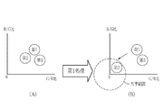
【解決手段】第1信号比と前記第2信号比で形成される特徴空間において、被検体内の観察対象が分布する第1範囲、第2範囲、及び第3範囲をそれぞれ離すために、第1範囲、第2範囲、及び第3範囲を移動させる移動処理を行う。移動処理後の第1信号比及び第2信号比から得られる特殊画像を表示する表示部において、第1範囲、第2範囲、及び第3範囲の間の色の差は、第1カラー画像信号が全て広帯域信号である場合の第1範囲、第2範囲、及び第3範囲の間の色の差よりも大きく表示される。
【選択図】 図24
Description
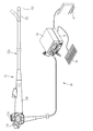
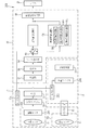
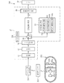
図1に示すように、第1実施形態の内視鏡システム10は、内視鏡12と、光源装置14と、プロセッサ装置16と、モニタ18(表示部)と、コンソール19とを有する。内視鏡12は光源装置14と光学的に接続されるとともに、プロセッサ装置16と電気的に接続される。内視鏡12は、被検体内に挿入される挿入部12aと、挿入部12aの基端部分に設けられた操作部12bと、挿入部12aの先端側に設けられる湾曲部12c及び先端部12dを有している。操作部12bのアングルノブ12eを操作することにより、湾曲部12cは湾曲動作する。この湾曲動作に伴って、先端部12dが所望の方向に向けられる。
(E1):R*=第2R画像信号の画素値×Yin/Yout
(E2):G*=第2G画像信号の画素値×Yin/Yout
(E3):B*=第2B画像信号の画素値×Yin/Yout
なお、「R*」は明るさ調整後の第2R画像信号を、「G*」は明るさ調整後の第2G画像信号を、「B*」は明るさ調整後の第2B画像信号を表している。また、「kr」、「kg」、「kb」は「0」〜「1」の範囲にある任意の定数である。
第2実施形態では、第1実施形態で示した4色のLED20a〜20dの代わりに、レーザ光源と蛍光体を用いて観察対象の照明を行う。それ以外については、第1実施形態と同様である。
第3実施形態では、第1実施形態で示した4色のLED20a〜20dの代わりに、キセノンランプなどの広帯域光源と回転フィルタを用いて観察対象の照明を行う。また、カラーの撮像センサ48に代えて、モノクロの撮像センサで観察対象の撮像を行う。それ以外については、第1実施形態と同様である。
第4実施形態では、挿入型の内視鏡12及び光源装置14に代えて、飲み込み式のカプセル内視鏡を用いて、通常画像、第1又は第2特殊画像の生成に必要なRGB画像信号を取得する。
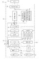
16 プロセッサ装置(医用画像処理装置)
72 信号比算出部
64a 第1特殊画像処理部
64b 第2特殊画像処理部
74 動径拡張・圧縮部
75 角度拡張・圧縮部
77 RGB変換部(カラー画像信号変換部)
81 明るさ調整部
Claims (12)
- 狭帯域信号を少なくとも1つ含む第1カラー画像信号を入力処理する画像信号入力処理部と、
前記第1カラー画像信号のうち2色の画像信号間の第1信号比と、前記第1信号比と異なる2色の画像信号間の第2信号比を算出する信号比算出部と、
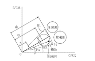
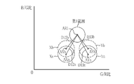
前記第1信号比と前記第2信号比で形成される特徴空間において、被検体内の観察対象が分布する第1範囲、第2範囲、及び第3範囲をそれぞれ離すために、前記第1範囲、前記第2範囲、及び前記第3範囲を移動させる移動処理を行う移動処理部と、
前記移動処理後の第1信号比及び第2信号比から得られる特殊画像を表示する表示部であり、前記第1範囲、前記第2範囲、及び前記第3範囲の間の色の差は、前記第1カラー画像信号が全て広帯域信号である場合の前記第1範囲、前記第2範囲、及び前記第3範囲の間の色の差よりも大きく表示される表示部を備える内視鏡システム。 - 前記移動処理部は、
前記第1範囲、前記第2範囲、及び前記第3範囲のうち前記第2範囲の座標を、前記特徴空間内に定める基準範囲に移動するように処理する第1処理と、前記第2範囲は移動せずに、前記第1範囲の座標と前記第3範囲の座標のうち少なくとも一方を移動するように処理する第2処理とを行う第1移動処理部を備える請求項1記載の内視鏡システム。 - 前記移動処理部は、
前記特徴空間において、前記第2範囲の座標を、前記特徴空間内に定める基準範囲に移動するように処理する第1処理と、前記特徴空間において、前記第1範囲の座標を維持した状態で、前記第3範囲を移動するように処理する第3処理を行う第2移動処理部を備える請求項1または2記載の内視鏡システム。 - 前記特徴空間において、前記第1カラー画像信号のうち少なくとも1色の画像信号が前記狭帯域信号である場合の前記第1範囲と前記第2範囲との差は、前記第1カラー画像信号が全て前記広帯域信号である場合の前記第1範囲と前記第2範囲との差よりも大きい、又は、前記第1カラー画像信号のうち少なくとも1色の画像信号が前記狭帯域信号である場合の前記第1範囲と前記第3範囲との差は、前記第1カラー画像信号が全て前記広帯域信号である場合の前記第1範囲と前記第3範囲との差よりも大きい請求項1記載の内視鏡システム。
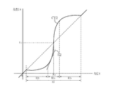
- 前記第1処理は、前記特徴空間において、前記第2範囲の座標の動径を変更して、前記第2範囲の座標を前記基準範囲に移動させる請求項2または3記載の内視鏡システム。
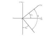
- 前記第2処理は、前記特徴空間において、前記第1範囲の座標の角度と前記第3範囲の座標の角度とを変更して、前記第1範囲の座標と前記第3範囲の座標とが互いに離れるように移動させる請求項2記載の内視鏡システム。
- 前記基準範囲は、前記特徴空間の原点を含み、且つ、前記第1範囲及び前記第3範囲を含まない範囲である請求項2または3記載の内視鏡システム。
- 前記第3処理では、前記第1及び第3処理後の第1信号比及び第2信号比から得られる第2特殊画像の色相が変化するように、前記第3範囲の座標を移動させる請求項3記載の内視鏡システム。
- 前記移動処理後の第1信号比及び第2信号比を第2カラー画像信号に変換するカラー画像信号変換部と、
前記第1カラー画像信号から得られる第1明るさ情報及び前記第2カラー画像信号から得られる第2明るさ情報から、前記第2カラー画像信号の画素値を調整する明るさ調整部とを有する請求項1ないし8いずれか1項記載の内視鏡システム。 - 前記第1信号比は血管深さと相関があり、前記第2信号比は血液量と相関がある請求項1ないし9いずれか1項記載の内視鏡システム。
- 前記第1信号比はB/G比で、前記第2信号比はG/R比である請求項1ないし10いずれか1項記載の内視鏡システム。
- 画像信号入力処理部は、狭帯域信号を少なくとも1つ含む第1カラー画像信号を入力処理するステップと、
信号比算出部は、前記第1カラー画像信号のうち2色の画像信号間の第1信号比と、前記第1信号比と異なる2色の画像信号間の第2信号比を算出するステップと、
移動処理部は、前記第1信号比と前記第2信号比で形成される特徴空間において、被検体内の観察対象が分布する第1範囲、第2範囲、及び第3範囲をそれぞれ離すために、前記第1範囲、前記第2範囲、及び前記第3範囲を移動させる移動処理を行うステップと、
表示部は、前記移動処理後の第1信号比及び第2信号比から得られる特殊画像を表示するステップであり、前記第1範囲、前記第2範囲、及び前記第3範囲の間の色の差は、前記第1カラー画像信号が全て広帯域信号である場合の前記第1範囲、前記第2範囲、及び前記第3範囲の間の色の差よりも大きく表示されるステップとを有する内視鏡システムの作動方法。
Applications Claiming Priority (4)
| Application Number | Priority Date | Filing Date | Title |
|---|---|---|---|
| JP2014074273 | 2014-03-31 | ||
| JP2014074273 | 2014-03-31 | ||
| JP2014133388 | 2014-06-27 | ||
| JP2014133388 | 2014-06-27 |
Related Parent Applications (1)
| Application Number | Title | Priority Date | Filing Date |
|---|---|---|---|
| JP2016511452A Division JP6150318B2 (ja) | 2014-03-31 | 2015-02-26 | 医用画像処理装置及びその作動方法並びに内視鏡システム |
Publications (2)
| Publication Number | Publication Date |
|---|---|
| JP2017185249A true JP2017185249A (ja) | 2017-10-12 |
| JP6313884B2 JP6313884B2 (ja) | 2018-04-18 |
Family
ID=54239998
Family Applications (2)
| Application Number | Title | Priority Date | Filing Date |
|---|---|---|---|
| JP2016511452A Active JP6150318B2 (ja) | 2014-03-31 | 2015-02-26 | 医用画像処理装置及びその作動方法並びに内視鏡システム |
| JP2017097333A Active JP6313884B2 (ja) | 2014-03-31 | 2017-05-16 | 内視鏡システム及びその作動方法 |
Family Applications Before (1)
| Application Number | Title | Priority Date | Filing Date |
|---|---|---|---|
| JP2016511452A Active JP6150318B2 (ja) | 2014-03-31 | 2015-02-26 | 医用画像処理装置及びその作動方法並びに内視鏡システム |
Country Status (5)
| Country | Link |
|---|---|
| US (2) | US9892512B2 (ja) |
| EP (1) | EP3127467B1 (ja) |
| JP (2) | JP6150318B2 (ja) |
| CN (2) | CN108830825B (ja) |
| WO (1) | WO2015151663A1 (ja) |
Families Citing this family (10)
| Publication number | Priority date | Publication date | Assignee | Title |
|---|---|---|---|---|
| JP6196900B2 (ja) * | 2013-12-18 | 2017-09-13 | オリンパス株式会社 | 内視鏡装置 |
| WO2015093295A1 (ja) * | 2013-12-20 | 2015-06-25 | オリンパス株式会社 | 内視鏡装置 |
| JP5972312B2 (ja) * | 2014-03-24 | 2016-08-17 | 富士フイルム株式会社 | 医用画像処理装置及びその作動方法 |
| MX2018012631A (es) * | 2016-04-13 | 2019-07-08 | Inspektor Res Systems B V | Examinación dental de dos frecuencias. |
| US11589777B2 (en) * | 2016-08-31 | 2023-02-28 | Hoya Corporation | Electronic endoscope processor and electronic endoscopic system |
| CN110418596B (zh) * | 2017-03-28 | 2021-12-24 | 富士胶片株式会社 | 测量辅助装置、内窥镜系统及处理器 |
| JP6666519B2 (ja) | 2017-03-28 | 2020-03-13 | 富士フイルム株式会社 | 計測支援装置、内視鏡システム、及びプロセッサ |
| CN111989027B (zh) * | 2018-04-24 | 2025-01-03 | 奥林巴斯株式会社 | 内窥镜系统和内窥镜系统的控制方法 |
| WO2020217669A1 (ja) * | 2019-04-24 | 2020-10-29 | パナソニックIpマネジメント株式会社 | 発光装置並びにそれを用いた医療システム、電子機器及び検査方法 |
| WO2021060158A1 (ja) * | 2019-09-24 | 2021-04-01 | 富士フイルム株式会社 | 内視鏡システム及びその作動方法 |
Citations (7)
| Publication number | Priority date | Publication date | Assignee | Title |
|---|---|---|---|---|
| JPS63153984A (ja) * | 1986-12-18 | 1988-06-27 | Olympus Optical Co Ltd | 画像処理装置 |
| JPS63173182A (ja) * | 1987-01-13 | 1988-07-16 | Olympus Optical Co Ltd | 色彩画像処理方式 |
| JPH01113018A (ja) * | 1987-10-26 | 1989-05-01 | Olympus Optical Co Ltd | 内視鏡色彩強調装置 |
| JPH01138876A (ja) * | 1987-11-26 | 1989-05-31 | Toshiba Corp | カラー画像処理装置 |
| JP2007307395A (ja) * | 2003-04-25 | 2007-11-29 | Olympus Corp | 画像表示装置、画像表示方法および画像表示プログラム |
| JP2010079522A (ja) * | 2008-09-25 | 2010-04-08 | Sapporo Medical Univ | 画像処理装置及び画像処理プログラム |
| JP2012085720A (ja) * | 2010-10-18 | 2012-05-10 | Fujifilm Corp | 内視鏡装置 |
Family Cites Families (19)
| Publication number | Priority date | Publication date | Assignee | Title |
|---|---|---|---|---|
| US4814859A (en) * | 1986-11-28 | 1989-03-21 | Olympus Optical Co., Ltd. | Video image processing apparatus for emphasizing color of an image by expanding hue and saturation |
| US5550582A (en) | 1993-03-19 | 1996-08-27 | Olympus Optical Co., Ltd. | Endoscope-image processing apparatus for performing image processing of emphasis in endoscope image by pigment concentration distribution |
| CN101288582A (zh) * | 2003-04-25 | 2008-10-22 | 奥林巴斯株式会社 | 图像显示装置和图像显示方法 |
| ES2425568T3 (es) * | 2005-01-21 | 2013-10-16 | Verisante Technology, Inc. | Método y aparato para la medición de cambios cancerosos a partir de mediciones espectrales de reflectancia obtenidas durante la toma de imágenes endoscópicas |
| KR20090011071A (ko) * | 2007-07-25 | 2009-02-02 | 삼성전자주식회사 | 디스플레이장치 및 그 제어방법 |
| JP5393554B2 (ja) * | 2010-03-23 | 2014-01-22 | 富士フイルム株式会社 | 電子内視鏡システム |
| JP5405373B2 (ja) * | 2010-03-26 | 2014-02-05 | 富士フイルム株式会社 | 電子内視鏡システム |
| JP5133386B2 (ja) * | 2010-10-12 | 2013-01-30 | 富士フイルム株式会社 | 内視鏡装置 |
| JP5231511B2 (ja) * | 2010-11-09 | 2013-07-10 | 富士フイルム株式会社 | 内視鏡診断装置 |
| JP5405445B2 (ja) * | 2010-12-17 | 2014-02-05 | 富士フイルム株式会社 | 内視鏡装置 |
| JP5550574B2 (ja) * | 2011-01-27 | 2014-07-16 | 富士フイルム株式会社 | 電子内視鏡システム |
| JP5451802B2 (ja) * | 2011-04-01 | 2014-03-26 | 富士フイルム株式会社 | 電子内視鏡システム及び電子内視鏡システムの校正方法 |
| JP5331904B2 (ja) * | 2011-04-15 | 2013-10-30 | 富士フイルム株式会社 | 内視鏡システム及び内視鏡システムの作動方法 |
| JP5419931B2 (ja) * | 2011-07-04 | 2014-02-19 | 富士フイルム株式会社 | 内視鏡システム、光源装置、及び内視鏡システムの作動方法 |
| EP2564760A1 (en) * | 2011-08-29 | 2013-03-06 | Fujifilm Corporation | Endoscopic diagnosis system |
| US9208750B2 (en) * | 2013-05-13 | 2015-12-08 | Asustek Computer Inc. | Color temperature adjusting method of display device |
| CN108109134B (zh) * | 2013-10-28 | 2021-12-03 | 富士胶片株式会社 | 图像处理装置及其工作方法 |
| JP5972312B2 (ja) * | 2014-03-24 | 2016-08-17 | 富士フイルム株式会社 | 医用画像処理装置及びその作動方法 |
| US9977232B2 (en) * | 2015-01-29 | 2018-05-22 | Fujifilm Corporation | Light source device for endoscope, endoscope system, and method for operating light source device for endoscope |
-
2015
- 2015-02-26 WO PCT/JP2015/055537 patent/WO2015151663A1/ja not_active Ceased
- 2015-02-26 EP EP15773911.1A patent/EP3127467B1/en not_active Not-in-force
- 2015-02-26 CN CN201810347183.2A patent/CN108830825B/zh active Active
- 2015-02-26 CN CN201580018139.1A patent/CN106163367B/zh active Active
- 2015-02-26 JP JP2016511452A patent/JP6150318B2/ja active Active
-
2016
- 2016-09-29 US US15/279,541 patent/US9892512B2/en active Active
-
2017
- 2017-05-16 JP JP2017097333A patent/JP6313884B2/ja active Active
-
2018
- 2018-01-08 US US15/864,324 patent/US10186033B2/en active Active
Patent Citations (7)
| Publication number | Priority date | Publication date | Assignee | Title |
|---|---|---|---|---|
| JPS63153984A (ja) * | 1986-12-18 | 1988-06-27 | Olympus Optical Co Ltd | 画像処理装置 |
| JPS63173182A (ja) * | 1987-01-13 | 1988-07-16 | Olympus Optical Co Ltd | 色彩画像処理方式 |
| JPH01113018A (ja) * | 1987-10-26 | 1989-05-01 | Olympus Optical Co Ltd | 内視鏡色彩強調装置 |
| JPH01138876A (ja) * | 1987-11-26 | 1989-05-31 | Toshiba Corp | カラー画像処理装置 |
| JP2007307395A (ja) * | 2003-04-25 | 2007-11-29 | Olympus Corp | 画像表示装置、画像表示方法および画像表示プログラム |
| JP2010079522A (ja) * | 2008-09-25 | 2010-04-08 | Sapporo Medical Univ | 画像処理装置及び画像処理プログラム |
| JP2012085720A (ja) * | 2010-10-18 | 2012-05-10 | Fujifilm Corp | 内視鏡装置 |
Also Published As
| Publication number | Publication date |
|---|---|
| CN108830825B (zh) | 2022-03-25 |
| US20180130208A1 (en) | 2018-05-10 |
| CN108830825A (zh) | 2018-11-16 |
| WO2015151663A1 (ja) | 2015-10-08 |
| EP3127467B1 (en) | 2018-03-28 |
| US10186033B2 (en) | 2019-01-22 |
| EP3127467A1 (en) | 2017-02-08 |
| JPWO2015151663A1 (ja) | 2017-04-13 |
| US9892512B2 (en) | 2018-02-13 |
| CN106163367B (zh) | 2018-05-08 |
| US20170018083A1 (en) | 2017-01-19 |
| CN106163367A (zh) | 2016-11-23 |
| EP3127467A4 (en) | 2017-04-12 |
| JP6313884B2 (ja) | 2018-04-18 |
| JP6150318B2 (ja) | 2017-06-21 |
Similar Documents
| Publication | Publication Date | Title |
|---|---|---|
| JP6121368B2 (ja) | 医用画像処理装置及びその作動方法並びに内視鏡システム | |
| JP6461739B2 (ja) | 画像処理装置及び内視鏡システム並びに画像処理装置の作動方法 | |
| JP6313884B2 (ja) | 内視鏡システム及びその作動方法 | |
| JP6099603B2 (ja) | 医用画像処理装置及びその作動方法並びに内視鏡システム | |
| JP5932894B2 (ja) | 医用画像処理装置及びその作動方法 | |
| JP6050286B2 (ja) | 医用画像処理装置及びその作動方法並びに内視鏡システム | |
| JP5972312B2 (ja) | 医用画像処理装置及びその作動方法 | |
| WO2018230396A1 (ja) | 医用画像処理装置及び内視鏡システム並びに医用画像処理装置の作動方法 | |
| JP6085649B2 (ja) | 内視鏡用光源装置及び内視鏡システム | |
| JP2016107003A (ja) | 医用画像処理装置及びその作動方法 | |
| JP6247784B2 (ja) | 医用画像処理装置及び内視鏡システム | |
| JP6047536B2 (ja) | 医用画像処理装置及びその作動方法 | |
| JP6254502B2 (ja) | 内視鏡用光源装置及び内視鏡システム | |
| JP7177897B2 (ja) | 医用画像処理装置 | |
| JP6113116B2 (ja) | 医用画像処理装置及びその作動方法並びに内視鏡システム | |
| JP6659817B2 (ja) | 医用画像処理装置 | |
| JP6383829B2 (ja) | 医用画像処理装置及びその作動方法並びに内視鏡システム | |
| JP6456459B2 (ja) | 医用画像処理装置 |
Legal Events
| Date | Code | Title | Description |
|---|---|---|---|
| TRDD | Decision of grant or rejection written | ||
| A977 | Report on retrieval |
Free format text: JAPANESE INTERMEDIATE CODE: A971007 Effective date: 20180228 |
|
| A01 | Written decision to grant a patent or to grant a registration (utility model) |
Free format text: JAPANESE INTERMEDIATE CODE: A01 Effective date: 20180307 |
|
| A61 | First payment of annual fees (during grant procedure) |
Free format text: JAPANESE INTERMEDIATE CODE: A61 Effective date: 20180323 |
|
| R150 | Certificate of patent or registration of utility model |
Ref document number: 6313884 Country of ref document: JP Free format text: JAPANESE INTERMEDIATE CODE: R150 |
|
| R250 | Receipt of annual fees |
Free format text: JAPANESE INTERMEDIATE CODE: R250 |
|
| R250 | Receipt of annual fees |
Free format text: JAPANESE INTERMEDIATE CODE: R250 |
|
| R250 | Receipt of annual fees |
Free format text: JAPANESE INTERMEDIATE CODE: R250 |
|
| R250 | Receipt of annual fees |
Free format text: JAPANESE INTERMEDIATE CODE: R250 |
|
| R250 | Receipt of annual fees |
Free format text: JAPANESE INTERMEDIATE CODE: R250 |
|
| R250 | Receipt of annual fees |
Free format text: JAPANESE INTERMEDIATE CODE: R250 |