JP4198130B2 - 超音波診断装置 - Google Patents
超音波診断装置 Download PDFInfo
- Publication number
- JP4198130B2 JP4198130B2 JP2005170121A JP2005170121A JP4198130B2 JP 4198130 B2 JP4198130 B2 JP 4198130B2 JP 2005170121 A JP2005170121 A JP 2005170121A JP 2005170121 A JP2005170121 A JP 2005170121A JP 4198130 B2 JP4198130 B2 JP 4198130B2
- Authority
- JP
- Japan
- Prior art keywords
- ultrasonic
- image
- endoscope
- position detection
- dimensional
- Prior art date
- Legal status (The legal status is an assumption and is not a legal conclusion. Google has not performed a legal analysis and makes no representation as to the accuracy of the status listed.)
- Expired - Lifetime
Links
- 238000001514 detection method Methods 0.000 claims description 102
- 238000003780 insertion Methods 0.000 claims description 85
- 230000037431 insertion Effects 0.000 claims description 85
- 238000011282 treatment Methods 0.000 claims description 63
- 239000000523 sample Substances 0.000 claims description 13
- 238000012545 processing Methods 0.000 description 85
- 238000002604 ultrasonography Methods 0.000 description 18
- 238000010276 construction Methods 0.000 description 15
- 238000010586 diagram Methods 0.000 description 15
- 230000002093 peripheral effect Effects 0.000 description 13
- 230000003287 optical effect Effects 0.000 description 11
- 238000000034 method Methods 0.000 description 10
- 238000005452 bending Methods 0.000 description 8
- 238000001574 biopsy Methods 0.000 description 6
- 238000006243 chemical reaction Methods 0.000 description 6
- 238000012360 testing method Methods 0.000 description 6
- 230000006870 function Effects 0.000 description 4
- RTAQQCXQSZGOHL-UHFFFAOYSA-N Titanium Chemical compound [Ti] RTAQQCXQSZGOHL-UHFFFAOYSA-N 0.000 description 3
- 238000007689 inspection Methods 0.000 description 3
- 230000005389 magnetism Effects 0.000 description 3
- 229910052719 titanium Inorganic materials 0.000 description 3
- 239000010936 titanium Substances 0.000 description 3
- 230000001133 acceleration Effects 0.000 description 2
- 230000003321 amplification Effects 0.000 description 2
- 238000002059 diagnostic imaging Methods 0.000 description 2
- 230000000694 effects Effects 0.000 description 2
- 239000000835 fiber Substances 0.000 description 2
- 239000000696 magnetic material Substances 0.000 description 2
- 238000003199 nucleic acid amplification method Methods 0.000 description 2
- 239000011347 resin Substances 0.000 description 2
- 229920005989 resin Polymers 0.000 description 2
- 238000012935 Averaging Methods 0.000 description 1
- XDTMQSROBMDMFD-UHFFFAOYSA-N C1CCCCC1 Chemical compound C1CCCCC1 XDTMQSROBMDMFD-UHFFFAOYSA-N 0.000 description 1
- FADJUGSUYLCNJQ-UHFFFAOYSA-N CC(CCC1)C1=C Chemical compound CC(CCC1)C1=C FADJUGSUYLCNJQ-UHFFFAOYSA-N 0.000 description 1
- 238000013459 approach Methods 0.000 description 1
- 230000005540 biological transmission Effects 0.000 description 1
- 239000000470 constituent Substances 0.000 description 1
- 238000002405 diagnostic procedure Methods 0.000 description 1
- 210000003238 esophagus Anatomy 0.000 description 1
- 238000005286 illumination Methods 0.000 description 1
- 238000002347 injection Methods 0.000 description 1
- 239000007924 injection Substances 0.000 description 1
- 210000002429 large intestine Anatomy 0.000 description 1
- 239000000463 material Substances 0.000 description 1
- 238000012986 modification Methods 0.000 description 1
- 230000004048 modification Effects 0.000 description 1
- 210000000056 organ Anatomy 0.000 description 1
- 230000005855 radiation Effects 0.000 description 1
- 210000002784 stomach Anatomy 0.000 description 1
- 239000000126 substance Substances 0.000 description 1
- 230000009466 transformation Effects 0.000 description 1
- 230000000007 visual effect Effects 0.000 description 1
Images






Landscapes
- Endoscopes (AREA)
- Ultra Sonic Daignosis Equipment (AREA)
Description
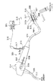
図1に示すように本実施例の内視鏡装置1は、例えば体腔内に挿入される細長な挿入部21を有する内視鏡2と、先端部分に周囲の空間部に磁場を作り位置検出手段を構成する磁気ソース31を備えた位置検出カテーテル3と、前記磁気ソース31が発生する磁場を検出して、磁場に応じた電圧を位置信号として出力する前記内視鏡2及び被検体の外部に固定配置された位置検出手段を構成する磁気センサ4と、この磁気センサ4が検出して出力した位置信号が伝送され、この検出した位置信号を基に前記磁気ソース31の位置を算出する位置信号処理部5とで主に構成されている。
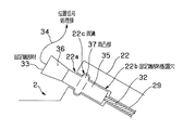
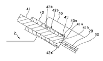
まず、位置検出カテーテル3のシャフト32を内視鏡2の操作部23に設けられている処置具挿通口22aを介して処置具チャンネル29内に挿通し、図1の矢印a方向に挿入していく。すると、図1の点線に示すように前記固定端部材33を処置具挿通口22aの近傍に接近していく。そして、この状態からさらに前記固定端部材33を前記処置具挿通口22aに近接させていくことにより、前記固定端部材33が端部配置穴22bに挿入されて、シャフト32が挿入部21の先端面21aより露出する。ここで、前記固定端部材33を端部配置穴22bに挿入することによって、図2に示すように固定端部材33の周凸部37が周溝22cに係入して、図1に示すように前記位置検出カテーテル3のシャフト32の先端部に設けられている磁気ソース31が内視鏡2の図示しない観察光学系の視野範囲の所定位置に突出した状態で保持固定される。このことによって、術者が位置検出カテーテル3を保持することなく、前記位置検出カテーテル3が内視鏡2の処置具チャンネル29に安定した状態で固定配置される。
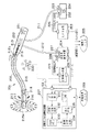
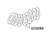
図4ないし図7は超音波診断装置の実施例に係り、図4は超音波診断装置の超音波内視鏡と位置検出カテーテルの構成を示し、超音波内視鏡の先端部のみを拡大した図、図5は超音波診断装置の超音波観測部と超音波画像処理部の構成を示すブロック図、図6は超音波画像処理部内の画像構築部によってラジアル画像と、リニア画像とを構築する方法を説明する図、図7はモニタの画面上に表示された超音画像の一例を示す図である。
図に示すように超音波内視鏡101は、手元側の操作部102から体腔内へ挿入するための細長の挿入部103を延設してなり、この操作部102の側部からは超音波ケーブル105を延設している。
超音波観測を行う際、超音波内視鏡101を被検体の体腔内に挿入して、DCモータ109を回転させて、フレキシブルシャフト108の先端に取り付けた超音波振動子107を回転駆動する。
図6(a)の超音波振動子107の回転の様子を示す図のように、超音波振動子107は、DCモータ109、フレキシブルシャフト108による回転駆動によって、図中点Pに示す超音波振動子107の回転中心を中心にして、例えば1回転あたり例えば0から511番まで512本の超音波ビームを順次送受する。
実施例3を図8及び図9を参照して説明する。
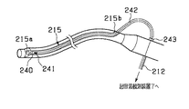
まず、穿刺針240を基端開口部215bからチャンネル215に挿通し、針先が先端開口部215aから突出することがないように配置し、穿刺針240の基端部に設けられているコネクタを超音波内視鏡201に設けられているコネクタ243に接続し、この状態の超音波内視鏡201が、医師などの使用者により、被検者の生体内の、例えば胃、食道、大腸などの管腔状臓器に挿入される。
実施例4を図10ないし図14を参照して説明する。
以上詳述したような本発明の前記実施形態によれば、以下の如き構成を得ることができる。
前記位置検出カテーテルは、前記内視鏡の挿通部に挿入され、前記位置検出手段からの位置信号を基に内視鏡の位置若しくは挿入形状に関する情報を得る内視鏡装置において、
前記位置検出カテーテルを前記内視鏡の挿通部に着脱自在に固定する固定手段を設けた内視鏡装置。
このことにより、位置検出カテーテルの固定端部材に設けた周凸部を、内視鏡の挿通部の固定端部材配置穴の周溝に係入させることによって、位置検出カテーテルと内視鏡とが所定の位置関係で一体的に固定される。
前記位置検出手段からの位置信号に基づき、特定の方向のエコーデータから第2の超音波診断像を構成する断層像構成手段と、前記第1の超音波断層像と第2の超音波断層像とを同時に表示する表示手段とを具備する超音波診断装置。
前記磁気センサ又は前記磁気ソースのどちらか一方が前記超音波振動子近傍に配された超音波診断装置において、
前記磁気センサからの位置信号に基づき、特定の方向のエコーデータから第2の超音波診断像を構成する断層像構成手段と、前記第1の超音波断層像と第2の超音波断層像とを同時に表示する表示手段とを具備する超音波診断装置。
103…挿入部
107…超音波振動子
116…磁気センサ
117…位置検出カテーテル
119…磁気ソース
121…超音波画像処理部
125…画像構築部
Claims (1)
- 挿入部に処置具を挿入するための挿通部を設け、前記挿入部の先端に設けた超音波振動子から超音波を送受波してエコーデータを得る超音波プローブと、
先端部に位置検出手段を設け、前記挿通部に挿入する処置具と、
前記超音波プローブにより得られたエコーデータと、前記挿通部に挿入された前記処置具における前記位置検出手段の出力とにより得られた位置情報に基づいて被検部位の3次元データを取得する3次元構成手段と、
を具備したことを特徴とする超音波診断装置。
Priority Applications (1)
| Application Number | Priority Date | Filing Date | Title |
|---|---|---|---|
| JP2005170121A JP4198130B2 (ja) | 2005-06-09 | 2005-06-09 | 超音波診断装置 |
Applications Claiming Priority (1)
| Application Number | Priority Date | Filing Date | Title |
|---|---|---|---|
| JP2005170121A JP4198130B2 (ja) | 2005-06-09 | 2005-06-09 | 超音波診断装置 |
Related Parent Applications (1)
| Application Number | Title | Priority Date | Filing Date |
|---|---|---|---|
| JP17171698A Division JP4119530B2 (ja) | 1998-06-18 | 1998-06-18 | 内視鏡装置および内視鏡に挿通する位置検出カテーテル |
Publications (2)
| Publication Number | Publication Date |
|---|---|
| JP2005254024A JP2005254024A (ja) | 2005-09-22 |
| JP4198130B2 true JP4198130B2 (ja) | 2008-12-17 |
Family
ID=35080292
Family Applications (1)
| Application Number | Title | Priority Date | Filing Date |
|---|---|---|---|
| JP2005170121A Expired - Lifetime JP4198130B2 (ja) | 2005-06-09 | 2005-06-09 | 超音波診断装置 |
Country Status (1)
| Country | Link |
|---|---|
| JP (1) | JP4198130B2 (ja) |
Families Citing this family (4)
| Publication number | Priority date | Publication date | Assignee | Title |
|---|---|---|---|---|
| KR101422558B1 (ko) * | 2007-01-29 | 2014-07-24 | 인튜어티브 서지컬 인코포레이티드 | 형상 센서를 이용하여 기계를 제어하는 시스템 |
| JP5102655B2 (ja) * | 2008-03-06 | 2012-12-19 | 山下 冨士夫 | 小型電動鍼治療装置 |
| CA2981461C (en) * | 2014-04-29 | 2023-05-09 | The Board Of Regents Of The University Of Texas System | Methods and systems for detecting sub-tissue anomalies |
| JP6854501B2 (ja) * | 2016-04-15 | 2021-04-07 | 公立大学法人岩手県立大学 | トレーニングシステム |
Family Cites Families (10)
| Publication number | Priority date | Publication date | Assignee | Title |
|---|---|---|---|---|
| JPH0829146B2 (ja) * | 1989-04-06 | 1996-03-27 | オリンパス光学工業株式会社 | 超音波プローブ |
| JPH05277113A (ja) * | 1992-03-31 | 1993-10-26 | Olympus Optical Co Ltd | 超音波診断装置 |
| JP3354619B2 (ja) * | 1993-03-12 | 2002-12-09 | オリンパス光学工業株式会社 | 超音波診断装置 |
| JP3506770B2 (ja) * | 1994-04-21 | 2004-03-15 | オリンパス株式会社 | 内視鏡位置検出装置 |
| JP2876510B2 (ja) * | 1993-11-05 | 1999-03-31 | 松下電器産業株式会社 | 機械走査式超音波探触子 |
| JP3374607B2 (ja) * | 1995-07-04 | 2003-02-10 | 富士写真光機株式会社 | 経内視鏡的に挿入される超音波検査装置 |
| JP3139334B2 (ja) * | 1995-07-26 | 2001-02-26 | 富士写真光機株式会社 | 超音波診断装置 |
| JP3650653B2 (ja) * | 1995-08-16 | 2005-05-25 | ジーイー横河メディカルシステム株式会社 | Ct装置および画像表示装置 |
| JPH09238946A (ja) * | 1996-03-13 | 1997-09-16 | Olympus Optical Co Ltd | 超音波プローブ |
| JPH1080420A (ja) * | 1996-09-06 | 1998-03-31 | Fuji Photo Optical Co Ltd | 超音波検査装置 |
-
2005
- 2005-06-09 JP JP2005170121A patent/JP4198130B2/ja not_active Expired - Lifetime
Also Published As
| Publication number | Publication date |
|---|---|
| JP2005254024A (ja) | 2005-09-22 |
Similar Documents
| Publication | Publication Date | Title |
|---|---|---|
| JP4551051B2 (ja) | 超音波診断装置 | |
| JP3354619B2 (ja) | 超音波診断装置 | |
| US20250082303A1 (en) | Devices and methods for ultrasound imaging | |
| WO2004028375A1 (ja) | 超音波診断装置 | |
| CN101516271B (zh) | 超声波图像处理装置以及超声波诊断装置 | |
| JP4119530B2 (ja) | 内視鏡装置および内視鏡に挿通する位置検出カテーテル | |
| WO2010050555A1 (ja) | 超音波観測装置 | |
| JP3808990B2 (ja) | 超音波画像診断装置 | |
| JP2004121488A (ja) | 超音波診断装置 | |
| CN102068285B (zh) | 具有彩色多普勒超声扫描功能的食管镜系统 | |
| JP2005254024A (ja) | 超音波診断装置 | |
| JP2001120550A (ja) | 体腔内超音波プローブシステム | |
| JPH11206778A (ja) | 体腔内超音波プローブ | |
| JP2005118133A (ja) | 超音波内視鏡、観察プローブ及び内視鏡観察装置 | |
| JP2014204904A (ja) | 医用ガイドシステム | |
| JP2000116655A (ja) | 診断装置 | |
| JP4672386B2 (ja) | 超音波プローブ及び超音波診断システム | |
| JPH11123187A (ja) | 超音波画像診断装置 | |
| JPH11113912A (ja) | 超音波画像診断装置 | |
| JP4624883B2 (ja) | 超音波画像処理装置及び超音波診断装置 | |
| JP4668592B2 (ja) | 体腔内プローブ装置 | |
| JP4474608B2 (ja) | 超音波診断装置 | |
| CN102028502B (zh) | 具有彩色多普勒超声扫描功能的电子喉镜系统 | |
| JPH04307042A (ja) | 超音波内視鏡 | |
| JP2007037790A (ja) | 超音波診断装置 |
Legal Events
| Date | Code | Title | Description |
|---|---|---|---|
| A131 | Notification of reasons for refusal |
Free format text: JAPANESE INTERMEDIATE CODE: A131 Effective date: 20080617 |
|
| A521 | Written amendment |
Free format text: JAPANESE INTERMEDIATE CODE: A523 Effective date: 20080818 |
|
| TRDD | Decision of grant or rejection written | ||
| A01 | Written decision to grant a patent or to grant a registration (utility model) |
Free format text: JAPANESE INTERMEDIATE CODE: A01 Effective date: 20080924 |
|
| A01 | Written decision to grant a patent or to grant a registration (utility model) |
Free format text: JAPANESE INTERMEDIATE CODE: A01 |
|
| A61 | First payment of annual fees (during grant procedure) |
Free format text: JAPANESE INTERMEDIATE CODE: A61 Effective date: 20080930 |
|
| FPAY | Renewal fee payment (event date is renewal date of database) |
Free format text: PAYMENT UNTIL: 20111010 Year of fee payment: 3 |
|
| FPAY | Renewal fee payment (event date is renewal date of database) |
Free format text: PAYMENT UNTIL: 20111010 Year of fee payment: 3 |
|
| FPAY | Renewal fee payment (event date is renewal date of database) |
Free format text: PAYMENT UNTIL: 20111010 Year of fee payment: 3 |
|
| FPAY | Renewal fee payment (event date is renewal date of database) |
Free format text: PAYMENT UNTIL: 20121010 Year of fee payment: 4 |