KR20140130752A - 환자에 특정된 혈류 모델로부터 정보를 제공하는 방법 및 시스템 - Google Patents
환자에 특정된 혈류 모델로부터 정보를 제공하는 방법 및 시스템 Download PDFInfo
- Publication number
- KR20140130752A KR20140130752A KR1020147029309A KR20147029309A KR20140130752A KR 20140130752 A KR20140130752 A KR 20140130752A KR 1020147029309 A KR1020147029309 A KR 1020147029309A KR 20147029309 A KR20147029309 A KR 20147029309A KR 20140130752 A KR20140130752 A KR 20140130752A
- Authority
- KR
- South Korea
- Prior art keywords
- information
- patient
- dimensional model
- touch screen
- computer system
- Prior art date
- Legal status (The legal status is an assumption and is not a legal conclusion. Google has not performed a legal analysis and makes no representation as to the accuracy of the status listed.)
- Granted
Links
Images



Classifications
-
- A—HUMAN NECESSITIES
- A61—MEDICAL OR VETERINARY SCIENCE; HYGIENE
- A61B—DIAGNOSIS; SURGERY; IDENTIFICATION
- A61B34/00—Computer-aided surgery; Manipulators or robots specially adapted for use in surgery
- A61B34/10—Computer-aided planning, simulation or modelling of surgical operations
-
- A—HUMAN NECESSITIES
- A61—MEDICAL OR VETERINARY SCIENCE; HYGIENE
- A61B—DIAGNOSIS; SURGERY; IDENTIFICATION
- A61B5/00—Measuring for diagnostic purposes; Identification of persons
- A61B5/02—Detecting, measuring or recording for evaluating the cardiovascular system, e.g. pulse, heart rate, blood pressure or blood flow
- A61B5/02007—Evaluating blood vessel condition, e.g. elasticity, compliance
-
- A—HUMAN NECESSITIES
- A61—MEDICAL OR VETERINARY SCIENCE; HYGIENE
- A61B—DIAGNOSIS; SURGERY; IDENTIFICATION
- A61B5/00—Measuring for diagnostic purposes; Identification of persons
- A61B5/02—Detecting, measuring or recording for evaluating the cardiovascular system, e.g. pulse, heart rate, blood pressure or blood flow
- A61B5/021—Measuring pressure in heart or blood vessels
-
- A—HUMAN NECESSITIES
- A61—MEDICAL OR VETERINARY SCIENCE; HYGIENE
- A61B—DIAGNOSIS; SURGERY; IDENTIFICATION
- A61B5/00—Measuring for diagnostic purposes; Identification of persons
- A61B5/02—Detecting, measuring or recording for evaluating the cardiovascular system, e.g. pulse, heart rate, blood pressure or blood flow
- A61B5/026—Measuring blood flow
-
- A—HUMAN NECESSITIES
- A61—MEDICAL OR VETERINARY SCIENCE; HYGIENE
- A61B—DIAGNOSIS; SURGERY; IDENTIFICATION
- A61B5/00—Measuring for diagnostic purposes; Identification of persons
- A61B5/02—Detecting, measuring or recording for evaluating the cardiovascular system, e.g. pulse, heart rate, blood pressure or blood flow
- A61B5/026—Measuring blood flow
- A61B5/0263—Measuring blood flow using NMR
-
- A—HUMAN NECESSITIES
- A61—MEDICAL OR VETERINARY SCIENCE; HYGIENE
- A61B—DIAGNOSIS; SURGERY; IDENTIFICATION
- A61B5/00—Measuring for diagnostic purposes; Identification of persons
- A61B5/72—Signal processing specially adapted for physiological signals or for diagnostic purposes
- A61B5/7271—Specific aspects of physiological measurement analysis
- A61B5/7275—Determining trends in physiological measurement data; Predicting development of a medical condition based on physiological measurements, e.g. determining a risk factor
-
- A—HUMAN NECESSITIES
- A61—MEDICAL OR VETERINARY SCIENCE; HYGIENE
- A61B—DIAGNOSIS; SURGERY; IDENTIFICATION
- A61B5/00—Measuring for diagnostic purposes; Identification of persons
- A61B5/72—Signal processing specially adapted for physiological signals or for diagnostic purposes
- A61B5/7271—Specific aspects of physiological measurement analysis
- A61B5/7278—Artificial waveform generation or derivation, e.g. synthesizing signals from measured signals
-
- A—HUMAN NECESSITIES
- A61—MEDICAL OR VETERINARY SCIENCE; HYGIENE
- A61B—DIAGNOSIS; SURGERY; IDENTIFICATION
- A61B5/00—Measuring for diagnostic purposes; Identification of persons
- A61B5/74—Details of notification to user or communication with user or patient; User input means
- A61B5/742—Details of notification to user or communication with user or patient; User input means using visual displays
- A61B5/7425—Displaying combinations of multiple images regardless of image source, e.g. displaying a reference anatomical image with a live image
-
- A—HUMAN NECESSITIES
- A61—MEDICAL OR VETERINARY SCIENCE; HYGIENE
- A61B—DIAGNOSIS; SURGERY; IDENTIFICATION
- A61B5/00—Measuring for diagnostic purposes; Identification of persons
- A61B5/74—Details of notification to user or communication with user or patient; User input means
- A61B5/742—Details of notification to user or communication with user or patient; User input means using visual displays
- A61B5/7445—Display arrangements, e.g. multiple display units
-
- A—HUMAN NECESSITIES
- A61—MEDICAL OR VETERINARY SCIENCE; HYGIENE
- A61B—DIAGNOSIS; SURGERY; IDENTIFICATION
- A61B5/00—Measuring for diagnostic purposes; Identification of persons
- A61B5/74—Details of notification to user or communication with user or patient; User input means
- A61B5/7475—User input or interface means, e.g. keyboard, pointing device, joystick
- A61B5/748—Selection of a region of interest, e.g. using a graphics tablet
-
- A—HUMAN NECESSITIES
- A61—MEDICAL OR VETERINARY SCIENCE; HYGIENE
- A61B—DIAGNOSIS; SURGERY; IDENTIFICATION
- A61B6/00—Apparatus or devices for radiation diagnosis; Apparatus or devices for radiation diagnosis combined with radiation therapy equipment
- A61B6/02—Arrangements for diagnosis sequentially in different planes; Stereoscopic radiation diagnosis
- A61B6/03—Computed tomography [CT]
- A61B6/032—Transmission computed tomography [CT]
-
- A—HUMAN NECESSITIES
- A61—MEDICAL OR VETERINARY SCIENCE; HYGIENE
- A61B—DIAGNOSIS; SURGERY; IDENTIFICATION
- A61B6/00—Apparatus or devices for radiation diagnosis; Apparatus or devices for radiation diagnosis combined with radiation therapy equipment
- A61B6/46—Arrangements for interfacing with the operator or the patient
- A61B6/461—Displaying means of special interest
- A61B6/466—Displaying means of special interest adapted to display 3D data
-
- A—HUMAN NECESSITIES
- A61—MEDICAL OR VETERINARY SCIENCE; HYGIENE
- A61B—DIAGNOSIS; SURGERY; IDENTIFICATION
- A61B6/00—Apparatus or devices for radiation diagnosis; Apparatus or devices for radiation diagnosis combined with radiation therapy equipment
- A61B6/50—Apparatus or devices for radiation diagnosis; Apparatus or devices for radiation diagnosis combined with radiation therapy equipment specially adapted for specific body parts; specially adapted for specific clinical applications
- A61B6/504—Apparatus or devices for radiation diagnosis; Apparatus or devices for radiation diagnosis combined with radiation therapy equipment specially adapted for specific body parts; specially adapted for specific clinical applications for diagnosis of blood vessels, e.g. by angiography
-
- A—HUMAN NECESSITIES
- A61—MEDICAL OR VETERINARY SCIENCE; HYGIENE
- A61B—DIAGNOSIS; SURGERY; IDENTIFICATION
- A61B6/00—Apparatus or devices for radiation diagnosis; Apparatus or devices for radiation diagnosis combined with radiation therapy equipment
- A61B6/52—Devices using data or image processing specially adapted for radiation diagnosis
- A61B6/5211—Devices using data or image processing specially adapted for radiation diagnosis involving processing of medical diagnostic data
- A61B6/5217—Devices using data or image processing specially adapted for radiation diagnosis involving processing of medical diagnostic data extracting a diagnostic or physiological parameter from medical diagnostic data
-
- A—HUMAN NECESSITIES
- A61—MEDICAL OR VETERINARY SCIENCE; HYGIENE
- A61B—DIAGNOSIS; SURGERY; IDENTIFICATION
- A61B8/00—Diagnosis using ultrasonic, sonic or infrasonic waves
- A61B8/06—Measuring blood flow
-
- G—PHYSICS
- G06—COMPUTING OR CALCULATING; COUNTING
- G06F—ELECTRIC DIGITAL DATA PROCESSING
- G06F3/00—Input arrangements for transferring data to be processed into a form capable of being handled by the computer; Output arrangements for transferring data from processing unit to output unit, e.g. interface arrangements
- G06F3/01—Input arrangements or combined input and output arrangements for interaction between user and computer
- G06F3/017—Gesture based interaction, e.g. based on a set of recognized hand gestures
-
- G—PHYSICS
- G06—COMPUTING OR CALCULATING; COUNTING
- G06F—ELECTRIC DIGITAL DATA PROCESSING
- G06F3/00—Input arrangements for transferring data to be processed into a form capable of being handled by the computer; Output arrangements for transferring data from processing unit to output unit, e.g. interface arrangements
- G06F3/01—Input arrangements or combined input and output arrangements for interaction between user and computer
- G06F3/048—Interaction techniques based on graphical user interfaces [GUI]
- G06F3/0481—Interaction techniques based on graphical user interfaces [GUI] based on specific properties of the displayed interaction object or a metaphor-based environment, e.g. interaction with desktop elements like windows or icons, or assisted by a cursor's changing behaviour or appearance
- G06F3/04815—Interaction with a metaphor-based environment or interaction object displayed as three-dimensional [3D], e.g. changing the user viewpoint with respect to the environment or object
-
- G—PHYSICS
- G06—COMPUTING OR CALCULATING; COUNTING
- G06F—ELECTRIC DIGITAL DATA PROCESSING
- G06F3/00—Input arrangements for transferring data to be processed into a form capable of being handled by the computer; Output arrangements for transferring data from processing unit to output unit, e.g. interface arrangements
- G06F3/01—Input arrangements or combined input and output arrangements for interaction between user and computer
- G06F3/048—Interaction techniques based on graphical user interfaces [GUI]
- G06F3/0484—Interaction techniques based on graphical user interfaces [GUI] for the control of specific functions or operations, e.g. selecting or manipulating an object, an image or a displayed text element, setting a parameter value or selecting a range
- G06F3/0486—Drag-and-drop
-
- G—PHYSICS
- G06—COMPUTING OR CALCULATING; COUNTING
- G06F—ELECTRIC DIGITAL DATA PROCESSING
- G06F3/00—Input arrangements for transferring data to be processed into a form capable of being handled by the computer; Output arrangements for transferring data from processing unit to output unit, e.g. interface arrangements
- G06F3/01—Input arrangements or combined input and output arrangements for interaction between user and computer
- G06F3/048—Interaction techniques based on graphical user interfaces [GUI]
- G06F3/0487—Interaction techniques based on graphical user interfaces [GUI] using specific features provided by the input device, e.g. functions controlled by the rotation of a mouse with dual sensing arrangements, or of the nature of the input device, e.g. tap gestures based on pressure sensed by a digitiser
- G06F3/0488—Interaction techniques based on graphical user interfaces [GUI] using specific features provided by the input device, e.g. functions controlled by the rotation of a mouse with dual sensing arrangements, or of the nature of the input device, e.g. tap gestures based on pressure sensed by a digitiser using a touch-screen or digitiser, e.g. input of commands through traced gestures
-
- G—PHYSICS
- G06—COMPUTING OR CALCULATING; COUNTING
- G06F—ELECTRIC DIGITAL DATA PROCESSING
- G06F30/00—Computer-aided design [CAD]
- G06F30/20—Design optimisation, verification or simulation
-
- G—PHYSICS
- G06—COMPUTING OR CALCULATING; COUNTING
- G06T—IMAGE DATA PROCESSING OR GENERATION, IN GENERAL
- G06T15/00—Three-dimensional [3D] image rendering
-
- G—PHYSICS
- G06—COMPUTING OR CALCULATING; COUNTING
- G06T—IMAGE DATA PROCESSING OR GENERATION, IN GENERAL
- G06T17/00—Three-dimensional [3D] modelling for computer graphics
-
- G—PHYSICS
- G06—COMPUTING OR CALCULATING; COUNTING
- G06T—IMAGE DATA PROCESSING OR GENERATION, IN GENERAL
- G06T19/00—Manipulating three-dimensional [3D] models or images for computer graphics
- G06T19/20—Editing of three-dimensional [3D] images, e.g. changing shapes or colours, aligning objects or positioning parts
-
- G—PHYSICS
- G16—INFORMATION AND COMMUNICATION TECHNOLOGY [ICT] SPECIALLY ADAPTED FOR SPECIFIC APPLICATION FIELDS
- G16B—BIOINFORMATICS, i.e. INFORMATION AND COMMUNICATION TECHNOLOGY [ICT] SPECIALLY ADAPTED FOR GENETIC OR PROTEIN-RELATED DATA PROCESSING IN COMPUTATIONAL MOLECULAR BIOLOGY
- G16B5/00—ICT specially adapted for modelling or simulations in systems biology, e.g. gene-regulatory networks, protein interaction networks or metabolic networks
-
- G—PHYSICS
- G16—INFORMATION AND COMMUNICATION TECHNOLOGY [ICT] SPECIALLY ADAPTED FOR SPECIFIC APPLICATION FIELDS
- G16H—HEALTHCARE INFORMATICS, i.e. INFORMATION AND COMMUNICATION TECHNOLOGY [ICT] SPECIALLY ADAPTED FOR THE HANDLING OR PROCESSING OF MEDICAL OR HEALTHCARE DATA
- G16H30/00—ICT specially adapted for the handling or processing of medical images
- G16H30/20—ICT specially adapted for the handling or processing of medical images for handling medical images, e.g. DICOM, HL7 or PACS
-
- G—PHYSICS
- G16—INFORMATION AND COMMUNICATION TECHNOLOGY [ICT] SPECIALLY ADAPTED FOR SPECIFIC APPLICATION FIELDS
- G16H—HEALTHCARE INFORMATICS, i.e. INFORMATION AND COMMUNICATION TECHNOLOGY [ICT] SPECIALLY ADAPTED FOR THE HANDLING OR PROCESSING OF MEDICAL OR HEALTHCARE DATA
- G16H50/00—ICT specially adapted for medical diagnosis, medical simulation or medical data mining; ICT specially adapted for detecting, monitoring or modelling epidemics or pandemics
- G16H50/50—ICT specially adapted for medical diagnosis, medical simulation or medical data mining; ICT specially adapted for detecting, monitoring or modelling epidemics or pandemics for simulation or modelling of medical disorders
-
- A—HUMAN NECESSITIES
- A61—MEDICAL OR VETERINARY SCIENCE; HYGIENE
- A61B—DIAGNOSIS; SURGERY; IDENTIFICATION
- A61B34/00—Computer-aided surgery; Manipulators or robots specially adapted for use in surgery
- A61B34/10—Computer-aided planning, simulation or modelling of surgical operations
- A61B2034/101—Computer-aided simulation of surgical operations
- A61B2034/102—Modelling of surgical devices, implants or prosthesis
- A61B2034/104—Modelling the effect of the tool, e.g. the effect of an implanted prosthesis or for predicting the effect of ablation or burring
-
- A—HUMAN NECESSITIES
- A61—MEDICAL OR VETERINARY SCIENCE; HYGIENE
- A61B—DIAGNOSIS; SURGERY; IDENTIFICATION
- A61B2576/00—Medical imaging apparatus involving image processing or analysis
- A61B2576/02—Medical imaging apparatus involving image processing or analysis specially adapted for a particular organ or body part
- A61B2576/023—Medical imaging apparatus involving image processing or analysis specially adapted for a particular organ or body part for the heart
-
- A—HUMAN NECESSITIES
- A61—MEDICAL OR VETERINARY SCIENCE; HYGIENE
- A61B—DIAGNOSIS; SURGERY; IDENTIFICATION
- A61B5/00—Measuring for diagnostic purposes; Identification of persons
- A61B5/02—Detecting, measuring or recording for evaluating the cardiovascular system, e.g. pulse, heart rate, blood pressure or blood flow
- A61B5/021—Measuring pressure in heart or blood vessels
- A61B5/022—Measuring pressure in heart or blood vessels by applying pressure to close blood vessels, e.g. against the skin; Ophthalmodynamometers
-
- A—HUMAN NECESSITIES
- A61—MEDICAL OR VETERINARY SCIENCE; HYGIENE
- A61B—DIAGNOSIS; SURGERY; IDENTIFICATION
- A61B6/00—Apparatus or devices for radiation diagnosis; Apparatus or devices for radiation diagnosis combined with radiation therapy equipment
- A61B6/02—Arrangements for diagnosis sequentially in different planes; Stereoscopic radiation diagnosis
- A61B6/03—Computed tomography [CT]
-
- G—PHYSICS
- G06—COMPUTING OR CALCULATING; COUNTING
- G06T—IMAGE DATA PROCESSING OR GENERATION, IN GENERAL
- G06T2200/00—Indexing scheme for image data processing or generation, in general
- G06T2200/04—Indexing scheme for image data processing or generation, in general involving 3D image data
-
- G—PHYSICS
- G06—COMPUTING OR CALCULATING; COUNTING
- G06T—IMAGE DATA PROCESSING OR GENERATION, IN GENERAL
- G06T2200/00—Indexing scheme for image data processing or generation, in general
- G06T2200/24—Indexing scheme for image data processing or generation, in general involving graphical user interfaces [GUIs]
-
- G—PHYSICS
- G06—COMPUTING OR CALCULATING; COUNTING
- G06T—IMAGE DATA PROCESSING OR GENERATION, IN GENERAL
- G06T2210/00—Indexing scheme for image generation or computer graphics
- G06T2210/41—Medical
-
- G—PHYSICS
- G06—COMPUTING OR CALCULATING; COUNTING
- G06T—IMAGE DATA PROCESSING OR GENERATION, IN GENERAL
- G06T2219/00—Indexing scheme for manipulating 3D models or images for computer graphics
- G06T2219/20—Indexing scheme for editing of 3D models
- G06T2219/2016—Rotation, translation, scaling
-
- Y—GENERAL TAGGING OF NEW TECHNOLOGICAL DEVELOPMENTS; GENERAL TAGGING OF CROSS-SECTIONAL TECHNOLOGIES SPANNING OVER SEVERAL SECTIONS OF THE IPC; TECHNICAL SUBJECTS COVERED BY FORMER USPC CROSS-REFERENCE ART COLLECTIONS [XRACs] AND DIGESTS
- Y02—TECHNOLOGIES OR APPLICATIONS FOR MITIGATION OR ADAPTATION AGAINST CLIMATE CHANGE
- Y02A—TECHNOLOGIES FOR ADAPTATION TO CLIMATE CHANGE
- Y02A90/00—Technologies having an indirect contribution to adaptation to climate change
- Y02A90/10—Information and communication technologies [ICT] supporting adaptation to climate change, e.g. for weather forecasting or climate simulation
Landscapes
- Health & Medical Sciences (AREA)
- Engineering & Computer Science (AREA)
- Life Sciences & Earth Sciences (AREA)
- Medical Informatics (AREA)
- Physics & Mathematics (AREA)
- Public Health (AREA)
- General Health & Medical Sciences (AREA)
- Biomedical Technology (AREA)
- Surgery (AREA)
- Molecular Biology (AREA)
- Pathology (AREA)
- Animal Behavior & Ethology (AREA)
- Heart & Thoracic Surgery (AREA)
- Veterinary Medicine (AREA)
- Biophysics (AREA)
- Theoretical Computer Science (AREA)
- General Engineering & Computer Science (AREA)
- Nuclear Medicine, Radiotherapy & Molecular Imaging (AREA)
- Physiology (AREA)
- General Physics & Mathematics (AREA)
- Radiology & Medical Imaging (AREA)
- Cardiology (AREA)
- High Energy & Nuclear Physics (AREA)
- Optics & Photonics (AREA)
- Human Computer Interaction (AREA)
- Hematology (AREA)
- Computer Graphics (AREA)
- Vascular Medicine (AREA)
- Epidemiology (AREA)
- Primary Health Care (AREA)
- Software Systems (AREA)
- Computer Vision & Pattern Recognition (AREA)
- Computer Hardware Design (AREA)
- Bioinformatics & Cheminformatics (AREA)
- Architecture (AREA)
- Oral & Maxillofacial Surgery (AREA)
- Dentistry (AREA)
- Databases & Information Systems (AREA)
- Data Mining & Analysis (AREA)
- Artificial Intelligence (AREA)
Abstract
Description
도 1은 일 실시형태에 따라 특정 환자의 혈류에 관한 여러 정보를 제공하는 시스템의 개략도;
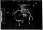
도 2는 일 실시형태에 따라 환자의 대동맥의 일부 및 환자의 대동맥으로부터 나오는 복수의 관상 동맥을 나타내는 3차원 모델 내 계산된 분획 유량 예비력(fractional flow reserve: FFR)을 보여주는 이미지;
도 3은 일 실시형태에 따라 환자의 대동맥의 일부 및 환자의 대동맥으로부터 나오는 복수의 관상 동맥을 나타내는 3차원 모델 내 계산된 압력 구배를 보여주는 이미지;
도 4는 일 실시형태에 따라 환자의 대동맥의 일부 및 환자의 대동맥으로부터 나오는 복수의 관상 동맥을 나타내는 3차원 모델 내 계산된 FFR 및 관상 동맥에 배치된 스텐트를 보여주는 이미지;
도 5는 일 실시형태에 따라 환자의 대동맥의 일부 및 환자의 대동맥으로부터 나오는 복수의 관상 동맥, 및 관상 동맥에 배치된 복수의 스텐트를 나타내는 3차원 모델을 보여주는 이미지;

도 6은 일 실시형태에 따라 하나의 스크린 부분에 도 4의 모델 및 스텐트를 구비하고 다른 스크린 부분에 스텐트의 배치에 기초하여 수정된 3차원 모델을 구비하는 분할된 스크린을 보여주는 이미지.
Claims (30)
- 환자의 혈류 정보(blood flow information)를 제공하는 시스템으로서, 상기 시스템은,
터치스크린을 구비하는 적어도 하나의 컴퓨터 시스템을 포함하되, 상기 적어도 하나의 컴퓨터 시스템은,
상기 터치스크린에, 환자에 특정된 데이터(patient-specific data)에 기초하여 상기 환자의 해부학적 구조의 적어도 일부를 나타내는 3차원 모델을 디스플레이하는 동작; 및
사용자에 의해 제어된 적어도 하나의 포인팅 객체에 의해 지시된 상기 터치 스크린 상의 제1 위치에 관한 제1 입력을 수신하는 동작으로서, 상기 터치스크린 상의 제1 위치는 상기 디스플레이된 3차원 모델 상의 제1 위치를 나타내는 것인, 상기 수신하는 동작, 및
상기 터치스크린에 제1 정보를 디스플레이하는 동작을 수행하도록 구성되고, 상기 제1 정보는 상기 제1 위치에서의 혈류 특성을 나타내는 것인, 환자의 혈류 정보 제공 시스템. - 제1항에 있어서, 상기 사용자가 상기 터치스크린의 표면을 따라 상기 디스플레이된 3차원 모델에서 상기 적어도 하나의 포인팅 객체를 드래그할 때 상기 적어도 하나의 컴퓨터 시스템은 상기 디스플레이된 제1 정보를 업데이트하도록 더 구성된 것인, 환자의 혈류 정보 제공 시스템.
- 제1항에 있어서, 상기 적어도 하나의 컴퓨터 시스템은,
상기 3차원 모델에서 상기 사용자에 의해 이동가능한 핀을 상기 제1 위치에 형성하는 동작; 및
상기 터치스크린에서, 상기 핀이 상기 사용자에 의해 이동될 때 상기 해부학적 구조에서 상기 혈류 특성에 관한 상기 제1 정보를 업데이트하는 동작을 수행하도록 더 구성된 것인, 환자의 혈류 정보 제공 시스템. - 제1항에 있어서, 상기 적어도 하나의 컴퓨터 시스템은 상기 3차원 모델에서 복수의 위치에서 상기 해부학적 구조의 상기 혈류 특성에 관한 제1 정보를 결정하는 동작, 및 명암, 패턴, 또는 컬러링 중 적어도 하나를 사용하여 상기 디스플레이된 3차원 모델에 상기 제1 정보를 나타내는 동작을 수행하도록 더 구성된 것인, 환자의 혈류 정보 제공 시스템.
- 제1항에 있어서, 상기 적어도 하나의 컴퓨터 시스템은 상기 사용자가 상기 터치스크린의 표면에 대해 상기 적어도 하나의 포인팅 객체를 스와이프(swipe)하는 것에 응답하여 상기 디스플레이된 3차원 모델을 회전시키도록 더 구성되고, 회전의 양과 방향은 상기 스와이프의 특성에 종속하는 것인, 환자의 혈류 정보 제공 시스템.
- 제1항에 있어서, 상기 적어도 하나의 컴퓨터 시스템은 상기 사용자가 상기 터치스크린에 대하여 별개의 위치에 위치된 상기 적어도 하나의 포인팅 객체 중 2개의 포인팅 객체를 핀치(pinch)하는 것에 응답하여 상기 디스플레이된 3차원 모델에 줌을 조절하도록 더 구성되고, 상기 줌의 양은 상기 핀치의 특성에 종속하는 것인, 환자의 혈류 정보 제공 시스템.
- 제1항에 있어서, 상기 적어도 하나의 컴퓨터 시스템은,
상기 제1 입력을 수신하고 제1 모드에서 상기 제1 정보를 디스플레이하는 동작;
제2 모드에서 상기 3차원 모델의 변형을 나타내는 제2 입력을 수신하는 동작; 및
상기 3차원 모델의 변형에 기초하여 상기 해부학적 구조의 상기 혈류 특성에 관한 제2 정보를 결정하는 동작을 수행하도록 더 구성된 것인, 환자의 혈류 정보 제공 시스템. - 제7항에 있어서, 상기 제2 입력은 상기 적어도 하나의 포인팅 객체에 의해 지시된 상기 터치스크린 상의 제2 위치에 관한 것이고, 상기 터치스크린 상의 제2 위치는 상기 변형을 위한 3차원 모델에서의 제2 위치를 나타내는 것인, 환자의 혈류 정보 제공 시스템.
- 제7항에 있어서, 상기 변형은 스텐트(stent)의 배치에 대응하고, 상기 적어도 하나의 컴퓨터 시스템은 상기 스텐트를 디스플레이하도록 더 구성된 것인, 환자의 혈류 정보 제공 시스템.
- 제9항에 있어서, 상기 적어도 하나의 컴퓨터 시스템은 상기 스텐트의 사이즈를 나타내는 제3 입력을 수신하도록 더 구성된 것인, 환자의 혈류 정보 제공 시스템.
- 제9항에 있어서, 상기 적어도 하나의 컴퓨터 시스템은 상기 스텐트의 위치의 변화 또는 사이즈의 변화를 나타내는 제3 입력을 수신하도록 더 구성된 것인, 환자의 혈류 정보 제공 시스템.
- 제9항에 있어서, 상기 제2 모드에서, 상기 적어도 하나의 컴퓨터 시스템은 제1 부분 및 제2 부분을 포함하는 분할된 스크린을 제공하도록 더 구성되고, 상기 제1 부분은 상기 해부학적 구조의 적어도 일부 및 상기 스텐트를 나타내는 상기 3차원 모델을 디스플레이하며, 상기 제2 부분은 상기 스텐트의 배치 후 상기 3차원 모델을 디스플레이하는 것인, 환자의 혈류 정보 제공 시스템.
- 제7항에 있어서, 상기 변형은 바이패스의 형성에 대응하고, 상기 적어도 하나의 컴퓨터 시스템은 상기 바이패스를 디스플레이하도록 더 구성된 것인, 환자의 혈류 정보 제공 시스템.
- 제13항에 있어서, 상기 적어도 하나의 컴퓨터 시스템은 상기 바이패스를 상기 디스플레이된 3차원 모델에 연결하는 적어도 하나의 위치를 나타내는 적어도 하나의 제3 입력을 수신하도록 더 구성된 것인, 환자의 혈류 정보 제공 시스템.
- 제13항에 있어서, 상기 적어도 하나의 컴퓨터 시스템은 상기 바이패스의 위치의 변화 또는 사이즈의 변화를 나타내는 제3 입력을 수신하도록 더 구성된 것인, 환자의 혈류 정보 제공 시스템.
- 제7항에 있어서, 상기 적어도 하나의 컴퓨터 시스템은 제1 부분 및 제2 부분을 포함하는 분할된 스크린을 제공하도록 더 구성되고, 상기 2개의 부분은 상기 각 디스플레이된 3차원 모델의 상이한 변형을 나타내는 상이한 제2 입력을 수신하도록 구성된 것인, 환자의 혈류 정보 제공 시스템.
- 제1항에 있어서, 상기 적어도 하나의 포인팅 객체는 상기 사용자 또는 스타일러스의 적어도 하나의 디지트(digit)를 포함하는 것인, 환자의 혈류 정보 제공 시스템.
- 제1항에 있어서, 상기 제1 정보는 압력 구배, 분획 유량 예비력(fractional flow reserve), 압력, 흐름률(flow rate), 및 속도 중 적어도 하나를 포함하는 것인, 환자의 혈류 정보 제공 시스템.
- 제1항에 있어서, 상기 환자의 해부학적 구조는 대동맥의 적어도 일부 및 상기 대동맥의 일부로부터 나오는 복수의 관상 동맥의 적어도 일부를 포함하는 것인, 환자의 혈류 정보 제공 시스템.
- 제19항에 있어서, 상기 제1 정보는 상기 복수의 관상 동맥 내 위치에서의 압력 및 상기 복수의 관상 동맥 내 위치로부터 상류 쪽 위치에서의 압력 사이의 비율을 나타내는 분획 유량 예비력을 포함하는 것인, 환자의 혈류 정보 제공 시스템.
- 제1항에 있어서, 상기 환자의 해부학적 구조는 상기 환자의 심장, 목, 머리, 흉부, 복부, 팔 또는 다리에 있는 복수의 동맥의 일부 중 적어도 하나를 포함하는 것인, 환자의 혈류 정보 제공 시스템.
- 적어도 하나의 컴퓨터 시스템을 사용하여 환자에 특정된 혈류 정보를 제공하는 방법으로서, 상기 적어도 하나의 컴퓨터 시스템은 터치스크린을 포함하며, 상기 방법은,
상기 터치스크린에, 환자에 특정된 데이터에 기초하여 3차원 모델을 디스플레이하는 단계로서, 상기 3차원 모델은 상기 환자의 해부학적 구조의 적어도 일부를 나타내는 것인, 상기 3차원 모델을 디스플레이하는 단계;
사용자에 의해 제어된 적어도 하나의 포인팅 객체에 의해 지시된 상기 터치스크린 상의 제1 위치에 관한 제1 입력을 수신하는 단계로서, 상기 터치스크린 상의 제1 위치는 상기 디스플레이된 3차원 모델에서의 제1 위치를 나타내는 것인, 상기 수신하는 단계;
상기 터치스크린에 제1 정보를 디스플레이하는 단계로서, 상기 제1 정보는 상기 제1 입력에 의해 지시된 상기 3차원 모델에서의 제1 위치에서의 혈류 특성을 나타내는 것인, 상기 제1 정보를 디스플레이하는 단계;
상기 3차원 모델의 변형을 나타내는 제2 입력을 수신하는 단계; 및
상기 3차원 모델의 변형에 기초하여 상기 해부학적 구조에서 상기 혈류 특성에 관한 제2 정보를 결정하는 단계를 포함하는, 환자에 특정된 혈류 정보의 제공 방법. - 제22항에 있어서,
검사 모드의 선택 또는 치료 계획 모드의 선택에 대응하는 모드 선택 입력을 수신하는 단계를 더 포함하되;
상기 제1 입력은 상기 검사 모드에서 수신되고 상기 제2 입력은 상기 치료 계획 모드에서 수신되는 것인, 환자에 특정된 혈류 정보의 제공 방법. - 제22항에 있어서, 상기 제2 입력은 상기 적어도 하나의 포인팅 객체에 의해 지시된 상기 터치스크린 상의 제2 위치에 관한 것이고, 상기 터치스크린 상의 제2 위치는 변형을 위한 상기 3차원 모델에서의 제2 위치를 나타내는 것인, 환자에 특정된 혈류 정보의 제공 방법.
- 환자에 특정된 혈류 정보를 제공하는 방법을 수행하는 컴퓨터-실행가능한 프로그래밍 명령을 포함하는 적어도 하나의 컴퓨터 시스템에 사용하기 위한 비일시적인 컴퓨터 판독가능한 매체로서, 상기 적어도 하나의 컴퓨터 시스템은 터치스크린을 포함하고, 상기 방법은,
환자에 특정된 데이터에 기초하여 상기 환자의 해부학적 구조의 적어도 일부를 나타내는 3차원 모델을 디스플레이하는 단계;
사용자에 의해 제어된 적어도 하나의 포인팅 객체에 의해 지시된 상기 터치스크린 상의 제1 위치에 관한 제1 입력을 수신하는 단계로서, 상기 제1 입력은 상기 해부학적 구조에 배치되는 스텐트의 위치를 나타내는 것인, 상기 수신하는 단계;
상기 3차원 모델 상의 상기 스텐트를 상기 터치스크린에 디스플레이하는 단계; 및
상기 제1 입력에 지시된 위치에 상기 스텐트의 배치를 반영한 상기 3차원 모델의 변형에 기초하여 상기 3차원 모델 내 복수의 위치에서의 혈류 특성에 관한 제2 정보를 결정하는 단계를 포함하는 것인, 비일시적인 컴퓨터 판독가능한 매체. - 제25항에 있어서, 상기 방법은,
상기 스텐트의 사이즈를 나타내는 제2 입력을 수신하는 단계; 및
상기 스텐트의 지시된 사이즈를 반영하는 상기 3차원 모델의 변형에 기초하여 상기 3차원 모델 내 복수의 위치에서의 혈류 특성에 관한 제3 정보를 결정하는 단계를 더 포함하는 것인, 비일시적인 컴퓨터 판독가능한 매체. - 제26항에 있어서, 상기 제2 입력을 수신하는 단계는,
상기 터치스크린에 대하여 별개의 위치에 위치된 상기 적어도 하나의 포인팅 객체 중 2개의 포인팅 객체를 핀치하는 단계, 및
상기 핀치의 특성에 기초하여 상기 스텐트의 사이즈의 변화를 결정하는 단계를 포함하는 것인, 비일시적인 컴퓨터 판독가능한 매체. - 제26항에 있어서, 상기 스텐트의 사이즈는 길이, 근위 직경 또는 원위 직경을 포함하는 것인, 비일시적인 컴퓨터 판독가능한 매체.
- 제25항에 있어서, 상기 방법은,
상기 스텐트의 위치 변화를 나타내는 제2 입력을 수신하는 단계; 및
상기 스텐트의 지시된 위치 변화를 반영하는 상기 3차원 모델의 변형에 기초하여 상기 3차원 모델 내 복수의 위치에서의 혈류 특성에 관한 제3 정보를 결정하는 단계를 더 포함하는 것인, 비일시적인 컴퓨터 판독가능한 매체. - 제29항에 있어서, 상기 제2 입력을 수신하는 단계는,
상기 디스플레이된 3차원 모델을 따라 상기 적어도 하나의 포인팅 객체를 드래그하는 단계, 및
상기 드래그의 특성에 기초하여 상기 스텐트의 위치 변화를 결정하는 단계를 포함하는 것인, 비일시적인 컴퓨터 판독가능한 매체.
Applications Claiming Priority (3)
| Application Number | Priority Date | Filing Date | Title |
|---|---|---|---|
| US13/470,802 | 2012-05-14 | ||
| US13/470,802 US8548778B1 (en) | 2012-05-14 | 2012-05-14 | Method and system for providing information from a patient-specific model of blood flow |
| PCT/US2013/040714 WO2013173208A1 (en) | 2012-05-14 | 2013-05-13 | Method and system for providing information from a patient-specific model of blood flow |
Related Child Applications (1)
| Application Number | Title | Priority Date | Filing Date |
|---|---|---|---|
| KR1020157006423A Division KR101544691B1 (ko) | 2012-05-14 | 2013-05-13 | 환자에 특정된 혈류 모델로부터 정보를 제공하는 방법 및 시스템 |
Publications (2)
| Publication Number | Publication Date |
|---|---|
| KR20140130752A true KR20140130752A (ko) | 2014-11-11 |
| KR101544339B1 KR101544339B1 (ko) | 2015-08-12 |
Family
ID=46545609
Family Applications (2)
| Application Number | Title | Priority Date | Filing Date |
|---|---|---|---|
| KR1020157006423A Active KR101544691B1 (ko) | 2012-05-14 | 2013-05-13 | 환자에 특정된 혈류 모델로부터 정보를 제공하는 방법 및 시스템 |
| KR1020147029309A Active KR101544339B1 (ko) | 2012-05-14 | 2013-05-13 | 환자에 특정된 혈류 모델로부터 정보를 제공하는 방법 및 시스템 |
Family Applications Before (1)
| Application Number | Title | Priority Date | Filing Date |
|---|---|---|---|
| KR1020157006423A Active KR101544691B1 (ko) | 2012-05-14 | 2013-05-13 | 환자에 특정된 혈류 모델로부터 정보를 제공하는 방법 및 시스템 |
Country Status (9)
| Country | Link |
|---|---|
| US (15) | US8548778B1 (ko) |
| EP (2) | EP3258446B1 (ko) |
| JP (7) | JP5715316B1 (ko) |
| KR (2) | KR101544691B1 (ko) |
| CN (2) | CN108629851A (ko) |
| AU (2) | AU2013263062B2 (ko) |
| CA (1) | CA2867839C (ko) |
| DE (3) | DE202012013322U1 (ko) |
| WO (1) | WO2013173208A1 (ko) |
Cited By (1)
| Publication number | Priority date | Publication date | Assignee | Title |
|---|---|---|---|---|
| DE102015215521A1 (de) | 2014-09-30 | 2016-03-31 | Hyundai Autron Co., Ltd. | Verfahren und Vorrichtung zum Diagnostizieren der Trennung eines Sauerstoffsensors |
Families Citing this family (194)
| Publication number | Priority date | Publication date | Assignee | Title |
|---|---|---|---|---|
| US9280916B2 (en) * | 2007-05-21 | 2016-03-08 | Johnson County Community College Foundation, Inc. | Healthcare training system and method |
| US9916773B2 (en) | 2007-05-21 | 2018-03-13 | Jc3 Innovations, Llc | Medical device and procedure simulation and training |
| US9886874B2 (en) | 2007-05-21 | 2018-02-06 | Johnson County Community College Foundation, Inc. | Medical device and procedure simulation and training |
| US9892659B2 (en) | 2007-05-21 | 2018-02-13 | Johnson County Community College Foundation, Inc. | Medical device and procedure simulation and training |
| US10186172B2 (en) | 2007-05-21 | 2019-01-22 | Jc3 Innovations, Llc | Blood glucose testing and monitoring system and method |
| US9905135B2 (en) | 2007-05-21 | 2018-02-27 | Jc3 Innovations, Llc | Medical device and procedure simulation and training |
| IL184151A0 (en) | 2007-06-21 | 2007-10-31 | Diagnostica Imaging Software Ltd | X-ray measurement method |
| US8097712B2 (en) | 2007-11-07 | 2012-01-17 | Beelogics Inc. | Compositions for conferring tolerance to viral disease in social insects, and the use thereof |
| US9521961B2 (en) | 2007-11-26 | 2016-12-20 | C. R. Bard, Inc. | Systems and methods for guiding a medical instrument |
| US8781555B2 (en) | 2007-11-26 | 2014-07-15 | C. R. Bard, Inc. | System for placement of a catheter including a signal-generating stylet |
| EP2992825B1 (en) | 2007-11-26 | 2017-11-01 | C.R. Bard Inc. | Integrated system for intravascular placement of a catheter |
| US8200466B2 (en) | 2008-07-21 | 2012-06-12 | The Board Of Trustees Of The Leland Stanford Junior University | Method for tuning patient-specific cardiovascular simulations |
| US9405886B2 (en) | 2009-03-17 | 2016-08-02 | The Board Of Trustees Of The Leland Stanford Junior University | Method for determining cardiovascular information |
| US9532724B2 (en) | 2009-06-12 | 2017-01-03 | Bard Access Systems, Inc. | Apparatus and method for catheter navigation using endovascular energy mapping |
| US9349176B2 (en) * | 2009-07-15 | 2016-05-24 | Mayo Foundation For Medical Education And Research | Computer-aided detection (CAD) of intracranial aneurysms |
| US8962584B2 (en) | 2009-10-14 | 2015-02-24 | Yissum Research Development Company Of The Hebrew University Of Jerusalem, Ltd. | Compositions for controlling Varroa mites in bees |
| ES2641642T3 (es) | 2010-03-08 | 2017-11-10 | Monsanto Technology Llc | Moléculas de polinucleótido para regulación génica en plantas |
| WO2011150376A1 (en) | 2010-05-28 | 2011-12-01 | C.R. Bard, Inc. | Apparatus for use with needle insertion guidance system |
| US8315812B2 (en) | 2010-08-12 | 2012-11-20 | Heartflow, Inc. | Method and system for patient-specific modeling of blood flow |
| CN103974967A (zh) | 2011-09-13 | 2014-08-06 | 孟山都技术公司 | 用于杂草控制的方法和组合物 |
| BR112014005958A2 (pt) | 2011-09-13 | 2020-10-13 | Monsanto Technology Llc | métodos e composições químicas agrícolas para controle de planta, método de redução de expressão de um gene accase em uma planta, cassete de expressão microbiana, método para fazer um polinucleotídeo, método de identificação de polinucleotídeos úteis na modulação de expressão do gene accase e composição herbicida |
| MX342856B (es) | 2011-09-13 | 2016-10-13 | Monsanto Technology Llc | Metodos y composiciones para el control de malezas. |
| UA116089C2 (uk) | 2011-09-13 | 2018-02-12 | Монсанто Текнолоджи Ллс | Спосіб та композиція для боротьби з бур'янами (варіанти) |
| US10760086B2 (en) | 2011-09-13 | 2020-09-01 | Monsanto Technology Llc | Methods and compositions for weed control |
| US10806146B2 (en) | 2011-09-13 | 2020-10-20 | Monsanto Technology Llc | Methods and compositions for weed control |
| US10829828B2 (en) | 2011-09-13 | 2020-11-10 | Monsanto Technology Llc | Methods and compositions for weed control |
| US20130211244A1 (en) * | 2012-01-25 | 2013-08-15 | Surgix Ltd. | Methods, Devices, Systems, Circuits and Associated Computer Executable Code for Detecting and Predicting the Position, Orientation and Trajectory of Surgical Tools |
| US8548778B1 (en) * | 2012-05-14 | 2013-10-01 | Heartflow, Inc. | Method and system for providing information from a patient-specific model of blood flow |
| US11331149B2 (en) | 2012-05-16 | 2022-05-17 | Feops Nv | Method and system for determining a risk of hemodynamic compromise after cardiac intervention |
| EP3358482B1 (en) | 2017-02-03 | 2026-04-08 | FEops NV | Method and system for determining a risk of hemodynamic compromise after cardiac intervention |
| US10240161B2 (en) | 2012-05-24 | 2019-03-26 | A.B. Seeds Ltd. | Compositions and methods for silencing gene expression |
| JP6301102B2 (ja) * | 2012-10-22 | 2018-03-28 | 学校法人藤田学園 | 医用画像診断装置、医用画像処理装置及び医用画像処理プログラム |
| US10595807B2 (en) | 2012-10-24 | 2020-03-24 | Cathworks Ltd | Calculating a fractional flow reserve |
| US9858387B2 (en) | 2013-01-15 | 2018-01-02 | CathWorks, LTD. | Vascular flow assessment |
| US9814433B2 (en) | 2012-10-24 | 2017-11-14 | Cathworks Ltd. | Creating a vascular tree model |
| US9943233B2 (en) | 2012-10-24 | 2018-04-17 | Cathworks Ltd. | Automated measurement system and method for coronary artery disease scoring |
| US10210956B2 (en) | 2012-10-24 | 2019-02-19 | Cathworks Ltd. | Diagnostically useful results in real time |
| WO2014084382A1 (ja) * | 2012-11-30 | 2014-06-05 | 株式会社 東芝 | 医用画像処理装置 |
| WO2014105743A1 (en) | 2012-12-28 | 2014-07-03 | Cyberheart, Inc. | Blood-tissue surface based radiosurgical renal treatment planning |
| WO2014106837A2 (en) | 2013-01-01 | 2014-07-10 | A. B. Seeds Ltd. | ISOLATED dsRNA MOLECULES AND METHODS OF USING SAME FOR SILENCING TARGET MOLECULES OF INTEREST |
| US10683505B2 (en) | 2013-01-01 | 2020-06-16 | Monsanto Technology Llc | Methods of introducing dsRNA to plant seeds for modulating gene expression |
| US9042613B2 (en) | 2013-03-01 | 2015-05-26 | Heartflow, Inc. | Method and system for determining treatments by modifying patient-specific geometrical models |
| BR112015022797A2 (pt) | 2013-03-13 | 2017-11-07 | Monsanto Technology Llc | método para controle de ervas daninhas, composição herbicida, cassete de expressão microbiano e método de produção de polinucleotídeo |
| EP2971185A4 (en) | 2013-03-13 | 2017-03-08 | Monsanto Technology LLC | Methods and compositions for weed control |
| US9639666B2 (en) | 2013-03-15 | 2017-05-02 | Covidien Lp | Pathway planning system and method |
| US10568328B2 (en) | 2013-03-15 | 2020-02-25 | Monsanto Technology Llc | Methods and compositions for weed control |
| US9135381B2 (en) | 2013-05-10 | 2015-09-15 | Stenomics, Inc. | Modeling and simulation system for optimizing prosthetic heart valve treatment |
| US20160216882A1 (en) * | 2013-06-26 | 2016-07-28 | Lucid Global, Llc | Virtual microscope tool for cardiac cycle |
| US9850496B2 (en) | 2013-07-19 | 2017-12-26 | Monsanto Technology Llc | Compositions and methods for controlling Leptinotarsa |
| RU2703498C2 (ru) | 2013-07-19 | 2019-10-17 | Монсанто Текнолоджи Ллс | Композиции и способы борьбы с leptinotarsa |
| CN105517492B (zh) * | 2013-09-06 | 2019-10-18 | 皇家飞利浦有限公司 | 用于处理心脏数据的处理设备 |
| US9092743B2 (en) | 2013-10-23 | 2015-07-28 | Stenomics, Inc. | Machine learning system for assessing heart valves and surrounding cardiovascular tracts |
| EP3061015A2 (en) | 2013-10-24 | 2016-08-31 | Cathworks Ltd. | Vascular characteristic determination with correspondence modeling of a vascular tree |
| US10993628B2 (en) * | 2013-10-25 | 2021-05-04 | Philips Image Guided Therapy Corporation | Devices, systems, and methods for vessel assessment |
| HUE070313T2 (hu) | 2013-11-04 | 2025-05-28 | Greenlight Biosciences Inc | Ízeltlábú paraziták és kártevõfertõzöttség szabályozására szolgáló készítmények és eljárások |
| UA119253C2 (uk) | 2013-12-10 | 2019-05-27 | Біолоджикс, Інк. | Спосіб боротьби із вірусом у кліща varroa та у бджіл |
| AU2015206585A1 (en) | 2014-01-15 | 2016-07-21 | Monsanto Technology Llc | Methods and compositions for weed control using EPSPS polynucleotides |
| JP6262027B2 (ja) | 2014-03-10 | 2018-01-17 | 東芝メディカルシステムズ株式会社 | 医用画像処理装置 |
| JP5835384B2 (ja) | 2014-03-18 | 2015-12-24 | 株式会社リコー | 情報処理方法、情報処理装置、およびプログラム |
| JP5835383B2 (ja) | 2014-03-18 | 2015-12-24 | 株式会社リコー | 情報処理方法、情報処理装置、およびプログラム |
| BR112016022711A2 (pt) | 2014-04-01 | 2017-10-31 | Monsanto Technology Llc | composições e métodos para controle de pragas de inseto |
| US9754082B2 (en) | 2014-05-30 | 2017-09-05 | Heartflow, Inc. | Systems and methods for reporting blood flow characteristics |
| US10988764B2 (en) | 2014-06-23 | 2021-04-27 | Monsanto Technology Llc | Compositions and methods for regulating gene expression via RNA interference |
| EP3161138A4 (en) | 2014-06-25 | 2017-12-06 | Monsanto Technology LLC | Methods and compositions for delivering nucleic acids to plant cells and regulating gene expression |
| RU2021123470A (ru) | 2014-07-29 | 2021-09-06 | Монсанто Текнолоджи Ллс | Композиции и способы борьбы с насекомыми-вредителями |
| US9386933B2 (en) * | 2014-08-29 | 2016-07-12 | Heartflow, Inc. | Systems and methods for determination of blood flow characteristics and pathologies through modeling of myocardial blood supply |
| US10373719B2 (en) * | 2014-09-10 | 2019-08-06 | Intuitive Surgical Operations, Inc. | Systems and methods for pre-operative modeling |
| US10080872B2 (en) | 2014-11-04 | 2018-09-25 | Abbott Cardiovascular Systems Inc. | System and method for FFR guidewire recovery |
| EP3218829B1 (en) | 2014-11-14 | 2020-10-28 | Koninklijke Philips N.V. | Percutaneous coronary intervention (pci) planning interface and associated devices, systems, and methods |
| JP6849592B2 (ja) * | 2014-12-08 | 2021-03-24 | コーニンクレッカ フィリップス エヌ ヴェKoninklijke Philips N.V. | 経皮的冠状動脈介入治療のための患者教育 |
| WO2016092420A1 (en) * | 2014-12-08 | 2016-06-16 | Koninklijke Philips N.V. | Devices, systems, and methods for vessel assessment and intervention recommendation |
| WO2016092422A1 (en) | 2014-12-08 | 2016-06-16 | Koninklijke Philips N.V. | Bedside interface for percutaneous coronary intervention planning |
| JP6942632B2 (ja) | 2015-01-22 | 2021-09-29 | モンサント テクノロジー エルエルシー | Leptinotarsa防除用組成物及びその方法 |
| KR101863240B1 (ko) * | 2015-01-28 | 2018-06-01 | 주식회사 인피니트헬스케어 | 스텐트 추천 시스템 및 방법 |
| US10945700B2 (en) * | 2015-03-02 | 2021-03-16 | B-K Medical Aps | Non-invasive estimation of intravascular pressure changes using vector velocity ultrasound (US) |
| DE102015208804A1 (de) * | 2015-05-12 | 2016-11-17 | Siemens Healthcare Gmbh | Vorrichtung und Verfahren zum rechnergestützten Simulieren von chirurgischen Eingriffen |
| JP6701502B2 (ja) * | 2015-05-21 | 2020-05-27 | ニプロ株式会社 | 治療装置 |
| US10883103B2 (en) | 2015-06-02 | 2021-01-05 | Monsanto Technology Llc | Compositions and methods for delivery of a polynucleotide into a plant |
| EP3302030A4 (en) | 2015-06-03 | 2019-04-24 | Monsanto Technology LLC | METHOD AND COMPOSITIONS FOR INTRODUCING NUCLEIC ACIDS IN PLANTS |
| US9785748B2 (en) | 2015-07-14 | 2017-10-10 | Heartflow, Inc. | Systems and methods for estimating hemodynamic forces acting on plaque and monitoring patient risk |
| US11087459B2 (en) | 2015-08-14 | 2021-08-10 | Elucid Bioimaging Inc. | Quantitative imaging for fractional flow reserve (FFR) |
| US12026868B2 (en) | 2015-08-14 | 2024-07-02 | Elucid Bioimaging Inc. | Quantitative imaging for detecting histopathologically defined plaque erosion non-invasively |
| US11113812B2 (en) | 2015-08-14 | 2021-09-07 | Elucid Bioimaging Inc. | Quantitative imaging for detecting vulnerable plaque |
| US11071501B2 (en) | 2015-08-14 | 2021-07-27 | Elucid Bioiwaging Inc. | Quantitative imaging for determining time to adverse event (TTE) |
| US11094058B2 (en) | 2015-08-14 | 2021-08-17 | Elucid Bioimaging Inc. | Systems and method for computer-aided phenotyping (CAP) using radiologic images |
| US12008751B2 (en) | 2015-08-14 | 2024-06-11 | Elucid Bioimaging Inc. | Quantitative imaging for detecting histopathologically defined plaque fissure non-invasively |
| US11676359B2 (en) | 2015-08-14 | 2023-06-13 | Elucid Bioimaging Inc. | Non-invasive quantitative imaging biomarkers of atherosclerotic plaque biology |
| US10176408B2 (en) | 2015-08-14 | 2019-01-08 | Elucid Bioimaging Inc. | Systems and methods for analyzing pathologies utilizing quantitative imaging |
| KR102449838B1 (ko) | 2015-09-01 | 2022-09-30 | 삼성전자주식회사 | 사용자의 인터랙션에 기초하여 3차원 오브젝트를 처리하는 방법 및 장치 |
| EP3355771B1 (en) * | 2015-09-29 | 2020-11-11 | Imperial Innovations Ltd | System for coronary intervention assessment, planning, and treatment based on desired outcome |
| CN108471994B (zh) * | 2015-10-07 | 2021-10-29 | 皇家飞利浦有限公司 | 移动ffr模拟 |
| EP3374895A1 (en) | 2015-11-10 | 2018-09-19 | HeartFlow, Inc. | Systems and methods for anatomical modeling using information from a procedure |
| EP3380968B1 (en) | 2015-11-23 | 2021-04-28 | HeartFlow, Inc. | Systems and methods for assessing organ and/or tissue transplantation by simulating one or more transplant characteristics |
| CN108369824B (zh) * | 2015-12-03 | 2022-04-12 | 哈特弗罗公司 | 用于将医学图像与患者相关联的系统和方法 |
| US11302435B2 (en) * | 2016-01-06 | 2022-04-12 | Boston Scientific Scimed, Inc. | Systems and methods for planning medical procedures |
| US10893832B2 (en) * | 2016-01-13 | 2021-01-19 | Tufts Medical Center, Inc. | Predictive instrument to identify patients for use of pharmacological cardiac metabolic support |
| US11090006B2 (en) | 2016-02-03 | 2021-08-17 | Cormetrics Llc | Modular sensing guidewire |
| US10278662B2 (en) | 2016-02-05 | 2019-05-07 | Toshiba Medical Systems Corporation | Image processing apparatus and medical image diagnostic apparatus |
| JP6833319B2 (ja) | 2016-02-12 | 2021-02-24 | 任天堂株式会社 | 情報処理プログラム、情報処理システム、情報処理方法、および情報処理装置 |
| JP6548199B2 (ja) * | 2016-02-16 | 2019-07-24 | 株式会社Pentas | ステントの長さ予測装置、ステントの長さ予測プログラム、およびステントの長さ予測方法 |
| US20170308997A1 (en) * | 2016-04-20 | 2017-10-26 | Abbott Laboratories Services Corp., Taiwan Branch | Method and system for processing an image frame showing a biodegradable medical device in a part of a human body |
| US10674986B2 (en) | 2016-05-13 | 2020-06-09 | General Electric Company | Methods for personalizing blood flow models |
| EP4241694A3 (en) | 2016-05-16 | 2023-12-20 | Cathworks Ltd. | Selection of vascular paths from images |
| JP7036742B2 (ja) | 2016-05-16 | 2022-03-15 | キャスワークス リミテッド | 血管評価システム |
| US11020563B2 (en) | 2016-07-14 | 2021-06-01 | C. R. Bard, Inc. | Automated catheter-to-vessel size comparison tool and related methods |
| EP3488386A4 (en) * | 2016-07-22 | 2020-02-19 | Cornell University | FAST PROTOTYPIZATION AND IN VITRO MODELING OF PATIENT-SPECIFIC CORONAL ARTERY BYPASS IMPLANTS |
| JP6987558B2 (ja) * | 2016-08-10 | 2022-01-05 | キヤノンメディカルシステムズ株式会社 | 医用処理装置、超音波診断装置および医用処理プログラム |
| CN106503482B (zh) * | 2016-09-14 | 2018-10-09 | 王�忠 | 一种用于定量分析药物干预前后生物分子网络中模块变化的方法 |
| US10779786B2 (en) | 2016-10-04 | 2020-09-22 | Canon Medical Systems Corporation | Medical information processing apparatus, X-ray CT apparatus, and medical information processing method |
| KR101865701B1 (ko) | 2016-10-06 | 2018-06-11 | 주식회사 메가젠임플란트 | 모바일 연동 임플란트 진단 시스템 |
| DE102016222102A1 (de) | 2016-11-10 | 2018-05-17 | Siemens Healthcare Gmbh | Behandlungsplanung für eine Stenose in einem Gefäßsegment anhand einer virtuellen hämodynamischen Analyse |
| US10163209B2 (en) * | 2016-11-23 | 2018-12-25 | Toshiba Medical Systems Corporation | Medical image processing apparatus, medical image processing method, and X-ray CT apparatus |
| DE102017221276B4 (de) | 2016-11-28 | 2025-03-27 | Canon Medical Systems Corporation | Medizinische Bildverarbeitungsvorrichtung, Röntgen-CT Vorrichtung und medizinisches Bildverarbeitungsverfahren |
| JP7032111B2 (ja) * | 2016-11-28 | 2022-03-08 | キヤノンメディカルシステムズ株式会社 | 医用画像処理装置、x線ct装置及び医用画像処理プログラム |
| ES2836104T3 (es) * | 2016-12-15 | 2021-06-24 | Sintef Tto As | Método y proceso para proporcionar un modelo computacional específico de un sujeto utilizado para apoyar la decisión y hacer el diagnóstico de enfermedades cardiovasculares |
| US20180174068A1 (en) * | 2016-12-15 | 2018-06-21 | Sintef Tto As | Method and process for providing a subject-specific computational model used for treatment of cardiovascular diseases |
| JP7077319B2 (ja) | 2016-12-23 | 2022-05-30 | ハートフロー, インコーポレイテッド | 解剖学的モデルパラメータの機械学習 |
| US10102665B2 (en) * | 2016-12-30 | 2018-10-16 | Biosense Webster (Israel) Ltd. | Selecting points on an electroanatomical map |
| KR102700668B1 (ko) * | 2017-01-10 | 2024-08-30 | 삼성메디슨 주식회사 | 대상체의 초음파 영상을 디스플레이하는 방법 및 장치 |
| EP3378398A1 (en) | 2017-03-24 | 2018-09-26 | Koninklijke Philips N.V. | Myocardial ct perfusion image synthesis |
| US10453193B2 (en) | 2017-05-05 | 2019-10-22 | General Electric Company | Methods and system for shading a two-dimensional ultrasound image |
| CN107411778B (zh) * | 2017-05-17 | 2020-01-17 | 上海交通大学 | 一种末梢血管血流调节功能的无创检测系统 |
| WO2019046309A1 (en) | 2017-08-29 | 2019-03-07 | Heartflow, Inc. | SYSTEMS AND METHODS FOR GENERATING ANONYMOUS INTERACTIVE DISPLAY IN AN EXTENDED TIMING PERIOD |
| KR20200052949A (ko) * | 2017-09-19 | 2020-05-15 | 코베스트로 엘엘씨 | 맞춤형 디자인 생성물에 대한 기술 |
| AU2018345850B2 (en) | 2017-10-06 | 2024-04-11 | Emory University | Methods and systems for determining hemodynamic information for one or more arterial segments |
| WO2019073927A1 (ja) * | 2017-10-10 | 2019-04-18 | 日本電気株式会社 | 生体情報処理システム、生体情報処理方法、および生体情報処理プログラム記録媒体 |
| US10489969B2 (en) * | 2017-11-08 | 2019-11-26 | General Electric Company | Method and system for presenting shaded descriptors corresponding with shaded ultrasound images |
| JP6530043B2 (ja) * | 2017-12-12 | 2019-06-12 | キヤノンメディカルシステムズ株式会社 | 医用画像処理装置、医用画像処理方法および記録媒体 |
| US11871995B2 (en) | 2017-12-18 | 2024-01-16 | Hemolens Diagnostics Sp. Z O.O. | Patient-specific modeling of hemodynamic parameters in coronary arteries |
| EP3542757A1 (en) | 2018-03-23 | 2019-09-25 | FEops NV | Method and system for patient-specific virtual percutaneous structural heart intervention |
| JP7134687B2 (ja) * | 2018-04-20 | 2022-09-12 | キヤノンメディカルシステムズ株式会社 | X線診断装置、医用画像処理装置、及び医用画像診断装置 |
| WO2019238754A1 (en) * | 2018-06-15 | 2019-12-19 | Pie Medical Imaging Bv | Method and apparatus for quantitative hemodynamic flow analysis |
| CN108932058B (zh) * | 2018-06-29 | 2021-05-18 | 联想(北京)有限公司 | 显示方法、装置及电子设备 |
| JP7340594B2 (ja) * | 2018-07-30 | 2023-09-07 | コーニンクレッカ フィリップス エヌ ヴェ | 血管内撮像プロシージャ特有のワークフローガイド並びに関連する装置、システム、及び方法 |
| CN112867443B (zh) | 2018-10-16 | 2024-04-26 | 巴德阿克塞斯系统股份有限公司 | 用于建立电连接的安全装备连接系统及其方法 |
| CN112889057A (zh) * | 2018-10-22 | 2021-06-01 | 科思创有限公司 | 用于定制设计产品的技术 |
| CN111166316B (zh) * | 2018-11-13 | 2023-03-21 | 苏州润迈德医疗科技有限公司 | 基于造影图像计算造影瞬时无波型比率和造影舒张期压力比率的方法 |
| US10860295B1 (en) * | 2019-01-03 | 2020-12-08 | Amazon Technologies, Inc. | Automated detection of ambiguities in software design diagrams |
| CN109583146A (zh) * | 2019-01-18 | 2019-04-05 | 深圳市阅影科技有限公司 | 冠状动脉血流动力仿真数据处理方法和装置 |
| US10813612B2 (en) | 2019-01-25 | 2020-10-27 | Cleerly, Inc. | Systems and method of characterizing high risk plaques |
| US12544101B2 (en) | 2019-01-30 | 2026-02-10 | Bard Access Systems, Inc. | Systems and methods for tracking medical devices |
| CN109758228A (zh) * | 2019-02-28 | 2019-05-17 | 四川大学华西医院 | 基于三维重建的经颈静脉门体分流手术支架安置优化方法 |
| JP7532402B2 (ja) | 2019-04-01 | 2024-08-13 | キャスワークス リミテッド | 血管造影画像選択のための方法および装置 |
| CN109994198A (zh) * | 2019-04-03 | 2019-07-09 | 东南大学 | 一种动态心脏三维模型的重建方法 |
| CN110368087B (zh) * | 2019-06-06 | 2021-01-08 | 中国人民解放军北部战区总医院 | 一种信息处理方法和装置、及可读存储介质 |
| WO2020263791A1 (en) * | 2019-06-28 | 2020-12-30 | Covestro Llc | Methods for graphical depiction of a value of a property of a material |
| CN121707942A (zh) | 2019-08-05 | 2026-03-20 | 易鲁希德生物成像公司 | 形态学和血管周疾病标志物的联合评估 |
| CN110693609B (zh) * | 2019-08-30 | 2020-07-10 | 上海杏脉信息科技有限公司 | 植入物介入模拟方法、选取方法、介质及设备 |
| US11759166B2 (en) | 2019-09-20 | 2023-09-19 | Bard Access Systems, Inc. | Automatic vessel detection tools and methods |
| EP4033964B1 (en) | 2019-09-23 | 2025-04-09 | Cathworks Ltd. | Methods, apparatus, and system for synchronization between a three-dimensional vascular model and an imaging device |
| CN110742689B (zh) * | 2019-10-31 | 2021-11-23 | 北京理工大学 | 动脉夹层手术评估方法、装置、电子设备及存储介质 |
| US20220392065A1 (en) | 2020-01-07 | 2022-12-08 | Cleerly, Inc. | Systems, methods, and devices for medical image analysis, diagnosis, risk stratification, decision making and/or disease tracking |
| US11969280B2 (en) | 2020-01-07 | 2024-04-30 | Cleerly, Inc. | Systems, methods, and devices for medical image analysis, diagnosis, risk stratification, decision making and/or disease tracking |
| JP2023509514A (ja) | 2020-01-07 | 2023-03-08 | クリールリー、 インコーポレーテッド | 医用画像分析、診断、重症度分類、意思決定、および/または疾患追跡のためのシステム、方法、およびデバイス |
| EP3866107A1 (en) * | 2020-02-14 | 2021-08-18 | Koninklijke Philips N.V. | Model-based image segmentation |
| USD938963S1 (en) * | 2020-02-21 | 2021-12-21 | Universität Zürich | Display screen or portion thereof with graphical user interface for visual clot display |
| CN111312375A (zh) * | 2020-03-12 | 2020-06-19 | 上海杏脉信息科技有限公司 | 虚拟冠脉手术的搭桥分析方法、系统、介质及设备 |
| CN113952031B (zh) | 2020-07-21 | 2025-10-17 | 巴德阿克塞斯系统股份有限公司 | 磁跟踪超声探头及生成其3d可视化的系统、方法和设备 |
| CN114052905A (zh) | 2020-08-04 | 2022-02-18 | 巴德阿克塞斯系统股份有限公司 | 用于优化的医疗部件插入监测和成像增强的系统和方法 |
| CN217907826U (zh) | 2020-08-10 | 2022-11-29 | 巴德阿克塞斯系统股份有限公司 | 医学分析系统 |
| CN114129181A (zh) | 2020-09-03 | 2022-03-04 | 巴德阿克塞斯系统股份有限公司 | 便携式超声探测器、系统和方法 |
| CN216135922U (zh) | 2020-09-08 | 2022-03-29 | 巴德阿克塞斯系统股份有限公司 | 动态调整超声成像系统 |
| CN114159098B (zh) | 2020-09-10 | 2026-01-02 | 巴德阿克塞斯系统股份有限公司 | 具有压力测量能力的超声探测器 |
| US12492953B2 (en) | 2020-09-18 | 2025-12-09 | Bard Access Systems, Inc. | Ultrasound probe with pointer remote control capability |
| CN114246614B (zh) | 2020-09-25 | 2025-09-23 | 巴德阿克塞斯系统股份有限公司 | 超声成像系统和最小导管长度工具 |
| WO2022072727A2 (en) | 2020-10-02 | 2022-04-07 | Bard Access Systems, Inc. | Ultrasound systems and methods for sustained spatial attention |
| CN121221162A (zh) | 2020-10-15 | 2025-12-30 | 巴德阿克塞斯系统股份有限公司 | 超声成像系统和使用其创建目标区域的三维超声图像的方法 |
| CN112353494B (zh) * | 2020-11-14 | 2021-11-19 | 大连理工大学 | 升主动脉、主动脉弓及其三大分支参数化建模方法 |
| EP4247267A1 (en) | 2020-11-24 | 2023-09-27 | Bard Access Systems, Inc. | Ultrasound system with target and medical instrument awareness |
| CN114569155A (zh) | 2020-12-01 | 2022-06-03 | 巴德阿克塞斯系统股份有限公司 | 超声成像系统和用于通过其获得超声图像的方法 |
| WO2022119856A1 (en) | 2020-12-01 | 2022-06-09 | Bard Access Systems, Inc. | Ultrasound system with pressure and flow determination capability |
| WO2022132651A1 (en) | 2020-12-14 | 2022-06-23 | Bard Access Systems, Inc. | Securement of hands-free ultrasound probe |
| CN217960146U (zh) | 2021-04-15 | 2022-12-06 | 巴德阿克塞斯系统股份有限公司 | 超声成像系统 |
| AU2021221667A1 (en) | 2021-04-21 | 2022-11-10 | Artrya Limited | A system for and method of identifying coronary artery disease |
| US12315076B1 (en) | 2021-09-22 | 2025-05-27 | Cathworks Ltd. | Four-dimensional motion analysis of a patient's coronary arteries and myocardial wall |
| EP4416686A1 (en) | 2021-10-14 | 2024-08-21 | FEops NV | Systems and methods for automatically detecting anatomical features for preoperative cardiac implant simulations |
| US12376817B2 (en) | 2021-11-03 | 2025-08-05 | Bard Access Systems, Inc. | Optimized functionality through interoperation of doppler and image based vessel differentiation |
| WO2023104599A1 (en) * | 2021-12-11 | 2023-06-15 | Koninklijke Philips N.V. | Automatic segmentation and treatment planning for a vessel with coregistration of physiology data and extraluminal data |
| KR20240148399A (ko) | 2022-02-10 | 2024-10-11 | 캐스웍스 엘티디. | 기계 학습 기반 센서 분석 및 혈관 트리 분할을 위한 시스템 및 방법 |
| US12514533B2 (en) | 2022-03-01 | 2026-01-06 | Bard Access Systems, Inc. | Ultrasound imaging system |
| US12514532B2 (en) | 2022-03-01 | 2026-01-06 | Bard Access Systems, Inc. | Ultrasound imaging system |
| US12406365B2 (en) | 2022-03-10 | 2025-09-02 | Cleerly, Inc. | Systems, devices, and methods for non-invasive image-based plaque analysis and risk determination |
| US20250217981A1 (en) | 2022-03-10 | 2025-07-03 | Cleerly, Inc. | Systems, methods, and devices for image-based plaque analysis and risk determination |
| US20250143657A1 (en) | 2022-03-10 | 2025-05-08 | Cleerly, Inc. | Systems, devices, and methods for non-invasive image-based plaque analysis and risk determination |
| US12440180B2 (en) | 2022-03-10 | 2025-10-14 | Cleerly, Inc. | Systems, devices, and methods for non-invasive image-based plaque analysis and risk determination |
| EP4489661B1 (en) | 2022-03-16 | 2025-10-29 | Bard Access Systems, Inc. | Ultrasound imaging system |
| US12102481B2 (en) | 2022-06-03 | 2024-10-01 | Bard Access Systems, Inc. | Ultrasound probe with smart accessory |
| US12137989B2 (en) | 2022-07-08 | 2024-11-12 | Bard Access Systems, Inc. | Systems and methods for intelligent ultrasound probe guidance |
| US12564373B2 (en) | 2022-08-15 | 2026-03-03 | Bard Access Systems, Inc. | Spatially aware medical device configured for performance of insertion pathway approximation |
| CN121942048A (zh) | 2023-08-09 | 2026-04-28 | 凯思沃克斯有限公司 | 针对血管指数测量的增强用户界面和串扰分析 |
| IL326432A (en) | 2023-08-09 | 2026-04-01 | Cathworks Ltd | Coronary artery assessment after PCI |
| WO2025106976A1 (en) * | 2023-11-17 | 2025-05-22 | The Johns Hopkins University | A method and system for predicting aneurysmal occlusion resulting from flow diverting stents |
| US12554645B1 (en) | 2024-04-03 | 2026-02-17 | Google Llc | Systems and methods for priority-aware cache replacement |
| US12512196B2 (en) | 2024-06-12 | 2025-12-30 | Cathworks Ltd. | Systems and methods for secure sharing of cardiac assessments using QR codes |
Family Cites Families (227)
| Publication number | Priority date | Publication date | Assignee | Title |
|---|---|---|---|---|
| US5205289A (en) | 1988-12-23 | 1993-04-27 | Medical Instrumentation And Diagnostics Corporation | Three-dimensional computer graphics simulation and computerized numerical optimization for dose delivery and treatment planning |
| US5151856A (en) * | 1989-08-30 | 1992-09-29 | Technion R & D Found. Ltd. | Method of displaying coronary function |
| US5119816A (en) | 1990-09-07 | 1992-06-09 | Sam Technology, Inc. | EEG spatial placement and enhancement method |
| EP0487110B1 (en) | 1990-11-22 | 1999-10-06 | Kabushiki Kaisha Toshiba | Computer-aided diagnosis system for medical use |
| DE69228741T2 (de) | 1991-10-02 | 1999-09-02 | Fujitsu Ltd. | Verfahren zur bestimmung der lokalen orientierung eines kontursegmentes und zur bestimmung von linien und ecken |
| US5343538A (en) | 1992-10-02 | 1994-08-30 | International Remote Imaging Systems, Inc. | Method and an apparatus for identifying an object using quantile partitions |
| US5687737A (en) | 1992-10-09 | 1997-11-18 | Washington University | Computerized three-dimensional cardiac mapping with interactive visual displays |
| EP0646263B1 (en) | 1993-04-20 | 2000-05-31 | General Electric Company | Computer graphic and live video system for enhancing visualisation of body structures during surgery |
| JPH0779959A (ja) * | 1993-09-14 | 1995-03-28 | Toshiba Corp | X線診断装置 |
| US5881124A (en) | 1994-03-31 | 1999-03-09 | Arch Development Corporation | Automated method and system for the detection of lesions in medical computed tomographic scans |
| US5920319A (en) | 1994-10-27 | 1999-07-06 | Wake Forest University | Automatic analysis in virtual endoscopy |
| US5782762A (en) | 1994-10-27 | 1998-07-21 | Wake Forest University | Method and system for producing interactive, three-dimensional renderings of selected body organs having hollow lumens to enable simulated movement through the lumen |
| US6694163B1 (en) | 1994-10-27 | 2004-02-17 | Wake Forest University Health Sciences | Method and system for producing interactive, three-dimensional renderings of selected body organs having hollow lumens to enable simulated movement through the lumen |
| JPH11506950A (ja) | 1995-05-31 | 1999-06-22 | モレキュラー バイオシステムズ,インコーポレイテッド | コントラスト強調撮像を用いた自動境界線引きおよび部位寸法記入 |
| US6151404A (en) | 1995-06-01 | 2000-11-21 | Medical Media Systems | Anatomical visualization system |
| EP0836729B1 (en) | 1995-06-09 | 2006-09-27 | Interact Medical Technologies Corporation | Anatomical visualization system |
| US5582173A (en) | 1995-09-18 | 1996-12-10 | Siemens Medical Systems, Inc. | System and method for 3-D medical imaging using 2-D scan data |
| US5687208A (en) | 1995-10-06 | 1997-11-11 | Bhb General Partnership | Method of and apparatus for predicting computed tomography contrast enhancement with feedback |
| US5970182A (en) | 1995-11-15 | 1999-10-19 | Focus Imaging, S. A. | Registration process for myocardial images |
| US5682886A (en) | 1995-12-26 | 1997-11-04 | Musculographics Inc | Computer-assisted surgical system |
| US5825908A (en) | 1995-12-29 | 1998-10-20 | Medical Media Systems | Anatomical visualization and measurement system |
| US5729670A (en) | 1996-01-16 | 1998-03-17 | Ford Global Technologies, Inc. | Method for producing a mesh of quadrilateral/hexahedral elements for a body to be analyzed using finite element analysis |
| US6047080A (en) | 1996-06-19 | 2000-04-04 | Arch Development Corporation | Method and apparatus for three-dimensional reconstruction of coronary vessels from angiographic images |
| US6026173A (en) | 1997-07-05 | 2000-02-15 | Svenson; Robert H. | Electromagnetic imaging and therapeutic (EMIT) systems |
| US5947899A (en) | 1996-08-23 | 1999-09-07 | Physiome Sciences | Computational system and method for modeling the heart |
| US7194117B2 (en) | 1999-06-29 | 2007-03-20 | The Research Foundation Of State University Of New York | System and method for performing a three-dimensional virtual examination of objects, such as internal organs |
| US5971767A (en) | 1996-09-16 | 1999-10-26 | The Research Foundation Of State University Of New York | System and method for performing a three-dimensional virtual examination |
| US6331116B1 (en) | 1996-09-16 | 2001-12-18 | The Research Foundation Of State University Of New York | System and method for performing a three-dimensional virtual segmentation and examination |
| US6343936B1 (en) | 1996-09-16 | 2002-02-05 | The Research Foundation Of State University Of New York | System and method for performing a three-dimensional virtual examination, navigation and visualization |
| US5891030A (en) | 1997-01-24 | 1999-04-06 | Mayo Foundation For Medical Education And Research | System for two dimensional and three dimensional imaging of tubular structures in the human body |
| US8682045B2 (en) | 1997-02-25 | 2014-03-25 | Wake Forest University Health Sciences | Virtual endoscopy with improved image segmentation and lesion detection |
| US6035056A (en) | 1997-03-27 | 2000-03-07 | R2 Technology, Inc. | Method and apparatus for automatic muscle segmentation in digital mammograms |
| IT1297396B1 (it) | 1997-12-30 | 1999-09-01 | Francesco Buzzigoli | Metodo e dispositivo per la ricostruzione di immagini tridimensionali di vasi sanguigni, in particolare di arterie coronarie, o di altre |
| US6176838B1 (en) | 1998-01-29 | 2001-01-23 | Anzai Medical Kabushiki Kaisha | Method and apparatus for measuring hepatic blood flow amount |
| EP1059874A4 (en) | 1998-02-03 | 2003-05-07 | Univ Illinois | MODEL OF THE CEREBRAL CIRCUIT AND APPLICATIONS |
| US7191110B1 (en) * | 1998-02-03 | 2007-03-13 | University Of Illinois, Board Of Trustees | Patient specific circulation model |
| EP1057161B1 (en) | 1998-02-23 | 2002-05-02 | Algotec Systems Ltd. | Automatic path planning system and method |
| US6117087A (en) | 1998-04-01 | 2000-09-12 | Massachusetts Institute Of Technology | Method and apparatus for noninvasive assessment of a subject's cardiovascular system |
| US6236878B1 (en) | 1998-05-22 | 2001-05-22 | Charles A. Taylor | Method for predictive modeling for planning medical interventions and simulating physiological conditions |
| US6045512A (en) | 1998-06-09 | 2000-04-04 | Baxter International Inc. | System and method for continuous estimation and display of cardiac ejection fraction and end diastolic volume |
| US6119574A (en) | 1998-07-02 | 2000-09-19 | Battelle Memorial Institute | Blast effects suppression system |
| AU5097599A (en) | 1998-07-13 | 2000-02-01 | Chandu Corporation | Configurable bio-transport system simulator |
| US6950689B1 (en) | 1998-08-03 | 2005-09-27 | Boston Scientific Scimed, Inc. | Dynamically alterable three-dimensional graphical model of a body region |
| US6379041B1 (en) | 1998-11-02 | 2002-04-30 | Siemens Aktiengesellschaft | X-ray apparatus for producing a 3D image from a set of 2D projections |
| US6466205B2 (en) | 1998-11-19 | 2002-10-15 | Push Entertainment, Inc. | System and method for creating 3D models from 2D sequential image data |
| EP1133257A4 (en) | 1998-11-25 | 2007-12-26 | Univ Wake Forest | IMPROVED VIRTUAL ENDOSCOPY SYSTEM WITH IMAGE SEGMENTATION AND DETECTION OF LESIONS |
| US6278460B1 (en) | 1998-12-15 | 2001-08-21 | Point Cloud, Inc. | Creating a three-dimensional model from two-dimensional images |
| KR20020003362A (ko) | 1999-01-15 | 2002-01-12 | 추후제출 | 좌표화된 투시법을 이용하여 해부학적 대상물을 측정하는장치 및 방법 |
| US6129722A (en) * | 1999-03-10 | 2000-10-10 | Ruiz; Luis Antonio | Interactive corrective eye surgery system with topography and laser system interface |
| US6319743B1 (en) | 1999-04-14 | 2001-11-20 | Mykrolis Corporation | Method of making thin film piezoresistive sensor |
| US6793496B2 (en) | 1999-04-15 | 2004-09-21 | General Electric Company | Mathematical model and a method and apparatus for utilizing the model |
| US20020168618A1 (en) | 2001-03-06 | 2002-11-14 | Johns Hopkins University School Of Medicine | Simulation system for image-guided medical procedures |
| DE19922279A1 (de) | 1999-05-11 | 2000-11-16 | Friedrich Schiller Uni Jena Bu | Verfahren zur Generierung patientenspezifischer Implantate |
| US7778688B2 (en) | 1999-05-18 | 2010-08-17 | MediGuide, Ltd. | System and method for delivering a stent to a selected position within a lumen |
| FR2793926B1 (fr) | 1999-05-20 | 2001-07-20 | Univ Rennes | Procede de construction en trois dimensions d'un organe virtuel representatif d'un organe reel |
| US6266453B1 (en) | 1999-07-26 | 2001-07-24 | Computerized Medical Systems, Inc. | Automated image fusion/alignment system and method |
| US6605053B1 (en) * | 1999-09-10 | 2003-08-12 | Percardia, Inc. | Conduit designs and related methods for optimal flow control |
| AU4021601A (en) | 1999-09-20 | 2001-04-24 | Board Of Trustees Of The University Of Illinois, The | Circulation model and applications |
| US6711433B1 (en) | 1999-09-30 | 2004-03-23 | Siemens Corporate Research, Inc. | Method for providing a virtual contrast agent for augmented angioscopy |
| US7333648B2 (en) | 1999-11-19 | 2008-02-19 | General Electric Company | Feature quantification from multidimensional image data |
| DE19962666A1 (de) | 1999-12-23 | 2001-07-05 | Siemens Ag | Verfahren zum Rekonstruieren von 3D-Bilddaten bezüglich eines interessierenden Volumens eines Untersuchungsobjekts |
| DE10000185A1 (de) | 2000-01-05 | 2001-07-12 | Philips Corp Intellectual Pty | Verfahren zur Darstellung des zeitlichen Verlaufs des Blutflusses in einem Untersuchungsobjekt |
| US7689014B2 (en) | 2000-01-18 | 2010-03-30 | Z-Kat Inc | Apparatus and method for measuring anatomical objects using coordinated fluoroscopy |
| US6606091B2 (en) | 2000-02-07 | 2003-08-12 | Siemens Corporate Research, Inc. | System for interactive 3D object extraction from slice-based medical images |
| US7321677B2 (en) | 2000-05-09 | 2008-01-22 | Paieon Inc. | System and method for three-dimensional reconstruction of an artery |
| WO2001093745A2 (en) | 2000-06-06 | 2001-12-13 | The Research Foundation Of State University Of New York | Computer aided visualization, fusion and treatment planning |
| US6650927B1 (en) | 2000-08-18 | 2003-11-18 | Biosense, Inc. | Rendering of diagnostic imaging data on a three-dimensional map |
| US6565514B2 (en) * | 2000-08-25 | 2003-05-20 | Radi Medical Systems Ab | Method and system for determining physiological variables |
| US20020035458A1 (en) | 2000-09-20 | 2002-03-21 | Chang-Hun Kim | Method and system for virtual surgery |
| JP4128082B2 (ja) | 2000-10-25 | 2008-07-30 | ザ ジョン ピー. ロバーツ リサーチ インスティテュート | 血流パラメータを算出する方法及び装置 |
| US6754376B1 (en) | 2000-11-22 | 2004-06-22 | General Electric Company | Method for automatic segmentation of medical images |
| US6829379B1 (en) * | 2000-11-27 | 2004-12-07 | Ge Medical Systems Global Technology Company, Llc | Methods and apparatus to assist and facilitate vessel analysis |
| US6666820B1 (en) | 2000-11-28 | 2003-12-23 | Michael D. Poole | Mathematical therapeutic outcomes model for predicting clinical efficacy therapies |
| US6487432B2 (en) | 2000-12-04 | 2002-11-26 | Ge Medical Systems Global Technologies Company Llc | Method and system for selecting and displaying medical image data |
| US7327862B2 (en) | 2001-04-30 | 2008-02-05 | Chase Medical, L.P. | System and method for facilitating cardiac intervention |
| US7526112B2 (en) | 2001-04-30 | 2009-04-28 | Chase Medical, L.P. | System and method for facilitating cardiac intervention |
| DE10122875C1 (de) | 2001-05-11 | 2003-02-13 | Siemens Ag | Kombiniertes 3D-Angio-Volumenrekonstruktionsverfahren |
| EP1390917B1 (en) | 2001-05-23 | 2008-12-10 | Vital Images, Inc. | Occlusion culling for object-order volume rendering |
| WO2002097735A1 (en) | 2001-05-31 | 2002-12-05 | Kent Ridge Digital Labs | System and method of anatomical modeling |
| US7853312B2 (en) | 2001-06-07 | 2010-12-14 | Varian Medical Systems, Inc. | Seed localization system for use in an ultrasound system and method of using the same |
| US6718004B2 (en) | 2001-06-28 | 2004-04-06 | General Electric Company | Methods and apparatus for coronary-specific imaging reconstruction |
| US20030023266A1 (en) | 2001-07-19 | 2003-01-30 | Borillo Thomas E. | Individually customized atrial appendage implant device |
| US7006955B2 (en) | 2001-10-15 | 2006-02-28 | General Electric Company | System and method for statistical design of ultrasound probe and imaging system |
| ATE314703T1 (de) | 2001-10-16 | 2006-01-15 | Koninkl Philips Electronics Nv | Verfahren zur automatischen etikettierung von zweigen |
| US7286866B2 (en) | 2001-11-05 | 2007-10-23 | Ge Medical Systems Global Technology Company, Llc | Method, system and computer product for cardiac interventional procedure planning |
| DE10162272A1 (de) | 2001-12-19 | 2003-07-10 | Philips Intellectual Property | Verfahren zur Unterstützung der Orientierung im Gefäßsystem |
| AU2002353325A1 (en) | 2001-12-28 | 2003-07-30 | Koninklijke Philips Electronics N.V. | Viewing system having means for processing a sequence of ultrasound images for performing a quantitative estimation of a flow in a body organ |
| US20040225213A1 (en) * | 2002-01-22 | 2004-11-11 | Xingwu Wang | Magnetic resonance imaging coated assembly |
| JP2003245360A (ja) | 2002-02-26 | 2003-09-02 | Piolax Medical Device:Kk | ステント設計支援装置、ステント設計支援方法、ステント設計支援プログラム、及びステント設計支援プログラムを記録した記録媒体 |
| DE10210650B4 (de) | 2002-03-11 | 2005-04-28 | Siemens Ag | Verfahren zur dreidimensionalen Darstellung eines Untersuchungsbereichs eines Patienten in Form eines 3D-Rekonstruktionsbilds und medizinische Untersuchungs- und/oder Behandlungseinrichtung |
| JP2005520661A (ja) | 2002-03-23 | 2005-07-14 | コーニンクレッカ フィリップス エレクトロニクス エヌ ヴィ | 対象に含まれる構造のインタラクティブなセグメンテーションの方法 |
| US6996262B2 (en) | 2002-05-20 | 2006-02-07 | General Electric Company | Method and apparatus of scoring an arterial obstruction |
| US20040034309A1 (en) | 2002-07-12 | 2004-02-19 | Auckland Uniservices Limited | Method and system of defining a model of one or more organs |
| JP4575157B2 (ja) | 2002-07-19 | 2010-11-04 | コーニンクレッカ フィリップス エレクトロニクス エヌ ヴィ | メッシュ適応による多数の対象又は複合的な対象の同時セグメンテーション |
| US7020510B2 (en) | 2002-07-25 | 2006-03-28 | Koninklijke Philips Electronics, N.V. | Optimal view map V.0.01 |
| TW558689B (en) | 2002-08-30 | 2003-10-21 | Univ Taipei Medical | Three-dimensional surgery simulation system and method |
| US7794230B2 (en) | 2002-09-10 | 2010-09-14 | University Of Vermont And State Agricultural College | Mathematical circulatory system model |
| US7182602B2 (en) | 2002-09-10 | 2007-02-27 | The University Of Vermont And State Agricultural College | Whole-body mathematical model for simulating intracranial pressure dynamics |
| AU2003275265A1 (en) | 2002-09-26 | 2004-04-19 | Robert Levine | Medical instruction using a virtual patient |
| US6628743B1 (en) | 2002-11-26 | 2003-09-30 | Ge Medical Systems Global Technology Company, Llc | Method and apparatus for acquiring and analyzing cardiac data from a patient |
| US20060173283A1 (en) * | 2002-11-27 | 2006-08-03 | Oskar Axelsson | Method of magnetic resonance imaging |
| WO2006020920A2 (en) | 2003-01-29 | 2006-02-23 | Medtronic, Inc. | Catheter apparatus for treatment of heart arrhythmia |
| US20050043609A1 (en) | 2003-01-30 | 2005-02-24 | Gregory Murphy | System and method for facilitating cardiac intervention |
| WO2004068406A2 (en) | 2003-01-30 | 2004-08-12 | Chase Medical, L.P. | A method and system for image processing and contour assessment |
| US20060142984A1 (en) | 2003-01-31 | 2006-06-29 | Jurgen Weese | Method for the reconstruction of three-dimensional objects |
| US7574026B2 (en) | 2003-02-12 | 2009-08-11 | Koninklijke Philips Electronics N.V. | Method for the 3d modeling of a tubular structure |
| JP4421203B2 (ja) | 2003-03-20 | 2010-02-24 | 株式会社東芝 | 管腔状構造体の解析処理装置 |
| JP2006524087A (ja) | 2003-04-22 | 2006-10-26 | コーニンクレッカ フィリップス エレクトロニクス エヌ ヴィ | 血管造影x線写真撮影用の機器 |
| US7343196B2 (en) | 2003-05-09 | 2008-03-11 | Ge Medical Systems Global Technology Company Llc | Cardiac CT system and method for planning and treatment of biventricular pacing using epicardial lead |
| US20050010105A1 (en) | 2003-07-01 | 2005-01-13 | Sra Jasbir S. | Method and system for Coronary arterial intervention |
| US7813785B2 (en) | 2003-07-01 | 2010-10-12 | General Electric Company | Cardiac imaging system and method for planning minimally invasive direct coronary artery bypass surgery |
| EP1646963A1 (en) | 2003-07-08 | 2006-04-19 | Philips Intellectual Property & Standards GmbH | Reconstruction of the current flow in a vessel system |
| CN1830003A (zh) | 2003-08-04 | 2006-09-06 | 西门子共同研究公司 | 针对可视化的虚拟器官展开(unfolding) |
| EP1658009B1 (en) | 2003-08-20 | 2011-10-12 | Philips Intellectual Property & Standards GmbH | Method and device for flow reconstruction |
| JP4644670B2 (ja) | 2003-08-21 | 2011-03-02 | コーニンクレッカ フィリップス エレクトロニクス エヌ ヴィ | 3次元の血管モデルを生成する装置及び方法 |
| DE10340544B4 (de) | 2003-09-01 | 2006-08-03 | Siemens Ag | Vorrichtung zur visuellen Unterstützung einer elektrophysiologischen Katheteranwendung im Herzen |
| WO2005031635A1 (en) | 2003-09-25 | 2005-04-07 | Paieon, Inc. | System and method for three-dimensional reconstruction of a tubular organ |
| CA2540280A1 (en) | 2003-10-07 | 2005-04-21 | Entelos, Inc. | Simulating patient-specific outcomes |
| US20070014452A1 (en) | 2003-12-01 | 2007-01-18 | Mitta Suresh | Method and system for image processing and assessment of a state of a heart |
| US20050143777A1 (en) | 2003-12-19 | 2005-06-30 | Sra Jasbir S. | Method and system of treatment of heart failure using 4D imaging |
| US7333643B2 (en) | 2004-01-30 | 2008-02-19 | Chase Medical, L.P. | System and method for facilitating cardiac intervention |
| US7526115B2 (en) | 2004-02-23 | 2009-04-28 | Siemens Medical Solutions Usa, Inc. | System and method for toboggan based object segmentation using divergent gradient field response in images |
| US8010175B2 (en) | 2004-05-05 | 2011-08-30 | Siemens Medical Solutions Usa, Inc. | Patient-specific coronary territory mapping |
| WO2005107601A2 (en) | 2004-05-06 | 2005-11-17 | Focus Surgery, Inc. | Method and apparatus for the selective treatment of tissue |
| EP1774454A2 (en) | 2004-06-02 | 2007-04-18 | M2S, Inc. | Anatomical visualization and measurement system |
| EP1811896A4 (en) | 2004-06-23 | 2009-08-19 | M2S Inc | ANATOMICAL VISUALIZATION AND MEASURING SYSTEM |
| US7462153B2 (en) | 2004-07-23 | 2008-12-09 | Sonomedica, Inc. | Method and system for modeling cardiovascular disease using a probability regession model |
| EP1805744A2 (en) | 2004-08-10 | 2007-07-11 | The General Hospital Corporation | Methods and apparatus for simulation of endovascular and endoluminal procedures |
| DE102004043676B4 (de) | 2004-09-09 | 2014-01-09 | Siemens Aktiengesellschaft | Verfahren zur Visualisierung von Plaqueablagerungen aus 3D-Bilddatensätzen von Gefäßstrukturen |
| WO2006063141A2 (en) * | 2004-12-07 | 2006-06-15 | Medical Metrx Solutions, Inc. | Intraoperative c-arm fluoroscope datafusion system |
| IL165636A0 (en) * | 2004-12-08 | 2006-01-15 | Paieon Inc | Method and apparatus for finding the coronary velocity and flow and related parameters |
| WO2006061815A1 (en) | 2004-12-08 | 2006-06-15 | Paieon Inc. | Method and apparatus for blood vessel parameter determinations |
| WO2006062958A2 (en) | 2004-12-10 | 2006-06-15 | Worcester Polytechnic Institute | Image-based computational mechanical analysis and indexing for cardiovascular diseases |
| JP5122743B2 (ja) * | 2004-12-20 | 2013-01-16 | ゼネラル・エレクトリック・カンパニイ | インターベンショナルシステム内で3d画像を位置合わせするシステム |
| WO2006076409A2 (en) | 2005-01-11 | 2006-07-20 | Volcano Corporation | Vascular image co-registration |
| EP1849120A2 (en) | 2005-01-21 | 2007-10-31 | The Board of Governors for Higher Education State of Rhode Island And Providence Plantations | Integrate finite element and circulatory model for predicting hemodynamic effects of left ventricular impairment, resynchronization and remodeling |
| WO2006082558A2 (en) | 2005-02-04 | 2006-08-10 | Koninklijke Philips Electronics N.V. | System for the determination of vessel geometry and flow characteristics |
| CA2603495A1 (en) | 2005-04-01 | 2006-10-12 | Visualsonics Inc. | System and method for 3-d visualization of vascular structures using ultrasound |
| DE102005018327A1 (de) | 2005-04-20 | 2006-10-26 | Siemens Ag | Betriebsverfahren für einen Rechner, Betriebsverfahren für eine bildgebende medizintechnische Anlage und hiermit korrespondierende Gegenstände |
| US20060241445A1 (en) | 2005-04-26 | 2006-10-26 | Altmann Andres C | Three-dimensional cardial imaging using ultrasound contour reconstruction |
| US20060253024A1 (en) | 2005-04-26 | 2006-11-09 | Altmann Andres C | Software product for three-dimensional cardiac imaging using ultrasound contour reconstruction |
| WO2006127960A2 (en) | 2005-05-26 | 2006-11-30 | The Board Of Regents University Of Oklahoma | 3-dimensional finite element modeling of human ear for sound transmission |
| JP4712631B2 (ja) | 2005-07-28 | 2011-06-29 | 京セラ株式会社 | 撮像装置 |
| US7236559B2 (en) * | 2005-08-17 | 2007-06-26 | General Electric Company | Dual energy scanning protocols for motion mitigation and material differentiation |
| KR20080042082A (ko) | 2005-08-17 | 2008-05-14 | 코닌클리케 필립스 일렉트로닉스 엔.브이. | 자동 4d관상 모델링 및 모션 벡터 필드 평가를 위한 방법및 장치 |
| US20090156933A1 (en) | 2005-09-07 | 2009-06-18 | Koninklijke Philips Electronics, N.V. | Ultrasound system for reliable 3d assessment of right ventricle of the heart and method of doing the same |
| JP4159571B2 (ja) | 2005-09-14 | 2008-10-01 | Necインフロンティア株式会社 | バーコードリーダおよびそのスタートマージン検出方法 |
| CN102662597B (zh) | 2005-11-14 | 2016-03-09 | 意美森公司 | 编辑用于仿真的物理系统的模型的系统和方法 |
| US8303505B2 (en) * | 2005-12-02 | 2012-11-06 | Abbott Cardiovascular Systems Inc. | Methods and apparatuses for image guided medical procedures |
| EP1960965A2 (en) | 2005-12-09 | 2008-08-27 | Koninklijke Philips Electronics N.V. | Model-based flow analysis and visualization |
| JP4721893B2 (ja) | 2005-12-15 | 2011-07-13 | パナソニック株式会社 | 超音波診断装置 |
| WO2007092054A2 (en) | 2006-02-06 | 2007-08-16 | Specht Donald F | Method and apparatus to visualize the coronary arteries using ultrasound |
| US20070231779A1 (en) | 2006-02-15 | 2007-10-04 | University Of Central Florida Research Foundation, Inc. | Systems and Methods for Simulation of Organ Dynamics |
| US8184367B2 (en) | 2006-02-15 | 2012-05-22 | University Of Central Florida Research Foundation | Dynamically focused optical instrument |
| US20070208277A1 (en) | 2006-03-06 | 2007-09-06 | Rioux Robert F | Vessel volume determination for embolization |
| US8090174B2 (en) | 2006-04-12 | 2012-01-03 | Nassir Navab | Virtual penetrating mirror device for visualizing virtual objects in angiographic applications |
| US20070293936A1 (en) | 2006-04-28 | 2007-12-20 | Dobak John D Iii | Systems and methods for creating customized endovascular stents and stent grafts |
| US7539532B2 (en) * | 2006-05-12 | 2009-05-26 | Bao Tran | Cuffless blood pressure monitoring appliance |
| US8364249B2 (en) | 2006-08-11 | 2013-01-29 | 3M Innovative Properties Company | Automatic generation of heart sounds and murmurs using a lumped-parameter recirculating pressure-flow model for the left heart |
| WO2008085193A2 (en) | 2006-08-14 | 2008-07-17 | University Of Maryland | Quantitative real-time 4d strees test analysis |
| EP2059165B1 (en) | 2006-09-06 | 2012-03-14 | Agency for Science, Technology and Research | Detection and localization of vascular occlusion from angiography data |
| DE102006046045B4 (de) | 2006-09-28 | 2014-05-28 | Siemens Aktiengesellschaft | Verfahren zur zweidimensionalen oder dreidimensionalen Bilddarstellung eines interessierenden Zielbereichs in einem Hohlorgan und medizinisches Untersuchungs- und Behandlungssystem |
| US8007437B2 (en) | 2006-11-08 | 2011-08-30 | Siemens Aktiengesellschaft | Method and apparatus for interactive 4-dimensional (4D) virtual endoscopy |
| US7792593B2 (en) | 2006-11-17 | 2010-09-07 | Siemens Aktiengesellschaft | Method and system for patient-specific production of a cardiac electrode |
| US7957574B2 (en) | 2006-11-22 | 2011-06-07 | General Electric Company | Methods and apparatus for generating a risk metric for soft plaque in vessels |
| US20080317310A1 (en) | 2006-12-08 | 2008-12-25 | Mitta Suresh | Method and system for image processing and assessment of blockages of heart blood vessels |
| US8543338B2 (en) | 2007-01-16 | 2013-09-24 | Simbionix Ltd. | System and method for performing computerized simulations for image-guided procedures using a patient specific model |
| US20080208068A1 (en) | 2007-02-26 | 2008-08-28 | Timothy Robertson | Dynamic positional information constrained heart model |
| JP5639764B2 (ja) * | 2007-03-08 | 2014-12-10 | シンク−アールエックス,リミティド | 運動する器官と共に使用するイメージング及びツール |
| US8781193B2 (en) * | 2007-03-08 | 2014-07-15 | Sync-Rx, Ltd. | Automatic quantitative vessel analysis |
| JP5449651B2 (ja) | 2007-03-09 | 2014-03-19 | 株式会社東芝 | X線ct装置および心筋パーフュージョン情報生成システム |
| US7773719B2 (en) | 2007-03-26 | 2010-08-10 | Siemens Medical Solutions Usa, Inc. | Model-based heart reconstruction and navigation |
| US7627085B2 (en) * | 2007-04-11 | 2009-12-01 | Searete Llc | Compton scattered X-ray depth visualization, imaging, or information provider |
| US9275190B2 (en) | 2007-04-23 | 2016-03-01 | Siemens Aktiengesellschaft | Method and system for generating a four-chamber heart model |
| EP2138091B1 (en) | 2007-04-24 | 2013-06-19 | Olympus Medical Systems Corp. | Medical image processing apparatus and medical image processing method |
| US20080269611A1 (en) | 2007-04-24 | 2008-10-30 | Gianni Pedrizzetti | Flow characteristic imaging in medical diagnostic ultrasound |
| US7957570B2 (en) | 2007-05-03 | 2011-06-07 | General Electric Company | System and method to generate an illustration of a cardiac region of interest |
| US8718944B2 (en) * | 2007-05-22 | 2014-05-06 | Worcester Polytechnic Institute | Patient-specific image-based computational modeling and techniques for human heart surgery optimization |
| WO2008155738A2 (en) | 2007-06-21 | 2008-12-24 | Koninklijke Philips Electronics N.V. | Adjusting acquisition protocols for dynamic medical imaging using dynamic models |
| CN102172330B (zh) | 2007-07-10 | 2013-03-27 | 株式会社东芝 | X射线摄影装置以及图像处理显示装置 |
| WO2009007910A2 (en) | 2007-07-11 | 2009-01-15 | Philips Intellectual Property & Standards Gmbh | Method for acquiring 3-dimensional images of coronary vessels, particularly of coronary veins |
| WO2009022940A1 (en) | 2007-08-09 | 2009-02-19 | Leo Antonovich Bokeriya | Method for forming a blood flow in surgically reconstituted segments of the blood circulatory system and devices for carrying our said method |
| US7942820B2 (en) | 2007-08-26 | 2011-05-17 | Philip Chidi Njemanze | Method and system for evaluation of the hemodynamic model in depression for diagnosis and treatment |
| JP2009070491A (ja) | 2007-09-13 | 2009-04-02 | Ricoh Co Ltd | 光情報記録媒体、光情報記録媒体への情報記録方法 |
| EP2219520B1 (en) | 2007-10-31 | 2012-12-12 | Cabra Technology A/S | Method for calculating pressures in a fluid stream through a tube section, especially a blood vessel with atherosclerotic plaque |
| US8355928B2 (en) * | 2007-12-05 | 2013-01-15 | Siemens Medical Solutions Usa, Inc. | Medical user interface and workflow management system |
| US20100036269A1 (en) * | 2008-08-07 | 2010-02-11 | Searete Llc, A Limited Liability Corporation Of The State Of Delaware | Circulatory monitoring systems and methods |
| US20090214438A1 (en) * | 2007-12-18 | 2009-08-27 | Institut Curie | Methods and compositions for the preparation and use of toxin conjugates |
| JP5567502B2 (ja) * | 2008-02-25 | 2014-08-06 | インベンティブ メディカル リミテッド | 医療用訓練方法及び装置 |
| US8128570B2 (en) | 2008-05-08 | 2012-03-06 | The General Electric Company | Personalized fluid assessment |
| US9427173B2 (en) | 2008-05-09 | 2016-08-30 | General Electric Company | Determining mechanical force on aneurysms from a fluid dynamic model driven by vessel blood flow information |
| JP5366612B2 (ja) | 2008-05-20 | 2013-12-11 | 株式会社東芝 | 画像処理装置、画像処理方法および画像処理プログラム |
| US8041095B2 (en) | 2008-06-11 | 2011-10-18 | Siemens Aktiengesellschaft | Method and apparatus for pretreatment planning of endovascular coil placement |
| US8200466B2 (en) | 2008-07-21 | 2012-06-12 | The Board Of Trustees Of The Leland Stanford Junior University | Method for tuning patient-specific cardiovascular simulations |
| US20100053209A1 (en) | 2008-08-29 | 2010-03-04 | Siemens Medical Solutions Usa, Inc. | System for Processing Medical Image data to Provide Vascular Function Information |
| US9405996B2 (en) | 2008-09-18 | 2016-08-02 | Siemens Aktiengesellschaft | Method and system for generating a personalized anatomical heart model |
| US8391950B2 (en) | 2008-09-30 | 2013-03-05 | Siemens Medical Solutions Usa, Inc. | System for multi-dimensional anatomical functional imaging |
| US7940886B2 (en) | 2008-10-03 | 2011-05-10 | Siemens Medical Solutions Usa, Inc. | 3D medical anatomical image system using 2D images |
| JP5662326B2 (ja) * | 2008-10-23 | 2015-01-28 | コーニンクレッカ フィリップス エヌ ヴェ | インターベンション・ラジオ波焼灼療法またはペースメーカー設置手順における、仮想的な解剖学的構造を豊かにしたリアルタイム2d撮像のための、心臓および/または呼吸同期画像取得システム |
| US20100130878A1 (en) | 2008-11-24 | 2010-05-27 | General Electric Company | Systems, apparatus and processes for automated blood flow assessment of vasculature |
| WO2010071896A2 (en) | 2008-12-19 | 2010-06-24 | Piedmont Healthcare, Inc. | System and method for lesion-specific coronary artery calcium quantification |
| US9405886B2 (en) * | 2009-03-17 | 2016-08-02 | The Board Of Trustees Of The Leland Stanford Junior University | Method for determining cardiovascular information |
| WO2010117025A1 (ja) * | 2009-04-10 | 2010-10-14 | 株式会社 日立メディコ | 超音波診断装置、および、血流動態の分布像の構成方法 |
| JP4926199B2 (ja) | 2009-04-16 | 2012-05-09 | 富士フイルム株式会社 | 診断支援装置、診断支援プログラムおよび診断支援方法 |
| US8428319B2 (en) | 2009-04-24 | 2013-04-23 | Siemens Aktiengesellschaft | Automatic measurement of morphometric and motion parameters of the coronary tree from a rotational X-ray sequence |
| US8527251B2 (en) | 2009-05-01 | 2013-09-03 | Siemens Aktiengesellschaft | Method and system for multi-component heart and aorta modeling for decision support in cardiac disease |
| US20100309228A1 (en) | 2009-06-04 | 2010-12-09 | Camilo Mattos | Displaying Multi-Dimensional Data Using a Rotatable Object |
| US9259290B2 (en) * | 2009-06-08 | 2016-02-16 | MRI Interventions, Inc. | MRI-guided surgical systems with proximity alerts |
| CN102860827B (zh) * | 2009-09-18 | 2017-05-17 | 东芝医疗系统株式会社 | 磁共振成像装置以及磁共振成像方法 |
| AU2010298333B2 (en) | 2009-09-23 | 2014-04-17 | Lightlab Imaging, Inc. | Lumen morphology and vascular resistance measurements data collection systems, apparatus and methods |
| US8311791B1 (en) * | 2009-10-19 | 2012-11-13 | Surgical Theater LLC | Method and system for simulating surgical procedures |
| US8369491B2 (en) | 2010-03-04 | 2013-02-05 | Verizon Patent And Licensing, Inc. | Automated answering party identification by a voice over internet protocol network |
| JP2011205345A (ja) | 2010-03-25 | 2011-10-13 | Nec Casio Mobile Communications Ltd | 撮像装置及びプログラム |
| JP4819964B2 (ja) * | 2010-03-31 | 2011-11-24 | 有限会社 杉浦技術士事務所 | 診察業務支援装置 |
| US8855396B2 (en) | 2010-05-25 | 2014-10-07 | Siemens Medical Solutions Usa, Inc. | System for detecting an invasive anatomical instrument |
| US20110316888A1 (en) * | 2010-06-28 | 2011-12-29 | Invensense, Inc. | Mobile device user interface combining input from motion sensors and other controls |
| US8315812B2 (en) * | 2010-08-12 | 2012-11-20 | Heartflow, Inc. | Method and system for patient-specific modeling of blood flow |
| US8157742B2 (en) * | 2010-08-12 | 2012-04-17 | Heartflow, Inc. | Method and system for patient-specific modeling of blood flow |
| JP5597069B2 (ja) | 2010-08-31 | 2014-10-01 | キヤノン株式会社 | 画像編集装置、その制御方法、及び、プログラム |
| US9119540B2 (en) | 2010-09-16 | 2015-09-01 | Siemens Aktiengesellschaft | Method and system for non-invasive assessment of coronary artery disease |
| US20120084064A1 (en) * | 2010-09-29 | 2012-04-05 | Nutech Ventures, Inc. | Model-based systems and methods for analyzing and predicting outcomes of vascular interventions and reconstructions |
| US9142061B2 (en) * | 2011-01-26 | 2015-09-22 | Rutgers, The State University Of New Jersey | Fluid flow analysis for cardiovascular diagnostics |
| US9377867B2 (en) | 2011-08-11 | 2016-06-28 | Eyesight Mobile Technologies Ltd. | Gesture based interface system and method |
| US10162932B2 (en) * | 2011-11-10 | 2018-12-25 | Siemens Healthcare Gmbh | Method and system for multi-scale anatomical and functional modeling of coronary circulation |
| US9877699B2 (en) | 2012-03-26 | 2018-01-30 | Teratech Corporation | Tablet ultrasound system |
| US8548778B1 (en) * | 2012-05-14 | 2013-10-01 | Heartflow, Inc. | Method and system for providing information from a patient-specific model of blood flow |
| US9516005B2 (en) | 2013-08-20 | 2016-12-06 | Airwatch Llc | Individual-specific content management |
-
2012
- 2012-05-14 US US13/470,802 patent/US8548778B1/en active Active
- 2012-06-13 DE DE202012013322.5U patent/DE202012013322U1/de not_active Expired - Lifetime
- 2012-06-13 EP EP17177516.6A patent/EP3258446B1/en active Active
- 2012-06-13 DE DE202012013321.7U patent/DE202012013321U1/de not_active Expired - Lifetime
- 2012-06-13 EP EP20120171882 patent/EP2665043A1/en not_active Ceased
- 2012-06-13 DE DE202012013320.9U patent/DE202012013320U1/de not_active Expired - Lifetime
-
2013
- 2013-05-13 CN CN201810467397.3A patent/CN108629851A/zh active Pending
- 2013-05-13 AU AU2013263062A patent/AU2013263062B2/en active Active
- 2013-05-13 KR KR1020157006423A patent/KR101544691B1/ko active Active
- 2013-05-13 WO PCT/US2013/040714 patent/WO2013173208A1/en not_active Ceased
- 2013-05-13 CN CN201380020548.6A patent/CN104246832B/zh active Active
- 2013-05-13 KR KR1020147029309A patent/KR101544339B1/ko active Active
- 2013-05-13 JP JP2015501959A patent/JP5715316B1/ja active Active
- 2013-05-13 CA CA2867839A patent/CA2867839C/en active Active
- 2013-09-18 US US14/030,801 patent/US8706457B2/en active Active
-
2014
- 2014-02-11 US US14/177,677 patent/US8768669B1/en active Active
- 2014-02-11 US US14/177,757 patent/US8768670B1/en active Active
- 2014-02-11 US US14/177,630 patent/US9517040B2/en active Active
- 2014-05-01 US US14/267,075 patent/US8855984B2/en active Active
- 2014-08-01 US US14/449,495 patent/US8914264B1/en active Active
- 2014-11-13 US US14/540,418 patent/US9063634B2/en active Active
- 2014-11-13 US US14/540,543 patent/US9063635B2/en active Active
- 2014-11-13 US US14/540,373 patent/US9168012B2/en active Active
- 2014-11-13 US US14/540,475 patent/US9002690B2/en active Active
-
2015
- 2015-02-04 US US14/614,109 patent/US20150173700A1/en not_active Abandoned
- 2015-03-12 JP JP2015049406A patent/JP5911156B2/ja active Active
- 2015-08-12 JP JP2015159314A patent/JP5926430B2/ja active Active
-
2016
- 2016-04-21 JP JP2016085094A patent/JP6034526B2/ja active Active
- 2016-09-16 US US15/268,031 patent/US10842568B2/en active Active
- 2016-10-27 JP JP2016210816A patent/JP6309591B2/ja active Active
-
2018
- 2018-01-31 AU AU2018200727A patent/AU2018200727B2/en active Active
- 2018-03-14 JP JP2018046990A patent/JP6524290B2/ja active Active
-
2019
- 2019-04-26 JP JP2019085519A patent/JP6770130B2/ja active Active
-
2020
- 2020-10-21 US US17/075,892 patent/US11826106B2/en active Active
-
2023
- 2023-10-24 US US18/493,171 patent/US20240050159A1/en active Pending
Cited By (1)
| Publication number | Priority date | Publication date | Assignee | Title |
|---|---|---|---|---|
| DE102015215521A1 (de) | 2014-09-30 | 2016-03-31 | Hyundai Autron Co., Ltd. | Verfahren und Vorrichtung zum Diagnostizieren der Trennung eines Sauerstoffsensors |
Also Published As
Similar Documents
| Publication | Publication Date | Title |
|---|---|---|
| US20240050159A1 (en) | Method and system for providing information from a patient-specific model of blood flow |
Legal Events
| Date | Code | Title | Description |
|---|---|---|---|
| PA0105 | International application |
St.27 status event code: A-0-1-A10-A15-nap-PA0105 |
|
| A201 | Request for examination | ||
| A302 | Request for accelerated examination | ||
| E13-X000 | Pre-grant limitation requested |
St.27 status event code: A-2-3-E10-E13-lim-X000 |
|
| P11-X000 | Amendment of application requested |
St.27 status event code: A-2-2-P10-P11-nap-X000 |
|
| P13-X000 | Application amended |
St.27 status event code: A-2-2-P10-P13-nap-X000 |
|
| PA0201 | Request for examination |
St.27 status event code: A-1-2-D10-D11-exm-PA0201 |
|
| PA0302 | Request for accelerated examination |
St.27 status event code: A-1-2-D10-D17-exm-PA0302 St.27 status event code: A-1-2-D10-D16-exm-PA0302 |
|
| PG1501 | Laying open of application |
St.27 status event code: A-1-1-Q10-Q12-nap-PG1501 |
|
| E902 | Notification of reason for refusal | ||
| PE0902 | Notice of grounds for rejection |
St.27 status event code: A-1-2-D10-D21-exm-PE0902 |
|
| T11-X000 | Administrative time limit extension requested |
St.27 status event code: U-3-3-T10-T11-oth-X000 |
|
| T11-X000 | Administrative time limit extension requested |
St.27 status event code: U-3-3-T10-T11-oth-X000 |
|
| P11-X000 | Amendment of application requested |
St.27 status event code: A-2-2-P10-P11-nap-X000 |
|
| P13-X000 | Application amended |
St.27 status event code: A-2-2-P10-P13-nap-X000 |
|
| A107 | Divisional application of patent | ||
| PA0104 | Divisional application for international application |
St.27 status event code: A-0-1-A10-A18-div-PA0104 St.27 status event code: A-0-1-A10-A16-div-PA0104 |
|
| R18-X000 | Changes to party contact information recorded |
St.27 status event code: A-3-3-R10-R18-oth-X000 |
|
| E701 | Decision to grant or registration of patent right | ||
| PE0701 | Decision of registration |
St.27 status event code: A-1-2-D10-D22-exm-PE0701 |
|
| GRNT | Written decision to grant | ||
| PR0701 | Registration of establishment |
St.27 status event code: A-2-4-F10-F11-exm-PR0701 |
|
| PR1002 | Payment of registration fee |
St.27 status event code: A-2-2-U10-U12-oth-PR1002 Fee payment year number: 1 |
|
| PG1601 | Publication of registration |
St.27 status event code: A-4-4-Q10-Q13-nap-PG1601 |
|
| PR1001 | Payment of annual fee |
St.27 status event code: A-4-4-U10-U11-oth-PR1001 Fee payment year number: 4 |
|
| FPAY | Annual fee payment |
Payment date: 20190725 Year of fee payment: 5 |
|
| PR1001 | Payment of annual fee |
St.27 status event code: A-4-4-U10-U11-oth-PR1001 Fee payment year number: 5 |
|
| PR1001 | Payment of annual fee |
St.27 status event code: A-4-4-U10-U11-oth-PR1001 Fee payment year number: 6 |
|
| PR1001 | Payment of annual fee |
St.27 status event code: A-4-4-U10-U11-oth-PR1001 Fee payment year number: 7 |
|
| PR1001 | Payment of annual fee |
St.27 status event code: A-4-4-U10-U11-oth-PR1001 Fee payment year number: 8 |
|
| PR1001 | Payment of annual fee |
St.27 status event code: A-4-4-U10-U11-oth-PR1001 Fee payment year number: 9 |
|
| PR1001 | Payment of annual fee |
St.27 status event code: A-4-4-U10-U11-oth-PR1001 Fee payment year number: 10 |
|
| PR1001 | Payment of annual fee |
St.27 status event code: A-4-4-U10-U11-oth-PR1001 Fee payment year number: 11 |
|
| U11 | Full renewal or maintenance fee paid |
Free format text: ST27 STATUS EVENT CODE: A-4-4-U10-U11-OTH-PR1001 (AS PROVIDED BY THE NATIONAL OFFICE) Year of fee payment: 11 |