RU2733503C2 - Method of vascular liver isolation during resection, using temporary endovascular occlusion - Google Patents
Method of vascular liver isolation during resection, using temporary endovascular occlusion Download PDFInfo
- Publication number
- RU2733503C2 RU2733503C2 RU2019101380A RU2019101380A RU2733503C2 RU 2733503 C2 RU2733503 C2 RU 2733503C2 RU 2019101380 A RU2019101380 A RU 2019101380A RU 2019101380 A RU2019101380 A RU 2019101380A RU 2733503 C2 RU2733503 C2 RU 2733503C2
- Authority
- RU
- Russia
- Prior art keywords
- liver
- vein
- hepatic
- lobe
- vascular
- Prior art date
Links
- 210000004185 liver Anatomy 0.000 title claims abstract description 72
- 238000002271 resection Methods 0.000 title claims abstract description 32
- 230000002792 vascular Effects 0.000 title claims abstract description 20
- 238000000034 method Methods 0.000 title claims abstract description 18
- 238000002955 isolation Methods 0.000 title claims abstract description 16
- 210000002989 hepatic vein Anatomy 0.000 claims abstract description 21
- 210000003462 vein Anatomy 0.000 claims abstract description 20
- 230000017531 blood circulation Effects 0.000 claims abstract description 13
- 230000003902 lesion Effects 0.000 claims description 4
- 238000002224 dissection Methods 0.000 claims description 2
- 238000009434 installation Methods 0.000 claims 1
- 239000008280 blood Substances 0.000 abstract description 16
- 210000004369 blood Anatomy 0.000 abstract description 16
- 210000001631 vena cava inferior Anatomy 0.000 abstract description 8
- 230000002440 hepatic effect Effects 0.000 abstract description 7
- 210000005161 hepatic lobe Anatomy 0.000 abstract description 6
- 239000003814 drug Substances 0.000 abstract description 3
- 230000023597 hemostasis Effects 0.000 abstract description 3
- 206010001526 Air embolism Diseases 0.000 abstract description 2
- 208000005443 Circulating Neoplastic Cells Diseases 0.000 abstract description 2
- 206010045168 Tumour embolism Diseases 0.000 abstract description 2
- 238000012084 abdominal surgery Methods 0.000 abstract description 2
- 208000037265 diseases, disorders, signs and symptoms Diseases 0.000 abstract description 2
- 230000000694 effects Effects 0.000 abstract description 2
- 206010056328 Hepatic ischaemia Diseases 0.000 abstract 1
- 206010057765 Procedural complication Diseases 0.000 abstract 1
- 238000001266 bandaging Methods 0.000 abstract 1
- 230000002357 endometrial effect Effects 0.000 abstract 1
- 230000001435 haemodynamic effect Effects 0.000 abstract 1
- 239000000126 substance Substances 0.000 abstract 1
- 238000001356 surgical procedure Methods 0.000 description 14
- 208000032843 Hemorrhage Diseases 0.000 description 8
- 230000000740 bleeding effect Effects 0.000 description 8
- 208000028867 ischemia Diseases 0.000 description 7
- 230000007717 exclusion Effects 0.000 description 6
- 201000011510 cancer Diseases 0.000 description 3
- 230000000004 hemodynamic effect Effects 0.000 description 3
- 206010053648 Vascular occlusion Diseases 0.000 description 2
- 210000003445 biliary tract Anatomy 0.000 description 2
- 230000015271 coagulation Effects 0.000 description 2
- 238000005345 coagulation Methods 0.000 description 2
- 238000002681 cryosurgery Methods 0.000 description 2
- 238000000315 cryotherapy Methods 0.000 description 2
- 230000006378 damage Effects 0.000 description 2
- 238000002474 experimental method Methods 0.000 description 2
- 210000002767 hepatic artery Anatomy 0.000 description 2
- 208000014674 injury Diseases 0.000 description 2
- 230000002530 ischemic preconditioning effect Effects 0.000 description 2
- 210000003041 ligament Anatomy 0.000 description 2
- 208000014018 liver neoplasm Diseases 0.000 description 2
- 210000000056 organ Anatomy 0.000 description 2
- 210000003240 portal vein Anatomy 0.000 description 2
- 210000000952 spleen Anatomy 0.000 description 2
- 230000008733 trauma Effects 0.000 description 2
- 208000021331 vascular occlusion disease Diseases 0.000 description 2
- 241000195940 Bryophyta Species 0.000 description 1
- 201000009030 Carcinoma Diseases 0.000 description 1
- 241000766026 Coregonus nasus Species 0.000 description 1
- 238000012752 Hepatectomy Methods 0.000 description 1
- 206010019695 Hepatic neoplasm Diseases 0.000 description 1
- 206010020565 Hyperaemia Diseases 0.000 description 1
- 206010028980 Neoplasm Diseases 0.000 description 1
- 206010063837 Reperfusion injury Diseases 0.000 description 1
- 230000003187 abdominal effect Effects 0.000 description 1
- 230000002411 adverse Effects 0.000 description 1
- 210000003484 anatomy Anatomy 0.000 description 1
- 238000002583 angiography Methods 0.000 description 1
- 238000013459 approach Methods 0.000 description 1
- 238000012550 audit Methods 0.000 description 1
- 230000015572 biosynthetic process Effects 0.000 description 1
- 239000002775 capsule Substances 0.000 description 1
- 230000000747 cardiac effect Effects 0.000 description 1
- 239000003795 chemical substances by application Substances 0.000 description 1
- 238000002192 cholecystectomy Methods 0.000 description 1
- 230000006835 compression Effects 0.000 description 1
- 238000007906 compression Methods 0.000 description 1
- 238000000605 extraction Methods 0.000 description 1
- 238000005755 formation reaction Methods 0.000 description 1
- 206010073071 hepatocellular carcinoma Diseases 0.000 description 1
- 231100000844 hepatocellular carcinoma Toxicity 0.000 description 1
- 230000002631 hypothermal effect Effects 0.000 description 1
- 238000011065 in-situ storage Methods 0.000 description 1
- 210000004731 jugular vein Anatomy 0.000 description 1
- 238000002357 laparoscopic surgery Methods 0.000 description 1
- 238000002350 laparotomy Methods 0.000 description 1
- 201000007270 liver cancer Diseases 0.000 description 1
- 208000019423 liver disease Diseases 0.000 description 1
- 230000036210 malignancy Effects 0.000 description 1
- 238000012986 modification Methods 0.000 description 1
- 230000004048 modification Effects 0.000 description 1
- 210000000496 pancreas Anatomy 0.000 description 1
- 230000010412 perfusion Effects 0.000 description 1
- 238000004393 prognosis Methods 0.000 description 1
- 230000003248 secreting effect Effects 0.000 description 1
- 230000011218 segmentation Effects 0.000 description 1
- 238000012876 topography Methods 0.000 description 1
- 230000009724 venous congestion Effects 0.000 description 1
- XLYOFNOQVPJJNP-UHFFFAOYSA-N water Substances O XLYOFNOQVPJJNP-UHFFFAOYSA-N 0.000 description 1
Images
Classifications
-
- A—HUMAN NECESSITIES
- A61—MEDICAL OR VETERINARY SCIENCE; HYGIENE
- A61B—DIAGNOSIS; SURGERY; IDENTIFICATION
- A61B17/00—Surgical instruments, devices or methods
-
- A—HUMAN NECESSITIES
- A61—MEDICAL OR VETERINARY SCIENCE; HYGIENE
- A61M—DEVICES FOR INTRODUCING MEDIA INTO, OR ONTO, THE BODY; DEVICES FOR TRANSDUCING BODY MEDIA OR FOR TAKING MEDIA FROM THE BODY; DEVICES FOR PRODUCING OR ENDING SLEEP OR STUPOR
- A61M25/00—Catheters; Hollow probes
- A61M25/10—Balloon catheters
Landscapes
- Health & Medical Sciences (AREA)
- Life Sciences & Earth Sciences (AREA)
- Heart & Thoracic Surgery (AREA)
- Surgery (AREA)
- Public Health (AREA)
- Veterinary Medicine (AREA)
- Engineering & Computer Science (AREA)
- General Health & Medical Sciences (AREA)
- Biomedical Technology (AREA)
- Animal Behavior & Ethology (AREA)
- Hematology (AREA)
- Anesthesiology (AREA)
- Biophysics (AREA)
- Pulmonology (AREA)
- Nuclear Medicine, Radiotherapy & Molecular Imaging (AREA)
- Child & Adolescent Psychology (AREA)
- Medical Informatics (AREA)
- Molecular Biology (AREA)
- Surgical Instruments (AREA)
- Materials For Medical Uses (AREA)
Abstract
Description
Изобретение относится к медицине, к абдоминальной хирургии, хирургии печени.The invention relates to medicine, abdominal surgery, liver surgery.
Цель изобретения - снижение интраоперационной кровопотери, травматичности, ишемии ремнанта печени за счет полной сосудистой изоляции удаляемой доли печени и венозного контроля.The purpose of the invention is to reduce intraoperative blood loss, trauma, ischemia of the liver remnant due to complete vascular isolation of the removed lobe of the liver and venous control.
Существует множество способов резекции печени. Количество кровопотери и объем переливаемой крови, ишемия ремнанта при резекции печени отрицательно сказываются на прогнозе при раке печени. Каждый способ резекции печени обладает своими преимуществами и недостатками. Однако ни один из способов не позволяет исключить полностью кровопотерю. [Ф.Ш. Ахметзянов, М.Н. Идрисов. Способы резекции печени. Казанский медицинский журнал, 2015 г., том 96, №4 стр. 563-567]There are many ways to resect the liver. The amount of blood loss and the volume of transfused blood, ischemia of the remnant during liver resection adversely affect the prognosis in liver cancer. Each method of liver resection has its own advantages and disadvantages. However, none of the methods can completely eliminate blood loss. [F.Sh. Akhmetzyanov, M.N. Idrisov. Liver resection methods. Kazan Medical Journal, 2015, volume 96, No. 4 pp. 563-567]
Основным способом, позволяющим избежать массивную кровопотерю при обширных резекциях, считают полную сосудистую изоляцию печени (THVE), сочетающую прием Прингла с выключением из кровотока нижней полой вены за счет ее пережатия в над- и подпеченочных сегментах [Вишневский В.А., Ефанов М.Г., Щёголев А.И. и др. Топографо-анатомическое основание атравматического экстрапаренхиматозного выделения глиссоновых ножек в воротах печени // Анн. хир. гепатол. - 2008. - Т. 13, №4. - С. 58-66]. [Blumgart L.H. Surgery of the liver and biliary tract. New York: Churchill Livingstone, 2006. - Ed. 4. - Vol. 2. - P. 1422]. [Habib N., Zografos G., Dalla Serra G. et al. Liver resection with total vascular exclusion for malignant tumors/ // Brit. J. Surg. - 1994. - Vol. 81. - P. 1181-1184]. Отрицательной стороной данного метода считается резкое изменение гемодинамики из-за уменьшения притока крови к сердцу и падения сердечного выброса за счет пережатия нижней полой вены, что не позволяет считать данный способ рутинным. [Azoulay D1, Lim С, Salloum С, Andreani Р, Maggi U, Bartelmaos T, Castaing D, Pascal G, Fesuy F. Complex Liver Resection Using Standard Total Vascular Exclusion, Venovenous Bypass, and In Situ Hypothermic Portal Perfusion: An Audit of 77 Consecutive Cases. Ann. Surg. Published July 1, 2015. Vol. 262, Issue 1; Pages 93-104 Kо S, Kirihataya Y, Matsumoto Y, Takagi T, Matsusaka M, Mukogawa T, Ishikawa H, Watanabe A. Retrocaval liver lifting maneuver and modifications of total hepatic vascular exclusion for liver tumor resection - World J Hepatol - March 18, 2016; 8 (8); 411-20]. При сочетании избирательного пережатия печеночных вен с приемом Прингла возможна полная сосудистая изоляция печени без прерывания кровотока по нижней полой вене, однако, этот метод подразумевает полную ишемию печени. [Pringle manoeuvre versus selective hepatic vascular exclusion: outcome of a case series in a tertiary referral centre. Wijesurendere CN, Silva FH, Pathirana AA, Wijeratne T - Ceylon Med J - December 1, 2014; 59 (4); 139-40.]The main way to avoid massive blood loss during extensive resections is considered to be complete vascular isolation of the liver (THVE), combining Pringle's reception with excluding the inferior vena cava from the blood flow due to its clamping in the supra- and subhepatic segments [Vishnevsky VA, Efanov M. G., Shchegolev A.I. et al. Topographic and anatomical basis of atraumatic extraparenchymal release of glisson legs in the gates of the liver // Ann. hir. hepatol. - 2008. - T. 13, No. 4. - S. 58-66]. [Blumgart L.H. Surgery of the liver and biliary tract. New York: Churchill Livingstone, 2006. - Ed. 4. - Vol. 2. - P. 1422]. [Habib N., Zografos G., Dalla Serra G. et al. Liver resection with total vascular exclusion for malignant tumors / // Brit. J. Surg. - 1994. - Vol. 81. - P. 1181-1184]. The negative side of this method is a sharp change in hemodynamics due to a decrease in blood flow to the heart and a drop in cardiac output due to compression of the inferior vena cava, which does not allow this method to be considered routine. [Azoulay D1, Lim C, Salloum C, Andreani P, Maggi U, Bartelmaos T, Castaing D, Pascal G, Fesuy F. Complex Liver Resection Using Standard Total Vascular Exclusion, Venovenous Bypass, and In Situ Hypothermic Portal Perfusion: An Audit of 77 Consecutive Cases. Ann. Surg. Published July 1, 2015. Vol. 262, Issue 1; Pages 93-104 Ko S, Kirihataya Y, Matsumoto Y, Takagi T, Matsusaka M, Mukogawa T, Ishikawa H, Watanabe A. Retrocaval liver lifting maneuver and modifications of total hepatic vascular exclusion for liver tumor resection - World J Hepatol - March 18 , 2016; 8 (8); 411-20]. With the combination of selective clamping of the hepatic veins with Pringle's reception, complete vascular isolation of the liver is possible without interrupting the blood flow through the inferior vena cava, however, this method implies complete ischemia of the liver. [Pringle manoeuvre versus selective hepatic vascular exclusion: outcome of a case series in a tertiary referral center. Wijesurendere CN, Silva FH, Pathirana AA, Wijeratne T - Ceylon Med J - December 1, 2014; 59 (4); 139-40.]
Маневр Прингла или пережатие гепатодуоденальной связки уменьшает кровопотерю во время резекции печени, но этот прием не влияет на ретроградное кровотечение из печеночных вен, которые чаще всего являются основным источником кровопотери при резекциях печени. [Weber S., Jarnagin W.R., Blumgart L.H. Techniques of Hepatic Resection. In: Chamberlain R.S., Blumgart L.H. Hepatobiliary Surgery. 2003. Landes Bioscience. Georgetown, Texas, U.S.A. P.203 Govil S.Hepatogastroenterology. July 1, 2014; 61 (133); 1350-2].The Pringle maneuver or clamping of the hepatoduodenal ligament reduces blood loss during liver resection, but this technique does not affect retrograde bleeding from the hepatic veins, which are most often the main source of blood loss during liver resections. [Weber S., Jarnagin W.R., Blumgart L.H. Techniques of Hepatic Resection. In: Chamberlain R.S., Blumgart L.H. Hepatobiliary Surgery. 2003. Landes Bioscience. Georgetown, Texas, U.S.A. P.203 Govil S. Hepatogastroenterology. July 1, 2014; 61 (133); 1350-2].
Необходимо также отметить, что использование Прингл-маневра не может предотвратить кровотечение из печеночных вен, кроме того приводит к поражению ишемией-реперфузией остающейся части печени. [Sommacale D, Rhaiem R, Piardi T, Cagniet A, Kianmanesh R - Hepatobiliary Surg Nutr - 1 июня 2017 года; 6 (3); 207-209]. [Complex hepatectomy under total vascular exclusion of the liver: impact of ischemic preconditioning on clinical outcomes. Jeon J, Watkins A, Wagener G, Samstein B, Guarrera J, Goldstein M, Meltzer J, Kato T, Emond JC - World J Surg - April 1, 2013; 37 (4); 838-46]It should also be noted that the use of the Pringle maneuver cannot prevent bleeding from the hepatic veins; moreover, it leads to ischemia-reperfusion damage to the remaining part of the liver. [Sommacale D, Rhaiem R, Piardi T, Cagniet A, Kianmanesh R - Hepatobiliary Surg Nutr - June 1, 2017; 6 (3); 207-209]. [Complex hepatectomy under total vascular exclusion of the liver: impact of ischemic preconditioning on clinical outcomes. Jeon J, Watkins A, Wagener G, Samstein B, Guarrera J, Goldstein M, Meltzer J, Kato T, Emond JC - World J Surg - April 1, 2013; 37 (4); 838-46]
Сосудистая окклюзия доли печени (THHVE) изолирует правую или левую доли печени, требующей резекции (перевязка долевой печеночной артерии и воротной вены и перевязка долевой печеночной вены), имеет преимущество относительно THVE, снижает, но не исключает кровопотерю, т.к. возникает ретроградное кровотечение из срединной вены и паренхимы остающейся доли [Sui CJ1, Lu JJ, Xu F, Shen WF, Geng L, Xie F, Dai BH, Yang JM. Zhonghua Wai Ke Za Zhi. Published April 1, 2013. Vol. 51, Issue 4; Pages 331-4], но позволяет избежать ишемии остающейся печени.Vascular occlusion of the hepatic lobe (THHVE) isolates the right or left lobe of the liver requiring resection (ligation of the lobar hepatic artery and portal vein and ligation of the lobar hepatic vein), has an advantage over THVE, reduces but does not exclude blood loss, because retrograde bleeding occurs from the median vein and parenchyma of the remaining lobe [Sui CJ1, Lu JJ, Xu F, Shen WF, Geng L, Xie F, Dai BH, Yang JM. Zhonghua Wai Ke Za Zhi. Published April 1, 2013. Vol. 51, Issue 4; Pages 331-4], but avoids ischemia of the remaining liver.
Существует несколько модификаций сосудистой изоляции печени (Вишневский В.А., Ефанов М.Г., патент №2442539 РФ, МПК А61В 17). Для уменьшения интраоперационного кровотечения существует методика рентгенэндоваскулярной эмболизации афферентного кровотока и предусматривает полное или частичное прекращение кровотока по ветвям печеночной артерии и/или воротной вены [Catalano О.А., Singh А.Н., Uppot R.N. et al. Vascular and biliary variants in the liver: implications for liver surgery // Radiographics. - 2008. - Vol. 28. - P. 359-378]. [Kinoshita H., Sakai K., Hirohashi K. et al. Preoperative portal veinembolization for hepatocellular carcinoma // Worid. J. Surg. - 1986. - Vol. 10. - P. 803-808]. Однако ни один метод не позволяет полностью исключить кровопотерю при резекции печени. Причины кровопотери при используемых способах сосудистой окклюзии известны, первая, главная - ретроградный кровоток из печеночных вен, вторая кровопотеря из паренхимы печени.There are several modifications of vascular isolation of the liver (Vishnevsky V.A., Efanov M.G., patent No. 2442539 RF, IPC A61B 17). To reduce intraoperative bleeding, there is a technique for endovascular embolization of afferent blood flow and provides for complete or partial cessation of blood flow through the branches of the hepatic artery and / or portal vein [Catalano OA, Singh AN, Uppot R.N. et al. Vascular and biliary variants in the liver: implications for liver surgery // Radiographics. - 2008. - Vol. 28. - P. 359-378]. [Kinoshita H., Sakai K., Hirohashi K. et al. Preoperative portal veinembolization for hepatocellular carcinoma // Worid. J. Surg. - 1986. - Vol. 10. - P. 803-808]. However, none of the methods can completely eliminate blood loss during liver resection. The causes of blood loss in the used methods of vascular occlusion are known, the first, the main one - retrograde blood flow from the hepatic veins, the second blood loss from the liver parenchyma.
Наиболее близким к предлагаемому способу является патент, предложенный Вишневским В.А. и Ефановым М.Г. (патент №2442539 РФ, МПК А61В 17).Closest to the proposed method is the patent proposed by V.A. Vishnevsky. and Efanov M.G. (patent No. 2442539 RF, IPC А61В 17).
Авторы предлагают способ управляемой атравматической сосудистой изоляции, который обеспечивает безопасное и доступное выделение глиссоновых ножек и печеночных вен в глиссоновых и кавальных воротах печени, обеспечивая полную раздельную сосудистую изоляцию долей печени без необходимости пережатия гепатодуоденальной связки. Данные преимущества позволили избежать тепловой ишемии всего органа и улучшить результаты операции. Раздельное выделение и пережатие печеночных вен в сочетании с селективным пережатием правой или левой долевой глиссоновой ножки позволило селективно выключать из кровотока правую или левую половину печени, сохраняя, таким образом, кровоток в контрлатеральной половине печени, избежать приема Прингла, венозного полнокровия органов брюшной полости и гемодинамических расстройств.The authors propose a method of controlled atraumatic vascular isolation, which provides safe and affordable isolation of the glisson pedicles and hepatic veins in the glisson and caval gates of the liver, providing complete separate vascular isolation of the liver lobes without the need to clamp the hepatoduodenal ligament. These advantages made it possible to avoid thermal ischemia of the entire organ and improve the results of the operation. Separate isolation and clamping of the hepatic veins in combination with selective clamping of the right or left lobar glisson pedicle made it possible to selectively turn off the right or left half of the liver from the bloodstream, thus maintaining blood flow in the contralateral half of the liver, avoid Pringle's intake, venous congestion of the abdominal organs and hemodynamic disorders.
Однако, указанный способ имеет ряд недостатков, на наш взгляд.However, this method has several disadvantages, in our opinion.
Во-первых, нельзя считать изолированной долю печени при пережатии долевой печеночной вены, артерии и долевой воротной вене, т.к. кровоток по срединной вене сохранен, т.к. срединная вена имеет притоки от правой и левой доли печени. Во-вторых, авторы предлагают пережатие ствола срединной вены, что на наш взгляд только усиливает кровотечение, так как, при сосудистой изоляции удаляемой доли печени при сохраненном контрлатеральном кровотоке происходит кровотечение из пересеченных притоков срединной вены из линии транссекции паренхимы, и даже усиление кровотечения.First, the liver lobe cannot be considered isolated when the lobar hepatic vein, artery and lobar portal vein are clamped, since blood flow through the median vein is preserved, because the median vein has tributaries from the right and left lobes of the liver. Secondly, the authors propose clamping the trunk of the median vein, which, in our opinion, only intensifies bleeding, since during vascular isolation of the removed lobe of the liver with preserved contralateral blood flow, bleeding occurs from the crossed tributaries of the median vein from the parenchymal transsection line, and even increased bleeding.
Собственный способ осуществляется следующим образом.The own way is carried out as follows.
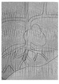
Выполняется пункция внутренней яремной вены передним доступом. Катетеризируется супраренальный отдел нижней полой вены с помощью J-проводника и моделированного катетера. Выполняется ретроградная супраренальная каваграфия для определения анатомии притока печеночных вен в нижнюю полую вену. Для селективной катетеризации и ангиографии средней печеночной вены используется моделированный катетер. Определяется диаметр и длина средней печеночной вены. 0,035 проводник проводится в дистальные отделы средней печеночной вены. По проводнику заводится баллонный катетер в среднюю печеночную вену. Выполняется дилятация баллона и временная окклюзия вены вместе с притоками на протяжении (рис 1.).Puncture of the internal jugular vein is performed using anterior approach. The suprarenal inferior vena cava is catheterized using a J-guide and a simulated catheter. Retrograde suprarenal cavagraphy is performed to determine the anatomy of the inflow of the hepatic veins into the inferior vena cava. A simulated catheter is used for selective catheterization and angiography of the middle hepatic vein. The diameter and length of the middle hepatic vein is determined. 0.035 the guidewire is passed into the distal middle hepatic vein. A balloon catheter is inserted through a guidewire into the middle hepatic vein. The balloon is dilated and the vein is temporarily occluded along with the tributaries throughout (Fig. 1).
Выполняют лапаротомию, мобилизацию печени, холецистэктомию. Далее в портальных воротах выделяют и пересекают долевые элементы глиссоновой ножки на стороне поражения. Производят отделение печени от нижней полой вены. Лигируют короткие вены печени, проводят тесьму между печенью и полой веной, выводят ее между правой и срединной печеночной веной и подвешивают печень над нижней полой веной. Выделяют структуры кавальных ворот, пережимают правую или левую печеночную вену соответственно стороне поражения. При общем стволе срединной и левой печеночной вены, берут на турникет устье ствола вены.Laparotomy, liver mobilization, cholecystectomy are performed. Further, in the portal gate, the share elements of the glisson leg are isolated and intersected on the side of the lesion. The liver is separated from the inferior vena cava. The short veins of the liver are ligated, the braid is drawn between the liver and the vena cava, it is brought out between the right and median hepatic veins, and the liver is suspended over the inferior vena cava. The structures of the caval gates are isolated, the right or left hepatic vein is squeezed according to the side of the lesion. With a common trunk of the median and left hepatic veins, the mouth of the vein trunk is taken on the turnstile.
При пересечении глиссоновой ножки в портальных воротах формируется линия демаркации удаляемой доли печени, производят разметку анатомической границы путем коагуляции капсулы печени. Выполняют рассечение паренхимы печени вдоль линии демаркации, отступя внутрь удаляемой доли на 1 мм, предотвращая повреждение паренхимы и сосудов остающейся доли.When the glisson stem is crossed in the portal gate, a line of demarcation of the removed lobe of the liver is formed, the anatomical border is marked by coagulation of the liver capsule. Dissection of the liver parenchyma is performed along the demarcation line, retreating inside the removed lobe by 1 mm, preventing damage to the parenchyma and vessels of the remaining lobe.
Технический результат настоящего изобретения состоит в уменьшении ретроградной кровопотери по эфферентным венам печени и травматичности резекции печени, т.к. полная сосудистая изоляция удаляемой доли, позволяет избежать кровотечения и снизить коагуляционное повреждение резекционной поверхности культи печени, а также исключает ишемию «ремнанта».The technical result of the present invention is to reduce retrograde blood loss through the efferent veins of the liver and the trauma of liver resection, since complete vascular isolation of the removed lobe allows avoiding bleeding and reducing coagulation damage to the resection surface of the liver stump, and also excludes ischemia of the "remnant".
Предлагаемый способ, по сравнению с известными, имеет ряд существенных преимуществ:The proposed method, in comparison with the known, has a number of significant advantages:
1. Позволяет исключить ретроградную кровопотерю из притоков срединной вены удаляемой доли при анатомических резекциях, что является основным источником кровотечения при анатомических резекциях.1. Allows to exclude retrograde blood loss from the tributaries of the median vein of the removed lobe during anatomical resections, which is the main source of bleeding during anatomical resections.
2. Позволяет сохранить срединную вену, изолированно обработать притоки срединной вены на стороне удаления, не повреждая ствол срединной вены, при этом исключается ретроградный кровоток, возможность воздушной и опухолевой эмболии при сохраненном контрлатеральном кровотоке.2. Allows you to preserve the median vein, treat the tributaries of the median vein on the removal side in isolation, without damaging the trunk of the median vein, while excluding retrograde blood flow, the possibility of air and tumor embolism with preserved contralateral blood flow.
3. Обеспечивает полную сосудистую изоляцию удаляемой доли печени, при этом позволяет избежать гемодинамических нарушений без снижения центрального венозного давления.3. Provides complete vascular isolation of the removed lobe of the liver, while avoiding hemodynamic disturbances without lowering central venous pressure.
4. Уменьшает травматичность операции за счет отсутствия ишемии культи печени, снижения общей кровопотери.4. Reduces the invasiveness of the operation due to the absence of ischemia of the liver stump, reducing total blood loss.
Способ разработан на кафедре общей и факультетской хирургии и прошел клиническую апробацию в Окружной клинической больнице г. Ханты-Мансийск.The method was developed at the Department of General and Faculty Surgery and was clinically tested at the District Clinical Hospital in Khanty-Mansiysk.
Список литературы:Bibliography:
1. Альперович Б.И., Парамонова Л.М., Мерзликин Н.В. Криохирургия печени и поджелудочной железы. - Томск: ТГУ, 1985. - 126 с. [
2. Вишневский B.A., Ефанов М.Г., Щёголев А.И. и др. Топографо-анатомическое основание атравматического экстрапаренхиматозного выделения глиссоновых ножек в воротах печени // Анн. хир. гепатол. - 2008. - Т. 13, №4. - С. 58-66. [Vishnevski V.A., Efanov M.G., Schegolev A.I. et al. Anatomic and topography basis of portal pedicle extraparenchimatous atraumatic isolation in the liver porta. Annaly khirurgicheskoy gepatologii. 2008; 13 (4): 58-66. (In Russ.)]2. Vishnevsky V.A., Efanov M.G., Shchegolev A.I. et al. Topographic and anatomical basis of atraumatic extraparenchymal release of glisson legs in the gates of the liver // Ann. hir. hepatol. - 2008. - T. 13, No. 4. - S. 58-66. [Vishnevski V.A., Efanov M.G., Schegolev A.I. et al. Anatomic and topography basis of portal pedicle extraparenchimatous atraumatic isolation in the liver porta. Annaly khirurgicheskoy gepatologii. 2008; 13 (4): 58-66. (In Russ.)]
3. Гальперин Э.И., Мочалов A.M. Пальцевое чреспеченочное выделение сосудисто-секреторных ножек долей и сегментов при анатомических резекциях печени // Хирургия. Ж. им. Н.И. Пирогова. - 1986. - №7. - С. 3-9. [Vishnevski V.A., Efanov M.G., Schegolev A.I. et al. Digital transhepatic extraction of vascular and secretory fascicles of liver lobes and segments at liver anatomic resections. Khirurgiya. Zhurnal imeni N.I. Pirogova. 1986; 7: 3-9. (In Russ.)]3. Galperin E.I., Mochalov A.M. Finger transhepatic isolation of the vascular-secretory legs of the lobes and segments during anatomical liver resections // Surgery. J. im. N.I. Pirogov. - 1986. - No. 7. - S. 3-9. [Vishnevski V.A., Efanov M.G., Schegolev A.I. et al. Digital transhepatic extraction of vascular and secretory fascicles of liver lobes and segments at liver anatomic resections. Khirurgiya. Zhurnal imeni N.I. Pirogova. 1986; 7: 3-9. (In Russ.)]
4. Джантуханова С.В., Старков Ю.Г., Шишин К.В. Лапароскопические резекции печени. // Хирургия. Ж. им. Н.И. Пирогова. - 2009. - №12. - С. 63-67. [Dzhantukhanova S.V., Starkov Iu.G., Shishin K.V. Laparoscopic liver resections. Khirurgiya. Zhurnal imeni N.I. Pirogova. 2009; 12: 63-67. (In Russ.)].4. Dzhantukhanova S.V., Starkov Yu.G., Shishin K.V. Laparoscopic liver resections. // Surgery. J. im. N.I. Pirogov. - 2009. - No. 12. - S. 63-67. [Dzhantukhanova S.V., Starkov Iu.G., Shishin K.V. Laparoscopic liver resections. Khirurgiya. Zhurnal imeni N.I. Pirogova. 2009; 12: 63-67. (In Russ.)].
5. Милонов О.Б., Колосс O.E., Минкина С.М. и др. Результаты применения криовоздействия в хирургическом лечении распространенного альвеококкоза печени // Хирургия. Ж. им. Н.И. Пирогова. - 1988. - №2. - С. 40-44. [Milonov О.В., Koloss О.Е., Minkina S.M. et al. Results of using cryosurgery in surgeries of generalized alveolar disease of liver. Khirurgiya. Zhurnal imeni N.I. Pirogova. 1988; 2: 40-44. (In Russ.)].5. Milonov O.B., Colossus O.E., Minkina S.M. and other Results of cryotherapy application in surgical treatment of widespread liver alveococcosis // Surgery. J. im. N.I. Pirogov. - 1988. - No. 2. - S. 40-44. [Milonov O. V., Koloss O. E., Minkina S.M. et al. Results of using cryosurgery in surgeries of generalized alveolar disease of liver. Khirurgiya. Zhurnal imeni N.I. Pirogova. 1988; 2: 40-44. (In Russ.)].
6. Патютко Ю.И., Пылев А.Л., Сагайдак И.В., Котельников А.Г. Расширенные резекции печени при злокачественных опухолях // Хирургия. Ж. им. Н.И. Пирогова. - 2009. - №2. - С. 16-2. [Patiutko Iu.L, Pylev A.L., 
7. Сандомирский Б.П., Коцько Н.И., Золочевская Л.И. Эффективность криовоздействия при резекции печени в эксперименте // Вестн. хир. им. И.И. Грекова. - 1979. - №4. - С. 24-26. [Sandomirskiy В.Р., 
8. Скобелкин O.K., Брехов Е.И., Елисеенко В.И. и др. Использование плазменных хирургических установок для гемостаза при операциях на печени и селезенке // Вестн. хир. им. И.И. Грекова. - 1988. - №5. - С. 32-33. [Skobelkin O.K., Brekhov E.I., Eliseenko V.I. et al. Using the units for plasma surgery for hemostasis in liver and spleen surgeries. Vestnik khirurgii imeni I.I. Grekova. 1988; 5: 32-33. (In Russ.)]8. Skobelkin O.K., Brekhov E.I., Eliseenko V.I. et al. The use of plasma surgical devices for hemostasis in operations on the liver and spleen // Vestn. hir. them. I.I. Grekov. - 1988. - No. 5. - S. 32-33. [Skobelkin O.K., Brekhov E.I., Eliseenko V.I. et al. Using the units for plasma surgery for hemostasis in liver and spleen surgeries. Vestnik khirurgii imeni I.I. Grekova. 1988; 5: 32-33. (In Russ.)]
9. Старков Ю.Г., Вишневский B.A., Шишин K.B. Лапароскопические операции при очаговых образованиях печени // Анн. хир. гепатол. - 2008. - Т. 13, №1. - С. 34-41. [Starkov Yu.G., Vishnevsky V.A., Shishin K.V. An experience of laparoscipic operations in the liver local lesions. Annaly khirurgicheskoy gepatologii. 2008; 13 (1): 34-41. (In Russ.)]9. Starkov Yu.G., Vishnevsky B.A., Shishin K.B. Laparoscopic operations for focal liver formations // Ann. hir. hepatol. - 2008. - T. 13, No. 1. - S. 34-41. [Starkov Yu.G., Vishnevsky V.A., Shishin K.V. An experience of laparoscipic operations in the liver local lesions. Annaly khirurgicheskoy gepatologii. 2008; 13 (1): 34-41. (In Russ.)]
10. Andrus C.H., Kaminski D.L. Segmental hepatic resection itilizing the ultrasonic dissector // Arch. Surg. - 1986. - Vol.121, N 5. - P. 515-521.10. Andrus C.H., Kaminski D.L. Segmental hepatic resection itilizing the ultrasonic dissector // Arch. Surg. - 1986. - Vol. 121, N 5. - P. 515-521.
11. Blumgart L.H. Surgery of the liver and biliary tract. New York: Churchill Livingstone, 2006. - Ed. 4. - Vol. 2. - P. 1422.11. Blumgart L.H. Surgery of the liver and biliary tract. New York: Churchill Livingstone, 2006. - Ed. 4. - Vol. 2. - P. 1422.
12. Catalano O.A., Singh A.H., Uppot R.N. et al. Vascular and biliary variants in the liver: implications for liver surgery // Radiographics. - 2008. - Vol. 28. - P. 359-378.12. Catalano O.A., Singh A.H., Uppot R.N. et al. Vascular and biliary variants in the liver: implications for liver surgery // Radiographics. - 2008. - Vol. 28. - P. 359-378.
13. Cerny J. Reseccia lateralneho segmentu laveho latoka pa-cene turnicetovu metodu // Rozhl. Chir. - 1973. - Vol. 52, N 3. - P. 169-172.13. Cerny J. Reseccia lateralneho segmentu laveho latoka pa-cene turnicetovu metodu // Rozhl. Chir. - 1973. - Vol. 52, No. 3. - P. 169-172.
14. Couinaud C. Plaidoyer pour une segmentation hepatique exacte et une technique anatomique de resection reglee du foie // Presse medic. - 1966. - Vol. 74, N 55. - P. 2849-2852.14. Couinaud C. Plaidoyer pour une segmentation hepatique exacte et une technique anatomique de resection reglee du foie // Presse medic. - 1966. - Vol. 74, No. 55.-P. 2849-2852.
15. Corcione F., Esposito C, Cuccurullo D. et al. Advantages and limits of robot-assisted laparoscopic surgery // Surg. Endosc. - 2005. - Vol. 19. - P. 117-119.15. Corcione F., Esposito C, Cuccurullo D. et al. Advantages and limits of robot-assisted laparoscopic surgery // Surg. Endosc. - 2005. - Vol. 19. - P. 117-119.
16. Fineberg C., Goldburgh W.P., Templeton I.Y. Right hepatic lobectomy for primary carcinoma of the liver // Ann. Surg. - 1956. - Vol. 144. - P. 882-892.16. Fineberg C., Goldburgh W.P., Templeton I.Y. Right hepatic lobectomy for primary carcinoma of the liver // Ann. Surg. - 1956. - Vol. 144. - P. 882-892.
17. Habib N., Zografos G., Dalla Serra G. et al. Liver resection with total vascular exclusion for malignant tumors/ // Brit. J. Surg. - 1994. - Vol. 81. - P. 1181-1184.17. Habib N., Zografos G., Dalla Serra G. et al. Liver resection with total vascular exclusion for malignant tumors / // Brit. J. Surg. - 1994. - Vol. 81. - P. 1181-1184.
18. Hodgson W.J., DelGuercio L.R. Preliminary experience in liver surgery using the ultrasonic scalpel // Surgery. - 1984. - Vol. 95, N 2. - P. 230-234.18. Hodgson W.J., DelGuercio L.R. Preliminary experience in liver surgery using the ultrasonic scalpel // Surgery. - 1984. - Vol. 95, No. 2. -P. 230-234.
19. Kim F.J., Charmas M.F., Gewehr E. et al. Temperature safety profile of laparoscopic devices: harmonic ACE (ACE), LigaSure V (LV), and plasma trisector (PT) // Surg. Endosc. - 2007. - Vol. 22, N 6. - P. 1464-1469.19. Kim F. J., Charmas M. F., Gewehr E. et al. Temperature safety profile of laparoscopic devices: harmonic ACE (ACE), LigaSure V (LV), and plasma trisector (PT) // Surg. Endosc. - 2007. - Vol. 22, No. 6. - P. 1464-1469.
20. Kinoshita H., Sakai K., Hirohashi K. et al. Preoperative portal veinembolization for hepatocellular carcinoma // Worid. J. Surg. - 1986. - Vol. 10. - P. 803-808.20. Kinoshita H., Sakai K., Hirohashi K. et al. Preoperative portal veinembolization for hepatocellular carcinoma // Worid. J. Surg. - 1986. - Vol. 10. - P. 803-808.
21. Kondo K., Chijiiwa K., Funagayama M. et al. Hepatic resection is justified for elderly patients with hepatocellular carcinoma // World J. Surg. - 2008. - Vol. 32. - P. 2223-2229.21. Kondo K., Chijiiwa K., Funagayama M. et al. Hepatic resection is justified for elderly patients with hepatocellular carcinoma // World J. Surg. - 2008. - Vol. 32 .-- P. 2223-2229.
22. Lin T.Y., Tsu K.Y., Mien C. et al. Study on lobectomy of the liver // J. Formos. Med. Ass. - 1958. - Vol. 11. - P. 742-759.22. Lin T.Y., Tsu K.Y., Mien C. et al. Study on lobectomy of the liver // J. Formos. Med. Ass. - 1958. - Vol. 11. - P. 742-759.
23. Longmire W.P., Marable S.A. Clinical experience with major hepatic resections // Ann. Surg. - 1961. - Vol. 154, N 3. - P. 460-474.23. Longmire W.P., Marable S.A. Clinical experience with major hepatic resections // Ann. Surg. - 1961. - Vol. 154, No. 3. - P. 460-474.
24. Papachristou D.N., Bartes R. Resection of the liver with a water jet // Brit. J. Surg. - 1982. - Vol. 69, N 2. - P. 93-94.24. Papachristou D.N., Bartes R. Resection of the liver with a water jet // Brit. J. Surg. - 1982. - Vol. 69, No. 2. - P. 93-94.
25. Quattelbaum J.K. Massive resection of the liver // Ann. Surg. - 1953. - Vol. 137. - P. 787-796.25. Quattelbaum J.K. Massive resection of the liver // Ann. Surg. - 1953. - Vol. 137. - P. 787-796.
26. Ravikumar T.S., Kane R., Cady B. et al. Hepatic cryosurgery with intraoperative ultrasound monitoring.26. Ravikumar T.S., Kane R., Cady B. et al. Hepatic cryosurgery with intraoperative ultrasound monitoring.
Claims (1)
Priority Applications (1)
| Application Number | Priority Date | Filing Date | Title |
|---|---|---|---|
| RU2019101380A RU2733503C2 (en) | 2019-01-17 | 2019-01-17 | Method of vascular liver isolation during resection, using temporary endovascular occlusion |
Applications Claiming Priority (1)
| Application Number | Priority Date | Filing Date | Title |
|---|---|---|---|
| RU2019101380A RU2733503C2 (en) | 2019-01-17 | 2019-01-17 | Method of vascular liver isolation during resection, using temporary endovascular occlusion |
Publications (3)
| Publication Number | Publication Date |
|---|---|
| RU2019101380A3 RU2019101380A3 (en) | 2020-07-17 |
| RU2019101380A RU2019101380A (en) | 2020-07-17 |
| RU2733503C2 true RU2733503C2 (en) | 2020-10-02 |
Family
ID=71617563
Family Applications (1)
| Application Number | Title | Priority Date | Filing Date |
|---|---|---|---|
| RU2019101380A RU2733503C2 (en) | 2019-01-17 | 2019-01-17 | Method of vascular liver isolation during resection, using temporary endovascular occlusion |
Country Status (1)
| Country | Link |
|---|---|
| RU (1) | RU2733503C2 (en) |
Cited By (1)
| Publication number | Priority date | Publication date | Assignee | Title |
|---|---|---|---|---|
| RU2850728C1 (en) * | 2025-03-27 | 2025-11-13 | Государственное Бюджетное Учреждение Здравоохранения Города Москвы "Московский Многопрофильный Научно-Клинический Центр Имени С.П. Боткина" Департамента Здравоохранения Города Москвы (Гбуз Ммнкц Им. С.П. Боткина Дзм) | Method for resection of focal lesions in segments ii-iii of liver located on visceral surface of liver |
Citations (2)
| Publication number | Priority date | Publication date | Assignee | Title |
|---|---|---|---|---|
| RU2263473C2 (en) * | 2003-08-08 | 2005-11-10 | Ростовский научно-исследовательский онкологический институт МЗ РФ | Method for left-hand hemihepatectomy |
| RU2442539C2 (en) * | 2009-04-01 | 2012-02-20 | Федеральное Государственное Учреждение "Институт хирургии им. А.В. Вишневского Федерального агентства по высокотехнологичной медицинской помощи" | The means of anotomic resection of the viii liver segment |
-
2019
- 2019-01-17 RU RU2019101380A patent/RU2733503C2/en not_active IP Right Cessation
Patent Citations (2)
| Publication number | Priority date | Publication date | Assignee | Title |
|---|---|---|---|---|
| RU2263473C2 (en) * | 2003-08-08 | 2005-11-10 | Ростовский научно-исследовательский онкологический институт МЗ РФ | Method for left-hand hemihepatectomy |
| RU2442539C2 (en) * | 2009-04-01 | 2012-02-20 | Федеральное Государственное Учреждение "Институт хирургии им. А.В. Вишневского Федерального агентства по высокотехнологичной медицинской помощи" | The means of anotomic resection of the viii liver segment |
Non-Patent Citations (3)
| Title |
|---|
| Deepak Sudheendra et, Intermittent Hepatic Vein Balloon Occlusion During Radiofrequency Ablation in the Liver, Cardiovasc Intervent Radiol. 2006; 29(6): 1088-1092. * |
| ВИШНЕВСКИЙ В.А. и др. Способ воротного доступа к сосудисто-секреторным элементам при анатомических сегментарных резекциях печени. Хирургия. Журнал им. Н.И.Пирогова, 2008, 9, с.33-40. * |
| ВИШНЕВСКИЙ В.А. и др. Способ воротного доступа к сосудисто-секреторным элементам при анатомических сегментарных резекциях печени. Хирургия. Журнал им. Н.И.Пирогова, 2008, 9, с.33-40. Deepak Sudheendra et, Intermittent Hepatic Vein Balloon Occlusion During Radiofrequency Ablation in the Liver, Cardiovasc Intervent Radiol. 2006; 29(6): 1088-1092. * |
Cited By (2)
| Publication number | Priority date | Publication date | Assignee | Title |
|---|---|---|---|---|
| RU2850728C1 (en) * | 2025-03-27 | 2025-11-13 | Государственное Бюджетное Учреждение Здравоохранения Города Москвы "Московский Многопрофильный Научно-Клинический Центр Имени С.П. Боткина" Департамента Здравоохранения Города Москвы (Гбуз Ммнкц Им. С.П. Боткина Дзм) | Method for resection of focal lesions in segments ii-iii of liver located on visceral surface of liver |
| RU2854248C1 (en) * | 2025-04-29 | 2025-12-29 | федеральное государственное бюджетное учреждение "Национальный медицинский исследовательский центр детской гематологии, онкологии и иммунологии имени Дмитрия Рогачева" Министерства здравоохранения Российской Федерации | Method for determining indications for total vascular isolation during liver resection in children |
Also Published As
| Publication number | Publication date |
|---|---|
| RU2019101380A3 (en) | 2020-07-17 |
| RU2019101380A (en) | 2020-07-17 |
Similar Documents
| Publication | Publication Date | Title |
|---|---|---|
| Dolenc | Direct microsurgical repair of intracavernous vascular lesions | |
| Mohit et al. | High-flow bypass grafts in the management of complex intracranial aneurysms | |
| Van As et al. | Management of paediatric liver trauma | |
| Smith et al. | Vacuum-assisted thrombectomy device (AngioVac) in the management of symptomatic iliocaval thrombosis | |
| Klein et al. | Contemporary management strategy for major inferior vena caval injuries | |
| Salsamendi et al. | Single center experience with the AngioVac aspiration system | |
| Mascitelli et al. | The oculomotor-tentorial triangle. Part 2: a microsurgical workspace for vascular lesions in the crural and ambient cisterns | |
| Ramaswamy et al. | Acute lower extremity deep venous thrombosis: the data, where we are, and how it is done | |
| Yang et al. | Bypass using V2-V3 segment of the vertebral artery as donor or recipient: technical nuances and results | |
| Gonzalez-Fajardo et al. | Transabdominal surgical approach in the management of renal tumors involving the retrohepatic inferior vena cava | |
| Angermeier et al. | Resection of nonrenal retroperitoneal tumors with large vena caval thrombi using cardiopulmonary bypass and hypothermic circulatory arrest | |
| Thompson et al. | Surgical techniques: operative decompression using the paraclavicular approach for venous thoracic outlet syndrome | |
| Ishida et al. | Video-assisted thoracoscopic surgery of bullous and bleb disorders of the lung using endoscopic stapling device | |
| Beitner et al. | Penetrating liver injury managed with a combination of balloon tamponade and venous stenting. A case report and literature review | |
| Aseni et al. | Basic operative techniques in abdominal injury | |
| ILLIG et al. | Venous and lymphatic disease: a historical review | |
| Fontenot et al. | Central Venous Stenosis Associated With Arteriovenous Access | |
| Harris et al. | Complications of Iliofemoral Thrombolysis and Stenting for Venous Disease | |
| Mengchao et al. | Experiences in 467 cases of hepatic resection | |
| Gloviczki et al. | Arteriovenous fistulas and vascular malformations | |
| Du et al. | Surgical management of congenital arteriovenous fistula near lateral cranial base | |
| Yu et al. | A Modified Transcarotid Artery Revascularization Technique for High-Risk Carotid Stenosis | |
| Huo et al. | Resection of recurrent parasagittal meningiomas with complete obstructed superior sagittal sinus | |
| RU2665119C1 (en) | Method of temporal hemostasis in laparoscopic resection of a kidney transplantat | |
| Keles et al. | Microsurgical resection of a scalp arteriovenous fistula with intracranial drainage: an illustrative case |
Legal Events
| Date | Code | Title | Description |
|---|---|---|---|
| MM4A | The patent is invalid due to non-payment of fees |
Effective date: 20210118 |