US6310190B1 - USPA1 and USPA2 antigens of Moraxella catarrhalis - Google Patents
USPA1 and USPA2 antigens of Moraxella catarrhalis Download PDFInfo
- Publication number
- US6310190B1 US6310190B1 US09/336,447 US33644799A US6310190B1 US 6310190 B1 US6310190 B1 US 6310190B1 US 33644799 A US33644799 A US 33644799A US 6310190 B1 US6310190 B1 US 6310190B1
- Authority
- US
- United States
- Prior art keywords
- uspa1
- uspa2
- seq
- catarrhalis
- protein
- Prior art date
- Legal status (The legal status is an assumption and is not a legal conclusion. Google has not performed a legal analysis and makes no representation as to the accuracy of the status listed.)
- Expired - Fee Related
Links
Images




Classifications
-
- C—CHEMISTRY; METALLURGY
- C07—ORGANIC CHEMISTRY
- C07K—PEPTIDES
- C07K14/00—Peptides having more than 20 amino acids; Gastrins; Somatostatins; Melanotropins; Derivatives thereof
-
- C—CHEMISTRY; METALLURGY
- C07—ORGANIC CHEMISTRY
- C07K—PEPTIDES
- C07K14/00—Peptides having more than 20 amino acids; Gastrins; Somatostatins; Melanotropins; Derivatives thereof
- C07K14/195—Peptides having more than 20 amino acids; Gastrins; Somatostatins; Melanotropins; Derivatives thereof from bacteria
- C07K14/21—Peptides having more than 20 amino acids; Gastrins; Somatostatins; Melanotropins; Derivatives thereof from bacteria from Pseudomonadaceae (F)
- C07K14/212—Moraxellaceae, e.g. Acinetobacter, Moraxella, Oligella, Psychrobacter
-
- A—HUMAN NECESSITIES
- A61—MEDICAL OR VETERINARY SCIENCE; HYGIENE
- A61P—SPECIFIC THERAPEUTIC ACTIVITY OF CHEMICAL COMPOUNDS OR MEDICINAL PREPARATIONS
- A61P31/00—Antiinfectives, i.e. antibiotics, antiseptics, chemotherapeutics
- A61P31/04—Antibacterial agents
-
- A—HUMAN NECESSITIES
- A61—MEDICAL OR VETERINARY SCIENCE; HYGIENE
- A61P—SPECIFIC THERAPEUTIC ACTIVITY OF CHEMICAL COMPOUNDS OR MEDICINAL PREPARATIONS
- A61P37/00—Drugs for immunological or allergic disorders
- A61P37/02—Immunomodulators
- A61P37/04—Immunostimulants
-
- C—CHEMISTRY; METALLURGY
- C07—ORGANIC CHEMISTRY
- C07K—PEPTIDES
- C07K16/00—Immunoglobulins [IG], e.g. monoclonal or polyclonal antibodies
- C07K16/12—Immunoglobulins [IG], e.g. monoclonal or polyclonal antibodies against material from bacteria
- C07K16/1203—Gram-negative bacteria
- C07K16/1217—Neisseriaceae (F)
-
- A—HUMAN NECESSITIES
- A61—MEDICAL OR VETERINARY SCIENCE; HYGIENE
- A61K—PREPARATIONS FOR MEDICAL, DENTAL OR TOILETRY PURPOSES
- A61K39/00—Medicinal preparations containing antigens or antibodies
Definitions
- the present invention relates generally to the fields of microbiology, and clinical bacteriology. More particularly, it concerns sequences of the uspA1 and uspA2 genes which encode the proteins UspA1 and UspA2, respectively, both of which encode an epitope reactive with monoclonal antibody (MAb) 17C7 and provide useful epitopes for immunodiagnosis and immunoprophylaxis.
- MAb monoclonal antibody
- Moraxella catarrhalis previously known as Branhamella catarrhalis or Neisseria catarrhalis , was a harmless saprophyte of the upper respiratory tract (Catlin, 1990; Berk, 1990). However, during the previous decade, it has been determined that this organism is an important human pathogen. Indeed, it has been established that this Gram-negative diplococcus is the cause of a number of human infections (Murphy, 1989). M. catarrhalis is now known to be the third most common cause of both acute and chronic otitis media (Catlin, 1990; Faden et al., 1990; 1991; Marchant, 1990), the most common disease for which infants and children receive health care according to the 1989 Consensus Report.
- This organism also causes acute maxillary sinusitis, generalized infections of the lower respiratory tract (Murphy and Loeb, 1989) and is an important cause of bronchopulmonary infections in patients with underlying chronic lung disease and, less frequently, of systemic infections in immunocompromised patients (Melendez and Johnson, 1990; Sarubbi et al., 1990; Schonheyder and Ejlertsen, 1989; Wright and Wallace, 1989).
- M. catarrhalis This is particularly troublesome in that M. catarrhalis now accounts for approximately 17-20% of all otitis media infection (Murphy, 1989). In addition, M. catarrhalis is also a significant cause of sinusitis (van Cauwenberge et al., 1993) and persistent cough (Gottfarb and Brauner, 1994) in children. In the elderly, it infects patients with predisposing conditions such as chronic obstructive pulmonary disease (COPD) and other chronic cardiopulmonary conditions (Boyle et al., 1991; Davies and Maesen, 1988; Hager et al., 1987).
- COPD chronic obstructive pulmonary disease
- M. catarrhalis otitis media An antibody response to M. catarrhalis otitis media has been documented by means of an ELISA system using whole M. catarrhalis cells as antigen and acute and convalescent sera or middle ear fluid as the source of antibody (Leinonen et al., 1981). The development of serum bactericidal antibody during M. catarrhalis infection in adults was shown to be dependent on the classical complement pathway (Chapman et al., 1985). And more recently, it was reported that young children with M. catarrhalis otitis media develop an antibody response in the middle ear but fail to develop a systemic antibody response in a uniform manner (Faden et al., 1992).
- M. catarrhalis antigens that would serve as potentially important targets of the human immune response to infection (Murphy, 1989; Goldblatt et al., 1990; Murphy et al., 1990).
- OMPs outer membrane proteins
- LOS lipooligosaccharide
- fimbriae fimbriae.
- M. catarrhalis appears to be somewhat distinct from other Gram-negative bacteria in that attempts to isolate the outer membrane of this organism using detergent fractionation of cell envelopes has generally proven to be unsuccessful in that the procedures did not yield consistent results (Murphy, 1989; Murphy and Loeb, 1989).
- preparations were found to be contaminated with cytoplasmic membranes, suggesting an unusual characteristic of the M. catarrhalis cell envelope.
- OMPs outer membrane proteins
- CopB protein and UspA antigen have been shown to bind antibodies which exert biological activity against M. catarrhalis in an animal model (Helminenet al., 1994; Murphy et al., 1993).
- the MAb designated 17C7, was described as binding to UspA, a very high molecular weight protein that migrated with an apparent molecular weight (in SDS-PAGE) of at least 250 kDa (Helminen et al., 1994; Klingman and Murphy, 1994).
- MAb 17C7 enhanced pulmonary clearance of M. catarrhalis from the lungs of mice when used in passive immunization studies and, in colony blot radioimmunoassay analysis, bound to every isolate of M. catarrhalis examined. This same MAb also reacted, although less intensely, with another antigen band of approximately 100 kDa, as described in U.S. Pat. No. 5,552,146 (incorporated herein by reference).
- a recombinant bacteriophage that contained a fragment of M. catarrhalis chromosomal DNA that expressed a protein product that bound MAb 17C7 was also identified and migrated at a rate similar or indistinguishable from that of the native UspA antigen from M. catarrhalis (Helminen et al., 1994).
- epitopic core sequences of UspA1 and UspA2 which can serve as the basis for the preparation of therapeutic or prophylactic compositions or vaccines which comprise peptides of 7, 10, 20, 30, 40, 50 or even 60 amino acids in length that elicit an antigenic reaction and a pharmaceutically acceptable buffer or diluent.
- These peptides may be coupled to a carrier, adjuvant, another peptide or other molecule such that an effective antigenic response to M. catarrhalis is retained or even enhanced.
- these peptides may act as carriers themselves when coupled to another peptide or other molecule that elicits an antigenic response to M. catarrhalis or another pathogen.
- UspA2 can serve as a carrier for an oligosaccharide.
- the epitopic core sequences of UspA1 and UspA2 comprise one or more more isolated peptides of 7, 10, 20, 30, 40, 50 or even 60 amino acids in length having the amino acid sequence AQQQDQH (SEQ ID NO:17).
- nucleic acids uspA1 and uspA2, which encode the UspA1 and the UspA2 antigens, respectively, as well as the amino acid sequences of the UspA1 and UspA2 antigens of the M. catarrhalis isolates O35E, TTA24, TTA37, and O46E. It is envisioned that nucleic acid segments and fragments of the genes uspA1 and uspA2 and the UspA1 and UspA2 antigens will be of value in the preparation and use of therapeutic or prophylactic compositions or vaccines for treating, inhibiting or even preventing M. catarrhalis infections.
- a method for inducing an immune response in a mammal comprising the step of providing to the mammal an antigenic composition that comprises an isolated peptide of about 20 to about 60 amino acids that contains the identified epitopic core sequence and a pharmaceutically acceptable buffer or diluent.
- a method for diagnosing M. catarrhalis infection which comprises the step of determining the presence, in a sample, of an M. catarrhalis amino acid sequence corresponding to residues of the epitopic core sequences of either the UspA1 or UspA2 antigen.
- This method may comprise PCR m detection of the nucleotide sequences or alternatively an immunologic reactivity of an antibody to either a UspA1 or UspA2 antigen.
- a method for treating an individual having an M. catarrhalis infection which comprises providing to the individual an isolated peptide of about 20 to about 60 amino acids that comprises at least about 10 consecutive residues of the amino acid sequence identified as an epitopic core sequence of UspA1 or UspA2.
- a method for preventing or limiting an M. catarrhalis infection comprises providing to a subject an antibody that reacts immunologically with the identified epitopic core region of either UspA1 or UspA2 of M. catarrhalis.
- a method for screening a peptide for reactivity with an antibody that binds immunologically to UspA1, UspA2 or both which comprises the steps of providing the peptide and contacting the peptide with the antibody and then determining the binding of the antibody to the peptide.
- This method may comprise an immunoassay such as a western blot, an ELISA, an RIA or an immunoaffinity separation.
- a method for screening a UspA1 or UspA2 peptide for its ability to induce a protective immune response against M. catarrhalis by providing the peptide, administering it in a suitable form to an experimental animal, challenging the animal with M. catarrhalis and then assaying for an M. catarrhalis infection in the animal.
- the animal used will be a mouse that is challenged by a pulmonary exposure to M. catarrhalis and that the assaying comprises assessing the degree of pulmonary clearance by the mouse.
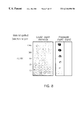
- FIG. 1 Southern blot analysis of PvuII-digested chromosomal DNA from strains of M. catarrhalis using a probe from the uspA1 gene. Bacterial strain designations are at the top; kilobase (kb) position markers are on the left.
- FIG. 2 A Proteins present in whole cell lysates of the wild-type strain O35E and the isogenic uspA1 mutant strain. These proteins were resolved by SDS-PAGE and stained with Coomassie blue.
- the left lane (WT) contains the wild-type strain and right lane (MUT) contains the mutant.
- the arrows indicate the protein, approximately 120 kDa in size, that is present in the wild-type and missing in the mutant. Kilodalton position markers are on the left.
- FIG. 2 B Western blot analysis of whole cell lysates of the wild-type strain O35E and the isogenic uspA1 mutant strain. These proteins were resolved by SDS-PAGE and probed with MAb 17C7 in western blot analysis.
- the left lane (WT) contains the wild-type strain and the right lane (MUT) contains the mutant. Kilodalton position markers are on the left. It can been seen that both strains possess the very high molecular weight band reactive with MAb 17C7 whereas only the wild-type strain also has a band of approximately 120 kDa that binds this MAb.
- FIG. 2 C Western blot analysis of whole cell lysate (WCL) and EDTA-extracted outer membrane vesicles (OMV) from the wild-type strain O35E (WT) and the isogenic uspA1 mutant (MUT) using MAb 17C7.
- Samples were either heated at 37° C. for 15 minutes (H) or at 100° C. for minutes (B) prior to SDS-PAGE.
- Molecular weight position markers (in kilodaltons) are indicated on the left.
- the open arrow indicates the position of the very high molecular weight form of the MAb 17C7-reactive antigen;
- the closed arrow indicates the position of the approximately 120 kDa protein;
- the open circle indicates the position of the approximately 70-80 kDa protein.
- FIG. 3 Southern blot analysis of chromosomal DNA from the wild-type M. catarrhalis strain O35E and the isogenic uspA1 mutant. Chromosomal DNA was digested with PvuII and probed with a 0.6 kb BglII-PvuII fragment from the uspA1 gene. The wild-type strain is listed as O35E at the top of this figure and the mutant strain is listed as O35E-uspA1 ⁇ . Kilobase position markers are present on the left side.
- FIG. 4 Western blot reactivity of proteins in M. catarrhalis strain O35E outer membrane vesicles (labeled O35E OMV) and the MF-4-1 GST fusion protein (labeled GST fusion protein) with MAb 17C7.
- FIG. 5 PCRTM products obtained by the use of the T3 and P10 primers (middle lane—0.9 kb product) and the T7 and P9 primers (right lane—1.7 kb product) when used in a PCRTM amplification with chromosomal DNA from the uspA1 mutant.
- a kb ladder is present in the first lane; several kb position markers are listed on the left side of this figure.
- FIGS. 6A-6C SDS-PAGE and westerns of purified proteins.
- FIG. 6 A Coomassie blue stained gel of purified UspA2 (lane 2).
- FIG. 6 B Coomassie blue stained gel of purified UspA1 prepared without heating of sample (lane 4), heated for 3 min at 100° C. (lane 5), heated for 5 min at 100° C. (lane 6), and heated for 10 min at 100° C. (lane 7).
- FIG. 6 C Western of the purified UspA2 (lane 9) and purified UspA1 (lane 10) probed with the 17C7 MAb. Both proteins were heated 10 min.
- the molecular size markers in lanes 1, 3, and 8 are as indicated in kilodaltons.
- FIG. 7 Interaction of purified UspA1 and UspA2 with HEp-2 cells as determined by ELISA.
- HEp-2 cell monolayers cultured in 96-well plate were incubated with serially diluted UspA1 or UspA2.
- O35E bacterial strain was used as the positive control.
- the bacteria were diluted analogous to the proteins beginning with a suspension with an A 550 of 1.0.
- the bound proteins or attached bacteria were detected with a 1:1 mixed antisera to UspA1 and UspA2 as described in the methods.
- FIG. 8 Interaction with fibronectin and vitronectin determined by dot blot.
- the bound vitronectin was detected with rabbit polyclonal antibodies, the protein bound to the fibronectin was detected with pooled sera made against the UspA1 and UspA2.
- FIG. 9 The levels of antibodies to the protein UspA1, UspA2 and M. catarrhalis O35E strain in normal human sera.
- Data are the log 10 transformed end-point titers of the IgG (FIGS. 9A-9C) and IgA (FIGS. 9D-9F) antibodies determined by ELISA.
- the individual titers were plotted according to age group and the geometric mean titer for each age group linked by a solid line. Sera for the 2-18 month old children were consecutive samples from a group of ten children.
- FIG. 10 Subclass distribution of IgG antibodies to UspA1 and UspA2 in normal human sera.
- FIG. 10A shows titers toward UspA1 and
- FIG. 10B shows titers to UspA2.
- FIG. 11 Relationship of serum IgG titers to UspA1 (FIG. 11A) and UspA2 (FIG. 11B) with the bactericidal liter against the O35E strain determined by logistic regression (p ⁇ 0.05).
- the solid line indicates the linear relationship between the IgG titer and bactericidal titer.
- Broken lines represent the 95% confidence intervals of the linear fit.
- FIG. 12 Schematic drawing showing the relative positions of decapeptides 10-24 within the region of UspA1 and UspA2 which binds to MAb 17C7.
- FIG. 13 Western dot blot analysis demonstrating reactivity of decapeptides 10-24 with MAb 17C7.
- FIG. 14 Partial restriction enzyme map of the uspA1 (FIG. 14A) and uspA2 (FIG. 14B) genes from M. catarrhalis strain O35E and the mutated versions of these genes.
- the shaded boxes indicate the open reading frame of each gene.
- Relevant restriction sites are indicated.
- PCRTM primer sites P1-P6 are indicated by arrows.
- the DNA fragments containing the partial uspA1 and uspA2 open reading frames that were derived from M. catarrhalis straina O35E chromosomal DNA by PCRTM and cloned into pBluescriptII SK+ are indicated by black bars. Dotted lines connect corresponding restriction sites on these DNA inserts and the chromosome.
- Open bars indicate the location of the kanamycin or chloramphenicol cassettes, respectively.
- the DNA probes specific for uspA1 or uspA2 are indicated by the appropriate cross-hatched bars and were amplified by PCRTM from M. catarrhalis strain O35E chromosomal DNA by the use of the oligonucleotide primer pairs
- P3 (5′-GACGCTCAACAGCACTAATACG-3′) (SEQ ID NO:20)/P4 (5′-CCAAGCTGATATCACTACC-3′) (SEQ ID NO:21) and
- P5 (5′-TCAATGCCMTTGATGGTC-3′) (SEQ ID NO:22)
- P6 (5′-TGTATGCCGCTACTCGCAGCT-3′) (SEQ ID NO:23), respectively.
- the closed arrow indicates the very high molecular weight form of the UspA antigen which is comprised of both UspA1 and UspA2.
- the bracket on the left indicates the very high molecular weight forms of the UspA1 and UspA2 proteins that bind MAb 17C7.
- the open arrow indicates the 120 kDa, putative monomeric form of UspA1.
- the closed arrow indicates the 85 kDa, putative monomeric form of UspA2.
- Molecular weight position markers in kilodaltons
- FIG. 16 Comparison of the rate and extent of growth Qf the wild-type and mutant strains of M. catarrhalis .
- the wild-type strain O35E (closed squares), the uspA1 mutant O35E.1 (open squares), the uspA2 mutant O35E.2 (closed circles), and the uspA1 uspA2 double mutant O35E.12 (open circles) of M. catarrhalis O35E from overnight broth cultures were diluted to a density of 35 Klett units in BHI broth and subsequently allowed to grow at 37° with shaking. Growth was followed by means of turbidity measurements.
- FIG. 17 Susceptibility of wild-type and mutant strains of M. catarrhalis to killing by normal human serum.
- Cells of the wild-type parent strain O35E (diamonds), uspA1 mutant O35E.1 (triangles), uspA2 mutant O35E.2 (circles), and uspA1 uspA2 double mutant O35E.12 (squares) from logarithmic-phase BHI broth cultures were incubated in the presence of 10% (v/v) normal human serum (closed symbols) or heat-inactivated normal human serum (open symbols). Data are presented as the percentage of the original inoculum remaining at each time point.
- the present invention relates to the identification of epitopes useful for developing potential vaccines against M. catarrhalis .
- Early work was directed at determining the molecular nature of the UspA antigen and characterize the epitope which is recognized by the MAb 17C7.
- Preliminary work indicated that MAb 17C7 recognizes a single antigenic epitope and it was believed that this epitope was encoded by a single gene.
- isolation of the protein which contained the epitope yielded unexpected results.
- MAb 17C7 recognized a single epitope, but the characteristics of the protein associated with the epitope suggested the existence of not one but two separate proteins. Further careful analyses led to a surprising discovery.
- a single epitope of the UspA antigen is recognized by the MAb 17C7, but this epitope is present in two different proteins, UspA1 and UspA2, which are encoded by two different genes uspA1 and uspA2, respectively, and only have 43% identity to each other.
- the present invention provides the nucleotide sequences of the genes uspA1 and uspA2, their respective protein products, UspA1 and UspA2, and the shared epitope recognized by MAb 17C7.
- the present invention provides insights into the antigenic structure of the UspA protein based on the analysis of the sequences of the UspA1 and UspA proteins which comprise the protein. Characterization of the epitopic region of the molecule that is targeted by the MAb 17C7 permits the development of agents that will be useful in protecting against M. catarrhalis infections, e.g., in the preparation of prophylactic reagents. Particular embodiments relate to the amino acid and nucleic acids corresponding to the UspA1 and UspA2 proteins, peptides and antigenic compositions derived therefrom, and methods for the diagnosis and treatment of M. catarrhalis disease.
- the present invention has identified the specific epitope to which MAb 17C7 binds.
- a common peptide sequence designated as the “3Q” peptide, found between amino acid residues 480-502 and 582-604 of the UspA1 protein (SEQ ID NO:1 and residues 355-377 of the UspA2 protein (SEQ ID NO:3) of M. catarrhalis strain O35E, encompasses the region which appears to be recognized by MAb 17C7.
- the present invention provides DNA segments, vectors and the like comprising at least one isolated gene, DNA segment or coding region that encodes a M. catarrhalis UspA1 or UspA2 protein, polypeptide, domain, peptide or any fusion protein thereof.
- a M. catarrhalis uspA1 gene comprising about 2493 base pairs (bp) (SEQ ID NO:2) of strain O35E, about 3381 bp (SEQ ID NO:6) of strain 046E, about 3538 bp (SEQ ID NO:10) of strain TTA24, or about 3292 bp (SEQ ID NO:14) of strain TTA37.
- M. catarrhalis uspA2 gene comprising about 1728 bp (SEQ ID NO:4) of strain O35E, about 3295 bp (SEQ ID NO:8) of strain 046E, about 2673 bp (SEQ ID NO:12), or about 4228 bp (SEQ ID NO:16) of strain TTA37. It is envisioned that the uspA1 and uspA2 genes will be useful in the preparation of proteins, antibodies, screening assays for potential candidate drugs and the like to treat or inhibit, or even prevent, M. catarrhalis infections.
- the present invention also provides for the use of the UspA1 or UspA2 proteins or peptides as immunogenic carriers of other agents which are useful for the treatment, inhibition or even prevention of other bacterial, viral or parasitic infections. It is envisioned that either the UspA1 or UspA2 antigen, or portions thereof, will be coupled, bonded, bound, conjugated or chemically-linked to one or more agents via linkers, polylinkers or derivatized amino acids such that a bispecific or multivalent composition or vaccine which is useful for the treatment, inhibition or even prevention of infection by M. catarrhalis and another pathogen's) is prepared.
- compositions will be familiar to those of skill in the art and, for example, similar to those used to prepare conjugates to keyhole limpet hemocyannin (KLH) or bovine serum albumin (BSA).
- KLH keyhole limpet hemocyannin
- BSA bovine serum albumin
- test peptides, mutant peptides and antibodies for their reactivity with each other and test peptides and antibodies for the ability to prevent infections in vivo provide powerful tools to develop clinically important reagents.
- the present invention in one embodiment, encompasses the two new protein sequences, UspA1 and UspA2, and the peptide sequence AQQQDQH (SEQ ID NO:17) identified as the target epitope of MAb 17C7.
- the amino acid sequences of the UspA1 and UspA2 proteins from four strains of M. catarrhalis indicated that each protein contained at least one copy of the peptide YELAQQQDQH (SEQ ID NO:18) which binds Mab 17C7 or, in one instance, a peptide nearly identical and having the amino acid sequence YDLAQQQDQH (SEQ ID NO:19).
- the peptide (YELAQQQDQH, SEQ ID NO:18) occurs twice in UspA1 from strain O35E at residues 486-495 and 588-597 (SEQ ID NO:1) and once in UspA2 from strain O35E at residues 358-367 (SEQ ID NO:3). It occurs once in UspA1 from strain TTA24 at residues 497-506 (SEQ ID NO:9) and twice in UspA2 from strain TTA24 at residues 225-234 and 413-422 (SEQ ID NO:11).
- the peptide YDLAQQQDQH (SEQ ID NO:19) occurs once in UspA1 from strain O46E at residues 448-457 (SEQ ID NO:5) whereas the peptide YELAQQQDQH (SEQ ID NO:18) occurs once in this same protein at residues 649-658 (SEQ ID NO:5).
- the peptide YELAQQQDQH (SEQ ID NO:18) occurs once in UspA2 from strain O46E at residues 416-425 (SEQ ID NO:7).
- the peptide YELAQQQDQH (SEQ ID NO:18) occurs twice in UspA1 from strain TTA37 at residues 478-487 and 630-639 (SEQ ID NO:13) and twice in UspA2 from strain TTA37 at residues 522-531 and 681-690 (SEQ ID NO:15).
- hybrid molecules containing portions from one UspA protein for example the UspA1 protein, fused with portions of the other UspA protein, in this example the UspA2 protein, or fused with other proteins which are useful for identification, such as kanamycin-resistance, or other purposes in the screening of potential vaccines or further characterization of the UspA1 and UspA2 proteins.
- a fusion could be generated with sequences from three, four or even five peptide regions represented in a single UspA antigen.
- fragments of the disclosed UspA1 and UspA2 molecules are also encompassed, as well as insertion, deletion or replacement mutants in which non-UspA sequences are introduced, UspA sequences are removed, or UspA sequences are replaced with non-UspA sequences, respectively.
- UspA1 and UspA2 proteins may be advantageously cleaved into fragments for use in further structural or functional analysis, or in the generation of reagents such as UspA-related polypeptides and UspA-specific antibodies.
- This can be accomplished by treating purified or unpurified UspA1 and/or UspA2 with a peptidase such as endoproteinase glu-C (Boehringer, Indianapolis, Ind.).
- Treatment with CNBr is another method by which UspA1 and/or UspA2 fragments may be produced from their natural respective proteins. Recombinant techniques also can be used to produce specific fragments of UspA1 or UspA2.
- amino acids may be substituted for other amino acids in a protein structure in order to modify or improve its antigenic or immunogenic activity (see, e.g., Kyte & Doolittle, 1982; Hopp, U.S. Pat. No. 4,554,101, incorporated herein by reference).
- substitution of alternative amino acids small conformational changes may be conferred upon a polypeptide which result in increased activity or stability.
- amino acid substitutions in certain polypeptides may be utilized to provide residues which may then be linked to other molecules to provide peptide-molecule conjugates which retain enough antigenicity of the starting peptide to be useful for other purposes.
- a selected UspA1 or UspA2 peptide bound to a solid support might be constructed which would have particular advantages in diagnostic embodiments.
- hydropathic index of amino acids in conferring interactive biological function on a protein has been discussed generally by Kyte & Doolittle (1982), wherein it is found that certain amino acids may be substituted for other amino acids having a similar hydropathic index or core and still retain a similar biological activity.
- amino acids are assigned a hydropathic index on the basis of their hydrophobicity and charge characteristics. It is believed that the relative hydropathic character of the amino acid determines the secondary structure of the resultant protein, which in turn defines the interaction of the protein with substrate molecules.
- Preferred substitution which result in an antigenically equivalent peptide or protein will generally involve amino acids having index scores within ⁇ 2 units of one another, and more preferably within ⁇ 1 unit, and even more preferably, within ⁇ 0.5 units.
- isoleucine which has a hydropathic index of +4.5
- an amino acid such as valine (+4.2) or leucine (+3.8).
- lysine ( ⁇ 3.9) will preferably be substituted for arginine ( ⁇ 4.5), and so on.
- substitution of like amino acids may also be made on the basis of hydrophilicity, particularly where the biological functional equivalent protein or peptide thereby created is intended for use in immunological embodiments.
- U.S. Pat. No. 4,554,101 incorporated herein by reference, states that the greatest local average hydrophilicity of a protein, as governed by the hydrophilicity of its adjacent amino acids, correlates with its immunogenicity and antigenicity, i.e. with an important biological property of the protein.
- each amino acid has also been assigned a hydrophilicity value. These values are detailed below in Table III.
- one amino acid can be substituted for another having a similar hydrophilicity value and still obtain a biologically equivalent, and in particular, an immunologically equivalent protein.
- substitution of amino acids whose hydrophilicity values are within ⁇ 2 is preferred, those which are within ⁇ 1 are particularly preferred, and those within ⁇ 0.5 are even more particularly preferred.
- amino acid substitutions are generally based on the relative similarity of R-group substituents, for example, in terms of size, electrophilic character, charge, and the like.
- preferred substitutions which take various of the foregoing characteristics into consideration will be known to those of skill in the art and include, for example, the following combinations: arginine and lysine; glutamate and aspartate; serine and threonine; glutamine and asparagine; and valine, leucine and isoleucine.
- peptides derived from these polypeptides are contemplated.
- such peptides may comprise about 7, 8, 9, 10, 11, 12, 13, 14, 15, 16, 17, 18, 19, 20, 21, 22, 23, 24, 25, 26, 27, 28, 29, 30, 31, 32, 33, 34, 35, 36, 37, 38, 39, 40, 41, 42, 43, 44, 45, 46, 47, 48, 49, 50, 51, 52, 53, 54, 55, 56, 57, 58, 59 or 60 consecutive residues.
- a peptide that comprises 6 consecutive amino acid residues may comprise residues 1 to 6, 2 to 7, 3 to 8 and so on of the UspA1 or UspA2 protein.
- Such peptides may be represented by the formula
- Syntheses of peptides are readily achieved using conventional synthetic techniques such as the solid phase method (e.g., through the use of a commercially available peptide synthesizer such as an Applied Biosystems Model 430A Peptide Synthesizer). Peptides synthesized in this manner may then be aliquoted in predetermined amounts and stored in conventional manners, such as in aqueous solutions or, even more preferably, in a powder or lyophilized state pending use.
- peptides may be readily stored in aqueous solutions for fairly long periods of time if desired, e.g., up to six months or more, in virtually any aqueous solution without appreciable degradation or loss of antigenic activity.
- agents including buffers such as Tris or phosphate buffers to maintain a pH of 7.0 to 7.5.
- agents which will inhibit microbial growth such as sodium azide or Merthiolate.
- the peptide(s) are stored in a lyophilized or powdered state, they may be stored virtually indefinitely, e.g., in metered aliquots that may be rehydrated with a predetermined amount of water (preferably distilled, deionized) or buffer prior to use.
- an epitopic core sequence is a relatively short stretch of amino acids that is structurally “complementary” to, and therefore will bind to, binding sites on antibodies or T-cell receptors. It will be understood that, in the context of the present disclosure, the term “complementary” refers to amino acids or peptides that exhibit an attractive force towards each other.
- certain epitopic core sequences of the present invention may be operationally defined in terms of their ability to compete with or perhaps displace the binding of the corresponding UspA antigen to the corresponding UspA-directed antisera.
- the size of the polypeptide antigen is not believed to be particularly crucial, so long as it is at least large enough to carry the identified core sequence or sequences.
- the smallest useful core sequence anticipated by the present disclosure would be on the order of about 6 amino acids in length.
- this size will generally correspond to the smallest peptide antigens prepared in accordance with the invention.
- the size of the antigen may be larger where desired, so long as it contains a basic epitopic core sequence.
- the present invention also encompasses nucleic acids encoding the UspA1 (SEQ ID NO:2, SEQ ID NO:6, SEQ ID NO:10 and SEQ ID NO:14) and UspA2 (SEQ ID NO:4, SEQ ID NO:8, SEQ ID NO:12 and SEQ ID NO:16) proteins from the exemplary M. catarrhalis strains O35E, O46E, TTA24 and TTA37, respectively. Because of the degeneracy of the genetic code, many other nucleic acids also may encode a given UspA1 or UspA2 protein.
- codons For example, four different three-base codons encode the amino acids alanine, glycine, proline, threonine and valine, while six different codons encode arginine, leucine and serine. Only methionine and tryptophan are encoded by a single codon. Table I provides a list of amino acids and their corresponding codons for use in such embodiments. In order to generate any nucleic acid encoding UspA1 or UspA2, one need only refer to the codon table provided herein. Substitution of the natural codon with any codon encoding the same amino acid will result in a distinct nucleic acid that encodes UspA1 or UspA2. As a practical matter, this can be accomplished by site-directed mutagenesis of an existing uspA1 or uspA2 gene or de novo chemical synthesis of one or more nucleic acids.
- substitutional mutants generated by site-directed changes in the nucleic acid sequence that are designed to alter one or more codons of a given polypeptide or epitope may provide a more convenient way of generating large numbers of mutants in a rapid fashion.
- nucleic acid sequence information provided by the present disclosure also allows for the preparation of relatively short DNA (or RNA) sequences that have the ability to specifically hybridize to gene sequences of the selected uspA1 or uspA2 gene.
- nucleic acid probes of an appropriate length are prepared based on a consideration of the coding sequence of the uspA1 or uspA2 gene, or flanking regions near the uspA1 or uspA2 gene, such as regions downstream and upstream in the M. catarrhalis chromosome.
- the ability of such nucleic acid probes to specifically hybridize to either uspA1 or uspA2 gene sequences lends them particular utility in a variety of embodiments.
- the probes can be used in a variety of diagnostic assays for detecting the presence of pathogenic organisms in a given sample.
- these oligonucleotides can be inserted, in frame, into expression constructs for the purpose of screening the corresponding peptides for reactivity with existing antibodies or for the ability to generate diagnostic or therapeutic reagents.
- the preferred nucleic acid sequence employed for hybridization studies or assays includes sequences that are complementary to at least a 10 to 20, or so, nucleotide stretch of the sequence, although sequences of 30 to 60 or so nucleotides are also envisioned to be useful.
- a size of at least 9 nucleotides in length helps to ensure that the fragment will be of sufficient length to form a duplex molecule that is both stable and selective.
- molecules having complementary sequences over stretches greater than 10 bases in length are generally preferred, in order to increase stability and selectivity of the hybrid, and thereby improve the quality and degree of the specific hybrid molecules obtained.
- nucleic acid molecules having either uspA1 or uspA2 gene-complementary stretches of 15 to 20 nucleotides, or even longer, such as 30 to 60, where desired.
- Such fragments may be readily prepared by, for example, directly synthesizing the fragment by chemical means, by application of nucleic acid reproduction technology, such as the PCRTM technology of U.S. Pat. No. 4,603,102, or by introducing selected sequences into recombinant vectors for recombinant production.
- probes that would be useful may be derived from any portion of the sequences of SEQ ID NO:2 or SEQ ID NO:4 or SEQ ID NO:6 or SEQ ID NO:8 or SEQ ID NO:10 or SEQ ID NO:12 or SEQ ID NO:14 or SEQ ID NO:16. Therefore, probes are specifically contemplated that comprise nucleotides 1 to 9, or 2 to 10, or 3 to 11 and so forth up to a probe comprising the last 9 nucleotides of the nucleotide sequence of SEQ ID NO:2 or SEQ ID NO:4 or SEQ ID NO:6 or SEQ ID NO:8 or SEQ ID NO:10 or SEQ ID NO:12 or SEQ ID NO:14 or SEQ ID NO:16.
- each probe would comprise at least about 9 linear nucleotides of the nucleotide sequence of SEQ ID NO:2 or SEQ ID NO:4 or SEQ ID NO:6 or SEQ ID NO:8 or SEQ ID NO:10 or SEQ ID NO:12 or SEQ ID NO:14 or SEQ ID NO:16, designated by the formula “n to n+8,” where n is an integer from 1 to the number of nucleotides in the sequence.
- probes that hybridize to the uspA1 or uspA2 gene under low, medium, medium-high and high stringency conditions are also contemplated, including those that comprise the entire nucleotide sequence of SEQ ID NO:2 or SEQ ID NO:4 or SEQ ID NO:6 or SEQ ID NO:8 or SEQ ID NO:IO or SEQ ID NO:12 or SEQ ID NO:14 or SEQ ID NO:16.
- This hypothetical may be repeated for probes having lengths of about 10, 11, 12, 13, 14, 15, 16, 17, 18, 19, 20, 25, 30, 35, 40, 45, 50, 60, 70, 80, 90, 100 and greater bases.
- the probes of the present invention will find particular utility as the basis for diagnostic hybridization assays for detecting UspA1 or UspA2 DNA in clinical samples.
- Exemplary clinical samples that can be used in the diagnosis of infections are thus any samples which could possibly include Moraxella nucleic acid, including middle ear fluid, sputum, mucus, bronchoalveolar fluid, amniotic fluid or the like.
- hybridization techniques and systems are known which can be used in connection with the hybridization aspects of the invention, including diagnostic assays such as those described in Falkow et al., U.S. Pat. No. 4,358,535.
- diagnostic assays such as those described in Falkow et al., U.S. Pat. No. 4,358,535.
- relatively stringent conditions for applications requiring a high degree of selectivity, one will typically desire to employ relatively stringent conditions to form the hybrids, for example, one will select relatively low salt and/or high temperature conditions, such as provided by 0.02M-0.15M NaCl at temperatures of 50° C. to 70° C. These conditions are particularly selective, and tolerate little, if any, mismatch between the probe and the template or target strand.
- mutant clone colonies growing on solid media which contain variants of the UspA1 and/or UspA2 sequence could be identified on duplicate filters using hybridization conditions and methods, such as those used in colony blot assays, to obtain hybridization only between probes containing sequence variants and nucleic acid sequence variants contained in specific colonies.
- small hybridization probes containing short variant sequences of either the uspA1 or uspA2 gene may be utilized to identify those clones growing on solid media which contain sequence variants of the entire uspA1 or uspA2 gene. These clones can then be grown to obtain desired quantities of the variant UspA1 or UspA2 nucleic acid sequences or the corresponding UspA antigen.
- nucleic acid sequences of the present invention are used in combination with an appropriate means, such as a label, for determining hybridization.
- appropriate indicator means include radioactive, enzymatic or other ligands, such as avidin/biotin, which are capable of giving a detectable signal.
- an enzyme tag such as urease, alkaline phosphatase or peroxidase, instead of radioactive or other environmental undesirable reagents.
- colorimetric indicator substrates are known which can be employed to provide a means visible to the human eye or spectrophotometrically, to identify specific hybridization with pathogen nucleic acid-containing samples.
- the hybridization probes described herein will be useful both as reagents in solution hybridizations as well as in embodiments employing a solid phase.
- the test DNA (or RNA) from suspected clinical samples such as exudates, body fluids (e.g., amniotic fluid, middle ear effusion, bronchoalveolar lavage fluid) or even tissues, is adsorbed or otherwise affixed to a selected matrix or surface. This fixed, single-stranded nucleic acid is then subjected to specific hybridization with selected probes under desired conditions.
- the selected conditions will depend on the particular circumstances based on the particular criteria required (depending, for example, on the G+C contents, type of target nucleic acid, source of nucleic acid, size of hybridization probe, etc.). Following washing of the hybridized surface so as to remove nonspecifically bound probe molecules, specific hybridization is detected, or even quantified, by means of the label.
- the nucleic acid sequences which encode for the UspA1 and/or UspA2 epitopes, or their variants, may be useful in conjunction with PCRTM methodology to detect M. catarrhalis .
- PCRTM technology as set out, e.g., in U.S. Pat. No. 4,603,102, one may utilize various portions of either the uspA1 or uspA2 sequence as oligonucleotide probes for the PCRTM amplification of a defined portion of a uspA1 or uspA2 nucleic acid in a sample.
- the amplified portion of the uspA1 or uspA2 sequence may then be detected by hybridization with a hybridization probe containing a complementary sequence. In this manner, extremely small concentrations of M. catarrhalis nucleic acid may detected in a sample utilizing uspA1 or uspA2 sequences.
- the expression cassette contains a UspA1 and/or UspA2-encoding nucleic acid under transcriptional control of a promoter.
- a “promoter” refers to a DNA sequence recognized by the synthetic machinery of the cell, or introduced synthetic machinery, required to initiate the specific transcription of a gene.
- under transcriptional control means that the promoter is in the correct location and orientation in relation to the nucleic acid to control RNA polymerase initiation and expression of the gene.
- promoters most commonly used in prokaryotic recombinant DNA construction include the B-lactamase (penicillinase) and lactose promoter systems (Chang et al., 1978; Itakura et al., 1977; Goeddel et al., 1979) and a tryptophan (trp) promoter system (Goeddel et al., 1980; EPO Appl. Publ. No. 0036776). While these are the most commonly used, other microbial promoters have been discovered and utilized, and details concerning their nucleotide sequences have been published, enabling a skilled worker to ligate them functionally with plasmid vectors (EPO Appl. Publ. No. 0036776). Additional examples of useful promoters are provided in Table IV below.
- the appropriate expression cassette can be inserted into a commercially available expression vector by standard subcloning techniques.
- the E. coli vectors pUC or pBluescript# may be used according to the present invention to produce recombinant UspA1 and/or UspA2 polypeptide in vitro.
- the manipulation of these vectors is well known in the art.
- plasmid vectors containing replicon and control sequences which are derived from species compatible with the host cell are used in connection with these hosts.
- the vector ordinarily carries a replication site, as well as marking sequences which are capable of providing phenotypic selection in transformed cells.
- E. coli is typically transformed using pBR322, a plasmid derived from an E.
- pBR322 contains genes for ampicillin and tetracycline resistance and thus provides easy means for identifying transformed cells.
- the pBR plasmid, or other microbial plasmid or phage must also contain, or be modified to contain, promoters which can be used by the microbial organism for expression of its own proteins.
- phage vectors containing replicon and control sequences that are compatible with the host microorganism can be used as a transforming vector in connection with these hosts.
- the phage lambda GEMTM-11 may be utilized in making recombinant phage vector which can be used to transform host cells, such as E. coli LE392.
- the UspA antigen is expressed as a fusion protein by using the pGEX4T-2 protein fusion system (Pharmacia LKB, Piscataway, N.J.), allowing characterization of the UspA antigen as comprising both the UspA1 and UspA2 proteins.
- fusion protein expression systems are the glutathione S-transferase system (Pharmacia, Piscataway, N.J.), the maltose binding protein system (NEB, Beverley, Mass.), the FLAG system (IBI, New Haven, Conn.), and the 6 ⁇ His system (Qiagen, Chatsworth, Calif.).
- fusion systems produce recombinant protein bearing only a small number of additional amino acids, which are unlikely to affect the functional capacity of the recombinant protein.
- both the FLAG system and the 6 ⁇ His system add only short sequences, both of which are known to be poorly antigenic and which do not adversely affect folding of the protein to its native conformation.
- Other fusion systems produce proteins where it is desirable to excise the fusion partner from the desired protein.
- the fusion partner is linked to the recombinant protein by a peptide sequence containing a specific recognition sequence for a protease. Examples of suitable sequences are those recognized by the Tobacco Etch Virus protease (Life Technologies, Gaithersburg, Md.) or Factor Xa (New England Biolabs, Beverley, Mass.).
- E. coli is a preferred prokaryotic host.
- E. coli strain RR1 is particularly useful.
- Other microbial strains which may be used include E. coli strains such as E. coli LE392 , E. coli B, and E. coli X 1776 (ATCC No. 31537).
- the aforementioned strains, as well as E. coli W3110 (F-, lambda-, prototrophic, ATCC No. 273325), bacilli such as Bacillus subtilis , or other enterobacteriaceae such as Salmonella typhimurium or Serratia marcescens , and various Pseudomonas species may be used.
- Recombinant bacterial cells for example E. coli
- suitable media for example LB
- the cells After culturing the bacteria for a further period of between 2 and 24 hours, the cells are collected by centrifugation and washed to remove residual media.
- the bacterial cells are then lysed, for example, by disruption in a cell homogenizer and centrifuged to separate the dense inclusion bodies and cell membranes from the soluble cell components. This centrifugation can be performed under conditions whereby the dense inclusion bodies are selectively enriched by incorporation of sugars such as sucrose into the buffer and centrifugation at a selective speed.
- the recombinant protein is expressed in the inclusion bodies, as is the case in many instances, these can be washed in any of several solutions to remove some of the contaminating host proteins, then solubilized in solutions containing high concentrations of urea (e.g. 8M) or chaotropic agents such as guanidine hydrochloride in the presence of reducing agents such as ⁇ -mercaptoethanol or DTT (dithiothreitol).
- urea e.g. 8M
- chaotropic agents such as guanidine hydrochloride
- reducing agents such as ⁇ -mercaptoethanol or DTT (dithiothreitol).
- polypeptide may be advantageous to incubate the polypeptide for several hours under conditions suitable for the protein to undergo a refolding process into a conformation which more closely resembles that of the native protein.
- conditions generally include low protein concentrations less than 500 ⁇ g/ml, low levels of reducing agent, concentrations of urea less than 2 M and often the presence of reagents such as a mixture of reduced and oxidized glutathione which facilitate the interchange of disulfide bonds within the protein molecule.
- the refolding process can be monitored, for example, by SDS-PAGE or with antibodies which are specific for the native molecule (which can be obtained from animals vaccinated with the native molecule isolated from bacteria).
- the protein can then be purified further and separated from the refolding mixture by chromatography on any of several supports including ion exchange resins, gel permeation resins or on a variety of affinity columns.
- the expression construct may comprise a virus or engineered construct derived from a viral genome.
- viruses to enter cells via receptor-mediated endocytosis and to integrate into host cell genome and express viral genes stably and efficiently have made them attractive candidates for the transfer of foreign genes into mammalian cells (Ridgeway, 1988; Nicolas and Rubenstein, 1988; Baichwal and Sugden, 1986; Temin, 1986).
- the first viruses used as vectors were DNA viruses including the papovaviruses (simian virus 40 (SV40), bovine papilloma virus, and polyoma) (Ridgeway, 1988; Baichwal and Sugden, 1986) and adenoviruses (Ridgeway, 1988; Baichwal and Sugden, 1986) and adeno-associated viruses. Retroviruses also are attractive gene transfer vehicles (Nicolas and Rubenstein, 1988; Temin, 1986) as are vaccina virus (Ridgewvay, 1988) adeno-associated virus (Ridgeway, 1988) and herpes simplex virus (HSV) (Glorioso et al., 1995). Such vectors may be used to (i) transform cell lines in vitro for the purpose of expressing proteins of interest or (ii) to transform cells in vitro or in vivo to provide therapeutic polypeptides in a gene therapy scenario.
- SV40 simian virus 40
- promoter will be used here to refer to a group of transcriptional control modules that are clustered around the initiation site for RNA polymerase II.
- Much of the thinking about how promoters are organized derives from analyses of several viral promoters, including those for the HSV thymidine kinase (tk) and SV40 early transcription units. These studies, augmented by more recent work, have shown that promoters are composed of discrete functional modules, each consisting of approximately 7-20 bp of DNA, and containing one or more recognition sites for transcriptional activator or repressor proteins.
- At least one module in each promoter functions to position the start site for RNA synthesis.
- the best known example of this is the TATA box, but in some promoters lacking a TATA box, such as the promoter for the mammalian terminal deoxynucleotidyl transferase gene and the promoter for the SV40 late genes, a discrete element overlying the start site itself helps to fix the place of initiation.
- promoter elements regulate the frequency of transcriptional initiation. Typically, these are located in the region 30-110 bp upstream of the start site, although a number of promoters have recently been shown to contain functional elements downstream of the start site as well.
- the spacing between promoter elements frequently is flexible, so that promoter function is preserved when elements are inverted or moved relative to one another. In the tk promoter, the spacing between promoter elements can be increased to 50 bp apart before activity begins to decline. Depending on the promoter, it appears that individual elements can function either cooperatively or independently to activate transcription.
- the particular promoter that is employed to control the expression of a nucleic acid is not believed to be critical, so long as it is capable of expressing the nucleic acid in the targeted cell.
- a human cell it is preferable to position the nucleic acid coding region adjacent to and under the control of a promoter that is capable of being expressed in a human cell.
- a promoter might include either a human or viral promoter.
- Preferred promoters include those derived from HSV, including the ⁇ 4 promoter.
- Another preferred embodiment is the tetracycline controlled promoter.
- the human cytomegalovirus (CMV) immediate early gene promoter, the SV40 early promoter and the Rous sarcoma virus long terminal repeat can be used to obtain high-level expression of transgenes.
- CMV cytomegalovirus
- the use of other viral or mammalian cellular or bacterial phage promoters which are well-known in the art to achieve expression of a transgene is contemplated as well, provided that the levels of expression are sufficient for a given purpose.
- Table IV lists several promoters which may be employed, in the context of the present invention, to regulate the expression of a transgene. This list is not intended to be exhaustive of all the possible elements involved in the promotion of transgene expression but, merely, to be exemplary thereof.
- Enhancers were originally detected as genetic elements that increased transcription from a promoter located at a distant position on the same molecule of DNA. This ability to act over a large distance had little precedent in classic studies of prokaryotic transcriptional regulation. Subsequent work showed that regions of DNA with enhancer activity are organized much like promoters. That is, they are composed of many individual elements, each of which binds to one or more transcriptional proteins.
- enhancers The basic distinction between enhancers and promoters is operational. An enhancer region as a whole must be able to stimulate transcription at a distance; this need not be true of a promoter region or its component elements. On the other hand, a promoter must have one or more elements that direct initiation of RNA synthesis at a particular site and in a particular orientation, whereas enhancers lack these specificities. Promoters and enhancers are often overlapping and contiguous, often seeming to have a very similar modular organization. Table V lists several enhancers, of course, this list is not meant to be limiting but exemplary.
- Eukaryotic Promoter Data Base EPDB any promoter/enhancer combination (as per the Eukaryotic Promoter Data Base EPDB) could also be used to drive expression of a transgene.
- Use of a T3, T7 or SP6 cytoplasmic expression system is another possible embodiment.
- Eukaryotic cells can support cytoplasmic transcription from certain bacterial promoters if the appropriate bacterial polymerase is provided, either as part of the delivery complex or as an additional genetic expression construct.
- Host cells include eukaryotic microbes, such as yeast cultures may also be used.
- Saccharomyces cerevisiae or common bakers yeast is the most commonly used among eukaryotic microorganisms, although a number of other strains are commonly available.
- the plasmid YRp7 for example, is commonly used (Stinchcomb et al., 1979; Kingsman et al., 1979; Tschemper et al., 1980).
- This plasmid already contains the trpl gene which provides a selection marker for a mutant strain of yeast lacking the ability to grow in tryptophan, for example ATCC No. 44076 or PEP4-1 (Jones, 1977).
- the presence of the trpl lesion as a characteristic of the yeast host cell genome then provides an effective environment for detecting transformation by growth in the absence of tryptophan.
- Suitable promoting sequences in yeast vectors include the promoters for 3-phosphoglycerate kinase (Hitzeman et al., 1980) or other glycolytic enzymes (Hess et al., 1968; Holland et al., 1978), such as enolase, glyceraldehyde-3-phosphate dehydrogenase, hexokinase, pyruvate decarboxylase, phosphofructokinase, glucose-6-phosphate isomerase, 3-phosphoglycerate mutase, pyruvate kinase, triosephosphate isomerase, phosphoglucose isomerase, and glucokinase.
- 3-phosphoglycerate kinase Hitzeman et al., 1980
- other glycolytic enzymes Hess et al., 1968; Holland et al., 1978
- enolase glyceraldehyde-3-phosphate de
- the termination sequences associated with these genes are also ligated into the expression vector 3′ of the sequence desired to be expressed to provide polyadenylation of the mRNA and termination.
- Other promoters which have the additional advantage of transcription controlled by growth conditions are the promoter region for alcohol dehydrogenase 2, isocytochrome C, acid phosphatase, degradative enzymes associated with nitrogen metabolism, and the aforementioned glyceraldehyde-3-phosphate dehydrogenase, and enzymes responsible for maltose and galactose utilization.
- Any plasmid vector containing a yeast-compatible promoter, origin of replication and termination sequences is suitable.
- cultures of cells derived from multicellular organisms may also be used as hosts.
- any such cell culture is workable, whether from vertebrate or invertebrate culture.
- interest has been greatest in vertebrate cells, and propagation of vertebrate cells in culture (tissue culture) has become a routine procedure in recent years ( Tissue Culture , 1973).
- useful host cell lines are VERO and HeLa cells, Chinese hamster ovary (CHO) cell lines, and W138, BHK, COS-7, 293 and MDCK. cell lines.
- Expression vectors for such cells ordinarily include (if necessary) an origin of replication, a promoter located in front of the gene to be expressed, along with any necessary ribosome binding sites, RNA splice sites, polyadenylation site, and transcriptional terminator sequences.
- Antibodies to UspA1 or UspA2 peptides or polypeptides may be readily prepared through use of well-known techniques, such as those exemplified in U.S. Pat. No. 4,196,265.
- this technique involves immunizing a suitable animal with a selected immunogen composition, e.g., purified or partially purified protein, synthetic protein or fragments thereof, as discussed in the section on vaccines.
- Animals to be immunized are mammals such as cats, dogs and horses, although there is no limitation other than that the subject be capable of mounting an immune response of some kind.
- the immunizing composition is administered in a manner effective to stimulate antibody producing cells. Rodents such as mice and rats are preferred animals, however, the use of rabbit, sheep or frog cells is possible. The use of rats may provide certain advantages, but mice are preferred, with the BALB/c mouse being most preferred as the most routinely used animal and one that generally gives a higher percentage of stable fusions.
- somatic cells with the potential for producing antibodies, specifically B lymphocytes (B cells) are selected for use in the MAb generating protocol.
- B cells B lymphocytes
- These cells may be obtained from biopsies spleens, tonsils or lymph nodes, or from a peripheral blood sample. Spleen cells and peripheral blood cells are preferred, the former because they are a rich source of antibody-producing cells that are in the dividing plasmablast stage, and the latter because peripheral blood is easily accessible.
- a panel of animals will have been immunized and the spleen of the animal with the highest antibody titer removed.
- Spleen lymphocytes are obtained by homogenizing the spleen with a syringe.
- a spleen from an immunized mouse contains approximately 5 ⁇ 10 7 to 2 ⁇ 10 8 lymphocytes.
- the antibody-producing B cells from the immunized animal are then fused with cells of an immortal myeloma cell line, generally one of the same species as the animal that was immunized.
- Myeloma cell lines suited for use in hybridoma-producing fusion procedures preferably are non-antibody-producing, have high fusion efficiency and enzyme deficiencies that render them incapable of growing in certain selective media which support the growth of only the desired fused cells, called “hybridomas.”
- any one of a number of myeloma cells may be used and these are known to those of skill in the art.
- the immunized animal is a mouse
- rats one may use R210.RCY3, Y3-Ag 1.2.3, IR983F and 4B210
- U-266, GM1500-GRG2, LICR-LON-HMy2 and UC729-6 are all useful in connection with human cell fusions.
- NS-1 myeloma cell line also tenned P3-NS-1-Ag4-1
- Another mouse myeloma cell line that may be used is the 8-azaguanine-resistant mouse murine myeloma SP2/0 non-producer cell line.
- Methods for generating hybrids of antibody-producing spleen or lymph node cells and myeloma cells usually comprise mixing somatic cells with myeloma cells in a 2:1 proportion, though the proportion may vary from about 20:1 to about 1:1, respectively, in the presence of an agent or agents (chemical or electrical) that promote the fusion of cell membranes.
- Fusion methods using Sendai virus have been described by Kohler & Milstein (1975; 1976), and those using polyethylene glycol (PEG), such as 37% (v/v) PEG, by Gefter et al. (1977).
- PEG polyethylene glycol
- the use cf electrically induced fusion methods is also appropriate.
- Fusion procedures usually produce viable hybrids at low frequencies, about 1 ⁇ 10 ⁇ 6 to 1 ⁇ 10 ⁇ 8 . This does not pose a problem, however, as the viable, fused hybrids are differentiated from the parental, unfused cells particularly the unfused myeloma cells that would normally continue to divide indefinitely) by culture in a selective medium.
- the selective medium generally is one that contains an agent that blocks the de novo synthesis of nucleotides in the tissue culture media.
- Exemplary and preferred agents are aminopterin, methotrexate and azaserine. Aminopterin and methotrexate block de novo synthesis of both purines and pyrimidines, whereas azaserine blocks only purine synthesis.
- the media is supplemented with hypoxanthine and thymidine as a source of nucleotides (HAT medium).
- HAT medium a source of nucleotides
- azaserine the media is supplemented with hypoxanthine.
- the preferred selection medium is HAT. Only cells capable of operating nucleotide salvage pathways are able to survive in HAT medium.
- the myeloma cells are defective in key enzymes of the salvage pathway, e.g., hypoxanthine phosphoribosyl transferase (HPRT), and they cannot survive.
- HPRT hypoxanthine phosphoribosyl transferase
- the B cells can operate this pathway, but they have a limited life span in culture and generally die within about two weeks. Therefore, the only cells that can survive in the selective media are those hybrids formed from myeloma and B cells.
- This culturing provides a population of hybridomas from which specific hybridomas are selected. Typically, selection of hybridomas is performed by single-clone dilution in microtiter plates, followed by testing the individual clonal supernatants (after about two to three weeks) for the desired reactivity.
- the assay should be sensitive, simple and rapid, such as radioimmunoassays, enzyme immunoassays, cytotoxicity assays, plaque assays, dot immunobinding assays, and the like.
- the selected hybridomas are then serially diluted and cloned into individual antibody-producing cell lines, which clones can then be propagated indefinitely to provide MAbs.
- the cell lines may be exploited for MAb production in two basic ways.
- a sample of the hybridoma can be injected, usually in the peritoneal cavity, into a histocompatible animal of the type that was used to provide the somatic and myeloma cells for the original fusion.
- the injected animal develops tumors secreting the specific monoclonal antibody produced by the fused cell hybrid.
- the body fluids of the animal such as serum or ascites fluid, can then be tapped to provide MAbs in high concentration.
- the individual cell lines could also be cultured in vitro, where the MAbs are naturally secreted into the culture medium from which they can be readily obtained in high concentrations.
- MAbs produced by either means may be further purified, if desired, using filtration, centrifugation and various chromatographic methods such as HPLC or affinity chromatography.
- Monoclonal antibodies of the present invention also include anti-idiotypic antibodies produced by methods well-known in the art.
- Monoclonal antibodies according to the present invention also may be monoclonal heteroconjugates, i.e., hybrids of two or more antibody molecules. In another embodiment, monoclonal antibodies according to the invention are chimeric monoclonal antibodies.
- the chimeric monoclonal antibody is engineered by cloning recombinant DNA containing the promoter, leader, and variable-region sequences from a mouse antibody producing cell and the constant-region exons from a human antibody gene.
- the antibody encoded by such a recombinant gene is a mouse-human chimera. Its antibody specificity is determined by the variable region derived from mouse sequences. Its isotype, which is determined by the constant region, is derived from human DNA.
- the monoclonal antibody according to the present invention is a “humanized” monoclonal antibody, produced by techniques well-known in the art. That is, mouse complementary determining regions (“CDRs”) are transferred from heavy and light V-chains of the mouse Ig into a human V-domain, followed by the replacement of some human residues in the framework regions of their murine counterparts. “Humanized” monoclonal antibodies in accordance with this invention are especially suitable for use in in vivo diagnostic and therapeutic methods for treating Moraxella infections.
- CDRs mouse complementary determining regions
- the monoclonal antibodies and fragments thereof according to this invention can be multiplied according to in vitro and in vivo methods well-known in the art.
- Multiplication in vitro is carried out in suitable culture media such as Dulbecco's modified Eagle medium or RPMI 1640 medium, optionally replenished by a mammalian serum such as fetal calf serum or trace elements and growth-sustaining supplements, e.g., feeder cells, such as normal mouse peritoneal exudate cells, spleen cells, bone marrow macrophages or the like.
- suitable culture media such as Dulbecco's modified Eagle medium or RPMI 1640 medium
- a mammalian serum such as fetal calf serum or trace elements
- growth-sustaining supplements e.g., feeder cells, such as normal mouse peritoneal exudate cells, spleen cells, bone marrow macrophages or the like.
- feeder cells such as normal mouse peritoneal exudate cells, spleen
- Monoclonal antibody of the present invention also may be obtained by multiplying hybridoma cells in vivo.
- Cell clones are injected into mammals which are histocompatible with the parent cells, e.g., syngeneic mice, to cause growth of antibody-producing tumors.
- the animals are primed with a hydrocarbon, especially oils such as Pristane (tetramethylpentadecane) prior to injection.
- fragments of the monoclonal antibody of the invention can be obtained from monoclonal antibodies produced as described above, by methods which include digestion with enzymes such as pepsin or papain and/or cleavage of disulfide bonds by chemical reduction.
- monoclonal antibody fragments encompassed by the present invention can be synthesized using an automated peptide synthesizer, or they may be produced manually using techniques well known in the art.
- the monoclonal conjugates of the present invention are prepared by methods known in the art, e.g., by reacting a monoclonal antibody prepared as described above with, for instance, an enzyme in the presence of a coupling agent such as glutaraldehyde or periodate. Conjugates with fluorescein markers are prepared in the presence of these coupling agents, or by reaction with an isothiocyanate. Conjugates with metal chelates are similarly produced.
- a coupling agent such as glutaraldehyde or periodate.
- Conjugates with fluorescein markers are prepared in the presence of these coupling agents, or by reaction with an isothiocyanate.
- Conjugates with metal chelates are similarly produced.
- Radio-labeled monoclonal antibodies of the present invention are produced according to well-known methods in the art. For instance, monoclonal antibodies can be iodinated by contact with sodium or potassium iodide and a chemical oxidizing agent such as sodium hypochlorite, or an enzymatic oxidizing agent, such as lactoperoxidase.
- Monoclonal antibodies according to the invention may be labeled with technetium- 99 m by ligand exchange process, for example, by reducing pertechnate with stannous solution, chelating the reduced technetium onto a Sephadex column and applying the antibody to this column or by direct labeling techniques, e.g., by incubating pertechnate, a reducing agent such as SNCl 2 , a buffer solution such as sodium-potassium phthalate solution, and the antibody.
- a reducing agent such as SNCl 2
- a buffer solution such as sodium-potassium phthalate solution
- the monoclonal antibodies of the present invention will find useful application in standard immunochemical procedures, such as ELISA and western blot methods, as well as other procedures which may utilize antibodies specific to CopB epitopes. While ELISAs are preferred, it will be readily appreciated that such assays include RIAs and other non-enzyme linked antibody binding assays or procedures. Additionally, it is proposed that monoclonal antibodies specific to the particular UspA epitope may be utilized in other useful applications. For example, their use in immunoabsorbent protocols may be useful in purifying native or recombinant UspA proteins or variants thereof.
- UspA1 and UspA2 peptides of the invention will find use as antigens for raising antibodies and in immunoassays for the detection of anti-UspA antigen-reactive antibodies.
- UspA1 and UspA2 mutant peptides may be screened, in immunoassay format, for reactivity against UspA1- or UspA2-specific antibodies, such as MAb 17C7. In this way, a mutational analysis of various epitopes may be performed. Results from such analyses may then be used to determine which additional UspA1 or UspA2 epitopes may be recognized by antibodies and useful in the preparation of potential vaccines for Moraxella.
- Diagnostic immunoassays include direct culturing of bodily fluids, either in liquid culture or on a solid support such as nutrient agar.
- a typical assay involves collecting a sample of bodily fluid from a patient and placing the sample in conditions optimum for growth of the pathogen. The determination can then be made as to whether the microbe exists in the sample. Further analysis can be carried out to determine the hemolyzing properties of the microbe.
- Immunoassays encompassed by the present invention include, but are not limited to those described in U.S. Pat. No. 4,367,110 (double monoclonal antibody sandwich assay) and U.S. Pat. No. 4,452,901 (western blot). Other assays include immunoprecipitation of labeled ligands and immunocytochemistry, both in-vitro and in vivo.
- Immunoassays in their most simple and direct sense, are binding assays. Certain preferred immunoassays are the various types of enzyme linked immunosorbent assays (ELISAs) and radioimmunoassays (RIAs) known in the art. Immunohistochemical detection using tissue sections is also particularly useful. However, it will be readily appreciated that detection is not limited to such techniques, and western blotting, dot blotting, FACS analyses, and the like may also be used.
- the anti-UspA antibodies of the invention are immobilized onto a selected surface exhibiting protein affinity, such as a well in a polystyrene microtiter plate. Then, a test composition suspected of containing the desired antigen, such as a clinical sample, is added to the wells. After binding and washing to remove non-specifically bound immune complexes, the bound antigen may be detected. Detection is generally achieved by the addition of another antibody, specific for the desired antigen, that is linked to a detectable label. This type of ELISA is a simple “sandwich ELISA”. Detection may also be achieved by the addition of a second antibody specific for the desired antigen, followed by the addition of a third antibody that has binding affinity for the second antibody, with the third antibody being linked to a detectable label.
- the samples suspected of containing the UspA antigen are immobilized onto the well surface and then contacted with the anti-UspA antibodies. After binding and appropriate washing, the bound immune complexes are detected. Where the initial antigen specific antibodies are linked to a detectable label, the immune complexes may be detected directly. Again, the immune complexes may be detected using a second antibody that has binding affinity for the first antigen specific antibody, with the second antibody being linked to a detectable label.
- Further methods include the detection of primary immune complexes by a two step approach.
- a second binding ligand such as an antibody, that has binding affinity for the primary antibody is used to form secondary immune complexes, as described above.
- the secondary immune complexes are contacted with a third binding ligand or antibody that has binding affinity for the second antibody, again under conditions effective and for a period of time sufficient to allow the formation of immune complexes (tertiary immune complexes).
- the third ligand or antibody is linked to a detectable label, allowing detection of the tertiary immune complexes thus formed.
- This system may provide for signal amplification if desired.
- Competition ELISAs are also possible in which test samples compete for binding with known amounts of labeled antigens or antibodies.
- the amount of reactive species in the unknown sample is determined by mixing the sample with the known labeled species before or during incubation with coated wells.
- Antigen or antibodies may also be linked to a solid support, such as in the form of beads, dipstick, membrane or column matrix, and the sample to be analyzed applied to the immobilized antigen or antibody.
- the presence of reactive species in the sample acts to reduce the amount of labeled species available for binding to the well and thus reduces the ultimate signal.
- ELISAs have certain features in common, such as coating, incubating or binding, washing to remove non-specifically bound species, and detecting the bound immune complexes. These are described below.
- a plate with either antigen or antibody In coating a plate with either antigen or antibody, one will generally incubate the wells of the plate with a solution of the antigen or antibody, either overnight or for a specified period. The wells of the plate will then be washed to remove incompletely adsorbed material. Any remaining available surfaces of the wells are then “coated” with a nonspecific protein that is antigenically neutral with regard to the test antisera. These include bovine serum albumin (BSA), casein and solutions of milk powder.
- BSA bovine serum albumin
- the coating allows for blocking of nonspecific adsorption sites on the immobilizing surface and thus reduces the background caused by nonspecific binding of antisera onto the surface.
- the immobilizing surface is contacted with the antisera or clinical or biological extract to be tested in a manner conducive to immune complex (antigen/antibody) formation.
- Such conditions preferably include diluting the antisera with diluents such as BSA, bovine gamma globulin (BGG) and phosphate buffered saline (PBS)/Tween. These added agents also tend to assist in the reduction of nonspecific background.
- BSA bovine gamma globulin
- PBS phosphate buffered saline
- the layered antisera is then allowed to incubate for from 2 to 4 hours, at temperatures preferably on the order of 25° to 27° C. Following incubation, the antisera-contacted surface is washed so as to remove non-immunocomplexed material.
- a preferred washing procedure includes washing with a solution such as PBS/Tween, or borate buffer.
- the occurrence and even amount of immunocomplex formation may be determined by subjecting same to a second antibody having specificity for the first.
- the second antibody will preferably be an antibody having specificity in general for human IgG.
- the second antibody will preferably have an associated enzyme that will generate a color development upon incubating with an appropriate chromogenic substrate.
- a urease or peroxidase-conjugated anti-human IgG for a period of time and under conditions which favor the development of immunocomplex formation (e.g., incubation for 2 hours at room temperature in a PBS-containing solution such as PBS-Tween).
- the amount of label is quantified by incubation with a chromogenic substrate such as urea and bromocresol purple or 2,2′-azino-di-(3-ethyl-benzthiazolirne-6-sulfonic acid [ABTS] and H 2 O 2 , in the case of peroxidase as the enzyme label. Quantification is then achieved by measuring the degree of color generation, e.g., using a visible spectra spectrophotometer. Alternatively, the label may be a chemilluminescent one. The use of such labels is described in U.S. Pat. Nos. 5,310,687, 5,238,808 and 5,221,605.
- UspA1 or UspA2 polypeptides or UspA1- or UspA2-derived peptides may be used as vaccine formulations to generate protective anti- M. catarrhalis antibody responses in vivo.
- protective it is only meant that the immune system of a treated individual is capable of generating a response that reduces, to any extent, the clinical impact of the bacterial infection. This may range from a minimal decrease in bacterial burden to outright prevention of infection. Ideally, the treated subject will not exhibit the more serious clinical manifestations of M. catarrhalis infection.
- immunoprophylaxis involves the administration, to a subject at risk, of a vaccine composition.
- the vaccine composition will contain a UspA1 and/or UspA2 polypeptide or immunogenic derivative thereof in a pharmaceutically acceptable carrier, diluent or excipient.
- a pharmaceutically acceptable carrier diluent or excipient.
- those of skill in the art are able, through a variety of mechanisms, to identify appropriate antigenic characteristics of UspA1 and UspA2 and, in so UspA2 polypeptide or immunogenic derivative thereof in a pharmaceutically acceptable carrier, catarrhalis.
- the stability and immunogenicity of UspA1 and UspA2 antigens may vary and, therefore, it may be desirable to couple the antigen to a carrier molecule.
- exemplary carriers are KLH, BSA, human serum albumin, myoglobin, ⁇ -galactosidase, penicillinase, CRM 197 and bacterial toxoids, such as diphtheria toxoid and tetanus toxoid.
- bacterial toxoids such as diphtheria toxoid and tetanus toxoid.
- Synthetic carriers such as multi-poly-DL-alanyl-poly-L-lysine and poly-L-lysine also are contemplated. Coupling generally is accomplished through amino or carboxyl-terminal residues of the antigen, thereby affording the peptide or polypeptide the greatest chance of assuming a relatively “native” conformation following coupling.
- UspA1 or UspA2 proteins or peptides could serve as immunogenic carriers for other vaccine components, for example, saccharides of pneumococcus, menigococcus or hemophylus influenza and could even be covalently coupled to these other components.
- adjuvants which are known to stimulate the appropriate portion of the immune system of the vaccinated animal.
- Suitable adjuvants for the vaccination of subjects include, but are not limited to oil emulsions such as Freund's complete or incomplete adjuvant (not suitable for livestock use), Marcol 52: Montanide 888 (Marcol is a Trademark of Esso, Montanide is a Trademark of SEPPIC, Paris), squalane or squalene, Adjuvant 65 (containing peanut oil, mannide monooleate and aluminum monostearate), MPLTM (3-O-deacylated monophosphoryl lipid A; RIBI ImmunoChem Research Inc., Hamilton, Utah), StimulonTM (QS-21; Aquila Biopharmaceuticals Inc., Wooster, Mass.), mineral gels such as aluminum hydroxide, aluminum phosphate, calcium phosphate and alum, surfactants such as
- Agents include synthetic polymers of sugars (Carbopol), emulsion in physiologically acceptable oil vehicles such as mannide mono-oleate (Aracel A) or emulsion with 20 percent solution of a perfluorocarbon (Fluosol-DA) also may be employed.
- Carbopol emulsion in physiologically acceptable oil vehicles
- mannide mono-oleate mannide mono-oleate
- Fluosol-DA perfluorocarbon
- vaccines which contain peptide sequences as active ingredients is generally well understood in the art, as exemplified by U.S. Pat. Nos. 4,608,251; 4,601,903; 4,599,231; 4,599,230; 4,596,792; and 4,578,770, all incorporated herein by reference.
- such vaccines are prepared as injectables.
- Solid forms suitable for solution in, or suspension in, liquid prior to injection may also be prepared.
- the preparation may also be emulsified.
- the active immunogenic ingredient is often mixed with excipients which are pharmaceutically acceptable and compatible with the active ingredient.
- Suitable excipients are, for example, water, saline, dextrose, glycerol, ethanol, or the like and combinations thereof.
- the vaccine may contain minor amounts of auxiliary substances such as wetting or emulsifying agents, pH buffering agents, or adjuvants which enhance the effectiveness of the vaccines. the like and combinations thereof.
- the vaccine may contain minor incorporation into non-toxic carriers such as liposomes or other microcarrier substances, or after conjugation to polysaccharides, proteins or polymers or in combination with Quil-A to form “iscoms” (immunostimulating complexes).
- complexes can serve to reduce the toxicity of the antigen, delay its clearance from the host and improve the immune response by acting as an adjuvant.
- suitable adjuvants for use this embodiment of the present invention include INF, IL-2, IL-4, IL-8, IL-12 and other immunostimulatory compounds.
- conjugates comprising the immunogen together with an integral membrane protein of prokaryotic origin, such as TraT (see PCT/AU87/00107) may prove advantageous.
- the vaccines are conventionally administered parenterally, by injection, for example, either subcutaneously or intramuscularly.
- Additional formulations which are suitable for other modes of administration include suppositories and, in some cases, oral formulations.
- traditional binders and carriers may include, for example, polyalkalene glycols or triglycerides: such suppositories may be formed from mixtures containing the active ingredient suppositories, traditional binders and carriers may include, for example, polyalkalene glycols, or employed excipients as, for example, pharmaceutical grades of mannitol, lactose, starch, magnesium stearate, sodium saccharine, cellulose, magnesium carbonate and the like.
- These compositions take the form of solutions, suspensions, tablets, pills, capsules, sustained release formulations or powders and contain 10-95% of active ingredient, preferably 25-70%.
- the peptides may be formulated into the vaccine as neutral or salt forms.
- Pharmaceutically acceptable salts include the acid addition salts (formed with the free amino groups of the peptide) and which are formed with inorganic acids such as, for example, hydrochloric or phosphoric acids, or such organic acids as acetic, oxalic, tartaric, mandelic, and the like. Salts formed with the free carboxyl groups may also be derived from inorganic bases such as, for example, sodium, potassium, ammonium, calcium, or ferric hydroxides, and such organic bases as isopropylamine, trimethylamine, 2-ethylamino ethanol, histidine, procaine, and the like.
- inorganic acids such as, for example, hydrochloric or phosphoric acids, or such organic acids as acetic, oxalic, tartaric, mandelic, and the like.
- Salts formed with the free carboxyl groups may also be derived from inorganic bases such as, for example, sodium, potassium, ammonium, calcium, or ferric hydroxides, and such organic
- the vaccines are administered in a manner compatible with the dosage formulation, and in such amount as will be therapeutically effective and immunogenic.
- the quantity to be administered depends on the subject to be treated, including, e.g., the capacity of the individual's immune system to synthesize antibodies, and the degree of protection desired.
- Precise amounts of active ingredient required to be administered depend on the judgment of the practitioner. However, suitable dosage ranges are of the order of several hundred micrograms active ingredient per vaccination. Suitable regimes for initial administration and booster shots are also variable, but are typified by an initial administration followed by subsequent inoculations or other administrations.
- Any of the conventional methods for administration of a vaccine are applicable. These are believed to include oral application on a solid physiologically acceptable base or in a physiologically acceptable dispersion, parenterally, by injection or the like.
- the dosage of the vaccine will depend on the route of administration and will vary according to the size of the host.
- the vaccine will be desirable to have multiple administrations of the vaccine, usually not exceeding six vaccinations, more usually not exceeding four vaccinations and preferably one or more, usually at least about three vaccinations.
- the vaccinations will normally be at from two to twelve week intervals, more usually from three to five week intervals. Periodic boosters at intervals of 1-5 years, usually three years, will be desirable to maintain protective levels of the antibodies.
- the course of the immunization may be followed by assays for antibodies for the supernatant antigens.
- the assays may be performed by labeling with conventional labels, such as radionuclides, enzymes, fluorescers, and the like. These techniques are well known and may be found in a wide variety of patents, such as U.S. Pat. Nos. 3,791,932; 4,174,384 and 3,949,064, as illustrative of these types of assays.
- Passive immunity is defined, for the purposes of this application, as the transfer to an organism of an immune response effector that was generated in another organism.
- the classic example of establishing passive immunity is to transfer antibodies produced in one organism into a second, immunologically compatible animal.
- immunologically compatible it is meant that the antibody can perform at least some of its immune functions in the new host animal.
- other effectors such as certain kinds of lymphocytes, including cytotoxic and helper T cells, NK cells and other immune effector cells.
- the present invention contemplates both of these approaches.
- Antibodies, antisera and immune effector cells are raised using standard vaccination regimes in appropriate animals, as discussed above.
- the primary animal is vaccinated with at least a microbe preparation or one bacterial product or by-product according to the present invention, with or without an adjuvant, to generate an immune response.
- the immune response may be monitored, for example, by measurement of the levels of antibodies produced, using standard ELISA methods.
- immune effector cells can be collected on a regular basis, usually from blood draws.
- the antibody fraction can be purified from the blood by standard means, e.g., by protein A or protein G chromatography.
- monoclonal antibody-producing hybridomas are prepared by standard means (Coligan et al., 1991). Monoclonal antibodies are then prepared from the hybridoma cells by standard means. If the primary hosts monoclonal antibodies are not compatible with the animal to be treated, it is possible that genetic engineering of the cells can be employed to modify the antibody to be tolerated by the animal to be treated. In the human context, murine antibodies, for example, may be “humanized” in this fashion.
- Antibodies, antisera or immune effector cells are injected into hosts to provide passive immunity against microbial infestation.
- an antibody composition is prepared by mixing, preferably homogeneously mixing, at least one antibody with at least one pharmaceutically or veterinarally acceptable carrier, diluent, or excipient using standard methods of pharmaceutical or veterinary preparation.
- the amount of antibody required to produce a single dosage form will vary depending upon the microbial species being vaccinated against, the individual to be treated and the particular mode of administration.
- the specific dose level for any particular individual will depend upon a variety of factors including the age, body weight, general health, sex, and diet of the individual, time of administration, route of administration, rate of excretion, drug combination and the severity of the microbial infestation.
- the antibody composition may be administered intravenously, subcutaneously, intranasally, orally, intramuscularly, vaginally, rectally, topically or via any other desired route. Repeated dosings may be necessary and will vary, for example, depending on the clinical setting, the particular microbe, the condition of the patient and the use of other therapies.
- the invention also relates to a vaccine comprising a nucleic acid molecule encoding a UspA1, UspA2 protein or a peptide comprising SEQ ID NO:17 wherein said UspA1, UspA2 protein or peptide retains immunogenicity and, when incorporated into an immunogenic composition or vaccine and administered to a vertebrate, provides protection without inducing enhanced disease upon subsequent infection of the vertebrate with M. catarrhalis , and a physiologically acceptable vehicle.
- a vaccine is referred to herein as a nucleic acid vaccine or DNA vaccine and is useful for the genetic immunization of vertebrates.
- nucleic acid vaccine refers to inoculation of a vertebrate, particularly a mammal such as a mouse or human, with a nucleic acid vaccine directed against a pathogenic agent, particularly M. catarrhalis , resulting in protection of the vertebrate against M. catarrhalis .
- a “nucleic acid vaccine” or “DNA vaccine” as used herein is a nucleic acid construct comprising a nucleic acid molecule encoding UspA1, UspA2 or an immunogenic epitope comprising SEQ ID NO:17.
- the nucleic acid construct can also include transcriptional promoter elements, enhancer elements, splicing signals, termination and polyadenylation signals, and other nucleic acid sequences.
- the nucleic acid vaccine can be produced by standard methods. For example, using known methods, a nucleic acid (e.g., DNA) encoding UspA1 or UspA2 can be inserted into an expression vector to construct a nucleic acid vaccine (see Maniatis et al., 1989). The individual vertebrate is inoculated with the nucleic acid vaccine (i.e., the nucleic acid vaccine is administered), using standard methods.
- a nucleic acid e.g., DNA
- the individual vertebrate is inoculated with the nucleic acid vaccine (i.e., the nucleic acid vaccine is administered), using standard methods.
- the vertebrate can be inoculated subcutaneously, intravenously, intraperitoneally, intradermally, intramuscularly, topically, orally, rectally, nasally, buccally, vaginally, by inhalation spray, or via an implanted reservoir in dosage formulations containing conventional non-toxic, physiologically acceptable carriers or vehicles.
- the vertebrate is inoculated with the nucleic acid vaccine through the use of a particle acceleration instrument (a “gene gun”).
- the form in which it is administered e.g., capsule, tablet, solution, emulsion
- the vertebrate is inoculated with the nucleic acid vaccine through the use of a
- the nucleic acid vaccine can be administered in conjunction with any suitable adjuvant.
- the adjuvant is administered in a sufficient amount, which is that amount that is sufficient to generate an enhanced immune response to the nucleic acid vaccine.
- the adjuvant can be administered prior to (e.g., 1 or more days before) inoculation with the nucleic acid vaccine; concurrently with (e.g., within 24 hours of) inoculation with the nucleic acid vaccine; contemporaneously (simultaneously) with the nucleic acid vaccine (e.g., the adjuvant is mixed with the nucleic acid vaccine, and the mixture is administered to the vertebrate); or after (e.g., 1 or more days after) inoculation with the nucleic acid vaccine.
- the adjuvant can also be administered at more than one time (e.g., prior to inoculation with the nucleic acid vaccine and also after inoculation with the nucleic acid vaccine).
- the term “in conjunction with” encompasses any time period, including those specifically described herein and combinations of the time periods specifically described herein, during which the adjuvant can be administered so as to generate an enhanced immune response to the nucleic acid vaccine (e.g., an increased antibody titer to the antigen encoded by the nucleic acid vaccine, or an increased antibody titer to M. catarrhalis ).
- the adjuvant and the nucleic acid vaccine can be administered at approximately the same location on the vertebrate; for example, both the adjuvant and the nucleic acid vaccine are administered at a marked site on a limb of the vertebrate.
- the nucleic acid construct is co-administered with a transfection-facilitating agent.
- the transfection-facilitating agent is dioctylglycylspermine (DOGS) (as exemplified in published PCT application publication no. WO 96/21356 and incorporated herein by reference).
- DOGS dioctylglycylspermine
- the transfection-facilitating agent is bupivicaine (as exemplified in U.S. Pat. No. 5,593,972 and incorporated herein by reference).
- Murine short-term pulmonary clearance models have now been developed (Unhanand et al., 1992; Verghese et al., 1990) which permit an evaluation of the interaction of M. cattrrhalis with the lower respiratory tract as well as assessment of pathologic changes in the lungs.
- This model reproducibly delivers an inoculum of bacteria to a localized oeripherial segment of the murine lung. Bacteria multiply within the lung, but are eventually cleared as a result of (i) resident defense mechanisms, (ii) the development of an inflammatory response, and/or (iii) the development of a specific immune response.
- the present invention provides methods for identifying new M. catarrhalis inhibitory compounds, which may be termed as “candidate substances,” by screening for immunogenic activity with peptides that include one or more mutations to the identified immunogenic epitopic region. It is contemplated that such screening techniques will prove useful in the general identification of any compound that will serve the purpose of inhibiting, or even killing, M. catarrhalis , and in preferred embodiments, will provide candidate vaccine compounds.
- useful compounds in this regard will in no way be limited to proteinaceous or peptidyl compounds.
- the most useful pharmacological compounds for identification through application of the screening assays will be non-peptidyl in nature and, e.g., which will serve to inhibit bacterial protein transcription through a tight binding or other chemical interaction.
- Candidate substances may be obtained from libraries of synthetic chemicals, or from natural samples, such as rain forest and marine samples.
- M. catarrhalis inhibitor To identify a M. catarrhalis inhibitor, one would simply conduct parallel or otherwise comparatively controlled immunoassays and identify a compound that inhibits the phenotype of M. catarrhalis . Those of skill in the art are familiar with the use of immunoassays for competitive screenings (for example refer to Sambrook et al. 1989).
- a candidate substance Once a candidate substance is identified, one would measure the ability of the candidate substance to inhibit M. catarrhalis in the presence of the candidate substance. In general, one will desire to measure or otherwise determine the activity of M. catarrhalis in the absence of the added candidate substance relative to the activity in the presence of the candidate substance in order to assess the relative inhibitory capability of the candidate substance.
- Site-specific mutagenesis is a technique useful in the preparation of individual peptides, or biologically functional equivalent proteins or peptides, through specific mutagenesis of the underlying DNA.
- the technique further provides a ready ability to prepare and test sequence variants, incorporating one or more of the foregoing considerations, by introducing one or more nucleotide sequence changes into the DNA.
- Site-specific mutagenesis allows the production of mutants through the use of specific oligonucleotide sequences which encode the DNA sequence of the desired mutation, as well as a sufficient number of adjacent nucleotides, to provide a primer sequence of sufficient size and sequence complexity to form a stable duplex on both sides of the deletion junction being traversed.
- a primer of about 17 to 25 nucleotides in length is preferred, with about 5 to 10 residues on both sides of the junction of the sequence being altered.
- the technique of site-specific mutagenesis is well known in the art, as will be appreciated, the technique typically employs a bacteriophage vector that exists in both a single stranded and double stranded form.
- Typical vectors useful in site-directed mutagenesis include vectors such as the M13 phage. These phage vectors are commercially available and their use is generally well known to those skilled in the art.
- Double stranded plasmids are also routinely employed in site directed mutagenesis, which eliminates the step of transferring the gene of interest from a phage to a plasmid. generally well known to those skilled in the art.
- Double stranded plasmids are also routinely vector, or melting of two strands of a double stranded vector which includes within its sequence a DNA sequence encoding the desired protein.
- An oligonucleotide primer bearing the desired mutated sequence is synthetically prepared. This primer is then annealed with the single-stranded DNA preparation, and subjected to DNA polymerizing enzymes such as E. coli polymerase I Klenow fragment, in order to complete the synthesis of the mutation-bearing strand.
- DNA polymerizing enzymes such as E. coli polymerase I Klenow fragment
- sequence variants of the selected gene using site-directed mutagenesis is provided as a means of producing potentially useful species and is not meant to be limiting, as there are other ways in which sequence variants of genes may be obtained.
- recombinant vectors encoding the desired gene may be treated with mutagenic agents, such as hydrox ylamine, to obtain sequence variants.
- the inventor also contemplates that other sterically similar compounds may be formulated to mimic the key portions of the structure of the inhibitors.
- Such compounds which may include peptidomimetics of peptide inhibitors, may be used in the same manner as the initial inhibitors.
- Certain mimetics that mimic elements of protein secondary structure are designed using the rationale that the peptide backbone of proteins exists chiefly to orientate amino acid side chains in such a way as to facilitate molecular interactions.
- a peptide mimetic is thus designed to permit molecular interactions similar to the natural molecule.
- ⁇ -turn structure within a polypeptide can be predicted by computer-based algorithms, as discussed herein. Once the component amino acids of the turn are determined, mimetics can be constructed to achieve a similar spatial orientation of the essential elements of the amino acid side chains.
- Nucleic acid sequence used as a template for amplification is isolated from cells contained in the biological sample, according to standard methodologies (Sambrook et al., 1989).
- the nucleic acid may be genomic DNA or fractionated or whole cell RNA. Where RNA is used, it may be desired to convert the RNA to a cDNA.
- primers that selectively hybridize to nucleic acids corresponding to UspA1 or UspA2 protein or a mutant thereof are contacted with the isolated nucleic acid under conditions that permit selective hybridization.
- the term “primer”, as defined herein, is meant to encompass any nucleic acid that is capable of priming the synthesis of a nascent nucleic acid in a template-dependent process.
- primers are oligonucleotides from ten to twenty base pairs in length, but longer sequences can be employed.
- Primers may be provided in double-stranded or single-stranded form, although the single-stranded form is preferred.
- the nucleic acid:primer complex is contacted with one or more enzymes that facilitate template-dependent nucleic acid synthesis. Multiple rounds of amplification, also referred to as “cycles,” are conducted until a sufficient amount of amplification product is produced.
- the amplification product is detected.
- the detection may be performed by visual means.
- the detection may involve indirect identification of the product via chemiluminescence, radioactive scintigraphy of incorporated radiolabel or fluorescent label or even via a system using electrical or thermal impulse signals (Affymax technology).
- PCRTM polymerase chain reaction
- PCRTM polymerase chain reaction
- An excess of deoxynucleoside triphosphates are added to a reaction mixture along with a DNA polymerase, e.g., Taq polymerase.
- the primers will bind to the marker and the polymerase will cause the primers to be extended along the marker sequence by adding on nucleotides.
- the extended primers will dissociate from the marker to form reaction products, excess primers will bind to the marker and to the reaction products and the process is repeated.
- a reverse transcriptase PCRTM (RT-PCRTM) amplification procedure may be performed in order to quantify the amount of mRNA amplified or to prepare cDNA from the desired mRNA.
- Methods of reverse transcribing RNA into cDNA are well known and described in Sambrook et al., 1989.
- Alternative methods for reverse transcription utilize thermoostable, in order to quantify the amount of RNA amplified or to prepare cDNA from the desired RNA.
- Methods of reverse transcribing RNA into cDNA are well known and described in methodologies are well known in the art.
- LCR ligase chain reaction
- Qbeta Replicase described in PCT Application No. PCT/US87/00880, incorporated herein by reference, may also be used as still another amplification method in the present invention.
- a replicative sequence of RNA that has a region complementary to that of a target is added to a sample in the presence of an RNA polymerase.
- the polyinerase will copy the replicative sequence that can then be detected.
- An isothermal amplification method in which restriction endonucleases and ligases are used to achieve the amplification of target molecules that contain nucleotide 5′-[alpha-thio]-triphosphates in one strand of a restriction site may also be useful in the amplification of nucleic acids in the present invention.
- Strand Displacement Amplification is another method of carrying out isothermal amplification of nucleic acids which involves multiple rounds of strand displacement and synthesis, i.e., nick translation.
- a similar method called Repair Chain Reaction (PCR)
- PCR Repair Chain Reaction
- a similar method involves annealing several probes throughout a region targeted for amplification, followed by a repair reaction in which only two of the four bases are present. The other two bases can be synthesis, i.e., nick translation.
- a similar method, called Repair Chain Reaction (PCR) specific sequences can also be detected using a cyclic probe reaction (CPR).
- CPR a probe having 3′ and 5′ sequences of non-specific DNA and a middle sequence of specific RNA is hybridized to DNA that is present in a sample.
- the reaction is treated with RNase H, and the products of the probe identified as distinctive products that are released after digestion.
- the original template is annealed to another cycling probe and the reaction is repeated.
- modified primers are used in a PCRTM-like, template- and enzyme-dependent synthesis.
- the primers may be modified by labeling with a capture moiety (e.g., biotin) and/or a detector moiety (e.g., enzyme).
- a capture moiety e.g., biotin
- a detector moiety e.g., enzyme
- an excess of labeled probes are added to a sample.
- the probe binds and is cleaved catalytically. After cleavage, the target sequence is released intact to be bound by excess probe. Cleavage of the labeled probe signals the presence of the target sequence.
- nucleic acid amplification procedures include transcription-based amplification systems (TAS), including nucleic acid sequence based amplification (NASBA) and 3SR Gingeras et al., PCT Application WO 88/10315, incorporated herein by reference.
- TAS transcription-based amplification systems
- NASBA nucleic acid sequence based amplification
- 3SR Gingeras et al. PCT Application WO 88/10315, incorporated herein by reference.
- the nucleic acids can be prepared for amplification by standard phenol/chloroform extraction, heat denaturation of a clinical sample, treatment with lysis buffer and minispin columns for isolation of DNA and RNA or guanidinium chloride extraction of RNA.
- amplification techniques involve annealing a primer which has target specific sequences.
- DNA/RNA hybrids are digested with RNase H while double stranded DNA molecules are heat denatured again.
- the single stranded DNA is made fully double stranded by addition of second target specific primer, followed by polymerization.
- the double-stranded DNA molecules are then multiply transcribed by an RNA polymerase such as T7 or SP6.
- an RNA polymerase such as T7 or SP6.
- the RNA's are reverse transcribed into single stranded DNA, which is then converted to double stranded DNA, and then transcribed once again with an RNA polymerase such as T7 or SP6.
- the resulting products whether truncated or complete, indicate target specific sequences.
- ssRNA single-stranded RNA
- dsDNA double-stranded DNA
- the ssRNA is a template for a first primer oligonucleotide, which is elongated by reverse transcriptase (RNA-dependent DNA polymerase).
- RNA-dependent DNA polymerase reverse transcriptase
- the RNA is then removed from the resulting DNA:RNA duplex by the action of ribonuclease H (RNase H, an RNase specific for RNA in duplex with either DNA or RNA).
- RNase H ribonuclease H
- the resultant ssDNA is a template for a second primer, which also includes the sequences of an RNA polyruerase promoter (exemplified by T7 RNA polymerase) 5′ to its homology to the template.
- This primer is then extended by DNA polymerase (exemplified by the large “Klenow” fragment of E. coli DNA polymerase I), resulting in a double-stranded DNA (“dsDNA”) molecule, halving a sequence identical to that of the original RNA between the primers and having additionally, at one end, a promoter sequence.
- This promoter sequence can be used by the appropriate RNA polymerase to make many RNA copies of the DNA. These copies can then re-enter the cycle leading to very swift amplification. With proper choice of enzymes, this amplification can be done isothermally without addition of enzymes at each cycle. Because of the cyclical nature of this process, the starting sequence can be chosen to be in the form of either DNA or RNA.
- Miller et al., PCT Application WO 89/06700 disclose a nucleic acid sequence amplification scheme based on the hybridization of a promoter/primer sequence to a target single-stranded DNA (“ssDNA”) followed by transcription of many RNA copies of the sequence. This scheme is not cyclic, i.e., new templates are not produced from the resultant RNA transcripts.
- Other amplification methods include “RACE” and “one-sided PCR” (Frohman, 1990, incorporated by reference).
- chromatographic techniques may be employed to effect separation.
- chromatography There are many kinds of chromatography which may be used in the present invention: adsorption, partition, ion-exchange and molecular sieve, and many specialized techniques for using them including column, paper, thin-layer and gas chromatography.
- Amplification products must be visualized in order to confirm amplification of the marker sequences.
- One typical visualization method involves staining of a gel with ethidium bromide and visualization under UV light.
- the amplification products can then be exposed to x-ray film or visualized under the appropriate stimulating spectra, following separation.
- visualization is achieved indirectly.
- a labeled, nucleic acid probe is brought into contact with the amplified marker sequence.
- the probe preferably is conjugated to a chromophore but may be radiolabeled.
- the probe is conjugated to a binding partner, such as an antibody or biotin, and the other member of the binding pair carries a detectable moiety.
- detection is by Southern blotting and hybridization with a labeled probe.
- the techniques involved in Southern blotting are well known to those of skill in the art and can be found in many standard books on molecular protocols. See Sambrook et al., 1989. Briefly, amplification products are separated by gel electrophoresis. The gel is then contacted with a membrane, such as nitrocellulose, permitting transfer of the nucleic acid and noncovalent binding. Subsequently, the membrane is incubated with a chromophore-conjugated probe that is capable of hybridizing with a target amplification product. Detection is by exposure of the membrane to x-ray film or ion-emitting detection devices.
- kits This generally will comprise preselected primers for specific markers. Also included may be enzymes suitable for amplifing nucleic acids including various polymerases (RT, Taq, etc.), deoxynucleotides and buffers to provide the necessary reaction mixture for amplification.
- enzymes suitable for amplifing nucleic acids including various polymerases (RT, Taq, etc.), deoxynucleotides and buffers to provide the necessary reaction mixture for amplification.
- kits generally will comprise, in suitable means, distinct containers for each individual reagent and enzyme as well as for each marker primer pair.
- Preferred pairs of primers for amplifying nucleic acids are selected to amplify the sequences specified in SEQ ID NO:2 or SEQ ID NO:4 or SEQ ID NO:6 or SEQ ID NO:8 or SEQ ID NO:10 or SEQ ID NO:12 or SEQ ID NO:14 or SEQ ID NO:16 such that, for example, nucleic acid fragments are prepared that include a contiguous stretch of nucleotides identical to for example about 15, 20, 25, 30, 35, etc.; 48, 49, 50, 51, etc.; 75, 76, 77, 78, 79, 80 etc.; 100, 101, 102, 103 etc.; 118, 119, 120, 121 etc.; 127, 128, 129, 130, 131, etc.; 316, 317, 318, 319, etc.; 322, 323, 324, 325, 326, etc.;
- SEQ ID NO:2 or SEQ ID NO:4 or SEQ ID NO:6 or SEQ ID NO:8 or SEQ ID NO:10 or SEQ ID NO:12 or SEQ ID NO:14 or SEQ ID NO:16 so long as the selected contiguous stretches are from spatially distinct regions.
- Similar ID NO:6 or SEQ ID NO:8 or SEQ ID NO:10 or SEQ ID NO:12 or SEQ ID NO:14 or SEQ ID NO:1 such that the fragments do not hybridize to, for example, SEQ ID NO:3.
- kits will comprise hybridization probes specific for UspA1 or UspA2 proteins chosen from a group including nucleic acids corresponding to the sequences specified in SEQ ID NO:2 or SEQ ID NO:4 or SEQ ID NO:6 or SEQ ID NO:8 or SEQ ID NO:10 or SEQ ID NO:12 or SEQ ID NO:14 or SEQ ID NO:16 or to intermediate lengths of the sequences specified.
- kits generally will comprise, in suitable means, distinct containers for each individual reagent and enzyme as well as for each marker hybridization probe.
- one method of screening for genetic variation is based on RNase cleavage alter normal cellular production and processing, in genomic DNA, cDNA or RNA samples may term “mismatch” is defined as a region of one or more unpaired or mispaired nucleotides in a double-stranded RNA/RNA, RNA/DNA or DNA/DNA molecule. This definition thus includes mismatches due to insertion/deletion mutations, as well as single and multiple base point mutations.
- U.S. Pat. No. 4,946,773 describes an RNase A mismatch cleavage assay that involves annealing single-stranded DNA or RNA test samples to an RNA probe, and subsequent treatment of the nucleic acid duplexes with RNase A. After the RNase cleavage reaction, the RNase is inactivated by proteolytic digestion and organic extraction, and the cleavage products are denatured by heating and analyzed by electrophoresis on denaturing polyacrylamideo gels. For the detection of mismatches, the single-stranded products of the RNase A treatment, electrophoretically separated according to size, are compared to similarly treated control duplexes. Samples containing smaller fragments (cleavage products) not seen in the control duplex are scored as +.
- RNase mismatch cleavage assays including those performed according to U.S. Pat. No. 4,946,773, require the use of radiolabeled RNA probes.
- Myers and Maniatis in U.S. Pat. No. 4,946,773 describe the detection of base pair mismatches using RNase A.
- Other investigators have described the use of E. coli enzyme, RNase I, in mismatch assays. Because it has broader cleavage specificity than RNase A, RNase I would be a desirable enzyme to employ in the detection of base pair mismatches if components can be found to decrease the extent of non-specific cleavage and increase the frequency of cleavage of mismatches.
- the use of RNase I for mismatch detection is described in literature from Promega Biotech. Promega markets a kit containing RNase I that is shown in their literature to cleave three out of four known mismatches, provided the enzyme level is sufficiently high.
- the RNase protection assay was first used to detect and map the ends of specific mRNA targets in solution.
- the assay relies on being able to easily generate high specific activity radiolabeled RNA probes complementary to the mRNA of interest by in vitro transcription.
- the templates for in vitro transcription were recombinant plasmids containing bacteriophage promoters.
- the probes are mixed with total cellular RNA samples to permit radiolabeled RNA probes complementary to the RNA of interest by in vitro transcription. excess unhybridized probe.
- the RNase used is specific for single-stranded RNA, so that hybridized double-stranded probe is protected from degradation. After inactivation and removal of the RNase, the protected probe (which is proportional in amount to the amount of target mRNA that was present) is recovered and analyzed on a polyacrylmide gel.
- the RNase Protection assay was adapted for detection of single base mutations.
- radiolabeled RNA probes transcribed in vitro from wild type sequences are hybridized to complementary target regions derived from test samples.
- the test target generally comprises DNA (either genomic DNA or DNA amplified by cloning in plasmids or by PCRTM), although RNA targets (endogenous mRNA) have occasionally been used. If single nucleotide (or greater) sequence differences occur between the hybridized probe and target, the resulting disruption in Watson-Crick hydrogen bonding at that position (“mismatch”) can be recognized and cleaved in some cases by single-strand specific ribonuclease.
- M. catarrhalis strains O35E, O46E, TTA24, 012E, FR2682, and B21 have been previously described (Helminen et al., 1993a; Helminen et al., 1994; Unhanand et al., 1992).
- M. catarrhalis strains FR3227 and FR2336 were obtained from Richard Wallace, University of Texas Health Center, Tyler, Tex.
- M. catarrhalis strain B6 was obtained from Elliot Juni, University of Michigan, Ann Arbor, Mich.
- M. catarrhalis strain TTA1 was obtained from Steven Berk, East Tennessee State University, Johnson City, Tenn. M.
- catarrhalis strain 25240 was obtained from the American Type Culture Collection, Rockville, Md. M. catarrhalis strains were routinely cultured in Brain Heart Infusion (BHI) broth (Difco Laboratories, Detroit, Mich.) at 37° C. or on BHI agar plates in an atmosphere of 95% air-5% CO 2 .
- Escherichia coli strains LE392 and XL1-Blue MRF′ (Stratagene, La Jolla, Calif.) were grown on Lubria-Bertani medium (Maniatis et al., 1982) supplemented with maltose (0.2% w/v) and 10 mM MgSO 4 at 37° C., with antimicrobial supplementation as necessary.
- MAb 17C7 is a murine IgG antibody reactive with the UspA proteinaceous material of all M. catarrhalis strains tested to date (Helminen et al., 1994). Additional MAbs specific for UspA material (i.e., 16A7, 17B1, and 5C12) were produced for this study by fusing spleen cells from mice immunized with outer membrane vesicles from M. catarrhalis 035E with the SP2/0-Ag14 plasmacytoma cell line, as described (Helminen et al., 1993a). These MAbs were used in the form of hybridoma culture supernatant fluid in western blot and dot blot analyses.
- Plasmid and bacteriophage cloning vectots utilized in this work and the recombinant derivatives of these vectors are listed in Table VI.
- Bacteriophages and Plasmids Bacteriophage or plasmid Description
- catarrhalis strain 035E Plasmids pBluescript II SK+ Cloning vector, AMP R Stratagene (pBS) pJL501.6 pBS containing the 1.6 kb
- AMP R Stratagene pBS
- Standard recombinant DNA techniques including plasmid isolation, restriction enzyme digestions, DNA modifications, ligation reactions and transformation of E. coli are familiar to those of skill in the art and were performed as previously described (Maniatis et al., 1982; Sambrook et al., 1989).
- PCRTM Polymerase Chain Reaction PCRTM was performed using the GeneAmp kit (Perkin-Elmer, Branchberg, N.J.). All reaction were carried out according to the manufacturer's instructions. To amplify products from total genomic DNA, 1 ⁇ g of M. catarrhalis chromosomal DNA and 100 ng of each primer were used in each 100 ⁇ l reaction.
- Nucleotide sequence analysis Nucleotide sequence analysis of DNA fragments in instructions. To amplify products from total genomic DNA, 1 ⁇ g of M. catarrhalis Biosystems Model 373A automated DNA sequencer (Applied Biosystems, Foster City, Calif.). DNA sequence information was analyzed using the Intelligenetics suite package and programs from the University of Wisconsin Genetics Computer Group software analysis package (Devereux et al., 1984). Analysis of protein hydrophilicity using the method of Kyte and Doolittle (1982) and analysis of repeated amino acid sequences within the UspA protein was performed using the MacVectorTM software protein matrix analysis package (Eastman Kodak Company, Rochester, N.Y.).
- coli XL1-Blue MRF′ cells was performed according to the manufacturer's washed with PBS containing 0.5% (v/v) Tween 20 and 5% (w/v) skim milk (PBS-T) and incubated with hybridoma culture supernatant containing the MAb for 4 hours at room temperature. After four washes with PBS-T, PBS-T containing 125 I-labeled goat aniti-mouse IgG was applied to each pad. After overnight incubation at 4° C., the pads were washed four times with PBS-T, blotted dry, and exposed to film.
- M. catarrhalis protein antigens Outer membrane vesicles were prepared from BHI broth-grown M. catarrhalis cells by the EDTA-buffer method (Murphy and Loeb, 1989). Proteins present in these vesicles were resolved by sodium dodecyl sulfate (SDS)-polyacrylamide gel electrophoresis (PAGE) using 7.5% (w/v) polyacrylamide separating gels. These SDS-PAGE-resolved proteins were electrophoretically transferred to nitrocellulose Loeb, 1989). Proteins present in these vesicles were resolved by sodium dodecyl sulfate (Kimura et al., 1985).
- SDS sodium dodecyl sulfate
- PAGE polyacrylamide gel electrophoresis
- the nucleotide sequence of the M. catarrhalis 035E uspA1 gene and the deduced amino acid sequence of the UspA1 protein are provided in SEQ ID NO:2 and SEQ ID NO:1, respectively.
- the predicted protein product of the uspA1 ORF had a pI or 4.7, was highly hydrophilic, and was characterized by oxtcnmiyely repeated motifs.
- the first motif consists of the consensus sequence NXAXXYSXIGGGXN (SEQ ID NO:24), which is extensively repeated between amino acid residues 80 and 170.
- the second region from amino acid residues 320 to 460, contains a long sequence which is repeated three times in its entirety, but which also contains smaller units which are repeated several times themselves. This “repeat within a repeat” arrangement is also true of the third region, which extends from amino acid residues 460 to 600.
- This last motif consists of many repeats of the small motif QADI (SEQ ID NO:25) and two large repeats which contain the QADI (SEQ ID NO:25) motif within themselves.
- MAb 17C7 binds to a very high molecular weight proteinaceous material of M. catarrhalis , designated UspA, that migrates with an apparent molecular weight (in SDS-PAGE) of at least 250 kDa.
- This same MAb also reacts with another antigen band of approximately 100 kDa, as described in U.S. Pat. No. 5,552,146 and incorporated herein by reference, and it is bound by a phage lysate from E. coli infected by a recombinant bacteriophage that contained a fragment of M. catarrhalis chromosomal DNA.
- the M. catarrhalis proteinaceous material in the phage lysate that binds this MAb migrates at a rate similar or indistinguishable from that of the native UspA material (Helminen et al., 1994).
- uspA1 Nucleotide sequence analysis of the M. catarrhalis strain O35E gene expressed by the recombinant bacteriophage, designated uspA1, revealed the presence of an ORF encoding a predicted protein product with a molecular mass of 88,271 (SEQ ID NO:1).
- Protease and cyanogen bromide (CNBr) Cleavage of High Molecular Weight UspA Proteinaceous Material Three tenths (0.3) mg of purified very high molecular weight UspA proteinaceous material (at the time of the purification this material was thought to be a single protein) was precipitated with 90% ethanol and the pellet was resuspended in 100 ml of 88% formic acid containing 12M urea. Following resuspension, 100 ml of 88% formic acid containing 2M CNBr was added and the mixture was incubated in the dark overnight at room temperature.
- CNBr cyanogen bromide
- One ml (2.0 mg) of purified UspA material was added directly to a vial containing 25 mg of either trypsin or chymotrypsin. The reaction mixtures were incubated for ⁇ 48 hours. at 37° C.
- One ml (2.0 mg) of purified UspA material was added directly to a vial containing 15 mg of endoproteinase Lys-C. The reaction mixtures were incubated for about 48 hours at 37° C.
- the cleavage reaction mixtures were clarified by centrifugation in an EppendorfTM centrifuge at 12,000 rpm for 5 minutes.
- the clarified supernatant was loaded directly onto a Vydac C4 HPLC column using a mobile phase of 0.1% (v/v) aqueous trifluoroacetic acid (Solvent A) and acetonitrile:H 2 O:trifluoroacetic acid, 80:20:0.1 (v/v/v) (Solvent B) at a flow rate of 1.0 ml/min.
- Solvent A 0.1% (v/v) aqueous trifluoroacetic acid
- Solvent B acetonitrile:H 2 O:trifluoroacetic acid
- Fractions were collected manually, dried overnight in a Speed-Vac and resuspended in House Pure Water. The resuspended HPLC-separated fractions were subjected to SDS-PAGE analysis using 10-18% gradient gels in a Tris-Tricine buffer system. The fractions which exhibited a single peptide band were submitted for direct N-terminal sequence analysis. Fractions displaying multiple peptide bands were transferred from SDS-PAGE onto a, PVDF membrane and individual bands excised and submitted for N-terminal sequence analysis.
- N-terminal amino acid sequences of these fragments then were determined using an Applied Biosystems Model 477A PTH Analyzer (Applied Biosystems, Foster City, Calif., U.S.A.). A summary of these sequences is given in Table VII. About half of the sequences were found to match the sequence deduced from the uspA1 gene, while the other half did not. Attempts at shifting the reading frame of the uspa1 gene sequence failed to account for the non-matching peptide sequences, indicating that the high molecular weight UspA protein may comprise either a multimer of more than one distinct protein or distinct multimers of two Attempts at shifting the reading frame of the uspa1 gene sequence failed to account for the
- the ambiguous residue is likely to be a serin; in SEQ ID NO: 33, position 13 is likely to be aspartic acid, position 14 is likely to be # glycine and position 15 is likely to be arginine; in SEQ ID NO: 35 both positions 13 and 19 are likely to be serines; in SEQ ID NO: 49 the ambiguous residue is likely to be an asparagine; and in SEQ ID NO: 50 position 4 is likely to be serine and position 8 is likely to be threonine.
- mutant uspA1 construct Preparation of mutant uspA1 construct.
- the nucleotide sequence of the cloned uspA1 gene was used to construct an isogenic uspA1 mutant.
- Oligonucleotide primers (BamHI-ended P1 and P16 in Table IX) were used to amplify a truncated version of the uspA1 ORF from M. catarrhalis strain O35E chromosomal DNA; this PCRTM product was cloned into the BamHI site of the plasmid vector pBluescript II SK+.
- a 0.6 kb BglII fragment from the middle of this cloned fragment was excised and was replaced by a BamHI-ended cassette encoding kanamycin resistance.
- This new plasmid was grown in E. coli DH5 ⁇ , purified by column chromatography, linearized by digestion with EcoRI, precipitated, and then dissolved in water.
- This linear DNA molecule was used to electroporate the wild-type M. catarrhalis strain O35E, using a technique described previously (Helminen et al., 1993b). Approximately 5,000 kanamycin-resistant linearized by digestion with EcoRI, precipitated, and then dissolved in water. This linear DNA 17C7. One of these kanamycin-resistantclones was randomly chosen for further examination and Southern blot analysis confirmed that this mutant was isogenic.
- EDTA-extracted outer membrane vesicles of both the wild-type strain (FIG. 2C, lanes 5 and 7) and mutant strain (FIG. 2C, lanes 6 and 8) possessed a protein of approximately 70-80 kDa that was reactive with MAb 17C7.
- This approximately 70-80 kDa band likely represents one form, perhaps the monomeric form, of the product of a second gene encoding the MAb 17C7-reactive epitope.
- fision proteins The epitope which binds MAb 17C7 was localized by using the nucleotide sequence of the uspa1 gene described above to construct fusion proteins.
- fusion proteins containing five peptides spanning the UspA1 protein were constructed by using the pGEX4T-2 protein fusion system (Pharmacia LKB).
- chromosomal DNA are listed in Table IX.
- Each of these had either a BamHI site or a XhoI site at the 5′ end, thereby allowing directional in-frame cloning of the amplified product into the BamHI- and XhoI-digested vector.
- E. coli strains expressing each of ltese five fusion proteins were used in a colony blot radioimmunoassay, only fusion protein MF-4 readily bound MAb 17C7.
- Further analysis of the uspA1-derived nuclcotide sequence in the MF4 fusion construct involved the production of fusion proteins containing 79 amino acid residues (MF-4-1) and 123 amino acid residues (MF4-2) derived from the MF-4 fusion protein (Table IX).
- FIG. 4 depicts the western blot reactivity of MAb 17C7 with the MF4-1 fusion protein.
- These two fusion proteins had in common only a 23-residue region NNINNIYELAQQQDQHSSDIKTL (SEQ ID NO:65), suggesting that this 23-residue region, designated as the “3Q” peptide, contains the epitope that binds MAb 17C7.
- Primer Pair a with MAb 17C7 MF-3 P5-P8 ⁇ MF-4 P6-P13 + MF-4.1 P7-P12 + MF-4.2 P11-P13 + a primer sequences are as follows: P5 GGTGCAGGTCAGATCAGTGAC SEQ ID NO: 66 P6 GCCACCAACCAAGCTGAC SEQ ID NO: 67 P7 AGCGGTCGCCTGCTTGATCAG SEQ ID NO: 68 P8 CTGATCAAGCAGGCGACCGCT SEQ ID NO: 69 P11 CAAGATCTGGCCGCTTACAA SEQ ID NO: 70 P12 TTGTAAGCGGCCAGATCTTG SEQ ID NO: 71 P13 TGCATGAGCCGCAAACCC SEQ ID NO: 72
- a ligation-based PCRTM system was used to verify this finding. Chromosomal DNA from the mutant strain was digested to completion with PvuII and was resolved by agarose gel electrophoresis. Fragments ranging in size from 2-3 kb were excised from the agarose, blunt-ended, and ligated into the EcoRV site in pBluescript II SK+. This ligation reaction mixture was precipitated and used in a PCRTM amplification reaction. Each PCRTM reaction contained either the T3 or T7 primer derived from the DNA encoding the 3Q peptide.
- Oligonucleotide primers P1 and P2 were used to amplify a 2.5-2.6 kb fragment from M. catarrhalis strain O35E chromosomal DNA. Nucleotide sequence analysis of this PCRTM product was used to confirm the nucleotide sequence of the uspA2 ORF determined from the ligation-based PCRTM study. These results proved that M. catarrhalis strain O35E contains two different ORFs (i.e., uspA1 and uspA2) which encode the same peptide (i.e., the 3Q peptide) which likely binds MAb 17C7. This 3Q peptide appears twice in UspA1 and once in UspA2 (SEQ ID NO:1 and SEQ ID NO:3).
- nucleotide sequences of the two DNA segments encoding these 3Q peptides in uspA1 are nearly identical, with three nucleotides being different. These nucleotide differences did not cause a change in the amino acid sequence.
- the nucleotide sequence of the DNA segment encoding the 3Q peptide in uspA2 is identical to the DNA encoding the first 3Q peptide in UspA1.
- lane 7 the three dominant MAb 17C7-reactive bands present in M. catarrhalis strain O35E outer membrane vesicles have apparent molecular weights of greater than 200 kDa, approximately 120 kDa, and approximately 70-80 kDa. It should be noted that the existence of several MAb 17C7-reactive bands, with apparent molecular weights of greater than 200 kDa, approximately 120 kDa, and approximately 70-80 kDa was also apparent in U.S. Pat. No. 5,552,146 (FIG. 1, lane H). Therefore, the existence of at least more than one M. catarrhalis antigens reactive with MAb 17C7 was apparent as early as 1991.
- the approximately 120 kDa band likely represents the monomeric form of the UspA1 antigen and the approximately 70-80 kDa band likely represents the monomeric form of the UspA2 antigen from M. catarrhalis strain O35E.
- One or more than one of these species may aggregate to form the very high molecular weight proteinaceous material (i.e. greater than 200 kDa) of the UspA antigen.
- a new M. catarrhalis strain O35E genomic library was constructed in the bacteriophage vector ZAP Express (Stratagene, La Jolla, Calif.). Chromosomal DNA from this strain was partially digested with Sau3A1 and 4-9 kb DNA fragments were ligated into the vector arms according to the instructions obtained from the manufacturer. This library was amplified in E. coli MRF′. An aliquot of this library was diluted and plated and the resultant plaques were screened for reactivity with MAb 17C7.
- TTA24 and O35E isolates were as previously described in Example I. Additional isolates were obtained from the University of Rochester and the American Type Culture Collection (ATCC). The bacteria were routinely passaged on Mueller-Hinton agar (Difco, Detroit, Mich.) incubated at 35° C. with 5% carbon dioxide. The bacteria used for the purification of the protein were grown in sterile broth containing 10 g casamino acids (Difco Detroit, Mich.) and 15 g yeast extract (BBL, Cockeysville, Md.) per liter. The isolates were stored at ⁇ 70° C. In Mueller-Hinton broth containing 40% glycerol.
- Bacterial cells ( ⁇ 400 g wet wt. of M. catarrhalis O35E) were washed twice with 2 liters of pH 6.0, 0.03 M sodium phosphate (NaPO4) containing 1.0% Triton® X-100 (TX-100) (J.T. Baker Inc., Philipsburg, N.J.) (pH 6.0) by stirring at room
- Bacterial cells ( ⁇ 400 g wet wt. of M. catarrhalis O35E) were 13,700 ⁇ g for 30 min at 4° C. Following centrifugation, the pellet was resuspended in 2 liters of pH 8.0, 0.03 M Tris(hydroxymethyl)aminomethane-HCl (Tris-HCl) containing 1.0% TX-100 and stirred overnight at 4° C. to extract the UspA2 protein. Cells were pelleted by centrifugation at 13,700 ⁇ g for 30 min at 4° C.
- Tris(hydroxymethyl)aminomethane-HCl Tris(hydroxymethyl)aminomethane-HCl
- the entire filtered crude extract preparation was loaded onto a 50 ⁇ 217 mm ( ⁇ 200 ml) TMAE column [650(S), 0.025-0.4 mm, EM Separations, Gibbstown, N.J.] equilibrated with pH 8.0, 0.03 M Tris-HCl buffer containing 0.1% TX-100 (THT).
- the column was washed with 400 ml of equilibration buffer followed by 600 ml of 0.25 M NaCl in 0.03 M THT.
- UspA2 was subsequently eluted with 800 ml of 1.0 M NaCl in 0.03 M THT. Fractions were screened for UspA2 by SDS-PAGE and pooled.
- the buffer exchanged material was subsequently loaded onto a 50 ⁇ 217 mm ( ⁇ 425 ml) ceramic hydroxyapatite column (Type I, 40 ⁇ m, Bio-Rad) equilibrated with 10 mM PT.
- the column was washed with 450 ml of the equilibration buffer followed by 900 ml of pH 7.0, 0.1M NaPO 4 containing 0.1% TX-100.
- UspA2 was then eluted with a linear pH 7.0 NaPO 4 concentration gradient between 0.1 and 0.2 M NaPO 4 containing 0.1% TX-100. An additional volume of pH 7.0, 0.2 M NaPO 4 containing 0.1% TX-100 was applied to the column and collected to maximize the recovery of UspA2.
- UspA1 enriched fractions collected during four separate purifications of UspA2 were pooled.
- the combined UspA1 pools were concentrated approximately threefold by ultrafiltration using an Amicon stirred cell with a YM-100 membrane under nitrogen pressure.
- the UspA1 concentrate was split into two 175 ml aliquots and the buffer exchanged by passage over a 50 ⁇ 280 mm ( ⁇ 550 ml) Sephadex G-25 column equilibrated with 10 mM PT.
- the buffer exchanged material was subsequently loaded onto a 50 ⁇ 217 mm ( ⁇ 425 ml) ceramic hydroxyapatite column (Bio-Rad) equilibrated with 10 mM and the buffer exchanged by passage over a 50 ⁇ 280 mm ( ⁇ 550 ml) Sephadex G-25 column 7.0, 0.25 M NaPO 4 containing 0.1% TX-100.
- UspA1 was subsequently eluted with a linear 50 ⁇ 217 mm ( ⁇ 425 ml) ceramic hydroxyapatite column (Bio-Rad) equilibrated with 10 mM containing UspA1 were identified by SDS-PAGE and pooled.
- SDS-PAGE and Western blot Analysis were carried out as described by Laemmli (1970) using 4 to 20% (w/v) gradient acrylamide gels (Integrated Separation Systems (ISS), Natick, Mass.), Proteins were visualized by staining the gels with Coomassie Brilliant Blue R250. Gels were scanned using a Personal Densitometer SI (Molecular Dynamics Inc., Sunnyvale, Calif.) and molecular weights were estimated with the Fragment Analysis software (version 1.1) using the prestained molecular weight markers from ISS as standards. Transfer of proteins to polyvinylidene difluoride (PVDF) membranes was accomplished with a semi-dry electroblotter and electroblot buffers (ISS).
- PVDF polyvinylidene difluoride
- the membranes were probed with protein specific antisera or MAb's followed by goat anti-mouse alkaline phosphatase conjugate as the secondary antibody (BioSource International, Camarillo, Calif.).
- Western blots were developed with the BCIP/NBT Phosphatase Substrate System (Kirkegaard and Perry Laboratories, Gaithersburg, Md.).
- Protein concentrations were estimated by the BCA assay (Pierce, Rockford, Ill.), using bovine serum albumin as the standard.
- Determination of subunit size Determination of molecular weight by Matrix Assisted Laser Desorption/Ionization-Time of Flight (MALDI-TOF) mass spectrometry (Hillenkamp and Karas, 1990) was done on a Lasermat 2000 Mass Analyzer (Finnigan Mat, Hemel Hempstead, UK) with 3,5-dimethoxy-4-hydroxy-cinnamic acid as the matrix.
- Cold ethanol precipitation was done on samples containing ⁇ 0.1% (v/v) TX-100 to remove the detergent. The final ethanol concentration was 90% (v/v). The precipitated protein was resuspended in water.
- the column was calibrated using the HMW Calibration Kit (Pharmacia) which contains aldolase with a size of 158,000, catalase with a size of 232,000; ferritin with a size of 440,000; thyroglobulin with a Pharmacia) equilibrated in PBS/0.1% TX-100 at a flow rate of 0.5 ml/min.
- the column was calibrated using the HMW Calibration Kit (Pharmacia) which contains aldolase with a size of 158,000, catalase with a size of 232,000; ferritin with a size of 440,000; thyroglobulin with a Pharmacia) equilibrated in PBS/0.1% TX-100 at a flow rate of 0.5 ml/min.
- the column was calibrated using the HMW Calibration Kit (Pharmacia) which contains aldolase with a size of 158,000, catalase with a size of 232,000; ferritin with a size of 440,000;
- N-terminal sequence analysis was carried out using an Applied Biosystems Model 477A Protein/Peptide Sequencer equipped with an on-line Model 120A PTH Analyzer (Applied Biosystems, Foster City, Calif.).
- the phenylthiohydantoin (PTH) derivatives were identified by reversed-phase HPLC using a Brownlee PTH C-18 column (particle size 5 ⁇ m, 2.1 mm i.d. ⁇ 22 cm 1.; Applied Biosystems).
- mice were injected with 5 ⁇ g of CRM 197 with the same adjuvants. Serum samples were collected before the first vaccination and two weeks after the second immunization. Mice were housed in a specific-pathogen free facility and provided water and food ad libitum.
- the 17C7 MAb was secreted by a hybridoma (ATCC HB11093).
- MAbs 13-1, 29-31, 45-2, and 6-3 were prepared as previously described (Chen et al., 1995).
- Murine model of M. catarrhalis pulmonary clearance This model was performed as described previously (Chen et al., 1995).
- Enzyme linked immunosorbent assay Two different ELISA procedures were used. One was used to examine the reactivity of sera to whole bacterial cells and the other the reactivity to the purified proteins.
- the bacteria were grown overnight on Mueller-Hinton agar and swabbed off the plate into PBS.
- the turbidity of the cells was adjusted to 0.10 at 600 nm and 100 ⁇ l added to the wells of a 96 well Nunc F Immunoplate (Nunc, Roskilde, Denmark).
- the cells were dried overnight at 37° C., sealed with a mylar plate sealer and stored at 4° C. until needed.
- the residual protein binding sites were blocked by adding 5% non-fat dry milk in PBS with 0.1% Tween 20 (Bovine Lacto Transfer Technique Optimizer [BLOTTO]) and incubating 37° C. for one hour.
- the blocking solution was then removed and 100 ⁇ l of sera serially diluted in the wells with blotto.
- the sera were allowed to incubate for 1 h at 37° C.
- the plate wells were soaked with 300 ml PBS containing 0.1% Tween 20 for 30 seconds and washed 3 times for 5 seconds with a Skatron plate washer and then incubated 1 hr at 37° C. with goat anti-mouse IgG conjugated to alkaline phosphatase (BioSource) diluted 1:1000 in blotto. After washing, the plates were developed at room temperature with 100 ⁇ l per well of 1 mg/ml p-nitrophenyl phosphate dissolved in diethanolamine buffer.
- the proteins were diluted to a concentration of 5 ⁇ g/ml in a 50 mM sodium carbonate buffer (pH 9.8) containing 0.02% sodium azide (Sigma Chemical Co.).
- a 50 mM sodium carbonate buffer pH 9.8
- sodium azide Sigma Chemical Co.
- One hundred microliters were added to each well of a 96 well E.I.A./R.I.A medium binding ELISA plate (Costar Corp., Cambridge, Mass.) and incubated for 16 hours at 4° C. The plates were washed and subsequently treated the same as described for whole cell ELISA procedure.
- Complement-dependent bactericidal assay For this assay, 20 ⁇ l of the bacterial suspension containing approximately 1200 cfu bacteria in PBS supplemented with 0.1 mM CaCl 2 :, MgCl 2 and 0.1% gelatin (PCMG) were mixed with 20 ⁇ l of serum diluted in PCMG and incubated for 30 min at 4° C. Complement, prepared as previously described (Chen et al., 1996), was added to a concentration of 20%, mixed, and incubated 30 min at 35° C. The assay was stopped by diluting with 200 ⁇ l of cold, 4° C., PCMG. 50 ⁇ l of this suspension was spread onto Mueller-Hinton plates. Relative killing was calculated as the percent reduction in cfu in the sample relative to that in a sample in which heat inactivated complement replaced active complement.
- the membrane was incubated with the MAb 17C7 diluted in blotto for 2 h at room temperature and then with goat anti-mouse immunoglobulin conjugated to alkaline phosphatase (BIO-RAD Lab. Hercules, Calif.) (1:2,000 in PBS with 5% dry milk, 2 h, room temperature).
- the membrane was finally developed with a substrate solution containing nitroblue tetrazolium and 5-bromo-chloro-3-indolyl phosphate in 0.1 M tris-HCl buffer (pH temperature).
- the membrane was finally developed with a substrate solution containing
- HEp-2 cells Interaction with HEp-2 cells by the purified protein.
- a 96 well cell culture plate (Costar Corp., Cambridge, Mass.) was seeded with 5 ⁇ 10 4 HEp-2 cells in 0.2 ml RPMI containing 10% fetal calf serum and the plate incubated overnight in a 37° C. incubator
- HEp-2 cells Interaction with HEp-2 cells by the purified protein.
- a 96 well cell culture plate (Costar Corp., Cambridge, Mass.) was seeded with 5 ⁇ 10 4 HEp-2 cells in 0.2 ml RPMI mouse antisera to either UspA1 or UspA2 (1:1000 dilution in PBS containing 5% dry milk), the plate was washed and incubated with rabbit anti-mouse IgG conjugated to horseradish peroxidase (1:5,000 in PBS containing 5% dry milk) (Brookwood Biomedical, Birmingham, Ala.) at room temperature for 1 h.
- the inventors developed a large-scale, high yield process for extracting and purifying UspA2 from a pellet of M. catarrhalis cells.
- the method consisted of three critical steps. First the UspA2 protein was extracted from the bacteria with pH 8.0, 0.03 M THT. Second, the cell extract was applied to a TMAE column and the UspA2 protein eluted with NaCl. Finally, the enriched fractions from the TMAE chromatography were applied to a ceramic hydroxyapatite column and the UspA2 eluted with a linear NaPO 4 gradient. A yield of 250 mg of purified UspA2 was typically obtained from ⁇ 400 g wet weight of M.
- catarrhalis O35E strain cells A single band was seen for the UspA2 in SDS-PAGE gels by Coomassie blue staining. It corresponded to a molecular size of ⁇ 240,000 and contained greater than 95% of the protein based on scanning densitometry (FIG. 6 A). A second band reacting with the 17C7 MAb at approximately 125,000 could be detected in the UspA2 preparation by western but not by Coomassie blue staining (FIG. 6 C). The cells need not be lysed to achieve this high yield, which suggested this protein is present in large amounts on the surface of the bacterium.
- UspA1 purified using this method migrated at three different apparent sizes on SDS-PAGE depending on the method of sample preparation. Unheated samples exhibited a single band at ⁇ 280,000, whereas samples heated at 100° C. for 3 min resulted in an apparent molecular weight shift to ⁇ 350,000. Prolonged healing at 100° C. resulted in a shift of the 350,000 band to one at 100,000 (FIG. 6 B). Following heating of the sample for 7 min at 100° C., the band at 100,000 contained greater than 95% of the protein based on scanning densitometry of the Coomassie stained gel. In contrast, UspA2 migrated at 240,000 regardless of the duration of the heating when examined by SDS-PAGE. The different migration behaviors indicated the preparations contained two distinctly different proteins
- UspA1 and UspA2 were independently run on a Superose-6 HR 10/30 gel filtration column (optimal separation range: 5,000-5,000,000) calibrated with molecular weight standards.
- Purified UspA1 exhibited a native molecular size of 1,150,000 and UspA2 a molecular size of 830,000. These sizes, however, may be affected by the presence of TX-100.
- Antibody reactivity to whole bacterial cells and bactericidal activity Antisera to the UspA1 and UspA2 were assayed by whole cell ELISA against the homologous O35E strain and several heterologous isolates (Table XIV). The antibodies to UspA1 and to UspA2 reacted strongest with the O35E strain. The reactivity of the sera toward the heterologous isolates indicated they bound antibodies elicited by both UspA1 and UspA2.
- the titer of the sera drawn before the first immunization was less than 50 for all isolates.
- Bactericidal titers were determined as the inverse of the highest serum dilution killing greater than 50% of the bacteria.
- the titers for the sera from mice immunized contemporaneously with CRM 197 were less than 100.
- the purified proteins appear to be homopolymers of their respective subunits held together by strong non-covalent forces. This is indicated by the fact that UspA2 lacks any cysteines and treatment of both proteins with reducing agents did not alter their mobilities in SDS-PAGE. Both gene sequences possess leucine zipper motifs that might mediate coil—coil interactions (O'Shea et al., 1991). Even so, it was surprising that the non-covalent bonds of both proteins were not only strong enough to resist dissociation by the conditions normally used SDS-PAGE. Both gene sequences possess leucine zipper motifs that might mediate coil—coil urea (Klingman and Murphy, 1994) and guanidine HCl.
- UspA2 appeared to be less tightly aggregated, this was indicated by the fact that its subunit size of 59,500 Da could be determined by mass spectrometry.
- UspA1 was recalcitrant to dissociation by all the methods tried, and this may be the reason its size could not be determined by mass spectrometry.
- SDS-PAGE the dominant UspA2 migrated with an apparent size of 240,000 while a far smaller portion migrated at about 125,000 and could only be detected by western analysis.
- the mobility of UspA1 varied depending on how long the sample was heated. The smallest form was about 100,000. This was consistent with the size of the gene product missing from the uspA1 mutant but not with the size predicted from the gene sequence of 88,000 Da.
- both proteins formed larger aggregates than those seen by SDS-PAGE. Their sizes, as measured by gel filtration chromatography, were 1,150,000 and 830,000 for UspA1 and UspA2 respectively. If the proteins behave this way in vivo, UspA1 and UspA2 likely occur as large molecular complexes on the bacterial surface of the bacterium.
- Fibronectin has been reported to be a host receptor for other pathogens (Ljungh and Wadström, 1995; Westerlund and Korhonen, 1993). Examination of the gene sequence, however, failed to reveal has been reported to be a host receptor for other pathogens (Ljungh and Wadström, 1995; (Westerlund and Korhonen, 1993). Thus, it is fairly clear that UspA1 plays a role in host adherence, possibly via cell associated fibronectin.
- vitronectin like all the extracellular matrix proteins, has many forms and serves multiple functions in the host (Preissner, 1991; Seiffert, 1997).
- the interaction of both UspA1 and UspA2 with the extracellular matrix proteins fibronectin and vitronectin may serve the bacterium in ways beyond subverting host defenses or as receptors for bacterial adhesion.
- mice immunized with either UspA1 or UspA2 developed high antibody titers toward the homologous and heterologous bacterial isolates. Further, the sera from these mice had complement dependent bactericidal activity toward all the isolates tested. In addition, immunized mice exhibited enhanced pulmonary clearance of the homologous isolate and heterologous isolates. It is important to note that antibodies elicited by the proteins were partially cross-reactive. This was expected since both react with the 17C7 MAb and share amino acid sequence.
- the M. catarrhalis strains O35E and TTA24 were as described in Example I.
- the membrane was then sequentially washed twice with PBS, 100 mM glycine (pH 2.5) and finally with PBS before incubation with the dialyzed antibody preparation. After incubating for 4 h at 4° C., the membrane was washed again with PBS, and then 10 mM Tris buffer (pH 8.0) containing 1 M sodium chloride to remove non-specific proteins. The bound antibodies were eluted by incubation in 5 ml of 100 mM glycine (pH 2.5) for 2 min with shaking. One ml of Tris-HCl (1M, pH 8.0) was immediately added to the eluate to neutralize the pH. The eluted antibodies were dialyzed against PBS and stored at ⁇ 20° C.
- Enzyme-linked immunosorbent assay ELISA.
- Antibody titers to the O35E and other M. catarrhalis strains were determined by a whole-cell ELISA as previously described using biotin-labeled rabbit anti-human IgG or IgA antibodies (Brookwood Biomedical, Birmingham, Ala.) (Chen et al., 1996).
- Antibody titers to UspA1 and UspA2 were determined by a similar method except that the plates were coated with 0.1 ⁇ g of purified protein in 100 ⁇ g of PBS per well overnight at room temperature.
- the IgG subclass antibodies to UspA1 or UspA2 were determined using sheep anti-human IgG subclass antibodies conjugated to alkaline phosphatase (The Binding Site Ltd., San Diego, Calif.). The antibody end point titer was defined as the highest serum dilution giving an A 415 greater than three times that of the control. The control wells received all treatments except human sera and usually had absorbance values ranging from 0.03 to 0.06.
- the specificity of biotin-labeled rabbit anti-human IgG and IgA antibodies was determined against purified human IgG, IgM and IgA (Pierce, Rockford, Ill.) by ELISA. No cross-reactivity was found.
- the assay sensitivity determined by testing against purified human antibodies of appropriate isotype in an ELISA was 15 and 60 ng/ml in the IgG and IgA assays, respectively.
- the specificity of the human IgG subclass antibody assays was confirmed in ELISA against purified human myeloma IgG subclass proteins (ICN Biomedicals, Inc., Irvine, Calif.), and the assay sensitivity was 15 ng/ml in the IgG1, IgG3 and IgG4 assays, and 120 ng/ml in the IgG2 assay. Two control sera were included to control for assay to assay variation.
- the bactericidal activity of the human sera was determined as described previously (Chen et al., 1996). In some studies, the sera were absorbed with purified UspA1 or UspA2 prior to the assay. The absorption of specific antibodies from these sera was accomplished by adding the purified proteins to 20 or 50 ⁇ g/ml final concentration. The final serum dilution was 1:10. The mixtures were incubated for 2 h at 4° C. and the precipitate removed by micro-centrifugation. The purified human antibodies specific for UspA1 and UspA2 were assayed against five M. catarrhalis strains in a similar manner.
- the adult sera had significantly higher IgG titers to the purified proteins than sera from children of various age groups(p ⁇ 0.01). Sera from children at 6-7 months of age had the lowest IgG titers to UspA proteins and the mean titer at this age was significantly lower than that at 2 months of age (p ⁇ 0.05).
- the level of IgA antibodies to UspA1, UspA2 and O35E bacterial cells were age dependent (FIG. 9 ).
- a serum IgA titer against the UspA1 and UspA2 was detected in all twenty-six adults and children of 18-36 months of age. For children less than 18 months of age, the proportion exhibiting antigen specific IgA titers increased with age.
- the mean IgA liters to UspA1, UspA2 or O35E bacterium in these sera were low for the first 7 months of age but gradually increased thereafter (FIG. 9 ).
- IgG subclass titers to the UspA1 and UspA2 antigens were determined on sera from ten adult sera and thirty-five children's sera. The subclass distribution was found to be age-dependent. The most prominent antibodies to the UspA1 and UspA2 antigens were of the IgG1 and IgG3 subclasses, which were detected in almost all sera. The IgG2 and IgG4 titers were either undetectable or extremely low. Therefore, only data on IgG1 and IgG3 subclasses are reported (FIG. 10 ).
- the IgG3 titers against UspA1 or UspA2 in the adult sera were significantly higher than the IgG1 titers (p ⁇ 0.05).
- the same subclass profile was seen in the sera from the 2 month old children, although the difference between IgG1 and IgG3 titers did not reach statistical significance, probably because of the smaller sample size.
- Sera from children between 4 and 36 months of age all had a similar subclass profile which was different from that of the adults, and 2 month old children.
- the IgG1 titers in children's sera were either higher than or equivalent to the IgG3 titers.
- the mean IgGI titer to either UspA1 or UspA2 was significantly higher than IgG3 titer to the same antigens in these children's sera (p ⁇ 0.05).
- the bactericidal titers of seventeen sera representing different age groups were determined (Table XVI). All the adult sera and three out of five sera from the two month old children which had high IgG titers to the UspA proteins had strong bactericidal activity. Sera from 6 month old children had the least bactericidal activity. All five sera form this age group had a marginal bactericidal titer of 50, the lowest dilution assayed. The bactericidal activity of the sera from 18 to 36 month old children was highly variable with titers ranging from less than 50 to 500. There was a significant linear relationship between the bactericidal titers and the IgG antibody titers against both UspA1 and UspA2 by logistic regression analysis (p ⁇ 0.01) (FIG. 11 ).
- b ELISA end point titers to purified UspA1 or UspA2 from the O35E strain were determined as the highest serum dilution giving an A 415 greater than three times the background.
- IgG titers against the UspA1 and UspA2 proteins were end point titers determined with a starting serum dilution of 1:500.
- the bactericidal titers of the absorbed sera were determined and compared with those seen before absorption (Table XVIII). Absorption with either UspA1 or UspA2 resulted in complete loss of bactericidal activity ( ⁇ 50) for all six sera when assayed against the O35E strain, the strain from which the purified proteins were made (Table XVIII). The bactericidal activity of the absorbed sera was also reduced by at least three fold when assayed against the a heterologous strain 1230-359. Absorption using UspA1 resulted in greater reduction of the bactericidal titer against the heterologous strain in 3 out of 6 samples compared to absorptions using UspA2 (Table XVIII).
- Bactericidal titer The bactericidal activity was measured against the O35E or 1230-359 strains with 3-fold diluted sera starting at 1:50. The highest serum dilution resulting in 50% or greater killing was determined as the bactericidal titer. The purified UspA1 and UspA2 proteins used for absorption were made from the O35E strain.
- a 415 The absorbance at 415 nm in ELISA using the mixture of UspA1 and UspA2 as detection antigen. Sera were tested at a 1:300 dilution.
- c BC titer Highest serum dilution resulting in 50% or greater killing of the O35E strain in the assay. Sera were assayed at dilutions 1:50, 200, and 500.
- Affinity purified antibodies to UspA1 and UspA2 To confirm their cross-reactivity and bactericidal activity, antibodies to UspA1 or UspA2 from adult plasma were isolated by an affinity purification procedure. The purified antibodies reacted specifically with the UspA1 and the UspA2 proteins but not with non-UspA proteins in the O35E lysates in a western blot assay. The purified antibodies to one protein also reacted to the other with almost equivalent titer in ELISA (Table XX). Both antibody preparations exhibited reactivity with five M. catarrhalis strains in the whole-cell ELISA and bactericidal assay (Table XXI). The bactericidal titers against all five M. catarrhalis strains ranged between 400 and 800, which was equivalent to ELISA (Table XX). Both antibody preparations exhibited reactivity with five M. catarrhalis
- b ELISA end point titers are the highest antibody dilutions giving an A 415 greater than three times the background when assayed against whole bacterial cells.
- c BC titer Highest antibody dilution resulting in 50% or greater killing of the bacterial inoculum in the assay. Antibodies (120 ⁇ g/ml) were assayed at dilutions 1:100, 200, 400, and 800.
- IgG antibodies were primarily of the IgG1 and IgG3 subclasses with IgG3 being higher. This is consistent with previous reports that the IgG3 subclass is a major constituent of the immune response to M. catarrhalis in adults and children greater than 4 years of age, but not in younger children (Carson et al., 1994; Goldblatt et al., 1990). Of the four IgG subclasses in humans, IgG3 constitutes only a minor component of the total immunoglobulin in serum.
- IgG3 antibody has the highest affinity to interact with C1q, the initial step in the classic complement pathway leading to elimination of the bacterium by both complement-dependent killing and opsono-phagocytosis (Roitt et al., 1985). Since IgG3 antibody is efficiently transferred across the placenta, it may also confer protective immunity to infants.
- the data from this study indicate that IgG3 antibody to the UspA proteins is an important component of the immune response to natural infection and has ini vitro biological activity.
- UspA2 as a Pneumococcal Saccharide Carrier. taken on wk 6. Each mouse was immunized subcutaneously (s.c.) in the abdomen with 1 ⁇ g carbohydrate per dose with aluminum phosphate as the adjuvant. A group of mice was immunized with the PP7F-CRM. conjugate as a control. The data for the sera from the 6 wk bleed are shown in Table XXII, Table XXII, and Table XXIV. The conjugate elicited antibodies against both the polysaccharide as well as bactericidal antibodies to M. catarrhalis . These results demonstrate that UspA2 can serve a carrier for eliciting antibodies to this pneumococcal saccharide and retain its immunogenicity to UspA2.
- HbO Haemophilus influenzae type b oligosaccharide
- the immunogenicity of the conjugate was examined by immunizing Swiss-Webster mice.
- the mice were immunized twice on wk 0 and wk 4 with 1 ⁇ g of carbohydrate. No adjuvant was used with the conjugate, but was used with UspA2.
- the sera were pooled and titered.
- the reactivity toward HbPS by the radioantigen binding assay (RABA) was similar to that seen when HbO is conjugated to CRM 197 (Table XXV).
- the whole cell titer toward the homologous M. catarrhalis isolate (O35E) was similar to that seen for non-conjugated UspA2 (Table XXVI), as were the bactericidal titers (Table XXVII).
- RABA radioantigen binding assay
- the O35E.2 and O35E.12 expressed a smaller truncated form UspA2 (tUspA2) that reacts with antibodies prepared by immunizing mice with purified UspA2.
- the tUspA2 could be detected in a western blot of bacterial lysates using either polyclonal anti-UspA2 sera or the MAb 13-1.
- the size of the smaller form was consistent with the gene truncation used for the construction of the two mutants.
- This bactericidal capacity was tested by mixing the non-immune mouse sera, a 1:5 dilution of human complement and a suspension of bacteria (Approx. 1000 cfu) in the wells of a microtiter plate.
- the mouse sera were tested at both a 1:50 and 1:100 dilution.
- the number of surviving bacteria was then determined by spreading a dilution of this bacterial suspension on agar growth medium. The killing was considered significant when fewer than 50% viable bacteria as cfu's were recovered relative to the samples without mouse sera. Killing by the non-immune sera was seen only for the mutants lacking a “complete” UspA2 (Table XXVII).
- overlapping synthetic decapeptides as shown in Table XXX and FIG. 12, that were N-terminally bound to a membrane composed of derivatized cellulose were obtained from Research Genetics Inc. (Huntsville, Ala.). After five washes with PBS-Tween containing 5% (w/v) non-fat dry milk, the membrane was subsequently incubated with MAb 17C7 (in the form of hybridoma culture supernatant) overnight at 4° C. Following three washes with PBS-Tween, the membrane was incubated overnight at 4° C.
- M. catarrhalis strains were routinely grown at 37° C. on Brain-Heart Infusion (BHI) agar plates (Difco Laboratories, Detroit, Mich.) In an atmosphere of 95% air-5% CO 2 supplemented, when necessary, with kanamycin (20 ⁇ g/ml)(Sigma Chemicals Co., St. Louis, Mo.) or chloramphenicol (0.5 ⁇ g/ml) (Sigma), or in BHI broth.
- BHI broth used to grow M. catarrhalis cells for attachment assays was sterilized by filtration.
- Escherichia coli strains were cultured on Luria-Bertani (LB) agar plates (Maniatis et al., 1982) supplemented, when necessary, with ampicillin (100 ⁇ g/ml), kanamycin (30 ⁇ g/ml), or chloramphenicol (30 ⁇ g/ml).
- LB Luria-Bertani
- MAb 17C7 Monoclonal antibodies
- MAb 17C7 a murine IgG antibody that reacts with a conserved epitope of both UspA1 and UspA2 from M. catarrhalis strain O35E, as described in earlier examples herein, was used for immunologic detection of these proteins.
- MAb 17C7 seas used in the form of hybridoma culture supernatant fluid in western blot analysis and in the indirect antibody-accessibility assay.
- MAb 3F12 an IgG MAb specific for the major outer membrane protein of Haemophilus ducreyi (Klesney-Tait et al., 1997), was used as a negaitive control in the indirect antibody-accessibility assay.
- Chromosomal DNA of M. catarrhalis strain O35E was used as the template in a polymerase chain reaction (PCRTM) system together with oligonucleotide primers derived from either just after the start of the strain O35E uspA1 open reading frame (i.e., P1 in FIG. 14) or just after the end of this open reading frame (i.e. P2 in FIG. 14 ). These primers were designed to contain a BamHI restriction site at their 5′-end. The sequence of these primers was:
- DNA fragments were amplified using a PTC 100 Programmable Thermal Controller (MJ Research, Inc., Cambridge, Mass.) and the GeneAmp PCRTM kit (Roche Molecular Systems, Inc., Branchburg, N.J.).
- PCRTM products were extracted from 0.7% agarose gel slices using the Qiaex Gel Extraction Kit (Qiagen, Inc., Chadsworth, Calif.) and digested with BamHI (New England Biolabs, Inc., Beverly, Mass.) for subsequent ligation into the BamHI site of pBluescript II SK+ (Stratagene, La Jolla, Calif.). Ligation reactions were performed with overnight incubation at 16° C.
- Competent E. coli DH5 ⁇ cells were transformed with the ligation reaction mixture according to a standard heat-shock procedure (Sambrook et al., 1989) and the desired recombinants were selected by culturing in the presence of an appropriate antimicrobial compound.
- the 1.3 kb chloramphenicol (cat) resistance cartridge was prepared by excision (using BamHI) from pUC ⁇ ECAT (Wyeth-Lederle, Rochester, N.Y.). The cat cartridge was subsequently ligated into BglII restriction sites located in the mid-portion of cloned segment from the uspA1 gene and, after transformation of competent E. coli DH5 cells, recombinant clones were identified by selection on solidified media containing chloramphenicol.
- Transformation of M. catarrhalis The electroporation method used for transformation of M. catarrhalis strain O35E has been described in detail (Helminen et al., 1 993b). Briefly, a 30-ml portion of a logarithmic-phase broth culture (10 9 colony forming units [cfu]/ml) Nvas harvested by centrifugation, washed three times with 10% (v/v) glycerol in distilled water, and resuspended in 100 ⁇ l of the same solution.
- a 20- ⁇ l portion of these cells was electroporated with 5 ⁇ g of linear DNA (i.e., the truncated uspA1 gene containing the cat cartridge) in 5 ⁇ l of water in a microelectroporation chamber (Cel-Porator Electroporation system; Bethesda Research Laboratories, Gaithersburg, Md.) by applying a field strength of 16.2 kV over a distance of 0.15 cm. Following electroporation, the cell suspension was transferred to 1 ml of BHI broth and incubated with shaking at 37° C. for 90 min. Ten 100- ⁇ l portions were then spread on BHI agar plates containing the appropriate antimicrobial compound.
- Southern blot analysis Chromosomal DNA purified from wild-type and mutant M. catarrhalis strains strains was digested with either PvuII or HindIII (New England Biolabs) and Southern blot analysis was performed as described (Sambrook et al., 1989). Double-stranded DNA probes were labeled with 32 P by using the Random Primed DNA Labeling Kit (Boehringer-Mannheim, Indianapolis, Ind.). and Southern blot analysis was performed as described (Sambrook et al., 1989).
- strain O35E and its isogenic mutants were diluted in PBS buffer containing 10% (v/v) ietal bovine serum and 0.025% (w/v) sodium azide (PBS-FBS-A) to density of 110 Klett units (ca. 10 9 cfu/ml) as measured with a Klett-Sumrnmerson calorimeter (Klett Manufacturing Co., New York, N.Y.). Portions (100 ⁇ l) of this suspension were added to 1 ml of MAb 17C7 or MAb 3F12 culture supernatant. After incubation at 4° C. for one hour with gentle agitation, the bacterial cells were washed once and suspended in 1 ml of PBS-FBS-A.
- Serum bactericidal assay Serum bactericidal assay.
- Complement-sufficient normal adult human serum was prepared by standard methods. Complement inactivation was achieved by heating the serum, for 30 min at 56° C.
- a M. catarrhalis broth culture in early logarithmic phase was diluted in Veronal-buffered saline containing 0.10% (w/v) gelatin (GVBS) to a concentration of 1-2 ⁇ 10 5 cfu/ml, and 20 ⁇ l portions were added to 20 ⁇ l of native or heat-inactivated normal human serum together with 160 ⁇ l of Veronal-buffered saline containing 5 mM MgCl 2 and 1.5 mM CaCl 2 . This mixture was incubated at 37° C. In a stationary water bath. At time 0 and at 15 and 30 min, 10 ⁇ l aliquots were removed, suspended in 75 ⁇ l of BHI broth and spread onto prewarmed BHI agar plates.
- Adherence assay A method used to measure adherence of Haemophilus infulenzae to Chang conjunctival cells in vitro (St. Geme III and Falkow, 1990) was adapted for use with M. catarrhalis . Briefly, 2-3 ⁇ 10 5 HEp-2 cells (ATCC CCL 23) or Chang conjunctival cells (ATCC CCL 20.2) were seeded into each well in a 24-well tissue culture plate (Corning-Costar) and incubated for 24 h before use. A 0.3 ml volume from an antibiotic-free overnight culture of M.
- catarrhalis was inoculated into 10 ml of fresh BHI medium lacking antibiotics and this (ATCC CCL 20.2) were seeded into each well in a 24-well tissue culture plate (Corning-Costar) (120 Klett units) with shaking in a gyrotory water bath. The culture was harvested by centrifugation at 6,000 ⁇ g at 4-8° C. for 10 min. The supernatant was discarded and a Pasteur pipet was used to gently resuspend the bacterial cells in 5 ml of pH 7.4 phosphate-buffered saline (PBS) or PBS containing 0.15% (w/v) gelatin (PBS-G). The bacterial cells were pipet was used to gently resuspend the bacterial cells in 5 ml of pH 7.4 phosphate-buffered
- Portions (25 ⁇ l) of this suspension (10 7 CFU) were inoculated into the wells of a 24-well tissue culture plate containing monolayers of HEp-2 or Chang cells. These tissue culture plates were centrifuged for 5 min at 165 ⁇ g and then incubated for 30 min at 37° C.
- Portions (25 ⁇ l) of this suspension (10 7 CFU) were inoculated into the wells of a the epithelial cells were then released from the plastic support by adding 200 ⁇ l of PBS containing 0.05% trypsin and 0.02% EDTA.
- This cell suspension was serially diluted in PBS or PBS-G and spread onto BHI plates to determine the number of viable M. catarrhalis present. Adherence was expressed as the percentage of bacteria attached to the human cells relative to the original inoculum added to the well.
- the 3.2 kb insert of pUSPA1CAT was amplified by PCRTM. This PCRTM product was used to electroporate the kanamycin-resistant uspA2 strain O35E.2 and yielded the cassette, yielding the recombinant plasmid pUSPA1CAT. Using the primers P1 and P2, the 3.2 kb insert of pUSPA1CAT was amplified by PCRTM. This PCRTM product was used to electroporate the kanamycin-resistant uspA2 strain O35E.2 and yielded the cassette, yielding the recombinant plasmid pUSPA1CAT. Using the primers P1 and P2, the 3.2
- the uspA1-specific DNA probe was obtained by PCRTM-based amplification of M. catarrhalis strain O35E chromosomal DNA using the primers P3 and P4 (FIG. 14 A).
- a 500-bp uspA2-specific DNA fragment was amplified from O35E chromosomal DNA by PCRTM with the primers P5 and P6 (FIG. 14 B).
- Use of these two gene-specific probes together with the kan and cat cartridges in Southern blot analysis confirmed that strain O35E.12 was a uspA1 uspA2 double mutant.
- the amount of very high molecular weight MAb 17C7-reactive antigen expressed by this uspA1 mutant appeared to be equivalent to that expressed by the wild-type strain.
- the uspA2 mutant O35E.2 (FIG. 15B, lane 3) expressed the 120 kDa antigen but lacked expression of the 85 kDa antigen which was proposed to be the monomeric form of the UspA2 protein.
- the uspA2 mutant had relatively little very high molecular weight antigen reactive with MAb 17C7.
- the uspA1 uspA2 double mutant O35E.12 (FIG. 15B, lane 4) expressed no detectable MAb 17C7-reactive antigens.
- the uspA2 mutant O35E.2 bound substantially less MAb 17C7 than did the wild-type strain, but the level of binding was still at least an order of magnitude greater than that obtained with an irrelevant IgG Mab directed against a H. ducreyi outer membrane protein (Table XXXI).
- the uspA1 uspA2 double mutant O35E.12 did not bind MAb 17C7 at a level greater than obtained with the negative controls involving the H. ducreyi -specific MAb (Table XXXI).
- MAb 3F12 a murine IgG antibody specific for a H. ducreyi outer membrane protein (Klesney-Tait et al., 1997), was included as a negative control.
- c The values represent the mean of two independent studies.
- catarrhalis isolates i.e., strains P-44 and P48
- strains P-44 and P48 previously characterized as having rapid or slow rates, respectively, of hemagglutination (Soto-Hernandez et al., 1989).
- UspA1 or UspA2 affected this adherence ability
- the wild-type and the three mutant strains were first used in an attachment assay with Hep-2 cells.
- PBS was used as the diluent for washing the HEp-2 cell monolayers and for serial dilution of the trysinized HEp-2 cell monolayer at the completion of the assay.
- b PBS was used for washing of the monolayers and for serial dilutions of adherent M. catarrhalis .
- c PBS-G was used for washing of the monolayers and for serial dilutions of adherent M. catarrhalis .
- d P value when compared to the wild-type strain O35E using the two-tailed Student t-test.
- compositions and methods disclosed and claimed herein can be made and executed without undue experimentation in light of the present disclosure. While the compositions and methods of this invention have been described in terms of preferred embodiments, it will be apparent to those of skill in the art that variations may be applied to the compositions and methods and in the steps or in the sequence of steps of the method described herein without departing from the concept, spirit and scope of the invention. More specifically, it will be apparent that certain agents which are both chemically and physiologically related may be substituted for the agents described herein while the same or similar results would be achieved. All such similar substitutes and modifications apparent to those skilled in the art are deemed to be within the spirit, scope and concept of the invention as defined by the appended claims.
- Nicolas and Rubenstein In: Vectors: A survey of molecular cloning vectors and their uses , Rodriguez and Denhardt (eds.), Stoneham: Butterworth, pp. 494-513, 1988.
Landscapes
- Health & Medical Sciences (AREA)
- Chemical & Material Sciences (AREA)
- Organic Chemistry (AREA)
- Medicinal Chemistry (AREA)
- General Health & Medical Sciences (AREA)
- Life Sciences & Earth Sciences (AREA)
- Genetics & Genomics (AREA)
- Proteomics, Peptides & Aminoacids (AREA)
- Molecular Biology (AREA)
- Immunology (AREA)
- Biophysics (AREA)
- Biochemistry (AREA)
- Gastroenterology & Hepatology (AREA)
- Animal Behavior & Ethology (AREA)
- Pharmacology & Pharmacy (AREA)
- Veterinary Medicine (AREA)
- Nuclear Medicine, Radiotherapy & Molecular Imaging (AREA)
- Public Health (AREA)
- Chemical Kinetics & Catalysis (AREA)
- General Chemical & Material Sciences (AREA)
- Oncology (AREA)
- Bioinformatics & Cheminformatics (AREA)
- Engineering & Computer Science (AREA)
- Communicable Diseases (AREA)
- Peptides Or Proteins (AREA)
- Medicines Containing Antibodies Or Antigens For Use As Internal Diagnostic Agents (AREA)
- Medicines Containing Material From Animals Or Micro-Organisms (AREA)
- Measuring Or Testing Involving Enzymes Or Micro-Organisms (AREA)
- Medicines That Contain Protein Lipid Enzymes And Other Medicines (AREA)
Priority Applications (6)
| Application Number | Priority Date | Filing Date | Title |
|---|---|---|---|
| US09/336,447 US6310190B1 (en) | 1996-12-20 | 1999-06-21 | USPA1 and USPA2 antigens of Moraxella catarrhalis |
| US09/952,267 US6753417B2 (en) | 1996-12-20 | 2001-09-12 | UspA1 and UspA2 antigens of Moraxella catarrhalis |
| US10/872,768 US7288646B2 (en) | 1996-12-20 | 2004-06-21 | USPA2 nucleic acid of Moraxella catarrhalis |
| US10/872,769 US7344724B2 (en) | 1996-12-20 | 2004-06-21 | UspA1 and UspA2 antigens of Moraxella catarrhalis |
| US12/044,805 US7576194B2 (en) | 1996-12-20 | 2008-03-07 | UspA2 nucleic acid of moraxella catarrhalis |
| US12/044,818 US7569683B2 (en) | 1996-12-20 | 2008-03-07 | UspA2 nucleic acid of Moraxella catarrhalis |
Applications Claiming Priority (3)
| Application Number | Priority Date | Filing Date | Title |
|---|---|---|---|
| US3359896P | 1996-12-20 | 1996-12-20 | |
| PCT/US1997/023930 WO1998028333A2 (fr) | 1996-12-20 | 1997-12-19 | ANTIGENES USPA1 ET USPA2 DE $i(MORAXELLA CATARRHALIS) |
| US09/336,447 US6310190B1 (en) | 1996-12-20 | 1999-06-21 | USPA1 and USPA2 antigens of Moraxella catarrhalis |
Related Parent Applications (1)
| Application Number | Title | Priority Date | Filing Date |
|---|---|---|---|
| PCT/US1997/023930 Continuation WO1998028333A2 (fr) | 1996-12-20 | 1997-12-19 | ANTIGENES USPA1 ET USPA2 DE $i(MORAXELLA CATARRHALIS) |
Related Child Applications (3)
| Application Number | Title | Priority Date | Filing Date |
|---|---|---|---|
| US09/952,267 Division US6753417B2 (en) | 1996-12-20 | 2001-09-12 | UspA1 and UspA2 antigens of Moraxella catarrhalis |
| US10/872,769 Division US7344724B2 (en) | 1996-12-20 | 2004-06-21 | UspA1 and UspA2 antigens of Moraxella catarrhalis |
| US10/872,768 Division US7288646B2 (en) | 1996-12-20 | 2004-06-21 | USPA2 nucleic acid of Moraxella catarrhalis |
Publications (1)
| Publication Number | Publication Date |
|---|---|
| US6310190B1 true US6310190B1 (en) | 2001-10-30 |
Family
ID=21871324
Family Applications (6)
| Application Number | Title | Priority Date | Filing Date |
|---|---|---|---|
| US09/336,447 Expired - Fee Related US6310190B1 (en) | 1996-12-20 | 1999-06-21 | USPA1 and USPA2 antigens of Moraxella catarrhalis |
| US09/952,267 Expired - Fee Related US6753417B2 (en) | 1996-12-20 | 2001-09-12 | UspA1 and UspA2 antigens of Moraxella catarrhalis |
| US10/872,768 Expired - Fee Related US7288646B2 (en) | 1996-12-20 | 2004-06-21 | USPA2 nucleic acid of Moraxella catarrhalis |
| US10/872,769 Expired - Fee Related US7344724B2 (en) | 1996-12-20 | 2004-06-21 | UspA1 and UspA2 antigens of Moraxella catarrhalis |
| US12/044,805 Expired - Fee Related US7576194B2 (en) | 1996-12-20 | 2008-03-07 | UspA2 nucleic acid of moraxella catarrhalis |
| US12/044,818 Expired - Fee Related US7569683B2 (en) | 1996-12-20 | 2008-03-07 | UspA2 nucleic acid of Moraxella catarrhalis |
Family Applications After (5)
| Application Number | Title | Priority Date | Filing Date |
|---|---|---|---|
| US09/952,267 Expired - Fee Related US6753417B2 (en) | 1996-12-20 | 2001-09-12 | UspA1 and UspA2 antigens of Moraxella catarrhalis |
| US10/872,768 Expired - Fee Related US7288646B2 (en) | 1996-12-20 | 2004-06-21 | USPA2 nucleic acid of Moraxella catarrhalis |
| US10/872,769 Expired - Fee Related US7344724B2 (en) | 1996-12-20 | 2004-06-21 | UspA1 and UspA2 antigens of Moraxella catarrhalis |
| US12/044,805 Expired - Fee Related US7576194B2 (en) | 1996-12-20 | 2008-03-07 | UspA2 nucleic acid of moraxella catarrhalis |
| US12/044,818 Expired - Fee Related US7569683B2 (en) | 1996-12-20 | 2008-03-07 | UspA2 nucleic acid of Moraxella catarrhalis |
Country Status (11)
| Country | Link |
|---|---|
| US (6) | US6310190B1 (fr) |
| EP (1) | EP0948625B1 (fr) |
| JP (2) | JP2001515467A (fr) |
| KR (1) | KR100615109B1 (fr) |
| CN (1) | CN1159441C (fr) |
| AT (1) | ATE497004T1 (fr) |
| AU (2) | AU746442B2 (fr) |
| BR (1) | BR9714160A (fr) |
| CA (1) | CA2274495C (fr) |
| DE (1) | DE69740108D1 (fr) |
| WO (1) | WO1998028333A2 (fr) |
Cited By (5)
| Publication number | Priority date | Publication date | Assignee | Title |
|---|---|---|---|---|
| US6440424B1 (en) * | 1995-05-01 | 2002-08-27 | Aventis Pasteur Limited | High molecular weight major outer membrane protein of moraxella |
| WO2002083710A3 (fr) * | 2001-04-13 | 2003-05-01 | Wyeth Corp | Elimination d'endotoxine bacterienne dans une solution proteique par chromatographie d'affinite metallique immobilisee |
| US20040086496A1 (en) * | 2002-11-04 | 2004-05-06 | National University Of Singapore | Composition derived from biological materials and method of use and preparation |
| EP2417983A1 (fr) | 2006-12-22 | 2012-02-15 | Wyeth LLC | Composition de conjugués multivalents polysaccharide pneumococcique-protéine |
| EP2425853A1 (fr) | 2005-04-08 | 2012-03-07 | Wyeth LLC | Composition conjuguée polysaccharide-protéine pneumococcique polyvalente |
Families Citing this family (27)
| Publication number | Priority date | Publication date | Assignee | Title |
|---|---|---|---|---|
| US6440425B1 (en) * | 1995-05-01 | 2002-08-27 | Aventis Pasteur Limited | High molecular weight major outer membrane protein of moraxella |
| EP2261347A3 (fr) * | 1998-05-01 | 2012-01-11 | Novartis Vaccines and Diagnostics, Inc. | Antigènes de Neisseria meningitidis et compositions |
| GB9817824D0 (en) * | 1998-08-14 | 1998-10-14 | Smithkline Beecham Biolog | Novel compounds |
| GB9820002D0 (en) * | 1998-09-14 | 1998-11-04 | Smithkline Beecham Biolog | Novel compounds |
| GB9904559D0 (en) | 1999-02-26 | 1999-04-21 | Smithkline Beecham Biolog | Novel compounds |
| WO2000061165A1 (fr) * | 1999-04-13 | 2000-10-19 | Smithkline Beecham Corporation | Motif d'adhesine conserve et ses procedes d'utilisation |
| CA2374364A1 (fr) * | 1999-05-24 | 2000-11-30 | Smithkline Beecham Biologicals S.A. | Popypeptides et polynucleotides de moraxella catarrhalis |
| DK1947187T5 (da) | 2000-02-28 | 2011-10-24 | Novartis Vaccines & Diagnostic | Hybrid ekspression af neisserial-proteiner |
| MY129765A (en) * | 2000-12-19 | 2007-04-30 | Wyeth Corp | Improved mycoplasma hyopneumoniae bacterin vaccine |
| US7449302B2 (en) | 2002-10-02 | 2008-11-11 | The University Of Bristol, University Walk | Therapeutic peptides |
| GB0222764D0 (en) * | 2002-10-02 | 2002-11-06 | Univ Bristol | Therapeutic peptide |
| BRPI0509936A (pt) * | 2004-04-15 | 2007-09-25 | Univ Bristol | peptìdios terapêuticos |
| AU2006277076B2 (en) | 2005-08-10 | 2012-03-29 | Arne Forsgren Ab | Interaction of Moraxella catarrhalis with epithelial cells, extracellular matrix proteins and the complement system |
| WO2008029271A2 (fr) | 2006-02-27 | 2008-03-13 | Gal Markel | Agents antibactériens à base de ceacam |
| AU2007264846A1 (en) * | 2006-06-29 | 2008-01-03 | Glykos Finland Oy | Novel cellular glycan compositions |
| JP5292303B2 (ja) * | 2007-11-02 | 2013-09-18 | 日本全薬工業株式会社 | 新規アジュバント |
| TW201302779A (zh) | 2011-04-13 | 2013-01-16 | Glaxosmithkline Biolog Sa | 融合蛋白質及組合疫苗 |
| UA114504C2 (uk) | 2012-04-04 | 2017-06-26 | Зоетіс Сервісіз Ллс | Комбінована вакцина pcv, mycoplasma hyopneumoniae та prrs |
| US9120859B2 (en) | 2012-04-04 | 2015-09-01 | Zoetis Services Llc | Mycoplasma hyopneumoniae vaccine |
| UA114503C2 (uk) | 2012-04-04 | 2017-06-26 | Зоетіс Сервісіз Ллс | Комбінована вакцина pcv та mycoplasma hyopneumoniae |
| TW201620927A (zh) | 2014-02-24 | 2016-06-16 | 葛蘭素史密斯克藍生物品公司 | Uspa2蛋白質構築體及其用途 |
| CN105203754B (zh) * | 2014-08-18 | 2017-02-01 | 董俊 | 基于磁性分离和量子点标记的卡他莫拉菌快速检测方法和试剂盒 |
| CN110996994A (zh) | 2017-08-14 | 2020-04-10 | 葛兰素史密丝克莱恩生物有限公司 | 加强免疫应答的方法 |
| GB201803692D0 (en) * | 2018-03-08 | 2018-04-25 | Glaxosmithkline Biologicals Sa | Immunogenic composition |
| CN110540594B (zh) * | 2018-12-20 | 2021-05-11 | 湖北工业大学 | 基于卡他莫拉菌表面蛋白的乳胶微球免疫层析试纸的制备方法 |
| CN110540964A (zh) * | 2018-12-20 | 2019-12-06 | 湖北诺美华抗体药物技术有限公司 | 人卡他莫拉菌表面蛋白单克隆抗体及应用 |
| EP4028771A1 (fr) * | 2019-09-11 | 2022-07-20 | GlaxoSmithKline Biologicals S.A. | Dosage |
Citations (1)
| Publication number | Priority date | Publication date | Assignee | Title |
|---|---|---|---|---|
| WO1993003761A1 (fr) | 1991-08-15 | 1993-03-04 | Board Of Regents, The University Of Texas System | PROCEDES ET COMPOSITIONS RELATIFS A DES ANTIGENES UTILES DE $i(MORAXELLA CATARRHALIS) |
Family Cites Families (4)
| Publication number | Priority date | Publication date | Assignee | Title |
|---|---|---|---|---|
| US4554101A (en) * | 1981-01-09 | 1985-11-19 | New York Blood Center, Inc. | Identification and preparation of epitopes on antigens and allergens on the basis of hydrophilicity |
| US5279721A (en) | 1993-04-22 | 1994-01-18 | Peter Schmid | Apparatus and method for an automated electrophoresis system |
| US6440425B1 (en) | 1995-05-01 | 2002-08-27 | Aventis Pasteur Limited | High molecular weight major outer membrane protein of moraxella |
| US6335018B1 (en) * | 1995-05-01 | 2002-01-01 | Aventis Pasteur Limited | High molecular weight major outer membrane protein of moraxella |
-
1997
- 1997-12-19 AU AU57201/98A patent/AU746442B2/en not_active Revoked
- 1997-12-19 EP EP97953461A patent/EP0948625B1/fr not_active Expired - Lifetime
- 1997-12-19 CA CA002274495A patent/CA2274495C/fr not_active Expired - Fee Related
- 1997-12-19 JP JP52907598A patent/JP2001515467A/ja not_active Withdrawn
- 1997-12-19 CN CNB971808430A patent/CN1159441C/zh not_active Expired - Fee Related
- 1997-12-19 WO PCT/US1997/023930 patent/WO1998028333A2/fr not_active Ceased
- 1997-12-19 KR KR1019997005332A patent/KR100615109B1/ko not_active Expired - Fee Related
- 1997-12-19 AT AT97953461T patent/ATE497004T1/de not_active IP Right Cessation
- 1997-12-19 BR BR9714160-7A patent/BR9714160A/pt not_active IP Right Cessation
- 1997-12-19 DE DE69740108T patent/DE69740108D1/de not_active Expired - Lifetime
-
1999
- 1999-06-21 US US09/336,447 patent/US6310190B1/en not_active Expired - Fee Related
-
2001
- 2001-09-12 US US09/952,267 patent/US6753417B2/en not_active Expired - Fee Related
-
2004
- 2004-06-21 US US10/872,768 patent/US7288646B2/en not_active Expired - Fee Related
- 2004-06-21 US US10/872,769 patent/US7344724B2/en not_active Expired - Fee Related
-
2008
- 2008-03-07 US US12/044,805 patent/US7576194B2/en not_active Expired - Fee Related
- 2008-03-07 US US12/044,818 patent/US7569683B2/en not_active Expired - Fee Related
- 2008-12-03 JP JP2008308878A patent/JP2009142276A/ja not_active Withdrawn
-
2011
- 2011-03-18 AU AU2011201228A patent/AU2011201228A1/en not_active Ceased
Patent Citations (1)
| Publication number | Priority date | Publication date | Assignee | Title |
|---|---|---|---|---|
| WO1993003761A1 (fr) | 1991-08-15 | 1993-03-04 | Board Of Regents, The University Of Texas System | PROCEDES ET COMPOSITIONS RELATIFS A DES ANTIGENES UTILES DE $i(MORAXELLA CATARRHALIS) |
Non-Patent Citations (6)
Cited By (17)
| Publication number | Priority date | Publication date | Assignee | Title |
|---|---|---|---|---|
| US6440424B1 (en) * | 1995-05-01 | 2002-08-27 | Aventis Pasteur Limited | High molecular weight major outer membrane protein of moraxella |
| WO2002083710A3 (fr) * | 2001-04-13 | 2003-05-01 | Wyeth Corp | Elimination d'endotoxine bacterienne dans une solution proteique par chromatographie d'affinite metallique immobilisee |
| US20040112832A1 (en) * | 2001-04-13 | 2004-06-17 | Sundberg Rhonda Lunt | Removal of bacterial endotoxin in a protein solution by immobilized metal affinity chromatography |
| US20050186218A1 (en) * | 2001-04-13 | 2005-08-25 | Wyeth Holdings Corporation | Removal of bacterial endotoxin in a protein solution by immobilized metal affinity chromatography |
| US6942802B2 (en) * | 2001-04-13 | 2005-09-13 | Wyeth Holdings Corporation | Removal of bacterial endotoxin in a protein solution by immobilized metal affinity chromatography |
| US7842187B2 (en) * | 2001-04-13 | 2010-11-30 | Wyeth Holdings Corporation | Removal of bacterial endotoxin in a protein solution by immobilized metal affinity chromatography |
| US20040086496A1 (en) * | 2002-11-04 | 2004-05-06 | National University Of Singapore | Composition derived from biological materials and method of use and preparation |
| US7407672B2 (en) * | 2002-11-04 | 2008-08-05 | National Heart Center | Composition derived from biological materials and method of use and preparation |
| EP2425856A1 (fr) | 2005-04-08 | 2012-03-07 | Wyeth LLC | Composition conjuguée polysaccharide-protéine pneumococcique polyvalente |
| EP2425853A1 (fr) | 2005-04-08 | 2012-03-07 | Wyeth LLC | Composition conjuguée polysaccharide-protéine pneumococcique polyvalente |
| EP2425855A1 (fr) | 2005-04-08 | 2012-03-07 | Wyeth LLC | Composition conjuguée polysaccharide-protéine pneumococcique polyvalente |
| EP2425851A1 (fr) | 2005-04-08 | 2012-03-07 | Wyeth LLC | Composition conjuguée polysaccharide-protéine pneumococcique polyvalente |
| EP2425852A1 (fr) | 2005-04-08 | 2012-03-07 | Wyeth LLC | Composition conjuguée polysaccharide-protéine pneumococcique polyvalente |
| EP2425854A1 (fr) | 2005-04-08 | 2012-03-07 | Wyeth LLC | Composition conjuguée polysaccharide-protéine pneumococcique polyvalente |
| EP3311836A1 (fr) | 2005-04-08 | 2018-04-25 | Wyeth LLC | Composition conjuguée polysaccharide-protéine pneumococcique polyvalente |
| EP4005595A1 (fr) | 2005-04-08 | 2022-06-01 | Wyeth LLC | Composition conjuguée polysaccharide-protéine pneumococcique polyvalente |
| EP2417983A1 (fr) | 2006-12-22 | 2012-02-15 | Wyeth LLC | Composition de conjugués multivalents polysaccharide pneumococcique-protéine |
Also Published As
| Publication number | Publication date |
|---|---|
| CA2274495A1 (fr) | 1998-07-02 |
| US7576194B2 (en) | 2009-08-18 |
| US7569683B2 (en) | 2009-08-04 |
| BR9714160A (pt) | 2000-05-02 |
| WO1998028333A2 (fr) | 1998-07-02 |
| KR100615109B1 (ko) | 2006-08-22 |
| WO1998028333A3 (fr) | 1999-01-07 |
| CN1159441C (zh) | 2004-07-28 |
| US20090137788A1 (en) | 2009-05-28 |
| US20090118486A1 (en) | 2009-05-07 |
| CA2274495C (fr) | 2009-06-02 |
| US20050137131A1 (en) | 2005-06-23 |
| KR20000057575A (ko) | 2000-09-25 |
| AU2011201228A1 (en) | 2011-04-14 |
| ATE497004T1 (de) | 2011-02-15 |
| JP2001515467A (ja) | 2001-09-18 |
| DE69740108D1 (de) | 2011-03-10 |
| US20050131221A1 (en) | 2005-06-16 |
| AU5720198A (en) | 1998-07-17 |
| US7288646B2 (en) | 2007-10-30 |
| AU746442B2 (en) | 2002-05-02 |
| JP2009142276A (ja) | 2009-07-02 |
| EP0948625A2 (fr) | 1999-10-13 |
| CN1251611A (zh) | 2000-04-26 |
| US7344724B2 (en) | 2008-03-18 |
| US20030032772A1 (en) | 2003-02-13 |
| US6753417B2 (en) | 2004-06-22 |
| EP0948625B1 (fr) | 2011-01-26 |
Similar Documents
| Publication | Publication Date | Title |
|---|---|---|
| US6753417B2 (en) | UspA1 and UspA2 antigens of Moraxella catarrhalis | |
| WO1998028333A9 (fr) | ANTIGENES USPA1 ET USPA2 DE $i(MORAXELLA CATARRHALIS) | |
| AU749382B2 (en) | Novel surface exposed proteins from chlamydia pneumoniae | |
| US5993826A (en) | Methods and compositions relating to useful antigens of moraxella catarrhalis | |
| US20030235592A1 (en) | Moraxella catarrhalis protein, gene sequence and uses thereof | |
| AU740481B2 (en) | Defining epitopes of the outer membrane protein CopB of moraxella catarrhalis | |
| WO2000078800A2 (fr) | Compositions combinant la proteine de liaison de decorine et la proteine de surface externe et procedes correspondants | |
| JP2002514425A (ja) | モラクセラ・カタラーリス(Moraxellacatarrhalis)由来の化合物 | |
| US20100075378A1 (en) | Moraxella catarrhalis basb115 polypeptides | |
| EP1206548B1 (fr) | Polypeptide basb118 et polynucleotide de moraxella catarrhalis | |
| ES2284521T3 (es) | Polipeptido basb119 y polinucleotido de moraxella catarrhalis. | |
| EP1082435A2 (fr) | PROTEINES ET GENES BASB027 DE $i(MORAXELLA CATARRHALIS), ANTIGENES, ANTICORPS, ET LEURS UTILISATIONS | |
| Hiltke et al. | Antigenic Structure of Outer Membrane | |
| EP1206546A2 (fr) | Moraxella catarrhalis antigene basb120 | |
| HK1045331B (en) | Basb111 polypeptide and polynucleotide from moraxella catharrhalis | |
| CA2380506A1 (fr) | Nouveaux composes |
Legal Events
| Date | Code | Title | Description |
|---|---|---|---|
| AS | Assignment |
Owner name: AMERICAN CYANAMID COMPANY, NEW JERSEY Free format text: ASSIGNMENT OF ASSIGNORS INTEREST;ASSIGNORS:FISKE, MICHAEL J.;FREDENBURG, ROSS;REEL/FRAME:010302/0375;SIGNING DATES FROM 19990902 TO 19990909 Owner name: BOARD OF REGENTS, THE UNIVERSITY OF TEXAS SYSTEM, Free format text: ASSIGNMENT OF ASSIGNORS INTEREST;ASSIGNORS:HANSEN, ERIC J.;AEBI, CHRISTOPH;COPE, LESLIE D.;AND OTHERS;REEL/FRAME:010302/0389;SIGNING DATES FROM 19990812 TO 19990908 |
|
| AS | Assignment |
Owner name: WYETH HOLDINGS CORPORATION, NEW JERSEY Free format text: CHANGE OF NAME;ASSIGNOR:AMERICAN CYANAMID COMPANY;REEL/FRAME:014065/0224 Effective date: 20021231 |
|
| FPAY | Fee payment |
Year of fee payment: 4 |
|
| FPAY | Fee payment |
Year of fee payment: 8 |
|
| CC | Certificate of correction | ||
| REMI | Maintenance fee reminder mailed | ||
| LAPS | Lapse for failure to pay maintenance fees | ||
| STCH | Information on status: patent discontinuation |
Free format text: PATENT EXPIRED DUE TO NONPAYMENT OF MAINTENANCE FEES UNDER 37 CFR 1.362 |
|
| FP | Lapsed due to failure to pay maintenance fee |
Effective date: 20131030 |