US8871204B2 - Active protease-resistant antibody FC mutants - Google Patents
Active protease-resistant antibody FC mutants Download PDFInfo
- Publication number
- US8871204B2 US8871204B2 US13/555,334 US201213555334A US8871204B2 US 8871204 B2 US8871204 B2 US 8871204B2 US 201213555334 A US201213555334 A US 201213555334A US 8871204 B2 US8871204 B2 US 8871204B2
- Authority
- US
- United States
- Prior art keywords
- antibody
- igg1
- mmp
- isolated
- molecule
- Prior art date
- Legal status (The legal status is an assumption and is not a legal conclusion. Google has not performed a legal analysis and makes no representation as to the accuracy of the status listed.)
- Expired - Fee Related, expires
Links
- 0 *P([V])N[C@@H](CCC(=O)O)C(=O)[V][V][V][V][V][V][V].[2H]C#C Chemical compound *P([V])N[C@@H](CCC(=O)O)C(=O)[V][V][V][V][V][V][V].[2H]C#C 0.000 description 4
Images


Classifications
-
- A—HUMAN NECESSITIES
- A61—MEDICAL OR VETERINARY SCIENCE; HYGIENE
- A61K—PREPARATIONS FOR MEDICAL, DENTAL OR TOILETRY PURPOSES
- A61K39/00—Medicinal preparations containing antigens or antibodies
- A61K39/395—Antibodies; Immunoglobulins; Immune serum, e.g. antilymphocytic serum
-
- A—HUMAN NECESSITIES
- A61—MEDICAL OR VETERINARY SCIENCE; HYGIENE
- A61P—SPECIFIC THERAPEUTIC ACTIVITY OF CHEMICAL COMPOUNDS OR MEDICINAL PREPARATIONS
- A61P31/00—Antiinfectives, i.e. antibiotics, antiseptics, chemotherapeutics
-
- C—CHEMISTRY; METALLURGY
- C07—ORGANIC CHEMISTRY
- C07K—PEPTIDES
- C07K16/00—Immunoglobulins [IG], e.g. monoclonal or polyclonal antibodies
- C07K16/18—Immunoglobulins [IG], e.g. monoclonal or polyclonal antibodies against material from animals or humans
- C07K16/28—Immunoglobulins [IG], e.g. monoclonal or polyclonal antibodies against material from animals or humans against receptors, cell surface antigens or cell surface determinants
- C07K16/30—Immunoglobulins [IG], e.g. monoclonal or polyclonal antibodies against material from animals or humans against receptors, cell surface antigens or cell surface determinants from tumour cells
-
- A—HUMAN NECESSITIES
- A61—MEDICAL OR VETERINARY SCIENCE; HYGIENE
- A61P—SPECIFIC THERAPEUTIC ACTIVITY OF CHEMICAL COMPOUNDS OR MEDICINAL PREPARATIONS
- A61P31/00—Antiinfectives, i.e. antibiotics, antiseptics, chemotherapeutics
- A61P31/04—Antibacterial agents
-
- A—HUMAN NECESSITIES
- A61—MEDICAL OR VETERINARY SCIENCE; HYGIENE
- A61P—SPECIFIC THERAPEUTIC ACTIVITY OF CHEMICAL COMPOUNDS OR MEDICINAL PREPARATIONS
- A61P35/00—Antineoplastic agents
-
- A—HUMAN NECESSITIES
- A61—MEDICAL OR VETERINARY SCIENCE; HYGIENE
- A61P—SPECIFIC THERAPEUTIC ACTIVITY OF CHEMICAL COMPOUNDS OR MEDICINAL PREPARATIONS
- A61P43/00—Drugs for specific purposes, not provided for in groups A61P1/00-A61P41/00
-
- C—CHEMISTRY; METALLURGY
- C07—ORGANIC CHEMISTRY
- C07K—PEPTIDES
- C07K16/00—Immunoglobulins [IG], e.g. monoclonal or polyclonal antibodies
-
- C—CHEMISTRY; METALLURGY
- C07—ORGANIC CHEMISTRY
- C07K—PEPTIDES
- C07K16/00—Immunoglobulins [IG], e.g. monoclonal or polyclonal antibodies
- C07K16/18—Immunoglobulins [IG], e.g. monoclonal or polyclonal antibodies against material from animals or humans
-
- C—CHEMISTRY; METALLURGY
- C07—ORGANIC CHEMISTRY
- C07K—PEPTIDES
- C07K16/00—Immunoglobulins [IG], e.g. monoclonal or polyclonal antibodies
- C07K16/18—Immunoglobulins [IG], e.g. monoclonal or polyclonal antibodies against material from animals or humans
- C07K16/22—Immunoglobulins [IG], e.g. monoclonal or polyclonal antibodies against material from animals or humans against growth factors ; against growth regulators
-
- C—CHEMISTRY; METALLURGY
- C07—ORGANIC CHEMISTRY
- C07K—PEPTIDES
- C07K16/00—Immunoglobulins [IG], e.g. monoclonal or polyclonal antibodies
- C07K16/18—Immunoglobulins [IG], e.g. monoclonal or polyclonal antibodies against material from animals or humans
- C07K16/28—Immunoglobulins [IG], e.g. monoclonal or polyclonal antibodies against material from animals or humans against receptors, cell surface antigens or cell surface determinants
- C07K16/2863—Immunoglobulins [IG], e.g. monoclonal or polyclonal antibodies against material from animals or humans against receptors, cell surface antigens or cell surface determinants against receptors for growth factors, growth regulators
-
- C—CHEMISTRY; METALLURGY
- C07—ORGANIC CHEMISTRY
- C07K—PEPTIDES
- C07K16/00—Immunoglobulins [IG], e.g. monoclonal or polyclonal antibodies
- C07K16/18—Immunoglobulins [IG], e.g. monoclonal or polyclonal antibodies against material from animals or humans
- C07K16/28—Immunoglobulins [IG], e.g. monoclonal or polyclonal antibodies against material from animals or humans against receptors, cell surface antigens or cell surface determinants
- C07K16/2887—Immunoglobulins [IG], e.g. monoclonal or polyclonal antibodies against material from animals or humans against receptors, cell surface antigens or cell surface determinants against CD20
-
- C—CHEMISTRY; METALLURGY
- C07—ORGANIC CHEMISTRY
- C07K—PEPTIDES
- C07K16/00—Immunoglobulins [IG], e.g. monoclonal or polyclonal antibodies
- C07K16/18—Immunoglobulins [IG], e.g. monoclonal or polyclonal antibodies against material from animals or humans
- C07K16/28—Immunoglobulins [IG], e.g. monoclonal or polyclonal antibodies against material from animals or humans against receptors, cell surface antigens or cell surface determinants
- C07K16/2896—Immunoglobulins [IG], e.g. monoclonal or polyclonal antibodies against material from animals or humans against receptors, cell surface antigens or cell surface determinants against molecules with a "CD"-designation, not provided for elsewhere
-
- C—CHEMISTRY; METALLURGY
- C07—ORGANIC CHEMISTRY
- C07K—PEPTIDES
- C07K16/00—Immunoglobulins [IG], e.g. monoclonal or polyclonal antibodies
- C07K16/40—Immunoglobulins [IG], e.g. monoclonal or polyclonal antibodies against enzymes
-
- C—CHEMISTRY; METALLURGY
- C07—ORGANIC CHEMISTRY
- C07K—PEPTIDES
- C07K2317/00—Immunoglobulins specific features
- C07K2317/50—Immunoglobulins specific features characterized by immunoglobulin fragments
- C07K2317/52—Constant or Fc region; Isotype
-
- C—CHEMISTRY; METALLURGY
- C07—ORGANIC CHEMISTRY
- C07K—PEPTIDES
- C07K2317/00—Immunoglobulins specific features
- C07K2317/50—Immunoglobulins specific features characterized by immunoglobulin fragments
- C07K2317/52—Constant or Fc region; Isotype
- C07K2317/524—CH2 domain
-
- C—CHEMISTRY; METALLURGY
- C07—ORGANIC CHEMISTRY
- C07K—PEPTIDES
- C07K2317/00—Immunoglobulins specific features
- C07K2317/50—Immunoglobulins specific features characterized by immunoglobulin fragments
- C07K2317/52—Constant or Fc region; Isotype
- C07K2317/53—Hinge
-
- C—CHEMISTRY; METALLURGY
- C07—ORGANIC CHEMISTRY
- C07K—PEPTIDES
- C07K2317/00—Immunoglobulins specific features
- C07K2317/70—Immunoglobulins specific features characterized by effect upon binding to a cell or to an antigen
- C07K2317/72—Increased effector function due to an Fc-modification
-
- C—CHEMISTRY; METALLURGY
- C07—ORGANIC CHEMISTRY
- C07K—PEPTIDES
- C07K2317/00—Immunoglobulins specific features
- C07K2317/70—Immunoglobulins specific features characterized by effect upon binding to a cell or to an antigen
- C07K2317/73—Inducing cell death, e.g. apoptosis, necrosis or inhibition of cell proliferation
- C07K2317/732—Antibody-dependent cellular cytotoxicity [ADCC]
-
- C—CHEMISTRY; METALLURGY
- C07—ORGANIC CHEMISTRY
- C07K—PEPTIDES
- C07K2317/00—Immunoglobulins specific features
- C07K2317/70—Immunoglobulins specific features characterized by effect upon binding to a cell or to an antigen
- C07K2317/73—Inducing cell death, e.g. apoptosis, necrosis or inhibition of cell proliferation
- C07K2317/734—Complement-dependent cytotoxicity [CDC]
-
- C—CHEMISTRY; METALLURGY
- C07—ORGANIC CHEMISTRY
- C07K—PEPTIDES
- C07K2317/00—Immunoglobulins specific features
- C07K2317/90—Immunoglobulins specific features characterized by (pharmaco)kinetic aspects or by stability of the immunoglobulin
- C07K2317/92—Affinity (KD), association rate (Ka), dissociation rate (Kd) or EC50 value
-
- C—CHEMISTRY; METALLURGY
- C07—ORGANIC CHEMISTRY
- C07K—PEPTIDES
- C07K2317/00—Immunoglobulins specific features
- C07K2317/90—Immunoglobulins specific features characterized by (pharmaco)kinetic aspects or by stability of the immunoglobulin
- C07K2317/94—Stability, e.g. half-life, pH, temperature or enzyme-resistance
Definitions
- the present invention relates to modified Fc-containing molecules including modified antibodies which are characterized by their increased resistance to host and pathogen-derived proteases, their ability to interact with Fc ⁇ R receptors except with Fc ⁇ RI, and their lack of induced IL-10 secretion by macrophages, and methods of using and making them.
- the IgG isotype of human antibodies consists of subtypes IgG1, IgG2, IgG3, and IgG4, each containing two antigen binding arms (Fabs) connected to a single Fc domain by the hinge region.
- IgG1 the predominant subclass represented in therapeutic monoclonal antibodies, are considered stable molecules with long half-life in circulation of 17.6 to 56.2 days (Salfeld, 2007 Nat Biotechnol 25:1369-72).
- IgG1 is susceptible to proteolysis in the hinge region by a number of physiologically-relevant proteases associated with invasive cancer (e.g. matrix metalloproteinases), inflammatory autoimmune diseases (e.g.
- MMP-3 and MMP-12 secretion in inflammatory bowel disease and rheumatoid arthritis) and pathological microorganisms Cleavage above the disulfide bonds (core hinge) between the heavy chains liberates the monovalent Fab and bilateral cleavage below the disulfide bonds liberates a bivalent structure, the F(ab′) 2 fragment.
- the antibody effector functions mediated by the Fc domain are important in the overall therapeutic effect of the antibody (mAb) (Bibeau et al., 2009 J Clin Oncol 27:1122-9; Cartron et al., 2002 Blood 99:754-8; Musolino et al., 2008 J Clin Oncol 26:1789-96).
- the Fc domain of the antibody interacting with Fc gamma receptors (Fc ⁇ R) expressed on immune cells, as well as Fc domain interactions with complement contribute to the action of several monoclonal antibodies (mAbs) directed against cell surface antigens. These interactions can lead to the elimination of the mAb targeted cell by antibody-dependent cellular cytotoxicity (ADCC), antibody-dependent cellular phagocytosis (ADCP), or complement-dependent cytotoxicity (CDC).
- ADCC antibody-dependent cellular cytotoxicity
- ADCP antibody-dependent cellular phagocytosis
- CDC complement-dependent cytotoxicity
- the Fc-dependent effector functions also include antibody-dependent cytokine release (ADCR).
- ADCR antibody-dependent cytokine release
- Some of the first Fc engineering efforts were aimed towards silencing Fc:Fc ⁇ R interactions in order to abrogate unwanted cytokine release when the intent of the mAb was to suppress immune responses, as was the case with the anti-CD3 epsilon targeting muromomab-CD3 (reviewed in (Labrijn et al., 2008 Curr Opin Immunol 20:479-85).
- Fc interactions with Fc ⁇ RI on macrophages converted the macrophages from a pro-inflammatory phenotype into a regulatory phenotype (Sutterwala et al., 1998 J Exp Med 188:217-22) characterized by the secretion of the anti-inflammatory cytokine IL-10.
- IL-10 Some of the anti-inflammatory properties attributed to IL-10 include both inhibition of antigen presentation and the expression of co-stimulatory molecules, blocking monocyte differentiation into dendritic cells (DC), inhibition of DC maturation, suppression of tumor cell killing by macrophages, and suppression of the release of pro-inflammatory cytokines such as IL-1, IL-6, IL-12, IFN ⁇ , and TNF (reviewed in Mosser and Zhang, 2008 Immunol Rev 226:205-18). These effects diminish the ability of antigen presenting cells (APCs) to drive pro-inflammatory, Th1 immune responses. IL-10 can also sustain the ability of T regulatory cells to inhibit Th1 immune responses. It was recently suggested that monoclonal antibodies targeting tumor-associated antigens (e.g.
- the anti-EFGR mAb, cetuximab can also induce macrophage IL-10 production, resulting in an anti-inflammatory, pro-tumor microenvironment and potential lack of efficacy of the anti-cancer therapeutics (Pander et al., 2011 Clin Cancer Res 17:5668-73).
- Antibodies have the ability to induce the release of pro-inflammatory cytokines by interacting with Fc ⁇ Rs on PBMCs.
- IFN ⁇ is a pro-inflammatory cytokine that can enhance macrophage tumor cell killing by increasing reactive nitrogen intermediates, augment cross-presentation, increase expression of co-stimulatory molecules and MHC I and MHC II, and drive Th1 cell differentiation.
- IFN ⁇ can also directly inhibit the growth of tumor cells and virally infected cells (Ikeda et. Al., 2002 Cytokine Growth Factor Rev 13:95-109). Triggering Fc ⁇ RIIIa on NK cells resulted in IFN ⁇ production (Cassatella et al., 1989 J Exp Med 169:549-67).
- Opsonization of tumor cells with the human wild type IgG1 anti-HER2 mAb trastuzumab resulted in induction of IFN ⁇ secretion from NK cells, and this secretion was influenced by the presence of IL-12 (Parihar et al., 2002 J Clin Invest 110:983-92).
- the ability of mAbs to induce NK cells to secrete IFN ⁇ is considered beneficial for tumor-antigen targeting mAbs.
- One aspect of the invention is an isolated modified Fc-containing molecule or a fragment thereof comprising a wild type human IgG1 Fc region of SEQ ID NO: 1 comprising a hinge, a CH2 domain and a CH3 domain, wherein the sequence of E233-L234-L235-G236 in the hinge is replaced with P233-V234-A235 with G236 deleted; the CH2 domain comprises at least one substitution selected from S239D/1332E; K326A/E333A; H268F/S324T/1332E; F243L/R292P/Y300L; S239D/H268F/S324T/1332E; S267E/H268F/S324T/1332E; K326A/1332E/E333A; S239D/K326A/E333A; S267E/I332E; and G237X/S239D/1332E where X is A, D, P, Q
- Another aspect of the invention is an isolated antibody or fragment thereof comprising a modified Fc-containing molecule comprising a wild type human IgG1 Fc region of SEQ ID NO: 1 comprising a hinge, a CH2 domain and a CH3 domain, wherein the sequence of E233-L234-L235-G236 in the hinge is replaced with P233-V234-A235 with G236 deleted; the CH2 domain comprises at least one substitution selected from S239D/1332E; K326A/E333A; H268F/S324T/1332E; F243L/R292P/Y300L; S239D/H268F/S324T/1332E; S267E/H268F/S324T/1332E; K326A/1332E/E333A; S239D/K326A/E333A; S267E/I332E; and G237X/S239D/1332E where X is A, D,
- Another aspect of the invention is a pharmaceutical composition comprising the isolated modified Fc-domain containing molecule of the invention.
- Another aspect of the invention is a method for treating a disease characterized by unwanted proliferation or migration of cells, comprising administering a therapeutically effective amount of a pharmaceutical composition of the invention to a patient in need thereof for a time sufficient to treat the disease characterized by unwanted proliferation or migration of cells.
- Another aspect of the invention is a method for treating an infection, comprising administering a therapeutically effective amount of a pharmaceutical composition of the invention to a patient in need thereof for a time sufficient to treat the infection.
- FIG. 1 is a depiction of a human IgG1 antibody accompanied by the amino acid sequence found in the hinge region, a region critical for interaction with Fc ⁇ Rs and complement, and mapped protease cleavage points.
- FIG. 2 shows an alignment of the amino acid sequences of portions of the constant regions of wild type human IgG1 (SEQ ID NO: 1) and IgG2 (SEQ ID NO: 2) aligned with new constructs 2h S239D/1332E (SEQ ID NO: 8) and 2h E333A/K334A (SEQ ID NO: 9) showing the corresponding EU numbering for each residue; where the hinge region of both IgG1 and IgG2 both comprise the cysteine residues at EU residue 226 and 229 as do the new constructs.
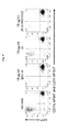
- FIGS. 3A-B show digestion analyses of antibodies comprising different IgG isotypes (A) and new constructs thereof as described in FIG. 2 (B) with the protease IdeS at different time points.
- FIG. 6A-C are graphs from separate ADCP assays performed with protease-resistant mAb constructs and wildtype IgG 1 and IgG 2 where the % Phagocytosis on the Y-axis is relative to the total number of sampled cells.
- FIG. 9 is a FACS analysis of a 24 hour ADCP assay depicting GFP-expressing MDA-MB-231 as a frequency of GFPpos, CD11bneg, CD14neg cells (upper left quadrant) incubated for 24 hours in the absence or presence of shown mAbs and macrophages to measure % cell tumor killing.
- FIG. 10 shows % tumor cell killing in a 24 hour ADCP assay performed with protease-resistant mAb constructs and wild type IgG1, IgG2, and IgG4 as indicated at various antibody concentrations. Error bars indicate standard deviation.
- FIG. 13 shows binding of protease-resistant mAb constructs to various Fc ⁇ R receptors as IC 50 values and fold changes of IC 50 values when compared to the wt IgG1.
- FIG. 14 shows a summary of ADCC, 24 hour ADCP, macrophage IL-10 secretion, PBMC IFN ⁇ release, and Fc ⁇ R-binding.
- ADCC antibody-dependent cellular cytotoxicity
- ADCP antibody-dependent cellular phagocytosis
- CDC complement-dependent cytotoxicity
- FDCR Fc-dependent cytokine release
- Fc ⁇ R or FcgammaR Fc gamma receptor
- GluV8 glutamyl endopeptidase V8 of Staphylococcus aureus
- IdeS Immunoglobulin degrading enzyme of Streptococcus pyogenes
- IgG immunoglobulin G
- ITAM immunoreceptor tyrosine-based activating motif
- ITIM immunoreceptor tyrosine-based inhibitory motif
- Mab monoclonal antibody
- MMP matrix metalloproteinase; the term protease is equivalent to proteinase and are used interchangeably
- PR protease resistant.
- Antibody-dependent cellular cytotoxicity refers to a form of cytotoxicity in which secreted Ig bound onto Fc receptors (FcRs) present on certain cytotoxic cells (e.g., Natural Killer (NK) cells, neutrophils, and macrophages) enables these cytotoxic effector cells to bind specifically to an antigen-bearing target cell and subsequently kill the target cell with cytotoxins.
- FcRs Fc receptors
- NK Natural Killer
- Ligand specific high-affinity IgG antibodies directed to the surface of target cells stimulate the cytotoxic cells and are absolutely required for such killing. Lysis of the target cell is extracellular, requires direct cell-to-cell contact, and does not involve complement.
- ADCC ability of any particular antibody to mediate lysis of the target cell by ADCC can be assayed.
- an antibody of interest is added to target cells displaying the target ligand in combination with immune effector cells, which may be activated by the antigen antibody complexes resulting in cytolysis of the target cell. Cytolysis is generally detected by the release of label (e.g. radioactive substrates, fluorescent dyes or natural intracellular proteins) from the lysed cells.
- label e.g. radioactive substrates, fluorescent dyes or natural intracellular proteins
- useful effector cells for such assays include peripheral blood mononuclear cells (PBMC) and Natural Killer (NK) cells.
- ADCC activity of the antibody of interest may be assessed in vivo, e.g., in an animal model such as that disclosed in Clynes et al., 1998, PNAS USA 95:652, the contents of which is incorporated by reference in its entirety.
- ADCP Antibody Dependent Cellular Phagocytosis
- “Complement-directed cytotoxicity” or CDC refers to the form of cytotoxicity in which the complement cascade is activated by the complement component Clq binding to antibody Fc.
- effector functions include those interactions of Fc with Fc gamma receptors (Fc ⁇ R) expressed on immune cells, as well as Fc domain interactions with complement leading to elimination of the antigen-expressing cell by lytic processes or phagocytosis by effector cells and complement components.
- Fc ⁇ R Fc gamma receptors
- Fc Fc-containing protein or Fc-containing molecule
- Fc-containing molecule refers to a monomeric, dimeric or heterodimeric protein having at least an immunoglobulin CH2 and CH3 domain.
- the CH2 and CH3 domains can form at least a part of the dimeric region of the protein/molecule (e.g., antibody).
- antibody as used herein is a specific form of an Fc-containing protein comprising at least one ligand binding domain which contains, or retains substantial homology to, at least one of a heavy or light chain antibody variable domain of at least one species of animal antibody.
- Wild type human IgG subclass constant sequences are cataloged in the UniProt database available on-line as P01857 (IgG1), P01859 (IgG2), P01860 (IgG3), and P01861 (IgG4).
- wild type human IgG1 Fc region refers to a human IgG Fc region that comprises the amino acid sequence of SEQ ID NO: 1 or a fragment thereof, which is from residue K214 to residue K447 of the human IgG heavy chain, according to the EU numbering of Kabat Amino acids in the constant region are numbered by alignment with the human IgG1 antibody, EU (see Cunningham et al., 1970 Biochemistry 9:3161-70).
- the heavy and light chains of an antibody are aligned with the heavy and light chains of EU to maximize amino acid sequence identity and each amino acid in the antibody is assigned the same number as the corresponding amino acid in EU.
- the EU numbering system is conventionally used in the art (see generally, Kabat et al, Sequences of Protein of Immunological Interest, NIH Publication No. 91-3242, US Department of Health and Human Services (1991)). According to the convention, the alignment between the wild type IgG2 constant region and that of EU results in an empty amino acid at positions 221-223 and 236 ( FIG. 2 , SEQ ID NO: 2).
- the constant domain sequences of the mammalian IgG heavy chain are designated in sequence as CH1-hinge-CH2-CH3.
- the “hinge”, “hinge region” or “hinge domain” of an IgG is generally defined as including Glu216 and terminating at Pro230 of human IgG1 according to the Kabat system but functionally, the flexible portion of the chain may be considered to include additional residues termed the upper and lower hinge regions, such as from Glu216 to Gly237 (Roux et al., 1998 J Immunol 161:4083) and the lower hinge has been referred to as residues 233 to 239 of the Fc region where FcgammaR binding was generally attributed.
- Hinge regions of other IgG isotypes may be aligned with the IgG1 sequence by placing the first and last cysteine residues forming inter-heavy chain S—S binds.
- the CH1 domain is adjacent to the VH domain and amino terminal to the hinge region of an immunoglobulin heavy chain molecule and includes the first (most amino terminal) constant region domain of an immunoglobulin heavy chain, e.g., from about EU positions 118-215.
- the Fc domain extends from amino acid 231 to amino acid 447; the CH2 domain is from about Ala231 to Lys340 or Gly341 and the CH3 from about Gly341 or Gln342 to Lys447.
- the residues of the IgG heavy chain constant region of the CH1 region terminate at Lys.
- proteolytic cleavage refers to the ability of a molecule comprised of peptide bonds, to resist hydrolytic cleavage of one or more of its peptide bonds in the presence of a proteolytic enzyme.
- the resistance to proteolytic enzymes is a relative property and is compared to a molecule which is less able to withstand hydrolytic cleavage of one or more of its peptide bonds over a specified time period and under specified conditions, including the pH and or temperature at which the cleavage resistance is tested.
- proteolytic cleavage indicative that cleavage has occurred is the generation of smaller fragments (lower molecular weight) as compared to the molecular weight of the intact, non-cleaved parent molecule.
- a modified Fc-containing molecule or a fragment thereof comprising a hinge, a CH2 domain and a CH3 domain is “protease resistant” or “resistant to proteolysis” or has “increased resistance to proteolysis” when more than about 60%, 65%, 70%, 75%, 80%, 85%, 90%, 95%, 96%, 97%, 98%, 99% or 100% of a full length immunoglobulin that incorporates a modified Fc of the invention remains intact for 24 hours when digested by matrix metalloprotease-3 (MMP-3), matrix metalloprotease-12 (MMP-12), glutamyl endopeptidase V8 of Staphylococcus aureus (GluV8), or immunoglobulin degrading enzyme of Streptococcus pyogenes (IdeS) in Tris-buffered saline at 37° C.
- MMP-3 matrix metalloprotease-3
- MMP-12 matrix metalloprotease-12
- GluV8 glutamyl endopeptidas
- IgG immunoglobulin G
- Modified refers to a molecule that comprises an Fc polypeptide sequence or a polynucleotide sequence encoding the Fc that differs from a human wild type IgG1 Fc sequence by one or more modifications, for example substitutions, insertions or deletions of nucleotides or amino acids.
- IC 50 fold change ratio value refers to the ratio of an IC 50 value for a wild type human IgG1 to an IC 50 value for the isolated modified Fc-domain containing molecule of the invention for Fc ⁇ R measured in a competition assay with biotinylated human IgG1 to 0.2 ⁇ g/ml soluble human Fc ⁇ RI. Competition can be measured using for example well known AlphaScreen® assays. AlphaScreen® is a registered trademark of PerkinElmer, Inc. or its subsidiaries.
- terapéuticaally effective amount refers to an amount of a therapeutic agent as described herein comprising an Fc-domain which may be an antibody, antibody fragment, or derivative to treat a disease or disorder in a subject.
- Proteases are divided into five major groups according to the structure of catalytic site and the amino acid (as one of the constituents) essential for its activity: serine proteinases, threonine proteinases, cysteine (thiol) proteinases, aspartic proteinases, and metalloproteinases.
- proteases function throughout the body and in body compartments performing critical regulatory and metabolic processes.
- Acid-resistant proteases secreted into the stomach such as pepsin
- serine proteases present in duodenum such as trypsin and chymotrypsin
- proteases present in blood or serum thrombin, plasmin, Hageman factor, etc.
- Proteases are present in or released from leukocytes (elastase, cathepsin G). Proteases determine the lifetime of other proteins thus playing an important metabolic role.
- proteolytic control one of the fastest regulatory switching mechanisms. Further, cooperative action of the proteases as in cascade reactions, results in a rapid and efficient amplification of an organism's response to a physiological signal.
- Human IgG isotypes (the subclasses of mature gamma globulin class G antibodies; IgG1, IgG2, IgG3 and IgG4) exhibit differential capacity to recruit immune functions. For example, antibody-dependent cellular cytotoxicity (ADCC) is promoted by IgG1 and IgG3, antibody-dependent cellular phagocytosis (ADCP) is promoted by IgG1, IgG2, IgG3 and IgG4, and complement dependent cytotoxicity (CDC) is promoted by IgG1 and IgG3.
- ADCC antibody-dependent cellular cytotoxicity
- ADCP antibody-dependent cellular phagocytosis
- CDC complement dependent cytotoxicity
- Isotype-specific engagement of such immune functions is based on selectivity for Fc receptors on distinct immune cells and the ability to bind Clq thereby activating the assembly of a membrane attack complex (MAC).
- MAC membrane attack complex
- Fc ⁇ receptors which include Fc ⁇ RI, Fc ⁇ RIIa/b/c, and Fc ⁇ RIIIa/b; is high for IgG1 and IgG3.
- Fc ⁇ affinity for IgG2 is considerably lower with the exception of Fc ⁇ RIIa H131 polymorphism and IgG4 only has measurable affinity for Fc ⁇ RI.
- the key contact residues for receptor binding have been mapped to the amino acid residues spanning the lower hinge and CH2 region ((Hezereh et al., 2001 J Virol 75:12161-8; Shields et al., 2001 J Biol Chem 276:6591-604).
- the present invention provides modified Fc-domain containing molecules having improved characteristics suitable to be used as and/or incorporated into anti-cancer therapeutics or therapeutics that can be used to treat conditions which require Th1 responses or cell-mediated immunity such as viral infections (e.g. Variola, Varicella zoster, Epstein-Barr virus, Influenza virus, mumps virus, measles virus, and human immunodeficiency virus), bacterial infections (e.g.
- viral infections e.g. Variola, Varicella zoster, Epstein-Barr virus, Influenza virus, mumps virus, measles virus, and human immunodeficiency virus
- bacterial infections e.g.
- Mycobacterium tuberculosis Mycobacterium leprae, Legionella pneumophila, Listeria monocytogenes, Brucella spp, Salmonella spp, Shigella spp, Coxiella burnetii, Anaplasma phagocytophilum, Ehrlichia chaffeensis , and Staphylococcus aureus ), and protozoa infections (e.g. Plasmodium spp., Toxoplasma gondii , and Leishmania spp.).
- the invention is based, at least in part, on the unexpected finding that Fc effector functions ADCC, ADCP and/or CDC in effector silenced IgG1 Fc mutant (E233P-L234V-L235A with G236 deleted; EU numbering) can be restored by mutations at the CH2 region with concomitant increased resistance of the modified Fc-domain containing molecule to proteolysis, while the Fc effector function of inducing macrophage IL-10 secretion is not restored.
- the present invention provides polynucleotides encoding the modified Fc-domain containing molecules of the invention and complementary nucleic acids thereof, vectors, host cells, and methods of using the modified Fc-domain containing molecules.
- the present invention provides compositions for use as a medicament for treating a spectrum of diseases.
- IgG1 molecules are known to be more susceptible to proteolysis than IgG2, due to the presence of multiple protease cleavage sites at the upper and lower hinge region in IgG1 ( FIG. 1 ). Therefore, it can be expected that the IgG1 molecule with IgG1 lower hinge residues E233, L234, L235 and G236 mutated to corresponding IgG2 lower hinge residues P233, V234, A235 (with IgG1 G236 deleted in IgG2), EU numbering, demonstrates increased protease resistance when compared to the wild type IgG1. However, the abolished effector function of this Fc domain may make it unsuitable to be coupled to antibody therapeutics where cell destruction and/or killing mediated by Fc effector functions are desired.
- the present invention provides modified Fc-containing molecules or fragments thereof comprising a wild type human IgG1 Fc region of SEQ ID NO: 1 comprising a hinge, a CH2 domain and a CH3 domain, wherein the sequence of E233-L234-L235-G236 in the hinge is replaced with P233-V234-A235 with G236 deleted, and further having additional mutations in the CH2 region (EU numbering).
- the Fc-domain containing molecules of the invention demonstrate increased resistance to proteolysis when compared to the wild type (wt) IgG1, furthermore several modified Fc-containing molecules unexpectedly demonstrated increased resistance to proteolysis when compared to the wt IgG2 molecule.
- the present invention demonstrates substitutions in multiple positions of the IgG1 constant regions (Fc) which unexpectedly provide a protease-resistant and functional (FcgR-engaging) Fc domain, except for Fc ⁇ RI binding.
- Fc IgG1 constant regions
- the protease-resistance conferred upon the compositions of the invention include protease resistance towards at least one of the proteases that cleave at residues of IgG1 hinge region that are substituted by alternate amino acids derived from the corresponding IgG2 residue, or proteases whose ability to cleave at IgG1 hinge region is affected by the substitutions.
- Exemplary proteases are matrix metalloproteases MMP-3, MMP-7, MMP-12, human neutrophile elastase (HNE), plasmin, cathepsin G, pepsin, IdeS, or glutamyl endopeptidase I from Staph aureus ( FIG. 1 ) (Ryan et al. 2008 supra).
- the compositions of the invention may be resistant to proteolysis by additional proteases. Protease resistant to additional proteases can be assessed using the methods described herein.
- Mutations to human wild type (wt) IgG1 (SEQ ID NO: 1) resulting in protease-resistant IgG1 molecules include replacing the IgG1 hinge region residues 214-237 in the EU numbering system (SEQ ID NO: 3) with the IgG2 wt hinge sequence (SEQ ID NO: 4), or replacing the IgG1 hinge region residues 214-237 in the EU numbering system (SEQ ID NO: 3) with a chimeric hinge (SEQ ID NO: 5) with IgG1 residues 233-235 replaced with corresponding IgG2 hinge residues as shown in Table 1.
- IgG Scaffold (EU Hinge/Proximal CH2 Fc Designation 214-447) (EU 214-236) IgG1 wt (UniProt hIgG1 KVEPKSCDKTHTCPPCPAPELLG P01857, 96-329) (SEQ ID NO: 3) IgG2 wt (UniProt hIgG2 TVERKCCVECPPCPAPPVA P01859, 97-326) (SEQ ID NO: 4) 2hc hIgG1 with SEQ ID NO: 4 complete IgG2 hinge region 2h hIgG1 KVEPKSCDKTHTCPPCPAPPVA (SEQ ID NO: 5)
- Compensating mutations in effector function enhancing regions can be selected from previously described substitutions as shown in Table 2 below.
- the multi-substituted IgG1 mutants were selected based on their relative affinities for human FcRs (Fc ⁇ RI, Fc ⁇ RIIa, Fc ⁇ RIIb, Fc ⁇ RIIIa assessed by AlphaScreen® competition assays). These mutants were further tested in the appropriate cellular systems for their ability to induce ADCC by PBMCs and ADCP by in vitro differentiated macrophages.
- the sites for substitution were chosen based on the desire to produce a composition having the structural features of a native antibody Fc, maintain stability, retain FcR binding and the capacity to stimulate the complement cascade, cell lysis, cell phagocytosis or cytokine release.
- Proteins, with altered or mutated amino acids can be created by routine molecular biology techniques such as site directed mutagenesis. Where a chimeric sequence is created, such as the Fc of the invention comprising portions of IgG1 and portions of IgG2, larger segments of the respective encoding nucleic acids may be spliced together or segments replaced by standard cloning techniques.
- the rate or extent to which a proteolytic enzyme degrades the different isolated Fc-containing compositions or antibodies is assessed. After a time period, the degradation is measured for the different compositions using a method capable of determining either scission of the chain directly, such as the formation of a unique cleavage site structure, or a measurement of newly formed fragments. Alternatively, where cleavage results in loss of activity, a functional assay can be performed, including a binding assay.
- Proteolytic cleavage of an IgG1 can occur at any of the four polypeptide chains of the dimeric heterodimeric structure. Cleavage of IgG results in the generation of well characterized fragments such as Fab, (Fab′) 2 , and Fc fragments of approximate but unique molecular weights.
- the separation of such fragments generated during a proteolysis experiment can be accomplished using a size exclusion chromatography (SEC), by gel electrophoresis, by MALDI-TOF-MS (matrix-assisted laser/desorption ionization time-of-flight mass spectrometry) analyses after deglycosylation using PNGase F (peptide N-glycosidase F) as previously described (WO2007024743A2, WO2009045894A1).
- SEC size exclusion chromatography
- MALDI-TOF-MS matrix-assisted laser/desorption ionization time-of-flight mass spectrometry
- an Fc-containing composition resistant to proteolytic cleavage is that the composition is less likely to be degraded, lose activity, lose affinity for an Fc-binding partner such as an FcR upon exposure to a proteolytic enzyme than a comparator molecule, such as a wild-type human IgG1.
- Fc-containing proteins can be compared for functionality by several well-known in vitro assays. Affinity for members of the Fc ⁇ RI, Fc ⁇ RII, and Fc ⁇ RIII family of Fey receptors can be made using recombinant soluble forms or cell-associated forms of the receptors. In addition, affinity for FcRn, the receptor responsible for the prolonged circulating half-life of IgGs, can be measured, for example using a ligand bound bead format such as “ALPHASCREEN” with recombinant soluble FcRn. AlphaScreen®, used in high throughput screening, is a homogenous assay technology which allows detection of molecular events such as binding.
- Coated “Donor” and “Acceptor” beads are the basis of the AlphaScreen® assay technology.
- AlphaScreen® a bead based assay, works through the interaction of the beads coming close to each other, resulting in a cascade of chemical reactions that act to produce a greatly amplified signal.
- Direct or indirect, e.g. competitive binding, measurements can be applied for assessing relative affinities and avidities among proteins.
- Each human IgG isotype e.g. IgG1, IgG2, IgG3 and IgG4 recruit immune functions, such as antibody-dependent cellular cytotoxicity (ADCC, e.g. IgG1 and IgG3), antibody-dependent cellular phagocytosis (ADCP, e.g. IgG1, IgG2, IgG3 and IgG4), and complement dependent cytotoxicity (CDC, e.g.
- ADCC antibody-dependent cellular cytotoxicity
- ADCP antibody-dependent cellular phagocytosis
- CDC complement dependent cytotoxicity
- IgG1, IgG3) based on their differential selectivity for Fc receptors which reside on distinct immune cells, and the ability to bind Clq and activate the assembly of a membrane attack complex (MAC) resulting in CDC and CDP (complement dependent phagocytosis) through specific receptors binding complement components on effector macrophages.
- MAC membrane attack complex
- CDP complement dependent phagocytosis
- the engineered Fc-containing molecules of the invention can be tested in cell-based functional assays, such as ADCC assays and CDC assays to assess functional consequences of particular substitutions.
- ADCC antibody-dependent cell-mediated cytotoxicity
- FcRs Fc receptors
- the ADCC assay is configured to have NK cells as the primary effector cell, reflecting the functional effects on the Fc ⁇ RIIIa which is the only activating Fc ⁇ -type receptor known to be expressed by these cells.
- Phagocytosis assays may also be used to compare immune effector functions of different mutants, as can assays that measure cellular responses, such as superoxide or inflammatory mediator release.
- In vivo models can be used, for example, in the case of using mutants of anti-CD3 antibodies to measure T cell activation in mice, an activity that is dependent on Fc domains engaging specific ligands, such as Fc ⁇ receptors.
- Antibody directed activation of macrophages mediates antibody-dependent cellular phagocytosis (ADCP), causing opsonized target cells to be engulfed and digested by macrophages.
- ADCP antibody-dependent cellular phagocytosis
- differentiated macrophages expressing high levels of FcRs can be differentiated into the M1 phenotype using INF ⁇ or GM-CSF to expressed elevated levels of all FcRs (Fc ⁇ RI, Fc ⁇ RIIa, Fc ⁇ RIIIa) relative to monocytes.
- ADCR antibody-dependent cytokine release
- a tumor cell line MDA-MB-231 as a target cell and a monocyte-derived macrophage or PBMC as an effector cell
- cytokine release into the cell culture supernatant upon administering antibodies having the modified Fc-domains of the invention and variable regions binding to a surface molecule on MDA-MB-231 cell, such as tissue factor using routine methods and as exemplified herein.
- Exemplary cytokines that can be measured are IL-10 and IFN ⁇ .
- IL-10 and IFN ⁇ can be measured using standard methods such as ELISA.
- compositions of the invention can be produced by engineered host cells using standard methods.
- the host cell chosen for expression of the recombinant Fc-containing protein or monoclonal antibody can contribute to the variation in for example oligosaccharide moieties in the immunoglobulin CH2 domain.
- one aspect of the invention involves the selection of appropriate host cells comprising polynucleotide sequences encoding the Fc-containing molecules of the invention for use as or development of a production cell expressing the desired therapeutic protein.
- the host cell may be of mammalian origin or may be selected from COS-1, COS-7, HEK293, BHK21, CHO, BSC-1, Hep G2, 653, SP2/0, HeLa, myeloma, lymphoma, yeast, insect or plant cells, or any derivative, immortalized or transformed cell thereof.
- the host cell may be selected from a species or organism incapable of glycosylating polypeptides, e.g. a prokaryotic cell or organism, such as a natural or engineered E. coli spp, Klebsiella spp., or Pseudomonas spp., engineered plant or insect cells.
- a prokaryotic cell or organism such as a natural or engineered E. coli spp, Klebsiella spp., or Pseudomonas spp., engineered plant or insect cells.
- Glycosylation at the naturally occurring glycosylation site within the IgG heavy chain also contributes to the Fc-binding affinity for Fc ⁇ R.
- N297, EU numbering also contributes to the Fc-binding affinity for Fc ⁇ R.
- each isotype possesses a distinct array of N-linked carbohydrate structures, which variably affect protein assembly, secretion or functional activity (Wright and Morrison 1997 Trends Biotech 15:26-32).
- the structure of the attached N-linked carbohydrate varies considerably, depending on the degree of processing, and can include high-mannose, multiply-branched as well as biantennary complex oligosaccharides and sialic acid (N-acetyl neuraminic acid or NANA), fucose, galactose and GlcNAc (N-acetyl glucosamine) residues as terminal sugars.
- NANA N-acetyl neuraminic acid
- fucose fucose
- galactose galactose
- GlcNAc N-acetyl glucosamine residues as terminal sugars.
- the impact on effector functions of the host cell and oligosaccharide content of the antibodies has been recognized (Lifely et al., 1995 Glycobiology 5:813-22; Jefferis et al., 1998 Immunol Rev 163:59-76; Wright and Morrison, supra; Presta 2003.
- a protease-resistant mAb of the invention using a host cell capable of, engineered to (as by knock-out or knock down of specific enzymes Shinkawa et al., 2003 J Biol Chem 278:3466-73; EP1176195A1) or induced, e.g. by environmental or nutritional manipulation, to produce a mAb having low fucose content is within the scope of the present invention.
- an antibody binding domain or fragments thereof can be produced using methods known to those skilled in the art and combined with the information provided herein and can include sequences or be derived from any mammal, for example human, mouse, rabbit, rat, rodent, primate, goat, or any combination thereof.
- Such antibodies may provide the basic structures and components of binding domains useful in producing the antibody constructs of the present invention.
- the antibody binding domains are obtained from hybridomas prepared by immunizing a mouse or other animal with the target peptides, cells or tissues extracts.
- the antibodies can be obtained using any of the hybridoma techniques well known in the art, see, e.g., Harlow and Lane, antibodies, a Laboratory Manual, Cold Spring Harbor, N.Y. (1989) entirely incorporated herein by reference or by selection of an antibody producing lymphocyte and cloning the nucleic acid sequences coding for the binding domains using techniques known in the art.
- the present invention is directed to the constant region of a human IgG. Therefore, any antibody or fusion protein comprising a human Fc-domain wherein it is desirable that the final composition display both proteolytic resistance (as described herein) and the ability to bind FcgR and promote ADCC, ADCP, and/or CDC in an in vitro assay is encompassed by the present invention.
- a targeting moiety including an antibody Fv or single variable domain may be fused to the Fc-composition as desired.
- An“Fv” consists of a dimer of one heavy- and one light-chain variable region domain in tight, non-covalent association.
- Single-chain Fv also abbreviated as “sFv” or “scFv” are antibody fragments that comprise the VH and VL antibody domains connected into a single polypeptide chain.
- the scFv polypeptide generally comprises a polypeptide linker between the VH and VL.
- the targeting moiety may also be selected from a paratope of an antibody (binding residues not limited to CDRs or variable domain structures); an enzyme; a hormone; a receptor; a cytokine; an immune cell surface antigen; and an adhesion molecule when the construct is produced entirely by recombinant methods.
- the targeting moiety may also be of non-proteinaceous nature such as a carbohydrate, a lipid, a lipopolysaccharide, an organic molecule, or a metal or metal complex. Generally, when present, the targeting molecule will be connected to the Fc- by a linker which may be a polypeptide or nonpolypeptide.
- Fc-containing proteins or Fc fragments described herein can be derived in several ways well known in the art.
- the antibody binding domains or Fc-fusion proteins or components and domains thereof may also be obtained from selecting from libraries of such domains or components, e.g., a phage library.
- a phage library can be created by inserting a library of random oligonucleotides or a library of polynucleotides containing sequences of interest, such as from the B-cells of an immunized animal or human (Smith, G. P. 1985. Science 228: 1315-1317).
- Antibody phage libraries contain heavy (H) and light (L) chain variable region pairs in one phage allowing the expression of single-chain Fv fragments or Fab fragments (Hoogenboom, et al. 2000, Immunol. Today 21(8) 371-8).
- the diversity of a phagemid library can be manipulated to increase and/or alter the immunospecificities of the monoclonal antibodies of the library to produce and subsequently identify additional, desirable, human monoclonal antibodies.
- the heavy (H) chain and light (L) chain immunoglobulin molecule encoding genes can be randomly mixed (shuffled) to create new HL pairs in an assembled immunoglobulin molecule.
- H and L chain encoding genes can be mutagenized in a complementarity determining region (CDR) of the variable region of the immunoglobulin polypeptide, and subsequently screened for desirable affinity and neutralization capabilities.
- CDR complementarity determining region
- Antibody or Fc libraries also can be created synthetically by selecting one or more human framework sequences and introducing collections of CDR cassettes derived from human antibody repertoires or through designed variation (Kretzschmar and von Ruden 2000, Current Opinion in Biotechnology 13:598-602). The positions of diversity are not limited to CDRs, but can also include the framework segments of the variable regions or may include other than antibody variable regions, such as peptides.
- a phage library capable of displaying dimeric Fc structures linked to the phage coat protein pIX as described in applicants co-pending application (U.S. Ser. No. 61/261,767) may be used to select novel Fc-comprising structures according to the present invention.
- Target binding components which may include target binding components other than antibody variable regions, may include ribosome display libraries, yeast display libraries, and bacterial display libraries.
- Ribosome display is a method of translating mRNAs into their cognate proteins while keeping the protein attached to the RNA. The nucleic acid coding sequence is recovered by RT-PCR (Mattheakis et al., 1994 Proc. Natl. Acad. Sci. USA 91:9022).
- Yeast display is based on the construction of fusion proteins of the membrane-associated alpha-agglutinin yeast adhesion receptor, aga1 and aga2, a part of the mating type system (Broder et al., 1997 Nature Biotechnology 15:553-7).
- Bacterial display is based on fusion of the target to exported bacterial proteins that associate with the cell membrane or cell wall (Chen and Georgiou 2002 Biotechnol Bioeng 79:496-503).
- the invention also provides for nucleic acids encoding the compositions of the invention as isolated polynucleotides or as portions of expression vectors including vectors compatible with prokaryotic, eukaryotic or filamentous phage expression, secretion and/or display of the compositions or directed mutagens thereof.
- Certain exemplary polynucleotides are disclosed herein, however, other polynucleotides which, given the degeneracy of the genetic code or codon preferences in a given expression system, encode modified Fc-containing molecules of the invention are also within the scope of the invention.
- the polynucleotides of the invention may be produced by chemical synthesis, such as solid phase polynucleotide synthesis on an automated polynucleotide synthesizer and assembled into complete single or double stranded molecules.
- the polynucleotides of the invention may be produced by other techniques, such as a PCR followed by routine cloning. Techniques for producing or obtaining polynucleotides of a given known sequence are well known in the art.
- compositions generated by any of the above described methods may be used to diagnose, treat, detect, or modulate human disease or specific pathologies in cells, tissues, organs, fluid, or, generally, a host.
- modification of the Fc portion of an antibody, Fc-fusion protein, or Fc fragment to reduce or ablate proteolytic degradation while retaining measurable Fc gamma receptor binding or specified effector functions can be combined with a binding domain, such as the paratope of an antibody or a ligand binding domain, which retains the original targeting specificity and biological activity.
- Exemplary binding domains are a paratope of an antibody, one or more antibody CDRs, or one or more antibody variable domains; an enzyme; a hormone; a receptor; an extracellular domain of a membrane receptor; a cytokine; an immune cell surface antigen; and an adhesion molecule.
- the resulting constructs provide for antibodies and Fc-constructs with a superior spectrum of activities, biophysical properties, stability and ability to persist in the body of a host.
- Fc sequences with unique combinations of resistance to physiologically-relevant proteases and ability or nonability to engage one or more Fcgamma receptors and/or the ability to affect cell lysis by activation of effector cells or complement provide for the ability to purpose the binding molecule for maximal effectiveness in a specified indication.
- the ability to target aberrant host cells such as those involved in neoplasia or other unwanted proliferation such as in inappropriate angiogenesis, inappropriate fibrosis would be best suited by a molecule comprising a eukaryotic protease-resistant Fc of the present invention capable of ADCC and ADCP.
- bacterial cells are readily destroyed by complement-mediated mechanisms. Therefore, bacterial infections may be treated by a suitable Fc-construct of the invention which is resistant to bacterial proteases and has the ability to invoke CDC.
- Molecules which eukaryotic protease-resistant and capable of one or more of ADCC, ADCP, and CDC include an Fc domain having the sequence of a human IgG1 in the hinge and CH2 regions, from about EU residues 214 to about residue 330 where at least residues 233-237 are substituted with PVA/(G236 deletion) and further comprising one or more substitutions in the CH2 domain whereby the molecule is capable of one or more of ADCC, ADCP, and CDC include the constructs 5, 7, 8, 9, 10, 11, 12, 14, 15, 16, and 17.
- such molecules include substitutions selected from 1332E in combination with other substitutions such as S239D/1332E (5, 14), S239D/H268F/1332E (not made), H268F/S324T/1332E (8), S239D/H268F/S324T/1332E (9), S267E/H268F/S324T/1332E (10), G237X/S239D/1332E where X is A or S (12); K326A/1332E/E333A (15), and S267E/I332E (17).
- Molecules which are resistant to a prokaryotic protease and capable of CDC include those molecules comprising an Fc domain having the sequence of a human IgG1 in the hinge and CH2 regions, from about EU residues 214 to about residue 330 where at least residues 233-237 are substituted with PVA/(G236 deletion) and further comprising one or more substitutions in the CH2 domain selected from K326A/E333A (11), S267E/H268F/S324T/1332E (10), K326A/1332E/E333A (15), S239D/K326A/E333A (16), and S267E/I332E (17).
- Molecules which are eukaryotic protease resistant but do not promote target cell lysis may also be advantageously used to treat a disease or conditions wherein target cell modulation and not target cell destruction is the objective such as by using an antibody or other Fc-construct as taught herein that is protease-resistant but has decreased ADCC, ADCP, or CDC as compared to wildtype IgG1.
- Molecules comprising a non-natural, non-wild type, Fc domain which are protease-resistant include those molecules comprising an Fc domain having the sequence of a human IgG1 in the hinge and CH2 regions, from about EU residues 214 to about residue 330 where at least residues 233-237 are substituted with PVA/(G236 deletion).
- the presently enabled Fc-comprising constructs demonstrating enhanced resistance to a protease occurring in the mammalian subject and, optionally, having the ability to target an antigen on a cell provide improved therapeutic molecules relative to the therapeutic molecule which is not protease resistant.
- the present invention provides modified Fc-containing molecules or fragments thereof comprising a wild type human IgG1 Fc region of SEQ ID NO: 1 comprising a hinge, a CH2 domain and a CH3 domain, wherein the sequence of E233-L234-L235-G236 in the hinge is replaced with P233-V234-A235 with G236 deleted, and having additional mutations in the CH2 region (EU numbering).
- the modified Fc-containing molecules have restored Fc ⁇ RIIa, Fc ⁇ RIIb and/or Fc ⁇ RIIIa binding and comparable and/or enhanced ADCC, ADCP and/or CDC when compared to the wild type IgG1, however, they do not bind Fc ⁇ RI.
- Fc ⁇ RI Lack of binding to Fc ⁇ RI correlates with the inability of the Fc-containing molecules of the invention to induce macrophage IL-10 secretion. Many of the variants further induce IFN ⁇ secretion from PBMCs, presumably from NK cells. IFN ⁇ stimulates the tumor-killing functions of macrophages, enhances cross-presentation, increases expression of co-stimulatory molecules and MHC I and II, and drives Th1 immune responses, thereby promoting the pro-inflammatory milieu beneficial for anti-tumor therapeutics.
- Regulatory macrophages are known to actively contribute to tumor growth via angiogenesis and immunosuppression (Biswas and Mantovani 2010 Nat. Immunol. 11:889-96). Activation of regulatory macrophages characterized by secretion of anti-inflammatory and proangiogenic mediators IL-10 and VEGF in a tumor microenvironment has been suggested to be causative for lack of efficacy of anti-EGFR monoclonal antibody cetuximab in combination with anti-VEGF mAb bevacizumab. It was suggested that activation of the regulatory macrophages occur via engagement with the high-affinity Fc ⁇ RIIIA (Pander et al., 2011 Clin Cancer Res 17:5668-73). In contrast, U.S. Pat. No. 6,660,266 describes Fc ⁇ RI ligation causative for induced secretion of IL-10 by macrophages.
- These molecules have beneficial characteristics to be used as and/or incorporated into therapeutic molecules, i.e. protease resistance and strong cellular cytotoxicity effects without potentially converting pro-inflammatory macrophages into IL-10 secreting regulatory macrophages.
- These Fc-containing molecules can be used to treat any disease associated with unwanted proliferation of cells as exemplified infra.
- Modified Fc-containing molecules or fragments thereof comprising a wild type human IgG1 Fc region of SEQ ID NO: 1 comprising a hinge, a CH2 domain and a CH3 domain, wherein the sequence of E233-L234-L235-G236 in the hinge is replaced with P233-V234-A235 with G236 deleted, and having additional mutations in the CH2 region (EU numbering) that are resistant to proteases, have strong CDC capacity but do not induce IL-10 secretion from macrophages include constructs, 10, 11, 15, 16 and 17 having CH2 mutations S267E/H268F/S324T/1332E (10), K326A/E333A (11), K326A/1332E/E333A (15), S239D/K326A/E333A (16) and S267E/I332E (17).
- Fc-containing molecules can be used to treat for example microbial infections.
- Intrinsic antibody-dependent enhancement (ADE) ligation of macrophage Fc ⁇ receptors by IgG immune complexes resulting in increased infections output by infected cells has been attributed at least part to increased immunosuppressive action of IL-10 produced by infected macrophages upon Fc ⁇ R ligation (Halstend et al., Lancet Infect Dis 10:712-22, 2010). Therefore, the Fc-containing molecules of the invention may be particularly useful in treatment of infections that are/may be associated with intrinsic ADE.
- Exemplary infections that may be treated with the modified protease resistant Fc-containing molecules of the invention with strong CDC and lack of IL-10 secretion are infections caused by intracellular parasites and bacteria, such as Mycobacterium tuberculosis, Mycobacterium leprae, Legionella pneumophila, Listeria monocytogenes, Brucella spp, Salmonella spp, Shigella spp, Coxiella burnetii, Anaplasma phagocytophilum, Ehrlichia chaffeensis , protozoans, Leishmania spp, Toxoplasma gondii , and fungi (eg, Histoplasma capsulatum ), viral infections for those viruses that replicate in macrophages in vivo, such as West Nile Virus, Ross River Virus (RRV) and dengue virus, and other conditions as exemplified supra.
- infections caused by intracellular parasites and bacteria such as Mycobacterium tuberculosis, Mycobacterium
- Fc-containing molecules of the invention may be useful in the treatment of methicillin-resistant Staphylococcus aureus (MSRA) infections.
- the Fc can be incorporated in an IgG1 antibody that comprises variable regions that recognize a surface antigen of the microbe.
- Exemplary surface antigens that can be used to target the modified Fc-containing molecule of the invention include Staphylococcus aureus iron regulated surface determinant B (IsdB), teichoic acid (TA), lipoteichoic acid (LTA), clumping factor A, clumping factor B, capsular polysaccharide (CP) types 5 (CP5) and 8 (CP8), autolysin, Protein A, sortase, poly-N-acetyl glucosamine (PNAG), peptidoglycan (PG), and other known cell wall components of Staph . Additional surface molecules that can be used are known to those skilled in the art.
- compositions of the invention are particularly suited for uses in which a body compartment is known to contain proteases or abnormally high protease content.
- the invention provides for stable formulations of a protease-resistant IgG composition such as an antibody, which is preferably an aqueous phosphate buffered saline or mixed salt solution, as well as preserved solutions and formulations as well as multi-use preserved formulations suitable for pharmaceutical or veterinary use, comprising at least one protease-resistant antibody in a pharmaceutically acceptable formulation.
- a protease-resistant IgG composition such as an antibody, which is preferably an aqueous phosphate buffered saline or mixed salt solution, as well as preserved solutions and formulations as well as multi-use preserved formulations suitable for pharmaceutical or veterinary use, comprising at least one protease-resistant antibody in a pharmaceutically acceptable formulation.
- Suitable vehicles and their formulation, inclusive of other human proteins, e.g., human serum albumin are described, for example, in e.g. Remington: The Science and Practice of Pharmacy, 21 st Edition, Troy, D. B. ed., Lipincott Williams and Wilkins,
- a protease-resistant IgG composition with effector function in stable or preserved formulations as described herein or known in the art can be administered to a patient in accordance with the present invention via a variety of delivery methods including intravenous (I.V.); intramuscular (I.M.); subcutaneously (S.C.); transdermal; pulmonary; transmucosal; using a formulation in an implant, osmotic pump, cartridge, micropump; or other means appreciated by the skilled artisan, as well-known in the art.
- I.V. intravenous
- I.M. intramuscular
- S.C. subcutaneously
- transdermal pulmonary
- transmucosal using a formulation in an implant, osmotic pump, cartridge, micropump; or other means appreciated by the skilled artisan, as well-known in the art.
- the administration may be intrarticular, intrabronchial, intraabdominal, intracapsular, intracartilaginous, intracavitary, intracelial, intracerebellar, intracerebroventricular, intracolic, intracervical, intragastric, intrahepatic, intramyocardial, intraosteal, intrapelvic, intrapericardiac, intraperitoneal, intrapleural, intraprostatic, intrapulmonary, intrarectal, intrarenal, intraretinal, intraspinal, intrasynovial, intrathoracic, intrauterine, intravesical, intralesional, vaginal, rectal, buccal, sublingual, intranasal, or by transdermal means.
- the diseases or pathologies that may be amenable to treatment using a composition provided by the invention include, but are not limited to, diseases in which unwanted proliferation, activation or migration of cells is deleterious such as malignancy, hyper-active or unbalanced immune responses, fibrotic tissue formation, or infection as the compositions provide for the activation of cytotoxic or cytolytic mechanisms of the host immune system through Fc ⁇ R-driven mechanisms.
- Such diseases include malignancies: leukemia, acute leukemia, acute lymphoblastic leukemia (ALL), B-cell, T-cell or FAB ALL, acute myeloid leukemia (AML), chronic myelocytic leukemia (CML), chronic lymphocytic leukemia (CLL), hairy cell leukemia, myelodyplastic syndrome (MDS), a lymphoproliferative disease, cutaneous T-cell lymphoma, Hodgkin's disease, Castleman's disease, glioma, glioblastoma, astracytoma, a malignant lymphoma, non-Hodgkin's lymphoma, Burkitt's lymphoma, multiple myeloma, Kaposi's sarcoma, colorectal carcinoma, pancreatic carcinoma, renal cell carcinoma, breast cancer, ductal carcinoma, lipoma, nasopharyngeal carcinoma, prostate cancer, testicular cancer, ovarian cancer, retinoblast
- Targeted molecules capable of binding antigens on malignant lymphocytes include B-cell antigens such as CD19, CD20, and CD22.
- Solid tumors derived from epidermal tissue often display and are stimulated by ligand binding to epidermal growth factor receptors known as the ErbB1, ErbB2, ErbB3 and other receptor capable of signaling or causing a proliferative response or an anti-apoptotic response leading to unchecked growth of the tumor.
- Other common antigens on solid tumors are tissue factor or RON.
- compositions when combined with an appropriate target binding domain, are also useful in treating an infectious disease caused by bacterial (such as Streptococcus, Staphylococcus , and E. coli .), viral (such as influenza, AIDS, RSV, SARS, and West Nile Virus), fungal (such as Aspergillosis, coccidiodomycosis, cryptococcosis, or Candidiasis), or protozoan infection (such as trypanosomiasis, toxoplasmosis, giardia , or malaria).
- infectious disease caused by bacterial (such as Streptococcus, Staphylococcus , and E. coli .), viral (such as influenza, AIDS, RSV, SARS, and West Nile Virus), fungal (such as Aspergillosis, coccidiodomycosis, cryptococcosis, or Candidiasis), or protozoan infection (such as trypanosomiasis, toxoplasm
- compositions are useful in treating general immunological and autoimmune disorders including but not limited to the rheumatic diseases, psoriasis, and scleroderma.
- compositions are useful in treating disorders associated with inappropriate angiogenesis.
- Angiogenesis is the process of generating new capillary blood vessels, and it results from activated proliferation of endothelial cells. Neovascularization is tightly regulated, and occurs only during embryonic development, tissue remodeling, wound healing and periodic cycle of corpus luteum development (Folkman and Cotran, 1976 Int. Rev. Exp. Pathol. 16, 207-48). Endothelial cells normally proliferate much more slowly than other types of cells in the body. However, if the proliferation rate of these cells becomes unregulated, pathological angiogenesis can result. Pathological angiogenesis is involved in many diseases.
- cardiovascular diseases such as angioma, angiofibroma, vascular deformity, atherosclerosis, synechia and edemic sclerosis
- opthalmological diseases such as neovascularization after cornea implantation, neovascular glaucoma, diabetic retinopathy, angiogenic corneal disease, macular degeneration, pterygium, retinal degeneration, retrolental fibroplasias, and granular conjunctivitis are related to angiogenesis.
- Chronic inflammatory diseases such as arthritis; dermatological diseases such as psoriasis, telangiectasis, pyogenic granuloma, seborrheic dermatitis, venous ulcers, acne, rosacea (acne rosacea or erythematosa), warts (verrucas), eczema, hemangiomas, lymphangiogenesis are also angiogenesis-dependent.
- Diabetic retinopathy can take one of two forms, non-proliferative or proliferative.
- Proliferative retinopathy is characterized by abnormal new vessel formation (neovascularization), which grows on the vitreous surface or extends into the vitreous cavity.
- Macular degeneration likewise takes two forms, dry and wet.
- wet form which is much less common, there is formation of a subretinal network of choroidal neovascularization often associated with intraretinal hemorrhage, subretinal fluid, pigment epithelial detachment, and hyperpigmentation.
- Neovascular glaucoma occurs in patients with diabetes or central retinal vein occlusion or inflammatory precipitates associated with uveitis pulling the iris up into the angle (Ch. 99. The Merck Manual 17th Ed. 1999).
- Rheumatoid arthritis an inflammatory disease, also results in inappropriate angiogenesis.
- the growth of vascular endothelial cells in the synovial cavity is activated by the inflammatory cytokines, and results in cartilage destruction and replacement with pannus in the articulation (Koch et al., 1986, Arth; 15 Rhenium, 29:471-79; Stupack et al., 1999 Braz. J. Med. Biol. Res., 32:578-81; Koch, 1998 Arthritis Rheum, 41:951-62).
- compositions are useful in treating psoriasis, which is caused by uncontrolled proliferation of skin cells. Fast growing cell requires sufficient blood supply, and abnormal angiogenesis is induced in psoriasis (Folkman, 1972 J. Invest. Dermatol. 59:40-8).
- vascular endothelial growth factor VEGF
- TNFalpha TNFalpha
- bFGF vascular endothelial growth factor
- cytokines including IL-6 and IL-12.
- alphaVbeta3 and aVb5 have been shown to mediate independent pathways in the angiogenic process.
- An antibody generated against alphaVbeta3 blocked basic fibroblast growth factor (bFGF) induced angiogenesis, whereas an antibody specific to aVb5 inhibited vascular endothelial growth factor (VEGF) induced angiogenesis (Eliceiri et al., 1999 J. Clin. Invest. 103:1227-30; Friedlander et al., 1995 Science 270:1500-2). Therefore, the invention encompasses the use of targeting binding domains directed to these targets in the compositions of the invention for use in treating diseases where the inhibition of angiogenesis is indicated.
- bFGF basic fibroblast growth factor
- VEGF vascular endothelial
- a series of constructs (shown in FIG. 2 and combining the hinge region of Table 1 with activity restoring mutations in the CH2 region selected from Table 2) were generated using standard recombinant technology.
- the designation 2hc denotes that the IgG1 constant domain corresponding to the Kabat numbering of the EU antibody (EU numbering) from 214 to 236 (SEQ ID NO: 3) have been replaced with the corresponding IgG2 sequence (SEQ ID NO: 4).
- the designation 2h denotes that the residues of IgG1 E233-L234-L235-G236 are replaced by the corresponding residues of IgG2 P233-V234-A235 with G235 deleted.
- variable regions of the one set of antibody constructs bind to CD142 (tissue factor) which allowed testing of Fc-dependent cell-killing of the antibodies in cellular assays using MDA-MB-231 (ATCC, HTB-26TM) expressing tissue factor (Brezski et al., Proc Natl Acad Sci USA 106:17864-17869 2009).
- An additional panel was generated with variable regions that bind to CD20 which allowed testing of CDC activity using WIL2-S cells displaying CD20 (ATCC, CRL-8885) (Brezski et al., Proc Natl Acad Sci USA 106:17864-17869 2009). All of the antibodies were expressed transiently in 293T cells using standard cloning methods and procedures. MAbs were purified using protein A columns to greater than 95% purity prior to experimental analysis.
- Protease digestions of purified IgGs were carried out at pH 7.5 in phosphate-buffered saline (PBS) or, for the MMPs (MMP-3, MMP-7, MMP-12 and MMP-13 were all obtained from Enzo Life Sciences), in Tris-buffered saline buffer at 37° C. IdeS was obtained from Genovis, and GluV8 was obtained from Pierce. CaCl 2 was included in the MMP reactions at 5 mM for all MMPs tested. Antibody concentrations were 0.5 mg/mL and reactions were initiated by addition of enzyme to approximately 1-2% (w/w) ratio to IgG unless otherwise specified. IgG cleavage was assessed by analysis of the electrophorograms after Agilent Biosizing microcapillary electrophoresis (Agilent Technologies). All digests were performed in duplicate.
- Biotinylated IgG1 (0.2 ⁇ g/ml final) and wild type and protease-resistant antibodies (10 ⁇ l) were sequentially added to each row of a 96-well plate in duplicates. Thereafter designated Fc ⁇ Rs were added followed by the sequential addition of 10 ⁇ A each of 1/50 diluted nickel chelate (Ni)-acceptor beads and streptavidin (SA)-donor beads.
- the opaque plate was covered with an aluminum seal to maintain light-safe conditions while shaking for 30 minutes on an Orbital shaker. Thereafter the seal was removed and the fluorescence was read on an ENVISIONTM plate reader (PerkinElmer) equipped with appropriate filter set of AlphaScreen® excitation/emission spectra.
- Raw data was transferred to GraphPad PRISMTM software and normalized for maximal signal and competition curves were plotted using non-linear regression curve-fitting software.
- Construct 4 (2h) and those constructs with the 2h lower hinge modification were resistant to all of the MMPs to more or less the same degree.
- Construct 4 was resistant to GluV8 but not IdeS.
- Construct 2 were also more resistant to GluV8 digestion.
- IdeS has been shown to cleave all human IgG isotypes (von Pawel-Rammingen et al., EMBO 21:1607-1615 2002).
- the data from a time course study ( FIG. 3A ) indicate that IdeS rapidly converted IgG1 (1) and IgG2 (2) to the F(ab′) 2 fragment (within 5 minutes of incubation) while the IgG1 sample 2h (4) had single-cleaved intermediate through 120 minutes.
- 2h S239D/1332E (5) was the most proteolytic resistant construct to IdeS ( FIG. 3B ), with intact IgG detected even after 24 hour incubation (the other antibody constructs had no detectable intact IgG by 5 minutes).
- the mutation of S239D in (5) which is near the IdeS cleavage point between G236 and G237, may contribute to the additional protease resistance as compared to IgG1 2h (4).
- a panel of 12 anti-CD142 antibody constructs (1-5, 7, 9-13, and 14) was tested along with their wildtype IgG1 and IgG2 counterparts for susceptibility to proteolysis by IdeS, GluV8, MMP-3, and MMP-12, and.
- the data for intact IgG remaining after 24 hour incubation was calculated from the electrophorograms and shown in FIG. 4A-D .
- IgG1 wt (1), IgG2 wt (2), 2hc (3), IgG1 2h (4), 2hc S239D/1332E (13), IgG2 S239D/1332E (14), and 2h K326A/E333A (11) were susceptible to proteolysis by IdeS after a 24 hour incubation.
- constructs 2h S239D/1332E (5), 2h G237A/S239D/1332E (12), 2h F243L/R292P/Y300L (7), 2h S239D/H268F/S324T/1332E (9), and 2h S267E/H268F/S324T/1332E (10) were resistant to proteolysis by IdeS.
- constructs with more IgG2 hinge region substitutions (comprising SEQ ID NO: 4) were not resistant to IdeS proteolysis.
- the construct IgG2 S239D/1332E had less than 40% intact IgG remaining after 24 hours digestion with IdeS.
- IgG2 wt (2), 2hc (3), 2hc S239D/1332E (14), IgG1 2h (4), IgG1 2h K326A/E333A (11), 2h S239D/H268F/S324T/1332E (9), and 2h S267E/H268F/S324T/1332E (10) showed increased resistance to proteolysis by GluV8 (all having greater than 75% intact IgG remaining) relative to IgG1 wt.
- MMP-3 and MMP-12 represent two types of cancer associated proteases. Less than 5% intact IgG1 wt was detected after digestion with both MMP-3 and MMP-12. In contrast, human IgG2 wt and all of the constructs tested displayed increased protease-resistance to both MMP-3 and MMP-12 as demonstrated by greater than 60% intact IgG remaining after 24 hours of digestion.
- the ability of some of the Fc- constructs to compete for binding to the Fc ⁇ family of receptors with wt IgG1 was assessed.
- the ability of the constructs to reduce maximum signal produced by the biotinylated IgG1 are shown in FIG. 5A-G .
- the initial screens included Constructs 1-2, 4-6.
- IgG2 (2), IgG1 2h (4), and 2h E333A/K334A (6) showed no detectable binding to the high affinity Fc ⁇ RI, while a wild-type IgG1 showed robust binding.
- the 2h S239D/1332E (5) showed detectable but reduced binding to Fc ⁇ RI compared to IgG1.
- the IgG1 2h (4) and 2h E333A/K334A (6) showed no detectable binding to Fc ⁇ RIIa, while the 2h S239D/1332E (5) construct showed comparable binding to IgG1 wt ( FIG. 5B ).
- Three constructs: IgG2 (2), IgG1 2h (4), and 2h E333A/K334A (6) showed no detectable binding to Fc ⁇ RIIb, while IgG1 (1) and 2h S239D/1332E (5) showed comparable binding ( FIG. 5D ).
- IgG2 (2), IgG1 2h (4), and 2h E333A/K334A (6) showed comparable, but reduced binding to Fc ⁇ RIIIa compared to IgG1.
- the 2h S239D/1332E (5) construct displayed the highest level of binding to Fc ⁇ RIIIa, even higher than IgG1 wt ( FIG. 5F ).
- the 2h S239D/H268F/S324T/1332E (9) construct displayed comparable binding to IgG1, whereas the 2h S267E/H268F/S324T/1332E (10) construct had increased binding to Fc ⁇ RIIa compared to IgG1 wt ( FIG. 5C ).
- the construct 2h K326A/E333A (11) showed minimal detectable binding to both Fc ⁇ RIIa ( FIG. 5C ) and Fc ⁇ RIIb ( FIG. 5E ).
- the construct 2h S267E/H268F/S324T/1332E (10) showed slightly decreased binding to Fc ⁇ RIIIa compared to IgG1 wt, whereas the construct 2h S239D/H268F/S324T/1332E (9) had increased binding to Fc ⁇ RIIIa ( FIG. 5G ).
- the 2h K326A/E333A (11) displayed weak binding to Fc ⁇ RIIIa compared to IgG1 wt ( FIG. 5G ).
- proteolytic resistant constructs comprising 2h S239D/1332E (5), 2h S239D/H268F/S324T/1332E (9), and 2h S267E/H268F/S324T/1332E (10) were capable of binding to Fc ⁇ RIIa, IIb, and IIIa to varying degrees, and all three of these constructs had increased binding relative to the 2h (4) mutation alone.
- ADCP Antibody-Dependent Cellular Phagocytosis
- ADCP assays were performed. In this assay, phagocytic cells are recruited to the target antigen displaying cell by antibody binding and target cell destruction is measured.
- PBMCs were isolated from normal human donors using Ficoll gradient centrifugation.
- CD14pos monocytes were purified from PBMCs by negative depletion using a CD14 Isolation kit that did not deplete CD16pos monocytes (Stem Cell Technologies).
- Monocytes were plated at 0.1 ⁇ 10 6 cells/cm 2 in X-VIVO-10 medium (Lonza) containing 10% FBS and 20 ng/ml GM-CSF (R&D Systems) for 7 days. 100 ng/ml of IFN ⁇ (R&D Systems) was added for the final 24 hours of differentiation.
- the target cells for the ADCP assay were GFP-expressing MDA-MB-231 cells.
- Isolated macrophages were incubated with GFP-expressing MDA-MB-231 at a ratio of 4:1 for 4 hours with or without wild type and protease-resistant mAb constructs in 96 well U-bottom plates. After incubation, cells were removed from the 96 well plates using Accutase (Sigma). Macrophages were identified with anti-CD11b and anti-CD14 antibodies (both from BD Biosciences) coupled to AlexaFluor 647 (Invitrogen), and then cells were acquired on a FACs Calibur (BD Biosciences). The data were analyzed using FloJo Software (Tree Star). The percent phagocytosis was determined by the following equation ((GFPpos, CD11bpos, CD14pos cells)/(GFPpos, CD11bpos, CD14pos cells plus GFPpos alone cells) ⁇ 100%.
- Isolated monocytes were differentiated in vitro using GM-CSF and IFN ⁇ as described. As was shown by others, the differentiated macrophages expressed all of the Fc ⁇ Rs (Fc ⁇ RI, Fc ⁇ RIIa, Fc ⁇ RIIb, and Fc ⁇ RIIIa) (data not shown).
- the data represented in FIG. 6A indicates that IgG1 wt (1) and 2h S239D/1332E (5) achieved the highest levels of ADCP.
- the ADCP capacity of IgG2 wt (2), IgG1 2h (4), and 2h E333A/K334A (6) produced low but detectable ADCP capacity.
- These results indicate that the protease-resistant construct 2h S239D/1332E (5) was capable of phagocytosing tumor cells at a level comparable to IgG1 wt.
- an additional panel of CH2 constructs containing the IgG1 2h hinge region were tested for ADCP capacity ( FIG. 6B ).
- the constructs 2h K326A/E333A (11) had low but detectable ADCP, similar to IgG2 wt (2) and IgG1 2h (4). Finally, the constructs containing the complete hinge of IgG2 were tested for ADCP.
- the construct IgG2 S239D/1332E (14) displayed similar ADCP as IgG1 wt, whereas the constructs 2hc (3) and 2hc S239D/1332E (13) displayed low but detectable ADCP.
- ADCC Antibody-Dependent Cellular Cytotoxicity
- ADCC assays were performed as previously described (Scallon et al., Mol Immunol 44:1524-1534 2007). Briefly, PBMCs were purified from human blood by Ficoll gradients and used as effector cells for ADCC assays. MDA-MB-231 human breast carcinoma cells (ATCC HTB-26) were used as target cells with a ratio of 1 target cell to 50 effector cells. Target cells were pre-labeled with BATDA (PerkinElmer) for 20 minutes at 37° C., washed twice and resuspended in DMEM, 10% heat-inactivated FBS, 2 mM L-glutamine (all from Invitrogen).
- BATDA PerkinElmer
- Target (1 ⁇ 10 4 cells) and effector cells (0.5 ⁇ 10 6 cells) were combined and 100 ⁇ l of cells were added to the wells of 96-well U-bottom plates. An additional 100 ⁇ l was added with or without wild type and protease-resistant mAb constructs. All samples were performed in duplicate. The plates were centrifuged at 200 g for 3 minutes, incubated at 37° C. for 2 hours, and then centrifuged again at 200 g for 3 minutes. A total of 20 ⁇ l of supernatant was removed per well and cell lysis was measured by the addition of 200 ⁇ l of the DELPHIA Europium-based reagent (PerkinElmer). Fluorescence was measured using an Envision 2101 Multilabel Reader (PerkinElmer).
- the data were plotted so that the level of cell lysis is represented on the Y-axis as a function of antibody concentration.
- the data shown in FIG. 7A indicate that 2h S239D/1332E (5) construct had the highest level of ADCC capacity which was approximately an 8-fold (as evidenced by the shift in apparent EC50) improvement over IgG1 wt in the depicted assay.
- FIG. 7B depicts the curves generated by the data.
- Three constructs (2h S239D/1332E (5), 2h F243L/R292P/Y300L (7), and 2h S239D/H268F/S324T/1332E (9) had increased ADCC capacity relative to IgG1 wt.
- the 2h G237A/S239D/1332E (12) and 2h H268F/S324T/1332E (8) constructs had slightly increased ADCC over IgG1 wt, whereas the constructs 2h K326A/E333A (11) and 2h S267E/H268F/S324T/1332E (10) had detectable, but decreased ADCC relative to IgG1 wt.
- FIG. 7C depicts ADCC results from a panel of constructs that contained the complete hinge region of IgG2.
- the IgG2 S239D/1332E (14) construct had a lower EC50 than IgG1 wt, but also a lower maximal lysis.
- complement components are recruited to the target antigen displaying cell and target cell destruction is measured.
- CDC assays were performed as previously described (Brezski et al. J. Immunol. 181(5):3183-3192 2008). WIL2-S cells were used as target cells for CDC assays. 50 ⁇ l of cells were added to the wells of 96-well plates for a final concentration of 8 ⁇ 10 4 cells per well in RPMI, 5% heat-inactivated FBS, 0.1 mM nonessential amino acids, 1 mM sodium pyruvate (all from Invitrogen). An additional 50 ⁇ l was added to the wells with or without antibodies and the plates were incubated at room temperature for 2 hours. 50 ⁇ l of 10% rabbit complement (Invitrogen) was added to the wells and the plates were incubated for 20 minutes at 37° C.
- the data shown in FIG. 8 indicate that the constructs 2h S267E/H268F/S324T/1332E (10) and 2h K326A/E333A (11) both achieved similar levels of cell lysis as IgG1 wt.
- the three constructs were tested using the materials and methods described in Example 1.
- the three constructs displayed resistance to MMP-3 and MMP-12 compared to IgG1 wt.
- IgG2 wt (2) was resistant to GluV8, whereas IgG1 wt (1) had less than 60% intact IgG left after a 24 hour digestion.
- the three constructs had increased resistance to GluV8 compared to IgG1 wt.
- constructs 2h K326A/1332E/E333A (15) and 2h S267E/I332E (17) had decreased resistance to GluV8 compared to IgG2 wt
- 2h S239D/K326A/E333A (16) had resistance comparable to IgG2 wt.
- the three constructs were tested for their ability to perform ADCP, ADCC, and CDC.
- the three constructs had increased ADCP capacity compared to both IgG2 wt and 2h (4), but decreased maximum ADCP compared to IgG1 wt.
- Two of the constructs, 2h K326A/1332E/E333A (15) and 2h S239D/K326A/E333A (16) had slightly increased ADCC relative to IgG1 wt.
- the construct 2h S267E/I332E (17) had decreased ADCC relative to IgG1 wt, but increased ADCC relative to IgG2 wt and 2h (4).
- All three constructs had increased CDC capacity relative to IgG2 wt and 2h (4); however, the CDC for all three was slightly decreased relative to IgG1 wt.
- the following eleven Fc variants were shown to provide an antibody composition resistant to one or more proteases capable of cleaving IgG1 in the lower hinge while providing one or more effector functions exhibited by the wild-type human IgG1.
- the symbol 2h designates IgG1 with E233P/L234V/L235A-G236 deleted.
- IgG1 2h S239D/I332E 2h DE IgG1 2h F243L/R292P/Y300L 2h LPL(7) IgG1 2h H268F/S324T/I332E 2h FTE (8) IgG1 2h S239D/H268F/S324T/I332E 2h DFTE (9) IgG1 2h S267E/H268F/S324T/I332E 2h EFTE (10) IgG1 2h K326A/E333A 2h AA (11) IgG1 2h G237A/S239D/I332E 2h XDE (12) IgG2 S239D/I332E IgG2 DE (14) IgG1 2h K326A/I332E/E333A 2h AEA (15) IgG1 2h S239D/K326A/E
- MDA-MB-231 cells were opsonized with protease-resistant mAbs and were then co-incubated with macrophages for 24 hours. At the end of the incubation, supernatants were collected for IL-10 quantification, and the percentage of tumor-cell killing was assessed by flow cytometry. All tested antibodies had variable region binding to CD142 (tissue factor).
- Macrophages were differentiated from monocytes purified from PBMCs as described in Example 2 except that 25 ng/ml of M-CSF (R&D Systems) was used in place of GM-CSF, and 50 ng/ml of IFN ⁇ (R&D Systems) was added for the final 24 hours of differentiation.
- the target cells for the assay were GFP-expressing MDA-MB-231 cells. Isolated macrophages were incubated in a 37° C.
- the cells were removed from the 96-well plates using Accutase (Sigma). Macrophages were identified with anti-CD11b and anti-CD14 antibodies (both from BD Biosciences) coupled to Alexa Fluor 647 (Invitrogen), and then cells were acquired on an LSRFortessa flow cytometer (BD Biosciences). The data were analyzed using FloJo Software (Tree Star).
- the percent (%) of cell-killing can be determined by measuring the reduction in GFP fluorescence resulting from its degradation in the lysosomes after internalization.
- the percent tumor cell killing in a 24 hour assay was determined by the following equation % tumor cells killed:((no mAb GFPpos,CD11bneg,CD14neg cells) ⁇ (with mAb GFPpos, CD11bneg, CD14neg cells))/(no mAb GFPpos,CD11bneg,CD14neg cells) ⁇ 100%
- FIG. 9 An example of the methodology for calculating tumor cell-killing is depicted in FIG. 9 .
- FIG. 10 shows the effect of antibodies on tumor killing.
- the constructs 2h (4), 2h complete (3), and 2h K326A/E333A (11) had slightly lower level or tumor cell-killing, about between 60-75%.
- the IgG1 isotype control mAb did not display detectable tumor cell-killing, demonstrating that tumor cell-killing was target specific.
- the macrophage IL-10 release from supernatants taken from the same experiment detailed above is shown in FIG. 11A .
- the two constructs IgG1 wt (1) and IgG4 wt (18) had a several fold increase in IL-10 release after 24 hours with peak IL-10 concentrations greater than or equal to 150 pg/ml.
- the amount of macrophage IL-10 release detected in this experiment for a tumor-targeting IgG1 wt mAb was similar to levels reported by others (Pander et al., Clin Cancer Res 17(17):5668-73 2011). All other constructs that were tested had several fold lower IL-10 release with peak IL-10 concentrations less than or equal to 60 pg/ml.
- FIG. 11A The two constructs IgG1 wt (1) and IgG4 wt (18) had a several fold increase in IL-10 release after 24 hours with peak IL-10 concentrations greater than or equal to 150 pg/ml.
- 11B depicts the average fold increase in IL-10 release from two independent experiments relative to the amount of IL-10 detected when MDA-MB-231 cells and macrophages were co-cultured in the absence of tumor-targeting mAb.
- the IgG1 wt construct (1) displayed about a 6-fold increase in IL-10 secretion at mAb concentrations of 5.0 ⁇ g/ml and 0.5 ⁇ g/ml, respectively, whereas the IgG4 wt construct (18) displayed an 8-fold increase in IL-10 secretion at those same mAb concentrations.
- constructs 2h (4), 2h complete (3), 2h K326A/E333A (11), and 2h S239D/K326A/E333A (16) did not show any appreciable increase in IL-10 secretion at any concentration tested (less than or equal to 1.5 fold increase).
- macrophage engagement of a wild type IgG1 opsonized tumor cell resulted in the destruction of the tumor cell and a subsequent release of IL-10, which is indicative of a conversion of the macrophage into an anti-inflammatory regulatory phenotype implicated as a mechanism for potential lack of efficacy of the anti-cancer therapeutics.
- protease resistant mAbs had the ability to elicit macrophage killing of protease resistant mAb opsonized tumor cells, but the macrophages did not subsequently release IL-10. This suggests that these mAbs could induce ADCP without converting the macrophage into a regulatory phenotype.
- MDA-MB-231 cells were opsonized with protease-resistant mAbs (all with variable regions targeting CD142) and were then co-incubated with PBMCs for 48 hours. At the end of the 48 hour incubation, the supernatants were collected and the amount of IFN ⁇ was quantified. All tested antibodies had variable regions that bind CD142 (tissue factor).
- PBMCs were isolated from leukopacks obtained from normal human donors using Ficoll gradient centrifugation. Prior to the start of the assay, PBMCs were rested overnight at approximately 10 ⁇ 10 6 cells per 10 mls of X-VIVO-10 (Lonza)+10% FBS. MDA-MB-231 cells were plated in the wells of a 96-well U-bottom plate at a final concentration of 20,000 cells per well. MAbs were added to the MDA-MB-231 cells, and then incubated for 1 hour at 4° C. PBMCS were then added to the wells of the plate at a final concentration of 200,000 cells per well to give a ratio of 10 PBMCs per 1 MDA-MB-231 cell.
- the final volume of medium (DMEM+10%) used for the assay was 200 ⁇ l.
- the plates were then incubated in a 37° C. incubator for 48 hours. At the end of the incubation, the supernatants were harvested, and the IFN ⁇ concentration was determined using the human IFN ⁇ Quantikine kit (R&D Systems).
- the constructs IgG2 wt (2), IgG4 wt (18), 2h (4), and the IgG1 wt isotype control did not appear to elicit any mAb concentration-dependent increase of IFN ⁇ .
- the constructs 2h complete (3) and 2h K326A/E333A (11) had a less than or equal to 2-fold increase in IFN ⁇ , and the constructs 2h S267E/H268F/S234T/1332E (10) and 2h S267E/I332E (17) had a less than or equal to 3-fold increase in IFN ⁇ .
- ADCR antibody-dependent cytokine release
- FIG. 14 Summary of characteristics of protease-resistant mAb is shown in FIG. 14 .
- 24 h ADCP is scored as follows: ⁇ : 0-20%, +: 20-40%, ++: 40-60%, +++: 60-80%, ++++: 80-100% ADCP when compared to ADCP induced by IgG1.
- Macrophage IL-10 release is scored as follows: ⁇ : 1-2, +: 2-3, ++: 3-4, +++: 4-5, ++++: 5-6, +++++6 and over fold change over sample having no mAb added.
- PBMC IFN ⁇ release is scored as follows: ⁇ : 1-2, +: 2-4, ++: 4-6, +++: 6-8, ++++: 8-10, +++++10 and over fold change; all characteristics scored at highest mAb concentration tested.
- construct 2h S239D/1332E (5) was found not to bind Fc ⁇ RI (see Table 3).
Landscapes
- Health & Medical Sciences (AREA)
- Chemical & Material Sciences (AREA)
- Organic Chemistry (AREA)
- Life Sciences & Earth Sciences (AREA)
- Medicinal Chemistry (AREA)
- Immunology (AREA)
- General Health & Medical Sciences (AREA)
- Biochemistry (AREA)
- Genetics & Genomics (AREA)
- Molecular Biology (AREA)
- Proteomics, Peptides & Aminoacids (AREA)
- Biophysics (AREA)
- Veterinary Medicine (AREA)
- Public Health (AREA)
- Animal Behavior & Ethology (AREA)
- Pharmacology & Pharmacy (AREA)
- Nuclear Medicine, Radiotherapy & Molecular Imaging (AREA)
- General Chemical & Material Sciences (AREA)
- Chemical Kinetics & Catalysis (AREA)
- Communicable Diseases (AREA)
- Oncology (AREA)
- Engineering & Computer Science (AREA)
- Bioinformatics & Cheminformatics (AREA)
- Cell Biology (AREA)
- Microbiology (AREA)
- Mycology (AREA)
- Epidemiology (AREA)
- Medicines Containing Antibodies Or Antigens For Use As Internal Diagnostic Agents (AREA)
- Peptides Or Proteins (AREA)
- Preparation Of Compounds By Using Micro-Organisms (AREA)
- Medicines That Contain Protein Lipid Enzymes And Other Medicines (AREA)
Priority Applications (3)
| Application Number | Priority Date | Filing Date | Title |
|---|---|---|---|
| US13/555,334 US8871204B2 (en) | 2010-12-23 | 2012-07-23 | Active protease-resistant antibody FC mutants |
| ECSP13012712 ECSP13012712A (es) | 2010-12-23 | 2013-06-21 | Mutantes fc de anticuerpos resistentes a proteasas activas |
| US14/492,250 US9611328B2 (en) | 2010-12-23 | 2014-09-22 | Active protease-resistant antibody FC mutants |
Applications Claiming Priority (4)
| Application Number | Priority Date | Filing Date | Title |
|---|---|---|---|
| US201061426619P | 2010-12-23 | 2010-12-23 | |
| US201161540882P | 2011-09-29 | 2011-09-29 | |
| PCT/US2011/065174 WO2012087746A1 (en) | 2010-12-23 | 2011-12-15 | Active protease-resistant antibody fc mutants |
| US13/555,334 US8871204B2 (en) | 2010-12-23 | 2012-07-23 | Active protease-resistant antibody FC mutants |
Related Parent Applications (1)
| Application Number | Title | Priority Date | Filing Date |
|---|---|---|---|
| PCT/US2011/065174 Continuation-In-Part WO2012087746A1 (en) | 2010-12-23 | 2011-12-15 | Active protease-resistant antibody fc mutants |
Related Child Applications (1)
| Application Number | Title | Priority Date | Filing Date |
|---|---|---|---|
| US14/492,250 Continuation US9611328B2 (en) | 2010-12-23 | 2014-09-22 | Active protease-resistant antibody FC mutants |
Publications (2)
| Publication Number | Publication Date |
|---|---|
| US20130011386A1 US20130011386A1 (en) | 2013-01-10 |
| US8871204B2 true US8871204B2 (en) | 2014-10-28 |
Family
ID=46314374
Family Applications (2)
| Application Number | Title | Priority Date | Filing Date |
|---|---|---|---|
| US13/555,334 Expired - Fee Related US8871204B2 (en) | 2010-12-23 | 2012-07-23 | Active protease-resistant antibody FC mutants |
| US14/492,250 Expired - Fee Related US9611328B2 (en) | 2010-12-23 | 2014-09-22 | Active protease-resistant antibody FC mutants |
Family Applications After (1)
| Application Number | Title | Priority Date | Filing Date |
|---|---|---|---|
| US14/492,250 Expired - Fee Related US9611328B2 (en) | 2010-12-23 | 2014-09-22 | Active protease-resistant antibody FC mutants |
Country Status (33)
| Country | Link |
|---|---|
| US (2) | US8871204B2 (sr) |
| EP (1) | EP2654780B1 (sr) |
| JP (1) | JP6012624B2 (sr) |
| KR (1) | KR101900280B1 (sr) |
| CN (1) | CN103260640B (sr) |
| AU (1) | AU2011349719B2 (sr) |
| BR (1) | BR112013018317A2 (sr) |
| CA (1) | CA2822366A1 (sr) |
| CO (1) | CO6741179A2 (sr) |
| CR (1) | CR20130292A (sr) |
| CY (1) | CY1118923T1 (sr) |
| DK (1) | DK2654780T3 (sr) |
| EA (1) | EA028658B1 (sr) |
| EC (1) | ECSP13012712A (sr) |
| ES (1) | ES2623912T3 (sr) |
| GT (1) | GT201300167A (sr) |
| HR (1) | HRP20170595T1 (sr) |
| HU (1) | HUE033205T2 (sr) |
| IL (1) | IL226987A (sr) |
| LT (1) | LT2654780T (sr) |
| MX (1) | MX347077B (sr) |
| MY (1) | MY162489A (sr) |
| NI (1) | NI201300057A (sr) |
| NZ (1) | NZ612379A (sr) |
| PH (1) | PH12013501332B1 (sr) |
| PL (1) | PL2654780T3 (sr) |
| PT (1) | PT2654780T (sr) |
| RS (1) | RS55906B1 (sr) |
| SG (1) | SG191233A1 (sr) |
| SI (1) | SI2654780T1 (sr) |
| SM (1) | SMT201700201T1 (sr) |
| WO (1) | WO2012087746A1 (sr) |
| ZA (1) | ZA201305538B (sr) |
Cited By (12)
| Publication number | Priority date | Publication date | Assignee | Title |
|---|---|---|---|---|
| US20150139984A1 (en) * | 2010-12-23 | 2015-05-21 | Janssen Biotech, Inc. | Active protease-resistant antibody fc mutants |
| WO2016197071A1 (en) | 2015-06-05 | 2016-12-08 | New York University | Compositions and methods for anti-staphylococcal biologic agents |
| WO2017024146A1 (en) | 2015-08-05 | 2017-02-09 | Janssen Biotech, Inc. | Anti-cd154 antibodies and methods of using them |
| WO2017059243A2 (en) | 2015-09-30 | 2017-04-06 | Janssen Biotech, Inc. | Agonistic antibodies specifically binding human cd40 and methods of use |
| US10196445B1 (en) | 2015-03-17 | 2019-02-05 | Bristol-Myers Squibb Company | Ipilimumab variant with enhanced ADCC |
| US10577416B2 (en) | 2012-02-07 | 2020-03-03 | Innate Pharma, S.A. | Mica binding agents |
| WO2020115277A1 (en) | 2018-12-07 | 2020-06-11 | Tillotts Pharma Ag | Topical treatment of immune checkpoint inhibitor induced diarrhoea, colitis or enterocolitis using antibodies and fragments thereof |
| WO2020229553A1 (en) | 2019-05-13 | 2020-11-19 | UltraHuman Six Limited | Activatable bispecific antibodies comprising a linker between the two binding domains which is a human immunoglobulin hinge region, or a variant thereof, and uses thereof |
| US20220169706A1 (en) * | 2019-03-28 | 2022-06-02 | Danisco Us Inc | Engineered antibodies |
| US11542308B2 (en) * | 2020-08-07 | 2023-01-03 | Genentech, Inc. | FLT3 ligand fusion proteins and methods of use |
| US12037380B2 (en) | 2020-05-21 | 2024-07-16 | Mabsol Ve Limited | Modified immunoglobulin Fc regions |
| US12077795B2 (en) | 2016-10-18 | 2024-09-03 | The Research Foundation For The State University Of New York | Method for biocatalytic protein-oligonucleotide conjugation |
Families Citing this family (41)
| Publication number | Priority date | Publication date | Assignee | Title |
|---|---|---|---|---|
| DK3153526T3 (da) | 2008-01-31 | 2020-12-14 | Inst Nat Sante Rech Med | Antistoffer mod human cd39 og anvendelse deraf til inhibering af aktivitet af t-regulatoriske celler |
| US10053513B2 (en) * | 2009-11-30 | 2018-08-21 | Janssen Biotech, Inc. | Antibody Fc mutants with ablated effector functions |
| US10400029B2 (en) | 2011-06-28 | 2019-09-03 | Inhibrx, Lp | Serpin fusion polypeptides and methods of use thereof |
| CN110551223A (zh) | 2011-06-28 | 2019-12-10 | 英伊布里克斯有限合伙公司 | 丝氨酸蛋白酶抑制蛋白融合多肽及其使用方法 |
| TWI682941B (zh) | 2013-02-01 | 2020-01-21 | 美商再生元醫藥公司 | 含嵌合恆定區之抗體 |
| TWI754319B (zh) | 2014-03-19 | 2022-02-01 | 美商再生元醫藥公司 | 用於腫瘤治療之方法及抗體組成物 |
| JP2017518965A (ja) | 2014-05-02 | 2017-07-13 | モメンタ ファーマシューティカルズ インコーポレイテッ | エンジニアリングされたFc構築物に関連する組成物および方法 |
| EP3169703B2 (en) * | 2014-07-16 | 2023-12-13 | Hinrich Abken | Chimeric antigen receptor and its use |
| CN104177496B (zh) * | 2014-09-02 | 2015-03-11 | 安源生物科技(上海)有限公司 | 人IgG2抗体铰链区修饰体 |
| IL250579B2 (en) | 2014-10-10 | 2023-05-01 | Innate Pharma | blocking cd73 |
| WO2016131950A1 (en) | 2015-02-20 | 2016-08-25 | Innate Pharma | Cd73 blockade |
| SG11201703390SA (en) * | 2014-10-27 | 2017-05-30 | Inhibrx Lp | Serpin fusion polypeptides and methods of use thereof |
| RS60739B1 (sr) | 2014-11-17 | 2020-09-30 | Regeneron Pharma | Postupci za lečenje tumora upotrebom cd3xcd20 bispecifičnog antitela |
| CA2981312C (en) | 2015-03-30 | 2023-09-26 | Regeneron Pharmaceuticals, Inc. | Heavy chain constant regions with reduced binding to fc gamma receptors |
| LT3325011T (lt) | 2015-07-24 | 2021-01-25 | Gliknik Inc. | Iš žmogaus baltymo fragmentų sulieti baltymai, siekiant sukurti tvarkingai multimerizuoto imunoglobulino fc kompozicijas su padidintu komplemento surišimu |
| LT3353212T (lt) | 2015-09-23 | 2021-12-27 | Regeneron Pharmaceuticals, Inc. | Optimizuoti anti-cd3 bispecifiniai antikūnai ir jų naudojimas |
| US11130817B2 (en) | 2015-10-12 | 2021-09-28 | Innate Pharma | CD73 blocking agents |
| WO2017068183A1 (en) * | 2015-10-23 | 2017-04-27 | Apogenix Ag | Single-chain cd137-receptor agonist proteins |
| WO2017151971A2 (en) | 2016-03-02 | 2017-09-08 | Momenta Pharmaceuticals, Inc. | METHODS RELATED TO ENGINEERED Fc CONSTRUCTS |
| US20190071514A1 (en) | 2016-03-14 | 2019-03-07 | Innate Pharma | Anti-cd39 antibodies |
| RU2018128215A (ru) | 2016-03-15 | 2020-04-15 | Иннейт Фарма | Антитела против mica |
| BR112018074056A2 (pt) | 2016-05-23 | 2019-03-06 | Momenta Pharmaceuticals, Inc. | composições e métodos relacionados a construtos de fc manipulados |
| WO2017211321A1 (zh) * | 2016-06-08 | 2017-12-14 | 上海交通大学医学院 | 增强激动型抗体活性的抗体重链恒定区序列 |
| GB2552473A (en) * | 2016-07-21 | 2018-01-31 | Evox Therapeutics Ltd | Surface decoration of extracellular vesicles |
| WO2018065552A1 (en) | 2016-10-06 | 2018-04-12 | Innate Pharma | Anti-cd39 antibodies |
| CN110382544B (zh) | 2017-03-16 | 2023-12-22 | 先天制药公司 | 用于治疗癌症的组合物和方法 |
| WO2018183520A1 (en) * | 2017-03-28 | 2018-10-04 | Lyvgen Biopharma Holdings Limited | Therapeutic agents and methods for enhancing immune responses in tumor microenvironment |
| KR20200003915A (ko) * | 2017-05-10 | 2020-01-10 | 알바후나 테라퓨틱스, 에스.엘. | 높은 이중 HIV 항바이러스 활성과 면역조절 활성을 갖는 Fc-융합 단백질 유도체 |
| HUE054261T2 (hu) | 2017-09-19 | 2021-08-30 | Tillotts Pharma Ag | Antitest variánsok |
| MX2020012107A (es) | 2018-06-18 | 2021-01-29 | Innate Pharma | Composiciones y procedimientos para el tratamiento del cancer. |
| RS66543B1 (sr) | 2018-08-31 | 2025-03-31 | Regeneron Pharma | Strategija doziranja koja ublažava sindrom oslobađanja citokina za cd3/cd20 bispecifična antitela |
| CN120463822A (zh) | 2018-09-27 | 2025-08-12 | 西里欧发展公司 | 掩蔽型细胞因子多肽 |
| EP3711772A1 (en) | 2019-03-20 | 2020-09-23 | Oslo Universitetssykehus HF | Recombinant proteins and fusion proteins |
| CN114729058B (zh) * | 2019-08-23 | 2025-08-01 | 希望之城 | IgG抗体组合物和其制备方法 |
| WO2022115865A2 (en) | 2020-11-25 | 2022-06-02 | Xilio Development, Inc. | Tumor-specific cleavable linkers |
| EP4288080A1 (en) * | 2021-02-04 | 2023-12-13 | Helsingin Yliopisto | A cross-hybrid fc-fusion polypeptide targeting pd-l1 and methods and uses related thereto |
| CN117597365A (zh) * | 2021-05-04 | 2024-02-23 | 再生元制药公司 | 多特异性fgf21受体激动剂及其应用 |
| WO2023109928A1 (zh) | 2021-12-16 | 2023-06-22 | 上海宝济药业有限公司 | 抗免疫球蛋白降解酶酶切的Fc变体 |
| EP4619443A2 (en) * | 2022-11-18 | 2025-09-24 | Seismic Therapeutic, Inc. | Fc fusion molecules and uses thereof |
| JP2026501731A (ja) | 2023-01-06 | 2026-01-16 | サイズミック セラピューティク インコーポレイテッド | プロテアーゼ変異体及びその使用 |
| CN121517545A (zh) * | 2024-08-13 | 2026-02-13 | 菲鹏生物股份有限公司 | 一种Fc突变体及其用途 |
Citations (11)
| Publication number | Priority date | Publication date | Assignee | Title |
|---|---|---|---|---|
| US6660266B1 (en) | 1998-05-06 | 2003-12-09 | Temple University - Of The Commonwealth System Of Higher Education | Reversal of proinflammatory response by ligating the macrophage FcγRI receptor |
| US6838254B1 (en) | 1993-04-29 | 2005-01-04 | Conopco, Inc. | Production of antibodies or (functionalized) fragments thereof derived from heavy chain immunoglobulins of camelidae |
| US20060024298A1 (en) * | 2002-09-27 | 2006-02-02 | Xencor, Inc. | Optimized Fc variants |
| US7129331B2 (en) | 2000-05-31 | 2006-10-31 | Pestka Biomedical Laboratories, Inc. | Phosphorylated polypeptides and uses related thereto |
| US20080206867A1 (en) | 2005-10-03 | 2008-08-28 | Desjarlais John R | Fc variants with optimized Fc receptor binding properties |
| WO2009023457A1 (en) | 2007-08-10 | 2009-02-19 | Centocor, Inc. | Immunoglobulin cleavage fragments as disease indicators and compositions for detecting and binding such |
| US20090136526A1 (en) | 2007-10-19 | 2009-05-28 | Seattle Genetics, Inc. | CD19 Binding Agents and Uses Thereof |
| US7666582B2 (en) | 2001-12-18 | 2010-02-23 | Hansa Medical Research Ab | IdeS, an IgG-degrading enzyme of Streptococcus pyogenes |
| US20100260751A1 (en) | 2007-09-28 | 2010-10-14 | Raju T Shantha | Methods and Structural Conformations of Antibody Preparations with Increased Resistance to Proteases |
| WO2010124018A1 (en) | 2009-04-21 | 2010-10-28 | Amgen Inc. | FRAGMENTATION RESISTANT IgG1 Fc-CONJUGATES |
| US20100298542A1 (en) | 2007-09-26 | 2010-11-25 | Chugai Seiyaku Kabushiki Kaisha | Modified Antibody Constant Region |
Family Cites Families (6)
| Publication number | Priority date | Publication date | Assignee | Title |
|---|---|---|---|---|
| HU230769B1 (hu) * | 1999-01-15 | 2018-03-28 | Genentech Inc. | Módosított effektor-funkciójú polipeptid-változatok |
| EP1176195B1 (en) | 1999-04-09 | 2013-05-22 | Kyowa Hakko Kirin Co., Ltd. | Method for controlling the activity of immunologically functional molecule |
| US20090087478A1 (en) | 2004-12-27 | 2009-04-02 | Progenics Pharmaceuticals (Nevada), Inc. | Orally Deliverable and Anti-Toxin Antibodies and Methods for Making and Using Them |
| EP1937306B1 (en) | 2005-08-19 | 2016-02-17 | Janssen Biotech, Inc. | Proteolysis resistant antibody preparations |
| CA2822366A1 (en) * | 2010-12-23 | 2012-06-28 | Janssen Biotech, Inc. | Active protease-resistant antibody fc mutants |
| US8729985B2 (en) | 2012-01-23 | 2014-05-20 | Electro-Mechanical Corporation | Switchgear visible disconnect mechanical interlock |
-
2011
- 2011-12-15 CA CA2822366A patent/CA2822366A1/en not_active Abandoned
- 2011-12-15 HU HUE11851794A patent/HUE033205T2/en unknown
- 2011-12-15 JP JP2013546229A patent/JP6012624B2/ja not_active Expired - Fee Related
- 2011-12-15 EA EA201390958A patent/EA028658B1/ru not_active IP Right Cessation
- 2011-12-15 PL PL11851794T patent/PL2654780T3/pl unknown
- 2011-12-15 ES ES11851794.5T patent/ES2623912T3/es active Active
- 2011-12-15 SI SI201131178A patent/SI2654780T1/sl unknown
- 2011-12-15 EP EP11851794.5A patent/EP2654780B1/en active Active
- 2011-12-15 DK DK11851794.5T patent/DK2654780T3/en active
- 2011-12-15 AU AU2011349719A patent/AU2011349719B2/en not_active Ceased
- 2011-12-15 PT PT118517945T patent/PT2654780T/pt unknown
- 2011-12-15 PH PH1/2013/501332A patent/PH12013501332B1/en unknown
- 2011-12-15 MY MYPI2013701050A patent/MY162489A/en unknown
- 2011-12-15 HR HRP20170595TT patent/HRP20170595T1/hr unknown
- 2011-12-15 LT LTEP11851794.5T patent/LT2654780T/lt unknown
- 2011-12-15 BR BR112013018317A patent/BR112013018317A2/pt not_active Application Discontinuation
- 2011-12-15 CN CN201180061808.5A patent/CN103260640B/zh not_active Expired - Fee Related
- 2011-12-15 SG SG2013046966A patent/SG191233A1/en unknown
- 2011-12-15 MX MX2013007291A patent/MX347077B/es active IP Right Grant
- 2011-12-15 KR KR1020137018973A patent/KR101900280B1/ko not_active Expired - Fee Related
- 2011-12-15 NZ NZ612379A patent/NZ612379A/en not_active IP Right Cessation
- 2011-12-15 SM SM20170201T patent/SMT201700201T1/it unknown
- 2011-12-15 RS RS20170386A patent/RS55906B1/sr unknown
- 2011-12-15 WO PCT/US2011/065174 patent/WO2012087746A1/en not_active Ceased
-
2012
- 2012-07-23 US US13/555,334 patent/US8871204B2/en not_active Expired - Fee Related
-
2013
- 2013-06-17 CR CR20130292A patent/CR20130292A/es unknown
- 2013-06-17 IL IL226987A patent/IL226987A/en active IP Right Grant
- 2013-06-21 NI NI201300057A patent/NI201300057A/es unknown
- 2013-06-21 GT GT201300167A patent/GT201300167A/es unknown
- 2013-06-21 CO CO13148253A patent/CO6741179A2/es unknown
- 2013-06-21 EC ECSP13012712 patent/ECSP13012712A/es unknown
- 2013-07-22 ZA ZA2013/05538A patent/ZA201305538B/en unknown
-
2014
- 2014-09-22 US US14/492,250 patent/US9611328B2/en not_active Expired - Fee Related
-
2017
- 2017-04-19 CY CY20171100444T patent/CY1118923T1/el unknown
Patent Citations (12)
| Publication number | Priority date | Publication date | Assignee | Title |
|---|---|---|---|---|
| US6838254B1 (en) | 1993-04-29 | 2005-01-04 | Conopco, Inc. | Production of antibodies or (functionalized) fragments thereof derived from heavy chain immunoglobulins of camelidae |
| US6660266B1 (en) | 1998-05-06 | 2003-12-09 | Temple University - Of The Commonwealth System Of Higher Education | Reversal of proinflammatory response by ligating the macrophage FcγRI receptor |
| US7129331B2 (en) | 2000-05-31 | 2006-10-31 | Pestka Biomedical Laboratories, Inc. | Phosphorylated polypeptides and uses related thereto |
| US7666582B2 (en) | 2001-12-18 | 2010-02-23 | Hansa Medical Research Ab | IdeS, an IgG-degrading enzyme of Streptococcus pyogenes |
| US20060024298A1 (en) * | 2002-09-27 | 2006-02-02 | Xencor, Inc. | Optimized Fc variants |
| US20080206867A1 (en) | 2005-10-03 | 2008-08-28 | Desjarlais John R | Fc variants with optimized Fc receptor binding properties |
| WO2009023457A1 (en) | 2007-08-10 | 2009-02-19 | Centocor, Inc. | Immunoglobulin cleavage fragments as disease indicators and compositions for detecting and binding such |
| US20090155280A1 (en) | 2007-08-10 | 2009-06-18 | Robert Jordan | Immunoglobulin Cleavage Fragments as Disease Indicators and Compositions for Detecting and Binding Such |
| US20100298542A1 (en) | 2007-09-26 | 2010-11-25 | Chugai Seiyaku Kabushiki Kaisha | Modified Antibody Constant Region |
| US20100260751A1 (en) | 2007-09-28 | 2010-10-14 | Raju T Shantha | Methods and Structural Conformations of Antibody Preparations with Increased Resistance to Proteases |
| US20090136526A1 (en) | 2007-10-19 | 2009-05-28 | Seattle Genetics, Inc. | CD19 Binding Agents and Uses Thereof |
| WO2010124018A1 (en) | 2009-04-21 | 2010-10-28 | Amgen Inc. | FRAGMENTATION RESISTANT IgG1 Fc-CONJUGATES |
Non-Patent Citations (68)
| Title |
|---|
| Armour et al (European Journal of Immunology, 1999, 29:2613-2624). * |
| Barrett et al., Handbook of Proteolytic Enzymes, vol. 2, pp. 1079-1083; Elsevier, Amsterdam (2004). |
| Barrett et al., Handbook of Proteolytic Enzymes, vol. 2, pp. 1104-1107; Elsevier, Amsterdam (2004). |
| Barrett et al., Handbook of Proteolytic Enzymes, vol. 2, pp. 1448-1451; Elsevier, Amsterdam (2004). |
| Barrett et al., Handbook of Proteolytic Enzymes, vol. 2, pp. 1489-1492. Elsevier, Amsterdam (2004). |
| Barrett et al., Handbook of Proteolytic Enzymes, vol. 2, pp. 1504-1507; Elsevier, Amsterdam (2004). |
| Barrett et al., Handbook of Proteolytic Enzymes, vol. 2, pp. 1517-1523; Elsevier, Amsterdam (2004). |
| Barrett et al., Handbook of Proteolytic Enzymes, vol. 2, pp. 1524-1525; Elsevier, Amsterdam (2004). |
| Barrett et al., Handbook of Proteolytic Enzymes, vol. 2, pp. 1526-1530; Elsevier, Amsterdam (2004). |
| Barrett et al., Handbook of Proteolytic Enzymes, vol. 2, pp. 1531-1534; Elsevier, Amsterdam (2004). |
| Barrett et al., Handbook of Proteolytic Enzymes, vol. 2, pp. 1577-1579; Elsevier, Amsterdam (2004). |
| Barrett et al., Handbook of Proteolytic Enzymes, vol. 2, pp. 1692-1695, Elsevier, Amsterdam (2004). |
| Barrett, et al, Handbook of Proteolytic Enzymes, vol. 1, pp. 43-48; Elsevier, Amsterdam (2004). |
| Barrett, et al, Handbook of Proteolytic Enzymes, vol. 1, pp. 472-480; Elsevier, Amsterdam (2004). |
| Barrett, et al, Handbook of Proteolytic Enzymes, vol. 1, pp. 480-483; Amsterdam (2004). |
| Barrett, et al, Handbook of Proteolytic Enzymes, vol. 1, pp. 512-523; Elsevier, Amsterdam (2004). |
| Barrett, et al, Handbook of Proteolytic Enzymes, vol. 1, pp. 532-537; Elsevier, Amsterdam (2004). |
| Barrett, et al, Handbook of Proteolytic Enzymes, vol. 1, pp. 540-544; Elsevier, Amsterdam (2004). |
| Bianchi et al., "Universal influenza B vaccine based on the maturational cleavage site of the hemaggluthinin precursor", Journal Virol., vol. 79, No. 12, pp. 7380-7388, Jun. 2005. |
| Brerski et al (Monoclonal Antibodies, 2011, 3:558-567; published Nov. 1, 2011). * |
| Brezski et al., "Cleavage of IgGs by proteases associated with invasive diseases: an evasion tactic against host immunity?", Mabs., vol. 2, No. 3, pp. 212-220 May 2010. |
| Brezski et al., "Human antilgGl hinge autoantibodies reconstitute the effector functions of proteolytically inactivated IgGs.", Journal of Immunology, vol. 181, No. 5, pp. 3183-3192 Sep. 2008. |
| Brezski et al., "Tumor-associated and microbial proteases compromise host IgG effector functions by a single cleavage proximal to the hinge", PNAS, pp. 1-6 (2009). |
| Brezski et al., Human anti-IgG1 hinge autoantibodies reconstitute the effector functions of proteoltically inactivated IgGs., Proc. Natl. Acad. Sci. USA, vol. 106, No. 42, pp. 17864-17869 Oct. 2009. |
| Centocor Ortho Biotech Inc. European Search Report for EP08782610.3 dated Jan. 31, 2011. |
| Centocor Ortho Biotech Inc., International Search Report for PCT/US10/57396 dated Jun. 9, 2010. |
| Centocor, Inc, International Search Report for PCT/US09/72083 dated Nov. 17, 2008. |
| Eckle et al., "Detection of Granulocyte Elastase Specific IgG Split Products in Rheumatoid Synovial Fluid", Avd. Exp. Med. Biol., 204: pp. 531-534 (1988). |
| Eriksson et al., "Cleavage of antigen-bound immunoglobulin G by SpeB contributes to streptococcal persistence in opsonizing blood", Infect Immun., vol. 71, No. 1, pp. 211-217, Jan. 2003. |
| Fick et al., "IgG Proteolytic Activity of Pseudomonas aeruginosa in Cystic Fibrosis", Journal of Infectious Diseases, vol. 151, No. 4, pp. 589-598 (1985). |
| Fumia et al., "Human F(ab)2-containing immune complexes together with anti-hinge natural antibodies stimulate complement amplification in vitro and in vivo", Molecular Immunology, vol. 45, pp. 2951-2961 (2008). |
| Gearing Ajh et al., "Selective cleavage of human IgG by the matrix metalloproteinases, atrilysin and stromelysin", Immunology Letters, vol. 81, No. 1, pp. 41-48 (2002). |
| Goldberg et al., "F(ab')2-like Fragments from Severely Burned Patients provide a New Serum Immunoglobin Component", Nature, vol. 228, pp. 160-162 (1970). |
| Goldberg et al., "F(ab′)2-like Fragments from Severely Burned Patients provide a New Serum Immunoglobin Component", Nature, vol. 228, pp. 160-162 (1970). |
| Hezareh et al., "Effector Function Activities of a Panel of Mutants of a Broadly Neutralizing Antibody against Human Immunodeficiency Virus Type 1", Journal of Virology, vol. 75, No. 24, pp. 12161-12168 (2001). |
| Idusogie et al (The Journal of Immunology, 2001, 166:2571-2575). * |
| Idusogie, "Engineered Antibodies with Increased Activity to Recruit Complement", Journal Immunology, vol. 166: pp. 2571-2575 (2001). |
| Janssen Biotech, Inc., International Search Report for PCT/US11/65174 dated Apr. 19, 2012. |
| Knight et al, "The Immunogenicity of the 7E3 Murine Monocolona FAB Antibody Fragment Variable Region is Dramatically Reducedin Humans By substitution of Human for Murine Constact Regions", Molecular Immunology, vol. 32; pp. 1271-1281, (1995). |
| Labrijn et al., "When binding is enough: non-activating antibody formats", Current Opinion Immunology, vol. 20: pp. 479-485 (2008). |
| Lazar, "Engineered antibody Fc variants with enhanced effector function", Proc Natl Acad Sci USA, vol. 103: pp. 4005-4010 (2006). |
| Marion Waller, "IgG Hydrolysis in Abscesses, I. A Study of the IgG in Human Abscess Fluid", Immunology, vol. 26, pp. 725-733 (1974). |
| Moore, et al., "Engineered Fc variant antibodies with enhanced ability to receuit complement and mediate effector functions" mAbs 2(2): pp. 181-189 (2010). |
| Nandakumar et al., "Blocking of experimental arthritis by cleavage of IgG antibodies in vivo", Arthritis Rheum., vol. 56, No. 10, pp. 3253-3260, Oct. 2007. |
| Nasu et al. 1980, "Characterization of anti-F(ab')2 antibodies in SLE patients evidence for cross-reacting autoanti-idiotypic antibodies", Clinical Immunology and Immunopathology, vol. 25., No. 1, pp. 80-90 (1982). |
| Nasu et al. 1980, "Characterization of anti-F(ab′)2 antibodies in SLE patients evidence for cross-reacting autoanti-idiotypic antibodies", Clinical Immunology and Immunopathology, vol. 25., No. 1, pp. 80-90 (1982). |
| Pander et al., "Activation of Tumor-Promoting Type 2 Macrophages by EGFR-Tageting Antibody Cetuximab", Clinical Cancer Research, vol. 17: pp. 5668-5673 (2011). |
| Persselin and Stevens, "Anti-FAB antibodies in humans: Predominance of minor immunoglobulin G subclasses in rheumatoid arthritis", J. Clinical Investigation, vol. 76, pp. 723-730, (1985). |
| Powers, JC., "Proteolytic Enzymes and Disease Treatment", Modification of Proteins: Food, Nutritional, and Pharmacological Aspects, Advances in Chemistry Series 198 ACS, Washington, D.C. pp. 347-367, Feeney and Whitaker (eds.) (1982). |
| Ryan et al., "Proteolysis of purified IgGs by human and bacterial enzymes in vitro and the detection of specific proteolytic fragments of endogenous IgG in rheumatoid synovial fluid", Molecular Immunology, vol. 45, pp. 1837-1846 (2008). |
| Schmidt et al., "A synthetic peptide approach for elucidating the points of natural auto-antibody reactivity to proteolytic fragments of human IgG", BIOPOLYMERS, New York, NY, vol. 88, No. 4, p. 556, XP09142664 (Jan. 1, 2007). |
| Shields et al., "High Resolution Mapping of the Binding Site on Human !gG1 for RCyRI, RcyRII, FcyRIII, and FcRn and Design of IgG1 Variants with Improved Binding to the FcyR", Journal of Biological Chemistry, vol. 276, No. 9, pp. 6591-6604 (2001). |
| Stavenhagen, "Fc Optimization of Therapeutic Antibodies enhances Their Ability to Kill Tumor Cells In vitro and Controls Tumor Expansion in vivo via Low-Affinity Activating Fcy Receptors", Cancer Res vol. 67(18): pp. 8882-8890 (2007). |
| Sun et al., "Plasminogen is a critical host pathogenicity factor for group A streptococcal infection", Science, vol. 305, pp. 1283-1286 (2004). |
| Süsal et al., "Induction of Anti-F(ab)2y Antibodies by Buffy Coat Transfusions and Their Effect in Kidney Transplantation", Transplantation Proceedings, vol. 22, No. 4, pp. 1893-1894 (1990). |
| Sutterwala et al., "Reversal of Proinflammatory Responses by Ligating the Macrophage Fcy Receptor Type I", Journal of Experimental Medicine, vol. 188:1, pp. 217-222 (1998). |
| Tchetverikov et al., "MMP Profile in paired serum and synovial fluid samples of patents with rheumatoid arthritis", Ann. Rheum. Dis, vol. 63, pp. 881-883, (2004). |
| Terness et al., "A Natural IgA-Anti-F(ab)2y Autoantibody Occurring in Healthy Individuals and Kidney Graft Recipients Recognizes an IgG1 Hinge Region Epitope", The American Association of Immunologists, pp. 4251-4257 (1996). |
| Terness et al., "The Natural Human IgG Anti-F(ab')2 Antibody Recognizes a Conformational IgG1 Hinge Epitope1", Journal of Immunology 154, pp. 6446-6452 (1995). |
| Terness et al., "The Natural Human IgG Anti-F(ab′)2 Antibody Recognizes a Conformational IgG1 Hinge Epitope1", Journal of Immunology 154, pp. 6446-6452 (1995). |
| Vincents et al., "Enzymatic characterization of the streptococcal endopeptidase, IdeS, reveals that it is a cysteine protease with strict specificity for IgG cleavage due to exosite binding", Biochemistry 43: pp. 15540-15549 (2004). |
| Welschof et al., "The Antigen Binding Domain of Non-idiotypic Human anti-F(ab'')2 Autoantibodies: Study of their interation with IgG Hinge Region Epitopes", Human Immunology, vol. 60, No. 4, pp. 282-290 (1999). |
| Welschof et al., "The Antigen Binding Domain of Non-idiotypic Human anti-F(ab″)2 Autoantibodies: Study of their interation with IgG Hinge Region Epitopes", Human Immunology, vol. 60, No. 4, pp. 282-290 (1999). |
| Welschof et al., "The antigen-binding domain of a human IgG-anti F9ab')2 autoantibody", Proc. Nat. Acad. Sci (USA), vol. 94, pp. 1902-1907 (1997). |
| Welschof et al., "The antigen-binding domain of a human IgG-anti F9ab′)2 autoantibody", Proc. Nat. Acad. Sci (USA), vol. 94, pp. 1902-1907 (1997). |
| Yamaguchi et al., "Proteolytic fragmentation with high specificity of mouse immunoglobulin G", Journal of Immunological Methods, vol. 181, pp. 259-267 (1995). |
| Yano et al., "Natural antibodies against the immunoglobulin F(ab')2 from the circulation", Eur. J. Immunol. vol. 25, No. 11, pp. 3128-3133 (1995). |
| Yano et al., "Natural antibodies against the immunoglobulin F(ab′)2 from the circulation", Eur. J. Immunol. vol. 25, No. 11, pp. 3128-3133 (1995). |
Cited By (17)
| Publication number | Priority date | Publication date | Assignee | Title |
|---|---|---|---|---|
| US9611328B2 (en) * | 2010-12-23 | 2017-04-04 | Janssen Biotech, Inc. | Active protease-resistant antibody FC mutants |
| US20150139984A1 (en) * | 2010-12-23 | 2015-05-21 | Janssen Biotech, Inc. | Active protease-resistant antibody fc mutants |
| US10577416B2 (en) | 2012-02-07 | 2020-03-03 | Innate Pharma, S.A. | Mica binding agents |
| US10196445B1 (en) | 2015-03-17 | 2019-02-05 | Bristol-Myers Squibb Company | Ipilimumab variant with enhanced ADCC |
| WO2016197071A1 (en) | 2015-06-05 | 2016-12-08 | New York University | Compositions and methods for anti-staphylococcal biologic agents |
| US11421037B2 (en) | 2015-08-05 | 2022-08-23 | Janssen Biotech, Inc. | Nucleic acids encoding anti-CD154 antibodies |
| WO2017024146A1 (en) | 2015-08-05 | 2017-02-09 | Janssen Biotech, Inc. | Anti-cd154 antibodies and methods of using them |
| US10669343B2 (en) | 2015-08-05 | 2020-06-02 | Janssen Biotech, Inc. | Anti-CD154 antibodies and methods of using them |
| WO2017059243A2 (en) | 2015-09-30 | 2017-04-06 | Janssen Biotech, Inc. | Agonistic antibodies specifically binding human cd40 and methods of use |
| US12077795B2 (en) | 2016-10-18 | 2024-09-03 | The Research Foundation For The State University Of New York | Method for biocatalytic protein-oligonucleotide conjugation |
| WO2020114616A1 (en) | 2018-12-07 | 2020-06-11 | Tillotts Pharma Ag | Topical treatment of immune checkpoint inhibitor induced diarrhoea, colitis or enterocolitis using antibodies and fragments thereof |
| WO2020115277A1 (en) | 2018-12-07 | 2020-06-11 | Tillotts Pharma Ag | Topical treatment of immune checkpoint inhibitor induced diarrhoea, colitis or enterocolitis using antibodies and fragments thereof |
| US20220169706A1 (en) * | 2019-03-28 | 2022-06-02 | Danisco Us Inc | Engineered antibodies |
| WO2020229553A1 (en) | 2019-05-13 | 2020-11-19 | UltraHuman Six Limited | Activatable bispecific antibodies comprising a linker between the two binding domains which is a human immunoglobulin hinge region, or a variant thereof, and uses thereof |
| US12037380B2 (en) | 2020-05-21 | 2024-07-16 | Mabsol Ve Limited | Modified immunoglobulin Fc regions |
| US12497442B2 (en) | 2020-05-21 | 2025-12-16 | Mabsolve Limited | Nucleic acids encoding modified immunoglobulin Fc regions |
| US11542308B2 (en) * | 2020-08-07 | 2023-01-03 | Genentech, Inc. | FLT3 ligand fusion proteins and methods of use |
Also Published As
Similar Documents
| Publication | Publication Date | Title |
|---|---|---|
| US8871204B2 (en) | Active protease-resistant antibody FC mutants | |
| USRE46585E1 (en) | Optimized FC variants | |
| CN111491950A (zh) | 效应子功能被消除的IgG1 Fc突变体 | |
| CN107108726A (zh) | 抗‑肌肉生长抑制因子抗体、包含变体fc区的多肽及使用方法 | |
| HK1190318B (en) | Active protease-resistant antibody fc mutants | |
| HK1190318A (en) | Active protease-resistant antibody fc mutants | |
| AU2016213792B2 (en) | Optimized Fc variants |
Legal Events
| Date | Code | Title | Description |
|---|---|---|---|
| AS | Assignment |
Owner name: JANSSEN BIOTECH, INC., PENNSYLVANIA Free format text: ASSIGNMENT OF ASSIGNORS INTEREST;ASSIGNORS:BREZSKI, RANDALL;JORDAN, ROBERT;STROHL, WILLIAM;REEL/FRAME:028705/0011 Effective date: 20120731 |
|
| STCF | Information on status: patent grant |
Free format text: PATENTED CASE |
|
| MAFP | Maintenance fee payment |
Free format text: PAYMENT OF MAINTENANCE FEE, 4TH YEAR, LARGE ENTITY (ORIGINAL EVENT CODE: M1551) Year of fee payment: 4 |
|
| FEPP | Fee payment procedure |
Free format text: MAINTENANCE FEE REMINDER MAILED (ORIGINAL EVENT CODE: REM.); ENTITY STATUS OF PATENT OWNER: LARGE ENTITY |
|
| LAPS | Lapse for failure to pay maintenance fees |
Free format text: PATENT EXPIRED FOR FAILURE TO PAY MAINTENANCE FEES (ORIGINAL EVENT CODE: EXP.); ENTITY STATUS OF PATENT OWNER: LARGE ENTITY |
|
| STCH | Information on status: patent discontinuation |
Free format text: PATENT EXPIRED DUE TO NONPAYMENT OF MAINTENANCE FEES UNDER 37 CFR 1.362 |
|
| FP | Lapsed due to failure to pay maintenance fee |
Effective date: 20221028 |