WO2017159574A1 - Dispositif de mesure de fonction cardiaque, procédé de mesure de fonction cardiaque et programme de mesure de fonction cardiaque - Google Patents
Dispositif de mesure de fonction cardiaque, procédé de mesure de fonction cardiaque et programme de mesure de fonction cardiaque Download PDFInfo
- Publication number
- WO2017159574A1 WO2017159574A1 PCT/JP2017/009785 JP2017009785W WO2017159574A1 WO 2017159574 A1 WO2017159574 A1 WO 2017159574A1 JP 2017009785 W JP2017009785 W JP 2017009785W WO 2017159574 A1 WO2017159574 A1 WO 2017159574A1
- Authority
- WO
- WIPO (PCT)
- Prior art keywords
- cardiac function
- shape
- complexity
- unit
- jugular vein
- Prior art date
- Legal status (The legal status is an assumption and is not a legal conclusion. Google has not performed a legal analysis and makes no representation as to the accuracy of the status listed.)
- Ceased
Links
Images




Classifications
-
- A—HUMAN NECESSITIES
- A61—MEDICAL OR VETERINARY SCIENCE; HYGIENE
- A61B—DIAGNOSIS; SURGERY; IDENTIFICATION
- A61B5/00—Measuring for diagnostic purposes; Identification of persons
- A61B5/02—Detecting, measuring or recording for evaluating the cardiovascular system, e.g. pulse, heart rate, blood pressure or blood flow
- A61B5/02007—Evaluating blood vessel condition, e.g. elasticity, compliance
-
- A—HUMAN NECESSITIES
- A61—MEDICAL OR VETERINARY SCIENCE; HYGIENE
- A61B—DIAGNOSIS; SURGERY; IDENTIFICATION
- A61B5/00—Measuring for diagnostic purposes; Identification of persons
- A61B5/0059—Measuring for diagnostic purposes; Identification of persons using light, e.g. diagnosis by transillumination, diascopy, fluorescence
-
- A—HUMAN NECESSITIES
- A61—MEDICAL OR VETERINARY SCIENCE; HYGIENE
- A61B—DIAGNOSIS; SURGERY; IDENTIFICATION
- A61B5/00—Measuring for diagnostic purposes; Identification of persons
- A61B5/103—Measuring devices for testing the shape, pattern, colour, size or movement of the body or parts thereof, for diagnostic purposes
- A61B5/107—Measuring physical dimensions, e.g. size of the entire body or parts thereof
- A61B5/1079—Measuring physical dimensions, e.g. size of the entire body or parts thereof using optical or photographic means
-
- G—PHYSICS
- G06—COMPUTING OR CALCULATING; COUNTING
- G06T—IMAGE DATA PROCESSING OR GENERATION, IN GENERAL
- G06T7/00—Image analysis
- G06T7/60—Analysis of geometric attributes
-
- A—HUMAN NECESSITIES
- A61—MEDICAL OR VETERINARY SCIENCE; HYGIENE
- A61B—DIAGNOSIS; SURGERY; IDENTIFICATION
- A61B2576/00—Medical imaging apparatus involving image processing or analysis
- A61B2576/02—Medical imaging apparatus involving image processing or analysis specially adapted for a particular organ or body part
-
- A—HUMAN NECESSITIES
- A61—MEDICAL OR VETERINARY SCIENCE; HYGIENE
- A61B—DIAGNOSIS; SURGERY; IDENTIFICATION
- A61B5/00—Measuring for diagnostic purposes; Identification of persons
- A61B5/74—Details of notification to user or communication with user or patient; User input means
- A61B5/742—Details of notification to user or communication with user or patient; User input means using visual displays
-
- A—HUMAN NECESSITIES
- A61—MEDICAL OR VETERINARY SCIENCE; HYGIENE
- A61B—DIAGNOSIS; SURGERY; IDENTIFICATION
- A61B5/00—Measuring for diagnostic purposes; Identification of persons
- A61B5/74—Details of notification to user or communication with user or patient; User input means
- A61B5/746—Alarms related to a physiological condition, e.g. details of setting alarm thresholds or avoiding false alarms
-
- A—HUMAN NECESSITIES
- A61—MEDICAL OR VETERINARY SCIENCE; HYGIENE
- A61B—DIAGNOSIS; SURGERY; IDENTIFICATION
- A61B5/00—Measuring for diagnostic purposes; Identification of persons
- A61B5/74—Details of notification to user or communication with user or patient; User input means
- A61B5/7475—User input or interface means, e.g. keyboard, pointing device, joystick
-
- G—PHYSICS
- G06—COMPUTING OR CALCULATING; COUNTING
- G06T—IMAGE DATA PROCESSING OR GENERATION, IN GENERAL
- G06T2207/00—Indexing scheme for image analysis or image enhancement
- G06T2207/30—Subject of image; Context of image processing
- G06T2207/30004—Biomedical image processing
- G06T2207/30101—Blood vessel; Artery; Vein; Vascular
-
- G—PHYSICS
- G16—INFORMATION AND COMMUNICATION TECHNOLOGY [ICT] SPECIALLY ADAPTED FOR SPECIFIC APPLICATION FIELDS
- G16H—HEALTHCARE INFORMATICS, i.e. INFORMATION AND COMMUNICATION TECHNOLOGY [ICT] SPECIALLY ADAPTED FOR THE HANDLING OR PROCESSING OF MEDICAL OR HEALTHCARE DATA
- G16H30/00—ICT specially adapted for the handling or processing of medical images
- G16H30/40—ICT specially adapted for the handling or processing of medical images for processing medical images, e.g. editing
Definitions
- the present invention relates to a cardiac function measuring device, a cardiac function measuring method, and a cardiac function measuring program for measuring data for evaluating cardiac function.
- Heart failure is not the name of a disease, but refers to such a “body condition resulting from insufficient cardiac function”.
- Patent Literature 1 describes a device that detects heart failure by monitoring respiration with a cannula inserted into the nose or a mask attached to the mouth.
- Patent Document 1 since the apparatus described in Patent Document 1 is large, it is difficult to wear and measure it on a daily basis, and the burden on the measurement subject is large.
- the present invention has been made to solve the above-described problems, and provides a cardiac function measuring device, a cardiac function measuring method, and a cardiac function measuring program that can monitor the heart for a long period of time and have a small burden on a measurement subject.
- the purpose is to provide.
- a cardiac function measuring apparatus that achieves the above-mentioned object is a cardiac function measuring apparatus for measuring data for evaluating cardiac function, and includes an irradiation unit for irradiating light to the neck and image data of the neck. And a vein discriminating unit that discriminates the shape of the jugular vein in the acquired image data and calculates the shape complexity representing the complexity of the shape.
- a cardiac function measurement method that achieves the above object is a cardiac function measurement method for measuring data for evaluating cardiac function, comprising: irradiating light to a neck; and Acquiring image data, determining a shape of the jugular vein in the acquired image data, and calculating a shape complexity representing the complexity of the determined shape of the jugular vein.
- a cardiac function measurement program that achieves the above object is a cardiac function measurement program that is executed by a computer to measure data for evaluating cardiac function.
- the cardiac function measuring device, the cardiac function measuring method, and the cardiac function measuring program configured as described above use the image data of the cervix to determine the complexity of the shape of the jugular vein that serves as an index of the decrease in cardiac function, Can be monitored by shape complexity. Since the cardiac function measuring device can be configured compactly and can be easily measured, the burden on the measurement subject is small and long-term monitoring is possible.
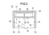
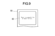
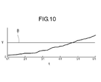
- FIG. 3 is a cross-sectional view taken along the line CC in FIG. 2. It is a top view which shows the state which made the measuring subject contact the cardiac function measuring apparatus. It is sectional drawing which shows the state which made the measuring subject contact the cardiac function measuring apparatus. It is a figure which shows the image data acquired by the imaging part. It is a figure which shows the outer diameter vein specified from the image data acquired by the imaging part. It is a flowchart which shows the flow of control of a cardiac function measuring apparatus. It is a top view which shows the display part on which the warning message was displayed. It is a graph which shows the change every day of the shape complexity computed by the cardiac function measuring device.
- the cardiac function measuring device 10 is a device that can observe the complexity of the shape of the jugular vein in the neck from the body surface.
- the jugular vein swells (hypertrophies) due to the reduced function of the right ventricle and becomes a complex shape.
- a complicated shape means the shape which has dispersion
- the jugular vein includes the external jugular vein and the internal jugular vein.
- the jugular vein observed by the cardiac function measuring device 10 may be the external jugular vein or the internal jugular vein.
- the cardiac function measuring device 10 is used on a daily basis by the measurement subject himself / herself and can monitor changes in the state of the heart over a long period of time. It can also be used for short-term monitoring applications in which the presence or absence of heart failure is determined by a doctor's instruction for a period of about 2 to 3 days, making it possible to make a diagnosis with higher accuracy than observing in a short time at the visit. .
- the cardiac function measuring apparatus 10 stores an image pickup unit 20 that picks up an image, a luminance detection unit 30 that detects luminance, an irradiation unit 80 that emits light, and various data. It has the memory
- the control unit 50 and the storage unit 40 constitute a computer for executing a program. Therefore, the operation of the cardiac function measuring apparatus 10 can be executed by a program.
- the housing 90 has an operation surface 91 on which the display unit 60 and the input unit 70 are arranged, and a measurement surface 92 on which the imaging unit 20, the luminance detection unit 30 and the irradiation unit 80 are arranged.
- the measurement surface 92 of the housing 90 has a recess 93, and the imaging unit 20, the luminance detection unit 30, and the irradiation unit 80 are disposed in the recess 93.
- a support portion 94 that contacts the neck portion during measurement and supports the housing 90 with respect to the neck portion is provided at the edge portion surrounding the recess 93.
- the recess 93 has a bottom surface 95 and a side surface 96 surrounding the bottom surface 95.
- a space 97 in which the storage unit 40 and the control unit 50 are accommodated is provided between the operation surface 91 and the bottom surface 95 of the housing 90.
- the imaging unit 20 is an image sensor that acquires cervical image data in order to observe the bulge of the jugular vein A in the cervical region from the body surface.
- the imaging unit 20 transmits the detected image data to the control unit 50.
- the imaging unit 20 is disposed at the center of the bottom surface 95 of the housing 90 and faces the opening direction of the recess 93.
- the imaging direction in which the imaging unit 20 faces is preferably perpendicular to the measurement surface 92 (the surface on which the support unit 94 exists) so as to be perpendicular to the body surface of the neck contacting the support unit 94.
- the distance from the imaging unit 20 to the measurement surface 92 (distance to the imaging target) is not particularly limited, but is, for example, 5 to 10 mm.
- the distance from the imaging unit 20 to the measurement surface 92 is preferably set based on the viewing angle of the imaging unit 20 and the size of the range to be captured.
- the luminance detection unit 30 is a luminance sensor that detects the luminance of the imaging environment.
- the luminance detection unit 30 transmits the detected luminance data to the control unit 50.
- the luminance detection unit 30 is disposed on the bottom surface 95 adjacent to the luminance detection unit 30.
- the position of the luminance detection unit 30 is not particularly limited as long as the luminance of the imaging environment can be detected.
- the irradiation unit 80 is a light source that emits light, for example, an LED.
- the irradiation unit 80 can be controlled by the control unit 50 to adjust the luminance.
- the irradiating unit 80 irradiates inclined light onto the imaging target on the extended surface of the measurement surface 92 and in front of the imaging unit 20.
- the irradiation unit 80 generates a shadow S (see FIG. 6) on the back side in the irradiation direction X with respect to the protruding part of the body surface. For this reason, the irradiation part 80 makes it easy to detect the jugular vein A by the imaging part 20.
- the irradiation direction X of the irradiation unit 80 is preferably substantially orthogonal to the jugular vein A as a measurement target so as to cause a shadow along the longitudinal direction of the jugular vein A.
- the angle ⁇ of the irradiation direction X with respect to the imaging direction (the direction in which the irradiation unit 80 is directed) is more than 0 degree and less than 90 degrees, preferably 0 to less than 90 degrees so that a shadow is formed on the protruding part of the body surface. It is 83 degrees, and more preferably 78 to 83 degrees.
- one irradiation unit 80 is disposed on each of the side surfaces 96 on both sides of the imaging unit 20.
- the angle ⁇ of each irradiation unit 80 may be the same or different.
- the number of the irradiation parts 80 is not limited, One may be sufficient and three or more may be sufficient.
- the storage unit 40 stores various operation programs such as a measurement program executed by the control unit 50 and various parameters (threshold value, etc.).
- the storage unit 40 further stores data detected by the imaging unit 20 and the luminance detection unit 30 and data calculated by the control unit 50.
- the control unit 50 includes a luminance adjustment unit 51, a vein determination unit 52, an irradiation switching unit 53, and a warning determination unit 54.
- the control unit 50 includes a CPU (Central Processing Unit) and an operation program.
- the control unit 50 includes an imaging unit 20, a luminance detection unit 30, a storage unit 40, a luminance adjustment unit 51, a vein determination unit 52, an irradiation switching unit 53, a warning determination unit 54, an input unit 70, an irradiation unit 80, and a display unit 60. Control the overall operation.
- the luminance adjusting unit 51 acquires luminance data from the luminance detecting unit 30.
- the luminance adjusting unit 51 also uses the acquired luminance data to calculate the condition (output) of the irradiation unit 80 where the luminance becomes a preset target value.
- the luminance adjustment unit 51 controls the irradiation unit 80 under the calculated conditions, and sets the luminance to a target value.
- the vein discriminating unit 52 acquires image data from the imaging unit 20.
- the vein discriminating unit 52 calculates a shape complexity V that is an index indicating the complexity of the shape of the jugular vein A from the image data by a preset method.
- the vein discrimination unit 52 classifies the gradation of each pixel from the acquired image data. For example, each of the three colors of red, green, and blue has 256 gradations.
- the vein discriminating unit 52 monochromeizes the image and further performs a posterization process.
- the vein discriminating unit 52 reads a preset threshold value in the storage unit 40 and performs a posterization process on two gradations to binarize into black and white. As a result, as shown in FIGS.
- a black shadow S appears in a predetermined range on the far side in the irradiation direction X with respect to the protrusion E protruding from the body surface due to the bulge of the jugular vein A.
- the vein determination unit 52 calculates a center line G passing through the center of the shadow S as shown in FIG.
- the vein determination unit 52 specifies the pixels P1 and P2 that are farthest from the specified shadow S.
- the vein discriminating unit 52 specifies an orthogonal line L2 that is orthogonal to the line L1 connecting the pixel P1 and the pixel P2.
- the vein determination unit 52 identifies the orthogonal lines L2 and the edge portions S1 and S2 extending from the pixel P1 to the pixel P2 along the edge of the shadow portion S. Subsequently, the vein discriminating unit 52 specifies the intersections P3 and P4 between the orthogonal line L2 and the edges S1 and S2. The vein discriminating unit 52 specifies an intermediate point P5 located between the intersection point P3 and the intersection point P4. The vein determination unit 52 moves the orthogonal line L2 between the pixel P1 and the pixel P2, and specifies and arranges the intermediate point P5 of each orthogonal line L2. Thereby, the vein discrimination
- the vein discriminating unit 52 calculates a regression curve of the center line G.
- the center line G is a smooth curve from which minute fluctuation components are removed.
- the vein discriminating unit 52 calculates the number of inflection points D of the regression curve.
- the vein discriminating unit 52 sets the inflection point D to a point at which the twice differential value of the regression curve becomes zero (a point at which the slope of the curve becomes zero), and sets the number of the inflection points D to the shape complexity V.
- the larger the shape complexity V the more complicated the shape of the jugular vein A.
- the vein discriminating unit 52 determines the number of inflection points D indicating the complexity of the meandering and the complexity of the shape of the outer diameter vein A.
- the shape complexity V can be set as V.
- the method for calculating the shape complexity V is not particularly limited as long as the shape complexity of the outer diameter vein A can be expressed.
- the shape complexity V may be the number of times the line L1 connecting the farthest pixel P1 and the pixel P2 intersects the center line G.
- the method for calculating the shape complexity V is obtained by dividing the length along the center line G between the farthest pixels P1 and P2 by the linear distance between the pixels P1 and P2 into the shape complexity.
- the degree V may be set.
- the shape complexity V is calculated using the shadow portion S in the method for calculating the shape complexity V.
- the shape complexity V may be calculated using a strong white range appearing on the near side.
- the shape complexity V can be calculated in the same manner as the method of calculating the white range by binarizing the image data and calculating from the shadow portion S described above.
- the method of calculating the shape complexity V specifies the shape of the outer diameter vein A from the shapes in the range where the colors of the acquired image data are different. You can also.
- the shape complexity V may be a change in the width W that is the distance between the intersection P3 and the intersection P4 between the orthogonal line L2 and the edges S1 and S2.
- the vein discriminating unit 52 calculates the width W at each point of a predetermined interval from the pixel P1 to the pixel P2.
- the vein discriminating unit 52 can set, for example, the degree of complexity of the plurality of widths W, the maximum value of the width W, the difference between the maximum value and the average value of the width W, and the like as the shape complexity V.
- the average value of the plurality of shape complexity V calculated using the plurality of irradiation units 80 can be set as the shape complexity V.
- the average value of the shape complexity V it becomes possible to identify the shape complexity with high accuracy. It is also possible to calculate a plurality of shape complexity V from a plurality of irradiation units 80 and select and use only the most favorable value instead of the average value.
- the irradiation switching unit 53 can select and switch the irradiation unit 80 that emits light from the two irradiation units 80.
- the irradiation switching unit 53 transmits a signal to one preset irradiation unit 80 to emit light.
- the irradiation switching unit 53 stops the light irradiation.
- the irradiation switching unit 53 causes the other irradiation units 80 to emit light after the irradiation by one irradiation unit 80 is completed.
- the irradiation switching unit 53 stops the irradiation after the irradiation by all the irradiation units 80 is completed.
- the warning determination unit 54 compares the shape complexity V with a threshold value.
- the threshold value is preset in the storage unit 40, input from the input unit 70, or calculated from past data.
- the warning determination unit 54 determines that the cardiac function is abnormal when the shape complexity V exceeds the threshold value.
- the warning determination part 54 determines with a cardiac function being normal, when the shape complexity V is below a threshold value.
- the warning determination unit 54 causes the display unit 60 to display the determination result.
- the threshold value of the warning determination unit 54 may be, for example, an upper limit value of an average value of a general person, or a value obtained by multiplying an average value for the past several months of a measurement target person by a coefficient. There may be.
- the input unit 70 is a part where the measurement subject performs an input operation.
- the input unit 70 includes, for example, a switch and a button.
- the input unit may be a touch panel, a keyboard, a mouse, or the like.
- the input unit 70 is used for inputting and resetting various parameters such as threshold values, starting and ending measurement, selecting measurement results and calculation results to be displayed, selecting graphs and tables to be displayed, and the like.
- the display unit 60 is a display device such as an LCD (Liquid Crystal Display) or an organic EL (Electro-Luminescence) display.
- the display unit 60 may be a touch panel that also serves as the input unit 70.
- the display unit 60 can display values, measurement results, calculation results, determination results, and the like input to the input unit 70 as characters, numerical values, tables, graphs, or the like.
- the measurement subject brings the support portion 94 of the measurement surface 92 of the cardiac function measuring device 10 into contact with the body surface near the jugular vein A.
- the jugular vein A is positioned in front of the imaging unit 20.
- the irradiation direction X of the irradiation part 80 is set to a direction orthogonal to the jugular vein A as much as possible.
- the position of the body surface with which the cardiac function measuring device 10 is brought into contact is the same position every time.
- the measurement subject operates the input unit 70 of the housing 90 to start measurement.
- the irradiation switching part 53 transmits an instruction
- the luminance adjusting unit 51 receives luminance data from the luminance detecting unit 30 (step 2).
- the luminance adjustment unit 51 calculates the condition (output) of the irradiation unit 80 from which the luminance becomes a preset target value from the received luminance data. Subsequently, the luminance adjustment unit 51 sends a signal to the irradiation unit 80 so as to satisfy the calculated condition, and sets the luminance to a target value (step 3). Thereby, the brightness of the imaging environment can be kept constant.
- the vein discriminating unit 52 receives image data from the imaging unit 20 (step 4). Next, the vein discriminating unit 52 identifies the shadow S of the protruding portion E of the body surface protruding by the jugular vein A as shown in FIG. 6 (step 5). Subsequently, the vein discriminating unit 52 calculates the shape complexity V from the information of the identified shadow part S (step 6).
- the vein discriminating unit 52 transmits information to that effect to the irradiation switching unit 53.
- the irradiation switching unit 53 that has received the information determines whether or not the irradiation unit 80 being irradiated is the last irradiation unit 80 (step 7).
- the irradiation switching unit 53 transmits an instruction to the irradiation unit 80 being irradiated to stop the light irradiation and transmits an instruction to the other irradiation units 80. (Step 8).
- Step 1 to Step 6 described above are repeated, and the second shape complexity V is calculated using the second irradiation unit 80.
- the vein discriminating unit 52 transmits information to that effect to the irradiation switching unit 53.
- the irradiation switching unit 53 that has received the information determines whether or not the irradiation unit 80 being irradiated is the last irradiation unit 80 (step 7).
- the irradiation switching unit 53 transmits an instruction to the irradiation unit 80 being irradiated to stop the light irradiation.
- the vein determination unit 52 calculates the average value of the shape complexity V calculated by each of the plurality of irradiation units 80 (step 9).
- the warning determination unit 54 compares the average value of the shape complexity V with a threshold value. Thereby, the warning determination part 54 determines whether the average value of the shape complexity V is normal (step 10). When the warning determination unit 54 determines that the average value of the shape complexity V is abnormal, the warning determination unit 54 displays a warning message on the display unit 60 as shown in FIG. 9 (step 11). The content of the warning message can be set as appropriate.
- the control unit 50 can cause the display unit 60 to display the change of the average value of the shape complexity V for each measurement together with the past data, with the horizontal axis as time t.
- the control unit 50 can display the upper limit B (threshold value) of the normal value of the shape complexity V with a line on the graph displayed on the display unit 60.
- the shape complexity V exceeds the upper limit B, it can be determined that the running path of the jugular vein A has been bent by the blood pressure, and that there is a possibility of heart failure or heart failure. Therefore, the measurement subject himself / herself can easily monitor the degree of progression of heart failure (decrease in cardiac function) over a long period of time.
- control unit 50 stores data such as the calculated shape complexity V and presence / absence of abnormality in the storage unit 40 (step 12). Thereby, the measurement for one time by the cardiac function measuring apparatus 10 is completed.
- the measurement by the cardiac function measuring device 10 is performed, for example, every day. Thereby, the cardiac function can be monitored over a long period. Note that the measurement may not be performed every day.
- the cardiac function measurement device 10 is a device for measuring data for evaluating cardiac function, and includes an irradiation unit 80 for irradiating light to the neck, and the neck.
- An image capturing unit 20 for acquiring image data of a portion, and a vein determining unit 52 for determining the shape of the jugular vein A in the acquired image data and calculating a shape complexity V representing the complexity of the shape.
- the cardiac function measuring apparatus 10 configured as described above can calculate the shape complexity V representing the complexity of the shape of the jugular vein A using the image data of the neck generated by the light irradiated from the irradiation unit 80. This makes it possible to monitor the complexity of the shape of the jugular vein A, which serves as an indicator of a decrease in cardiac function, by the shape complexity V, and it becomes easy to monitor the cardiac function. Moreover, since the cardiac function measuring apparatus 10 can be configured in a compact manner and is easy to measure, the burden on the measurement subject is small and long-term monitoring is possible.
- the cardiac function measuring apparatus 10 further includes a storage unit 40 capable of storing the shape complexity V, and can compare the shape complexity V measured and calculated at different times. Thereby, since the temporal change of the cardiac function can be observed, the cardiac function can be monitored for a long time and easily.
- the vein discriminating unit 52 calculates a center line G (line) along the determined shape of the jugular vein A, calculates a regression curve of the center line G, and calculates the number of inflection points D of the regression curve.
- the complexity can be V.
- the shape complexity V which is an index of a decrease in cardiac function, can be calculated easily and accurately from the shape of the jugular vein A.
- the line along the shape of the jugular vein A is not limited to the center line G as long as the line represents the characteristics of the jugular vein A. For example, the line along the edge of the identified jugular vein A (edges S1, S2). ).
- the cardiac function measuring apparatus 10 further includes a warning determination unit 54 that compares the calculated shape complexity V with a threshold value and determines whether or not to issue a warning. Thereby, since determination of a cardiac function is performed automatically, determination of the fall of a cardiac function becomes easy.
- the cardiac function measuring apparatus 10 further includes a luminance detection unit 30 that detects luminance, and a luminance adjustment unit 51 that adjusts the irradiation unit 80 so that the luminance detected by the luminance detection unit 30 approaches a target value. .
- the cardiac function measuring apparatus 10 further includes a support portion 94 that is positioned on the imaging direction side of the imaging unit 20 and can contact the neck. Thereby, the distance from the imaging part 20 to a neck can be appropriately set for every measurement by making the support part 94 contact a neck. For this reason, the imaging conditions for each measurement are equal, and the cardiac function can be monitored with higher accuracy.
- the present invention also includes a cardiac function measurement method for measuring data for evaluating cardiac function.
- the cardiac function measurement method includes a step of irradiating light to a neck, a step of acquiring image data of the neck irradiated with light, a step of determining a shape of the jugular vein A in the acquired image data, and a determination Calculating a shape complexity V representing the complexity of the shape of the jugular vein A.
- the cardiac function measuring method configured as described above can calculate the shape complexity V representing the complexity of the shape of the jugular vein A using the image data of the neck generated by the light irradiated from the irradiation unit 80. This makes it possible to monitor the complexity of the shape of the jugular vein A, which serves as an indicator of a decrease in cardiac function, by the shape complexity V, and it becomes easy to monitor the cardiac function. In addition, since measurement is easy, the burden on the measurement subject is small, and long-term monitoring is possible.
- the cardiac function measuring method further includes a step of comparing the shape complexity V measured and calculated at different times. Thereby, since the temporal change of the cardiac function can be observed, the cardiac function can be monitored for a long time and easily.
- a center line G (line) along the determined shape of the jugular vein A is calculated, a regression curve of the center line G is calculated, and the regression is performed.
- the number of inflection points D of the curve can be made the shape complexity V.
- the shape complexity V which is an index of decrease in cardiac function, can be easily calculated from the shape of the jugular vein A with high accuracy.
- the cardiac function measuring method further includes a step of comparing the calculated shape complexity V with a threshold value and determining whether or not to issue a warning.
- the present invention also includes a cardiac function measurement program executed by a computer to measure data for evaluating cardiac function.
- the cardiac function measurement program includes a step of acquiring cervical image data from the imaging unit 20 capable of imaging the cervix, a step of determining the shape of the jugular vein A in the acquired image data, and the determined jugular vein A Calculating a shape complexity V representing the complexity of the shape.
- the cardiac function measurement program configured as described above can calculate the shape complexity V representing the complexity of the shape of the jugular vein A using the image data of the neck generated by the light irradiated from the irradiation unit 80.
- the complexity of the shape of the jugular vein A which is an index of the decrease in cardiac function, can be observed by the shape complexity V, and monitoring of the cardiac function is facilitated.
- the burden on the measurement subject is small, and long-term monitoring is possible.
- the cardiac function measurement program further includes a step of reading out the shape complexity V measured and calculated at different times from the storage unit, and a step of comparing a plurality of shape complexity V measured at different times. .
- the cardiac function measurement program calculates a center line G (line) along the determined shape of the jugular vein A in the step of calculating the shape complexity V, calculates a regression curve of the center line G, and calculates the regression.
- the number of inflection points D of the curve can be made the shape complexity V.
- the shape complexity V which is an index of decrease in cardiac function, can be easily calculated from the shape of the jugular vein A with high accuracy.
- the cardiac function measurement program further includes a step of comparing the calculated shape complexity V with a preset threshold value and determining whether or not to issue a warning.
- the configuration of the housing is not particularly limited. Even if at least a part of functions of the cardiac function measuring device is configured by a portable terminal such as a smartphone or a tablet terminal and an application (program), or a server computer terminal connected to the Internet and an application (program), Good. In this case, some functions may be wirelessly connected to the portable terminal using a communication technology such as NFC (Near Field Communication) or Wifi (registered trademark).
- a communication technology such as NFC (Near Field Communication) or Wifi (registered trademark).
- the cardiac function measuring device 10 is a device that is assumed to be used by the measurement target person, a medical worker such as a doctor or a nurse may use it for the measurement target person.
- the external jugular vein is easy to observe from the body surface, but the internal jugular vein is located deep in the body, so that it may not be observed on the body surface by a general image sensor (imaging device). For this reason, when observing the internal jugular vein, it is preferable that the imaging unit can observe the deep part of the body.
- the imaging unit that can observe the deep part of the body is, for example, an ultrasonic probe using an ultrasonic echo, an optical coherence tomography diagnosis device (OCT: Optical Coherence Tomography), or an optical frequency domain imaging method (OFDI: Optical Frequency Domain Imaging). Etc. is a device using the above.
- the imaging unit that can observe the deep part of the body measures the sound of the pulsation of the jugular vein at a plurality of locations on the body surface, and identifies the shape of the jugular vein from the intensity of the pulsation sound at each measurement location It may be.
- a cardiac function measuring device, a cardiac function measuring method, and a cardiac function measuring program can be configured using an imaging unit capable of observing the deep part of the body as described above.
- 10 cardiac function measuring device 20 imaging unit, 30 luminance detector, 40 storage unit, 50 control unit, 51 brightness control unit, 52 Vein discrimination unit, 53 Irradiation switching part, 54 Warning determination unit, 80 Irradiation part, 94 support part, A jugular vein, B Upper limit (threshold), D inflection point, G center line (line), X irradiation direction, V Shape complexity.
Landscapes
- Health & Medical Sciences (AREA)
- Life Sciences & Earth Sciences (AREA)
- Engineering & Computer Science (AREA)
- Physics & Mathematics (AREA)
- General Health & Medical Sciences (AREA)
- Biomedical Technology (AREA)
- Heart & Thoracic Surgery (AREA)
- Medical Informatics (AREA)
- Molecular Biology (AREA)
- Surgery (AREA)
- Animal Behavior & Ethology (AREA)
- Biophysics (AREA)
- Public Health (AREA)
- Veterinary Medicine (AREA)
- Pathology (AREA)
- Physiology (AREA)
- Dentistry (AREA)
- Geometry (AREA)
- Computer Vision & Pattern Recognition (AREA)
- General Physics & Mathematics (AREA)
- Theoretical Computer Science (AREA)
- Vascular Medicine (AREA)
- Cardiology (AREA)
- Oral & Maxillofacial Surgery (AREA)
- Measurement Of The Respiration, Hearing Ability, Form, And Blood Characteristics Of Living Organisms (AREA)
- Measuring Pulse, Heart Rate, Blood Pressure Or Blood Flow (AREA)
Abstract
L'invention concerne un dispositif de mesure de fonction cardiaque, un procédé de mesure de fonction cardiaque et un programme de mesure de fonction cardiaque, la fonction cardiaque pouvant être surveillée à long terme et présentant une faible pénibilité pour le sujet de mesure. Le dispositif (10) de mesure de fonction cardiaque pour mesurer des données pour évaluer une fonction cardiaque comprend : une unité d'irradiation (80) qui irradie de la lumière sur le cou ; une unité d'imagerie (20) pour obtenir des données d'image du cou ; et une unité (52) de discrimination veineuse qui distingue la forme d'une veine jugulaire (A) dans les données d'image obtenues et calcule un degré de complexité (V) indiquant la complexité de la forme.
Priority Applications (2)
| Application Number | Priority Date | Filing Date | Title |
|---|---|---|---|
| JP2018505901A JP6743129B2 (ja) | 2016-03-18 | 2017-03-10 | 心機能測定装置、心機能測定方法および心機能測定プログラム |
| US16/134,396 US11229366B2 (en) | 2016-03-18 | 2018-09-18 | Cardiac function measurement device, cardiac function measurement method, and cardiac function measuring program |
Applications Claiming Priority (2)
| Application Number | Priority Date | Filing Date | Title |
|---|---|---|---|
| JP2016-055197 | 2016-03-18 | ||
| JP2016055197 | 2016-03-18 |
Related Child Applications (1)
| Application Number | Title | Priority Date | Filing Date |
|---|---|---|---|
| US16/134,396 Continuation US11229366B2 (en) | 2016-03-18 | 2018-09-18 | Cardiac function measurement device, cardiac function measurement method, and cardiac function measuring program |
Publications (1)
| Publication Number | Publication Date |
|---|---|
| WO2017159574A1 true WO2017159574A1 (fr) | 2017-09-21 |
Family
ID=59850383
Family Applications (1)
| Application Number | Title | Priority Date | Filing Date |
|---|---|---|---|
| PCT/JP2017/009785 Ceased WO2017159574A1 (fr) | 2016-03-18 | 2017-03-10 | Dispositif de mesure de fonction cardiaque, procédé de mesure de fonction cardiaque et programme de mesure de fonction cardiaque |
Country Status (3)
| Country | Link |
|---|---|
| US (1) | US11229366B2 (fr) |
| JP (1) | JP6743129B2 (fr) |
| WO (1) | WO2017159574A1 (fr) |
Families Citing this family (16)
| Publication number | Priority date | Publication date | Assignee | Title |
|---|---|---|---|---|
| US9901722B2 (en) | 2014-06-01 | 2018-02-27 | White Swell Medical Ltd | System and method for treatment of pulmonary edema |
| IL324387A (en) | 2015-05-11 | 2026-01-01 | White Swell Medical Ltd | System and method for reducing outlet pressure from a duct |
| EP3534985B1 (fr) | 2016-11-01 | 2024-10-23 | White Swell Medical Ltd | Systèmes et procédés de traitement de surcharge de liquides |
| IL268950B (en) | 2017-03-02 | 2022-08-01 | White Swell Medical Ltd | Systems and methods for reducing pressure at the exit of a pipe |
| US11406393B2 (en) * | 2017-03-19 | 2022-08-09 | White Swell Medical Ltd | Methods and devices for reducing pressure |
| US12144977B2 (en) | 2018-02-13 | 2024-11-19 | White Swell Medical Ltd | Intravascular catheters |
| US11660426B2 (en) | 2019-02-26 | 2023-05-30 | White Swell Medical Ltd | Devices and methods for treating edema |
| US11717652B2 (en) | 2019-02-26 | 2023-08-08 | White Swell Medical Ltd | Devices and methods for treating edema |
| US11793996B2 (en) | 2019-02-26 | 2023-10-24 | White Swell Medical Ltd | Devices and methods for treating edema |
| US11931560B2 (en) | 2019-02-26 | 2024-03-19 | White Swell Medical Ltd | Devices and methods for treating edema |
| US11724095B2 (en) | 2019-02-26 | 2023-08-15 | White Swell Medical Ltd | Devices and methods for treating edema |
| WO2021061525A1 (fr) | 2019-09-25 | 2021-04-01 | White Swell Medical Ltd | Dispositifs et procédés de décongestion interstitielle |
| US12458359B2 (en) | 2020-06-08 | 2025-11-04 | White Swell Medical Ltd | Non-thrombogenic devices for treating edema |
| US12673180B2 (en) | 2020-06-08 | 2026-07-07 | White Swell Medical Ltd | Non-thrombogenic devices for treating edema |
| IL301494A (en) | 2020-10-01 | 2023-05-01 | White Swell Medical Ltd | Intravascular devices that limit flow for the treatment of edema |
| US20220015739A1 (en) * | 2021-09-28 | 2022-01-20 | Gaurang Nandkishor Vaidya | Respiratory variation in internal jugular vein diameter as a method for estimating patient's volume status and ventricular function |
Citations (5)
| Publication number | Priority date | Publication date | Assignee | Title |
|---|---|---|---|---|
| JP2005069918A (ja) * | 2003-08-26 | 2005-03-17 | Sharp Corp | 画像処理装置および画像処理方法 |
| JP2009028058A (ja) * | 2007-07-24 | 2009-02-12 | Saieco:Kk | 舌診システム、舌診装置、舌診方法、舌診プログラム |
| JP2010517711A (ja) * | 2007-02-16 | 2010-05-27 | メスピア・ライフサイエンシズ・インコーポレイテッド | 心臓機能のパラメータを測定する方法及び装置 |
| JP2011178799A (ja) * | 2004-03-17 | 2011-09-15 | Novartis Ag | 治療におけるレニン阻害剤の使用 |
| JP2011229615A (ja) * | 2010-04-26 | 2011-11-17 | Toyo Univ | 外耳道内圧測定システム及び外耳道内圧測定方法 |
Family Cites Families (5)
| Publication number | Priority date | Publication date | Assignee | Title |
|---|---|---|---|---|
| JP2008287433A (ja) * | 2007-05-16 | 2008-11-27 | Sony Corp | 静脈パターン管理システム、静脈パターン登録装置、静脈パターン認証装置、静脈パターン登録方法、静脈パターン認証方法、プログラムおよび静脈データ構造 |
| US8844525B2 (en) | 2008-07-25 | 2014-09-30 | Resmed Limited | Method and apparatus for detecting and treating heart failure |
| JP5938890B2 (ja) * | 2011-02-24 | 2016-06-22 | 株式会社リコー | 画像形成装置、パターン位置検出方法、画像形成システム |
| JP5990906B2 (ja) * | 2011-12-19 | 2016-09-14 | ソニー株式会社 | 測定装置、測定方法、プログラムおよび記録媒体 |
| US11185241B2 (en) * | 2014-03-05 | 2021-11-30 | Whoop, Inc. | Continuous heart rate monitoring and interpretation |
-
2017
- 2017-03-10 WO PCT/JP2017/009785 patent/WO2017159574A1/fr not_active Ceased
- 2017-03-10 JP JP2018505901A patent/JP6743129B2/ja active Active
-
2018
- 2018-09-18 US US16/134,396 patent/US11229366B2/en active Active
Patent Citations (5)
| Publication number | Priority date | Publication date | Assignee | Title |
|---|---|---|---|---|
| JP2005069918A (ja) * | 2003-08-26 | 2005-03-17 | Sharp Corp | 画像処理装置および画像処理方法 |
| JP2011178799A (ja) * | 2004-03-17 | 2011-09-15 | Novartis Ag | 治療におけるレニン阻害剤の使用 |
| JP2010517711A (ja) * | 2007-02-16 | 2010-05-27 | メスピア・ライフサイエンシズ・インコーポレイテッド | 心臓機能のパラメータを測定する方法及び装置 |
| JP2009028058A (ja) * | 2007-07-24 | 2009-02-12 | Saieco:Kk | 舌診システム、舌診装置、舌診方法、舌診プログラム |
| JP2011229615A (ja) * | 2010-04-26 | 2011-11-17 | Toyo Univ | 外耳道内圧測定システム及び外耳道内圧測定方法 |
Also Published As
| Publication number | Publication date |
|---|---|
| JPWO2017159574A1 (ja) | 2019-01-24 |
| JP6743129B2 (ja) | 2020-08-19 |
| US11229366B2 (en) | 2022-01-25 |
| US20190014991A1 (en) | 2019-01-17 |
Similar Documents
| Publication | Publication Date | Title |
|---|---|---|
| JP6743129B2 (ja) | 心機能測定装置、心機能測定方法および心機能測定プログラム | |
| US10524671B2 (en) | Electronic device that computes health data | |
| CN109906052B (zh) | 包括血压传感器的设备以及用于控制该设备的方法 | |
| CN106659397B (zh) | 非接触式测温系统和方法 | |
| US20200082695A1 (en) | Method for estimating a quantity of a blood component in a fluid receiver and corresponding error | |
| US11103208B2 (en) | Pulmonary function measurement device, pulmonary function measurement method, and pulmonary function measurement program | |
| KR101984993B1 (ko) | 사용자 맞춤형 시표 제어가 가능한 시야검사기 | |
| JP2017209280A5 (fr) | ||
| Desebbe et al. | A novel mobile phone application for pulse pressure variation monitoring based on feature extraction technology: a method comparison study in a simulated environment | |
| CN108289608A (zh) | 诊疗辅助装置、诊疗辅助方法、诊疗辅助程序、生物体信息测定装置 | |
| KR20150036395A (ko) | 사시 검출 | |
| US20160309998A1 (en) | System and Methods for Assessing Vision Using a Computing Device | |
| US20170035325A1 (en) | Physiological information measuring apparatus, respiration interval displaying method, and computer readable medium | |
| WO2017159573A1 (fr) | Dispositif de mesure de la fonction cardiaque, procédé de mesure de la fonction cardiaque, et programme de mesure de la fonction cardiaque | |
| KR20210074008A (ko) | 이미지 분석을 이용한 사시 진단 시스템 및 방법 | |
| US12588814B2 (en) | Vision screening device including oversampling sensor | |
| US20230240563A1 (en) | System and method of measuring body fluids | |
| WO2017159572A1 (fr) | Dispositif de mesure de la fonction cardiaque, procédé de mesure de la fonction cardiaque, et programme de mesure de la fonction cardiaque | |
| AU2021212056B2 (en) | Fundus imaging for microvascular assessment | |
| JP6779979B2 (ja) | 肺機能測定装置、肺機能測定方法および肺機能測定プログラム | |
| KR20240093283A (ko) | 디스플레이 장치 및 디스플레이 방법 | |
| US20230329570A1 (en) | Electronic device, storage medium for electronic device, and control method for electronic device | |
| WO2013179178A1 (fr) | Surveillance de la fatigue oculaire | |
| JP2016036411A (ja) | 生体情報測定装置および生体情報測定方法 | |
| EP3127479A1 (fr) | Appareil de mesure d'informations physiologiques, procédé d'affichage d'intervalle de respiration et programme |
Legal Events
| Date | Code | Title | Description |
|---|---|---|---|
| WWE | Wipo information: entry into national phase |
Ref document number: 2018505901 Country of ref document: JP |
|
| NENP | Non-entry into the national phase |
Ref country code: DE |
|
| 121 | Ep: the epo has been informed by wipo that ep was designated in this application |
Ref document number: 17766574 Country of ref document: EP Kind code of ref document: A1 |
|
| 122 | Ep: pct application non-entry in european phase |
Ref document number: 17766574 Country of ref document: EP Kind code of ref document: A1 |