WO2020152791A1 - Endoscope à ultrasons et système d'endoscope - Google Patents
Endoscope à ultrasons et système d'endoscope Download PDFInfo
- Publication number
- WO2020152791A1 WO2020152791A1 PCT/JP2019/001929 JP2019001929W WO2020152791A1 WO 2020152791 A1 WO2020152791 A1 WO 2020152791A1 JP 2019001929 W JP2019001929 W JP 2019001929W WO 2020152791 A1 WO2020152791 A1 WO 2020152791A1
- Authority
- WO
- WIPO (PCT)
- Prior art keywords
- ultrasonic
- angle
- endoscope
- ultrasonic transducer
- insertion portion
- Prior art date
- Legal status (The legal status is an assumption and is not a legal conclusion. Google has not performed a legal analysis and makes no representation as to the accuracy of the status listed.)
- Ceased
Links
Images




Classifications
-
- A—HUMAN NECESSITIES
- A61—MEDICAL OR VETERINARY SCIENCE; HYGIENE
- A61B—DIAGNOSIS; SURGERY; IDENTIFICATION
- A61B8/00—Diagnosis using ultrasonic, sonic or infrasonic waves
- A61B8/44—Constructional features of the ultrasonic, sonic or infrasonic diagnostic device
- A61B8/4444—Constructional features of the ultrasonic, sonic or infrasonic diagnostic device related to the probe
- A61B8/4461—Features of the scanning mechanism, e.g. for moving the transducer within the housing of the probe
-
- A—HUMAN NECESSITIES
- A61—MEDICAL OR VETERINARY SCIENCE; HYGIENE
- A61B—DIAGNOSIS; SURGERY; IDENTIFICATION
- A61B1/00—Instruments for performing medical examinations of the interior of cavities or tubes of the body by visual or photographical inspection, e.g. endoscopes; Illuminating arrangements therefor
- A61B1/00002—Operational features of endoscopes
- A61B1/00043—Operational features of endoscopes provided with output arrangements
- A61B1/00045—Display arrangement
-
- A—HUMAN NECESSITIES
- A61—MEDICAL OR VETERINARY SCIENCE; HYGIENE
- A61B—DIAGNOSIS; SURGERY; IDENTIFICATION
- A61B1/00—Instruments for performing medical examinations of the interior of cavities or tubes of the body by visual or photographical inspection, e.g. endoscopes; Illuminating arrangements therefor
- A61B1/00064—Constructional details of the endoscope body
- A61B1/00071—Insertion part of the endoscope body
- A61B1/0008—Insertion part of the endoscope body characterised by distal tip features
- A61B1/00087—Tools
-
- A—HUMAN NECESSITIES
- A61—MEDICAL OR VETERINARY SCIENCE; HYGIENE
- A61B—DIAGNOSIS; SURGERY; IDENTIFICATION
- A61B1/00—Instruments for performing medical examinations of the interior of cavities or tubes of the body by visual or photographical inspection, e.g. endoscopes; Illuminating arrangements therefor
- A61B1/005—Flexible endoscopes
-
- A—HUMAN NECESSITIES
- A61—MEDICAL OR VETERINARY SCIENCE; HYGIENE
- A61B—DIAGNOSIS; SURGERY; IDENTIFICATION
- A61B1/00—Instruments for performing medical examinations of the interior of cavities or tubes of the body by visual or photographical inspection, e.g. endoscopes; Illuminating arrangements therefor
- A61B1/012—Instruments for performing medical examinations of the interior of cavities or tubes of the body by visual or photographical inspection, e.g. endoscopes; Illuminating arrangements therefor characterised by internal passages or accessories therefor
- A61B1/018—Instruments for performing medical examinations of the interior of cavities or tubes of the body by visual or photographical inspection, e.g. endoscopes; Illuminating arrangements therefor characterised by internal passages or accessories therefor for receiving instruments
-
- A—HUMAN NECESSITIES
- A61—MEDICAL OR VETERINARY SCIENCE; HYGIENE
- A61B—DIAGNOSIS; SURGERY; IDENTIFICATION
- A61B8/00—Diagnosis using ultrasonic, sonic or infrasonic waves
- A61B8/12—Diagnosis using ultrasonic, sonic or infrasonic waves in body cavities or body tracts, e.g. by using catheters
-
- A—HUMAN NECESSITIES
- A61—MEDICAL OR VETERINARY SCIENCE; HYGIENE
- A61B—DIAGNOSIS; SURGERY; IDENTIFICATION
- A61B8/00—Diagnosis using ultrasonic, sonic or infrasonic waves
- A61B8/44—Constructional features of the ultrasonic, sonic or infrasonic diagnostic device
- A61B8/4483—Constructional features of the ultrasonic, sonic or infrasonic diagnostic device characterised by features of the ultrasound transducer
- A61B8/4494—Constructional features of the ultrasonic, sonic or infrasonic diagnostic device characterised by features of the ultrasound transducer characterised by the arrangement of the transducer elements
-
- A—HUMAN NECESSITIES
- A61—MEDICAL OR VETERINARY SCIENCE; HYGIENE
- A61B—DIAGNOSIS; SURGERY; IDENTIFICATION
- A61B8/00—Diagnosis using ultrasonic, sonic or infrasonic waves
- A61B8/46—Ultrasonic, sonic or infrasonic diagnostic devices with special arrangements for interfacing with the operator or the patient
- A61B8/461—Displaying means of special interest
- A61B8/466—Displaying means of special interest adapted to display 3D data
-
- A—HUMAN NECESSITIES
- A61—MEDICAL OR VETERINARY SCIENCE; HYGIENE
- A61B—DIAGNOSIS; SURGERY; IDENTIFICATION
- A61B8/00—Diagnosis using ultrasonic, sonic or infrasonic waves
- A61B8/48—Diagnostic techniques
- A61B8/483—Diagnostic techniques involving the acquisition of a 3D volume of data
-
- A—HUMAN NECESSITIES
- A61—MEDICAL OR VETERINARY SCIENCE; HYGIENE
- A61B—DIAGNOSIS; SURGERY; IDENTIFICATION
- A61B90/00—Instruments, implements or accessories specially adapted for surgery or diagnosis and not covered by any of the groups A61B1/00 - A61B50/00, e.g. for luxation treatment or for protecting wound edges
- A61B90/06—Measuring instruments not otherwise provided for
-
- A—HUMAN NECESSITIES
- A61—MEDICAL OR VETERINARY SCIENCE; HYGIENE
- A61B—DIAGNOSIS; SURGERY; IDENTIFICATION
- A61B1/00—Instruments for performing medical examinations of the interior of cavities or tubes of the body by visual or photographical inspection, e.g. endoscopes; Illuminating arrangements therefor
- A61B1/00064—Constructional details of the endoscope body
- A61B1/00071—Insertion part of the endoscope body
- A61B1/0008—Insertion part of the endoscope body characterised by distal tip features
- A61B1/00082—Balloons
-
- A—HUMAN NECESSITIES
- A61—MEDICAL OR VETERINARY SCIENCE; HYGIENE
- A61B—DIAGNOSIS; SURGERY; IDENTIFICATION
- A61B90/00—Instruments, implements or accessories specially adapted for surgery or diagnosis and not covered by any of the groups A61B1/00 - A61B50/00, e.g. for luxation treatment or for protecting wound edges
- A61B90/06—Measuring instruments not otherwise provided for
- A61B2090/067—Measuring instruments not otherwise provided for for measuring angles
Definitions
- the first rigid portion is made of resin.
- the rotating mechanism is a motor that rotates the ultrasonic transducer.
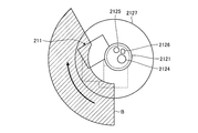
- the endoscope system causes the display device to display the angle of the ultrasonic transducer that minimizes the projected area of the insertion portion along the longitudinal direction of the insertion portion.
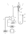
- FIG. 1 is a diagram schematically showing an endoscope system including an ultrasonic endoscope according to an embodiment of the present invention.
- the endoscope system 1 is a system that performs ultrasonic diagnosis in a subject such as a person using an ultrasonic endoscope.
- the endoscope system 1 includes an ultrasonic endoscope 2, an ultrasonic observation device 3, an endoscope observation device 4, a display device 5, and a light source device 6. ..
- the ultrasonic endoscope 2 converts an electrical pulse signal received from the ultrasonic observation device 3 into an ultrasonic pulse (acoustic pulse) and irradiates the subject with an ultrasonic transducer provided at the tip thereof. At the same time, the ultrasonic echo reflected by the subject is converted into an electric echo signal represented by a voltage change and output.
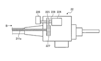
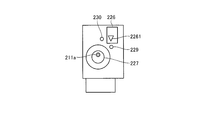
- the operation section 22 is a section that is connected to the proximal end side of the insertion section 21 and receives various operations from an operator such as a doctor. As shown in FIG. 1, the operation section 22 includes a bending knob 221 for bending the bending section 213 and a plurality of operation members 222 for performing various operations. Further, the operation portion 22 is formed with a treatment instrument insertion opening 223 for communicating with the treatment instrument insertion passage and inserting the treatment instrument in the treatment instrument insertion passage.
- the connector 24 is provided at the tip of the universal cord 23.
- the connector 24 includes first to third connector portions 241 to 243 to which the ultrasonic cable 31, the video cable 41 and the light source device 6 are respectively connected.
- the ultrasonic observation device 3 is electrically connected to the ultrasonic endoscope 2 via the ultrasonic cable 31 (see FIG. 1) and electrically connected to the ultrasonic endoscope 2 via the ultrasonic cable 31.
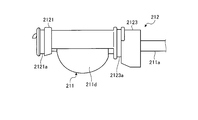
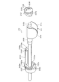
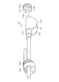
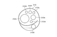
- the second rigid portion 2123 also includes a columnar portion 2123h extending to the first rigid portion 2121 as shown in FIG.
- An image pickup signal line conduit 2123e, a light guide conduit 2123f, and a water supply conduit 2123g are formed inside the columnar portion 2123h.
- the tip of the columnar portion 2123h is fixed to the first rigid portion 2121.
- the columnar section 2123h may be integrally formed with the second hard section 2123, or the columnar section 2123h formed as a separate body from the second hard section 2123 may be fixed to the second hard section 2123.
- the flexible shaft 211a extending from the second rigid portion 2123 to the proximal end side of the insertion portion 21 is preferably arranged so as to be inserted through the inside of a tube or the like. This is to prevent the rotating flexible shaft 211a from interfering with and damaging the light guide or the like because a light guide or the like is arranged inside the insertion portion 21.
- the ultrasonic observation apparatus 3 that sequentially acquires and stores these pieces of information can generate a three-dimensional ultrasonic image based on these pieces of information.
Landscapes
- Health & Medical Sciences (AREA)
- Life Sciences & Earth Sciences (AREA)
- Surgery (AREA)
- Engineering & Computer Science (AREA)
- General Health & Medical Sciences (AREA)
- Veterinary Medicine (AREA)
- Pathology (AREA)
- Public Health (AREA)
- Nuclear Medicine, Radiotherapy & Molecular Imaging (AREA)
- Animal Behavior & Ethology (AREA)
- Biomedical Technology (AREA)
- Heart & Thoracic Surgery (AREA)
- Medical Informatics (AREA)
- Molecular Biology (AREA)
- Biophysics (AREA)
- Physics & Mathematics (AREA)
- Radiology & Medical Imaging (AREA)
- Optics & Photonics (AREA)
- Gynecology & Obstetrics (AREA)
- Computer Graphics (AREA)
- General Engineering & Computer Science (AREA)
- Oral & Maxillofacial Surgery (AREA)
- Ultra Sonic Daignosis Equipment (AREA)
Abstract
Le présent endoscope à ultrasons est équipé : d'une première partie dure positionnée à l'extrémité distale d'une section d'insertion destinée à être insérée à l'intérieur d'un corps subissant un examen ; d'une partie de support reliée au côté d'extrémité proximale de la première partie dure qui forme une forme allongée le long de la direction de la longueur de la section d'insertion ; d'une seconde partie dure reliée au côté d'extrémité proximale de la partie de support ; d'un transducteur à ultrasons fixé à la partie de support et comprenant une pluralité d'éléments piézoélectriques agencés le long de la direction de la longueur de la section d'insertion ; et d'un mécanisme de rotation amenant la partie de support et le transducteur à ultrasons à tourner comme un corps unique. De cette manière, l'invention concerne un endoscope à ultrasons ayant une large plage d'observation.
Priority Applications (3)
| Application Number | Priority Date | Filing Date | Title |
|---|---|---|---|
| JP2020567285A JP7184928B2 (ja) | 2019-01-22 | 2019-01-22 | 超音波内視鏡及び内視鏡システム |
| PCT/JP2019/001929 WO2020152791A1 (fr) | 2019-01-22 | 2019-01-22 | Endoscope à ultrasons et système d'endoscope |
| US17/376,698 US12193877B2 (en) | 2019-01-22 | 2021-07-15 | Ultrasound endoscope and endoscope system |
Applications Claiming Priority (1)
| Application Number | Priority Date | Filing Date | Title |
|---|---|---|---|
| PCT/JP2019/001929 WO2020152791A1 (fr) | 2019-01-22 | 2019-01-22 | Endoscope à ultrasons et système d'endoscope |
Related Child Applications (1)
| Application Number | Title | Priority Date | Filing Date |
|---|---|---|---|
| US17/376,698 Continuation US12193877B2 (en) | 2019-01-22 | 2021-07-15 | Ultrasound endoscope and endoscope system |
Publications (1)
| Publication Number | Publication Date |
|---|---|
| WO2020152791A1 true WO2020152791A1 (fr) | 2020-07-30 |
Family
ID=71735679
Family Applications (1)
| Application Number | Title | Priority Date | Filing Date |
|---|---|---|---|
| PCT/JP2019/001929 Ceased WO2020152791A1 (fr) | 2019-01-22 | 2019-01-22 | Endoscope à ultrasons et système d'endoscope |
Country Status (3)
| Country | Link |
|---|---|
| US (1) | US12193877B2 (fr) |
| JP (1) | JP7184928B2 (fr) |
| WO (1) | WO2020152791A1 (fr) |
Families Citing this family (1)
| Publication number | Priority date | Publication date | Assignee | Title |
|---|---|---|---|---|
| WO2021193404A1 (fr) * | 2020-03-26 | 2021-09-30 | 富士フイルム株式会社 | Endoscope à ultrasons |
Citations (3)
| Publication number | Priority date | Publication date | Assignee | Title |
|---|---|---|---|---|
| JPH078496A (ja) * | 1993-06-25 | 1995-01-13 | Toshiba Corp | 超音波診断装置 |
| JPH07227395A (ja) * | 1993-12-24 | 1995-08-29 | Olympus Optical Co Ltd | 超音波診断治療システム |
| JP2010000210A (ja) * | 2008-06-20 | 2010-01-07 | Fujinon Corp | プローブ |
Family Cites Families (12)
| Publication number | Priority date | Publication date | Assignee | Title |
|---|---|---|---|---|
| US5060632A (en) * | 1989-09-05 | 1991-10-29 | Olympus Optical Co., Ltd. | Endoscope apparatus |
| US5471988A (en) * | 1993-12-24 | 1995-12-05 | Olympus Optical Co., Ltd. | Ultrasonic diagnosis and therapy system in which focusing point of therapeutic ultrasonic wave is locked at predetermined position within observation ultrasonic scanning range |
| US5485845A (en) * | 1995-05-04 | 1996-01-23 | Hewlett Packard Company | Rotary encoder for intravascular ultrasound catheter |
| US6461314B1 (en) * | 1999-02-02 | 2002-10-08 | Transurgical, Inc. | Intrabody hifu applicator |
| JP4261202B2 (ja) | 2003-01-08 | 2009-04-30 | Hoya株式会社 | ラジアル走査型超音波内視鏡の先端部 |
| IL170404A (en) * | 2004-08-26 | 2012-03-29 | C2Cure Inc | Wireless determination of endoscope orientation |
| US8317711B2 (en) * | 2007-06-16 | 2012-11-27 | St. Jude Medical, Atrial Fibrillation Division, Inc. | Oscillating phased-array ultrasound imaging catheter system |
| US8167809B2 (en) * | 2007-12-20 | 2012-05-01 | Silicon Valley Medical Instruments, Inc. | Imaging probe housing with fluid flushing |
| US8974472B2 (en) * | 2013-04-16 | 2015-03-10 | Calcula Technologies, Inc. | Method for removing kidney stones |
| KR101548646B1 (ko) * | 2014-01-21 | 2015-09-01 | 가톨릭관동대학교산학협력단 | 트랜스-플랫폼 장치 및 그의 용도 |
| US11583251B2 (en) * | 2015-06-22 | 2023-02-21 | Bk Medical Aps | US imaging probe with an US transducer array and an integrated optical imaging sub-system |
| US11389164B2 (en) * | 2017-12-28 | 2022-07-19 | Cilag Gmbh International | Method of using reinforced flexible circuits with multiple sensors to optimize performance of radio frequency devices |
-
2019
- 2019-01-22 WO PCT/JP2019/001929 patent/WO2020152791A1/fr not_active Ceased
- 2019-01-22 JP JP2020567285A patent/JP7184928B2/ja active Active
-
2021
- 2021-07-15 US US17/376,698 patent/US12193877B2/en active Active
Patent Citations (3)
| Publication number | Priority date | Publication date | Assignee | Title |
|---|---|---|---|---|
| JPH078496A (ja) * | 1993-06-25 | 1995-01-13 | Toshiba Corp | 超音波診断装置 |
| JPH07227395A (ja) * | 1993-12-24 | 1995-08-29 | Olympus Optical Co Ltd | 超音波診断治療システム |
| JP2010000210A (ja) * | 2008-06-20 | 2010-01-07 | Fujinon Corp | プローブ |
Also Published As
| Publication number | Publication date |
|---|---|
| JP7184928B2 (ja) | 2022-12-06 |
| US12193877B2 (en) | 2025-01-14 |
| US20210338066A1 (en) | 2021-11-04 |
| JPWO2020152791A1 (ja) | 2021-10-14 |
Similar Documents
| Publication | Publication Date | Title |
|---|---|---|
| JP6637609B2 (ja) | 超音波内視鏡および超音波内視鏡システム | |
| JP4551051B2 (ja) | 超音波診断装置 | |
| US20110282209A1 (en) | Ultrasound observation apparatus and control method of ultrasound observation apparatus | |
| JP6197145B2 (ja) | 超音波内視鏡 | |
| JPWO2018025679A1 (ja) | 超音波振動子モジュールの製造方法および超音波内視鏡 | |
| JP7184928B2 (ja) | 超音波内視鏡及び内視鏡システム | |
| JP2020156730A (ja) | 超音波観測装置及び超音波内視鏡システム | |
| JP6133001B1 (ja) | 超音波振動子モジュールおよび超音波内視鏡 | |
| JP2001120550A (ja) | 体腔内超音波プローブシステム | |
| JP4119530B2 (ja) | 内視鏡装置および内視鏡に挿通する位置検出カテーテル | |
| JP7249788B2 (ja) | 内視鏡 | |
| JPH08126644A (ja) | 超音波内視鏡 | |
| JP2000116655A (ja) | 診断装置 | |
| JP6697962B2 (ja) | 超音波振動子および超音波内視鏡 | |
| JP4632748B2 (ja) | カプセル型医療装置 | |
| JP3050085B2 (ja) | 超音波内視鏡 | |
| JP4198130B2 (ja) | 超音波診断装置 | |
| JP2017074231A (ja) | 超音波内視鏡の製造方法および超音波内視鏡 | |
| JPH0352286B2 (fr) | ||
| JP2017144091A (ja) | 内視鏡及びイメージガイド | |
| JP7422616B2 (ja) | 医療器具および内視鏡 | |
| JPH08117233A (ja) | 超音波内視鏡装置 | |
| JP7223871B2 (ja) | 超音波内視鏡 | |
| EP4410219A1 (fr) | Système d'endoscope à ultrasons et procédé de fonctionnement de système d'endoscope à ultrasons | |
| JPH11332867A (ja) | 超音波内視鏡装置 |
Legal Events
| Date | Code | Title | Description |
|---|---|---|---|
| 121 | Ep: the epo has been informed by wipo that ep was designated in this application |
Ref document number: 19911945 Country of ref document: EP Kind code of ref document: A1 |
|
| ENP | Entry into the national phase |
Ref document number: 2020567285 Country of ref document: JP Kind code of ref document: A |
|
| NENP | Non-entry into the national phase |
Ref country code: DE |
|
| 122 | Ep: pct application non-entry in european phase |
Ref document number: 19911945 Country of ref document: EP Kind code of ref document: A1 |