EP0192175A2 - Polypeptides de la souche rhinovirus HRV2 et molécules de l'ADN les codant - Google Patents
Polypeptides de la souche rhinovirus HRV2 et molécules de l'ADN les codant Download PDFInfo
- Publication number
- EP0192175A2 EP0192175A2 EP86101746A EP86101746A EP0192175A2 EP 0192175 A2 EP0192175 A2 EP 0192175A2 EP 86101746 A EP86101746 A EP 86101746A EP 86101746 A EP86101746 A EP 86101746A EP 0192175 A2 EP0192175 A2 EP 0192175A2
- Authority
- EP
- European Patent Office
- Prior art keywords
- viral
- hrv2
- dna molecule
- dna
- rna
- Prior art date
- Legal status (The legal status is an assumption and is not a legal conclusion. Google has not performed a legal analysis and makes no representation as to the accuracy of the status listed.)
- Ceased
Links
- 108090000765 processed proteins & peptides Proteins 0.000 title claims abstract description 44
- 102000004196 processed proteins & peptides Human genes 0.000 title claims abstract description 44
- 241000709661 Enterovirus Species 0.000 title claims description 42
- 229920001184 polypeptide Polymers 0.000 title claims description 39
- 108020004414 DNA Proteins 0.000 claims abstract description 88
- 108010067390 Viral Proteins Proteins 0.000 claims abstract description 27
- 102000053602 DNA Human genes 0.000 claims abstract description 17
- 108090000623 proteins and genes Proteins 0.000 claims description 62
- 239000013612 plasmid Substances 0.000 claims description 40
- 102000004169 proteins and genes Human genes 0.000 claims description 37
- 239000002299 complementary DNA Substances 0.000 claims description 18
- 101710118046 RNA-directed RNA polymerase Proteins 0.000 claims description 17
- 239000013598 vector Substances 0.000 claims description 17
- 230000003612 virological effect Effects 0.000 claims description 17
- 241000894006 Bacteria Species 0.000 claims description 14
- 241000588724 Escherichia coli Species 0.000 claims description 14
- 108020000999 Viral RNA Proteins 0.000 claims description 12
- 230000014509 gene expression Effects 0.000 claims description 11
- 101800001641 NTPase Proteins 0.000 claims description 8
- 101800001064 Protein 2C Proteins 0.000 claims description 8
- 230000001413 cellular effect Effects 0.000 claims description 7
- 230000002068 genetic effect Effects 0.000 claims description 6
- 210000004962 mammalian cell Anatomy 0.000 claims description 6
- 244000005700 microbiome Species 0.000 claims description 6
- 241000206602 Eukaryota Species 0.000 claims description 5
- 230000004071 biological effect Effects 0.000 claims description 5
- 230000000295 complement effect Effects 0.000 claims description 5
- 238000004519 manufacturing process Methods 0.000 claims description 4
- 239000000969 carrier Substances 0.000 claims description 2
- 239000003814 drug Substances 0.000 claims description 2
- 230000001225 therapeutic effect Effects 0.000 claims description 2
- 125000003275 alpha amino acid group Chemical group 0.000 claims 3
- 108091026890 Coding region Proteins 0.000 claims 1
- 238000002360 preparation method Methods 0.000 abstract description 9
- 241000430519 Human rhinovirus sp. Species 0.000 abstract description 3
- 239000000126 substance Substances 0.000 abstract description 2
- 210000004027 cell Anatomy 0.000 description 44
- LFQSCWFLJHTTHZ-UHFFFAOYSA-N Ethanol Chemical compound CCO LFQSCWFLJHTTHZ-UHFFFAOYSA-N 0.000 description 39
- 239000012634 fragment Substances 0.000 description 38
- 235000018102 proteins Nutrition 0.000 description 34
- 239000002773 nucleotide Substances 0.000 description 31
- 125000003729 nucleotide group Chemical group 0.000 description 31
- FAPWRFPIFSIZLT-UHFFFAOYSA-M Sodium chloride Chemical compound [Na+].[Cl-] FAPWRFPIFSIZLT-UHFFFAOYSA-M 0.000 description 29
- 241000700605 Viruses Species 0.000 description 28
- 239000000872 buffer Substances 0.000 description 28
- 239000000499 gel Substances 0.000 description 22
- 150000001413 amino acids Chemical group 0.000 description 21
- 239000002609 medium Substances 0.000 description 19
- 238000000034 method Methods 0.000 description 18
- 101710197658 Capsid protein VP1 Proteins 0.000 description 16
- 238000006243 chemical reaction Methods 0.000 description 16
- 101710132601 Capsid protein Proteins 0.000 description 15
- 239000007983 Tris buffer Substances 0.000 description 15
- 238000012163 sequencing technique Methods 0.000 description 15
- 239000011780 sodium chloride Substances 0.000 description 15
- LENZDBCJOHFCAS-UHFFFAOYSA-N tris Chemical compound OCC(N)(CO)CO LENZDBCJOHFCAS-UHFFFAOYSA-N 0.000 description 15
- 108091032973 (ribonucleotides)n+m Proteins 0.000 description 14
- HEDRZPFGACZZDS-UHFFFAOYSA-N Chloroform Chemical compound ClC(Cl)Cl HEDRZPFGACZZDS-UHFFFAOYSA-N 0.000 description 14
- ISWSIDIOOBJBQZ-UHFFFAOYSA-N Phenol Chemical compound OC1=CC=CC=C1 ISWSIDIOOBJBQZ-UHFFFAOYSA-N 0.000 description 14
- 101710108545 Viral protein 1 Proteins 0.000 description 14
- 238000003776 cleavage reaction Methods 0.000 description 14
- 108091008146 restriction endonucleases Proteins 0.000 description 14
- 230000007017 scission Effects 0.000 description 14
- KCXVZYZYPLLWCC-UHFFFAOYSA-N EDTA Chemical compound OC(=O)CN(CC(O)=O)CCN(CC(O)=O)CC(O)=O KCXVZYZYPLLWCC-UHFFFAOYSA-N 0.000 description 13
- 108020004511 Recombinant DNA Proteins 0.000 description 13
- 239000000243 solution Substances 0.000 description 12
- 241000991587 Enterovirus C Species 0.000 description 11
- 229960000723 ampicillin Drugs 0.000 description 11
- AVKUERGKIZMTKX-NJBDSQKTSA-N ampicillin Chemical compound C1([C@@H](N)C(=O)N[C@H]2[C@H]3SC([C@@H](N3C2=O)C(O)=O)(C)C)=CC=CC=C1 AVKUERGKIZMTKX-NJBDSQKTSA-N 0.000 description 11
- 240000004808 Saccharomyces cerevisiae Species 0.000 description 10
- DBMJMQXJHONAFJ-UHFFFAOYSA-M Sodium laurylsulphate Chemical compound [Na+].CCCCCCCCCCCCOS([O-])(=O)=O DBMJMQXJHONAFJ-UHFFFAOYSA-M 0.000 description 10
- 208000015181 infectious disease Diseases 0.000 description 10
- 239000000203 mixture Substances 0.000 description 10
- 235000019333 sodium laurylsulphate Nutrition 0.000 description 10
- 239000007984 Tris EDTA buffer Substances 0.000 description 9
- 239000002244 precipitate Substances 0.000 description 9
- 238000011160 research Methods 0.000 description 9
- 230000009466 transformation Effects 0.000 description 9
- 241000709664 Picornaviridae Species 0.000 description 8
- 108010076039 Polyproteins Proteins 0.000 description 8
- 235000014680 Saccharomyces cerevisiae Nutrition 0.000 description 8
- 230000029087 digestion Effects 0.000 description 8
- UYTPUPDQBNUYGX-UHFFFAOYSA-N guanine Chemical compound O=C1NC(N)=NC2=C1N=CN2 UYTPUPDQBNUYGX-UHFFFAOYSA-N 0.000 description 8
- 108020003175 receptors Proteins 0.000 description 8
- 102000005962 receptors Human genes 0.000 description 8
- ZHNUHDYFZUAESO-UHFFFAOYSA-N Formamide Chemical compound NC=O ZHNUHDYFZUAESO-UHFFFAOYSA-N 0.000 description 7
- 239000004098 Tetracycline Substances 0.000 description 7
- 239000011543 agarose gel Substances 0.000 description 7
- RGWHQCVHVJXOKC-SHYZEUOFSA-J dCTP(4-) Chemical compound O=C1N=C(N)C=CN1[C@@H]1O[C@H](COP([O-])(=O)OP([O-])(=O)OP([O-])([O-])=O)[C@@H](O)C1 RGWHQCVHVJXOKC-SHYZEUOFSA-J 0.000 description 7
- 238000009396 hybridization Methods 0.000 description 7
- 229920002401 polyacrylamide Polymers 0.000 description 7
- 230000010076 replication Effects 0.000 description 7
- 239000000725 suspension Substances 0.000 description 7
- 229960002180 tetracycline Drugs 0.000 description 7
- 229930101283 tetracycline Natural products 0.000 description 7
- 235000019364 tetracycline Nutrition 0.000 description 7
- 150000003522 tetracyclines Chemical class 0.000 description 7
- 239000000020 Nitrocellulose Substances 0.000 description 6
- 230000001580 bacterial effect Effects 0.000 description 6
- OPTASPLRGRRNAP-UHFFFAOYSA-N cytosine Chemical compound NC=1C=CNC(=O)N=1 OPTASPLRGRRNAP-UHFFFAOYSA-N 0.000 description 6
- 230000004048 modification Effects 0.000 description 6
- 238000012986 modification Methods 0.000 description 6
- 229920001220 nitrocellulos Polymers 0.000 description 6
- 108020004707 nucleic acids Proteins 0.000 description 6
- 102000039446 nucleic acids Human genes 0.000 description 6
- 150000007523 nucleic acids Chemical class 0.000 description 6
- 210000002966 serum Anatomy 0.000 description 6
- OSBLTNPMIGYQGY-UHFFFAOYSA-N 2-amino-2-(hydroxymethyl)propane-1,3-diol;2-[2-[bis(carboxymethyl)amino]ethyl-(carboxymethyl)amino]acetic acid;boric acid Chemical compound OB(O)O.OCC(N)(CO)CO.OC(=O)CN(CC(O)=O)CCN(CC(O)=O)CC(O)=O OSBLTNPMIGYQGY-UHFFFAOYSA-N 0.000 description 5
- OAKJQQAXSVQMHS-UHFFFAOYSA-N Hydrazine Chemical compound NN OAKJQQAXSVQMHS-UHFFFAOYSA-N 0.000 description 5
- 102100034349 Integrase Human genes 0.000 description 5
- 108010038807 Oligopeptides Proteins 0.000 description 5
- 102000015636 Oligopeptides Human genes 0.000 description 5
- 239000008051 TBE buffer Substances 0.000 description 5
- 229940024606 amino acid Drugs 0.000 description 5
- 235000001014 amino acid Nutrition 0.000 description 5
- 239000006285 cell suspension Substances 0.000 description 5
- 238000001962 electrophoresis Methods 0.000 description 5
- 238000002955 isolation Methods 0.000 description 5
- 239000003550 marker Substances 0.000 description 5
- OPIFSICVWOWJMJ-AEOCFKNESA-N 5-bromo-4-chloro-3-indolyl beta-D-galactoside Chemical compound O[C@@H]1[C@@H](O)[C@@H](O)[C@@H](CO)O[C@H]1OC1=CNC2=CC=C(Br)C(Cl)=C12 OPIFSICVWOWJMJ-AEOCFKNESA-N 0.000 description 4
- 102000002260 Alkaline Phosphatase Human genes 0.000 description 4
- 108020004774 Alkaline Phosphatase Proteins 0.000 description 4
- BDAGIHXWWSANSR-UHFFFAOYSA-N Formic acid Chemical compound OC=O BDAGIHXWWSANSR-UHFFFAOYSA-N 0.000 description 4
- 241000709701 Human poliovirus 1 Species 0.000 description 4
- GUBGYTABKSRVRQ-QKKXKWKRSA-N Lactose Natural products OC[C@H]1O[C@@H](O[C@H]2[C@H](O)[C@@H](O)C(O)O[C@@H]2CO)[C@H](O)[C@@H](O)[C@H]1O GUBGYTABKSRVRQ-QKKXKWKRSA-N 0.000 description 4
- 125000001429 N-terminal alpha-amino-acid group Chemical group 0.000 description 4
- 108091028043 Nucleic acid sequence Proteins 0.000 description 4
- 230000015572 biosynthetic process Effects 0.000 description 4
- 238000010367 cloning Methods 0.000 description 4
- ZMMJGEGLRURXTF-UHFFFAOYSA-N ethidium bromide Chemical compound [Br-].C12=CC(N)=CC=C2C2=CC=C(N)C=C2[N+](CC)=C1C1=CC=CC=C1 ZMMJGEGLRURXTF-UHFFFAOYSA-N 0.000 description 4
- 229960005542 ethidium bromide Drugs 0.000 description 4
- 230000028993 immune response Effects 0.000 description 4
- 238000011534 incubation Methods 0.000 description 4
- 239000008101 lactose Substances 0.000 description 4
- 238000013507 mapping Methods 0.000 description 4
- 230000013011 mating Effects 0.000 description 4
- 230000006337 proteolytic cleavage Effects 0.000 description 4
- 230000001105 regulatory effect Effects 0.000 description 4
- 238000010839 reverse transcription Methods 0.000 description 4
- QTBSBXVTEAMEQO-UHFFFAOYSA-N Acetic acid Chemical compound CC(O)=O QTBSBXVTEAMEQO-UHFFFAOYSA-N 0.000 description 3
- 241000972773 Aulopiformes Species 0.000 description 3
- 102000004163 DNA-directed RNA polymerases Human genes 0.000 description 3
- 108090000626 DNA-directed RNA polymerases Proteins 0.000 description 3
- 102000007260 Deoxyribonuclease I Human genes 0.000 description 3
- 108010008532 Deoxyribonuclease I Proteins 0.000 description 3
- 108010067770 Endopeptidase K Proteins 0.000 description 3
- 102000004190 Enzymes Human genes 0.000 description 3
- 108090000790 Enzymes Proteins 0.000 description 3
- ZDXPYRJPNDTMRX-VKHMYHEASA-N L-glutamine Chemical compound OC(=O)[C@@H](N)CCC(N)=O ZDXPYRJPNDTMRX-VKHMYHEASA-N 0.000 description 3
- OKKJLVBELUTLKV-UHFFFAOYSA-N Methanol Chemical compound OC OKKJLVBELUTLKV-UHFFFAOYSA-N 0.000 description 3
- ZMXDDKWLCZADIW-UHFFFAOYSA-N N,N-Dimethylformamide Chemical compound CN(C)C=O ZMXDDKWLCZADIW-UHFFFAOYSA-N 0.000 description 3
- 241000283973 Oryctolagus cuniculus Species 0.000 description 3
- 239000008118 PEG 6000 Substances 0.000 description 3
- 229920002584 Polyethylene Glycol 6000 Polymers 0.000 description 3
- 229920001213 Polysorbate 20 Polymers 0.000 description 3
- 108010092799 RNA-directed DNA polymerase Proteins 0.000 description 3
- 206010061494 Rhinovirus infection Diseases 0.000 description 3
- 108091036066 Three prime untranslated region Proteins 0.000 description 3
- 241000251539 Vertebrata <Metazoa> Species 0.000 description 3
- 108020005202 Viral DNA Proteins 0.000 description 3
- 108010003533 Viral Envelope Proteins Proteins 0.000 description 3
- 239000008346 aqueous phase Substances 0.000 description 3
- 108010005774 beta-Galactosidase Proteins 0.000 description 3
- 230000015556 catabolic process Effects 0.000 description 3
- 238000012512 characterization method Methods 0.000 description 3
- 230000001419 dependent effect Effects 0.000 description 3
- 201000010099 disease Diseases 0.000 description 3
- 208000037265 diseases, disorders, signs and symptoms Diseases 0.000 description 3
- VHJLVAABSRFDPM-ZXZARUISSA-N dithioerythritol Chemical compound SC[C@H](O)[C@H](O)CS VHJLVAABSRFDPM-ZXZARUISSA-N 0.000 description 3
- 230000000694 effects Effects 0.000 description 3
- 239000013604 expression vector Substances 0.000 description 3
- ZDXPYRJPNDTMRX-UHFFFAOYSA-N glutamine Natural products OC(=O)C(N)CCC(N)=O ZDXPYRJPNDTMRX-UHFFFAOYSA-N 0.000 description 3
- 230000012010 growth Effects 0.000 description 3
- 230000035772 mutation Effects 0.000 description 3
- 238000006386 neutralization reaction Methods 0.000 description 3
- 239000008188 pellet Substances 0.000 description 3
- 238000013492 plasmid preparation Methods 0.000 description 3
- 239000013600 plasmid vector Substances 0.000 description 3
- 239000000256 polyoxyethylene sorbitan monolaurate Substances 0.000 description 3
- 235000010486 polyoxyethylene sorbitan monolaurate Nutrition 0.000 description 3
- 238000012545 processing Methods 0.000 description 3
- 239000000047 product Substances 0.000 description 3
- 230000002285 radioactive effect Effects 0.000 description 3
- 239000011541 reaction mixture Substances 0.000 description 3
- 235000019515 salmon Nutrition 0.000 description 3
- 238000000926 separation method Methods 0.000 description 3
- 241000894007 species Species 0.000 description 3
- 238000010186 staining Methods 0.000 description 3
- 239000006228 supernatant Substances 0.000 description 3
- 238000003786 synthesis reaction Methods 0.000 description 3
- 238000012360 testing method Methods 0.000 description 3
- RWQNBRDOKXIBIV-UHFFFAOYSA-N thymine Chemical compound CC1=CNC(=O)NC1=O RWQNBRDOKXIBIV-UHFFFAOYSA-N 0.000 description 3
- 238000013519 translation Methods 0.000 description 3
- HRPVXLWXLXDGHG-UHFFFAOYSA-N Acrylamide Chemical compound NC(=O)C=C HRPVXLWXLXDGHG-UHFFFAOYSA-N 0.000 description 2
- 229930024421 Adenine Natural products 0.000 description 2
- GFFGJBXGBJISGV-UHFFFAOYSA-N Adenine Chemical compound NC1=NC=NC2=C1N=CN2 GFFGJBXGBJISGV-UHFFFAOYSA-N 0.000 description 2
- 229920001817 Agar Polymers 0.000 description 2
- IJGRMHOSHXDMSA-UHFFFAOYSA-N Atomic nitrogen Chemical compound N#N IJGRMHOSHXDMSA-UHFFFAOYSA-N 0.000 description 2
- OYPRJOBELJOOCE-UHFFFAOYSA-N Calcium Chemical compound [Ca] OYPRJOBELJOOCE-UHFFFAOYSA-N 0.000 description 2
- CXRFDZFCGOPDTD-UHFFFAOYSA-M Cetrimide Chemical compound [Br-].CCCCCCCCCCCCCC[N+](C)(C)C CXRFDZFCGOPDTD-UHFFFAOYSA-M 0.000 description 2
- SRBFZHDQGSBBOR-IOVATXLUSA-N D-xylopyranose Chemical compound O[C@@H]1COC(O)[C@H](O)[C@H]1O SRBFZHDQGSBBOR-IOVATXLUSA-N 0.000 description 2
- 108010008286 DNA nucleotidylexotransferase Proteins 0.000 description 2
- 238000001712 DNA sequencing Methods 0.000 description 2
- 102100029764 DNA-directed DNA/RNA polymerase mu Human genes 0.000 description 2
- IAZDPXIOMUYVGZ-UHFFFAOYSA-N Dimethylsulphoxide Chemical compound CS(C)=O IAZDPXIOMUYVGZ-UHFFFAOYSA-N 0.000 description 2
- 102100038132 Endogenous retrovirus group K member 6 Pro protein Human genes 0.000 description 2
- 101710091045 Envelope protein Proteins 0.000 description 2
- 241001131785 Escherichia coli HB101 Species 0.000 description 2
- 241000710198 Foot-and-mouth disease virus Species 0.000 description 2
- DHMQDGOQFOQNFH-UHFFFAOYSA-N Glycine Chemical compound NCC(O)=O DHMQDGOQFOQNFH-UHFFFAOYSA-N 0.000 description 2
- 241000282412 Homo Species 0.000 description 2
- AGPKZVBTJJNPAG-WHFBIAKZSA-N L-isoleucine Chemical compound CC[C@H](C)[C@H](N)C(O)=O AGPKZVBTJJNPAG-WHFBIAKZSA-N 0.000 description 2
- ROHFNLRQFUQHCH-YFKPBYRVSA-N L-leucine Chemical compound CC(C)C[C@H](N)C(O)=O ROHFNLRQFUQHCH-YFKPBYRVSA-N 0.000 description 2
- QIVBCDIJIAJPQS-VIFPVBQESA-N L-tryptophane Chemical compound C1=CC=C2C(C[C@H](N)C(O)=O)=CNC2=C1 QIVBCDIJIAJPQS-VIFPVBQESA-N 0.000 description 2
- 239000006142 Luria-Bertani Agar Substances 0.000 description 2
- CTQNGGLPUBDAKN-UHFFFAOYSA-N O-Xylene Chemical compound CC1=CC=CC=C1C CTQNGGLPUBDAKN-UHFFFAOYSA-N 0.000 description 2
- 108700026244 Open Reading Frames Proteins 0.000 description 2
- 108091005804 Peptidases Proteins 0.000 description 2
- NQRYJNQNLNOLGT-UHFFFAOYSA-N Piperidine Chemical compound C1CCNCC1 NQRYJNQNLNOLGT-UHFFFAOYSA-N 0.000 description 2
- 108010021757 Polynucleotide 5'-Hydroxyl-Kinase Proteins 0.000 description 2
- 102000008422 Polynucleotide 5'-hydroxyl-kinase Human genes 0.000 description 2
- 239000004365 Protease Substances 0.000 description 2
- 101710188315 Protein X Proteins 0.000 description 2
- 102000006382 Ribonucleases Human genes 0.000 description 2
- 108010083644 Ribonucleases Proteins 0.000 description 2
- VMHLLURERBWHNL-UHFFFAOYSA-M Sodium acetate Chemical compound [Na+].CC([O-])=O VMHLLURERBWHNL-UHFFFAOYSA-M 0.000 description 2
- 229930006000 Sucrose Natural products 0.000 description 2
- CZMRCDWAGMRECN-UGDNZRGBSA-N Sucrose Chemical compound O[C@H]1[C@H](O)[C@@H](CO)O[C@@]1(CO)O[C@@H]1[C@H](O)[C@@H](O)[C@H](O)[C@@H](CO)O1 CZMRCDWAGMRECN-UGDNZRGBSA-N 0.000 description 2
- QIVBCDIJIAJPQS-UHFFFAOYSA-N Tryptophan Natural products C1=CC=C2C(CC(N)C(O)=O)=CNC2=C1 QIVBCDIJIAJPQS-UHFFFAOYSA-N 0.000 description 2
- XSQUKJJJFZCRTK-UHFFFAOYSA-N Urea Chemical compound NC(N)=O XSQUKJJJFZCRTK-UHFFFAOYSA-N 0.000 description 2
- 108700010756 Viral Polyproteins Proteins 0.000 description 2
- 238000002835 absorbance Methods 0.000 description 2
- 239000002671 adjuvant Substances 0.000 description 2
- 239000008272 agar Substances 0.000 description 2
- 238000004458 analytical method Methods 0.000 description 2
- 239000000427 antigen Substances 0.000 description 2
- 108091007433 antigens Proteins 0.000 description 2
- 102000036639 antigens Human genes 0.000 description 2
- PYMYPHUHKUWMLA-UHFFFAOYSA-N arabinose Natural products OCC(O)C(O)C(O)C=O PYMYPHUHKUWMLA-UHFFFAOYSA-N 0.000 description 2
- SRBFZHDQGSBBOR-UHFFFAOYSA-N beta-D-Pyranose-Lyxose Natural products OC1COC(O)C(O)C1O SRBFZHDQGSBBOR-UHFFFAOYSA-N 0.000 description 2
- UDSAIICHUKSCKT-UHFFFAOYSA-N bromophenol blue Chemical compound C1=C(Br)C(O)=C(Br)C=C1C1(C=2C=C(Br)C(O)=C(Br)C=2)C2=CC=CC=C2S(=O)(=O)O1 UDSAIICHUKSCKT-UHFFFAOYSA-N 0.000 description 2
- 239000011575 calcium Substances 0.000 description 2
- 229910052791 calcium Inorganic materials 0.000 description 2
- 229940104302 cytosine Drugs 0.000 description 2
- SUYVUBYJARFZHO-RRKCRQDMSA-N dATP Chemical compound C1=NC=2C(N)=NC=NC=2N1[C@H]1C[C@H](O)[C@@H](COP(O)(=O)OP(O)(=O)OP(O)(O)=O)O1 SUYVUBYJARFZHO-RRKCRQDMSA-N 0.000 description 2
- SUYVUBYJARFZHO-UHFFFAOYSA-N dATP Natural products C1=NC=2C(N)=NC=NC=2N1C1CC(O)C(COP(O)(=O)OP(O)(=O)OP(O)(O)=O)O1 SUYVUBYJARFZHO-UHFFFAOYSA-N 0.000 description 2
- HAAZLUGHYHWQIW-KVQBGUIXSA-N dGTP Chemical compound C1=NC=2C(=O)NC(N)=NC=2N1[C@H]1C[C@H](O)[C@@H](COP(O)(=O)OP(O)(=O)OP(O)(O)=O)O1 HAAZLUGHYHWQIW-KVQBGUIXSA-N 0.000 description 2
- NHVNXKFIZYSCEB-XLPZGREQSA-N dTTP Chemical compound O=C1NC(=O)C(C)=CN1[C@@H]1O[C@H](COP(O)(=O)OP(O)(=O)OP(O)(O)=O)[C@@H](O)C1 NHVNXKFIZYSCEB-XLPZGREQSA-N 0.000 description 2
- 238000006731 degradation reaction Methods 0.000 description 2
- 238000001514 detection method Methods 0.000 description 2
- 238000010828 elution Methods 0.000 description 2
- 238000012869 ethanol precipitation Methods 0.000 description 2
- DNJIEGIFACGWOD-UHFFFAOYSA-N ethyl mercaptane Natural products CCS DNJIEGIFACGWOD-UHFFFAOYSA-N 0.000 description 2
- 238000002474 experimental method Methods 0.000 description 2
- 239000013613 expression plasmid Substances 0.000 description 2
- 238000000605 extraction Methods 0.000 description 2
- 229930182830 galactose Natural products 0.000 description 2
- 238000010353 genetic engineering Methods 0.000 description 2
- 108020004445 glyceraldehyde-3-phosphate dehydrogenase Proteins 0.000 description 2
- 102000006602 glyceraldehyde-3-phosphate dehydrogenase Human genes 0.000 description 2
- IKDUDTNKRLTJSI-UHFFFAOYSA-N hydrazine hydrate Chemical compound O.NN IKDUDTNKRLTJSI-UHFFFAOYSA-N 0.000 description 2
- 230000001900 immune effect Effects 0.000 description 2
- 230000036039 immunity Effects 0.000 description 2
- 230000003053 immunization Effects 0.000 description 2
- 238000002649 immunization Methods 0.000 description 2
- 230000010354 integration Effects 0.000 description 2
- 229960000310 isoleucine Drugs 0.000 description 2
- AGPKZVBTJJNPAG-UHFFFAOYSA-N isoleucine Natural products CCC(C)C(N)C(O)=O AGPKZVBTJJNPAG-UHFFFAOYSA-N 0.000 description 2
- BPHPUYQFMNQIOC-NXRLNHOXSA-N isopropyl beta-D-thiogalactopyranoside Chemical compound CC(C)S[C@@H]1O[C@H](CO)[C@H](O)[C@H](O)[C@H]1O BPHPUYQFMNQIOC-NXRLNHOXSA-N 0.000 description 2
- 239000000463 material Substances 0.000 description 2
- 108020004999 messenger RNA Proteins 0.000 description 2
- 230000000813 microbial effect Effects 0.000 description 2
- ZIUHHBKFKCYYJD-UHFFFAOYSA-N n,n'-methylenebisacrylamide Chemical compound C=CC(=O)NCNC(=O)C=C ZIUHHBKFKCYYJD-UHFFFAOYSA-N 0.000 description 2
- 239000002985 plastic film Substances 0.000 description 2
- 229920006255 plastic film Polymers 0.000 description 2
- 230000008488 polyadenylation Effects 0.000 description 2
- 235000019419 proteases Nutrition 0.000 description 2
- 238000000746 purification Methods 0.000 description 2
- RXWNCPJZOCPEPQ-NVWDDTSBSA-N puromycin Chemical compound C1=CC(OC)=CC=C1C[C@H](N)C(=O)N[C@H]1[C@@H](O)[C@H](N2C3=NC=NC(=C3N=C2)N(C)C)O[C@@H]1CO RXWNCPJZOCPEPQ-NVWDDTSBSA-N 0.000 description 2
- 210000002345 respiratory system Anatomy 0.000 description 2
- 230000002441 reversible effect Effects 0.000 description 2
- 239000001632 sodium acetate Substances 0.000 description 2
- 235000017281 sodium acetate Nutrition 0.000 description 2
- 239000005720 sucrose Substances 0.000 description 2
- 238000004114 suspension culture Methods 0.000 description 2
- 210000001519 tissue Anatomy 0.000 description 2
- 230000001131 transforming effect Effects 0.000 description 2
- 230000029812 viral genome replication Effects 0.000 description 2
- 239000008096 xylene Substances 0.000 description 2
- DGVVWUTYPXICAM-UHFFFAOYSA-N β‐Mercaptoethanol Chemical compound OCCS DGVVWUTYPXICAM-UHFFFAOYSA-N 0.000 description 2
- MTCFGRXMJLQNBG-REOHCLBHSA-N (2S)-2-Amino-3-hydroxypropansäure Chemical compound OC[C@H](N)C(O)=O MTCFGRXMJLQNBG-REOHCLBHSA-N 0.000 description 1
- GZCWLCBFPRFLKL-UHFFFAOYSA-N 1-prop-2-ynoxypropan-2-ol Chemical compound CC(O)COCC#C GZCWLCBFPRFLKL-UHFFFAOYSA-N 0.000 description 1
- OWEGMIWEEQEYGQ-UHFFFAOYSA-N 100676-05-9 Natural products OC1C(O)C(O)C(CO)OC1OCC1C(O)C(O)C(O)C(OC2C(OC(O)C(O)C2O)CO)O1 OWEGMIWEEQEYGQ-UHFFFAOYSA-N 0.000 description 1
- IHPYMWDTONKSCO-UHFFFAOYSA-N 2,2'-piperazine-1,4-diylbisethanesulfonic acid Chemical compound OS(=O)(=O)CCN1CCN(CCS(O)(=O)=O)CC1 IHPYMWDTONKSCO-UHFFFAOYSA-N 0.000 description 1
- OSJPPGNTCRNQQC-UWTATZPHSA-N 3-phospho-D-glyceric acid Chemical compound OC(=O)[C@H](O)COP(O)(O)=O OSJPPGNTCRNQQC-UWTATZPHSA-N 0.000 description 1
- OSWFIVFLDKOXQC-UHFFFAOYSA-N 4-(3-methoxyphenyl)aniline Chemical compound COC1=CC=CC(C=2C=CC(N)=CC=2)=C1 OSWFIVFLDKOXQC-UHFFFAOYSA-N 0.000 description 1
- 108020005029 5' Flanking Region Proteins 0.000 description 1
- 102100030310 5,6-dihydroxyindole-2-carboxylic acid oxidase Human genes 0.000 description 1
- 101710163881 5,6-dihydroxyindole-2-carboxylic acid oxidase Proteins 0.000 description 1
- MPVDXIMFBOLMNW-ISLYRVAYSA-N 7-hydroxy-8-[(E)-phenyldiazenyl]naphthalene-1,3-disulfonic acid Chemical compound OC1=CC=C2C=C(S(O)(=O)=O)C=C(S(O)(=O)=O)C2=C1\N=N\C1=CC=CC=C1 MPVDXIMFBOLMNW-ISLYRVAYSA-N 0.000 description 1
- QTBSBXVTEAMEQO-UHFFFAOYSA-M Acetate Chemical compound CC([O-])=O QTBSBXVTEAMEQO-UHFFFAOYSA-M 0.000 description 1
- 102000013563 Acid Phosphatase Human genes 0.000 description 1
- 108010051457 Acid Phosphatase Proteins 0.000 description 1
- 229920000936 Agarose Polymers 0.000 description 1
- 101710187578 Alcohol dehydrogenase 1 Proteins 0.000 description 1
- 101710187573 Alcohol dehydrogenase 2 Proteins 0.000 description 1
- 101710133776 Alcohol dehydrogenase class-3 Proteins 0.000 description 1
- 241001136792 Alle Species 0.000 description 1
- 108700028369 Alleles Proteins 0.000 description 1
- 239000004475 Arginine Substances 0.000 description 1
- DCXYFEDJOCDNAF-UHFFFAOYSA-N Asparagine Natural products OC(=O)C(N)CC(N)=O DCXYFEDJOCDNAF-UHFFFAOYSA-N 0.000 description 1
- 241000304886 Bacilli Species 0.000 description 1
- 244000063299 Bacillus subtilis Species 0.000 description 1
- 235000014469 Bacillus subtilis Nutrition 0.000 description 1
- BTBUEUYNUDRHOZ-UHFFFAOYSA-N Borate Chemical compound [O-]B([O-])[O-] BTBUEUYNUDRHOZ-UHFFFAOYSA-N 0.000 description 1
- 101001047514 Bos taurus Lethal(2) giant larvae protein homolog 1 Proteins 0.000 description 1
- 108091003079 Bovine Serum Albumin Proteins 0.000 description 1
- OOVDSMKUZOTCAM-UHFFFAOYSA-N BrC1=C(C=CC=C1)O.N#CC#N Chemical compound BrC1=C(C=CC=C1)O.N#CC#N OOVDSMKUZOTCAM-UHFFFAOYSA-N 0.000 description 1
- SGHZXLIDFTYFHQ-UHFFFAOYSA-L Brilliant Blue Chemical compound [Na+].[Na+].C=1C=C(C(=C2C=CC(C=C2)=[N+](CC)CC=2C=C(C=CC=2)S([O-])(=O)=O)C=2C(=CC=CC=2)S([O-])(=O)=O)C=CC=1N(CC)CC1=CC=CC(S([O-])(=O)=O)=C1 SGHZXLIDFTYFHQ-UHFFFAOYSA-L 0.000 description 1
- 108010072454 CTGCAG-specific type II deoxyribonucleases Proteins 0.000 description 1
- 108090000565 Capsid Proteins Proteins 0.000 description 1
- 108010001857 Cell Surface Receptors Proteins 0.000 description 1
- 101710094648 Coat protein Proteins 0.000 description 1
- 208000003322 Coinfection Diseases 0.000 description 1
- 108010073254 Colicins Proteins 0.000 description 1
- 208000035473 Communicable disease Diseases 0.000 description 1
- 108020004635 Complementary DNA Proteins 0.000 description 1
- 206010011224 Cough Diseases 0.000 description 1
- 241000709677 Coxsackievirus B1 Species 0.000 description 1
- 241000699800 Cricetinae Species 0.000 description 1
- 108090000204 Dipeptidase 1 Proteins 0.000 description 1
- 206010013952 Dysphonia Diseases 0.000 description 1
- 241000588921 Enterobacteriaceae Species 0.000 description 1
- 241001646716 Escherichia coli K-12 Species 0.000 description 1
- 241001302584 Escherichia coli str. K-12 substr. W3110 Species 0.000 description 1
- 241000701959 Escherichia virus Lambda Species 0.000 description 1
- 229920001917 Ficoll Polymers 0.000 description 1
- 102000020897 Formins Human genes 0.000 description 1
- 108091022623 Formins Proteins 0.000 description 1
- 108700039691 Genetic Promoter Regions Proteins 0.000 description 1
- 102000030595 Glucokinase Human genes 0.000 description 1
- 108010021582 Glucokinase Proteins 0.000 description 1
- 108010070600 Glucose-6-phosphate isomerase Proteins 0.000 description 1
- 102000005731 Glucose-6-phosphate isomerase Human genes 0.000 description 1
- 239000004471 Glycine Substances 0.000 description 1
- 102100021181 Golgi phosphoprotein 3 Human genes 0.000 description 1
- 102000005548 Hexokinase Human genes 0.000 description 1
- 108700040460 Hexokinases Proteins 0.000 description 1
- 208000010473 Hoarseness Diseases 0.000 description 1
- 101000904787 Homo sapiens Serine/threonine-protein kinase ATR Proteins 0.000 description 1
- 241000701109 Human adenovirus 2 Species 0.000 description 1
- DCXYFEDJOCDNAF-REOHCLBHSA-N L-asparagine Chemical compound OC(=O)[C@@H](N)CC(N)=O DCXYFEDJOCDNAF-REOHCLBHSA-N 0.000 description 1
- OUYCCCASQSFEME-QMMMGPOBSA-N L-tyrosine Chemical compound OC(=O)[C@@H](N)CC1=CC=C(O)C=C1 OUYCCCASQSFEME-QMMMGPOBSA-N 0.000 description 1
- KZSNJWFQEVHDMF-BYPYZUCNSA-N L-valine Chemical compound CC(C)[C@H](N)C(O)=O KZSNJWFQEVHDMF-BYPYZUCNSA-N 0.000 description 1
- ROHFNLRQFUQHCH-UHFFFAOYSA-N Leucine Natural products CC(C)CC(N)C(O)=O ROHFNLRQFUQHCH-UHFFFAOYSA-N 0.000 description 1
- 102000003960 Ligases Human genes 0.000 description 1
- 108090000364 Ligases Proteins 0.000 description 1
- 239000004472 Lysine Substances 0.000 description 1
- KDXKERNSBIXSRK-UHFFFAOYSA-N Lysine Natural products NCCCCC(N)C(O)=O KDXKERNSBIXSRK-UHFFFAOYSA-N 0.000 description 1
- 241000829100 Macaca mulatta polyomavirus 1 Species 0.000 description 1
- FYYHWMGAXLPEAU-UHFFFAOYSA-N Magnesium Chemical compound [Mg] FYYHWMGAXLPEAU-UHFFFAOYSA-N 0.000 description 1
- 101710125418 Major capsid protein Proteins 0.000 description 1
- GUBGYTABKSRVRQ-PICCSMPSSA-N Maltose Natural products O[C@@H]1[C@@H](O)[C@H](O)[C@@H](CO)O[C@@H]1O[C@@H]1[C@@H](CO)OC(O)[C@H](O)[C@H]1O GUBGYTABKSRVRQ-PICCSMPSSA-N 0.000 description 1
- 241001465754 Metazoa Species 0.000 description 1
- 101100018717 Mus musculus Il1rl1 gene Proteins 0.000 description 1
- 101710141454 Nucleoprotein Proteins 0.000 description 1
- 239000007990 PIPES buffer Substances 0.000 description 1
- 108020002230 Pancreatic Ribonuclease Proteins 0.000 description 1
- 102000005891 Pancreatic ribonuclease Human genes 0.000 description 1
- 108010087702 Penicillinase Proteins 0.000 description 1
- 102000001105 Phosphofructokinases Human genes 0.000 description 1
- 108010069341 Phosphofructokinases Proteins 0.000 description 1
- 102000012288 Phosphopyruvate Hydratase Human genes 0.000 description 1
- 108010022181 Phosphopyruvate Hydratase Proteins 0.000 description 1
- 108091000080 Phosphotransferase Proteins 0.000 description 1
- ZLMJMSJWJFRBEC-UHFFFAOYSA-N Potassium Chemical compound [K] ZLMJMSJWJFRBEC-UHFFFAOYSA-N 0.000 description 1
- 101710083689 Probable capsid protein Proteins 0.000 description 1
- 241000589516 Pseudomonas Species 0.000 description 1
- 108010011939 Pyruvate Decarboxylase Proteins 0.000 description 1
- 108020005067 RNA Splice Sites Proteins 0.000 description 1
- 238000002123 RNA extraction Methods 0.000 description 1
- 208000036071 Rhinorrhea Diseases 0.000 description 1
- 206010039101 Rhinorrhoea Diseases 0.000 description 1
- 101710141795 Ribonuclease inhibitor Proteins 0.000 description 1
- 229940122208 Ribonuclease inhibitor Drugs 0.000 description 1
- 102100037968 Ribonuclease inhibitor Human genes 0.000 description 1
- 101150006985 STE2 gene Proteins 0.000 description 1
- 241000235070 Saccharomyces Species 0.000 description 1
- 101100204213 Saccharomyces cerevisiae (strain ATCC 204508 / S288c) STE3 gene Proteins 0.000 description 1
- 101100204216 Saccharomyces cerevisiae (strain ATCC 204508 / S288c) STE5 gene Proteins 0.000 description 1
- 241000293869 Salmonella enterica subsp. enterica serovar Typhimurium Species 0.000 description 1
- 239000012506 Sephacryl® Substances 0.000 description 1
- 229920005654 Sephadex Polymers 0.000 description 1
- 239000012507 Sephadex™ Substances 0.000 description 1
- MTCFGRXMJLQNBG-UHFFFAOYSA-N Serine Natural products OCC(N)C(O)=O MTCFGRXMJLQNBG-UHFFFAOYSA-N 0.000 description 1
- 102100023921 Serine/threonine-protein kinase ATR Human genes 0.000 description 1
- 241000607715 Serratia marcescens Species 0.000 description 1
- 108020004682 Single-Stranded DNA Proteins 0.000 description 1
- 241000191967 Staphylococcus aureus Species 0.000 description 1
- 239000008049 TAE buffer Substances 0.000 description 1
- 101150006914 TRP1 gene Proteins 0.000 description 1
- 108020005038 Terminator Codon Proteins 0.000 description 1
- KZSNJWFQEVHDMF-UHFFFAOYSA-N Valine Natural products CC(C)C(N)C(O)=O KZSNJWFQEVHDMF-UHFFFAOYSA-N 0.000 description 1
- 108700005077 Viral Genes Proteins 0.000 description 1
- HGEVZDLYZYVYHD-UHFFFAOYSA-N acetic acid;2-amino-2-(hydroxymethyl)propane-1,3-diol;2-[2-[bis(carboxymethyl)amino]ethyl-(carboxymethyl)amino]acetic acid Chemical compound CC(O)=O.OCC(N)(CO)CO.OC(=O)CN(CC(O)=O)CCN(CC(O)=O)CC(O)=O HGEVZDLYZYVYHD-UHFFFAOYSA-N 0.000 description 1
- 230000001154 acute effect Effects 0.000 description 1
- 229960000643 adenine Drugs 0.000 description 1
- WQZGKKKJIJFFOK-PHYPRBDBSA-N alpha-D-galactose Chemical compound OC[C@H]1O[C@H](O)[C@H](O)[C@@H](O)[C@H]1O WQZGKKKJIJFFOK-PHYPRBDBSA-N 0.000 description 1
- 239000012491 analyte Substances 0.000 description 1
- 238000000137 annealing Methods 0.000 description 1
- 239000007864 aqueous solution Substances 0.000 description 1
- PYMYPHUHKUWMLA-WDCZJNDASA-N arabinose Chemical compound OC[C@@H](O)[C@@H](O)[C@H](O)C=O PYMYPHUHKUWMLA-WDCZJNDASA-N 0.000 description 1
- ODKSFYDXXFIFQN-UHFFFAOYSA-N arginine Natural products OC(=O)C(N)CCCNC(N)=N ODKSFYDXXFIFQN-UHFFFAOYSA-N 0.000 description 1
- 229960001230 asparagine Drugs 0.000 description 1
- 235000009582 asparagine Nutrition 0.000 description 1
- 238000000211 autoradiogram Methods 0.000 description 1
- 238000000376 autoradiography Methods 0.000 description 1
- 230000008901 benefit Effects 0.000 description 1
- 102000005936 beta-Galactosidase Human genes 0.000 description 1
- 102000006635 beta-lactamase Human genes 0.000 description 1
- GUBGYTABKSRVRQ-QUYVBRFLSA-N beta-maltose Chemical compound OC[C@H]1O[C@H](O[C@H]2[C@H](O)[C@@H](O)[C@H](O)O[C@@H]2CO)[C@H](O)[C@@H](O)[C@@H]1O GUBGYTABKSRVRQ-QUYVBRFLSA-N 0.000 description 1
- 230000033228 biological regulation Effects 0.000 description 1
- 230000000903 blocking effect Effects 0.000 description 1
- 229940098773 bovine serum albumin Drugs 0.000 description 1
- 238000010804 cDNA synthesis Methods 0.000 description 1
- 239000001506 calcium phosphate Substances 0.000 description 1
- 229910000389 calcium phosphate Inorganic materials 0.000 description 1
- 235000011010 calcium phosphates Nutrition 0.000 description 1
- 229940041514 candida albicans extract Drugs 0.000 description 1
- 210000000234 capsid Anatomy 0.000 description 1
- 239000004202 carbamide Substances 0.000 description 1
- 238000004113 cell culture Methods 0.000 description 1
- 230000010307 cell transformation Effects 0.000 description 1
- 238000005119 centrifugation Methods 0.000 description 1
- 239000003795 chemical substances by application Substances 0.000 description 1
- 238000013375 chromatographic separation Methods 0.000 description 1
- 239000013611 chromosomal DNA Substances 0.000 description 1
- 230000002759 chromosomal effect Effects 0.000 description 1
- 210000000349 chromosome Anatomy 0.000 description 1
- 210000000078 claw Anatomy 0.000 description 1
- 238000010276 construction Methods 0.000 description 1
- 238000011109 contamination Methods 0.000 description 1
- NKLPQNGYXWVELD-UHFFFAOYSA-M coomassie brilliant blue Chemical compound [Na+].C1=CC(OCC)=CC=C1NC1=CC=C(C(=C2C=CC(C=C2)=[N+](CC)CC=2C=C(C=CC=2)S([O-])(=O)=O)C=2C=CC(=CC=2)N(CC)CC=2C=C(C=CC=2)S([O-])(=O)=O)C=C1 NKLPQNGYXWVELD-UHFFFAOYSA-M 0.000 description 1
- 230000037029 cross reaction Effects 0.000 description 1
- 230000000593 degrading effect Effects 0.000 description 1
- VHJLVAABSRFDPM-QWWZWVQMSA-N dithiothreitol Chemical compound SC[C@@H](O)[C@H](O)CS VHJLVAABSRFDPM-QWWZWVQMSA-N 0.000 description 1
- 241001493065 dsRNA viruses Species 0.000 description 1
- 210000003527 eukaryotic cell Anatomy 0.000 description 1
- 230000007717 exclusion Effects 0.000 description 1
- 230000029142 excretion Effects 0.000 description 1
- 238000000855 fermentation Methods 0.000 description 1
- 230000004151 fermentation Effects 0.000 description 1
- 235000019253 formic acid Nutrition 0.000 description 1
- 238000004108 freeze drying Methods 0.000 description 1
- 108020001507 fusion proteins Proteins 0.000 description 1
- 102000037865 fusion proteins Human genes 0.000 description 1
- 230000002414 glycolytic effect Effects 0.000 description 1
- 239000001963 growth medium Substances 0.000 description 1
- 238000004128 high performance liquid chromatography Methods 0.000 description 1
- 229920001519 homopolymer Polymers 0.000 description 1
- 230000000521 hyperimmunizing effect Effects 0.000 description 1
- 239000005457 ice water Substances 0.000 description 1
- 210000000987 immune system Anatomy 0.000 description 1
- 238000010348 incorporation Methods 0.000 description 1
- 230000002458 infectious effect Effects 0.000 description 1
- 238000011081 inoculation Methods 0.000 description 1
- 238000003780 insertion Methods 0.000 description 1
- 230000037431 insertion Effects 0.000 description 1
- -1 isocytochrome C Proteins 0.000 description 1
- 238000007169 ligase reaction Methods 0.000 description 1
- 230000005923 long-lasting effect Effects 0.000 description 1
- 239000011777 magnesium Substances 0.000 description 1
- 229910052749 magnesium Inorganic materials 0.000 description 1
- 230000035800 maturation Effects 0.000 description 1
- 230000007246 mechanism Effects 0.000 description 1
- 239000012528 membrane Substances 0.000 description 1
- 102000006240 membrane receptors Human genes 0.000 description 1
- 230000004060 metabolic process Effects 0.000 description 1
- 229910052757 nitrogen Inorganic materials 0.000 description 1
- 210000001672 ovary Anatomy 0.000 description 1
- 239000002245 particle Substances 0.000 description 1
- 244000052769 pathogen Species 0.000 description 1
- 230000001717 pathogenic effect Effects 0.000 description 1
- 229950009506 penicillinase Drugs 0.000 description 1
- 210000001322 periplasm Anatomy 0.000 description 1
- 108010083127 phage repressor proteins Proteins 0.000 description 1
- 238000005191 phase separation Methods 0.000 description 1
- 239000003016 pheromone Substances 0.000 description 1
- 102000020233 phosphotransferase Human genes 0.000 description 1
- 229920003023 plastic Polymers 0.000 description 1
- 239000004033 plastic Substances 0.000 description 1
- 229940093429 polyethylene glycol 6000 Drugs 0.000 description 1
- 239000011591 potassium Substances 0.000 description 1
- 229910052700 potassium Inorganic materials 0.000 description 1
- 239000002243 precursor Substances 0.000 description 1
- 229950010131 puromycin Drugs 0.000 description 1
- 230000035484 reaction time Effects 0.000 description 1
- 230000000284 resting effect Effects 0.000 description 1
- 238000012552 review Methods 0.000 description 1
- 239000003161 ribonuclease inhibitor Substances 0.000 description 1
- 108020004418 ribosomal RNA Proteins 0.000 description 1
- 229920002477 rna polymer Polymers 0.000 description 1
- 239000012047 saturated solution Substances 0.000 description 1
- 230000035945 sensitivity Effects 0.000 description 1
- 230000035939 shock Effects 0.000 description 1
- 239000011734 sodium Substances 0.000 description 1
- 239000001509 sodium citrate Substances 0.000 description 1
- NLJMYIDDQXHKNR-UHFFFAOYSA-K sodium citrate Chemical compound O.O.[Na+].[Na+].[Na+].[O-]C(=O)CC(O)(CC([O-])=O)C([O-])=O NLJMYIDDQXHKNR-UHFFFAOYSA-K 0.000 description 1
- 239000001488 sodium phosphate Substances 0.000 description 1
- 229910000162 sodium phosphate Inorganic materials 0.000 description 1
- 238000001228 spectrum Methods 0.000 description 1
- 239000007858 starting material Substances 0.000 description 1
- 230000004936 stimulating effect Effects 0.000 description 1
- 238000003756 stirring Methods 0.000 description 1
- 239000011550 stock solution Substances 0.000 description 1
- 239000012089 stop solution Substances 0.000 description 1
- 238000006467 substitution reaction Methods 0.000 description 1
- 239000013589 supplement Substances 0.000 description 1
- 229940113082 thymine Drugs 0.000 description 1
- 238000013518 transcription Methods 0.000 description 1
- 230000035897 transcription Effects 0.000 description 1
- 230000002103 transcriptional effect Effects 0.000 description 1
- 238000010361 transduction Methods 0.000 description 1
- 230000026683 transduction Effects 0.000 description 1
- 238000005820 transferase reaction Methods 0.000 description 1
- 230000017105 transposition Effects 0.000 description 1
- QORWJWZARLRLPR-UHFFFAOYSA-H tricalcium bis(phosphate) Chemical compound [Ca+2].[Ca+2].[Ca+2].[O-]P([O-])([O-])=O.[O-]P([O-])([O-])=O QORWJWZARLRLPR-UHFFFAOYSA-H 0.000 description 1
- YNJBWRMUSHSURL-UHFFFAOYSA-N trichloroacetic acid Chemical compound OC(=O)C(Cl)(Cl)Cl YNJBWRMUSHSURL-UHFFFAOYSA-N 0.000 description 1
- RYFMWSXOAZQYPI-UHFFFAOYSA-K trisodium phosphate Chemical compound [Na+].[Na+].[Na+].[O-]P([O-])([O-])=O RYFMWSXOAZQYPI-UHFFFAOYSA-K 0.000 description 1
- 239000012137 tryptone Substances 0.000 description 1
- OUYCCCASQSFEME-UHFFFAOYSA-N tyrosine Natural products OC(=O)C(N)CC1=CC=C(O)C=C1 OUYCCCASQSFEME-UHFFFAOYSA-N 0.000 description 1
- 241001515965 unidentified phage Species 0.000 description 1
- 229960005486 vaccine Drugs 0.000 description 1
- 239000004474 valine Substances 0.000 description 1
- 238000005406 washing Methods 0.000 description 1
- XLYOFNOQVPJJNP-UHFFFAOYSA-N water Substances O XLYOFNOQVPJJNP-UHFFFAOYSA-N 0.000 description 1
- 230000003313 weakening effect Effects 0.000 description 1
- 238000001262 western blot Methods 0.000 description 1
- NLIVDORGVGAOOJ-MAHBNPEESA-M xylene cyanol Chemical compound [Na+].C1=C(C)C(NCC)=CC=C1C(\C=1C(=CC(OS([O-])=O)=CC=1)OS([O-])=O)=C\1C=C(C)\C(=[NH+]/CC)\C=C/1 NLIVDORGVGAOOJ-MAHBNPEESA-M 0.000 description 1
- 239000012138 yeast extract Substances 0.000 description 1
Images




Classifications
-
- C—CHEMISTRY; METALLURGY
- C12—BIOCHEMISTRY; BEER; SPIRITS; WINE; VINEGAR; MICROBIOLOGY; ENZYMOLOGY; MUTATION OR GENETIC ENGINEERING
- C12N—MICROORGANISMS OR ENZYMES; COMPOSITIONS THEREOF; PROPAGATING, PRESERVING, OR MAINTAINING MICROORGANISMS; MUTATION OR GENETIC ENGINEERING; CULTURE MEDIA
- C12N15/00—Mutation or genetic engineering; DNA or RNA concerning genetic engineering, vectors, e.g. plasmids, or their isolation, preparation or purification; Use of hosts therefor
-
- C—CHEMISTRY; METALLURGY
- C07—ORGANIC CHEMISTRY
- C07K—PEPTIDES
- C07K14/00—Peptides having more than 20 amino acids; Gastrins; Somatostatins; Melanotropins; Derivatives thereof
- C07K14/005—Peptides having more than 20 amino acids; Gastrins; Somatostatins; Melanotropins; Derivatives thereof from viruses
-
- A—HUMAN NECESSITIES
- A61—MEDICAL OR VETERINARY SCIENCE; HYGIENE
- A61K—PREPARATIONS FOR MEDICAL, DENTAL OR TOILETRY PURPOSES
- A61K39/00—Medicinal preparations containing antigens or antibodies
-
- C—CHEMISTRY; METALLURGY
- C12—BIOCHEMISTRY; BEER; SPIRITS; WINE; VINEGAR; MICROBIOLOGY; ENZYMOLOGY; MUTATION OR GENETIC ENGINEERING
- C12N—MICROORGANISMS OR ENZYMES; COMPOSITIONS THEREOF; PROPAGATING, PRESERVING, OR MAINTAINING MICROORGANISMS; MUTATION OR GENETIC ENGINEERING; CULTURE MEDIA
- C12N2770/00—MICROORGANISMS OR ENZYMES; COMPOSITIONS THEREOF; PROPAGATING, PRESERVING, OR MAINTAINING MICROORGANISMS; MUTATION OR GENETIC ENGINEERING; CULTURE MEDIA ssRNA viruses positive-sense
- C12N2770/00011—Details
- C12N2770/32011—Picornaviridae
- C12N2770/32711—Rhinovirus
- C12N2770/32722—New viral proteins or individual genes, new structural or functional aspects of known viral proteins or genes
Definitions
- the present invention relates to peptides which correspond in whole or in part to those of the viral proteins of human rhinovirus strain 2 (HRV2), the deoxyribonucleic acid molecules coding for these peptides, and the preparation and use of these substances.
- HRV2 human rhinovirus strain 2
- Rhinoviruses are RNA viruses and, according to the conventional classification of the viruses, represent a genus within the Picornavirus family (Cooper, PD, Agol, HL, Bachrach, HL, Brown, F., Ghendon, Y., Gibbs, AJ, Gillespie, JH, Lonberg-Holm, K., Mandel, B., Melnick, JL, Mohanty, SB, Povey, RC, Rueckert, RR, Schaffer, FL and Tyrrell, DAJ, 1978, Intervirology, 10, 165-180; MacNaughton, 1982, Current Top. Microbiol. Immunol. 97, 1-26),
- Rhinovirus infections are among the most common human diseases. Although the course of these diseases is mostly harmless, colds lead to a temporary weakening of the organism. This can lead to secondary infections from other viruses or bacteria, which can then result in serious illnesses. In addition, the economic damage caused by rhinoviruses is considerable.
- a further object of the invention was to provide oligopeptides which correspond in whole or in part to the viral proteins, which can either be used to stimulate an immune response directed against intact viruses or can be used to bind and block cellular receptors.
- rhinoviruses contain a single-stranded RNA surrounded by a capsid consisting of 4 polypeptides called VP1 (P1D), VP2 (P1B), VP3 (P1C) and VP4 (P1A) (Medappa, KC, McLean, C. and Rueckert, RR, 1971, Virology 44, 259-270; the terms in parentheses are the new names according to the proposed nomenclature by Rueckert, RR and Wimmer, E., 1984, J. Virol. 50, 957-959 ).
- a single virus particle contains 60 copies of each of these polypeptides.
- the relative molecular masses for different rhinoviruses for VP1 are 34 - 36000, for VP2 27 - 30000, for VP3 24 - 28000 and for VP4 7 - 8000 MacNaughton, - MR, 1982, loc.cit.). It is also characteristic of rhinoviruses that they are rapidly inactivated at pH values below 5 and are sensitive to high concentration saline solutions. Furthermore, most rhinoviruses show optimum growth at 33 to 34 0 C (Luria, SE, Darnell, Jr., JE, Baltimore, D. and Campbell, A., 1978, General Virology, Third Edition, John Wiley & Sons, New York, 308 ff.).
- the single-stranded RNA with a length of approx. 7100 nucleotides represents the genome of the virus and can also serve as a template for the synthesis of the viral proteins.
- a large part of the nucleotide sequence is first translated into a long polyprotein, from which the finished viral proteins are formed by proteolytic cleavage (Butterworth, BE, 1973, Virology 56, 439-453; McLean, C. and Rueckert, RR, 1973, J. Virol. 11, 341-344; McLean, C., Matthews, TJ and Rueckert, RR, 1976, J. Virol. 19, 903-914).
- P3B (usually referred to as VPg) is covalently bound to the 5'-terminal nucleotide of the HRV2-RNA.
- P3C is a protease which is partly responsible for the processing of the viral polyprotein (MacNaughton, M.R., 1982, loc.cit.). Accordingly, P3D is the polymerase for replication of the viral RNA. Little is known about the function of the P2C protein.
- HeLa cells were infected with HRV2 in a suitable infection medium and incubated for several hours under optimal growth conditions.
- Virus could be obtained from the cells as well as from the medium.
- the viral RNA was obtained from the virus preparation, for example by phenol extraction, and purified; it was then transcribed using reverse transcriptase and oligo dT as Primer.cDNA.
- the RNA / cDNA hybrids obtained were extended homopolymerically and incorporated into a suitable plasmid, for example pBR322.
- the plasmid DNA was isolated using the "mini-plasmid preparation technique" and the size of the recombinant DNA was determined.
- the recombinant plasmids were cut using the restriction endonuclease PstI, the fragments separated electrophoretically and compared with known Lambda-HindIII marker DNA.
- the DNA fragments obtained and purified by PstI digestion of the recombinant pBR322 clones were inserted into a suitable vector, for example into the plasmid pUC9, and the recombinant vectors were transformed into a suitable host, for example E. coli JM101.
- a suitable host for example E. coli JM101.
- the subclones which had been successfully transformed with recombinant vectors were grown up and their plasmid DNA was isolated and purified.
- fragment A Two different length primer fragments 59 nucleotides (fragment A) and 68 nucleotides (fragment BL7) were isolated and purified from two subclones. Using this complementary single-strand DNA, a 32p- labeled reverse transcript of H R V2-RNA was produced.
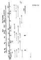
- the plasmid DNA was digested with PstI, electrophoretically analyzed and the DNA fragments were transferred from the gel to nitrocellulose filter and fixed. These DNA-carrying filters were hybridized with the radioactively labeled HRV2 cDNA, which filters were then exposed. The restriction sites were mapped and sequenced from the recombinant DNA fragments thus determined which were complementary to the HRV2-RNA. Clones representing the genome of HRV2 were obtained.
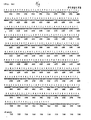
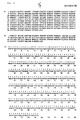
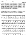
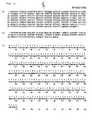
- the sequence of the HRV2 genome is shown in Fig. 4. It is shown in the form of the DNA as obtained from the sequencing of the recombinant DNA. To determine the sequence, 17 clones shown in FIG. 3 and an additional 25 additional clones were used. The sequence comprises 7102 nucleotides, the 3'-terminal poly-A not being counted. It contains an open reading frame which codes for a polyprotein with a length of 2150 amino acids. The reading frame begins with an AUG in position 611 and ends with a UAA stop codon 42 nucleotides before the start of Poly-A.
- the 5'-terminal sequence was obtained from clones Nos. 61, 100 and 109, using fragment A as a primer (see FIG. 3). Cleavage with PstI gave a fragment of constant length. It was determined by sequencing that all three clones contained the sequence TTAAAAC immediately subsequent to oligo-G, which comes from the integration site in the plasmid. It was concluded that these clones represent the 5'-terminus of the HRV2 genome. This sequence corresponds to the first 7 nucleotides of the three types of poliovirus, Coxsackie virus B1 (Hewlett, MJ and Florkiewicz, RZ, 1980, Proc. Natl. Acad.
- HRV2 and poliovirus type 1 also shows 5 blocks of identical sequence, of which only 2 are found in HRV14. This is a sequence of 16 nucleotides starting with base 436 and a sequence of 23 nucleotides starting with base 531 in HRV2.
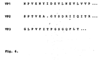
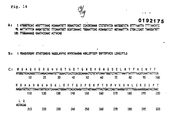
- the viral proteins are obtained from the polyprotein by proteolytic cleavage. To determine the cleavage sites, the viral coat proteins were isolated and the N-terminal amino acid sequence was determined (see FIG. 6). This not only unambiguously identified the cleavage sites in the viral envelope proteins, but also the reading frame which had been derived from the nucleotide sequence was confirmed. A comparison with the amino acid sequence derived from the nucleotide sequence shows that the VP4 / VP2 cleavage between glutamine and serine, the VP2 / VP3 cleavage between glutamine and glycine and the VP3 / VP1 cleavage between glutamine and asparagine.
- the present invention makes it possible to generate a DNA which contains the information for the viral RNA of HRV2.
- the invention relates not only to the gene sequences which code specifically for the viral proteins, but also to the modifications which can be obtained, for example, by mutation, degradation, transposition or addition. Any sequence that is degenerate compared to those shown is included. Also the sequences preferred under stringent conditions, for example under conditions used for more than 85% Selecting more than 90% homology, hybridizing with the sequences shown or parts thereof and coding for the proteins with the viral activity spectrum are included. The hybridizations are carried out in 6 x SSC / 5 x Denhardt's solution / 0.1% SDS at 65 ° C. The degree of stringency is determined in the washing step.
- the conditions which are suitable for selection for DNA sequence with approximately 85% or more homology are as follows: 0.2 x SSC / 0.01% SDS / 65 ° C. for selection for DNA sequences with approx. 90% or more homology: O, 1 x SSC / O, Ol% SDS / 65 ° C.
- this DNA can be incorporated both completely and in fragments into suitable plasmid vectors in order to either multiply the DNA after transformation of suitable host organisms or to achieve the expression of the proteins themselves.
- suitable hosts, vectors and the conditions for these operations are well known to those skilled in the art. Many works have also been published in which the synthesis of foreign proteins in bacteria is described using genetic engineering methods (review article, see Harris, TJR in "Genetic Engineering", Williamson, R., ed., 1983, Vol. 4. Academic Press, London, 127 ff.).
- the foreign DNA is introduced into the vicinity of suitable bacterial control regions (promoters, ribosome binding sites) of plasmids, which make it possible to express this information - usually in the form of fusion proteins - in high yields and thus to obtain the corresponding proteins.
- suitable bacterial control regions promoters, ribosome binding sites
- Prokaryotes for example E. coli K 12, strain 294 (ATCC No. 31 446) are preferred for expression.
- Other strains that are suitable include E. coli X 1776 (ATCC No. 31,537).
- E. coli W 3110 F - Lambda, Prototroph, ATCC No. 27325)
- bacilli such as Bacillus subtilis
- other Enterobacteriaceae such as Salmonella typhimurium or Serratia marcescens and various pseudomonas can also be used.
- plasmid vectors containing replicon and control sequences derived from species compatible with the host cells can be used in connection with these hosts.
- the vector usually carries recognition sequences which make it possible to select phenotypically in transformed cells.
- E. coli is usually transformed with pBR322, a plasmid derived from E. coli species (Bolivar, et al., Gene 2, 95 (1977)).
- pBR 322 contains genes for ampicillin and tetracycline resistance and thus provides simple means to identify transformed cells.
- the pBR322 plasmid or other plasmids must also contain promoters on their own or must be modified in such a way that they contain promoters which can be used by the microbial organism to express their own proteins.
- the promoters most commonly used in the production of recombinant DNA include beta-lactamase (penicillinase) and lactose promoter systems (Chang et al., Nature 275, 615 (1978); Itäkura et al., Science 198, 1056 (1977); Goeddel et al., Nature 281, 544 (1979)) and tryptophan (trp) promoter systems (Goeddel et al., Nucleic Acids Res. 8, 4057 (1980); European application, publication no 00 36 776). While the mentioned are the most common promoters, other microbial promoters have also been developed and used.
- the gene sequence according to the invention can be used, for example, under the control of the leftward promoter of the bacteriophage lambda (P L ).
- This promoter is one of the promoters known to be particularly strong that can be controlled. Control is made possible by the lambda repressor, from which neighboring restriction interfaces are known.
- a temperature sensitive allele of this gene can be inserted into a vector containing a viral DNA sequence. If the temperature is increased to 42 u C, the repressor is inactivated, and to its maximum concentration expressed the promoter. The sum of the mRNA produced under these conditions should be sufficient to obtain a cell that contains approximately 10% of its new synthetic ribonucleic acids derived from the P L promoter. In this way it is possible to establish a clone bank in which a functional viral DNA sequence
- a viral DNA sequence can also take place under the control of other regulatory systems which can be considered “homologous" to the organism in its untransformed form.
- regulatory systems which can be considered "homologous" to the organism in its untransformed form.
- chromosomal DNA from a lactose-dependent E. coli a lactose or lac operon, which enables the lactose breakdown by releasing the enzyme beta-galactosidase.
- the Lac control elements can be obtained from the bacteriophage Lambda-plac5, which is infectious to E. coli.
- the phage lac operon can be derived from the same bacterial species by transduction. Regulation systems that can be used in the method according to the invention can originate from plasmid DNA which is inherent in the organism.
- the Lac promoter operator system can be induced by IPTG.
- promoter-operator systems or parts thereof can also be used: for example arabinose operator, colicine E 1 operator, galactose operator, alkaline phosphatase operator, trp operator, xylose A operator, tac promoter and the like.
- the genes can preferably be expressed in the expression plasmid pERl03 (E. Rastl-Dworkin et al., Gene 21, 237-248 (1983); see also European patent application No. 83112812.9, deposit DSM 2773, December 20, 1983). These vectors contain all regulatory elements that lead to a high expression rate of the cloned genes.
- Saccharomyces cerevisiae is the most widely used among eukaryotic microorganisms, although a number of other species are widely available.
- Saccharomyces for example, the plasmid YRp7 (Stinchcomb, et al. Nature 282, 39 (1979); Kingsman et al., Gene 7, 141 (1979); Tschumper, et al., Gene 10, 157 (1980)) and the plasmid YEp 13 (Bwach et al., Gene 8, 121-133 (1979)) are commonly used.
- Plasmid YRp7 contains the TRP1 gene, which provides a selection marker: for a mutant yeast that is unable to grow in tryptophan-free medium; for example ATCC No. 44076.
- TRP1 damage as a characteristic of the yeast host genome is then an effective tool to detect the transformation by cultivating without tryptophan.
- the situation is very similar for the plasmid YEp13, which contains the yeast gene LEU 2, which can be used to supplement a LEU-2 mutant.
- Suitable promoter sequences for yeast vectors include the 5 'flanking region of ADH I (Ammerer, G., Methods of Enzymology 101, 192-201 (1983)), 3-phosphoglycerate kinase (Hitzeman, et al., J. Biol. Chem.
- glycolytic enzymes such as enolase, glyceraldehyde-3-phosphate dehydrogenase, hexokinase, pyruvate decarboxylase, phosphofructokinase, Glucose-6-phosphate isomerase, and glucokinase
- the termination sequences associated with these genes can also be inserted into the expression vector at the 3 'end of the sequence to be expressed in order to provide for polyadenylation and termination of the mRNA .
- promoters that also have the advantage of growth-controlled transcription are the promoter regions of alcohol dehydrogenase-2, isocytochrome C, acid phosphatase, degrading enzymes linked to nitrogen metabolism, the above-mentioned glyceraldehyde-3 -Phosphate dehydrogenase and enzymes that are responsible for processing maltose and galactose.
- Promoters that are regulated by the yeast mating type locus for example promoters of the BARI, MEC1, STE2, STE3, STE5 genes, can be used in temperature-regulated systems by using temperature-dependent sir mutations.
- cultures of multicellular organisms are also suitable host organisms.
- any of these cultures can be used, from vertebrate or invertebrate animal cultures.
- vertebrate or invertebrate animal cultures there is great interest in vertebrate cells, so that the multiplication of vertebrate cells in culture (tissue culture) has become a routine method in recent years.
- the gene is located, along with any necessary ribosome binding site, RNA splice site, polyadenylation site and transcriptional termination sequences.
- the control functions on the expression vectors are often designed from viral material.
- the promoters commonly used come from Polyoma, adenovirus 2, and particularly often from Simian virus 40 (SV 40).
- SV 40 Simian virus 40
- the SV 40 start and end promoters are particularly useful because both are easily obtained from the virus as a fragment that also contains the SV 40 viral replication site (Fiers et al.,
- Smaller or larger fragments of the SV 40 can also be used, provided that they contain the approximately 250 bp long sequence which extends from the Hind III site to the Bgl 1 site in the viral replication site.
- promoter or control sequences that are normally linked to the desired gene sequences, provided that these control sequences are compatible with the host cell systems.
- a replication site can either be provided by appropriate vector construction in order to incorporate an exogenous site, for example from SV 40 or other viral sources (for example Polyoma, Adeno, VSV, etc.) or can be provided by the host cell's chromosomal replication mechanisms. If the vector is integrated into the host cell chromosome, the latter measure is usually sufficient.
- Transformation of the cells with the vahicles can be achieved by a variety of methods. For example, it can be done by calcium, whereby either the cells are washed in magnesium and the DNA is added to the cells suspended in calcium or the cells are coprecipitated by DNA and. Calcium phosphate will be exposed. With subsequent gene expression, the cells are transferred to media which select for transformed cells.
- the product After transformation of the host, expression of the gene and fermentation or cell cultivation under conditions in which the protein is expressed, the product can usually be extracted by known chromatographic separation methods in order to obtain a material which contains the viral protein with or without leader and Contains tailing sequences.
- the protein can be expressed with a leader sequence at the N-terminus (pre-protein), which can be removed by some host cells. If not, cleavage of the leader polypeptide (if present) is required to obtain mature protein. Alternatively, the mature protein can be produced directly in the microorganism.
- the precursor sequence of the yeast mating pheromone MF-alpha-1 can be used in order to ensure correct "maturation" of the fused protein and the excretion of the products in the growth medium or in the periplasmic space.
- the DNA sequence for functional or mature protein can be linked to M p- alpha-1 at the suspected interface.
- DNA according to the invention for the production of the proteins in question in bacteria or eukaryotic cells, it can also be used to produce the amino acid sequences derived from the nucleotide sequence synthetically, in whole or in part.
- oligopeptides like the genetically engineered proteins, can then either be used to stimulate an immune response against intact viruses, or to bind and block cellular receptors.
- Studies on the use of oligopeptides to stimulate an immune response against polioviruses have been published (Emini, EA, Jameson, BA and Wimmer, E., 1983, Nature (London) 30, 699-703; similar studies are also available in foot and mouth Claw disease viruses have been performed (Bittle, JL, Houghton, RA, Alexander, H., Shinnick, TM, Sutcliffe, JG, Lerner, RA, Rowlands, DJ and Brown, F., 1982 Nature (London), 298, 30-33; Pfaff, E., Mussgay, M., Böhm, HO, Schulz, GE and Schaller, H., 1982, EMBO J.
- This invention also extends to the oligo- and polypeptide portions of HRV2 proteins which, as a result of the synthesis or for the purpose of application, for example as a vaccine, are linked to other oligo- or polypeptides.
- the invention also relates to DNA molecules which code for at least one viral protein of the rhinovirus strain HRV2 and which, under stringent conditions which make it possible to recognize a homology which is greater than 85%, with one of the specified DNA molecules or with degenerate variations of these molecules hybridize.
- the DNA molecules according to the invention can be incorporated into a suitable expression vehicle, for example into a plasmid which is replicable in microorganisms, preferably in prokaryotes, eukaryotes or in mammalian cells.
- the present invention also relates to DNA molecules which code for proteins which have the biological activity of at least one of the proteins according to the invention.
- the invention furthermore relates to transformed host organisms which contain the genetic information coding for the viral proteins according to the invention, preferably prokaryotes, eukaryotes or mammalian cell lines, in particular E. coli.
- the genetic information is preferably contained in vehicles that are replicable in the host organism in question.
- the invention further relates to polypeptides which have the biological activity of at least one of the viral proteins of the rhinovirus strain HRV2 and / or are encoded by one of the DNA molecules described, in particular polypeptides with the amino acid sequence for VP1, VP2, VP3, VP4, P2A, P2B , P2C, P3A or P3C preferably with the amino acid sequence according to FIG. 4 or parts thereof.
- Polypeptides which correspond in part or in total to at least one of the viral proteins of the rhinovirus strain HRV2 polypeptides which contain at least two of the viral proteins linked in any combination and order and polypeptides which are derived from the amino acid sequence in accordance with. 4 or derived from parts thereof and / or bind to and / or block the cellular receptors for the rhinovirus strain HRV2 are also the subject of the present invention.
- the DNA molecules according to the invention are produced by isolating the viral RNA of the rhinovirus strain HRV2, producing the complementary DNA, incorporating the viral cDNA / RNA hybrid into a suitable replicable vector and transforming a suitable host organism with this vector.
- polypeptides according to the invention are obtained by transforming a suitable host organism, preferably a prokaryote, eukaryote or a mammalian cell, in particular E. coli, with the genetic information according to the invention which codes for a viral polypeptide according to the invention, expressing the information and a viral polypeptide according to the Invention is isolated.
- a suitable host organism preferably a prokaryote, eukaryote or a mammalian cell, in particular E. coli
- polypeptides according to the invention can be used for therapeutic treatment, for example for stimulating the immune system and for binding and / or blocking the cellular receptors for the rhinovirus strain HRV2.
- polypeptides according to the invention can be used in the form of medicaments which, in addition to pharmaceutically inert auxiliaries or carriers, contain an effective amount of at least one of the polypeptides according to the invention.
- HeLa cells HeLa-Ohio strain, 03-147, Flow Laboratories, England
- the suspension medium Thimas, DC, Conant, RM and Hamparian, VU, 1970, Proc. Soc. Exp. Biol. Med. 133, -62-65; Stott, EJ and Heath, GF, 1970, J. Gen. Virol, 6, 15-24
- the suspension medium consisted of a Joklik modification of MEM for suspension (Gibco 072-1300) and 7% horse serum (Seromed 0135).
- the inoculation was 5 - 10 x 10 4 cells / ml, the volume 500 ml
- the suspension was Ellen at a cell density of 1 x 10 6 cells / ml centrifuged under sterile conditions at 300 g for 10 min..
- the supernatant was aspirated and the cells were resuspended in 100 ml of infection medium (Joklik modification of MEM for suspension culture with 2% horse serum and 2 mM MgCl 2 ).
- the cells were distributed homogeneously in the infection medium by repeated careful suction in a 20 ml pipette. It was then made up to 500 ml.
- the cell suspension was then brought to 34 ° C.
- HRV2 human immunodeficiency virus
- the HRV2 strain was developed by Dr. D. T y rrell (Common Cold Center, Salisbury, England) and can also be obtained from the American Type Culture Collection (ATCC VR-482 and ATCC VR-1112). The strain used was neutralized by antiserum against HRV2 (American Type Culture Collection, Cat.No. ATCC VR-1112AS / GP). An antiserum against HRV7 (Cat.No. ATCC VR-1117 AS / GP), which showed no neutralization, served as control serum. After 40 hours at 34 ° C., the virus was harvested.
- Virus was obtained from the cells or cell fragments as well as from the medium.
- the medium was separated from infected cells and cell fragments by centrifugation at 1500 g for 10 min and suctioned off.
- the precipitate was frozen at -70 ° C.
- the cell precipitates from 12 l suspension culture were combined, resuspended in 40 ml TM buffer (20 mM Tris / HCl, pH 7.5, 2 mM MgCl 2 ), placed on ice for 15 min, then broken up in a dounce homogenizer and the mixture 30 centrifuged at 6000 g for min. The precipitate was then washed again in 10 ml of TM buffer. The two supernatants were combined and centrifuged at 5000 g for 3 hours to pellet the virus.
- TM buffer (20 mM Tris / HCl, pH 7.5, 2 mM MgCl 2
- the virus pellet was then taken up in 10 ml of KTMP buffer (50 mM KCl, 50 mM Tris / HCl, pH 7.5, 5 mM MgCl 2 , 2 mM mercaptoethanol, 1 mM puromycin, 0.5 mM GTP) and after the addition of 150 ⁇ g DNase I (Sigma, ribonuclease free) incubated on ice for 1 hour.
- KTMP buffer 50 mM KCl, 50 mM Tris / HCl, pH 7.5, 5 mM MgCl 2 , 2 mM mercaptoethanol, 1 mM puromycin, 0.5 mM GTP
- 150 ⁇ g DNase I Sigma, ribonuclease free
- the virus was precipitated from the infection medium with stirring at 4 ° C. with polyethylene glycol 6000 (PEG 6000; Merck) at a concentration of 7% and 450 mM NaCl (Korant, BD, Lonberg-Holm, K., Noble, J. and Stasny , JT, 1972, Virology 48, 71-86). After 4 hours in the cold, the virus was centrifuged at 1500 g for 30 min, the precipitate was resuspended in 10 ml of KTMP buffer containing 75 ⁇ g DNase I, the mixture was incubated on ice for 1 hour and then frozen at -70 ° C.
- PEG 6000 polyethylene glycol 6000
- the virus suspensions which had been obtained from the cells and from the medium, were combined, incubated for 5 min at 37 ° C., by adding 60 ml of cold TE buffer (10 mM Tris / HCl, pH 7.4, 1 mM EDTA ) cooled and then sonicated in an ice bath for 5 minutes.
- cold TE buffer (10 mM Tris / HCl, pH 7.4, 1 mM EDTA
- the mixture was then centrifuged at 6000 g for 30 min. 920 ml of TE buffer containing 7% PEG 6000 and 450 mM NaCl were added to the supernatant, the mixture was carefully stirred at 4 ° C. for 4 hours and the precipitate formed was pelleted at 6000 g for 30 minutes. The precipitate was again taken up in 100 ml of TM buffer, the virus was precipitated as above by adding PEG 6000 and NaCl and pelleted. The precipitate was resuspended in 40 ml of TM buffer, the suspension was centrifuged at 6000 g for 30 min, and the virus was pelleted at 85000 g for 3 hours.
- the precipitate was dissolved in 1 ml of TM buffer, after the addition of 50 ⁇ g DNase I incubated at 4 ° C. for 1 hour and then 1 ml of TE buffer was added.
- the virus suspension was centrifuged on sucrose gradients (10-30% w / w in TE buffer) for 4 hours at 4 ° C at 85000 g.
- the fractions containing the virus were determined from the absorbance at 260 nm and diluted with TM buffer so that the final concentration of sucrose was 10%.
- the mixture was then centrifuged at 85,000 g for 8 hours.
- the virus pellet was taken up in 1 ml of TM buffer and stored at -70 ° C.
- the virus was suspended in 1 ml of NTES buffer (100 mM NaCl, 10 mM Tris / HCl, pH 9, 1 mM EDTA, 0.1% sodium dodecyl sulfate) and extracted with phenol. Chloroform was added to improve phase separation. 20 ⁇ l of 5 M NaCl and 20 ⁇ l of 3 M sodium acetate (pH 5.6) were added to the aqueous phase and the viral RNA was precipitated with twice the volume of ethanol.
- NTES buffer 100 mM NaCl, 10 mM Tris / HCl, pH 9, 1 mM EDTA, 0.1% sodium dodecyl sulfate
- the RNA was then digested in NTES buffer with 1 mg / ml Proteinase K (Merck) for 15 min at 37 ° C.
- the proteinase K stock solution 50 mg / ml had been preincubated at 37 ° C. for 15 minutes before use.
- the solution was extracted with phenol / chloroform as described above and the RNA was precipitated by adding ethanol.
- RNA was then loaded onto a 2% agarose electrophoresed with 0.1% sodium dodecyl sulphate agarose gel in TAE buffer (10 mM NaAc, 40 mM T ris / acetate, p H 8.2, 2 mM EDTA). After staining with ethidium bromide, the band of the intact HRV2-RNA was visible (Fig. 2). A weak, diffuse band underneath indicated a slight degradation of the HRV2-RNA.
- the extension of the HRV2-RNA-cDNA hybrids was carried out according to the method of Roychoudhury and Wu (1980, loc.cit.).
- the HRV2-RNA-cDNA hybrid was dissolved in 50 ⁇ l TT buffer (200 mM potassium cocodylate, 25 mM Tris / HCl, pH 6.9, 0.5 mM CoCl 2 , 2 mM dithiothreitol in the presence of 2 nmol [X 32 P] dCTP (5 Ci / mmol) with 25 U terminal transferase (Pharmacia PL Biochemicals) incubated for 5 min at 37 ° C.
- RNA-cDNA hybrid was in 100 ul NTE buffer (100 mM NaCl, 10 mM Tris / HCl, pH 7.6, 1 mM EDTA) with 0.3 pmol pBR322 plasmid (which was cut with PstI and to which oligo-dG residues had been polymerized; Bethesda Research Laboratories) were added, heated first at 65 ° C. for 5 min, then at 42 ° C. for 2 hours, slowly cooled to room temperature overnight and stored at 4 ° C.
- the HB 101 strain was grown in 50 ml LB medium (10 g tryptone, 5 g yeast extract, 10 g NaCl in 1 l) (Mandel, M. and Higa, A., 1970, J. Mol.Biol. 53, 159-162).
- the bacteria were pelleted and in 25 ml of TR buffer (1.50 mM KCl, 50 mM CaCl 2 , 1 mM Tris / HCl, pH 7, 3 mM MgCl 2 ) taken up, placed on ice for 30 min, centrifuged again, resuspended in 2 ml of TR buffer and placed on ice for 1 hour.
- the plasmid DNA was in 25 ul RE buffer (6 mM MgCl 2 , 10 mM Tris / HCl, pH 7.5, 6 mM mercaptoethanol) with 50 mM NaCl in the presence of 2 U of the restriction enzyme PstI (Bethesda Research Laboratories) and 5 ⁇ g ribonuclease A incubated at 37 ° C for 2 hours. The samples were then electrophoresed on a 1.4% agarose gel. The sizes of the inserts could be determined by staining with ethidium bromide and comparison with Lambda Hind III marker DNA. These were between 300 and 2000 base pairs.
- plasmids were obtained as described above from 200 ml cultures of tetracycline-resistant, ampicillin-sensitive bacterial clones and digested with PstI.
- the plasmid pUC9 (Vieisa, J. and Messing, JG, 1982, Gene 19, 259-268) contains a gene for ampicillin resistance, a region for the start of replication, which originates from the plasmid pBR322, and part of the Lac- Z coli from E. coli. A small section of DNA containing a number of restriction sites is in this Lac-Z region, so cloning DNA in one of these sites disrupts the Lac-Z gene region.
- X-Gal 5-Bromo-4 chloro-3-indolyl- ⁇ -D-galactoside, Bethesda Research Laboratories
- indicator plates those without inserts appear blue (Rüther , U., 1980, Mol. Gen. Genet. 178, 475-478).
- recombinant pBR322 clones (approx. 7 ⁇ g) were digested with PstI and, after separation on the agarose gel (1.2% - 1.4%), the DNA inserts from the vector DNA separated.
- the DNA was recovered from the gel by electroelution and ethanol precipitation as described above.
- the isolated DNA insert was in 20 ul RE buffer with 0.4 ug pUC9 vector (cut with PstI and pretreated with bacterial alkaline phosphatase), in the presence of 1 mM ATP and 3 UT 4 ligase (Bethesde Research Laboratories) 1 Incubated for 1 hour at 15 ° C and stored at 4 ° C (Vieisa, J. and Messing, JG, 1982, loc.cit).
- E. coli strain JM101 cells New England Biolabs
- 200 ⁇ l of the competent cell suspension were mixed with 20 ⁇ l mixture of the pUC9 ligase reaction and incubated for 1 hour at 0 ° C.
- the cells were with 10 ⁇ 1 200 mM isopropylthiogalactoside (Sigma), 50 ul of a solution of 20 mg X-Gal in 1 ml of dimethylformamide and 1 ml of LB medium was added and 1 hour at Incubated at 37 ° C. Each 200 ⁇ l of this cell suspension was then transferred to an ampicillin LB agar plate (100 ⁇ g / ml) and incubated overnight at 37 ° C in the incubator. Positive transformants were identified as white colonies, which were examined for DNA inserts using the "mini-plasmid preparation technique".
- E. coli JM 101 which had been transformed with pUC9 and contained DNA inserts, were grown up in 200 ml LB medium (with 100 ⁇ g ampicillin / ml) and the plasmid DNA isolated (Birnboim, HC and Doly, J., 1979 loc. Cit.)
- the plasmid DNA was then dissolved in 100 ⁇ l of TE buffer and the solution was chromatographed on a Sephacryl 100 column (1 ⁇ 20 cm) with TE buffer.
- the fractions containing the purified plasmid were localized based on the absorbance, pooled and lyophilized.
- the plasmid was taken up in 500 ⁇ l TE buffer, incubated for 5 min at 65 ° C. and extracted again with phenol / chloroform and precipitated with ethanol.
- Clones 773 and 87 which contained corresponding DNA inserts, were subcloned into pUC9, grown up in 200 ml LB medium (with 100 ⁇ g ampicillin / ml) and the plasmids isolated as described above.
- the primer fragment (59 nucleotides) from clone 773 was obtained by digestion with the restriction enzymes AhaIII and EcoRI (the primer fragment is designated as fragment A in FIG. 3).
- the band corresponding to the 59 base EcoRI / AhaIII fragment was cut out after ethidium bromide staining, the fragment was electroeluted in 0.05 ⁇ TBE buffer and the DNA was precipitated with ethanol.
- the DNA - fragment was then incubated in 50 ul 100 mM Tris / HCl, pH 8, with 200 U of bacterial alkaline phosphatase (Bethesda Research Laboratories) for 1 hour at 65 ° C. After the reaction, the aqueous phase 2 was added 2.5 ul 0.5 M EDTA, pH 8, was added, extracted with P x henol / chloroform and the DNA precipitated by ethanol precipitation.
- the DNA was dissolved in water and in 50 ⁇ l K buffer (10 mM MgCl 2 , 50 mM Tris / HCl, pH 8.5, 5 mM dithioerythritol) with 20 ⁇ Ci ⁇ - [ 32 P] -ATP (specific activity 5000 Ci / mmol; Amersham International) and 5 U polynucleotide kinase (Pharmacia-PL Laboratories) at 37 ° C for 30 min. The P-labeled fragment was then precipitated and taken up in 30 ul 30% dimethyl sulfoxide containing 1 mM EDTA and 0.01% xylene cyanogen bromophenol blue. This solution was incubated at 90 ° C.
- K buffer 10 mM MgCl 2 , 50 mM Tris / HCl, pH 8.5, 5 mM dithioerythritol
- 20 ⁇ Ci ⁇ - [ 32 P] -ATP specific activity
- the strips were then moistened with 2 ⁇ SSC and in a plastic film together in 1 ml of H buffer (400 mM NaCl, 40 mM PIPES, pH 6.4, 1 mM EDTA, 80% formamide) with 4 ⁇ g denatured salmon sperm DNA (Sigma; incubated at 100 ° C for 2 min and cooled to 0 ° C) incubated at 42 ° C for 1 hour.
- H buffer 400 mM NaCl, 40 mM PIPES, pH 6.4, 1 mM EDTA, 80% formamide
- 4 ⁇ g denatured salmon sperm DNA Sigma; incubated at 100 ° C for 2 min and cooled to 0 ° C
- the two nitrocellulose strips were then sealed separately in plastic films with 0.5 ml H buffer, 4 ⁇ g denatured salmon sperm DNA and an aliquot of the isolated strands (20,000 cpm) and hybridized overnight at 42 ° C.
- the filters were washed twice with 2 x SSC for 10 min at 50 ° C and twice with 0.1 x SSC, 0.1% sodium dodecyl sulfate for 30 min at 50 ° C and the radioactivity was determined.
- the primer fragment from pHRV2-87 was isolated in the same way.
- the fragment lies between two RsaI restriction sites (68 nucleotides) and was obtained by digestion with this restriction enzyme.
- the RsaI fragment is designated as fragment B in FIG. 3. Strand separation and hybridization were carried out as described above.
- the solution was then in 100 ul RT buffer in the presence of 140 U reverse transcriptase (Anglian Biotechnology Co, Cambridge), 8 U ribonuclease inhibitor (RNasin, Bethesda Research Laboratories), each 0.2 mM dATP, dCTP, dGTP and dTTP , '30 ⁇ Ci [X 32 P] dCTP and 5 mM dithioerythritol incubated at 42 ° C for 2 hours.
- the resulting reverse transcript was processed as described above.
- Plasmid DNA from the recombinants was isolated from 3 ml cultures (Birnboim, H.C. and Doly, J., 1979, loc. Cit).
- the DNA was then incubated with the restriction enzyme PstI and the samples were analyzed by electrophoresis on 1.4% agarose gel. The gels were stained with ethidium bromide. The DNA was then transferred from the gel to nitrocellulose filters. (Southern, EM, 1975, J. Mol, Biol. 98, 503-517) and fixed on the nitrocellulose by a 2-hour incubation at 80 ° C. The filters were washed in 50% formamide, 1 x Denhardts solution (Denhardt, DT, 1966, Biochem.Biophys. Res.
- Radioactive HRV2 cDNA was produced as described above, only the reaction mixture contained 50 ⁇ Ci [X- 32 P] dCTP.
- the cDNA-HRV2-RNA hybrid was denatured at 100 ° C for 90 seconds.
- the filters were incubated with radioactive HRV2 cDNA as described above at 42 ° C.
- the DNA inserts were digested using restriction enzymes (New England Biolabs and Bethesda Research Laboratories) using the incubation conditions specified by the manufacturers.
- the result of the restriction mapping is shown in Fig. 3.
- the plasmids pHRV2-1 and pHRV2-24 contained - immediately following the oligo-C, which is due to the chain extension by the terminal transferase reaction - a longer sequence of A residues, which part of the 3'-terminal poly- Represent A of HRV2 RNA.
- the other plasmids were arranged relative to pHRV2-1 and pHRV2-24 with the help of characteristic cleavage sites of individual restriction enzymes. DNA inserts that were smaller than 500 base pairs were not classified by restriction enzyme mapping, but were immediately subcloned into pUC9 and sequenced (see Example 3).
- the identification of the remaining clones is obtained by colony hybridization using already mapped DNA inserts as "nick translation” samples according to the method of Grunstein and Hogness (Grunstein, M. and Hogness, DS, 1975, Proc. Natl. Acad. Sci USA 72, 3961-3965).
- 32 P-labeled DNA samples are obtained with the aid of a "nick translation kit” from Amersham International (England; Amersham Kit No. 5000) according to the manufacturer's instructions with [X -32 P] dCTP (3000 Ci / mmol ).
- the labeled DNA was separated on a Biogel P-30 column in a Pasteur pipette in TE buffer.
- DNA inserts were subcloned into pUC-9 as described above and thus competent E.coli JM 101 cells were transformed. Positive transformants were isolated as white colonies and grown up in LB medium (with 100 ⁇ g / ml ampicillin). 10-20 ⁇ g of DNA were digested in 100 ⁇ l overnight under standard reaction conditions with restriction enzymes that have a cleavage site in the plasmid, for example in the polylinker region of pUC9 (for example BamHI, EcoRI, AccI, HindIII). The restricted fragment was then dephosphorylated.
- restriction enzymes that have a cleavage site in the plasmid, for example in the polylinker region of pUC9 (for example BamHI, EcoRI, AccI, HindIII). The restricted fragment was then dephosphorylated.
- the DNA was then in 50 ⁇ l 50 mM Tris / HCl, pH8, 10 mM MgCl 2 , 5 mM dithioerythritol, with 25 ⁇ Ci [X- 32 P] ATP (5000 Ci / mmol, Amersham International) and 4 UT 4 polynucleotide kinase ( Pharmacia-PL Biochemicals) incubated for 30 min at 37 ° C and the labeled DNA precipitated with ethanol. With the aid of a further restriction enzyme which cleaves in the polylinker region of pUC9, recombinant DNA which was labeled with 32 P in one strand was obtained.
- the first gel run of a reaction took until the bromophenol blue marker, the second until the xylene cyan marker reached the gel end.
- the gels were then fixed in 10% acetic acid and 10% methanol (approx. 2 liters) for 20 min, transferred to 3 MM filter paper and dried at 80 ° C. on a gel dryer. Then the gels without intensifying screen at -70 0 C were determined using a XAR-Omat film (Kodak) is exposed (about 18 to 36 hours).
- the recombinant DNA was cut with the restriction enzyme Sau3AI and cloned into the BamHI site of M13 mp9 using a "Sequencing pack" (New England Biolabs, Cat. No. 409). Sequencing was carried out according to the chain termination method (Sanger, F., Nicklen, S. and Coulson, A.S., 1977, Proc. Natl. Aca. Sci USA 74, 5463 - 5467).
- the viral proteins are obtained from the polyprotein by proteolytic cleavage. In order to identify the cleavage sites, the viral envelope proteins were isolated and the N -terminal amino acid sequence was determined. For this purpose, the proteins of 2 mg HRV2 were separated electrophoretically on a 12.5% polyacrylamide gel (Laemmli, UK, 1970, loc.cit.). The gels were stained with a saturated solution of Coomassie brilliant blue in 50 mM Tris / HCl, pH 7.4, and the protein bands were cut out. The individual proteins were electroeluted in an ISCO elution apparatus at 50 V for 16 hours and precipitated with trichloroacetic acid.
- N-terminal amino acids were determined using an AB-470A protein sequencer (Applied Biosystems, Inc., Foster City, CA, USA). 2 nmol VP1 and VP2 and 1 nmol VP3 were used for sequencing. The derivatized amino acids were analyzed by HPLC. The N-terminal sequences of the individual proteins are given in FIG. 6.
- the filter with the bound proteins was 16 hours in 2 0 ml of PBS (137 mM NaCl, 2.7 mM KC1, 6.6 mM Na 2 HP0 4, 1.5 mM KH 2 P0 4) with 3% bovine serum albumin (BSA ) and 3% Tween 20 bathed.
- PBS 137 mM NaCl, 2.7 mM KC1, 6.6 mM Na 2 HP0 4, 1.5 mM KH 2 P0 4
- BSA bovine serum albumin
- the filter was washed three times for 20 min in 20 ml PBS with 1% BSA and 1% Tween 20 (PBSBT) and then Incubated for 2 hours in 20 ml PBS BT with 1 0 ⁇ Ci 125 I-labeled protein from Staphylococcus aureus (approx. 1 mCi / mg), washed three times for 20 min with PBSBT and 3 x 5 min in PBS, briefly rinsed twice with H 2 0 and dried overnight between several layers of filter paper.
- the radioactivity bound to the filter was determined by autoradiography on Kodak XAR5 X-ray film (20 hours / -70 ° C). As can be seen from FIG. 7, antibodies against VP1 are mainly present in the antiserum.
Landscapes
- Life Sciences & Earth Sciences (AREA)
- Health & Medical Sciences (AREA)
- Chemical & Material Sciences (AREA)
- Organic Chemistry (AREA)
- Genetics & Genomics (AREA)
- Engineering & Computer Science (AREA)
- Biophysics (AREA)
- Zoology (AREA)
- Biochemistry (AREA)
- General Health & Medical Sciences (AREA)
- Molecular Biology (AREA)
- Wood Science & Technology (AREA)
- Bioinformatics & Cheminformatics (AREA)
- General Engineering & Computer Science (AREA)
- Biotechnology (AREA)
- Biomedical Technology (AREA)
- Virology (AREA)
- Physics & Mathematics (AREA)
- Plant Pathology (AREA)
- Gastroenterology & Hepatology (AREA)
- Microbiology (AREA)
- Proteomics, Peptides & Aminoacids (AREA)
- Medicinal Chemistry (AREA)
- Micro-Organisms Or Cultivation Processes Thereof (AREA)
- Preparation Of Compounds By Using Micro-Organisms (AREA)
- Peptides Or Proteins (AREA)
- Medicines Containing Antibodies Or Antigens For Use As Internal Diagnostic Agents (AREA)
Applications Claiming Priority (2)
| Application Number | Priority Date | Filing Date | Title |
|---|---|---|---|
| DE3505148 | 1985-02-15 | ||
| DE19853505148 DE3505148A1 (de) | 1985-02-15 | 1985-02-15 | Polypeptide des rhinovirusstammes hrv2 sowie die hierfuer codierenden dna-molekuele |
Publications (2)
| Publication Number | Publication Date |
|---|---|
| EP0192175A2 true EP0192175A2 (fr) | 1986-08-27 |
| EP0192175A3 EP0192175A3 (fr) | 1988-01-20 |
Family
ID=6262562
Family Applications (1)
| Application Number | Title | Priority Date | Filing Date |
|---|---|---|---|
| EP86101746A Ceased EP0192175A3 (fr) | 1985-02-15 | 1986-02-12 | Polypeptides de la souche rhinovirus HRV2 et molécules de l'ADN les codant |
Country Status (13)
| Country | Link |
|---|---|
| EP (1) | EP0192175A3 (fr) |
| JP (1) | JPS62279A (fr) |
| KR (1) | KR860006547A (fr) |
| AU (1) | AU599895B2 (fr) |
| DE (1) | DE3505148A1 (fr) |
| DK (1) | DK71986A (fr) |
| ES (2) | ES8704207A1 (fr) |
| FI (1) | FI860677A7 (fr) |
| GR (1) | GR860432B (fr) |
| NO (1) | NO860550L (fr) |
| NZ (1) | NZ215173A (fr) |
| PT (1) | PT82026B (fr) |
| ZA (1) | ZA861100B (fr) |
Cited By (12)
| Publication number | Priority date | Publication date | Assignee | Title |
|---|---|---|---|---|
| EP0261403A3 (fr) * | 1986-08-23 | 1988-04-13 | BOEHRINGER INGELHEIM INTERNATIONAL GmbH | Polypeptide de rhinovirus souche HRV89, et molécule d'ADN qui l'encode |
| EP0287395A1 (fr) * | 1987-04-16 | 1988-10-19 | The Wellcome Foundation Limited | Peptides de rhinovirus humain |
| WO1992003555A1 (fr) * | 1990-08-28 | 1992-03-05 | Boehringer Ingelheim International Gmbh | Mutation du rvh2 2a |
| US5589453A (en) * | 1988-09-01 | 1996-12-31 | Molecular Therapeutics, Inc. | Human rhinovirus receptor protein (ICAM-1) that inhibits rhinovirus attachment and infectivity |
| US5674982A (en) * | 1990-07-20 | 1997-10-07 | Bayer Corporation | Multimeric form of human rhinovirus receptor protein |
| US5686582A (en) * | 1990-07-20 | 1997-11-11 | Bayer Corporation | Multimeric forms of human rhinovirus receptor protein |
| US6051231A (en) * | 1988-09-01 | 2000-04-18 | Bayer Corporation | Antiviral methods and prepations |
| US6096862A (en) * | 1990-07-20 | 2000-08-01 | Bayer Corporation | Multimeric antiviral agent |
| US6107461A (en) * | 1990-07-20 | 2000-08-22 | Bayer Corporation | Multimeric forms of human rhinovirus receptor and fragments thereof, and method of use |
| US6143298A (en) * | 1988-09-01 | 2000-11-07 | Bayer Corporation | Soluble truncated forms of ICAM-1 |
| US6326004B1 (en) | 1988-09-01 | 2001-12-04 | Bayer Corporation | Antiviral methods using fragments of human rhinovirus receptor (ICAM-1) |
| US7132395B1 (en) | 1988-09-01 | 2006-11-07 | Bayer Pharmaceuticals Corporation | Antiviral methods using human rhinovirus receptor (ICAM-1) |
Families Citing this family (3)
| Publication number | Priority date | Publication date | Assignee | Title |
|---|---|---|---|---|
| EP0321973B1 (fr) * | 1987-12-23 | 1993-11-03 | BOEHRINGER INGELHEIM INTERNATIONAL GmbH | Expression de la protéase P2A de HRV2 codée par un virus |
| ES2068855T3 (es) * | 1988-07-25 | 1995-05-01 | Boehringer Ingelheim Int | Sintesis in vitro de un arn infeccioso. |
| DE4136443A1 (de) * | 1991-11-06 | 1993-05-13 | Boehringer Ingelheim Int | Expression der reifen proteinase 2a, ihre partielle reinigung und bereitstellung kompetitiv wirkender substrate |
Family Cites Families (4)
| Publication number | Priority date | Publication date | Assignee | Title |
|---|---|---|---|---|
| NL8303501A (nl) * | 1983-10-12 | 1985-05-01 | Centraal Diergeneeskundig Inst | Deletiemutant van een herpesvirus en vaccin, dat dit virus bevat. |
| CA1339735C (fr) * | 1984-04-27 | 1998-03-17 | The University Of Melbourne | Clonage et sequencage du gene majeur de glycoproteine de la capside exterieure d'un rotavirus humain |
| EP0169146A3 (fr) * | 1984-07-20 | 1988-07-20 | Merck & Co. Inc. | Anticoprs monoclonaux dirigés contre le récepteur cellulaire du rhinovirus humain |
| AU5736286A (en) * | 1985-05-14 | 1986-11-20 | Commonwealth Scientific And Industrial Research Organisation | Rotavirus antigens |
-
1985
- 1985-02-15 DE DE19853505148 patent/DE3505148A1/de not_active Ceased
-
1986
- 1986-02-12 EP EP86101746A patent/EP0192175A3/fr not_active Ceased
- 1986-02-13 GR GR860432A patent/GR860432B/el unknown
- 1986-02-14 ES ES552026A patent/ES8704207A1/es not_active Expired
- 1986-02-14 ZA ZA861100A patent/ZA861100B/xx unknown
- 1986-02-14 FI FI860677A patent/FI860677A7/fi not_active IP Right Cessation
- 1986-02-14 NO NO860550A patent/NO860550L/no unknown
- 1986-02-14 DK DK71986A patent/DK71986A/da not_active Application Discontinuation
- 1986-02-14 NZ NZ215173A patent/NZ215173A/en unknown
- 1986-02-14 JP JP61030592A patent/JPS62279A/ja active Pending
- 1986-02-14 PT PT82026A patent/PT82026B/pt not_active IP Right Cessation
- 1986-02-14 AU AU53610/86A patent/AU599895B2/en not_active Ceased
- 1986-02-15 KR KR1019860001053A patent/KR860006547A/ko not_active Ceased
- 1986-11-04 ES ES557193A patent/ES8706827A1/es not_active Expired
Cited By (15)
| Publication number | Priority date | Publication date | Assignee | Title |
|---|---|---|---|---|
| EP0261403A3 (fr) * | 1986-08-23 | 1988-04-13 | BOEHRINGER INGELHEIM INTERNATIONAL GmbH | Polypeptide de rhinovirus souche HRV89, et molécule d'ADN qui l'encode |
| EP0287395A1 (fr) * | 1987-04-16 | 1988-10-19 | The Wellcome Foundation Limited | Peptides de rhinovirus humain |
| US6051231A (en) * | 1988-09-01 | 2000-04-18 | Bayer Corporation | Antiviral methods and prepations |
| US7132395B1 (en) | 1988-09-01 | 2006-11-07 | Bayer Pharmaceuticals Corporation | Antiviral methods using human rhinovirus receptor (ICAM-1) |
| US5589453A (en) * | 1988-09-01 | 1996-12-31 | Molecular Therapeutics, Inc. | Human rhinovirus receptor protein (ICAM-1) that inhibits rhinovirus attachment and infectivity |
| US6514936B1 (en) | 1988-09-01 | 2003-02-04 | Bayer Corporation | Antiviral methods using human rhinovirus receptor (ICAM-1) |
| US6326004B1 (en) | 1988-09-01 | 2001-12-04 | Bayer Corporation | Antiviral methods using fragments of human rhinovirus receptor (ICAM-1) |
| US6143298A (en) * | 1988-09-01 | 2000-11-07 | Bayer Corporation | Soluble truncated forms of ICAM-1 |
| US5686582A (en) * | 1990-07-20 | 1997-11-11 | Bayer Corporation | Multimeric forms of human rhinovirus receptor protein |
| US6096862A (en) * | 1990-07-20 | 2000-08-01 | Bayer Corporation | Multimeric antiviral agent |
| US6107461A (en) * | 1990-07-20 | 2000-08-22 | Bayer Corporation | Multimeric forms of human rhinovirus receptor and fragments thereof, and method of use |
| US5871733A (en) * | 1990-07-20 | 1999-02-16 | Bayer Corporation | Multimeric forms of human rhinovirus receptor protein |
| US5686581A (en) * | 1990-07-20 | 1997-11-11 | Bayer Corporation | Multimeric form of human rhinovirus receptor protein |
| US5674982A (en) * | 1990-07-20 | 1997-10-07 | Bayer Corporation | Multimeric form of human rhinovirus receptor protein |
| WO1992003555A1 (fr) * | 1990-08-28 | 1992-03-05 | Boehringer Ingelheim International Gmbh | Mutation du rvh2 2a |
Also Published As
| Publication number | Publication date |
|---|---|
| EP0192175A3 (fr) | 1988-01-20 |
| FI860677A0 (fi) | 1986-02-14 |
| DK71986D0 (da) | 1986-02-14 |
| AU5361086A (en) | 1986-08-21 |
| AU599895B2 (en) | 1990-08-02 |
| NO860550L (no) | 1986-08-18 |
| GR860432B (en) | 1986-06-12 |
| ZA861100B (en) | 1987-10-28 |
| DK71986A (da) | 1986-08-16 |
| PT82026A (de) | 1986-03-01 |
| FI860677L (fi) | 1986-08-16 |
| KR860006547A (ko) | 1986-09-13 |
| ES8704207A1 (es) | 1987-03-16 |
| DE3505148A1 (de) | 1986-10-30 |
| PT82026B (pt) | 1988-07-01 |
| ES552026A0 (es) | 1987-03-16 |
| JPS62279A (ja) | 1987-01-06 |
| FI860677A7 (fi) | 1986-08-16 |
| ES8706827A1 (es) | 1987-06-16 |
| NZ215173A (en) | 1991-07-26 |
| ES557193A0 (es) | 1987-06-16 |
Similar Documents
| Publication | Publication Date | Title |
|---|---|---|
| EP0192175A2 (fr) | Polypeptides de la souche rhinovirus HRV2 et molécules de l'ADN les codant | |
| Costanzo et al. | Cloning and sequencing of a full length cDNA coding for a human apoferritin H chain: evidence for a multigene family. | |
| Goeddel et al. | The structure of eight distinct cloned human leukocyte interferon cDNAs | |
| US4959314A (en) | Cysteine-depleted muteins of biologically active proteins | |
| Crosby et al. | Molecular analysis of formaldehyde‐induced mutations in human lymphoblasts and e. coli | |
| US4912039A (en) | Multidrug resistance in mammalian cell lines and isolation of determinant glycoprotein DNA | |
| DD203071A5 (de) | Verfahren zur herstellung von rekombinant-dna-molekuele | |
| EP0076489B1 (fr) | Acides désoxyribonucléiques, acides désoxyribonucléiques recombinants, hôtes les contenant, polypeptides et procédé pour leur préparation | |
| EP0256843A1 (fr) | Expression de G-CSF et des mutéines de celui-ci et leur utilisation | |
| EP0271824B1 (fr) | Gamma-Interféron de cheval | |
| IE56026B1 (en) | Cysteine-depleted muteins of biologically active proteins | |
| DD160280A5 (de) | Verfahren zur herstellung eines eine immunologische oder biologische aktivitaet von humanfibroblast-interferon aufweisenden polypeptids | |
| EP0186098A1 (fr) | Interférons de cheval | |
| JP2633227B2 (ja) | 動物インターフエロン | |
| DE3628658A1 (de) | Polypeptide des rhinovirusstammes hrv89 sowie die hierfuer codierenden dna-molekuele | |
| DD202045A5 (de) | Verfahren zum klonieren einer deoxynucleotidsequenz | |
| US4761375A (en) | Human interleukin-2 cDNA sequence | |
| EP0261403A2 (fr) | Polypeptide de rhinovirus souche HRV89, et molécule d'ADN qui l'encode | |
| US4743553A (en) | Synthetic genes for bovine parainfluenza virus | |
| Segal et al. | In situ hybridization of repetitive monkey genome sequences isolated from defective simian virus 40 DNA | |
| DE3103714A1 (de) | Dns-rekombinationsverfahren zur herstellung eines dem menschlichen interferon aehnlichen proteins | |
| Tomarev et al. | Molecular cloning of double-stranded cDNA from the eye lens of the frog Rana temporaria: construction of the cDNA clonotheque and identification of a clone containing the nucleotide sequences of the γ-crystallin gene: Gene bank; clone library; recombinant DNA; ophthalmology; poly (A)+ RNA | |
| EP0163603B1 (fr) | Facteur humain de croissance de cellule-T | |
| Padgett et al. | Properties of dispersed, highly repeated DNA within and near the hamster CAD gene | |
| DE69729996T2 (de) | Methode zur identifizierung von attenuiertem varicella virus vom stamm oka oder von einem davon abstammenden stamm |
Legal Events
| Date | Code | Title | Description |
|---|---|---|---|
| PUAI | Public reference made under article 153(3) epc to a published international application that has entered the european phase |
Free format text: ORIGINAL CODE: 0009012 |
|
| AK | Designated contracting states |
Kind code of ref document: A2 Designated state(s): AT BE CH DE FR GB IT LI LU NL SE |
|
| PUAL | Search report despatched |
Free format text: ORIGINAL CODE: 0009013 |
|
| AK | Designated contracting states |
Kind code of ref document: A3 Designated state(s): AT BE CH DE FR GB IT LI LU NL SE |
|
| 17P | Request for examination filed |
Effective date: 19880706 |
|
| 17Q | First examination report despatched |
Effective date: 19901024 |
|
| STAA | Information on the status of an ep patent application or granted ep patent |
Free format text: STATUS: THE APPLICATION HAS BEEN REFUSED |
|
| 18R | Application refused |
Effective date: 19930311 |
|
| RIN1 | Information on inventor provided before grant (corrected) |
Inventor name: KUECHLER, ERNST, PROF.DR. Inventor name: FRAUNDORFER, FRANZ Inventor name: GRUENDLER, PETER Inventor name: SKERN, TIMOTHY, DR. Inventor name: SOMMERGRUBER, WOLFGANG, DR. Inventor name: BLAAS, DIETER, DR.DIPL.-ING. Inventor name: DUECHLER, MARKUS |