EP0221108B1 - Verfahren und aufbau einer nukleinsäure zur erzeugung von reagenzienkomplexen zur verwendung bei der bestimmung von zielnukleotidsequenzen - Google Patents
Verfahren und aufbau einer nukleinsäure zur erzeugung von reagenzienkomplexen zur verwendung bei der bestimmung von zielnukleotidsequenzen Download PDFInfo
- Publication number
- EP0221108B1 EP0221108B1 EP86902701A EP86902701A EP0221108B1 EP 0221108 B1 EP0221108 B1 EP 0221108B1 EP 86902701 A EP86902701 A EP 86902701A EP 86902701 A EP86902701 A EP 86902701A EP 0221108 B1 EP0221108 B1 EP 0221108B1
- Authority
- EP
- European Patent Office
- Prior art keywords
- nucleic acid
- binding region
- target binding
- segment
- signal strand
- Prior art date
- Legal status (The legal status is an assumption and is not a legal conclusion. Google has not performed a legal analysis and makes no representation as to the accuracy of the status listed.)
- Expired - Lifetime
Links
- 150000007523 nucleic acids Chemical class 0.000 title claims abstract description 105
- 102000039446 nucleic acids Human genes 0.000 title claims abstract description 101
- 108020004707 nucleic acids Proteins 0.000 title claims abstract description 101
- 239000003153 chemical reaction reagent Substances 0.000 title claims abstract description 59
- 108091028043 Nucleic acid sequence Proteins 0.000 title claims abstract description 10
- 238000000034 method Methods 0.000 title claims description 47
- 230000008569 process Effects 0.000 title claims description 28
- 230000000295 complement effect Effects 0.000 claims abstract description 75
- 230000010076 replication Effects 0.000 claims abstract description 24
- 125000002467 phosphate group Chemical group [H]OP(=O)(O[H])O[*] 0.000 claims abstract description 8
- 239000012472 biological sample Substances 0.000 claims abstract description 7
- 108020004414 DNA Proteins 0.000 claims description 84
- 239000002773 nucleotide Substances 0.000 claims description 53
- 125000003729 nucleotide group Chemical group 0.000 claims description 53
- 238000004519 manufacturing process Methods 0.000 claims description 9
- 229910019142 PO4 Inorganic materials 0.000 claims description 5
- NBIIXXVUZAFLBC-UHFFFAOYSA-K phosphate Chemical compound [O-]P([O-])([O-])=O NBIIXXVUZAFLBC-UHFFFAOYSA-K 0.000 claims description 5
- 239000010452 phosphate Substances 0.000 claims description 5
- 239000007787 solid Substances 0.000 claims description 5
- 235000000346 sugar Nutrition 0.000 claims description 5
- 108091008146 restriction endonucleases Proteins 0.000 abstract description 13
- 238000003776 cleavage reaction Methods 0.000 abstract description 10
- 230000007017 scission Effects 0.000 abstract description 10
- 238000002372 labelling Methods 0.000 abstract description 8
- 235000012308 Tagetes Nutrition 0.000 abstract 1
- 241000736851 Tagetes Species 0.000 abstract 1
- 238000009795 derivation Methods 0.000 abstract 1
- 239000012634 fragment Substances 0.000 description 81
- 102000053602 DNA Human genes 0.000 description 80
- 102000040430 polynucleotide Human genes 0.000 description 68
- 108091033319 polynucleotide Proteins 0.000 description 68
- 239000002157 polynucleotide Substances 0.000 description 68
- 239000013612 plasmid Substances 0.000 description 64
- 238000006243 chemical reaction Methods 0.000 description 56
- 239000000523 sample Substances 0.000 description 55
- 238000006073 displacement reaction Methods 0.000 description 51
- YBJHBAHKTGYVGT-ZKWXMUAHSA-N (+)-Biotin Chemical compound N1C(=O)N[C@@H]2[C@H](CCCCC(=O)O)SC[C@@H]21 YBJHBAHKTGYVGT-ZKWXMUAHSA-N 0.000 description 38
- 239000012491 analyte Substances 0.000 description 32
- 239000013598 vector Substances 0.000 description 28
- 239000000499 gel Substances 0.000 description 24
- 235000020958 biotin Nutrition 0.000 description 22
- 108010017826 DNA Polymerase I Proteins 0.000 description 21
- 102000004594 DNA Polymerase I Human genes 0.000 description 21
- 108091034117 Oligonucleotide Proteins 0.000 description 21
- 229960002685 biotin Drugs 0.000 description 20
- 239000011616 biotin Substances 0.000 description 20
- 238000009396 hybridization Methods 0.000 description 19
- 230000029087 digestion Effects 0.000 description 16
- 108010054576 Deoxyribonuclease EcoRI Proteins 0.000 description 15
- 229920002477 rna polymer Polymers 0.000 description 15
- 238000003556 assay Methods 0.000 description 14
- 108020004682 Single-Stranded DNA Proteins 0.000 description 12
- 238000001502 gel electrophoresis Methods 0.000 description 12
- 239000008188 pellet Substances 0.000 description 12
- 241000700605 Viruses Species 0.000 description 11
- 239000002299 complementary DNA Substances 0.000 description 11
- 239000002243 precursor Substances 0.000 description 11
- FAPWRFPIFSIZLT-UHFFFAOYSA-M Sodium chloride Chemical compound [Na+].[Cl-] FAPWRFPIFSIZLT-UHFFFAOYSA-M 0.000 description 10
- 108010088751 Albumins Proteins 0.000 description 9
- 102000009027 Albumins Human genes 0.000 description 9
- LFQSCWFLJHTTHZ-UHFFFAOYSA-N Ethanol Chemical compound CCO LFQSCWFLJHTTHZ-UHFFFAOYSA-N 0.000 description 9
- 102100033215 DNA nucleotidylexotransferase Human genes 0.000 description 8
- 108010008286 DNA nucleotidylexotransferase Proteins 0.000 description 8
- 238000002360 preparation method Methods 0.000 description 8
- 108090000623 proteins and genes Proteins 0.000 description 8
- 239000007790 solid phase Substances 0.000 description 8
- 238000004458 analytical method Methods 0.000 description 7
- 239000000872 buffer Substances 0.000 description 7
- 238000003780 insertion Methods 0.000 description 7
- 230000037431 insertion Effects 0.000 description 7
- 238000010367 cloning Methods 0.000 description 6
- 230000000694 effects Effects 0.000 description 6
- 239000000543 intermediate Substances 0.000 description 6
- 238000012986 modification Methods 0.000 description 6
- 230000004048 modification Effects 0.000 description 6
- 108010051423 streptavidin-agarose Proteins 0.000 description 6
- 239000000126 substance Substances 0.000 description 6
- QKNYBSVHEMOAJP-UHFFFAOYSA-N 2-amino-2-(hydroxymethyl)propane-1,3-diol;hydron;chloride Chemical compound Cl.OCC(N)(CO)CO QKNYBSVHEMOAJP-UHFFFAOYSA-N 0.000 description 5
- KDCGOANMDULRCW-UHFFFAOYSA-N 7H-purine Chemical compound N1=CNC2=NC=NC2=C1 KDCGOANMDULRCW-UHFFFAOYSA-N 0.000 description 5
- 102000004190 Enzymes Human genes 0.000 description 5
- 108090000790 Enzymes Proteins 0.000 description 5
- 241001524679 Escherichia virus M13 Species 0.000 description 5
- DRTQHJPVMGBUCF-XVFCMESISA-N Uridine Chemical compound O[C@@H]1[C@H](O)[C@@H](CO)O[C@H]1N1C(=O)NC(=O)C=C1 DRTQHJPVMGBUCF-XVFCMESISA-N 0.000 description 5
- 230000009471 action Effects 0.000 description 5
- 229960005091 chloramphenicol Drugs 0.000 description 5
- WIIZWVCIJKGZOK-RKDXNWHRSA-N chloramphenicol Chemical compound ClC(Cl)C(=O)N[C@H](CO)[C@H](O)C1=CC=C([N+]([O-])=O)C=C1 WIIZWVCIJKGZOK-RKDXNWHRSA-N 0.000 description 5
- 238000005520 cutting process Methods 0.000 description 5
- 238000002474 experimental method Methods 0.000 description 5
- 229910052588 hydroxylapatite Inorganic materials 0.000 description 5
- 230000006872 improvement Effects 0.000 description 5
- 244000005700 microbiome Species 0.000 description 5
- XYJRXVWERLGGKC-UHFFFAOYSA-D pentacalcium;hydroxide;triphosphate Chemical compound [OH-].[Ca+2].[Ca+2].[Ca+2].[Ca+2].[Ca+2].[O-]P([O-])([O-])=O.[O-]P([O-])([O-])=O.[O-]P([O-])([O-])=O XYJRXVWERLGGKC-UHFFFAOYSA-D 0.000 description 5
- 239000011780 sodium chloride Substances 0.000 description 5
- 239000000243 solution Substances 0.000 description 5
- 241001515965 unidentified phage Species 0.000 description 5
- 238000005406 washing Methods 0.000 description 5
- 241000894006 Bacteria Species 0.000 description 4
- 241000588724 Escherichia coli Species 0.000 description 4
- 208000034454 F12-related hereditary angioedema with normal C1Inh Diseases 0.000 description 4
- 108020005187 Oligonucleotide Probes Proteins 0.000 description 4
- 108010090804 Streptavidin Proteins 0.000 description 4
- 206010042566 Superinfection Diseases 0.000 description 4
- 239000011543 agarose gel Substances 0.000 description 4
- 238000000376 autoradiography Methods 0.000 description 4
- 239000012148 binding buffer Substances 0.000 description 4
- RGWHQCVHVJXOKC-SHYZEUOFSA-J dCTP(4-) Chemical compound O=C1N=C(N)C=CN1[C@@H]1O[C@H](COP([O-])(=O)OP([O-])(=O)OP([O-])([O-])=O)[C@@H](O)C1 RGWHQCVHVJXOKC-SHYZEUOFSA-J 0.000 description 4
- 238000001962 electrophoresis Methods 0.000 description 4
- UYTPUPDQBNUYGX-UHFFFAOYSA-N guanine Chemical compound O=C1NC(N)=NC2=C1N=CN2 UYTPUPDQBNUYGX-UHFFFAOYSA-N 0.000 description 4
- 208000016861 hereditary angioedema type 3 Diseases 0.000 description 4
- 239000002751 oligonucleotide probe Substances 0.000 description 4
- 150000002972 pentoses Chemical class 0.000 description 4
- 239000012071 phase Substances 0.000 description 4
- 238000000926 separation method Methods 0.000 description 4
- 239000006228 supernatant Substances 0.000 description 4
- 238000013518 transcription Methods 0.000 description 4
- 230000035897 transcription Effects 0.000 description 4
- 229920000936 Agarose Polymers 0.000 description 3
- KCXVZYZYPLLWCC-UHFFFAOYSA-N EDTA Chemical compound OC(=O)CN(CC(O)=O)CCN(CC(O)=O)CC(O)=O KCXVZYZYPLLWCC-UHFFFAOYSA-N 0.000 description 3
- 102000003951 Erythropoietin Human genes 0.000 description 3
- 108090000394 Erythropoietin Proteins 0.000 description 3
- 101001066878 Homo sapiens Polyribonucleotide nucleotidyltransferase 1, mitochondrial Proteins 0.000 description 3
- 102000008100 Human Serum Albumin Human genes 0.000 description 3
- 108091006905 Human Serum Albumin Proteins 0.000 description 3
- 208000026350 Inborn Genetic disease Diseases 0.000 description 3
- 108091092195 Intron Proteins 0.000 description 3
- 102000003960 Ligases Human genes 0.000 description 3
- 108090000364 Ligases Proteins 0.000 description 3
- 102000002681 Polyribonucleotide nucleotidyltransferase Human genes 0.000 description 3
- CZPWVGJYEJSRLH-UHFFFAOYSA-N Pyrimidine Chemical compound C1=CN=CN=C1 CZPWVGJYEJSRLH-UHFFFAOYSA-N 0.000 description 3
- HEMHJVSKTPXQMS-UHFFFAOYSA-M Sodium hydroxide Chemical compound [OH-].[Na+] HEMHJVSKTPXQMS-UHFFFAOYSA-M 0.000 description 3
- OIRDTQYFTABQOQ-KQYNXXCUSA-N adenosine Chemical compound C1=NC=2C(N)=NC=NC=2N1[C@@H]1O[C@H](CO)[C@@H](O)[C@H]1O OIRDTQYFTABQOQ-KQYNXXCUSA-N 0.000 description 3
- 150000001615 biotins Chemical class 0.000 description 3
- BTANRVKWQNVYAZ-UHFFFAOYSA-N butan-2-ol Chemical compound CCC(C)O BTANRVKWQNVYAZ-UHFFFAOYSA-N 0.000 description 3
- 229910052799 carbon Inorganic materials 0.000 description 3
- 238000005119 centrifugation Methods 0.000 description 3
- 238000007385 chemical modification Methods 0.000 description 3
- 238000010276 construction Methods 0.000 description 3
- 229940105423 erythropoietin Drugs 0.000 description 3
- 208000016361 genetic disease Diseases 0.000 description 3
- 229910052739 hydrogen Inorganic materials 0.000 description 3
- 239000001257 hydrogen Substances 0.000 description 3
- 230000035772 mutation Effects 0.000 description 3
- 239000002245 particle Substances 0.000 description 3
- 239000008363 phosphate buffer Substances 0.000 description 3
- OXCMYAYHXIHQOA-UHFFFAOYSA-N potassium;[2-butyl-5-chloro-3-[[4-[2-(1,2,4-triaza-3-azanidacyclopenta-1,4-dien-5-yl)phenyl]phenyl]methyl]imidazol-4-yl]methanol Chemical compound [K+].CCCCC1=NC(Cl)=C(CO)N1CC1=CC=C(C=2C(=CC=CC=2)C2=N[N-]N=N2)C=C1 OXCMYAYHXIHQOA-UHFFFAOYSA-N 0.000 description 3
- 238000000746 purification Methods 0.000 description 3
- 238000011282 treatment Methods 0.000 description 3
- XLYOFNOQVPJJNP-UHFFFAOYSA-N water Substances O XLYOFNOQVPJJNP-UHFFFAOYSA-N 0.000 description 3
- ASJSAQIRZKANQN-CRCLSJGQSA-N 2-deoxy-D-ribose Chemical compound OC[C@@H](O)[C@@H](O)CC=O ASJSAQIRZKANQN-CRCLSJGQSA-N 0.000 description 2
- DEQPBRIACBATHE-FXQIFTODSA-N 5-[(3as,4s,6ar)-2-oxo-1,3,3a,4,6,6a-hexahydrothieno[3,4-d]imidazol-4-yl]-2-iminopentanoic acid Chemical compound N1C(=O)N[C@@H]2[C@H](CCCC(=N)C(=O)O)SC[C@@H]21 DEQPBRIACBATHE-FXQIFTODSA-N 0.000 description 2
- HEDRZPFGACZZDS-UHFFFAOYSA-N Chloroform Chemical compound ClC(Cl)Cl HEDRZPFGACZZDS-UHFFFAOYSA-N 0.000 description 2
- HMFHBZSHGGEWLO-SOOFDHNKSA-N D-ribofuranose Chemical compound OC[C@H]1OC(O)[C@H](O)[C@@H]1O HMFHBZSHGGEWLO-SOOFDHNKSA-N 0.000 description 2
- 102000012410 DNA Ligases Human genes 0.000 description 2
- 108010061982 DNA Ligases Proteins 0.000 description 2
- 102100036263 Glutamyl-tRNA(Gln) amidotransferase subunit C, mitochondrial Human genes 0.000 description 2
- 101000987586 Homo sapiens Eosinophil peroxidase Proteins 0.000 description 2
- 101001001786 Homo sapiens Glutamyl-tRNA(Gln) amidotransferase subunit C, mitochondrial Proteins 0.000 description 2
- TWRXJAOTZQYOKJ-UHFFFAOYSA-L Magnesium chloride Chemical compound [Mg+2].[Cl-].[Cl-] TWRXJAOTZQYOKJ-UHFFFAOYSA-L 0.000 description 2
- ISWSIDIOOBJBQZ-UHFFFAOYSA-N Phenol Chemical compound OC1=CC=CC=C1 ISWSIDIOOBJBQZ-UHFFFAOYSA-N 0.000 description 2
- 108010021757 Polynucleotide 5'-Hydroxyl-Kinase Proteins 0.000 description 2
- 102000008422 Polynucleotide 5'-hydroxyl-kinase Human genes 0.000 description 2
- 108091028664 Ribonucleotide Proteins 0.000 description 2
- PYMYPHUHKUWMLA-LMVFSUKVSA-N Ribose Natural products OC[C@@H](O)[C@@H](O)[C@@H](O)C=O PYMYPHUHKUWMLA-LMVFSUKVSA-N 0.000 description 2
- 108091027568 Single-stranded nucleotide Proteins 0.000 description 2
- ISAKRJDGNUQOIC-UHFFFAOYSA-N Uracil Chemical compound O=C1C=CNC(=O)N1 ISAKRJDGNUQOIC-UHFFFAOYSA-N 0.000 description 2
- 229960005305 adenosine Drugs 0.000 description 2
- HMFHBZSHGGEWLO-UHFFFAOYSA-N alpha-D-Furanose-Ribose Natural products OCC1OC(O)C(O)C1O HMFHBZSHGGEWLO-UHFFFAOYSA-N 0.000 description 2
- 229960000723 ampicillin Drugs 0.000 description 2
- AVKUERGKIZMTKX-NJBDSQKTSA-N ampicillin Chemical compound C1([C@@H](N)C(=O)N[C@H]2[C@H]3SC([C@@H](N3C2=O)C(O)=O)(C)C)=CC=CC=C1 AVKUERGKIZMTKX-NJBDSQKTSA-N 0.000 description 2
- 239000011324 bead Substances 0.000 description 2
- DRTQHJPVMGBUCF-PSQAKQOGSA-N beta-L-uridine Natural products O[C@H]1[C@@H](O)[C@H](CO)O[C@@H]1N1C(=O)NC(=O)C=C1 DRTQHJPVMGBUCF-PSQAKQOGSA-N 0.000 description 2
- 230000015572 biosynthetic process Effects 0.000 description 2
- 230000006287 biotinylation Effects 0.000 description 2
- 238000007413 biotinylation Methods 0.000 description 2
- 101150055766 cat gene Proteins 0.000 description 2
- 239000013599 cloning vector Substances 0.000 description 2
- OPTASPLRGRRNAP-UHFFFAOYSA-N cytosine Chemical compound NC=1C=CNC(=O)N=1 OPTASPLRGRRNAP-UHFFFAOYSA-N 0.000 description 2
- HAAZLUGHYHWQIW-KVQBGUIXSA-N dGTP Chemical compound C1=NC=2C(=O)NC(N)=NC=2N1[C@H]1C[C@H](O)[C@@H](COP(O)(=O)OP(O)(=O)OP(O)(O)=O)O1 HAAZLUGHYHWQIW-KVQBGUIXSA-N 0.000 description 2
- 238000012217 deletion Methods 0.000 description 2
- 230000037430 deletion Effects 0.000 description 2
- 230000001419 dependent effect Effects 0.000 description 2
- 238000001514 detection method Methods 0.000 description 2
- 239000000539 dimer Substances 0.000 description 2
- 238000011049 filling Methods 0.000 description 2
- 230000014509 gene expression Effects 0.000 description 2
- 102000044890 human EPO Human genes 0.000 description 2
- 125000002887 hydroxy group Chemical group [H]O* 0.000 description 2
- 239000012678 infectious agent Substances 0.000 description 2
- 239000004816 latex Substances 0.000 description 2
- 229920000126 latex Polymers 0.000 description 2
- 239000007791 liquid phase Substances 0.000 description 2
- 238000005259 measurement Methods 0.000 description 2
- 238000013508 migration Methods 0.000 description 2
- 230000005012 migration Effects 0.000 description 2
- 150000004712 monophosphates Chemical class 0.000 description 2
- 239000000047 product Substances 0.000 description 2
- 239000002336 ribonucleotide Substances 0.000 description 2
- 125000002652 ribonucleotide group Chemical group 0.000 description 2
- 238000012216 screening Methods 0.000 description 2
- 238000012163 sequencing technique Methods 0.000 description 2
- 239000007858 starting material Substances 0.000 description 2
- RWQNBRDOKXIBIV-UHFFFAOYSA-N thymine Chemical compound CC1=CNC(=O)NC1=O RWQNBRDOKXIBIV-UHFFFAOYSA-N 0.000 description 2
- DRTQHJPVMGBUCF-UHFFFAOYSA-N uracil arabinoside Natural products OC1C(O)C(CO)OC1N1C(=O)NC(=O)C=C1 DRTQHJPVMGBUCF-UHFFFAOYSA-N 0.000 description 2
- 229940045145 uridine Drugs 0.000 description 2
- BOFUZZAQNVYZFF-UHFFFAOYSA-N 2-(3-chlorophenyl)-3-methylmorpholine Chemical compound CC1NCCOC1C1=CC=CC(Cl)=C1 BOFUZZAQNVYZFF-UHFFFAOYSA-N 0.000 description 1
- LRSASMSXMSNRBT-UHFFFAOYSA-N 5-methylcytosine Chemical compound CC1=CNC(=O)N=C1N LRSASMSXMSNRBT-UHFFFAOYSA-N 0.000 description 1
- SPBDXSGPUHCETR-JFUDTMANSA-N 8883yp2r6d Chemical compound O1[C@@H](C)[C@H](O)[C@@H](OC)C[C@@H]1O[C@@H]1[C@@H](OC)C[C@H](O[C@@H]2C(=C/C[C@@H]3C[C@@H](C[C@@]4(O[C@@H]([C@@H](C)CC4)C(C)C)O3)OC(=O)[C@@H]3C=C(C)[C@@H](O)[C@H]4OC\C([C@@]34O)=C/C=C/[C@@H]2C)/C)O[C@H]1C.C1C[C@H](C)[C@@H]([C@@H](C)CC)O[C@@]21O[C@H](C\C=C(C)\[C@@H](O[C@@H]1O[C@@H](C)[C@H](O[C@@H]3O[C@@H](C)[C@H](O)[C@@H](OC)C3)[C@@H](OC)C1)[C@@H](C)\C=C\C=C/1[C@]3([C@H](C(=O)O4)C=C(C)[C@@H](O)[C@H]3OC\1)O)C[C@H]4C2 SPBDXSGPUHCETR-JFUDTMANSA-N 0.000 description 1
- QTBSBXVTEAMEQO-UHFFFAOYSA-M Acetate Chemical compound CC([O-])=O QTBSBXVTEAMEQO-UHFFFAOYSA-M 0.000 description 1
- 229930024421 Adenine Natural products 0.000 description 1
- GFFGJBXGBJISGV-UHFFFAOYSA-N Adenine Chemical compound NC1=NC=NC2=C1N=CN2 GFFGJBXGBJISGV-UHFFFAOYSA-N 0.000 description 1
- 241000370685 Arge Species 0.000 description 1
- 108090001008 Avidin Proteins 0.000 description 1
- 102100025570 Cancer/testis antigen 1 Human genes 0.000 description 1
- 108020004638 Circular DNA Proteins 0.000 description 1
- 108020004705 Codon Proteins 0.000 description 1
- 206010011385 Cri-du-chat syndrome Diseases 0.000 description 1
- 241000701022 Cytomegalovirus Species 0.000 description 1
- 108010053770 Deoxyribonucleases Proteins 0.000 description 1
- 102000016911 Deoxyribonucleases Human genes 0.000 description 1
- AHCYMLUZIRLXAA-SHYZEUOFSA-N Deoxyuridine 5'-triphosphate Chemical compound O1[C@H](COP(O)(=O)OP(O)(=O)OP(O)(O)=O)[C@@H](O)C[C@@H]1N1C(=O)NC(=O)C=C1 AHCYMLUZIRLXAA-SHYZEUOFSA-N 0.000 description 1
- 101100478056 Dictyostelium discoideum cotE gene Proteins 0.000 description 1
- 241000702371 Enterobacteria phage f1 Species 0.000 description 1
- 241000701959 Escherichia virus Lambda Species 0.000 description 1
- QTANTQQOYSUMLC-UHFFFAOYSA-O Ethidium cation Chemical compound C12=CC(N)=CC=C2C2=CC=C(N)C=C2[N+](CC)=C1C1=CC=CC=C1 QTANTQQOYSUMLC-UHFFFAOYSA-O 0.000 description 1
- 241000233866 Fungi Species 0.000 description 1
- 101000856237 Homo sapiens Cancer/testis antigen 1 Proteins 0.000 description 1
- 101000920686 Homo sapiens Erythropoietin Proteins 0.000 description 1
- 102100034343 Integrase Human genes 0.000 description 1
- 101710203526 Integrase Proteins 0.000 description 1
- 102100024319 Intestinal-type alkaline phosphatase Human genes 0.000 description 1
- 101710184243 Intestinal-type alkaline phosphatase Proteins 0.000 description 1
- FFEARJCKVFRZRR-BYPYZUCNSA-N L-methionine Chemical compound CSCC[C@H](N)C(O)=O FFEARJCKVFRZRR-BYPYZUCNSA-N 0.000 description 1
- 208000001804 Monosomy 5p Diseases 0.000 description 1
- 241000588652 Neisseria gonorrhoeae Species 0.000 description 1
- 239000000020 Nitrocellulose Substances 0.000 description 1
- 108700020796 Oncogene Proteins 0.000 description 1
- 102000043276 Oncogene Human genes 0.000 description 1
- 108700005075 Regulator Genes Proteins 0.000 description 1
- 201000000582 Retinoblastoma Diseases 0.000 description 1
- 240000004808 Saccharomyces cerevisiae Species 0.000 description 1
- 229920005654 Sephadex Polymers 0.000 description 1
- 239000012507 Sephadex™ Substances 0.000 description 1
- 238000012300 Sequence Analysis Methods 0.000 description 1
- RZCIEJXAILMSQK-JXOAFFINSA-N TTP Chemical compound O=C1NC(=O)C(C)=CN1[C@H]1[C@H](O)[C@H](O)[C@@H](COP(O)(=O)OP(O)(=O)OP(O)(O)=O)O1 RZCIEJXAILMSQK-JXOAFFINSA-N 0.000 description 1
- 208000002903 Thalassemia Diseases 0.000 description 1
- 239000007983 Tris buffer Substances 0.000 description 1
- JLCPHMBAVCMARE-UHFFFAOYSA-N [3-[[3-[[3-[[3-[[3-[[3-[[3-[[3-[[3-[[3-[[3-[[5-(2-amino-6-oxo-1H-purin-9-yl)-3-[[3-[[3-[[3-[[3-[[3-[[5-(2-amino-6-oxo-1H-purin-9-yl)-3-[[5-(2-amino-6-oxo-1H-purin-9-yl)-3-hydroxyoxolan-2-yl]methoxy-hydroxyphosphoryl]oxyoxolan-2-yl]methoxy-hydroxyphosphoryl]oxy-5-(5-methyl-2,4-dioxopyrimidin-1-yl)oxolan-2-yl]methoxy-hydroxyphosphoryl]oxy-5-(6-aminopurin-9-yl)oxolan-2-yl]methoxy-hydroxyphosphoryl]oxy-5-(6-aminopurin-9-yl)oxolan-2-yl]methoxy-hydroxyphosphoryl]oxy-5-(6-aminopurin-9-yl)oxolan-2-yl]methoxy-hydroxyphosphoryl]oxy-5-(6-aminopurin-9-yl)oxolan-2-yl]methoxy-hydroxyphosphoryl]oxyoxolan-2-yl]methoxy-hydroxyphosphoryl]oxy-5-(5-methyl-2,4-dioxopyrimidin-1-yl)oxolan-2-yl]methoxy-hydroxyphosphoryl]oxy-5-(4-amino-2-oxopyrimidin-1-yl)oxolan-2-yl]methoxy-hydroxyphosphoryl]oxy-5-(5-methyl-2,4-dioxopyrimidin-1-yl)oxolan-2-yl]methoxy-hydroxyphosphoryl]oxy-5-(5-methyl-2,4-dioxopyrimidin-1-yl)oxolan-2-yl]methoxy-hydroxyphosphoryl]oxy-5-(6-aminopurin-9-yl)oxolan-2-yl]methoxy-hydroxyphosphoryl]oxy-5-(6-aminopurin-9-yl)oxolan-2-yl]methoxy-hydroxyphosphoryl]oxy-5-(4-amino-2-oxopyrimidin-1-yl)oxolan-2-yl]methoxy-hydroxyphosphoryl]oxy-5-(4-amino-2-oxopyrimidin-1-yl)oxolan-2-yl]methoxy-hydroxyphosphoryl]oxy-5-(4-amino-2-oxopyrimidin-1-yl)oxolan-2-yl]methoxy-hydroxyphosphoryl]oxy-5-(6-aminopurin-9-yl)oxolan-2-yl]methoxy-hydroxyphosphoryl]oxy-5-(4-amino-2-oxopyrimidin-1-yl)oxolan-2-yl]methyl [5-(6-aminopurin-9-yl)-2-(hydroxymethyl)oxolan-3-yl] hydrogen phosphate Polymers Cc1cn(C2CC(OP(O)(=O)OCC3OC(CC3OP(O)(=O)OCC3OC(CC3O)n3cnc4c3nc(N)[nH]c4=O)n3cnc4c3nc(N)[nH]c4=O)C(COP(O)(=O)OC3CC(OC3COP(O)(=O)OC3CC(OC3COP(O)(=O)OC3CC(OC3COP(O)(=O)OC3CC(OC3COP(O)(=O)OC3CC(OC3COP(O)(=O)OC3CC(OC3COP(O)(=O)OC3CC(OC3COP(O)(=O)OC3CC(OC3COP(O)(=O)OC3CC(OC3COP(O)(=O)OC3CC(OC3COP(O)(=O)OC3CC(OC3COP(O)(=O)OC3CC(OC3COP(O)(=O)OC3CC(OC3COP(O)(=O)OC3CC(OC3COP(O)(=O)OC3CC(OC3COP(O)(=O)OC3CC(OC3COP(O)(=O)OC3CC(OC3CO)n3cnc4c(N)ncnc34)n3ccc(N)nc3=O)n3cnc4c(N)ncnc34)n3ccc(N)nc3=O)n3ccc(N)nc3=O)n3ccc(N)nc3=O)n3cnc4c(N)ncnc34)n3cnc4c(N)ncnc34)n3cc(C)c(=O)[nH]c3=O)n3cc(C)c(=O)[nH]c3=O)n3ccc(N)nc3=O)n3cc(C)c(=O)[nH]c3=O)n3cnc4c3nc(N)[nH]c4=O)n3cnc4c(N)ncnc34)n3cnc4c(N)ncnc34)n3cnc4c(N)ncnc34)n3cnc4c(N)ncnc34)O2)c(=O)[nH]c1=O JLCPHMBAVCMARE-UHFFFAOYSA-N 0.000 description 1
- 230000004913 activation Effects 0.000 description 1
- 229960000643 adenine Drugs 0.000 description 1
- 238000013019 agitation Methods 0.000 description 1
- 108010063086 avidin-agarose Proteins 0.000 description 1
- 208000005980 beta thalassemia Diseases 0.000 description 1
- 230000003115 biocidal effect Effects 0.000 description 1
- 244000309466 calf Species 0.000 description 1
- 238000004587 chromatography analysis Methods 0.000 description 1
- 238000004440 column chromatography Methods 0.000 description 1
- 238000012790 confirmation Methods 0.000 description 1
- 239000013068 control sample Substances 0.000 description 1
- 239000012228 culture supernatant Substances 0.000 description 1
- 229940104302 cytosine Drugs 0.000 description 1
- SUYVUBYJARFZHO-RRKCRQDMSA-N dATP Chemical compound C1=NC=2C(N)=NC=NC=2N1[C@H]1C[C@H](O)[C@@H](COP(O)(=O)OP(O)(=O)OP(O)(O)=O)O1 SUYVUBYJARFZHO-RRKCRQDMSA-N 0.000 description 1
- SUYVUBYJARFZHO-UHFFFAOYSA-N dATP Natural products C1=NC=2C(N)=NC=NC=2N1C1CC(O)C(COP(O)(=O)OP(O)(=O)OP(O)(O)=O)O1 SUYVUBYJARFZHO-UHFFFAOYSA-N 0.000 description 1
- NHVNXKFIZYSCEB-XLPZGREQSA-N dTTP Chemical compound O=C1NC(=O)C(C)=CN1[C@@H]1O[C@H](COP(O)(=O)OP(O)(=O)OP(O)(O)=O)[C@@H](O)C1 NHVNXKFIZYSCEB-XLPZGREQSA-N 0.000 description 1
- 230000007423 decrease Effects 0.000 description 1
- 230000003247 decreasing effect Effects 0.000 description 1
- 238000010586 diagram Methods 0.000 description 1
- 238000010494 dissociation reaction Methods 0.000 description 1
- 230000005593 dissociations Effects 0.000 description 1
- 230000002255 enzymatic effect Effects 0.000 description 1
- 238000012869 ethanol precipitation Methods 0.000 description 1
- ZMMJGEGLRURXTF-UHFFFAOYSA-N ethidium bromide Chemical compound [Br-].C12=CC(N)=CC=C2C2=CC=C(N)C=C2[N+](CC)=C1C1=CC=CC=C1 ZMMJGEGLRURXTF-UHFFFAOYSA-N 0.000 description 1
- 229960005542 ethidium bromide Drugs 0.000 description 1
- 238000000605 extraction Methods 0.000 description 1
- 230000002349 favourable effect Effects 0.000 description 1
- 238000001914 filtration Methods 0.000 description 1
- 235000013305 food Nutrition 0.000 description 1
- 101150045500 galK gene Proteins 0.000 description 1
- 101150041954 galU gene Proteins 0.000 description 1
- 239000011521 glass Substances 0.000 description 1
- 101150096208 gtaB gene Proteins 0.000 description 1
- 229920000140 heteropolymer Polymers 0.000 description 1
- 101150085823 hsdR gene Proteins 0.000 description 1
- 230000003100 immobilizing effect Effects 0.000 description 1
- 238000010348 incorporation Methods 0.000 description 1
- 208000015181 infectious disease Diseases 0.000 description 1
- 239000003999 initiator Substances 0.000 description 1
- 238000002955 isolation Methods 0.000 description 1
- 238000005304 joining Methods 0.000 description 1
- 229910001629 magnesium chloride Inorganic materials 0.000 description 1
- 238000013507 mapping Methods 0.000 description 1
- 239000003550 marker Substances 0.000 description 1
- 239000000463 material Substances 0.000 description 1
- 230000007246 mechanism Effects 0.000 description 1
- 230000001404 mediated effect Effects 0.000 description 1
- 229930182817 methionine Natural products 0.000 description 1
- 239000006151 minimal media Substances 0.000 description 1
- 229920001220 nitrocellulos Polymers 0.000 description 1
- 238000007826 nucleic acid assay Methods 0.000 description 1
- 239000002777 nucleoside Substances 0.000 description 1
- 150000003833 nucleoside derivatives Chemical class 0.000 description 1
- 230000001717 pathogenic effect Effects 0.000 description 1
- -1 pentose sugars Chemical class 0.000 description 1
- 230000000865 phosphorylative effect Effects 0.000 description 1
- 239000011295 pitch Substances 0.000 description 1
- IGFXRKMLLMBKSA-UHFFFAOYSA-N purine Chemical compound N1=C[N]C2=NC=NC2=C1 IGFXRKMLLMBKSA-UHFFFAOYSA-N 0.000 description 1
- 239000011541 reaction mixture Substances 0.000 description 1
- 230000008707 rearrangement Effects 0.000 description 1
- 101150079601 recA gene Proteins 0.000 description 1
- 238000009877 rendering Methods 0.000 description 1
- 230000003362 replicative effect Effects 0.000 description 1
- 238000011160 research Methods 0.000 description 1
- 101150098466 rpsL gene Proteins 0.000 description 1
- 150000003839 salts Chemical class 0.000 description 1
- 238000010561 standard procedure Methods 0.000 description 1
- 238000006467 substitution reaction Methods 0.000 description 1
- 238000003786 synthesis reaction Methods 0.000 description 1
- 238000012360 testing method Methods 0.000 description 1
- 229940113082 thymine Drugs 0.000 description 1
- 238000002054 transplantation Methods 0.000 description 1
- 230000017105 transposition Effects 0.000 description 1
- 235000011178 triphosphate Nutrition 0.000 description 1
- 239000001226 triphosphate Substances 0.000 description 1
- UNXRWKVEANCORM-UHFFFAOYSA-N triphosphoric acid Chemical compound OP(O)(=O)OP(O)(=O)OP(O)(O)=O UNXRWKVEANCORM-UHFFFAOYSA-N 0.000 description 1
- LENZDBCJOHFCAS-UHFFFAOYSA-N tris Chemical compound OCC(N)(CO)CO LENZDBCJOHFCAS-UHFFFAOYSA-N 0.000 description 1
- 238000011144 upstream manufacturing Methods 0.000 description 1
- 229940035893 uracil Drugs 0.000 description 1
Images



Classifications
-
- C—CHEMISTRY; METALLURGY
- C12—BIOCHEMISTRY; BEER; SPIRITS; WINE; VINEGAR; MICROBIOLOGY; ENZYMOLOGY; MUTATION OR GENETIC ENGINEERING
- C12Q—MEASURING OR TESTING PROCESSES INVOLVING ENZYMES, NUCLEIC ACIDS OR MICROORGANISMS; COMPOSITIONS OR TEST PAPERS THEREFOR; PROCESSES OF PREPARING SUCH COMPOSITIONS; CONDITION-RESPONSIVE CONTROL IN MICROBIOLOGICAL OR ENZYMOLOGICAL PROCESSES
- C12Q1/00—Measuring or testing processes involving enzymes, nucleic acids or microorganisms; Compositions therefor; Processes of preparing such compositions
- C12Q1/68—Measuring or testing processes involving enzymes, nucleic acids or microorganisms; Compositions therefor; Processes of preparing such compositions involving nucleic acids
- C12Q1/6813—Hybridisation assays
- C12Q1/6816—Hybridisation assays characterised by the detection means
- C12Q1/682—Signal amplification
-
- C—CHEMISTRY; METALLURGY
- C12—BIOCHEMISTRY; BEER; SPIRITS; WINE; VINEGAR; MICROBIOLOGY; ENZYMOLOGY; MUTATION OR GENETIC ENGINEERING
- C12Q—MEASURING OR TESTING PROCESSES INVOLVING ENZYMES, NUCLEIC ACIDS OR MICROORGANISMS; COMPOSITIONS OR TEST PAPERS THEREFOR; PROCESSES OF PREPARING SUCH COMPOSITIONS; CONDITION-RESPONSIVE CONTROL IN MICROBIOLOGICAL OR ENZYMOLOGICAL PROCESSES
- C12Q1/00—Measuring or testing processes involving enzymes, nucleic acids or microorganisms; Compositions therefor; Processes of preparing such compositions
- C12Q1/68—Measuring or testing processes involving enzymes, nucleic acids or microorganisms; Compositions therefor; Processes of preparing such compositions involving nucleic acids
- C12Q1/6813—Hybridisation assays
-
- C—CHEMISTRY; METALLURGY
- C12—BIOCHEMISTRY; BEER; SPIRITS; WINE; VINEGAR; MICROBIOLOGY; ENZYMOLOGY; MUTATION OR GENETIC ENGINEERING
- C12Q—MEASURING OR TESTING PROCESSES INVOLVING ENZYMES, NUCLEIC ACIDS OR MICROORGANISMS; COMPOSITIONS OR TEST PAPERS THEREFOR; PROCESSES OF PREPARING SUCH COMPOSITIONS; CONDITION-RESPONSIVE CONTROL IN MICROBIOLOGICAL OR ENZYMOLOGICAL PROCESSES
- C12Q1/00—Measuring or testing processes involving enzymes, nucleic acids or microorganisms; Compositions therefor; Processes of preparing such compositions
- C12Q1/68—Measuring or testing processes involving enzymes, nucleic acids or microorganisms; Compositions therefor; Processes of preparing such compositions involving nucleic acids
- C12Q1/6813—Hybridisation assays
- C12Q1/6816—Hybridisation assays characterised by the detection means
- C12Q1/6823—Release of bound markers
-
- C—CHEMISTRY; METALLURGY
- C12—BIOCHEMISTRY; BEER; SPIRITS; WINE; VINEGAR; MICROBIOLOGY; ENZYMOLOGY; MUTATION OR GENETIC ENGINEERING
- C12Q—MEASURING OR TESTING PROCESSES INVOLVING ENZYMES, NUCLEIC ACIDS OR MICROORGANISMS; COMPOSITIONS OR TEST PAPERS THEREFOR; PROCESSES OF PREPARING SUCH COMPOSITIONS; CONDITION-RESPONSIVE CONTROL IN MICROBIOLOGICAL OR ENZYMOLOGICAL PROCESSES
- C12Q1/00—Measuring or testing processes involving enzymes, nucleic acids or microorganisms; Compositions therefor; Processes of preparing such compositions
- C12Q1/68—Measuring or testing processes involving enzymes, nucleic acids or microorganisms; Compositions therefor; Processes of preparing such compositions involving nucleic acids
- C12Q1/6813—Hybridisation assays
- C12Q1/6827—Hybridisation assays for detection of mutation or polymorphism
- C12Q1/683—Hybridisation assays for detection of mutation or polymorphism involving restriction enzymes, e.g. restriction fragment length polymorphism [RFLP]
-
- C—CHEMISTRY; METALLURGY
- C12—BIOCHEMISTRY; BEER; SPIRITS; WINE; VINEGAR; MICROBIOLOGY; ENZYMOLOGY; MUTATION OR GENETIC ENGINEERING
- C12Q—MEASURING OR TESTING PROCESSES INVOLVING ENZYMES, NUCLEIC ACIDS OR MICROORGANISMS; COMPOSITIONS OR TEST PAPERS THEREFOR; PROCESSES OF PREPARING SUCH COMPOSITIONS; CONDITION-RESPONSIVE CONTROL IN MICROBIOLOGICAL OR ENZYMOLOGICAL PROCESSES
- C12Q1/00—Measuring or testing processes involving enzymes, nucleic acids or microorganisms; Compositions therefor; Processes of preparing such compositions
- C12Q1/68—Measuring or testing processes involving enzymes, nucleic acids or microorganisms; Compositions therefor; Processes of preparing such compositions involving nucleic acids
- C12Q1/6876—Nucleic acid products used in the analysis of nucleic acids, e.g. primers or probes
Definitions
- the present invention relates to the production of reagent complexes for the determination of target nucleotide sequences; in particular, novel nucleic acid molecules and constructs are prepared by the process of the present invention which can be replicated and which contain all of the essential nucleic acid components of a reagent complex for a strand displacement assay.
- nucleic acid assays involve a labeled probe polynucleotide.
- This probe polynucleotide contains a target binding reagent which is complementary to the target nucleotide sequence sought to be assayed.
- hybridization will occur if and when the target nucleotide sequence is present in the immobilized sample.
- washing away unhybridized labeled probe polynucleotide and assaying for label remaining on the surface a determination can be made whether and how much of the target nucleotide sequence is present in the sample. See U.S. Patent 4,358,535 of Falkow, et al. (1982).
- Probe strands complementary to the target nucleotide sequence have been cloned into various vectors for replication before use.
- Two such types of vectors are viruses having a mature circular single-stranded form (e.g., bacteriophage M13 and bacteriophage F1) and double-stranded circular plasmids (e.g., plasmids pBR322 and PUC) which are replicated in double-stranded form.
- the cells bearing the plasmid having a cloned target binding reagent insert which are infected by the bacteriophage will produce virus particles, some of which contain the plasmid-cloned insert DNA in single-stranded form.
- origins for replication of a circular single-stranded virus e.g., the M13 origin of replication
- the cells bearing the plasmid having a cloned target binding reagent insert which are infected by the bacteriophage will produce virus particles, some of which contain the plasmid-cloned insert DNA in single-stranded form.
- Reagent complexes for such assays require two polynucleotides: (1) a probe polynucleotide containing the target binding reagent complementary to the target nucleotide sequence to be assayed and (2) a labeled polynucleotide (or signal strand) containing a pairing segment complementary to at least a portion of the target binding region of the probe polynucleotide.
- the present invention provides a method and various intermediate structures in the form of nucleic acid constructs which enable the production and replication of novel precursors for reagent complexes useful in nucleic acids strand displacement assays for virtually any target nucleotide sequence.
- the present invention provides a process of preparing a reagent complex for determining a target nucleotide sequence in the nucleic acid of a biological sample, which process comprises the steps:
- the forming step (b) (forming the exterior ends) and the cleaving step (d) (forming the interior ends) may be performed in either order or simultaneously.
- the attaching step (c) normally follows one or both of the forming step (b) and cleaving step (d) and can involve attachment to an interior end, to an exterior end or attachment elsewhere.
- the present invention further provides a nucleic acid construct useful in preparing reagent complexes for determining target nucleotide sequences in the nucleic acid of a biological sample, the construct comprising:
- the present invention further provides a replicable continuous nucleic acid molecule having:
- the replicable nucleic acid molecule described above may be in single- or double-stranded form (i.e., with or isolated from its complementary strand) and may still contain one or more origins of replication.
- the replicable nucleic acid molecule is replicated in a double-stranded form (e.g., a plasmid) bearing origins of replication both for replication in the double-stranded mode by the host cell and replication to form unique single-stranded structures when the host cell is infected with an appropriate virus whose mature form contains single-stranded nucleic acid.
- a double-stranded form e.g., a plasmid
- Subsequent treatment of the single-stranded form of the replicable continuous nucleic acid molecule of the present invention produces a form of the nucleic acid construct of the present invention (see, e.g., Figure 1E).
- Polynucleotide or Polynucleotide Strand refers to a linear polymeric structure of pentose sugars (generally ribose or deoxyribose) linked to each other by 3', 5' - phosphodiester linkages, and linked by carbon-nitrogen bonds at the 1-carbon of the sugar to pendant purine or pyrimidine bases such as, but not limited to, uracil (linked naturally to ribose only as rU), thymine (linked naturally to deoxyribose only as dT), cytosine (dC or rC), adenine (dA or rA) and guanine (dG or rG).
- Polynucleotides thus include strands of deoxyribonucleic acid (DNA) and strands of ribonucleic acid (RNA) or continuous heteropolymers of both types of polynucleo
- the ends of such Polynucleotide Strands are referred to as the Five Prime (5') ends, where the 5-carbon of the pentose is not linked to another pentose (but may bear hydroxyl, monophosphate or other natural or synthetic moieties), or the Three Prime (3') ends, where the 3-carbon of the pentose is not linked to another pentose (but may similarly bear hydroxyl, monophosphate or other natural or synthetic moieties).
- Complementary Base Pairing or Purine/Pyrimidine Base Pairing refers to the hydrogen bonding between opposite bases pendant on two antiparallel Polynucleotide Strands, which is most energetically favorable for natural DNA when dG is opposite dC and dA is opposite dT.
- Bases other than the five naturally-prevalent ones also have preferential pairing: for example, 5-methylcytosine binds preferentially to guanine.
- this pairing is shown in many of the Figures by parallel straight lines with apposite strands directed in antiparallel directions (in the 5' to 3' sense). It should be appreciated, however, that the actual geometry of double-standard segment will normally be helical (the well-known double helix) of various pitches, as schematically illustrated in Figure 1D of EP-A-0 167 238.
- Hybridization is used herein to refer to admixing two Polynucleotides under these conditions conducive to the formation of double-stranded structures, with Complementary Base Pairing causing such double stranded structures to form where complementary sequences or nearly complementary sequences are present.
- the present invention will be described, first, in terms of the process of the invention. Thereafter, the continuous nucleic acid molecule of the invention, which is present in early steps of the process, will be described further. Next, the nucleic acid construct, which is present in later steps of the process, will be described further. Finally, the reagent complexes (which may be made by the process) and their use will be described. It should be understood that such reagent complexes and methods for their use are, in general, parts of related inventions such as that of EPA 164,876 and 167,238. Certain forms of the use will be described by reference to these related inventions.
- the present invention is also useful in preparing inverse reagent complexes, as described in WO-A-8 703 911
- the label is attached to or incorporated into the construct near the target binding region (in the sense of staying with the target binding region during manufacture and hybridization with the sample).
- the point of attachment is generally near or in the pairing segment so that the labeled target binding region can be easily separated from the immobilized pairing segment when displacement has occurred.
- the disclosure of WO-A-8 703 911 provides a further discussion of inverse displacement.
- Present Examples 13 and 18 illustrate the use of embodiments of the present invention to prepare inverse reagent complexes.
- the starting material for the process of the present invention is a replicable continuous nucleic acid molecule. It may be in the form of a nucleic acid strand of a virus (e.g., a bacteriophage), of a plasmid or of other vectors, or of a replicating organism (e.g., a bacterium or yeast).
- the replicable nucleic acid molecule or strand is of a bacteriophage or plasmid and contains one or more origins of replication recognizable by the replication system either naturally present in, or induced by an infecting virus to be in, the host cell.
- the replicable continuous nucleic acid molecule is normally DNA; forms of the invention wherein the construct may be RNA are described separately below.
- the two essential portions of the replicable continuous nucleic acid molecule are a target binding region and a signal strand pairing segment.
- These two nucleic acid sequences are the precursors for corresponding segments of the reagent complex used in the method of EP-A-0 167 238 to determine or detect a target nucleotide sequence in a biological sample.
- the overall target binding (TBR) may be of any desired length to impart sufficient specificity, but is commonly about 35 to about 2000 nucleotides in length, and is preferably about 100 to about 1000 nucleotides in length.
- the manufacturing process of the present invention imposes no substantial limitations upon the choice or size of target binding region.
- the signal strand pairing segment is the precursor for the portion of the labeled polynucleotide (L in EP-A-0 167 238), called herein the signal strand, that is bound by complementary base pairing to a portion (LBR) of TBR in the reagent complex.
- LBR the length of LBR can be, for example, 20 to 1000 nucleotides, and is preferably 50 to 500 nucleotides and is more preferably 100 to 300 nucleotides. It will be appreciated that a target binding region larger than LBR leaves one or more initial binding regions (IBR) as discussed in EP-A-0 167 238.
- the pairing segment has a corresponding length to LBR, but contains the complementary bases (except for any permissable mismatches) and is in inverse order within the replicable nucleic acid molecule.
- the inverse order of complementary bases in PS compared to LBR permits the continuous nucleic acid molecule, once in single-stranded form (isolated from its complementary strand) to form a double-stranded region with PS bound to LBR.
- some mismatched bases may exist in the region of complementarity.
- a restriction site recognizable by and cleavable by a restriction endonuclease is also formed in the continuous nucleic acid molecule isolated from its complementary strand (see the Bam HI site in Figure 1D). Cleaving that site cuts the continuous nucleic acid molecule at two points. By locating those two points on the exterior of a segment including and connecting TBR and PS covalently (by the phosphate sugar backbone), a length of nucleic acid can be isolated from the remainder of the replicable nucleic acid molecule. Alternatively, the two points can be restriction sites created by separate inverted repeats (as is segment 16 in the Figures).
- This length either has the LBR/PS double-stranded segment or can form such segment. There is no critical maximum on the nucleotides not part of TBR or of PS. This length of nucleic acid, containing both TBR and PS, and having PS bound by base pairing to a portion (LBR) of TBR is called herein the nucleic acid construct.
- the nucleic acid construct may be RNA transcribed from a DNA nucleic acid molecule, either directly or after cutting a long DNA molecule which contains an origin of transcription.
- the nucleic acid construct has two ends (a 5 Prime End and a 3 Prime End), one of which is exterior of TBR along the nucleic acid chain, the other of which is exterior of PS along the nucleic acid chain.
- each of these two ends may be used in preparing reagent complexes or, alternatively, one of these ends and one new end created by cutting between TBR and PS may be used. While the additional cut will create two new ends (a second 5' Prime End and a second 3' Prime End), normally, at least one of the initially-created (exterior) ends is used.
- the continuous nucleic acid molecule can be labeled and used in a displacement/capture assay without creating either exterior or interior ends.
- a detectable tag is attached to an end adjacent to the signal strand pairing segment.
- the tag is normally attached to an end adjacent to the target binding region.
- adjacent to in this context means sufficiently close, as measured topologically along the polynucleotide chain (as distinct from spatially) that the tag will remain with the signal strand pairing segment (or, for inverse reagent complexes, with the target binding region) throughout the process of preparing reagent complexes, and will remain with the signal strand (or, for inverse reaent complexes with the target binding region) if it is displaced from TBR (or from PS) by a target nucleotide sequence.
- the end to which the detectable tag is attached may be an interior end or an exterior end, and may be a 5 Prime end or a 3 Prime end.
- Suitable detectable tags include those described in EP-A-167238 including radioisotopes, fluorescent molecules, enzymes and chemiluminescent tags.
- Other suitable detectable tags are lengths of ribonucleotides and especially of poly (riboadenosine) as described in C. Vary, et al., EP-A-0 200 057. At the 3 Prime End, various such tags can be incorporated by chain elongation, especially with terminal deoxynucleotidyl transferase (TdT). Elongation with ribonucleotides and especially riboadenosine may involve the enzyme polynucleotide phosphorylase (PNP), as described in that application.
- PNP polynucleotide phosphorylase
- Detectable tags may also be attached by chemical modification, by ligation using splint oligonucleotide or by a combination of ligation and chemical modification. Such techniques may be specific for any 5 Prime end, for any 3 Prime end or (especially when ligation is involved) for 5 Prime or 3 Prime ends having specified sequences.
- an end (interior or exterior) adjacent to the target binding region (TBR) is also used.
- TBR target binding region
- an immobilized probe polynucleotide as described in EP-A-0 167 238, or is attached to a moiety (e.g., biotin), that can be selectively attached to a solid support at a later time (normally after the assay method).
- a moiety e.g., biotin
- an end (interior or exterior) adjacent to the pairing segment (PS) can be either attached to a solid phase (immobilized) or rendered immobilizable.
- Techniques for rendering such end immobilizable include elongation with biotinylated dUTP, elongation with dCTP (to form poly-dC) or attachment chemically of biotin or some other affinity moiety.
- the reagent complexes made in accordance with the present invention can be used for the detection and determination of a variety of target nucleotide sequences in a variety of concentrations.
- microorganisms including infectious agents whose nucleic acid (genomic or otherwise) could be targeted include pathogenic viruses, bacteria and fungi; e.g., cytomegalovirus or Neisseria gonorrhea.
- Exemplary genetic disorders or conditions which could be targeted include ⁇ thalassemias, ⁇ 1-thalassemias, cri du chat syndrome and some retinoblastomas.
- the methods and reagents of EP-A-0 167 238 are applicable to detecting genetic disorders or variations primarily when a multi-base nucleotide deletion, insertion, substitution or transposition is involved in distinguishing the target sequence.
- the complement of the substituted base or other point of mutation is desirably part of the target binding region of the probe polynucleotide, with the location of that base within the region likely to affect the selectivity of the method.
- changes in structural or regulatory genes changes or differences in the expression, activation or rearrangement of oncogenes can be detected by the present process. Other perturbations in the expression of structural genes can be similarly detected.
- Such reagent complexes can also be applied to HLA typing for tissue transplantation, determination of antibiotic resistance genes in microorganisms, and to the screening of food, medicinal and water samples for specific infectious agents or other microorganisms.
- Selecting a target sequence for a particular test may involve determining a sequence which is unique or relatively unique to the target organism or condition. Such target sequences would be used to develop the target binding region as complementary thereto, and then a pairing segment for the signal strand polynucleotide of appropriate length would be developed to bind to a part of the target binding region. Such segments can then be inserted into a cloning vector as described below.
- Figure 1A illustrates one embodiment of a double-stranded nucleic acid 10, the top strand of which forms a part of the replicable nucleic acid molecule of the present invention.
- the top strand of segment 10 has, from left to right (being the 5'to 3' direction), a first segment 12, an intervening region with an Sma I restriction site, a second segment 16, an intervening region with an Xba I restriction site, an Hinc II restriction site and a Pst I restriction site and a third segment 14 at the 3' end of the top strand of nucleic acid 10.
- the bottom strand in antiparallel direction, is fully complementary and has from right to left (its 5' to 3' direction): a segment 14' of the bottom strand complementary to the third segment 14, an intervening region, a segment 16' of the bottom strand complementary to second segment 16, an intervening region and a segment 12' of the bottom strand complementary to first segment 12.
- the intervening regions being fully complementary, complete, respectively, the Sma I restriction site between the 12/12' region and the 16/16' region and the Xba I, Hinc II and Pst I restriction sites between the segment 16/16' region and the 14/14' region.
- Nucleic acid 10 may be part of any of a variety of double-stranded nucleic acids, including genomic DNA of a microorganism of various cells, and including plasmids, viruses and other vectors capable of replication in double-stranded form in a host cell.
- Figure 1B illustrate the top strand of nucleic acid 10 shown in Figure 1A in continuous (primarily single-stranded) form.
- the single-stranded form may be produced by physicochemical manipulation or may represent the native form of a virus such as M13 (which is in double-stranded form during replication).
- the single-stranded form is the result of incorporation of one strand of a normally double-stranded polynucleotide into virus particles (e.g., by M13 infection of a microorganism containing a double-stranded plasmid DNA having an M13 origin of replication).
- restriction site is meant to indicate a double-stranded region having a sequence recognizable by and cleavable by a specific restriction endonuclease.
- half-restriction site is meant to indicate a single-stranded nucleotide segment which, when it is hybridized to the perfectly matched nucleotide segment, forms such a restriction site.
- segments 12 and 14 of this top strand from Figure 1A are complementary to each other, they form a 22 base pair duplex segment having a Bam HI restriction site at bases 10-15 of segment 12 (counting from the 5' end of segment 12) and at the corresponding bases of segment 14.
- Segment 16 is self-complementary, such that this 52 nucleotide segment forms a 26 base pair duplex hairpin structure, with an Eco RI restriction site at bases 9-14 and 39-44 of segment 16.
- the first intervening sequence (with an Sma I half-restriction site) and the second intervening sequence (with Xba I, Hinc II and Pst I half restriction sites) each join the segment 12/14 duplex to the segment 16 duplex hairpin structure with short single-stranded nucleotide sequences.
- a double-stranded polynucleotide TBR is inserted having a left portion (LBR) and a right portion (IBR).
- LBR left portion
- IBR right portion
- the combination of LBR and IBR will be chosen such that a defined segment (TBR) of one strand is complementary to a target nucleotide sequence that is desired to be determined or detected.
- the TBR insert is shown here as having LBR on the exterior (left) side and IBR in the interior (right) side.
- a segment PS is inserted that is substantially homologous (in an inverse orientation) to the LBR portion of the first insert TBR.
- any sequence of virtually any length can be synthesized, cloned or otherwise obtained and inserted in the desired orientation as the first insert (at the Sma I site) for use as the target binding region TBR of the probe polynucleotide. Thereafter, any convenient portion of the first insert can be chosen and its complementary sequence either chemically synthesized, replicated or both to serve as the second insert PS (which will ultimately serve as the pairing segment of the signal strand or labeled polynucleotide).
- Figure 1D shows a complete circular single-stranded DNA molecule (the replicable nucleic acid molecule of the invention in single-stranded form) containing the top strand from the product of insertion as illustrated in Figure 1C. It has been isolated from its complementary strand (which contains segments 10', 12', 14' and 16').
- the main loop of the circular DNA is connected by segment 12 and a short single-stranded segment to segment LBR, then segment IBR, then the hairpin 16, then a single-stranded segment containing half restriction sites Xba I and Hinc II, then segment PS (hybridized to segment LBR), then a short single-stranded sequence, then segment 14 (hybridized to segment 12) and finally back to the other (5') end of the main loop.
- the main loop contains a segment CE which, when it is in double-stranded form, could serve as an origin for replication in double-stranded form (col EI origin in the embodiment of the Examples).
- the main loop also has an origin for M13 replication (shown as M13 ori).
- nucleic acid construct is now susceptible to a variety of the specific chemical or biochemical modifications at either the existing 3' end, the existing 5' end or at 3' and 5' ends that will be created when the Eco RI restriction site of hairpin 16 is cleaved.
- a first example of such series of modifications is illustrated in Figures 1F and 1G.
- the 3' end can be modified to attach a detectable tag T as shown in Figure 1F.
- Purely chemical modifications may be used such as described in U.S.S.N. 607,885.
- Biochemical modifications that may be made specifically at this 3' end include those based either upon filling in of the bases CTAG complementary to the GATC overhang left in single-stranded form by the action of Bam HI restriction endonuclease (see above sequence of the 5' end), or by elongation at the 3' end by the action of various enzymes such as terminal deoxynucleotidyl transferase.
- radioactively labeled nucleotides may be added to the 3' end.
- Nucleotides such as biotinylated uridine may also be added to the 3' end by elongation with deoxynucleotidyl terminal transferase; such elongation may be followed by binding streptavidinenzyme conjugates to the pendant biotins.
- a series of riboadenosine nucleotides may be added by the combined action of deoxynucleotidyl terminal transferase followed by polynucleotide phosphorylase as described in EP-A-0 200 057.
- the tagged construct of Figure 1F may now be attached to a solid phase specifically by the free 5' end as illustrated in Figure 1G.
- a DNA attachment moiety AM is covalently attached to a solid phase and a splint oligonucleotide SP is used to hybridize both to the 5' end of the tagged second construct and the 3' end of the attachment moiety AM. Ligation forms a complete covalent attachment of the tagged construct to the solid phase.
- the steps for this attachment may be presented in several orders, with one preferred order being to: (1) hybridize attachment moiety, splint and nucleic acid construct, (2) ligate to form a covalent link between the attachment moiety and the free 5' end of the construct adjacent to TBR, and (3) chemical attachment of the other end of the attachment moiety to the support.
- the construct can be cut at the hairpin 16, causing the probe polynucleotide P (containing segments LBR and IBR) to cease to be covalently linked to a labeled polynucleotide L (as in Figure 1G) This severing may occur before or after attachment to the support.
- the labeled polynucleotides contain segment PS and the half-restriction sites Hinc II and Xba I, as well as the tag T.
- the labeled polynucleotide L remains bound to the probe polynucleotide P via the purine/pyrimidine hydrogen bonding between segment PS and segment LBR.
- this reagent complex may now be used to assay for a target nucleotide sequence complementary to segments LBR and IBR.
- a sample if it contains the target nucleotide sequence, will hybridize first at the single-stranded portion IBR of the probe polynucleotide P (the right end of probe P in Figure 1G), forming a duplex structure, and thereafter, branch migration will occur toward the left in Figure 1G.
- branching point may shift back and forth to the left and right, especially within segment LBR which is complementary both to the pairing segment PS of the labeled polynucleotide L and a portion of the target nucleotide sequence, eventually the branch will shift sufficiently to the left that the labeled polynucleotide L will dissociate from the probe polynucleotide P.
- displacment of the labeled polynucleotide L can occur quickly after the beginning of hybridization of the target nucleotide sequence to the initial binding region IBR (typically within 30 minutes of total assay time and, on a microscopic level, within seconds of initial hybridization at segment IBR).
- Figure 1H shows an immobilizable reagent complex that can be prepared from the tagged construct of Figure 1F.
- the hairpin 16 is cleaved, removing the covalent attachment of the probe polynucleotide to the labeled polynucleotide L.
- a second 3' end is now created near the 3' end of segment IBR.
- two internal half-restriction sites between segment TBR and segment PS within the construct can pair to form a restriction site (hairpin 16 having an Eco RI site in Figures 1D and 1E) which is cut at some point in the process.
- a restriction site hairpin 16 having an Eco RI site in Figures 1D and 1E
- Other forms of recognizable cleavage sites could be used in place of such hairpin.
- a second tag may be present on the probe polynucleotide which is in spatial proximity to the tag on the labeled polynucleotide when PS is bound to LBR.
- the two tags Upon displacement of PS from LBR, the two tags become separated spatially, even though still attached by the nucleotide chain.
- one tag is a fluorescer and the other tag is a quencher, then the fluorescent signal can be detected selectively from constructs which have been subjected to displacement by target nucleotide strand. Referring to present Figure 1E, such a construct can be formed by attaching the fluorescer to the (exterior) 3' end adjacent to LBR and by attaching the quencher to the (exterior) 5' end adjacent to PS.
- Figure 2A illustrates a nucleic acid precursor 110 of double stranded nucleic acid.
- the region of DNA shown in, Figure 1A comprises a sequence having segments 12, 16 and 14 and the half-restriction sites Sma I, Xba I, Hinc II and Pst I, which have been placed in nucleic acid 110 in a vector (which includes the M13 and plasmid origins of replication and a selectable marker) in an orientation opposite to that of Figure 1A.
- the top strand contains from left to right (the 5' to 3' direction): segment 14', the Pst I, Hinc II and Xba I half-restriction sites, segment 16', the Sma I half-restriction site and segment 12'.
- Figure 2B illustrates the top strand of nucleic acid precursor 110 shown in Figure 2A in continuous (primarily single-stranded) form, and thus corresponds to the view of Figure 1B.
- Figure 2B shows that insertion of the segment in the opposite orientation places segments 14', 16' and 12' in the top strand, which is the strand found in mature virus particles. Therefore, the Sma I site is now near the 3' end that will be created when segment 12' is cleaved; and the Pst I site is now near the 5' end that will be, created when segment 14' is cleaved.
- Figure 2C illustrates a second embodiment of the nucleic acid construct of the present invention that can be, prepared from the precursor shown in Figures 2A and 2B.
- Figure 2C is analogous to Figure 1E in that the TBR segment (including LBR and IBR) has been inserted at the Sma I site and the PS segment inserted at the Pst I site and the resulting circular single-stranded DNA has been cleaved with restriction endonuclease Bam HI.
- This construct now contains a free 3' end adjacent segment LBR and contains a free 5' end adjacent segment PS.
- Attachment of the tag specifically at the 5' end can proceed via a variety of techniques: as described below polynucleotide kinase can replace the terminal 5' phosphate with a P-32 labeled phosphate. Furthermore, chemical attachment schemes can be used to attach a variety of detectable tags including fluoroscent, enzymatic and chemilumiscent tags specifically to the 5' end. Furthermore, ligation or a combination of ligation and chemical attachment may be used.
- the tagged (at the 5' end) nucleic acid construct may then be digested with the restriction endonuclease Eco RI.
- Eco RI This enzyme cleaves the hairpin 16, forming the linear structure shown on the left side of Figure 2D.
- the labeled polynucleotide L contains the pairing segment PS and at its 5' end the detectable tag T.
- the longer polynucleotide, probe polynucleotide P contains segments LBR and IBR. While the polynucleotides L and P are no longer covalently joined, they are joined by hydrogen bonding between segments PS and LBR. Furthermore, while both polynucleotide L and P have free 3' ends, only the probe polynucleotide P has a free 5' end.
- the 5' end of the probe polynucleotide P of the reagent complex may now be attached to a solid surface SU by an attachment moiety AM and a splint SP (followed by ligation). Thereafter, the immoblized reagent complex may be used for a displacement assay as described in EP-A-0 164 876.
- a moiety B may be attached at the 5' end of the probe polynucleotide P (such as biotin or iminobiotin via ligation to a second attachment moiety containing biotin or iminobiotin) so that the displacement reaction may be run in solution.
- the attached moiety at the 5' end of the probe P can be used to trap with an mobilized affinity reagent (e.g., streptavidin) all of the intact reagent complexes.
- a replicable nucleic acid molecule and embodiments of the nucleic acid construct having similar 5' ends adjacent to PS and similar 3' ends adjacent to TBR can also be produced by reversing the insertion shown in Figure 1C into the precursor strand 10 shown in Figure 1A.
- the specificity of which segment (TBR or PS) is adjacent to a free (exterior) 5' or 3' end can be determined on the basis of which precursor replicable nucleic acid molecule is used (molecule 10 of Figure 1A or molecule 110 of Figure 2A) and which sites (Sma I or Pst I) the TBR or PS are inserted at. It should be appreciated that the Sma I and Pst I sites are specified as such for illustrative purposes based upon the precursor nucleic acid molecule of Example 4. One skilled in the art could find other half-restriction sites or other techniques to specifically place or clone segments TBR and PS in the desired location and orientation.
- Inverse reagent complexes can similarly be constructed from the constructs either of Figure 1E or of Figure 2C, with the attachment of a tag now being directed specifically to the end (the 5' End in Figure 1E; the 3' End in Figure 2C) adjacent to the target binding region TBR (IBR plus LBR). If one desires to then immobilize the pairing segment PS or render it immobilizable, appropriate chemical or biochemical modification can then occur at the other exterior end (the 3' End in Figure 1E; the 5' End in Figure 2C) or, after cleaving the hairpin, at the interior end formed adjacent to the pairing segment PS.
- Figure 3A, 3C and 3E illustrate three modifications of the nucleic acid construct of Figure 1E.
- Each of these modified forms can be produced by methods similar to those shown in Figure 1C, but using pairing segments PS complementary to different portions of the first insert TBR.
- a pairing segment PS has been chosen for insertion at the Pst I site (see Figure 1A) which is complementary to the second fifth of the target binding region TBR.
- the second construct of Figure 3A contains a significantly longer (compared to the second construct of Figure 1E) single-stranded portion near the 5' end, which portion now contains the 5' end of the target binding region (IBR-1).
- Figure 3C illustrates a nucleic acid construct, similar to that shown in Figure 1E, but now with pairing segment PS complementary to a labeled polynucleotide binding region LBR near the middle of the target binding region TBR. This shift increases the size of the 5' single-stranded region (including IBR-3) of the second construct and decreases the looped portion (including IBR-4) between segment LBR and the hairpin 16.
- Figure 3D shows a reagent complex that can be prepared therefrom.
- Figure 3E illustrates a nucleic acid construct similar to that illustrated in Figure 1E, but now with the pairing segment PS complementary to a segment LBR at the 3' end of the target binding region TBR. Now all of the initial binding region IBR is closer to the 5' end than the labeled binding region LBR. Therefore the hairpin 16 forms the major portion of the loop connecting the duplex PS/LBR.
- a reagent complex will be created in which the labeled polynucleotide L is hybridized to the distal end of the probe polynucleotide P relative to the solid phase to which the 5' end of the probe polynucleotide is attached.
- Additional nucleotides inserted there, either as a part of the precursor nucleic acid or along with LBR and IBR in the insert, will lengthen the spacing in Figure 1F between the exterior 5' end and segment LBR.
- Such lengthening would (like the constructs of Figures 3A, 3C, and 3E) increase the spacing between a 5' end attachment (e.g., to the support as in Figure 1G) without splitting IBR or placing any of IBR closer to the 5' end than LBR.
- Exemplary constructs formed from RNA may be also made using, for example, the SP6 vectors of D.A. Melton, et al., Nucleic Acids Res ., vol. 12, pp. 7035-7056 (1984). These vectors, commercially available from Promega Biotec, are derived from pUC12 and contain an SP6 transcription promoter and a polylinker next to the promoter. Fragments of interest, having segments analogous to TBR and PS, would be cloned into different sites in the polylinker region, in an inverted orientation with respect to each other.
- SP6 vectors of D.A. Melton, et al., Nucleic Acids Res ., vol. 12, pp. 7035-7056 (1984). These vectors, commercially available from Promega Biotec, are derived from pUC12 and contain an SP6 transcription promoter and a polylinker next to the promoter. Fragments of interest, having segments analogous to TBR and PS, would be cloned into
- segment 16 a short unique segment would be present between the TBR and PS inserts, such segment being provided by additional nucleotides in the polylinker, or by a third insert in between TBR and PS.
- a restriction enzyme site which is not present in any of the inserts, is present at the end of the inserted region farthest from the SP6 promoter.
- the plasmid DNA would be prepared and digested with the restriction enzyme at the end of the insert region to linearize the plasmid.
- Full-length RNA would be transcribed from the linearized plasmid using purified SP6 polymerase. DNA would be removed with (RNase-free) DNase digestion, and the RNA purified by phenol/chloroform extraction and ethanol precipitation or Sephadex Column Chromatography. Under optimal conditions, up to 10 ug of RNA can be obtained from 1 ug of plasmid in a 50 ul reaction mixture.
- the product of this reaction would be an RNA molecule with a duplex region formed by intramolecular hybridization of PS to the LBR region of TBR.
- TBR and PS would be connected by the single stranded RNA segment.
- DNA oligomer typically 6 or more nucleotides in length
- RNase H By hybridizing with DNA oligomer and digesting the DNA/RNA hybrid specifically with RNase H (see H. Donis-Keller, Nucleic Acids Res ., vol. 7, pp. 179-192 (1979)), specific cleavage of the loop connecting TBR to PS could be accomplished.
- RNA constructs involve steps analogous to Figures 1A-1E with the DNA counterpart, but with the segment defined by Bam HI cleavage in the DNA counterpart being defined by transcription of the region between the SP6 promoter and the restriction enzyme cleavage site distal to the inserted region (for example, the Eco RI site when SP64 constructs are used, by Hind III when SP65 constructs are used).
- Forming the RNA/RNA hybrid segments could be performed simultaneously with forming the DNA oligomer/RNA hybrid (analogous to segment 16). Labeling could follow or precede such hybrids being formed and/or cleavage with RNAse H. Attachment and/or labeling could be performed.
- This example describes the preparation of a replicable, continuous nucleic acid molecule in double-stranded form (i.e., with its complementary strand) containing a target binding region for 500 bases of a human album in gene.
- the starting materials were E. Coli plasmids, each containing an M13 origin of replication, designated pMLC12 and pMLC13. These two plasmids were, in turn, prepared by partial Hind III digestion and Klenow end fill of the plasmids pSDL12 and pSDL13 (see Figure 5 and accompanying description of A. Levinson et al., J. Mol. & Appl. Gen. , vol. 2, pp. 507-517 (1984).
- pMLC12 and pMLC13 were digested with EcoRI to completion.
- the large fragment from pMLC12 (Fragment A) and the small fragment from pMLC13 (Fragment B) were combined, ligated and transformed into MC1061 (F ⁇ ;hsdR, ⁇ ara-leu 7697, araD139, ⁇ lac X 74, galU, galK, rpsL (str r ) Casadaban Y. Cohen, J. Mol. Biol. vol 138 pp. 179-207.
- Chloramphenicol resistant colonies were picked and the correct plasmid (pMLC12/13 ⁇ ) was identified on the basis of loss of the BamHI cleavage site seen in both the original pMLC12 and pMLC13 plasmids.
- pMLC12/13 ⁇ was digested partially with EcoRI by use of limited amounts of EcoRI so that only one of the two EcoRI sites is digested in most molecules.
- the partially cut, linearized pMLC12/13 ⁇ was isolated following gel electrophoresis.
- Mp7 DNA was digested with PvuII and a 383 bp fragment was isolated following gel electrophoresis.
- the PvuII fragment was digested with EcoRI to produce the 52 bp EcoRI fragment containing the Mp7 polylinker and two other PuvII/EcoRI fragments (about 123 bp and about 208 bp).
- pMLC12 was digested to completion with HincII.
- the plasmid pAlbB6 which contains a portion of a human albumin cDNA clone (see Lawn et al., 1980) was digested to completion with PvuII and HincII.
- the fragment which we wish to clone is the 915 bp PuvII/HincII fragment.
- the pMLC12/HincII vector and pAlbB6/PuvII + HincII were ligated together and transformed into MC1061.
- F31a was digested to completion with EcoRI + BamHI and the two large fragments were isolated.
- F31c was digested to completion with EcoRI + BglII and the small (approximately 550 bp) fragment (between the RI and Bgl II sites) was gel isolated.
- the gel isolated fragments from F31a and F31c were ligated together and transformed into DHl (ATCC #33849) a recA ⁇ bacterium. (Note: A recA ⁇ host was used at this point to reduce the possibility of deletion of one or both copies of the inverted repeat through a recA mediated mechanism.
- PstI, EcoRI plus HindIII, or BglII plus Bam HI was used for further analysis.
- F41a was digested with EcoRI and HindIII and blunted with the klenow fragment of DNA Polymerase I. The approximately 1500 bp fragment was isolated following gel electrophoresis.
- pMLC 12/13 ⁇ M7 was digested to completion with Acc I and blunted with the klenow fragment of DNA Polymerase I.
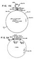
- the pMLC12/13 ⁇ M7/AccI + klenow [fragment] and the gel-isolated fragment from F41a were joined by DNA ligase and transformed into MC1061. Plasmids which had incorporated the gel isolated fragment were identified by hybridization to the albumin 32 mer and were verified by digestion with PstI, BamHI, or XbaI. This plasmid, termed pMLC12/13 ⁇ M71VR, is shown in Figure 4C.
- plasmid MpTL poly was used for the next step. This plasmid had been previously prepared by phosphorylating the lower oligonucleotide strand of: After ligating, a dimer was isolated having the middle portion: recognizable because of the Stu I site that had formed. The dimer was filled with Klenow fragment of DNA polymerase I so as to have the end portions: with the underlined base having been filled in by Klenow. This duplex was now cloned into Mp8/Hinc II and a clone picked on the basis of acquisition of a Stu I site. After sequencing for confirmation, the resultant plasmid was MpTL poly.
- pMLC12/13 ⁇ M71VR was partially digested with XbaI (which cuts four tees within the plasmid), blunted as above, and full length linearized plasmid DNA was isolated by gel electrophoresis.
- the plasmid MpTL poly was digested to completion with BamHI + HindIII and was blunted as above.
- the 80 bp blunted fragment was isolated by gel electrophoresis and ligated to the gel isolated, linearized, XbaI partially cut, blunted pMLC12/13 ⁇ M71VR.
- the DNA was transformed into MC1061 and screened with the oligonucleotide which is complimentary to the TL polylinker.
- pMLC12/13 ⁇ M71VRTL This plasmid, termed pMLC12/13 ⁇ M71VRTL, is shown in Figure 4D, and is analogous to Figure 1C.
- the plasmid pMLC12/13 ⁇ M71VRTL (see Figure 4D) was transformed into the strain XS127 (argE(am), thi-1, ⁇ lacproXIII/F'lacproAB, traD36, lacIq, lacZ ⁇ MlS/p3) or JM101 (ATCC #33876) which is used to produce single-stranded DNA from M13 ori plasmids.
- An overnight culture, (about 3X10⁹ cells) was mixed with 5X10¹⁰ wild type M13 phage, diluted to 100 ml in SOBM media and grown for 5 hours at 37°C.
- Single-stranded DNA was prepared from the culture supernatant using standard procedures.
- Figure 4E shows diagrammatically the regions of single- and double-stranded DNA which should form in this nucleic acid molecule from its complementary strand.
- Single-stranded DNA from pMLC12/13 ⁇ M71VRTL was digested with BamHI and the resulting 3' recessed BamHI end was filled in with alpha ⁇ 32P-dATP and cold dCTP, dGTP, TTP.
- the reaction was incubated at 68°C for 10 minutes to heat inactivate the klenow fragment of DNA Polymerase I and digested to completion with EcoRI.
- the resulting DNA was chromatographed twice over an hydroxylapatite (HAP) column in 0.12 M phosphate buffer (pH 6.8) to isolate the partially double-stranded labeled molecule from the major portion of the vector. Following binding to the HAP column in 0.12 M phosphate buffer and washing, partially double-stranded labeled construct was eluted from the HAP column with 0.3 M phosphate buffer and concentrated.
- HAP hydroxylapatite
- the isolated reagent complex (about 12 ng) of Example 2 was then incubated in a 10 ul volume of 20mM Tris pH 8, 1M NaCl, 10 mM EDTA, at 65° C for 60 minutes alone or with about 300 ng of single-stranded DNA from M13 clones G7c and G7e, which contain the album in Hinc III/Pvu II 915 base pair fragment cloned into the Sma I site of the vector Mp8 in opposite orientations or no DNA.
- the labeled strand was completely displaced from the reagent complex in the presence of the competing strand (G7c) and was not displaced in the absence of competing strand (G7e; no DNA), as measured by gel electrophoresis and autoradiography.
- the double-stranded IV polylinker was prepared by chemical synthesis and inserted into a modified pUC plasmid in both orientations (a and b).
- the plasmid pUC is described by Viera and Messing, Gene , vol. 19, pp. 259-268 (1982).
- the pUC plasmid was modified in several ways, the only one of which is relevant to present Example being the addition of an SfiI linker at a site in the plasmid other than the site at which the IV polylinker was subsequently added.
- the orientation (A) where the SfiI site was closest to the BglII site was digested with SfiI and Kpn I and the small fragment was isolated.
- the orientation (B) where the Sfi I site was closest to the Kpn I site was digested with Sfi I and Kpn I, and the large fragment was isolated.
- the gel isolated fragments were ligated and transformed into MC1061.
- the desired clone, containing two copies of the polylinker in opposite orientation with the KpnI site in the middle was identified by BglII digestion, end labelling with ⁇ 32P-dATP and the klenow fragment of DNA Polymerase I and gel electrophoresis before and after KpnI digestion.
- the resulting plasmid is termed F58a.
- F58a was digested to completion with BglII.
- the plasmid pMLC12 was digested with Bam HI and treated with Calf Intestinal Alkaline Phosphatase to remove 5' phosphate groups.
- the two DNAs were ligated and transformed into MC1061 and chloramphenicol resistant colonies were selected. Plasmids which had correctly inserted the BglII fragment with the IV polylinker were identified by ability of ClaI, XhoI, and KpnI to linearize the plasmid.
- the correct clone is termed F62b (see Figure 5A).
- F62b was digested with SacI and HindIII and blunted using the klenow fragment of DNA Polymerase I.
- the small 89 bp fragment was isolated following gel electrophoresis.
- the vector pMLC12/13 ⁇ M7 (see Example 1, part A and Figure 4A) and the 89 bp fragment were ligated and transformed into MC1061. Chloramphenicol resistant colonies were grown up and DNA was prepared. Colonies containing the 89 bp fragment were identified by XhoI or KpnI digestion (which will linearize the plasmid) and the orientation of the insert was determined by PstI plus SacI digestion.
- pUDV-A and pUDV-B The two orientations of the plasmid designated pUDV-A and pUDV-B, shown in Figure 5B, were assigned as follows: Orientation A produced two 253 bp SacI/PstI fragments and thus the PstI site was nearest to the 5' end of the CAT gene. Orientation B produced SacI + PstI fragments of 183 and 323 bp and thus the PstI site was farthest from the 5' end of the CAT gene.
- the nucleotide sequences of the inserts in both orientations of pUDV are given in Figures 1B and 2B, for orientations B and A, respectively.
- the target sequence is cloned into the blunted PstI site of pUDV-A, prepared in Example 4.
- the fragment cloned should have the target binding region TBR including the complement to the signal strand (LBR).
- the orientation of the first cloning step can be easily determined using either PuvII or SacII (which cut in the vector) and an enzyme which digests the insert DNA.
- Mini prep DNA is then prepared and digested with SmaI. This provides a blunt end site in which to clone the signal strand fragment.
- the orientation of the signal strand fragment is determined similarly and an orientation is chosen which is opposite to that of the target strand.
- the cloning into the sane sites is used for the sane fragments but the vector pUDV-B is used.
- a similar vector to that prepared in Example 5 could be prepared in which the IV polylinker is replaced by the TL polylinker, whose structure is that described above in connection with plasmid MpTL poly (having EcoRI sites near each end and a StuI site at the middle of the polylinker).
- This polylinker would serve the purpose of segment 16/16' in Figures 1A and 1B.
- the plasmid pUDV-A (see Example 4 and Figure 2B) was digested with the enzyme Pvu II and the 399 nt fragment containing the cloned polylinker sequence was gel isolated (Fragment A). Pvu II cuts at two sites outside of the polylinker, but within the lac region sites shown in Figure 2B.
- the vector pUC 118 obtained from Dr. Jeffrey Viera, is similar to vector pUC 18 (which is available commercially from New England Biolabs), but contains the M13 origin sequence (nucleotides 5465 (HgiAl site) - 5941 (Aha I site) of the standard M13 map) inserted at the Nde I site of puC 18.
- That vector pUC 118 was digested with PVU II and the 2840 nt fragment containing the plasmid origin of replication, the ampicillin resistance gene and the M13 origin of replication was isolated (fragment B). Fragments A and B were ligated together and transformed into competent bacteria.
- the Pst I fragment of lambda containing this region was isolated by digestion with Pst I and gel electrophoresis.
- the 86 nt Pst I - Hae III fragment from position 16236 to position 16322 was subcloned by digesting the isolated Pst I fragment of lambda with Hae III, isolating the 86 nt fragment and blunting the Pst I end with the Klenow fragment of DNA polymerase I. This fragment was then cloned into the Hinc II site of bacteriophage mp7 and the correct clone (mp7lambdaA) identified by hybridization with a specific oligonucleotide and by DNA sequence analysis.
- the 163 nt Xmn I - Hinc II fragment from position 16914 to position 17077 was subcloned by digesting the lambda Pst I fragment with Xmn I and Hinc II and gel isolating the resulting 163 nt fragment. This fragment was then cloned into the Hinc II site of mp7 resulting in mp7lambdaD.
- the clone mp7lambdaA (or mp7lambdaD) was digested with Bam HI and the insert fragment was blunted with the Klenow fragment of DNA polymerase I. This fragment was then cloned into the Sma I site of p61A and the desired orientation was selected by hybridization with the strand specific oligonucleotide #1091 (for A; 5'-CCGTTATCCACGATGGCCTC-3') or #1090 (for D;5'-CATCGCCCGGTACATGGCG-3') resulting in the clones pA6 and pD10, respectively.
- Clones pA6lambda2c and pD10lambda1a (which contains the 86 nt and 163 nt pairing segments, respectively, but also containing the 1159 nt target binding region) are grown up in E. coli, superinfected with M13 and the harvested DNA is digested with Bam HI (producing constructs analogous to Figures 3E and 3C, respectively). The 5' ends of such constructs are labeled by the procedures of Examples 11 and 12, below, with an oligonucleotide containing a biotin moiety (see U.S.S.N. 729,700 of Brown, et al.).
- the cloning vector p61a described in Example 7 was used in this Example. Sequences from the cDNA sequence for erythropoietin (EPO) were used in this Example for target binding region, pairing segment and analyte.
- the relevant cDNA region, clone pG61aEPO3III, has the map shown by Jacobs, et al in Nature , vol. 313, pp. 806-810 (1985) including: Site Position Eco RI 1 (inserted 6 bases upstream from initiator methionine codon) Kpn I 133 Pst I 188; 363 Hinc II 335 Pvu II 365
- the 175 base pair Pst I fragment (188 - 363) was cloned into the Pst I site of p61a plasmid DNA. Clones containing the insert were identified by hybridization to a kinased oligonucleotide probe. Single stranded plasmid DNA was prepared from positive colonies after superinfection with an M13 helper phage. The orientation of the Pst I fragment in the vector was determined by binding this single stranded DNA to nitrocellulose filters and hybridizing it with kinased oligonucleotide probes of opposite orientations. Plasmid p61A-EPO3 contains the Pst I fragment in the orientation with the Hinc II site closest to the 3' end of the insertion.
- Double stranded plasmid DNA was isolated from p61AEPO3 and digested with Sma I.
- the 365 base pair Eco RI-Pvu II fragment (1 - 365) from the human EPO cDNA clone was gel purified and the Eco RI site was filled in with the Klenow fragment of DNA Polymerase I. This fragment was then ligated into the Sma I site of p61A-EPO3; plasmids containing inserts were identified by screening colonies with a kinased oligonucleotide probe specific for this second insert. The orientation of the second insert was determined by analyzing Kpn I digests of the resulting double stranded plasmid DNA.
- plasmids with the target binding region and pairing segments in an inverted orientation contain a Kpn fragment of 166 base pairs, while those in the direct orientation contain a Kpn fragment of 264 base pairs.
- p61A-EPO3-II was identified as having the target strand inverted with respect to the signal strand and was isolated for preparation of displacement complexes.
- Single stranded p61A-EPO3-II DNA was prepared as described in Example 1, except that the overnight culture was superinfected with the M13 phage derivative, MK107 (see Example 9) diluted to 100 ml in minimal media containing 50 ug/ml ampicillin and grown overnight at 37°C.
- the DNA was digested with Bam HI and the 3' end (near the 175 nt pairing segment) labeled with 32P-dATP using the Klenow fragment of DNA Polymerase I.
- the DNA was then digested with Eco RI to create final displacement complexes, by analogy with Figure 1F.
- the 370 base pair Eco RI-Pvu II fragment described above was also cloned into the Acc I site of the M13 phage vector mp7 after both the Acc I site and the Eco RI site were filled in with Klenow. Phage containing EPO inserts, and the orientation of those inserts, were identified by hybridization with kinased oligonucleotide probes. Single stranded template DNA was prepared from positive phage of each orientation and digested with Bam HI. The 370 nucleotide inserts were gel purified and used as model analytes.
- the construct p66b was constructed by gel-isolating the double-stranded PvuII fragment containing the sequence of interest from the starting clone and ligating it to the gel-isolated Pvu II backbone of the M13 origin plasmid pUC 119 (similar to commercially available plasmid PUC 19, but containing the same M13 origin sequence as does pUC118, and also obtained from Dr. Jeffrey Viera). Single-stranded forms were obtained as described in Example 1, except that the DNA was transformed into the E. coli host strain MV1193 obtained from Dr. Michael Volkert (JM101 del(srlR-recA) 306::Tn10). Superinfection was with bacteriophage M13K07. The use of the different pUC backbone, different E. coli host strain and different superinfecting M13 phage resulted in improved yields of the same construct after Bam HI digestion.
- Inverse reagent complexes used in Examples 12 and 13 resulted from labelling the 5' end of p66b with P32.
- p66d was constructed as described for p66b, except that the PvuII fragment was inserted into the pUC119 vector in the opposite orientation.
- the single stranded form of p66d produced after superinfection contains a displacement complex which is the complement of the p66b strand.
- displacement complexes made from p66b contain the signal strand at the 5' end and the target binding region at the 3' end.
- Model analytes were constructed by gel purifying a 2 kb Hind III-Eco RI fragment from a plasmid, pAllAlb, which contains the entire cDNA sequence of human albumin.
- the HindIII site is the HindIII site in the 3' end of the albumin cDNA.
- the EcoRI site is present in adjacent vector sequences.
- the vector sequences present on the HindIII EcoRI fragment have no bearing on the following examples.
- the HindIII - EcoRI fragment was ligated into Hind III-EcoRI digested M13mp8 and M13mp11 to give mp8AllAlb and mp11.AllAlb, respectively.
- Model Capturers Several different model capturers were constructed to help define the best geometry for capturing nucleic acid constructs.
- the capturing strands mp7deltaALbXba1+ or mp7deltaAlbXba3- were biotinylated using the commercially obtained Photoprobe Biotin (Vector Laboratories) essentially as described by the manufacturer and repeated below.
- PhotoprobeTM biotin 500 ug was resuspended in 500 microliter water as recommended by the manufacturer and stored in the dark at -20°C.
- the DNA was mixed with 10 microliter Photoprobe biotin solution under a safelight, sealed in a glass microcapillary pipette and irradiated by a sunlamp (GE infrared lamp 2JOR40/1) for 20 or 30 minutes in separate reactions.
- the sample was kept in an ice-H2O bath during the entire irradiation procedure.
- the sample was removed from the capillary, diluted with 100 microliters of 0.1 M Tris-HCl, pH 8.0, extracted twice with 2-butanol and precipatated following addition of 1/10 volume 3M Na Acetate and 2 volumes of ethanol.
- the precipatated sample was resuspended in 10 microliters 0.1 mM EDTA, pH 8.0.
- the beads were removed from the solution by centrifugation (2 minutes, Eppendorf centrifuge) washed once and the combined solution phases were electrophoresed in a parallel lane to the control sample. Following electrophoresis and autoradiography, the results indicated that nearly all the 32-P labeled oligonucleotide sample that was hybridized to the mp7deltaAlbXba1+ DNA was removed from the sample that was exposed to the streptavidin latex, indicating that the majority of template DNAs (capture strands) had at least one biotin group attached.
- p66b Xba complex was labeled by ligating a kinased oligonucleotide to the 3' end of the molecule. 10 pm of a 15 base oligonucleotide with the sequence 5' CTAGAGGCCTCTGCA3' was labeled at the 5' end with 32P -gamma ATP and polynucleotide kinase (see Maniatis et al, Cloning Manual (Cold Spring Harbor Laboratory 1982)).
- the labeled oligonucleotide was purified away from unincorporated 32P - ⁇ ATP by centrifuging the reaction twice at 6000 rpm for 30 minutes in a total volume of 500 ul of TE in a Centricon 10 filtration device from Amicon.
- the kinased oligonucleotide and 1 pm of Xba I cut p66b gel purified complex were precipitated together with ethanol, and resuspended in 14 ul of TE. 4 ul of 5X ligase buffer (Maniatis et al Manual) and 2 ul of DNA ligase were added and the reaction was incubated at 15° for 4 hours.
- the reaction was diluted to 50 ul with TE, heated to 42°C for 10 minutes to melt any non-ligated oligonucleotides and electrophoresed on a 1% agarose gel. Following electrophoresis, the gel was stained with ethidium bromide and viewed with a uv light box. Approximately 66% of the complexes ligated to each other, so that only 0.33 pm of complex were available for ligation to the kinased oligonucleotide. The kinased complex was purified and had an estimated specific activity of about 106 cpm per pm.
- Displacement and capturing reactions were carried out by incubating with either 0.001 pm (1000 cpm) or 0.005 pm (5000 cpm) of displacement complex with varying amounts of analyte and 0.2 pm of capturer in a total of 20 ul of 0.3 M NaCl and 0.1 M Tris HCl, pH 8.0, for 45 minutes at 65°C.
- no non-specific capturing was observed in reactions with 0.001 pm of complex and 0.02 pm of either mp7, mp19, mp7deltaAlbXba3- or mp19.AlbTaqPst.
- Example 11 Displacement and capturing with Bam HI p66b and mp7deltaAlbXba capturers.
- Single stranded p66b DNA was digested to completion with Bam HI, and the covalent complex was isolated by gel purification.
- a 32P-kinased oligonucleotide with the sequence 5'GATCCGCGGCGGTAC3' was ligated to the 3' end of the complex as described in Example [10] except that 2.4 pm of the complex and 10 pm of the oligonucleotide were used in the ligation reaction.
- the specific activity of the resulting complex was estimated at 8x10⁴ cpm/pm.
- Reactions were done by incubating either 0.01 pm (1000 cpm) or 0.03 pm (3000 cpm) in the presence or absence of 0.01 pm Hae III cut mp8.AllAlb analyte, with 0.1 pm of capture DNA in a final volume of 20 ul of hybridization buffer (0.3M NaCl, 0.1 M Tris HCl,pH8.0, and 10 mM EDTA) for 60 minutes at 65°C. Reactions were analyzed by gel electrophoresis (1% agarose gel) and autoradiography.
- Example 12 Comparision of displacement and capturing with covalent and non-covalent p66d complexes.
- Covalent p66d displacement complexes were prepared and labeled at the 5' end by ligation of the kinased 16mer using the EF21 splint as described in Example 13.
- the specific activity of the resulting complexes was about 1 x 10⁶ cpm/pm.
- Non-covalent p66d complexes were produced by complete digestion of approximately 50 ug of single stranded templated DNA with Bam HI and Eco RI. Complete digestion was ascertained by the appearance of equimolar amounts of three bands, corresponding to vector backbone, target strand, and signal strand, after electrophoresis of a small aliquot of the digest on an alkaline gel. Non-covalent p66d complexes were labeled at the 5' end of the signal strand as described for covalent complexes, by EF21 splint ligation of a kinased 16mer, with a resultant specific activity of 3 x 10⁶ cpm/pm.
- the four reactions outlined in Table 3 were set up in a total volume of 50 ul of hybridization buffer and incubated for 60 minutes at 65°C using HaeIII digested mp11.AllAlb DNA as analyte and biotinylated mp7deltaAlbXba3- capturere. 10 ul of each reaction were analyzed by gel electrophoresis and autoradiography, and the remaining 40 ul by binding to steptavidin agarose. 200 ul packed volume streptavidin agarose was used per reaction. Binding and washing was as described in Example 13 (below), except that, after binding, the pellet was rinsed 3 times for 30 minutes at room temperature and once for 60 minutes at 65°C. The final pellet and all supernatants were counted. Data showing the cpm bound to agarose after each rinse are given in Table 1. These results show that displacement and capturing are approximately equally effective for covalent and non-covalent complexes.
- Example 13 Large scale displacement and capture with trapping on streptavidin agarose.
- the Bam p66b displacement complex was labeled to a specific activity of about 106 cpm/pm by ligating a 32P-kinased oligonucleotide to the 5' end of the complex with the use of a 21 base splint (EF21). 10 pm of the kinased 16mer (indicated below by the asterisk), 10 pm of splint, and 1 pm of p66b Bam (underlined below) were incubated together at 22°C for 15 minutes in 10 ul of 1X ligase buffer; 1 ul of ligase was added and the reaction incubated for an additional 30 minutes. The three molecules form the structure diagrammed below.
- Example 13 and Example 14, below provide labeling at the 5'-end, and thus adjacent to the target binding region rather than to the pairing segment.
- Such a geometry is described further in a copending application of Collins et al, U.S.S.N. 809,992, filed December 16, 1985.
- Example 14 Prehybridization of complex and analyte, followed by capturing and trapping.
- Example 15 Strand displacement, capturing and biotin displacement.
- Example 12 the non-covalent p66d Bam HI/EcoRI cut displacement complex described in Example 12 was used.
- Hae III cut mp11.AllAlb DNA was used as the analyte.
- a capturer which has a single biotin at the 5' end was synthesized by primer extension of a 5' biotinylated M13 sequencing primer (biotinylated according to the method described in U.S.S.N.
- reaction 3 was not incubated.
- 5 ul of reactions 1 and 2 were then removed for gel analysis.
- the rest of reactions 1 and 2, and reaction 3 were then bound as described in Example 8 to approximately 200 ul packed volume streptavidin agarose.
- the samples were rinsed by adding 1 ml of binding buffer to the pellet in an eppendorf tube, inverting the tube five times, and centrifuging it for 3 minutes.
- Reaction 1 to 3 as described above were used. Each sample was the final avidin-agarose pellet after the 30 minute, 60°C wash described above. One ml BB containing 0.1% NP-40 (BB+) was added to each pellet at room temperature, shaken briefly by inversion and centrifuged to separate the phases (RT wash). One ml BB+ (at 65°C) was then added and the samples were incubated for 5 minutes at 65°C. The samples were then centrifuged to separate the phases and washed twice with one ml BB+ at room temperature. The combined supernatant pahses were then pooled (65°C/5'/-bio). The -biotin, 65°C wash was repeated once more for sample 3 only.
- the improvement brought about by the biotin displacement can be measured by the ratio of % counts released by biotin in reaction 1 over the percent counts released by biotin in either reaction 2 or 3.
- the poor improvement seen in the reaction 2 sample is likely due to the fact that the reaction 2 capturer contains a small amount of M13 polylinker sequence which does result in some capturing by the biotnylated capturer in the absence of analyte. This capturing, though small, would lead to counts released by biotin.
- the reaction 3 sample (complex only) represents the type of background most likely to be found in an actual displacement measurement and therefore gives a better representation of the background improvement expected.
Landscapes
- Chemical & Material Sciences (AREA)
- Life Sciences & Earth Sciences (AREA)
- Organic Chemistry (AREA)
- Proteomics, Peptides & Aminoacids (AREA)
- Zoology (AREA)
- Wood Science & Technology (AREA)
- Health & Medical Sciences (AREA)
- Engineering & Computer Science (AREA)
- Analytical Chemistry (AREA)
- Microbiology (AREA)
- Bioinformatics & Cheminformatics (AREA)
- Molecular Biology (AREA)
- Biotechnology (AREA)
- Biophysics (AREA)
- Physics & Mathematics (AREA)
- Biochemistry (AREA)
- Immunology (AREA)
- General Engineering & Computer Science (AREA)
- General Health & Medical Sciences (AREA)
- Genetics & Genomics (AREA)
- Measuring Or Testing Involving Enzymes Or Micro-Organisms (AREA)
- Saccharide Compounds (AREA)
- Investigating Or Analysing Biological Materials (AREA)
Claims (25)
- Nucleinsäureaufbau, brauchbar zur Herstellung von Reagenzien zur Bestimmung von Zielnucleotidsequenzen in der Nucleinsäure einer biologischen Probe, wobei der Aufbaua) einen Zielbindungsbereich mit einem ersten und zweiten Abschnitt, wobei dieser Bereich im wesentlichen komplementär zu der Zielnucleotidsequenz ist,b) ein Signalstrangsegment, das in dem Aufbau durch komplementäre Basenpaarung an den ersten Abschnitt des Zielbindungsbereichs gebunden ist und so einen doppelstrangigen Abschnitt des Aufbaues bildet, wobei der zweite Abschnitt des Zielbindungsbereichs einzelstrangig ist, undc) eine feststellbare Markierung in Nachbarschaft zu dem Signalstrangsegment umfaßt, worinder Bereich komplementärer Basenpaarung zwischen dem Signalstrangsegment und dem ersten Abschnitt des Zielbindungsbereiches eine Länge von 15 bis 1000 Basenpaaren hat und der Zielbindungsbereich und das Signalstrangsegment durch ein Phosphat-Zuckergrundgerüst kovalent verknüpft sind.
- Nucleinsäureaufbau nach Anspruch 1, bei dem die gesamten durch komplementäre Basenpaarung an das Signalstrangsegment gebundenen Nucleotide eine Teilmenge der Nucleotide des Zielbindungsbereichs sind.
- Nucleinsäureaufbau nach Anspruch 1, bei dem die durch komplementäre Basenpaarung an das Signalstrangsegment gebundenen Nucleotide einem Ende des Zielbindungsbereiches benachbart sind.
- Nucleinsäureaufbau nach einem der vorausgehenden Ansprüche, das eine im wesentlichen einzelstrangige Schleife mit einem 5'-Ende und einem 3'-Ende ist, wobei das Signalsstrangsegment einem der Enden benachbart ist und der Zielbindungsbereich dem anderen Ende der einzelstrangigen Schleife benachbart ist.
- Nucleinsäureaufbau nach Anspruch 4, bei dem die durch komplementäre Basenpaarung an das Signalstrangsegment gebundenen Nucleotide dem anderen Ende der einzelstrangigen Schleife banachbart sind.
- Nucleinsäureaufbau nach Anspruch 5, bei dem das eine Ende das 3'-Ende und das andere Ende das 5'-Ende ist.
- Nucleinsäureaufbau nach Anspruch 4, bei dem das eine Ende das 3'-Ende und das andere Ende das 5'-Ende ist.
- Nucleinsäureaufbau nach einem dervorausgehenden Ansprüche, weiterhin mit einer DNA-Haarnadelschleifen-Einschränkungsstelle zwischen dem Zielbindungsbereich und dem Signalstrangbindungsbereich entlang dem Phosphat/Zuckergrundgerüst.
- Nucleinsäureaufbau nach einem der vorausgehenden Ansprüche, der DNA ist.
- Nucleinsäureaufbau nach einem der vorausgehenden Ansprüche, bei dem der Bereich komplementärer Basenpaarung zwischen dem Signalstrangsegment und dem ersten Abschnitt des Zelbindungsbereiches eine Länge von 20 bis 50 Basenpaaren hat.
- Verfahren zur Herstellung eines Reagenzkomplexes zur Bestimmung einer Zielnucleotidsequenz in der Nucleinsäure einer biologischen Probe mit den Stufen, in denen mana) durch Replikation ein kontinuierliches Nucleinsäuremolekül vorherrschend in einzelstrangiger Form erzeugt, das(i) einen Zielbindungsbereich im wesentlichen komplementär zu der Zielnucleotidsequenz und(ii) ein Signalstrangsegment, das durch komplementäre Basenpaarung an einen ersten Abschnitt des Zielbindungsbereichs gebunden ist, hat, wobei ein zweiter Abschnitt des Zielbindungsbereiches einzelstrangig ist und der Zielbindungsbereich und der Signalstrangbindungsbereich kovalent durch ein Phosphat/Zuckergrundgerüst verknüpft sind,b) einen ersten Endpunkt in Nachbarschaft zu dem Signalstrangsegment und einen zweiten Endpunkt in Nachbarschaft zu dem Zielbindungsbereich bildet undc) eine feststellbare Markierung in Nachbarschaft zu dem Signalstrang anfügt.
- Verfahren nach Anspruch 11 zusätzlich mit der Stufe, in der mand) die kovalente Verknüpfung des Signalstrangsegmentes mit dem Zielbindungsbereich durch das Phosphat/Zuckergrundgerüst spaltet.
- Verfahren nach Anspruch 12 zusätzlich mit der Stufe, in der mane) ein Ende des Nucleinsäuremoleküls in Nachbarschaft zu dem Zielbindungsbereich an einen festen Träger bindet.
- Verfahren nach Anspruch 13, bei dem die Bindungsstufe (e) der Spaltungsstufe (d) vorausgeht und das gebundene Ende von dem Signalstrangsegment distal angeordnet ist.
- Vertahren nach Anspruch 13, bei dem die Bindungsstufe (e) der Spaltungsstufe (d) folgt und das gebundene Ende durch die Spaltungsstufe (d) erzeugt wird.
- Vertahren nach Anspruch 12 zusätzlich mit der Stufe, in der mand) einen Rest des Nucleinsäuremoleküls an einem Endpunkt in Nachbarschaft zu dem Zielbindungsbereich bindet, wobei der Rest durch ein Affinitätsreagenz immobilisierbar ist.
- Vertahren nach Anspruch 11, bei dem der erste Endpunkt und der zweite Endpunkt, die in der Ausbildungsstufe (b) erzeugt werden, ein 3'-Ende und ein 5'-Ende sind.
- Verfahren nach Anspruch 17, bei dem der erste Endpunkt ein 3'-Ende ist und die Bindungsstufe (c) eine Verlängerung des 3'-Endes mit feststellbaren Nucleotiden umfaßt.
- Verfahren nach Anspruch 17, bei dem der zweite Endpunkt ein 5'-Ende ist und das Verfahren zusätzlich eine Bindung des 5'-Endes an einen festen Träger umfaßt.
- Verfahren nach Anspruch 17, bei dem der zweite Endpunkt ein 5'-Ende ist und das Verfahren zusätzlich eine Bindung des 5'-Endes an einen Rest umfaßt, der durch ein Affinitätsreagenz immobilisierbar ist.
- Replizierbare Nucleinsäure, brauchbar zur Herstellung von Reagenzien zur Bestimmung von Zielnucleotidsequenzen in der Nucleinsäure einer biologischen Probe und mit einem kontinuierlichen Strang, dera) einen Replikationsursprung,b) einen Zelbindungsbereich mit einem ersten und einem zweiten Abschnitt, wobei dieser Bereich im wesentlichen komplementär zu der Zielnucleotidsequenz ist,c) ein zu dem ersten Abschnitt des Zielbindungsbereiches im wesentlichen komplementäres Signalstrangsegment, wobei dieser zweite Abschnitt des Zielbindungsbereiches einzelstrangig ist und wobei der Signalstrang in einer umgekehrten Richtung gegenüber dem Zielbindungsbereich ausgerichtet ist,d) eine feststellbare Markierung in Nachbarschaft zu dem Signalstrangsegment unde) zwei Halbrestriktionsstellen, die eine Einschränkungsstelle bilden können und dazwischen eine den Zelbindungsbereich und das Signalstrangsegment enthaltende Nucleinsäurelänge begrenzen können, umfaßt.
- Replizierbares Nucleinsäuremolekül nach Anspruch 21, das mit seinem Komplementärstrang in doppelstrangiger Form vorliegt.
- Replizierbares Nucleinsäuremolekül nach Anspruch 21, das frei von seinem Komplementärstrang in vorherrschend einzelstrangiger Form vorliegt.
- Replizierbare Nucleinsäure nach Anspruch 23, bei der die beiden Halbrestriktionsstellen unter Bildung einer Restriktionsstelle hybridisiert sind, die Nucleinsäurelänge eine an jedem Ende mit einer der Halbrestriktionsstellen verbundene Schleife bildet und das Signalstrangsegment durch komplementäre Basenpaarung an den ersten Abschnitt des Zielbindungsbereiches in der Schleife gebunden ist.
- Replizierbares Nucleinsäuremolekül nach Anspruch 24, bei dem da Signalstrangsegment und der erste Abschnitt des Zielbindungsbereiches jeweils in Nachbarschaft zu einer der Halbrestriktionsstellen angeordnet sind.
Applications Claiming Priority (2)
| Application Number | Priority Date | Filing Date | Title |
|---|---|---|---|
| US72950485A | 1985-05-02 | 1985-05-02 | |
| US729504 | 1985-05-02 |
Publications (3)
| Publication Number | Publication Date |
|---|---|
| EP0221108A1 EP0221108A1 (de) | 1987-05-13 |
| EP0221108A4 EP0221108A4 (de) | 1989-07-26 |
| EP0221108B1 true EP0221108B1 (de) | 1993-09-01 |
Family
ID=24931351
Family Applications (1)
| Application Number | Title | Priority Date | Filing Date |
|---|---|---|---|
| EP86902701A Expired - Lifetime EP0221108B1 (de) | 1985-05-02 | 1986-04-11 | Verfahren und aufbau einer nukleinsäure zur erzeugung von reagenzienkomplexen zur verwendung bei der bestimmung von zielnukleotidsequenzen |
Country Status (6)
| Country | Link |
|---|---|
| EP (1) | EP0221108B1 (de) |
| JP (1) | JP2609238B2 (de) |
| AU (1) | AU600900B2 (de) |
| CA (1) | CA1338557C (de) |
| DE (1) | DE3688965T2 (de) |
| WO (1) | WO1986006412A1 (de) |
Families Citing this family (12)
| Publication number | Priority date | Publication date | Assignee | Title |
|---|---|---|---|---|
| US4868105A (en) * | 1985-12-11 | 1989-09-19 | Chiron Corporation | Solution phase nucleic acid sandwich assay |
| DE3639109A1 (de) * | 1986-11-15 | 1988-05-19 | Merck Patent Gmbh | Verfahren zur bestimmung von nucleinsaeuren |
| US5118801A (en) * | 1988-09-30 | 1992-06-02 | The Public Health Research Institute | Nucleic acid process containing improved molecular switch |
| DE3929030A1 (de) * | 1989-09-01 | 1991-03-07 | Boehringer Mannheim Gmbh | Verfahren zur vermehrung von nukleinsaeuren |
| CA2037349C (en) * | 1990-03-26 | 2008-06-17 | James G. Wetmur | Branch migration of nucleotides |
| US5925517A (en) * | 1993-11-12 | 1999-07-20 | The Public Health Research Institute Of The City Of New York, Inc. | Detectably labeled dual conformation oligonucleotide probes, assays and kits |
| CA2239683A1 (en) * | 1995-12-22 | 1997-07-03 | Dade Behring Marburg Gmbh | Homogeneous amplification and detection of nucleic acids |
| AU713667B2 (en) * | 1996-04-12 | 1999-12-09 | Phri Properties, Inc. | Detection probes, kits and assays |
| US6482590B1 (en) | 1996-12-20 | 2002-11-19 | Aventis Behring Gmbh | Method for polynucleotide amplification |
| US6037130A (en) * | 1998-07-28 | 2000-03-14 | The Public Health Institute Of The City Of New York, Inc. | Wavelength-shifting probes and primers and their use in assays and kits |
| CN1348096A (zh) | 2000-10-10 | 2002-05-08 | 栾国彦 | 一种均相特异性检测核酸的探针及应用方法 |
| WO2009140374A2 (en) | 2008-05-13 | 2009-11-19 | Gen-Probe Incorporated | Inactivatable target capture oligomers for use in the selective hybridization and capture of target nucleic acid sequences |
Citations (2)
| Publication number | Priority date | Publication date | Assignee | Title |
|---|---|---|---|---|
| EP0164876A1 (de) * | 1984-05-07 | 1985-12-18 | Allied Corporation | Polynukleotidanalyse durch Verschiebung unter Verwendung eines Rekombinationsproteins und Reagenzsatz |
| EP0167238A1 (de) * | 1984-05-07 | 1986-01-08 | Allied Corporation | Verfahren zur Polynukleotidanalyse durch Verschiebung und Polynukleotidkomplexreagenz dafür |
Family Cites Families (3)
| Publication number | Priority date | Publication date | Assignee | Title |
|---|---|---|---|---|
| US4321365A (en) * | 1977-10-19 | 1982-03-23 | Research Corporation | Oligonucleotides useful as adaptors in DNA cloning, adapted DNA molecules, and methods of preparing adaptors and adapted molecules |
| US4358535A (en) * | 1980-12-08 | 1982-11-09 | Board Of Regents Of The University Of Washington | Specific DNA probes in diagnostic microbiology |
| DE177497T1 (de) * | 1984-04-05 | 1986-11-27 | Life Technologies Inc., Gaithersburg, Md. | Immobilisierung von nukleinsaeuren. |
-
1986
- 1986-04-11 JP JP61502319A patent/JP2609238B2/ja not_active Expired - Lifetime
- 1986-04-11 WO PCT/US1986/000742 patent/WO1986006412A1/en not_active Ceased
- 1986-04-11 EP EP86902701A patent/EP0221108B1/de not_active Expired - Lifetime
- 1986-04-11 DE DE86902701T patent/DE3688965T2/de not_active Expired - Fee Related
- 1986-04-11 AU AU57754/86A patent/AU600900B2/en not_active Ceased
- 1986-04-30 CA CA 507935 patent/CA1338557C/en not_active Expired - Fee Related
Patent Citations (2)
| Publication number | Priority date | Publication date | Assignee | Title |
|---|---|---|---|---|
| EP0164876A1 (de) * | 1984-05-07 | 1985-12-18 | Allied Corporation | Polynukleotidanalyse durch Verschiebung unter Verwendung eines Rekombinationsproteins und Reagenzsatz |
| EP0167238A1 (de) * | 1984-05-07 | 1986-01-08 | Allied Corporation | Verfahren zur Polynukleotidanalyse durch Verschiebung und Polynukleotidkomplexreagenz dafür |
Non-Patent Citations (1)
| Title |
|---|
| METHODS IN ENZYMOLOGY, vol. 101, 1983, Academic Press, New York, NY (US); M.D.BEEN et al., pp. 90,91,93,97# * |
Also Published As
| Publication number | Publication date |
|---|---|
| AU600900B2 (en) | 1990-08-30 |
| JP2609238B2 (ja) | 1997-05-14 |
| AU5775486A (en) | 1986-11-18 |
| JPS62502686A (ja) | 1987-10-15 |
| WO1986006412A1 (en) | 1986-11-06 |
| DE3688965D1 (de) | 1993-10-07 |
| EP0221108A1 (de) | 1987-05-13 |
| CA1338557C (en) | 1996-08-27 |
| DE3688965T2 (de) | 1994-03-24 |
| EP0221108A4 (de) | 1989-07-26 |
Similar Documents
| Publication | Publication Date | Title |
|---|---|---|
| US4818680A (en) | Method and kit involving displacement and rehybridization of labeled polynucleotide | |
| JP2788034B2 (ja) | ポリヌクレオチドのアツセイのための増幅方法 | |
| JP3415150B2 (ja) | 分枝核酸プローブの使用方法 | |
| JP2846018B2 (ja) | 核酸配列の増幅および検出 | |
| US5660988A (en) | Cycling probe cleavage detection of nucleic acid sequences | |
| JP2839608B2 (ja) | 核酸配列の検出方法 | |
| JP2674737B2 (ja) | 核酸増幅の検出 | |
| US4752566A (en) | Displacement polynucleotide method and reagent complex employing labeled probe polynucleotide | |
| US4725537A (en) | Assay, reagent and kit employing nucleic acid strand displacement and restriction endonuclease cleavage | |
| US6013439A (en) | Detection of differences in nucleic acids | |
| EP0450370A1 (de) | Verzweigungsmigration von Nukleinsäuren | |
| CA2070632A1 (en) | Use of dna-dependent rna polymerase transcripts as reporter molecules for signal amplification in nucleic acid hybridization assays | |
| US5268266A (en) | Process and nucleic acid construct for producing reagent complexes useful in determining target nucleotide sequences | |
| JPH07509365A (ja) | ポリヌクレオチド類の3’末端に特定配列を導入する方法 | |
| JPH10509594A (ja) | 遺伝的多型の検出のための複合ミクロサテライトプライマー | |
| JPH11504517A (ja) | オリゴヌクレオチドの化学結合による核酸検出及び増幅 | |
| JPH08511684A (ja) | 熱安定性dnaポリメラーゼ由来の5’ヌクレアーゼ | |
| CA2308368C (en) | Specific and sensitive nucleic acid detection method | |
| JPS60244300A (ja) | 組換えタンパク質を用いる置換ポリヌクレオチド検定および診断用キツド | |
| CA1338557C (en) | Process and nucleic acid construct for producing reagent complexes useful in determining target nucleotide sequences | |
| EP0387452B1 (de) | Verfahren zur Vorbereitung von Nukleotid-Sonden unter Verwendung eines hybridizierbaren Komplementärelements | |
| JP2001512301A (ja) | 核酸増幅のための内部陽性対照 | |
| JPH11506932A (ja) | 点変異の検出及びインビトロ突然変異誘発のための核酸修復酵素方法 | |
| WO2000043543A1 (en) | Detection of differences between polynucleotides | |
| CA2037673A1 (en) | Ribonuclease scission chain reaction |
Legal Events
| Date | Code | Title | Description |
|---|---|---|---|
| PUAI | Public reference made under article 153(3) epc to a published international application that has entered the european phase |
Free format text: ORIGINAL CODE: 0009012 |
|
| 17P | Request for examination filed |
Effective date: 19861125 |
|
| AK | Designated contracting states |
Kind code of ref document: A1 Designated state(s): AT BE CH DE FR GB IT LI LU NL SE |
|
| RBV | Designated contracting states (corrected) |
Designated state(s): DE FR GB IT NL |
|
| A4 | Supplementary search report drawn up and despatched |
Effective date: 19890726 |
|
| 17Q | First examination report despatched |
Effective date: 19901018 |
|
| GRAA | (expected) grant |
Free format text: ORIGINAL CODE: 0009210 |
|
| AK | Designated contracting states |
Kind code of ref document: B1 Designated state(s): DE FR GB IT NL |
|
| REF | Corresponds to: |
Ref document number: 3688965 Country of ref document: DE Date of ref document: 19931007 |
|
| ITF | It: translation for a ep patent filed | ||
| ET | Fr: translation filed | ||
| PLBE | No opposition filed within time limit |
Free format text: ORIGINAL CODE: 0009261 |
|
| STAA | Information on the status of an ep patent application or granted ep patent |
Free format text: STATUS: NO OPPOSITION FILED WITHIN TIME LIMIT |
|
| 26N | No opposition filed | ||
| PGFP | Annual fee paid to national office [announced via postgrant information from national office to epo] |
Ref country code: GB Payment date: 19980417 Year of fee payment: 13 |
|
| PGFP | Annual fee paid to national office [announced via postgrant information from national office to epo] |
Ref country code: DE Payment date: 19980429 Year of fee payment: 13 |
|
| PGFP | Annual fee paid to national office [announced via postgrant information from national office to epo] |
Ref country code: FR Payment date: 19980430 Year of fee payment: 13 Ref country code: NL Payment date: 19980430 Year of fee payment: 13 |
|
| PG25 | Lapsed in a contracting state [announced via postgrant information from national office to epo] |
Ref country code: GB Free format text: LAPSE BECAUSE OF NON-PAYMENT OF DUE FEES Effective date: 19990411 |
|
| PG25 | Lapsed in a contracting state [announced via postgrant information from national office to epo] |
Ref country code: NL Free format text: LAPSE BECAUSE OF NON-PAYMENT OF DUE FEES Effective date: 19991101 |
|
| GBPC | Gb: european patent ceased through non-payment of renewal fee |
Effective date: 19990411 |
|
| PG25 | Lapsed in a contracting state [announced via postgrant information from national office to epo] |
Ref country code: FR Free format text: LAPSE BECAUSE OF NON-PAYMENT OF DUE FEES Effective date: 19991231 |
|
| NLV4 | Nl: lapsed or anulled due to non-payment of the annual fee |
Effective date: 19991101 |
|
| REG | Reference to a national code |
Ref country code: FR Ref legal event code: ST |
|
| PG25 | Lapsed in a contracting state [announced via postgrant information from national office to epo] |
Ref country code: DE Free format text: LAPSE BECAUSE OF NON-PAYMENT OF DUE FEES Effective date: 20000201 |
|
| PG25 | Lapsed in a contracting state [announced via postgrant information from national office to epo] |
Ref country code: IT Free format text: LAPSE BECAUSE OF NON-PAYMENT OF DUE FEES;WARNING: LAPSES OF ITALIAN PATENTS WITH EFFECTIVE DATE BEFORE 2007 MAY HAVE OCCURRED AT ANY TIME BEFORE 2007. THE CORRECT EFFECTIVE DATE MAY BE DIFFERENT FROM THE ONE RECORDED. Effective date: 20050411 |