EP0425381A1 - Vorrichtung zum Zählen und zur Bestimmung mindestens einer Leukozytensubpopulation - Google Patents
Vorrichtung zum Zählen und zur Bestimmung mindestens einer Leukozytensubpopulation Download PDFInfo
- Publication number
- EP0425381A1 EP0425381A1 EP90403006A EP90403006A EP0425381A1 EP 0425381 A1 EP0425381 A1 EP 0425381A1 EP 90403006 A EP90403006 A EP 90403006A EP 90403006 A EP90403006 A EP 90403006A EP 0425381 A1 EP0425381 A1 EP 0425381A1
- Authority
- EP
- European Patent Office
- Prior art keywords
- tank
- cell
- optical
- sleeving
- injection
- Prior art date
- Legal status (The legal status is an assumption and is not a legal conclusion. Google has not performed a legal analysis and makes no representation as to the accuracy of the status listed.)
- Granted
Links
Images


Classifications
-
- A—HUMAN NECESSITIES
- A61—MEDICAL OR VETERINARY SCIENCE; HYGIENE
- A61B—DIAGNOSIS; SURGERY; IDENTIFICATION
- A61B5/00—Measuring for diagnostic purposes; Identification of persons
-
- G—PHYSICS
- G01—MEASURING; TESTING
- G01N—INVESTIGATING OR ANALYSING MATERIALS BY DETERMINING THEIR CHEMICAL OR PHYSICAL PROPERTIES
- G01N15/00—Investigating characteristics of particles; Investigating permeability, pore-volume or surface-area of porous materials
- G01N15/10—Investigating individual particles
- G01N15/1031—Investigating individual particles by measuring electrical or magnetic effects
- G01N15/12—Investigating individual particles by measuring electrical or magnetic effects by observing changes in resistance or impedance across apertures when traversed by individual particles, e.g. by using the Coulter principle
-
- C—CHEMISTRY; METALLURGY
- C12—BIOCHEMISTRY; BEER; SPIRITS; WINE; VINEGAR; MICROBIOLOGY; ENZYMOLOGY; MUTATION OR GENETIC ENGINEERING
- C12N—MICROORGANISMS OR ENZYMES; COMPOSITIONS THEREOF; PROPAGATING, PRESERVING, OR MAINTAINING MICROORGANISMS; MUTATION OR GENETIC ENGINEERING; CULTURE MEDIA
- C12N1/00—Microorganisms; Compositions thereof; Processes of propagating, maintaining or preserving microorganisms or compositions thereof; Processes of preparing or isolating a composition containing a microorganism; Culture media therefor
- C12N1/34—Processes using foam culture
-
- G—PHYSICS
- G01—MEASURING; TESTING
- G01N—INVESTIGATING OR ANALYSING MATERIALS BY DETERMINING THEIR CHEMICAL OR PHYSICAL PROPERTIES
- G01N15/00—Investigating characteristics of particles; Investigating permeability, pore-volume or surface-area of porous materials
- G01N15/01—Investigating characteristics of particles; Investigating permeability, pore-volume or surface-area of porous materials specially adapted for biological cells, e.g. blood cells
- G01N2015/016—White blood cells
-
- G—PHYSICS
- G01—MEASURING; TESTING
- G01N—INVESTIGATING OR ANALYSING MATERIALS BY DETERMINING THEIR CHEMICAL OR PHYSICAL PROPERTIES
- G01N15/00—Investigating characteristics of particles; Investigating permeability, pore-volume or surface-area of porous materials
- G01N15/10—Investigating individual particles
- G01N2015/1019—Associating Coulter-counter and optical flow cytometer [OFC]
-
- G—PHYSICS
- G01—MEASURING; TESTING
- G01N—INVESTIGATING OR ANALYSING MATERIALS BY DETERMINING THEIR CHEMICAL OR PHYSICAL PROPERTIES
- G01N15/00—Investigating characteristics of particles; Investigating permeability, pore-volume or surface-area of porous materials
- G01N15/10—Investigating individual particles
- G01N2015/1024—Counting particles by non-optical means
-
- G—PHYSICS
- G01—MEASURING; TESTING
- G01N—INVESTIGATING OR ANALYSING MATERIALS BY DETERMINING THEIR CHEMICAL OR PHYSICAL PROPERTIES
- G01N15/00—Investigating characteristics of particles; Investigating permeability, pore-volume or surface-area of porous materials
- G01N15/10—Investigating individual particles
- G01N15/14—Optical investigation techniques, e.g. flow cytometry
- G01N15/1404—Handling flow, e.g. hydrodynamic focusing
- G01N2015/1413—Hydrodynamic focussing
Definitions
- the subject of the invention is the counting and analysis of at least one leukocyte subpopulation and more specifically relates to an apparatus ensuring this identification by optical and electronic measurement.
- Automatic resistivity analysis and counting are based on the principle of passage of a cell in an electric field where a constant current is maintained.
- the resistance of this cell in the field causes an increase in the voltage necessary for the constancy of the current according to Ohm's law.
- the voltage pulse, generated by the passage of this cell is proportional to the opposite resistance, therefore to its volume regardless of shape.
- it is necessary to treat the blood sample beforehand using specific cytochemical agents allowing the partial destruction of the cells, not to be considered in order to obtain size discrimination as precise as the treatment is specific.
- the most commonly used method of differentiation with this principle is the so-called "Leukocyte Screening” method, which makes it possible to obtain an approach to the blood formula in three populations: Lymphocytes, Mononuclear cells and Granulocytes.
- the main part of the analysis is based on the use of a lytic reagent with differential action.
- the leukocyte membranes leak the content of the cytoplasm.
- Cells without granules contained in their cytoplasm are therefore found, with the cytoplasmic membrane coating their nucleus; granulocytes, on the other hand, retain part of the cytosol, their granules partially preventing its escape.
- the major drawback of this method lies in the fact that leukocytes are only differentiated by their final size and that the action of lysis on certain cells, in particular eosinophils and basophils, is not completely controlled, this causing frequently an overlap or even a superposition of populations which make differentiation impossible or very hazardous.
- the analysis and counting of a leukocyte subpopulation by optical methods uses either the principle of measuring optical diffraction, or the principle of measuring optical density of a cell, or a combination of the two. .
- a cell crossing a light ray generates a diffraction of the incident light, the intensity of which at different angles is a function of the size of the cell and the quantity of light absorbed by it.
- An optical collector having at its center a disc with a black background is placed in alignment with the optical path. The transmitted light is stopped by the disc while the diffracted light is collected on a photosensitive sensor whose response is proportional to the diffraction of the cell.
- a cell crossing a light ray generates an absorbance of the incident light.
- the transmitted light is filtered at the wavelength corresponding to the coloring of the cell, then is collected on a photosensitive sensor whose response is proportional to the light absorbed at the specified wavelength.
- Combinations of these principles are used in order to obtain, for the same cell, diffraction and coloring intensity values or diffraction values at different wavelengths, or even diffraction values at different angles. .
- Cell specific discrimination is obtained through specific cytochemicals.
- Apparatuses using these principles suffer from a high sensitivity of optical alignment making the diffraction measurements of relative stability. Many factors also weaken the measurement such as, in particular, the fouling of the reading tank, the sensitivity of the optical parts to atmospheric dust and temperature and the humidity of the room. In addition, the high technicality required for an efficient optical assembly makes the cost of the device unattractive.
- the subject of the invention is therefore an apparatus which avoids the drawbacks inherent in apparatuses applying the above methods and which allows the identification, counting and / or analysis of at least one leukocyte subpopulation and in particular the eosinophilic polymorphonuclear cells.
- the object of the present invention resides in an apparatus for counting and determining at least one leukocyte subpopulation using at least in part a method of analysis and counting by resistivity based on the principle of passage of a cell in an electric field where a constant current is maintained providing an interpreted signal in order to obtain information on the volume of the cell crossing this field and using at least part of an optical method consisting in passing a cell through a ray bright and collect in an optical reader, the light absorbed by the cell to provide an interpreted signal in order to obtain information on the absorbance of the cell, an apparatus which essentially comprises a housing for injecting the sample stream to be analyzed by inside a measuring tank, which is crossed by a light beam collected by a sensor, the sample flow being hydrodynamically sleeved inside the tank by at least one pressurized sleeving liquid injected into the tank by at minus a supply conduit, an apparatus in which the electrodes for the resistivity measurements of the leukocyte subpopulation are constituent elements of the injection unit, and according to which circuits for
- an external injection nozzle is mounted inside the housing and ends with a calibrated orifice which makes the interior chamber of said nozzle communicate with the tank, for the emission of a first sleeving fluid.
- annular chamber is formed between a conical part of the external injection nozzle and an internal face of the housing; it communicates with the measurement tank and it is supplied with pressurized fluid by at least one conduit.
- an internal injection nozzle is mounted inside the external nozzle and carries an orifice for injecting pressurized fluid from a conduit which opens into a chamber internal to said external nozzle to the right of the calibrated orifice.
- the invention also provides glass walls on either side of the measurement tank, and which are framed by, on one side a lamp and on the other by a sensor which receives the beam emitted by the lamp and focused by optical elements, which has passed through the sleeved flow of the solution to be analyzed, passing into the tank.
- the resistivity measuring electrodes consist on the one hand, of the end of the duct for discharging the sleeved flow of solution leaving the tank, and on the other hand of the internal injection nozzle of the solution to analyze.
- the interpretation of the results provided by the device is advantageously obtained according to the invention by the fact that the information coming from the resistivity measurements are restored on a curve of distribution of sizes making it possible to locate between a low threshold and a high threshold the population main leukocyte, separating stromas and platelets as well as very large particles.
- the information coming from optical measurements is restored by an absorbance distribution curve making it possible to locate between a low threshold and a high threshold the absorbance of leukocytes by separating the populations of poorly absorbing cells as well as the populations of cells with intense absorption. .
- the device shown in FIG. 1 comprises an injection box 1 of generally cylindrical shape, the upper part of which has a conical profile 2.
- the orifice 3 of the box 1 is capped on at least two opposite sides, with glass walls 4 between which is arranged a measuring tank 5 communicating by its base with the injection box and opening at its upper part in a discharge duct 6.
- a plug 7, applied to the glass walls 4, holds the tank 5 and also serves as a support for the end piece 8 of the exhaust duct.
- an external injection nozzle 10 Inside the injection box 1 is mounted in a sealed manner by means of O-rings 9 an external injection nozzle 10, the upper end 11 of conical shape opens at the base of the measuring tank 5. It is ends with a calibrated orifice 12, for example a hole stone which communicates the inner chamber 18 of said nozzle with the tank 5. It will be noted that between the internal face of the conical part 2 of the injection box, and the face external of the conical end 11 of the nozzle 10, there is provided an annular chamber 13 which also communicates with the measuring tank. This chamber 13 is supplied with pressurized fluid through a conduit 14.
- an internal injection nozzle 16 Inside the external injection nozzle 10 is mounted in a sealed manner by means of O-rings 15 an internal injection nozzle 16 whose upper end 17, also of conical shape, extends in the internal chamber 18 of the external nozzle 10, below the calibrated orifice 12.
- the chamber 18 is itself supplied with pressurized fluid by a conduit 19.
- the internal nozzle is traversed by a conduit 20 allowing the injection of fluid under pressure through the orifice 21 of said nozzle, in line with the calibrated orifice 12.
- the two glass walls 4 are framed by, on one side a lamp 22 and on the other a sensor 23 so that the light beam 24 emitted by the lamp and focused by a suitable optical projector 25 passes through these walls and the measurement tank 5 and are collected by the sensor after passage through another optical system 26.
- the leukocyte solution to be analyzed is injected through the central conduit 20 and escapes through the orifice 21 into the chamber 18.
- a liquid under pressure known as the sleeving liquid is injected through the conduit 19 carrying out a hydrodynamic sheathing of the solution in the calibrated orifice 12.
- the sleeved flow coming from this first sleeving therefore crosses the calibrated orifice 12, and is then subjected to a second sleeving carried out by injection of liquid under pressure through the conduit 14 and oriented inside the annular chamber 13.
- This sleeving opens through the orifice 3 in the measuring tank 5, crossed, perpendicular to the flow, of a light beam 24, concentrated and focused on the flow of leukocyte solution.
- the flow of flows in the measuring assembly is done from bottom to top, along a vertical axis, thus avoiding the stagnation of possible bubbles.
- Using double sleeving in this way has a number of advantages.
- the first sleeving achieves perfect centering of the flow of cells in the counting orifice and prevents, by the smoothness of the flow produced and the dilution ratio, the coincident passage of several cells.
- the phenomena of cell deformation due to edge effects as well as rebound of the cells in the sensitivity zone of the counting orifice are also eliminated.
- the second sleeving envelops the flow leaving the orifice and keeps it concentric and stabilized throughout the journey in the optical tank, thus allowing one or more multiple readings at different angles and at different levels.
- This second sleeving makes it possible to use an optical reading tank with a large interior passage eliminating the turbulence of the edge effects and the risk of fouling which would have the effect of making the flow unstable and the optical quality poor.
- Counting and volume detection are ensured by resistivity measurements.
- a current is applied to the terminals of two electrodes situated on either side of the orifice 12, namely an anode constituted by the end piece 8 of the discharge conduit and a cathode constituted by the internal nozzle of injection 16.
- the leukocyte counting is carried out when the solution passes through the calibrated orifice 12; each cell passing through the orifice causes an increase in the resistivity of the medium located between the electrodes (8, 16), thus creating a voltage pulse proportional to the volume of the leukocyte.
- Optical detection that is to say the determination of the intensity of the absorption of leukocytes is measured by means of the light beam 24 passing through the tank 5 perpendicular to the flow to be analyzed.
- the light beam is produced by means of the lamp 22, the light energy of which passes through a filter of wavelength corresponding to the absorption of the cell, then of a window is concentrated on a diaphragm behind which is placed the optical projector assembly 25, focused on the flow of leukocyte solution.
- the image of the light window traversed by the solution flow passes through a collimator then is projected by the other optical collecting system 26 onto a photodiode 23 at the terminals of which an amplifier assembly is connected.
- Each leukocyte crossing the light beam causes a reduction in the light intensity measured on the photodiode, proportional to the intensity of its absorption. This results in an electrical pulse at the terminals of the amplifier, the amplitude of which is itself, proportional to the optical density of the leukocyte.
- the calibrated orifice 12 is, as noted in Figure 1, slightly away from the light beam 24, passing through the tank.
- micro-bubbles are naturally eliminated from the counting by the resistance to the vertical flows opposed by the air bubble, thus generating a shift in the resistive and optical counts greater than the standard shift generated by a cell.
- a blood cell must, before optical measurement, be measured resistively by its passage through the calibrated orifice 12.
- the possible particles contained in the liquid sleeve injected after the counting orifice cannot be taken into account, having not generated a resistive pulse.
- the blood sample Before carrying out the blood injections in the apparatus described above, it is advisable to prepare the blood sample to be analyzed so as to obtain a solution containing mainly the leukocytes in their most natural form possible If necessary the cells can be specifically stained by a specific cytochemical means.
- FIG. 2 shows the processing circuits associated with the device of FIG. 1.
- the box 1 and its associated optical assembly consisting of the lamp 22, the optical projector 25, the optical collector system 26 and the receiving photodiode 23.
- the electrodes located in the housing 1 supply signals proportional to the volume of the leukocytes received in an analog circuit 29 for measuring resistivity, signals converted into digital value in a unit 30 for digital processing of information.
- the photodiode 23, for its part, provides signals proportional to the optical density of the leukocytes received in an analog absorbance measurement circuit 31, signals also processed in the unit 30 which then provides graphical 32 or digital 33 results.
- L The set of signals from the same sample is processed using a calculator to determine the number of leukocytes counted in a preset time as well as relative values of volume and optical density for each of ax.
- each cell When the treated blood solution passes through the measuring device 1, each cell alternately causes a pulse proportional to its size when it passes through the calibrated orifice 12, then a pulse proportional to its absorbance when it passes through the beam. luminous 24.
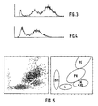
- a volume value and an absorbance value are memorized and the total results of the volumes and absorbances are distributed according to a histogram.
- the other two populations on the right are considered to be all of the leukocytes in the sample analyzed.
- a distribution curve of the absorbances also presented by way of example in FIG. 4, one obtains a first population on the left made up of weakly or weakly absorbing cells, a second central population made up of cells with medium absorbance, and a third population more on the right, made up of high absorbance cells.
- the invention is not limited to the embodiments described nor to this type of analysis.
- the optical filter with a filter wheel, it is conceivable to liter multiple colors allowing the analysis of other cell types.
- an optical measurement tank According to an alternative embodiment it would be possible to measure the light diffraction of a cell passing through this tank.
- One or more sensors being arranged focused on the tank, in alignment with a light beam passing through it. The information collected, depending on whether the total diffraction or the diffractions at several angles are measured, makes it possible to determine either the relative size of the cell or, in the case of two diffraction measurements at two different angles, the size and relative optical density.
- cytofluorescence measurement it would also be possible to carry out a cytofluorescence measurement by positioning a sensor assembly focused on the measurement tank, 90 degrees from a laser beam passing through it.
- the entire sample preparation must be adapted in order to obtain the desired cell fluorescence.
- the measurements thus obtained make it possible to evaluate the volume of the cell by resistivity, and its fluorescence.
- the current applied to the electrodes and flowing in the counting orifice may be a high frequency current making it possible to obtain, depending on the form of the pulses collected, an identification on the internal composition of the cell, that is the relative size of the nucleus, which is denser compared to the external envelope (cytoplasm) of the cell.
Landscapes
- Life Sciences & Earth Sciences (AREA)
- Chemical & Material Sciences (AREA)
- Health & Medical Sciences (AREA)
- General Health & Medical Sciences (AREA)
- Engineering & Computer Science (AREA)
- Biochemistry (AREA)
- Physics & Mathematics (AREA)
- Pathology (AREA)
- Dispersion Chemistry (AREA)
- General Physics & Mathematics (AREA)
- Immunology (AREA)
- Analytical Chemistry (AREA)
- Organic Chemistry (AREA)
- Biomedical Technology (AREA)
- Zoology (AREA)
- Wood Science & Technology (AREA)
- Genetics & Genomics (AREA)
- Bioinformatics & Cheminformatics (AREA)
- Biotechnology (AREA)
- Animal Behavior & Ethology (AREA)
- Veterinary Medicine (AREA)
- Public Health (AREA)
- Medicinal Chemistry (AREA)
- Tropical Medicine & Parasitology (AREA)
- Virology (AREA)
- Surgery (AREA)
- Molecular Biology (AREA)
- Microbiology (AREA)
- Medical Informatics (AREA)
- Heart & Thoracic Surgery (AREA)
- General Engineering & Computer Science (AREA)
- Biophysics (AREA)
- Investigating Or Analysing Biological Materials (AREA)
- Saccharide Compounds (AREA)
- Medicines That Contain Protein Lipid Enzymes And Other Medicines (AREA)
- Optical Measuring Cells (AREA)
Applications Claiming Priority (2)
| Application Number | Priority Date | Filing Date | Title |
|---|---|---|---|
| FR8914120A FR2653885B1 (fr) | 1989-10-27 | 1989-10-27 | Appareil pour le comptage et la determination d'au moins une sous-population leucocytaire. |
| FR8914120 | 1989-10-27 |
Publications (2)
| Publication Number | Publication Date |
|---|---|
| EP0425381A1 true EP0425381A1 (de) | 1991-05-02 |
| EP0425381B1 EP0425381B1 (de) | 1995-02-22 |
Family
ID=9386856
Family Applications (1)
| Application Number | Title | Priority Date | Filing Date |
|---|---|---|---|
| EP90403006A Expired - Lifetime EP0425381B1 (de) | 1989-10-27 | 1990-10-25 | Vorrichtung zum Zählen und zur Bestimmung mindestens einer Leukozytensubpopulation |
Country Status (17)
| Country | Link |
|---|---|
| US (1) | US5138181A (de) |
| EP (1) | EP0425381B1 (de) |
| JP (1) | JP3069795B2 (de) |
| KR (1) | KR0164208B1 (de) |
| AT (1) | ATE118888T1 (de) |
| AU (1) | AU628679B2 (de) |
| CA (1) | CA2028474C (de) |
| DE (1) | DE69017144T2 (de) |
| DK (1) | DK0425381T3 (de) |
| ES (1) | ES2071061T3 (de) |
| FI (1) | FI102324B1 (de) |
| FR (1) | FR2653885B1 (de) |
| HK (1) | HK53796A (de) |
| IE (1) | IE67378B1 (de) |
| NO (1) | NO312487B1 (de) |
| PT (1) | PT95709B (de) |
| ZA (1) | ZA908601B (de) |
Cited By (5)
| Publication number | Priority date | Publication date | Assignee | Title |
|---|---|---|---|---|
| EP0548983A1 (de) * | 1991-12-27 | 1993-06-30 | Toa Medical Electronics Co., Ltd. | Verfahren zur Bestimmung von Retikulozyten |
| WO1996004542A1 (en) * | 1994-08-01 | 1996-02-15 | Abbott Laboratories | Fluid nozzle and method of introducing a fluid |
| EP0744613A1 (de) * | 1995-05-24 | 1996-11-27 | Abx | Vorrichtung zur optischen Untersuchung eines Fluids, insbesondere zur hämatologischen Analyse |
| FR2734636A1 (fr) * | 1995-05-24 | 1996-11-29 | Abx Sa | Dispositif d'inspection optique d'un fluide, notamment pour analyses hematologiques |
| FR2933192A1 (fr) * | 2008-06-25 | 2010-01-01 | Horiba Abx Sas | Dispositif et procede de mesure electro optique destines a la classification et au comptage d'elements microscopiques. |
Families Citing this family (46)
| Publication number | Priority date | Publication date | Assignee | Title |
|---|---|---|---|---|
| JP3052665B2 (ja) * | 1993-01-26 | 2000-06-19 | 株式会社日立製作所 | フローセル装置 |
| US5530540A (en) * | 1994-08-03 | 1996-06-25 | Wyatt Technology Corporation | Light scattering measurement cell for very small volumes |
| FR2733835B1 (fr) * | 1995-05-03 | 1997-07-18 | Hycel Groupe Lisabio | Procede et dispositif de detection du point de lyse de globules rouges |
| JP3587607B2 (ja) * | 1995-12-22 | 2004-11-10 | シスメックス株式会社 | 粒子測定装置およびその方法 |
| EP1562035B1 (de) | 1997-01-31 | 2017-01-25 | Xy, Llc | Optischer Apparat und optisches Verfahren |
| US6149867A (en) | 1997-12-31 | 2000-11-21 | Xy, Inc. | Sheath fluids and collection systems for sex-specific cytometer sorting of sperm |
| US6071689A (en) | 1997-12-31 | 2000-06-06 | Xy, Inc. | System for improving yield of sexed embryos in mammals |
| KR100346118B1 (ko) * | 1998-07-13 | 2002-10-25 | 삼성전자 주식회사 | 하드디스크드라이브에서커버의전기적노이즈제거장치및방법 |
| US6473171B1 (en) * | 1999-01-15 | 2002-10-29 | Coors Brewing Company | Biocompatible apparatus for ultrasensitive and rapid detection of contaminants in liquids |
| US6507400B1 (en) | 1999-02-27 | 2003-01-14 | Mwi, Inc. | Optical system for multi-part differential particle discrimination and an apparatus using the same |
| US7208265B1 (en) | 1999-11-24 | 2007-04-24 | Xy, Inc. | Method of cryopreserving selected sperm cells |
| US6646742B1 (en) | 2000-02-19 | 2003-11-11 | Mwi, Inc. | Optical device and method for multi-angle laser light scatter |
| AU2002237689B2 (en) | 2000-11-29 | 2008-01-10 | Xy, Llc. | System to separate frozen-thawed spermatozoa into X-chromosome bearing and Y-chromosome bearing populations |
| US7713687B2 (en) | 2000-11-29 | 2010-05-11 | Xy, Inc. | System to separate frozen-thawed spermatozoa into x-chromosome bearing and y-chromosome bearing populations |
| CN1674780A (zh) | 2002-08-01 | 2005-09-28 | Xy公司 | 低压精子分离系统 |
| US8486618B2 (en) | 2002-08-01 | 2013-07-16 | Xy, Llc | Heterogeneous inseminate system |
| US7855078B2 (en) | 2002-08-15 | 2010-12-21 | Xy, Llc | High resolution flow cytometer |
| US7169548B2 (en) | 2002-09-13 | 2007-01-30 | Xy, Inc. | Sperm cell processing and preservation systems |
| US8323984B2 (en) * | 2002-12-19 | 2012-12-04 | Beckman Coulter, Inc. | Method and apparatus for mixing blood samples for cell analysis |
| EP2305173B1 (de) | 2003-03-28 | 2016-05-11 | Inguran, LLC | Vorrichtung und Verfahren zur Bereitstellung von sortierten Partikeln |
| DE10320870A1 (de) * | 2003-05-09 | 2004-12-09 | Evotec Technologies Gmbh | Partikelinjektor für einen Zellsortierer |
| WO2004104178A2 (en) | 2003-05-15 | 2004-12-02 | Xy, Inc. | Efficient haploid cell sorting for flow cytometer systems |
| JP2007537727A (ja) | 2004-03-29 | 2007-12-27 | モンサント テクノロジー エルエルシー | 受精で用いられる精子分散液 |
| MX2007000888A (es) | 2004-07-22 | 2007-04-02 | Monsanto Technology Llc | Procedimiento para enriquecer una poblacion de celulas de esperma. |
| FR2878032B1 (fr) * | 2004-11-18 | 2007-03-02 | Horiba Abx Sa Sa | Dispositif d'inspection d'un fluide par illumination uniforme au moyen d'un guide de lumiere conforme |
| US7355696B2 (en) * | 2005-02-01 | 2008-04-08 | Arryx, Inc | Method and apparatus for sorting cells |
| JP2006313151A (ja) * | 2005-04-07 | 2006-11-16 | Sysmex Corp | 血液分析装置、試料分析装置及びフローサイトメータ |
| US8062609B2 (en) * | 2006-11-10 | 2011-11-22 | Luminex Corporation | Flow cytometer and fluidic line assembly with multiple injection needles |
| US7804594B2 (en) * | 2006-12-29 | 2010-09-28 | Abbott Laboratories, Inc. | Method and apparatus for rapidly counting and identifying biological particles in a flow stream |
| EP4151985A1 (de) | 2007-02-01 | 2023-03-22 | Sysmex Corporation | Probenanalysegerät |
| JP4994920B2 (ja) * | 2007-02-01 | 2012-08-08 | シスメックス株式会社 | 検体分析装置 |
| EP1953526B1 (de) | 2007-02-01 | 2017-08-30 | Sysmex Corporation | Hämatologisches Analysegerät, Verfahren zur Analyse von Körperflüssigkeiten und Computerprogrammprodukt |
| US8159670B2 (en) * | 2007-11-05 | 2012-04-17 | Abbott Laboratories | Method and apparatus for rapidly counting and identifying biological particles in a flow stream |
| FR2939199B1 (fr) * | 2008-12-02 | 2011-02-11 | C2 Diagnostics | Procede et dispositif de cytometrie en flux sans fluide de gainage |
| EP2425241A4 (de) | 2009-04-27 | 2015-05-13 | Abbott Lab | Verfahren zur unterscheidung roter blutzellen von weissen blutzellen durch vorwärtsstreuung aus einem laser in einem automatisierten hämatologischen analysegerät |
| FR2956207B1 (fr) * | 2010-02-10 | 2012-05-04 | Horiba Abx Sas | Dispositif et procede de mesures multiparametriques de microparticules dans un fluide |
| US9097704B2 (en) | 2010-05-05 | 2015-08-04 | Abbott Laboratories | Method for hematology analysis |
| FR2971337B1 (fr) | 2011-02-04 | 2013-03-01 | Horiba Abx Sas | Dispositif et procede de mesures multiparametriques de microparticules dans un fluide |
| CN103575637B (zh) * | 2013-11-12 | 2016-02-10 | 桂林优利特医疗电子有限公司 | 一种鞘流阻抗、光学同步计数装置 |
| FR3022998B1 (fr) * | 2014-06-30 | 2016-07-15 | Alain Rousseau Techniques & Innovations Arteion | Systeme et ensemble de cytometrie en flux, dispositif d’analyse comprenant un tel ensemble de cytometrie et ensemble comprenant un tel systeme de cytometrie |
| FR3034198B1 (fr) | 2015-03-26 | 2019-05-31 | C2 Diagnostics | Appareil hydrofocus a solution d'analyse unique |
| FR3034520B1 (fr) | 2015-04-02 | 2020-02-14 | Horiba Abx Sas | Dispositif de comptage de particules |
| FR3068469B1 (fr) * | 2017-06-28 | 2020-09-11 | Diagdev | Cuve de mesure pour le denombrement et/ou la caracterisation de cellules |
| FR3106408B1 (fr) | 2020-01-17 | 2024-06-07 | Horiba Abx Sas | Dispositif électro-optique de mesures en flux |
| CN115468892B (zh) * | 2021-06-10 | 2025-09-05 | 深圳市帝迈生物技术有限公司 | 鞘流电阻抗粒子计数装置及检测方法 |
| FR3138211B1 (fr) | 2022-07-25 | 2026-03-13 | Horiba Abx Sas | Dispositif pour le comptage et la différenciation de particules d’un flux d’échantillon |
Citations (5)
| Publication number | Priority date | Publication date | Assignee | Title |
|---|---|---|---|---|
| US3710933A (en) * | 1971-12-23 | 1973-01-16 | Atomic Energy Commission | Multisensor particle sorter |
| DE3310551A1 (de) * | 1983-03-24 | 1984-09-27 | Coulter Corp., Hialeah, Fla. | Teilchenuntersuchungs- und -klassiervorrichtung |
| EP0121261A2 (de) * | 1983-04-05 | 1984-10-10 | Becton Dickinson and Company | Verfahren und Einrichtung zur Unterscheidung von Leukozyten-Unterklassen in einer Probe |
| EP0165868A2 (de) * | 1984-06-20 | 1985-12-27 | Commissariat A L'energie Atomique | Optische Vorrichtung mit hoher Sammelwirkung und Zytofluorimeter mit Verwendung der Vorrichtung |
| EP0310504A1 (de) * | 1987-09-30 | 1989-04-05 | Commissariat A L'energie Atomique | Verfahren zur Herstellung einer Vorrichtung zur optischen Analyse eines Mikroteilchenflusses und Anwendung desselben zur Herstellung eines Zytofluorimeters |
Family Cites Families (6)
| Publication number | Priority date | Publication date | Assignee | Title |
|---|---|---|---|---|
| US3781112A (en) * | 1972-12-15 | 1973-12-25 | Technicon Instr | Method and apparatus for analysis of leukocytes using light scattered by each leukocyte at absorbing and non-absorbing wavelength |
| DE2943116C2 (de) * | 1979-10-25 | 1986-06-19 | Gesellschaft für Strahlen- und Umweltforschung mbH, 8000 München | Einrichtung zur durchflußcytometrischen Reaktions- und/oder Diffusionsmessung |
| US4348107A (en) * | 1980-07-18 | 1982-09-07 | Coulter Electronics, Inc. | Orifice inside optical element |
| US4515274A (en) * | 1981-12-02 | 1985-05-07 | Coulter Corporation | Particle analyzing and sorting apparatus |
| JPS63262565A (ja) * | 1987-04-20 | 1988-10-28 | Hitachi Ltd | フロ−セル |
| US5030002A (en) * | 1989-08-11 | 1991-07-09 | Becton, Dickinson And Company | Method and apparatus for sorting particles with a moving catcher tube |
-
1989
- 1989-10-27 FR FR8914120A patent/FR2653885B1/fr not_active Expired - Fee Related
-
1990
- 1990-10-24 CA CA002028474A patent/CA2028474C/en not_active Expired - Fee Related
- 1990-10-25 DK DK90403006.1T patent/DK0425381T3/da active
- 1990-10-25 ES ES90403006T patent/ES2071061T3/es not_active Expired - Lifetime
- 1990-10-25 DE DE69017144T patent/DE69017144T2/de not_active Expired - Fee Related
- 1990-10-25 IE IE384790A patent/IE67378B1/en not_active IP Right Cessation
- 1990-10-25 AT AT90403006T patent/ATE118888T1/de not_active IP Right Cessation
- 1990-10-25 EP EP90403006A patent/EP0425381B1/de not_active Expired - Lifetime
- 1990-10-26 NO NO19904640A patent/NO312487B1/no unknown
- 1990-10-26 ZA ZA908601A patent/ZA908601B/xx unknown
- 1990-10-26 KR KR1019900017192A patent/KR0164208B1/ko not_active Expired - Fee Related
- 1990-10-26 AU AU65502/90A patent/AU628679B2/en not_active Ceased
- 1990-10-26 US US07/602,952 patent/US5138181A/en not_active Expired - Lifetime
- 1990-10-26 PT PT95709A patent/PT95709B/pt not_active IP Right Cessation
- 1990-10-26 FI FI905296A patent/FI102324B1/fi not_active IP Right Cessation
- 1990-10-29 JP JP2293503A patent/JP3069795B2/ja not_active Expired - Fee Related
-
1996
- 1996-03-28 HK HK53796A patent/HK53796A/xx not_active IP Right Cessation
Patent Citations (5)
| Publication number | Priority date | Publication date | Assignee | Title |
|---|---|---|---|---|
| US3710933A (en) * | 1971-12-23 | 1973-01-16 | Atomic Energy Commission | Multisensor particle sorter |
| DE3310551A1 (de) * | 1983-03-24 | 1984-09-27 | Coulter Corp., Hialeah, Fla. | Teilchenuntersuchungs- und -klassiervorrichtung |
| EP0121261A2 (de) * | 1983-04-05 | 1984-10-10 | Becton Dickinson and Company | Verfahren und Einrichtung zur Unterscheidung von Leukozyten-Unterklassen in einer Probe |
| EP0165868A2 (de) * | 1984-06-20 | 1985-12-27 | Commissariat A L'energie Atomique | Optische Vorrichtung mit hoher Sammelwirkung und Zytofluorimeter mit Verwendung der Vorrichtung |
| EP0310504A1 (de) * | 1987-09-30 | 1989-04-05 | Commissariat A L'energie Atomique | Verfahren zur Herstellung einer Vorrichtung zur optischen Analyse eines Mikroteilchenflusses und Anwendung desselben zur Herstellung eines Zytofluorimeters |
Cited By (11)
| Publication number | Priority date | Publication date | Assignee | Title |
|---|---|---|---|---|
| EP0548983A1 (de) * | 1991-12-27 | 1993-06-30 | Toa Medical Electronics Co., Ltd. | Verfahren zur Bestimmung von Retikulozyten |
| WO1996004542A1 (en) * | 1994-08-01 | 1996-02-15 | Abbott Laboratories | Fluid nozzle and method of introducing a fluid |
| US5601234A (en) * | 1994-08-01 | 1997-02-11 | Abbott Laboratories | Fluid nozzle and method of introducing a fluid |
| EP1271128A3 (de) * | 1994-08-01 | 2003-05-14 | Abbott Laboratories | Fluiddüse und Verfahren zum Einführen eines Fluids |
| EP0744613A1 (de) * | 1995-05-24 | 1996-11-27 | Abx | Vorrichtung zur optischen Untersuchung eines Fluids, insbesondere zur hämatologischen Analyse |
| FR2734637A1 (fr) * | 1995-05-24 | 1996-11-29 | Abx Sa | Dispositif d'inspection optique d'un fluide, notamment pour analyses hematologiques |
| FR2734636A1 (fr) * | 1995-05-24 | 1996-11-29 | Abx Sa | Dispositif d'inspection optique d'un fluide, notamment pour analyses hematologiques |
| US5730941A (en) * | 1995-05-24 | 1998-03-24 | A B X | Device for the optical inspection of a fluid, especially for hematological analyses |
| FR2933192A1 (fr) * | 2008-06-25 | 2010-01-01 | Horiba Abx Sas | Dispositif et procede de mesure electro optique destines a la classification et au comptage d'elements microscopiques. |
| WO2010004173A1 (fr) * | 2008-06-25 | 2010-01-14 | Horiba Abx Sas | Dispositif et procede de mesure electro optique destines a la classification et au comptage d'elements microscopiques |
| US8575568B2 (en) | 2008-06-25 | 2013-11-05 | Horiba Abx Sas | Electrooptic measurement device and method intended for classifying and counting microscopic elements |
Also Published As
| Publication number | Publication date |
|---|---|
| PT95709B (pt) | 1998-07-31 |
| KR0164208B1 (ko) | 1999-01-15 |
| AU6550290A (en) | 1991-05-02 |
| JPH03170843A (ja) | 1991-07-24 |
| CA2028474C (en) | 2002-06-11 |
| IE903847A1 (en) | 1991-05-08 |
| AU628679B2 (en) | 1992-09-17 |
| FR2653885A1 (fr) | 1991-05-03 |
| EP0425381B1 (de) | 1995-02-22 |
| HK53796A (en) | 1996-04-03 |
| NO904640L (no) | 1991-04-29 |
| FI102324B (fi) | 1998-11-13 |
| JP3069795B2 (ja) | 2000-07-24 |
| ES2071061T3 (es) | 1995-06-16 |
| FI905296A0 (fi) | 1990-10-26 |
| IE67378B1 (en) | 1996-03-20 |
| DE69017144D1 (de) | 1995-03-30 |
| FI102324B1 (fi) | 1998-11-13 |
| NO312487B1 (no) | 2002-05-13 |
| DE69017144T2 (de) | 1995-06-22 |
| ZA908601B (en) | 1991-08-28 |
| NO904640D0 (no) | 1990-10-26 |
| PT95709A (pt) | 1991-09-13 |
| KR910007483A (ko) | 1991-05-30 |
| ATE118888T1 (de) | 1995-03-15 |
| US5138181A (en) | 1992-08-11 |
| DK0425381T3 (da) | 1995-05-01 |
| CA2028474A1 (en) | 1991-04-28 |
| FR2653885B1 (fr) | 1994-01-14 |
Similar Documents
| Publication | Publication Date | Title |
|---|---|---|
| EP0425381B1 (de) | Vorrichtung zum Zählen und zur Bestimmung mindestens einer Leukozytensubpopulation | |
| EP2671063B1 (de) | Vorrichtung und verfahren für multiparametermessungen von mikropartikeln in einer flüssigkeit | |
| CA1144280A (en) | Automated method for cell volume determination | |
| JPS62503192A (ja) | フロ−・サイトメ−タ−用のフロ−・チャンバ−装置 | |
| WO2010058102A1 (fr) | Procede et systeme d'analyse de particules solides dans un milieu | |
| EP3646002B1 (de) | Messküvette zur zählung und/oder charakterisierung von zellen | |
| FR2712977A1 (fr) | Détecteur d'itensité lumineuse diffusée par des objets submicroniques en concentration élevée. | |
| Latkoczy et al. | Development of a mobile fast-screening laser-induced breakdown detection (LIBD) system for field-based measurements of nanometre sized particles in aqueous solutions | |
| EP4090941A1 (de) | Elektro-optische vorrichtung für messungen des durchflusses | |
| CN103278432B (zh) | 一种滤膜完整性的检测装置及其检测方法 | |
| FR3138211A1 (fr) | Dispositif pour le comptage et la différenciation de particules d’un flux d’échantillon | |
| WO2023118708A1 (fr) | Elément pour système de mesure optique | |
| FR2604257A1 (fr) | Instrument de mesure de particules ultra-fines | |
| JPS6171336A (ja) | 粒度測定装置 | |
| CN111504888A (zh) | 一种微粒分析分选装置及方法 | |
| FR2865807A1 (fr) | Dispositif permettant de collecter et de compter des aerosols. |
Legal Events
| Date | Code | Title | Description |
|---|---|---|---|
| PUAI | Public reference made under article 153(3) epc to a published international application that has entered the european phase |
Free format text: ORIGINAL CODE: 0009012 |
|
| AK | Designated contracting states |
Kind code of ref document: A1 Designated state(s): AT BE CH DE DK ES FR GB GR IT LI LU NL SE |
|
| 17P | Request for examination filed |
Effective date: 19911003 |
|
| 17Q | First examination report despatched |
Effective date: 19930712 |
|
| GRAA | (expected) grant |
Free format text: ORIGINAL CODE: 0009210 |
|
| AK | Designated contracting states |
Kind code of ref document: B1 Designated state(s): AT BE CH DE DK ES FR GB GR IT LI LU NL SE |
|
| REF | Corresponds to: |
Ref document number: 118888 Country of ref document: AT Date of ref document: 19950315 Kind code of ref document: T |
|
| REF | Corresponds to: |
Ref document number: 69017144 Country of ref document: DE Date of ref document: 19950330 |
|
| GBT | Gb: translation of ep patent filed (gb section 77(6)(a)/1977) |
Effective date: 19950328 |
|
| REG | Reference to a national code |
Ref country code: DK Ref legal event code: T3 |
|
| ITF | It: translation for a ep patent filed | ||
| REG | Reference to a national code |
Ref country code: ES Ref legal event code: FG2A Ref document number: 2071061 Country of ref document: ES Kind code of ref document: T3 |
|
| REG | Reference to a national code |
Ref country code: GR Ref legal event code: FG4A Free format text: 3016015 |
|
| PLBE | No opposition filed within time limit |
Free format text: ORIGINAL CODE: 0009261 |
|
| STAA | Information on the status of an ep patent application or granted ep patent |
Free format text: STATUS: NO OPPOSITION FILED WITHIN TIME LIMIT |
|
| 26N | No opposition filed | ||
| REG | Reference to a national code |
Ref country code: GB Ref legal event code: IF02 |
|
| PGFP | Annual fee paid to national office [announced via postgrant information from national office to epo] |
Ref country code: NL Payment date: 20050930 Year of fee payment: 16 |
|
| PGFP | Annual fee paid to national office [announced via postgrant information from national office to epo] |
Ref country code: SE Payment date: 20051005 Year of fee payment: 16 Ref country code: AT Payment date: 20051005 Year of fee payment: 16 |
|
| PGFP | Annual fee paid to national office [announced via postgrant information from national office to epo] |
Ref country code: GR Payment date: 20051007 Year of fee payment: 16 |
|
| PGFP | Annual fee paid to national office [announced via postgrant information from national office to epo] |
Ref country code: GB Payment date: 20051014 Year of fee payment: 16 |
|
| PGFP | Annual fee paid to national office [announced via postgrant information from national office to epo] |
Ref country code: DK Payment date: 20051018 Year of fee payment: 16 |
|
| PGFP | Annual fee paid to national office [announced via postgrant information from national office to epo] |
Ref country code: BE Payment date: 20051021 Year of fee payment: 16 |
|
| PGFP | Annual fee paid to national office [announced via postgrant information from national office to epo] |
Ref country code: FR Payment date: 20051026 Year of fee payment: 16 |
|
| PGFP | Annual fee paid to national office [announced via postgrant information from national office to epo] |
Ref country code: ES Payment date: 20051027 Year of fee payment: 16 |
|
| PGFP | Annual fee paid to national office [announced via postgrant information from national office to epo] |
Ref country code: LU Payment date: 20051028 Year of fee payment: 16 |
|
| PGFP | Annual fee paid to national office [announced via postgrant information from national office to epo] |
Ref country code: CH Payment date: 20051107 Year of fee payment: 16 |
|
| PGFP | Annual fee paid to national office [announced via postgrant information from national office to epo] |
Ref country code: DE Payment date: 20051222 Year of fee payment: 16 |
|
| PG25 | Lapsed in a contracting state [announced via postgrant information from national office to epo] |
Ref country code: AT Free format text: LAPSE BECAUSE OF NON-PAYMENT OF DUE FEES Effective date: 20061025 |
|
| PG25 | Lapsed in a contracting state [announced via postgrant information from national office to epo] |
Ref country code: SE Free format text: LAPSE BECAUSE OF NON-PAYMENT OF DUE FEES Effective date: 20061026 |
|
| PG25 | Lapsed in a contracting state [announced via postgrant information from national office to epo] |
Ref country code: CH Free format text: LAPSE BECAUSE OF NON-PAYMENT OF DUE FEES Effective date: 20061031 Ref country code: LI Free format text: LAPSE BECAUSE OF NON-PAYMENT OF DUE FEES Effective date: 20061031 Ref country code: DK Free format text: LAPSE BECAUSE OF NON-PAYMENT OF DUE FEES Effective date: 20061031 |
|
| PGFP | Annual fee paid to national office [announced via postgrant information from national office to epo] |
Ref country code: IT Payment date: 20061031 Year of fee payment: 17 |
|
| PG25 | Lapsed in a contracting state [announced via postgrant information from national office to epo] |
Ref country code: DE Free format text: LAPSE BECAUSE OF NON-PAYMENT OF DUE FEES Effective date: 20070501 Ref country code: NL Free format text: LAPSE BECAUSE OF NON-PAYMENT OF DUE FEES Effective date: 20070501 |
|
| REG | Reference to a national code |
Ref country code: CH Ref legal event code: PL |
|
| EUG | Se: european patent has lapsed | ||
| GBPC | Gb: european patent ceased through non-payment of renewal fee |
Effective date: 20061025 |
|
| NLV4 | Nl: lapsed or anulled due to non-payment of the annual fee |
Effective date: 20070501 |
|
| REG | Reference to a national code |
Ref country code: FR Ref legal event code: ST Effective date: 20070629 |
|
| PG25 | Lapsed in a contracting state [announced via postgrant information from national office to epo] |
Ref country code: GB Free format text: LAPSE BECAUSE OF NON-PAYMENT OF DUE FEES Effective date: 20061025 |
|
| BERE | Be: lapsed |
Owner name: *ABX Effective date: 20061031 |
|
| REG | Reference to a national code |
Ref country code: ES Ref legal event code: FD2A Effective date: 20061026 |
|
| PG25 | Lapsed in a contracting state [announced via postgrant information from national office to epo] |
Ref country code: FR Free format text: LAPSE BECAUSE OF NON-PAYMENT OF DUE FEES Effective date: 20061031 Ref country code: ES Free format text: LAPSE BECAUSE OF NON-PAYMENT OF DUE FEES Effective date: 20061026 |
|
| PG25 | Lapsed in a contracting state [announced via postgrant information from national office to epo] |
Ref country code: LU Free format text: LAPSE BECAUSE OF NON-PAYMENT OF DUE FEES Effective date: 20061025 |
|
| PG25 | Lapsed in a contracting state [announced via postgrant information from national office to epo] |
Ref country code: GR Free format text: LAPSE BECAUSE OF NON-PAYMENT OF DUE FEES Effective date: 20070503 |
|
| PG25 | Lapsed in a contracting state [announced via postgrant information from national office to epo] |
Ref country code: BE Free format text: LAPSE BECAUSE OF FAILURE TO SUBMIT A TRANSLATION OF THE DESCRIPTION OR TO PAY THE FEE WITHIN THE PRESCRIBED TIME-LIMIT Effective date: 20061031 Ref country code: IT Free format text: LAPSE BECAUSE OF NON-PAYMENT OF DUE FEES Effective date: 20071025 |