EP0467932B1 - Recepteur de l'activateur du plasminogene du type urokinase - Google Patents
Recepteur de l'activateur du plasminogene du type urokinase Download PDFInfo
- Publication number
- EP0467932B1 EP0467932B1 EP90906148A EP90906148A EP0467932B1 EP 0467932 B1 EP0467932 B1 EP 0467932B1 EP 90906148 A EP90906148 A EP 90906148A EP 90906148 A EP90906148 A EP 90906148A EP 0467932 B1 EP0467932 B1 EP 0467932B1
- Authority
- EP
- European Patent Office
- Prior art keywords
- par
- cells
- binding
- polypeptide
- amino acid
- Prior art date
- Legal status (The legal status is an assumption and is not a legal conclusion. Google has not performed a legal analysis and makes no representation as to the accuracy of the status listed.)
- Expired - Lifetime
Links
Images


Classifications
-
- C—CHEMISTRY; METALLURGY
- C12—BIOCHEMISTRY; BEER; SPIRITS; WINE; VINEGAR; MICROBIOLOGY; ENZYMOLOGY; MUTATION OR GENETIC ENGINEERING
- C12N—MICROORGANISMS OR ENZYMES; COMPOSITIONS THEREOF; PROPAGATING, PRESERVING, OR MAINTAINING MICROORGANISMS; MUTATION OR GENETIC ENGINEERING; CULTURE MEDIA
- C12N9/00—Enzymes; Proenzymes; Compositions thereof; Processes for preparing, activating, inhibiting, separating or purifying enzymes
- C12N9/14—Hydrolases (3)
- C12N9/48—Hydrolases (3) acting on peptide bonds (3.4)
- C12N9/50—Proteinases, e.g. Endopeptidases (3.4.21-3.4.25)
- C12N9/64—Proteinases, e.g. Endopeptidases (3.4.21-3.4.25) derived from animal tissue
- C12N9/6421—Proteinases, e.g. Endopeptidases (3.4.21-3.4.25) derived from animal tissue from mammals
- C12N9/6424—Serine endopeptidases (3.4.21)
- C12N9/6456—Plasminogen activators
- C12N9/6462—Plasminogen activators u-Plasminogen activator (3.4.21.73), i.e. urokinase
-
- A—HUMAN NECESSITIES
- A61—MEDICAL OR VETERINARY SCIENCE; HYGIENE
- A61K—PREPARATIONS FOR MEDICAL, DENTAL OR TOILETRY PURPOSES
- A61K38/00—Medicinal preparations containing peptides
- A61K38/16—Peptides having more than 20 amino acids; Gastrins; Somatostatins; Melanotropins; Derivatives thereof
- A61K38/17—Peptides having more than 20 amino acids; Gastrins; Somatostatins; Melanotropins; Derivatives thereof from animals; from humans
- A61K38/18—Growth factors; Growth regulators
- A61K38/1808—Epidermal growth factor [EGF] urogastrone
-
- A—HUMAN NECESSITIES
- A61—MEDICAL OR VETERINARY SCIENCE; HYGIENE
- A61K—PREPARATIONS FOR MEDICAL, DENTAL OR TOILETRY PURPOSES
- A61K38/00—Medicinal preparations containing peptides
- A61K38/16—Peptides having more than 20 amino acids; Gastrins; Somatostatins; Melanotropins; Derivatives thereof
- A61K38/43—Enzymes; Proenzymes; Derivatives thereof
- A61K38/46—Hydrolases (3)
- A61K38/48—Hydrolases (3) acting on peptide bonds (3.4)
- A61K38/49—Urokinase; Tissue plasminogen activator
-
- A—HUMAN NECESSITIES
- A61—MEDICAL OR VETERINARY SCIENCE; HYGIENE
- A61K—PREPARATIONS FOR MEDICAL, DENTAL OR TOILETRY PURPOSES
- A61K39/00—Medicinal preparations containing antigens or antibodies
- A61K39/395—Antibodies; Immunoglobulins; Immune serum, e.g. antilymphocytic serum
- A61K39/39533—Antibodies; Immunoglobulins; Immune serum, e.g. antilymphocytic serum against materials from animals
- A61K39/3955—Antibodies; Immunoglobulins; Immune serum, e.g. antilymphocytic serum against materials from animals against proteinaceous materials, e.g. enzymes, hormones, lymphokines
-
- A—HUMAN NECESSITIES
- A61—MEDICAL OR VETERINARY SCIENCE; HYGIENE
- A61P—SPECIFIC THERAPEUTIC ACTIVITY OF CHEMICAL COMPOUNDS OR MEDICINAL PREPARATIONS
- A61P35/00—Antineoplastic agents
-
- A—HUMAN NECESSITIES
- A61—MEDICAL OR VETERINARY SCIENCE; HYGIENE
- A61P—SPECIFIC THERAPEUTIC ACTIVITY OF CHEMICAL COMPOUNDS OR MEDICINAL PREPARATIONS
- A61P7/00—Drugs for disorders of the blood or the extracellular fluid
- A61P7/04—Antihaemorrhagics; Procoagulants; Haemostatic agents; Antifibrinolytic agents
-
- C—CHEMISTRY; METALLURGY
- C07—ORGANIC CHEMISTRY
- C07K—PEPTIDES
- C07K14/00—Peptides having more than 20 amino acids; Gastrins; Somatostatins; Melanotropins; Derivatives thereof
- C07K14/435—Peptides having more than 20 amino acids; Gastrins; Somatostatins; Melanotropins; Derivatives thereof from animals; from humans
- C07K14/705—Receptors; Cell surface antigens; Cell surface determinants
-
- C—CHEMISTRY; METALLURGY
- C07—ORGANIC CHEMISTRY
- C07K—PEPTIDES
- C07K16/00—Immunoglobulins [IG], e.g. monoclonal or polyclonal antibodies
- C07K16/18—Immunoglobulins [IG], e.g. monoclonal or polyclonal antibodies against material from animals or humans
- C07K16/28—Immunoglobulins [IG], e.g. monoclonal or polyclonal antibodies against material from animals or humans against receptors, cell surface antigens or cell surface determinants
- C07K16/2896—Immunoglobulins [IG], e.g. monoclonal or polyclonal antibodies against material from animals or humans against receptors, cell surface antigens or cell surface determinants against molecules with a "CD"-designation, not provided for elsewhere
-
- C—CHEMISTRY; METALLURGY
- C12—BIOCHEMISTRY; BEER; SPIRITS; WINE; VINEGAR; MICROBIOLOGY; ENZYMOLOGY; MUTATION OR GENETIC ENGINEERING
- C12Q—MEASURING OR TESTING PROCESSES INVOLVING ENZYMES, NUCLEIC ACIDS OR MICROORGANISMS; COMPOSITIONS OR TEST PAPERS THEREFOR; PROCESSES OF PREPARING SUCH COMPOSITIONS; CONDITION-RESPONSIVE CONTROL IN MICROBIOLOGICAL OR ENZYMOLOGICAL PROCESSES
- C12Q1/00—Measuring or testing processes involving enzymes, nucleic acids or microorganisms; Compositions therefor; Processes of preparing such compositions
- C12Q1/68—Measuring or testing processes involving enzymes, nucleic acids or microorganisms; Compositions therefor; Processes of preparing such compositions involving nucleic acids
- C12Q1/6876—Nucleic acid products used in the analysis of nucleic acids, e.g. primers or probes
- C12Q1/6883—Nucleic acid products used in the analysis of nucleic acids, e.g. primers or probes for diseases caused by alterations of genetic material
- C12Q1/6886—Nucleic acid products used in the analysis of nucleic acids, e.g. primers or probes for diseases caused by alterations of genetic material for cancer
-
- C—CHEMISTRY; METALLURGY
- C12—BIOCHEMISTRY; BEER; SPIRITS; WINE; VINEGAR; MICROBIOLOGY; ENZYMOLOGY; MUTATION OR GENETIC ENGINEERING
- C12Y—ENZYMES
- C12Y304/00—Hydrolases acting on peptide bonds, i.e. peptidases (3.4)
- C12Y304/21—Serine endopeptidases (3.4.21)
- C12Y304/21073—Serine endopeptidases (3.4.21) u-Plasminogen activator (3.4.21.73), i.e. urokinase
-
- G—PHYSICS
- G01—MEASURING; TESTING
- G01N—INVESTIGATING OR ANALYSING MATERIALS BY DETERMINING THEIR CHEMICAL OR PHYSICAL PROPERTIES
- G01N33/00—Investigating or analysing materials by specific methods not covered by groups G01N1/00 - G01N31/00
- G01N33/48—Biological material, e.g. blood, urine; Haemocytometers
- G01N33/50—Chemical analysis of biological material, e.g. blood, urine; Testing involving biospecific ligand binding methods; Immunological testing
- G01N33/53—Immunoassay; Biospecific binding assay; Materials therefor
- G01N33/575—Immunoassay; Biospecific binding assay; Materials therefor for cancer
- G01N33/5753—Immunoassay; Biospecific binding assay; Materials therefor for cancer of the stomach or small intestine
-
- G—PHYSICS
- G01—MEASURING; TESTING
- G01N—INVESTIGATING OR ANALYSING MATERIALS BY DETERMINING THEIR CHEMICAL OR PHYSICAL PROPERTIES
- G01N33/00—Investigating or analysing materials by specific methods not covered by groups G01N1/00 - G01N31/00
- G01N33/48—Biological material, e.g. blood, urine; Haemocytometers
- G01N33/50—Chemical analysis of biological material, e.g. blood, urine; Testing involving biospecific ligand binding methods; Immunological testing
- G01N33/86—Chemical analysis of biological material, e.g. blood, urine; Testing involving biospecific ligand binding methods; Immunological testing involving blood coagulating time or factors, or their receptors
-
- A—HUMAN NECESSITIES
- A61—MEDICAL OR VETERINARY SCIENCE; HYGIENE
- A61K—PREPARATIONS FOR MEDICAL, DENTAL OR TOILETRY PURPOSES
- A61K38/00—Medicinal preparations containing peptides
-
- C—CHEMISTRY; METALLURGY
- C07—ORGANIC CHEMISTRY
- C07K—PEPTIDES
- C07K2317/00—Immunoglobulins specific features
- C07K2317/70—Immunoglobulins specific features characterized by effect upon binding to a cell or to an antigen
- C07K2317/76—Antagonist effect on antigen, e.g. neutralization or inhibition of binding
-
- C—CHEMISTRY; METALLURGY
- C07—ORGANIC CHEMISTRY
- C07K—PEPTIDES
- C07K2317/00—Immunoglobulins specific features
- C07K2317/70—Immunoglobulins specific features characterized by effect upon binding to a cell or to an antigen
- C07K2317/77—Internalization into the cell
-
- C—CHEMISTRY; METALLURGY
- C12—BIOCHEMISTRY; BEER; SPIRITS; WINE; VINEGAR; MICROBIOLOGY; ENZYMOLOGY; MUTATION OR GENETIC ENGINEERING
- C12Q—MEASURING OR TESTING PROCESSES INVOLVING ENZYMES, NUCLEIC ACIDS OR MICROORGANISMS; COMPOSITIONS OR TEST PAPERS THEREFOR; PROCESSES OF PREPARING SUCH COMPOSITIONS; CONDITION-RESPONSIVE CONTROL IN MICROBIOLOGICAL OR ENZYMOLOGICAL PROCESSES
- C12Q2600/00—Oligonucleotides characterized by their use
- C12Q2600/118—Prognosis of disease development
-
- C—CHEMISTRY; METALLURGY
- C12—BIOCHEMISTRY; BEER; SPIRITS; WINE; VINEGAR; MICROBIOLOGY; ENZYMOLOGY; MUTATION OR GENETIC ENGINEERING
- C12Q—MEASURING OR TESTING PROCESSES INVOLVING ENZYMES, NUCLEIC ACIDS OR MICROORGANISMS; COMPOSITIONS OR TEST PAPERS THEREFOR; PROCESSES OF PREPARING SUCH COMPOSITIONS; CONDITION-RESPONSIVE CONTROL IN MICROBIOLOGICAL OR ENZYMOLOGICAL PROCESSES
- C12Q2600/00—Oligonucleotides characterized by their use
- C12Q2600/158—Expression markers
-
- G—PHYSICS
- G01—MEASURING; TESTING
- G01N—INVESTIGATING OR ANALYSING MATERIALS BY DETERMINING THEIR CHEMICAL OR PHYSICAL PROPERTIES
- G01N2333/00—Assays involving biological materials from specific organisms or of a specific nature
- G01N2333/90—Enzymes; Proenzymes
- G01N2333/914—Hydrolases (3)
- G01N2333/948—Hydrolases (3) acting on peptide bonds (3.4)
- G01N2333/972—Plasminogen activators
- G01N2333/9723—Urokinase
Definitions
- the present invention relates to a method for preventing or counteracting localized proteolytic activity in a mammal, in particular a human, the method comprising inhibiting the activation of plasminogen to plasmin by preventing the binding of a receptor binding form of urokinase-type plasminogen activator (in the following termed u-PA) to a u-PA receptor in the mammal and thereby preventing the u-PA from converting plasminogen into plasmin; the invention also relates to a pure u-PA receptor (in the following termed u-PAR), to DNA coding for the u-PAR, to the production of u-PAR or parts thereof for use as a therapeutic or diagnostic component, to u-PAR antibodies and the production of u-PA receptor binding u-PA molecules for use as a therapeutic or diagnostic component.
- u-PA urokinase-type plasminogen activator
- the invention relates to the regulation of the activity of a receptor binding form of u-PA, the activation of pro-u-PA to u-PA by plasmin and the regulation of the number of u-PARs on the cell and the binding affinity of the u-PAR/u-PA binding as well as the therapeutic aspects of these findings.
- the invention relates to the detection of u-PAR by labelled u-PA.
- urokinase-type plasminogen activator (u-PA) has been found in all mammalian species so far investigated.
- u-PA urokinase-type plasminogen activator
- u-PA plays a role in the degradative phase of inflammation, and there have also been reports that u-PA interferes with the lymphocyte-mediated cytotoxicity against a variety of cells, and a direct role of u-PA in the cytotoxic effect of natural killer cells has been proposed.
- a role of u-PA has been proposed in angiogenesis and in endothelial cell migration, a process important in tumor growth.
- u-PA is produced by many cultured cell types of neoplastic origin. It has been found that explants of tumor tissue released more u-PA than the corresponding normal tissue. u-PA has been identified in extracts from human lung, colon, endometrial, breast, prostate and renal carcinomas, human melanomas, murine mammary tumors, the murine Lewis lung tumor, and in ascites from human peritoneal carcinomatosis. An immunohistochemical study of invasively growing and metastasing Lewis lung carcinomas in mice consistently showed the presence of u-PA, but also a pronounced heterogenecity in the content of u-PA in different parts of the individual tumors. A high u-PA content was found in areas with invasive growth and degradation of surrounding normal tissue, while other areas were devoid of detectable u-PA. The u-PA was located in the cytoplasm of the tumor cells and extracellularly surrounding the tumor cells.
- Degradation of the surrounding normal tissue is a central feature of invasiveness of malignant tumors.
- the constant finding of u-PA in malignant tumors and the findings indicating that u-PA plays a role in tissue degradation in normal physiological events have led to the assumption that u-PA plays a similar role in cancer development.
- the hypothesis of u-PA playing a role in tissue destruction involves the assumption that plasmin, together with other proteolytic enzymes, degrades the extracellular matrix. It is noteworthy in this context that most components of the extracellular matrix can be degraded by plasmin. These include laminin, fibronectin, proteoglycans, and possibly some types of collagen, but not all.
- plasmin can activate latent collagenases which in turn can degrade the other types of collagen (see Dan ⁇ et al., 1988).
- the extracellular matrix is composed of glycoproteins such as fibronectin and laminin, collagen and proteoglycans. Extracellular matrix becomes focally permeable to cell movement only during tissue healing and remodelling, inflammation, and neoplasia.
- Liotta (1986) has proposed a three-step hypothesis: The first step is tumor cell attachment via cell surface receptors. The anchored tumor cell next secretes hydrolytic enzymes (or induces host cells to secrete enzymes) which can degrade the matrix locally (including degradation of the attachment components).
- Matrix lysis most probably takes place in a highly localized region close to the tumor cell surface.
- the third step is tumor cell locomotion into the region of the matrix modified by proteolysis.
- invasion of the matrix is not merely due to passive growth pressure but requires active biochemical mechanisms.
- a requirement for the regulation of a proteolytic cascade system in extracellular processes is the precise localization of its initiation and progression.
- cellular receptors for various components are known and serve to localize reactions that either promote or terminate the reaction sequence (Müller-Eberhard, 1988, Mann at al., 1988).
- t-PA tissue-type plasminogen activator
- u-PA is located at the membrane of the tumor cells (Skriver et al., 1984), and recent findings indicate that at cell surfaces, u-PA is generally bound to a specific receptor and that this localization may be crucial for the regulation of u-PA catalyzed plasminogen activation in time and space (see Blasi at al., 1987).
- u-PAR The cellular receptor for u-PA (u-PAR) was originally identified in blood monocytes and in the monocyte-like U937 cell line (Vassalli at al., 1985), and its presence has been demonstrated on a variety of cultured cells, including several types of malignant cells (Stoppelli et al., 1985, Vassalli et al., 1985, Plow at al., 1986, Boyd at al., 1988a, Nielsen at al., 1988), human fibroblasts (Bajpai and Baker, 1985), and also in human breast carcinoma tissue (Needham at al., 1987).
- the receptor binds active 54 kD u-PA, its one-polypeptide chain proenzyme, pro-u-PA (see below), as well as 54 kD u-PA inhibited by the active site reagent DFP, but shows no binding of the low molecular weight (33 kD) form of active u-PA (Vassalli at al., 1985; Cubellis at al., 1986).
- binding to the receptor does not require the catalytic site of u-PA, and in agreement with these findings, the binding determinant of u-PA has been identified in the amino-terminal part of the enzyme, in a region which in the primary structure is remote from the catalytic site.
- the receptor binding domain is located in the 15 kD amino-terminal fragment (ATF, residues 1-135) of the u-PA molecule, more precisely within the cysteine-rich region termed the growth factor region as this region shows homologies to the part of epidermal growth factor (EGF) which is responsible for binding to the EGF receptor.
- the amino acid residues which appear to be critical for binding are located within the sequence 12-32 (Appella at al., 1987).
- Synthetic peptides have been constructed that inhibit the binding of very low (100 nM) concentrations. The lack of cross-reactivity between the murine and the human peptides indicates that the binding between u-PA and u-PAR is strongly species specific.
- Binding of u-PA to u-PAR is specific in the sense that as yet no other protein has been found to compete for binding to the receptor, though several proteins structurally related to u-PA, including t-PA and plasminogen, have been tested (Stoppelli at al., 1985, Vassalli et al., 1985, Nielsen et al., 1988). Fragments of u-PA containing only the receptor binding domain, e.g.
- ATF ensure specificity of the binding to the receptor, since other molecules that might bind u-PA (protease nexin and the specific plasminogen activator inhibitors PAI-1 and PAI-2) recognize the catalytically active region (Stoppelli et al., 1985; Nielsen at al., 1988). PAI-1 is able to form a covalent complex with u-PA but not with pro-u-PA (Andreasen et al., 1986).
- the number of receptors can be regulated by the addition of various agents such as phorbol myristate acetate (PMA) in U937 cells (Stoppelli at al., 1985, Nielsen at al., 1988), epidermal growth factor in A431 cells (Blasi et al., 1986) and HeLa cells (Estreicher et al., 1989) and dimethylformamide in colon carcinoma cells (Boyd at al., 1988b).
- PMA phorbol myristate acetate
- WO 86/06100 discloses a receptor originally called "phospholipase inhibitory protein" (PIP) which was shown to be effective in controlling undesired inflammation.
- PIP phospholipase inhibitory protein
- the amino acid sequence of the u-PA receptor is identical to a substantial part of the amino acid sequence of PIP.
- the purity of the PIP polypeptide apparent from WO 86/06100 is not as high as the purity of the u-PA receptor described herein, and the function that was ascribed to the purified PIP protein in WO 86/06100 is not a property of the u-PA receptor, i.e. it could have been due to an impurity. Neither a specific functional role, nor a mechanism of action for the PIP protein in invasive proteolytic degradation of matrix is described in WO 86/06100.
- the purified u-PAR preparation shows essentially one radiolabelled band after SDS-PAGE followed by auto-radiography. This analysis, however, does not show the purity of the preparation as it does not detect unlabelled proteins that may be present in an amount that may be higher than that of the u-PAR. Similar considerations hold true for a recent study by Estreicher et al. (1989), in which attempts at purifying u-PAR were done from cells that had been surface-labelled with 125 I.
- Nucleotide sequencing primers complementary to the u-PAR nucleotide sequence and a library screening assay have been described in Roldan et al., 1990.
- u-PA is released from many types of cultured cells as a single-chain proenzyme with little or no plasminogen activating capacity (Nielsen et al. , 1982, Skriver et al. 1982, Eaton et al., 1984, Kasai et al., 1985, Pannell and Gurewich 1987).
- this proenzyme can be converted to its active two-chain counterpart.
- Such assays for pro-u-PA are self-activating and are strongly influenced by small amounts of contaminating or generated two-chain u-PA or plasmin. As discussed in detail elsewhere (Petersen et al., 1988), it is therefore possible that the high activity of one-chain u-PA found in these studies was apparent and not due to intrinsic activity of single-chain u-PA. Consistent with this interpretation is a report on a variant of recombinant single-chain u-PA which by site-directed mutagenesis was made partly resistant to plasmin cleavage. This variant of single-chain u-PA had an activity that in coupled assays was 200-fold lower than that of two-chain u-PA (Nelles et al., 1987).
- pro-u-PA is the predominant form of u-PA in intracellular stores, and it also constitutes a sizeable fraction of the u-PA in extracellular fluids (Skriver et al., 1984, Kielberg et al., 1985). Extracellular activation of pro-u-PA may therefore be a crucial step in the physiological regulation of the u-PA pathway of plasminogen activation.
- the plasmin-catalyzed activation of pro-u-PA provides a positive feedback mechanism that accelerates and amplifies the effect of activation of a small amount of pro-u-PA.
- the initiation of the u-PA pathway of plasminogen activation under physiological conditions involves triggering factors that activate pro-u-PA as described herein.
- Mutants of human single-chain pro-u-PA in which lysine 158 is changed to another amino acid are not, or are only to a small extent, converted to active two-chain u-PA (Nelles et al., 1987).
- u-PA has been found to be unevenly distributed, distinctly located at cell-cell contact sites and at focal contacts that are the sites of closest apposition between the cells and the substratum (Pöllänen et al., 1987, 1988, Hébert and Baker 1988). u-PA was not detected in the two other types of cell-substratum contact, i.e. close contacts and fibronexuses, making it an intrinsic component at focal contact sites (Pöllänen et al., 1988). u-PA at the focal contact sites is receptor-bound (Hébert and Baker, 1988).
- the focal contact sites are located at the termini of actin-containing microfilament bundles, the so-called stress fibers or actin cables (Burridge, 1986). These sites contain several structural components (actin, talin) and regulatory factors (the tyrosine kinase protooncogene products P60 src , P120 gag-abl , P90 gag-yes , P80 gag-yes ), that are all located on the cytoplasmic side (see Burridge, 1986).
- Plasminogen as well as plasmin, binds to many types of cultured cells, including thrombocytes, endothelial cells and several cell types of neoplastic origin (Miles and Plow, 1985, Hajjar et al., 1986, Plow et al., 1986, Miles and Plow 1987, Burtin and Fondaneche, 1988).
- the binding is saturable with a rather low affinity for plasminogen (K D 1 ⁇ M).
- binding of plasmin appears to utilize the same site as plasminogen, but the binding parameters for plasmin indicate that more than one type of binding site for plasminogen and plasmin may exist.
- plasmin and plasminogen bind with almost equal affinity (Plow et al., 1986), while on others plasmin apparently binds with a higher affinity (K D 50 nM) than plasminogen (Burtin and Fondaneche, 1988).
- the binding is inhibited by low amounts of lysine and lysine analogues and appears to involve the kringle structure of the heavy chains of plasminogen and plasmin (Miles et al., 1988).
- the binding capacity varies between cell types and in many cell types is quite high (10 5 -10 7 binding sites per cell). The chemical nature of the binding sites are not known.
- a membrane protein, GPIIb/IIIa seems to be involved in the binding of plasminogen to thrombocytes (Miles et al., 1986) and, particularly on thrombin-stimulated thrombocytes, also fibrin may be involved in plasminogen binding (Miles et al., 1986).
- the thrombocyte protein thrombospondin joints complexes (K D 35 nM) with plasminogen (Silverstein et al., 1984).
- immobilized laminin (Salonen et al., 1984) and fibronectin (Salonen et al., 1985) bind plasminogen (K D 3 nM and 90 nM, respectively)
- Receptor-bound pro-u-PA can be activated by plasmin (Cubellis et al., 1986) and, at least in part, receptor-bound two-chain u-PA retains its ability to activate plasminogen (Vassalli et al., 1985).
- Ellis et al. (1989) recently published evidence indicating that the reactions leading to plasminogen activation can take place when single-chain u-PA and plasminogen are added to U937 cells, and that they occur more efficiently when both plasminogen and pro-u-PA are bound to the surface.
- This experiment was performed in the absence of serum, i.e. under conditions where the plasminogen activation with the preparations used by Ellis et al. will also take place in solution (cf. Ellis et al., 1987), and these studies do not exclude the possibility that one or more of the processes involved (e.g. the plasminogen activation catalyzed by two-chain u-PA) actually occurred when the u-PA was not receptor-bound.
- the present invention is based upon the discovery that under conditions similar to those present extracellularly in the intact organism (i.e. in the presence of serum containing inhibitors of plasmin and of plasminogen activators), plasminogen activation initiated by endogenous u-PA occurs only when the u-PA is receptor-bound, upon the provision of pure u-PA receptor, and upon the provision of the possibility of producing the u-PA receptor or characteristic and valuable sequences thereof or analogues to sequences thereof by recombinant DNA technology.
- new and potentially extremely valuable therapeutic, prophylactic and diagnostic methods and products, together with associated basic methods and products are provided by the present invention.
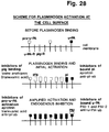
- Plasminogen binds to cell surfaces, and surprisingly it was found that a large part, if not all, of the cell surface plasminogen activation is catalyzed by surface-bound u-PA, and that binding of plasmin to the surface is necessary for the activation of pro-u-PA.
- most of the cell surface u-PA is present in its single-chain proenzyme form (pro-u-PA), while addition of plasminogen leads to the formation of receptor-bound two-chain u-PA. The latter reaction is catalyzed by cell-bound plasmin.
- Receptor-bound u-PA is accessible to inhibition by endogenous PAI-1 and by added PAI-2, while the cell-bound plasmin is inaccessible to serum inhibitors.
- a model for cell-surface plasminogen activation can be made in which plasminogen binding to cells is followed by plasminogen activation by trace amounts of bound active u-PA, to form bound plasmin, which in turn serves to produce more active u-PA from bound pro-u-PA.
- This exponential process is subject to regulation by endogenous PAI-1, and limited to the pericellular space.
- the new findings include the requirement, in the presence of serum, for binding of plasminogen, the ability of bound u-PA under these conditions to activate plasminogen, the presence of pro-u-PA on the cells, the ability of bound plasmin to activate pro-u-PA, and the ability of endogenous plasminogen activator inhibitor PAI-1, as well as added plasminogen activator inhibitor PAI-2, to regulate the surface plasminogen activation.
- tumor cells can acquire the broad-spectrum proteolytic activity of plasmin, bound to their surface in such a way that it is protected from inactivation by serum protease inhibitors, and ideally situated to be employed in the degradation of the pericellular matrix.
- Binding of u-PA to its receptor localizes u-PA not only to the cell surface, but focalizes it to distinct parts of the surface that at least in some cell types are the cell-cell and cell-substrate contact.
- the location of pro-u-PA at the focal contact sites suggests that u-PA catalyzed plasminogen activation is involved in the breakdown of the contacts, e.g. during cell movement.
- a selective activation of pro-u-PA at these sites provides a means of obtaining a directional pericellular proteolysis.
- pro-u-PA activation might be intracellularly initiated and mediated by a transmembrane signal through the u-PA receptor.
- Human tumor cells are very commonly found to secrete plasminogen activator of the urokinase type (u-PA). By this means they are able to recruit the proteolytic potential available in the high concentration of plasminogen in plasma and other body fluids.
- the invasive properties of tumor cells may be at least partly dependent on their proteolytic capability mediated through the broad spectrum of activity of plasmin and including its indirect actions in activating other latent proteases, such as collagenases.
- the expression of protease activity by tumor cells facilitates their penetration of basement membranes, capillary walls and interstitial connective tissues, allowing spread to other sites and establishment of metastases.
- a stepwise pathway of pericellular proteolysis geared to cell migration can be envisaged: binding of u-PA and plasminogen to the cell surface will lead to extracellular proteolysis and to the local severing of cell-cell and cell-substrate connections. This region of the cell is therefore free to move and this will transpose u-PA to a region in which PAI-1 is present. PAI-1 will inactivate u-PA and in the absence of local proteolytic activity, the cell will form new connections with the matrix, a process required for further migration.
- the expression of the u-PA gene is finely regulated by a variety of agents that affect cell growth; however, until recently very little was known of the regulation of the u-PAR function and synthesis. It is known that the affinity of the u-PA receptor can be modified by e.g. the tumor promoter PMA. This indicates that the cells are endowed with mechanisms that modulate the u-PA:u-PAR interaction. While this interaction appears to act at the level of the receptor itself, the effect of the plasminogen activator inhibitors demonstrates a second level of modulation, i.e. at the level of the active ligand itself. It is possible that the two levels of regulation might actually be interconnected, i.e. that the binding of the inhibitor to surface-bound u-PA influences the affinity of the receptor.
- the change in affinity is a regulatory mechanism capable of modifying the ratio between soluble and surface-bound u-PA, i.e. regulating the location of u-PA. It is possible that the effects on synthesis and affinity of u-PAR normally take place either in different cells or in the same cells, but in response to different stimuli.
- the physiological signal for the affinity-regulating mechanism may be connected with the level and possibly the fine localization of the u-PA activity on the cell surface.
- Example 8 show that blocking binding to the u-PA receptor, or to PAI-1 and/or PAI-2 inhibitors, should result in an increase in the half life of therapeutically administered pro-u-PA and u-PA, thus allowing a decrease of the therapeutically efficient dosage.
- Example 1 Further characterization of the interaction of u-PA and u-PAR required the purification of the u-PAR.
- the number of u-PAR produced by the monocyte-like cell U937 can be increased several fold by phorbol esters like PMA. This fact was used to produce sufficient quantities of the receptor for purification.
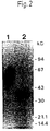
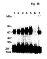
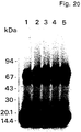
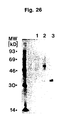
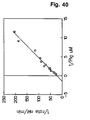
- Example 1 a complete purification of the u-PA receptor is described, involving temperature-induced phase separation of a detergent extract from cells, and affinity chromatography with immobilized DFP-inactivated u-PA. This resulted in a preparation that shows one band at approximately 55-60 kD after SDS-PAGE and silver staining, with a load of approximately 1 ⁇ g of the receptor.
- the purified protein could be chemically cross-linked with u-PA. Its amino acid composition and N-terminal sequence were determined (30 residues, some of which with some uncertainty). It was found to be heavily N-glycosylated, deglycosylation resulting in a protein with an apparent molecular weight of about 30-35 kD. The apparent molecular weight of u-PAR from different cell lines and from PMA-stimulated and non-stimulated U937 cells varied somewhat. This heterogeneity disappeared after deglycosylation and was thus due to differences in glycosylation of u-PAR from the various sources.
- Example 1 The modulation of the u-PAR molecules demonstrated in Example 1 may represent an important feature in the regulation of extracellular proteolysis and thus in the degradation of the extracellular matrix and basement membrane components, processes that are at the core of cell migration and invasiveness.
- Example 2 isolation of a ligand binding domain of u-PAR is identified and characterized. This provides potentially therapeutically valuable information on peptides that may inhibit the ligand binding.
- u-PAR is produced as a 313 residues long protein with a 282 residues long hydrophilic N terminal part (probably extracellular) followed by 21 rather hydrophobic amino acids (probably a trans-membrane domain).
- the potential extracellular part is organised in 3 repeats with striking homologies, particularly with respect to the pattern of cysteines. This may indicate the presence of distinct domains that may bind different ligands.
- the receptor purification and cDNA cloning allowed to recognize that the u-PAR is at least in some cases terminally processed and anchored to the cell surface via a glycolipid anchor, and that the surface location can be regulated by the phospholipase PI-PLC, but not by the phospholipases A 2 and D (Example 4). Furthermore, it was found that also harvest fluid from cells that were not treated contain some free u-PAR, indicating release from the cells that may be mediated by an endogenous phospholipase. This may be a physiological mechanism and it is possible that measurement of free receptor, e.g. in serum, may be a diagnostically valuable indicator of some pathological processes.
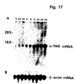
- the u-PAR cDNA was used to show that u-PAR mRNA could be regulated in some cell types by substances such as PMA, dexamethasone, mEGF and TGF- ⁇ -1 (Example 5).
- substances such as PMA, dexamethasone, mEGF and TGF- ⁇ -1 (Example 5).
- the findings indicate that these and similar substances may be therapeutically useful in regulating u-PAR synthesis.
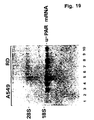
- u-PAR cDNA was used to produce fragments of u-PAR antisense mRNA which proved useful for the detection of u-PAR mRNA in tissue sections by in situ hybridization. Particularly interesting is the finding that u-PAR mRNA is consistently present in human colon carcinomas and is located in cells at the invasive front of the tumors, thus indicating the production of u-PAR by these cells and a role of u-PAR in localizing u-PA at these sites.
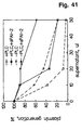
- Example 7 it is demonstrated that after incubation of monolayer cultures of human HT-1080 fibrosarcoma cells with purified native human plasminogen in serum containing medium, bound plasmin activity can be eluted from the cells with tranexamic acid, an analogue of lysine.
- the bound plasmin is the result of plasminogen activation on the cell surface; plasmin activity is nor taken up onto cells after deliberate addition of plasmin to the serum containing medium.
- the cell surface plasmin formation is inhibited by an anti-catalytic monoclonal antibody to u-PA, indicating that this enzyme is responsible for the activation.
- Plasmin released from the cells was partially inactivated in the serum medium. As long as the plasmin remained bound, it was protected from serum inhibitors but could be inhibited by aprotinin or an anti-catalytic monoclonal antibody.
- Plasmin has been shown to be produced in this condition, yet one would expect that it would be inactivated by serum inhibitors. If a significant fraction is bound to cells, however, this may escape inhibition and retard development of healing tissue, until an effective inhibitor is applied externally.
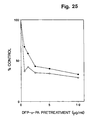
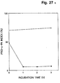
- Example 7 it is shown that preincubation of the cells with DFP-incubated u-PA led to a decrease in surface-bound plasmin, indicating that a large part, if not all, of the cell surface plasminogen activation was catalyzed by surface-bound u-PA.
- plasminogen activator activity is largely dependent on addition of exogenous u-PA.
- u-PA is administered in a binding step followed by washing of the cells and assay.
- the activity can be competed by receptor binding u-PA antagonists, e.g. synthetic peptides and DFP-treated u-PA, and can be inhibited by the addition of PAI-1.
- PAI-1 also binds to, and inhibits the activity of receptor-bound u-PA in U937 cells.
- Example 8 also shows that PAI-1/u-PA complexes bind the receptor of U937 cells with the same specificity and affinity as free u-PA.
- PAI-1 is able to interact also with u-PA pre-bound on the receptor on the U937 cells. This results in the formation of a typical covalent PAI-1/u-PA complex that is not detectably internalized, and in the inhibition of the u-PA activity.
- the affinity of complexed u-PA is slightly decreased compared to free u-PA. Possibly the presence of the bulky PAI-1 molecule may pose a problem of steric hindrance.
- Example 8 it is further shown that when u-PA/PAI-1 complexes are bound to the U937 cells, they are subsequently degraded and internalized.
- Example 9 shows that PAI-1 and PAI-2 rather rapidly inhibit receptor-bound u-PA, although the respedtive association rate constants are lower than those for inhibition of u-PA in solution.
- u-PA binding of u-PA to solubilized (Example 10) or purified (Example 13) u-PAR inhibits the ability of u-PA to activate plasminogen in solution, in contrast to the stimulation of the activity which is observed when u-PA is bound to u-PAR on a cell surface concomitantly with surface binding of plasminogen.
- Purified u-PAR also inhibits plasmin catalyzed pro-u-PA activation in solution (Example 13).
- Polyclonal antibodies were developed by immunization of mice with purified u-PAR (Example 11). These antibodies precipitated 125 I-labelled purified u-PAR in a dose-dependent manner, with a significant precipitation being obtained by the antiserum in a dilution of 1:7500.
- the antiserum was found to immunocapture radiolabelled u-PAR, and in an ELISA immobilized u-PAR in an amount of 1 ng was detected with the immune serum diluted 1:8000.
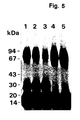
- Western blotting the antibodies detected both purified u-PAR and u-PAR in the crude detergent phase of extracts of PMA-treated U937 cells.
- the Example also includes a description of methods for development of monoclonal antibodies using the above-mentioned methods for screening of hybridomas for antibody production.
- Example 11 describes that antibodies to u-PAR can be used to specifically prevent ligand binding. It is furthermore shown (Example 10) that u-PAR antibodies inhibit u-PA catalyzed cell surface plasminogen activation. These or similar antibodies may also be used to specifically target bacterial or vegetable toxins with the purpose of destroying potentially metastatic tumor cells.
- Example 12 a method for the visualization of the u-PAR on cells and in tissue sections is described, using biotinylated DFP-treated u-PA followed by incubation with streptavidin-fluorescin isothiocyanate.
- the method is very sensitive, and its specificity can readily be tested by competition experiments (e.g. with the amino-terminal fragment of u-PA (ATF), t-PA, EGF, etc.).
- the present invention provides inhibition of receptor binding of u-PA as a means of inhibiting some of its physiological functions in relationship to therapeutic prevention of localized proteolytic activity, e.g. invasion and metastasis of cancer cells, inflammatory bowel disease, premalignant colonic adenomas, septic arthritis, osteoarthritis, rheumatoid arthritis (for which a direct involvement of excess u-PA production has been demonstrated), osteoporosis, cholesteatoma, and a number of skin and corneal diseases for which an excess plasminogen activation has been shown to be the pathogenetic cause, such as corneal ulcers, keratitis, epidermolysis bullosa, psoriasis, and pemphigus.
- localized proteolytic activity e.g. invasion and metastasis of cancer cells, inflammatory bowel disease, premalignant colonic adenomas, septic arthritis, osteoarthritis, rheumatoid arthritis (for which a direct involvement
- u-PA receptors are present in several blood and endothelial cells, their regulation might also significantly affect intravascular fibrinolytic activity in physiological, pathological and pharmacological conditions.
- the above-mentioned diseases would be the first obvious targets for a therapy based on administration of substances that block or decrease cell surface plasminogen activation.
- a contraceptive effect is expected of measures that inhibit receptor binding.
- the therapy and prophylaxis will involve systemic or topical treatment with agents that block or reduce receptor bound plasminogen activator activity, such as will be explained below.
- the present invention also provides valuable reagents and methods for diagnostic or research purposes, such as the u-PA receptor (u-PAR), the u-PAR cDNA, anti u-PAR antibodies, obtained by chemical, biological, or synthetic means, such as explained below.
- u-PAR u-PA receptor
- cDNA u-PAR cDNA
- anti u-PAR antibodies obtained by chemical, biological, or synthetic means, such as explained below.
- the invention relates to certain u-PAR polypeptides and DNA coding for these.
- the invention relates to the use of particular polypeptides or antibodies (as defined in the claims) capable of preventing the binding of a receptor binding form of u-PA to a u-PA receptor (u-PAR) in the mammal and thereby preventing u-PA from converting plasminogen into plasmin for the preparation of a pharmaceutical composition for preventing or counteracting localized proteolytic activity in a mammal, in particular a human.
- the term "localized proteolytic activity” is intended to designate a proteolytic activity which is located at one or several distinct regions in a human body, or at distinct cells, as opposed to an overall proteolytic activity exerting itself substantially anywhere in the body.
- the localized proteolytic activity can be inhibited generally in a mammal, in particular a human, or locally.
- the term "preventing or counteracting” is intended to designate a situation where the binding of u-PA to u-PAR is completely inhibited, or a situation where the binding is sufficiently inhibited so as to inhibit the undesired effect of the plasminogen activator.
- the term "inducing the specific degradation” is intended to designate a process by which the receptor-binding form of u-PA is degraded in a specific manner, e.g. internalized such as described in Example 8 in which the specific degradation is induced by adding PAI-1.
- a receptor binding form of u-PA is intended to mean any form of u-PA possessing a site that binds to a site at a u-PAR, that is to say that the u-PA contains the u-PAR binding site.
- the receptor binding form of u-PA can thus be pro-u-PA, u-PA, an amino-terminal fragment of u-PA, a u-PA that is irreversibly inhibited by e.g. diisopropyl fluorophosphate (DFP), p-nitrophenyl-p'-guanidinobenzoate (NPGB), or any other inhibitor or any other modification of u-PA that can bind to a u-PAR.
- DFP diisopropyl fluorophosphate
- NPGB p-nitrophenyl-p'-guanidinobenzoate
- a u-PAR indicates that even though the polypeptide part of u-PAR in a species might be the same for all u-PARs, there is a plurality of u-PARs as for example the carbohydrate part or the mechanism of surface attachment of the u-PAR can be different. It may even be so that some cells, e.g. cancer cells, have substantially different u-PARs which might have important therapeutic significance as it might be possible to block the binding of u-PA to u-PARs residing on a cancer cell without affecting the binding of u-PA to u-PARs on non-pathological cells or of specifically killing cancer cells that express u-PAR.
- u-PA urokinase-type plasminogen activator
- plasminogen By cleavage at Arg 560 , plasminogen is activated to the broad spectrum protease plasmin.
- preventing u-PA from converting plasminogen into plasmin is therefore meant that this activation by u-PA is substantially inhibited or a situation where the activation is sufficiently inhibited so as to inhibit or reduce the undesired effect of the plasmin.
- One way of preventing the binding of a receptor binding form of u-PA to a u-PAR is to administer a u-PA-binding modification of a u-PAR to the mammal so as to occupy the cell receptor-binding site of u-PA, thereby preventing the receptor binding form of u-PA from binding to the cell-bound receptor.
- a water-soluble form of a u-PAR in other words a part thereof comprising a u-PA binding sequence.
- Such a part will often have been made by truncation of a larger sequence by removing part of the cDNA sequence of a plasmid vector containing the human u-PAR cDNA, cf.
- u-PAR can be solubilized by removal of the glycerol-phosphoinositol anchor, e.g. with a lipase as shown in Example 4.
- the soluble forms of u-PAR are also useful in that they may be coupled by chemical methods or recombinant DNA methods to plasminogen activator inhibitors such as plasminogen activator inhibitor Type 1 (PAI-1) or Type 2 (PAI-2), whereby an interesting double effect comprising either receptor blocking or ligand blocking, or both, may be obtained.
- PAI-1 plasminogen activator inhibitor Type 1
- PAI-2 Type 2
- a very interesting method of preventing the binding of a receptor binding form of u-PA to a u-PAR and thereby preventing the cell surface plasminogen activation is the use of antibodies against u-PAR such as demonstrated in Example 11 (prevention of binding) and Example 10 (prevention of cell surface plasminogen activation).
- the antibodies may be polyclonal antibodies, preferably of high specificity such as the antibodies illustrated in Example 11, or a monoclonal antibody.
- the antibody may be an antibody that is reactive with non-carbohydrate moieties of the u-PAR, or it may be an antibody that is reactive with carbohydrate moieties of the u-PAR, the latter permitting a valuable distinction between target cells where cells expressing distinct variants of u-PAR are the cells involved in the undesired proteolysis.
- the antibodies may be administered in various ways as described below.
- a potentially therapeutically very valuable application of u-PAR polypeptides, solubilized u-PAR and variants thereof is as an inhibitor of u-PA-catalyzed plasminogen activation in solution.
- the hitherto known specific inhibitors of plasminogen activators that is, PAI-1 and PAI-2, inhibit both u-PA and t-PA.
- Purified u-PAR as well as u-PAR solubilized by removal of the glycerol-phosphoinositol anchor inhibits u-PA in solution as demonstrated in Examples 10 and 13.
- u-PAR does not bind t-PA.
- u-PAR is contemplated to be more advantageous than PAI-1 and PAI-2 in cases where specific inhibition of u-PA is needed.
- Potentially very valuable is also the therapeutic use of u-PAR polypeptides, solubilized u-PAR and variants thereof for inhibiting the activation of the virtually inactive single-chain pro-u-PA molecule to active two chain u-PA.
- the administration of the various above-mentioned principles to a mammal, preferably a human being, may be performed by any administration method which is suitable for administering proteins or peptides or antibodies.
- Typical administration routes are parenteral, oral, nasal, topical or rectal administration.
- the active ingredient to be administered should be formulated in a manner which will protect the active ingredient against degradation, in particular by enzymes.
- the parenteral administration is the safest way of administering proteins and peptides.
- the parenteral administration route should be selected dependent on where the active ingredient is to be released, e.g. intravenously, intramuscularly or subcutaneously, etc. It is also important to consider the necessity of "packing" the active ingredient in a suitable manner in order to
- suitable measures should be taken to protect the active ingredient from enzymatic degradation in the gastrointestinal tract, e.g. by packing the active ingredient in such a way that it will not be released from the formulation (i.e. the pharmaceutical composition) until it has reached the site where either the active ingredient is to exert its activity locally (i.e. in the gastrointestinal tract) or from where the absorption may take place (e.g. M-cells in the colon).
- Nasal administration is an administration form which is presently intensively investigated in order to provide absorption of substances of the peptide type from the nasal cavity. In principle, this may take place in two ways, firstly by using enhancers, and secondly by using the bioadhesion principle in which the active ingredient may be maintained for a long period of time at a suitable domain in the nose.
- Topical administration may be performed by formulating the active ingredient in a salve, an ointment, a lotion, a creme, etc.
- compositions of the invention may for example include pharmaceutically acceptable excipients adapted to the character of the active ingredients in accordance with the above discussion. Suitable excipients may include liposomes and/or microspheres.
- the preparation of the pharmaceutical compositions may be performed in accordance with methods described in the literature for compositions of the types described herein.
- the treatments will normally be continued for weeks or often months and are suitably combined with treatment with other medicaments against the conditions in question.
- An important field of the present invention is a number of diagnostic methods which methods, or the importance thereof, are based upon the findings according to the invention.
- One important aspect thereof is a method of detecting a u-PAR in a tissue section comprising treating the tissue section with an antibody that binds to a u-PAR, and visualizing the presence of the bound antibody.
- the antibody may be a labelled antibody or an unlabelled antibody which is subsequently detected by an immunostaining method.
- the antibody may be a polyclonal or monoclonal antibody, and particularly interesting antibodies are antibodies that distinguish between various forms of u-PAR. A detailed description of diagnostic kits, materials and methods based on antibodies is given further below.
- Another field of the invention is the use of antibodies against a u-PAR for the quantification of the u-PAR in biological material using antibodies against the u-PAR. While this method may be performed using either polyclonal or monoclonal antibodies, an interesting embodiment uses a combination of polyclonal and monoclonal antibodies, the monoclonal antibodies being more specific and the polyclonal antibodies generally having a higher binding affinity.
- the quantification method may be of the ELISA type or may be a radio-immunoassay. These assays may be produced by methods known per se .
- One aspect of the invention relates to a method of producing pure u-PAR polypeptides, the method comprising subjecting a u-PAR-containing material to affinity chromatography with immobilized antibodies to u-PAR and eluting the u-PAR, e.g. under acidic conditions.
- Pure u-PAR in glycosylated form shows, in an SDS-PAGE at a load of approximately 1 ⁇ g, substantially one and only one silver stained band having an apparent molecular weight in the range of about 55-60 kD.
- the presence of substantially one and only one silver stained band in this SDS-PAGE is a proof of the purity of the u-PAR.
- Another proof of the purity of the u-PAR is the presence of a single amino-terminal amino acid sequence in purified u-PAR preparations.
- the glycosylated u-PARs upon deglycosylation, were all found to have an identical electrophoretic mobility (corresponding to substantially one and only one band at about 30-35 kD in an SDS-PAGE), indicating that the peptide part of the molecule is identical in all cases.
- pure u-PAR in glycosylated form may be prepared from a biological material containing u-PAR by temperature-induced phase separation of detergent extracts followed by affinity chromatography purification with immobilized DFP-u-PA.
- the detergent is preferably a non-ionic detergent such as a polyethylene glycol ether, e.g. Triton X-114.
- the temperature was found to be relatively critical in the range of 34-40°C, such as about 37°C, for 10 minutes.
- the pure u-PAR in unglycosylated form may be prepared by deglycosylation with, e.g., peptide/N-glycosidase F.
- a 24-mer nucleotide probe was synthesized and used to screen a library to identify and isolate recombinant clones carrying the cDNA for u-PAR.
- the identity of the cDNA clones was confirmed by comparing the nucleotide sequence of this cDNA clone with the amino terminal sequence of the purified u-PAR, and by expressing said cDNA in mouse L cells and assaying their u-PA-binding properties.
- amino acids used herein are the following: Amino acid Three-letter abbreviation One-letter symbol Alanine Ala A Arginine Arg R Asparagine Asn N Aspartic acid Asp D Asparagine or aspartic acid Asx B Cysteine Cys C Glutamine Gln Q Glutamic acid Glu E Glutamine or glutamic acid Glx Z Glycine Gly G Histidine His H Isoleucine Ile I Leucine Leu L Lysine Lys K Methionine Met M Phenylalanine Phe F Proline Pro P Serine Ser S Threonine Thr T Tryptophan Trp W Tyrosine Tyr Y Valine Val V
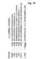
- One aspect of the invention relates to a polypeptide comprising a characteristic amino acid sequence derived from a u-PAR which polypeptide comprises at least 5 amino acids and up to the sequence of u-PAR from amino acid 1-286 as shown below as the DNA sequence and the deducted amino acid sequence of the clone p-uPAR-1.
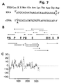
- the signal peptide is underlined and the first 30 amino acids, the sequence of which has been determined on the purified protein with an Applied Biosystems gas phase sequencer (see Example 1), are overlined.
- the putative transmembrane domain is doubly underlined.
- the star symbols indicate the potential N-linked glycosylation sites. or an analogue thereof.
- the invention relates to any polypeptide comprising at least 5 amino acids and up to the sequence of u-PAR from amino acid 1 to 286 and any analogue to such a polypeptide.
- the polypeptide may be coupled to a carbohydrate or lipid moiety. It may typically be glycosylated as mentioned above.
- characteristic amino acid sequence derived from the u-PAR is intended to mean an amino acid sequence, such as an epitope, which comprises amino acids constituting a substantially consecutive stretch (in terms of linear or spatial conformation) in u-PAR, or amino acids found in a more or less non-consecutive conformation in u-PAR, which amino acids constitute a secondary or tertiary conformation having interesting and useful properties, e.g. as therapeutics or diagnostics.
- amino acids present at different positions in u-PAR but held together e.g. by chemical or physical bonds, e.g. by disulphide bridges, and thereby forming interesting tertiary configurations are to be understood as "characteristic amino acid sequences".
- the characteristic amino acid sequence may comprise a consecutive subsequence of the amino acid sequence of u-PAR of greater or smaller length or a combination of two or more parts of such subsequences which may be separated by one or more amino acid sequences not related to u-PAR.
- the characteristic amino acid sequences may be directly bonded to each other.
- epitope refers to a sequence or subsequence of the polypeptides of the invention or a derivative or an analogue thereof capable of stimulating or interacting with immunocompetent cells, especially epitopes against which antibodies showing desirable properties in regard to diagnosis can be raised.
- analogue is used in the present context to indicate a protein or polypeptide of a similar amino acid composition or sequence as the characteristic amino acid sequence derived from the u-PAR, allowing for minor variations which do not have an adverse effect on the immunogenicity of the analogue.
- the analogous polypeptide or protein may be derived from mammals or may be partially or completely of synthetic origin.
- the term "substantially pure” is understood to mean that the polypeptide in question is substantially free from other components, e.g. other polypeptides or carbohydrates, which may result from the production and/or recovery of the polypeptide or otherwise be found together with the polypeptide.
- the high purity of the polypeptide of the invention is advantageous when the polypeptide is to be used for, e.g., the production of antibodies. Also due to its high purity, the substantially pure polypeptide may be used in a lower amount than a polypeptide of a conventional lower purity for most purposes.
- the purification of the polypeptide of the invention may be performed by methods known to a person skilled in the art, but particularly the low concentrations of u-PAR in biological material and the strongly hydrophobic nature of the receptor has hitherto hampered its purification. Now, however, the combination of temperature-induced phase separation of detergent extracts of cells and affinity chromatography with immobilized DFP-treated u-PA has led to its successful purification in amounts high enough (100-200 ⁇ g) to have enabled a partial amino acid sequencing and further characterization.
- the invention in another aspect, relates to a DNA fragment comprising a nucleotide sequence encoding the u-PAR described above.
- the DNA fragment may be used in a method of preparing the u-PAR or parts thereof by recombinant DNA techniques or as a diagnostic agent (i.e. a DNA probe).
- a diagnostic agent i.e. a DNA probe.
- the use of the DNA fragment of the invention in the production of a recombinant polypeptide e.g. by inserting the fragment in a suitable vector, transforming the vector into a suitable host organism (microorganism or cultured animal cell), cultivating the organism so as to produce the polypeptide and subsequently recovering the polypeptide from the organisms) includes a number of advantages.
- the DNA fragment of the invention may also be used as a diagnostic agent for the detection of mRNA encoding u-PAR or parts thereof in a sample, which diagnostic agent comprises a labelled DNA sequence which is homologous with a DNA sequence coding for at least part of u-PAR.
- the pure u-PAR polypeptide (natural or recombinant) of the invention may be used in the preparation of polyclonal or monoclonal antibodies.
- the antibodies may be used for the identification and/or quantification of at least part of the above described polypeptide present in a sample thus making it possible to diagnose diseases related to presence of abnormal numbers of the u-PAR on the surface of mammalian cells.
- the sample may be any part of the human organism, e.g. be a body fluid or tissue part containing the polypeptide, e.g. a tissue sample such as a biopsy, e.g.
- a bone marrow tissue sample a blood sample, a urine sample, a sample of cerebrospinal fluid, serum, plasma or any product prepared from blood or lymph, secretions or any sample obtained from a human cavity containing cells with a u-PA receptor.
- the polypeptide of the invention may be coupled to a carbohydrate, a lipid moiety or modified in other ways.
- the polypeptide may be glycosylated, the coupled carbohydrate moiety having molecular weights of 20-30 kD in the natural molecule. Coupling of the polypeptide to one or more moieties may for instance be due to a posttranslational modification of the polypeptide performed by an organism producing the polypeptide or a modification resulting from chemical synthesis.
- the polypeptide of the invention may also be a fusion protein in which characteristic amino acid sequence(s) from u-PAR is/are fused to another polypeptide sequence.
- the polypeptide to which the characteristic amino acid sequence(s) from u-PAR is/are fused may be one which results in an increased expression of the protein when expressed in an organism, or facilitates or improves the purification and recovery of the fusion protein from said organism in terms of a more easy and economical recovery, or confers to the u-PAR the property of inhibiting u-PA (as it would be in the case of a u-PAR-PAI-1 fusion).
- the characteristic amino acid sequence(s) from u-PAR is/are preferably fused to a polypeptide sequence which may be specifically recognized by a cleaving agent, e.g. a chemical such as cyanogen bromide, hydroxylamine and 2-nitro-5-thiocyanobenzoate, or an enzyme, e.g. a peptidase, proteinase or protease, e.g. trypsin, chlostripain, and staphylococcal protease or factor Xa.
- a cleaving agent e.g. a chemical such as cyanogen bromide, hydroxylamine and 2-nitro-5-thiocyanobenzoate
- an enzyme e.g. a peptidase, proteinase or protease, e.g. trypsin, chlostripain, and staphylococcal protease or factor Xa.
- one aspect of the present invention relates to a DNA fragment encoding the polypeptide of the invention.
- the invention relates to a DNA fragment comprising substantially the nucleotide sequence (1), or a subsequence thereof coding for a subsequence of the polypeptide of the invention.
- This nucleotide sequence encodes the entire protein part of the claimed u-PAR.
- the DNA sequence shown above has been established as described in Example 3.
- the cDNA of the u-PAR represents a rather rare clone, based on the fact that it is expressed at the most at 800,000 molecules/cell. It has in fact been found with a frequency of less than 6 x 10 -6 .
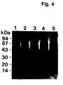
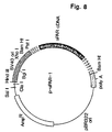
- the cDNA is about 1.4 kb long based on its restriction map (Fig. 4B), has a 5' untranslated sequence of about 40 residues and an about 40 nucleotides long poly-A stretch at the 3' end.
- DNA hybridization is a useful method. Hybridization may be performed as follows: Pure DNA comprising the gene encoding u-PAR from the plasmid p-uPAR-1 is prepared using the large scale method described in Maniatis et al. (1982), pages 86-96. More specifically, the u-PAR gene may be excised from the plasmid by digestion of the plasmid DNA with suitable restriction enzymes. The insert is then separated from the plasmid DNA by use of agarose gel electrophoresis. The insert is labelled by any labelling principle, such as the ones disclosed herein.
- the foreign DNA to be examined is coupled to a matrix, e.g. a nitrocellulose filter.
- the filter is subjected to a suitable treatment suited to the kind of matrix employed so as to couple the DNA to the matrix, in the case of a nitrocellulose filter e.g. by baking the filter at a temperature of 80°C for 2 hours.
- the membrane is exposed to a prehybridization solution of a composition, at a temperature and for a period of time recommended suited to the membrane in question.
- the membrane is then placed in the hybridization solution containing the labelled denatured DNA probe obtained from the p-uPAR-1 plasmid (the u-PAR gene). Hybridization is preferably carried out overnight at a suitable temperature.
- the membrane is then washed and incubated with a volume of 50 ml 2xSSC at 65°C for 30 minutes. The procedure is repeated once.
- the membrane is then incubated in 15 ml 2xSSC containing 0.1% SDS. Incubation is performed at 65°C for 30 minutes. All incubations including prehybridization and washings are performed with gentle agitation.
- the filter is air-dried and wrapped in a suitable plastic wrap (e.g. Saran Wrap), the filter is then applied to an x-ray film so as to obtain an auto-radiographic image. Exposition is preferably carried out at -70°C with intensifying screens for a period of time which is determined by the positive control used.
- any hybridization of the foreign DNA and the u-PAR gene is an indication of similarity of the two DNA probes, i.e. that the foreign DNA is a DNA fragment of the invention.
- Another approach of determining similarity between DNA sequences is by determining the nucleotide sequence of the DNA sequence to be compared with the DNA sequence of the invention by conventional DNA sequencing analysis, and comparing the degree of homology with the DNA sequence of the invention.
- a degree of homology of at least about 70%, e.g. at least about 80% such as at least about 95% is obtained.
- the DNA fragment of the invention may comprise a nucleotide sequence encoding a polypeptide fused in frame to the nucleotide sequence encoding the characteristic amino acid sequence with the purpose of producing a fused polypeptide.
- the fused sequence may be inserted into an appropriate vector which is transformed into a suitable host organism.
- the DNA fragment of the invention may be inserted in the vector in frame with a gene carried by the vector, which gene encodes a suitable polypeptide.
- the host organism which might be of eukaryotic or prokaryotic origin, for instance a yeast or a mammalian cell line, is grown under conditions ensuring expression of the fused sequence after which the fused polypeptide may be recovered from the culture by physico-chemical procedures, and the fused polypeptide may be subjected to gel filtration and affinity chromatography using an antibody directed against the antigenic part(s) of the fused polypeptide.
- the polypeptide of the invention and the polypeptide to which it is fused may be separated, for instance by suitable proteolytic cleavage, and the polypeptide of the invention may be recovered, e.g. by affinity purification or another suitable method.
- the DNA fragment may also comprise a suitable nucleotide sequence controlling the expression of the DNA fragment.
- the regulatory nucleotide sequence is conveniently a part of the expression vector used for the production of the polypeptides, when such a vector is employed.
- the DNA fragment described above may be obtained directly from genomic DNA or by isolating mRNA and transferring it into the corresponding DNA sequence by using reverse transcriptase producing cDNA.
- obtaining the DNA fragment from genomic DNA it is derived directly by screening for genomic sequences, hybridizing to a DNA probe prepared on the basis of the full or partial amino acid sequence of u-PAR.
- the DNA is of complementary DNA (cDNA) origin, it may be obtained by preparing a cDNA library on the basis of mRNA from cells containing a u-PAR or parts thereof. Hybridization experiments may then be carried out using synthetic oligonucleotides as probes to identify the cDNA sequence encoding the u-PAR or part thereof.
- cDNA differs from genomic DNA in, e.g. that it lacks certain transcriptional control elements and introns which are non-coding sequences within the coding DNA sequence. These elements and introns are normally contained in the genomic DNA.
- the DNA fragment may also be of synthetic origin, i.e. prepared by conventional DNA synthesizing method, e.g. by using a nucleotide synthesizer. The DNA fragment may also be produced using a combination of these methods.
- the invention relates to an expression vector which is capable of replicating in a host organism and which carries a DNA fragment as described above.
- the vector may be any vector which conveniently can be subjected to recombinant DNA procedures, the choice of vector often depending on the host cell into which it is to be introduced.
- the vector may either be one which is capable of autonomous replication, i.e. a vector which exists as an extrachromosomal entity, the replication of which is independent of chromosomal replication, such as a plasmid, or a vector which is replicated together with the host chromosome, such as a bacteriophage.
- plasmids such as natural or synthetic plasmids, eg. plasmids related to pBR322 such as pEX 1-3, the pRIT-family, the pUC-family and the like, and viruses such as adenovirus, vaccinia virus, retrovirus, Baculo virus, Epstein-Barr-virus, SV40-related virus and bovine papilloma virus.
- suitable bacteriophages include M13 and lambda.
- the invention also relates to an organism which carries and is capable of expressing a DNA fragment as defined above and which not in its native form expresses said DNA fragment.
- the DNA fragment may be carried on a vector as described above or may be integrated in the genome of the organism.
- suitable organisms include microorganisms such as bacteria, yeasts, fungi and higher eucaryotic organisms or cells including plant and mammalian cells.
- the present invention also relates to a method of producing the polypeptides described above.
- the polypeptides are prepared using recombinant DNA-technology e.g. the methods disclosed in Maniatis et al. op. cit. More specifically, the polypeptides may be produced by a method which comprises cultivating or breeding an organism carrying a DNA-fragment encoding a characteristic amino acid sequence from an u-PAR, e.g. the above described DNA fragment, under conditions leading to expression of said DNA fragment, and subsequently recovering the polypeptide from the organism.
- the organism which is used for the production of the polypeptide may be a lower organism, e.g. a microorganism.
- the DNA fragment encoding the characteristic amino acid sequence from an u-PAR should be introduced in the organism.
- the DNA fragment is inserted in an expression vector, e.g. a vector as defined above, which is subsequently introduced into the host organism.
- the DNA fragment may also be directly inserted in the genome of the host organism.
- the insertion of the DNA fragment in the genome may be accomplished by use of a DNA fragment as such or cloned in bacteria, phage lambda or other vectors, carrying the DNA fragment and being capable of mediating the insertion into the host organism genome.
- the insertion of the DNA fragment into an expression vector or into the genome of the host organism may be accomplished as described e.g. by Colbere-Garapin F. et al., J. Molec. Biol., 150; 1-14 (1981): A New Dominant Hybrid Selective Marker for Higher Eucaryotic Cells.
- the DNA fragment when using an expression vector for the production of the polypeptide of the invention, may be inserted in frame with a second DNA fragment encoding another polypeptide so as to obtain an expression of fusion protein.
- the polypeptide of the invention comprises one or more distinct parts, e.g. being a fusion protein comprising on the one hand characteristic amino acid sequence(s) from u-PAR and on the other hand amino acid sequence(s) constituting a polypeptide which is not related to u-PAR
- the DNA fragments encoding each of these polypeptides may be inserted in the genome or expression vector separately or may be coupled before insertion into the genome or expression vector by use of conventional DNA techniques such as described in Maniatis et al. op. cit.
- the conditions under which the organism producing the polypeptide of the invention is cultured or breeded should of course be adapted to the organism employed.
- Conventional cultivation and breeding techniques may be employed.
- the cultivation is e.g. carried out in a culture medium conventionally used for fermentation purposes, e.g. Luria Broth medium, and under conditions with respect to pH, temperature, aeration, etc. suited to the type of microorganism in question, e.g. as disclosed in Maniatis et al. op. cit.
- the polypeptide is recovered or isolated from the organism.
- the polypeptide may be isolated or recovered from the culture by a method comprising one or more affinity chromatography and/or size chromatography steps, and optionally employing a step using an antibody reactive with and/or being raised against said polypeptide.
- the procedure used for recovering of the polypeptide depends on the kind of host organism used as well as the polypeptide produced.
- the recovery and isolation of the polypeptide will also of course depend on the kind of microorganism employed.
- the recovering of the polypeptide from the microorganism comprises treatment of the microorganism so as to release the polypeptide, e.g. by rupturing the microorganism, i.e. partly or totally, and subsequently recovering the polypeptide by well-known methods such as precipitation, gel filtration, ion exchange chromatography, or HPLC reverse phase chromatography or immuno affinity chromatography or the like.
- the polypeptide of the invention may be isolated from a biological material containing the polypeptide, e.g. a suspension of cells producing the polypeptide, by use of a method comprising adsorbing the biological material to a matrix comprising an immobilized monoclonal or polyclonal antibody as described herein, eluting the polypeptide from the matrix, and recovering the polypeptide from the eluate.
- a biological material containing the polypeptide e.g. a suspension of cells producing the polypeptide
- a method comprising adsorbing the biological material to a matrix comprising an immobilized monoclonal or polyclonal antibody as described herein, eluting the polypeptide from the matrix, and recovering the polypeptide from the eluate.
- procedures for isolating the polypeptide are:
- the DNA fragment encoding the polypeptide of the invention may be subjected to modification, before or after the DNA fragment has been inserted in the vector.
- the polypeptide produced may also be subjected to modification.
- the modification may comprise deletion or rearrangement of one or more nucleotides and amino acids in the DNA fragment and the polypeptide, respectively, or a combination of these modifications.
- the term “deletion” is intended to indicate that one or more amino acids or nucleotides have been deleted from the full amino acid or nucleotide sequence whether at either end of the sequence or at any suitable point within it.
- “Rearrangement” is intended to indicate that one or more amino acids or nucleotides or the sequence has been exchanged with each other.
- nucleotide substitutions which do not give rise to another amino acid sequence of the protein, but which, e.g., correspond to the codon usage of the specific organism in which the sequence is inserted; a subsequence of the DNA sequence shown above encoding a polypeptide which has retained the receptor properties of the native u-PAR; or a DNA sequence hybridizing to at least part of a DNA prepared on the basis of the DNA sequence shown above, provided that it encodes a polypeptide which has the biological property of u-PAR.
- polypeptide produced as described above may be subjected to posttranslational modifications such as for instance a suitable proteolytic enzyme, e.g. a peptidase or proteinase, such as trypsin, phospholipases, glycopeptidases.
- a suitable proteolytic enzyme e.g. a peptidase or proteinase, such as trypsin, phospholipases, glycopeptidases.
- truncated polypeptide refers to a polypeptide deleted for one or more amino acid residues eventually resulting in changing of the properties of the polypeptide, e.g. solubility.
- truncated polypeptide refers to a mixture of polypeptides all derived from one polypeptide or expressed from the gene encoding said polypeptide. Such truncated polypeptides might arise for instance in vector/host cell systems in which part of the cDNA has been deleted by restriction enzyme digestion or other suitable methods, resulting in the expression of a protein not normally produced in that system.
- the polypeptide of the invention may be prepared by the well-known methods of liquid or solid phase peptide synthesis utilizing the successive coupling of the individual amino acids of the polypeptide sequence or the coupling of individual amino acids forming fragments of the polypeptide sequence which fragments subsequently are coupled so as to result in the desired polypeptide.
- the solid phase peptide synthesis may e.g. be performed as described by R. B. Merrifield, J. Am. Chem. Soc. 85 , 1963, p. 2149.
- the amino acid sequence is constructed by coupling an initial amino acid to a solid support and then sequentially adding the other amino acids in the sequence by peptide bonding until the desired length has been obtained.
- the solid support may also serve as the carrier for the polypeptide of the invention in a vaccine preparation as described below.
- the preparation of synthetic peptides may be carried out essentially as described in Shinnick, Ann. Rev. Microbiol. 37 , 1983, pp. 425-446.
- the invention relates to a diagnostic agent capable of detecting and/or quantitating u-PAR or a derivative thereof in a sample.
- such diagnostic agent may be valuable in diagnosis of cancer and other disorders involving tissue invasion and tissue remodelling, considering the involvement of u-PAR in these processes.
- serum from breast cancer patients has an increased concentration of u-PA compared with normal individuals (Gr ⁇ ndahl-Hansen et al., 1988) and that the u-PA content in breast cancer tissue has been shown to be a valuable prognostic marker in this disease such as has been published in the priority year of the present application (Jänicke et. al., 1989, 1990).
- u-PAR u-PAR content in cancer tissue is an even better diagnostic and prognostic marker.
- a new aspect of the potential diagnostic and prognostic use of u-PAR determinations is the release of u-PAR from cultured cells (described in Example 4) that occurs even in the absence of exogeneously added phospholipase. This finding raises the possibility that u-PAR is also released into body fluids under some physiological and pathophysiological conditions and particularly in cancer. Determination of concentrations of u-PAR or degradation products thereof in body fluids, such as serum, urine, and ascites fluid may therefore prove to be diagnostically and/or prognostically valuable.
- the diagnostic agent may, e.g. be an antibody which is monospecific to the u-PAR.
- the diagnostic agent may be in the form of a test kit comprising in a container a polypeptide comprising a characteristic amino acid sequence of u-PAR, e.g. a sequence including or included in the sequence (1).
- the diagnostic agent may be used in the diagnosis of diseases related to abnormal numbers of u-PARs residing on the cell.
- the diagnostic agent may be one which is suited for use in an agglutination assay in which the solid particles to which the antibody is coupled agglutinate in the presence of a polypeptide of the invention in the sample subjected to testing. In this type of testing, no labelling of the antibody is necessary. For most uses it is, however, preferred that the antibody is provided with a label for the detection of bound antibody or, alternatively (such as in a double antibody assay), a combination of labelled and unlabelled antibody may be employed.
- the substance used as label may be selected from any substance which is in itself detectable or which may be reacted with another substance to produce a detectable product. Thus, the label may be selected from radioactive isotopes, enzymes, chromophores, fluorescent or chemiluminescent substances, and complexing agents.
- enzymes useful as labels are ⁇ -galactosidase, urease, glucose oxidase, carbonic anhydrase, peroxidases (e.g. horseradish peroxidase), phosphatases (e.g. alkaline or acid phosphatase), glucose-6-phosphate dehydrogenase and ribonuclease.
- peroxidases e.g. horseradish peroxidase
- phosphatases e.g. alkaline or acid phosphatase
- glucose-6-phosphate dehydrogenase ribonuclease.
- Enzymes are not in themselves detectable, but must be combined with a substrate to catalyze a reaction the end product of which is detectable.
- a substrate may be added to the reaction mixture resulting in a coloured, fluorescent or chemiluminescent product or in a colour change or in a change in the intensity of the colour, fluorescence or chemiluminescence.
- substrates which are useful in the present method as substrates for the enzymes mentioned above are H 2 O 2 , p-nitrophenylphosphate, lactose, urea, ⁇ -D-glucose, CO 2 , RNA, starch, or malate.
- the substrate may be combined with, e.g. a chromophore which is either a donor or acceptor.
- Fluorescent substances which may be used as labels for the detection of the components as used according to the of invention may be 4-methylumbelliferyl-phosphate, 4-methylumbelliferyl-D-galactopyranoside, and 3-(p-hydroxyphenyl) propionic acid. These substances may be detected by means of a fluorescence spectrophotometer. Chemiluminescent substances which may be peroxidase/eosin/EDTA, isoluminol/EDTA/H 2 O 2 and a substrate therefor.
- Chromophores may be o-phenylenediamine or similar compounds. These substances may be detected by means of a spectrophotometer. Radioactive isotopes may be any detectable and in a laboratory acceptable isotope, e.g. 125 I, 131 I, 3 H, 35 P, 35 S or 14 C. The radioactivity may be measured in a ⁇ -counter or a scintillation counter or by radio-autography followed by densitometry.
- Complexing agents may be Protein A, Protein G (which form a complex with immunoglobulins), biotin (which forms a complex with avidin and streptavidin), and lectin (which forms a complex with carbohydrate determinants, e.g. receptors).
- the complex is not in itself directly detectable, necessitating labelling of the substance with which the complexing agent forms a complex.
- the marking may be performed with any of the labelling substances described above.
- an antibody or a polypeptide of the invention may be coupled to a bridging compound coupled to a solid support.
- the bridging compound which is designed to link the solid support and the antibody may be hydrazide, Protein A, glutaraldehyde, carbodiimide, or lysine.
- the solid support employed is e.g. a polymer or it may be a matrix coated with a polymer.
- the matrix may be of any suitable solid material, e.g. glass, paper or plastic.
- the polymer may be a plastic, cellulose such as specially treated paper. nitrocellulose paper or cyanogenbromide-activated paper.
- suitable plastics are latex, a polystyrene, polyvinylchloride, polyurethane, polyacrylamide, polyvinylacetate and any suitable copolymer thereof.
- silicone polymers include siloxane.
- the solid support may be in the form of a tray, a plate such as a mitrotiter plate, e.g. a thin layer or, preferably, strip, film, threads, solid particles such as beads, including Protein A-coated bacteria, or paper.
- a mitrotiter plate e.g. a thin layer or, preferably, strip, film, threads, solid particles such as beads, including Protein A-coated bacteria, or paper.
- the polypeptide and antibody of the invention may be used in an assay for the identification and/or quantification of at least a form and/or a part of said polypeptide present in a sample.
- the identification and/or quantification performed by the use according to the present invention may be any identification and/or quantification involving u-PAR compounds or a form of u-PAR compounds. Thus, both a qualitative and a quantitative determination of u-PAR compounds may be obtained according to the use of the present invention.
- the identification and/or quantification may be performed for both a scientific, a clinical and an industrial purpose. As will be further described below, it is especially important in clinical routine to identify or quantify u-PAR compounds.
- the sample may be a specimen obtained from a living organism such as a human or an animal.
- the specimen may be blood, e.g. an erythrocyte enriched fraction, or a tissue sample e.g. comprising liver cells.
- the specimen is urine.
- the antibody used in the method of the invention is a monoclonal antibody as this generally provides a higher precision and accuracy of the assay, at the same time possibly requiring less time to perform. Furthermore, a mixture of two or more different monoclonal antibodies may be employed as this may increase the detection limit and sensitivity of the test.
- the monoclonal antibody may be obtained by the method described below. Antibodies possessing high avidity may be selected for catching techniques.
- the antibody used in the present method is preferably in substantially pure form (purified according to suitable techniques or by the methods of the invention, see below) in order to improve the precision and/or accuracy of the assays of the invention.
- the determination of antibodies reactive with the polypeptide of the invention and being present in a sample may be carried out by use of a method comprising contacting the sample with the polypeptide of the invention and detecting the presence of bound antibody resulting from said contacting and correlating the result with a reference value.
- the polypeptide of the invention When the polypeptide of the invention is to be employed in an assay for determining the presence of u-PAR compounds in a sample, it may be in the form of a diagnostic reagent or a diagnostic agent. As will be apparent to a person skilled in the art several techniques may be applied in connection with such diagnostic reagents.
- any part of said polypeptide is coupled to a solid support
- an antibody against the component may then be added to the solid support.
- the antibody is coupled to a solid support.
- any u-PAR compounds present in the sample is coupled to a solid support. It may then be incubated with the polypeptide component by addition of the component to the solid support followed by adding an antibody labelled with a detectable marker.
- SDS-PAGE SDS-PAGE .
- SDS-PAGE was performed according to Laemmli, U.K., "Cleavage of structural proteins during the assembly of the head of bacteriophage T4", Nature 227: 680-682, 1970, using 6-16% gradient slab gels. Pretreatment of samples under nonreducing conditions was performed without boiling. When reducing conditions were used, the samples were boiled for 5 minutes in the presence of 20 mM DTT.
- Phast-gel SDS-PAGE was performed on a Phast gel apparatus (Pharmacia), using ready-made 10-15% gradient gels. Electrophoresis was performed according to the recommendations of the manufacturer. Silver staining was performed according to Heukeshoven and Dernick, 1988.
- Tricine-SDS-PAGE of samples to be electroblotted for amino acid analysis or NH 2 -terminal amino-acid sequencing was performed in a Mini Protean II apparatus (BioRad) according to Shugger and von Jagow, 1987, on a 0.75 mm homogeneous 7.7% T, 3% C gel.
- the gel was pre-electrophoresed for 3 hours at 15 mA in the gel buffer with 12 mM 3-mercaptopropanoic acid added as a scavenger.
- the freeze-dried sample was dissolved directly in 50 ⁇ l of the sample buffer with 40 mM dithioerythritol as the reducing agent, and boiled for 2 minutes.
- the gel buffer used for pre-electrophoresis was replaced with electrophoresis buffer, after which electrophoresis was performed for 4 hours at 60 V.
- Electroblotting of samples for amino acid analysis or NH 2 - terminal amino acid sequencing was electroblotted onto a polyvinylidene difluoride (PVDF) membrane (Millipore), using a semi-dry electroblotting apparatus (JKA Instruments, Denmark). Electroblotting took place at pH 11.0 in 10 mM CAPS (3-(cyclohexylamino)-1-propanesulfonic acid), including 0.4 mM dithioerythritol and 10% methanol, and was performed at 0.8 mA/cm 2 for 2 hours. The protein was localized by staining with Coomassie R250 for 2 minutes and brief destaining, followed by wash in water (Matsudaira, 1987).
- PVDF polyvinylidene difluoride
- the Coomassie-stained protein band was cut out from the PVDF-membrane and treated with 25 mM iodoacetamide in 50 mM sodium borate, pH 8.0, for 1 hour in the dark at room temperature. After the reaction, it was washed extensively with water and dried under argon. The protein on the dried filter was sequenced on an Applied Biosystems protein sequencer, model 477A.
- the on-line HPLC identification system for the PTH amino acid derivatives included the derivative of carboxymethylcysteine (produced by deamidation of the amidomethyl derivative during conversion). The correct identification of this derivative was assured by a test-sequencing of chicken lysozyme (with cysteine at residue no. 6) after parallel preparative electrophoresis, electroblotting and alkylation.
- U937a histiocytic lymphoma cell line U937 (here designated as U937a) (E.K.O. Kruithof, University Hospital Center, Lausanne, Switzerland), a variant of this cell line, designated U937b (A. Fattorossi, Research Lab of Aeronautica Militare, Rome, Italy), the promyeloid leukemic cell line HL-60 (American Type Culture Collection (ATCC)), the bladder carcinoma cell line 5637 (ATCC), the larynx epidermoid carcinoma cell line HEp-2 (ATCC), the epidermoid carcinoma cell line A-431 (E.
- the cervix carcinoma cell line HeLa (ATCC), the colon carcinoma cell line HCT 116 (ATCC), the conjunctiva cell line Chang (ATCC), the choriocarcinoma cell line JEG-3 (A. Vaheri, University of Helsinki, Finland), the anion cell line AV3 (ATCC), and the fibrosarcoma cell line HT-1080 (A. Vaheri).
- the U937a and b and HL-60 cells were grown in suspension, while all the other cell lines were grown as monolayers.
- the HT-1080 and A-431 cells were grown in Dulbecco's modified Eagle's medium with 10% heat-inactivated fetal calf serum.
- U937a cells Large-scale production of U937a cells.
- the U937a cells were grown in 1-liter spinner flasks to reach a density of 1.0-1.5 x 10 6 cells/ml in RPMI 1640 medium supplemented with 2 mM L-glutamine, 5% fetal calf serum (heat inactivated), 200 units/ml penicillin, 25 ⁇ g/ml streptomycin (or without antibiotics). Each flask contained 500 ml cell culture.
- Phorbol 12-myristate 13-acetate (PMA) induction and harvest of U937a cells The 500 ml cell suspension of one spinner flask was added to 1 liter of fresh medium without serum. 150 ⁇ l of PMA stock solution in dimethylsulfoxide (1 mg PMA/ml) was added, to reach a final concentration of 150 nM PMA. The cul- ture was transferred to a 10-layer cell factory (Nunc, Denmark) and grown for 3.5 days in the factory. Upon addition of the PMA solution, the cells stop dividing and attach to the surface.
- PMA Phorbol 12-myristate 13-acetate
- the clarified lysate was subjected to temperature-induced phase separation (Bordier, 1981) by incubation at 37°C for 10 minutes, after which the detergent phase was collected by centrifugation for 10 minutes at 20°C, 1,800 x g. The upper phase was discarded.
- the lower phase (approximately 2 ml) was washed by addition of 18 ml 0.1 H Tris/HCl, pH 8.1, at 0°C, followed by complete mixing to restore a clear, one-phase solution, and repeated phase separation by warming and centrifugation, as above.
- the lower phase was made up to 20 ml by addition of 0.1 M Tris/HCl, pH 8.1.
- 500 ⁇ l 10% w/v 3-((3-cholamidopropyl)dimethylammonio)-1-propanesulfonate (CHAPS) was added to yield a clear, single-phase detergent fraction. Minor amounts of non-dissolved material were removed from this solution by centrifugation for 15 minutes at 4°C, 3,300 x g.
- Lysates and detergent phases from other cell types were prepared in the same manner, except that smaller amounts of cell material were used. The amounts of all reagents were reduced proportionally. In one experiment, 0.5% CHAPS was used as the lysis detergent instead of 1% Triton X114. In that experiment, no phase separation was performed.
- the reaction was stopped by extensive dialysis at 0°C against 0.25 M NaHCO 3 , 0.5 M NaCl, 0.1% Triton X-100, pH 8.5.
- the dialyzed material was coupled to 12.5 ml of CNBr-activated Sepharose (Pharmacia) that had been freshly equilibrated with 0.25 M NaHCO 3 , 0.5 M NaCl, pH 8.5 (coupling buffer).
- the reaction proceeded overnight at 4°C and was stopped by equilibration of the matrix with 1 M ethanolamine/HCl, pH 8.0 and incubation for 24 hours at 4°C.
- the matrix DFP-u-PA-Sepharose
- the clarified detergent fraction obtained from 6 x 10 9 U937a cells was diluted with 1 vol washing buffer-1 (10 mM sodium phosphate, 140 mM sodium chloride, 0.1% CHAPS, pH 7.4) and chromatographed on a column containing 8 ml DFP-u-PA-Sepharose, equilibrated with the same buffer. After application of the sample, the column was washed with washing buffer-1, followed by washing buffer-2 (10 mM sodium phosphate, 1 M sodium chloride, 0.1% CHAPS, pH 7.4). The column was eluted from below with elution buffer (0.1 M acetic acid, 0.5 M sodium chloride, 0.1% CHAPS, pH 2.5).
- Elution fractions were immediately titrated to pH 7.5 by addition of the appropriate volume of 0.1 M sodium phosphate, 1.0 M sodium carbonate, pH 9.0.
- u-PAR-containing fractions were identified by chemical cross-linking to the 125 I-labelled amino terminal (ATF) fragment of urokinase, followed by SDS-PAGE and autoradiography.
- Purified u-PAR samples for amino acid analysis or NH 2 -terminal amino acid sequencing were dialyzed against 0.1% acetic acid and lyophilized.
- Protein labelling with 125 I was performed as described previously (Nielsen et al., 1988), except that 0.1% Triton X100 was replaced by 0.01% Tween 80. Purified u-PAR, concentrated by freeze-drying after dialysis against 0.1% acetic acid, was iodinated in the same manner, except that 1.5 ⁇ g protein was treated with 250 ⁇ Ci 125 I in a volume of 25 ⁇ l.
- Lyophilized, purified u-PAR was radioiodinated directly.
- the samples were denatured under mildly reducing conditions by the addition of SDS and dithiothreitol to final concentrations of 0.5% and 1.6 mM, respectively, and boiling for 3 minutes. Aliquots of the denatured samples (10 ⁇ l) were adjusted to include 200 mM sodium phosphate, pH 8.6, 1.5% Triton X-100, 10 mM 1,10 phenanthroline (added from a methanol stock solution) and either 1 unit of peptide:N-glycosidase F (N-glycanase, Genzyme), or no enzyme, in a total volume 30 ⁇ l. Deglycosylation was performed at 37°C for 20 hours.
- lysate samples labelled by cross-linking to 125 I-ATF were made up to 200 ⁇ l with 0.05 M sodium acetate, pH 5.0. 90 ⁇ l aliquots of the mixture received either 14 ⁇ l of 33 ng/ ⁇ l neuraminidase (Boehringer-Mannheim) or no enzyme. Desialylation was performed overnight at 37°C.
- PMA-stimulated U937a cells were acid-treated to remove any surface-bound u-PA and lysed in a Triton X114 containing buffer.
- the detergent extract was subjected to temperature-induced phase separation, and the isolated detergent phase was used as the raw material for affinity chromatography.
- the acid eluates were neutralized and analyzed, either directly or after concentration by dialysis against 0.1% acetic acid and lyophilization.
- the electrophoretic appearance of the purified material is shown in Fig. 1.
- Fig. 1A After SDS-PAGE and silver staining (Fig. 1A), the eluted protein migrated as one broad band, covering the range from approximately 55 to 60 kDa. Outside this range, no protein material was detected. A single band with the same apparent molecular mass was also found when SDS-PAGE was performed under nonreducing conditions. (Fig. 1C, lane 5).
- Binding and cross-linking to 125 I-labelled ATF was specific and saturable. Thus, it could be competed for by an excess of unlabelled ATF, active u-PA or DFP-treated u-PA, while no competition was obtained with unrelated proteins such as, for example, bovine serum albumin, or with related proteins, such as t-PA, plasminogen or epidermal growth factor (Fig. 1B).
- unrelated proteins such as, for example, bovine serum albumin, or with related proteins, such as t-PA, plasminogen or epidermal growth factor (Fig. 1B).
- Quantification by amino acid analysis indicated a purification yield of 6-9 ⁇ g polypeptide (corresponding to about 10-15 ⁇ g u-PAR glycoprotein; see below) from 6 x 10 9 cells.
- Amino acid composition and NH 2 -terminal amino acid sequences are shown in table 1.
- This composition includes a strikingly high content of cysteine residues. Further, it is noted that rather few lysine residues are present.
- the analysis system employed allows the quantification of glucosamine and galactosamine in addition to the amino acids. Glucosamine was detected in an amount corresponding to approximately 30 mol of N-acetylglucosamine per mol protein, correcting for loss during hydrolysis. In contrast, no galactosamine was identified.
- the amino terminus like the amino acid composition of the entire protein, is rich in cysteine residues.
- Example 2 Data for probe construction (Example 2) were derived from the sequencing shown in Table 2A. For this construction, position 6 of the amino acid sequence was tentatively assigned Asn; see footnote a of Table 2A.
- Fig. 2 shows the electrophoretic appearance of the deglycosylated protein. The electrophoretic band observed after autoradiography of the 125 I-labelled protein was always slightly broader than that seen after direct protein staining.
- the reaction turned the heterogeneous 55-60 kDa receptor (lane 1) into a deglycosylated protein of only 35 kDa that migrated as a much sharper band (lane 2), thus further confirming that the initially heterogeneous material all represented variants of the same protein.
- the fibrosarcoma cell line HT-1080 contained a third u-PAR variant, giving rise to a single conjugate band of a slightly lower molecular weight (approximately 65 kDa; not shown).
- N-bound glycosylation was responsible, not only for molecular u-PAR heterogeneity within the PMA-stimulated U937 line and occurrence of two bands in the PMA-stimulated U937b line, but also for the electrophoretic difference between u-PARs from non-stimulated and PMA-stimulated U937 cells and for the variation among different cell lines (i.e. HT-1080 fibrosarcoma cells compared to the other cell lines tested).
- Cys indicates the identification of the PTH derivative of carboxymethyl cysteine in the alkylated protein. Res. no. 1 2 3 4 5 6 7 8 9 10 Amino acid residue Leu ? Cys Met Gln Cys Lys Thr Asn Gly Res. no. 11 12 13 14 15 16 17 18 19 20 Amino acid residue Asp Cys (Arg) Val Glu Glu (His) Ala Leu Gly Res. no. 21 22 23 24 25 26 27 28 29 30 Amino acid residue Gln ? Leu ? (Arg) Thr (Thr) lie Val ?
- Enzymatic degradation Affinity purified u-PAR was dialyzed against 0.1% acetic acid and lyophilized as described in Example 1. The freeze-dried material was redissolved in incubation buffer (0.05 M Tris/HCl, 0.05% CHAPS, pH 8.1) to yield a protein concentration of approx 25 ⁇ g/ml. 9 ⁇ l samples of this u-PAR solution were treated with chymotrypsin (Worthington; final concentrations ranging from 8-200 ng/ml), by addition of 1 ⁇ l of the appropriate stock solution of the enzyme, dissolved in incubation buffer.
- incubation buffer 0.05 M Tris/HCl, 0.05% CHAPS, pH 8.1
- the samples were incubated for 16 h at 37°C after which the degradation was stopped by addition of 0.5 ⁇ l of 20 mM phenylmethylsulfonylfluoride, dissolved in dimethylsulfoxide. The samples were stored at -80°C until analysis.
- Samples to be analyzed by chemical cross-linking to 125 I-ATF were 50-fold diluted in 0.1 M Tris/HCl, 1% Triton X-114, pH 8.1.
- the diluted samples were either clarified by addition of 0.25% w/v CHAPS (final concentration) or subjected to a single round of temperature induced phase separation (see Example 1).
- each phase i.e., the detergent and buffer fraction, respectively
- Enzymatic deglycosylation with N-Glycanase was performed according to example 1, except that the actual concentrations during the deglycosylation step were the following: 0.08% SDS; 0.26 mM dithiothreitol; 0.11 M sodium phosphate; 0.9% Triton X-100; 5.3 mM 1,10 phenanthroline; 33.3 units/ml N-glycanase.
- Identification of the fragment will subsequently be done by comparison to the amino acid sequence derived from u-PAR cDNA.
- synthetic peptides covering the chymotryptic fragment will be constructed. The peptides will be assayed for their potential inhibitory activity against the binding reaction between u-PAR and the ligand, as studied by cell binding assays (Nielsen et al., 1988; Appella et al., 1987) or by chemical cross-linking assay.
- the samples were analyzed in the chemical cross-linking assay, using 125 I-ATF as the ligand (Fig. 5). While the non-degraded samples (lanes 4 and 5) showed the 70-75 kD conjugate band which is characteristic for the intact u-PAR (see Example 1), the intensity of this band was much reduced in the degraded samples (lanes 1-3). In contrast, the degraded samples showed an approx. 30 kD cross-linked conjugate; i.e. the size to be expected for a conjugate formed between the above mentioned, 16 kD u-PAR degradation product and the 15 kD ATF.
- the ligand binding fragment proved hydrophilic in the Triton X-114 system, suggesting that this fragment does not include the diacylglycerol part of the protein (see Example 4).
- the deglycosylation experiment showed that the ligand binding fragment is glycosylated and suggested that the polypeptide part of the fragment comprised only 6-10 kD, corresponding to approx. 50-90 amino acid residues.
- a human cDNA library was used made from SV 40 transformed human GM637 fibroblasts in a plasmid vector based on pBR322 (carrying an ampicillin resistance gene) (Okayama H, Berg F, "High-efficiency cloning of full-length cDNA", Mol. Cell. Biol. 2: 161-170, 1982). The library was kindly donated by Dr. Okayama. This library was selected on the basis of the known high number of u-PAR in GM637 cells (Blasi, unpublished).
- the plasmid vector (Fig. 8) uses the SV 40 promoter and has high expression in various eukaryotic cells, but very low or no expression in prokaryotes.
- the library was screened with synthetic oligonucleotide probes made on the basis of amino acid sequence data from purified receptor protein (Tables 4-5).
- the melting temperatures were calculated from Lathe, J. Mol. Biol. 183: 1-12, 1985.
- the melting temperature calculated from the equation applies to an infinitely long stretch of DNA.
- t w t m - (820/l) - 1.2 (100 - h) in which 1 is the length of the DNA (number of bases), and h is the percent homology.
- nitrocellulose filters containing DNA from the plasmid library were hybridized to the end-labelled oligonucleotide probe at various temperatures and salt concentrations (all within the range calculated from Lathe, supra ).
- the filters were produced according to Grunstein and Hogness ("Colony hybridization: A method for the isolation of cloned DNAs that contain a specific gene", Proc. Natl. Acad. Sci. USA 72: 3961, 1975).
- the hybridization conditions to be used for the screening were chosen as the ones giving the minimum amount of background hybridization.
- Table 3 the amino acid sequence derived from a preliminary amino-terminal sequencing of purified u-PAR (see Example 1) and the derived oligonucleotide sequence are presented.
- the plasmid library was screened with the N-terminal probe using the procedure of Grunstein and Hogness ( supra ). The detailed procedure is described below. Several positive clones were found but after the third rescreening, only one remained. The purity of the clone was checked and DNA was prepared from it (see large scale DNA preparation below). The DNA was digested with several different restrictions enzymes, and a map of the restriction sites found in the clone was constructed (see procedure in Maniatis et al., Molecular Cloning: A Laboratory Manual. Cold Spring Harbor Laboratory, 1982). The insert was further analysed by DNA sequencing (see procedure below).
- the clone was able to code for 7 out of the 8 amino acids in the N-terminal peptide used to construct the 24-mer probe.
- the sequence in the probe starts with an A whereas the clone had a T in this position, resulting in the substitution of Cys for Met.
- the clone was thus isolated by a specific hybridization but could not code for the correct peptide.
- the library was then rescreened using chloramphenicol amplification of plasmids (Maniatis et al., supra ) to increase the signal intensity. This procedure resulted in a total of 7 positive clones (see Table 4) of which two were eliminated on the basis of hybridization to the probe made from the original false positive clone.
- This primer was made in the inventors' laboratory on an Applied Biosystems 391 A DNA Synthesizer. Primers for the pEMBL18 vector were purchased from Biolabs.
- Plasmid DNA was prepared according to described procedures (Maniatis, supra ).
- the synthetic oligonucleotides were end-labelled using T4 polynucleotide kinase and - ⁇ - 32 P-ATP.
- Gel purified DNA fragments were nick-translated using a BRL nick translation kit and ⁇ - 32 P-ATP.
- the probes were purified on NENSORB 20 columns (NEN) following the manufacturer's specifications. Nick-translated probes were denatured for 10 minutes at 100°C before use.
- the complete sequence of one of the isolated clones was obtained on double-stranded DNA in both orientations using commercial primers for pEMBL18 (M13 primers) and internal synthetic primers (see above).
- the sequence is shown in Sequence (1) in the Detailed Description of the invention.
- the restriction map and the sequencing strategy are illustrated in Fig. 7.
- the cDNA clone is 1364 nucleotides long from the 5' end to the beginning of the polyA stretch. At the 5' end, 46 nucleotides precede the first ATG codon which is followed by a 1005 nucleotides sequence with an open reading frame, ending with a nonanucleotide containing two in frame stop codons.
- 312 nucleotides of 3' untranslated sequence separate the first stop (TAA) codon from the polyA sequence.
- TAA first stop codon
- the assignment of the ATG at nucleotide 47 as the translation start site agrees with the consensus for initiating regions (Kozak, 1987) as discussed above.
- the translated sequence starts with a hydrophobic sequence which conforms to the rules for the signal peptide (von Heijne, 1986) (see above).
- the putative signal peptide is followed by 313 amino acid residues.
- the sequence shown in Sequence (1) was compared with the initial amino terminal sequence (Fig. 7A), and it was observed that in fact the original sequence contained an error at position 6 (Asn instead of Cys) which, however, did not prevent the isolation of the right cDNA clone.
- the human u-PAR is a relatively small protein of 313 amino acid residues.
- the amino acid sequence contains five potential N-linked glycosylation sites, in agreement with the high level of glycosylation of the protein (see Example 1).
- a sequence of 21 hydrophobic amino acids flanked by arginine residues may represent a membrane spanning domain of the u-PAR (Fig. 7C).
- the arginine is followed by 9 additional hydrophobic amino acids ending with a carboxy-terminal threonine.
- u-PAR may contain no intracytoplasmic domain at all, i.e. also the carboxy-terminal 10 residues may be buried in the membrane.
- the sequence of the carboxy-terminal about 30 amino acid residues would also be compatible with a signal peptide for glycolipid-anchored, phospholipase C-sensitive membrane attachment (Ferguson and Williams, 1988).
- the u-PAR is a slightly acidic protein (6 net acid charges), is very rich in cysteine, rich in glycine and leucine, and poor in lysine.
- the u-PAR is also rich in serine and threonine residues, which might indicate O-linked glycosylation (Russell et al., 1984). However, deglycosylation and sugar composition studies indicate that the receptor contains only N-linked carbohydrates (see Example 1).
- the u-PAR sequence is not similar to any known protein: a search in the Georgetown University data bank did not yield any extended homology. In particular, it bears no resemblance to the tissue factor, a receptor for factor VII of the coagulation pathway, which in common with u-PAR has the low molecular weight and the unusually large extent of glycosylation (Morrissey et al., 1987).
- the very high proportion of cysteine residues is common to many extracellular portions of receptors, like the epidermal growth factor receptor (Yarden & Ullrich, 1988), the epidermal growth factor precursor (Bell et al., 1986), and many others (Appella et al., 1988). However, there does not appear to be a common pattern of cysteine spacings in these proteins.
- the second and third repeats are the most closely related (about 25 percent identity). Significantly, the pattern of cysteines is strikingly similar in these two repeats.
- p-u-PAR-1 clone The functionality of p-u-PAR-1 clone was tested by transfecting it into mouse LB6 cells and testing transfectants by the caseinolytic plaque assay. This assay is based on the ability of plasmin to degrade casein which gives rise to clear plaques in an opaque background. Since LB6 cells produce no plasminogen activator, plasmin cannot be produced. In the presence of u-PA receptors, however, cells can bind u-PA and hence acquire the ability to degrade casein (Vassalli et al., 1985). The murine LB6 cells produce no plasminogen activator (unpublished observation) but have u-PA receptors.
- LB6 cells cannot bind human u-PA.
- Expression of human u-PAR cDNA by LB6 cells should provide these cells with the ability to bind human u-PA which can be visualized by the formation of clear plaques in the caseinolytic plaque assay.
- the vector used in the cDNA library is an expression vector that contains the SV40 promoter at the 5' end and polyadenylation and splice sites at the 3' end (Okayama & Berg, 1983). Expression of human u-PA receptors in transfected cells will, therefore, prove that the p-u-PAR-1 clone encodes a complete cDNA sequence.
- Mouse LB6 cells (Corsaro and Pearson, 1981) were cultured in Dulbecco's modified minimal essential medium (DMEM) supplemented with 10% foetal calf serum, 2 mM glutamine and 10 IU/ml of penicillin and streptomycin.
- Human high molecular weight urokinase and prourokinase were provided by Lepetit SpA (Nolli et al., 1989).
- the amino terminal fragment of human u-PA, ATF was a gift from Abbott Laboratories.
- the synthetic peptides human u-PA[12-32(ala19)] and mouse u-PA[13-33(ala20)] have been described before (Appella et al., 1987). Plasminogen was from Sigma Chemical Co.
- 2 x 10 5 LB6 cells were transfected either with 9 ⁇ g of p-u-PAR-1 DNA plus 1 ⁇ g of pRSVneo DNA, or with 9 ⁇ g of pRSVCAT plous 1 ⁇ g of pRSVneo DNA using a modification of the calcium phosphate coprecipitation technique (Pozzatti et al., 1986). Cells were plated in 0.8 mg/ml G418-containing DMEM, 10% foetal calf serum, and colonies were isolated after about 13 days.
- the pools of transfected clones were tested (in the case of p-u-PAR-1 DNA) by the caseinolytic plaque assay (Vassalli et al., 1977) and positive clones were picked. After one subcloning, several clones from each transfection were tested for human u-PA binding using the same technique.
- Cells (plated one day before at 100,000/dish) were washed with PBS, incubated in the presence of 0.2 nM human u-PA for 1 hour at 37°C, washed extensively and covered with a thin agar layer containing 1.3% casein and 17 ⁇ g/ml plasminogen. The plates were incubated at 37°C for 3 hours, stained with Coomassie brilliant blue R and photographed. In some experiments, specific competitors were used during the binding step.
- p-u-PAR-1 and pRSVneo DNA was cotransfected into mouse LB6 cells and a pool of G418 resistant clones was isolated and analysed for human u-PA binding (0.2 nM) by the caseinolytic plaque technique. Control experiments showed that all cells were negative in this assay in the absence of added u-PA or plasminogen. After incubation with human u-PA and in the presence of plasminogen, the pool of G418 resistant cells that had received p-u-PAR-1 DNA gave a high number of caseinolytic plaques; control cells (transfected with pRSVCAT and pRSVneo DNA) were negative (data not shown).
- LB6 cells transfected with p-u-PAR-1 DNA formed caseinolytic plaques upon binding human u-PA (see panel C vs. A), whereas those transfected with pRSVCAT DNA did not (see panel B).
- ATF amino-terminal fragment of u-PA
- a truncated u-PA molecule maintaining the binding capacity but deprived of the catalytic activity (Stoppelli et al., 1985) (Fig.
- the expression of the human u-PAR by mouse LB6 cells transfected with p-u-PAR-1 was further analysed by binding competition experiments using unlabelled and iodinated ATF.
- the molecular properties of the u-PAR expressed by the transfected cells were analysed by SDS-PAGE and radiography of material from these cells cross-linked to iodinated ATF.
- Mouse LB6 cells were grown in DMEM as described in this Example. Iodination of ATF has been described previously by Stoppelli et al. (1985). The cross-linking reagent disuccinimidyl suberate was from Pierce Chemical Co.
- LB6/RSVCAT or LB6/p-u-PAR-1 cells in a 30 mm dish were washed with PBS containing 1 mg/ml bovine serum albumin, incubated in serum-free medium for 1 hour at 37°C, and then incubated with 47,000 cpm 125 I-ATF (1500 cpm/fmole) at 37°C for 60 minutes in the presence of different concentrations of unlabelled ATF.
- the experiment was carried out in duplicate.
- the cells were washed with PBS-bovine serum albumin, incubated for 15 minutes at 37°C in 0.5 N NaOH, and the cell lysate was collected and counted (Stoppelli et al., 1985). Specific binding was calculated by subtracting the radioactivity not competed by 100 nM ATF.
- Cross-linking was stopped with 10 mM (final concentration) ammonium acetate and incubated for 10 minutes at room temperature.
- Cells were scraped with PBS containing 1 mM EDTA, 1 mM PMSF, collected by centrifugation, resuspended in 25 ⁇ l of distilled water, and counted. The cells were then lysed directly in Laemmli buffer containing 5% ⁇ -mercaptoethanol (Laemmli, 1970). In control samples, 100 nM unlabelled ATF was present during the binding step.
- the cell extract was analysed by SDS-polyacrylamide (12.5%) gel electrophoresis under reducing conditions (Laemmli, 1970), along with molecular weight markers (Rainbow, Amersham) (myosin; phosphorylase b; bovine serum albumin; ovalbumin; carbonic anhydrase; trypsin inhibitor; lysozyme).
- SDS-polyacrylamide (12.5%) gel electrophoresis under reducing conditions (Laemmli, 1970), along with molecular weight markers (Rainbow, Amersham) (myosin; phosphorylase b; bovine serum albumin; ovalbumin; carbonic anhydrase; trypsin inhibitor; lysozyme).
- the gel was dried and exposed to X-ray film.
- Fig. 10A shows a binding-competition plot in which control LB6 cells (LBS/RSVCAT) do not bind 125 I-ATF, whereas LB6 cells transfected with p-u-PAR-1 DNA do. The binding is specifically competed by unlabelled ATF. Scatchard plot of the data gave a Ka of about 10 8 moles -1 and about 25,000 receptors/cell.
- the ligand migrates with a molecular of about 17,000 daltons
- migration of the cross-linked ligand corresponds to a molecular weight of slightly less than 69,000 identical to that obtained with human GM637 cells (from which the cDNA clone is derived).
- This is the molecular weight expected for the intact ATF-u-PAR complex (Nielsen et al., 1988).
- the data presented in Fig. 10B are in perfect agreement with those obtained with purified u-PAR (Nielsen et al., 1988).
- This Example shows expression of the human u-PAR gene in mouse LB6 cells by the following findings: p-u-PAR-1 DNA transfected LB6 cells bind labelled human ATF and unlabelled human u-PA as shown by direct binding assay (Fig. 10A) and the caseinolytic plaque assay (Fig. 9). The binding is specific as shown by the ability of human ATF, human synthetic peptide u-PA[12-32(ala19)], but not mouse synthetic peptide u-PA[13-33(ala20)] to compete for binding (Figs. 9 and 10A). The ATF-u-PAR complex has the correct molecular weight (Fig. 10B).
- Receptors are anchored at the plasma membrane by a stretch of hydrophobic amino acids (the trans-membrane domain) or through a glycolipid anchor.
- Most integral membrane proteins have a single transmembrane domain, although cases have been described of multiple trans-membrane domains.
- the trans-membrane domain is present in the middle of the protein sequence, i.e. between the carboxy terminal portion (generally intracellular) and the amino terminus (generally extracellular, containing the binding site for the ligand in the case of most receptors).
- a carboxy-terminal hydrophobic region is also a signal for glycolipid-anchor processing.
- the available information on the structure of the u-PAR indicates that it is a protein of about 35,000 daltons, i.e. about 330 amino acids.
- the sequence cut by the restriction endonuclease PFLM-1 is:
- p-u-PAR-PFLM-1 clone has been transfected into LB6 cells as described above and its expression compared with that of wildtype p-u-PAR-1 cDNA.
- this mutant expresses a u-PAR molecule that is partly recovered in the medium and partly retained in the cells.
- cross-linking to iodinated ATF shows a single band in the medium and two bands in the Triton X-114 extract (prepared as described in Example 1).
- the lower molecular weight band corresponds to a molecular weight of the non-glycosylated u-PAR. Only the high molecular weight band is present on the cell surface (see below).
- the data presented in Fig. 11 indicate that approximately 10 times as much protein is present in the medium with respect to what is retained in the cell.
- a second mutant has been prepared in which the carboxy-terminal 36 amino acids have been deleted from the u-PA receptor, thus leaving a protein with no trans-membrane and no glycolipid anchor domain.
- oligonucleotide-directed mutagenesis was employed, using the system commercially available from Amersham, to insert a single EcoRV site. To this end, the following oligonucleotide was used which hybridizes to the nucleotides 935-952 of the u-PAR cDNA sequence:
- PVDF membranes Immobilon-P were from Millipore. N W ,N 'W -dimethyl-Arg was from Sigma, N W -monomethyl-Arg from Calbiochem, whereas N W ,N W -dimethyl-Arg was a kind gift from Dr. T. Ogawa (University of Tokushima, Japan). Ethanolamine was from Merck. Na 125 I, [9,10(n)- 3 H]-myristic acid (53 Ci/mmol), myo -[2- 3 H]inositol (18.3 Ci/mmol) and [1- 3 H]ethanolamine hydrochloride (19 Ci/mmol) were from Amersham.
- Acetylcholinesterases from human and bovine erythrocytes, phospholipase A 2 from bee venom and myelin basic protein from bovine brain were from Sigma.
- Phospholipase D from cabbage and phosphatidylinositol-specific phospholipase C from Bacillus cereus (PI-PLC) were from Boehringer Mannheim.
- u-PAR was purified from PMA-stimulated U937 cells as in Example 1. Active human u-PA was purchaged from Serono and was DFP-inactivated as described (Nielsen et al., 1988); the amino terminal fragment (ATF) of u-PA was a kind gift from Dr.
- ATF, u-PAR and DFP-inhibited u-PA were radio-labelled as described (Nielsen et al., 1988) except that 0.1% (v/v) Triton X-100 was replaced by 0.1% (w/v) CHAPS in the case of u-PAR and by 0,01% (v/v) Tween 80 in the case of ATF and DFP-u-PA.
- Preparation of polyclonal rabbit antibodies against human u-PAR was carried out as described in Example 11.
- exogenously added 125 I-labelled DFP-uPA (1 nM) were allowed to rebind to the unoccupied u-PAR by incubation for 2 hours at 4°C in buffer A followed by 3 x wash in the same buffer without added ligand. Incubation of these adherent U937 cells with the various phospholipases were performed in buffer A at 37°C on a shaking table.
- Both media were supplemented with: 2 mM L-glutamine, 5 mM Na-pyruvate, 200 units/ml penicillin, 25 ⁇ g/ml streptomycin, 25 mM HEPES (pH 7.4), 0.5 mg/ml defatted BSA and 4 x normal concentration of non-essential amino acids. All tracers were added from stock solutions in 25 mg/ml defatted BSA, 0.1 M HEPES (pH 7.4) to a final concentration of 0.1 mCi/ml in 10 ml media and metabolic labelling was allowed to proceed for 15 hours at 37°C.
- the supernatant was recovered by centrifugation (5 minutes at 5,000 x g) and incubation was proceeded overnight at 4°C after addition of 12 ⁇ g of polyclonal anti-u-PAR rabbit IgG and finally for an additional 3 hours with a new aliquote of Protein A Sepharose as above.
- the immobilized immunocomplexes were then extensively washed in 0.1 M Tris (pH 8.1)/0.1% CHAPS including either 0.1% (w/v) defatted BSA (once), 0.1% defatted BSA/1 M NaCl (once) or without further additions (twice).
- the Protein A Sepharose thus washed was collected by centrifugation and finally suspended in 50 ⁇ l of 0.1 M Tris (pH 6.8) containing 2% (w/v) SDS and boiled for 5 minutes before analysis by SDS-PAGE.
- Tricine-SDS-polyacrylamide gels were prepared according to Schwarz and von Jagow, 1987 in a Bio-Rad Mini-Protean II apparatus (8 cm x 7 cm x 0.75 mm). The homogenous gel (7.5% T and 3% C) was cast 1 day in advance and subjected to pre-electrophoresis at pH 8.45 with 0.5 M Tris, 0.1% (w/v) SDS and 12 mM 3-mercaptopropionic acid (added as scavenger) for 4 hours at 15 mA/gel.
- Purified, lyophilized u-PAR was reduced by boiling for 2 minutes in 4% (w/v) SDS, 12% (w/v) glycerol, 50 mM Tris and 40 mM dithiotreitol at pH 6.8.
- the gel buffer used for pre-electrophoresis was replaced with the original electrophoresis buffer (Schägger and von Janow, 1987) except that 1 mM 3-mercaptopropionic acid was included in the catode buffer. Electrophoresis was performed at 60 V for 4 hours.
- Electrotransfer onto a 0.45 ⁇ m PVDF-membrane was performed at pH 11 in 10 mM 3-(cyclohexylamino)-1-propane sulfonic acid, 10% v/v methanol and 0.4 mM dithiotreitol by the semi-dry approach at 0.8 mA/cm 2 for 2 hours as previously described (Ploug et al., 1989).
- the Coomassie stained u-PAR was prepared for amino acid analysis by acid hydrolysis directly on the excised PVDF-membrane at 110°C in 100 ⁇ l of redistilled 6M HCl including 0.05% (w/v) phenol and 5 ⁇ l of 1% (w/v) DTDPA in 2 M NaOH as published (Ploug et al., 1989). Amino acid analysis was performed on a Waters amino acid analyzer, equipped with o-phtaldialdehyde derivatization essentially as described (Barkholt and Jensen, 1989). However, the chromatographic system was modified slightly to increase resolution of basic amino acids.
- Elution was still performed by a pH-gradient resulting from mixing two non-halide buffers A and B (for composition see Barkholt and Jensen, 1989), but the gradient consisted of the following linear segments: initial eluant 100% A, 88% A and 12% B at 15 min, 60% A and 40% B at 24 min, 55% A and 45% B at 26 min, 50% A and 50% B at 36 min, 30% A and 70 B at 40 min, 25% A and 75% B at 64 min, 100% A at 65 min and 100% A from 65 to 70 min.
- Direct autoradiography 125 I
- fluorography 3 H
- X-ray film Kodak X-Omat
- intensifying screens Cronex
- the X-ray film was pre-exposed (0,2 - 0,3 A) and the polyacrylamide gels were impregnated with Amplify according to the manufacturer's instructions (Amersham).
- u-PAR does contain ethanolamine, covalently linked to the protein by acid labile bonds (e.g. ester or aide bonds).
- acid labile bonds e.g. ester or aide bonds.
- Quantitative analysis of the data in Fig. 12 shows that each u-PAR molecule contains 2-3 ethanolamine residues (see also Table 5).
- GPI glycosylphosphatidylinositol
- PI-PLC bacterial phosphatidylinositol-specific phospholipase C
- u-PA released to the medium by PI-PLC was essentially non-degraded and consisted primarily of intact two-chain u-PA (Mr 50,000) along with a smaller amount of its amino terminal fragment (ATF, Mr 17,000).
- the receptor-binding domain of u-PA resides in both of these components (Appella et al., 1987). Accordingly, these two molecular species did bind to the cell surface during preincubation with 125 I-labelled DFP-u-PA. In contrast, the low molecular weight form of u-PA (Mr 33,000), devoid of the receptor-binding domain, was eliminated by the washing procedures.
- this soluble protein resembles cell-associated u-PAR in both functional (binding specificity) and structural terms (Mr and antigenicity).
- Analysis of non-stimulated U937 cells in suspension revealed a similar PI-PLC dependent release of u-PAR (not shown).
- a slow, endogenous release of u-PAR could, however, be detected after prolonged incubation in serum-free media without PI-PLC treatment (Fig. 14C); this finding may indicate that the cells either produce and secrete a soluble u-PAR or more likely, that they produce a GPI-specific phospholipase.
- Biosynthetic labelling of a component capable of binding to DFP-u-PA, was obtained after incubation of PMA-stimulated U937 cells with either [ 3 H]-ethanolanine, myo -[ 3 H]-inositol or [ 3 H]-myristic acid (data not shown).
- This protein was isolated from the detergent lysates of U937 cells by immunoprecipitation with specific polyclonal antibodies against u-PAR and analysed by SDS-PAGE and fluorography (see Materials and Methods).
- Phorbol 12-myristate 13-acetate (PMA), dexamethasone and dibutyl cyclic AMP were obtained from Sigma.
- Deoxycytidine 5'-[ ⁇ - 32 P] triphosphate (specific activity 3000 Ci/mmol), and Rainbow [ 14 C] protein molecular weight markers were purchased from The Radiochemical Centre, Amersham, U.K.
- a kit for random primed labelling reaction and murine epidermal growth factor (mEGF) were purchased from Boehringer Mannheim, BRG.
- Porcine transforming growth factor ⁇ -type 1 (TGF- ⁇ 1) was obtained from R and D Systems, Minneapolis, MN, USA.
- the human histiocytic lymphoma cell line U937 (American Type Culture Collection (ATCC), CRL 1593) was obtained from Dr. A. Fattorssi (Research Lab. of Aeronautica Militare, Rome, Italy) and cultured in RPMI 1640 medium with 10% heat inactivated fetal calf serum and 2 mM L-glutamine at a density of 0,5 x 10 6 cells/ml at the onset of the experiment. The medium was supplemented with 100 units/ml of penicillin and 25 ug/ml streptomycin.
- the human rhabdomyosarcoma (RD) and adenocarcinoma (A549) cell lines (ATCC CCL 136 and ATCC CCl 185, respectively) were obtained from Flow laboratories, Irvine, U.K., and kept in Dulbecco's modified Eagle's medium supplemented with 10% fetal calf serum until confluency, as described earlier (Lund et al., 1988). The cell lines were tested for and found free from Mycoplasma infection. PMA and the other compounds were present during different time periods and in varying concentrations, as indicated for each experiment. The adherent cells were released by a rubber policeman, and harvested for RNA analysis as described (Marcher et al., 1988).
- the plasmid used as a probe for u-PAR mRNA (p-u-PAR-1) carries cDNA covering the entire coding region and the 3'- and the 5' -untranslated regions (Example 3).
- the Northern filter was stripped and rehybridized with a human ⁇ -actin cDNA probe (Ponte et al., 1983). No or only a little effect on the level of hybridization with the ⁇ -actin cDNA was seen after PMA treatment.
- 125 I-labelled aminoterminal fragment (ATF) of the urokinase were chemically cross linked to the detergent phase of phase-separated Triton X-114 extracts prepared from U937 cells treated with PMA for different time periods.
- Fig. 18 shows a weak signal of 125 I-ATF cross-linked to the u-PAR in control U937 cells. After increasing time of PMA treatment both an increase in the strength of signal and a change to a lower electrophoretic mobility was seen.
- Fig. 19 shows that u-PAR mRNA levels are increased after 48 hours of stimulation with PMA (150 nM), mEGF (20 ng/ml and TGF- ⁇ 1 7,5 ng/ml) in both A549 and RD cells.
- PMA 150 nM
- mEGF 20 ng/ml
- TGF- ⁇ 1 7,5 ng/ml TGF- ⁇ 1 7,5 ng/ml
- Fig. 20 shows a weak signal of 125 I-ATF cross-linked to the u-PAR in control U937 cells. After increasing time of dibuturyl cAMP treatment both an increase in the strength of signal and a change to a lower electrophoretic mobility was seen.
- T7 and T3 polymerase pBluescriptKS(+) plasmid vector (Stratagene; CA, USA); RNasin and DNase I (Promega, WI, USA); [35]S-UTP (1300 Ci/mmol) (NEN Dupont, MA, USA); Dithiothretiol and restriction endonucleases (Boehringer Mannheim, Mannheim, FRG); K5 autoradiographic emulsion (Ilford, Cheshire, England); Formamide (Fluka, Buchs, Switzerland); Salmon Sperm DNA (Type III, Sigma, MO, USA). All other materials were as described previously (Kristensen et al., 1984; Kristensen et al., 1990), or of the best commercially available grade.
- tissue preparation Following surgery, tissue specimens from 13 patients with adenocarcinoma of the colon were dissected and placed in 4 % or 10 % (wt/vol) formalin - 0.9 % NaCl solution for 24-48 hours before embedding in paraffin wax.
- RNA probes Preparation of RNA probes . Fragments of the complete human u-PAR cDNA (see Example 3) were subcloned using standard techniques (Maniatis et al., 1982), and two subclones were prepared: pHUR04: PstI(184)-PstI(451) fragment and pHUR06: BamHI(497)-BamHI(1081) fragment in pBluescriptKS(+), base pair numbers corresponding to sequence as listed in Example 3. Pure plasmid preparations were prepared by banding in CsCl gradients and the plasmids were linearized for transcription using SmaI restriction endonuclease (pHUR04) or SpeI and EcoRI (pHUR06).
- the pHUR04 template was transcribed with the T3 polymerase and the pHUR06 template linearized with EcoRI was transcribed with T7 polymerase, yielding antisense transcripts.
- the pHUR06 template linearized by digestion with SpeI was transcribed with the T3 polymerase yielding sense transcripts.
- RNA probe was redissolved in 10 mM DTT and radioactivity measured using scintillation counting. Probe preparations always contained more than 4 x 10 6 cpm/ ⁇ l, and the amount of TCA precipitable material was usually above 90%.
- the two corresponding RNA probes transcribed from the opposite strands of the pHUR06 plasmid template were adjusted to the same radioactivity concentration by addition of 10 mM DTT, and deionized formamide was added to a final concentration of 50%. Probes were stored at -20°C until use.
- In situ hybridization was performed using a method adapted from a number of published procedures (e.g. Cox et al., 1984; Angerer et al., 1987). Slides were dipped in 0.5% gelatin, 0.5% chrome-alum, dried at room temperature, baked at 180°C for 3 hours and stored dust-free at room temperature. Paraffin sections were cut, placed on slides, heated to 60°C for 30 minutes, deparaffinized in xylen and rehydrated through graded alcohols to PBS (0.01 M sodium phosphate buffer pH 7.4, containing 0.14 M NaCl). The slides were then washed twice in PBS, acid treated in 0.2 M HCl for 20 minutes and washed for 5 minutes in PBS.
- PBS sodium phosphate buffer pH 7.4, containing 0.14 M NaCl
- the probe was heated to 80°C for 3 min and allowed to cool before addition to the hybridization mix.
- the final hybridization solution contained RNA probe (80 pg/ ⁇ l), deionized formamide (50%), dextran sulphate (10%), t-RNA (1 ⁇ g/ ⁇ l), Ficoll 400 (0.02% (wt/vol), polyvinylpyrrolidone (0.02% (wt/vol)), BSA Fraction V (0.02% (wt/vol)), 10 mM DTT, 0.3 M NaCl, 0.5 mM EDTA, 10 mM Tris-Cl and 10 mM NaPO4 (pH 6.8).
- the hybridization solution was applied to the slides (approx. 20 ⁇ l pr.
- Sections were hybridized at 47°C overnight (16 - 18 hours) in a chamber humidified with 10 ml of a mixture similar to the hybridization solution, except for probe, dextran sulphate, DTT and t-RNA (washing mix). After hybridization, the position of air bubbles occasionally formed over the section was marked, and coverslips were removed by incubation in washing mix for 1 hour at 50°C. The washing mix was changed, and washing continued for 1 hour at 50°C.
- Sections were washed in 0.5 M NaCl, 1 mM EDTA, 10 mM Tris-Cl (pH 7.2, NTE) with 10 mM DTT at 37°C for 15 min, and treated with RNase A (20 ⁇ g/ml) in NTE at 37°C for 30 min. This was followed by washing in NTE at 37°C (2 x 30 min), and washing in 2 liters of 15 mM sodium chloride, 1.5 mM sodium citrate, pH 7.0 with 1 mM DTT for 30 min at room temperature with stirring. Sections were then dehydrated in grading solutions of ethanol, all containing 300 mM ammonium acetate until 99% ethanol, and air-dried. Finally, autoradiographic emulsion was applied following the manufacturer's recommendations, and sections were stored in black airtight boxes with dessicant at 4°C until developed after 1-2 weeks of exposure.
- Tissues were analyzed with antisense transcripts from the two non-overlapping clones pHUR04 and pHUR06 and with sense transcripts from pHUR06. Areas of normally appearing mucosa were in all cases devoid of hybridization signal (not shown).
- hybridization signal was consistently seen when using pHUR06 antisense transcripts.
- a particularly prominent hybridization signal was found above cells at the leading edge of disrupted tumor glands in areas with clear signs of inflammation and degradation of surrounding mesenchymal tissue (Fig. 21a-b).
- hybridization signal was located above cells closely associated with coherent strands of tumor cells (Fig. 21-d) or above cells integrated at the serosal surface of the neoplastic epithelium itself (Fig. 21-c). It was not possible from the sections to identify with certainty the cell type(s) in question, nor could the identity of some cells in areas of neovascularization that showed hybridization signal be firmly established (Fig.
- the hybridization signals obtained with pHUR06 antisense transcripts were confirmed on adjacent sections using antisense transcripts from pHUR04 (not shown). Unspecific binding of radioactive probe was demonstrated using sense transcripts from pHUR06 and in all tumors analyzed gave rise to a signal uniformly distributed above tissue sections and with an intensity comparable to that obtained with pHUR06 antisense transcripts in areas of no hybridization (e.g. normally appearing mucosa) (not shown).
- Human fibrosarcoma cells (HT-1080, CCL 121) were obtained from the American Type Culture Collection, Rockville, Maryland. Confluent cell layers were grown in plastic Linbro wells (2 cm 2 ; Flow Laboratories) in Eagle's minimal essential medium (MEM) supplemented with 10% heat-inactivated (56°C for 60 minutes) fetal calf serum (Gibco), 100 IU/ml penicillin and 50 ⁇ g/ml streptomycin. After reaching confluence, the cells were rinsed three times with MEM containing 0.2% bovine serum albumin (BSA), then changed to either serum-free medium (0.5 ml) or medium containing 10% heat-inactivated and plasminogen-depleted (i.e. absorbed with lysine-Sepharose; Pharmacia, Uppsala, Sweden) fetal calf serum as indicated in the Examples.
- MEM Eagle's minimal essential medium
- BSA bovine serum albumin
- human plasmin (approximately 18 CU/mg; Kabi Diagnostica, Sweden) was added to the cultures at final concentrations of 0-5 ⁇ g/ml. The cells were incubated for 3 hours at 37°C before assay of cell-bound and supernatant plasmin (see below). For plasmin release experiments, cells were loaded for 1 hour at 37°C with 0-5 ⁇ g/ml plasmin in serum-free medium, then rinsed three times with MEM.
- Human plasminogen (with glutamic acid N-terminal) was prepared by affinity chromatography on lysine-Sepharose (Deutsch, D.G., and E.T. Mertz, "Plasminogen: Purification from human plasma by affinity chromatography", Science 170: 1095-1097, 1970) from freshly separated, unfrozen human plasma pretreated with 10 ⁇ M p-nitrophenyl guanidinobenzoate, 1 mM phenylmethylsulfonylfluoride and 0.1 ⁇ g/ml of an anti-catalytic murine monoclonal IgG antibody to human t-PA (ESP-2; see MacGregor, I.R. et al., "Characterization of epitopes on human tissue plasminogen activator recognised by a group of monoclonal antibodies", Thromb. Haem. 53: 45-50, 1985); American Diagnostica, Greenwich, Connecticut).
- ESP-2 anti-catalytic murine monoclonal IgG antibody
- aprotinin Trasylol, Bayer, Leverkusen, FRG; 200 KIU/ml
- tranexamic acid Cyclokapron, Kabi Vitrum, Swiss; 10 ⁇ M and 100 ⁇ M
- human type-2 plasminogen activator inhibitor minactivin see Golder, J.P. et al., "Minactivin: A human monocyte product which specifically inactivates urokinase-type plasminogen activators", Eur. J. Biochem. 136: 517-522, 1983
- PAI-2 purified from cultures of human U-937 histiocytic lymphoma cells (see Leung, K-C.
- DFP diisopropyl fluorophosphate
- Active two-chain u-PA (Ukidan, Serono) was dissolved in 0.1 M Tris-HCl, pH 8.1, 0.1% Tween 80 (Tris/Tween).
- the sample was incubated for 2 hours at 37°C, after which period addition of DFP was repeated as above. After renewed incubation for 2 hours at 37°C, the reaction was terminated by thorough dialysis at 0°C against Tris/Tween. No residual DFP inhibitor could be detected when the preparation was tested in an activity assay of soluble urokinase.
- HT-1080 cells Confluent layers of HT-1080 cells were rinsed three times with methionine-free MEM medium containing 0.2% BSA, then prelabelled for 5 hours at 37°C with 170 ⁇ Ci/ml ( 35 S)methionine (800 Ci/mmol, Amersham).
- Human plasminogen 50 ⁇ g/ml
- the neutralising monoclonal antibody to human PAI-1 10 ⁇ g/ml
- Aprotinin 200 KIU/ml was added to both cultures before the medium was removed, after which the cells were rinsed three times with Dulbecco's medium containing 0.2% BSA.
- the cell-bound u-PA was then eluted with 50 mM glycine/HCl (pH 3.0) containing 0.1 M NaCl for 3 minutes at 23°C (Stoppelli et al., 1986).
- the acid eluate was neutralised with 0.5 M Tris-HCl (pH 7.8) before immunoprecipitation for 2 hours at 23°C with 3 ⁇ g/ml of goat IgG antibodies to human u-PA (American Diagnostica) or 3 ⁇ g/ml goat IgG antibodies to human t-PA (American Diagnostica) as control.
- Immune complexes were collected by adsorption to protein A-Sepharose in an end-over mixer for 1 hour.
- Immunoprecipitates were washed several times with immunoprecipitation buffer [10 mM Tris-HCl (pH 7.5), 50 mM NaCl, 0.5% sodium deoxycholate, 0.5% NP-40, 0.1% sodium dodecyl sulfate (SDS)] containing 100 KIU/ml aprotinin, twice with PBS and finally with 20 mM Tris-HCl (pH 7.5).
- Immunocomplexes were solubilised by boiling in Laemmli's sample buffer (cf. Laemmli, supra ) under reducing conditions (10% ⁇ -mercaptoethanol), and electrophoresed in 10% SDS-polyacrylamide gels. Fixed gels were treated with Amplify® (Amersham) and exposed to Kodak XAR-5 film at -70°C.
- Cell culture supernatants were assayed for pro-u-PA and active u-PA by the following modification of an immunocapture method (Stephens et al., 1988; Stephens et al., 1987).
- Microtitre wells of polystyrene immunoplates (type 269620, A/S Nunc, Roskilde, Denmark) were coated overnight at 37°C with 50 ⁇ l of a solution of goat IgG antibodies to human u-PA (cat. # 398, American Diagnostica).
- the coating solution contained 2.5 ⁇ g of IgG per ml of 0.1 M sodium carbonate (pH 9.8).
- the wells were treated with conditioned medium (50 ml) for 2 hours at 23°C, then rinsed again.
- Half the wells were then treated with 50 ⁇ l of freshly prepared 2 ⁇ M p-nitrophenyl guanidinobenzoate (NPGB, Sigma) (Dan ⁇ , K., and E. Reich, "Plasminogen activator from cells transformed by an oncogenic virus - Inhibitors of the activator reaction", Biochim. Biophys. Acta 566: 138-151, 1979) for 20 minutes at 37°C.
- the other half (controls) received 50 ⁇ l of rinsing buffer (0.05% Tween 20 in PBS).
- u-PA was assayed in all the wells by addition of 40 ⁇ l of plasminogen solution (100 ⁇ g/ml in assay buffer consisting of 50 mM sodium glycinate (pH 7.8), 0.1% Triton X-100, 0.1% gelatin and 10 mM 6-aminocaproic acid which also contained a very low concentration of plasmin (10 ng/ml)), and incubation took place for 30 minutes at 37°C. This concentration of plasmin in the plasminogen incubation was sufficient to enable full realization of the potential activity of pro-u-PA (cf. Petersen et al., 1988).
- the plasmin produced by this incubation was assayed by its thioesterase activity (Green, G.D.G., and E. Shaw, "Thiobenzyl benzyloxycarbonyl-L-lysinate, substrate for a sensitive colorimetric assay for trypsin-like enzymes", Anal. Biochem.
- Pro-u-PA and active u-PA bound to the cell layer were recovered for immunocapture assays by the same method as was used in the metabolic labelling (see above). Each culture well (2 cm 2 ) was eluted with 150 ⁇ l of acid glycine at pH 3 (Stoppelli et al., 1986). For conditioned medium and cell-bound u-PA, the u-PA activity assayed after NPGB treatment was expressed as a percentage of the total activity obtained without NPGB treatment, and this percentage used as an index of pro-u-PA content (pro-u-PA index).
- the plasmin activity of culture supernatant samples was assayed directly by incubation with the thioester substrate solution above (200 ⁇ l) for 30 minutes (serum-free supernatants) or 3 hours (serum-containing supernatants) at 37°C.
- An estimate of the amount of active plasmin present was made from calibration curves using human plasmin dilutions in serum-free medium covering the appropriate ranges of activity.
- Plasmin bound to the cell layer was recovered and assayed as follows. After harvest of culture medium, the cells were rinsed three times with PBS (plasmin assays of further rinses were negative); then the bound plasmin was specifically eluted (Miles, L.A., and E.F. Plow, "Binding and activation of plasminogen on the platelet surface", J. Biol. Chem. 260: 4303-4311, 1985) with a solution of 1 mM tranexamic acid in the same rinsing solution (150 ⁇ l/well). Plasmin activity was assayed in eluate samples (50 ⁇ l) as above with an incubation time of 3 hours at 37°C. Tranexamic acid at 1 mM had no effect on the thioesterase activity of plasmin in these assays.
- Plasminogen is activated on the cell surface
- plasmin activity could be recovered as a bound fraction from the cell layer.
- the bound plasmin activity increased in a dose-dependent manner (Fig. 22). The binding was specific so that after rinsing of the cells with isotonic buffer, the plasmin could be released by 1 mM tranexamic acid. This agent disrupts interactions with plasminogen or plasmin which involve the lysine affinity sites of the heavy-chain kringles (Miles, supra ).
- the plasmin released from HT-1080 cell surfaces was conveniently measured by its thioesterase activity, a method which was unaffected by the presence of tranexamic acid. Some plasmin activity was also detected in the medium.
- a concentration of 40 ⁇ g/ml human plasminogen added to 0.5 ml of medium above a confluent 2 cm 2 cell layer activity corresponding to 28 ng of plasmin could be recovered from the cell layer with tranexamic acid, while 10 ng was measurable in the medium after 3 hours of incubation at 37°C. This concentration of plasminogen is well below the 200 ⁇ g/ml present in normal human plasma.
- plasmin was added to the culture medium of HT-1080 cells. As shown in Fig. 23, virtually no plasmin activity was detected on the cell surface when the medium contained 10% fetal calf serum, while there was a considerable dose-dependent plasmin binding in the absence of serum.
- HT-1080 cells are prolific producers of u-PA (Saksela, O., et al., "Plasminogen activators, activation inhibitors and alpha-2-macroglobulin produced by cultured normal and malignant human cells", Int. J. Cancer 33: 609-616, 1984), but although they synthesize some t-PA, this does not appear to be secreted (R. Stephens, unpublished observations).
- the activators were incubated with plasminogen in the presence of monoclonal antibodies that inhibit each of the activators.
- u-PA is present both in the medium and bound to the cell surface (Nielsen et al., 1988).
- the cells were preincubated with either the anti-catalytic u-PA antibody or PAI-2, and the cells were then thoroughly washed before being incubated with plasminogen in serum medium. Both inhibitors caused a significant decrease in the cell-bound plasmin activity, while no inhibition of the u-PA activity in the medium was detected (Table 6).
- u-PA is bound to its receptor, u-PAR, at the surface of HT-1080 cells (Nielsen et al., 1988). This binding does not involve the active site of u-PA (Blasi, F., 1988), and u-PAR therefore also binds u-PA that has been treated with the irreversible active-site titrant, DFP (Nielsen et al., 1988).
- the HT-1080 cells were preincubated for 18 hours with DFP-inactivated u-PA which, when present in a large molar excess, resulted in a decrease of approximately 70% in surface-bound u-PA that was released by acid treatment (Stoppelli et al., 1986). Concomitantly, there was a comparable decrease in the amount of plasmin generated on the cell surface (Fig. 25).
- u-PA is released into the medium of HT-1080 cells as a single-chain proenzyme, pro-u-PA, that can be converted to two-chain active u-PA by plasmin (Nielsen et al., 1982; Sim, supra ).
- the enzymatic activity of the proenzyme is at least 250-fold lower than that of the two-chain u-PA (Petersen et al., 1988), and it does not react with PAI-1 (Andreasen et al., 1986) or PAI-2 (Stephens et al., 1987; Wun et al., 1987).
- pro-u-PA As an alternative way of distinguishing between pro-u-PA and active u-PA, the fact that low molecular weight active site reagents for u-PA do not bind to pro-u-PA (Nielsen et al., 1982) was utilized.
- One of these, NPGB was used in a convenient test to distinguish between the two u-PA forms in an immunocapture assay (Stephens et al., 1988; Stephens et al., 1987).
- the u-PA present in samples was first absorbed to u-PA antibodies bound to microtitre wells. Half the wells for each sample were treated with NPGB, the other half were control treated.
- Fig. 27A shows that the proportion of the total surface-bound u-PA that was present as pro-u-PA decreased dramatically when the cells were incubated with human plasminogen in serum medium in comparison with plasminogen-free cultures (from approximately 90% to approximately 10%).
- the neutralizing PAI-1 antibody was therefore included in the next experiment in which the effect of the plasmin inhibitor aprotinin and the effect of an anti-catalytic monoclonal antibody to human plasmin on the conversion of pro-u-PA to active u-PA were studied. As shown in Table 7, both these inhibitors increased the relative amount of pro-u-PA, thus demonstrating that the activation of cell-bound pro-u-PA was catalyzed by plasmin.
- Confluent cell layers in serum medium were preincubated for 1 hour at 37°C with an anti-catalytic monoclonal antibody to human u-PA (10 ⁇ g/ml) or PAI-2 (titration equivalent of 3.6 IU u-PA/ml).
- the cells were then rinsed three times with serum-free MEM medium before incubation for 3 hours at 37°C with MEM medium containing 10% heat-inactivated and plasminogen-depleted fetal calf serum and native human plasminogen (Plg. 40 ⁇ g/ml).
- the incubation with plasminogen was used as the 100% control for bound plasmin while the control for u-PA was the incubation without plasminogen.
- Confluent cell layers were incubated for 2 hours at 37°C with MEM medium (0.5 ml) containing 10% heat-inactivated and plasminogen-depleted fetal calf serum with the following additions: native human plasminogen (Plg, 40 ⁇ g/ml); neutralizing monoclonal antibody to human PAI-1 (10 ⁇ g/ml); aprotinin (200 KIU/ml); anti-catalytic monoclonal antibody to human plasmin (20 ⁇ g/ml); and tranexamic acid (TA, as shown).
- Half the wells were then treated with aprotinin (200 KIU/ml) and used for assay of bound u-PA and its pro-u-PA index. The other half were used for elution and assay of bound plasmin.
- PAI-1 was purified as described previously (Nielsen et al., 1986).
- Pro-u-PA was purified from human A431 epidermoid carcinoma cells (Fabricant et al., Proc. Natl. Acad. Sci. USA 74, 565-569, 1977) as described by Corti et al. in Peptides of Biological Fluids (H.H. Peeters, Ed.), 33, 623-626, 1985, and was a kind gift from E. Sarubbi and A. Soffientini.
- Human monocyte-like U937 cells derived from a histiocytic lymphoma (Sundstrom C. and Nilsson, K., Int. J. Cancer 17: 565-577, 1976) were grown in RPMI 1640 medium supplemented with 10% fetal calf serum.
- u-PA/PAI-1 complex Preparation of u-PA/PAI-1 complex.
- PAI-1 was activated before use by treatment with 4M guanidine-HCl for 1 hour at 37°C (Hekman, C.M. and Loskutoff, D.J., J. Biol. Chem. 260, 11581-11587, 1985). Guanidine was removed by centrifugation through Centricon 10 (Amicon, Danvers, Massachusetts). The u-PA/PAI-1 complex was formed after incubation of the proteins at the ratios indicated in the results section for 1 hour at room temperature.
- Iodinations 1 ⁇ g portions of protein (ATF, u-PA or pro-u-PA) in 30 mM sodium phosphate buffer (pH 7.4) were iodinated with 1 mCi of Na 125 I (Amersham Ltd., Amersham, UK) and 5 ⁇ g of Iodogen (Pierce Chemical Co., Rockford, Illinois) for 4 minutes at 4°C, and the reaction was stopped with excess N-acetyltyrosine. Iodinated proteins were separated from unincorporated radioactivity by gel filtration on Sephadex G-25. The specific activity obtained ranged within 80-150 ⁇ Ci/ ⁇ g protein. Iodinated u-PA was further purified by chromatography on benzamidine-Sepharose (Holmberg et al., 1976) to isolate molecules still retaining enzymatic activity.
- Binding assay Before binding, U937 cells were incubated for 1 hour at 4°C in RPMI 1640 medium supplemented with 0.1% bovine serum albumin and 50 mM Hepes (pH 7.4). The cells were then acid-treated in 50 mM glycine-HCl, 100 mM NaCl (pH 3) for 3 minutes at 4°C and quickly neutralized with half a volume of 0.5 M Hepes, 100 mM NaCl (pH 7.4).
- binding buffer phosphate buffered saline supplemented with 0.1% bovine serum albumin
- iodinated ligands about 50,000 cpm corresponding to 0.1 nM for ATF and 0.05 nM for pro-u-PA and u-PA
- binding buffer phosphate buffered saline supplemented with 0.1% bovine serum albumin
- iodinated ligands about 50,000 cpm corresponding to 0.1 nM for ATF and 0.05 nM for pro-u-PA and u-PA
- Plasmin cleavage of pro-u-PA 125 I-pro-u-PA was allowed to bind to cells as described above. After washing, the cells were incubated in the presence of plasmin (10 ⁇ g/ml) at room temperature for 10 minutes. Iodinated pro-u-PA in solution was activated under the same conditions. The reaction was stopped by the addition of aprotinin to a final concentration of 125 ⁇ g/ml.
- u-PA activity was assayed by incubating 100 ⁇ l aliquots of binding mixtures or supernatants of binding assays in 0.05 M Tris-HCl (pH 7.5), 40 mM NaCl, 0.01% Tween 80, with 1 mM of the plasmin-specific substrate S2390 (Kabi Vitrum, Sweden) and 0.5 ⁇ M plasminogen in a final volume of 0.3 ml.
- the time dependence of the colour development was measured following the absorbance at 405 nm (Petersen et al., 1988).
- Plasminogen activator activity in electrophoretic gels was revealed by zymography (Granelli-Piperno, A. and E. Reich, J. Exp. Med. 148: 223-234 (1978)), layering the polyacrylamide gel over an agarose gel (1%) containing casein (2% non-fat dry milk) and plasminogen (40 ⁇ g/ml), in 255 mM Tris-HCl (pH 7.5).
- the caseinolytic plaque assay was carried out essentially as described by Goldberg, A.R. (Cell 2: 95-102, 1974); briefly, U937 cells were resuspended in RPMI 1640 medium containing 0.8% agar, 1.3% nonfat dry milk, and 13 ⁇ g/ml plasminogen, and layered into a 30 mm plastic dish. After incubation for 3 hours at 37°C, the plates were visually scored and photographed. The whole plate was then dried and stained in 70% methanol, 10% acetic acid and 0.2% Coomassie blue.
- PAI-1 PAI-1 with receptor-bound u-PA to assess if u-PA could, at the same time, interact with its inhibitor and its receptor, and if the two moieties of u-PA, the receptor-binding amino terminus and the inhibitor-binding, catalytically active carboxy terminus (Stoppelli et al., 1985), are completely independent.
- the ability of PAI-1 to bind and inhibit receptor-bound u-PA would also demonstrate that the latter can be regulated mainly as the soluble u-PA and would strongly suggest that bound u-PA is catalytically active.
- Fig. 29 shows the dependence of the inhibition of 125 I-ATF binding to U937 cells on the concentration of unlabelled u-PA or u-PA/PAI-1 complex.
- preformed 125 I-u-PA/PAI-1 complexes were incubated with U937 cells for 1 hour at 4°C. After the binding step, the cells were wasehd and lysed, and the cell-associated radioactivity was analyzed by SDS-PAGE under non-reducing conditions. As shown in Fig. 30, in the absence of PAI-1 and of any unlabelled competitors, cell-bound radioactivity migrates mostly as a 50 kD band. However, with preformed u-PA/PAI-1 complexes, a cell-bound 90 kD band appears, corresponding to the migration of the u-PA/PAI-1 complex. This band represents receptor-bound u-PA/PAI-1 complex as it is competed for by unlabelled 85 nM ATF or u-PA.
- Fig. 30 shows that 125 I-u-PA used for this experiment was in fact contaminated with 33 kD low molecular weight u-PA.
- PAI-1 much of the u-PA of the binding mixture is complexed to give 75 kD and 90 kD PAI-1 complexes, the former representing that with low molecular weight u-PA.
- the 75 kD band is not found associated to U937 cells, which is in keeping with the notion that u-PA binds its receptor via the amino-terminal domain, which is missing in the low molecular weight u-PA.
- u-PA binds the receptor through its amino-terminal extremity, and the binding is competed equally well by ATF or u-PA (Stoppelli et al., 1985). Accordingly, it was found that the binding of the u-PA/PAI-1 complex can be competed to the same extent by ATF and u-PA, with 50% competition reached around 1-2 nM (data not shown). Thus, even when complexed to its inhibitor, u-PA still binds specifically to its receptor.
- pro-u-PA Plasmin activation of pro-u-PA coincides with the appearance of the 90 kD band, the intensity of which is proportional to the amount of added PAI-1. Also in this case, however, although weak, the cell-associated 90 kD band is observed in the absence of exogenous PAI-1. Since it only appears after pro-u-PA activation, it most likely represents a complex of u-PA with a plasminogen activator inhibitor.
- u-PA/PAI-1 complex formation inhibits receptor-bound u-PA activity.
- caseinolytic plaque assay was employed in which cells are plated in agar in the presence of plasminogen and casein. The presence of a plasminogen activator activity is visualized by the appearance of a clear plaque, due to the digestion of casein by plasmin.
- caseinolytic plaques observed around individual U937 cells are shown, which are representative of the entire cell population. In all instances, plaque formation was plasminogen-dependent (not shown). Since U937 cells produce a very small amount of u-PA, some very small plaques are observed in the absence of exogenous enzyme (Fig. 32a), and the entire plate actually scores as negative (not shown).
- U937 cells are incubated for 60 minutes with 10 nM u-PA, washed and then plated, they now clearly score as positive (Fig. 32b).
- DFP-treated u-PA obviously does not confer activity to U937 cells (Fig. 32c), and preincubation of u-PA with a 75 fold excess of PAI-1 completely blocks the activity (Fig. 32d).
- u-PA/PAI-1 complex is inactive also when cell-associated. It was then tested whether the activity that is seen in these assays is indeed receptor-bound.
- U937 cells were first incubated with 10 nM DFP-u-PA for 60 minutes, washed and reincubated with active u-PA. Under these conditions, no activity can be detected (Fig. 32e).
- the previously measured activity can be competed by a u-PA analogue with a blocked active site.
- a u-PA analogue with a blocked active site.
- the reverse experiment first incubation with u-PA followed by a second incubation with DFP-u-PA, the latter does not prevent plaque formation (Fig. 32g).
- Fig. 32g very little dissociation of receptor-bound u-PA occurs during the time of the experiment.
- U937 cells are first incubated with u-PA and subsequently with excess PAI-1 in the presence of DFP-u-PA, the activity can be completely blocked (Fig. 32f).
- PAI-1 is indeed capable of inhibiting the activity of receptor-bound u-PA, in suspension-growing U937 cells.
- receptor-bound u-PA remains associated with the cell surface, is not detectably internalized or degraded, and can be dissociated from the surface by a mild acidic treatment (Stoppelli et al., 1985; Stoppelli et al., 1986; Vassalli et al., 1985). It was investigated whether receptor-bound u-PA/PAI-1 complex has a similar fate.
- step 1 labelled ligands were incubated with acid-washed cells at 4°C for 90 minutes (step 1). In all cases, more than 90% of the binding occurring during step 1 was inhibited by u-PA or ATF while no inhibition was obtained with low molecular weight u-PA, demonstrating the specificity of the interaction (data not shown).
- step 2 the cells were incubated at 37°C for 3 hours in binding buffer containing no ligands. The amount and the state of the ligand were assayed by quantitation of the radioactivity (and the extent of TCA precipitability) at different times during step 2. To this end, both the radioactivity recovered in the cell-associated form and in the supernatant were measured. The former was distinguished in radioactivity extracted by an acid wash (see Methods) representing receptor-bound ligand, and radioactivity resistant to the acid wash, representing cell-trapped internalized ligand (Haigler, 1980; Stoppelli et al., 1986).
- u-PA two-chain u-PA
- DFP-inactivated u-PA DFP-u-PA
- ATF amino-terminal fragment of u-PA
- PAI-1 preformed u-PA:PAI-1 complex.
- the mount of receptor-bound ligand (cell-associated, acid-extracted radioactivity), of cell-trapped ligand (cell-associated, acid-resistant radioactivity) and of degraded (i.e. not precipitable by 10% TCA) ligand in the supernatant were measured at different times during step 2 incubation.
- Fig. 33 shows the fate of the ligand during step 2 incubation at 37°C.
- ATF and DFP-u-PA the receptor-bound fraction decreases slowly in agreement with previous data (Stoppelli et al., 1985); for u-PA, the decrease is somewhat faster.
- u-PA:PAI-1 the initial sharp loss of receptor-bound ligand continues throughout the incubation and after 2-3 hours at 37°C, very little complex is found still to be surface-bound.
- the non-degraded, internalized ligand constitutes a small fraction in the case of ATF, but is clearly higher in all other cases.
- the u-PA:PAI-1 complex it increases rapidly reaching about 40% of the total radioactivity around 30 minutes, and decreasing thereafter. While very little ligand is degraded in the case of ATF and DFP-u-PA, a larger fraction is degraded in the case of u-PA (20% after 3 hours) and much more in the case of the u-PA:PAI-1 complex (65% after 3 hours). In the latter case, the time course suggests a precursor-product relationship between the cell-trapped and the degraded ligand.
- u-PA:PAI-1 complex but not ATF and DFP-u-PA, is internalized and then degraded.
- u-PA might represent an intermediate case (low-level internalization and degradation) (see below).
- the low-level degradation of u-PA might also reflect internalization of covalent u-PA:PAI-2 complexes since U937 cells produce this inhibitor.
- the experiments were repeated including 50 nM low molecular weight u-PA during steps 1, 2 and in all washing buffers with the aim of titrating endogenous u-PA-binding PAI-like proteins. Quantitative data obtained under these conditions (Fig.
- chloroquine is a drug inhibiting lysosomal protein degradation (De Duve et al., 1974; Carpenter and Cohen, 1976; McKanna et al., 1979).
- Fig. 35, part A a typical precursor-product relationship is observed in step 2 between the rate of accumulation of ligand in the pellet in a non-acid-extractable form and the rate of release of degraded ligand in the supernatant (in a TCA-soluble form). The latter reaches 60% of the total bound ligand after 3 hours.
- u-PA is apparently internalized and degraded by U937 cells, although at a slower rate (Fig. 33). This is due to internalization of complexes formed between u-PA and endogenous proteins which interact with the u-PA active site, possibly the inhibitor PAI-2 (Vassalli et al., 1984; Genton et al., 1987).
- the u-PA:PAI-1 complex is bound to the receptor itself (see Example 8) and subsequently undergoes internalization and degradation.
- This receptor therefore, must alternate between two possible configurations: one in which it binds active u-PA and in which it dictates plasminogen activation on the cell surface; and another in which it binds the inhibited enzyme and in which it favours internalization and degradation of the ligand. This property could be exploited for internalizing toxins and thus specifically kill the cells that express the u-PA receptor, or by forcing the state of the receptor from one state (i.e. exposed) to another, through PAI-1 or PAI-1 analogues.
- Plasminogen was purified from fresh human plasma as previously described (Dan ⁇ and Reich, 1979), and was further separated into its two isoforms by elution from lysine-Sepharose with a linear gradient of 6-amino-hexanoic acid. Plasminogen isoform 2 was used in all experiments described here, u-PA (M r 55,000) was obtained either by plasmin activation of pro-uPA (Ellis et al., 1987) or as Ukidan (Serono). Both preparations were greater than 95% high molecular weight u-PA by SDS-polyacrylamide gel electrophoresis.
- the concentration of active u-PA in these preparations was determined by active-site titration with p-nitrophenyl-p-guanidinobenzoate (Sigma Chem. Co.).
- DFP-inactivated u-PA was prepared as described in Example 1.
- the murine monoclonal antibody to u-PA was clone 2 from Nielsen et al., 1986.
- Active PAI-1 was purified from the serum-free conditioned medium of Hep G2 cells by affinity chromatography on immobilized anhydro-urokinase (Wun et al., 1989).
- PAI-2 was purified from U937 cell lysates by chromatofocusing as described (Kruithof et al., 1986).
- the concentrations of active inhibitor in the various PAI preparations were determined by titration against u-PA immediately before use in the kinetic experiments.
- PAI-1 or PAI-2 at varying concentrations between 1 nM and 100 nM were incubated with active-site titrated u-PA (20 nM) for 1 hour at 37°C in 0,05 M Tris, 0,1 M NaCl pH 7,4 containing 0,2% bovine serum albumin. Residual u-PA activity was then measured by hydrolysis of 0,2 mM Glu-Gly-Arg-AMC (Bachem, Switzerland).
- U937 cells were grown in suspension in RPMI 1640 medium supplemented with 5% heat-inactivated fetal calf serum.
- PMA-stimulation of U937 cells was performed at a cell density of 0.5 x 10 6 cells/ml with 150 nM PMA for 4 days.
- the adherent cell population was harvested with a rubber scraper and resuspended in PBS.
- the cells were then incubated at a final concentration of 1 x 10 6 cells/ml in 0.05 M Tris-HCl, 0.1 M NaCl with plasminogen 2 (0.175 ⁇ M) and 0.2 mM of the plasmin specific fluorogenic peptide substrate H-D-Val-Leu-Lys-AMC (Bachem, Switzerland). These incubations were made in 10-mm plastic fluorimeter cuvettes which were maintained at 37°C and gently stirred in a Perkin-Elmer LS-5 spectrofluorimeter equipped with a micro magnetic stirrer.
- the fluorescence was measured at 1 minute intervals at an excitation wavelength of 380 nm and an emission wavelength of 480 nm, with both slits set to 5 nm. These data were converted to plasmin concentrations by calculating the rate of change in fluorescence between each time point and comparison with a calibration curve constructed using active-site titrated plasmin.
- PAI's The effect of PAI's on the activity of cell-bound u-PA was determined by the addition of varying concentrations of PAI-1 (0.18-18.4 nM) or PAI-2 (1.13-56.7 nM) to the incubations at the same time as the addition of plasminogen. Curves were then constructed of plasmin concentration against time.
- the experimentally obtained plasmin generation curves were fitted to equation 1 by non-linear regression.
- Association rate constants (second-order rate constants) were calculated from the slope of the line of double reciprocal plots of k app against inhibitor concentration.
- u-PA rather than tPA or unidentified activator
- Fig. 36 The identity of the bound enzyme was confirmed as u-PA, rather than tPA or unidentified activator, by its complete inhibition by an anticatalytic monoclonal antibody to u-PA (Fig. 36).
- Incubation of the cells with exogenously added u-PA resulted in an increased rate of plasminogen activation (Fig. 36), due to saturation of the previously unoccupied receptors.
- the endogenously-bound u-PA could be eluted from the cells by brief acid treatment (Stoppelli at al., 1986) and the cells then saturated with exogenously added u-PA, which gave rise to an approximately 50% higher rate of plasmin generation (Fig. 36).
- the binding of u-PA to the cells could be competed by preincubation of the acid-washed cells with a 100-fold molar excess of DFP-inactivated u-PA (87% inhibition of plasminogen activation), demonstrating that the binding was via specific interaction with the u-PA receptor. This was further demonstrated by preincubation of the cells with 25 ug/ml of a polyclonal antibody raised against the purified human u-PA receptor, which resulted in 82% inhibition of plasminogen activation as detailed in Example 10. Pre-immune IgG from the same animal in which this antibody was raised had no effect on u-PA binding. In control experiments cells prepared by each of the above methods were found to be indistinguishable with respect to their inhibition by PAI's, therefore the studies subsequently described were performed using cells with endogenously-bound u-PA.
- PAI-1 used for these studies was purified from the conditioned medium of Hep G2 cells, and in contrast to PAI-1 preparations purified from other cell-types does not require pre-treatment with denaturants for inhibitory activity.
- This preparation contains the NH 2 -terminal fragment(s) of vitronectin which may be reponsible for the stabilization of the PAI-1 activity.
- the effect of this PAI-1 preparation on plasminogen activation by cell-bound u-PA is shown in Fig. 37.
- PAI-1 inhibits u-PA catalyzed plasminogen activation on the cell-surface in a time and concentration dependent manner and also that at the higher concentrations of PAI-1 used there is complete inhibition of u-PA activity within the time-course of the experiments.
- These data give an association rate constant for the inhibition of cell-bound u-PA by PAI-1 of 4.5 x 10 6 M -1 s -1 (Fig. 39).
- the inhibition by PAI-1 of plasminogen activation by u-PA in solution was determined as 7.9 x 10 6 M -1 s -1 (Fig.
- Vitronectin is able to interact with cells through an Arg-Gly-Asp adhesion sequence and thereby promote cell attachment and spreading. This sequence is apparently still available in PAI-1/vitronectin complexes (Salonen et al., 1989) and may therefore be playing a role in the inhibition of cell-bound u-PA. To determine whether this occurs U937 cells were pre-incubated with the peptide Gly-Arg-Gly-Asp-Ser (0.5 mg/ml) for 30 minutes prior to incubation with the PAI-1 preparation (0.9 or 4.5 nM). The rates of inhibition of cell-bound u-PA were found to be the same in the presence or absence of the peptide.
- Fig. 38 shows the inhibition of cell-bound u-PA by varying concentrations of PAI-2. There is once again a concentration and time dependent inhibition of plasminogen activation, and complete inhibition of plasminogen activation was observed within the time-course of the experiment at the higher concentrations of PAI-2.
- PAI-1 was found to inhibit cell-bound u-PA with a lower association-rate constant than on unstimulated cells, 1.7 x 10 6 M -1 s -1 compared to 4.5 x 10 6 M -1 s -1 (Table 9), which represents approximately 20% of the rate of inhibition of u-PA in solution.
- the inhibition of cell-bound u-PA by PAI-2 was also reduced to a similar extent upon PMA stimulation of the cells, from 3.3 x 10 5 M -1 s -1 to 1.1 x 10 5 M -1 s -1 (Table 9).
- Plasminogen activation by u-PAR-bound u-PA on U937 cells was determined as described in detail in Example 9. Briefly, varying concentrations of plasminogen (0.09 uM and 2.26 ⁇ M) were incubated with U937 cells (pre-incubated with active u-PA and subsequently washed) in the presence of the plasmin specific fluorogenic substrate Val-Leu-Lys-AMC (Bachem, Switzerland). Plasmin generation was determined from the rate of change of the increase in fluorescence due to substrate hydrolysis, measured at excitation and emission wavelengths of 380 nm and 480 nm, respectively.
- the concentration of u-PA bound to u-PAR on U937 cells was determined using 125 I-u-PA (prepared as described in Example 8) which was incubated with the cells in parallel incubations to the kinetic experiments and treated identically. 125 I-u-PA bound to u-PAR was then quantitated using standard gamma-counting techniques.
- u-PA bound to u-PAR on U937 cells was found to activate its natural substrate plasminogen with different kinetic characteristics from those displayed in the absence of u-PAR.
- the activation of Glu-plasminogen by u-PAR-bound u-PA followed an apparently Michaelis-Menton type kinetic mechanism. This was characterized by a K m of 0.67 ⁇ M and a k cat of 5.6 min -1 (Fig. 40). Both of these constants were different from those obtained with u-PA in solution, i.e. the absence of U937 cells.
- plasminogen As plasminogen (and plasmin) is known to bind to U937 cells, as well as a wide variety of other cells (Ellis et al., 1989; Plow et al., 1986), these constants measure plasminogen activation taking place at the surface of cells possessing u-PAR, i.e. cell-surface plasminogen activation.
- Table 10 also shows similar data for plasminogen activation by u-PA bound to u-PAR on PMA-stimulated U937 cells.
- the K m for plasminogen activation is now 1.43 ⁇ M, still much lower than for the reaction in solution.
- the k cat also falls from 5.6 min -1 to 1.23 min -1 , resulting in an overall reduction in plasminogen activation (k cat /K m ) of approximately 10-fold when compared to unstimulated cells.
- the polyclonal rabbit antibody raised against purified u-PAR was used to demonstrate that the cell-surface plasminogen activating activity of u-PA demonstrated in the previous section was indeed due to u-PA binding to u-PAR, and also to demonstrate that this antibody did block binding of u-PA to u-PAR in solution.
- the anti-u-PAR antibody When pre-incubated with U937 cells at a concentration of 25 ⁇ g/ml for 30 minutes, the anti-u-PAR antibody resulted in a decrease in the subsequent plasminogen activating activity of 76% (mean of three experiments, range 66% - 82%). In contrast the preimmune IgG gave ⁇ 1% inhibition, whilst DFP-u-PA gave 90% inhibition (range 74% - 100% in three experiments). Therefore the anti-u-PAR polyclonal antibody effectively inhibits the cell-surface plasminogen activation.
- Supernatants from PI-PLC-treated PMA-stimulated U937 cells contain a soluble form of u-PAR, as determined by DSS crosslinking to 125 I-ATF.
- a concentration-dependent decrease in u-PA activity (Fig. 41) which was much larger than the decrease in u-PA activity caused by control supernatants, i.e. not treated with PI-PLC and not containing significant mounts of soluble u-PAR.
- a proportion of the inhibitory activity of both supernatants was due to PAI-2 secreted from the cells, and this inhibitory activity could be neutralized with antibodies to PAI-2 (Fig. 41).
- PMA-stimulated U937 cells were treated with PI-PLC for 120 minutes.
- Supernatants from both treated and control cells were incubated with monoclonal antibodies to PAI-2.
- 20 ⁇ l of supernatant was incubated with u-PA in a final volume of 100 ⁇ l.
- mice of the BALB/c strain were immunized with u-PAR purified on a diisopropylfluoride urokinase-type plasminogen activator (DFP-u-PA) ligand affinity column.
- DFP-u-PA diisopropylfluoride urokinase-type plasminogen activator
- Radioimmunoprecipitation assay (RIPA) This assay as well as the reverse solid phase radioimmune assay were developed because the amount of purified antigen was limited.
- Samples of purified human u-PA receptor (Example 1) were subjected to SDS-polyacrylamide gel electrophoresis under non-reducing conditions on a 6-16% gradient gel. By the use of fluorescent molecular weight markers run in neighbouring lanes, the electrophoretic region corresponding to the antigen was excised. The gel piece was lyophilized and subsequently macerated in a Mikro-Dismembrator II apparatus (B. Braun AG, Federal Republic of Germany). The polyacrylamide powder was reconstituted in Tris-buffered saline, mixed with Freund's incomplete adjuvant and used for injection of a New Zealand white rabbit.
- the animal received 5 injections, each containing approximately 3 ⁇ g of the antigen, over a 10 week period, followed by a single 8 ⁇ g injection after an additional 7 weeks. Serum was drawn 1 week after the last injection, and IgG was prepared by Protein A-Sepharose chromatography. In order to remove antibodies against trace impurities in the injected antigen, the antibody was absorbed by consecutive passages through columns containing immobilized human u-PA and the protein mixture constituting the Triton X-114 detergent phase from PMA-stimulated U937 cells (see Example 1), respectively. The antibody preparation obtained did not inhibit the amidolytic or plasminogen activator activity of u-PA in solution.
- Electrophoresis SDS-PAGE was carried out in slab gels with a linear 6-16% polyacrylamide concentration gradient according to Laemmli ( supra ). Samples were run under reducing conditions. The samples were reduced immediately before electrophoresis in Laemmli buffer except that 2-mercaptoethanol was replaced with dithiothreitol for 3 minutes at 100°C.
- molecular weight markers were used: phosphorylase b (molecular weight about 94,000), bovine serum albumin (molecular weight about 67,000), ovalbumin (molecular weight about 43,000), carbonic anhydrase (molecular weight about 30,000), soybean trypsin inhibitor (molecular weight about 20,100), and ⁇ -lactalbumin (molecular weight about 14,400).
- the sheets were rinsed, incubated with secondary antibody (alkaline phosphatase-conjugated rabbit anti-mouse Ig (Dakopatts, Copenhagen)), and developed with nitro blue tetrazolium/5-bromo-4-chloro-3-indolyl phosphate/Levamisol.
- secondary antibody alkaline phosphatase-conjugated rabbit anti-mouse Ig (Dakopatts, Copenhagen)
- Western blotting analysis of rabbit u-PAR antibody was performed in the same manner, except for the following modifications: SDS-PAGE was performed under non-reducing conditions. Newborn calf serum was used instead of fetal calf serum. Only 10% serum was included in the primary antibody incubation step. Alkaline phosphatase conjugated swine anti-rabbit Ig (Dakopatts code 306), 100-fold dilution, was used as the secondary antibody.
- the final concentration of 125 I-ATF was 2.2 nM, and the final dilutions of anti-u-PAR serum/control serum ranged from 1:300 to 1:153,600.
- the cells were then washed 3 times with 1 ml of PBS-bovine serum albumin, and the bound radioactivity was measured in a gamma counter. Under these conditions, 12% of the radioactivity became cell-bound when no antiserum had been added. 90% of the bound radioactivity was displaced when the cells were preincubated with 700 nM non-labelled u-PA.
- the antiserum was used to immunocapture 125 I-labelled purified u-PAR (Fig. 43).
- a 2-fold serial dilution of the anti-u-PAR serum 1:500-1:32000 showed that the sac mount of 125 I-u-PAR (about 2% of total) was captured at a serum dilution up to 1:4000 and dropped to half the amount at 1:32000.
- the same serial dilution of non-immune serum and the other controls resulted in a capture of 125 I-u-PAR of about 0.5% of total.
- Fig 44 The reaction of immune versus non-immune serum in an ELISA is shown in Fig 44. 1 ng of purified u-PAR coated per well was sufficient to be detected with the immune serum diluted 1:8000. Both the non-immune serum at all dilutions and other controls gave reaction values at background level.
- the mouse antiserum against human u-PAR was used in a competition experiment in which U937 cells were preincubated with the antiserum followed by addition of 125 I-ATF. As shown in Fig. 45, the anti-u-PAR serum was able to completely inhibit the specific binding of 125 I-ATF to the cells. 50% inhibition was obtained at a 1:2400 dilution. Under the same conditions, a control serum showed only slight inhibition, i.e. about 20% at the highest concentration used (a 1:300 dilution). In Western blotting, the u-PAR contained within the detergent phase from PMA-treated U937 cells, as well as the purified u-PAR, were detected by the anti-u-PAR serum (Fig. 45, inset, lanes 1 and 2). The control immune serum gave no reaction with the same preparations (lanes 3 and 4).
- Rabbit polyclonal antibodies were prepared by immunizing a rabbit with polyacrylamide gel material containing affinity-purified u-PAR that had subsequently been subjected to preparative SDS-PAGE.
- the IgG fraction was isolated from the obtained antiserum and absorbed by passage through columns with immobilized human u-PA and immobilized membrane-protein mixture derived from PMA-stimulated U937 cells, respectively.
- the antibody recognized u-PAR in the Triton X-114 detergent phase from PMA-stimulated U937 cells (Fig. 46) (A).
- a protein in the 50-65 kD range was recognized (lanes 1 and 2) which could be identified as being u-PAR by the ability to form a 100-110 kD conjugate with DFP-treated u-PA after the performance of chemical cross-linking (see Example 1 for methods) (lane 3).
- No staining was obtained with DFP-treated u-PA alone (lanes 5 and 6), and the cross-linking procedure did not alter the electrophoretic appearance of u-PAR when no DFP-treated u-PA was added (lane 2).
- none of the samples was any band stained with the pre-immune IgG from the sane rabbit, prepared in the sane manner (B).
- the effect of the rabbit antibody on the ligand binding capability of u-PAR was studied in a different experiment (not shown) in which a purified sample of u-PAR (Example 1; approximately 20 ng/ml) was pre-incubated with the purified and absorbed IgG from the rabbit anti-u-PAR serum (final IgG concentration 90 ⁇ g/ml during preincubation). This treatment completely hindered the subsequent formation of cross-linked conjugates with 125 I-ATF. The IgG from the pre-immune serum had no effect on the cross-linking assay at the same concentration.
- u-PA was purified by affinity chromatography on monoclonal antibodies and activated by treatment with plasmin (Nielsen at al., 1982). Alternatively, two-chain u-PA was obtained commercially (Serono u-PA). u-PA was DFP-inactivated as described for the preparation of columns for purification of the receptor.
- u-PA was dialyzed overnight against 0.1 M Na 2 HCO 3 with 0.1% Triton X-100.
- N-biotin-hydroxysuccinimide was dissolved in N,N-dimethylformamide (5 mM).
- To the u-PA preparation was added 0,1 ⁇ l of this solution per ⁇ g of u-PA, and the reaction was allowed to run for 1 hour at room temperature. Excess labelling compound was removed by dialysis overnight against 0.1 M NaHPO 4 , pH 8.0, with 0.5 M NaCl and 0.1% Triton X-100.
- Cultured cells (PMA-treated U937) or cryostat sections of freshly frozen human chorion were treated for 3 minutes at room temperature with 0.05 M glycine, pH 3.0 with 0.1 M NaCl, neutralized with 0.5 M HEPES, pH 7.5 with 0.1 M NaCl and incubated at 4°C with 200 nM of biotinylated DFP-treated u-PA dissolved in PBS with 0.1% BSA (PBS-BSA). Competition experiments were performed by simultaneous incubation with biotinylated DFP-treated u-PA (200 nM) and purified unlabelled u-PA (2 ⁇ M).
- the u-PA receptor could be visualized on the surface of U937 cells, as shown in Fig. 47A.
- the signal was particularly prominent over cellular extensions. Incubation with a surplus of unlabelled u-PA showed competition of the signal (Fig. 47B).
- Human pro-u-PA was preincubated with u-PAR or buffer for 10 min at room temperature. Plasmin (10 ng/ml final concentration) was added and the samples were incubated at 37°C. Aliquots were taken after the following periods of incubation: 1, 2, 5, 10, 20, 30 and 60 min. After the periods indicated, plasmin activity within each sample was stopped by the addition of Trasylol (10 ⁇ g/ml final concentration).
- the u-PAR concentration used led to an apparent 50% inhibition of u-PA activity in the dynamic u-PA concentration range of 32-256 pg/ml.
- the standard curves obtained in the presence and absence of u-PAR were superimposable according to a model in which the activity of any u-PA concentration in the presence of u-PAR was equivalent to the activity of 50% of the same concentration in the absence of u-PAR.
- the generated u-PA activity in each aliquot was measured spectrophotometrically after addition of the chromogenic u-PA substrate (S2) (see Methods; 400 ⁇ M final concentration) and incubation for 19 h at 37°C.
- the activity was expressed as the equivalent concentration of commercial two-chain u-PA (see Methods), as read from a standard curve drawn from a simulteneous and parallel experiment.
- the curves of u-PA activity vs. time of plasmin treatment were linear in the range from 2-20 min.
- u-PA activity was generated at a velocity of 0.60 ng/ml equivalent two-chain u-PA per min.
- the activation velocity was reduced to 0.18 ng/ml equivalent two-chain u-PA per min. This reduction was due to a real inhibition of pro-u-PA activation since the presence of u-PAR had no effect on the activity of two-chain u-PA against the substrate S2.
Landscapes
- Health & Medical Sciences (AREA)
- Life Sciences & Earth Sciences (AREA)
- Chemical & Material Sciences (AREA)
- Engineering & Computer Science (AREA)
- Immunology (AREA)
- Organic Chemistry (AREA)
- General Health & Medical Sciences (AREA)
- Medicinal Chemistry (AREA)
- Molecular Biology (AREA)
- Biochemistry (AREA)
- Genetics & Genomics (AREA)
- Proteomics, Peptides & Aminoacids (AREA)
- Bioinformatics & Cheminformatics (AREA)
- Biomedical Technology (AREA)
- Zoology (AREA)
- Hematology (AREA)
- Wood Science & Technology (AREA)
- Microbiology (AREA)
- Urology & Nephrology (AREA)
- Pathology (AREA)
- Gastroenterology & Hepatology (AREA)
- Analytical Chemistry (AREA)
- Biotechnology (AREA)
- Biophysics (AREA)
- Pharmacology & Pharmacy (AREA)
- Animal Behavior & Ethology (AREA)
- Public Health (AREA)
- Veterinary Medicine (AREA)
- Physics & Mathematics (AREA)
- General Engineering & Computer Science (AREA)
- Cell Biology (AREA)
- Epidemiology (AREA)
- Food Science & Technology (AREA)
- General Physics & Mathematics (AREA)
- Toxicology (AREA)
- Chemical Kinetics & Catalysis (AREA)
- General Chemical & Material Sciences (AREA)
- Hospice & Palliative Care (AREA)
- Oncology (AREA)
- Nuclear Medicine, Radiotherapy & Molecular Imaging (AREA)
Claims (30)
- u-PAR (récepteur d'activateur du plasminogène de type urokinase) hydrosoluble, consistant en une sous-séquence de la séquence d'acides aminés (1) telle que représentée dans la description, qui est choisie dans le groupe constitué par la séquence d'acides aminés 1-280, la séquence d'acides aminés 1-281, la séquence d'acides aminés 1-282, la séquence d'acides aminés 1-283, la séquence d'acides aminés 1-284, la séquence d'acides aminés 1-285 et la séquence d'acides aminés 1-286.
- Protéine de liaison à u-PA selon la revendication 1, qui inclut une ancre glycosylphosphatidylinositol ou une partie de celle-ci, la protéine étant ainsi rendue insoluble dans l'eau.
- Fragment de u-PAR, consistant en la séquence d'acides aminés 1-92 de la séquence (1).
- Sous-séquence d'un polypeptide selon l'une quelconque des revendications 1 à 3, qui se lie à u-PA actif de 54 kDa, à sa proenzyme à une seule chaíne polypeptidique, pro-u-PA, ainsi qu'à u-PA de 54 kDa inhibé par le réactif de site actif fluorophosphate de diisopropyle, mais qui ne présente pas de liaison avec la forme à faible masse moléculaire (33 kDa) de u-PA actif, la sous-séquence comprenant au moins 5 acides aminés et étant une séquence d'acides aminés qui comprend des acides aminés constituant un segment consécutif relativement à la conformation linéaire dans ladite séquence d'acides aminés, étant entendu que la séquence n'est pas Glu-Asn-Leu-Pro-Gln-Asn-Gly ni la séquence d'acides aminés 127-247 de la séquence (1).
- Protéine de liaison à u-PA selon l'une quelconque des revendications 1 à 4, couplée à un fragment d'hydrate de carbone ou lipidique.
- Protéine de liaison à u-PA selon l'une quelconque des revendications 1 à 4, qui est glycosylée.
- Polypeptide de fusion, comprenant un polypeptide selon l'une quelconque des revendications 1 à 4, fusionnée avec un autre polypeptide qui n'interfère pas avec la capacité de liaison à u-PA du premier polypeptide.
- Fragment d'ADN codant pour un polypeptide selon l'une quelconque des revendications 1-2 ou 7.
- Fragment d'ADN codant pour un polypeptide choisi dans le groupe constitué par la séquence d'acides aminés 1-280, ta séquence d'acides aminés 1-281, la séquence d'acides aminés 1-282, la séquence d'acides aminés 1-283, la séquence d'acides aminés 1-284, la séquence d'acides aminés 1-285 et la séquence d'acides aminés 1-286.
- Fragment d'ADN codant pour un polypeptide ayant la séquence d'acides aminés 1-92 de la séquence (1).
- Vecteur d'expression, qui est capable de réplication dans une cellule ou un organisme hôte et qui porte un fragment d'ADN selon l'une quelconque des revendications 8 à 10.
- Cellule ou organisme hôte qui n'est pas un être humain ni un animal et qui porte le vecteur d'expression selon la revendication 11 et est capable d'exprimer le fragment d'ADN selon l'une quelconque des revendications 8 à 10.
- Procédé de production du polypeptide défini dans l'une quelconque des revendications 1 à 7, comprenant la culture ou la sélection d'une cellule ou d'un organisme hôte selon la revendication 12, dans des conditions conduisant à l'expression dudit polypeptide, et la récupération du polypeptide.
- Procédé selon la revendication 13, dans lequel le polypeptide est isolé à partir de la culture par un procédé comprenant une ou plusieurs étapes de chromatographie d'affinité utilisant, par exemple, DFP-u-PA, ATF, pro-u-PA immobilisés ou des anticorps immobilisés, réactifs avec ledit polypeptide, et/ou une ou plusieurs étapes de chromatographie de séparation en fonction de la taille.
- Procédé de production d'un polypeptide tel que défini dans l'une quelconque des revendications 1 à 7, le procédé comprenant l'exécution d'une synthèse de peptides en phase liquide ou solide.
- Utilisation d'un polypeptide selon l'une quelconque des revendications 1 à 7, qui se lie à u-PA actif de 54 kDa, à sa proenzyme à une seule chaíne polypeptidique, pro-u-PA, ainsi qu'à u-PA de 54 kDa inhibé par le réactif de site actif fluorophosphate de diisopropyle, mais qui ne présente pas de liaison avec la forme à faible masse moléculaire (33 kDa) de u-PA actif, ou d'un polypeptide selon l'une quelconque des revendications 1 à 7, ledit polypeptide étant capable de prévenir ou de s'opposer à la liaison d'une forme de liaison au récepteur de conversion du plasminogène de u-PA ou de se proenzyme pro-u-PA à un u-PAR chez un mammifère et ainsi de prévenir ou de s'opposer à la conversion du plasminogène en plasmine par ladite forme de u-PA ou de sa proenzyme pro-u-PA, pour la préparation d'une composition destinée à prévenir ou à contrarier une activité protéolytique localisée chez un mammifère, en particulier un être humain.
- Procédé pour la détection ou la détermination quantitative d'un u-PAR dans un matériel biologique, comprenant la liaison spécifique à l'u-PAR d'un anticorps qui est monospécifique de l'u-PAR, et la détection de la présence d'anticorps fixé ou la détermination de la quantité d'anticorps fixé, le procédé étant utilisé pour le diagnostic du cancer.
- Procédé pour la détection ou la détermination quantitative d'un u-PAR dans un matériel biologique, comprenant la liaison spécifique à l'u-PAR d'un anticorps qui est monospécifique de l'u-PAR, et la détection de la présence d'anticorps fixé ou la détermination de la quantité d'anticorps fixé, le procédé étant utilisé pour l'évaluation du pronostic dans le cancer.
- Procédé selon la revendication 17 ou 18, dans lequel le matériel biologique est un échantillon de sang.
- Procédé selon la revendication 17 ou 18, dans lequel le matériel biologique est un échantillon de sérum.
- Procédé selon la revendication 17 ou 18, dans lequel le matériel biologique est un échantillon de plasma.
- Procédé selon la revendication 17 ou 18, dans lequel le matériel biologique est un échantillon d'urine.
- Procédé selon la revendication 17 ou 18, dans lequel le matériel biologique est une coupe de tissu.
- Procédé selon l'une quelconque des revendications 17 à 23, dans lequel la détermination de la quantité d'anticorps fixé est effectuée par une méthode du type ELISA (essai immunoenzymatique).
- Procédé selon la revendication 23, dans lequel le cancer est un cancer du côlon.
- Utilisation d'un anticorps qui est monospécifique d'un u-PAR, pour prévenir ou s'opposer à la liaison d'une forme de liaison au récepteur de conversion du plasminogène de u-PA ou de sa proenzyme pro-u-PA à un u-PAR chez un mammifère et ainsi de prévenir ou de s'opposer à la conversion du plasminogène en plasmine par ladite forme de u-PA ou de sa proenzyme pro-u-PA, pour la préparation d'une composition destinée à prévenir ou à contrarier une activité protéolytique localisée chez un mammifère, en particulier un être humain.
- Procédé pour la détection et/ou la détermination quantitative d'ARNm codant pour un polypeptide selon l'une quelconque des revendications 1 à 7, qui se lie à u-PA actif de 54 kDa, à sa proenzyme à une seule chaíne polypeptidique, pro-u-PA, ainsi qu'à u-PA de 54 kDa inhibé par le réactif de site actif fluorophosphate de diisopropyle, mais qui ne manifeste pas de liaison avec la forme à faible masse moléculaire (33 kDa) de u-PA actif, comprenant l'extraction d'ARN de cellules ou de tissus et la détection et/ou la mesure de l'hybridation de l'ARN avec une séquence d'ADN marquée contenant une partie ou la totalité de l'insert d'ADNc recombinant déposé le 5 avril 1989 à la collection de la Deutsche Sammlung von Mikroorganismen und Zellkulturen (DSM) conformément aux clauses du Traité de Budapest, qui a reçu le numéro de dépôt DSM 5277, en particulier par analyse northern blot ou dot blot.
- Procédé pour la détection de la présence d'ARNm de u-PAR dans des coupes de tissus, comprenant l'hybridation in situ avec des séquences d'ADN marquées contenant une partie ou la totalité de l'insert d'ADNc recombinant déposé le 5 avril 1989 à la collection de le Deutsche Sammlung von Mikroorganismen und Zellkulturen (DSM) conformément aux clauses du Traité de Budapest, qui a reçu le numéro de dépôt DSM 5277, ou avec des sondes d'ARNm antisens marquées correspondantes.
- Procédé selon la revendication 27 ou 28, dans lequel le tissu est un tissu cancéreux ou un tissu cancéreux potentiel.
- Procédé selon la revendication 29, dans lequel le tissu est un tissu de cancer du côlon ou un tissu de cancer du côlon potentiel.
Priority Applications (1)
| Application Number | Priority Date | Filing Date | Title |
|---|---|---|---|
| EP00104681A EP1013285A3 (fr) | 1989-04-07 | 1990-04-09 | Récepteur de l'activateur du plasminogène du type urokinase |
Applications Claiming Priority (5)
| Application Number | Priority Date | Filing Date | Title |
|---|---|---|---|
| US33461389A | 1989-04-07 | 1989-04-07 | |
| US334613 | 1989-04-07 | ||
| US37485489A | 1989-07-03 | 1989-07-03 | |
| US374854 | 1989-07-03 | ||
| PCT/DK1990/000090 WO1990012091A1 (fr) | 1989-04-07 | 1990-04-09 | Recepteur de l'activateur du plasminogene du type urokinase |
Related Child Applications (1)
| Application Number | Title | Priority Date | Filing Date |
|---|---|---|---|
| EP00104681A Division EP1013285A3 (fr) | 1989-04-07 | 1990-04-09 | Récepteur de l'activateur du plasminogène du type urokinase |
Publications (2)
| Publication Number | Publication Date |
|---|---|
| EP0467932A1 EP0467932A1 (fr) | 1992-01-29 |
| EP0467932B1 true EP0467932B1 (fr) | 2000-10-18 |
Family
ID=26989284
Family Applications (2)
| Application Number | Title | Priority Date | Filing Date |
|---|---|---|---|
| EP90906148A Expired - Lifetime EP0467932B1 (fr) | 1989-04-07 | 1990-04-09 | Recepteur de l'activateur du plasminogene du type urokinase |
| EP00104681A Withdrawn EP1013285A3 (fr) | 1989-04-07 | 1990-04-09 | Récepteur de l'activateur du plasminogène du type urokinase |
Family Applications After (1)
| Application Number | Title | Priority Date | Filing Date |
|---|---|---|---|
| EP00104681A Withdrawn EP1013285A3 (fr) | 1989-04-07 | 1990-04-09 | Récepteur de l'activateur du plasminogène du type urokinase |
Country Status (9)
| Country | Link |
|---|---|
| US (1) | US6248712B1 (fr) |
| EP (2) | EP0467932B1 (fr) |
| JP (2) | JPH04506148A (fr) |
| AT (1) | ATE197067T1 (fr) |
| AU (2) | AU652917B2 (fr) |
| CA (1) | CA2051676A1 (fr) |
| DE (2) | DE69033653T2 (fr) |
| DK (1) | DK0467932T3 (fr) |
| WO (1) | WO1990012091A1 (fr) |
Families Citing this family (33)
| Publication number | Priority date | Publication date | Assignee | Title |
|---|---|---|---|---|
| GB2246779B (en) * | 1990-08-03 | 1994-08-17 | Delta Biotechnology Ltd | Tumour-associated protease inhibitors targeted to tumour cells |
| ES2179036T3 (es) * | 1990-10-18 | 2003-01-16 | Cancerforskningsfonden Af 1989 | .nticuerpos contra el receptor uroquinasa y su uso |
| GB9026737D0 (en) * | 1990-12-08 | 1991-01-30 | Delta Biotechnology Ltd | Cancer treatment |
| US5679350A (en) * | 1992-05-28 | 1997-10-21 | The University Of Toledo | Method of delivery of a medicament to a cancer cell using a pathway of plasminogen activator material |
| WO1993024141A1 (fr) * | 1992-05-28 | 1993-12-09 | The University Of Toledo | Apport d'un compose cytotoxique dans une cellule cancereuse au moyen d'un canal de materiau activateur du plasminogene |
| JP2852192B2 (ja) * | 1994-07-08 | 1999-01-27 | カンセアフォースクニングスフォンデン・アフ・1989(フォンデン・チル・フレメ・アフ・エクスペリメンテル・カンセアフォースクニング) | uPARのドメイン2+3のuPA結合部位および抗体 |
| IT1271684B (it) * | 1994-07-22 | 1997-06-04 | Consiglio Nazionale Ricerche | Oligonucleotidi antisenso complementari all'rna messaggero del recettore umano della urochinasi |
| US6277818B1 (en) | 1998-10-29 | 2001-08-21 | Angstrom Pharmaceuticals, Inc. | Cyclic peptide ligands that target urokinase plasminogen activator receptor |
| US5942492A (en) * | 1996-11-12 | 1999-08-24 | Angstrom Pharmaceuticals, Inc. | Cyclic peptides that bind to urokinase-type plasminogen activator receptor |
| US5994309A (en) | 1997-07-25 | 1999-11-30 | Angstrom Pharmaceuticals, Inc. | Anti-invasive and anti-angiogenic compositions and methods |
| US6896870B1 (en) | 1999-10-01 | 2005-05-24 | Angstrom Pharmaceuticals, Inc. | Diagnostic probes and therapeutics targeting uPA and uPAR |
| WO2001025410A2 (fr) * | 1999-10-01 | 2001-04-12 | Angstrom Pharmaceuticals, Inc. | SONDES DE DIAGNOSTIC ET AGENTS THERAPEUTIQUES DESTINES A CIBLER uPA ET uPAR |
| US6649597B1 (en) | 2000-06-22 | 2003-11-18 | The University Of Iowa Research Foundation | Targeting vector to the urokinase plasminogen activator receptor |
| US7045283B2 (en) * | 2000-10-18 | 2006-05-16 | The Regents Of The University Of California | Methods of high-throughput screening for internalizing antibodies |
| US7737256B2 (en) * | 2001-01-25 | 2010-06-15 | Fondazione Centro San Raffaele Del Monte Tabor | Antibody against uPA/uPAR |
| SK288005B6 (sk) | 2001-05-21 | 2012-10-02 | Omrix Biopharmaceuticals S. A. | In vitro method for specifically removing or isolating plasminogen or plasmin and support used in this method |
| DE10153601A1 (de) * | 2001-11-02 | 2003-05-22 | Paion Gmbh | DSPA zur Behandlung von Schlaganfall |
| CA2517027A1 (fr) | 2002-02-25 | 2003-09-04 | Vaxiion Therapeutics, Inc. | Compositions minicellulaires et methodes associees |
| JP2004137151A (ja) * | 2002-10-15 | 2004-05-13 | Keio Gijuku | 脳腫瘍の治療・診断薬 |
| KR20060130588A (ko) * | 2003-11-18 | 2006-12-19 | 아테뉴온, 엘엘씨 | 우로키나제의 아미노 말단 단편에 결합하는 항체 및/또는이의 접합체, 조성물 및 이의 용도 |
| US7449196B2 (en) * | 2004-07-09 | 2008-11-11 | Robert Sabin | Anti tumor compositions and methods of use |
| CA2595935A1 (fr) * | 2005-01-31 | 2006-08-03 | Fondazione Centro San Raffaele Del Monte Tabor | Composition |
| TW200813091A (en) | 2006-04-10 | 2008-03-16 | Amgen Fremont Inc | Targeted binding agents directed to uPAR and uses thereof |
| US20080081051A1 (en) * | 2006-09-28 | 2008-04-03 | Robert Sabin | Method of manufacturing anti-tumor and anti-viral compositions |
| KR101065571B1 (ko) * | 2006-09-28 | 2011-09-19 | 가부시키가이샤 시마즈세이사쿠쇼 | 파라핀 포매 표본의 탈파라핀법 및 파라핀 포매 표본의 해석법 |
| DK2117571T3 (en) * | 2006-12-08 | 2017-05-08 | Monopar Therapeutics Inc | Urokinase-type plasminogen activator receptor epitope |
| US8815519B2 (en) * | 2006-12-22 | 2014-08-26 | Hvidovre Hospital | Method for predicting cancer and other diseases |
| EP2126067B1 (fr) * | 2007-02-23 | 2014-07-02 | Landing Biotech, Inc. | Traitement de l'atherosclerose |
| US20100249184A1 (en) * | 2007-03-16 | 2010-09-30 | Mount Sinai School Of Medicine | Induction and/or maintenance of tumor dormancy by disruption of urokinase plasminogen activator receptor-integrin interaction |
| US9333280B2 (en) | 2009-02-25 | 2016-05-10 | Teleflex Medical Incorporated | Stabilized enzyme compositions |
| JP6185662B2 (ja) * | 2013-09-30 | 2017-08-23 | テレフレックス メディカル インコーポレイテッド | 安定化酵素組成物 |
| CA3176021A1 (fr) | 2018-12-28 | 2020-07-02 | Catalyst Biosciences, Inc. | Polypeptides activateurs de plasminogene de type urokinase modifies et leurs procedes d'utilisation |
| US11613744B2 (en) | 2018-12-28 | 2023-03-28 | Vertex Pharmaceuticals Incorporated | Modified urokinase-type plasminogen activator polypeptides and methods of use |
Citations (1)
| Publication number | Priority date | Publication date | Assignee | Title |
|---|---|---|---|---|
| WO1986006100A1 (fr) * | 1985-04-15 | 1986-10-23 | Biotechnology Research Partners, Limited | Proteine anti-inflammatoire d'inhibition de la phospholipase humaine |
Family Cites Families (7)
| Publication number | Priority date | Publication date | Assignee | Title |
|---|---|---|---|---|
| US4571421A (en) * | 1979-11-05 | 1986-02-18 | Genentech, Inc. | Mammalian gene for microbial expression |
| DOP1982004086A (es) * | 1981-08-27 | 1988-03-22 | Lilly Co Eli | Formula farmaceutica que comprende insulina humana y proinsulina humana |
| US4791068A (en) * | 1984-06-22 | 1988-12-13 | Scripps Clinic And Research Foundation | Diagnostic assay for inhibitor of tissue-type and urokinase-type plasminogen activators |
| DK319685A (da) * | 1985-07-12 | 1987-01-13 | Fonden Til Fremme Af Eksperime | Monoklonale antistoffer, fremgangsmaade til frembringelse af antistofferne, hybridomaceller, der producerer antistofferne, og anvendelse af antistofferne |
| US5053386A (en) * | 1987-07-24 | 1991-10-01 | Tung Ta C | Orally administrable anti-metastatic lectin compositions and methods |
| GB2246779B (en) * | 1990-08-03 | 1994-08-17 | Delta Biotechnology Ltd | Tumour-associated protease inhibitors targeted to tumour cells |
| US5340833A (en) * | 1992-05-01 | 1994-08-23 | Eisai Co., Ltd. | Urokinase inhibitors |
-
1990
- 1990-04-09 JP JP2506307A patent/JPH04506148A/ja active Pending
- 1990-04-09 WO PCT/DK1990/000090 patent/WO1990012091A1/fr not_active Ceased
- 1990-04-09 CA CA002051676A patent/CA2051676A1/fr not_active Abandoned
- 1990-04-09 DE DE69033653T patent/DE69033653T2/de not_active Expired - Lifetime
- 1990-04-09 DK DK90906148T patent/DK0467932T3/da active
- 1990-04-09 AU AU55280/90A patent/AU652917B2/en not_active Expired
- 1990-04-09 AT AT90906148T patent/ATE197067T1/de not_active IP Right Cessation
- 1990-04-09 DE DE199090906148T patent/DE467932T1/de active Pending
- 1990-04-09 EP EP90906148A patent/EP0467932B1/fr not_active Expired - Lifetime
- 1990-04-09 EP EP00104681A patent/EP1013285A3/fr not_active Withdrawn
-
1994
- 1994-12-14 AU AU80463/94A patent/AU683951B2/en not_active Expired
-
1995
- 1995-05-16 US US08/442,108 patent/US6248712B1/en not_active Expired - Lifetime
-
2001
- 2001-06-27 JP JP2001195047A patent/JP2002062295A/ja active Pending
Patent Citations (1)
| Publication number | Priority date | Publication date | Assignee | Title |
|---|---|---|---|---|
| WO1986006100A1 (fr) * | 1985-04-15 | 1986-10-23 | Biotechnology Research Partners, Limited | Proteine anti-inflammatoire d'inhibition de la phospholipase humaine |
Also Published As
| Publication number | Publication date |
|---|---|
| AU683951B2 (en) | 1997-11-27 |
| AU5528090A (en) | 1990-11-05 |
| EP1013285A3 (fr) | 2004-05-19 |
| WO1990012091A1 (fr) | 1990-10-18 |
| DK0467932T3 (da) | 2001-02-12 |
| DE467932T1 (de) | 1993-01-14 |
| AU652917B2 (en) | 1994-09-15 |
| CA2051676A1 (fr) | 1990-10-08 |
| EP0467932A1 (fr) | 1992-01-29 |
| AU8046394A (en) | 1995-04-13 |
| EP1013285A2 (fr) | 2000-06-28 |
| ATE197067T1 (de) | 2000-11-15 |
| DE69033653T2 (de) | 2001-06-07 |
| US6248712B1 (en) | 2001-06-19 |
| JP2002062295A (ja) | 2002-02-28 |
| DE69033653D1 (de) | 2000-11-23 |
| JPH04506148A (ja) | 1992-10-29 |
Similar Documents
| Publication | Publication Date | Title |
|---|---|---|
| EP0467932B1 (fr) | Recepteur de l'activateur du plasminogene du type urokinase | |
| US6113897A (en) | Antibodies and their use | |
| Wang | The role and regulation of urokinase‐type plasminogen activator receptor gene expression in cancer invasion and metastasis | |
| Schäfer et al. | Plasminogen activation in healing human wounds | |
| Hajjar | The endothelial cell tissue plasminogen activator receptor. Specific interaction with plasminogen. | |
| EP0574391B1 (fr) | Anticorps contre le recepteur de l'urokinase et leur utilisation | |
| US5891664A (en) | Vectors and methods for recombinant production of uPA-binding fragments of the human urokinase-type plasminogen receptor (uPAR) | |
| Krüger et al. | Reduction of breast carcinoma tumor growth and lung colonization by overexpression of the soluble urokinase-type plasminogen activator receptor (CD87) | |
| Pappot et al. | Plasminogen activator inhibitor type 1 in cancer: therapeutic and prognostic implications | |
| US20040213789A1 (en) | Heparanase activity neutralizing anti-heparanase monoclonal antibody and other anti-heparanase antibodies | |
| Wojtukiewicz et al. | Fibrin formation on vessel walls in hyperplastic and malignant prostate tissue | |
| EP1064005B1 (fr) | Utilisation du recepteur des activateurs du plasminogene d'urokinase comme cible pour le depistage des micrometastases | |
| KR20050048647A (ko) | 폰 빌레브란트 인자 특이적 절단 효소에 대한 항체 및그것을 이용한 분석계 | |
| EP0691350B1 (fr) | Sites de liaison de uPA sur le domaine 2+3 de uPAR et anticorps réagissant avec ceux-ci | |
| Maréchal et al. | EGF precursor mRNA and membrane-associated EGF precursor protein in rat exorbital lacrimal gland | |
| US5422090A (en) | Human PAI-2 | |
| Yang et al. | Thrombin increases expression of urokinase receptor by activation of the thrombin receptor. | |
| WILL et al. | Expression of urokinase-type plasminogen-activator (uPA) and Its receptor (uPAR) in human ovarian-cancer cells and in-vitro invasion capacity | |
| CA2400497A1 (fr) | Procedes et compositions permettant de produire de l'angiostatine | |
| JP2003021632A (ja) | ウロキナーゼレセプターに対する抗体とその用途 | |
| Combe | Hepatic receptor (s) for serine protease-inhibitor complexes | |
| Stoppelli | The Plasminogen Activation System | |
| Schmitt et al. | Biological and Clinical Relevance of the Tumor-Associated Serine Protease uPA | |
| Cochran | Understanding the role of plasminogen activator inhibitor type-2 (PAI-2, SerpinB2) in cancer: the relationship between biochemical properties and cellular function | |
| Leung | The Regulation of Plasminogen Activators by Human Minactivin, a Plasminogen Activator Inhibitor (PAI-2) |
Legal Events
| Date | Code | Title | Description |
|---|---|---|---|
| PUAI | Public reference made under article 153(3) epc to a published international application that has entered the european phase |
Free format text: ORIGINAL CODE: 0009012 |
|
| 17P | Request for examination filed |
Effective date: 19911023 |
|
| AK | Designated contracting states |
Kind code of ref document: A1 Designated state(s): AT BE CH DE DK ES FR GB IT LI LU NL SE |
|
| DET | De: translation of patent claims | ||
| 17Q | First examination report despatched |
Effective date: 19931217 |
|
| RAP3 | Party data changed (applicant data changed or rights of an application transferred) |
Owner name: CANCERFORSKNINGSFONDEN AF 1989 (FONDEN TIL FREMME |
|
| GRAG | Despatch of communication of intention to grant |
Free format text: ORIGINAL CODE: EPIDOS AGRA |
|
| GRAG | Despatch of communication of intention to grant |
Free format text: ORIGINAL CODE: EPIDOS AGRA |
|
| GRAG | Despatch of communication of intention to grant |
Free format text: ORIGINAL CODE: EPIDOS AGRA |
|
| GRAG | Despatch of communication of intention to grant |
Free format text: ORIGINAL CODE: EPIDOS AGRA |
|
| GRAG | Despatch of communication of intention to grant |
Free format text: ORIGINAL CODE: EPIDOS AGRA |
|
| GRAH | Despatch of communication of intention to grant a patent |
Free format text: ORIGINAL CODE: EPIDOS IGRA |
|
| RIN1 | Information on inventor provided before grant (corrected) |
Inventor name: APPELLA, ETTORE Inventor name: CUBELLIS, MARIA, VITTORIA Inventor name: BLASI, FRANCESCO Inventor name: MASUCCI, MARIA, TERESA Inventor name: BEHRENDT, NIELS Inventor name: DANO, KELD Inventor name: ROLDAN, ANN, LOURING Inventor name: SCHLEUNING, W., D. |
|
| GRAH | Despatch of communication of intention to grant a patent |
Free format text: ORIGINAL CODE: EPIDOS IGRA |
|
| GRAA | (expected) grant |
Free format text: ORIGINAL CODE: 0009210 |
|
| AK | Designated contracting states |
Kind code of ref document: B1 Designated state(s): AT BE CH DE DK ES FR GB IT LI LU NL SE |
|
| PG25 | Lapsed in a contracting state [announced via postgrant information from national office to epo] |
Ref country code: ES Free format text: THE PATENT HAS BEEN ANNULLED BY A DECISION OF A NATIONAL AUTHORITY Effective date: 20001018 Ref country code: NL Free format text: LAPSE BECAUSE OF FAILURE TO SUBMIT A TRANSLATION OF THE DESCRIPTION OR TO PAY THE FEE WITHIN THE PRESCRIBED TIME-LIMIT Effective date: 20001018 Ref country code: AT Free format text: LAPSE BECAUSE OF FAILURE TO SUBMIT A TRANSLATION OF THE DESCRIPTION OR TO PAY THE FEE WITHIN THE PRESCRIBED TIME-LIMIT Effective date: 20001018 |
|
| REF | Corresponds to: |
Ref document number: 197067 Country of ref document: AT Date of ref document: 20001115 Kind code of ref document: T |
|
| RIN1 | Information on inventor provided before grant (corrected) |
Inventor name: BEHRENDT, NIELS Inventor name: BLASI, FRANCESCO Inventor name: APPELLA, ETTORE Inventor name: CUBELLIS, MARIA, VITTORIA Inventor name: SCHLEUNING, W., D. Inventor name: MASUCCI, MARIA, TERESA Inventor name: DANO, KELD Inventor name: ROLDAN, ANN, LOURING |
|
| REG | Reference to a national code |
Ref country code: CH Ref legal event code: EP |
|
| REF | Corresponds to: |
Ref document number: 69033653 Country of ref document: DE Date of ref document: 20001123 |
|
| ITF | It: translation for a ep patent filed | ||
| PG25 | Lapsed in a contracting state [announced via postgrant information from national office to epo] |
Ref country code: SE Free format text: LAPSE BECAUSE OF FAILURE TO SUBMIT A TRANSLATION OF THE DESCRIPTION OR TO PAY THE FEE WITHIN THE PRESCRIBED TIME-LIMIT Effective date: 20010118 |
|
| REG | Reference to a national code |
Ref country code: DK Ref legal event code: T3 |
|
| REG | Reference to a national code |
Ref country code: CH Ref legal event code: NV Representative=s name: BRAUN & PARTNER PATENT-, MARKEN-, RECHTSANWAELTE |
|
| ET | Fr: translation filed | ||
| NLV1 | Nl: lapsed or annulled due to failure to fulfill the requirements of art. 29p and 29m of the patents act | ||
| PG25 | Lapsed in a contracting state [announced via postgrant information from national office to epo] |
Ref country code: LU Free format text: LAPSE BECAUSE OF NON-PAYMENT OF DUE FEES Effective date: 20010409 |
|
| PLBE | No opposition filed within time limit |
Free format text: ORIGINAL CODE: 0009261 |
|
| STAA | Information on the status of an ep patent application or granted ep patent |
Free format text: STATUS: NO OPPOSITION FILED WITHIN TIME LIMIT |
|
| 26N | No opposition filed | ||
| REG | Reference to a national code |
Ref country code: GB Ref legal event code: IF02 |
|
| REG | Reference to a national code |
Ref country code: HK Ref legal event code: WD Ref document number: 1010738 Country of ref document: HK |
|
| PGFP | Annual fee paid to national office [announced via postgrant information from national office to epo] |
Ref country code: DK Payment date: 20090415 Year of fee payment: 20 |
|
| PGFP | Annual fee paid to national office [announced via postgrant information from national office to epo] |
Ref country code: FR Payment date: 20090414 Year of fee payment: 20 Ref country code: IT Payment date: 20090428 Year of fee payment: 20 Ref country code: DE Payment date: 20090422 Year of fee payment: 20 |
|
| PGFP | Annual fee paid to national office [announced via postgrant information from national office to epo] |
Ref country code: BE Payment date: 20090528 Year of fee payment: 20 |
|
| PGFP | Annual fee paid to national office [announced via postgrant information from national office to epo] |
Ref country code: CH Payment date: 20090417 Year of fee payment: 20 |
|
| PGFP | Annual fee paid to national office [announced via postgrant information from national office to epo] |
Ref country code: GB Payment date: 20090421 Year of fee payment: 20 |
|
| REG | Reference to a national code |
Ref country code: CH Ref legal event code: PL |
|
| REG | Reference to a national code |
Ref country code: DK Ref legal event code: EUP |
|
| REG | Reference to a national code |
Ref country code: GB Ref legal event code: PE20 Expiry date: 20100408 |
|
| BE20 | Be: patent expired |
Owner name: *CANCERFORSKNINGSFONDEN AF 1989 (FONDEN TIL FREMME Effective date: 20100409 |
|
| PG25 | Lapsed in a contracting state [announced via postgrant information from national office to epo] |
Ref country code: GB Free format text: LAPSE BECAUSE OF EXPIRATION OF PROTECTION Effective date: 20100408 |
|
| PG25 | Lapsed in a contracting state [announced via postgrant information from national office to epo] |
Ref country code: DE Free format text: LAPSE BECAUSE OF EXPIRATION OF PROTECTION Effective date: 20100409 |