EP0533199A2 - Procédé de production d'anticorps chimères "humanisés". Application à un anticorps chimérique dirigé contre le ganglioside GD3 - Google Patents
Procédé de production d'anticorps chimères "humanisés". Application à un anticorps chimérique dirigé contre le ganglioside GD3 Download PDFInfo
- Publication number
- EP0533199A2 EP0533199A2 EP92116026A EP92116026A EP0533199A2 EP 0533199 A2 EP0533199 A2 EP 0533199A2 EP 92116026 A EP92116026 A EP 92116026A EP 92116026 A EP92116026 A EP 92116026A EP 0533199 A2 EP0533199 A2 EP 0533199A2
- Authority
- EP
- European Patent Office
- Prior art keywords
- antibody
- dna
- plasmid
- ganglioside
- variable region
- Prior art date
- Legal status (The legal status is an assumption and is not a legal conclusion. Google has not performed a legal analysis and makes no representation as to the accuracy of the status listed.)
- Granted
Links
Images












Classifications
-
- C—CHEMISTRY; METALLURGY
- C07—ORGANIC CHEMISTRY
- C07K—PEPTIDES
- C07K16/00—Immunoglobulins [IG], e.g. monoclonal or polyclonal antibodies
- C07K16/18—Immunoglobulins [IG], e.g. monoclonal or polyclonal antibodies against material from animals or humans
- C07K16/28—Immunoglobulins [IG], e.g. monoclonal or polyclonal antibodies against material from animals or humans against receptors, cell surface antigens or cell surface determinants
- C07K16/30—Immunoglobulins [IG], e.g. monoclonal or polyclonal antibodies against material from animals or humans against receptors, cell surface antigens or cell surface determinants from tumour cells
- C07K16/3076—Immunoglobulins [IG], e.g. monoclonal or polyclonal antibodies against material from animals or humans against receptors, cell surface antigens or cell surface determinants from tumour cells against structure-related tumour-associated moieties
- C07K16/3084—Immunoglobulins [IG], e.g. monoclonal or polyclonal antibodies against material from animals or humans against receptors, cell surface antigens or cell surface determinants from tumour cells against structure-related tumour-associated moieties against tumour-associated gangliosides
-
- A—HUMAN NECESSITIES
- A61—MEDICAL OR VETERINARY SCIENCE; HYGIENE
- A61P—SPECIFIC THERAPEUTIC ACTIVITY OF CHEMICAL COMPOUNDS OR MEDICINAL PREPARATIONS
- A61P35/00—Antineoplastic agents
-
- C—CHEMISTRY; METALLURGY
- C07—ORGANIC CHEMISTRY
- C07K—PEPTIDES
- C07K16/00—Immunoglobulins [IG], e.g. monoclonal or polyclonal antibodies
- C07K16/18—Immunoglobulins [IG], e.g. monoclonal or polyclonal antibodies against material from animals or humans
-
- C—CHEMISTRY; METALLURGY
- C07—ORGANIC CHEMISTRY
- C07K—PEPTIDES
- C07K16/00—Immunoglobulins [IG], e.g. monoclonal or polyclonal antibodies
- C07K16/46—Hybrid immunoglobulins
- C07K16/461—Igs containing Ig-regions, -domains or -residues form different species
- C07K16/462—Igs containing a variable region (Fv) from one specie and a constant region (Fc) from another
-
- A—HUMAN NECESSITIES
- A61—MEDICAL OR VETERINARY SCIENCE; HYGIENE
- A61K—PREPARATIONS FOR MEDICAL, DENTAL OR TOILETRY PURPOSES
- A61K39/00—Medicinal preparations containing antigens or antibodies
- A61K2039/505—Medicinal preparations containing antigens or antibodies comprising antibodies
-
- A—HUMAN NECESSITIES
- A61—MEDICAL OR VETERINARY SCIENCE; HYGIENE
- A61K—PREPARATIONS FOR MEDICAL, DENTAL OR TOILETRY PURPOSES
- A61K38/00—Medicinal preparations containing peptides
-
- C—CHEMISTRY; METALLURGY
- C07—ORGANIC CHEMISTRY
- C07K—PEPTIDES
- C07K2317/00—Immunoglobulins specific features
- C07K2317/20—Immunoglobulins specific features characterized by taxonomic origin
- C07K2317/24—Immunoglobulins specific features characterized by taxonomic origin containing regions, domains or residues from different species, e.g. chimeric, humanized or veneered
-
- C—CHEMISTRY; METALLURGY
- C07—ORGANIC CHEMISTRY
- C07K—PEPTIDES
- C07K2317/00—Immunoglobulins specific features
- C07K2317/70—Immunoglobulins specific features characterized by effect upon binding to a cell or to an antigen
- C07K2317/73—Inducing cell death, e.g. apoptosis, necrosis or inhibition of cell proliferation
- C07K2317/732—Antibody-dependent cellular cytotoxicity [ADCC]
-
- C—CHEMISTRY; METALLURGY
- C07—ORGANIC CHEMISTRY
- C07K—PEPTIDES
- C07K2317/00—Immunoglobulins specific features
- C07K2317/70—Immunoglobulins specific features characterized by effect upon binding to a cell or to an antigen
- C07K2317/73—Inducing cell death, e.g. apoptosis, necrosis or inhibition of cell proliferation
- C07K2317/734—Complement-dependent cytotoxicity [CDC]
-
- C—CHEMISTRY; METALLURGY
- C07—ORGANIC CHEMISTRY
- C07K—PEPTIDES
- C07K2319/00—Fusion polypeptide
-
- C—CHEMISTRY; METALLURGY
- C07—ORGANIC CHEMISTRY
- C07K—PEPTIDES
- C07K2319/00—Fusion polypeptide
- C07K2319/01—Fusion polypeptide containing a localisation/targetting motif
- C07K2319/02—Fusion polypeptide containing a localisation/targetting motif containing a signal sequence
Definitions
- This invention relates to a process for the production of humanized chimera antibody.
- humanized chimera antibody does not cause formation of anti-mouse immunoglobulin antibody in the body of a patient.
- side effects are reduced or eliminated and half life in blood increases when the chimera antibody is used.
- Therapeutic effects which are superior to those obtained in the case of using mouse monoclonal antibody can be obtained in the treatment of human cancers and the like.
- the antitumor activity of mouse monoclonal antibody to ganglioside GD2 which is effected via human effector cells, is improved when the monoclonal antibody is converted into chimera antibody that has the human antibody Fc region (J. Immunol., 144 , 1382-1386 (1990)).
- Ganglioside is one of the animal cell membrane-constituting glycolipids and is composed of a sugar chain as a hydrophilic side chain, sphingosine as a hydrophobic side chain and fatty acids. It is known that expression of ganglioside varies depending on the type of cells, organs and animal species. In addition, it has been revealed that quantity and quality of the expressed ganglioside change during the canceration process of cells (Cancer Res., 45 , 2405 (1985)).
- gangliosides GD2, GD3, GM2 and the like which hardly exist in normal cells were found in the cells of neuroblastoma, lung small cell carcinoma and melanoma belonging to neuroectodermal-origin tumor which is said to be highly malignant (J: Exp. Med., 155 , 1133 (1982); J. Biol. Chem., 257, 12752 (1982); Cancer Res., 47 , 225 (1987); ibid., 47 , 1098 (1987); ibid., 45 , 2642 (1985); Proc. Natl. Acad. Sci. U.S.A., 80 , 5392 (1983)).
- Ganglioside GD3 has been found most frequently in melanoma cells among the neuroectodermal-origin tumors, and anti-ganglioside GD3 monoclonal antibodies (to be referred to as "anti-GD3 monoclonal antibody” hereinafter) belonging to the mouse IgM class and IgG class have been reported (Int. J. Cancer, 29 , 269 (1982); J. Biol. Chem., 257 , 12752 (1982); Cancer Res., 47 , 225 (1987); Acta Neuropathol., 79 , 317 (1989); Proc. Natl. Acad. Sci. U.S.A., 77 , 6114 (1980); J. Exp. Med., 155 , 1133 (1982); Proc. Natl. Acad. Sci. U.S.A., 81 , 5767 (1984)).
- KM-641 (FERM BP-3116) disclosed in EP-A-0 493 686 is an anti-GD3 monoclonal antibody belonging to the mouse IgG3 class, which reacts not only with ganglioside GD3 but also with ganglioside 3',8'-LD1 and is possessed of a broad range of antitumor spectrum.
- KM-641 has stronger binding activities to antigens than anti-GD3 monoclonal antibody R24 which has been disclosed in J. Exp. Med., 155 , 1133 (1982) and it shows strong antitumor activities.
- mouse monoclonal antibody R24 to the ganglioside GD3 was once used for the treatment of melanoma, but the administered mouse monoclonal antibody R24 did not fully exert its effect due to the formation of anti-mouse immunoglobulin antibody in the patient's body (Eur. J. Cancer Clin. Oncol., 24 , suppl 2, s 65 (1988)).
- chimera antibody for anti-GD3 monoclonal antibody would be advantageous in that anti-mouse immunoglobulin antibody does not form in the body, side effects are reduced or eliminated, its half life in blood is prolonged and its antitumor effector effect increases, and thus therapeutic effects of the chimera antibody which are superior to those of mouse monoclonal antibody can be obtained in the treatment of human cancers and the like.
- Humanized chimera antibody in which constant regions of the heavy chain (to be referred to as “H chain” hereinafter) and the light chain (to be referred to as “L chain” hereinafter) of mouse monoclonal antibody are converted into human constant regions, is produced in animal cells making use of recombinant DNA techniques.
- Examples of such processes include a process in which humanized chimera antibody is produced using chromosomal DNA as a gene which encodes mouse H chain variable region (to be referred to as “V H “ hereinafter) and L chain variable region (to be referred to as “V L “ hereinafter) (Morrison et al., Proc. Natl. Acad. Sci.
- Liu et al. discloses a process for the expression of humanized chimera antibody in animal cells, which comprises using an expression vector for animal cells having inserted therein a chimera H chain cDNA obtained by linking mouse V H -encoding cDNA with human C H -encoding cDNA and a chimera L chain cDNA obtained by linking mouse V L -encoding cDNA with human C L -encoding cDNA.
- Riechmann et al. have prepared CDR graft antibody by grafting a rat antibody CDR into a human antibody framework and reported that binding activity of the antibody was reduced by the framework conversion and the antibody activity increased when amino acid sequence of the framework was partially changed (Nature, 332 , 323 (1988)). Consequently, there is a possibility that the binding activity of humanized chimera antibody is undesirably reduced when the antibody is produced by the mouse Jk5-aided process disclosed by Liu et al.
- the present invention relates to a process for producing humanized chimera antibody which comprises the steps of:
- the cassette vector to be used in the present invention is a vector which is obtained by inserting a cDNA that encodes a constant region of human antibody into an expression vector for animal cell use, in which a cloning site is located in the upstream region of the constant region for inserting a cDNA that encodes a variable region of nonhuman animal antibody.
- An expression vector for humanized chimera antibody can be constructed easily by inserting a variable region of nonhuman animal antibody into the cloning site of the cassette vector, using a synthetic DNA which comprises a base sequence corresponding to the 5'-end side of a constant region of human antibody and a base sequence corresponding to the 3'-end side of a variable region of nonhuman animal antibody and is possessed of restriction enzyme recognition sites on its both ends.
- the present invention also relates to a humanized chimera antibody obtainable by the above-described process, a pharmaceutical composition comprising the humanized chimera antibody and a pharmaceutically acceptable carrier, and use of said humanized chimera antibody for the manufacture of the medicament for treating cancer.
- Fig. 1 shows a restriction enzyme cleavage map of a 9.3 kb XbaI fragment of KM50 cell chromosomal DNA.
- Fig. 2 shows a construction scheme for plasmid pKMB11.
- Fig. 3 shows a construction scheme for plasmid pKMD6.
- Fig. 4 shows a construction scheme for plasmid pEPKMA1.
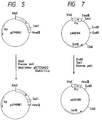
- Fig. 5 shows a construction scheme for plasmid pEPKMB1.
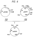
- Fig. 6 shows a construction scheme for plasmid pAGE501.
- Fig. 7 shows a construction scheme for plasmid pAGE109.
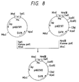
- Fig. 8 shows a construction scheme for plasmid pAGE502.
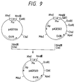
- Fig. 9 shows a construction scheme for plasmid pAGE503.
- Fig. 10 shows a construction scheme for plasmid pSEd1.
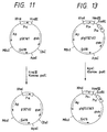
- Fig. 11 shows a construction scheme for plasmid pSE1D2.
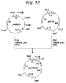
- Fig. 12 shows a construction scheme for plasmid pIG1SE1d2.
- Fig. 13 shows a construction scheme for plasmid pIG1SE1d3.
- Fig. 14 shows a construction scheme for plasmid pIG1SE1d4.
- Fig. 15 shows a construction scheme for plasmid pPMOL2.
- Fig. 16 shows a construction scheme for plasmid pPMOL3.
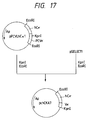
- Fig. 17 shows a construction scheme for plasmid pchCKA7.
- Fig. 18 shows a construction scheme for plasmid pchCKB1.
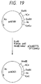
- Fig. 19 shows a construction scheme for plasmid pckCKC1.
- Fig. 20 shows a construction scheme for plasmid pChiIgHB2.
- Fig. 21 shows a construction scheme for plasmid pChiIgLA1.
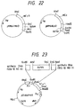
- Fig. 22 shows plasmids pKM641HA3 and pKM641LA2.
- Fig. 23 shows plasmid pChi641HA1.
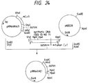
- Fig. 24 shows a construction scheme for plasmid pKM641HE1.
- Fig. 25 shows a construction scheme for plasmid pKM641HF1.
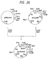
- Fig. 26 shows a construction scheme for plasmid pChi641HA1.
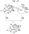
- Fig. 27 shows a construction scheme for plasmid pChi641HAM1.
- Fig. 28 shows plasmid pChi641LG11.
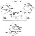
- Fig. 29 shows a construction scheme for plasmid pChi641LG11.
- Fig. 30 shows a construction scheme for plasmid pChi641LGM11.
- Fig. 31 shows a pattern of SDS-PAGE (4 to 15% gradient gel) of purified anti-GD3 chimera antibody KM-871 (about 5 ⁇ g/lane) carried out under reductive condition (A) or non-reductive condition (B), where the lanes starting from the left respectively indicate electrophoretic patterns of molecular weight markers, human IgG standard, mouse anti-GD3 antibody KM-641 and anti-GD3 chimera antibody KM-871.
- A reductive condition
- B non-reductive condition
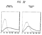
- Fig. 32 is a graph showing reactivity of anti-GD3 chimera antibody KM-871 with ganglioside GD3-positive G361 and SK-MEL-28 cells measured by fluorescent antibody technique with the cell number on the ordinate and the fluorescence intensity on the abscissa.
- a dotted line shows reactivity in the absence of the antibody, while a solid line shows reactivity in the presence of KM-871.
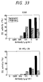
- Fig. 33 is a graph showing complement-dependent cytotoxicity (CDC) of anti-GD3 chimera antibody KM-871 and anti-GD3 mouse antibody KM-641 against ganglioside GD3-positive G361 and SK-MEL-28 cells with cytotoxicity on the ordinate and an antibody concentration on the abscissa.
- a blackened bar shows CDC activity of KM-871, while a striped bar shows that of KM-641.
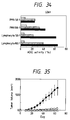
- Fig. 34 is a graph showing antibody-dependent cell-mediated cytotoxicity (ADCC) of KM-871 and KM-641 against ganglioside GD3-positive cell G361 with a ratio of effector cells to target cells on the ordinate and ADCC activity on the abscissa.
- a blackened bar shows ADCC activity of KM-871
- a dotted bar shows ADCC activity of KM-641
- a striped bar shows control (in the absence of the antibody).
- PMN means polymorphonuclear leukocyte.
- Fig. 35 is a graph showing therapeutic effect of KM-871 on transplanted tumors with the tumor size on the ordinate and days after transplantation of tumors on the abscissa, in which closed circle shows effect of anti-Sialyl Le a monoclonal antibody AMC-462, open square shows that of KM-641 and open triangle shows that of KM-871.
- the cassette vector to be used in the present invention is constructed by inserting a cDNA which encodes a human antibody constant region into an expression vector for animal cell use.
- Essential components in the expression vector for animal cell use include promoter, enhancer, polyA signal, splicing signal, drug resistance gene as a selection marker (e.g., ampicillin resistance gene, etc.) and the like. Any expression vector for animal cell use may be used for this purpose, as long as it can contain and express the cDNA molecule which encodes a human antibody constant region.
- pAGE107 (Cytotechnology, 3 , 133 (1990)) is useful as such an expression vector.
- Examples of the promoter and enhancer for use in the expression vector for animal cell use include: SV40 early promoter and enhancer (J.
- the immunoglobulin H chain promoter and enhancer can be prepared using appropriate antibody-producing hybridoma cells, such as rat hybridoma KM50 cells which produce anti-human serum albumin antibody as disclosed in JP-A-60-258128 (the term "JP-A" as used herein means an "unexamined published Japanese patent application”).
- JP-A as used herein means an "unexamined published Japanese patent application”
- Each chromosomal DNA is obtained from cultured KM50 cells, and P3X63Ag8U.1 (to be referred to as "P3U1" hereinafter) cells (ATCC CRL1597) which are to be fused with KM50 and rat kidney cells in accordance with the procedure disclosed in Molecular Cloning (2nd. ed., Cold Spring Harbor Laboratory Press, 1989, p9.14).
- P3U1 P3X63Ag8U.1
- ATCC CRL1597 P3X63Ag8U.1 cells
- Plasmid pIg1SE1d4 is an illustrative example of the animal cell expression vector which contains the immunoglobulin H chain promoter and enhancer.
- a cloning site is established in the upstream region of a human constant region of a cassette vector, for inserting a cDNA which encodes a variable region of nonhuman animal antibody.
- a cDNA which encodes a variable region of nonhuman animal antibody, using a synthetic DNA which comprises a base sequence corresponding to the 5'-end side of a constant region of human antibody and a base sequence corresponding to the 3'-end side of a variable region of nonhuman animal antibody and is possessed of restriction enzyme recognition sites on both of its ends.
- a humanized chimera antibody expression vector is constructed in which the cDNA coding for human antibody constant region and the cDNA coding for the variable region of nonhuman animal antibody are linked together through the synthetic DNA.
- the synthetic DNA to be used may be prepared using a DNA synthesizer, based on the base sequence which corresponds to the 5'-end side of a constant region of human antibody and the base sequence that corresponds to the 3'-end side of a variable region of nonhuman animal antibody.
- Illustrative examples of cloning site-containing cassette vectors include a cassette vector pChiIgHB2 which is used for the construction of an expression vector for the expression of humanized chimera antibody H chain and a cassette vector pChiIgLA2 which is used for the construction of an expression vector for the expression of humanized chimera antibody L chain.
- a cassette vector for use in the construction of an expression vector for the expression of humanized chimera antibody H chain is constructed, for example, by cutting out a human C H -encoding cDNA-containing fragment, from an Apa I site in the vicinity of the 5'-end of the cDNA to its 3'-end, and inserting the fragment into an appropriate expression vector for animal cell use such as plasmid pIg1SE1d4 or the like. Then, a cloning site is established in the thus constructed cassette vector for inserting a cDNA which encodes a V H of nonhuman animal antibody.
- a cDNA fragment encoding a nonhuman animal antibody V H which is obtained by digesting a V H -encoding cDNA with an appropriate restriction enzyme, using a synthetic DNA molecule which comprises a base sequence corresponding to the 5'-end side (5'-end to ApaI site) of a human antibody C H and a base sequence corresponding to the 3'-end side of a nonhuman animal antibody V H and is possessed of restriction enzyme recognition sites on both of its ends.
- a synthetic DNA molecule which comprises a base sequence corresponding to the 5'-end side (5'-end to ApaI site) of a human antibody C H and a base sequence corresponding to the 3'-end side of a nonhuman animal antibody V H and is possessed of restriction enzyme recognition sites on both of its ends.
- a cassette vector for constructing of an expression vector for the expression of humanized chimera antibody L chain may be constructed for example by introducing an Eco RV site into the vicinity of 5'-end side of a human C L -encoding cDNA by means of mutation, cutting out a fragment from the resulting human cDNA from the Eco RV site to the 3'-end and inserting the fragment into an appropriate expression vector such as plasmid pIg1SE1d4 or the like. Then, a cloning site is established in the thus constructed cassette vector for inserting a cDNA which encodes a nonhuman animal antibody V L .
- a cDNA fragment encoding a nonhuman animal antibody V L which is obtained by digesting a V L -encoding cDNA with an appropriate restriction enzyme, using a synthetic DNA which comprises a base sequence corresponding to the 5'-end side (5'-end to Eco RV site) of a human antibody C L and a base sequence corresponding to the 3'-end side of a nonhuman animal antibody V L and is possessed of restriction enzyme recognition sites on both of its ends.
- a synthetic DNA which comprises a base sequence corresponding to the 5'-end side (5'-end to Eco RV site) of a human antibody C L and a base sequence corresponding to the 3'-end side of a nonhuman animal antibody V L and is possessed of restriction enzyme recognition sites on both of its ends.
- cDNAs which encode the human C H and human C L described above are disclosed, for instance, in Cell 22 , 197 (1982).
- Such cDNAs can be prepared from human antibody-producing myeloma cells, humanized monoclonal antibody-producing hybridoma cells, humanized chimera antibody-producing cells (SP2-PC chimera; FEBS Letters, 244 , 301 (1989)) and the like, in accordance with known procedures disclosed for instance in Proc. Natl. Acad. Sci. U.S.A. 82 , 7025 (1985) and ibid., 79 7025 (1985) .
- cDNA is synthesized using mRNA extracted from the above-described cells, in accordance with the procedure disclosed in Molecular Cloning 2nd. ed.; 1989, p8.1.
- a library is prepared from the thus synthesized cDNA using a phage vector or a plasmid vector, in accordance with the procedure disclosed in Molecular Cloning 2nd. ed.; 1989, p8.1, 1.53.
- a recombinant phage or a recombinant plasmid which contains humane C H -encoding cDNA or human C L -encoding cDNA is obtained from the thus prepared library using a human antibody constant region or a human antibody variable region as a probe, in accordance with the procedure disclosed in Molecular Cloning 2nd. ed.; 1989, p8.1, 1.53.
- Base sequences of the human C H -encoding cDNA and the human C L -encoding cDNA are determined in accordance with the procedure disclosed in Molecular Cloning, 2nd. ed.; 1989, p13.1.
- Introduction of an appropriate restriction enzyme recognition site into the human C L -encoding cDNA may be effected in accordance with the procedure disclosed in Molecular Cloning, 2nd. ed.; 1989, p15.1.
- cDNAs which encode V H and V L of nonhuman animal antibody, such as mouse anti-GD3 monoclonal antibody are prepared in the following manner.
- cDNA is synthesized using mRNA extracted from appropriate hybridoma cells which produce mouse anti-GD3 monoclonal antibody, such as mouse anti-GD3 monoclonal antibody KM-641 (FERM BP-3116).
- a library is prepared from the thus synthesized cDNA using a phage vector or a plasmid vector.
- a recombinant phage or a recombinant plasmid which contains V H -encoding cDNA or V L -encoding cDNA is obtained from the thus prepared library using a constant region or a variable region of nonhuman antibody, such as mouse antibody, as a probe.
- Base sequences of the V H -encoding cDNA and the V L -encoding cDNA are determined in accordance with the aforementioned procedure.
- an expression vector for use in the expression of humanized chimera antibody H chain is constructed by linking the human antibody C H -encoding cDNA with the nonhuman antibody V H -encoding cDNA through the synthetic DNA.
- a fragment of the V L -encoding cDNA ranging from the 5'-end to an appropriate restriction enzyme site near the 3'-end (to be referred to as "site B" hereinafter), is cut out and inserted into the cloning site of the aforementioned cassette vector, using a synthetic DNA molecule which comprises a base sequence corresponding to the 5'-end side of a human antibody C L and a base sequence corresponding to the 3'-end side (from 3'-end to site B) of a nonhuman animal antibody V L and is possessed of restriction enzyme recognition sites on both of its ends.
- an expression vector for use in the expression of humanized chimera antibody L chain is constructed by linking the human antibody C L -encoding cDNA with the nonhuman antibody V L -
- a transformant which is capable of producing humanized chimera antibody is obtained by transforming appropriate host cells with the thus prepared expression vectors for use in the expression of the H chain and L chain of humanized chimera antibody.
- any type of cells may be used as host cells for use in the introduction of the humanized chimera antibody expression vectors, as long as these cells are capable of expressing the humanized chimera antibody.
- Illustrative examples of such host cells include mouse SP2/0-Ag14 cells (ATCC CRL1581; to be referred to as “SP2/0 cells” hereinafter), mouse P3X63-Ag8.653 (ATCC CRL1580) and CHO cells which are deficient in dihydrofolate reductase gene (to be referred to as "dhfr” hereinafter) (Urlaub et al., Proc. Natl. Acad. Sci. U.S.A., 77 , 4216 (1980)).
- Introduction of the expression vectors for use in the expression of the H chain and L chain of humanized chimera antibody into host cells may be effected for example by the electroporation technique disclosed in JP-A-2-257891.
- a transformant capable of producing the humanized chimera antibody may be selected using RPMI1640 medium supplemented with G418 and fetal calf serum, in accordance with the procedure disclosed in JP-A-2-257891.
- a transformant, KM-871, which produces humanized chimera antibody that reacts with ganglioside GD3 is an illustrative example of the transformant capable of producing humanized chimera antibody.
- KM-871 has been deposited on August 13, 1991, with Fermentation Research Institute, Agency of Industrial Science and Technology of 1-3, Higashi 1-chome, Tsukuba-shi, Ibaraki, Japan under the Budapest Treaty, and has been assigned the accession number FERM BP-3512.
- any medium can be used as long as the desired antibody can be produced and accumulated in the medium.
- An example of such medium is RPMI1640 medium supplemented with G418 and fetal calf serum.
- the transformants may be inoculated into 200 ⁇ l to 100 ml of the above-mentioned medium to give a cell concentration of 1 x 10⁵ to 1 x 10⁷ cells/ml and cultivated at 37°C in a 5% CO2 incubator for 1 to 7 days.
- the desired chimera antibody is produced and accumulated in the culture medium.
- the humanized chimera antibody thus produced can be purified from supernatant fluid of the aforementioned cultured mixture making use of a protein A column (E. Harlow et al., Manual of Antibody Experiments, Cold Spring Harbor Laboratory Press, 1988).
- Illustrative examples of humanized chimera antibodies obtained in this way include those which react with ganglioside GD3, such as humanized chimera antibody KM-871 and the like.
- Reactivity of humanized chimera antibody is measured by ELISA method.
- the molecular weight of the H chain, the L chain or the entire molecule of purified humanized chimera antibody is measured by means of polyacrylamide gel electrophoresis (SDS-PAGE), Western blotting method (E. Harlow et al., Manual of Antibody Experiments, Cold Spring Harbor Laboratory Press, 1988) or the like.
- Binding activity, or avidity, of the humanized chimera antibody to ganglioside GD3 to a cultured cancer cell line is measured by means of the fluorescent antibody technique, the ELISA method or the like.
- Complement-dependent cytotoxicity (CDC activity) and antibody-dependent cell-mediated cytotoxicity (ADCC activity) of humanized chimera antibody to a cultured cancer cell line are measured in accordance with the procedures disclosed in Menekigaku Jikken Nyumon, (Manual of Immunological Experiments) Matsuhashi et al., Gakkai Shuppan Center, Japan, 1981).
- the humanized chimera antibodies according to the present invention can be used alone as an anticancer agent. They may be formulated into an anticancer composition together with at least one pharmaceutically acceptable carrier. For instance, the humanized chimera antibodies are dissolved in physiological saline, an aqueous solution of glucose, lactose or mannitol and the like.
- the powder of the humanized chimera antibodies for injection can be prepared by lyophilizing the humanized chimera antibodies in accordance with the conventional method and mixing the lyophilized products with sodium chloride.
- the anticancer composition may further contain additives conventionally used well known in the art of medical preparation, for example, pharmaceutically acceptable salts.
- the humanized chimera antibodies according to the present invention can be administered in the form of the above-described anticancer composition to mammals including human in a dose of 0.2 to 20 mg/kg/day.
- the dose may vary depending on the age, condition, etc. of patients.
- the administration of the anticancer composition can be effected by intraveous injection once a day (single administration or consecutive administration) or intermittently one to three times a week or once every two to three weeks.
- the antincancer composition is expected to be useful for treating cancer such as melanoma, neuroblastoma and glioma.
- Chromosomal DNA was prepared in the following manner in accordance with the procedure disclosed in Molecular Cloning, Maniatis et al., 1989, p9.16.
- 1.2 x 10⁸ KM50 cells, 2 x 10⁸ P3U1 cells (ATCC CRL1597) and 1.6 g of rat kidney were each suspended in 2 ml of a buffer solution (pH 7.5) containing 10 mM Tris-HCl, 150 mM sodium chloride and 10 mM sodium ethylenediaminetetraacetate (to be referred to as "EDTA” hereinafter).
- EDTA sodium ethylenediaminetetraacetate
- the thus treated solution was extracted with the same volume of phenol, chloroform and ether (twice for each) in this order, and the extract was dialyzed for 10 hours against a buffer solution (pH 7.5) containing of 10 mM Tris-HCl and 1 mM EDTA.
- the DNA solution was recovered from the dialysis tube and used as a chromosomal DNA sample.
- a DNA concentration of each sample was determined by measuring the absorbance at 260 nm and, as a result, it was found that 1.6 mg, 1.5 mg and 1.9 mg of chromosomal DNA was obtained from 1.2 x 10⁸ KM50 cells, 2 x 10⁸ P3U1 cells and 1.6 g of rat kidney, respectively.
- a 3 ⁇ g portion of each of the chromosomal DNA samples obtained in the above step (1) from KM50 cells, P3U1 cells and rat kidney was dissolved in 25 ⁇ l of a buffer solution containing 10 mM Tris-HCl (pH 7.5), 6 mM magnesium chloride and 100 mM sodium chloride.
- a buffer solution containing 10 mM Tris-HCl (pH 7.5), 6 mM magnesium chloride and 100 mM sodium chloride.
- Each of the thus prepared solution was mixed with 15 units of Xba I (Takara Shuzo Co., Ltd.; all restriction enzymes used in the following experiments were purchased from the same company) and incubated at 37°C for 2 hours to cleave the chromosomal DNA at the Xba I site.
- the reaction mixture was subjected to agarose gel electrophoresis, resulting DNA fragments were transferred to a nitrocellulose filter in accordance with the method of Southern et al. (J. Mol. Biol., 98 , 503, (1975)) and then subjected to hybridization in the known method (Kameyama et al., FEBS Letters, 244 , 301-306 (1989)) using a mouse JH probe which is disclosed in the FEBS Letters article.
- a band equivalent to about 9.3 kb was observed only in the DNA sample of KM50 cells. In consequence, it was considered that the Xba I fragment of immunoglobulin DNA found in this band contained the activated immunoglobulin H chain gene derived from KM50 cells.
- a 60 ⁇ g portion of the KM50 cell chromosomal DNA obtained in the above step (2) was dissolved in 250 ⁇ l of a buffer solution containing 10 mM Tris-HCl (pH 7.5), 6 mM magnesium chloride and 100 mM sodium chloride.
- the thus prepared solution was mixed with 150 units of Xba I and incubated at 37°C for 2 hours to cleave the chromosomal DNA at the Xba I site.
- the reaction mixture was subjected to agarose gel electrophoresis and a 9.3 kb-equivalent fraction was recovered as about 2 ⁇ g of 9.3 kb DNA sample of KM50 cells, making use of the DEAE paper method (Maniatis et al ., Molecular Cloning, 1989, p6.24).
- a 3 ⁇ g portion of lambda-ZAP (Stratagene Cloning Systems) to be used as a vector was dissolved in 200 ⁇ l of a buffer solution containing 10 mM Tris-HCl (pH 7.5), 6 mM magnesium chloride and 100 mM sodium chloride.
- the thus prepared solution was mixed with 50 units of Xba I and incubated at 37°C for 2 hours to cleave the DNA at the Xba I site.
- the resulting reaction mixture was extracted with phenol-chloroform and then treated with ethanol to precipitate and recover about 3 ⁇ g of DNA.
- the thus recovered DNA sample was dissolved in a 100 ⁇ l of 100 mM Tris-HCl buffer (pH 7.5), and the resulting solution was mixed with 1 unit of alkaline phosphatase (Takara Shuzo Co., Ltd.) to effect dephosphorylation of restriction enzyme cleavage ends of the vector DNA.
- the resulting reaction mixture was extracted with phenol-chloroform and then treated with ethanol to precipitate and recover 2 ⁇ g of DNA.
- the thus recovered DNA sample was dissolved in 10 ⁇ l of a buffer solution containing 10 mM Tris-HCl (pH 7.5) and 1 mM EDTA to serve as a vector DNA sample.
- 0.2 ⁇ g of the thus prepared vector DNA sample and 0.2 ⁇ g of the KM50 cell-derived 9.3 kb DNA sample were dissolved in 5 ⁇ l of a buffer solution containing 66 mM Tris-HCl (pH 7.5), 6.6 mM magnesium chloride, 10 mM dithiothreitol (to be referred to as “DTT” hereinafter) and 0.1 mM adenosine triphosphate (to be referred to as "ATP” hereinafter)(to be referred to as "T4 ligase buffer” hereinafter).
- the resulting solution was mixed with 175 units of T4 DNA ligase (Takara Shuzo Co., Ltd.) and incubated at 4°C for 3 days.
- a 2 ⁇ l portion of the resulting reaction mixture was subjected to lambda phage packaging in the known method (Maniatis et al., Molecular Cloning, 1989, p2.95) using GigaPack Gold purchased from Stratagene Cloning Systems.
- E. coli BB4 cells were infected with this phage to obtain 200,000 phage clones. 100,000 out of these phage clones were fixed on nitrocellulose filters in the known method (Maniatis et al., Molecular Cloning, 1989, p2.112).
- Restriction enzyme cleavage maps of the two clones obtained in the above step (4) was prepared by digesting them with various restriction enzymes and it was found that completely the same DNA fragment (9.3 kb) has been inserted into these clones (Fig. 1).
- base sequence of a part of the 9.3 kb DNA fragment, which was considered to contain the promoter and variable regions of the rat immunoglobulin H chain was determined in accordance with the Sanger method (Sanger et al., Proc. Natl. Acad. Sci. U.S.A., 74 , 5463 (1977); M13 Cloning and Sequencing Handbook, Amersham).
- SEQ ID NO: 1 a region containing octamer sequences such as ATGCAAAT and TATA box sequences such as TTGAAAA and the like can be regarded as the immunoglobulin promoter region.
- a 1 ⁇ g portion of the 9.3 kb fragment of the immunoglobulin H chain variable region gene obtained in 1-(5) was dissolved in 30 ⁇ l of a buffer solution containing 10 mM Tris-HCl (pH 7.5), 6 mM magnesium chloride and 100 mM sodium chloride.
- the thus prepared solution was mixed with 10 units of Bgl II and 10 units of Hind III and incubated at 37°C for 2 hours to cleave the DNA fragment at the Bgl II and Hind III sites.
- the resulting reaction mixture was subjected to agarose gel electrophoresis and 0.01 ⁇ g of a DNA fragment containing 0.8 kb immunoglobulin promoter was recovered.
- a 1 ⁇ g portion of a plasmid pBR322- Bgl II (Kuwana et al., FEBS Letters, 219 , 360 (1987)) was dissolved in 30 ⁇ l of a buffer solution containing 10 mM Tris-HCl (pH 7.5), 6 mM magnesium chloride and 100 mM sodium chloride.
- the thus prepared solution was mixed with 10 units of Bgl II and 10 units of Hind III and incubated at 37°C for 2 hours to cleave the plasmid at the Bgl II and Hind III sites.
- the resulting reaction mixture was subjected to agarose gel electrophoresis, a DNA fragment of about 4.2 kb was recovered.
- a 0.1. ⁇ g portion of the thus obtained pBR322- Bgl II derived DNA fragment of about 4.2 kb and 0.01 ⁇ g of the immunoglobulin promoter-containing DNA fragment were dissolved in 20 ⁇ l of a T4 ligase buffer, and the resulting solution was mixed with 175 units of T4 DNA ligase (Takara Shuzo Co., Ltd.) and incubated at 4°C for 24 hours. Using the resulting reaction mixture, transformation of E . coli HB101 (J. Mol. Biol., 41 , 459 (1969)) was carried out in accordance with the method of Scott et al.
- the plasmid pKMB11 constructed in the above step (1) was digested with nuclease BAL31 from the Nco I site.
- a 10 ⁇ g portion of the plasmid pKMB11 was dissolved in 100 ⁇ l of a buffer solution containing 10 mM Tris-HCl (pH 7.5), 6 mM magnesium chloride and 50 mM potassium chloride.
- the thus prepared solution was mixed with 30 units of Nco I and incubated at 37°C for 2 hours to cleave the plasmid at the Nco I site.
- the resulting reaction mixture was extracted with phenol and chloroform and treated with ethanol.
- the thus precipitated DNA fragments were dissolved in 100 ⁇ l of BAL31 buffer which contained 20 mM Tris-HCl (pH 8.0), 600 mM sodium chloride, 12 mM calcium chloride, 12 mM magnesium chloride and 1 mM EDTA, and the resulting solution was mixed with 0.25 unit of BAL31 (Bethesda Research Laboratories, Inc. (BRL)) and incubated at 37°C for 5 seconds. The reaction was stopped by extracting the reaction mixture with phenol. After extraction with chloroform and precipitation with ethanol, 1 ⁇ g of DNA was recovered.
- BAL31 buffer contained 20 mM Tris-HCl (pH 8.0), 600 mM sodium chloride, 12 mM calcium chloride, 12 mM magnesium chloride and 1 mM EDTA
- BAL31 Bethesda Research Laboratories, Inc.
- the base sequence of the BAL31-digested portion of this plasmid was determined in accordance with the Sanger method and it was found that bases up to the third base (the 303 position base in the SEQ ID NO: 1) upstream from the initiation codon ATG of the immunoglobulin gene.
- a 1 ⁇ g portion of the 9.3 kb fragment of the immunoglobulin H chain variable region gene obtained in 1-(5) was dissolved in 30 ⁇ l of a buffer solution containing 10 mM Tris-HCl (pH 7.5), 6 mM magnesium chloride and 100 mM sodium chloride.
- the thus prepared solution was mixed with 10 units of Eco RV and 10 units of Xba I and incubated at 37°C for 2 hours to cleave the DNA fragment at the Eco RV and Xba I sites.
- the resulting reaction mixture was subjected to agarose gel electrophoresis and 0.1 ⁇ g of a DNA fragment of about 1 kb containing the immunoglobulin enhancer region was recovered.
- a 1 ⁇ g portion of the plasmid pKMD6 obtained in the above step (2) was dissolved in 100 ⁇ l of a buffer solution containing 10 mM Tris-HCl (pH 7.5), 6 mM magnesium chloride and 100 mM sodium chloride.
- the thus prepared solution was mixed with 10 units of Bgl II and incubated at 37°C for 2 hours to cleave the plasmid at the Bgl II site.
- the resulting reaction mixture was extracted with phenol and chloroform and precipitated with ethanol.
- the thus precipitated DNA fragments were dissolved in 40 ⁇ l of DNA polymerase I buffer containing 50 mM Tris-HCl (pH 7.5), 10 mM magnesium chloride, 0.1 mM dATP (deoxyadenosine triphosphate), 0.1 mM dCTP (deoxycytidine triphosphate), 0.1 mM dGTP (deoxyguanosine triphosphate) and 0.1 mM dTTP (deoxythymidine triphosphate).
- the resulting solution was mixed with 6 units of E. coli DNA polymerase I Klenow fragment and incubated at 16°C for 90 minutes to convert the cohesive 5'-end formed by the Bgl II digestion into blunt end.
- the reaction was stopped by extracting the reaction mixture with phenol. After extraction with chloroform and precipitation with ethanol, the resulting DNA fragments were dissolved in 30 ⁇ l of a buffer solution containing 10 mM Tris-HCl (pH 7.5), 6 mM magnesium chloride and 50 mM sodium chloride. The thus prepared solution was mixed with 10 units of Hind III and incubated at 37°C for 2 hours to cleave the DNA fragment at the Hind III site. The resulting reaction mixture was subjected to agarose gel electrophoresis, 0.1 ⁇ g of a DNA fragment of about 0.8 kb containing the immunoglobulin promoter region was recovered.
- plasmid pUC18 (Messing, Methods in Enzymology, 101 , 20 (1983)) was dissolved in 30 ⁇ l of a buffer solution containing 10 mM Tris-HCl (pH 7.5), 6 mM magnesium chloride and 100 mM sodium chloride.
- the thus prepared solution was mixed with 10 units of Hind III and 10 units of Xba I and incubated at 37°C for 2 hours to cleave the plasmid at the Hind III and Xba I sites.
- the resulting reaction mixture was subjected to agarose gel electrophoresis, 0.1 ⁇ g of a DNA fragment of about 2.7 kb was recovered.
- a 0.1 ⁇ g portion of the thus obtained pKMD6-derived 0.8 kb DNA fragment, 0.02 ⁇ g of the DNA fragment containing the immunoglobulin enhancer region and 0.1 ⁇ g of the pUC18 fragment were dissolved in 20 ⁇ l of the T4 ligase buffer, and the resulting solution was mixed with 175 units of T4 DNA ligase and incubated at 4°C for 24 hours. Using the resulting reaction mixture, transformation of E. coli HB101 was carried out to isolate an ApR colony. Plasmid DNA was recovered from the colony to obtain pEPKMA1 as shown in Fig. 4.
- a 1 ⁇ g portion of the plasmid pEPKMA1 was dissolved in 100 ⁇ l of a buffer solution containing 10 mM Tris-HCl (pH 7.5), 6 mM magnesium chloride and 100 mM sodium chloride.
- the thus prepared solution was mixed with 10 units of Xba I and incubated at 37°C for 2 hours to cleave the plasmid at the Xba I site.
- the resulting reaction mixture was extracted with phenol and chloroform and precipitated with ethanol.
- the thus precipitated DNA fragments were dissolved in 40 ⁇ l of the aforemetioned DNA polymerase I buffer solution, and the resulting solution was mixed with 6 units of E.
- SV40 early gene promoter and enhancer regions (to be referred to as "P SE " hereinafter) of an expression vector pAGE107 for use in the expression of heterologous genes in animal cells (Miyaji et al., Cytotechnology, 3 , 133 - 140 (1990)) were converted into KM50-derived immunoglobulin H chain promoter and enhancer (to be referred to as “P IH " hereinafter) of pEPKMB1 in the following manner.
- a 1 ⁇ g portion of the plasmid pAGE107 was dissolved in 30 ⁇ l of a buffer solution containing 10 mM Tris-HCl (pH 7.5), 6 mM magnesium chloride and 150 mM sodium chloride.
- the thus prepared solution was mixed with 10 units of Sal I and 10 units of Xho I and incubated at 37°C for 2 hours to cleave the plasmid at the Sal I and Xho I sites.
- the resulting reaction mixture was subjected to agarose gel electrophoresis and 0.5 ⁇ g of a DNA fragment of about 5.95 kb containing G418 resistance gene was recovered.
- a 1 ⁇ g portion of the plasmid pEPKMB1 was dissolved in 30 ⁇ l of a buffer solution containing 10 mM Tris-HCl (pH 7.5), 6 mM magnesium chloride and 150 mM sodium chloride.
- the thus prepared solution was mixed with 10 units of Sal I and 10 units of Xho I and incubated at 37°C for 2 hours to cleave the plasmid at the Sal I and Xho I sites.
- the resulting reaction mixture was subjected to agarose gel electrophoresis and 0.1 ⁇ g of a DNA fragment of about 1.7 kb containing immunoglobulin promoter and enhancer regions was recovered.
- a 2 ⁇ g portion of the expression vector pAGE106 for use in the expression of heterologous genes in animal cells was dissolved in 100 ⁇ l of a buffer solution containing 10 mM Tris-HCl (pH 7.5), 6 mM magnesium chloride and 50 mM sodium chloride.
- the thus prepared solution was mixed with 10 units of Eco RI and 10 units of Sac I and incubated at 37°C for 4 hours.
- the resulting reaction mixture was subjected to agarose gel electrophoresis and about 1.5 ⁇ g of a pAGE106 DNA fragment (4.3 kb) was recovered which contained the SV40 early gene promoter and G418 resistance gene cleaved with Eco RI and Sac I.
- the thus recovered DNA fragment was dissolved in 40 ⁇ l of the DNA polymerase I buffer solution, and the resulting solution was mixed with 5 units of E. coli DNA polymerase I large fragment and incubated at 16°C for 2 hours to convert the cohesive 3'-end formed by the Sac I digestion and the cohesive 5'-end formed by the Eco RI digestion into blunt ends.
- the resulting reaction mixture was extracted with phenol and chloroform and then treated with ethanol.
- the thus precipitated sample was dissolved in 20 ⁇ l of the T4 ligase buffer, and the resulting solution was mixed with 350 units of T4 DNA ligase and incubated at 4°C for 4 hours.
- transformation of E. coli HB101 was carried out to obtain plasmid pAGE109 as shown in Fig. 7.
- Plasmid pAGE502 was constructed in the following manner in order to convert the SV40 promoter and enhancer of pAGE107 into immunoglobulin H chain promoter and enhancer.
- a 2 ⁇ g portion of the plasmid pAGE107 disclosed in JP-A-3-22979 or EP-A-0 405 285 was dissolved in 100 ⁇ l of a buffer solution containing 10 mM Tris-HCl (pH 7.5), 6 mM magnesium chloride and 50 mM sodium chloride.
- the thus prepared solution was mixed with 10 units of Hind III and incubated at 37°C for 4 hours.
- the resulting reaction mixture was subjected to phenol-chloroform extraction and ethanol precipitation and the thus recovered sample was dissolved in 40 ⁇ l of the DNA polymerase I buffer solution.
- the resulting solution was mixed with 5 units of E.
- the resulting reaction mixture was subjected to agarose gel electrophoresis and about 1.5 ⁇ g of a pAGE107 DNA fragment of about 5.95 kb was obtained which contained G418 resistance gene and ApR gene cleaved with Xho I and Hind III.
- a 2 ⁇ g portion of the plasmid pAGE501 obtained in the above step (3) was dissolved in 100 ⁇ l of a buffer solution containing 10 mM Tris-HCl (pH 7.5), 6 mM magnesium chloride and 175 mM sodium chloride.
- the thus prepared solution was mixed with 10 units of Sal I and incubated at 37°C for 4 hours.
- the thus recovered sample was dissolved in 40 ⁇ l of the DNA polymerase I buffer solution.
- the resulting solution was mixed with 5 units of E. coli DNA polymerase I Klenow fragment and incubated at 16°C for 2 hours to convert the cohesive 5'-end formed by the Sal I digestion into blunt end.
- the resulting reaction mixture was extracted with phenol and chloroform and then treated with ethanol.
- the thus precipitated sample was dissolved in 30 ⁇ l of a buffer solution containing 10 mM Tris-HCl (pH 7.5), 6 mM magnesium chloride and 100 mM sodium chloride.
- the thus prepared solution was mixed with 10 units of Xho I and incubated at 37°C for 4 hours.
- the resulting reaction mixture was subjected to agarose gel electrophoresis and about 0.2 ⁇ g of a pAGE501 DNA fragment of about 1.8 kb was obtained which contained KM50 immunoglobulin H chain promoter and enhancer genes cleaved with Xho I and Sal I.
- a 2 ⁇ g portion of the plasmid pAGE109 obtained in the above step (4) was dissolved in 30 ⁇ l of a buffer solution containing 10 mM Tris-HCl (pH 7.5), 6 mM magnesium chloride and 50 mM sodium chloride.
- the thus prepared solution was mixed with 10 units of Hind III and 10 units of Cla I and incubated at 37°C for 4 hours.
- the resulting reaction mixture was subjected to agarose gel electrophoresis and about 0.2 ⁇ g of a pAGE109 DNA fragment of about 1 kb was recovered which contained the poly(A) signal gene of beta-globin and SV40 early genes cleaved with Cla I and Hind III.
- a 2 ⁇ g portion of the plasmid pAGE502 obtained in the above step (5) was dissolved in 30 ⁇ l of a buffer solution containing 10 mM Tris-HCl (pH 7.5), 6 mM magnesium chloride and 50 mM sodium chloride.
- the thus prepared solution was mixed with 10 units of Hind III and 10 units of Cla I and incubated at 37°C for 4 hours.
- the resulting reaction mixture was subjected to agarose gel electrophoresis and then to the aforementioned DEAE paper method to recover about 1 ⁇ g of a pAGE502 DNA fragment of about 6.1 kb which contained KM50 immunoglobulin H chain promoter and enhancer genes, ApR gene and G418 resistance gene cleaved with Hind III and Cla I.
- a dhfr gene was introduced into plasmid pAGE107 in the following manner to construct plasmid pSE1d1.
- a 2 ⁇ g portion of the plasmid pAGE107 disclosed in JP-A 3-22979 or EP-A-0 405 285 was dissolved in 100 ⁇ l of a buffer solution containing 100 mM Tris-HCl (pH 7.5), 6 mM magnesium chloride and 50 mM sodium chloride.
- the thus prepared solution was mixed with 10 units of Eco RI and incubated at 37°C for 4 hours. After subjecting the resulting reaction mixture to phenol-chloroform extraction and ethanol precipitation, the thus recovered sample was dissolved in 40 ⁇ l of the DNA polymerase I buffer solution. The resulting solution was mixed with 5 units of E.
- the resulting reaction mixture was subjected to agarose gel electrophoresis and about 1.5 ⁇ g of a pAGE107 DNA fragment of about 5.6 kb was recovered which contained G418 resistance gene and ApR gene cleaved with Eco RI and Hind III.
- a 2 ⁇ g portion of a plasmid pSV2-dhfr (Subramani et al., Mol. Cell. Biology, 1 , 854 (1981)) was dissolved in 100 ⁇ l of a buffer solution containing 10 mM Tris-HCl (pH 7.5), 6 mM magnesium chloride and 100 mM sodium chloride.
- the thus prepared solution was mixed with 10 units of Bgl II and incubated at 37°C for 4 hours. After subjecting the resulting reaction mixture to phenol-chloroform extraction and ethanol precipitation, the thus recovered sample was dissolved in 40 ⁇ l of the DNA polymerase I buffer solution. The resulting solution was mixed with 5 units of E.
- the resulting reaction mixture was subjected to agarose gel electrophoresis, about 0.2 ⁇ g of a pSV2-dhfr DNA fragment of about 0.76 kb was recovered which contained dhfr gene cleaved with Bgl II and Hind III.
- the Hind III cleavage site was removed from the plasmid pSE1d1 in the following manner to construct plasmid pSE1d2.
- a 2 ⁇ g portion of the plasmid pSE1d1 obtained in the above step (7) was dissolved in 100 ⁇ l of a buffer solution containing 10 mM Tris-HCl (pH 7.5), 6 mM magnesium chloride and 50 mM sodium chloride.
- the thus prepared solution was mixed with 10 units of Hind III and incubated at 37°C for 4 hours. After subjecting the resulting reaction mixture to phenol-chloroform extraction and ethanol precipitation, the thus recovered sample was dissolved in 40 ⁇ l of the DNA polymerase I buffer solution. The resulting solution was mixed with 5 units of E .
- the dhfr gene was introduced into plasmid pAGE503 in the following manner to construct plasmid pIg1SE1d2.
- a 2 ⁇ g portion of the plasmid pAGE503 obtained in the above step (6) was dissolved in 100 ⁇ l of a buffer solution containing 100 mM Tris-HCl (pH 7.5), 6 mM magnesium chloride and 50 mM sodium chloride.
- the thus prepared solution was mixed with 10 units of Cla I and incubated at 37°C for 4 hours.
- the thus recovered sample was dissolved in 40 ⁇ l of the DNA polymerase I buffer solution.
- the resulting solution was mixed with 5 units of E. coli DNA polymerase I Klenow fragment and incubated at 16°C for 2 hours to convert the cohesive 5'-end formed by the Cla I digestion into blunt end.
- the resulting reaction mixture was extracted with phenol and chloroform and then treated with ethanol.
- the thus precipitated sample was dissolved in 30 ⁇ l of a buffer solution containing 10 mM Tris-HCl (pH 7.5), 6 mM magnesium chloride and 50 mM sodium chloride.
- the thus prepared solution was mixed with 10 units of Mlu I and incubated at 37°C for 4 hours.
- the resulting reaction mixture was subjected to agarose gel electrophoresis and about 1 ⁇ g of a pAGE503 DNA fragment of about 5.4 kb was recovered which contained the KM50 immunoglobulin H chain promoter and enhancer genes cleaved with Cla I and Mlu I.
- a 2 ⁇ g portion of the plasmid pSE1d2 obtained in the above step (8) was dissolved in 100 ⁇ l of a buffer solution containing 10 mM Tris-HCl (pH 7.5), 6 mM magnesium chloride and 100 mM sodium chloride.
- the thus prepared solution was mixed with 10 units of Xho I and incubated at 37°C for 4 hours.
- the resulting reaction mixture was subjected to phenol-chloroform extraction and ethanol precipitation and the thus recovered sample was dissolved in 40 ⁇ l of the DNA polymerase I buffer solution.
- the resulting solution was mixed with 5 units of E.
- the resulting reaction mixture was subjected to agarose gel electrophoresis and about 1 ⁇ g of a pSE1d2 DNA fragment of about 3.8 kb was recovered which contained dhfr gene cleaved with Xho I and Mlu I.
- the ApaI cleavage site was removed from the plasmid pIg1SE1d2 in the following manner to construct plasmid pIg1SE1d3.
- a 2 ⁇ g portion of the plasmid pIg1SE1d2 obtained in the above step (9) was dissolved in 100 ⁇ l of a buffer solution containing 10 mM Tris-HCl (pH 7.5) and 6 mM magnesium chloride.
- the thus prepared solution was mixed with 10 units of Apa I and incubated at 37°C for 4 hours.
- the thus recovered sample was dissolved in 40 ⁇ l of the DNA polymerase I buffer solution.
- the resulting solution was mixed with 5 units of E. coli DNA polymerase I Klenow fragment and incubated at 16°C for 2 hours to convert the cohesive 3'-end formed by the Apa I digestion into blunt end.
- the resulting reaction mixture was extracted with phenol and chloroform and then treated with ethanol.
- the thus precipitated sample was dissolved in 20 ⁇ l of the T4 ligase buffer, and the resulting solution was mixed with 350 units of T4 DNA ligase and incubated at 4°C for 24 hours.
- transformation of E. coli HB101 was carried out to obtain plasmid pIg1SE1d3 as shown in Fig. 13.
- plasmid pIg1SE1d4 was constructed by inserting the synthetic DNA shown in SEQ ID NO:2 into the plasmid pIg1SE1d3 in the following manner.
- a 2 ⁇ g portion of the plasmid pIg1SE1d3 obtained in the above step (10) was dissolved in 30 ⁇ l of a buffer solution containing 10 mM Tris-HCl (pH 7.5), 6 mM magnesium chloride and 50 mM sodium chloride.
- the thus prepared solution was mixed with 10 units of Hind III and 10 units of Eco RI and incubated at 37°C for 4 hours.
- the resulting reaction mixture was subjected to agarose gel electrophoresis and about 1 ⁇ g of a pIg1SE1d3 DNA fragment of about 9.2 kb was recovered which contained the KM50 immunoglobulin H chain promoter and enhancer genes, ApR gene, G418 resistance gene and dhfr gene cleaved with Hind III and Eco RI.
- MoLTR Since MoLTR is known to have promoter and enhancer activities (Kuwana et al., Biochem. Biophys. Res. Cons., 149 , 960 (1987)), a plasmid pPMOL3 containing MoLTR was prepared in the following manner in order to use MoLTR as cassette vector promoter and enhancer.
- a 3 ⁇ g portion of the plasmid pPMOL1 disclosed in JP-A 1-63394 was dissolved in 30 ⁇ l of a buffer solution containing 10 mM Tris-HCl (pH 7.5), 7 mM magnesium chloride and 6 mM 2-mercaptoethanol.
- the thus prepared solution was mixed with 10 units of Cla I and incubated at 37°C for 4 hours. After subjecting the resulting reaction mixture to phenol-chloroform extraction and ethanol precipitation, the thus recovered sample was dissolved in 40 ⁇ l of the DNA polymerase I buffer solution. The resulting solution was mixed with 5 units of E .
- coli HB101 was carried out to obtain plasmid pPMOL2 as shown in Fig. 15.
- a 3 ⁇ g portion of the thus obtained plasmid pPMOL2 was dissolved in 30 ⁇ l of a buffer solution containing 10 mM Tris-HCl (pH 7.5), 7 mM magnesium chloride, 10 mM sodium chloride and 6 mM 2-mercaptoethanol.
- the thus prepared solution was mixed with 10 units of Sma I and incubated at 37°C for 4 hours.
- the resulting reaction mixture was subjected to phenol-chloroform extraction and ethanol precipitation and 2 ⁇ g of DNA fragments were recovered.
- mRNA extraction kit Fast Track (No. K1593-02, available from Invitrogen)
- 6.2 ⁇ g of mRNA was obtained from 1 x 10⁸ cells of chimera antibody-producing SP2-PC Chimera-1 which has anti-phosphorylcholine activity and is disclosed in FEBS Letters ( 244 , 301 - 306 (1989)).
- a 2 ⁇ g portion of the mRNA obtained in the above step (1) was subjected to Eco RI adaptor addition using cDNA Synthesis Kit (No. 27-9260-01, available from Pharmacia) followed by kination.
- the resulting cDNA solution was subjected to phenol-chloroform extraction and ethanol precipitation to recover 4 ⁇ g of cDNA.
- the thus recovered cDNA was dissolved in 20 ⁇ l of sterile water, and the resulting solution was subjected to agarose gel electrophoresis to recover about 0.3 ⁇ g of a DNA fragment of about 1.8 kb and about 0.3 ⁇ g of a DNA fragment of about 1.0 kb.
- a 5 ⁇ g portion of the vector pUC18 was dissolved in 100 ⁇ l of a buffer solution containing 100 mM Tris-HCl (pH 7.5), 6 mM magnesium chloride and 100 mM sodium chloride.
- the thus prepared solution was mixed with 50 units of Eco RI and incubated at 37°C for 4 hours to cleave the pUC18 DNA at its Eco RI cleavage site.
- the resulting reaction mixture was subjected to phenol-chloroform extraction and ethanol precipitation to recover about 3 ⁇ g of a pUC18 DNA fragment cleaved with Eco RI.
- An Eco RV site was introduced into 5'-end side of the human Ig k chain constant region by means of site-specific mutagenesis using a kit purchased from Promega (Catalogue No. Q6210).
- a 2 ⁇ g portion of the plasmid pPCVLhCK1 was dissolved in 30 ⁇ l of a buffer solution containing 10 mM Tris-HCl (pH 7.5), 6 mM magnesium chloride and 50 mM sodium chloride.
- the thus prepared solution was mixed with 10 units of Eco RI and 10 units of Kpn I and incubated at 37°C for 4 hours.
- the resulting reaction mixture was subjected to agarose gel electrophoresis and about 0.2 ⁇ g of a pPCVLhCK1 DNA fragment of about 0.8 kb was recovered which contained the human immunoglobulin L chain constant region cleaved with Eco RI and Kpn I.
- pSELECT1 a kit available from Promega, Catalogue No. Q6210
- a buffer solution containing 10 mM Tris-HCl (pH 7.5), 6 mM magnesium chloride and 50 mM sodium chloride was dissolved in 30 ⁇ l of a buffer solution containing 10 mM Tris-HCl (pH 7.5), 6 mM magnesium chloride and 50 mM sodium chloride.
- the thus prepared solution was mixed with 10 units of Eco RI and 10 units of Kpn I and incubated at 37°C for 4 hours.
- the resulting reaction mixture was subjected to agarose gel electrophoresis and about 1 ⁇ g of a pSELECT1 DNA fragment of about 5.7 kb cleaved with Eco RI and Kpn I was recovered.
- 0.1 ⁇ g of the pPCVLhCK1 Eco RI- Kpn I fragment (about 0.8 kb) and 0.1 ⁇ g of the pSELECT1 Eco RI- Kpn I fragment (about 5.7 kb) obtained above were dissolved in 20 ⁇ l of the T4 ligase buffer, and the resulting solution was mixed with 350 units of T4 DNA ligase and incubated at 4°C for 24 hours.
- transformation of E . coli JM109 was carried out to obtain plasmid pchCKA7 as shown in Fig. 17.
- the ACC sequence of the human immunoglobulin L chain constant region (12 to 14 position bases from the N-terminal) was converted into GAT in order to construct a plasmid pchCKB1 (Fig. 18) in which an Eco Rv site was introduced into the converted site.
- the Eco RV site of the plasmid pchCKB1 was converted into Hind III cleavage site in the following manner.
- a 2 ⁇ g portion of the plasmid pchCKB1 obtained above was dissolved in 10 ⁇ l of a buffer solution containing 100 mM Tris-HCl (pH 7.5), 6 mM magnesium chloride and 100 mM sodium chloride.
- the thus prepared solution was mixed with 10 units of Eco RI and incubated at 37°C for 4 hours. After subjecting the resulting reaction mixture to phenol-chloroform extraction and ethanol precipitation, the thus recovered sample was dissolved in 40 ⁇ l of the DNA polymerase I buffer solution. The resulting solution was mixed with 5 units of E.
- a 2 ⁇ g portion of the plasmid pIg1SEId4 obtained in the aforementioned step 2-(11) was dissolved in 30 ⁇ l of a buffer solution containing 10 mM Tris-HCl (pH 7.5), 6 mM magnesium chloride and 100 mM sodium chloride.
- the thus prepared solution was mixed with 10 units of Eco RV and 10 units of Apa I and incubated at 37°C for 4 hours.
- the resulting reaction mixture was subjected to agarose gel electrophoresis and about 1.5 ⁇ g of a pIg1SEId4 DNA fragment of about 9.2 kb cleaved with Eco RV and Apa I was recovered.
- a 2 ⁇ g portion of the plasmid pPCVHhCGI1 obtained in the aforementioned step 4-(2) was dissolved in 30 ⁇ l of a buffer solution containing 10 mM Tris-HCl (pH 7.5) and 6 mM magnesium chloride.
- the thus prepared solution was mixed with 10 units of Apa I and 10 units of Sma I and incubated at 37°C for 1 hour.
- the resulting reaction mixture was subjeced to agarose gel electrophoresis and about 0.2 ⁇ g of a pPCVHhCGI1 DNA fragment of about 1 kb was recovered which contained the human immunoglobulin H chain constant region gene cleaved with Apa I and Sma I.
- 0.1 ⁇ g of the pIg1SEId4 Eco Rv- Apa I fragment (about 9.2 kb) and 0.1 ⁇ g of the pPCVHhCGI1 Apa I- Sma I fragment (about 1 kb) prepared above were dissolved in 20 ⁇ l of the T4 ligase buffer.
- the resulting solution was mixed with 350 units of T4 DNA ligase and incubated at 4°C for 24 hours.
- transformation of E. coli HB101 was carried out to obtain a plasmid pChiIgHB2 (Fig. 20) as a cassette vector for use in the construction of a humanized chimera antibody H chain expression vector.
- a 2 ⁇ g portion of the plasmid pIg1SEId4 obtained in the aforementioned step 2-(11) was dissolved in 30 ⁇ l of a buffer solution containing 10 mM Tris-HCl (pH 7.5), 6 mM magnesium chloride and 100 mM sodium chloride.
- the thus prepared solution was mixed with 10 units of Eco RV and 10 units of Hind III and incubated at 37°C for 4 hours.
- the resulting reaction mixture was subjected to agarose gel electrophoresis and about 1.5 ⁇ g of a pIg1SEId4 DNA fragment of about 9.2 kb cleaved with Eco RV and Hind III was recovered.
- a 2 ⁇ g portion of the plasmid pchCKC1 obtained in the aforementioned step 4-(3) was dissolved in 30 ⁇ l of a buffer solution containing 10 mM Tris-HCl (pH 7.5), 6 mM magnesium chloride and 100 mM sodium chloride.
- the thus prepared solution was mixed with 10 units of Eco RV and 10 units of Hind III and incubated at 37°C for 1 hour.
- the resulting reaction mixture was subjected to agarose gel electrophoresis and about 0.2 ⁇ g of a pPCVLhCK1 DNA fragment of about 0.6 kb was recovered which contained the human immunoglobulin L chain constant region gene cleaved with Eco RV and Hind III.
- 0.1 ⁇ g of the pIg1SEId4 Eco Rv- Hin dIII fragment (about 9.2 kb) and 0.1 ⁇ g of the pchCKC1 Eco Rv- Hind III fragment (about 0.6 kb) prepared above were dissolved in 20 ⁇ l of the T4 ligase buffer.
- the resulting solution was mixed with 350 units of T4 DNA ligase and incubated at 4°C for 24 hours.
- transformation of E. coli HB101 was carried out to obtain a plasmid pChiIgLA1 (Fig. 21) as a cassette vector for use in the construction of a humanized chimera antibody L chain expression vector.
- cDNA having Eco RI adaptor on its 5'-end and Xho I adaptor on its 3'-end was prepared from 3 ⁇ g of the mRNA obtained in the above procedure 1.
- About 6 ⁇ g of the cDNA was dissolved in 10 ⁇ l of sterile water and subjected to agarose gel electrophoresis to recover 0.1 ⁇ g of an H chain-corresponding cDNA fragment of about 1.8 kb and 0.1 ⁇ g of an L chain-corresponding cDNA fragment of about 1.0 kb.
- 0.1 ⁇ g of the 1.8 kb cDNA fragment, 0.1 ⁇ g of the 1.0 kb cDNA fragment and 1 ⁇ g of UniZAP XR available from Stratagene Cloning Systems; a preparation obtained by digesting Lambda ZAPII vector with Eco RI and Xho I, followed by treatment with calf intestine alkaline phosphatase) to be used as a vector were dissolved in 11.5 ⁇ l of the T4 ligase buffer, and the resulting solution was mixed with 175 units of T4 DNA ligase and incubated at 12°C for 10 hours and then at room temperature for 2 hours.
- UniZAP XR available from Stratagene Cloning Systems; a preparation obtained by digesting Lambda ZAPII vector with Eco RI and Xho I, followed by treatment with calf intestine alkaline phosphatase
- a 4 ⁇ l portion of the resulting reaction mixture was subjected to lambda phage packaging using Giga Pack Gold (Stratagene Cloning Systems) in accordance with the conventional method (Maniatis et al., Molecular Cloning, 1989, p2.95).
- An E. coli strain PLK-F was infected with the thus packaged product in accordance with the conventional method (Maniatis et al., Molecular Cloning, 1989, p2.95-107) to obtain an H chain cDNA library and an L chain cDNA library, each containing about 10,000 phage clones.
- these phage particles were fixed on nitrocellulose filters in accordance with the conventional method (Maniatis et al., Molecular Cloning, 1989, p2.112).
- Two 32P-labeled probes were prepared from an Eco RI fragment of about 6.8 kb containing a mouse immunoglobulin constant region chromosomal gene Cg1 (Roeder et al., Proc. Natl. Acad. Sci. U.S.A., 78 , 474 (1981)) and a mouse Ck genecontaining Hind III- Bam HI fragment of about 3 kb (Sakano et al., Nature, 280 , 288 (1979)).
- a phage clone which showed strong reaction at 65°C with one of these two probes were obtained from each of the H chain cDNA library and the L chain cDNA library prepared in the above procedure 2 in accordance with the conventional method (Maniatis et al., Molecular Cloning, 1989, p2.108). Next, using ZAP-cDNA Synthesis Kit (No.
- Immunoglobulin variable region base sequences of the plasmids pKM641HA3 and pKM641LA2 obtained in the above procedure 3 were determined by the dideoxy method (Maniatis et al., Molecular Cloning, 1989, p13.42) using Sequenase Version 2.0 DNA Sequencing Kit (United States Biochemical Corporation). The results are shown in SEQ ID NO:4 and SEQ ID NO:5.
- the plasmid pKM641LA2 was a complete cDNA containing a leader sequence and having a methionine-corresponding sequence which was assumed to be the initiation codon ATG located close to the 5'-end.
- the plasmid pKM641HA3 did not have such a methionine-corresponding initiation codon-like sequence on its 5'-end side, and its leader sequence was partially deficient.
- H chain variable region gene obtained by cleaving the plasmid pKM641HA3 variable region at the 5'-end Alu I site and 3'-end Sty I site was ligated with the cassette vector for use in the construction of the humanized chimera antibody H chain obtained in Example 1 using the synthetic DNA sequences shown in SEQ ID NO:6 and SEQ ID NO:7, thereby constructing a humanized chimera antibody H chain expression vector pchi641HA1 (Fig. 23).
- the DNA shown in SEQ ID NO:7 was synthesized using a DNA synthesizer.
- This synthetic DNA comprises a base sequence derived from plasmid pKM641HA3 ranging from the 3'-end of its immunoglobulin H chain variable region to a Sty I cleavage site in the vicinity of the 3'-end and a base sequence derived from plasmid pAGE28 ranging from the 5'-end of its immunoglobulin H chain constant region to an Apa I cleavage site in the vicinity of the 5'-end.
- the synthetic DNA has a Sty I cleavage site and an Apa I cleavage site on both of its end.
- the thus synthesized DNA was introduced into the plasmid pKM641HA3 in the following manner.
- a 3 ⁇ g portion of the plasmid pKM641HA3 was dissolved in 30 ⁇ l of a buffer solution containing 50 mM Tris-HCl (pH 7.5), 10 mM magnesium chloride, 50 mM sodium chloride and 1 mM DTT.
- the thus prepared solution was mixed with 10 units of Eco RI and 10 units of Sty I and incubated at 37°C for 4 hours.
- the resulting reaction mixture was subjected to agarose gel electrophoresis and about 0.3 ⁇ g of a DNA fragment of about 0.41 kb was recovered.
- pAGE28 Mizukami et al., J.
- 0.1 ⁇ g of the pKM641HA3 Eco RI- Sty I fragment (about 0.41 kb) and 0.1 ⁇ g of the pAGE28 Eco RI- Apa I fragment (about 2.45 kb) prepared above and 0.3 ⁇ g of the synthetic DNA of SEQ ID NO:7 were dissolved in 20 ⁇ l of the T4 ligase buffer solution.
- the resulting solution was mixed with 350 units of T4 DNA ligase and incubated at 4°C for 24 hours.
- transformation of E. coli HB101 was carried out to obtain a plasmid pKM641HE1 as shown in Fig. 24.
- a 3 ⁇ g portion of the plasmid pKM641HE1 was dissolved in 30 ⁇ l of a buffer solution containing 10 mM Tris-HCl (pH 7.5), 7 mM magnesium chloride and 6 mM 2-mercaptoethanol.
- the thus prepared solution was mixed with 10 units of Eco RI and 10 units of Apa I and incubated at 37°C for 4 hours.
- the resulting reaction mixture was subjected to agarose gel electrophoresis and about 0.4 ⁇ g of a DNA fragment of about 0.42 kb was recovered.
- a 0.4 ⁇ g portion of the thus prepared pKM641HE1 Eco RI- Apa I fragment (about 0.42 kb) was dissolved in 30 ⁇ l of a buffer solution containing 10 mM Tris-HCl (pH 7.5), 7 mM magnesium chloride, 50 mM sodium chloride and 6 mM 2-mercaptoethanol.
- the thus prepared solution was mixed with 10 units of Alu I and incubated at 37°C for 4 hours.
- the resulting reaction mixture was subjected to phenol-chloroform extraction and ethanol precipitation and about 0.3 ⁇ g of a DNA fragment of about 0.4 kb was recovered.
- 0.1 ⁇ g of the pKM641HE1 Alu I- Apa I fragment (about 0.4 kb) and 0.1 ⁇ g of the pAGE28 Eco RI- Apa I fragment (about 2.45 kb) prepared above and 0.3 ⁇ g of the synthetic DNA of SEQ ID NO:6 were dissolved in 20 ⁇ l of the T4 ligase buffer solution. The resulting solution was mixed with 350 units of T4 DNA ligase and incubated at 4°C for 24 hours. Using the thus obtained recombinant plasmid DNA, transformation of E. coli HB101 was carried out to obtain a plasmid pKM641HF1 as shown in Fig. 25.
- immunoglobulin H chain variable region of the thus obtained plasmid pKM641HF1 was introduced into the aforementioned cassette vector pChiIgHB2 in the following manner.
- a 3 ⁇ g portion of the plasmid pKM641HF1 was dissolved in 30 ⁇ l of a buffer solution containing 10 mM Tris-HCl (pH 7.5), 7 mM magnesium chloride and 6 mM 2-mercaptoethanol.
- the thus prepared solution was mixed with 10 units of Eco RI and 10 units of Apa I and incubated at 37°C for 4 hours.
- the resulting reaction mixture was subjected to agarose gel electrophoresis and about 0.5 ⁇ g of a DNA fragment of about 0.44 kb was recovered.
- a 3 ⁇ g portion of the pChiIgHB2 was dissolved in 30 ⁇ l of a buffer solution containing 10 mM Tris-HCl (pH 7.5), 7 mM magnesium chloride and 6 mM 2-mercaptoethanol.
- the thus prepared solution was mixed with 10 units of Eco RI and 10 units of Apa I and incubated at 37°C for 4 hours.
- the resulting reaction mixture was subjected to phenol-chloroform extraction and ethanol precipitation and about 3 ⁇ g of DNA was recovered.
- 0.1 ⁇ g of the pKM641HF1 Eco RI- Apa I fragment (about 0.44 kb) and 0.1 ⁇ g of the pChiIgHB2 Eco RI- Apa I fragment (about 10.1 kb) prepared above were dissolved in 20 ⁇ l of the T4 ligase buffer.
- the resulting solution was mixed with 350 units of T4 DNA ligase and incubated at 4°C for 24 hours.
- transformation of E. coli HB101 was carried out to obtain a plasmid pChi641HA1 as shown in Fig. 26.
- KM50-derived immunoglobulin H chain promoter and enhancer regions of the thus obtained plasmid pChi641HA1 were converted into MoLTR in the following manner.
- a 3 ⁇ g portion of the plasmid pChi641HA1 was dissolved in 30 ⁇ l of a buffer solution containing 50 mM Tris-HCl (pH 7.5), 10 mM magnesium chloride, 50 mM sodium chloride and 1 mM DTT.
- the thus prepared solution was mixed with 10 units of Eco RI and 10 units of Xho I and incubated at 37°C for 4 hours.
- the resulting reaction mixture was subjected to agarose gel electrophoresis and about 0.2 ⁇ g of a DNA fragment of about 8.8 kb was recovered.
- a 3 ⁇ g portion of the pPMOL3 prepared in procedure 2 of Example 1 was dissolved in 30 ⁇ l of a buffer solution containing 50 mM Tris-HCl (pH 7.5), 10 mM magnesium chloride, 50 mM sodium chloride and 1 mM DTT.
- the thus prepared solution was mixed with 10 units of Eco RI and 10 units of Xho I and incubated at 37°C for 4 hours.
- the resulting reaction mixture was subjected to agarose gel electrophoresis and about 0.3 ⁇ g of a DNA fragment of about 0.63 kb containing MoLTR was recovered.
- 0.1 ⁇ g of the pChi641HA1 Eco RI- Xho I fragment and 0.1 ⁇ g of the pPMOL3 Eco RI- Xho I fragment prepared above were dissolved in 20 ⁇ l of the T4 ligase buffer solution.
- the resulting solution was mixed with 175 units of T4 DNA ligase and incubated at 4°C for 24 hours.
- transformation of E. coli HB101 was carried out to obtain a plasmid pChi641HAM1 (Fig. 27) as a KM-641 chimera H chain expression vector.
- L chain variable region gene obtained by cleaving the plasmid pKM641LA2 variable region gene at its 5'-end Eco RI site and 3'-end Hind III site was ligated with the cassette vector for the expression of chimera L chain, using the synthetic DNA shown in SEQ ID NO: 8, thereby constructing an L chain expression vector pchi641LG11 (Fig. 28).
- the DNA of SEQ ID NO: 8 (see Fig. 29) was synthesized using a DNA synthesizer.
- This synthetic DNA comprises a base sequence corresponding to a region of the plasmid pKM641LA2 ranging from the 3'-end of the immunoglobulin L chain variable region to a Hind III cleavage site in the vicinity of the 3'-end and a base sequence corresponding to a region of the plasmid pChiIgLA1 ranging from the 5'-end to an Eco RV cleavage site in the vicinity of the 5'-end.
- it has a Hind III cleavage site and an Eco Rv cleavage site on both ends.
- the thus synthesized DNA was introduced into the plasmid pKM641LA2 in the following manner.
- a 3 ⁇ g portion of the plasmid pKM641LA2 was dissolved in 30 ⁇ l of a buffer solution containing 10 mM Tris-HCl (pH 7.5), 7 mM magnesium chloride, 50 mM sodium chloride and 6 mM 2-mercaptoethanol.
- the thus prepared solution was mixed with 10 units of Eco RI and 10 units of Hind III and incubated at 37°C for 4 hours.
- the resulting reaction mixture was subjected to agarose gel electrophoresis and about 0.3 ⁇ g of a DNA fragment of about 0.35 kb was recovered.
- pChiIgLA1 a 3 ⁇ g portion of pChiIgLA1 was dissolved in 30 ⁇ l of a buffer solution containing 50 mM Tris-HCl (pH 7.5), 10 mM magnesium chloride, 50 mM sodium chloride and 1 mM DTT.
- the thus prepared solution was mixed with 10 units of Eco RI and 10 units of Eco RV and incubated at 37°C for 4 hours.
- the resulting reaction mixture was subjected to phenol-chloroform extraction and ethanol precipitation and about 3 ⁇ g of DNA was recovered and dissolved in 10 ⁇ l of the TE solution (a buffer solution containing 10 mM Tris-HCl and 1 mM EDTA (pH 7.5)).
- 0.1 ⁇ g of the pKM641LA2 Eco RI- Hind III fragment (about 0.35 kb) and 0.1 ⁇ g of the pChiIgLA1 Eco RI- Eco RV fragment (about 9.7 kb) prepared above and 0.3 ⁇ g of the synthetic DNA of SEQ ID NO:8 were dissolved in 20 ⁇ l of the T4 ligase buffer solution. The resulting solution was mixed with 350 units of T4 DNA ligase and incubated at 4°C for 24 hours. Using the thus obtained recombinant plasmid DNA, transformation of E. coli HB101 was carried out to obtain a plasmid pChi641LG11 as shown in Fig. 29.
- KM50-derived immunoglobulin H chain promoter and enhancer regions of the thus obtained plasmid pChi641LG11 were converted into MoLTR in the following manner.
- a 3 ⁇ g portion of the plasmid pChi641LG11 was dissolved in 30 ⁇ l of a buffer solution containing 50 mM Tris-HCl (pH 7.5), 10 mM magnesium chloride, 50 mM sodium chloride and 1 mM DTT.
- the thus prepared solution was mixed with 10 units of Eco RI and 10 units of Xho I and incubated at 37°C for 4 hours.
- the resulting reaction mixture was subjected to agarose gel electrophoresis and about 0.2 ⁇ g of a DNA fragment of about 8.3 kb was recovered.
- 0.1 ⁇ g of the pChi641LG11 Eco RI- Xho I fragment and 0.1 ⁇ g of the pPMOL3 Eco RI- Xho I fragment prepared above were dissolved in 20 ⁇ l of the T4 ligase buffer.
- the resulting solution was mixed with 175 units of T4 DNA ligase and incubated at 4°C for 24 hours.
- transformation of E. coli HB101 was carried out to obtain a plasmid pChi641LGM11 (Fig. 30) as a KM-641 chimera L chain expression vector.
- RPMI1640-FCS(10) which has been prepared by supplementing RPMI1640 medium (Nissui Pharmaceutical Co., Ltd.) with 10% of FCS, 1/40 volume
- the thus prepared cell suspension was distributed in 200 ⁇ l-portions into wells of a 96-well microtiter plate (Flow Laboratories), and the cells were cultured at 37°C in a CO2 incubator. After 24 hours of the culturing, G418 (GIBCO) was added to the cell suspension to a final concentration of 0.5 mg/ml, and the culturing was continued for 1 to 2 weeks. When transformant colonies were developed and grown into confluent stages, culture broths were recovered from the wells to measure anti-GD3 chimera antibody activities by ELISA method in the following manner.
- a 2 ng portion of GD3 (available from Iatron) or other type of ganglioside was dissolved in 2 ⁇ l of ethanol solution containing 5 ng of phosphatidylcholine (Sigma Chemical Co.) and 2.5 ng of cholesterol (Sigma Chemical Co.).
- a 20 ⁇ l portion of the thus prepared solution or the same volume of its dilution solution was distributed into each well of a 96-well microtiter plate (available from Greiner). After air-drying, blocking was effected with PBS containing 1% BSA. To each well was added 50 to 100 ⁇ l of a culture supernatant of a transformant, a purified mouse monoclonal antibody solution or a purified chimera antibody solution.
- each well was washed with PBS and charged with 50 to 100 ⁇ l of peroxidase-labeled protein A (Funakoshi Pharmaceutical Co., Ltd.), followed by 1 to 2 hours of reaction at room temperature.
- 50 to 100 ⁇ l of ABTS substrate solution prepared by dissolving 550 mg of diammonium 2,2'-azinobis(3-ethylbenzothiazoline-6-sulfonate) in 0.1 M citrate buffer (pH 4.2) and adding 1 ⁇ l/ml of hydrogen peroxide to the resulting solution just before its use was added to each well to develop color, and OD415 of the reaction mixture was measured.
- culture broth of a clone having the highest activity measured by ELISA method contained anti-GD3 chimera antibody in an amount of about 0.1 ⁇ g/ml.
- the clone having anti-GD3 chimera antibody activity was suspended in the aforementioned RPMI1640-FCS(10) medium supplemented with 0.5 mg/ml of G418 and 50 nM of methotrexate (to be referred to as "MTX" hereinafter) to a final cell density of 1-2 x 10⁵ cells /ml.
- the thus prepared cell suspension was distributed in 2-ml portions into wells of a 24 well plate, and the cells were cultured at 37°C for 2 to 3 weeks in a CO2 incubator to induce clones resistant to 50 nM MTX.
- anti-GD3 chimera antibody activities in the culture broths were measured by the ELISA method.
- culture broth of a 50 nM MTX-resistant clone having the highest activity measured by ELISA method contained anti-GD3 chimera antibody in an amount of about 0.3 ⁇ g/ml.
- the 50 nM MTX-resistant clone was suspended in the RPMI1640-FCS(10) medium supplemented with 0.5 mg/ml of G418 and 200 nM of MTX to a final cell density of 1-2 x 10⁵ cells /ml.
- the thus prepared cell suspension was distributed in 2-ml portions into wells of a 24 well plate, and the cells were cultured at 37°C for 2 to 3 weeks in a CO2 incubator to induce clones resistant to 200 nM MTX.
- anti-GD3 chimera antibody activities in the culture broths were measured by the ELISA method.
- culture broth of a 200 nM MTX-resistant clone having the highest activity measured by ELISA method contained anti-GD3 chimera antibody in an amount of about 2 ⁇ g/ml.
- This 200 nM MTX-resistant clone was named transformant KM-871.
- the transformant KM-871 was suspended in GIT medium (Nihon Seiyaku Co., Ltd.) supplemented with 0.5 mg/ml of G418 and 200 nM of MTX to a final cell density of 1-2 x 10⁵ cells/ml.
- GIT medium Nihon Seiyaku Co., Ltd.
- the thus prepared cell suspension was distributed in 100-ml portions in 175 cm2 flasks (available from Greiner), and the cells were cultured at 37°C for 3 to 5 days in a CO2 incubator. When the cells were grown into confluent stage, the resulting culture broth (about 900 ml) was recovered and subjected to salting-out with 50% ammonium sulfate.
- molecular weights of the chimera H chain and the chimera L chain were found to be about 50 kilodaltons and about 25 kilodaltons, respectively, thus confirming expression of the H and L chains having correct molecular weights.
- molecular weight of the chimera antibody was found to be about 150 kilodaltons which confirmed expression of the correct size antibody consisting of two H chains and two L chains.
- GM1 and GD 1a were purified from bovine brain, N-glycolyl GM2 and N-glycolyl GM3 from mouse liver, N-acetyl GM3 from dog erythrocytes and GD2 from a cultured cell line IMR32 (ATCC CCL127), in accordance with the conventional method (J. Biol. Chem., 263 , 10915 (1988)). The results are shown in Table 1.
- anti-GD3 chimera antibody KM-871 and mouse anti-GD3 antibody KM-641 reacted only with GD3, thus showing no changes in the reaction specificity by the chimera formation.
- the cultured human malignant melanoma SK-MEL-28 (ATCC HTB72) and G361 cells (JCRB) both of which produced ganglioside GD3 were placed in a microtube (Treff) to give a cell number of 1 x 10⁶ cells per tube and washed by centrifugation (1,200 rpm, 5 minutes) with PBS. 50 ⁇ l of anti-GD3 chimera antibody KM-871 (10 ⁇ g/ml) was added to the microtube and the mixture was allowed to react at 4°C for 30 minutes.
- the cells were washed three times by centrifugation (1,200 rpm, 5 minutes) with PBS, then 20 ⁇ l of fluorescein isocyanate-labeled Protein A (Boehringer Mannheim-Yamanouchi, 30-fold diluted) was added and, after stirring, the mixture was allowed to react at 4°C for 30 minutes. Thereafter, the cells were washed three times by centrifugation (1,200 rpm, 5 minutes) with PBS, further suspended in PBS, and submitted for analysis using FCS-1 flow cell sorter (Nippon Bunko).
- the results are shown in Fig. 32.
- the fluorescence intensity peak for KM-871 showed shifting to the right (increased fluorescence intensity) as compared with the control, indicating that this antibody had reacted directly with ganglioside GD3 on the surface of the SK-MEL-28 and G361 cells.
- Suspensions of the target cells namely SK-MEL-28 cells and G361 cells, in RPMI-1640 medium supplemented with 10% FCS were respectively adjusted to a cell concentration of 1 x 10⁷ cells/ml, Na251CrO4 was added to a concentration of 100 ⁇ Ci/1 x 107 cells, reaction was performed at 37°C for 1 hour and, thereafter, the cells were washed three times with the medium. The cells were allowed to stand in the medium at 4°C for 30 minutes for spontaneous dissociation and then centrifuged (1,200 rpm, 5 minutes), and the medium was added to adjust the cell concentration to 4 x 10⁶ cells/ml.
- Serum from three healthy subjects were mixed to serve as a source of human complement.
- the amount of 51Cr resulting from spontaneous dissociation was determined by adding to target cells the medium alone in place of the antibody and complement solution and determining the amount of 51Cr in the supernatant in the same manner as described above.
- the total amount of free 51Cr was determined by adding 5 N sodium hydroxide in place of the antibody and complement solution, proceeding as described above, and determining the amount of 51Cr in the supernatant.
- CDC activity was calculated as follows: The results are shown in Fig. 33. It was found from the results that chimera antibody KM-871 showed strong cytotoxicity against the SK-MEL-28 and G361 cells as compared to mouse antibody KM-641, which indicates that chimera antibody KM-871 would be clinically more useful than mouse antibody KM-641.
- the target SK-MEL-28 and G361 cells were prepared in the same manner as described above under 10 (a).
- the amount of 51Cr resulting from spontaneous dissociation was determined by adding to target cells the medium alone in place of the antibody and effector cells and measuring the amount of 51Cr in the supernatant in the same manner as described above.
- the total amount of free 51Cr was determined by adding 5 N sodium hydroxide in place of the antibody and effector cells, proceeding as described above, and determining the amount of 51Cr in the supernatant.
- the ADCC activity was calculated as follows: As a control, the medium was added in place of the antibodies, the procudure mentioned above was followed, and the amount of 51Cr in the control supernatant was determined for ADCC activity calculation.
- chimera antibody KM-871 showed strong antibody-dependent cell-mediated cytotoxicity against the G361 cells as compared to mouse antibody KM-641, which indicates that chimera antibody KM-871 would be clinically more useful than mouse antibody KM-641.
- mice was immunized by administering 0.5 ml of the antigen solution obtained in the above step (1) into the caudal vein once every week for 7 weeks.
- ganglioside GD3-positive SK-MEL-28 (ATCC HTB 72) cells (1 x 10⁷ cells) were intraperitoneally administered once every week for three weeks.
- spleen cells were prepared from each mouse for use in the following cell fusion.
- a mouse myeloma cell line P3-U1 having 8-azaguanine resistance was cultured in normal medium (RPMI1640 medium containing 10% fetal calf serum (FCS)) to obtain 2 x 10⁷ or more cells for use in the following cell fusion as parent cells.
- normal medium RPMI1640 medium containing 10% fetal calf serum (FCS)
- HAT medium prepared by supplementing normal medium with hypoxanthin (10 ⁇ 4 M), thymidine (1.5 x 10 ⁇ 5 M) and aminopterine (4 x 10 ⁇ 7 M)
- HT medium HAT medium minus aminopterine
- hybridomas which showed specific reaction with ganglioside GD3 were selected by enzyme immunoassay or immunohistological evaluation (ABC method). That is, 2 ng of ganglioside GM3 (purified from dog erythrocytes in accordance with the method of Nores et al., J. Immunol., 139 , 3171 (1987)) and 2 ng of ganglioside GD3 (Iatron) were dissolved in 2 ml of ethanol solution containing 5 ng of phosphatidylcholine (Sigma Chemical Co.) and 2.5 ng of cholesterol (Sigma Chemical Co.).
- the thus prepared solution was distributed in 20- ⁇ l portions into wells of a 96 well microtiter plate (Flow Laboratories), air-dried and then subjected to blocking using 1% BSA-PBS solution.
- Each of the resulting hybridoma culture supernatant was distributed in 50- ⁇ l portions into the plate wells carrying a ganglioside GD3 adsorbed and the plate carrying ganglioside GM3 adsorbed thereon, and the reaction was allowed to proceed at 4°C for 18 hours.
- a hybridoma strain capable of producing mouse monoclonal antibody specifically reactive with ganglioside GD3 but not with ganglioside GM3 were selected in accordance with the known method (Cancer Res., 46 , 4438 (1986)).
- This mouse monoclonal antibody was named "mouse monoclonal antibody KM-641", and the hybridoma which produces this antibody was named "hybridoma KM-641".
- the hybridoma KM-641 has been deposited on September 27, 1990, with Fermentation Research Institute, Agency of Industrial Science and Technology, under the Budapest Treaty and has been assigned the designation as FERM BP-3116.
- the present invention provides a process for the production of humanized chimera antibody wherein the chimera antibody can be produced easily without changing any one of amino acids of its mouse antibody variable region, as well as a humanized chimera antibody specific for ganglioside GD3.
Landscapes
- Chemical & Material Sciences (AREA)
- Health & Medical Sciences (AREA)
- Organic Chemistry (AREA)
- Immunology (AREA)
- Life Sciences & Earth Sciences (AREA)
- Medicinal Chemistry (AREA)
- General Health & Medical Sciences (AREA)
- Proteomics, Peptides & Aminoacids (AREA)
- Biochemistry (AREA)
- Molecular Biology (AREA)
- Genetics & Genomics (AREA)
- Biophysics (AREA)
- Cell Biology (AREA)
- Veterinary Medicine (AREA)
- General Chemical & Material Sciences (AREA)
- Pharmacology & Pharmacy (AREA)
- Public Health (AREA)
- Nuclear Medicine, Radiotherapy & Molecular Imaging (AREA)
- Chemical Kinetics & Catalysis (AREA)
- Animal Behavior & Ethology (AREA)
- Peptides Or Proteins (AREA)
- Preparation Of Compounds By Using Micro-Organisms (AREA)
- Medicines Containing Antibodies Or Antigens For Use As Internal Diagnostic Agents (AREA)
- Micro-Organisms Or Cultivation Processes Thereof (AREA)
- Saccharide Compounds (AREA)
Priority Applications (1)
| Application Number | Priority Date | Filing Date | Title |
|---|---|---|---|
| EP99124345A EP1013761B1 (fr) | 1991-09-18 | 1992-09-18 | Anticorps chimériques humanisés contre le ganglioside GD3 |
Applications Claiming Priority (3)
| Application Number | Priority Date | Filing Date | Title |
|---|---|---|---|
| JP23837591 | 1991-09-18 | ||
| JP23837591 | 1991-09-18 | ||
| JP238375/91 | 1991-09-18 |
Related Child Applications (1)
| Application Number | Title | Priority Date | Filing Date |
|---|---|---|---|
| EP99124345A Division EP1013761B1 (fr) | 1991-09-18 | 1992-09-18 | Anticorps chimériques humanisés contre le ganglioside GD3 |
Publications (3)
| Publication Number | Publication Date |
|---|---|
| EP0533199A2 true EP0533199A2 (fr) | 1993-03-24 |
| EP0533199A3 EP0533199A3 (en) | 1993-10-06 |
| EP0533199B1 EP0533199B1 (fr) | 2003-05-02 |
Family
ID=17029253
Family Applications (2)
| Application Number | Title | Priority Date | Filing Date |
|---|---|---|---|
| EP99124345A Expired - Lifetime EP1013761B1 (fr) | 1991-09-18 | 1992-09-18 | Anticorps chimériques humanisés contre le ganglioside GD3 |
| EP92116026A Expired - Lifetime EP0533199B1 (fr) | 1991-09-18 | 1992-09-18 | Procédé de production d'anticorps chimères "humanisés". Application à un anticorps chimérique dirigé contre le ganglioside GD3 |
Family Applications Before (1)
| Application Number | Title | Priority Date | Filing Date |
|---|---|---|---|
| EP99124345A Expired - Lifetime EP1013761B1 (fr) | 1991-09-18 | 1992-09-18 | Anticorps chimériques humanisés contre le ganglioside GD3 |
Country Status (10)
| Country | Link |
|---|---|
| US (10) | US5750078A (fr) |
| EP (2) | EP1013761B1 (fr) |
| JP (1) | JP3440104B2 (fr) |
| AT (2) | ATE239078T1 (fr) |
| AU (2) | AU669124B2 (fr) |
| CA (1) | CA2078539C (fr) |
| DE (2) | DE69233706T2 (fr) |
| DK (1) | DK0533199T3 (fr) |
| ES (1) | ES2195995T3 (fr) |
| PT (1) | PT533199E (fr) |
Cited By (8)
| Publication number | Priority date | Publication date | Assignee | Title |
|---|---|---|---|---|
| EP0729976A4 (fr) * | 1993-11-19 | 1996-10-16 | ||
| EP0598998A3 (fr) * | 1992-09-07 | 1997-01-22 | Kyowa Hakko Kogyo Kk | Anticorps humanisés réagissant avec le ganglioside GM2. |
| US5939532A (en) * | 1993-09-07 | 1999-08-17 | Kyowa Hakko Kogyo Co., Ltd | Humanized antibodies to ganglioside GM2 |
| US6423511B1 (en) | 1992-09-07 | 2002-07-23 | Kyowa Hakko Kogyo Co., Ltd. | Humanized antibodies |
| WO2002078739A1 (fr) * | 2001-03-29 | 2002-10-10 | Kyowa Hakko Kogyo Co., Ltd. | Medicaments contenant un anticorps genetiquement modifie dirige contre un ganglioside gd3 |
| EP1279677A4 (fr) * | 2000-04-25 | 2004-06-09 | Otsuka Pharma Co Ltd | Peptides gd3 mimetiques |
| EP1238985A4 (fr) * | 1999-09-30 | 2005-10-05 | Kyowa Hakko Kogyo Kk | Anticorps humain de transplantation d'une region de determination de la complementarite agissant contre le ganglioside gd3, et derives dudit anticorps |
| EP2314685A1 (fr) | 2000-10-06 | 2011-04-27 | Kyowa Hakko Kirin Co., Ltd. | Cellules produisant une composition d'anticorps |
Families Citing this family (274)
| Publication number | Priority date | Publication date | Assignee | Title |
|---|---|---|---|---|
| AU669124B2 (en) | 1991-09-18 | 1996-05-30 | Kyowa Hakko Kirin Co., Ltd. | Process for producing humanized chimera antibody |
| US6824777B1 (en) | 1992-10-09 | 2004-11-30 | Licentia Ltd. | Flt4 (VEGFR-3) as a target for tumor imaging and anti-tumor therapy |
| US7105159B1 (en) | 1992-11-05 | 2006-09-12 | Sloan-Kettering Institute For Cancer Research | Antibodies to prostate-specific membrane antigen |
| US6088323A (en) * | 1996-07-16 | 2000-07-11 | Sony Corporation | Optical disk, optical disk device, and optical disk recording method |
| US6986890B1 (en) | 1996-11-21 | 2006-01-17 | Kyowa Hakko Kogyo Co., Ltd. | Anti-human VEGF receptor Flt-1 monoclonal antibody |
| DE69736222T2 (de) | 1996-11-21 | 2007-05-24 | Kyowa Hakko Kogyo K.K. | Gegen humanen vegf-rezeptor flt-1 gerichteter, monoklonaler antikörper. |
| US20030170248A1 (en) * | 1997-12-23 | 2003-09-11 | Jeffrey R. Stinson | Humanized monoclonal antibodies that protect against shiga toxin induced disease |
| AU767731B2 (en) * | 1998-05-20 | 2003-11-20 | Kyowa Hakko Kirin Co., Ltd. | Gene recombinant antibodies |
| CA2333147C (fr) | 1998-07-13 | 2012-02-07 | Board Of Regents, The University Of Texas System | Procedes de traitement du cancer reposant sur l'utilisation d'anticorps vis-a-vis des aminophospholipides |
| ATE489108T1 (de) * | 1998-10-09 | 2010-12-15 | Vegenics Ltd | Flt4 (vegfr-3) als ziel für tumornachweis (imaging) und antitumortherapie |
| US7026449B2 (en) * | 1999-01-05 | 2006-04-11 | Genentech, Inc. | Secreted and transmembrane polypeptides and nucleic acids encoding the same |
| CA2704600C (fr) * | 1999-04-09 | 2016-10-25 | Kyowa Kirin Co., Ltd. | Methode de production d'anticorps avec activite adcc accrue |
| AU4884001A (en) | 2000-04-21 | 2001-11-07 | Fuso Pharmaceutical Ind | Novel collectins |
| US6946292B2 (en) | 2000-10-06 | 2005-09-20 | Kyowa Hakko Kogyo Co., Ltd. | Cells producing antibody compositions with increased antibody dependent cytotoxic activity |
| CA2435503C (fr) * | 2001-01-19 | 2011-02-22 | Ludwig Institute For Cancer Research | Flt4 (vegfr-3) utilise comme cible pour l'imagerie des tumeurs et le traitement antitumoral |
| US6820011B2 (en) * | 2001-04-11 | 2004-11-16 | The Regents Of The University Of Colorado | Three-dimensional structure of complement receptor type 2 and uses thereof |
| US20030138425A1 (en) * | 2001-09-21 | 2003-07-24 | Mather Jennie Powell | Antibodies that bind to cancer-associated antigen cytokeratin 8 and methods of use thereof |
| US7148038B2 (en) * | 2001-10-16 | 2006-12-12 | Raven Biotechnologies, Inc. | Antibodies that bind to cancer-associated antigen CD46 and methods of use thereof |
| US20110045005A1 (en) | 2001-10-19 | 2011-02-24 | Craig Crowley | Compositions and methods for the treatment of tumor of hematopoietic origin |
| JP5355839B2 (ja) * | 2001-10-23 | 2013-11-27 | ピーエスエムエー デベロプメント カンパニー, エル.エル.シー. | Psma抗体およびタンパク質マルチマー |
| US20050215472A1 (en) * | 2001-10-23 | 2005-09-29 | Psma Development Company, Llc | PSMA formulations and uses thereof |
| EP1500400A4 (fr) | 2002-04-09 | 2006-10-11 | Kyowa Hakko Kogyo Kk | Medicament contenant une composition anticorps |
| CA2481922A1 (fr) * | 2002-04-12 | 2003-10-23 | Raven Biotechnologies, Inc. | Anticorps se liant a l'integrine alpha-v-beta-6 et leurs methodes d'utilisation |
| US20040048319A1 (en) * | 2002-05-03 | 2004-03-11 | Mather Jennie P. | ALCAM and ALCAM modulators |
| US7893218B2 (en) | 2003-06-16 | 2011-02-22 | Stowers Institute For Medical Research | Antibodies that specifically bind SOST peptides |
| ATE488530T1 (de) * | 2002-06-19 | 2010-12-15 | Raven Biotechnologies Inc | Internalisierende antikörper spezifisch für das raag10 zelloberflächenziel |
| KR20120068769A (ko) | 2002-07-15 | 2012-06-27 | 더 보드 오브 리전츠 오브 더 유니버시티 오브 텍사스 시스템 | 음이온성 인지질 및 아미노인지질에 결합하는 선택된 항체 및 치료하는데 있어서 이의 용도 |
| CA2500901A1 (fr) * | 2002-10-04 | 2004-04-22 | Rinat Neuroscience Corp. | Procedes permettant de traiter l'arythmie cardiaque et de prevenir la mort due a l'arythmie cardiaque au moyen d'antagonistes du ngf |
| ZA200502612B (en) | 2002-10-08 | 2007-07-25 | Rinat Neuroscience Corp | Methods for treating post-surgical pain by administering a nerve crowth factor antagonist and compositions containing the same |
| WO2005000194A2 (fr) * | 2002-10-08 | 2005-01-06 | Rinat Neuroscience Corp. | Procedes permettant de traiter des douleurs post-chirurgicales par administration d'un anticorps dirige contre le facteur de croissance neuronal, et compositions contenant cet anticorps |
| UA80447C2 (en) | 2002-10-08 | 2007-09-25 | Methods for treating pain by administering nerve growth factor antagonist and opioid analgesic | |
| US20040146512A1 (en) * | 2002-10-09 | 2004-07-29 | Arnon Rosenthal | Methods of treating Alzheimer's disease using antibodies directed against amyloid beta peptide and compositions thereof |
| WO2004044161A2 (fr) * | 2002-11-06 | 2004-05-27 | Fraunhofer Usa | Expression de sequences etrangeres dans des vegetaux utilisant un systeme de transactivation |
| US7692063B2 (en) * | 2002-11-12 | 2010-04-06 | Ibio, Inc. | Production of foreign nucleic acids and polypeptides in sprout systems |
| US7683238B2 (en) * | 2002-11-12 | 2010-03-23 | iBio, Inc. and Fraunhofer USA, Inc. | Production of pharmaceutically active proteins in sprouted seedlings |
| US7405061B2 (en) * | 2002-11-13 | 2008-07-29 | Raven Biotechnologies, Inc. | Antigen PIPA and antibodies that bind thereto |
| JP4741242B2 (ja) | 2002-11-26 | 2011-08-03 | アボット バイオセラピューティクス コーポレイション | 新脈管形成を調節するα5β1インテグリンへのキメラ及びヒト化抗体 |
| US7276589B2 (en) * | 2002-11-26 | 2007-10-02 | Pdl Biopharma, Inc. | Chimeric and humanized antibodies to α5β1 integrin that modulate angiogenesis |
| US7285268B2 (en) | 2002-11-26 | 2007-10-23 | Pdl Biopharma, Inc. | Chimeric and humanized antibodies to α5β1 integrin that modulate angiogenesis |
| NZ540879A (en) * | 2002-12-23 | 2008-08-29 | Rinat Neuroscience Corp | Methods for treating taxol-induced sensory neuropathy |
| US7569364B2 (en) | 2002-12-24 | 2009-08-04 | Pfizer Inc. | Anti-NGF antibodies and methods using same |
| DK2270048T3 (en) | 2002-12-24 | 2016-01-18 | Rinat Neuroscience Corp | Anti-NGF antibodies and methods for their use |
| US9498530B2 (en) | 2002-12-24 | 2016-11-22 | Rinat Neuroscience Corp. | Methods for treating osteoarthritis pain by administering a nerve growth factor antagonist and compositions containing the same |
| ES2531125T3 (es) | 2003-02-03 | 2015-03-10 | Ibio Inc | Sistema para la expresión de genes en plantas |
| BRPI0407375A (pt) * | 2003-02-19 | 2006-02-07 | Rinat Neuroscience Corp | Métodos para tratar dor por meio da administração de um antagonista do fator de crescimento neural e u nsaid e composições contendo os mesmos |
| JP2006520806A (ja) * | 2003-03-20 | 2006-09-14 | ライナット ニューロサイエンス コーポレイション | タキソール誘導性腸障害を処置する方法 |
| JP2007524605A (ja) * | 2003-04-03 | 2007-08-30 | ピーディーエル バイオファーマ,インコーポレイティド | インテグリンα5β1のインヒビターおよび組織顆粒化の制御のためのそれらの使用 |
| CU23403A1 (es) * | 2003-04-23 | 2009-08-04 | Centro Inmunologia Molecular | Anticuerpos recombinantes y fragmentos que reconocen el gangliósido n-glicolil gm3 y su uso para diagnóstico y tratamiento de tumores |
| WO2005026375A2 (fr) | 2003-05-22 | 2005-03-24 | Fraunhofer Usa, Inc. | Molecule support recombinee pour l'expression, l'administration et la purification de polypeptides cibles |
| US20050232926A1 (en) * | 2003-06-06 | 2005-10-20 | Oncomax Acquisition Corp. | Antibodies specific for cancer associated antigen SM5-1 and uses thereof |
| CN1279056C (zh) * | 2003-06-06 | 2006-10-11 | 马菁 | 肿瘤相关抗原sm5-1的特异性抗体及其应用 |
| AU2004267802B2 (en) * | 2003-08-18 | 2011-03-17 | Tethys Bioscience, Inc. | Methods for reducing complexity of a sample using small epitope antibodies |
| CN101048425B (zh) * | 2003-09-18 | 2012-12-26 | 雷文生物技术公司 | Kid3及与其结合的kid3抗体 |
| HUE026793T2 (en) | 2003-10-01 | 2016-07-28 | Kyowa Hakko Kirin Co Ltd | A method for stabilizing an antibody and a stabilized solution-type antibody preparation |
| AP2006003670A0 (en) | 2003-12-23 | 2006-06-30 | Rinat Neuroscience Corp | Agonist anti-TRKC antibodies and methods using same |
| EP1769068B1 (fr) * | 2004-02-20 | 2014-12-31 | iBio, Inc. | Systemes et methodes d'expression clonale dans des plantes |
| DK1755659T3 (da) * | 2004-03-24 | 2012-02-27 | Abbott Biotherapeutics Corp | Anvendelse af anti-alfa5beta1-antistoffer til inhibering af kræftcelleproliferation |
| EP3372614B1 (fr) | 2004-04-07 | 2022-06-08 | Rinat Neuroscience Corp. | Méthodes de traitement de la douleur associée au cancer des os par administration d'un antagoniste des facteurs de croissance neuronale |
| JP4824025B2 (ja) | 2004-06-07 | 2011-11-24 | マクロジェニックス ウエスト,インコーポレイテッド | トランスフェリンレセプター抗体 |
| AU2005327194B2 (en) | 2004-07-09 | 2010-09-09 | The Henry M. Jackson Foundation For The Advancement Of Military Medicine, Inc. | Soluble forms of Hendra and Nipah virus G glycoprotein |
| MX2007000998A (es) | 2004-07-30 | 2007-07-11 | Rinat Neuroscience Corp | Anticuerpos dirigidos peptido beta-amiloide y procedimientos que usan los mismos. |
| US7687242B2 (en) * | 2005-01-12 | 2010-03-30 | Raven Biotechnologies, Inc. | KID31 and antibodies that bind thereto |
| US7847070B2 (en) * | 2005-01-31 | 2010-12-07 | Raven Biotechnologies, Inc. | LUCA2 and antibodies that bind thereto |
| US20060171952A1 (en) * | 2005-02-02 | 2006-08-03 | Mather Jennie P | JAM-3 and antibodies that bind thereto |
| WO2006084075A2 (fr) | 2005-02-02 | 2006-08-10 | Raven Biotechnologies, Inc. | Modulateurs adam-9 |
| US7572896B2 (en) * | 2005-02-03 | 2009-08-11 | Raven Biotechnologies, Inc. | Antibodies to oncostatin M receptor |
| CN101155830B (zh) * | 2005-02-04 | 2014-06-18 | 雷文生物技术公司 | 结合epha2的抗体及其使用方法 |
| US20070003557A1 (en) * | 2005-04-21 | 2007-01-04 | Andres Forero | Method for treating cancer using premedication |
| AR054260A1 (es) * | 2005-04-26 | 2007-06-13 | Rinat Neuroscience Corp | Metodos de tratamiento de enfermedades de la neurona motora inferior y composiciones utilizadas en los mismos |
| UY29504A1 (es) * | 2005-04-29 | 2006-10-31 | Rinat Neuroscience Corp | Anticuerpos dirigidos contra el péptido amiloide beta y métodos que utilizan los mismos. |
| EP1907425B1 (fr) * | 2005-07-22 | 2014-01-08 | Y's Therapeutics Co., Ltd. | Anticorps anti-cd26 et methodes d'utilisation de ces derniers |
| JP2009502207A (ja) * | 2005-08-03 | 2009-01-29 | フラウンホーファー ユーエスエー, インコーポレイテッド | イムノグロブリンの産生のための組成物および方法 |
| EP3069731A1 (fr) | 2005-11-14 | 2016-09-21 | Labrys Biologics Inc. | Antagonistes d'anticorps dirigés contre un peptide associé au gène de la calcitonine et procédés les utilisant |
| BRPI0707733B1 (pt) * | 2006-02-13 | 2019-12-31 | Ibio, Inc. | antígeno isolado, composição de vacina, uso da referida composição e método para produção de uma proteína de antígeno |
| US8277816B2 (en) * | 2006-02-13 | 2012-10-02 | Fraunhofer Usa, Inc. | Bacillus anthracis antigens, vaccine compositions, and related methods |
| KR20080106434A (ko) * | 2006-02-13 | 2008-12-05 | 프라운호퍼 유에스에이, 인코포레이티드 | Hpv 항원, 백신 조성물 및 관련된 방법 |
| US7985548B2 (en) * | 2006-03-03 | 2011-07-26 | The United States Of America As Represented By The Department Of Health And Human Services | Materials and methods directed to asparagine synthetase and asparaginase therapies |
| CA2645159A1 (fr) * | 2006-03-10 | 2008-06-26 | Michael S. Urdea | Fractionnement multiplex de proteines |
| WO2007109347A2 (fr) * | 2006-03-21 | 2007-09-27 | The Regents Of The University Of California | N-cadhérine et ly6 e: cibles pour diagnostic et traitement du cancer |
| US20100278821A1 (en) * | 2006-03-21 | 2010-11-04 | The Regents Of The University Of California | N-cadherin: target for cancer diagnosis and therapy |
| US8663635B2 (en) | 2006-03-21 | 2014-03-04 | The Regents Of The University Of California | N-cadherin: target for cancer diagnosis and therapy |
| US8293245B2 (en) * | 2006-04-20 | 2012-10-23 | The Henry M. Jackson Foundation For The Advancement Of Military Medicine, Inc. | Methods and compositions based on Shiga toxin type 1 protein |
| CA2652703C (fr) * | 2006-06-07 | 2018-08-28 | Bioalliance C.V. | Anticorps reconnaissant un epitope contenant un hydrate de carbone du cd-43 et de l'ace exprime sur des cellules cancereuses, et procedes les utilisant |
| AU2007325872B2 (en) * | 2006-11-08 | 2012-12-13 | Macrogenics West, Inc. | TES7 and antibodies that bind thereto |
| EP3345607B1 (fr) | 2006-12-29 | 2022-10-26 | Ossifi-Mab LLC | Procédés de modification de croissance osseuse par administration d'antagoniste ou d'agoniste sost ou wise |
| WO2008118324A2 (fr) | 2007-03-26 | 2008-10-02 | Macrogenics, Inc. | Composition et procédé de traitement du cancer avec un anticorps anti-uroplakine ib |
| US20100261640A1 (en) | 2007-04-10 | 2010-10-14 | Branco Luis M | Soluble and membrane anchored forms of lassa virus subunit proteins |
| EP2152301A4 (fr) * | 2007-04-28 | 2010-07-28 | Fraunhofer Usa Inc | Antigènes de trypanosome, compositions de vaccins et procédés associés |
| WO2009009759A2 (fr) | 2007-07-11 | 2009-01-15 | Fraunhofer Usa, Inc. | Antigènes yersinia pestis, compositions de vaccins, et méthodes associées |
| PE20090943A1 (es) | 2007-07-16 | 2009-08-05 | Genentech Inc | Anticuerpos anti-cd79b e inmunoconjugados |
| EP2176295B1 (fr) | 2007-07-16 | 2014-11-19 | Genentech, Inc. | Anticorps anti-cd79b humanisés, immunoconjugués, et procédés d'utilisation |
| WO2009018226A2 (fr) * | 2007-07-27 | 2009-02-05 | Facet Biotech Corporation | Combinaisons pharmaceutiques |
| HUE026846T2 (en) | 2007-12-18 | 2016-08-29 | Bioalliance Cv | Antibodies to Recognize Carbohydrate-Containing Epitope on CD43 and CEA Expressed on Cancer Cells and Procedures for their Use |
| KR101607346B1 (ko) | 2008-01-31 | 2016-03-29 | 제넨테크, 인크. | 항-cd79b 항체 및 면역접합체 및 사용 방법 |
| ES2589912T3 (es) | 2008-04-02 | 2016-11-17 | Macrogenics, Inc. | Anticuerpos específicos para el complejo BCR y procedimientos de uso de los mismos |
| KR20100132998A (ko) * | 2008-04-04 | 2010-12-20 | 더 리젠츠 오브 더 유니버시티 오브 캘리포니아 | 종양 성장, 혈관신생 및 전이를 차단하는 것을 목표로 하는 암에 대한 항체 |
| WO2009150623A1 (fr) | 2008-06-13 | 2009-12-17 | Pfizer Inc | Traitement de la prostatite chronique |
| TWI516501B (zh) | 2008-09-12 | 2016-01-11 | 禮納特神經系統科學公司 | Pcsk9拮抗劑類 |
| PT2331090T (pt) | 2008-09-19 | 2018-02-07 | Pfizer | Formulação de anticorpo líquida estável |
| US8734803B2 (en) | 2008-09-28 | 2014-05-27 | Ibio Inc. | Humanized neuraminidase antibody and methods of use thereof |
| US8703920B2 (en) * | 2008-11-10 | 2014-04-22 | The Regents Of The University Of California | Fully human antibodies against N-cadherin |
| WO2010065544A2 (fr) * | 2008-12-01 | 2010-06-10 | The Johns Hopkins University | Méthodes de diagnostic et de traitement du cancer sur la base d'inhibiteurs immunitaires |
| WO2010085539A1 (fr) | 2009-01-23 | 2010-07-29 | The Henry M. Jackson Foundation For The Advancement Of Military Medicine, Inc. | Méthodes et compositions basées sur la protéine shiga-toxine de type 2 |
| WO2010086828A2 (fr) | 2009-02-02 | 2010-08-05 | Rinat Neuroscience Corporation | Anticorps monoclonaux agonistes anti-trkb |
| CA2761310C (fr) | 2009-05-07 | 2017-02-28 | Charles S. Craik | Anticorps et procedes d'utilisation de ceux-ci |
| WO2010146511A1 (fr) | 2009-06-17 | 2010-12-23 | Pfizer Limited | Traitement d'une vessie hyperactive |
| EP3431501A1 (fr) | 2009-06-18 | 2019-01-23 | Pfizer Inc | Anticorps anti-notch-1 |
| CA2769822C (fr) | 2009-08-13 | 2019-02-19 | The Johns Hopkins University | Methodes de modulation de la fonction immunitaire |
| US8784819B2 (en) | 2009-09-29 | 2014-07-22 | Ibio Inc. | Influenza hemagglutinin antibodies, compositions and related methods |
| US20110118178A1 (en) * | 2009-11-13 | 2011-05-19 | Sanofi-Aventis Deutschland Gmbh | Method of treatment of diabetes type 2 comprising add-on therapy to insulin glargine and metformin |
| US20110165161A1 (en) * | 2009-12-23 | 2011-07-07 | Shih-Yao Lin | Anti-epcam antibodies that induce apoptosis of cancer cells and methods using same |
| AR080291A1 (es) | 2010-02-24 | 2012-03-28 | Rinat Neuroscience Corp | Anticuerpos antagonistas anti receptor de il-7 y procedimientos |
| PH12012501751A1 (en) | 2010-03-04 | 2012-11-12 | Macrogenics Inc | Antibodies reactive with b7-h3, immunologically active fragments thereof and uses thereof |
| EP2542256B1 (fr) | 2010-03-04 | 2019-05-22 | MacroGenics, Inc. | Anticorps réagissant avec b7-h3, fragments immunologiquement actifs associés et utilisations associées |
| JP5932670B2 (ja) | 2010-03-11 | 2016-06-08 | ライナット ニューロサイエンス コーポレイション | pH依存性の抗原結合を有する抗体 |
| PT3034608T (pt) | 2010-06-22 | 2019-05-28 | Regeneron Pharma | Ratinhos expressando uma cadeia leve híbrida de imunoglobulina com uma região variável humana |
| JP2013533286A (ja) | 2010-07-30 | 2013-08-22 | セントルイス ユニバーシティ | 疼痛を治療する方法 |
| SMT201700023T1 (it) | 2010-08-02 | 2017-03-08 | Regeneron Pharma | Topi che producono proteine leganti vl |
| WO2012024650A2 (fr) | 2010-08-19 | 2012-02-23 | Abbott Laboratories | Anticorps anti-ngf et leur utilisation |
| US9441048B2 (en) * | 2010-11-22 | 2016-09-13 | The United States Of America As Represented By The Secretary Of Health And Human Services | Antibody for 3′-isoLM1 and 3′,6′-iso-LD1 gangliosides |
| AU2011342799B2 (en) | 2010-12-15 | 2016-06-09 | Wyeth Llc | Anti-Notch1 antibodies |
| KR102060389B1 (ko) | 2011-05-21 | 2019-12-31 | 마크로제닉스, 인크. | 사람 및 비-사람 cd3에 결합할 수 있는 cd3-결합 분자 |
| WO2012171020A1 (fr) | 2011-06-10 | 2012-12-13 | Mersana Therapeutics, Inc. | Conjugués de médicament-protéine-polymère |
| PH12014500544B1 (en) | 2011-09-09 | 2019-03-13 | Dept Of Medical Sciences Dmsc | Dengue-virus serotype neutralizing antibodies |
| TWI495644B (zh) | 2011-11-11 | 2015-08-11 | Rinat Neuroscience Corp | 營養層細胞表面抗原(Trop-2)之專一性抗體類及彼等之用途 |
| SG10201605675WA (en) | 2011-12-20 | 2016-09-29 | Regeneron Pharma | Humanized light chain mice |
| US9249224B2 (en) | 2011-12-22 | 2016-02-02 | Rinat Neuroscience Corp. | Human growth hormone receptor antagonist antibodies and methods of use thereof |
| WO2013093693A1 (fr) | 2011-12-22 | 2013-06-27 | Rinat Neuroscience Corp. | Anticorps spécifiques de staphylococcus aureus et leurs utilisations |
| WO2013158485A1 (fr) | 2012-04-18 | 2013-10-24 | Massachusetts Institute Of Technology | Menainv et invasion et métastase cancéreuses |
| CA2875783C (fr) | 2012-06-06 | 2018-12-11 | Zoetis Llc | Anticorps anti-ngf caninises et procedes associes |
| CA2877948C (fr) | 2012-07-02 | 2019-07-30 | The Henry M. Jackson Foundation For The Advancement Of Military Medicine, Inc. | Paramyxovirus et methodes d'utilisation |
| WO2014068079A1 (fr) | 2012-11-01 | 2014-05-08 | Max-Delbrück-Centrum für Molekulare Medizin | Anticorps qui se lie à cd269 (bcma) pouvant être utilisé dans le traitement de maladies des cellules plasmatiques comme le myélome multiple et les maladies auto-immunes |
| CA2890483A1 (fr) | 2012-11-09 | 2014-05-15 | Robert ARCH | Anticorps specifiques du facteur de croissance b derive des plaquettes, leurs compositions et leurs utilisations |
| AU2013341711A1 (en) | 2012-11-12 | 2015-05-21 | Redwood Bioscience, Inc. | Compounds and methods for producing a conjugate |
| WO2014078733A1 (fr) | 2012-11-16 | 2014-05-22 | The Regents Of The University Of California | Ligature pictet-spengler pour la modification chimique de protéines |
| US9310374B2 (en) | 2012-11-16 | 2016-04-12 | Redwood Bioscience, Inc. | Hydrazinyl-indole compounds and methods for producing a conjugate |
| CA2892863C (fr) | 2012-12-10 | 2022-03-15 | Mersana Therapeutics, Inc. | Echafaudage polymere fonde sur des filaments helicoidaux apparies (phf) pour l'administration ciblee de medicaments |
| WO2014093379A1 (fr) | 2012-12-10 | 2014-06-19 | Mersana Therapeutics, Inc. | Composés auristatine et leurs conjugués |
| RS60026B1 (sr) | 2013-02-18 | 2020-04-30 | Vegenics Pty Ltd | Molekuli koji vezuju ligande i njihove upotrebe |
| US9487587B2 (en) | 2013-03-05 | 2016-11-08 | Macrogenics, Inc. | Bispecific molecules that are immunoreactive with immune effector cells of a companion animal that express an activating receptor and cells that express B7-H3 and uses thereof |
| SMT202100464T1 (it) | 2013-03-14 | 2021-11-12 | Macrogenics Inc | Molecole bispecifiche che sono immunoreattive con cellule effettrici immunitarie che esprimono un recettore di attivazione |
| US9302005B2 (en) | 2013-03-14 | 2016-04-05 | Mayo Foundation For Medical Education And Research | Methods and materials for treating cancer |
| WO2014176696A1 (fr) | 2013-04-30 | 2014-11-06 | Université de Montréal | Nouveaux biomarqueurs pour la leucémie myéloïde aiguë |
| WO2014181229A2 (fr) | 2013-05-07 | 2014-11-13 | Rinat Neuroscience Corp. | Anticorps anti-récepteur du glucagon et leurs procédés d'utilisation |
| WO2014192915A1 (fr) | 2013-05-30 | 2014-12-04 | 国立大学法人 千葉大学 | Composition de traitement d'une maladie inflammatoire comprenant un anticorps anti-polypeptide régulateur de chaîne légère de myosine |
| CA2919790C (fr) | 2013-08-02 | 2018-06-19 | Pfizer Inc. | Anticorps anti-cxcr4 et conjugues anticorps-medicaments |
| ES2714708T3 (es) | 2013-10-01 | 2019-05-29 | Mayo Found Medical Education & Res | Procedimientos para el tratamiento de cáncer en pacientes con niveles elevados de Bim |
| ES2754397T3 (es) | 2013-10-11 | 2020-04-17 | Asana Biosciences Llc | Conjugados de proteína-polímero-fármaco |
| KR102087850B1 (ko) | 2013-10-11 | 2020-03-12 | 메르사나 테라퓨틱스, 인코포레이티드 | 단백질-고분자-약물 접합체 |
| US9683998B2 (en) | 2013-11-13 | 2017-06-20 | Pfizer Inc. | Tumor necrosis factor-like ligand 1A specific antibodies and compositions and uses thereof |
| CN115504924A (zh) | 2013-11-27 | 2022-12-23 | 雷德伍德生物科技股份有限公司 | 肼基-吡咯并化合物及用于生成缀合物的方法 |
| WO2015087187A1 (fr) | 2013-12-10 | 2015-06-18 | Rinat Neuroscience Corp. | Anticorps anti-sclérostine |
| WO2015109212A1 (fr) | 2014-01-17 | 2015-07-23 | Pfizer Inc. | Anticorps anti-il-2 et compositions et utilisations de ceux-ci |
| US20170335281A1 (en) | 2014-03-15 | 2017-11-23 | Novartis Ag | Treatment of cancer using chimeric antigen receptor |
| AU2015230933B2 (en) | 2014-03-21 | 2020-08-13 | Teva Pharmaceuticals International Gmbh | Antagonist antibodies directed against calcitonin gene-related peptide and methods using same |
| CA3124228C (fr) | 2014-03-21 | 2024-05-14 | Regeneron Pharmaceuticals, Inc. | Animaux non humains qui produisent des proteines de liaison monodomaine |
| AU2015231025A1 (en) | 2014-03-21 | 2016-09-15 | Regeneron Pharmaceuticals, Inc. | Vl antigen binding proteins exhibiting distinct binding characteristics |
| RU2708075C2 (ru) | 2014-04-30 | 2019-12-04 | Пфайзер Инк. | Конъюгаты анти-ртк7 антитело-лекарственное средство |
| US10302653B2 (en) | 2014-05-22 | 2019-05-28 | Mayo Foundation For Medical Education And Research | Distinguishing antagonistic and agonistic anti B7-H1 antibodies |
| EA035419B9 (ru) | 2014-05-29 | 2020-08-07 | Мэкроудженикс, Инк. | Триспецифичные связывающие молекулы и способы их применения |
| KR102486180B1 (ko) | 2014-06-06 | 2023-01-11 | 레드우드 바이오사이언스 인코포레이티드 | 항-her2 항체-메이탄신 컨쥬게이트 및 이것의 사용 방법 |
| TWI693232B (zh) | 2014-06-26 | 2020-05-11 | 美商宏觀基因股份有限公司 | 與pd-1和lag-3具有免疫反應性的共價結合的雙抗體和其使用方法 |
| JP2017528433A (ja) | 2014-07-21 | 2017-09-28 | ノバルティス アーゲー | 低い免疫増強用量のmTOR阻害剤とCARの組み合わせ |
| WO2016014148A1 (fr) | 2014-07-23 | 2016-01-28 | Mayo Foundation For Medical Education And Research | Ciblage d'adn-pkcs et de b7-h1 pour traiter le cancer |
| WO2016034968A1 (fr) | 2014-09-02 | 2016-03-10 | Pfizer Inc. | Anticorps thérapeutique |
| HUE049175T2 (hu) | 2014-09-23 | 2020-09-28 | Hoffmann La Roche | Eljárás anti-CD79b immunkonjugátumok alkalmazására |
| MX375669B (es) | 2014-09-26 | 2025-03-06 | Macrogenics Inc | Diacuerpos monovalentes biespecificos que son capaces de unir cd19 y cd3, y usos de los mismos. |
| MX386297B (es) | 2014-09-29 | 2025-03-18 | Univ Duke | Moleculas biespecificas que comprenden un brazo orientado a la envoltura vih-1. |
| CN107073113A (zh) | 2014-10-18 | 2017-08-18 | 辉瑞大药厂 | 抗il‑7r抗体组合物 |
| CA2959821C (fr) | 2014-10-24 | 2024-10-29 | The Board Of Trustees Of The Leland Stanford Junior University | Compositions et procedes pour induire la phagocytose de cellules positives de classe i du cmh et pour contrer la resistance aux agents anti-cd47/sirpa |
| US9982057B2 (en) | 2014-11-17 | 2018-05-29 | Pelican Therapeutics, Inc. | Human TNFRSF25 antibody |
| TWI595006B (zh) | 2014-12-09 | 2017-08-11 | 禮納特神經系統科學公司 | 抗pd-1抗體類和使用彼等之方法 |
| EP3237000A1 (fr) | 2014-12-23 | 2017-11-01 | Pfizer Inc | Formulation d'anticorps aqueuse stable pour des anticorps anti tnf alpha |
| RU2017129236A (ru) | 2015-01-26 | 2019-03-07 | Макродженикс, Инк. | Мультивалентные молекулы, содержащие dr5-связывающие домены |
| MX2017009759A (es) | 2015-01-28 | 2017-10-27 | Pfizer | Formulacion de anticuerpo del anti-factor de crecimiento endotelial vascular (vegf) acuosa estable. |
| WO2016126608A1 (fr) | 2015-02-02 | 2016-08-11 | Novartis Ag | Cellules exprimant car dirigées contre de multiples antigènes tumoraux et leurs utilisations |
| CA2979702A1 (fr) | 2015-03-19 | 2016-09-22 | Regeneron Pharmaceuticals, Inc. | Animaux non humains qui selectionnent des regions variables de chaines legeres qui se lient a l'antigene |
| TWI703159B (zh) | 2015-04-13 | 2020-09-01 | 美商輝瑞股份有限公司 | Bcma特異性治療性抗體及其用途 |
| EP3286211A1 (fr) | 2015-04-23 | 2018-02-28 | Novartis AG | Traitement du cancer à l'aide de protéine récepteur antigénique chimérique et un inhibiteur de protéine kinase |
| TWI773646B (zh) | 2015-06-08 | 2022-08-11 | 美商宏觀基因股份有限公司 | 結合lag-3的分子和其使用方法 |
| TWI870335B (zh) | 2015-06-12 | 2025-01-21 | 美商宏觀基因股份有限公司 | 變異的嵌合4d5抗體及其與抗pd-1抗體聯合用於治療癌症的應用 |
| WO2017015334A1 (fr) | 2015-07-21 | 2017-01-26 | Saint Louis University | Compositions et méthodes pour diagnostiquer et traiter l'infertilité associée à l'endométriose |
| US11066481B2 (en) | 2015-07-23 | 2021-07-20 | The Regents Of The University Of California | Antibodies to coagulation factor XIa and uses thereof |
| HUE068868T2 (hu) | 2015-07-30 | 2025-02-28 | Macrogenics Inc | PD-1-hez kötõdõ molekulák és alkalmazásukra szolgáló eljárások |
| US11667691B2 (en) | 2015-08-07 | 2023-06-06 | Novartis Ag | Treatment of cancer using chimeric CD3 receptor proteins |
| BR112018006817A2 (pt) | 2015-10-08 | 2018-10-23 | Macrogenics Inc | método de tratamento do câncer |
| CN115636880A (zh) | 2015-10-23 | 2023-01-24 | 辉瑞有限公司 | 抗il-2抗体及其组合物和用途 |
| WO2017075045A2 (fr) | 2015-10-30 | 2017-05-04 | Mayo Foundation For Medical Education And Research | Anticorps anti-b7-h1 |
| EP3370764A4 (fr) | 2015-11-05 | 2019-07-17 | The Regents of The University of California | Cellules marquées par des conjugués lipidiques et méthodes d'utilisation de ces dernières |
| EP3373937B1 (fr) | 2015-11-09 | 2021-12-22 | R.P. Scherer Technologies, LLC | Conjugués anticorps anti-her22-maytansine et méthodes d'utilisation de ceux-ci |
| FI3380620T3 (fi) | 2015-11-23 | 2024-08-01 | Novartis Ag | Optimoituja lentiviruksen siirtovektoreita ja niiden käyttötapoja |
| IL260021B (en) | 2015-12-14 | 2022-09-01 | Macrogenics Inc | Bispecific molecules that are immunoreactive for pd1 and ctla4 and methods for using them |
| RU2018127657A (ru) | 2015-12-30 | 2020-01-31 | Новартис Аг | Виды терапии на основе иммуноэффекторных клеток с улучшенной эффективностью |
| IL260289B (en) | 2016-01-08 | 2022-08-01 | Bioalliance Cv | Tetravalent anti-psgl-1 antibodies and uses thereof |
| CN109219618B (zh) | 2016-01-21 | 2022-08-09 | 辉瑞大药厂 | 针对表皮生长因子受体变体iii和cd3的单特异性和双特异性抗体及其用途 |
| WO2017142928A1 (fr) | 2016-02-17 | 2017-08-24 | Macrogenics, Inc. | Molécules de liaison de ror1, et procédés d'utilisation de celles-ci |
| US10443054B2 (en) | 2016-03-06 | 2019-10-15 | Massachusetts Institute Of Technology | Methods for identifying and treating invasive/metastatic breast cancers |
| EP3432924A1 (fr) | 2016-03-23 | 2019-01-30 | Novartis AG | Mini-corps sécrétés par des cellules et leurs usages |
| GEP20227398B (en) | 2016-04-15 | 2022-07-25 | Macrogenics Inc | Novel b7-h3 binding molecules, antibody drug conjugates thereof and usage thereof |
| SG10202012157QA (en) | 2016-06-07 | 2021-01-28 | Max Delbrueck Centrum Fuer Molekulare Medizin Helmholtz Gemeinschaft | Chimeric antigen receptor and car-t cells that bind bcma |
| CN109219620B (zh) | 2016-06-09 | 2023-01-31 | 派立卡恩治疗公司 | 抗-tnfrsf25抗体 |
| EP3529268B1 (fr) | 2016-10-19 | 2025-02-26 | The Scripps Research Institute | Commutateurs de cellules effectrices à récepteurs chimériques d'antigène à fractions de ciblage humanisées et/ou à domaines d'interaction de récepteurs chimériques d'antigène optimisés et leurs utilisations |
| CN110913891A (zh) | 2016-12-05 | 2020-03-24 | 图兰恩教育基金管理人 | 沙粒病毒单克隆抗体和用途 |
| WO2018111340A1 (fr) | 2016-12-16 | 2018-06-21 | Novartis Ag | Procédés de détermination de la puissance et de la fonction proliférative de lymphocytes t à récepteur antigénique chimérique (car) |
| CA3048211A1 (fr) | 2016-12-23 | 2018-06-28 | Macrogenics, Inc. | Molecules de liaison a adam9 et leurs procedes d'utilisation |
| PL3558391T3 (pl) | 2016-12-23 | 2022-05-16 | Immunogen, Inc. | Immunokoniugaty skierowane wobec białka adam9 i sposoby ich stosowania |
| ES2912408T3 (es) | 2017-01-26 | 2022-05-25 | Novartis Ag | Composiciones de CD28 y métodos para terapia con receptores quiméricos para antígenos |
| EP4317431A3 (fr) | 2017-01-27 | 2024-05-22 | A-Clip Institute, Co., Ltd. | Agent préventif et/ou thérapeutique contre des maladies infectieuses ou inflammatoires |
| CN110582509A (zh) | 2017-01-31 | 2019-12-17 | 诺华股份有限公司 | 使用具有多特异性的嵌合t细胞受体蛋白治疗癌症 |
| AU2018224094B2 (en) | 2017-02-24 | 2025-04-17 | Macrogenics, Inc. | Bispecific binding molecules that are capable of binding CD137 and tumor antigens, and uses thereof |
| WO2018160731A1 (fr) | 2017-02-28 | 2018-09-07 | Novartis Ag | Compositions d'inhibiteur shp et utilisations pour une thérapie de récepteur d'antigène chimère |
| CA3054885A1 (fr) | 2017-03-03 | 2018-09-07 | Rinat Neuroscience Corp. | Anticorps anti-gitr et leurs methodes d'utilisation |
| CA3059542A1 (fr) | 2017-04-12 | 2018-10-18 | Pfizer Inc. | Anticorps ayant une affinite conditionnelle et leurs procedes d'utilisation |
| MY201312A (en) | 2017-06-02 | 2024-02-16 | Pfizer | Antibodies specific for flt3 and their uses |
| WO2018229715A1 (fr) | 2017-06-16 | 2018-12-20 | Novartis Ag | Compositions comprenant des anticorps anti-cd32b et procédés d'utilisation correspondants |
| SG11202001011XA (en) | 2017-08-23 | 2020-03-30 | Max Delbrueck Centrum Fuer Molekulare Medizin Helmholtz Gemeinschaft | Chimeric antigen receptor and car-t cells that bind cxcr5 |
| WO2019081983A1 (fr) | 2017-10-25 | 2019-05-02 | Novartis Ag | Anticorps ciblant cd32b et leurs procédés d'utilisation |
| RU2020116579A (ru) | 2017-10-25 | 2021-11-25 | Новартис Аг | Способы получения клеток, экспрессирующих химерный антигенный рецептор |
| US20210179709A1 (en) | 2017-10-31 | 2021-06-17 | Novartis Ag | Anti-car compositions and methods |
| HUE060608T2 (hu) * | 2017-12-05 | 2023-03-28 | Regeneron Pharma | Genetikailag módosított immunglobulin lambda könnyûlánccal rendelkezõ egerek és azok alkalmazása |
| KR20200098590A (ko) | 2017-12-12 | 2020-08-20 | 마크로제닉스, 인크. | 이중특이적 cd16-결합 분자 및 질환 치료에서의 그것의 용도 |
| CN112020518A (zh) | 2018-02-01 | 2020-12-01 | 辉瑞公司 | 靶向cd70的嵌合抗原受体 |
| PE20210708A1 (es) | 2018-02-01 | 2021-04-16 | Pfizer | Anticuerpos especificos para cd70 y sus usos |
| JP7337079B2 (ja) | 2018-02-15 | 2023-09-01 | マクロジェニクス,インコーポレーテッド | 変異型cd3結合ドメイン、及び疾患の治療のための併用療法におけるその使用 |
| AU2019228381B2 (en) | 2018-02-28 | 2021-12-16 | Pfizer Inc. | IL-15 variants and uses thereof |
| BR112020017701A2 (pt) | 2018-03-12 | 2020-12-29 | Zoetis Services Llc | Anticorpos anti-ngf e métodos dos mesmos |
| WO2019201995A1 (fr) | 2018-04-20 | 2019-10-24 | Medizinische Hochschule Hannover | Récepteur antigénique chimérique et lymphocytes t-car se liant à l'antigène du virus de l'herpès |
| WO2019210153A1 (fr) | 2018-04-27 | 2019-10-31 | Novartis Ag | Thérapies reposant sur des cellules car-t présentant une efficacité améliorée |
| EP3788369A1 (fr) | 2018-05-01 | 2021-03-10 | Novartis Ag | Biomarqueurs pour évaluer des cellules car-t pour prédire un résultat clinique |
| KR102602329B1 (ko) | 2018-05-23 | 2023-11-16 | 화이자 인코포레이티드 | Cd3에 특이적인 항체 및 이의 용도 |
| MX2020012607A (es) | 2018-05-23 | 2021-01-29 | Pfizer | Anticuerpos especificos para gucy2c y sus usos. |
| EP3801769A1 (fr) | 2018-05-25 | 2021-04-14 | Novartis AG | Polythérapie comprenant des thérapies par récepteur antigénique chimérique (car) |
| CA3045370A1 (fr) | 2018-06-08 | 2019-12-08 | Pfizer Inc. | Methodes de traitement de la maladie metabolique |
| WO2019237035A1 (fr) | 2018-06-08 | 2019-12-12 | Intellia Therapeutics, Inc. | Compositions et procédés d'immuno-oncologie |
| MX2020013466A (es) | 2018-06-26 | 2021-04-19 | Immunogen Inc | Inmunoconjugados dirigidos a adam9 y métodos para usarlos. |
| AR116109A1 (es) | 2018-07-10 | 2021-03-31 | Novartis Ag | Derivados de 3-(5-amino-1-oxoisoindolin-2-il)piperidina-2,6-diona y usos de los mismos |
| CN110760002A (zh) | 2018-07-25 | 2020-02-07 | 南京金斯瑞生物科技有限公司 | 人源化抗人ctla4单克隆抗体及其制备方法和用途 |
| CN112566936B (zh) | 2018-08-21 | 2024-07-12 | 阿尔伯特爱因斯坦医学院 | 针对人tim-3的单克隆抗体 |
| EP3873540A4 (fr) | 2018-10-31 | 2022-07-27 | Mayo Foundation for Medical Education and Research | Procédés et matériaux pour le traitement du cancer |
| WO2020092839A1 (fr) | 2018-10-31 | 2020-05-07 | Mayo Foundation For Medical Education And Research | Méthodes et matériaux de traitement du cancer |
| WO2020102779A1 (fr) | 2018-11-16 | 2020-05-22 | Albert Einstein College Of Medicine | Anticorps monoclonaux dirigés contre le domaine igv de b7-h3 et ses utilisations |
| MX2021006389A (es) | 2018-12-07 | 2021-07-15 | Jiangsu Hengrui Medicine Co | Anticuerpo cd3 y uso farmaceutico del mismo. |
| KR20210106437A (ko) | 2018-12-20 | 2021-08-30 | 노파르티스 아게 | 3-(1-옥소이소인돌린-2-일)피페리딘-2,6-디온 유도체를 포함하는 투약 요법 및 약학적 조합물 |
| CA3123519A1 (fr) | 2019-02-15 | 2020-08-20 | Novartis Ag | Derives de 3-(1-oxoisoindoline-2-yl)piperidine-2,6-dione substitues et leurs utilisations |
| CN113490528B (zh) | 2019-02-15 | 2024-12-03 | 诺华股份有限公司 | 3-(1-氧代-5-(哌啶-4-基)异吲哚啉-2-基)哌啶-2,6-二酮衍生物及其用途 |
| EP3941524A1 (fr) | 2019-03-19 | 2022-01-26 | Albert Einstein College of Medicine | Anticorps monoclonaux pour la prévention et le traitement d'infections virales de l'herpès simplex |
| WO2020193628A1 (fr) | 2019-03-25 | 2020-10-01 | Max-Delbrück-Centrum Für Molekulare Medizin In Der Helmholtz-Gemeinschaft | Amélioration de l'activité des cellules t cytolytiques par inhibition de ebag9 |
| IL291897A (en) | 2019-10-04 | 2022-06-01 | Albert Einstein College Medicine | Kir3dl3 inhibitory receptor of the immune system and its uses |
| BR112022011902A2 (pt) | 2019-12-20 | 2022-09-06 | Novartis Ag | Terapias de combinação |
| IL295434A (en) | 2020-02-21 | 2022-10-01 | Macrogenics Inc | cd137 binding molecules and their uses |
| EP4118105A2 (fr) | 2020-03-09 | 2023-01-18 | Pfizer Inc. | Protéines de fusion et leurs utilisations |
| WO2021207348A1 (fr) | 2020-04-07 | 2021-10-14 | Albert Einstein College Of Medicine | Procédé de traitement et de prévention d'une maladie oculaire avec hsv-2 delta gd |
| JP2023529211A (ja) | 2020-06-11 | 2023-07-07 | ノバルティス アーゲー | Zbtb32阻害剤及びその使用 |
| KR20230027056A (ko) | 2020-06-23 | 2023-02-27 | 노파르티스 아게 | 3-(1-옥소이소인돌린-2-일)피페리딘-2,6-디온 유도체를 포함하는 투약 요법 |
| EP4175650A1 (fr) | 2020-07-06 | 2023-05-10 | Kiromic BioPharma, Inc. | Molécules de liaison à l'isoforme de mésothéline et molécules de récepteur pd1 chimériques, cellules les contenant et leurs utilisations |
| AU2021310520A1 (en) | 2020-07-14 | 2023-02-16 | Pfizer Inc. | Recombinant vaccinia virus |
| MX2023000662A (es) | 2020-07-17 | 2023-02-27 | Pfizer | Anticuerpos terapeuticos y sus usos. |
| JP7819176B2 (ja) | 2020-08-03 | 2026-02-24 | ノバルティス アーゲー | ヘテロアリール置換3-(1-オキソイソインドリン-2-イル)ピペリジン-2,6-ジオン誘導体及びその使用 |
| AU2021334950A1 (en) | 2020-08-24 | 2023-03-02 | Charité - Universitätsmedizin Berlin | Chimeric antigen receptor (CAR)-expressing cells recognizing CEA |
| WO2022043315A1 (fr) | 2020-08-24 | 2022-03-03 | Charité - Universitätsmedizin Berlin | Construction de récepteur antigénique chimérique codant pour une molécule inhibitrice de point de contrôle et une cytokine immunostimulante et cellules exprimant car reconnaissant cd44v6 |
| WO2022108627A1 (fr) | 2020-11-18 | 2022-05-27 | Kiromic Biopharma, Inc.Kiromic Biopharma, Inc. | Procédés de fabrication de lymphocytes t gamma-delta et molécules de récepteurs pd1 chimériques |
| US12187810B2 (en) | 2020-11-20 | 2025-01-07 | R.P. Scherer Technologies, Llc | Glycoside dual-cleavage linkers for antibody-drug conjugates |
| TW202304979A (zh) | 2021-04-07 | 2023-02-01 | 瑞士商諾華公司 | 抗TGFβ抗體及其他治療劑用於治療增殖性疾病之用途 |
| WO2022229853A1 (fr) | 2021-04-27 | 2022-11-03 | Novartis Ag | Système de production de vecteurs viraux |
| US12043671B2 (en) | 2021-06-16 | 2024-07-23 | Gundersen Lutheran Medical Foundation, Inc. | Antibodies targeting an amphiregulin-derived cell surface neo-epitope |
| WO2023214325A1 (fr) | 2022-05-05 | 2023-11-09 | Novartis Ag | Dérivés de pyrazolopyrimidine et leurs utilisations en tant qu'inhibiteurs de tet2 |
| AU2023369684A1 (en) | 2022-10-26 | 2025-04-17 | Novartis Ag | Lentiviral formulations |
| WO2024192373A1 (fr) | 2023-03-16 | 2024-09-19 | A-Alpha Bio | Variants d'interferon alpha-2 |
| PE20252789A1 (es) | 2023-05-24 | 2025-12-22 | Kumquat Biosciences Inc | Compuestos heterociclicos y usos de estos |
| US12281147B2 (en) | 2023-08-14 | 2025-04-22 | A-Alpha Bio, Inc. | Interleukin 21 variants |
| CN121816192A (zh) | 2023-09-14 | 2026-04-07 | 因斯布鲁克医科大学 | 通过抑制NR2f6增强CAR-T细胞疗效 |
| EP4570262A1 (fr) | 2023-12-15 | 2025-06-18 | Max-Delbrück-Centrum für Molekulare Medizin in der Helmholtz-Gemeinschaft | Récepteur antigénique chimérique dirigé contre le récepteur de chimiokine ccr7 |
| WO2025176860A1 (fr) | 2024-02-23 | 2025-08-28 | Charité - Universitätsmedizin Berlin Körperschaft des öffentlichen Rechts | Construction de récepteur antigénique chimérique double ciblant cea et epcam |
| EP4674430A1 (fr) | 2024-07-01 | 2026-01-07 | Universität zu Köln | Cellules cytotoxiques éditées avec des récepteurs antigéniques chimériques ciblant cd30 pour le traitement d'infections |
Family Cites Families (14)
| Publication number | Priority date | Publication date | Assignee | Title |
|---|---|---|---|---|
| JPS60258128A (ja) * | 1984-06-06 | 1985-12-20 | Kyowa Hakko Kogyo Co Ltd | ラツトモノクロ−ナル抗体 |
| JPS6147500A (ja) * | 1984-08-15 | 1986-03-07 | Res Dev Corp Of Japan | キメラモノクロ−ナル抗体及びその製造法 |
| EP0173494A3 (fr) * | 1984-08-27 | 1987-11-25 | The Board Of Trustees Of The Leland Stanford Junior University | Récepteurs chimériques par liaison et expression de l'ADN |
| GB8422238D0 (en) * | 1984-09-03 | 1984-10-10 | Neuberger M S | Chimeric proteins |
| GB8607679D0 (en) * | 1986-03-27 | 1986-04-30 | Winter G P | Recombinant dna product |
| NO873164L (no) * | 1986-07-30 | 1988-02-01 | Teijin Ltd | Muse-humane kimaere antistoffer. |
| IL84285A (en) * | 1986-10-27 | 1993-03-15 | Int Genetic Engineering | Chimeric antibody with specificity to human tumor antigen |
| AU617295B2 (en) * | 1986-11-13 | 1991-11-28 | Sloan-Kettering Institute For Cancer Research | Compositions and method for treatment of cancer using monoclonal antibody against gd3 ganglioside together with il-2 |
| US4849509A (en) * | 1987-02-20 | 1989-07-18 | The Wistar Institute | Monoclonal antibodies against melanoma-associated antigens and hybrid cell lines producing these antibodies |
| FI884924L (fi) * | 1987-10-28 | 1989-04-29 | Oncogen | Humanimmuglobulin som producerats med hybrid-dna-teknik. |
| JP2706777B2 (ja) * | 1988-02-19 | 1998-01-28 | メクト株式会社 | 非天然型gd▲下3▼を認識するモノクローナル抗体 |
| GB8928874D0 (en) * | 1989-12-21 | 1990-02-28 | Celltech Ltd | Humanised antibodies |
| DE69124683T2 (de) * | 1990-11-30 | 1997-09-11 | Kyowa Hakko Kogyo Kk | Monoklonale Antikörper gegen Glykolipid-Karbohydratketten |
| AU669124B2 (en) * | 1991-09-18 | 1996-05-30 | Kyowa Hakko Kirin Co., Ltd. | Process for producing humanized chimera antibody |
-
1992
- 1992-09-17 AU AU24581/92A patent/AU669124B2/en not_active Expired
- 1992-09-17 CA CA002078539A patent/CA2078539C/fr not_active Expired - Lifetime
- 1992-09-18 ES ES92116026T patent/ES2195995T3/es not_active Expired - Lifetime
- 1992-09-18 DE DE69233706T patent/DE69233706T2/de not_active Expired - Lifetime
- 1992-09-18 DE DE69233027T patent/DE69233027T2/de not_active Expired - Lifetime
- 1992-09-18 EP EP99124345A patent/EP1013761B1/fr not_active Expired - Lifetime
- 1992-09-18 AT AT92116026T patent/ATE239078T1/de active
- 1992-09-18 AT AT99124345T patent/ATE369424T1/de not_active IP Right Cessation
- 1992-09-18 JP JP24911892A patent/JP3440104B2/ja not_active Expired - Lifetime
- 1992-09-18 EP EP92116026A patent/EP0533199B1/fr not_active Expired - Lifetime
- 1992-09-18 PT PT92116026T patent/PT533199E/pt unknown
- 1992-09-18 DK DK92116026T patent/DK0533199T3/da active
-
1995
- 1995-03-21 US US08/408,133 patent/US5750078A/en not_active Expired - Lifetime
- 1995-05-31 US US08/454,680 patent/US5866692A/en not_active Expired - Lifetime
- 1995-05-31 US US08/454,683 patent/US5807548A/en not_active Expired - Lifetime
-
1996
- 1996-07-24 AU AU60694/96A patent/AU691116B2/en not_active Expired
-
1999
- 1999-01-05 US US09/225,322 patent/US6437098B1/en not_active Expired - Fee Related
-
2001
- 2001-01-19 US US09/764,304 patent/US6495666B2/en not_active Expired - Fee Related
-
2002
- 2002-06-12 US US10/166,626 patent/US7045129B2/en not_active Expired - Fee Related
- 2002-10-08 US US10/265,713 patent/US6965024B2/en not_active Expired - Fee Related
-
2005
- 2005-09-19 US US11/228,206 patent/US20070073043A1/en not_active Abandoned
- 2005-09-19 US US11/228,319 patent/US20060058512A1/en not_active Abandoned
- 2005-10-25 US US11/228,293 patent/US20060057139A1/en not_active Abandoned
Cited By (18)
| Publication number | Priority date | Publication date | Assignee | Title |
|---|---|---|---|---|
| US6423511B1 (en) | 1992-09-07 | 2002-07-23 | Kyowa Hakko Kogyo Co., Ltd. | Humanized antibodies |
| EP0598998A3 (fr) * | 1992-09-07 | 1997-01-22 | Kyowa Hakko Kogyo Kk | Anticorps humanisés réagissant avec le ganglioside GM2. |
| US5830470A (en) * | 1992-09-07 | 1998-11-03 | Kyowa Hakko Kogyo Co., Ltd. | Humanized antibodies to ganglioside GM2 |
| US6872392B2 (en) | 1992-09-07 | 2005-03-29 | Kyowa Hakko Kogyo Co., Ltd. | Humanized antibodies |
| US5939532A (en) * | 1993-09-07 | 1999-08-17 | Kyowa Hakko Kogyo Co., Ltd | Humanized antibodies to ganglioside GM2 |
| US6214973B1 (en) | 1993-11-19 | 2001-04-10 | Chugai Seiyaku Kabushiki Kaisha | Reshaped human antibody to human medulloblastoma cells |
| EP0729976A4 (fr) * | 1993-11-19 | 1996-10-16 | ||
| US7563599B2 (en) | 1993-11-19 | 2009-07-21 | Chugai Seiyaku Kabushiki Kaisha | Reshaped human antibody to human medulloblastoma cells |
| EP1238985A4 (fr) * | 1999-09-30 | 2005-10-05 | Kyowa Hakko Kogyo Kk | Anticorps humain de transplantation d'une region de determination de la complementarite agissant contre le ganglioside gd3, et derives dudit anticorps |
| US7253263B1 (en) | 1999-09-30 | 2007-08-07 | Kyowa Hakko Kogyo Co., Ltd. | Complementarity determining region-grafted antibody against ganglioside GD3 and derivative of antibody against ganglioside GD3 |
| EP1279677A4 (fr) * | 2000-04-25 | 2004-06-09 | Otsuka Pharma Co Ltd | Peptides gd3 mimetiques |
| US7341994B2 (en) | 2000-04-25 | 2008-03-11 | Otsuka Pharmaceutical Co., Ltd. | GD3-mimetic peptides |
| EP2314685A1 (fr) | 2000-10-06 | 2011-04-27 | Kyowa Hakko Kirin Co., Ltd. | Cellules produisant une composition d'anticorps |
| EP2314686A1 (fr) | 2000-10-06 | 2011-04-27 | Kyowa Hakko Kirin Co., Ltd. | Cellules produisant une composition d'anticorps |
| EP3263702A1 (fr) | 2000-10-06 | 2018-01-03 | Kyowa Hakko Kirin Co., Ltd. | Compositions d'anticorps produisant des cellules |
| EP3690043A1 (fr) | 2000-10-06 | 2020-08-05 | Kyowa Kirin Co., Ltd. | Cellule produisant une composition d'anticorps |
| WO2002078739A1 (fr) * | 2001-03-29 | 2002-10-10 | Kyowa Hakko Kogyo Co., Ltd. | Medicaments contenant un anticorps genetiquement modifie dirige contre un ganglioside gd3 |
| AU2002244946B2 (en) * | 2001-03-29 | 2007-05-10 | Kyowa Hakko Kirin Co., Ltd. | Drugs containing genetically modified antibody against ganglioside GD3 |
Also Published As
Similar Documents
| Publication | Publication Date | Title |
|---|---|---|
| EP1013761B1 (fr) | Anticorps chimériques humanisés contre le ganglioside GD3 | |
| EP0598998B1 (fr) | Anticorps humanisés réagissant avec le ganglioside GM2 | |
| US5354847A (en) | Chimeric antibody with specificity to human tumor antigen | |
| US6872392B2 (en) | Humanized antibodies | |
| EP0731167B1 (fr) | Assemblage modulaire de gènes d'anticorps, anticorps ainsi préparés et leurs utilisations | |
| EP1035132B1 (fr) | Anticorps monoclonal induisant l'apoptose | |
| EP0364096A2 (fr) | Eléments d'expression de gènes et production d'anticorps souris-humains chimériques | |
| AU751948B2 (en) | Human complementarity determining region (CDR)-grafted antibody to ganglioside GM2 | |
| JP3565572B2 (ja) | ヒト化抗体 | |
| EP0404003A2 (fr) | Anticorps KM10 chimériques souris-humains, ayant une spécificité pour un antigène de cellules humaines tumorales | |
| HK1023366A (en) | Reconstituted human anti-hm1.24 antibody | |
| HK1027741A (en) | Remedies for lymphocytic tumors |
Legal Events
| Date | Code | Title | Description |
|---|---|---|---|
| PUAI | Public reference made under article 153(3) epc to a published international application that has entered the european phase |
Free format text: ORIGINAL CODE: 0009012 |
|
| AK | Designated contracting states |
Kind code of ref document: A2 Designated state(s): AT BE CH DE DK ES FR GB GR IE IT LI LU MC NL PT SE |
|
| PUAL | Search report despatched |
Free format text: ORIGINAL CODE: 0009013 |
|
| AK | Designated contracting states |
Kind code of ref document: A3 Designated state(s): AT BE CH DE DK ES FR GB GR IE IT LI LU MC NL PT SE |
|
| 17P | Request for examination filed |
Effective date: 19940315 |
|
| 17Q | First examination report despatched |
Effective date: 19980505 |
|
| GRAG | Despatch of communication of intention to grant |
Free format text: ORIGINAL CODE: EPIDOS AGRA |
|
| GRAG | Despatch of communication of intention to grant |
Free format text: ORIGINAL CODE: EPIDOS AGRA |
|
| GRAG | Despatch of communication of intention to grant |
Free format text: ORIGINAL CODE: EPIDOS AGRA |
|
| GRAH | Despatch of communication of intention to grant a patent |
Free format text: ORIGINAL CODE: EPIDOS IGRA |
|
| RTI1 | Title (correction) |
Free format text: PROCESS FOR PRODUCING HUMANIZED CHIMERA ANTIBODIES: APPLICATION TO A CHIMERA ANTIBODY DIRECTED AGAINST GANGLIOSIDE GD3 |
|
| GRAH | Despatch of communication of intention to grant a patent |
Free format text: ORIGINAL CODE: EPIDOS IGRA |
|
| GRAA | (expected) grant |
Free format text: ORIGINAL CODE: 0009210 |
|
| AK | Designated contracting states |
Designated state(s): AT BE CH DE DK ES FR GB GR IE IT LI LU MC NL PT SE |
|
| REG | Reference to a national code |
Ref country code: GB Ref legal event code: FG4D |
|
| REG | Reference to a national code |
Ref country code: CH Ref legal event code: EP |
|
| REF | Corresponds to: |
Ref document number: 69233027 Country of ref document: DE Date of ref document: 20030605 Kind code of ref document: P |
|
| REG | Reference to a national code |
Ref country code: IE Ref legal event code: FG4D |
|
| REG | Reference to a national code |
Ref country code: SE Ref legal event code: TRGR |
|
| REG | Reference to a national code |
Ref country code: DK Ref legal event code: T3 |
|
| REG | Reference to a national code |
Ref country code: CH Ref legal event code: NV Representative=s name: ARNOLD & SIEDSMA AG |
|
| REG | Reference to a national code |
Ref country code: GR Ref legal event code: EP Ref document number: 20030402933 Country of ref document: GR |
|
| REG | Reference to a national code |
Ref country code: PT Ref legal event code: SC4A Free format text: AVAILABILITY OF NATIONAL TRANSLATION Effective date: 20030801 |
|
| REG | Reference to a national code |
Ref country code: ES Ref legal event code: FG2A Ref document number: 2195995 Country of ref document: ES Kind code of ref document: T3 |
|
| ET | Fr: translation filed | ||
| PLBE | No opposition filed within time limit |
Free format text: ORIGINAL CODE: 0009261 |
|
| 26N | No opposition filed |
Effective date: 20040203 |
|
| REG | Reference to a national code |
Ref country code: CH Ref legal event code: PFA Owner name: KYOWA HAKKO KIRIN CO., LTD. Free format text: KYOWA HAKKO KOGYO CO., LTD.#6-1, OHTEMACHI 1-CHOME#CHIYODA-KU, TOKYO-TO (JP) -TRANSFER TO- KYOWA HAKKO KIRIN CO., LTD.#1-6-1, OHTEMACHI CHIYODA-KU#TOKYO 100-8185 (JP) |
|
| NLT1 | Nl: modifications of names registered in virtue of documents presented to the patent office pursuant to art. 16 a, paragraph 1 |
Owner name: KYOWA HAKKO KIRIN CO., LTD. |
|
| REG | Reference to a national code |
Ref country code: FR Ref legal event code: CD |
|
| PGFP | Annual fee paid to national office [announced via postgrant information from national office to epo] |
Ref country code: DE Payment date: 20100915 Year of fee payment: 19 |
|
| REG | Reference to a national code |
Ref country code: CH Ref legal event code: PFA Owner name: KYOWA HAKKO KIRIN CO., LTD. Free format text: KYOWA HAKKO KIRIN CO., LTD.#1-6-1, OHTEMACHI CHIYODA-KU#TOKYO 100-8185 (JP) -TRANSFER TO- KYOWA HAKKO KIRIN CO., LTD.#1-6-1, OHTEMACHI CHIYODA-KU#TOKYO 100-8185 (JP) |
|
| PGFP | Annual fee paid to national office [announced via postgrant information from national office to epo] |
Ref country code: IE Payment date: 20110919 Year of fee payment: 20 Ref country code: LU Payment date: 20110914 Year of fee payment: 20 Ref country code: CH Payment date: 20110913 Year of fee payment: 20 Ref country code: MC Payment date: 20110919 Year of fee payment: 20 Ref country code: DK Payment date: 20110912 Year of fee payment: 20 |
|
| PGFP | Annual fee paid to national office [announced via postgrant information from national office to epo] |
Ref country code: GR Payment date: 20110818 Year of fee payment: 20 Ref country code: PT Payment date: 20110919 Year of fee payment: 20 Ref country code: SE Payment date: 20110913 Year of fee payment: 20 Ref country code: FR Payment date: 20110922 Year of fee payment: 20 Ref country code: ES Payment date: 20110921 Year of fee payment: 20 Ref country code: AT Payment date: 20110826 Year of fee payment: 20 Ref country code: GB Payment date: 20110914 Year of fee payment: 20 |
|
| PGFP | Annual fee paid to national office [announced via postgrant information from national office to epo] |
Ref country code: NL Payment date: 20110922 Year of fee payment: 20 Ref country code: IT Payment date: 20110920 Year of fee payment: 20 |
|
| PGFP | Annual fee paid to national office [announced via postgrant information from national office to epo] |
Ref country code: BE Payment date: 20110913 Year of fee payment: 20 |
|
| REG | Reference to a national code |
Ref country code: DE Ref legal event code: R071 Ref document number: 69233027 Country of ref document: DE |
|
| REG | Reference to a national code |
Ref country code: DE Ref legal event code: R071 Ref document number: 69233027 Country of ref document: DE |
|
| REG | Reference to a national code |
Ref country code: DK Ref legal event code: EUP |
|
| REG | Reference to a national code |
Ref country code: PT Ref legal event code: MM4A Free format text: MAXIMUM VALIDITY LIMIT REACHED Effective date: 20120918 |
|
| REG | Reference to a national code |
Ref country code: NL Ref legal event code: V4 Effective date: 20120918 |
|
| REG | Reference to a national code |
Ref country code: CH Ref legal event code: PL |
|
| BE20 | Be: patent expired |
Owner name: KYOWA HAKKO KIRIN CO. LTD Effective date: 20120918 |
|
| REG | Reference to a national code |
Ref country code: GB Ref legal event code: PE20 Expiry date: 20120917 |
|
| REG | Reference to a national code |
Ref country code: SE Ref legal event code: EUG |
|
| PG25 | Lapsed in a contracting state [announced via postgrant information from national office to epo] |
Ref country code: DE Free format text: LAPSE BECAUSE OF EXPIRATION OF PROTECTION Effective date: 20120919 Ref country code: GB Free format text: LAPSE BECAUSE OF EXPIRATION OF PROTECTION Effective date: 20120917 |
|
| PG25 | Lapsed in a contracting state [announced via postgrant information from national office to epo] |
Ref country code: PT Free format text: LAPSE BECAUSE OF EXPIRATION OF PROTECTION Effective date: 20120925 |
|
| REG | Reference to a national code |
Ref country code: GR Ref legal event code: MA Ref document number: 20030402933 Country of ref document: GR Effective date: 20120919 |
|
| REG | Reference to a national code |
Ref country code: AT Ref legal event code: MK07 Ref document number: 239078 Country of ref document: AT Kind code of ref document: T Effective date: 20120918 |
|
| REG | Reference to a national code |
Ref country code: IE Ref legal event code: MM4A |
|
| PG25 | Lapsed in a contracting state [announced via postgrant information from national office to epo] |
Ref country code: IE Free format text: LAPSE BECAUSE OF EXPIRATION OF PROTECTION Effective date: 20120918 |
|
| REG | Reference to a national code |
Ref country code: ES Ref legal event code: FD2A Effective date: 20140827 |
|
| PG25 | Lapsed in a contracting state [announced via postgrant information from national office to epo] |
Ref country code: ES Free format text: LAPSE BECAUSE OF EXPIRATION OF PROTECTION Effective date: 20120919 |