EP0628164B1 - Verfahren und vorrichtung zur konfokalen fluoreszenzabtastung einer kapillarfilteranordnung - Google Patents
Verfahren und vorrichtung zur konfokalen fluoreszenzabtastung einer kapillarfilteranordnung Download PDFInfo
- Publication number
- EP0628164B1 EP0628164B1 EP93906997A EP93906997A EP0628164B1 EP 0628164 B1 EP0628164 B1 EP 0628164B1 EP 93906997 A EP93906997 A EP 93906997A EP 93906997 A EP93906997 A EP 93906997A EP 0628164 B1 EP0628164 B1 EP 0628164B1
- Authority
- EP
- European Patent Office
- Prior art keywords
- capillary
- capillaries
- scanner
- capillary passages
- passages
- Prior art date
- Legal status (The legal status is an assumption and is not a legal conclusion. Google has not performed a legal analysis and makes no representation as to the accuracy of the status listed.)
- Expired - Lifetime
Links
Images






Classifications
-
- G—PHYSICS
- G01—MEASURING; TESTING
- G01N—INVESTIGATING OR ANALYSING MATERIALS BY DETERMINING THEIR CHEMICAL OR PHYSICAL PROPERTIES
- G01N21/00—Investigating or analysing materials by the use of optical means, i.e. using sub-millimetre waves, infrared, visible or ultraviolet light
- G01N21/62—Systems in which the material investigated is excited whereby it emits light or causes a change in wavelength of the incident light
- G01N21/63—Systems in which the material investigated is excited whereby it emits light or causes a change in wavelength of the incident light optically excited
- G01N21/64—Fluorescence; Phosphorescence
- G01N21/645—Specially adapted constructive features of fluorimeters
-
- G—PHYSICS
- G01—MEASURING; TESTING
- G01N—INVESTIGATING OR ANALYSING MATERIALS BY DETERMINING THEIR CHEMICAL OR PHYSICAL PROPERTIES
- G01N21/00—Investigating or analysing materials by the use of optical means, i.e. using sub-millimetre waves, infrared, visible or ultraviolet light
- G01N21/62—Systems in which the material investigated is excited whereby it emits light or causes a change in wavelength of the incident light
- G01N21/63—Systems in which the material investigated is excited whereby it emits light or causes a change in wavelength of the incident light optically excited
- G01N21/64—Fluorescence; Phosphorescence
- G01N21/6428—Measuring fluorescence of fluorescent products of reactions or of fluorochrome labelled reactive substances, e.g. measuring quenching effects, using measuring "optrodes"
-
- G—PHYSICS
- G01—MEASURING; TESTING
- G01N—INVESTIGATING OR ANALYSING MATERIALS BY DETERMINING THEIR CHEMICAL OR PHYSICAL PROPERTIES
- G01N21/00—Investigating or analysing materials by the use of optical means, i.e. using sub-millimetre waves, infrared, visible or ultraviolet light
- G01N21/62—Systems in which the material investigated is excited whereby it emits light or causes a change in wavelength of the incident light
- G01N21/63—Systems in which the material investigated is excited whereby it emits light or causes a change in wavelength of the incident light optically excited
- G01N21/64—Fluorescence; Phosphorescence
- G01N21/645—Specially adapted constructive features of fluorimeters
- G01N21/6452—Individual samples arranged in a regular 2D-array, e.g. multiwell plates
-
- G—PHYSICS
- G01—MEASURING; TESTING
- G01N—INVESTIGATING OR ANALYSING MATERIALS BY DETERMINING THEIR CHEMICAL OR PHYSICAL PROPERTIES
- G01N21/00—Investigating or analysing materials by the use of optical means, i.e. using sub-millimetre waves, infrared, visible or ultraviolet light
- G01N21/62—Systems in which the material investigated is excited whereby it emits light or causes a change in wavelength of the incident light
- G01N21/63—Systems in which the material investigated is excited whereby it emits light or causes a change in wavelength of the incident light optically excited
- G01N21/64—Fluorescence; Phosphorescence
- G01N21/645—Specially adapted constructive features of fluorimeters
- G01N21/6456—Spatial resolved fluorescence measurements; Imaging
- G01N21/6458—Fluorescence microscopy
-
- G—PHYSICS
- G01—MEASURING; TESTING
- G01N—INVESTIGATING OR ANALYSING MATERIALS BY DETERMINING THEIR CHEMICAL OR PHYSICAL PROPERTIES
- G01N27/00—Investigating or analysing materials by the use of electric, electrochemical, or magnetic means
- G01N27/26—Investigating or analysing materials by the use of electric, electrochemical, or magnetic means by investigating electrochemical variables; by using electrolysis or electrophoresis
- G01N27/416—Systems
- G01N27/447—Systems using electrophoresis
- G01N27/44704—Details; Accessories
- G01N27/44717—Arrangements for investigating the separated zones, e.g. localising zones
- G01N27/44721—Arrangements for investigating the separated zones, e.g. localising zones by optical means
-
- G—PHYSICS
- G01—MEASURING; TESTING
- G01N—INVESTIGATING OR ANALYSING MATERIALS BY DETERMINING THEIR CHEMICAL OR PHYSICAL PROPERTIES
- G01N30/00—Investigating or analysing materials by separation into components using adsorption, absorption or similar phenomena or using ion-exchange, e.g. chromatography or field flow fractionation
- G01N30/02—Column chromatography
- G01N30/62—Detectors specially adapted therefor
- G01N30/74—Optical detectors
-
- G—PHYSICS
- G02—OPTICS
- G02B—OPTICAL ELEMENTS, SYSTEMS OR APPARATUS
- G02B21/00—Microscopes
- G02B21/0004—Microscopes specially adapted for specific applications
- G02B21/002—Scanning microscopes
- G02B21/0024—Confocal scanning microscopes (CSOMs) or confocal "macroscopes"; Accessories which are not restricted to use with CSOMs, e.g. sample holders
- G02B21/0028—Confocal scanning microscopes (CSOMs) or confocal "macroscopes"; Accessories which are not restricted to use with CSOMs, e.g. sample holders specially adapted for specific applications, e.g. for endoscopes, ophthalmoscopes, attachments to conventional microscopes
-
- G—PHYSICS
- G02—OPTICS
- G02B—OPTICAL ELEMENTS, SYSTEMS OR APPARATUS
- G02B21/00—Microscopes
- G02B21/0004—Microscopes specially adapted for specific applications
- G02B21/002—Scanning microscopes
- G02B21/0024—Confocal scanning microscopes (CSOMs) or confocal "macroscopes"; Accessories which are not restricted to use with CSOMs, e.g. sample holders
- G02B21/0036—Scanning details, e.g. scanning stages
- G02B21/004—Scanning details, e.g. scanning stages fixed arrays, e.g. switchable aperture arrays
-
- G—PHYSICS
- G02—OPTICS
- G02B—OPTICAL ELEMENTS, SYSTEMS OR APPARATUS
- G02B21/00—Microscopes
- G02B21/0004—Microscopes specially adapted for specific applications
- G02B21/002—Scanning microscopes
- G02B21/0024—Confocal scanning microscopes (CSOMs) or confocal "macroscopes"; Accessories which are not restricted to use with CSOMs, e.g. sample holders
- G02B21/0052—Optical details of the image generation
- G02B21/0076—Optical details of the image generation arrangements using fluorescence or luminescence
-
- G—PHYSICS
- G01—MEASURING; TESTING
- G01N—INVESTIGATING OR ANALYSING MATERIALS BY DETERMINING THEIR CHEMICAL OR PHYSICAL PROPERTIES
- G01N21/00—Investigating or analysing materials by the use of optical means, i.e. using sub-millimetre waves, infrared, visible or ultraviolet light
- G01N21/62—Systems in which the material investigated is excited whereby it emits light or causes a change in wavelength of the incident light
- G01N21/63—Systems in which the material investigated is excited whereby it emits light or causes a change in wavelength of the incident light optically excited
- G01N21/64—Fluorescence; Phosphorescence
- G01N2021/6417—Spectrofluorimetric devices
- G01N2021/6421—Measuring at two or more wavelengths
-
- G—PHYSICS
- G01—MEASURING; TESTING
- G01N—INVESTIGATING OR ANALYSING MATERIALS BY DETERMINING THEIR CHEMICAL OR PHYSICAL PROPERTIES
- G01N21/00—Investigating or analysing materials by the use of optical means, i.e. using sub-millimetre waves, infrared, visible or ultraviolet light
- G01N21/62—Systems in which the material investigated is excited whereby it emits light or causes a change in wavelength of the incident light
- G01N21/63—Systems in which the material investigated is excited whereby it emits light or causes a change in wavelength of the incident light optically excited
- G01N21/64—Fluorescence; Phosphorescence
- G01N21/6428—Measuring fluorescence of fluorescent products of reactions or of fluorochrome labelled reactive substances, e.g. measuring quenching effects, using measuring "optrodes"
- G01N2021/6439—Measuring fluorescence of fluorescent products of reactions or of fluorochrome labelled reactive substances, e.g. measuring quenching effects, using measuring "optrodes" with indicators, stains, dyes, tags, labels, marks
-
- G—PHYSICS
- G01—MEASURING; TESTING
- G01N—INVESTIGATING OR ANALYSING MATERIALS BY DETERMINING THEIR CHEMICAL OR PHYSICAL PROPERTIES
- G01N21/00—Investigating or analysing materials by the use of optical means, i.e. using sub-millimetre waves, infrared, visible or ultraviolet light
- G01N21/62—Systems in which the material investigated is excited whereby it emits light or causes a change in wavelength of the incident light
- G01N21/63—Systems in which the material investigated is excited whereby it emits light or causes a change in wavelength of the incident light optically excited
- G01N21/64—Fluorescence; Phosphorescence
- G01N21/645—Specially adapted constructive features of fluorimeters
- G01N2021/6463—Optics
- G01N2021/6473—In-line geometry
- G01N2021/6476—Front end, i.e. backscatter, geometry
-
- G—PHYSICS
- G01—MEASURING; TESTING
- G01N—INVESTIGATING OR ANALYSING MATERIALS BY DETERMINING THEIR CHEMICAL OR PHYSICAL PROPERTIES
- G01N21/00—Investigating or analysing materials by the use of optical means, i.e. using sub-millimetre waves, infrared, visible or ultraviolet light
- G01N21/62—Systems in which the material investigated is excited whereby it emits light or causes a change in wavelength of the incident light
- G01N21/63—Systems in which the material investigated is excited whereby it emits light or causes a change in wavelength of the incident light optically excited
- G01N21/64—Fluorescence; Phosphorescence
- G01N21/645—Specially adapted constructive features of fluorimeters
- G01N21/6456—Spatial resolved fluorescence measurements; Imaging
-
- G—PHYSICS
- G01—MEASURING; TESTING
- G01N—INVESTIGATING OR ANALYSING MATERIALS BY DETERMINING THEIR CHEMICAL OR PHYSICAL PROPERTIES
- G01N30/00—Investigating or analysing materials by separation into components using adsorption, absorption or similar phenomena or using ion-exchange, e.g. chromatography or field flow fractionation
- G01N30/90—Plate chromatography, e.g. thin layer or paper chromatography
- G01N30/95—Detectors specially adapted therefor; Signal analysis
Definitions
- This invention relates generally to a capillary array scanner and method, and more particularly, to a capillary array confocal fluorescence scanner and method for detecting electrophoretic, chromatographic or other separations performed on arrays of capillaries.
- Capillary electrophoresis has found widespread application in analytical and biomedical research, and the scope and sophistication of CE is still rapidly advancing 1-5 .
- Gel-filled capillaries have been employed for the rapid separation and analysis of synthetic polynucleotides 6 , DNA sequencing fragments 7-11 and DNA restriction fragments 12,13 .
- Open-tube capillary electrophoresis has attained subattomole detection levels in amino acid separations 14 , and proven its utility for the separation of proteins, viruses and bacteria 15 . Separation of the optical isomers of dansyl amino acids has also been successfully demonstrated 16 .
- Micellar electrokinetic capillary chromatography, isoelectric focusing, and on-column derivation can all be performed in capillary columns, demonstrating the utility of capillaries as an analytical and micropreparative tool 4,5 .
- CE arise intrinsically from the use of a small inside diameter (20-200 ⁇ m) capillary.
- High electric fields can be applied along small diameter fused-silica capillaries without a significant increase in the temperature of the separation medium or column. Since the electrophoretic velocity of the charged species is proportional to the applied field, CE can achieve rapid, high-resolution separation.
- the reduced Joule-heating in CE is a result of the very low current passing through the capillary, the large surface-to-volume ratio of the capillary channel, the use of thin capillary walls (-50-150 ⁇ m), and the high thermal conductivity of the wall material 1 .
- CE provides rapid analysis, thus far the total throughput is not high because generally only one capillary can be analyzed at a time.
- Developing a method to increase the throughput of CE is a challenging and important task.
- One possible approach is to employ a much higher electric field which would provide faster separations. Higher electric fields, however, often introduce overheating of the columns and column failure.
- CAE capillary array electrophoresis
- This detection system uses an epi-illumination format where the laser is focused on the sample by a microscope objective and the emitted fluorescence is gathered by the same objective using a 180° geometry followed by confocal detection.
- This geometry is ideal for on-column detection of capillaries.
- confocal excitation and detection the depth of a field of the optical system is sufficiently small that only the interior of the capillary is probed. Background scattering, stray fluorescence and reflections from capillary wall are rejected by spatial and spectroscopic filters.
- the high numerical aperture of the microscope objective gathers the fluorescence very efficiently and presents a high quality image to the pinhole spatial filter.
- the utility of fluorescence microscope detection for CE has been recognized in previous studies using static optical systems to detect single capillaries 27,28 .
- the invention provides a scanner as set out in claim 1, and a method of detecting fluorescence from DNA sequencing fragments as set out in claim 7.
- the throughput in capillary electrophoresis is increased by employing a large number of capillaries in parallel.
- the most important problem confronting capillary array electrophoresis is detection.
- US patent 5,091,652 and EP 0 440 342 there is described a laser-excited confocal fluorescence gel scanner which provides enhanced detection of fluorescently labelled DNA in slab gels.
- This detection system uses an epi-illumination format where the laser is focused on the sample by a microscope objective and the emitted fluorescence is gathered by the same objective using a 180° retro-optical geometry followed by confocal detection.
- Sensitive detection of fluorescently-labeled analytes separated in small diameter capillaries is a difficult task. Because the capillaries have a 100 ⁇ m I.D. or less, a small focal volume is needed. The detection system must reject potentially strong Rayleigh scattering, fluorescence, and reflections from the capillary walls. Using confocal excitation and detection, the depth of field of the optical system is sufficiently small that only the interior of the 100 ⁇ m I.D. capillary is probed. The lateral resolution which is dictated by the scan stage and the laser beam diameter can be as small as a few microns. Background scattering and reflections from the capillary wall are rejected by the spatial and spectroscopic filters in front of the photodetector.
- a confocal fluorescence detection system for use with capillary arrays is shown in Figure 1.
- An argon ion laser (Model 2020, Spectra-Physics, Mountain View, California), not shown, is used as the excitation source.
- the laser beam is expanded to 5 mm diameter, collimated, and then directed through a 32X, N.A. 0.4 infinite conjugate objective 11 (LD Plan-Achromat 440850, Carl Zeiss, West Germany) by a long-pass dichroic beamsplitter 12 (480DM, Omega Optical, Brattleboro, VT).
- the dichroic beam splitter 12 reflects the excitation laser beam into the objective 11 but transmits fluorescent light collected by the objective which is Stokes shifted to longer wavelengths.
- the objective focuses the exciting laser on the sample and gathers the fluorescence with very high collection efficiency.
- the use of an infinite conjugate objective permits vertical adjustment of the probe volume by translating the objective with the mount 13 secured to the base 14 with no significant perturbation of the optical alignment.
- the focused 1 mW, 488 nm wavelength beam is focused to a 10 ⁇ m beam diameter and a 25 ⁇ m confocal beam parameter.
- the fluorescence emission is passed back through the long-pass dichroic beam splitter 12 mounted on the base 14 to reduce laser interference and to separate the excitation and detection paths.
- the fluorescence is then focused by a 75mm focal length lens 16 mounted on the base 14 onto a 400 ⁇ m pinhole which serves as the confocal spatial filter.
- the light passing through the pinhole is filtered by a 488nm rejection band filter (488 RB filter, Omega Optical, Brattleboro, VT), a long-pass cutoff filter (Schott GG-495, Esco, Oakridge, NJ), a bandpass fluorescence filter (530 DF60, Omega Optical, Brattleboro, VT), all mounted within the housing 17, followed by detection with a cooled photomultiplier tube 18 (RCA 31034A, Burle Industries, Lancaster, PA).
- the spatial filter, the optical filters and photomultiplier tube are mounted on base 14.
- the output of the phototube is amplified and filtered with a low-noise amplifier (SR560, Stanford Research Systems, Sunnyvale, CA), digitized with a 12 bit analog-to-digital board (DASH-16F, Metra-Byte, Taunton, MA) and stored in an IBM PS/2 microcomputer.
- the electronic filter used for the phototube output was a first-order, active, low-pass filter (DC to 400 Hz) with a 12 dB/octave rolloff.
- the capillary array comprises a plurality of capillaries 21 having their ends 22,23 extending into wells 24, 26 between which a high voltage is applied for electrophoresis.
- the ends 22 may be separated for individual manipulation and loading.
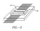
- a portion 27 of the capillaries is maintained in side-by-side parallel coplanar relationship by a holder 28, Figure 2.
- the holder 28 includes a window through which the beam can be focused on the interior volume of the capillaries.
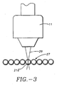
- Figure 3 shows the beam 29 focused in an interior volume of a capillary 21a.
- scanning the beam and detection system across the capillary is better than just probing in the center of the capillary.
- the probe laser is fixed at the center of the capillary, the sample stream will be rapidly photo-bleached by the laser. Scanning the beam laterally across the capillary interior is much better than sitting in one spot because all of the band is probed (laterally) and photo-bleaching is reduced.
- the off-axis probing is advantageous because, as shown in Figures 4A and 4B, the cylindrical lens effect actually brings the beam waist back into the capillary gel so the detection system probes the gel for a longer period of time during the scan than would have been nominally predicted from the capillary diameter and scan rate.
- the holder 28 is mounted on a translation stage 30 (Model 4000, Design Components, Franklin, MA).
- the stage is programmed to continuously scan the capillary array back and forth at 20 mm/sec in a direction perpendicular to the electrophoresis direction.
- the image acquired in this way has two dimensions. One is a spatial dimension representing the physical image of the capillaries. The other is a temporal dimension proportional to the elapsed time.
- fluorescence data from the photodetector is sampled at 2000 Hz so the nominal image resolution is 10 ⁇ m/pixel; thus, 10 pixels represent the interior 100 ⁇ m width of any given capillary.
- the electronic low-pass filter cutoff was set at 300 Hz to provide high frequency noise rejection while still passing the spatial frequencies required to define the 100 ⁇ m I.D. of the capillaries.
- An image of the migrating bands is built up as a function of time by accumulating periodic one-second sweeps of the illuminated region of the capillaries.
- the transit time of the migrating DNA past the probe region, under the conditions employed here ranges from approximately 10 seconds for the low molecular weight fragments (40-50 mers) to 14 seconds for the higher molecular weight fragments (380-390 mers). With one-second repeat cycles, this gives 10-14 samples of each band.
- the computer processes the data and displays the acquired image in real time.
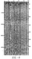
- Image processing can be performed with the NIH program, Image 1.29, and commercial image processing package, Canvas TM , to provide an image, Figure 5.
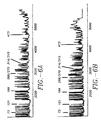
- the image data can be reduced to a one-dimensional line plot or electropherogram by averaging the pixels across the width of each lane using Image 1.29, Figure 6, and sections can be expanded as shown in Figure 7.
- zero-crosslinked polyacrylamide gel-filled capillaries were prepared using a modified method of the procedure described by Cohen, et al. 6,7 .
- a 3 mm wide detection window was produced in each 100 ⁇ m I.D. 200 ⁇ m O.D. fused-silica capillaries (Polymicro Technologies, Phoenix, AZ) by burning off the polyimide coating with a hot wire.
- the window was burned - 25 cm from the inlet side of the 40 cm long capillary.
- the inner wall of the capillaries was then treated overnight with a bifunctional reagent, ⁇ -methacryloxypropyltrimethoxy-silane to prepare the walls for acrylamide adhesion 6 .
- Freshly-made acrylamide gel solution (9%T, O%C) in a 1X TBE buffer (tris-boric acid-EDTA) with 7M urea was filtered with an 0.2 ⁇ m syringe filter and degassed under vacuum for about one hour.
- 10% TEMED (tetraethylmethylenediamine) and 10% APS (ammonium persulfate) solution were added to the gel solution at a final concentration of approximately 0.03%.
- the solution was immediately vacuum siphoned into the capillaries and then allowed to polymerize overnight in a cold room. Prior to use, both ends of the column were trimmed by about 1 cm and then pre-electrophoresed for 30 to 60 minutes at 7 kV.
- the capillary array was sandwiched in the capillary holder 28 that is mounted onto the translation stage 30.
- the capillary holder 28, Figure 2 serves the dual purpose of (1) uniformly constraining each capillary in the array to an identical height above the top of the translation stage, and (2) exposing a small window through which the confocal zone probed the capillary interior. Constraining the capillaries to substantially the same plane is necessary for achieving uniform detection sensitivity from each capillary because the depth of focus of the microscope objective is only - 25-50 ⁇ m.
- a combination of the labeling mixture and diluted enzyme (Sequenase 2.0TM) were added, and the mixture was incubated for five minutes at room temperature. This mixture was then transferred to the tube having the termination mixture and allowed to incubate for another five minutes at 37 °C. Instead of adding stop solution, ethanol precipitation was immediately used to terminate the reaction and recover the DNA sequencing sample.
- the high concentration of conductive ions present in the DNA sequencing sample after the termination step would reduce the amount of DNA that can be loaded into each capillary by electrokinetic injection.
- ethanol precipitation was performed on all DNA samples followed by resuspension in 6 ⁇ l of 80% (v/v) formamide to give a concentration about ten-fold higher than that used in slab gels.
- the sample was heated at 90°C for three minutes to ensure denaturation and then placed on ice until sample injection.
- the flexibility of the capillary columns allows coupling of the individual capillaries of the array to individual sample wells.
- the sample was placed in a single 500 ⁇ l centrifuge tube for electrokinetic injection into the capillaries.
- the same electric field strength (200 volt/cm) used during separation was also applied during sample injection.
- the typical injection time was ten seconds.
- the inlets of the capillaries were removed from the centrifuge tube and placed into a buffer reservoir or well 24 filled with fresh running buffer.
- the 9% T gels are sufficiently stable that four to five sequencing runs could be run on each capillary.
- Figure 5 presents an image obtained from on-line confocal scanning of a four-capillary array during electrophoresis of a mixture of DNA sequencing fragments.
- the horizontal direction is the physical dimension representing the geometric arrangement of the array while the vertical direction is temporal, representing the passage of fluorescent DNA fragments through the detection window.
- identical samples of "G" base DNA fragments were simultaneously, electrokinetically injected into each capillary.
- the overall elapsed data acquisition time is -80 minutes after passage of the primer.
- An expanded region of the image is included in Figure 5.
- the bands in all four lanes are well resolved and the resolution extends throughout the sequencing run with sufficient signal-to-noise to detect bands more than 500 bases beyond the primer. From Figure 5, one can clearly see that the cylindrical capillaries do not significantly distort the image.
- Figures 6 and 7 present line plots of the DNA signal integrated across the width of each capillary.
- a signal-to-noise ratio of approximately 20 is observed out to base 385 (-65 minutes) and bands are detected out to base 500 with the present experimental conditions.
- the number of theoretical plates is >1.9 x 10 6 (at base 385) over a 24 cm effective column length.
- the overall throughput of the system depends upon the total number of capillaries, N, that can be scanned.
- N The equation, N - vT/2D, defines how N depends on the scan speed (v), the scan repetition period (T), and the capillary outside diameter (D).
- v the scan speed
- T the scan repetition period
- D the capillary outside diameter
- one hundred 200 ⁇ m wide capillaries can be easily seen using a scan rate of four cm/sec and a one-sec scan repetition period.
- Increasing the array size would require (1) an increase in the scan speed; (2) the use of smaller O.D. capillaries; and (3) an increase in the scan repetition period which would reduce the temporal resolution of the electrophoretic separation. Since reliable systems have velocities up to ten cm/sec and capillaries with O.D.'s of 150 ⁇ m are commercially available, a limit of approximately 330 capillaries/array can be projected, assuming a one-second scan repetition period.
- the velocity shift of the DNA bands from lane-to-lane may preclude sequencing DNA with CAE using a single fluorophore and four different capillaries, one for each base.
- the present apparatus must be expanded to a multi-color detection system to sequence all four bases in a single capillary.
- Such four-color detection schemes have been developed for single capillaries 8 and for slab gels 19 .
- the basic idea is that one is separating four sets of DNA fragments which terminate with either a G, A, T or C. Each set is labeled with a different fluorescent tag by any of several procedures and then the fragment sets are pooled and separated on the same capillary. If the fluorescent tags emit in a sufficiently distinctive wavelength region, the four sets of fragments can be uniquely detected by using a four-color detection system.
- FIG. 9 A schematic diagram of a four-color confocal fluorescence capillary array scanner is shown in Figure 9.
- the scanner includes a laser source such as an argon laser which projects a beam 30 into the dichroic beamsplitter 31 which directs the beam to the objective 32.
- the objective collects the fluorescent energy from the focal volume and directs it through the beamsplitter.
- the output of the beam splitter is directed to a first beam splitter 33 which reflects energy at one wavelength, for example, 540 ⁇ m, and passes other, longer wavelengths.
- the next dichroic beamsplitter 34 which reflects energy at a second wavelength, for example, 560 ⁇ m, and passes other, longer wavelengths.
- a third beamsplitter 36 reflects energy at another wavelength, for example, 580 ⁇ m, and passes energy at 610 ⁇ m.
- the energy from each of the beamsplitters 33, 34, and 36 and the transmitted energy is applied to confocal, spatial and spectrial filters 37, 38, 39 to photomultipliers 41, 42, 43 and 44 which provide output signals that are processed and applied to computer 47 which generates an image for each of the wavelengths, for each of the capillaries.
- Each color image then records the passage of a particular labeled set of DNA sequencing fragments through the detection zone--one color for the A-fragments, a second for the G-fragments, a third for the T-fragments and a fourth for the C-fragments.
- capillary arrays comprising a plurality of capillary tubes have been shown and discussed that it is possible to form parallel capillary passages in a block of material by photoetching, micromachining, casting and other techniques used in the semiconductor industry.
- capillary array as used herein is meant to encompass all types of capillary passages arranged in an array.
- capillary arrays resolves the fundamental throughput problems that limit the utility of CE in, for example, DNA sequencing 29 .
- CAE provides an opportunity for the large-scale optimization of analytical separations. A large number of capillaries can be run in parallel each with a different buffer pH, buffer composition, or load to determine the best separation conditions. Commercially made capillary arrays could be constructed which plug into multi-well devices for large-scale parallel sample introduction.
- CAE should be a valuable new technique for rapid, parallel separation and analysis.
- the apparatus has been described in connection with capillary array electrophoresis. However, it is to be understood that it can be used in connection with other types of capillary separations, such as capillary chromatography, isoelectric focusing and column derivations.
Landscapes
- Physics & Mathematics (AREA)
- Health & Medical Sciences (AREA)
- Chemical & Material Sciences (AREA)
- General Physics & Mathematics (AREA)
- Analytical Chemistry (AREA)
- Life Sciences & Earth Sciences (AREA)
- Immunology (AREA)
- General Health & Medical Sciences (AREA)
- Pathology (AREA)
- Biochemistry (AREA)
- Nuclear Medicine, Radiotherapy & Molecular Imaging (AREA)
- Optics & Photonics (AREA)
- Chemical Kinetics & Catalysis (AREA)
- Molecular Biology (AREA)
- Electrochemistry (AREA)
- Ophthalmology & Optometry (AREA)
- Radiology & Medical Imaging (AREA)
- Surgery (AREA)
- Spectroscopy & Molecular Physics (AREA)
- Investigating, Analyzing Materials By Fluorescence Or Luminescence (AREA)
- Investigating Or Analysing Biological Materials (AREA)
- Investigating Or Analysing Materials By Optical Means (AREA)
Claims (7)
- Abtaster zum Erregen und Erfassen von Strahlung aus einer Vielzahl von benachbarten Kapillarkanälen, wobei der Abtaster- eine Vielzahl von transparenten Kapillaren (21), die in einer koplanaren Beziehung Seite-an-Seite stehen und zylindrische Wände haben, die Kapillarkanäle bilden, welche sich in einer ersten Richtung erstrecken,- eine Quelle für Strahlungsenergie mit einer ersten Wellenlänge,- eine Objektivlinse (11) für die Aufnahme und das Fokussieren der Strahlungsenergie (29) bei einem Erregungsvolumen (21a) in den Kanälen,- Einrichtungen (30), die zur Bildung einer kontinuierlichen Relativbewegung zwischen jedem der Kanäle und der Strahlungsenergie in einer Richtung senkrecht zu den Kapillaren angeordnet sind, so dass das Erregungsvolumen (21a) an einer spezifischen Position in jedem der Kapillarkanäle sequentiell ist und sich wiederholt, wodurch das Material in dem Erregungsvolumen (21 a) erregt und das Material dazu veranlasst wird, Energie bei einer unterschiedlichen Wellenlänge abzustrahlen,- wobei die Objektivlinse (11) dazu dient, die Strahlungsenergie zu sammeln und zu einem optischen System zu richten, das eine konfokale räumliche Filtereinrichtung (16) und eine spektrale Filtereinrichtung (17) zur Übertragung emittierter Strahlungsenergie bei der unterschiedlichen Wellenlänge und zur Zurückweisung von Strahlung bei anderen Wellenlängen- ein Erfassungssystem (18) zur Aufnahme der emittierten Strahlung und zur Erzeugung eines Signals sowie- Rechnereinrichtungen aufweist, die so angeordnet sind, dass sie das Signal empfangen und verarbeiten und ein Ausgangssignal bilden, welches das Material bei dem Erregungsvolumen (21a) in jedem der Kapillarkanäle (21) darstellt.
- Abtaster nach Anspruch 1, bei welchem die Einrichtungen (30), die zur Bildung einer kontinuierlichen Relativbewegung angeordnet sind, von den Rechnereinrichtungen gesteuert werden, wodurch das Ausgangssignal mit den Kapillarkanälen korreliert werden kann.
- Abtaster nach Anspruch 2, bei welchem jeder Kapillarkanal Teil einer langgestreckten zylindrischen Kapillaren ist.
- Abtaster nach Anspruch 3 mit Einrichtungen zum Halten eines Bereichs der Kapillaren (21) in einer koplanaren Beziehung Seite-an-Seite für ein Darbieten der fokussierten Strahlungsenergie.
- Abtaster nach Anspruch 2 oder 4, bei welchem die Enden (22, 23) der Kapillaren (21) für ein individuelles Handhaben und Beladen trennbar sind.
- Abtaster nach Anspruch 1, bei welchem die Erregungs-Strahlungsenergie Material erregt, das Strahlungsenergie bei einer Vielzahl von Wellenlängen emittiert, und das optische System eine Vielzahl von konfokalen räumlichen und spektralen Filtereinrichtungen (11, 12, 16, 17) zum selektiven Richten der mit unterschiedlichen Wellenlängen emittierten Strahlungsenergie zu unterschiedlichen Stellen sowie eine Vielzahl von Erfassungseinrichtungen aufweist, von denen jede emittierte Energie einer ausgewählten Wellenlänge empfängt und ein entsprechendes Ausgangssignal liefert.
- Verfahren zum Erfassen von Fluoreszenz aus DNA-Sequenzierfragmenten, die elektrophoretisch in Kapillarkanälen einer Vielzahl von zylindrischen Kapillaren (21) getrennt werden, wobei das Verfahren die Schritte aufweist- Positionieren eines Bereichs der Vielzahl von Kapillaren (21) in einer koplanaren Beziehung Seite an Seite,- Erregen eines Erregungsvolumens (21a) in den Kapillarkanälen mit Lichtenergie einer ersten Wellenlänge, die darin durch eine Objektivlinse (11) fokussiert wird, um Fragmente zum Fluoreszieren mit einer unterschiedlichen Wellenlänge zu veranlassen,- Bereitstellen einer relativen kontinuierlichen Bewegung zwischen den zylindrischen Kapillaren (21) und dem fokussierten Licht in einer Richtung senkrecht zu den Kapillaren, wodurch das Erregungsvolumen an einer spezifischen Position in jedem der Kapillarkanäle sequentiell ist und sich wiederholt,- Sammeln des fluoreszenzemittierten Lichts aus den vorgegebenen Volumina in jedem der Kapillarkanäle mit der Objektivlinse (11),- spektrales und konfokales räumliches Filtern der fluoreszenzemittierten Lichtenergie, um Licht bei der ersten Wellenlänge zurückzuweisen und das emittierte Licht bei der unterschiedlichen Wellenlänge durchzulassen,- Aufbringen des gefilterten emittierten Lichts auf einen Detektor (18) zur Erzeugung eines Ausgangssignals, das die Fluoreszenz aus den Fragmenten in jedem der Kapillarkanäle darstellt, und- Empfangen und Verarbeiten des Signals zur Bereitstellung eines Ausgangssignals, welches das Material bei dem Erregungsvolumen (21 a) in jedem der Kapillarkanäle (21) darstellt.
Priority Applications (1)
| Application Number | Priority Date | Filing Date | Title |
|---|---|---|---|
| EP06075115A EP1647822A1 (de) | 1992-02-24 | 1993-02-23 | Verfahren und Vorrichtung zur konfokalen fluoreszenzabtastung einer kapillarfilteranordnung |
Applications Claiming Priority (3)
| Application Number | Priority Date | Filing Date | Title |
|---|---|---|---|
| US840501 | 1992-02-24 | ||
| US07/840,501 US5274240A (en) | 1990-01-12 | 1992-02-24 | Capillary array confocal fluorescence scanner and method |
| PCT/US1993/001607 WO1993017325A1 (en) | 1992-02-24 | 1993-02-23 | Capillary array confocal fluorescence scanner and method |
Related Child Applications (1)
| Application Number | Title | Priority Date | Filing Date |
|---|---|---|---|
| EP06075115A Division EP1647822A1 (de) | 1992-02-24 | 1993-02-23 | Verfahren und Vorrichtung zur konfokalen fluoreszenzabtastung einer kapillarfilteranordnung |
Publications (3)
| Publication Number | Publication Date |
|---|---|
| EP0628164A4 EP0628164A4 (de) | 1994-10-11 |
| EP0628164A1 EP0628164A1 (de) | 1994-12-14 |
| EP0628164B1 true EP0628164B1 (de) | 2006-11-22 |
Family
ID=25282540
Family Applications (2)
| Application Number | Title | Priority Date | Filing Date |
|---|---|---|---|
| EP93906997A Expired - Lifetime EP0628164B1 (de) | 1992-02-24 | 1993-02-23 | Verfahren und vorrichtung zur konfokalen fluoreszenzabtastung einer kapillarfilteranordnung |
| EP06075115A Withdrawn EP1647822A1 (de) | 1992-02-24 | 1993-02-23 | Verfahren und Vorrichtung zur konfokalen fluoreszenzabtastung einer kapillarfilteranordnung |
Family Applications After (1)
| Application Number | Title | Priority Date | Filing Date |
|---|---|---|---|
| EP06075115A Withdrawn EP1647822A1 (de) | 1992-02-24 | 1993-02-23 | Verfahren und Vorrichtung zur konfokalen fluoreszenzabtastung einer kapillarfilteranordnung |
Country Status (5)
| Country | Link |
|---|---|
| US (1) | US5274240A (de) |
| EP (2) | EP0628164B1 (de) |
| JP (1) | JP2539172B2 (de) |
| DE (1) | DE69334085T2 (de) |
| WO (1) | WO1993017325A1 (de) |
Families Citing this family (199)
| Publication number | Priority date | Publication date | Assignee | Title |
|---|---|---|---|---|
| US5529679A (en) | 1992-02-28 | 1996-06-25 | Hitachi, Ltd. | DNA detector and DNA detection method |
| US6156177A (en) * | 1991-02-28 | 2000-12-05 | Hitachi, Ltd. | DNA detector and DNA detection method |
| US5324401A (en) * | 1993-02-05 | 1994-06-28 | Iowa State University Research Foundation, Inc. | Multiplexed fluorescence detector system for capillary electrophoresis |
| US5547849A (en) * | 1993-02-17 | 1996-08-20 | Biometric Imaging, Inc. | Apparatus and method for volumetric capillary cytometry |
| JP3563140B2 (ja) * | 1995-01-19 | 2004-09-08 | 株式会社日立製作所 | キャピラリーアレイ電気泳動装置 |
| US5381224A (en) * | 1993-08-30 | 1995-01-10 | A. E. Dixon | Scanning laser imaging system |
| US5491344A (en) * | 1993-12-01 | 1996-02-13 | Tufts University | Method and system for examining the composition of a fluid or solid sample using fluorescence and/or absorption spectroscopy |
| US5459325A (en) * | 1994-07-19 | 1995-10-17 | Molecular Dynamics, Inc. | High-speed fluorescence scanner |
| US6001229A (en) * | 1994-08-01 | 1999-12-14 | Lockheed Martin Energy Systems, Inc. | Apparatus and method for performing microfluidic manipulations for chemical analysis |
| AU702267B2 (en) * | 1994-09-02 | 1999-02-18 | Biometric Imaging, Inc. | Calibration method and apparatus for optical scanner |
| DE4438833A1 (de) * | 1994-10-31 | 1996-05-02 | Bayer Ag | Verfahren zur analytischen Trennung von Viren |
| US5483075A (en) * | 1994-11-01 | 1996-01-09 | Perkin-Elmer Corporation | Rotary scanning apparatus |
| US6014213A (en) * | 1994-12-12 | 2000-01-11 | Visible Genetics Inc. | High dynamic range apparatus for separation and detection of polynucleotide fragments |
| US5710628A (en) * | 1994-12-12 | 1998-01-20 | Visible Genetics Inc. | Automated electrophoresis and fluorescence detection apparatus and method |
| US5560811A (en) * | 1995-03-21 | 1996-10-01 | Seurat Analytical Systems Incorporated | Capillary electrophoresis apparatus and method |
| US5900132A (en) * | 1995-03-23 | 1999-05-04 | Beckman Coulter, Inc. | Capillary holder |
| US5675155A (en) * | 1995-04-26 | 1997-10-07 | Beckman Instruments, Inc. | Multicapillary fluorescent detection system |
| US5582705A (en) * | 1995-05-19 | 1996-12-10 | Iowa State University Research Foundation, Inc. | Multiplexed capillary electrophoresis system |
| US5713364A (en) * | 1995-08-01 | 1998-02-03 | Medispectra, Inc. | Spectral volume microprobe analysis of materials |
| US5813987A (en) * | 1995-08-01 | 1998-09-29 | Medispectra, Inc. | Spectral volume microprobe for analysis of materials |
| US6104945A (en) * | 1995-08-01 | 2000-08-15 | Medispectra, Inc. | Spectral volume microprobe arrays |
| US5545901A (en) * | 1995-08-23 | 1996-08-13 | Beckman Instruments, Inc. | Automated optical alignment using a galvometric scanner |
| US5759779A (en) * | 1995-08-29 | 1998-06-02 | Dehlinger; Peter J. | Polynucleotide-array assay and methods |
| DE29514084U1 (de) * | 1995-09-01 | 1995-11-02 | Biosafe Diagnostics Corp., Lincolnshire, Ill. | Blutentnahme- und -testvorrichtung |
| USD378233S (en) * | 1995-11-08 | 1997-02-25 | Biosafe Diagnostics Corporation | Stylet holder |
| US5763263A (en) * | 1995-11-27 | 1998-06-09 | Dehlinger; Peter J. | Method and apparatus for producing position addressable combinatorial libraries |
| US6794127B1 (en) * | 1997-06-16 | 2004-09-21 | Diversa Corporation | Capillary array-based sample screening |
| US20020048809A1 (en) * | 1997-06-16 | 2002-04-25 | Lafferty William Micharl | Capillary array-based sample screening |
| US6972183B1 (en) | 1997-06-16 | 2005-12-06 | Diversa Corporation | Capillary array-based enzyme screening |
| US5748491A (en) * | 1995-12-20 | 1998-05-05 | The Perkin-Elmer Corporation | Deconvolution method for the analysis of data resulting from analytical separation processes |
| US5567294A (en) * | 1996-01-30 | 1996-10-22 | Board Of Governors, University Of Alberta | Multiple capillary biochemical analyzer with barrier member |
| US5885430A (en) * | 1996-10-04 | 1999-03-23 | Spectrumedix Corporation | Capillary tube holder for an electrophoretic apparatus |
| US6063251A (en) * | 1997-05-30 | 2000-05-16 | Spectrumedix Corporation | Electrically insulated capillary arrays for electrophoretic applications |
| US6066245A (en) * | 1996-12-27 | 2000-05-23 | Genetic Biosystems, Inc. | Method and apparatus for scanning fluorescently labeled particles |
| US6826422B1 (en) | 1997-01-13 | 2004-11-30 | Medispectra, Inc. | Spectral volume microprobe arrays |
| US6847490B1 (en) | 1997-01-13 | 2005-01-25 | Medispectra, Inc. | Optical probe accessory device for use in vivo diagnostic procedures |
| EP0854362A3 (de) * | 1997-01-16 | 2000-12-20 | Japan Science and Technology Corporation | Mehrkapillarelektroforesevorrichtung |
| EP2264427B1 (de) | 1997-01-31 | 2017-05-03 | Xy, Llc | Optisches Gerät mit fokussierendem Reflektor zur Konvergenz der Strahlung auf einen Partikelfluss, und zugehöriges Analyseverfahren |
| US6403311B1 (en) | 1997-02-12 | 2002-06-11 | Us Genomics | Methods of analyzing polymers using ordered label strategies |
| WO1998035012A2 (en) | 1997-02-12 | 1998-08-13 | Chan Eugene Y | Methods and products for analyzing polymers |
| JPH10227740A (ja) * | 1997-02-18 | 1998-08-25 | Hitachi Ltd | 多色蛍光検出電気泳動分析装置 |
| US6914250B2 (en) * | 1997-03-07 | 2005-07-05 | Clare Chemical Research, Inc. | Fluorometric detection using visible light |
| DE69837839T2 (de) | 1997-03-07 | 2007-12-13 | Clare Chemical Research LLC, Denver | Fluorometrischer Nachweis mit sichtbarem Light |
| EP0864860A1 (de) * | 1997-03-10 | 1998-09-16 | Japan Science and Technology Corporation | Probenplatte und Vielfachkapillarelektrophoresegerät |
| US6084667A (en) * | 1997-03-12 | 2000-07-04 | Nz Applied Technologies | System and method for molecular sample measurement |
| US6445448B1 (en) | 1997-03-12 | 2002-09-03 | Corning Applied Technologies, Corp. | System and method for molecular sample measurement |
| US5903348A (en) * | 1997-03-12 | 1999-05-11 | Nz Applied Technologies, Inc. | System and method for molecular sample measurements |
| US5876946A (en) * | 1997-06-03 | 1999-03-02 | Pharmacopeia, Inc. | High-throughput assay |
| US20030054543A1 (en) * | 1997-06-16 | 2003-03-20 | Lafferty William Michael | Device for moving a selected station of a holding plate to a predetermined location for interaction with a probe |
| US20020015997A1 (en) * | 1997-06-16 | 2002-02-07 | Lafferty William Michael | Capillary array-based sample screening |
| US20030013115A1 (en) * | 1997-06-16 | 2003-01-16 | Diversa Corporation, A Delaware Corporation | Capillary array-based sample screening |
| US20040241759A1 (en) * | 1997-06-16 | 2004-12-02 | Eileen Tozer | High throughput screening of libraries |
| US20050070005A1 (en) * | 1997-06-16 | 2005-03-31 | Martin Keller | High throughput or capillary-based screening for a bioactivity or biomolecule |
| US20020102598A1 (en) * | 1997-06-16 | 2002-08-01 | Lafferty William Michael | Positioning system for moving a selected station of a holding plate to a predetermined location for interaction with a probe |
| US6365024B1 (en) | 1997-06-30 | 2002-04-02 | Spectrumedix Corporation | Motorized positioning apparatus having coaxial carrousels |
| US6027627A (en) * | 1997-06-30 | 2000-02-22 | Spectrumedix Corporation | Automated parallel capillary electrophoretic system |
| JP3481828B2 (ja) * | 1997-08-26 | 2003-12-22 | 株式会社日立製作所 | 電気泳動分析装置,電気泳動分析方法及びそれに用いる試料容器 |
| GB9719673D0 (en) | 1997-09-17 | 1997-11-19 | Glaxo Group Ltd | Novel apparatus |
| US6008379A (en) | 1997-10-01 | 1999-12-28 | The Perkin-Elmer Corporation | Aromatic-substituted xanthene dyes |
| US6583168B1 (en) | 1997-11-25 | 2003-06-24 | Applera Corporation | Sulfonated diarylrhodamine dyes |
| US5936087A (en) | 1997-11-25 | 1999-08-10 | The Perkin-Elmer Corporation | Dibenzorhodamine dyes |
| US6432637B1 (en) * | 1997-12-15 | 2002-08-13 | Joseph R. Lakowicz | Method for determining a base sequence of a nucleotide strand |
| US6071689A (en) | 1997-12-31 | 2000-06-06 | Xy, Inc. | System for improving yield of sexed embryos in mammals |
| US6149867A (en) | 1997-12-31 | 2000-11-21 | Xy, Inc. | Sheath fluids and collection systems for sex-specific cytometer sorting of sperm |
| US6201639B1 (en) | 1998-03-20 | 2001-03-13 | James W. Overbeck | Wide field of view and high speed scanning microscopy |
| US6100535A (en) * | 1998-01-29 | 2000-08-08 | The Regents Of The University Of California | Rotary confocal scanner for detection of capillary arrays |
| US6560859B1 (en) | 1998-02-16 | 2003-05-13 | The Institute Of Physical And Chemical Research Shimadzu Corporation | Capillary cassette and method of producing the same |
| US6103083A (en) * | 1998-02-20 | 2000-08-15 | Tetragen | Capillary electrophoresis apparatus and method |
| US6475361B1 (en) | 1998-02-20 | 2002-11-05 | Tetragen Sa | Capillary electrophoresis apparatus having filling/refilling system and methods for use thereof |
| US20030036855A1 (en) * | 1998-03-16 | 2003-02-20 | Praelux Incorporated, A Corporation Of New Jersey | Method and apparatus for screening chemical compounds |
| KR100560588B1 (ko) * | 1998-03-16 | 2006-03-16 | 에머샘 바이오사이언시즈 코프 | 공초점 마이크로스코피 영상 시스템 |
| US6185030B1 (en) | 1998-03-20 | 2001-02-06 | James W. Overbeck | Wide field of view and high speed scanning microscopy |
| US6472671B1 (en) * | 2000-02-09 | 2002-10-29 | Jean I. Montagu | Quantified fluorescence microscopy |
| US6096875A (en) | 1998-05-29 | 2000-08-01 | The Perlein-Elmer Corporation | Nucleotide compounds including a rigid linker |
| US6342397B1 (en) * | 1998-06-04 | 2002-01-29 | Erkki Soini | Homogeneous biospecific assay using a solid phase, two-photon excitation and confocal fluorescence detection |
| US6627446B1 (en) | 1998-07-02 | 2003-09-30 | Amersham Biosciences (Sv) Corp | Robotic microchannel bioanalytical instrument |
| US6787111B2 (en) | 1998-07-02 | 2004-09-07 | Amersham Biosciences (Sv) Corp. | Apparatus and method for filling and cleaning channels and inlet ports in microchips used for biological analysis |
| SE9802558D0 (sv) | 1998-07-16 | 1998-07-16 | Hanning Instr Ab | Device for detection of fluorescent |
| WO2000006996A1 (en) * | 1998-07-28 | 2000-02-10 | Ce Resources Pte Ltd. | Optical detection system |
| US6225635B1 (en) * | 1998-08-07 | 2001-05-01 | The Regents Of The University Of California | System and method for optically locating microchannel positions |
| US6210896B1 (en) | 1998-08-13 | 2001-04-03 | Us Genomics | Molecular motors |
| US6245507B1 (en) | 1998-08-18 | 2001-06-12 | Orchid Biosciences, Inc. | In-line complete hyperspectral fluorescent imaging of nucleic acid molecules |
| US6162341A (en) * | 1998-09-11 | 2000-12-19 | The Perkin-Elmer Corporation | Multi-channel capillary electrophoresis device including sheath-flow cuvette and replacable capillary array |
| US6132582A (en) * | 1998-09-14 | 2000-10-17 | The Perkin-Elmer Corporation | Sample handling system for a multi-channel capillary electrophoresis device |
| US6103199A (en) * | 1998-09-15 | 2000-08-15 | Aclara Biosciences, Inc. | Capillary electroflow apparatus and method |
| KR20010110408A (ko) | 1998-11-25 | 2001-12-13 | 추후제출 | 단일 헤드를 갖는 형광 스크린 스캐닝 장치 |
| WO2000036973A1 (en) | 1998-12-23 | 2000-06-29 | Medispectra, Inc. | Optical methods and systems for cervical screening |
| JP2002533142A (ja) | 1998-12-23 | 2002-10-08 | メディスペクトラ, インコーポレイテッド | サンプルの光学的試験のためのシステムおよび方法 |
| US6246046B1 (en) | 1999-01-21 | 2001-06-12 | University Of Pittsburgh | Method and apparatus for electronically controlled scanning of micro-area devices |
| US6270644B1 (en) | 1999-01-27 | 2001-08-07 | Affymetrix, Inc. | Capillary array electrophoresis scanner |
| US6937330B2 (en) | 1999-04-23 | 2005-08-30 | Ppd Biomarker Discovery Sciences, Llc | Disposable optical cuvette cartridge with low fluorescence material |
| US20030235832A1 (en) * | 2000-06-21 | 2003-12-25 | Ahmed Chenna | Multiplexed analysis by chromatographic separation of molecular tags |
| JP2001021558A (ja) * | 1999-04-30 | 2001-01-26 | Agilent Technol Inc | ポリヌクレオチドアレイの製作法 |
| EP1983331B1 (de) * | 1999-05-28 | 2011-07-13 | Yokogawa Electric Corporation | Optische Anordnung zur Auswertung eines Biochips |
| RU2145078C1 (ru) * | 1999-07-13 | 2000-01-27 | Общество с ограниченной ответственностью Научно-производственная фирма "АТГ-Биотех" | Многоканальный капиллярный генетический анализатор |
| US6687395B1 (en) | 1999-07-21 | 2004-02-03 | Surromed, Inc. | System for microvolume laser scanning cytometry |
| US6696022B1 (en) | 1999-08-13 | 2004-02-24 | U.S. Genomics, Inc. | Methods and apparatuses for stretching polymers |
| US6352633B1 (en) * | 1999-08-31 | 2002-03-05 | Spectrumedix Corporation | Automated parallel capillary electrophoresis system with hydrodynamic sample injection |
| CA2360750A1 (en) * | 1999-11-22 | 2001-05-31 | Diversa Corporation | Capillary array-based sample screening |
| US7208265B1 (en) | 1999-11-24 | 2007-04-24 | Xy, Inc. | Method of cryopreserving selected sperm cells |
| US7187810B2 (en) | 1999-12-15 | 2007-03-06 | Medispectra, Inc. | Methods and systems for correcting image misalignment |
| US20020007122A1 (en) | 1999-12-15 | 2002-01-17 | Howard Kaufman | Methods of diagnosing disease |
| US7260248B2 (en) | 1999-12-15 | 2007-08-21 | Medispectra, Inc. | Image processing using measures of similarity |
| US20030138774A1 (en) * | 2000-01-19 | 2003-07-24 | Jones Chris L. | Methods and apparatus for separating and detecting nucleic acid |
| US6587579B1 (en) | 2000-01-26 | 2003-07-01 | Agilent Technologies Inc. | Feature quality in array fabrication |
| US6358387B1 (en) | 2000-03-27 | 2002-03-19 | Caliper Technologies Corporation | Ultra high throughput microfluidic analytical systems and methods |
| JP4103302B2 (ja) * | 2000-05-15 | 2008-06-18 | 株式会社日立製作所 | キャピラリアレイを用いた電気泳動装置及びそれに用いられるサンプルプレートアセンブリ |
| US6805784B2 (en) | 2000-05-15 | 2004-10-19 | Hitachi, Ltd. | Capillary array |
| JP3918403B2 (ja) * | 2000-05-15 | 2007-05-23 | 株式会社日立製作所 | キャピラリアレイ |
| JP4003374B2 (ja) * | 2000-05-15 | 2007-11-07 | 株式会社日立製作所 | キャピラリアレイ電気泳動装置及び試料の分離・分析方法 |
| US20040224421A1 (en) * | 2000-06-15 | 2004-11-11 | Deweerd Herman | Bi-directional scanning method |
| US6531041B1 (en) | 2000-07-20 | 2003-03-11 | Symyx Technologies, Inc. | Multiplexed capillary electrophoresis system with rotatable photodetector |
| US6544396B1 (en) | 2000-07-20 | 2003-04-08 | Symyx Technologies, Inc. | Multiplexed capillary electrophoresis system |
| US6572750B1 (en) * | 2000-07-21 | 2003-06-03 | Symyx Technologies, Inc. | Hydrodynamic injector |
| US6462816B1 (en) | 2000-07-21 | 2002-10-08 | Symyx Technologies, Inc. | Parallel capillary electrophoresis system having signal averaging and noise cancellation |
| US6789040B2 (en) | 2000-08-22 | 2004-09-07 | Affymetrix, Inc. | System, method, and computer software product for specifying a scanning area of a substrate |
| EP1317464B1 (de) * | 2000-10-11 | 2009-05-13 | Applera Corporation | Fluoreszierende nukleobasekonjugate mit anionische linker |
| US6787761B2 (en) | 2000-11-27 | 2004-09-07 | Surromed, Inc. | Median filter for liquid chromatography-mass spectrometry data |
| US7713687B2 (en) | 2000-11-29 | 2010-05-11 | Xy, Inc. | System to separate frozen-thawed spermatozoa into x-chromosome bearing and y-chromosome bearing populations |
| WO2002043486A1 (en) | 2000-11-29 | 2002-06-06 | Xy, Inc. | System for in-vitro fertilization with spermatozoa separated into x-chromosome and y-chromosome bearing populations |
| AU2002227156A1 (en) | 2000-12-01 | 2002-06-11 | Visigen Biotechnologies, Inc. | Enzymatic nucleic acid synthesis: compositions and methods for altering monomer incorporation fidelity |
| US6839661B2 (en) | 2000-12-15 | 2005-01-04 | Medispectra, Inc. | System for normalizing spectra |
| US6800438B2 (en) * | 2000-12-28 | 2004-10-05 | Xerox Corporation | Imager for DNA sequencer |
| US20030132117A1 (en) * | 2002-01-14 | 2003-07-17 | Andras Guttman | Thin film electrophoresis apparatus and method |
| US20040266021A9 (en) * | 2001-01-26 | 2004-12-30 | Andras Guttman | Multicapillary fraction collection system and method |
| US6797139B2 (en) | 2001-03-19 | 2004-09-28 | Applera Corporation | Detection cell for guiding excitation light therein and method for using same |
| US6839564B2 (en) * | 2001-04-25 | 2005-01-04 | Nokia Corporation | Synchronization of database data |
| US6490533B2 (en) * | 2001-04-26 | 2002-12-03 | Affymetrix, Inc. | System, method, and product for dynamic noise reduction in scanning of biological materials |
| US6943036B2 (en) | 2001-04-30 | 2005-09-13 | Agilent Technologies, Inc. | Error detection in chemical array fabrication |
| CA2445053A1 (en) | 2001-05-21 | 2002-11-28 | Aclara Biosciences, Inc. | Methods and compositions for analyzing proteins |
| KR100442412B1 (ko) * | 2001-07-20 | 2004-07-30 | 학교법인 포항공과대학교 | 모세관으로 연결된 랩온어칩 시스템 |
| US20030110840A1 (en) * | 2001-07-24 | 2003-06-19 | Arriaga Edgar A. | Systems and methods for detecting a particle |
| US6873915B2 (en) | 2001-08-24 | 2005-03-29 | Surromed, Inc. | Peak selection in multidimensional data |
| US6750457B2 (en) * | 2001-08-29 | 2004-06-15 | Becton Dickinson And Company | System for high throughput analysis |
| US20030181826A1 (en) * | 2001-12-04 | 2003-09-25 | Dave Smith | Hydrophobic/hydrophilic sample collection tip |
| US7189361B2 (en) * | 2001-12-19 | 2007-03-13 | 3M Innovative Properties Company | Analytical device with lightguide Illumination of capillary and microgrooves arrays |
| WO2003087773A2 (en) * | 2002-04-12 | 2003-10-23 | Amersham Biosciences (Sv) Corp | Multiplexed capillary electrophoresis systems |
| US6989100B2 (en) | 2002-05-09 | 2006-01-24 | Ppd Biomarker Discovery Sciences, Llc | Methods for time-alignment of liquid chromatography-mass spectrometry data |
| US20030208936A1 (en) * | 2002-05-09 | 2003-11-13 | Lee Charles Hee | Method for manufacturing embroidery decorated cards and envelopes |
| US6818903B2 (en) * | 2002-07-09 | 2004-11-16 | Medispectra, Inc. | Method and apparatus for identifying spectral artifacts |
| US7136518B2 (en) | 2003-04-18 | 2006-11-14 | Medispectra, Inc. | Methods and apparatus for displaying diagnostic data |
| US7309867B2 (en) | 2003-04-18 | 2007-12-18 | Medispectra, Inc. | Methods and apparatus for characterization of tissue samples |
| US6933154B2 (en) | 2002-07-09 | 2005-08-23 | Medispectra, Inc. | Optimal windows for obtaining optical data for characterization of tissue samples |
| US7282723B2 (en) | 2002-07-09 | 2007-10-16 | Medispectra, Inc. | Methods and apparatus for processing spectral data for use in tissue characterization |
| US7469160B2 (en) | 2003-04-18 | 2008-12-23 | Banks Perry S | Methods and apparatus for evaluating image focus |
| US7459696B2 (en) | 2003-04-18 | 2008-12-02 | Schomacker Kevin T | Methods and apparatus for calibrating spectral data |
| US7103401B2 (en) | 2002-07-10 | 2006-09-05 | Medispectra, Inc. | Colonic polyp discrimination by tissue fluorescence and fiberoptic probe |
| US6768918B2 (en) | 2002-07-10 | 2004-07-27 | Medispectra, Inc. | Fluorescent fiberoptic probe for tissue health discrimination and method of use thereof |
| US8486618B2 (en) | 2002-08-01 | 2013-07-16 | Xy, Llc | Heterogeneous inseminate system |
| JP4595067B2 (ja) | 2002-08-01 | 2010-12-08 | エックスワイ,エルエルシー | 低圧精子細胞分離システム |
| CA2534394C (en) | 2002-08-15 | 2013-01-08 | Xy, Inc. | High resolution flow cytometer |
| US7169548B2 (en) | 2002-09-13 | 2007-01-30 | Xy, Inc. | Sperm cell processing and preservation systems |
| US7108775B2 (en) | 2002-11-08 | 2006-09-19 | Applera Corporation | Apparatus and method for confining eluted samples in electrophoresis systems |
| US6970240B2 (en) | 2003-03-10 | 2005-11-29 | Applera Corporation | Combination reader |
| BRPI0408857B1 (pt) | 2003-03-28 | 2018-09-11 | Inguran Llc | aparelho, métodos e processos para separar partículas e para prover esperma de animal separado por sexo |
| EP1620203A2 (de) * | 2003-04-10 | 2006-02-01 | U.S. Genomics, Inc. | Handhabung von polymeren in mikrokanälen |
| US20050012057A1 (en) * | 2003-05-08 | 2005-01-20 | Alara, Inc. | Method and apparatus for radiation image erasure |
| ES2541121T3 (es) | 2003-05-15 | 2015-07-16 | Xy, Llc | Clasificación eficiente de células haploides por sistemas de citometría de flujo |
| US7402398B2 (en) | 2003-07-17 | 2008-07-22 | Monogram Biosciences, Inc. | Measuring receptor homodimerization |
| EP1673399B1 (de) | 2003-08-11 | 2017-04-05 | Monogram BioSciences, Inc. | Nachweis und erstellung des profils eines molekularen komplexes |
| US7407630B2 (en) | 2003-09-19 | 2008-08-05 | Applera Corporation | High density plate filler |
| US8277760B2 (en) | 2003-09-19 | 2012-10-02 | Applied Biosystems, Llc | High density plate filler |
| US7570443B2 (en) * | 2003-09-19 | 2009-08-04 | Applied Biosystems, Llc | Optical camera alignment |
| US7998435B2 (en) | 2003-09-19 | 2011-08-16 | Life Technologies Corporation | High density plate filler |
| US20050115837A1 (en) * | 2003-12-01 | 2005-06-02 | Dean Burgi | Analyte identification in transformed electropherograms |
| US7177023B2 (en) * | 2004-03-19 | 2007-02-13 | Applera Corporation | Fluorescent light detection |
| DK2801363T3 (en) | 2004-03-29 | 2018-05-28 | Inguran Llc | PROCEDURE FOR STORING SORTED SPERMATOZOES |
| US7248360B2 (en) | 2004-04-02 | 2007-07-24 | Ppd Biomarker Discovery Sciences, Llc | Polychronic laser scanning system and method of use |
| CA2574499C (en) | 2004-07-22 | 2016-11-29 | Monsanto Technology Llc | Process for enriching a population of sperm cells |
| US8685711B2 (en) * | 2004-09-28 | 2014-04-01 | Singulex, Inc. | Methods and compositions for highly sensitive detection of molecules |
| JP2008514955A (ja) * | 2004-09-28 | 2008-05-08 | シンギュレックス・インコーポレイテッド | サンプル分析システムおよび方法 |
| US9040305B2 (en) | 2004-09-28 | 2015-05-26 | Singulex, Inc. | Method of analysis for determining a specific protein in blood samples using fluorescence spectrometry |
| DE102004058565B4 (de) | 2004-10-18 | 2022-04-21 | Leica Microsystems Cms Gmbh | Scanmikroskop |
| JP2008535518A (ja) | 2005-04-14 | 2008-09-04 | アプレラ コーポレイション | プソイドイソシトシン核酸塩基誘導体を含む3’改変オリゴヌクレオチド、およびプライマーまたはプローブとしてのその適用 |
| US8351026B2 (en) | 2005-04-22 | 2013-01-08 | Affymetrix, Inc. | Methods and devices for reading microarrays |
| US20070042406A1 (en) * | 2005-07-18 | 2007-02-22 | U.S. Genomics, Inc. | Diffusion mediated clean-up of a target carrier fluid |
| US20070128083A1 (en) * | 2005-07-18 | 2007-06-07 | U.S. Genomics, Inc. | Microfluidic methods and apparatuses for sample preparation and analysis |
| EP1904653A4 (de) * | 2005-07-18 | 2010-04-14 | Us Genomics Inc | Mikrofluidische verfahren und vorrichtungen zur probenvorbereitung und -analyse |
| JP5308328B2 (ja) | 2006-04-04 | 2013-10-09 | シングレックス,インコーポレイテッド | トロポニンの分析のための高感度のシステムおよび方法 |
| EP3156799B1 (de) | 2006-04-04 | 2024-01-24 | Novilux, LLC | Analysator und verfahren zur hochempfindlichen detektion von analyten |
| US8009889B2 (en) | 2006-06-27 | 2011-08-30 | Affymetrix, Inc. | Feature intensity reconstruction of biological probe array |
| US7518727B2 (en) * | 2007-02-28 | 2009-04-14 | Beckman Coulter, Inc. | Multicapillary multilaser detection system |
| US20090087860A1 (en) * | 2007-08-24 | 2009-04-02 | Todd John A | Highly sensitive system and methods for analysis of prostate specific antigen (psa) |
| JP2011501189A (ja) * | 2007-10-25 | 2011-01-06 | ザ・リサーチ・ファウンデーション・オブ・ステイト・ユニバーシティー・オブ・ニューヨーク | 単一光子分光計 |
| JP4509166B2 (ja) | 2007-11-02 | 2010-07-21 | ソニー株式会社 | 微小粒子の測定方法、及び測定装置 |
| US7914734B2 (en) | 2007-12-19 | 2011-03-29 | Singulex, Inc. | Scanning analyzer for single molecule detection and methods of use |
| CA2716522A1 (en) * | 2008-03-05 | 2009-10-15 | Singulex, Inc. | Methods and compositions for highly sensitive detection of molecules |
| GB2464183A (en) * | 2008-09-19 | 2010-04-14 | Singulex Inc | Sandwich assay |
| US8374818B2 (en) * | 2008-12-19 | 2013-02-12 | Affymetrix, Inc. | System, method and apparatus for calibrating inspection tools |
| DE112010002222B4 (de) | 2009-06-04 | 2024-01-25 | Leidos Innovations Technology, Inc. (n.d.Ges.d. Staates Delaware) | Mehr-Proben-Mikrofluidchip fur DNA-Analyse |
| WO2010144358A1 (en) | 2009-06-08 | 2010-12-16 | Singulex, Inc. | Highly sensitive biomarker panels |
| AU2011249908A1 (en) | 2010-05-06 | 2012-11-22 | Singulex, Inc. | Methods for diagnosing, staging, predicting risk for developing and identifying treatment responders for rheumatoid arthritis |
| GB2497501A (en) | 2010-10-15 | 2013-06-12 | Lockheed Corp | Micro fluidic optic design |
| US9322054B2 (en) | 2012-02-22 | 2016-04-26 | Lockheed Martin Corporation | Microfluidic cartridge |
| US9028776B2 (en) | 2012-04-18 | 2015-05-12 | Toxic Report Llc | Device for stretching a polymer in a fluid sample |
| DE102012108158B4 (de) | 2012-09-03 | 2016-03-17 | Johann Wolfgang Goethe-Universität | Kapillarzelle, Anordnung und Verfahren zur Aufnahme, zur Positionierung und zur Untersuchung einer mikroskopischen Probe |
| LT3150988T (lt) | 2015-10-01 | 2021-06-25 | Nanotemper Technologies Gmbh | Dalelių stabilumo ir agregacijos optinio matavimo sistema bei būdas |
| WO2017123970A1 (en) * | 2016-01-13 | 2017-07-20 | ProteinSimple | Systems and methods for capillary electrophoresis, isoelectric point, and molecular weight analysis |
| US12163918B1 (en) | 2018-08-23 | 2024-12-10 | ProteinSimple | Compositions and methods for protein electrophoresis |
Family Cites Families (22)
| Publication number | Priority date | Publication date | Assignee | Title |
|---|---|---|---|---|
| GB405480A (en) * | 1933-03-31 | 1934-02-08 | Dardelet Threadlock Corp | Improvements in or relating to machines for shaping work pieces by grinding, abrading or cutting |
| US3755132A (en) * | 1971-11-08 | 1973-08-28 | Univ California | Fluid belt electrophoresis apparatus |
| US4162405A (en) * | 1978-05-23 | 1979-07-24 | Britton Chance | Flying spot fluoro-meter for oxidized flavoprotein and reduced pyridine nucleotide |
| US4354114A (en) * | 1979-10-09 | 1982-10-12 | Karnaukhov Valery N | Apparatus for investigation of fluorescence characteristics of microscopic objects |
| US4424127A (en) * | 1980-03-07 | 1984-01-03 | Johan Roeraade | Column for liquid and gas chromatography |
| JPS5917136A (ja) * | 1982-07-20 | 1984-01-28 | Olympus Optical Co Ltd | 電気泳動用測光装置 |
| AT390840B (de) * | 1983-06-08 | 1990-07-10 | Avl Verbrennungskraft Messtech | Verfahren zur fluorimetrischen bestimmung der konzentration von in einer substanz enthaltenen stoffen und anordnung zur durchfuehrung dieses verfahrens |
| CA1258611A (en) * | 1984-01-16 | 1989-08-22 | Lloyd M. Smith | Method of dna sequencing |
| US4729947A (en) * | 1984-03-29 | 1988-03-08 | The Board Of Regents Of The University Of Nebraska | DNA sequencing |
| DE3610165A1 (de) * | 1985-03-27 | 1986-10-02 | Olympus Optical Co., Ltd., Tokio/Tokyo | Optisches abtastmikroskop |
| GB8513538D0 (en) * | 1985-05-29 | 1985-07-03 | Mackay C D | Electrophoresis |
| DE3620235A1 (de) * | 1985-06-17 | 1986-12-18 | EG & G., Inc., Wellesley, Mass. | System zur bestimmung der groessen von biologischen makromolekuelen |
| US4855225A (en) * | 1986-02-07 | 1989-08-08 | Applied Biosystems, Inc. | Method of detecting electrophoretically separated oligonucleotides |
| US4781464A (en) * | 1986-04-14 | 1988-11-01 | Isco, Inc. | Gel scanner |
| US4811218A (en) * | 1986-06-02 | 1989-03-07 | Applied Biosystems, Inc. | Real time scanning electrophoresis apparatus for DNA sequencing |
| US4791310A (en) * | 1986-10-02 | 1988-12-13 | Syracuse University | Fluorescence microscopy |
| JPS63306413A (ja) * | 1987-06-09 | 1988-12-14 | Olympus Optical Co Ltd | 走査型光学顕微鏡 |
| US4833332A (en) * | 1987-06-12 | 1989-05-23 | E. I. Du Pont De Nemours And Company | Scanning fluorescent detection system |
| US4902132A (en) * | 1988-06-13 | 1990-02-20 | Hipple Cancer Research Corporation | Automated capillary scanning system |
| US5006210A (en) * | 1989-02-06 | 1991-04-09 | Iowa State University Research Foundation, Inc. | Means and method for capillary zone electrophoresis with laser-induced indirect fluorescence detection |
| US5108179A (en) * | 1989-08-09 | 1992-04-28 | Myers Stephen A | System and method for determining changes in fluorescence of stained nucleic acid in electrophoretically separated bands |
| US5091652A (en) * | 1990-01-12 | 1992-02-25 | The Regents Of The University Of California | Laser excited confocal microscope fluorescence scanner and method |
-
1992
- 1992-02-24 US US07/840,501 patent/US5274240A/en not_active Expired - Lifetime
-
1993
- 1993-02-23 WO PCT/US1993/001607 patent/WO1993017325A1/en not_active Ceased
- 1993-02-23 DE DE69334085T patent/DE69334085T2/de not_active Expired - Lifetime
- 1993-02-23 EP EP93906997A patent/EP0628164B1/de not_active Expired - Lifetime
- 1993-02-23 EP EP06075115A patent/EP1647822A1/de not_active Withdrawn
- 1993-02-23 JP JP5515031A patent/JP2539172B2/ja not_active Expired - Lifetime
Also Published As
| Publication number | Publication date |
|---|---|
| DE69334085T2 (de) | 2007-05-16 |
| EP0628164A1 (de) | 1994-12-14 |
| US5274240A (en) | 1993-12-28 |
| EP1647822A1 (de) | 2006-04-19 |
| WO1993017325A1 (en) | 1993-09-02 |
| DE69334085D1 (de) | 2007-01-04 |
| JP2539172B2 (ja) | 1996-10-02 |
| EP0628164A4 (de) | 1994-10-11 |
| JPH07503322A (ja) | 1995-04-06 |
Similar Documents
| Publication | Publication Date | Title |
|---|---|---|
| EP0628164B1 (de) | Verfahren und vorrichtung zur konfokalen fluoreszenzabtastung einer kapillarfilteranordnung | |
| Huang et al. | Capillary array electrophoresis using laser-excited confocal fluorescence detection | |
| EP1837647B1 (de) | Multiplexvorrichtung für Kapillarelektrophorese | |
| EP0666982B1 (de) | Verfahren und detektor für trennungsprozesse | |
| EP0823052B1 (de) | Vielfachkapillarfluoreszenzsensoranordnung | |
| Karger et al. | Multiwavelength fluorescence detection for DNA sequencing using capillary electrophoresis | |
| Ueno et al. | Simultaneous monitoring of DNA fragments separated by electrophoresis in a multiplexed array of 100 capillaries | |
| US6118127A (en) | Detector having a transmission grating beam splitter for multi-wavelength sample analysis | |
| JP4489479B2 (ja) | キャピラリアレイ検出用回転共焦スキャナ | |
| EP0723149A2 (de) | Elektrophoresesystem mit Kapillarenmatrix | |
| JPH07209251A (ja) | 電気泳動装置 | |
| Huang et al. | Capillary array electrophoresis: An approach to high-speed high-throughput DNA sequencing | |
| JPH09243562A (ja) | Dnaシーケンサ | |
| Sweedler et al. | Charge-coupled device based fluorescence detection in capillary electrophoresis | |
| Yeung et al. | Multiplex capillary electrophoresis system |
Legal Events
| Date | Code | Title | Description |
|---|---|---|---|
| A4 | Supplementary search report drawn up and despatched | ||
| AK | Designated contracting states |
Kind code of ref document: A4 Designated state(s): CH DE FR GB IT LI NL SE |
|
| PUAI | Public reference made under article 153(3) epc to a published international application that has entered the european phase |
Free format text: ORIGINAL CODE: 0009012 |
|
| 17P | Request for examination filed |
Effective date: 19940817 |
|
| AK | Designated contracting states |
Kind code of ref document: A1 Designated state(s): CH DE FR GB IT LI NL SE |
|
| 17Q | First examination report despatched |
Effective date: 19961030 |
|
| APBR | Date of receipt of statement of grounds of appeal recorded |
Free format text: ORIGINAL CODE: EPIDOSNNOA3E |
|
| APAF | Appeal reference modified |
Free format text: ORIGINAL CODE: EPIDOSCREFNE |
|
| APBT | Appeal procedure closed |
Free format text: ORIGINAL CODE: EPIDOSNNOA9E |
|
| GRAP | Despatch of communication of intention to grant a patent |
Free format text: ORIGINAL CODE: EPIDOSNIGR1 |
|
| GRAS | Grant fee paid |
Free format text: ORIGINAL CODE: EPIDOSNIGR3 |
|
| GRAA | (expected) grant |
Free format text: ORIGINAL CODE: 0009210 |
|
| AK | Designated contracting states |
Kind code of ref document: B1 Designated state(s): CH DE FR GB IT LI NL SE |
|
| PG25 | Lapsed in a contracting state [announced via postgrant information from national office to epo] |
Ref country code: IT Free format text: LAPSE BECAUSE OF FAILURE TO SUBMIT A TRANSLATION OF THE DESCRIPTION OR TO PAY THE FEE WITHIN THE PRE;WARNING: LAPSES OF ITALIAN PATENTS WITH EFFECTIVE DATE BEFORE 2007 MAY HAVE OCCURRED AT ANY TIME BEFORE 2007. THE CORRECT EFFECTIVE DATE MAY BE DIFFERENT FROM THE ONE RECORDED.SCRIBED TIME-LIMIT Effective date: 20061122 |
|
| REG | Reference to a national code |
Ref country code: GB Ref legal event code: FG4D |
|
| REG | Reference to a national code |
Ref country code: CH Ref legal event code: EP |
|
| REF | Corresponds to: |
Ref document number: 69334085 Country of ref document: DE Date of ref document: 20070104 Kind code of ref document: P |
|
| REG | Reference to a national code |
Ref country code: SE Ref legal event code: TRGR |
|
| REG | Reference to a national code |
Ref country code: CH Ref legal event code: NV Representative=s name: BOVARD AG PATENTANWAELTE |
|
| ET | Fr: translation filed | ||
| PLBE | No opposition filed within time limit |
Free format text: ORIGINAL CODE: 0009261 |
|
| STAA | Information on the status of an ep patent application or granted ep patent |
Free format text: STATUS: NO OPPOSITION FILED WITHIN TIME LIMIT |
|
| 26N | No opposition filed |
Effective date: 20070823 |
|
| REG | Reference to a national code |
Ref country code: CH Ref legal event code: PFA Owner name: THE REGENTS OF THE UNIVERSITY OF CALIFORNIA Free format text: THE REGENTS OF THE UNIVERSITY OF CALIFORNIA#300 LAKESIDE DRIVE, 22ND FLOOR#OAKLAND, CALIFORNIA 94612-3550 (US) -TRANSFER TO- THE REGENTS OF THE UNIVERSITY OF CALIFORNIA#300 LAKESIDE DRIVE, 22ND FLOOR#OAKLAND, CALIFORNIA 94612-3550 (US) |
|
| PGFP | Annual fee paid to national office [announced via postgrant information from national office to epo] |
Ref country code: CH Payment date: 20120224 Year of fee payment: 20 Ref country code: FR Payment date: 20120306 Year of fee payment: 20 |
|
| PGFP | Annual fee paid to national office [announced via postgrant information from national office to epo] |
Ref country code: DE Payment date: 20120228 Year of fee payment: 20 |
|
| PGFP | Annual fee paid to national office [announced via postgrant information from national office to epo] |
Ref country code: GB Payment date: 20120224 Year of fee payment: 20 Ref country code: SE Payment date: 20120228 Year of fee payment: 20 |
|
| PGFP | Annual fee paid to national office [announced via postgrant information from national office to epo] |
Ref country code: NL Payment date: 20120228 Year of fee payment: 20 |
|
| REG | Reference to a national code |
Ref country code: DE Ref legal event code: R071 Ref document number: 69334085 Country of ref document: DE |
|
| REG | Reference to a national code |
Ref country code: CH Ref legal event code: PL |
|
| REG | Reference to a national code |
Ref country code: NL Ref legal event code: V4 Effective date: 20130223 |
|
| REG | Reference to a national code |
Ref country code: GB Ref legal event code: PE20 Expiry date: 20130222 |
|
| REG | Reference to a national code |
Ref country code: SE Ref legal event code: EUG |
|
| PG25 | Lapsed in a contracting state [announced via postgrant information from national office to epo] |
Ref country code: DE Free format text: LAPSE BECAUSE OF EXPIRATION OF PROTECTION Effective date: 20130226 Ref country code: GB Free format text: LAPSE BECAUSE OF EXPIRATION OF PROTECTION Effective date: 20130222 |