EP0643767B1 - Osteogene peptide - Google Patents
Osteogene peptide Download PDFInfo
- Publication number
- EP0643767B1 EP0643767B1 EP91919612A EP91919612A EP0643767B1 EP 0643767 B1 EP0643767 B1 EP 0643767B1 EP 91919612 A EP91919612 A EP 91919612A EP 91919612 A EP91919612 A EP 91919612A EP 0643767 B1 EP0643767 B1 EP 0643767B1
- Authority
- EP
- European Patent Office
- Prior art keywords
- seq
- protein
- residues
- bone
- polypeptide chain
- Prior art date
- Legal status (The legal status is an assumption and is not a legal conclusion. Google has not performed a legal analysis and makes no representation as to the accuracy of the status listed.)
- Expired - Lifetime
Links
- 230000002188 osteogenic effect Effects 0.000 title claims abstract description 74
- 108090000765 processed proteins & peptides Proteins 0.000 title claims abstract description 50
- 102000004196 processed proteins & peptides Human genes 0.000 title claims abstract description 49
- 108090000623 proteins and genes Proteins 0.000 claims abstract description 177
- 102000004169 proteins and genes Human genes 0.000 claims abstract description 162
- 239000011159 matrix material Substances 0.000 claims abstract description 88
- 229920001184 polypeptide Polymers 0.000 claims abstract description 48
- 125000003275 alpha amino acid group Chemical group 0.000 claims abstract description 42
- 238000000034 method Methods 0.000 claims abstract description 35
- 241000124008 Mammalia Species 0.000 claims abstract description 15
- 108020004511 Recombinant DNA Proteins 0.000 claims abstract description 11
- 230000035194 endochondral ossification Effects 0.000 claims abstract description 11
- 235000018102 proteins Nutrition 0.000 claims description 158
- 210000000988 bone and bone Anatomy 0.000 claims description 77
- WEVYAHXRMPXWCK-UHFFFAOYSA-N Acetonitrile Chemical compound CC#N WEVYAHXRMPXWCK-UHFFFAOYSA-N 0.000 claims description 33
- 108020004414 DNA Proteins 0.000 claims description 29
- 125000000151 cysteine group Chemical group N[C@@H](CS)C(=O)* 0.000 claims description 28
- 239000002245 particle Substances 0.000 claims description 25
- 210000004027 cell Anatomy 0.000 claims description 24
- 230000011164 ossification Effects 0.000 claims description 22
- YMWUJEATGCHHMB-UHFFFAOYSA-N Dichloromethane Chemical compound ClCCl YMWUJEATGCHHMB-UHFFFAOYSA-N 0.000 claims description 21
- DTQVDTLACAAQTR-UHFFFAOYSA-N Trifluoroacetic acid Chemical compound OC(=O)C(F)(F)F DTQVDTLACAAQTR-UHFFFAOYSA-N 0.000 claims description 20
- 239000003795 chemical substances by application Substances 0.000 claims description 18
- 239000000463 material Substances 0.000 claims description 18
- 102000008186 Collagen Human genes 0.000 claims description 17
- 108010035532 Collagen Proteins 0.000 claims description 17
- 229920001436 collagen Polymers 0.000 claims description 17
- 101100339496 Caenorhabditis elegans hop-1 gene Proteins 0.000 claims description 16
- 108091028043 Nucleic acid sequence Proteins 0.000 claims description 13
- ZRALSGWEFCBTJO-UHFFFAOYSA-N Guanidine Chemical compound NC(N)=N ZRALSGWEFCBTJO-UHFFFAOYSA-N 0.000 claims description 12
- 230000004069 differentiation Effects 0.000 claims description 12
- 230000001939 inductive effect Effects 0.000 claims description 12
- 241000894007 species Species 0.000 claims description 12
- 239000012736 aqueous medium Substances 0.000 claims description 10
- 238000001727 in vivo Methods 0.000 claims description 10
- KRHYYFGTRYWZRS-UHFFFAOYSA-N Fluorane Chemical compound F KRHYYFGTRYWZRS-UHFFFAOYSA-N 0.000 claims description 8
- 239000002253 acid Substances 0.000 claims description 8
- 210000000845 cartilage Anatomy 0.000 claims description 8
- 229910000040 hydrogen fluoride Inorganic materials 0.000 claims description 8
- 241000588724 Escherichia coli Species 0.000 claims description 6
- CHJJGSNFBQVOTG-UHFFFAOYSA-N N-methyl-guanidine Natural products CNC(N)=N CHJJGSNFBQVOTG-UHFFFAOYSA-N 0.000 claims description 6
- 230000022159 cartilage development Effects 0.000 claims description 6
- SWSQBOPZIKWTGO-UHFFFAOYSA-N dimethylaminoamidine Natural products CN(C)C(N)=N SWSQBOPZIKWTGO-UHFFFAOYSA-N 0.000 claims description 6
- 150000007523 nucleic acids Chemical class 0.000 claims description 6
- 210000001519 tissue Anatomy 0.000 claims description 6
- 239000001506 calcium phosphate Substances 0.000 claims description 5
- 238000009396 hybridization Methods 0.000 claims description 5
- 229910052588 hydroxylapatite Inorganic materials 0.000 claims description 5
- 238000002513 implantation Methods 0.000 claims description 5
- 108020004707 nucleic acids Proteins 0.000 claims description 5
- 102000039446 nucleic acids Human genes 0.000 claims description 5
- XYJRXVWERLGGKC-UHFFFAOYSA-D pentacalcium;hydroxide;triphosphate Chemical compound [OH-].[Ca+2].[Ca+2].[Ca+2].[Ca+2].[Ca+2].[O-]P([O-])([O-])=O.[O-]P([O-])([O-])=O.[O-]P([O-])([O-])=O XYJRXVWERLGGKC-UHFFFAOYSA-D 0.000 claims description 5
- 230000035755 proliferation Effects 0.000 claims description 5
- QORWJWZARLRLPR-UHFFFAOYSA-H tricalcium bis(phosphate) Chemical compound [Ca+2].[Ca+2].[Ca+2].[O-]P([O-])([O-])=O.[O-]P([O-])([O-])=O QORWJWZARLRLPR-UHFFFAOYSA-H 0.000 claims description 5
- AEMRFAOFKBGASW-UHFFFAOYSA-N Glycolic acid Chemical compound OCC(O)=O AEMRFAOFKBGASW-UHFFFAOYSA-N 0.000 claims description 4
- 230000015572 biosynthetic process Effects 0.000 claims description 4
- JVTAAEKCZFNVCJ-UHFFFAOYSA-N lactic acid Chemical compound CC(O)C(O)=O JVTAAEKCZFNVCJ-UHFFFAOYSA-N 0.000 claims description 4
- 210000003205 muscle Anatomy 0.000 claims description 4
- 239000007787 solid Substances 0.000 claims description 4
- 239000002904 solvent Substances 0.000 claims description 4
- 210000000130 stem cell Anatomy 0.000 claims description 4
- 108091005804 Peptidases Proteins 0.000 claims description 3
- 239000004365 Protease Substances 0.000 claims description 3
- 229920001577 copolymer Polymers 0.000 claims description 3
- 239000011236 particulate material Substances 0.000 claims description 3
- 239000011148 porous material Substances 0.000 claims description 3
- 229910000391 tricalcium phosphate Inorganic materials 0.000 claims description 3
- 235000019731 tricalcium phosphate Nutrition 0.000 claims description 3
- 229940078499 tricalcium phosphate Drugs 0.000 claims description 3
- 206010035226 Plasma cell myeloma Diseases 0.000 claims description 2
- 241000235070 Saccharomyces Species 0.000 claims description 2
- 102000004142 Trypsin Human genes 0.000 claims description 2
- 108090000631 Trypsin Proteins 0.000 claims description 2
- 235000011010 calcium phosphates Nutrition 0.000 claims description 2
- 229920001519 homopolymer Polymers 0.000 claims description 2
- 239000004310 lactic acid Substances 0.000 claims description 2
- 235000014655 lactic acid Nutrition 0.000 claims description 2
- 230000001617 migratory effect Effects 0.000 claims description 2
- 201000000050 myeloid neoplasm Diseases 0.000 claims description 2
- YNJBWRMUSHSURL-UHFFFAOYSA-N trichloroacetic acid Chemical compound OC(=O)C(Cl)(Cl)Cl YNJBWRMUSHSURL-UHFFFAOYSA-N 0.000 claims description 2
- 239000012588 trypsin Substances 0.000 claims description 2
- 102000053602 DNA Human genes 0.000 claims 2
- 102100037486 Reverse transcriptase/ribonuclease H Human genes 0.000 claims 2
- 229910000389 calcium phosphate Inorganic materials 0.000 claims 1
- 230000004941 influx Effects 0.000 claims 1
- 239000002773 nucleotide Substances 0.000 claims 1
- 125000003729 nucleotide group Chemical group 0.000 claims 1
- 239000002299 complementary DNA Substances 0.000 abstract description 21
- 230000003278 mimic effect Effects 0.000 abstract 1
- 239000007943 implant Substances 0.000 description 25
- 235000001014 amino acid Nutrition 0.000 description 23
- 150000001413 amino acids Chemical class 0.000 description 23
- 241000283690 Bos taurus Species 0.000 description 17
- 102100022544 Bone morphogenetic protein 7 Human genes 0.000 description 16
- QTBSBXVTEAMEQO-UHFFFAOYSA-N Acetic acid Natural products CC(O)=O QTBSBXVTEAMEQO-UHFFFAOYSA-N 0.000 description 15
- 241000282414 Homo sapiens Species 0.000 description 15
- 210000002805 bone matrix Anatomy 0.000 description 14
- 230000000694 effects Effects 0.000 description 14
- 238000011282 treatment Methods 0.000 description 13
- 239000000523 sample Substances 0.000 description 12
- 241000700159 Rattus Species 0.000 description 10
- XSQUKJJJFZCRTK-UHFFFAOYSA-N Urea Chemical compound NC(N)=O XSQUKJJJFZCRTK-UHFFFAOYSA-N 0.000 description 10
- XLYOFNOQVPJJNP-UHFFFAOYSA-N water Substances O XLYOFNOQVPJJNP-UHFFFAOYSA-N 0.000 description 10
- LFQSCWFLJHTTHZ-UHFFFAOYSA-N Ethanol Chemical compound CCO LFQSCWFLJHTTHZ-UHFFFAOYSA-N 0.000 description 8
- 239000000872 buffer Substances 0.000 description 8
- 229960004198 guanidine Drugs 0.000 description 8
- 238000002360 preparation method Methods 0.000 description 8
- 239000000047 product Substances 0.000 description 8
- 239000000284 extract Substances 0.000 description 7
- 230000006698 induction Effects 0.000 description 7
- 239000000843 powder Substances 0.000 description 7
- HEDRZPFGACZZDS-UHFFFAOYSA-N Chloroform Chemical compound ClC(Cl)Cl HEDRZPFGACZZDS-UHFFFAOYSA-N 0.000 description 6
- KCXVZYZYPLLWCC-UHFFFAOYSA-N EDTA Chemical compound OC(=O)CN(CC(O)=O)CCN(CC(O)=O)CC(O)=O KCXVZYZYPLLWCC-UHFFFAOYSA-N 0.000 description 6
- KFZMGEQAYNKOFK-UHFFFAOYSA-N Isopropanol Chemical compound CC(C)O KFZMGEQAYNKOFK-UHFFFAOYSA-N 0.000 description 6
- 241000699666 Mus <mouse, genus> Species 0.000 description 6
- 108010076504 Protein Sorting Signals Proteins 0.000 description 6
- FAPWRFPIFSIZLT-UHFFFAOYSA-M Sodium chloride Chemical compound [Na+].[Cl-] FAPWRFPIFSIZLT-UHFFFAOYSA-M 0.000 description 6
- 230000002378 acidificating effect Effects 0.000 description 6
- 238000004166 bioassay Methods 0.000 description 6
- 210000004899 c-terminal region Anatomy 0.000 description 6
- 238000000746 purification Methods 0.000 description 6
- 238000003756 stirring Methods 0.000 description 6
- 238000005406 washing Methods 0.000 description 6
- 125000000539 amino acid group Chemical group 0.000 description 5
- 239000004202 carbamide Substances 0.000 description 5
- 239000003153 chemical reaction reagent Substances 0.000 description 5
- 239000012634 fragment Substances 0.000 description 5
- 230000006870 function Effects 0.000 description 5
- 238000010438 heat treatment Methods 0.000 description 5
- 210000004962 mammalian cell Anatomy 0.000 description 5
- 239000000203 mixture Substances 0.000 description 5
- 238000006386 neutralization reaction Methods 0.000 description 5
- 238000012163 sequencing technique Methods 0.000 description 5
- 102000007056 Recombinant Fusion Proteins Human genes 0.000 description 4
- 108010008281 Recombinant Fusion Proteins Proteins 0.000 description 4
- VYPSYNLAJGMNEJ-UHFFFAOYSA-N Silicium dioxide Chemical compound O=[Si]=O VYPSYNLAJGMNEJ-UHFFFAOYSA-N 0.000 description 4
- 150000007513 acids Chemical class 0.000 description 4
- 238000003776 cleavage reaction Methods 0.000 description 4
- 230000007547 defect Effects 0.000 description 4
- 238000002474 experimental method Methods 0.000 description 4
- 238000004108 freeze drying Methods 0.000 description 4
- PJJJBBJSCAKJQF-UHFFFAOYSA-N guanidinium chloride Chemical compound [Cl-].NC(N)=[NH2+] PJJJBBJSCAKJQF-UHFFFAOYSA-N 0.000 description 4
- 210000001320 hippocampus Anatomy 0.000 description 4
- 239000003960 organic solvent Substances 0.000 description 4
- 230000007017 scission Effects 0.000 description 4
- 230000008961 swelling Effects 0.000 description 4
- 239000003656 tris buffered saline Substances 0.000 description 4
- 108010049870 Bone Morphogenetic Protein 7 Proteins 0.000 description 3
- 108010007726 Bone Morphogenetic Proteins Proteins 0.000 description 3
- 102000007350 Bone Morphogenetic Proteins Human genes 0.000 description 3
- RTZKZFJDLAIYFH-UHFFFAOYSA-N Diethyl ether Chemical compound CCOCC RTZKZFJDLAIYFH-UHFFFAOYSA-N 0.000 description 3
- 102000004190 Enzymes Human genes 0.000 description 3
- 108090000790 Enzymes Proteins 0.000 description 3
- OKKJLVBELUTLKV-UHFFFAOYSA-N Methanol Chemical compound OC OKKJLVBELUTLKV-UHFFFAOYSA-N 0.000 description 3
- 108091034117 Oligonucleotide Proteins 0.000 description 3
- 229920000954 Polyglycolide Polymers 0.000 description 3
- JLCPHMBAVCMARE-UHFFFAOYSA-N [3-[[3-[[3-[[3-[[3-[[3-[[3-[[3-[[3-[[3-[[3-[[5-(2-amino-6-oxo-1H-purin-9-yl)-3-[[3-[[3-[[3-[[3-[[3-[[5-(2-amino-6-oxo-1H-purin-9-yl)-3-[[5-(2-amino-6-oxo-1H-purin-9-yl)-3-hydroxyoxolan-2-yl]methoxy-hydroxyphosphoryl]oxyoxolan-2-yl]methoxy-hydroxyphosphoryl]oxy-5-(5-methyl-2,4-dioxopyrimidin-1-yl)oxolan-2-yl]methoxy-hydroxyphosphoryl]oxy-5-(6-aminopurin-9-yl)oxolan-2-yl]methoxy-hydroxyphosphoryl]oxy-5-(6-aminopurin-9-yl)oxolan-2-yl]methoxy-hydroxyphosphoryl]oxy-5-(6-aminopurin-9-yl)oxolan-2-yl]methoxy-hydroxyphosphoryl]oxy-5-(6-aminopurin-9-yl)oxolan-2-yl]methoxy-hydroxyphosphoryl]oxyoxolan-2-yl]methoxy-hydroxyphosphoryl]oxy-5-(5-methyl-2,4-dioxopyrimidin-1-yl)oxolan-2-yl]methoxy-hydroxyphosphoryl]oxy-5-(4-amino-2-oxopyrimidin-1-yl)oxolan-2-yl]methoxy-hydroxyphosphoryl]oxy-5-(5-methyl-2,4-dioxopyrimidin-1-yl)oxolan-2-yl]methoxy-hydroxyphosphoryl]oxy-5-(5-methyl-2,4-dioxopyrimidin-1-yl)oxolan-2-yl]methoxy-hydroxyphosphoryl]oxy-5-(6-aminopurin-9-yl)oxolan-2-yl]methoxy-hydroxyphosphoryl]oxy-5-(6-aminopurin-9-yl)oxolan-2-yl]methoxy-hydroxyphosphoryl]oxy-5-(4-amino-2-oxopyrimidin-1-yl)oxolan-2-yl]methoxy-hydroxyphosphoryl]oxy-5-(4-amino-2-oxopyrimidin-1-yl)oxolan-2-yl]methoxy-hydroxyphosphoryl]oxy-5-(4-amino-2-oxopyrimidin-1-yl)oxolan-2-yl]methoxy-hydroxyphosphoryl]oxy-5-(6-aminopurin-9-yl)oxolan-2-yl]methoxy-hydroxyphosphoryl]oxy-5-(4-amino-2-oxopyrimidin-1-yl)oxolan-2-yl]methyl [5-(6-aminopurin-9-yl)-2-(hydroxymethyl)oxolan-3-yl] hydrogen phosphate Polymers Cc1cn(C2CC(OP(O)(=O)OCC3OC(CC3OP(O)(=O)OCC3OC(CC3O)n3cnc4c3nc(N)[nH]c4=O)n3cnc4c3nc(N)[nH]c4=O)C(COP(O)(=O)OC3CC(OC3COP(O)(=O)OC3CC(OC3COP(O)(=O)OC3CC(OC3COP(O)(=O)OC3CC(OC3COP(O)(=O)OC3CC(OC3COP(O)(=O)OC3CC(OC3COP(O)(=O)OC3CC(OC3COP(O)(=O)OC3CC(OC3COP(O)(=O)OC3CC(OC3COP(O)(=O)OC3CC(OC3COP(O)(=O)OC3CC(OC3COP(O)(=O)OC3CC(OC3COP(O)(=O)OC3CC(OC3COP(O)(=O)OC3CC(OC3COP(O)(=O)OC3CC(OC3COP(O)(=O)OC3CC(OC3COP(O)(=O)OC3CC(OC3CO)n3cnc4c(N)ncnc34)n3ccc(N)nc3=O)n3cnc4c(N)ncnc34)n3ccc(N)nc3=O)n3ccc(N)nc3=O)n3ccc(N)nc3=O)n3cnc4c(N)ncnc34)n3cnc4c(N)ncnc34)n3cc(C)c(=O)[nH]c3=O)n3cc(C)c(=O)[nH]c3=O)n3ccc(N)nc3=O)n3cc(C)c(=O)[nH]c3=O)n3cnc4c3nc(N)[nH]c4=O)n3cnc4c(N)ncnc34)n3cnc4c(N)ncnc34)n3cnc4c(N)ncnc34)n3cnc4c(N)ncnc34)O2)c(=O)[nH]c1=O JLCPHMBAVCMARE-UHFFFAOYSA-N 0.000 description 3
- 238000003556 assay Methods 0.000 description 3
- 230000014461 bone development Effects 0.000 description 3
- 210000001185 bone marrow Anatomy 0.000 description 3
- 230000010072 bone remodeling Effects 0.000 description 3
- 235000014633 carbohydrates Nutrition 0.000 description 3
- 150000001720 carbohydrates Chemical class 0.000 description 3
- 239000012876 carrier material Substances 0.000 description 3
- 230000001413 cellular effect Effects 0.000 description 3
- 239000002738 chelating agent Substances 0.000 description 3
- 238000010276 construction Methods 0.000 description 3
- 238000011161 development Methods 0.000 description 3
- 230000018109 developmental process Effects 0.000 description 3
- 238000004090 dissolution Methods 0.000 description 3
- 238000000605 extraction Methods 0.000 description 3
- 102000037865 fusion proteins Human genes 0.000 description 3
- 108020001507 fusion proteins Proteins 0.000 description 3
- 239000000499 gel Substances 0.000 description 3
- 102000034356 gene-regulatory proteins Human genes 0.000 description 3
- 108091006104 gene-regulatory proteins Proteins 0.000 description 3
- 229910001385 heavy metal Inorganic materials 0.000 description 3
- 229910052500 inorganic mineral Inorganic materials 0.000 description 3
- 238000002955 isolation Methods 0.000 description 3
- 239000011707 mineral Substances 0.000 description 3
- 230000000921 morphogenic effect Effects 0.000 description 3
- 230000002138 osteoinductive effect Effects 0.000 description 3
- 230000003239 periodontal effect Effects 0.000 description 3
- 239000004633 polyglycolic acid Substances 0.000 description 3
- 239000004626 polylactic acid Substances 0.000 description 3
- 230000004044 response Effects 0.000 description 3
- 238000012216 screening Methods 0.000 description 3
- 239000011780 sodium chloride Substances 0.000 description 3
- 238000013268 sustained release Methods 0.000 description 3
- 239000012730 sustained-release form Substances 0.000 description 3
- 238000012360 testing method Methods 0.000 description 3
- 238000013519 translation Methods 0.000 description 3
- 230000009790 vascular invasion Effects 0.000 description 3
- QKNYBSVHEMOAJP-UHFFFAOYSA-N 2-amino-2-(hydroxymethyl)propane-1,3-diol;hydron;chloride Chemical compound Cl.OCC(N)(CO)CO QKNYBSVHEMOAJP-UHFFFAOYSA-N 0.000 description 2
- 102000002260 Alkaline Phosphatase Human genes 0.000 description 2
- 108020004774 Alkaline Phosphatase Proteins 0.000 description 2
- IJGRMHOSHXDMSA-UHFFFAOYSA-N Atomic nitrogen Chemical compound N#N IJGRMHOSHXDMSA-UHFFFAOYSA-N 0.000 description 2
- 108020004635 Complementary DNA Proteins 0.000 description 2
- ZHNUHDYFZUAESO-UHFFFAOYSA-N Formamide Chemical compound NC=O ZHNUHDYFZUAESO-UHFFFAOYSA-N 0.000 description 2
- 241000282412 Homo Species 0.000 description 2
- 241001465754 Metazoa Species 0.000 description 2
- 208000037065 Subacute sclerosing leukoencephalitis Diseases 0.000 description 2
- 206010042297 Subacute sclerosing panencephalitis Diseases 0.000 description 2
- 102000004887 Transforming Growth Factor beta Human genes 0.000 description 2
- 108090001012 Transforming Growth Factor beta Proteins 0.000 description 2
- 238000010306 acid treatment Methods 0.000 description 2
- 239000011149 active material Substances 0.000 description 2
- 238000004458 analytical method Methods 0.000 description 2
- 230000008468 bone growth Effects 0.000 description 2
- 229940112869 bone morphogenetic protein Drugs 0.000 description 2
- 230000002308 calcification Effects 0.000 description 2
- 239000000969 carrier Substances 0.000 description 2
- 230000012292 cell migration Effects 0.000 description 2
- 238000006243 chemical reaction Methods 0.000 description 2
- 210000001612 chondrocyte Anatomy 0.000 description 2
- 239000011248 coating agent Substances 0.000 description 2
- 238000000576 coating method Methods 0.000 description 2
- 230000001054 cortical effect Effects 0.000 description 2
- OPTASPLRGRRNAP-UHFFFAOYSA-N cytosine Chemical compound NC=1C=CNC(=O)N=1 OPTASPLRGRRNAP-UHFFFAOYSA-N 0.000 description 2
- 239000000539 dimer Substances 0.000 description 2
- 230000013595 glycosylation Effects 0.000 description 2
- 238000006206 glycosylation reaction Methods 0.000 description 2
- 239000003102 growth factor Substances 0.000 description 2
- 238000004128 high performance liquid chromatography Methods 0.000 description 2
- 239000005556 hormone Substances 0.000 description 2
- 229940088597 hormone Drugs 0.000 description 2
- 238000000338 in vitro Methods 0.000 description 2
- 230000002401 inhibitory effect Effects 0.000 description 2
- 239000002198 insoluble material Substances 0.000 description 2
- NOESYZHRGYRDHS-UHFFFAOYSA-N insulin Chemical compound N1C(=O)C(NC(=O)C(CCC(N)=O)NC(=O)C(CCC(O)=O)NC(=O)C(C(C)C)NC(=O)C(NC(=O)CN)C(C)CC)CSSCC(C(NC(CO)C(=O)NC(CC(C)C)C(=O)NC(CC=2C=CC(O)=CC=2)C(=O)NC(CCC(N)=O)C(=O)NC(CC(C)C)C(=O)NC(CCC(O)=O)C(=O)NC(CC(N)=O)C(=O)NC(CC=2C=CC(O)=CC=2)C(=O)NC(CSSCC(NC(=O)C(C(C)C)NC(=O)C(CC(C)C)NC(=O)C(CC=2C=CC(O)=CC=2)NC(=O)C(CC(C)C)NC(=O)C(C)NC(=O)C(CCC(O)=O)NC(=O)C(C(C)C)NC(=O)C(CC(C)C)NC(=O)C(CC=2NC=NC=2)NC(=O)C(CO)NC(=O)CNC2=O)C(=O)NCC(=O)NC(CCC(O)=O)C(=O)NC(CCCNC(N)=N)C(=O)NCC(=O)NC(CC=3C=CC=CC=3)C(=O)NC(CC=3C=CC=CC=3)C(=O)NC(CC=3C=CC(O)=CC=3)C(=O)NC(C(C)O)C(=O)N3C(CCC3)C(=O)NC(CCCCN)C(=O)NC(C)C(O)=O)C(=O)NC(CC(N)=O)C(O)=O)=O)NC(=O)C(C(C)CC)NC(=O)C(CO)NC(=O)C(C(C)O)NC(=O)C1CSSCC2NC(=O)C(CC(C)C)NC(=O)C(NC(=O)C(CCC(N)=O)NC(=O)C(CC(N)=O)NC(=O)C(NC(=O)C(N)CC=1C=CC=CC=1)C(C)C)CC1=CN=CN1 NOESYZHRGYRDHS-UHFFFAOYSA-N 0.000 description 2
- 230000003993 interaction Effects 0.000 description 2
- 238000004519 manufacturing process Methods 0.000 description 2
- 239000002609 medium Substances 0.000 description 2
- 229910021645 metal ion Inorganic materials 0.000 description 2
- BDAGIHXWWSANSR-UHFFFAOYSA-N methanoic acid Natural products OC=O BDAGIHXWWSANSR-UHFFFAOYSA-N 0.000 description 2
- 230000000399 orthopedic effect Effects 0.000 description 2
- 230000001590 oxidative effect Effects 0.000 description 2
- 238000012856 packing Methods 0.000 description 2
- 230000036961 partial effect Effects 0.000 description 2
- 238000010647 peptide synthesis reaction Methods 0.000 description 2
- 239000013612 plasmid Substances 0.000 description 2
- 229920000747 poly(lactic acid) Polymers 0.000 description 2
- 238000002264 polyacrylamide gel electrophoresis Methods 0.000 description 2
- 238000011552 rat model Methods 0.000 description 2
- 230000007115 recruitment Effects 0.000 description 2
- 238000007634 remodeling Methods 0.000 description 2
- 239000000377 silicon dioxide Substances 0.000 description 2
- 239000000243 solution Substances 0.000 description 2
- 238000010186 staining Methods 0.000 description 2
- 238000007920 subcutaneous administration Methods 0.000 description 2
- 239000006228 supernatant Substances 0.000 description 2
- 238000001356 surgical procedure Methods 0.000 description 2
- ZRKFYGHZFMAOKI-QMGMOQQFSA-N tgfbeta Chemical compound C([C@H](NC(=O)[C@H](C(C)C)NC(=O)CNC(=O)[C@H](CCC(O)=O)NC(=O)[C@H](CCCNC(N)=N)NC(=O)[C@H](CC(N)=O)NC(=O)[C@H](CC(C)C)NC(=O)[C@H]([C@@H](C)O)NC(=O)[C@H](CCC(O)=O)NC(=O)[C@H]([C@@H](C)O)NC(=O)[C@H](CC(C)C)NC(=O)CNC(=O)[C@H](C)NC(=O)[C@H](CO)NC(=O)[C@H](CCC(N)=O)NC(=O)[C@@H](NC(=O)[C@H](C)NC(=O)[C@H](C)NC(=O)[C@@H](NC(=O)[C@H](CC(C)C)NC(=O)[C@@H](N)CCSC)C(C)C)[C@@H](C)CC)C(=O)N[C@@H]([C@@H](C)O)C(=O)N[C@@H](C(C)C)C(=O)N[C@@H](CC=1C=CC=CC=1)C(=O)N[C@@H](C)C(=O)N1[C@@H](CCC1)C(=O)N[C@@H]([C@@H](C)O)C(=O)N[C@@H](CC(N)=O)C(=O)N[C@@H](CCC(O)=O)C(=O)N[C@@H](C)C(=O)N[C@@H](CC=1C=CC=CC=1)C(=O)N[C@@H](CCCNC(N)=N)C(=O)N[C@@H](C)C(=O)N[C@@H](CC(C)C)C(=O)N1[C@@H](CCC1)C(=O)N1[C@@H](CCC1)C(=O)N[C@@H](CCCNC(N)=N)C(=O)N[C@@H](CCC(O)=O)C(=O)N[C@@H](CCCNC(N)=N)C(=O)N[C@@H](CO)C(=O)N[C@@H](CCCNC(N)=N)C(=O)N[C@@H](CC(C)C)C(=O)N[C@@H](CC(C)C)C(O)=O)C1=CC=C(O)C=C1 ZRKFYGHZFMAOKI-QMGMOQQFSA-N 0.000 description 2
- 238000007669 thermal treatment Methods 0.000 description 2
- 108091032973 (ribonucleotides)n+m Proteins 0.000 description 1
- 102000040650 (ribonucleotides)n+m Human genes 0.000 description 1
- KDELTXNPUXUBMU-UHFFFAOYSA-N 2-[2-[bis(carboxymethyl)amino]ethyl-(carboxymethyl)amino]acetic acid boric acid Chemical compound OB(O)O.OB(O)O.OB(O)O.OC(=O)CN(CC(O)=O)CCN(CC(O)=O)CC(O)=O KDELTXNPUXUBMU-UHFFFAOYSA-N 0.000 description 1
- OSWFIVFLDKOXQC-UHFFFAOYSA-N 4-(3-methoxyphenyl)aniline Chemical compound COC1=CC=CC(C=2C=CC(N)=CC=2)=C1 OSWFIVFLDKOXQC-UHFFFAOYSA-N 0.000 description 1
- 108020003589 5' Untranslated Regions Proteins 0.000 description 1
- 206010002091 Anaesthesia Diseases 0.000 description 1
- 108010049951 Bone Morphogenetic Protein 3 Proteins 0.000 description 1
- 102100024504 Bone morphogenetic protein 3 Human genes 0.000 description 1
- 239000011547 Bouin solution Substances 0.000 description 1
- OYPRJOBELJOOCE-UHFFFAOYSA-N Calcium Chemical compound [Ca] OYPRJOBELJOOCE-UHFFFAOYSA-N 0.000 description 1
- 241000282693 Cercopithecidae Species 0.000 description 1
- 102000012422 Collagen Type I Human genes 0.000 description 1
- 108010022452 Collagen Type I Proteins 0.000 description 1
- 108010062580 Concanavalin A Proteins 0.000 description 1
- 206010010356 Congenital anomaly Diseases 0.000 description 1
- 108091035707 Consensus sequence Proteins 0.000 description 1
- 235000019750 Crude protein Nutrition 0.000 description 1
- 239000003298 DNA probe Substances 0.000 description 1
- 230000006820 DNA synthesis Effects 0.000 description 1
- 241000255581 Drosophila <fruit fly, genus> Species 0.000 description 1
- 241000701959 Escherichia virus Lambda Species 0.000 description 1
- 241000206602 Eukaryota Species 0.000 description 1
- 108700024394 Exon Proteins 0.000 description 1
- 101150021185 FGF gene Proteins 0.000 description 1
- 241000282326 Felis catus Species 0.000 description 1
- 102000009123 Fibrin Human genes 0.000 description 1
- 108010073385 Fibrin Proteins 0.000 description 1
- BWGVNKXGVNDBDI-UHFFFAOYSA-N Fibrin monomer Chemical compound CNC(=O)CNC(=O)CN BWGVNKXGVNDBDI-UHFFFAOYSA-N 0.000 description 1
- 102000016359 Fibronectins Human genes 0.000 description 1
- 108010067306 Fibronectins Proteins 0.000 description 1
- 108010010803 Gelatin Proteins 0.000 description 1
- 108700028146 Genetic Enhancer Elements Proteins 0.000 description 1
- 102000003886 Glycoproteins Human genes 0.000 description 1
- 108090000288 Glycoproteins Proteins 0.000 description 1
- 241000238631 Hexapoda Species 0.000 description 1
- 101000899361 Homo sapiens Bone morphogenetic protein 7 Proteins 0.000 description 1
- 101000599951 Homo sapiens Insulin-like growth factor I Proteins 0.000 description 1
- 108090001061 Insulin Proteins 0.000 description 1
- 102000004877 Insulin Human genes 0.000 description 1
- 102000055008 Matrilin Proteins Human genes 0.000 description 1
- 108010072582 Matrilin Proteins Proteins 0.000 description 1
- 101001055320 Myxine glutinosa Insulin-like growth factor Proteins 0.000 description 1
- 230000004988 N-glycosylation Effects 0.000 description 1
- 241000283973 Oryctolagus cuniculus Species 0.000 description 1
- 102100039467 P3 protein Human genes 0.000 description 1
- 101710117080 P3 protein Proteins 0.000 description 1
- 102000035195 Peptidases Human genes 0.000 description 1
- 102000007079 Peptide Fragments Human genes 0.000 description 1
- 108010033276 Peptide Fragments Proteins 0.000 description 1
- 208000006735 Periostitis Diseases 0.000 description 1
- 241000144952 Peromyscus californicus Species 0.000 description 1
- 108010021757 Polynucleotide 5'-Hydroxyl-Kinase Proteins 0.000 description 1
- 102000008422 Polynucleotide 5'-hydroxyl-kinase Human genes 0.000 description 1
- 101710093543 Probable non-specific lipid-transfer protein Proteins 0.000 description 1
- 208000003028 Stuttering Diseases 0.000 description 1
- 102000043168 TGF-beta family Human genes 0.000 description 1
- 108091085018 TGF-beta family Proteins 0.000 description 1
- 241000327799 Thallomys paedulcus Species 0.000 description 1
- 102400001320 Transforming growth factor alpha Human genes 0.000 description 1
- 101800004564 Transforming growth factor alpha Proteins 0.000 description 1
- 101710100170 Unknown protein Proteins 0.000 description 1
- 241000269370 Xenopus <genus> Species 0.000 description 1
- PMZXXNPJQYDFJX-UHFFFAOYSA-N acetonitrile;2,2,2-trifluoroacetic acid Chemical compound CC#N.OC(=O)C(F)(F)F PMZXXNPJQYDFJX-UHFFFAOYSA-N 0.000 description 1
- 230000002776 aggregation Effects 0.000 description 1
- 238000004220 aggregation Methods 0.000 description 1
- 235000008206 alpha-amino acids Nutrition 0.000 description 1
- 150000001371 alpha-amino acids Chemical class 0.000 description 1
- 230000037005 anaesthesia Effects 0.000 description 1
- 238000004873 anchoring Methods 0.000 description 1
- 238000010171 animal model Methods 0.000 description 1
- 239000003242 anti bacterial agent Substances 0.000 description 1
- 230000000692 anti-sense effect Effects 0.000 description 1
- 229940088710 antibiotic agent Drugs 0.000 description 1
- 239000000427 antigen Substances 0.000 description 1
- 230000000890 antigenic effect Effects 0.000 description 1
- 102000036639 antigens Human genes 0.000 description 1
- 108091007433 antigens Proteins 0.000 description 1
- 229910052787 antimony Inorganic materials 0.000 description 1
- 239000002246 antineoplastic agent Substances 0.000 description 1
- 229910052785 arsenic Inorganic materials 0.000 description 1
- 239000011324 bead Substances 0.000 description 1
- 229910052790 beryllium Inorganic materials 0.000 description 1
- 230000000975 bioactive effect Effects 0.000 description 1
- 230000004071 biological effect Effects 0.000 description 1
- 239000000090 biomarker Substances 0.000 description 1
- 230000001851 biosynthetic effect Effects 0.000 description 1
- 230000010478 bone regeneration Effects 0.000 description 1
- 239000007975 buffered saline Substances 0.000 description 1
- 229910052793 cadmium Inorganic materials 0.000 description 1
- 229910052791 calcium Inorganic materials 0.000 description 1
- 239000011575 calcium Substances 0.000 description 1
- LXLAVBSMYITIFM-UHFFFAOYSA-L calcium urea dichloride Chemical compound [Cl-].[Cl-].[Ca+2].NC(N)=O LXLAVBSMYITIFM-UHFFFAOYSA-L 0.000 description 1
- 238000012511 carbohydrate analysis Methods 0.000 description 1
- 238000012219 cassette mutagenesis Methods 0.000 description 1
- 239000003518 caustics Substances 0.000 description 1
- 230000004663 cell proliferation Effects 0.000 description 1
- 230000036755 cellular response Effects 0.000 description 1
- 238000005119 centrifugation Methods 0.000 description 1
- 238000012512 characterization method Methods 0.000 description 1
- 238000007385 chemical modification Methods 0.000 description 1
- 230000035605 chemotaxis Effects 0.000 description 1
- 230000002648 chondrogenic effect Effects 0.000 description 1
- 238000004587 chromatography analysis Methods 0.000 description 1
- 229910052804 chromium Inorganic materials 0.000 description 1
- 238000010367 cloning Methods 0.000 description 1
- 230000006835 compression Effects 0.000 description 1
- 238000007906 compression Methods 0.000 description 1
- 238000011109 contamination Methods 0.000 description 1
- 229910052802 copper Inorganic materials 0.000 description 1
- 238000002316 cosmetic surgery Methods 0.000 description 1
- 235000018417 cysteine Nutrition 0.000 description 1
- XUJNEKJLAYXESH-UHFFFAOYSA-N cysteine Natural products SCC(N)C(O)=O XUJNEKJLAYXESH-UHFFFAOYSA-N 0.000 description 1
- 229940104302 cytosine Drugs 0.000 description 1
- 229940127089 cytotoxic agent Drugs 0.000 description 1
- 230000022811 deglycosylation Effects 0.000 description 1
- 230000001419 dependent effect Effects 0.000 description 1
- 238000013461 design Methods 0.000 description 1
- 238000002224 dissection Methods 0.000 description 1
- 238000004821 distillation Methods 0.000 description 1
- 231100000673 dose–response relationship Toxicity 0.000 description 1
- 239000003814 drug Substances 0.000 description 1
- 238000001962 electrophoresis Methods 0.000 description 1
- 238000010828 elution Methods 0.000 description 1
- 229940088598 enzyme Drugs 0.000 description 1
- 239000002532 enzyme inhibitor Substances 0.000 description 1
- YQGOJNYOYNNSMM-UHFFFAOYSA-N eosin Chemical compound [Na+].OC(=O)C1=CC=CC=C1C1=C2C=C(Br)C(=O)C(Br)=C2OC2=C(Br)C(O)=C(Br)C=C21 YQGOJNYOYNNSMM-UHFFFAOYSA-N 0.000 description 1
- 238000012869 ethanol precipitation Methods 0.000 description 1
- 238000011156 evaluation Methods 0.000 description 1
- 238000001704 evaporation Methods 0.000 description 1
- 230000008020 evaporation Effects 0.000 description 1
- 239000013604 expression vector Substances 0.000 description 1
- 229950003499 fibrin Drugs 0.000 description 1
- 210000002950 fibroblast Anatomy 0.000 description 1
- 210000002082 fibula Anatomy 0.000 description 1
- 235000019253 formic acid Nutrition 0.000 description 1
- 238000001502 gel electrophoresis Methods 0.000 description 1
- 239000008273 gelatin Substances 0.000 description 1
- 229920000159 gelatin Polymers 0.000 description 1
- 235000019322 gelatine Nutrition 0.000 description 1
- 235000011852 gelatine desserts Nutrition 0.000 description 1
- 230000002068 genetic effect Effects 0.000 description 1
- 239000011521 glass Substances 0.000 description 1
- 210000000224 granular leucocyte Anatomy 0.000 description 1
- 230000012010 growth Effects 0.000 description 1
- 239000001963 growth medium Substances 0.000 description 1
- 229960000789 guanidine hydrochloride Drugs 0.000 description 1
- 230000003394 haemopoietic effect Effects 0.000 description 1
- 230000035876 healing Effects 0.000 description 1
- 238000000265 homogenisation Methods 0.000 description 1
- 102000046107 human BMP7 Human genes 0.000 description 1
- 230000002209 hydrophobic effect Effects 0.000 description 1
- 230000028993 immune response Effects 0.000 description 1
- 230000002163 immunogen Effects 0.000 description 1
- 238000011534 incubation Methods 0.000 description 1
- 230000008595 infiltration Effects 0.000 description 1
- 238000001764 infiltration Methods 0.000 description 1
- 230000002757 inflammatory effect Effects 0.000 description 1
- 230000028709 inflammatory response Effects 0.000 description 1
- 239000003533 inorganic-organic solvent mixture Substances 0.000 description 1
- 229940125396 insulin Drugs 0.000 description 1
- 230000002452 interceptive effect Effects 0.000 description 1
- 238000007918 intramuscular administration Methods 0.000 description 1
- 238000007912 intraperitoneal administration Methods 0.000 description 1
- 229910052745 lead Inorganic materials 0.000 description 1
- 239000007788 liquid Substances 0.000 description 1
- 238000013289 male long evans rat Methods 0.000 description 1
- 210000001161 mammalian embryo Anatomy 0.000 description 1
- 239000003550 marker Substances 0.000 description 1
- 230000007246 mechanism Effects 0.000 description 1
- 229910052751 metal Inorganic materials 0.000 description 1
- 239000002184 metal Substances 0.000 description 1
- 150000002739 metals Chemical class 0.000 description 1
- 230000004048 modification Effects 0.000 description 1
- 238000012986 modification Methods 0.000 description 1
- 238000001823 molecular biology technique Methods 0.000 description 1
- 230000009756 muscle regeneration Effects 0.000 description 1
- 230000035772 mutation Effects 0.000 description 1
- 239000005445 natural material Substances 0.000 description 1
- 230000003472 neutralizing effect Effects 0.000 description 1
- 229910052757 nitrogen Inorganic materials 0.000 description 1
- 201000008482 osteoarthritis Diseases 0.000 description 1
- 210000000963 osteoblast Anatomy 0.000 description 1
- 230000001582 osteoblastic effect Effects 0.000 description 1
- 239000012188 paraffin wax Substances 0.000 description 1
- 244000052769 pathogen Species 0.000 description 1
- 239000008188 pellet Substances 0.000 description 1
- 210000003460 periosteum Anatomy 0.000 description 1
- 230000003094 perturbing effect Effects 0.000 description 1
- 239000002504 physiological saline solution Substances 0.000 description 1
- 229920002401 polyacrylamide Polymers 0.000 description 1
- 230000003389 potentiating effect Effects 0.000 description 1
- 238000001556 precipitation Methods 0.000 description 1
- 230000017854 proteolysis Effects 0.000 description 1
- 239000011541 reaction mixture Substances 0.000 description 1
- 238000003259 recombinant expression Methods 0.000 description 1
- 230000002829 reductive effect Effects 0.000 description 1
- 230000001105 regulatory effect Effects 0.000 description 1
- 230000008439 repair process Effects 0.000 description 1
- 238000011160 research Methods 0.000 description 1
- 150000003839 salts Chemical class 0.000 description 1
- 230000028327 secretion Effects 0.000 description 1
- 238000000926 separation method Methods 0.000 description 1
- 210000003625 skull Anatomy 0.000 description 1
- 239000001488 sodium phosphate Substances 0.000 description 1
- 229910000162 sodium phosphate Inorganic materials 0.000 description 1
- 239000012064 sodium phosphate buffer Substances 0.000 description 1
- 238000000547 structure data Methods 0.000 description 1
- 239000000126 substance Substances 0.000 description 1
- 239000000725 suspension Substances 0.000 description 1
- UEUXEKPTXMALOB-UHFFFAOYSA-J tetrasodium;2-[2-[bis(carboxylatomethyl)amino]ethyl-(carboxylatomethyl)amino]acetate Chemical compound [Na+].[Na+].[Na+].[Na+].[O-]C(=O)CN(CC([O-])=O)CCN(CC([O-])=O)CC([O-])=O UEUXEKPTXMALOB-UHFFFAOYSA-J 0.000 description 1
- 229940126585 therapeutic drug Drugs 0.000 description 1
- 230000001225 therapeutic effect Effects 0.000 description 1
- 210000000115 thoracic cavity Anatomy 0.000 description 1
- 210000002303 tibia Anatomy 0.000 description 1
- 229950003937 tolonium Drugs 0.000 description 1
- HNONEKILPDHFOL-UHFFFAOYSA-M tolonium chloride Chemical compound [Cl-].C1=C(C)C(N)=CC2=[S+]C3=CC(N(C)C)=CC=C3N=C21 HNONEKILPDHFOL-UHFFFAOYSA-M 0.000 description 1
- 230000000699 topical effect Effects 0.000 description 1
- 238000013518 transcription Methods 0.000 description 1
- 230000035897 transcription Effects 0.000 description 1
- 230000009466 transformation Effects 0.000 description 1
- 238000000844 transformation Methods 0.000 description 1
- 230000001052 transient effect Effects 0.000 description 1
- RYFMWSXOAZQYPI-UHFFFAOYSA-K trisodium phosphate Chemical compound [Na+].[Na+].[Na+].[O-]P([O-])([O-])=O RYFMWSXOAZQYPI-UHFFFAOYSA-K 0.000 description 1
- 241000701447 unidentified baculovirus Species 0.000 description 1
- 210000000689 upper leg Anatomy 0.000 description 1
- 239000013598 vector Substances 0.000 description 1
- 229910052725 zinc Inorganic materials 0.000 description 1
Images



Classifications
-
- A—HUMAN NECESSITIES
- A61—MEDICAL OR VETERINARY SCIENCE; HYGIENE
- A61L—METHODS OR APPARATUS FOR STERILISING MATERIALS OR OBJECTS IN GENERAL; DISINFECTION, STERILISATION OR DEODORISATION OF AIR; CHEMICAL ASPECTS OF BANDAGES, DRESSINGS, ABSORBENT PADS OR SURGICAL ARTICLES; MATERIALS FOR BANDAGES, DRESSINGS, ABSORBENT PADS OR SURGICAL ARTICLES
- A61L27/00—Materials for grafts or prostheses or for coating grafts or prostheses
- A61L27/14—Macromolecular materials
- A61L27/22—Polypeptides or derivatives thereof, e.g. degradation products
- A61L27/227—Other specific proteins or polypeptides not covered by A61L27/222, A61L27/225 or A61L27/24
-
- A—HUMAN NECESSITIES
- A61—MEDICAL OR VETERINARY SCIENCE; HYGIENE
- A61P—SPECIFIC THERAPEUTIC ACTIVITY OF CHEMICAL COMPOUNDS OR MEDICINAL PREPARATIONS
- A61P19/00—Drugs for skeletal disorders
-
- A—HUMAN NECESSITIES
- A61—MEDICAL OR VETERINARY SCIENCE; HYGIENE
- A61P—SPECIFIC THERAPEUTIC ACTIVITY OF CHEMICAL COMPOUNDS OR MEDICINAL PREPARATIONS
- A61P43/00—Drugs for specific purposes, not provided for in groups A61P1/00-A61P41/00
-
- C—CHEMISTRY; METALLURGY
- C07—ORGANIC CHEMISTRY
- C07K—PEPTIDES
- C07K14/00—Peptides having more than 20 amino acids; Gastrins; Somatostatins; Melanotropins; Derivatives thereof
- C07K14/435—Peptides having more than 20 amino acids; Gastrins; Somatostatins; Melanotropins; Derivatives thereof from animals; from humans
- C07K14/475—Growth factors; Growth regulators
- C07K14/51—Bone morphogenetic factor; Osteogenins; Osteogenic factor; Bone-inducing factor
-
- A—HUMAN NECESSITIES
- A61—MEDICAL OR VETERINARY SCIENCE; HYGIENE
- A61K—PREPARATIONS FOR MEDICAL, DENTAL OR TOILETRY PURPOSES
- A61K38/00—Medicinal preparations containing peptides
-
- A—HUMAN NECESSITIES
- A61—MEDICAL OR VETERINARY SCIENCE; HYGIENE
- A61L—METHODS OR APPARATUS FOR STERILISING MATERIALS OR OBJECTS IN GENERAL; DISINFECTION, STERILISATION OR DEODORISATION OF AIR; CHEMICAL ASPECTS OF BANDAGES, DRESSINGS, ABSORBENT PADS OR SURGICAL ARTICLES; MATERIALS FOR BANDAGES, DRESSINGS, ABSORBENT PADS OR SURGICAL ARTICLES
- A61L2430/00—Materials or treatment for tissue regeneration
- A61L2430/02—Materials or treatment for tissue regeneration for reconstruction of bones; weight-bearing implants
Definitions
- Mammalian bone tissue is known to contain one or more proteinaceous materials, presumably active during growth and natural bone healing, which can induce a developmental cascade of cellular events resulting in endochondral bone formation.
- This active factor (or factors) has variously been referred to in the literature as bone morphogenetic or morphogenic protein, bone inductive protein, osteogenic protein, osteogenin, or osteoinductive protein.
- the developmental cascade of bone differentiation consists of recruitment of mesenchymal cells, proliferation of progenitor cells, calcification of cartilage, vascular invasion, bone formation, remodeling, and finally marrow differentiation (Reddi (1981) Collagen Rel. Res. 1 :209-226).
- Urist et al. (1984) Proc. Natl. Acad. Sci. USA 81: 371-375 disclose a bovine bone morphogenetic protein extract having the properties of an acidic polypeptide and a molecular weight of approximately 18 kD. The authors reported that the protein was present in a fraction separated by hydroxyapatite chromatography, and that it induced bone formation in mouse hindquarter muscle and bone regeneration in trephine defects in rat and dog skulls. Their method of obtaining the extract from bone results in ill-defined and impure preparations.
- P3 OF 31-34 a family of osteogenic factors called "P3 OF 31-34".
- the protein family contains at least four proteins, which are characterized by peptide fragment sequences.
- the impure mixture P3 OF 31-34 is assayed for osteogenic activity. The activity of the individual proteins is neither assessed nor discussed.
- polypeptide chains useful as subunits of dimeric osteogenic proteins capable of endochondral bone formation in allogenic and xenogenic implants in mammals, including humans. Also described are genes encoding these polypeptide chains and methods for the production of osteogenic proteins comprising these polypeptide chains using recombinant DNA techniques, as well as antibodies capable of binding specifically to these proteins.
- This invention concerns the subject matter of the claims, and provides novel polypeptide chains useful as either one or both subunits of dimeric osteogenic proteins which, when implanted in a mammalian body in association with a matrix, can induce at the locus of the implant the full developmental cascade of endochondral bone formation and bone marrow differentiation.
- Consensus DNA sequences based on partial sequence data and observed homologies with regulatory proteins disclosed in the literature were used as probes for extracting genes encoding osteogenic protein from human genomic and cDNA libraries.
- One of the consensus sequences was used to isolate a previously unidentified gene which, when expressed, encoded a protein comprising a region capable of inducing endochondral bone formation when properly modified, incorporated in a suitable matrix, and implanted as disclosed herein.
- the gene called "hOP1” or "OP-1” (human OP-1), is described in greater detail in copending U.S. 422,699, the disclosure of which is herein incorporated by reference.
- hOP1 expression yields an immature translation product ("hOP1-PP", where "PP” refers to "prepro form") of about 400 amino acids that subsequently is processed to yield a mature sequence of 139 amino acids (“OP1-18").
- the active region (functional domain) of the protein comprises the C-terminal 97 amino acids of the hOP1 sequence ("OPS").
- a long active sequence is OP7 (comprising the C-terminal 102 amino acids).
- OP2 novel OP1-like sequences herein referred to as "OP2"
- hOP2 novel OP1-like sequences herein referred to as "OP2"
- mOP2 novel OP1-like sequences herein referred to as "OP2"
- the OP2 proteins share significant amino acid sequence homology, approximately 74%, with the active region of the OP1 proteins (e.g., OP7), and less homology with the intact mature form (e.g., OP1-18, 58% amino acid homology).
- the amino acid sequence of the osteogenic proteins disclosed herein also share significant homology with various of the regulatory proteins on which the consensus probe was modeled.
- the proteins share significant homology in their C-terminal sequences, which comprise the active region of the osteogenic proteins.
- OP7 with DPP from Drosophila and Vgl from Xenopus See, for example, U.S. Pat. No. 5,011,691).
- these proteins share a conserved six or seven cysteine skeleton in this region (e.g., the linear arrangement of these C-terminal cysteine residues is conserved in the different proteins.) See, for example, OP7, whose sequence defines the seven cysteine skeleton, or OPS, whose sequence defines the six cysteine skeleton.
- the OP2 proteins also contain an additional cysteine residue within this region.
- the invention comprises osteogenic proteins comprising a polypeptide chain comprising an amino acid sequence described by Seq. ID No. 3 or 5 such that a dimeric protein comprising this polypeptide chain has a conformation capable of inducing endochondral bone formation when implanted in a mammal in association with a suitable matrix.
- Useful proteins include the fuLl-length protein and proteins comprising the functional domain described by the C-terminal.
- the osteogenic proteins of this invention comprising any of these polypeptide chains may include forms having varying glycosylation patterns, varying N-termini, a family of related proteins having regions of amino acid sequence homology which may be naturally occurring or biosynthetically derived, and active truncated forms of the native amino acid sequence, produced by expression of recombinant DNA in procaryotic or eucaryotic host cells.
- Active squances useful as osteogenic proteins of this invention are envisioned to include proteins capable of inducing endochondral bone formation when implanted in a mammal in association wiht a matrix and having at least a 70% sequence homology, preferably at least 80%, with the amino acid sequence of OPS.
- novel polypeptide chains and the osteogenic proteins they comprise can be expressed from intact or truncated cDNA or from synthetic DNAs in procaryotic or eucaryotic host cells, and then purified, cleaved, refolded, dimerized, and implanted in experimental animals.
- host cells include E.coli or mammalian cells, such as CHO, COS or 35C cells.
- the osteogenic protein of the invention may include forms having varying glycosylation patterns, varying N-termini, a family of related proteins having regions of amino acid sequence homology, and active truncated forms of native or biosynthetic proteins, produced by expression of recombinant DNA in host cells.
- skilled genetic engineers can isolate genes from cDNA or genomic libraries of various different species which encode appropriate amino acid sequences, or construct DNAs from oligonucleotides, and then can express them in various types of host cells, including both procaryotes and eucaryotes, to produce large quantities of active proteins capable of inducing bone formation in mammals including humans.
- host cells including both procaryotes and eucaryotes, to produce large quantities of active proteins capable of inducing bone formation in mammals including humans.
- using standard immunology techniques also may create antibodies capable of binding specifically to the osteogenic proteins disclosed herein, including fragments thereof.
- the osteogenic proteins are useful in clinical applications in conjunction with a suitable delivery or support system (matrix).
- the matrix is made up of particles of porous materials.
- the pores must be of a dimension to permit progenitor cell migration and subsequent differentiation and proliferation.
- the particle size should be within the range of 70 - 850 mm, preferably 150mm - 420mm. It may be fabricated by close packing particulate material into a shape spanning the bone defect, or by otherwise structuring as desired a material that is biocompatible (non-inflammatory) and, biodegradable in vivo to serve as a "temporary scaffold” and substratum for recruitment of migratory progenitor cells, and as a base for their subsequent anchoring and proliferation.
- xenogenic bone powder matrices also may be treated with proteases such as trypsin and/or fibril modifying agents to increase the intraparticle intrusion volume and surface area.
- useful agents include solvents such as dichloromethane, trichloroacetic acid, acetonitrile and acids such as trifluoroacetic acid and hydrogen fluoride.
- the matrix may be treated with a hot aqueous medium having a temperature within the range of about 37°C to 75°C, including a heated acidic aqueous medium.
- Other potentially useful matrix materials comprise collagen, homopolymers and copolymers of glycolic acid and lactic acid, hydroxyapatite, tricalcium phosphate and other calcium phosphates.
- the osteogenic proteins and implantable osteogenic devices enabled and disclosed herein will permit the physician to obtain optimal predictable bone formation to correct, for example, acquired and congenital craniofacial and other skeletal or dental anomalies (Glowacki et al. (1981) Lancet 1 :959-963).
- the devices may be used to induce local endochondral bone formation in non-union fractures as demonstrated in animal tests, and in other clinical applications including dental and periodontal applications where bone formation is required.
- Another potential clinical application is in cartilage repair, for example, in the treatment of osteoarthritis.
- OP1 One of the DNA sequences isolated from human genomic and cDNA libraries encoded a previously unidentified gene, referred to herein as OP1.
- the protein encoded by the isolated DNA was identified originally by amino acid homology with proteins in the TGF- ⁇ family. Consensus splice signals were found where amino acid homologies ended, designating exon-intron boundaries. Three exons were combined to obtain a functional TGF- ⁇ -like domain containing seven cysteines. (See, for example, U.S. Patent No. 5,011,691, or Ozkaynak, E. et al., (1990) EMBO. 9: 2085-2093).
- hOP1-PP The full-length cDNA sequence for hOP1, and its encoded “prepro” form “hOP1-PP,” which includes an N-terminal signal peptide sequence, are disclosed in Seq. ID No. 1 (residues 1-431).
- the full length form of hOP1, as well as various truncated forms of the gene, and fused genes, have been expressed in E. coli and numerous mammalian cells (see, for example, published PCT application WO 91/05802, published 2-MAY-91) and all have been shown to have osteogenic activity when implanted in a mammal in association with a suitable matrix.
- RNAs and DNAs can be constructed which encode at least the active region of the hOP1 protein (e.g., OPS or OP7) and various analogs thereof (including allelic and species variants and those containing genetically engineered mutations), as well as fusion proteins, truncated forms of the mature proteins, and similar constructs.
- DNA hybridization probes can be constructed from fragments of the hOP1 DNA or designed de novo based on the hOP1 DNA or amino acid sequence. These probes then can be used to screen different genomic and cDNA libraries to identify additional osteogenic proteins.
- the DNAs can be produced by those skilled in the art using well known DNA manipulation techniques involving genomic and cDNA isolation, construction of synthetic DNA from synthesized oligonucleotides, and cassette mutagenesis techniques.
- 15-100mer oligonucleotides may be synthesized on a Biosearch DNA Model 8600 Synthesizer, and purified by polyacrylamide gel electrophoresis (PAGE) in Tris-Borate-EDTA buffer. The DNA then may be electroeluted from the gel.
- Overlapping oligomers may be phosphorylated by T4 polynucleotide kinase and ligated into larger blocks which may also be purified by PAGE.
- DNAs used as hybridization probes may be labelled (e.g., as with a radioisotope, by nick-translation) and used to identify clones in a given library containing DNA to which the probe hybridizes, following techniques well known in the art.
- the libraries may be obtained commercially or they may constructed de novo using conventional molecular biology techniques. Further information on DNA library construction and hybridization techniques can be found in numerous texts known to those skilled in the art. See, for example, F.M. Ausubel., ed., Current Protocols in Molecular Biology-Vol. 1, (1989). In particular, see unit 5, "Construction of Recombinant DNA Libraries” and Unit 6, "Screening of Recombinant Libraries.”
- Plasmids containing sequences of interest then can be transfected into an appropriate host cell for protein expression and further characterization.
- the host may be a procaryotic or eucaryotic cell since the former's inability to glycosylate protein will not destroy the protein's osteogenic activity.
- Useful host cells include E. coli, Saccharomyces, the insect/baculovirus cell system, myeloma cells, and various mammalian cells.
- the vector additionally may encode various sequences to promote correct expression of the recombinant protein, including transcription promoter and termination sequences, enhancer sequences, preferred ribosome binding site sequences, preferred mRNA leader sequences, preferred signal sequences for protein secretion, and the like.
- the DNA sequence encoding the gene of interest also may be manipulated to remove potentially inhibiting sequences or to minimize unwanted secondary structure formation.
- the recombinant osteogenic protein also may be expressed as a fusion protein. After being translated, the protein may be purified from the cells themselves or recovered from the culture medium.
- All biologically active protein forms comprise dimeric species joined by disulfide bonds or otherwise associated, produced by oxidizing and refolding one or more of the various recombinant polypeptide chains within an appropriate eucaryotic cell or in vitro after expression of individual subunits.
- a detailed description of osteogenic protein expressed from recombinant DNA in E . coli is disclosed in U.S. Serial No. 660,162, filed 27-FEB-91, the disclosure of which incorporated by reference herein.
- a detailed description of osteogenic protein expressed from recombinant DNA in numerous different mammalian cells is disclosed in PCT WO91/05802, also incorporated herein by reference.
- a hybridization probe specific to the C-terminus of the DNA of mature OP-1 was prepared using a StuI-EcoR1 digest fragment of OP-1 (base pairs 1034-1354 in Sequence ID No. 1), and labelled with 32 P by nick translation, as described in the art.
- the OP1 C-terminus encodes a key functional domain, e.g., the "active region" for osteogenic activity.
- the C-terminus also is the region of the protein whose amino acid sequence shares specific amino acid sequence homology with particular proteins in the TGF- ⁇ super-family of regulatory proteins, and which includes the conserved cysteine skeleton.
- oligo(dT) primed 17.5 days p.c. mouse embryo 5' stretch cDNA (gt10) library (Clonetech, Inc., Palo Alto, CA) was screened with the labelled probe.
- the screen was performed using the following stringent hybridization conditions: 40% formamide, 5 x SSPE, 5 x Denhart's solution, 0.1% SDS, at 37°C overnight, and washing in 0.1 x SSPE, 0.1% SDS at 50°C.
- Phage DNA was prepared from all five phages, subjected to an EcoR1 digest, subcloned into the EcoR1 site of a common pUC-type plasmid modified to allow single strand sequencing, and sequenced using means well known in the art.
- mOP1 Two different DNAs were identified by this procedure.
- mOP2 One DNA, referred to herein as mOP1
- mOP2 A second DNA, encoding the C-terminus of a related gene and referred to herein as mOP2, also was identified by this procedure.
- the N-terminus of the gene encoding mOP2 was identified subsequently by screening a second mouse cDNA library (Mouse PCC4 cDNA (ZAP) library, Stratagene, Inc., La Jolla, CA).
- Mouse OP2 (mOP2) protein shares significant amino acid sequence homology with the amino acid sequence of the hOP1 active region, e.g., OPS or OP7, about 74% homology, and less homology with the intact mature form, e.g., OP1-18, about 58% homology.
- the cDNA sequence, and the encoded amino acid sequence, for the full length mOP-2 protein is depicted in Sequence ID No. 3.
- the full-length form of the protein is referred to as the prepro form of mOP-2 ("mOP2-PP"), and includes a signal peptide sequence at its N-terminus.
- the amino acid sequence Leu-Ala-Leu-Cys-Ala-Leu (amino acid residues 13-18 of Sequence ID No.
- a human hippocampus library was screened (human hippocampus cDNA lambda (ZAP II library Stratagene, Inc., La Jolla, CA) following essentially the same procedure as for the mouse library screens.
- the procedure identified the N-terminus of a novel DNA encoding an amino acid sequence having substantial homology with mOP2.
- the C-terminus of the gene subsequently was identified by probing a human genomic library (in lambda phage EMBL-3, Clonetech, Inc., Palo Alto, CA) with a labelled fragment from the novel human DNA in hand.
- the novel polypeptide chain encoded by this DNA is referred to herein as hOP2 protein, and shares almost complete amino acid identity (about 92% amino acid sequence homology) with mOP2-A (see Fig. 1 and infra).
- the cDNA sequence, and the encoded amino acid sequence, for the prepro form of hOP2, "hOP2-PP", is described in Sequence ID No. 5.
- This full-length form of the protein also includes a signal peptide sequence at its N-terminus.
- the amino acid sequence Leu-Ala-Leu-Cys-Ala-Leu (amino acid residues 13-18 of Sequence ID No. 5) is believed to constitute the cleavage site for the removal of the signal peptide sequence, leaving an intermediate form of the protein, the "pro” form, to be secreted from the expressing cell.
- the amino acid sequence Arg-Thr-Pro-Arg-Ala is believed to constitute the cleavage site that produces what is believed to be the mature form of the protein, herein referred to as hOP2-A" and described by residues 261-399 of Seq. ID No. 5.
- hOP2 truncated sequences, "hOP2-P” (described by residues 264-399 of Seq. ID No. 5) and “hOP2-R” (described by residues 267-399 of Seq. ID No. 5), and a slightly longer sequence ("hOP2-S", described by residues 240-399 of Seq. ID No. 5).
- Residues 303-399 of Seq. ID No. 5 correspond to the region defining the conserved six cysteine skeleton.
- Residues 298-399 of Seq. ID No. 5 correspond to the region defining the conserved seven cystein skeleton.
- nucleic acid sequence encoding the N-terminus of the prepro form of both mOP2 and hOP2 is rich in guanidine and cytosine base pairs.
- sequencing such a "G-C rich" region can be problematic, due to stutter and/or band compression. Accordingly, the possibility of sequencing errors in this region can not be ruled out.
- the definitive amino acid sequence for these and other, similarly identified proteins can be determined readily by expressing the protein from recombinant DNA using, for example, any of the means disclosed herein, and sequencing the polypeptide chain by conventional peptide sequencing methods well known in the art.
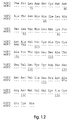
- Figure 1 compares the amino acid sequences of mature mOP2 and hOP2. Identity is indicated by three dots straight in the mOP2 sequence. As is evident from the figure, the amino acid sequence homology between the mature forms of these two proteins is substantial (92% homology between the mature sequences, about 95% homology within the C-terminal active region (e.g., residues 38-139 or 42-139 of Fig. 1.)
- Fig. 2 compares the amino acid sequences for the mature forms of all four species of OP1 and OP2 proteins. Here again, identity is indicated by three dots within.
- the hOP2 protein shares significant homology (about 74%) with the amino acid sequence defining the OP1 active region (OPS or OP7, residues 43-139 and 38-139, respectively, in Fig. 2), and less homology with OP1-18 (about 58% homology).
- Both OP2 proteins share the conserved seven cysteine skeleton seen in the OP1 proteins.
- the OP2 proteins comprise an eighth cysteine residue within this region (see position 78 in FIG. 2).
- the novel osteogenic proteins identified herein will purify essentially as OP1 does, or with only minor modifications of the protocols disclosed for OP1.
- the purified mOP1, mOP2, and hOP2 proteins are predicted to have an apparent molecular weight of about 18 kDa as reduced single subunits, and an apparent molecular weight of about 36 kDa as oxidized dimers, as determined by comparison with molecular weight standards on an SDS-polyacrylamide electrophoresis gel.
- Unglycosylated dimers e.g., proteins produced by recombinant expression in E. coli
- osteogenic proteins having an active region comprising eight cysteine residues also allows one to construct osteogenic polypeptide chains patterned after either of the following template amino acid sequences, or to identify additional osteogenic proteins having this sequence.
- the template sequences contemplated are "OPX-7C", comprising the conserved six cysteine skeleton plus the additional cysteine residue identified in the OP2 proteins, and "OPX-8C”, comprising the conserved seven cysteine skeleton plus the additional cysteine residue identified in the OP2 proteins.
- the OPX-7C and OPX-8C sequences are described below and in Seq. ID Nos. 8 and 9, respectively.
- Each Xaa in these template sequences independently represents one of the 20 naturally-occurring L-isomer, ⁇ -amino acids, or a derivative thereof.
- Biosynthetic constructs patterned after this template readily are constructed using conventional DNA synthesis or peptide synthesis techniques well known in the art. Once constructed, osteogenic proteins comprising these polypeptide chains can be tested as disclosed herein.
- "OPX-8C" (Sequence ID No. 9 comprising additional five residues at the N-terminus, including a conserved cysteine residue):
- the currently preferred carrier material is a xenogenic bone-derived particulate matrix treated as disclosed herein.
- This carrier may be replaced by either a biodegradable-synthetic or synthetic-inorganic matrix (e.g., hydroxylapatite (HAP), collagen, tricalcium phosphate or polylactic acid, polyglycolic acid and various copolymers thereof.)
- HAP hydroxylapatite
- collagen e.g., hydroxylapatite (HAP), collagen, tricalcium phosphate or polylactic acid, polyglycolic acid and various copolymers thereof.
- the sequential cellular reactions in the interface of the bone matrix/osteogenic protein implants are complex.
- the multistep cascade includes: binding of fibrin and fibronectin to implated matrix, chemotaxis of cells, proliferation of fibroblasts, differentiation into chondroblasts, cartilage formation, vascular invasion, bone formation, remodeling, and bone marrow differentiation.
- a successful carrier for osteogenic protein must perform several important functions. It must bind osteogenic protein and act as a slow release delivery system, accommodate each step of the cellular response during bone development, and protect the osteogenic protein from nonspecific proteolysis.
- selected materials must be biocompatible in vivo and preferably biodegradable; the carrier must act as a temporary scaffold until replaced completely by new bone.
- Polylactic acid (PLA), polyglycolic acid (PGA), and various combinations have different dissolution rates in vivo. In bones, the dissolution rates can vary according to whether the implant is placed in cortical or trabecular bone.
- Matrix geometry, particle size, the presence of surface charge, and the degree of both intra-and-inter-particle porosity are all important to successful matrix performance. It is preferred to shape the matrix to the desired form of the new bone and to have dimensions which span non-union defects. Rat studies show that the new bone is formed essentially having the dimensions of the device implanted.
- the matrix may comprise a shape-retaining solid made of loosely adhered particulate material, e.g., with collagen. It may also comprise a molded, porous solid, or simply an aggregation of close-packed particles held in place by surrounding tissue. Masticated muscle or other tissue may also be used. Large allogenic bone implants can act as a carrier for the matrix if their marrow cavities are cleaned and packed with particle and the dispersed osteogenic protein.
- the preferred matrix material prepared from xenogenic bone and treated as disclosed herein, produces an implantable material useful in a variety of clinical settings.
- the matrix also may be used as a sustained release carrier, or as a collagenous coating for implants.
- the matrix may be shaped as desired in anticipation of surgery or shaped by the physician or technician during surgery.
- the material may be used for topical, subcutaneous, intraperitoneal, or intramuscular implants; it may be shaped to span a nonunion fracture or to fill a bone defect.
- the material is slowly absorbed by the body and is replaced by bone in the shape of or very nearly the shape of the implant.
- Various growth factors, hormones, enzymes, therapeutic compositions, antibiotics, and other body treating agents also may be absorbed onto the carrier material and will be released over time when implanted as the matrix material is slowly absorbed.
- various known growth factors such as EGF, PDGF, IGF, FGF, TGF- ⁇ , and TGF- ⁇ may be released in vivo.
- the material can be used to release chemotherapeutic agents, insulin, enzymes, or enzyme inhibitors.
- Demineralized bone matrix preferably bovine bone matrix
- bovine bone matrix is prepared by previously published procedures (Sampath and Reddi (1983) Proc. Natl. Acad. Sci. USA 80 :6591-6595).
- Bovine diaphyseal bones (age 1-10 days) are obtained from a local slaughterhouse and used fresh. The bones are stripped of muscle and fat, cleaned of periosteum, demarrowed by pressure with cold water, dipped in cold absolute ethanol, and stored at -20°C. They are then dried and fragmented by crushing and pulverized in a large mill. Care is taken to prevent heating by using liquid nitrogen.
- the pulverized bone is milled to a particle size in the range of 70-850 ⁇ m, preferably 150-420 ⁇ m, and is defatted by two washes of approximately two hours duration with three volumes of chloroform and methanol (3:1).
- the particulate bone is then washed with one volume of absolute ethanol and dried over one volume of anhydrous ether yielding defatted bone powder.
- the defatted bone powder is then demineralized by four successive treatments with 10 volumes of 0.5 N HCl at 4°C for 40 min. Finally, neutralizing washes are done on the demineralized bone powder with a large volume of water.
- Demineralized bone matrix thus prepared is extracted with 5 volumes of 4 M guanidine-HCl, 50mM Tris-HCl, pH 7.0 for 16 hr. at 4°C. The suspension is filtered. The insoluble material is collected and used to fabricate the matrix. The material is mostly collagenous in nature. It is devoid of osteogenic or chondrogenic activity.
- Type-I collagen The major component of all bone matrices is Type-I collagen.
- demineralized bone extracted as disclosed above includes non-collagenous proteins which may account for 5% of its mass. In a xenogenic matrix, these noncollagenous components may present themselves as potent antigens, and may constitute immunogenic and/or inhibitory components. These components also may inhibit osteogenesis in allogenic implants by interfering with the developmental cascade of bone differentiation.
- treatment of the matrix particles with a collagen fibril-modifying agent extracts potentially unwanted components from the matrix, and alters the surface structure of the matrix material.
- Useful agents include acids, organic solvents or heated aqueous media. Various treatments are described below. A detailed physical analysis of the effect these fibril-modifying agents have on demineralized, quanidine-extracted bone collagen particles is disclosed in PCT WO 90/10018, published 7-SEP-90.
- the treated matrix is washed to remove any extracted components, following a form of the procedure set forth below:
- Trifluoroacetic acid is a strong non-oxidizing acid that is a known swelling agent for proteins, and which modifies collagen fibrils.
- Bovine bone residue prepared as described above is sieved, and particles of the appropriate size are collected. These particles are extracted with various percentages (1.0% to 100%) of trifluoroacetic acid and water (v/v) at 0°C or room temperature for 1-2 hours with constant stirring. The treated matrix is filtered, lyophilized, or washed with water/salt and then lyophilized.
- hydrogen fluoride is a strong acid and swelling agent, and also is capable of altering intraparticle surface structure. Hydrogen fluoride is also a known deglycosylating agent. As such, HF may function to increase the osteogenic activity of these matrices by removing the antigenic carbohydrate content of any glycoproteins still associated with the matrix after guanidine extraction.
- Bovine bone residue prepared as described above is sieved, and particles of the appropriate size are collected.
- the sample is dried in vacuo over P 2 O 5 , transferred to the reaction vessel and exposed to anhydrous hydrogen fluoride (10-20 ml/g of matrix) by distillation onto the sample at -70°C.
- the vessel is allowed to warm to 0°C and the reaction mixture is stirred at this temperature for 120 minutes.
- the residue is dried thoroughly in vacuo over KOH pellets to remove any remaining traces of acid.
- Extent of deglycosylation can be determined from carbohydrate analysis of matrix samples taken before and after treatment with hydrogen fluoride, after washing the samples appropriately to remove non-covalently bound carbohydrates.
- SDS-extracted protein from HF-treated material is negative for carbohydrate as determined by Con A blotting.
- the deglycosylated bone matrix is next washed twice in TBS (Tris-buffered saline) or UTBS, water-washed, and then lyophilized.
- TBS Tris-buffered saline
- UTBS Tris-buffered saline
- TFA is a currently preferred acidifying reagent in these treatments because of its volatility.
- other, potentially less caustic acids may be used, such as acetic or formic acid.
- Dichloromethane is an organic solvent capable of denaturing proteins without affecting their primary structure.
- This swelling agent is a common reagent in automated peptide synthesis, and is used in washing steps to remove components.
- Bovine bone residue prepared as described above, is sieved, and particles of the appropriate size are incubated in 100% DCM or, preferably, 99.9% DCM/0.1% TFA.
- the matrix is incubated with the swelling agent for one or two hours at 0°C or at room temperature. Alternatively, the matrix is treated with the agent at least three times with short washes (20 minutes each) with no incubation.
- Acetonitrile is an organic solvent, capable of denaturing proteins without affecting their primary structure. It is a common reagent used in high-performance liquid chromatography, and is used to elute proteins from silica-based columns by perturbing hydrophobic interactions.
- Bovine bone residue particles of the appropriate size are treated with 100% ACN (1.0 g/30 ml) or, preferably, 99.9% ACN/0.1% TFA at room temperature for 1-2 hours with constant stirring.
- the treated matrix is then water-washed, or washed with urea buffer, or 4 M NaCl and lyophilized.
- the ACN or ACN/TFA treated matrix may be lyophilized without wash.
- Isopropanol is also an organic solvent capable of denaturing proteins without affecting their primary structure. It is a common reagent used to elute proteins from silica HPLC columns.
- Bovine bone residue particles of the appropriate size prepared as described above are treated with 100% isopropanol (1.0 g/30 ml) or, preferably, in the presence of 0.1% TFA, at room temperature for 1-2 hours with constant stirring. The matrix is then water-washed or washed with urea buffer or 4 M NaCl before being lyophilized.
- Chloroform also may be used to increase surface area of bone matrix like the reagents set forth above, either alone or acidified.
- Treatment as set forth above is effective to assure that the material is free of pathogens prior to implantation.
- the currently most preferred agent is a heated aqueous fibril-modifying medium such as water, to increase the matrix particle surface area and porosity.
- the currently most preferred aqueous medium is an acidic aqueous medium having a pH of less than about 4.5, e.g., within the range of pH 2 - pH 4. which may help to "swell" the collagen before heating. 0.1% acetic acid, which has a pH of about 3, currently is preferred. 0.1 M acetic acid also may be used.
- aqueous medium (1g matrix/30ml aqueous medium) under constant stirring in a water jacketed glass flask, and maintained at a given temperature for a predetermined period of time.
- Preferred treatment times are about one hour, although exposure times of between about 0.5 to two hours appear acceptable.
- the temperature employed is held constant at a temperature generally within the range of about 37°C to 75°C.
- the currently preferred heat treatment temperature is within the range of 45°C to 60°C.
- the matrix is filtered, washed, lyophilized and used for implant.
- the matrix also is preferably neutralized prior to washing and lyophilization.
- a currently preferred neutralization buffer is a 200mM sodium phosphate buffer, pH 7.0.
- the matrix preferably first is allowed to cool following thermal treatment, the acidic aqueous medium (e.g., 0.1% acetic acid) then is removed and replaced with the neutralization buffer and the matrix agitated for about 30 minutes. The neutralization buffer then may be removed and the matrix washed and lyophilized (see infra).
- the matrix also may be treated to remove contaminating heavy metals, such as by exposing the matrix to a metal ion chelator.
- a metal ion chelator For example, following thermal treatment with 0.1% acetic acid, the matrix may be neutralized in a neutralization buffer containing EDTA (sodium ethylenediaminetetraacetic acid), e.g., 200 mM sodium phosphate, 5mM EDTA, pH 7.0. 5 mM EDTA provides about a 100-fold molar excess of chelator to residual heavy metals present in the most contaminated matrix tested to date. Subsequent washing of the matrix following neutralization appears to remove the bulk of the EDTA.
- EDTA sodium ethylenediaminetetraacetic acid
- EDTA treatment of matrix particles reduces the residual heavy metal content of all metals tested (Sb, As, Be, Cd, Cr, Cu, Co, Pb, Hg, Ni, Se, Ag, Zn, T1) to less than about 1 ppm.
- Bioassays with EDTA-treated matrices indicate that treatment with the metal ion chelator does not inhibit bone inducing activity.
- the collagen matrix materials preferably take the form of a fine powder, insoluble in water, comprising nonadherent particles. It may be used simply by packing into the volume where new bone growth or sustained release is desired, held in place by surrounding tissue.
- the powder may be encapsulated in, e.g., a gelatin or polylactic acid coating, which is adsorbed readily by the body.
- the powder may be shaped to a volume of given dimensions and held in that shape by interadhering the particles using, for example, soluble, species-biocompatible collagen.
- the material may also be produced in sheet, rod, bead, or other macroscopic shapes.
- Demineralized rat bone matrix used as an allogenic matrix in certain of the experiments disclosed herein is prepared from several of the dehydrated diaphyseal shafts of rat femur and tibia as described herein to produce a bone particle size which passes through a 420 ⁇ m sieve.
- the bone particles are subjected to dissociative extraction with 4 M guanidine-HCl.
- Such treatment results in a complete loss of the inherent ability of the bone matrix to induce endochondral bone differentiation.
- the remaining insoluble material is used to fabricate the matrix.
- the material is mostly collagenous in nature, and upon implantation, does not induce cartilage and bone. All new preparations are tested for mineral content and osteogenic activity before use. The total loss of biological activity of bone matrix is restored when an active osteoinductive protein fraction or a pure osteoinductive protein preparation is reconstituted with the biologically inactive insoluble collagenous matrix.
- the naturally sourced and recombinant protein as set forth above, and other constructs, can be combined and dispersed in a suitable matrix preparation using any of the methods described below.
- 50-100 ng of active protein is combined with the inactive carrier matrix (e.g., 25 mg for rat bioassays). Greater amounts may be used for large implants.
- Matrix is added to osteogenic protein dissolved in guanidine-HCl. Samples are vortexed and incubated at a low temperature (e.g., 4°C). Samples are then further vortexed. Cold absolute ethanol (5 volumes) is added to the mixture which is then stirred and incubated, preferably for 30 minutes at -20°C. After centrifugation (microfuge, high speed) the supernatant is discarded. The reconstituted matrix is washed twice with cold concentrated ethanol in water (85% EtOH) and then lyophilized.
- osteogenic protein in an acetonitrile trifluroacetic acid (ACN/TFA) solution is added to the carrier material.
- Samples are vigorously vortexed many times and then lyophilized. This method is currently preferred, and has been tested with osteogenic protein at varying concentrations and different levels of purity.
- the protein is mixed with the matrix material, vortexed many times, and then lyophilized.
- the lyophilized material may be used "as is" for implants.
- OP1 and OP2 preparations in physiological saline may also be vortexed with the matrix and lyophilized to produce osteogenically active material.
- the functioning of the various proteins and devices of this invention can be evaluated with an in vivo bioassay.
- Studies in rats show the osteogenic effect in an appropriate matrix to be dependent on the dose of osteogenic protein dispersed in the matrix. No activity is observed if the matrix is implanted alone.
- In vivo bioassays performed in the rat model also have shown that demineralized, guanidine-extracted xenogenic bone matrix materials of the type described in the literature are ineffective as a carrier, fail to induce bone, and produce an inflammatory and immunological response when implanted unless treated as disclosed above. In certain species (e.g., monkey) allogenic matrix materials also apparently are ineffective as carriers.
- the bioassay for bone induction as described by Sampath and Reddi may be used to monitor endochondral bone differentiation activity.
- This assay consists of implanting test samples in subcutaneous sites in recipient rats under ether anesthesia. Male Long-Evans rats, aged 28-32 days, were used. A vertical incision (1 cm) is made under sterile conditions in the skin over the thoracic region, and a pocket is prepared by blunt dissection. Approximately 25 mg of the test sample is implanted deep into the pocket and the incision is closed with a metallic skin clip. The day of implantation is designated as day one of the experiment. Implants were removed on day 12. The heterotropic site allows for the study of bone induction without the possible ambiguities resulting from the use of orthotropic sites. As disclosed herein, both allogenic (rat bone matrix) and xenogenic (bovine bone matrix) implants were assayed.
- Successful implants exhibit a controlled progression through the stages of protein-induced endochondral bone development, including: (1) transient infiltration by polymorphonuclear leukocytes on day one; (2) mesenchymal cell migration and proliferation on days two and three; (3) chondrocyte appearance on days five and six; (4) cartilage matrix formation on day seven; (5) cartilage calcification on day eight; (6) vascular invasion, appearance of osteoblasts, and formation of new bone on days nine and ten; (7) appearance of osteoblastic and bone remodeling and dissolution of the implanted matrix on days twelve to eighteen; and (8) hematopoietic bone marrow differentiation in the ossicle on day twenty-one.
- the results show that the shape of the new bone conforms to the shape of the implanted matrix.
- Implants are fixed in Bouins Solution, embedded in paraffin, and cut into 6-8 ⁇ m sections. Staining with toluidine blue or hemotoxylin/eosin demonstrates clearly the ultimate development of endochondral bone. Twelve day implants are usually sufficient to determine whether the implants contain newly induced bone.
- Alkaline phosphatase activity may be used as a marker for osteogenesis.
- the enzyme activity may be determined spectrophotometrically after homogenization of the implant. The activity peaks at 9-10 days in vivo and thereafter slowly declines. Implants showing no bone development by histology have little or no alkaline phosphatase activity under these assay conditions.
- the assay is useful for quantitation and obtaining an estimate of bone formation quickly after the implants are removed from the rat. Alternatively, the amount of bone formation can be determined by measuring the calcium content of the implant.
Landscapes
- Health & Medical Sciences (AREA)
- Chemical & Material Sciences (AREA)
- Life Sciences & Earth Sciences (AREA)
- Medicinal Chemistry (AREA)
- Organic Chemistry (AREA)
- General Health & Medical Sciences (AREA)
- Animal Behavior & Ethology (AREA)
- Public Health (AREA)
- Veterinary Medicine (AREA)
- Chemical Kinetics & Catalysis (AREA)
- General Chemical & Material Sciences (AREA)
- Engineering & Computer Science (AREA)
- Nuclear Medicine, Radiotherapy & Molecular Imaging (AREA)
- Pharmacology & Pharmacy (AREA)
- Bioinformatics & Cheminformatics (AREA)
- Oral & Maxillofacial Surgery (AREA)
- Biochemistry (AREA)
- Transplantation (AREA)
- Epidemiology (AREA)
- Physical Education & Sports Medicine (AREA)
- Orthopedic Medicine & Surgery (AREA)
- Toxicology (AREA)
- Zoology (AREA)
- Gastroenterology & Hepatology (AREA)
- Dermatology (AREA)
- Biophysics (AREA)
- Genetics & Genomics (AREA)
- Molecular Biology (AREA)
- Proteomics, Peptides & Aminoacids (AREA)
- Peptides Or Proteins (AREA)
- Medicines That Contain Protein Lipid Enzymes And Other Medicines (AREA)
- Preparation Of Compounds By Using Micro-Organisms (AREA)
- Saccharide Compounds (AREA)
- Organic Low-Molecular-Weight Compounds And Preparation Thereof (AREA)
Claims (27)
- Polypeptidkette, die eine Aminosäure-Sequenz enthält, die die Gruppen 303-399 der Sequenz ID Nr. 5 enthält.
- Polypeptidkette nach Anspruch 1, die eine Aminosäure-Sequenz enthält, die die Gruppenenthält.a) 298-399 der Sequenz ID Nr. 5 oderb) 267-399 der Sequenz ID Nr. 5 oderc) 264-399 der Sequenz ID Nr. 5 oderd) 240-399 der Sequenz ID Nr. 5 odere) 1-399 der Sequenz ID Nr. 5
- Polypeptidkette, die eine Aminosäure-Sequenz enthält, die die Gruppen 301-397 der Sequenz ID Nr. 3 enthält.
- Polypeptidkette nach Anspruch 3, die eine Aminosäure-Sequenz enthält, die die Gruppenenthält.a) 296-397 der Sequenz ID Nr. 3 oderb) 259-397 der Sequenz ID Nr. 3 oderc) 1-397 der Sequenz ID Nr. 3
- Polypeptidkette, verwendbar als Untereinheit für ein dimeres osteogenes Protein, das ein Paar disulfidgebundene Polypeptidketten, wobei die Polypeptidkette eine Aminosäure-Sequenz enthält, die entwederso daß das diese Polypeptidkette enthaltende, dimere osteogene Protein einen Aufbau hat, der eine endochondrale Knochenbildung zu induzieren in der Lage ist, wenn er in Verbindung mit einer Matrix einem Säuger implantiert wird.a) das erhaltene sechs Cystein-Gerüst von menschlichem OP1, dargestellt in Fig. 2.1 bis 2.3 von Leu (Position 43) bis His (Position 139), umschließt und weiterhin eine zusätzliche Cystein-Gruppe in diesem sechs Cystein-Gerüst enthält oderb) das erhaltene sieben Cystein-Gerüst von menschlichem OP1, dargestellt in Fig. 2.1 bis 2.3 von Cys (Position 38) bis His (Position 139), umschließt und weiterhin eine zusätzliche Cystein-Gruppe darin enthält,
- Polypeptidkette nach Anspruch 5, wobei die beschriebene Aminosäure-Sequenz die Gruppenenthält.a) 261-399 der Sequenz ID Nr. 5 oderb) 301-397 der Sequenz ID Nr. 3 oderc) 259-397 der Sequenz ID Nr. 3 oderd) 298-299 der Sequenz ID Nr. 5
- Dimeres osteogenes Protein, das eine endochondrale Knochenbildung in einem Säuger zu induzieren in der Lage ist, wenn es diesem Säuger in Verbindung mit einer Matrix implantiert wird, und ein Paar disulfidgebundene Polypeptidketten, die eine dimere Spezies bilden, enthält, wobei jede dieser Polypeptidketten die Polypeptidkette nach Anspruch 5 ist.
- Polypeptidkette nach einem der Ansprüche 1 bis 6, hergestellt durch die Produktion neuer Gene recombinanter DNS in einer Wirtszelle, z. B. einer prokaryontischen Wirtszelle oder einer Wirtszelle eines Säugers.
- Polypeptidkette nach Anspruch 8, wobei die Wirtszelle aus Wirtszellen von E. coli, CHO, COS, BSC, Saccharomyces oder myeloma ausgewählt ist.
- Polypeptidkette nach einem der Ansprüche 1 bis 6, die glycosyliert ist.
- Nukleinsäure, die die Polypeptidkette nach einem der Ansprüche 1 bis 6 codiert.
- Polypeptidkette, die mit einem Gen codiert ist, dasa) die DNS-Sequenz der Sequenz ID Nr. 3;b) die DNS-Sequenz der Sequenz ID Nr. 5;c) eine unter zwingend hybridisierenden Bedingungen hybridisierbare DNS-Sequenz mit den Nucleotiden 467-771 der Sequenz ID Nr. 3 enthält und eine Polypeptidkette codiert, die eine Aminosäure-Sequenz enthält, die entwederso daß das dimere osteogene Protein, welches diese Polypeptidkette enthält, einen Aufbau hat, der eine endochondrale Knochenbildung zu induzieren in der Lage ist, wenn er in Verbindung mit einer Matrix einem Säuger implantiert wird.a) das erhaltene sechs Cystein-Gerüst von menschlichem OP1, dargestellt in Fig. 2.1 bis 2.3 von Leu (Position 43) bis His (Position 139), umschließt und weiterhin eine zusätzliche Cystein-Gruppe in diesem sechs Cystein-Gerüst enthält oderb) das erhaltene sieben Cystein-Gerüst von menschlichem OP1, dargestellt in Fig. 2.1 bis 2.3 von Cys (Position 38) bis His (Position 139), umfaßt und weiterhin eine zusätzliche Cystein-Gruppe darin enthält,
- Isolierter Antikörper, z. B. polyklonaler oder monoklonaler Antikörper, der eine Bindungsspezifität nur für ein Polypeptid oder ein Protein nach einem der Ansprüche 1 bis 10 und 12 aufweist.
- Antikörper nach Anspruch 13, der eine Bindungsspezifität für ein Protein mit einer Aminosäure-Sequenz aufweist, die die Gruppenenthält.a) 303-399 der Sequenz ID Nr. 5;b) 297-399 der Sequenz ID Nr. 5;c) 264-399 der Sequenz ID Nr. 5;d) 1-399 der Sequenz ID Nr. 5;e) 18-257 der Sequenz ID Nr. 5;f) 301-397 der Sequenz ID Nr. 3;g) 296-397 der Sequenz ID Nr. 3;h) 259-397 der Sequenz ID Nr. 3;i) 1-397 der Sequenz ID Nr. 3 oderj) 18-259 der Sequenz ID Nr. 3
- DNS-Molekül, das das Protein nach Anspruch 12 codiert.
- DNS-Molekül nach Anspruch 15, dasenthält.a) die DNS-Sequenz der Sequenz ID Nr. 3;b) die DNS-Sequenz der Sequenz ID Nr. 5 oderc) die hybridisierbare DNS-Sequenz mit den Nukleotiden 467-771 der Sequenz ID Nr. 3
- Protein, das für die Knorpel- oder Knochenbildung bei einem Säuger maßgeblich ist und ein Paar Polypeptidketten enthält, die beide eine durch OPX (Sequenz ID Nr. 7) definierte Aminosäure-Sequenz enthalten, wobei Xaa in der Gruppe 41 Cys ist.
- Protein nach Anspruch 7 oder 12 in Kombination mit einem biokompatiblen, in vivo biologisch abbaubaren Träger, der als langsam freisetzendes Versorgungssystem wirkt.
- Implantierbares osteogenes Mittel, das die Polypeptidkette nach einem der Ansprüche 1 bis 6, 8 bis 10 und 12 oder das Protein nach einem der Ansprüche 7, 17 und 18 enthält.
- Osteogenes Mittel für die Implantation in einen Säuger, dasenthält.a) eine biokompaktible, in vivo biologisch abbaubare Matrix mit einer ausreichenden Porengröße für die Zufuhr, die Differenzierung und das Wachstum von umgesiedelten Vorläufer-Zellen aus dem Körper dieses Säugers undb) die Polypeptidkette nach einem der Ansprüche 1 bis 6, 8 bis 10 und 12, oder das Protein nach einem der Ansprüche 7, 17 und 18
- Mittel nach Anspruch 20, wobei die Matrix allogenes oder xenogenes Knochenmaterial enthält.
- Mittel nach einem der Ansprüche 19 bis 21, wobei die Matrix demineralisierte, delipidisierte, Typ I unlösliche Knochen-Kollagenpartikeln enthält, deren nicht kollagene Proteine abgereichert sind, und die zur Vergrößerung des intrapartikulären Eindringvolumens und der Oberfläche z. B. mit:behandelt sind.a) einem Kollagenfibrillen modifizierenden Mittel oderb) einer Protease (z. B. Trypsin) oderc) einem Lösungsmittel (z. B. Dichlormethan, Trichloressigsäure und Acetonitril) oderd) einer Säure (z. B. Trifluoressigsäure oder Fluorwasserstoff) odere) einem heißen wäßrigen Medium mit einer Temperatur im Bereich von 37°C bis 75°C
- Verfahren zur Herstellung des Mittels nach Anspruch 22 mit dem Schritt der Kollagenbehandlung zur Vergrößerung des intrapartikulären Eindringvolumens und der Oberfläche.
- Polypeptidkette nach einem der Ansprüche 1 bis 6, 8 bis 10 und 12, oder Protein nach einem der Ansprüche 7, 17 und 18 in Verbindung mit einer Matrix, dieenthält.a) allogenes Knochenmaterial (z. B. partikelförmiges, demineralisiertes und Guanidin extrahiertes allogenes Knochenmaterial) oderb) xenogenes Knochenmaterial (z. B. partikelförmiges, demineralisiertes, Protein extrahiertes xenogenes Knochenmaterial) oderc) partikelförmiges, demineralisiertes, Protein extrahiertes xenogenes Knochenmaterial, das mit einer Protease oder einem Fibrillen modifizierenden Mittel zur Vergrößerung des intrapartikulären Eindringvolumens und der Oberfläche, z. B. einem Lösungsmittel, behandelt ist oderd) aus Kollagen ausgewähltes Material, Homo- oder - Copolymeren von Hydroxyessigsäure und Milchsäure, Hydroxyapatit und Calciumphosphat (z. B. Tricalciumphosphat) odere) ein formstabiles, festes oder lose zusammenhängendes, partikelförmiges Material, z. B. Kollagen oderf) einen geformten, porösen Feststoff oderg) zerkleinertes Gewebe (z. B. Muskelgewebe)
- Aktives heterodimeres osteogenes Protein, das die Polypeptidkette nach einem der Ansprüche 1 bis 6, 8 bis 10 und 12, oder das Protein nach einem der Ansprüche 7, 17 und 18 enthält.
- Heterodimeres Protein nach Anspruch 25, wobei die Heterodimere über Disulfidbindungen oder anderweitig gebunden sind.
- Verfahren zur Herstellung des heterodimeren Proteins nach Anspruch 25 oder 26 durch Oxidieren und Falten von zwei oder mehr Polypeptidketten.
Applications Claiming Priority (3)
| Application Number | Priority Date | Filing Date | Title |
|---|---|---|---|
| US59954390A | 1990-10-18 | 1990-10-18 | |
| US599543 | 1990-10-18 | ||
| PCT/US1991/007635 WO1992007073A1 (en) | 1990-10-18 | 1991-10-18 | Osteogenic peptides |
Publications (2)
| Publication Number | Publication Date |
|---|---|
| EP0643767A1 EP0643767A1 (de) | 1995-03-22 |
| EP0643767B1 true EP0643767B1 (de) | 1998-07-22 |
Family
ID=24400058
Family Applications (1)
| Application Number | Title | Priority Date | Filing Date |
|---|---|---|---|
| EP91919612A Expired - Lifetime EP0643767B1 (de) | 1990-10-18 | 1991-10-18 | Osteogene peptide |
Country Status (9)
| Country | Link |
|---|---|
| EP (1) | EP0643767B1 (de) |
| JP (2) | JPH07500487A (de) |
| AT (1) | ATE168719T1 (de) |
| AU (1) | AU663689B2 (de) |
| CA (1) | CA2094027C (de) |
| DE (1) | DE69129865T2 (de) |
| DK (1) | DK0643767T3 (de) |
| ES (1) | ES2120964T3 (de) |
| WO (1) | WO1992007073A1 (de) |
Cited By (4)
| Publication number | Priority date | Publication date | Assignee | Title |
|---|---|---|---|---|
| US7253254B1 (en) | 1999-02-13 | 2007-08-07 | Osteopharma Inc. | Polypeptide variants with increased heparin-binding capacity |
| US7763270B2 (en) | 2002-09-10 | 2010-07-27 | Scil Technology Gmbh | Metal implant coated under reduced oxygen concentration with osteoinductive protein |
| US8372419B2 (en) | 2004-03-10 | 2013-02-12 | Scil Technology Gmbh | Coated implants, their manufacturing and use thereof |
| US8546334B2 (en) | 2001-11-19 | 2013-10-01 | Scil Technology Gmbh | Device having osteoinductive and osteoconductive properties |
Families Citing this family (29)
| Publication number | Priority date | Publication date | Assignee | Title |
|---|---|---|---|---|
| US6150328A (en) | 1986-07-01 | 2000-11-21 | Genetics Institute, Inc. | BMP products |
| US7378392B1 (en) | 1990-05-16 | 2008-05-27 | Genetics Institute, Llc | Bone and cartilage inductive proteins |
| JPH06500991A (ja) * | 1990-05-16 | 1994-01-27 | ジェネティックス・インスティテュート・インコーポレイテッド | 骨および軟骨誘導蛋白質 |
| US5688678A (en) * | 1990-05-16 | 1997-11-18 | Genetics Institute, Inc. | DNA encoding and methods for producing BMP-8 proteins |
| US5652118A (en) * | 1991-03-11 | 1997-07-29 | Creative Biomolecules, Inc. | Nucleic acid encoding a novel morphogenic protein, OP-3 |
| US6077823A (en) * | 1991-03-11 | 2000-06-20 | Creative Biomolecules, Inc. | Method for reducing tissue damage associated with ischemia-reperfusion or hypoxia injury |
| US6090776A (en) * | 1991-03-11 | 2000-07-18 | Creative Bio Molecules, Inc. | Morphogen treatment of organ implants |
| US5739107A (en) | 1991-03-11 | 1998-04-14 | Creative Biomolecules, Inc. | Morphogen treatment of gastrointestinal ulcers |
| US6395883B1 (en) | 1991-03-11 | 2002-05-28 | Curis, Inc. | Soluble morphogenic protein complex compositions of matter |
| US6194376B1 (en) | 1991-03-11 | 2001-02-27 | Creative Biomolecules, Inc. | Method for modulating inflammatory response comprising administering morphogen |
| AU652472B2 (en) * | 1991-06-25 | 1994-08-25 | Genetics Institute, Llc | BMP-9 compositions |
| US6287816B1 (en) | 1991-06-25 | 2001-09-11 | Genetics Institute, Inc. | BMP-9 compositions |
| ATE192931T1 (de) * | 1991-08-30 | 2000-06-15 | Creative Biomolecules Inc | Morphogen-induzierte modulation von entzündlichen antworten |
| ATE287949T1 (de) * | 1992-07-31 | 2005-02-15 | Stryker Corp | Löslicher komplex morphogener proteine und zusammensetzungen davon |
| JP3645258B2 (ja) * | 1993-01-12 | 2005-05-11 | ジョーンズ ホプキンス ユニバーシティー スクール オブ メディシン | 増殖分化因子−5 |
| EP0717633A4 (de) * | 1993-07-09 | 1998-05-20 | Univ Johns Hopkins Med | Wachstums-differenzierungsfaktor-7 |
| US6291206B1 (en) | 1993-09-17 | 2001-09-18 | Genetics Institute, Inc. | BMP receptor proteins |
| DE69434651T2 (de) | 1993-12-07 | 2007-03-08 | Genetics Institute, Inc., Cambridge | Bmp-12, bmp-13 und diese enthaltende sehne-induzierende zusammensetzungen |
| US6040431A (en) * | 1995-06-07 | 2000-03-21 | Stryker Corporation | Single chain analogs of the TGF-β superfamily (morphons) |
| EP0845006A2 (de) * | 1995-08-14 | 1998-06-03 | Creative Biomolecules, Inc. | Bindung des osteogenen proteins-i (op-1) und dessen analoge an den zelloberflächenrezeptor alk-1 und dessen analoge |
| JP2000501744A (ja) * | 1996-01-22 | 2000-02-15 | クリエイティブ バイオモレキゥルズ,インコーポレーテッド | モルホゲン類似体およびその製法 |
| KR20000064752A (ko) | 1996-03-22 | 2000-11-06 | 더 제네랄 호스피탈 코포레이션 | 중추신경계허혈또는외상의발현후폴리펩티드성장인자를투여하는방법 |
| US6034062A (en) * | 1997-03-13 | 2000-03-07 | Genetics Institute, Inc. | Bone morphogenetic protein (BMP)-9 compositions and their uses |
| US6727224B1 (en) | 1999-02-01 | 2004-04-27 | Genetics Institute, Llc. | Methods and compositions for healing and repair of articular cartilage |
| PT1223990E (pt) | 1999-10-15 | 2004-12-31 | Fidia Advanced Biopolymers Srl | Formulacoes de acido hialuronico para administracao de proteinas osteogenicas |
| EP1266961A4 (de) * | 2000-03-16 | 2004-12-08 | Kyowa Hakko Kogyo Kk | Leberheilender faktor |
| EP1399023B1 (de) | 2001-06-01 | 2008-04-30 | Wyeth | Zusammensetzungen für die systemische verabreichung von sequenzen, die für knochenmorphogenese-proteinen kodieren |
| EP3147296A1 (de) * | 2005-11-14 | 2017-03-29 | Merial, Inc. | Gentherapie für nierenversagen |
| WO2023272658A1 (zh) * | 2021-06-30 | 2023-01-05 | 北京泽勤生物医药有限公司 | 增强成骨细胞活性的多肽及其在治疗骨科疾病中的应用 |
Family Cites Families (2)
| Publication number | Priority date | Publication date | Assignee | Title |
|---|---|---|---|---|
| JP2522568B2 (ja) * | 1988-04-08 | 1996-08-07 | ストライカー・コーポレーション | 骨形成デバイス |
| DE69031939T2 (de) * | 1989-03-28 | 1998-09-10 | Genetics Institute, Inc., Cambridge, Mass. | Osteoinduktive zusammensetzungen |
-
1991
- 1991-10-18 EP EP91919612A patent/EP0643767B1/de not_active Expired - Lifetime
- 1991-10-18 DE DE69129865T patent/DE69129865T2/de not_active Expired - Lifetime
- 1991-10-18 ES ES91919612T patent/ES2120964T3/es not_active Expired - Lifetime
- 1991-10-18 JP JP3518322A patent/JPH07500487A/ja active Pending
- 1991-10-18 DK DK91919612T patent/DK0643767T3/da active
- 1991-10-18 WO PCT/US1991/007635 patent/WO1992007073A1/en not_active Ceased
- 1991-10-18 AU AU89000/91A patent/AU663689B2/en not_active Expired
- 1991-10-18 AT AT91919612T patent/ATE168719T1/de not_active IP Right Cessation
- 1991-10-18 CA CA002094027A patent/CA2094027C/en not_active Expired - Lifetime
-
1998
- 1998-11-25 JP JP10352101A patent/JPH11262395A/ja active Pending
Cited By (5)
| Publication number | Priority date | Publication date | Assignee | Title |
|---|---|---|---|---|
| US7253254B1 (en) | 1999-02-13 | 2007-08-07 | Osteopharma Inc. | Polypeptide variants with increased heparin-binding capacity |
| US8546334B2 (en) | 2001-11-19 | 2013-10-01 | Scil Technology Gmbh | Device having osteoinductive and osteoconductive properties |
| US7763270B2 (en) | 2002-09-10 | 2010-07-27 | Scil Technology Gmbh | Metal implant coated under reduced oxygen concentration with osteoinductive protein |
| US8257728B2 (en) | 2002-09-10 | 2012-09-04 | Scil Technology Gmbh | Metal implant coated under reduced oxygen concentration with osteoinductive protein |
| US8372419B2 (en) | 2004-03-10 | 2013-02-12 | Scil Technology Gmbh | Coated implants, their manufacturing and use thereof |
Also Published As
| Publication number | Publication date |
|---|---|
| CA2094027A1 (en) | 1992-04-19 |
| DK0643767T3 (da) | 1999-04-26 |
| CA2094027C (en) | 2001-12-25 |
| ES2120964T3 (es) | 1998-11-16 |
| JPH07500487A (ja) | 1995-01-19 |
| WO1992007073A1 (en) | 1992-04-30 |
| DE69129865D1 (de) | 1998-08-27 |
| EP0643767A1 (de) | 1995-03-22 |
| DE69129865T2 (de) | 1999-03-04 |
| JPH11262395A (ja) | 1999-09-28 |
| AU663689B2 (en) | 1995-10-19 |
| AU8900091A (en) | 1992-05-20 |
| ATE168719T1 (de) | 1998-08-15 |
Similar Documents
| Publication | Publication Date | Title |
|---|---|---|
| EP0643767B1 (de) | Osteogene peptide | |
| JP2548414B2 (ja) | 生合成による骨形成蛋白質および該蛋白質を含む骨形成装置 | |
| US7078221B2 (en) | Nucleic acid molecules encoding osteogenic proteins | |
| US5011691A (en) | Osteogenic devices | |
| US5324819A (en) | Osteogenic proteins | |
| EP0448704B1 (de) | Osteogene vorrichtungen | |
| US5496552A (en) | Osteogenic devices | |
| US5670336A (en) | Method for recombinant production of osteogenic protein | |
| WO1992007004A1 (en) | Osteogenic protein | |
| US7429484B2 (en) | Nucleic acid molecules encoding osteogenic proteins | |
| AU648997C (en) | Osteogenic devices |
Legal Events
| Date | Code | Title | Description |
|---|---|---|---|
| PUAI | Public reference made under article 153(3) epc to a published international application that has entered the european phase |
Free format text: ORIGINAL CODE: 0009012 |
|
| 17P | Request for examination filed |
Effective date: 19930429 |
|
| AK | Designated contracting states |
Kind code of ref document: A1 Designated state(s): AT BE CH DE DK ES FR GB GR IT LI LU NL SE |
|
| 17Q | First examination report despatched |
Effective date: 19950619 |
|
| GRAG | Despatch of communication of intention to grant |
Free format text: ORIGINAL CODE: EPIDOS AGRA |
|
| GRAG | Despatch of communication of intention to grant |
Free format text: ORIGINAL CODE: EPIDOS AGRA |
|
| GRAG | Despatch of communication of intention to grant |
Free format text: ORIGINAL CODE: EPIDOS AGRA |
|
| GRAH | Despatch of communication of intention to grant a patent |
Free format text: ORIGINAL CODE: EPIDOS IGRA |
|
| GRAH | Despatch of communication of intention to grant a patent |
Free format text: ORIGINAL CODE: EPIDOS IGRA |
|
| GRAA | (expected) grant |
Free format text: ORIGINAL CODE: 0009210 |
|
| AK | Designated contracting states |
Kind code of ref document: B1 Designated state(s): AT BE CH DE DK ES FR GB GR IT LI LU NL SE |
|
| REF | Corresponds to: |
Ref document number: 168719 Country of ref document: AT Date of ref document: 19980815 Kind code of ref document: T |
|
| REG | Reference to a national code |
Ref country code: CH Ref legal event code: EP |
|
| REF | Corresponds to: |
Ref document number: 69129865 Country of ref document: DE Date of ref document: 19980827 |
|
| REG | Reference to a national code |
Ref country code: ES Ref legal event code: FG2A Ref document number: 2120964 Country of ref document: ES Kind code of ref document: T3 |
|
| ET | Fr: translation filed | ||
| REG | Reference to a national code |
Ref country code: DK Ref legal event code: T3 |
|
| PLBE | No opposition filed within time limit |
Free format text: ORIGINAL CODE: 0009261 |
|
| STAA | Information on the status of an ep patent application or granted ep patent |
Free format text: STATUS: NO OPPOSITION FILED WITHIN TIME LIMIT |
|
| 26N | No opposition filed | ||
| PGFP | Annual fee paid to national office [announced via postgrant information from national office to epo] |
Ref country code: SE Payment date: 20011003 Year of fee payment: 11 |
|
| PGFP | Annual fee paid to national office [announced via postgrant information from national office to epo] |
Ref country code: CH Payment date: 20011004 Year of fee payment: 11 Ref country code: DK Payment date: 20011004 Year of fee payment: 11 Ref country code: AT Payment date: 20011004 Year of fee payment: 11 |
|
| PGFP | Annual fee paid to national office [announced via postgrant information from national office to epo] |
Ref country code: NL Payment date: 20011009 Year of fee payment: 11 |
|
| PGFP | Annual fee paid to national office [announced via postgrant information from national office to epo] |
Ref country code: LU Payment date: 20011012 Year of fee payment: 11 |
|
| PGFP | Annual fee paid to national office [announced via postgrant information from national office to epo] |
Ref country code: GR Payment date: 20011017 Year of fee payment: 11 |
|
| PGFP | Annual fee paid to national office [announced via postgrant information from national office to epo] |
Ref country code: BE Payment date: 20011025 Year of fee payment: 11 |
|
| PGFP | Annual fee paid to national office [announced via postgrant information from national office to epo] |
Ref country code: ES Payment date: 20011108 Year of fee payment: 11 |
|
| REG | Reference to a national code |
Ref country code: GB Ref legal event code: IF02 |
|
| PG25 | Lapsed in a contracting state [announced via postgrant information from national office to epo] |
Ref country code: LU Free format text: LAPSE BECAUSE OF NON-PAYMENT OF DUE FEES Effective date: 20021018 Ref country code: AT Free format text: LAPSE BECAUSE OF NON-PAYMENT OF DUE FEES Effective date: 20021018 |
|
| PG25 | Lapsed in a contracting state [announced via postgrant information from national office to epo] |
Ref country code: ES Free format text: LAPSE BECAUSE OF NON-PAYMENT OF DUE FEES Effective date: 20021019 Ref country code: SE Free format text: LAPSE BECAUSE OF NON-PAYMENT OF DUE FEES Effective date: 20021019 |
|
| PG25 | Lapsed in a contracting state [announced via postgrant information from national office to epo] |
Ref country code: LI Free format text: LAPSE BECAUSE OF NON-PAYMENT OF DUE FEES Effective date: 20021031 Ref country code: DK Free format text: LAPSE BECAUSE OF NON-PAYMENT OF DUE FEES Effective date: 20021031 Ref country code: CH Free format text: LAPSE BECAUSE OF NON-PAYMENT OF DUE FEES Effective date: 20021031 Ref country code: BE Free format text: LAPSE BECAUSE OF NON-PAYMENT OF DUE FEES Effective date: 20021031 |
|
| BERE | Be: lapsed |
Owner name: *STRYKER CORP. Effective date: 20021031 |
|
| PG25 | Lapsed in a contracting state [announced via postgrant information from national office to epo] |
Ref country code: NL Free format text: LAPSE BECAUSE OF NON-PAYMENT OF DUE FEES Effective date: 20030501 |
|
| PG25 | Lapsed in a contracting state [announced via postgrant information from national office to epo] |
Ref country code: GR Free format text: LAPSE BECAUSE OF NON-PAYMENT OF DUE FEES Effective date: 20030506 |
|
| EUG | Se: european patent has lapsed | ||
| REG | Reference to a national code |
Ref country code: CH Ref legal event code: PL |
|
| REG | Reference to a national code |
Ref country code: DK Ref legal event code: EBP |
|
| NLV4 | Nl: lapsed or anulled due to non-payment of the annual fee |
Effective date: 20030501 |
|
| REG | Reference to a national code |
Ref country code: ES Ref legal event code: FD2A Effective date: 20031112 |
|
| PGFP | Annual fee paid to national office [announced via postgrant information from national office to epo] |
Ref country code: FR Payment date: 20101105 Year of fee payment: 20 |
|
| PGFP | Annual fee paid to national office [announced via postgrant information from national office to epo] |
Ref country code: DE Payment date: 20101027 Year of fee payment: 20 |
|
| PGFP | Annual fee paid to national office [announced via postgrant information from national office to epo] |
Ref country code: IT Payment date: 20101028 Year of fee payment: 20 Ref country code: GB Payment date: 20101025 Year of fee payment: 20 |
|
| REG | Reference to a national code |
Ref country code: DE Ref legal event code: R071 Ref document number: 69129865 Country of ref document: DE |
|
| REG | Reference to a national code |
Ref country code: DE Ref legal event code: R071 Ref document number: 69129865 Country of ref document: DE |
|
| REG | Reference to a national code |
Ref country code: GB Ref legal event code: PE20 Expiry date: 20111017 |
|
| PG25 | Lapsed in a contracting state [announced via postgrant information from national office to epo] |
Ref country code: GB Free format text: LAPSE BECAUSE OF EXPIRATION OF PROTECTION Effective date: 20111017 |
|
| PG25 | Lapsed in a contracting state [announced via postgrant information from national office to epo] |
Ref country code: DE Free format text: LAPSE BECAUSE OF EXPIRATION OF PROTECTION Effective date: 20111019 |