EP0700402B1 - Procedes et matieres de modulation de l'activite immunodepressive et de la toxicite d'anticorps monoclonaux - Google Patents
Procedes et matieres de modulation de l'activite immunodepressive et de la toxicite d'anticorps monoclonaux Download PDFInfo
- Publication number
- EP0700402B1 EP0700402B1 EP94920062A EP94920062A EP0700402B1 EP 0700402 B1 EP0700402 B1 EP 0700402B1 EP 94920062 A EP94920062 A EP 94920062A EP 94920062 A EP94920062 A EP 94920062A EP 0700402 B1 EP0700402 B1 EP 0700402B1
- Authority
- EP
- European Patent Office
- Prior art keywords
- cells
- okt3
- human
- mab
- cell
- Prior art date
- Legal status (The legal status is an assumption and is not a legal conclusion. Google has not performed a legal analysis and makes no representation as to the accuracy of the status listed.)
- Expired - Lifetime
Links
Images









Classifications
-
- C—CHEMISTRY; METALLURGY
- C07—ORGANIC CHEMISTRY
- C07K—PEPTIDES
- C07K16/00—Immunoglobulins [IG], e.g. monoclonal or polyclonal antibodies
- C07K16/18—Immunoglobulins [IG], e.g. monoclonal or polyclonal antibodies against material from animals or humans
- C07K16/28—Immunoglobulins [IG], e.g. monoclonal or polyclonal antibodies against material from animals or humans against receptors, cell surface antigens or cell surface determinants
- C07K16/2803—Immunoglobulins [IG], e.g. monoclonal or polyclonal antibodies against material from animals or humans against receptors, cell surface antigens or cell surface determinants against the immunoglobulin superfamily
- C07K16/2809—Immunoglobulins [IG], e.g. monoclonal or polyclonal antibodies against material from animals or humans against receptors, cell surface antigens or cell surface determinants against the immunoglobulin superfamily against the T-cell receptor (TcR)-CD3 complex
-
- A—HUMAN NECESSITIES
- A61—MEDICAL OR VETERINARY SCIENCE; HYGIENE
- A61P—SPECIFIC THERAPEUTIC ACTIVITY OF CHEMICAL COMPOUNDS OR MEDICINAL PREPARATIONS
- A61P37/00—Drugs for immunological or allergic disorders
- A61P37/02—Immunomodulators
-
- A—HUMAN NECESSITIES
- A61—MEDICAL OR VETERINARY SCIENCE; HYGIENE
- A61P—SPECIFIC THERAPEUTIC ACTIVITY OF CHEMICAL COMPOUNDS OR MEDICINAL PREPARATIONS
- A61P37/00—Drugs for immunological or allergic disorders
- A61P37/02—Immunomodulators
- A61P37/06—Immunosuppressants, e.g. drugs for graft rejection
-
- A—HUMAN NECESSITIES
- A61—MEDICAL OR VETERINARY SCIENCE; HYGIENE
- A61K—PREPARATIONS FOR MEDICAL, DENTAL OR TOILETRY PURPOSES
- A61K38/00—Medicinal preparations containing peptides
-
- C—CHEMISTRY; METALLURGY
- C07—ORGANIC CHEMISTRY
- C07K—PEPTIDES
- C07K2317/00—Immunoglobulins specific features
- C07K2317/30—Immunoglobulins specific features characterized by aspects of specificity or valency
- C07K2317/34—Identification of a linear epitope shorter than 20 amino acid residues or of a conformational epitope defined by amino acid residues
-
- C—CHEMISTRY; METALLURGY
- C07—ORGANIC CHEMISTRY
- C07K—PEPTIDES
- C07K2319/00—Fusion polypeptide
- C07K2319/01—Fusion polypeptide containing a localisation/targetting motif
- C07K2319/02—Fusion polypeptide containing a localisation/targetting motif containing a signal sequence
Definitions
- This invention relates generally to methods and materials for modulation of the immunological activity and toxicity of immunosuppressive agents derived from murine OKT3 used in organ transplantation and in the treatment of auto-immune diseases.
- OKT3 is a murine monoclonal antibody (mAb) which recognizes an epitope on the ⁇ -subunit within the human CD3 complex (Salmeron, 1991; Transy, 1989; see also, U.S. Patent No. 4,658,019. Studies have demonstrated that OKT3 possesses potent T cell activating and suppressive properties depending on the assay used (Landgren, 1982; Van Seventer, 1987; Weiss, 1986). Binding of OKT3 to the TcR results in coating of the TcR and or modulation, thus mediating TcR blockade, and inhibiting alloantigen recognition and cell-mediated cytotoxicity.
- mAb murine monoclonal antibody
- TcR-bound anti-CD3 mAb results in T cell activation marker expression, and proliferation (Weiss, 1986).
- OKT3 results in both T cell activation and suppression of immune responses (Ellenhorn, 1992; Chatenoud, 1990).
- Repeated daily administration of OKT3 results in profound immunosuppression, and provides effective treatment of rejection following renal transplantation (Thistlethwaite, 1984).
- Antibody domains have been the subject of detailed examination. (See for example, Looney, 1986, and references therein.)
- OKT3 is limited by problems of "first dose” side effects, ranging from mild flu-like symptoms to severe toxicity, which are believed to be caused by lymphokine production stimulated by OKT3.
- HAMA human anti-mouse antibody
- OHTG human anti-mouse antibody response
- Jaffers, 1984 a proportion of the response being directed to the variable region of the antibody.
- low titre HAMA may present no significant problem, some patients do develop high titre anti-isotype and/or anti-idiotype responses. These can result in specific inactivation and/or the rapid clearance of the drug.
- Reported side effects of OKT3 therapy include flu-like symptoms, respiratory distress, neurological symptoms, and acute tubular necrosis that may follow the first and sometimes the second injection of the mAb (Abramowicz, 1989; Chatenoud, 1989; Toussaint, 1989; Thistlethwaite, 1988; Goldman, 1990). It has been shown that the activating properties of OKT3 result from TCR cross-linking mediated by the mAb bound to T cells (via its F(ab') 2 portion) and to FcrR-bearing cells via its Fc portion) (Palacios, 1985; Ceuppens, 1985; Kan, 1986).
- OKT3 triggers activation of mAb-bound T cells and FcrR-bearing cells, resulting in a massive systemic release of cytokines responsible for the acute toxicity of the mAb (Abramowicz, 1989; Chatenoud, 1989).
- Data obtained using experimental models in chimpanzees and mice have suggested that preventing or neutralizing the cellular activation induced by anti-CD3 mAbs reduces the toxicity of these agents (Parleviet, 1990; Rao, 1991; Alegre, Eur. J. Immunol., 1990; Alegre, Transplant Proc., 1990; Alegre, Transplantation , 1991; Alegre, J. Immun., 1991; Ferran, Transplantation, 1990).
- humanized antibody will still be immunogenic because of the presence of the non-CDR residues which need to be transferred in order to regenerate suitable antigen binding activity, in addition to any antiparatope antibodies that may be generated.
- Humanized antibodies such as CAMPATH-1 H and Hu2PLAP, have been administered to patients (LoBuglio, 1989). Both of these antibodies used the rodent amino acid sequences In CDRs as defined by Kabat, 1987 along with the rodent framework residues at position 27, where the amino acid is buried, and position 30 where the residue Is predicted to be solvent accessible near CDR1.
- T cell receptor/CD3 complex TCR/CD3 complex
- MHC MHC
- T cell receptor/CD3 complex TCR/CD3 complex
- CD4 and CD3 Two of these molecules, CD4 and CD3 have been found to be physically associated on the T cell (Saizawa, 1987, Anderson, 1988, Rojo, 1989, Mittler, 1989, Dianzani, 1992). This association is critical to T cell receptor mediated signal transduction, in part due to their associated kinase and phosphates activities (Ledbetter, 1990). Molecules which can interrupt or prevent these interactions (i.e.
- antibodies are currently recognized as therapeutically useful in the treatment of kidney allograft rejection (Ortho Multicenter Transplant Group, 1985).
- a modification of antibody treatment one in which several of the T cell surface proteins are directly bound together by one antibody might prove useful in current immunotherapy protocols.
- antibodies which are capable of cross-linking several cell surface proteins may result in stimulation of T cell activity or induction of aberrant signalling and thus produce modulation of the immune response (Ledbetter, 1990).
- Bringing together molecules involved in T cell activation such as CD3 and CD4, or CD3 and CD8, may be a potent method for immunoactivation.
- Previous studies have shown that cross-linking CD3 and CD4 with heteroconjugates composed of anti-CD3 and anti-CD4 antibodies result in a greater stimulation of Ca 2+ flux than that observed with CD3 cross linked to itself or simultaneous cross-linking of CD3 and CD4 by separate reagents (Ledbetter, 1990).
- cross-linking CD3 and CD8 with immobilized antibody mixtures resulted in synergistic effects on T cell proliferation and IL-2 receptor expression (Emmrich, 1986 and 1987).
- the immunomodulatory effect of cross linking various T cell surface molecules can be both immunosuppressive and immunostimulatory.
- Linkage of CD4 with itself or other T cell surface molecules has been shown to result in a different pattern of protein phosphorylation compared to cross-linking CD3 to itself (Ledbetter, 1990). This aberrant signalling may result as a consequence of binding both CD3 and CD4 simultaneously by a single cross-linking reagent.
- Previous studies have shown that pretreatment of T cells with antibody to cross-link CD4 to itself before anti-CD3 treatment inhibits T cell activation and promotes apoptosis (Newell, 1990). These results would argue that a reagent that crosslinks CD4 with CD3, or other T cell surface molecules, could be a potent immunosuppressant by virtue of inappropriate signalling through the TCR/CDS complex.
- this invention contemplates the generation of anti-human CD3 mAbs with reduced activating properties as compared with OKT3.
- One way to acheive this is by transferring the complementary determining regions of OKT3 onto human IgG frameworks and then performing point mutations that reduce the affinity of the "humanized" anti-CD3 mAbs for FcrRs.
- OKT3 and the parental humanized anti-CD3 mAbs activate T cells similarly, a humanized Fc variant fails to do so. Both the Fc variant and the activating anti-CD3 mAbs induce comparable modulation of the TCR and suppression of cytolytic T cell activity.
- the invention further contemplates prolongation of human allograft survival with the nonactivating anti-CD3 mAbs, which retain significant immunosuppresive properties in vivo .
- the use of an Fc variant in clinical transplantation should result in fewer side effects than observed with OKT3, while maintaining its clinical efficacy.
- the present invention further contemplates the exploitation of an experimental model in which human splenocytes from cadaveric organ donors are inoculated into severe combined immunodeficient mice (hu-SPL-SCID mice) to test the activating and immunosuppressive properties of these anti-human CD3 mAbs in vivo.
- hu-SPL-SCID mice severe combined immunodeficient mice
- administration of the Fc variant does not result in T cell activation in vivo, as evidenced by the lack of induction of surface markers of activation, and of systemic human cytokines, including IL-2.
- the present invention contemplates a "humanized" version of the murine OKT3 antibody, a powerful immunosupressive agent, which comprises a point mutation from phenylalanine to alanine at position 234 of the CH 2 portion within the human constant region, and a point mutation from leucine to alanine at position 235 of the CH 2 portion within the human constant region.
- Another aspect of the present invention is a pharmaceutical composition
- a pharmaceutical composition comprising an antibody of the present invention along with a physiologically acceptable carrier.
- Another object of the invention is the use of a monoclonal antibody according to the invention or of the composition comprising such antibody for the manufacture of a medicament for the suppression of an immune response-triggered rejection of transplanted organ tissue, said medicament being administered to an organ transplant patient, either before, during, or after transplantation in a physiologically acceptable carrier.
- the anti-CD3 monoclonal antibodies have reduced T cell activating properties relative to murine OKT3.
- "Humanized" murine OKT3 antibody having a human Fc region and a murine antigen binding region, forms the basis for the production of the antibody.
- the human Fc region can be an IgG1 or an IgG4 Fc portion.
- the human Fc region is an IgG1 portion.
- the antibody has a mutated Fc receptor binding region, which leads to the antibody having reduced T cell activating properties relative to murine OKT3.
- the Fc receptor binding region is found from about position 220 to about position 250 of the antibody, and mutations within this region are anticipated to have the potential to reduce the T cell activation properties of the antibodies by disrupting the region's ability to bind to Fc.
- the inventors have discovered that mutations in the region spanning about position 230 to about position 240 of the "humanized" antibodies can produce particular advantages. Comparisons of antibodies that bind to Fc with those that do not bind to Fc suggest that changes in this region result in anti-CD3 antibodies that do not activate T cells.
- the preferred antibody comprises an alanine at position 234 and at position 235.
- Anti-CD3 antibodies comprising one, two, three, four, five, or more mutations at one or more of positions 230, 231, 232, 233, 234, 235, 236, 237, 238, 239, or 240, are expected to have
- the purpose of the mutations is to disrupt the structure of the Fc receptor binding region. It is expected that mutations that insert an amino acid that differs significantly from the one that is deleted are most likely to disrupt the structure and have the desired effect. For example, the inventors have had success by substituting charged amino acids such as glutamic acid for neutral amino acids such as leucine. The inventors have also had success inserting relatively general amino acids such as alanine for relatively complex amino acids such as phenylalanine. Those of skill in the art will understand the wide variety of mutations that can lead to the disruption of the region. For example, a neutral, positively, or negatively charged amino acid can be replaced with an amino acid of a different charge. Hydrophilic amino acids can replace hydrophobic amino acids, and vice versa. Large amino acids can replace small amino acids, and vice versa. A ⁇ -helix breaking, or other secondary structure disrupting, amino acid can be inserted.
- the "humanized” OKT3 antibody is gOKT3-7.
- gOKT3-7-based antibodies comprise a mutation from phenylalanine to alanine at position 234, and a mutation from leucine to alanine at position 235.
- Certain preferred antibodies comprise a mutation from phenylalanine to alanine at position 234 and a second mutation from leucine to alanine at position 235, with a specific example being Ala-Ala-IgG4.
- gOKT3-7 having an IgG1 Fc region and mutated to have alanine at both positions 234 and 235 (gOKT3-7( ⁇ 4 -a/a) does not bind to complement. Specifically, this antibody does not bind to the C1q component and start the complement-mediated cascade. This result was totally unexpected and has the advantage of removing concerns about complement activation upon treatment with the antibodies. Those of skill will understand the relative difficulties that complement activation could cause in human subjects.
- compositions comprising the claimed anti-CD3 antibodies and a physiologically acceptable carrier.
- the physiologically acceptable carrier can be any carrier that will allow the introduction of the claimed antibody in a therapeutic manner.
- Suppression of immune response-triggered rejection of transplanted organ tissue is accomplished by administering an antibody that modulates an immune response through binding to a first T-cell surface protein, designated CD3, and, simultaneously, to a second T-cell surface protein.
- a first T-cell surface protein designated CD3, and, simultaneously, to a second T-cell surface protein.
- the second T-cell surface protein can be CD3, CD4, or CD8.
- the potent immunosuppressive agent OKT3 is a murine IgG2a mAb directed against the CD3 complex associated with the human TCR (Van Wauwe, 1980).
- OKT3 to transplant recipients induces the systematic release of several cytokines, including IL-2, IL-6, TNF- ⁇ and IFN- ⁇ (Abramowicz, 1989; Chatenoud, 1989).
- This production of cytokines has been correlated with the adverse side-effects frequently observed after the first injection of OKT3 (Van Wauwe, 1980; Chatenoud, 1989; Thistlethwaite, 1988), and may augment the production of anti-isotopic and anti-idiotypic antibodies occurring in some patients after one or two weeks of treatment, then can neutralize OKT3 and preclude subsequent treatments of rejection episodes (Thistlethwaite, 1988).
- a hamster mAb directed against the murine CD3 complex could be blocked by the anti-FcR Ab, 2.4G2; 4.) the injection into mice of F(ab') 2 fragments of 145-2C11 induced significant immunosuppression without triggering full T cell activation (Hirsch, 1990) and was less toxic in mice than the whole mAb (Alegre, 1990); 5.) the administration of an OKT3 IgA switch variant that displayed a reduced FcR-mediated T cell activation as compared with OKT3 IgG2a, resulted in fewer side effects in chimpanzees in vivo (Parleviet, 1990).
- T cells thymus derived cells
- B cells bone marrow derived cells
- Mature T cells emerge from the thymus and circulate between the tissues, lymphatics, and the bloodstream. T cells exhibit immunological specificity and are directly involved in cell-mediated immune responses (such as graft rejection). T cells act against or in response to a variety of foreign structures (antigens). In many instances these foreign antigens are expressed on host cells as a result of infection. However, foreign antigens can also come from the host having been altered by neoplasia or infection. Although T cells do not themselves secrete antibodies, they are usually required for antibody secretion by the second class of lymphocytes, B cells.
- T cells There are various subsets of T cells , which are generally defined by antigenic determinants found on their cell surfaces, as well as functional activity and foreign antigen recognition. Some subsets of T cells, such as CD8 + cells, are killer/suppressor cells that play a regulating function in the immune system, while others, such as CD4 + cells, serve to promote inflammatory and humoral responses.
- CD refers to cell differentiation cluster; the accompanying numbers are provided in accordance with terminology set forth by the International Workshops on Leukocyte Differentiation, Immunology Today, 10:254 (1989).
- a general reference for all aspects of the immune system may be found in Klein, J. Immunology: The Science of Self-Nonself Discrimination, Wiley & Sons, N.Y. (1982).
- Human peripheral T lymphocytes can be stimulated to undergo mitosis by a variety of agents including foreign antigens, monoclonal antibodies and lectins such as phytohemagglutinin and concanavalin A. Although activation presumably occurs by binding of the mitogens to specific sites on cell membranes, the nature of these receptors, and their mechanism of activation, is not completely elucidated. Induction of proliferation is only one indication of T cell activation. Other indications of activation, defined as alterations in the basal or resting state of the cell, include increased lymphokine production and cytotoxic cell activity.
- T cell activation is an unexpectedly complex phenomenon that depends on the participation of a variety of cell surface molecules expressed on the responding T cell population (Leo, 1987; Weiss, 1984).
- the antigen-specific T cell receptor (TcR) is composed of a disulfide-linked heterodimer, containing two clonally distributed, integral membrane glycoprotein chains, ⁇ and ⁇ , or ⁇ and ⁇ , non-covalently associated with a complex of low molecular weight invariant proteins, commonly designated as CD3 (the older terminology is T3) Leo, 1987).
- the TcR ⁇ and ⁇ chains determine antigen specificities (Saito, 1987).
- the CD3 structures are thought to represent accessory molecules that may be the transducing elements of activation signals initiated upon binding of the TcR ⁇ to its ligand.
- Polymorphic TcR variable regions define subsets of T cells, with distinct specificities.
- the TcR complex interacts with small peptidic antigen presented in the context of major histocompatibility complex (MHC) proteins.
- MHC proteins represent another highly polymorphic set of molecules randomly dispersed throughout the species. Thus, activation usually requires the tripartite interaction of the TcR and foreign peptidic antigen bound to the major MHC proteins.
- Antibodies comprise a large family of glycoproteins with common structural features.
- An antibody comprises of four polypeptides that form a three dimensional structure which resembles the letter Y.
- an antibody comprises of two different polypeptides, the heavy chain and the light chain.
- An antibody molecule typically consists of three functional domains: the Fc, Fab, and antigen binding site.
- the Fc domain is located at the base of the Y.
- the arms of the Y comprise the Fab domains.
- the antigen binding site is located at the end of each arm of the Y.
- heavy chain polypeptides There are five different types of heavy chain polypeptides which types are designated ⁇ , ⁇ , ⁇ , ⁇ , and ⁇ .
- light chain polypeptides There are two different types of light chain polypeptides designated k and ⁇ .
- An antibody typically contains only one type of heavy chain and only one type of light chain, although any light chain can associate with any heavy chain.
- Antibody molecules are categorized into five classes, IgG, IgM, IgA, IgE and IgD.
- An antibody molecule comprises one or more Y-units, each Y comprising two heavy chains and two light chains.
- IgG consists of a single Y-unit and has the formula 2 k 2 or 2 ⁇ 2 .
- IgM comprises of 5 Y-like units.
- each heavy light chain polypeptide is known as the constant (C) region.
- the carboxyl terminal of each heavy and light chain polypeptide is known as the variable (V) region.
- Within the variable regions of the chains are Hypervariable regions known as the complementarity determining region (CDR).
- CDR complementarity determining region
- the variable regions of one heavy chain and one light chain associate to form an antigen binding site.
- Each heavy chain and each light chain includes three CDRs.
- the six CDRs of an antigen binding site define the amino acid residues that form the actual binding site for the antigen.
- the variability of the CDRs account for the diversity of antigen recognition.
- the principal function of the immune system is to protect animals from infectious organisms and from their toxic products.
- This system has evolved a powerful range of mechanisms to locate foreign cells, viruses, or macromolecules; to neutralize these invaders; and to eliminate them from the body.
- This surveillance is performed by proteins and cells that circulate throughout the body. Many different mechanisms constitute this surveillance, and they can be divided into two broad categories -- nonadaptive and adaptive immunity.
- Adaptive immunity is directed against specific molecules and is enhanced by re-exposure. Adaptive immunity is mediated by cells called lymphocytes , which synthesize cell-surface receptors or secrete proteins that bind specifically to foreign molecules. These secreted proteins are known as antibodies. Any molecule that can bind to an antibody is known as an antigen. When a molecule is used to induce an adaptive response it is called an immunogen.
- immunogen an immunogen.
- the terms "antigen” and "immunogen” are used to describe different properties of a molecule. Immunogenicity is not an intrinsic property of any molecule, but is defined only by its ability to induce an adaptive response. Antigenicity also is not an intrinsic property of a molecule, but is defined by its ability to be bound by an antibody.
- immunoglobulin is often used interchangeably with "antibody.”
- an antibody is a molecule that binds to a known antigen, while immunoglobulin refers to this group of proteins irrespective of whether or not their binding target is known. This distinction is trivial and the terms are used interchangeably.
- lymphocytes with different functions have been identified. Most of the cellular functions of the immune system can be described by grouping lymphocytes into three basic types -- B cells, cytotoxic T cells, and helper T cells. All three carry cell-surface receptors that can bind antigens. B cells secrete antibodies, and carry a modified form of the same antibody on their surface, where it acts as a receptor for antigens. Cytotoxic T cells lyse foreign or infected cells, and they bind to these target cells through their surface antigen receptor, known as the T-cell receptor. Helper T cells play a key regulatory role in controlling the response of B cells and cytotoxic T cells, and they also have T-cell receptors on their surface.
- B cells secrete antibodies, and carry a modified form of the same antibody on their surface, where it acts as a receptor for antigens. Cytotoxic T cells lyse foreign or infected cells, and they bind to these target cells through their surface antigen receptor, known as the T-cell receptor.
- Helper T cells play
- the immune system is challenged constantly by an enormous number of antigens.
- One of the key features of the immune system is that it can synthesize a vast repertoire of antibodies and cell-surface receptors, each with a different antigen binding site.
- the binding of the antibodies and T-cell receptors to foreign molecules provides the molecular basis for the specificity of the immune response.
- the specificity of the immune response is controlled by a simple mechanism -- one cell recognizes one antigen because all of the antigen receptors on a single lymphocyte are identical. This is true for both T and B lymphocytes, even though the types of responses made by these cells are different.
- antigen receptors are glycoproteins found on the surface of mature lymphocytes. Somatic recombination, mutation, and other mechanisms generate more than 10 7 different binding sites, and antigen specificity is maintained by processes that ensure that only one type of receptor is synthesized within any one cell. The production of antigen receptors occurs in the absence of antigen. Therefore, a diverse repertoire of antigen receptors is available before antigen is seen.
- the surface antibodies on B cells and the T-cell receptors found on T cells are encoded by separate gene families; their expression is cell-type specific.
- the surface antibodies on B cells can bind to soluble antigens, while the T-cell receptors recognize antigens only when displayed on the surface of other cells.
- B-cell surface antibodies bind antigen
- the B lymphocyte is activated to secrete antibody and is stimulated to proliferate. T cells respond in a similar fashion. This burst of cell division increases the number of antigen-specific lymphocytes, and this clonal expansion is the first step in the development of an effective immune response. As long as the antigen persists, the activation of lymphocytes continues, thus increasing the strength of the immune response. After the antigen has been eliminated, some cells from the expanded pools of antigen-specific lymphocytes remain in circulation. These cells are primed to respond to any subsequent exposure to the same antigen, providing the cellular basis for immunological memory.
- the antigen is engulfed by an antigen presenting cell (APC).
- APC antigen presenting cell
- the APC degrades the antigen and pieces of the antigen are presented on the cell surface by a glycoprotein known as the major histocompatibility complex class II proteins (MHC II).
- MHC II major histocompatibility complex class II proteins
- Helper T-cells bind to the APC by recognizing the antigen and the class II protein.
- the protein on the T-cell which is responsible for recognizing the antigen and the class II protein is the T-cell receptor (TCR).
- helper T-cell proliferate exponentially.
- B cells respond to an antigen and proliferate in the immune response.
- the TCR acts in conjunction with a protein that is also expressed on the surface of the T-cell called CD3.
- the complex is the TCR-CD3 complex.
- the lymphocyte can also express other cell surface proteins which include CD2, CD4, CD8, and CD45. The interactions between these cell surface proteins are important in the stimulation of T cell response.
- CD4 lymphocytes can present on its cell surface, the CD4 protein, CD3 and its respective T cell receptor.
- CD8 lymphocytes can present on its cell surface, the CD8 protein, CD3 and its respective T cell receptor.
- CD4 lymphocytes generally include the T-helper and T-delayed type hypersensitivity subsets.
- the CD4 protein typically interacts with Class II major histocompatibility complex.
- CD4 may function to increase the avidity between the T cell and its MHC class II APC or stimulator cell and enhance T cell proliferation.
- CD8 lymphocytes are generally cytotoxic T-cells, whose function is to identify and kill foreign cells or host cells displaying foreign antigens.
- the CD8 protein typically interacts with Class I major histocompatibility complex.
- OKT3 therapy was the profound mitogenic effect of the mAb in vivo (Ellenhorn, 1988).
- T cell surface molecules have been identified that can activate T cell function, but are not necessarily part of the T cell surface receptor complex.
- Monoclonal antibodies against Thy-1, TAP, Ly-6, CD2, or CD28 molecules can activate T cells in the absence of foreign antigen in vitro (Leo, 1989; Takada, 1984).
- certain bacterial proteins although differing in structure from mAbs, also have been shown to bind to subsets of T cells and activate them in vitro (White, 1989).
- xenogeneic monoclonal or polyclonal antibodies (collectively referred to here as xlg) against different epitopes of the patients' CD4 + cells (Cruse, 1989; Diamantstein 1986), administered alone or in combination with immunosuppressive drugs for the treatment of rheumatoid arthritis and other autoimmune diseases, or for the suppression of graft-versus-host reactions and the immune rejection of organ transplants (Cruse, 1989).
- a polyclonal antibody is prepared by immunizing an animal with an immunogen, and collecting antisera from that immunized animal.
- a wide range of animal species can be used for the production of antisera.
- an animal used for production of anti-antisera is a rabbit, a mouse, a rat, a hamster or a guinea pig. Because of the relatively large blood volume of rabbits, a rabbit is a preferred choice for production of polyclonal antibodies.
- a given polypeptide or polynucleotide may vary in its immunogenicity. It is often necessary therefore to couple the immunogen with a carrier.
- exemplary and preferred carriers are keyhole limpet hemocyanin (KLH) and bovine serum albumin (BSA). Other albumins such as ovalbumin, mouse serum albumin or rabbit serum albumin can also be used as carriers.
- Means for conjugating a polypeptide or a polynucleotide to a carrier protein are well known in the art and include glutaraldehyde, m-maleimidobencoyl-N-hydroxysuccinimide ester, carbodiimide and bis-biazotized benzidine.
- immunogencity to a particular immunogen can be enhanced by the use of non-specific stimulators of the immune response known as adjuvants.
- adjuvants include complete Freund's adjuvant, incomplete Freund's adjuvants and aluminum hydroxide adjuvant.
- the amount of immunogen used of the production of polyclohal antibodies varies inter alia, upon the nature of the immunogen as well as the animal used for immunization.
- routes can be used to administer the immunogen (subcutaneous, intramuscular, intradermal, intravenous and intraperitoneal.
- the production of polyclonal antibodies is monitored by sampling blood of the immunized animal at various points following immunization. When a desired level of immunogenicity is obtained, the immunized animal can be bled and the serum isolated and stored.
- a monoclonal antibody of the present invention can be readily prepared through use of well-known techniques such as those exemplified in U.S. Pat. No 4,196,265.
- a technique involves first immunizing a suitable animal with a selected antigen (e.g., a polypeptide or polynucleotide of the present invention) in a manner sufficient to provide an immune response. Rodents such as mice and rats are preferred animals. Spleen cells from the immunized animal are then fused with cells of an immortal myeloma cell. Where the immunized animal is a mouse, a preferred myeloma cell is a murine NS-1 myeloma cell.
- a selected antigen e.g., a polypeptide or polynucleotide of the present invention
- the fused spleen/myeloma cells are cultured in a selective medium to select fused spleen/myeloma cells from the parental cells.
- Fused cells are separated from the mixture of non-fused parental cells, for example, by the addition of agents that block the de novo synthesis of nucleotides in the tissue culture media.
- agents that block the de novo synthesis of nucleotides in the tissue culture media are aminopterin, methotrexate, and azaserine. Aminopterin and methotrexate block de novo synthesis of both purines and pyrimidines, whereas azaserine blocks only purine synthesis.
- the media is supplemented with hypoxanthine and thymidine as a source of nucleotides.
- azaserine is used, the media is supplemented with hypoxanthine.
- This culturing provides a population of hybridomas from which specific hybridomas are selected.
- selection of hybridomas is performed by culturing the cells by single-clone dilution in microtiter plates, followed by testing the individual clonal supernatants for reactivity with an antigen-polypeptides. The selected clones can then be propagated indefinitely to provide the monoclonal antibody.
- mice are injected intraperitoneally with between about 1-200 ⁇ g of an antigen comprising a polypeptide of the present invention.
- B lymphocyte cells are stimulated to grow by injecting the antigen in association with an adjuvant such as complete Freund's adjuvant (a non-specific stimulator of the immune response containing killed Mycobacterium tuberculosis).
- an adjuvant such as complete Freund's adjuvant (a non-specific stimulator of the immune response containing killed Mycobacterium tuberculosis).
- mice are boosted by injection with a second dose of the antigen mixed with incomplete Freund's adjuvant.
- mice are tail bled and the sera titered by immunoprecipitation against radiolabeled antigen.
- the process of boosting and titering is repeated until a suitable titer is achieved.
- the spleen of the mouse with the highest titer is removed and the spleen lymphocytes are obtained by homogenizing the spleen with a syringe.
- a spleen from an immunized mouse contains approximately 5 X 10 7 to 2 X 10 8 lymphocytes.
- myeloma cells are obtained from laboratory animals in which such cells have been induced to grow by a variety of well-known methods. Myeloma cells lack the salvage pathway of nucleotide biosynthesis. Because myeloma cells are tumor cells, they can be propagated indefinitely in tissue culture, and are thus denominated immortal. Numerous cultured cell lines of myeloma cells from mice and rats, such as murine NS-1 myeloma cells, have been established.
- Myeloma cells are combined under conditions appropriate to foster fusion with the normal antibody-producing cells from the spleen of the mouse or rat injected with the antigen/polypeptide of the present invention. Fusion conditions include, for example, the presence of polyethylene glycol. The resulting fused cells are hybridoma cells. Like myeloma cells, hybridoma cells grow indefinitely in culture.
- Hybridoma cells are separated from unfused myeloma cells by culturing in a selection medium such as HAT media (hypoxanthine, aminopterin, thymidine).
- HAT media hyperxanthine, aminopterin, thymidine.
- Unfused myeloma cells lack the enzymes necessary to synthesize nucleotides from the salvage pathway because they are killed in the presence of aminopterin, methotrexate, or azaserine. Unfused lymphocytes also do not continue to grow in tissue culture. Thus, only cells that have successfully fused (hybridoma cells) can grow in the selection media.
- Each of the surviving hybridoma cells produces a single antibody. These cells are then screened for the production of the specific antibody immunoreactive with an antigen/polypeptide of the present invention.
- Single cell hybridomas are isolated by limiting dilutions of the hybridomas. The hybridomas are serially diluted many times and, after the dilutions are allowed to grow, the supernatant is tested for the presence of the monoclonal antibody. The clones producing that antibody are then cultured in large amounts to produce an antibody of the present invention in convenient quantity.
- a "humanized” OKT3 (gOKT3-5), comprised of the complementary determining regions (CDR) of the murine anti-CD3 mAb and of the variable framework and constant regions of a human IgG4, was developed.
- CDR complementary determining regions
- gOKT3-5 produces, in vitro , similar activation to OKT3, it is quite likely that the same side-effects might also occur with this drug in vivo.
- F(ab') 2 fragments of OKT3 have led to potent immunosuppression and TCR modulation, in vitro.
- Non-activating F(ab') 2 fragments of anti-CD3 mAbs to mice was as efficacious as whole anti-CD3 in delaying skin graft rejection, while the F(ab') 2 fragments exhibited significantly reduced T cell activation and fewer side-effects in mice.
- the production of F(ab') 2 fragments in large quantities remains difficult.
- the half-life of this drug in the blood stream is relatively short, as compared with whole mAb.
- frequent injections of the F(ab') 2 fragments of anti-CD3 were necessary to achieve maximal immunosuppression, making the use of this mAb fragment inappropriate for clinical transplantation.
- recent studies have shown that even a small contaminant of whole mAb in the F(ab') 2 preparation ( ⁇ 1/10 4 molecules) has a synergistic effect on T cell activation.
- the Fc portion of the murine IgG2a Abs binds preferentially to the high affinity 72 kD FcR I (CD64) present on human macrophages and IFN- ⁇ -stimulated polymorphonuclear leukocytes (Anderson, 1986; Lynch, 1990; Shen, 1987), but also to the low affinity 40 kD FcR II (CD32) that is found on human macrophages, ⁇ cells and polymorphonuclear neutrophils (Anderson, 1986; Petroni, 1988; Bentin, 1991).
- CD64 high affinity 72 kD FcR I
- CD32 low affinity 40 kD FcR II
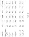
- the CH2 region in the Fc portion of IgGs has been found to be the domain that selectively binds FcR I and II (Ollo, 1983; Woof, 1984; Burton, 1985; Partridge, 1986; Duncan, 1988). In fact, the exact binding segment has been localized to an area corresponding to amino acids 234 to 238 (Duncan, 1988) and the respective affinity of several isotypes has been determined (Gergely, 1990).
- Duncan et al. have shown that the mutation of a single amino acid in the FcR binding segment of a murine IgG2b, converting the sequence to that found in a murine IgG2a, resulted in a 100-fold enhancement of the binding to FcR (1988).
- a mutation was introduced into the Fc region of an anti-CD3 human lgG4 antibody resulting in a sequence similar to the low affinity sequence of the murine IgG2b.
- This mAb contains a glutamic acid rather than a leucine at position 235 of the human IgG4 heavy chain (Glu-235 mAb).
- the mutational analysis was performed on a "humanized" anti-CD3 mAb, the gOKT3-5 mAb by splicing the murine complementarily determining regions into the human IgG4 framework gene sequence.
- the gOKT3-5 mAb was previously shown to retain binding affinity for the CD3 complex similar to murine OKT3 and all the in vitro activation and immunosuppressive properties of OKT3.
- the gOKT3-5 mAb had an FcR binding sequence differing by only two amino acids from the same region on the murine IgG2b or by one amino acid in the murine IgG2a/human IgG1. Since a mutation in the FcR binding region of the mAb could modify the conformation of the molecule and thus be responsible for a decrease in FcR binding regardless of the amino acid sequence obtained, we performed a control mutation of amino acid 234 from a phenylalanine into a leucine in order to mimic the FcR binding area found in the high affinity murine IgG2a and human IgG1. This mAb was designated Leu-234.
- the site-specific mutations described above were introduced into the Fc portion of the gOKT3-5 mAb to affect the binding of the Ab to FcR.
- the appropriate mutant of the anti-CD3 mAb was designed to exhibit the low-activating properties of F(ab') 2 fragments, the purity of a monoclonal antibody and an increased serum half-life as compared with F(ab') 2 fragments or possibly even with murine OKT3, since chimeric mouse/human antibodies have been shown to circulate longer their murine counterpart.
- the resulting mAb thus avoids the acute toxicity and the immunization induced by OKT3, in vivo, although, theoretically, the substitution of glutamic acid at position 235 in order to mimic murine lgG2b could also create an immunogenic epitope in the constant region of the humanized antibody.
- the reduced binding of the Glu-235 mAb correlated with a marked decrease in the T cell activation induced by this Ab, as assessed by the absence of T cell proliferation, the decreased expression of cell surface markers of activation, the diminished release of TNF- ⁇ and GM-CSF and the lack of secretion of IFN- ⁇ .
- the anti-CD3 mAbs employed in this study displayed an FcR binding as expected, with the human IgG4 gOKT3-5 mAb binding less avidly to U937 cells than murine IgG2a OKT3 or Leu-234 mAb, but with much higher affinity than the Glu-235 mAb.
- the extent of the functional changes generated in the FcR binding region of the gOKT3-5 mAb that form the Glu-235 mAb has further implications.
- the ability of certain isotypes of anti-CD3 mAbs to activate T cells and mediate ADCC has been shown to vary in the population.
- Murine IgG2a and IgG3 anti-CD3 mAbs are mitogenic for virtually all individuals.
- murine IgG1 and IgG2b mAbs induce proliferation in only 70% and 5% to 10%, respectively.
- the Glu mAb which appears to function as a non-activator lgG2b in a small fraction of the population.
- IgG2b mAbs seen to trigger a different pathway of activation in contrast to other anti-CD3 isotypes, IgG2b mAbs do not induce the production of IL-2 or IFN- ⁇ .
- the proliferation observed in the small subset of the patient population may be an IL-2 independent T cell mitogenesis, which has previously been reported in other settings.
- the reduced FcR binding of the Glu-235 mAb to FcR as compared with murine IgG2b Abs, may be sufficient to abrogate the activation of even this subset of individuals.
- the present invention contemplates a class of homo-bifunctional antibodies, a humanized version of OKT3 which also interacts with CD4.
- This humanized antibody has an Fv region containing the CD3 ⁇ antigen specificity of OKT3 and an Fc region from either human IgG1 or lgG4 antibody.
- the humanized anti CD3 antibody binds CD4 directly, either immobilized on plastic or on CD4 + , CD3 - , FcR cells.
- the antibody of the present invention binds with either a ⁇ 1 or a ⁇ 4 heavy chain.
- the CD4 binding site on humanized OKT3 has been mapped to the Fab fragment and probably resides in the framework sequences of the variable region.
- polypeptides an polynucleotides of the invention can be recognized as antigens, and thus identified. Once identified, those polypeptides and polynucleotides can be isolated and purified by techniques such as antibody-affinity chromatography. In antibody-affinity chromatography, a monoclonal antibody is bound to a solid substrate and exposed to a solution containing the desired antigen. The antigen is removed from the solution through an immunospecific reaction with the bound antibody. The polypeptide or polynucleotide is then easily removed from the substrate and purified.
- the present invention provides pharmaceutical compositions comprising antibodies immunoreactive with CD3 and CD4 cell surface antigens.
- a composition of the present invention is typically administered parenterally in dosage unit formulations containing standard, well-known nontoxic physiologically acceptable carriers, adjuvants, and vehicles as desired.
- parenteral as used herein includes intravenous, intramuscular, intraarterial injection, or infusion techniques.
- Injectable preparations for example sterile injectable aqueous or oleaginous suspensions, are formulated according to the known art using suitable dispersing or wetting agents and suspending agents.
- the sterile injectable preparation can also be a sterile injectable solution or suspension in a nontoxic parenterally acceptable diluent or solvent, for example, as a solution in 1,3-butanediol.
- Suitable vehicles and solvents that may be employed are water, Ringer's solution, and isotonic sodium chloride solution.
- sterile, fixed oils are conventionally employed as a solvent or suspending medium.
- any bland fixed oil can be employed including synthetic mono- or di-glycerides.
- fatty acids such as oleic acid find use in the preparation of injectables.
- Preferred carriers include neutral saline solutions buffered with phosphate, lactate, Tris, and the like.
- a preferred means of purifying the vector involves the use of buoyant density gradients, such as cesium chloride gradient centrifugation.
- a carrier can also be a liposome.
- Means for using liposomes as delivery vehicles are well known in the art [ See, e.g., Gabizon et al., 1990; Ferruti et al., 1986; and Ranade, V.V., 1989].
- a transfected cell can also serve as a carrier.
- a liver cell can be removed from an organism, transfected with a polynucleotide of the present invention using methods set forth above and then the transfected cell returned to the organism ( e.g . injected intravascularly).
- coli were infected with helper phage M-13 (pfu) (Stratagen) to generate uridine incorporated single stranded template.
- An oligonucleotide synthesized with thymidine and containing the desired mutation was then annealed to the uridine-single-stranded template to serve as a primer for the replication of the plasmid after the addition of deoxynucleotides, T7 polymerase and T4 ligase; the wild type DNA thus contains uridine, while the mutated plasmid obtained utilizes thymidine.
- the synthesis reaction was stopped with EDTA 0.5M and Tris HCI-EDTA 1M, and 10 ⁇ l were transformed into competent DH5 E.
- the plasmid was isolated by Qiagen minipreps; the mutated sequence in pSG5 was co-introduced with the psG5 vector containing the light chain of the mAb into COS-1 cells for transient expression of the mutant immunoglobulin.
- OKT3 variable region sequences were derived from oligo-dT primed cDNA from OKT3 hybridoma cells using the Amersham International Plc. cDNA synthesis kit. The cDNA was cloned in pSP64 using EcoR1 linkers. E.
- coli clones containing light and heavy chain cDNAs were identified by oligonucleotide screening of bacterial colonies using the oligonucleotides: 5' TCCAGATGTTAACTGCTCAC (SEQ ID NO: 15) for the light chain, which is complementary to a sequence in the mouse kappa constant region, and 5' CAGGGGCCAGTGGATGGATAGAC (SEQ ID NO: 16) for the heavy chain, which is complementary to a sequence in the mouse igG2a constant CH1 domain region.
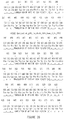
- FIG. 1A row 1 for the light chain
- FIG. 1B row 1 for the heavy chain
- the CDR's are shown with the single underlining.
- the light chain is a member of the mouse V L subgroup VI and uses a J K 4 minigene.
- the heavy chain is probably a member of the mouse V H subgroup II, most probably IIb, although it also has significant homology to the consensus for group Va.
- the D region is currently unclassified and the J H region is J H 2.
- the loops can be assigned to canonical structures 1 for L1, 2 for L2 and 1 for L3, and to canonical structures 1 for H1 and 2 for H2, Chothia et al., have not yet predicted canonical forms for H3.
- the light chain variable region amino acid sequence shows a high degree of homology to the Ox-1 germline gene and to the published antibodies 45.2.21.1, 14.6b.1 and 26.4.1 (Sikder, 1985).
- the heavy chain variable region amino acid sequence shows reasonable homology to a subgroup of the J558 family including 14.6b.1.
- variable region domains for the humanized antibodies were designed with mouse variable region optimal codon usage (Grantham, 1986) and used the signal sequences of the light and heavy chains of mAb B72.3 (Whittle, 1987). Immediately 5' to the initiator ATG a 9bp Kozak sequence (Kozak, 1987), GCCGCCACC (SEQ ID NO: 17), was inserted. 5' and 3' terminal restriction sites were added so that the variable regions could be attached directly to the DNA sequences for the human IgG4 and Kappa constant regions prior to cloning into the eukaryotic expression vectors.
- variable regions were built either by simultaneously replacing all of the CDR and loop regions by oligonucleotide directed, site-specific mutagenesis (Ollo, 1983) of a previously constructed humanized variable region for B72.3 cloned in M13 (Emtage et al. ), or by assembling the sequence using synthetic oligonucleotides ranging in size from 27-67 base pairs and with 6 base overhangs.
- the oligonucleotides were synthesized on an Applied Biosystems Model 380B DNA Synthesizer and purified by HPLC.
- the oligonucleotides were enzymatically phosphorylated, paired, annealed and then equimolar aliquots of each pair were mixed and ligated.
- the cloning sites were exposed by restriction digestion of the ligation mixture and the correctly sized fragments were identified and cloned directly into the expression vectors, 5' to the constant regions, prior to sequencing and expression.
- variable region sequences of the human acceptor frameworks are shown in FIG. 1A and FIG. 1B (row 2) (SEQ ID. NOS: 7 and 11).
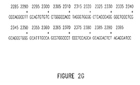
- amino acid and nucleotide sequences for murine OKT3 are provided in FIG. 2A to FIG. 2G.
- FIG. 1A shows the sequences for the variable regions of the initial design, gL and gH. Only differences from the human acceptor sequence are shown.
- OKT3 CDR's as suggested by reference to Kabat et al., were substituted into the KOL sequence along with the murine residues at positions 27, 28 and 30 which are normally bound in a loop region adjacent to CDR1 (Chothia, 1987; 1989).
- residue 27 is the same in both KOL and OKT3 (FIG. 1B) and therefore does not require to be altered.
- the DNA sequences coding for the initial humanized light and heavy variable regions were constructed by simultaneous replacement through site-directed mutagenesis of sequences in previously generated light and heavy chain DNAs of a humanized form of antibody B72.3.
- the DNA sequences coding for the humanized variable regions were then attached to the human gamma-4 and kappa constant region sequences and inserted into expression vectors as described for the chimeric genes.
- the gL and gH genes, when co-expressed in COS cells yield antibody gOKT3-1.
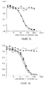
- gOKT3-1 binds poorly to HPB-ALL cells and is not able to block the binding of mOKT3 to the cells (FIG.3A and FIG.3B). Therefore it was clear that further OKT3 residues outside of the CDRs needed to be considered for substitution into the humanized antibody.
- these positions are at 1 and 3 which by reference to known structures for antibody variable regions are probable surface residues located near to the CDR's, residue 46 which is usually at the domain interface and the packing residue at 47, gLA has all four residues derived from the murine sequence while gLC has murine residues at positions 46 and 47 only.
- FIG. 1A, row 4 shows the sequence of gLC which differs from gL by having the murine sequences at residues 46 and 47.
- FIG. 1B row 4 shows the sequence of gLC which differs from gL by having the murine sequences at residues 46 and 47.
- FIG. 3A. and FIG 3B. show results from two such experiments.
- the affinity of mOKT3 for antigen (K a ) was measured to be 1.2 x 10 9 M -1 by Scatchard analysis. This value for mOKT3 compares well to that of 1.3 x 10 9 M -1 by Scatchard analysis. This value for mOKT3 compares well to that of 1.3 x 10 9 M -1 determined previously (Gergely, 1990).
- FIG. 3A gOKTE3-5 was compared with cOKT3 and mOKT3 for competition against mOKT3. Values of 1.2 x 10 9 M -1 and 1.1 x 10 9 M -1 2343 obtained for the cOKT3 and gOKT3-5 antibodies respectively.
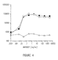
- gOKT3-7 the activation potency of gOKT3-7 antibody was assessed by quantitating proliferating responses.
- gOKTE-7 demonstrated mitogenic potency equivalent to that of mOKT3 (FIG. 4). This suggests that cross-linking of the bound antibody still occurs with the ⁇ 4 isotype leading to proliferative signals.
- a therapeutic humanized OKT3 antibody may need further alterations to the constant region to minimize such effects.
- EXAMPLE 4 Construction and expression of chimeric OKT3 genes .
- the murine cDNAs were assembled into expression vector controls for the biological function of the humanized antibodies.
- the murine variable region cDNA sequences were attached to human k light chain and ⁇ 4 heavy chain constant region DNA sequences following a previously described strategy to generate chimeric OKT3 (cOKT3) genes which were then inserted into eukaryotic expression vectors.
- cOKT3 chimeric OKT3
- Murine OKT3, cOKT3, and murine/chimeric hybrid antibodies expressed from COS cells were shown to bind to antigen equivalently to mOKT3 and to block the binding of MOKT3 to CD3 positive cells.
- EXAMPLE 5 Transient expression of murine and human-OKT3 mAbs genes.
- COS-1 cell expression studies were performed using reagents and procedures from a transient expression kit (Specialty media, Lavallette, NJ) modified for use in roller bottles (T. Livelli, Specialty Media, personal communication). Product supernatants for purification of the test Abs were harvested 6 days after transfection.
- ELISA assays were performed to determine the yield of assembled "humanized” antibody in COS cells supernatants.
- Ninety-six well plates were coated with F(ab') 2 goat anti-human Fc antibody.
- COS cell supernatants were added and incubated for one hour at room temperature and washed.
- Horseradish peroxidase-conjugated goat anti-human kappa chain (Caltag) was used with o-phenylenediamine (OPD) for detection.
- Purified human IgG was used as standard.
- EXAMPLE 6 Mutated "humanized” OKT3 mAbs bind to the CD3 complex of T cells with the same affinity as murine OKT3 .
- the Fc portion of the gOKT3-5 mAb was mutated according to procedures described above in order to alter its binding to FcR-bearing cells.
- a phenylalanine was substituted for a leucine in position 234 (Leu-234), or the adjacent leucine (235) was transformed into a glutamic acid (Glu-235).
- the affinity of the gOKT3-5 mAb for the TCR complex was previously shown to be similar to that of OKT3 (Van Wauwe, et al., 1980).
- a displacement assay was performed to examine the ability of the mutated Abs to competitively inhibit the binding of murine OKT3 to human T cells.
- Human peripheral blood acute lymphocytic leukemia cells were re-suspended in flow cytofluorimetry (FCM) buffer at 5 x 10 5 cells/mL. Dilutions of the anti-CD3 mAbs were added and incubated at 4°C for 1 hour.
- Fluorescein isothiocyanate (FITC) was dissolved in N,N-dimethyl formamide (DMF) to give a 10 mg/ml solution.
- FITC/DMF was added to purified mAb at 1:10 w/w and incubated at 25°C for four hours, followed by dialysis into PBS containing an anion exchange resin (AG1-X8, 200-400 mesh, chloride form; Bio-Rad). Aggregates were removed prior to use by airfuge centrifugation (Becton-Dickinson). A fixed saturating amount of OKT3-FITC was added, and the cells were further incubated for 1 hour at 4°C, washed and analyzed by flow cytofluorimetry (FCM).
- FCM flow cytofluorimetry
- One or two-color FCM were performed using a FACScan flow cytometer, interfaced to a Hewlett-Packard 310 computer. Data analysis were performed using Consort-30 software. Logarithmically amplified fluorescence data were collected on 10,000 viable cells, as determined by forward and right angle light scatter intensity. One-color fluorescence data were displayed in histogram mode with fluorescence intensity on the x axis and cell number of the y axis. Two-color fluorescence data were displayed as contour plots with green (FITC) fluorescence on the x axis and orange (phycoerythrin) fluorescence on the y axis. All FCM staining procedures were performed at 4°C in FCM buffer.
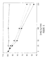
- results of this assay are shown in FIG. 5.
- the data is presented as % inhibition of maximal fluorescence intensity (determined by OKT3-FITC binding in the absence of blocking Ab).
- Both mutant Abs displayed a similar affinity for their epitope as the parental gOKT3-5 mAb.
- the gOKT3-6 mAb a different "humanized" OKT3 which has a very weak binding activity for the CD3 antigen (Van Wauwe, et al., 1980), was unable to displace the OKT3 mAb.
- the anti-CD3 mAbs expressing different isotypes had a comparable avidity for the TCR complex as assessed by Scatchard analysis (Van Wauwe, et al. , 1980), or by precipitation of the TCR complex and cross-blocking experiments.
- Scatchard analysis Van Wauwe, et al. , 1980
- precipitation of the TCR complex and cross-blocking experiments.
- any differences in the activation or suppressive properties of the mutated Abs could not be attributed to a modified affinity of the combining site of the anti-CD3 mAbs for T cells.
- the mutations generated in the CH2 region of the human IgG4 gOKT3-5 either mimicked the amino acid sequence of the FcR binding region of a human IgG1 (Leu-234), which has a higher affinity for human FcR I than human IgG4, or of a murine IgG2b (Glu-235) that binds weakly to FcR I but still binds to human FcR II.
- Phycoerythrin-coupled (PE) anti-CD2 and anti-CD5 used as counterstains in the activation assays were purchased from Coulter Immunology. Modulation and coating of the TCR were determined using FITC-coupled OKT3 IgG2a and OKT3D IgG2a as described below.
- FcR binding assays were performed using the FcR I- and II-bearing U937 human cell line.
- the pelleted cells (bound 125 I-hulgG) separated from the medium containing free 125 I-hulgG.
- the tubes were then frozen in dry ice and the bottom of the tube containing the pelleted cells was removed for analysis of the bound 125 I-hulgG.
- the maximum binding of 125 I-hulgG was determined in the absence of the inhibitor. The results are expressed as a percentage of the 125 I-hulgG bound in the presence of the inhibitor relative to the maximum binding. Non-specific binding is seen as the percentage bound in the presence of excess inhibitor (150 ⁇ g/ml murine OKT3). All controls and samples were assayed in triplicate tubes.
- Murine OKT3 IgG2a had, as expected, the highest affinity of all the anti-CD3 mAbs tested for FcR on U937 cells. As previously shown for human IgG4 mAbs, the gOKT3-5 required a 10-fold higher concentration to achieve the same inhibition. The Leu-234 mAb, that was expected to enhance FcR binding, has consistently proven to compete more efficiency for FcR binding than the gOKT3-5 mAb.
- the Glu-235 mAb bearihg the FcR binding region similar to murine IgG2b, bound poorly to U937 cells, requiring a 10-fold higher concentration than the gOKT3-5 and approximately a 100-fold greater concentration than the murine OKT3 to achieve the same percent inhibition.
- PBMC peripheral blood mononuclear cells
- PBMC peripheral blood mononuclear cells
- EBV-transformed lymphoblastoid cell lines (LCL) and human histiocytoma-derived U937 cell-line were maintained in continuous culture in complete media (DMEM supplemented with 2mM L-glutamine), 2 mM non-essential amino acids, 100 U/mL penicillin-streptomycin (Gibco), 5x10 5 M 2-mercapto-ethanol (Gibco) and 25 ⁇ M HEPES (Gibco) with 10% fetal calf serum (FCS, Gibco).
- DMEM penicillin-streptomycin
- Gibco 5x10 5 M 2-mercapto-ethanol
- FCS fetal calf serum
- PBMC preparations were re-suspended in complete DMEM with 1% FCS and aliquotted to 96-well round bottom tissue culture plates (Costar) at 1x10 6 cells/well.
- the different Abs were added to the wells by serial log dilutions in culture media. After 72 hours of culture at 37°C in a 5% CO 2 incubator, 1 ⁇ Ci of 3 H-thymidine was added to each well and followed by an additional 24 hour incubation. Cells were harvested on a semi-automatic cell harvester and 3 H-thymidine incorporation was measured in a liquid scintillation counter. All data were expressed as mean CPM of triplicate determinations.
- EXAMPLE 9 Activation of T cells by CDR-grafted mutant mAbs .
- PBMC peripheral blood mononuclear cells
- EXAMPLE 11 Induction of modulation and coating of the TCR complex by molecularly engineered OKT3 mAbs .
- the immunosuppressive properties of the different mAbs was compared in vitro .
- the mAbs were examined for their capacity to modulate and/or coat the TCR complex.
- Human peripheral blood mononuclear cells (PBMC) were incubated at 1x10 6 cells/mL for 12 hours in 24 well plates with known concentrations of anti-CD3 mAb.
- PBMC from each group were harvested and stained with either OKT3-FITC or OKT3D-FITC.
- the fluorescein-stained cells were counterstained with anti-CD5-PE to identify T lymphocytes and analyzed by flow cytofluorimetry (FCM).
- OKT3D-FITC was selected because of its binding to an epitope distinct from the one binding OKT3 mAb.
- FCM flow cytofluorimetry
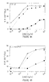
- the combined modulation and coating of the TCR complex achieved by the gOKT3-5 (FIG. 10B) and murine OKT3 (FIG.10A) were very similar, with half-maximal TCR blocking achieved at approximately 1 ng/ml.
- the half-maximum modulation plus coating observed with the Glu-235 (FIG.10C) mAb required a 100-fold greater concentrations of mAb (1 ⁇ g/mL) than of murine OKT3.
- the major difference between the Glu-235 mAb and the other Abs was due to a change in kinetics since, by 48 hours, the mAb coated and modulated the TCR complex similarly to OKT3 (data not shown).
- the achievement by Glu-235 mAb of internalization of the TCR which may depend on multivalent cross-linking, was delayed as compared with the other anti-CD3 mAbs.
- HLA-A2-specific CTL were generated from a normal HLA-A1 donor. Cytolytic activity was assessed on FcR negative-EBV-transformed HLA-A2 target cells. CTL were generated by a bulk allogeneic MLC technique. Normal human donors were phenotyped for HLA-A expression. Responder and stimulator combinations were selected specifically to generate HLA-A2-specific CTL effectors.
- Responder and stimulator PBMC were prepared by Ficoll-hypaque density gradient centrifugation as described above and re-suspended in RPMI 1640 with 2mM L-glutamine, 100 U/mL penicillin-streptomycin, 25 ⁇ M HEPES and 15%. decomplemented normal human serum.
- Stimulator PBMC (1 x 10 7 /mL) were irradiated (3000 rad) and cultured with responder PBMC (1 x 10 7 /10mL) in upright 25 cm tissue culture flasks.
- HLA-A2-specific CTL effectors were generated as described above, harvested and aliquotted to a 96 well U-bottom tissue culture plate at four different effector/target ratios. Effectors were pre-incubated with serial dilutions of each anti-CD3 mAb for 30 minutes. Following incubation with mAbs, 51 Cr-labeled Fc receptor negative-target cells [HLA-A2 expressing LCL line (Z2B) or HLA-A1 expressing LCL line (G12B) used as a non-specific target] were added. Spontaneous lysis was measured by incubation of targets alone in media and maximal lysis was achieved by addition of 0.05 N HCL. Effectors and targets were co-cultured; supernatant aliquots were harvested and radioactivity was measured in a gamma-counter.
- T cell cytotoxicity was specific as demonstrated by the absence of lysis of a syngeneic HLA-A1 EBV-transformed cell-line (data not shown). Inhibition of lysis by anti-CD3 mAbs previously has been attributed to the inability of the T cells to recognize their targets, due to TCR blockade by the mAb. In the present study, murine OKT3, gOKT3-5 mAb and Glu-235 exhibited a comparable inhibitory effect on the cytolytic activity of the alloreactive T cells. These results suggest that the ability of the different mAbs to coat the TCR within the 30 min incubation time was similar (see FIG. 11).
- PBMCs isolated from Ficoll-Hypaque density gradient centrifugation were incubated at 1 x 10 6 cell/mL with known concentrations of OKT3 antibodies at 37° C for 24 hours.
- the cells were harvested and stained with FITC-OKT4.
- the cells were counterstained with PE-labelled anti-CD5 (PE-Leu1, Becton Dickinson Immunocytometry Systems, San Jose, CA) to distinguish T lymphocytes from other PBMCs, and analyzed by FACScan. Data from the resulting studies are reported in FIG.12A and FIG.12B (Transy, 1989).
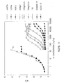
- %CD4 modulation was calculated as follows: Control MCN FITC-OKT4 - Ab treated MCN FITC-OKT4 Control MCN FITC-OKT4 ⁇ 100
- Fig. 12A The data in Fig. 12A reveal that the humanized antibodies studied induce the modulation of CD4 in a dose-dependent manner.
- mOKT3 solid circles
- the antibody from which the humanized and mutated antibodies were constructed had no effect on CD4, as indicated by a straight line plot between antibody concentrations of from 0.01 to 10.0 ⁇ g/mL.
- the same can be said for the mOKT3D IgG2b antibody (solid triangles) which has also been neither humanized nor mutated.
- FIG.12B indicates that, as expected, there is no modulation of CD8 for any of the antibodies studied.
- RES-KW3 cells were washed with PBS+0.2%BSA+0.1% sodium azide (staining buffer), and first incubated with various concentrations of OKT3 antibodies for 1 hour on ice. The cells were washed three times with cold staining buffer, and FITC-labelled goat anti-human or goat anti-mouse antibodies were added (Caltac Lab. So. San Francisco, CA). The cells were incubated on ice for another hour before being washed and subject to FCM.
- FCM was performed using a FACScan (Becton-Dickinson Immunocytometry Systems, Mountain View, CA) flow cytometer interfaced to a Hewlett-Packard 340 computer, data analyzed using lysis II software (Becton Dickinson). Fluorescence data were collected using logarithmic amplification on 10,000 viable cells as determined by forward and right angle light scatter intensity. One-color fluorescence data were displayed in histogram mode with fluorescence intensity on the x axis and relative cell number on the y axis.
- FACScan Becton-Dickinson Immunocytometry Systems, Mountain View, CA
- lysis II software Becton Dickinson
- HIVgp120/CD4 receptor EIA coated microplates from DuPont were used in the CD4 binding assay.
- 100 ⁇ L/well of CDR-grafted OKT4AIgG1 at various concentrations (1:2 dilution at starting concentration of 50 ng/mL) was added into the wells duplicate for the construction of standard curve.
- 100 ⁇ L/well of OKT3 antibody samples at various dilutions were then added.
- the diluent is PBS + 10% calf serum + 0.05% Tween-20. The plates were incubated at room temperature for 2 hours.
- the plates were washed with PBS+0.05% Tween-20 six times before 100 ⁇ L/well of 1:15000 diluted HRPO-conjugated goat anti-human x(f+B) antibodies in diluent was added. The plates were incubated at room temperature for another 2 hours. The plates were washed six times again, and 100 ⁇ L/well of the OPD/hydrogen peroxide solution (five 2-mg OPD tablets were added in 13 mL of Mili-Q water; after they were dissolved, 5 ⁇ L of 30% hydrogen peroxide were then added) was added into each well. The plates were incubated at room temperature in the dark for 30 minutes, and 50 ⁇ L/well of 2.5N HCI was added to stop the reaction. The plates were then read at 490 nm.
- FIG.13 and FIG.14 The resulting data are reported in FIG.13 and FIG.14. These data indicate that the humanized OKT3 binds to CD4, either immobilized to ELISA plates or bound to the surface of RES-KW3 cells. It will be appreciated by one skilled in the art that data such as that indicated in FIG.14 for 209IgG1A/A-1 (open circles) are unexpected, and suggest that divalent binding (binding to both CD3 and CD4, for example), is needed for stable attachment of this antibody to the plate.
- EXAMPLE 15 Generation of a Non-Activating Anti-CD3 mAb Based on gOKT3-7 .
- the inventors To generate an anti-human CD3 mAb with an improved therapeutic index, the inventors have developed a panel of "humanized" anti-CD3 mAbs derived from OKT3, by molecularly transferring the complementary determining regions (CDRs) of OKT3 onto human IgG1 and IgG4 molecules (Woodle et al., 1992; Adair et al., submitted for publication). In addition, the inventors examined whether immunosuppression can be achieved by anti-CD3 mAbs in the absence of the initial step of cellular activation.
- CDRs complementary determining regions
- the "humanized” mAb formally named gOKT3- 7( ⁇ 1 ), abbreviated 209-IgG1, that has a high affinity for human FcrRs was shown, in vitro, to have similar activating properties to OKT3 (Alegre, 1992; Xu et al. , manuscript in preparation) and would therefore be expected to induce in patients the acute toxicity associated with lymphokine release by activated T cells and Fc ⁇ R-bearing cells.
- a second mAb formally named gOKT3-7( ⁇ 4 -a/a); abbreviated Ala-Ala-IgG4, was developed with 2 amino acid substitutions in the CH 2 portion (from a phenylalanine-leucine to an alanine-alanine at positions 234-235) of the "humanized" gOKT3-7( ⁇ 4) (209-IgG4) mAb.
- These mutations significantly reduced binding of the mAb to human and murine Fc ⁇ RI and II and led to markedly reduced activating characteristics in vitro (Alegre, 1992; Xu et al., manuscript in preparation).
- this variant mAb retained the capacity to induce TCR modulation and to prevent cytolysis in vitro (Xu et al., manuscript in preparation), and thus represents a potential new immunosuppressive therapeutic agent.
- SCID mice Severe combined immunodeficient mice carry an autosomal recessive, spontaneously arising mutation that results in the inability to successfully rearrange immunoglobulin and TCRs. These animals are therefore devoid of T and B lymphocytes (McCune, Annu. Rev. Immun., 1991; McCune, Curr. Opin. Immun., 1991; Bosma, 1983; Bosma, 1991).
- the inventors have recently developed a model in which lightly irradiated SCID mice are injected with human splenocytes from cadaveric organ donors (Alegre et al., manuscript submitted).
- hu-SPL-SCID mice maintain functional human T cells capable of responding to mitogens and alloantigens in vitro, and of acutely rejecting human foreskin allografts in vivo .
- the inventors have utilized hu-SPL-SCID mice to assess the immunosuppressive properties of the non-activating "humanized" anti- CD3 mAbs in vivo. MATERIALS AND METHODS Abbreviations.
- mice Homozygous C.B-17 scid/scid (SCID) H-2 d founder mice were obtained from Dr. M. Bosma (Fox Chase, Phila, PA) and were subsequently bred in the specific pathogen-free animal barrier facility at the University of Chicago.
- Antibodies. 145-2C11 a hamster anti-mouse CD3 mAb, was purified from hybridoma supernatant using a protein A column (Sigma, Saint Louis, MO), as previously described (Leo, 1987). OKT3, 209-IgG1 and Ala-Ala-lgG4 were generated as described below.
- Phycoerythrin (PE)-coupled anti-human CD4 and CD8, as markers of T cells were obtained from Coulter Immunology (Hialeah, FL).
- the fluorescein isothiocyanate (FITC)-coupled anti-CD69 an early marker of T cell activation, was purchased from Becton Dickinson (San Jose, Ca). All anti-human Abs were tested to exclude cross-reactivity on murine cells.
- hu-SPL-SCID mice Fresh human spleens were obtained from cadaveric organ donors, under a protocol approved by the University of Chicago Institutional Review Board. A single cell suspension was prepared as previously described (Alegre et al., manuscript submitted). Briefly, 4 to 6 week-old SCID mice were r -irradiated (200 rad), prior to the intraperitoneal (ip) injection of 10 8 cells/mouse. The percentage of human cells in the peripheral blood was determined by flow cytometry (FCM). First, the peripheral blood mononuclear cells (PBMCs) were incubated (15 minutes) with unlabelled murine IgG antibodies to block subsequent Fc ⁇ R binding.
- PBMCs peripheral blood mononuclear cells
- the cells were stained with PE-coupled anti-murine class I (PharMingen, San Diego, Ca) and counterstained with FITC-coupled anti-human CD45 mAb (Coulter Immunology, Hialeah, FL) to identify the population of human cells.
- the proportion of human cells is expressed as a percentage of the total number of cells.
- the animals bearing between 5 and 20% human cells in the PBMCs were selected for further experiments.
- mice matched for their level of engraftment of human cells in the peripheral blood, received either PBS (1 ml), 145-2C11, OKT3, 209-IgG1 or Ala-Ala-IgG4 (100 ⁇ g resuspended in 1 ml of PBS, unless stated otherwise in the text), intraperitoneally (ip) 11 days to 3 weeks after the injection of the human splenocytes.
- Human PBMCs were incubated with 3 serial dilutions of each serum (1:10, 1:30 and 1:90), and then stained with FITC-coupled goat anti-mouse Ig (Boehringer-Mannheim, Indianapolis, IN) for detection of OKT3, and with goat anti-human Ig (Caltag Laboratories, San Francisco, CA) for detection of the humanized antibodies. Serum levels were extrapolated from the mean fluorescence of anti-CD3 stained cells, as compared with a corresponding concentration of the purified anti-CD3 mAbs on the standard curves.
- an anti-murine IL-4 mAb [11B11 (Ohara, 1985)]
- an anti-murine IL-2 mAb [S4B6, (Cherwinski, 1987)]
- Neonatal human foreskin was grafted on SCID and hu-SPL-SCID mice 11 days after the inoculation of human splenocytes.
- Mice were anesthetized with 60 ⁇ g/ml of chlorohydrate (120 ⁇ l delivered ip) (Sigma, St. Louis, MO) and intermittent inhalation of hydroxyflurane (Metophane, Pitman-Moore, Mundelein, IL). Skin grafts were positioned on the dorsal thorax of the mice.
- Each foreskin was used to graft 4 animals, each from a different group (SCID, PBS-treated, 145-2C11-treated and antl-CD3-treated hu-SPL-SCID mice).
- Mice received OKT3, 209-IgG1, Ala-Ala-IgG4 or 145-2C11 (50 ⁇ g/day for 5 days, followed by 10 ⁇ /g/day for 10 days) diluted in 1 ml of PBS, or 1 ml of PBS alone.
- the grafts were unwrapped at 7 days and the status of the graft was scored blindly and independently by 2 investigators daily for the first 30 days, and once a week afterwards.
- grade 0 represented skin grafts intact and soft; grade 1, skin grafts with a modified pigmentation in a small area; grade 2, soft skin grafts with larger areas of depigmentation; grade 3, those hardened or slightly scabbed; grade 4, shrinking or scabbing skin grafts. Rejection was recorded when scores were grade 3 or greater.
- mice were tested for the percentage of human cells engrafting their peripheral blood. As previously described, graft versus host disease (GVHD) was apparent in mice bearing more than 25 to 30% human cells (Alegre et al., manuscript submitted). Therefore, in order to minimize the level of human T cell activation prior to anti-CD3 treatment, animals with 5% to 20% circulating human CD45 + cells were selected for subsequent experiments. Mice matched for their level of engraftment with human cells were assigned to different groups for treatment with OKT3, 209-IgG1, Ala-Ala-lgG4 or PBS.
- hu-SPL-SCID splenocytes harvested 3 days after the completion of this protocol were unable to proliferate to immobilized OKT3, in vitro (data not shown). It is interesting to note that the ability of OKT3 to deplete T cells from human lymphoid compartments such as spleen or lymph nodes is unknown. However, studies using the anti- mouse CD3 mAb, 145-2C11, have shown that T cells are also depleted from the peripheral lymphoid organs of the immunocompetent mice.
- IL-2 Production of IL-2 after anti-CD3 therapy .
- the administration of OKT3 to patients has been shown to induce the rapid systemic release of cytokines such as TNF- ⁇ , IL-2, IL-6 and IFN- ⁇ , peaking 2 to 6h after the injection (Abramowicz, 1989; Chatenoud, 1989).
- This cytokine production results in the acute toxicity associated with anti-CD3 therapy in transplant recipients.
- a bioassay was used to measure the serum level of human IL-2 2h after treatment of hu-SPL-SCID mice with PBS, OKT3, 209-IgG1, Ala-Ala-IgG4 or 145-2C11, a hamster anti-murine CD3 mAb.
- hu-SPL-SCID mice are capable of rejecting human foreskin allografts and that human T cells participate in this process (Alegre et al., manuscript submitted).
- SCID and hu-SPL-SCID mice were grafted with human foreskin obtained from circumcisions and assumed to be allogeneic with respect to the human cells used for the adoptive transfer.
- Hu-SPL-SCID mice matched for their level of human CD45 expression in the peripheral blood received either PBS or daily doses of OKT3, 209-IgG1, Ala-Ala-IgG4, or 145-2C11 for 15 consecutive days, beginning on the day of the skin graft. As shown in FIG.
- the PBS-treated animals retained a significant percentage of human CD45 + and CD3 + cells in the different compartments although the absolute numbers were reduced over time, as compared with the initial engraftment (data not shown).
- Three additional skin graft experiments have been performed with 5-7 animals per group. In these experiments, 66-80% of the animals treated with OKT3, 209-lgG1 and Ala-Ala-IgG4 maintained their grafts for as long as the animals were examined. In two of the three experiments, a higher percentage of mice treated with the Ala-Ala-IgG4 maintained their skin grafts permanently. No statistical difference was found between these 3 groups.
- OKT3 has been shown to mediate T cell activation by cross-linking T lymphocytes and Fc ⁇ R + cells (Palacios, 1985; Ceuppens, 1985; Kan, 1985). Because hu-SPL-SCID mice are chimeric animals comprising both murine and human FcR + cells, it was important to use mAbs that would have similar avidities for human and murine Fc ⁇ Rs. Thus, OKT3, a murine IgG2a, and the human 209-IgG1 mAb have a high affinity for Fc ⁇ Rs of both species (Xu et al., manuscript in preparation).
- the human Ala-Ala-IgG4 bears mutations dramatically reducing its binding to murine and human Fc ⁇ Rs.
- the efficacy of engraftment of the different cellular compartments with human B cells, monocytes/macrophages and NK cells, as providers of human Fc ⁇ R, is relatively low in this hu-SPL-SCID model [10% in the peritoneal cavity and the peripheral blood and 20% in the spleen (Alegre et al., manuscript submitted)], when compared to the proportion of human T lymphocytes observed.
- murine monocytes/macrophages and NK cells are functionally normal in SCID mice and express normal levels of murine FcrR (Bosma, 1991; Kumar, 1989).
- OKT3 and 209-IgG1-triggered activation of the human T lymphocytes was evident in the treated mice, as determined by the production of human IL-2 and the accumulation of human IFN-r mRNA, as well as by the increased expression of the surface marker of activation, CD69, on T cells.
- the inability of Ala-Ala-IgG4 to interact with Fc r Rs rendered this mAb incapable of fully triggering T cell activation.
- T lymphocytes and Fc ⁇ R + cells in patients treated with OKT3 are associated with adverse reactions such as fever, chills, headaches, acute tubular necrosis, diarrhea, acute respiratory distress syndrome etc. (Abramowicz, 1989; Chatenoud, 1989; Toussaint, 1989; Thistlethwaite, 1988; Goldman, 1990).
- immunocompetent mice injected with 145-2C11 develop hypothermia, hypoglycemia, lethargy, liver steatosis and acute tubular necrosis (Alegre, Eur. J. Immun. , 1990; Alegre, Transplantation , 1991; Feran, 1990).
- Hu-SPL-SCID mice did not exhibit detectable symptoms after OKT3 or 209-IgG1 therapy if the percentage of human cell engraftment was moderate. However, when animals with more than 30% human cells in their PBMCs were injected with OKT3 or 209-IgG1, they became extremely lethargic and an increased percentage of animal deaths was observed. As shown previously, animals engrafted with a high percentage of human T cells often undergo a GVHD-like syndrome, that results in a number of pathological symptoms including pancreatitis, diffuse hemorrhagic necrosis and in many instances animal death. Interestingly, the administration of Ala-Ala-IgG4 to highly engrafted animals seemed to reduce the symptoms of GVHD and perhaps even prevent some deaths. The number of animals examined was, however, too small to generate statistical differences.
- the Ala-Ala-lgG4, a mAb bearing 2 amino acid mutations in the Fc portion of a "humanized” OKT3, may prove useful in clinical transplantation to induce immunosuppression while being less immunogenic and induce less adverse reactions than OKT3.
- the use of a "humanized” mAb may lessen the generation of anti-xenotypic Abs that often arise after repeated administrations of OKT3 (Thistlethwaite, 1988).
- the non-activating Ala-Ala-lgG4 mAb might also widen the applications of anti-CD3 mAbs to patients suffering from autoimmune diseases, in whom treatment with OKT3 was never realized because of the potential adverse reactions and the strong humoral responses induced by the mAb.
- EXAMPLE 16 In Vitro Uses of Antibodies .
- the claimed antibodies will have a variety of in vitro uses. Some of these are described below, others will be understood by those of skill in the art.
- the antibodies of the invention will find utility in immunoassays for the detection of CD3.
- preferred immunoassays of the invention include the various types of enzyme linked immunosorbent assays (ELISAs) known to the art.
- ELISAs enzyme linked immunosorbent assays
- the utility of antibodies is not limited to such assays, and that other useful embodiments include RIAs and other non-enzyme linked antibody binding assays or procedures.
- samples to be tested for CD3 are immobilized onto a selected surface, preferably a surface exhibiting a protein affinity such as the wells of a polystyrene microtiter plate.
- a nonspecific protein such as bovine serum albumin (BSA), casein or solutions of milk powder onto the well that is known to be antigenically neutral with regard to the anti-CD3 antibody.
- BSA bovine serum albumin
- casein casein
- solutions of milk powder onto the well that is known to be antigenically neutral with regard to the anti-CD3 antibody.
- the immobilizing surface is contacted with the anti-CD3 antibody in a manner conducive to immune complex (antigen/antibody) formation.
- Such conditions preferably include diluting with diluents such as BSA, bovine gamma globulin (BGG) and phosphate buffered saline (PBS)/Tween. These added agents also tend to assist in the reduction of nonspecific background.
- BSA bovine gamma globulin
- PBS phosphate buffered saline
- the layered antibody is then allowed to incubate for from 2 to 4 hours, at temperatures preferably on the order of 25° to 27°C. Following incubation, the antibody-contacted surface is washed so as to remove non-immunocomplexed material.
- a preferred washing procedure includes washing with a solution such as PBS/Tween, or borate buffer.
- the occurrence and even amount of immunocomplex formation may be determined by subjecting same to a second antibody having specificity for the anti-CD3 antibody.
- the second antibody will preferably be an antibody having specificity in general for human IgG.
- the second antibody will preferably have an associated enzyme that will generate a color development upon incubating with an appropriate chromogenic substrate.
- a urease or peroxidase-conjugated anti-human IgG for a period of time and under conditions which favor the development of immunocomplex formation (e.g ., incubation for 2 hours at room temperature in a PBS-containing solution such as PBS-Tween).
- the amount of label is quantified by incubation with a chromogenic substrate such as urea and bromocresol purple or 2,2'-azino-di-(3-ethyl-benzthiazoline-6-sulfonic acid [ABTS] and H 2 O 2 , in the case of peroxidase as the enzyme label. Quantification is then achieved by measuring the degree of color generation, e.g. , using a visible spectra spectrophotometer.
- a chromogenic substrate such as urea and bromocresol purple or 2,2'-azino-di-(3-ethyl-benzthiazoline-6-sulfonic acid [ABTS] and H 2 O 2 , in the case of peroxidase as the enzyme label.
- Quantification is then achieved by measuring the degree of color generation, e.g. , using a visible spectra spectrophotometer.
- Fluorescent activated cell sorting, flow cytometry or flow microfluorometry provides the means of scanning individual cells for the presence of an antigen.
- the method employs instrumentation that is capable of activating, and detecting the exitation emissions of labeled cells in a liquid medium.
- FACS Fluorescence Activated Cell Sorting
- the antibodies of the present invention may also be used in conjunction with both fresh-frozen and formalin-fixed, paraffin-embedded tissue blocks prepared from study by immunohistochemisty (IHC).
- IHC immunohistochemisty
- each tissue block consists of 50 mg of residual "pulverized" tumor.
- the method of preparing tissue blocks from these particulate specimens was developed and has been successfully used in previous IHC studies of various prognostic factors, and is well known to those of skill in the art (Brown, et al. (1990); Abbondanzo, et al. (1990); Allred et al. (1990)).
- frozen-sections may be prepared by (A) rehydrating 50 ng of frozen "pulverized” breast tumor at room temperature in PBS in small plastic capsules, (B) pelleting the particles by centrifugation, (C) resuspending them in a viscous embedding medium (OCT), (D) inverting the capsule and pelleting again by centrifugation, (E) snap-freezing in -70°C isopentane, (F) cutting the plastic capsule and removing the frozen cylinder of tissue, (G) securing the tissue cylinder on a cryostat microtome chuck, and (H) cutting 25-50 serial sections containing an average of about 500 remarkably intact tumor cells.
- Permanent-sections may be prepared by a similar method involving (A) rehydration of the 50 mg sample in a plastic microfuge tube, (B) pelleting, (C) resuspending in 10% formalin for 4 hours fixation, (D) washing/pelteting, (E) resuspending in warm 2.5% agar, (F) pelleting, (G) cooling in ice water to harden the agar, (H) removing the tissue/agar block from the tube, (I) infiltrating and embedding the block in paraffin, and (F) cutting up to 50 serial permanent sections.
- the antibodies of the present invention are particularly useful for the isolation of CD3 by immunoprecipitation.
- Immunoprecipitation involves the separation of the target antigen component from a complex mixture, and is used to discriminate or isolate minute amounts of protein.
- For the isolation of membrane proteins cells must be solubilized into detergent micelles.
- Nonionic salts are preferred, since other agents such as bile salts, precipitate at acid pH or in the presence of bivalent cations.
Landscapes
- Health & Medical Sciences (AREA)
- Immunology (AREA)
- Chemical & Material Sciences (AREA)
- Organic Chemistry (AREA)
- General Health & Medical Sciences (AREA)
- Medicinal Chemistry (AREA)
- Life Sciences & Earth Sciences (AREA)
- Engineering & Computer Science (AREA)
- Bioinformatics & Cheminformatics (AREA)
- Animal Behavior & Ethology (AREA)
- Biochemistry (AREA)
- Nuclear Medicine, Radiotherapy & Molecular Imaging (AREA)
- General Chemical & Material Sciences (AREA)
- Chemical Kinetics & Catalysis (AREA)
- Public Health (AREA)
- Veterinary Medicine (AREA)
- Pharmacology & Pharmacy (AREA)
- Biophysics (AREA)
- Genetics & Genomics (AREA)
- Molecular Biology (AREA)
- Proteomics, Peptides & Aminoacids (AREA)
- Transplantation (AREA)
- Medicines Containing Antibodies Or Antigens For Use As Internal Diagnostic Agents (AREA)
- Peptides Or Proteins (AREA)
- Preparation Of Compounds By Using Micro-Organisms (AREA)
Claims (5)
- Anticorps monoclonal anti-CD3 comprenant une région de liaison d'antigène de murine OKT3 et des réglons constantes et de structure variable humaine, ledit anticorps monoclonal anti-CD3 comprenant des résidus d'alanine à des positions 234 et 235 de la partie CH2, dans lequel l'anticorps a des propriétés d'activité de lymphocyte T réduites par rapport à la murine OKT3.
- Anticorps monoclonal de la Revendication 1, dans lequel la structure IgG humaine est une partie IgG4 ou IgG1.
- Anticorps monoclonal de la Revendication 2, dans lequel la structure IgG humaine est une IgG1.
- Composition pharmaceutique comprenant l'anticorps monoclonal de l'une quelconque des revendications précédentes et un porteur acceptable sur le plan physiologique.
- Utilisation de l'anticorps monoclonal de l'une quelconque des revendications précédentes ou de la composition de la Revendication 4 pour la fabrication d'un médicament pour la suppression d'un rejet de tissu d'organe transplanté déclenché par réponse immunitaire.
Applications Claiming Priority (3)
| Application Number | Priority Date | Filing Date | Title |
|---|---|---|---|
| US08/070,116 US5885573A (en) | 1993-06-01 | 1993-06-01 | Methods and materials for modulation of the immunosuppressive activity and toxicity of monoclonal antibodies |
| US70116 | 1993-06-01 | ||
| PCT/US1994/006198 WO1994028027A1 (fr) | 1993-06-01 | 1994-06-01 | Procedes et matieres de modulation de l'activite immunodepressive et de la toxicite d'anticorps monoclonaux |
Publications (2)
| Publication Number | Publication Date |
|---|---|
| EP0700402A1 EP0700402A1 (fr) | 1996-03-13 |
| EP0700402B1 true EP0700402B1 (fr) | 2002-11-27 |
Family
ID=22093211
Family Applications (1)
| Application Number | Title | Priority Date | Filing Date |
|---|---|---|---|
| EP94920062A Expired - Lifetime EP0700402B1 (fr) | 1993-06-01 | 1994-06-01 | Procedes et matieres de modulation de l'activite immunodepressive et de la toxicite d'anticorps monoclonaux |
Country Status (7)
| Country | Link |
|---|---|
| US (1) | US5885573A (fr) |
| EP (1) | EP0700402B1 (fr) |
| JP (1) | JP3611573B2 (fr) |
| AU (1) | AU7098094A (fr) |
| CA (1) | CA2163989C (fr) |
| DE (1) | DE69431797T2 (fr) |
| WO (1) | WO1994028027A1 (fr) |
Families Citing this family (306)
| Publication number | Priority date | Publication date | Assignee | Title |
|---|---|---|---|---|
| WO1991006319A1 (fr) * | 1989-10-27 | 1991-05-16 | Arch Development Corporation | Procedes et compositions de promotion de l'immunopotentialisation |
| US6406696B1 (en) | 1989-10-27 | 2002-06-18 | Tolerance Therapeutics, Inc. | Methods of stimulating the immune system with anti-CD3 antibodies |
| US20030108548A1 (en) * | 1993-06-01 | 2003-06-12 | Bluestone Jeffrey A. | Methods and materials for modulation of the immunosuppressive activity and toxicity of monoclonal antibodies |
| US5834597A (en) * | 1996-05-20 | 1998-11-10 | Protein Design Labs, Inc. | Mutated nonactivating IgG2 domains and anti CD3 antibodies incorporating the same |
| US6737057B1 (en) * | 1997-01-07 | 2004-05-18 | The University Of Tennessee Research Corporation | Compounds, compositions and methods for the endocytic presentation of immunosuppressive factors |
| WO1998047531A2 (fr) * | 1997-04-21 | 1998-10-29 | Arch Development Corporation | Anticorps monoclonaux anti-cd3 ne se liant pas au recepteur fc, fournissant un signal tcr partiel et induisant l'anergie clonale |
| DE19721700C1 (de) * | 1997-05-23 | 1998-11-19 | Deutsches Krebsforsch | Mutierter OKT3-Antikörper |
| GB9809951D0 (en) | 1998-05-08 | 1998-07-08 | Univ Cambridge Tech | Binding molecules |
| PL209392B1 (pl) * | 1999-01-15 | 2011-08-31 | Genentech Inc | Przeciwciało, komórka gospodarza, sposób wytwarzania przeciwciała oraz zastosowanie przeciwciała |
| WO2001021215A1 (fr) * | 1999-09-22 | 2001-03-29 | Ortho-Mcneil Pharmaceutical, Inc. | Dosage cellulaire |
| AUPQ431299A0 (en) * | 1999-11-26 | 1999-12-23 | Unisearch Limited | Method of inducing immune tolerance |
| US20070154399A1 (en) * | 2000-10-27 | 2007-07-05 | Hadden John W | Immunotherapy for immune suppressed patients |
| CA2436595C (fr) | 2000-12-08 | 2011-11-08 | Alexion Pharmaceuticals, Inc. | Lignee cellulaire de la leucemie lymphoide chronique et son utilisation pour la production d'un anticorps |
| US7408041B2 (en) | 2000-12-08 | 2008-08-05 | Alexion Pharmaceuticals, Inc. | Polypeptides and antibodies derived from chronic lymphocytic leukemia cells and uses thereof |
| US20060057651A1 (en) | 2000-12-08 | 2006-03-16 | Bowdish Katherine S | Polypeptides and antibodies derived from chronic lymphocytic leukemia cells and uses thereof |
| US9249229B2 (en) | 2000-12-08 | 2016-02-02 | Alexion Pharmaceuticals, Inc. | Polypeptides and antibodies derived from chronic lymphocytic leukemia cells and uses thereof |
| US7316673B2 (en) * | 2001-04-20 | 2008-01-08 | Sca Hygiene Products Ab | Absorbent article with improved fit |
| US20050106138A1 (en) * | 2001-09-18 | 2005-05-19 | Hamid Rabb | Specific depletion of cd4+ t cells |
| US20040132101A1 (en) | 2002-09-27 | 2004-07-08 | Xencor | Optimized Fc variants and methods for their generation |
| US20100311954A1 (en) * | 2002-03-01 | 2010-12-09 | Xencor, Inc. | Optimized Proteins that Target Ep-CAM |
| US20080260731A1 (en) * | 2002-03-01 | 2008-10-23 | Bernett Matthew J | Optimized antibodies that target cd19 |
| US7662925B2 (en) * | 2002-03-01 | 2010-02-16 | Xencor, Inc. | Optimized Fc variants and methods for their generation |
| US7317091B2 (en) | 2002-03-01 | 2008-01-08 | Xencor, Inc. | Optimized Fc variants |
| US20090042291A1 (en) * | 2002-03-01 | 2009-02-12 | Xencor, Inc. | Optimized Fc variants |
| US20070148171A1 (en) * | 2002-09-27 | 2007-06-28 | Xencor, Inc. | Optimized anti-CD30 antibodies |
| US8188231B2 (en) | 2002-09-27 | 2012-05-29 | Xencor, Inc. | Optimized FC variants |
| US20080254027A1 (en) * | 2002-03-01 | 2008-10-16 | Bernett Matthew J | Optimized CD5 antibodies and methods of using the same |
| EP1553975B8 (fr) | 2002-09-27 | 2023-04-12 | Xencor, Inc. | Variants fc optimises et methodes destinees a leur generation |
| US20060235208A1 (en) * | 2002-09-27 | 2006-10-19 | Xencor, Inc. | Fc variants with optimized properties |
| JP3803790B2 (ja) | 2003-02-17 | 2006-08-02 | 株式会社東北テクノアーチ | 新規なダイアボディ型二重特異性抗体 |
| US8084582B2 (en) | 2003-03-03 | 2011-12-27 | Xencor, Inc. | Optimized anti-CD20 monoclonal antibodies having Fc variants |
| US20070275460A1 (en) * | 2003-03-03 | 2007-11-29 | Xencor.Inc. | Fc Variants With Optimized Fc Receptor Binding Properties |
| US20090010920A1 (en) * | 2003-03-03 | 2009-01-08 | Xencor, Inc. | Fc Variants Having Decreased Affinity for FcyRIIb |
| US8388955B2 (en) * | 2003-03-03 | 2013-03-05 | Xencor, Inc. | Fc variants |
| US9051373B2 (en) | 2003-05-02 | 2015-06-09 | Xencor, Inc. | Optimized Fc variants |
| JP2007501021A (ja) * | 2003-05-30 | 2007-01-25 | アレクシオン ファーマシューティカルズ, インコーポレイテッド | 遺伝子操作された定常領域を含む、抗体および融合タンパク質 |
| JP2006526414A (ja) * | 2003-06-02 | 2006-11-24 | アレクシオン ファーマシューティカルズ, インコーポレイテッド | 脱免疫化抗cd3抗体 |
| AU2004266159A1 (en) | 2003-08-22 | 2005-03-03 | Biogen Idec Ma Inc. | Improved antibodies having altered effector function and methods for making the same |
| US9714282B2 (en) | 2003-09-26 | 2017-07-25 | Xencor, Inc. | Optimized Fc variants and methods for their generation |
| US8101720B2 (en) * | 2004-10-21 | 2012-01-24 | Xencor, Inc. | Immunoglobulin insertions, deletions and substitutions |
| MXPA06004035A (es) * | 2003-10-16 | 2006-08-31 | Micromet Ag | Aglutinantes cd3 de-inmunizados multi-especificos. |
| EP1697520A2 (fr) * | 2003-12-22 | 2006-09-06 | Xencor, Inc. | Polypeptides fc a nouveaux sites de liaison de ligands fc |
| EP1725254A4 (fr) * | 2004-02-04 | 2008-02-13 | Univ Columbia | Immunotherapie anti-cd3 et specifique d'antigenes pour le traitement de l'auto-immunite |
| EP1716178B1 (fr) | 2004-02-16 | 2010-08-11 | Micromet AG | Molecules de liaison moins immunogenes |
| EP1737890A2 (fr) * | 2004-03-24 | 2007-01-03 | Xencor, Inc. | Variantes d'immunoglobuline a l'exterieur de la region fc |
| CA2569509C (fr) * | 2004-06-03 | 2014-08-12 | Novimmune S.A. | Anticorps anti-cd3 et leurs methodes d'utilisation |
| US20150010550A1 (en) | 2004-07-15 | 2015-01-08 | Xencor, Inc. | OPTIMIZED Fc VARIANTS |
| EP2471813B1 (fr) | 2004-07-15 | 2014-12-31 | Xencor, Inc. | Variantes optimisées de Fc |
| AU2005287406B2 (en) | 2004-07-26 | 2011-08-18 | Biogen Ma Inc. | Anti-CD154 antibodies |
| EP1778726A4 (fr) * | 2004-08-16 | 2009-03-18 | Medimmune Inc | Antagonistes de l'intégrine possédant une action cytotoxique à médiation cellulaire dépendante de l'anticorps améliorée |
| US20060074225A1 (en) * | 2004-09-14 | 2006-04-06 | Xencor, Inc. | Monomeric immunoglobulin Fc domains |
| US8802820B2 (en) * | 2004-11-12 | 2014-08-12 | Xencor, Inc. | Fc variants with altered binding to FcRn |
| US20060222653A1 (en) * | 2004-11-12 | 2006-10-05 | Xencor, Inc. | Antibodies operably linked to selected chemoattractants |
| US8367805B2 (en) | 2004-11-12 | 2013-02-05 | Xencor, Inc. | Fc variants with altered binding to FcRn |
| US20070135620A1 (en) * | 2004-11-12 | 2007-06-14 | Xencor, Inc. | Fc variants with altered binding to FcRn |
| US8546543B2 (en) | 2004-11-12 | 2013-10-01 | Xencor, Inc. | Fc variants that extend antibody half-life |
| AU2005304624B2 (en) * | 2004-11-12 | 2010-10-07 | Xencor, Inc. | Fc variants with altered binding to FcRn |
| EP1858925A2 (fr) * | 2005-01-12 | 2007-11-28 | Xencor, Inc. | Anticorps et proteines de fusion fc a immunogenicite modifiee |
| HUE025945T2 (en) | 2005-02-15 | 2016-07-28 | Univ Duke | Anti-CD19 antibodies and their applications in oncology |
| ES2971647T3 (es) | 2005-04-15 | 2024-06-06 | Macrogenics Inc | Diacuerpos covalentes y usos de los mismos |
| US8008443B2 (en) * | 2005-04-26 | 2011-08-30 | Medimmune, Llc | Modulation of antibody effector function by hinge domain engineering |
| EP1885755A4 (fr) | 2005-05-05 | 2009-07-29 | Univ Duke | Traitements des maladies auto-immunes par anticorps anti-cd19 |
| BRPI0611445A2 (pt) * | 2005-05-09 | 2010-09-08 | Glycart Biotechnology Ag | molécula de ligação a antìgeno glicomanipulada, polinucleotìdeo, polipeptìdeo, vetor, célula hospedeira, método para produção, uso e composição farmacêutica |
| KR20080025174A (ko) | 2005-06-23 | 2008-03-19 | 메디뮨 인코포레이티드 | 응집 및 단편화 프로파일이 최적화된 항체 제제 |
| AU2006265936A1 (en) * | 2005-07-01 | 2007-01-11 | Medimmune, Llc | An integrated approach for generating multidomain protein therapeutics |
| WO2007008943A2 (fr) | 2005-07-08 | 2007-01-18 | Xencor, Inc. | Proteines optimisees qui ciblent la molecule ep-cam |
| WO2007009064A2 (fr) * | 2005-07-11 | 2007-01-18 | Macrogenics, Inc. | Methodes de traitement de troubles auto-immuns au moyen d'anticorps monoclonaux immunosuppresseurs a toxicite reduite |
| PL2573114T3 (pl) | 2005-08-10 | 2016-10-31 | Identyfikacja i inżynieria przeciwciał z wariantami regionów FC oraz sposoby ich stosowania | |
| CN101309703A (zh) * | 2005-09-12 | 2008-11-19 | 诺维莫尼公司 | 抗cd3抗体组合物 |
| US20070190052A1 (en) * | 2005-09-14 | 2007-08-16 | The Trustees Of Columbia University In The City Of New York | Regulatory CD8cells induced with anti-CD3 antibody |
| WO2007041635A2 (fr) * | 2005-10-03 | 2007-04-12 | Xencor, Inc. | Variants de fc dotés de propriétés de liaison aux récepteurs fc optimisées |
| WO2007044616A2 (fr) | 2005-10-06 | 2007-04-19 | Xencor, Inc. | Anticorps anti-cd30 optimises |
| KR20080073293A (ko) | 2005-10-14 | 2008-08-08 | 메디뮨 엘엘씨 | 항체 라이브러리의 세포 디스플레이 |
| SG10201400426XA (en) | 2006-01-12 | 2014-07-30 | Alexion Pharma Inc | Antibodies to ox-2/cd200 and uses thereof |
| CA2638811A1 (fr) * | 2006-02-03 | 2007-08-16 | Medimmune, Llc | Formulations de proteines |
| EP1999148B8 (fr) | 2006-03-06 | 2014-03-05 | Medlmmune, LLC | Anticorps humanisés dirigés contre l'antigène cd22 et leur utilisation à des fins thérapeutiques en oncologie, ainsi que dans le cadre de transplantations et de maladies auto-immunes |
| EP2433650A3 (fr) * | 2006-06-06 | 2012-12-19 | Tolerrx Inc. | Administration d'anticorps anti-CD3 dans le traitement de maladies auto-immunes |
| SG177907A1 (en) * | 2006-06-14 | 2012-02-28 | Macrogenics Inc | Methods for the treatment of autoimmune disorders using immunosuppressive monoclonal antibodies with reduced toxicity |
| PL2383297T3 (pl) | 2006-08-14 | 2013-06-28 | Xencor Inc | Zoptymalizowane przeciwciała ukierunkowane na CD19 |
| RU2495882C2 (ru) | 2006-09-08 | 2013-10-20 | Медиммун, Ллк. | Гуманизированные антитела к cd19 и их применение для лечения онкологического, связанного с трансплантацией и аутоиммунного заболевания |
| WO2008036688A2 (fr) * | 2006-09-18 | 2008-03-27 | Xencor, Inc. | Anticorps optimisés ciblant l'antigène hm1.24 |
| CN111518218A (zh) | 2006-11-02 | 2020-08-11 | 阿塞勒隆制药公司 | Alk1受体和配体拮抗剂及其用途 |
| CA2673470A1 (fr) * | 2006-12-21 | 2008-07-03 | Macrogenics, Inc. | Procedes pour le traitement du diabete de type lada et d'autres diabetes auto-immuns de l'adulte a l'aide d'anticorps monoclonaux immunosuppresseurs presentant une toxicite reduite |
| PL3199180T3 (pl) | 2007-03-08 | 2022-08-08 | Humanigen, Inc. | Przeciwciała przeciwko epha3 do leczenia guzów litych |
| ME00832B (fr) | 2007-03-22 | 2012-03-20 | Ucb Biopharma Sprl | Protéines de liaison, incluant des anticorps, dérivés d'anticorps et fragments d'anticorps, qui se lient spécifiquement à cd154 et leurs utilisations |
| WO2008118324A2 (fr) | 2007-03-26 | 2008-10-02 | Macrogenics, Inc. | Composition et procédé de traitement du cancer avec un anticorps anti-uroplakine ib |
| AU2008247382B2 (en) | 2007-05-07 | 2014-06-05 | Medimmune, Llc | Anti-ICOS antibodies and their use in treatment of oncology, transplantation and autoimmune disease |
| US7580304B2 (en) * | 2007-06-15 | 2009-08-25 | United Memories, Inc. | Multiple bus charge sharing |
| EP2158221B1 (fr) | 2007-06-21 | 2018-08-29 | MacroGenics, Inc. | Di-anticorps covalents et leurs utilisations |
| US20110077383A1 (en) * | 2007-07-03 | 2011-03-31 | Medimmune, Llc | Hinge domain engineering |
| AU2008279618B2 (en) | 2007-07-25 | 2014-05-22 | Alexion Pharmaceuticals, Inc. | Methods and compositions for treating autoimmune disease |
| KR20100058509A (ko) | 2007-07-31 | 2010-06-03 | 메디뮨 엘엘씨 | 다중특이적 에피토프 결합 단백질 및 이의 용도 |
| CA2696164C (fr) | 2007-08-13 | 2018-06-12 | Vasgene Therapeutics, Inc. | Traitement contre le cancer utilisant des anticorps humanises qui se lient a ephb4 |
| KR101759457B1 (ko) | 2007-12-21 | 2017-07-31 | 메디뮨 리미티드 | 인터루킨-4 수용체 알파(IL-4Rα)에 대한 결합 구성원-173 |
| US8092804B2 (en) | 2007-12-21 | 2012-01-10 | Medimmune Limited | Binding members for interleukin-4 receptor alpha (IL-4Rα)-173 |
| HRP20150279T1 (hr) | 2007-12-26 | 2015-05-08 | Xencor, Inc. | Fc inaäśice s promijenjenim vezanjem na fcrn |
| BRPI0907046A2 (pt) | 2008-01-18 | 2015-07-28 | Medimmune Llc | Anticorpo de cisteína engenheirada, ácido nucleico isolado, vetor, célula hospedeira, conjugado de anticorpo, composição farmacêutica, métodos de detecção de câncer, doenças ou distúrbios autoimunes, inflamatórios ou infecciosos em um indivíduo e de inibição de proliferação de uma célula alvo |
| KR20160070165A (ko) | 2008-02-08 | 2016-06-17 | 메디뮨 엘엘씨 | Fc 리간드 친화성이 감소된 항-IFNAR1 항체 |
| US12492253B1 (en) | 2008-02-25 | 2025-12-09 | Xencor, Inc. | Anti-human C5 antibodies |
| ES2589912T3 (es) | 2008-04-02 | 2016-11-17 | Macrogenics, Inc. | Anticuerpos específicos para el complejo BCR y procedimientos de uso de los mismos |
| WO2009123894A2 (fr) | 2008-04-02 | 2009-10-08 | Macrogenics, Inc. | Anticorps spécifiques de her2/neu et procédés d’utilisation de ceux-ci |
| JP5719766B2 (ja) | 2008-05-02 | 2015-05-20 | アクセルロン ファーマ, インコーポレイテッド | 血管新生および周皮細胞組成物を調節するための方法および組成物 |
| US20110081347A1 (en) | 2008-06-04 | 2011-04-07 | Macrogenics, Inc. | Antibodies with Altered Binding to FcRn and Methods of Using Same |
| AU2009294415B2 (en) | 2008-09-19 | 2015-09-24 | Medimmune Llc | Antibodies directed to DLL4 and uses thereof |
| AU2009303318B2 (en) * | 2008-10-10 | 2016-06-30 | Aptevo Research And Development Llc | TCR complex immunotherapeutics |
| US8298533B2 (en) | 2008-11-07 | 2012-10-30 | Medimmune Limited | Antibodies to IL-1R1 |
| JP5933975B2 (ja) | 2008-11-12 | 2016-06-15 | メディミューン,エルエルシー | 抗体製剤 |
| DK2786762T3 (da) | 2008-12-19 | 2019-05-06 | Macrogenics Inc | Kovalente diabodies og anvendelser deraf |
| CA2748158A1 (fr) | 2008-12-23 | 2010-07-01 | Astrazeneca Ab | Agents de liaison cibles diriges contre .alpha.5.beta.1 et leurs applications |
| ES2610356T3 (es) | 2009-02-03 | 2017-04-27 | Amunix Operating Inc. | Polipéptidos recombinantes extendidos y composiciones que comprenden los mismos |
| WO2010096394A2 (fr) | 2009-02-17 | 2010-08-26 | Redwood Biosciences, Inc. | Véhicules de médicaments à base de protéine marqués par aldéhyde et procédés d'utilisation |
| AU2010221159B2 (en) | 2009-03-06 | 2015-11-26 | Humanigen, Inc. | Treatment of leukemias and chronic myeloproliferative diseases with antibodies to EphA3 |
| EP2711018A1 (fr) | 2009-06-22 | 2014-03-26 | MedImmune, LLC | Régions Fc modifiées pour conjugaison spécifique d'un site |
| WO2011028952A1 (fr) | 2009-09-02 | 2011-03-10 | Xencor, Inc. | Compositions et procédés pour une co-liaison bivalente et monovalente simultanée d'antigènes |
| JP5898082B2 (ja) | 2009-10-07 | 2016-04-06 | マクロジェニクス,インコーポレーテッド | フコシル化程度の変更により改良されたエフェクター機能を示すFc領域含有ポリペプチドおよびその使用法 |
| CA2777068C (fr) | 2009-10-14 | 2020-05-26 | Humanigen, Inc. | Anticorps anti-epha3 |
| PL2504364T3 (pl) | 2009-11-24 | 2017-12-29 | Medimmune Limited | Ukierunkowane środki wiążące przeciwko B7-H1 |
| SG182408A1 (en) | 2010-01-11 | 2012-08-30 | Alexion Pharma Inc | Biomarkers of immunomodulatory effects in humans treated with anti-cd200 antibodies |
| WO2011091078A2 (fr) | 2010-01-19 | 2011-07-28 | Xencor, Inc. | Variants d'anticorps possédant une activité complémentaire accrue |
| KR20130009760A (ko) | 2010-02-10 | 2013-01-23 | 이뮤노젠 아이엔씨 | Cd20 항체 및 이의 용도 |
| KR20130036192A (ko) | 2010-02-11 | 2013-04-11 | 알렉시온 파마슈티칼스, 인코포레이티드 | 항-cd200 항체들을 이용한 치료 및 진단 방법 |
| PH12012501751A1 (en) | 2010-03-04 | 2012-11-12 | Macrogenics Inc | Antibodies reactive with b7-h3, immunologically active fragments thereof and uses thereof |
| EP2542256B1 (fr) | 2010-03-04 | 2019-05-22 | MacroGenics, Inc. | Anticorps réagissant avec b7-h3, fragments immunologiquement actifs associés et utilisations associées |
| JP2013528357A (ja) | 2010-03-29 | 2013-07-11 | ザイムワークス,インコーポレイテッド | 強化又は抑制されたエフェクター機能を有する抗体 |
| MX340696B (es) | 2010-04-30 | 2016-07-21 | Alexion Pharma Inc | Anticuerpos anti-c5a y metodos para usar los anticuerpos. |
| SG186397A1 (en) | 2010-06-22 | 2013-01-30 | Univ Colorado Regents | Antibodies to the c3d fragment of complement component 3 |
| US9018006B2 (en) | 2010-07-23 | 2015-04-28 | The University Of Toledo | Stable Tregs and related materials and methods |
| AU2011286024B2 (en) | 2010-08-02 | 2014-08-07 | Macrogenics, Inc. | Covalent diabodies and uses thereof |
| JP2013537416A (ja) | 2010-08-13 | 2013-10-03 | メディミューン リミテッド | 変異型Fc領域を含むモノマーポリペプチド及び使用方法 |
| WO2012022734A2 (fr) | 2010-08-16 | 2012-02-23 | Medimmune Limited | Anticorps anti-icam-1 et procédés d'utilisation |
| ES2758994T3 (es) | 2010-11-05 | 2020-05-07 | Zymeworks Inc | Diseño anticuerpo heterodimérico estable con mutaciones en el dominio Fc |
| EP3974453A3 (fr) | 2010-11-16 | 2022-08-03 | Amgen Inc. | Agents et procédés pour traiter les maladies en corrélation avec l'expression bcma |
| WO2012097333A2 (fr) | 2011-01-14 | 2012-07-19 | Redwood Bioscience, Inc. | Immunoglobulines polypeptidiques à marquage aldéhydique et leur méthode d'utilisation |
| WO2012106634A1 (fr) | 2011-02-03 | 2012-08-09 | Alexion Pharmaceuticals, Inc. | Utilisation d'un anticorps anti-cd200 pour prolonger la survie d'allogreffes |
| ES2692268T5 (en) | 2011-03-29 | 2025-02-26 | Roche Glycart Ag | Antibody fc variants |
| US10239952B2 (en) | 2011-04-01 | 2019-03-26 | Memorial Sloan Kettering Cancer Center | Anti-WT1/HLA bi-specific antibody |
| JP6250533B2 (ja) | 2011-04-20 | 2017-12-20 | アクセレロン ファーマ インコーポレイテッドAcceleron Pharma,Inc. | エンドグリンポリペプチドおよびその使用 |
| KR102060389B1 (ko) | 2011-05-21 | 2019-12-31 | 마크로제닉스, 인크. | 사람 및 비-사람 cd3에 결합할 수 있는 cd3-결합 분자 |
| BR112013029892A2 (pt) | 2011-05-21 | 2016-12-20 | Macrogenics Inc | polipeptídeo, molécula de ligação a antígeno, diacorpo e uso de uma porção polipeptídica de uma proteína de ligação a soro desimunizada |
| EP2714738B1 (fr) | 2011-05-24 | 2018-10-10 | Zyngenia, Inc. | Complexes plurispécifiques multivalents et monovalents, et leurs utilisations |
| US9244074B2 (en) | 2011-06-07 | 2016-01-26 | University Of Hawaii | Biomarker of asbestos exposure and mesothelioma |
| WO2012170742A2 (fr) | 2011-06-07 | 2012-12-13 | University Of Hawaii | Traitement et prévention du cancer avec des antagonistes du hmgb1 |
| US9221907B2 (en) | 2011-11-01 | 2015-12-29 | Bionomics Inc. | Anti-GPR49 monoclonal antibodies |
| US10598653B2 (en) | 2011-11-01 | 2020-03-24 | Bionomics Inc. | Methods of blocking cancer stem cell growth |
| WO2013067057A1 (fr) | 2011-11-01 | 2013-05-10 | Bionomics, Inc. | Anticorps anti-gpr49 |
| CN104053671A (zh) | 2011-11-01 | 2014-09-17 | 生态学有限公司 | 治疗癌症的抗体和方法 |
| BR112014010580B1 (pt) | 2011-11-04 | 2021-01-12 | Zymeworks, Inc. | constructo de fc heteromultimérico isolado, composição, uso de um constructo de fc heteromultimérico isolado, composição de ácido nucléico e método para expressar o constructo de fc heteromultimérico isolado |
| US9580509B2 (en) | 2011-11-07 | 2017-02-28 | Medimmune, Llc | Multispecific and multivalent binding proteins and uses thereof |
| RS60499B1 (sr) | 2011-12-20 | 2020-08-31 | Medimmune Llc | Modifikovani polipeptidi za bispecifične skelete antitela |
| JP2015502397A (ja) | 2011-12-23 | 2015-01-22 | ファイザー・インク | 部位特異的コンジュゲーションのための操作された抗体定常領域、ならびにそのための方法および使用 |
| JP6509724B2 (ja) | 2012-04-20 | 2019-05-08 | アプティーボ リサーチ アンド デベロップメント エルエルシー | Cd3結合ポリペプチド |
| EP2892924B1 (fr) * | 2012-06-14 | 2020-11-25 | Therapix Biosciences Ltd. | Anticorps humanisés pour le groupe de différentiation 3 (cd3) |
| HK1209034A1 (en) | 2012-06-21 | 2016-03-24 | Indiana University Research And Technology Corporation | Incretin receptor ligand polypeptide fc-region fusion polypeptides and conjugates with altered fc-effector function |
| US9499634B2 (en) | 2012-06-25 | 2016-11-22 | Zymeworks Inc. | Process and methods for efficient manufacturing of highly pure asymmetric antibodies in mammalian cells |
| SG11201408538PA (en) | 2012-07-13 | 2015-02-27 | Roche Glycart Ag | Bispecific anti-vegf/anti-ang-2 antibodies and their use in the treatment of ocular vascular diseases |
| NZ630363A (en) | 2012-07-25 | 2018-09-28 | Celldex Therapeutics Inc | Anti-kit antibodies and uses thereof |
| CA2890483A1 (fr) | 2012-11-09 | 2014-05-15 | Robert ARCH | Anticorps specifiques du facteur de croissance b derive des plaquettes, leurs compositions et leurs utilisations |
| US9914785B2 (en) | 2012-11-28 | 2018-03-13 | Zymeworks Inc. | Engineered immunoglobulin heavy chain-light chain pairs and uses thereof |
| EP2762496A1 (fr) | 2013-02-05 | 2014-08-06 | EngMab AG | Procédé pour la sélection d'anticorps contre BCMA |
| WO2014122143A1 (fr) | 2013-02-05 | 2014-08-14 | Engmab Ag | Procédé de sélection d'anticorps anti-bcma |
| US9487587B2 (en) | 2013-03-05 | 2016-11-08 | Macrogenics, Inc. | Bispecific molecules that are immunoreactive with immune effector cells of a companion animal that express an activating receptor and cells that express B7-H3 and uses thereof |
| US10150800B2 (en) | 2013-03-15 | 2018-12-11 | Zyngenia, Inc. | EGFR-binding modular recognition domains |
| KR102332303B1 (ko) | 2013-05-31 | 2021-11-26 | 자임워크스 인코포레이티드 | 감소되거나 침묵화된 효과기 기능을 갖는 이종다량체 |
| CN105722859B (zh) | 2013-07-25 | 2021-05-07 | 西托姆克斯治疗公司 | 多特异性抗体、多特异性可活化抗体及其使用方法 |
| HK1225740A1 (zh) | 2013-08-22 | 2017-09-15 | Acceleron Pharma Inc. | TGF-β受体II型变体及其用途 |
| WO2015050959A1 (fr) | 2013-10-01 | 2015-04-09 | Yale University | Anticorps anti-kits et leurs méthodes d'utilisation |
| SI3052192T1 (sl) | 2013-10-02 | 2020-11-30 | Medimmune, Llc | Nevtralizarijoča protitelesa proti-influenci A in njihove uporabe |
| EP3851118A1 (fr) | 2013-10-25 | 2021-07-21 | Acceleron Pharma Inc. | Peptides d'endogline pour traiter des maladies fibrotiques |
| EP3066130B1 (fr) * | 2013-11-07 | 2019-05-15 | Memorial Sloan-Kettering Cancer Center | Anticorps bispécifique anti-wt1/hla |
| US9683998B2 (en) | 2013-11-13 | 2017-06-20 | Pfizer Inc. | Tumor necrosis factor-like ligand 1A specific antibodies and compositions and uses thereof |
| AU2014357292B2 (en) | 2013-11-27 | 2020-06-25 | Zymeworks Bc Inc. | Bispecific antigen-binding constructs targeting HER2 |
| NZ720769A (en) | 2013-12-09 | 2022-10-28 | Allakos Inc | Anti-siglec-8 antibodies and methods of use thereof |
| US8986691B1 (en) | 2014-07-15 | 2015-03-24 | Kymab Limited | Method of treating atopic dermatitis or asthma using antibody to IL4RA |
| US8980273B1 (en) | 2014-07-15 | 2015-03-17 | Kymab Limited | Method of treating atopic dermatitis or asthma using antibody to IL4RA |
| SG11201605203UA (en) | 2013-12-24 | 2016-07-28 | Argen X N V | Fcrn antagonists and methods of use |
| WO2015109212A1 (fr) | 2014-01-17 | 2015-07-23 | Pfizer Inc. | Anticorps anti-il-2 et compositions et utilisations de ceux-ci |
| KR20160135190A (ko) | 2014-02-14 | 2016-11-25 | 앤드류 에스. 카이 | 혈관형성 암의 치료를 위한 개선된 방법 |
| DK3110446T3 (da) | 2014-02-28 | 2022-02-28 | Allakos Inc | Fremgangsmåder og sammensætninger til behandling af Siglec-8-associerede sygdomme |
| US10265424B2 (en) | 2014-03-04 | 2019-04-23 | Trust-Biosonics, Inc. | Molecular imaging contrast agents and uses thereof |
| NZ631007A (en) | 2014-03-07 | 2015-10-30 | Alexion Pharma Inc | Anti-c5 antibodies having improved pharmacokinetics |
| CA2944649C (fr) | 2014-04-04 | 2022-06-21 | Bionomics, Inc. | Anticorps humanises qui se lient a lgr5 |
| EA035419B9 (ru) | 2014-05-29 | 2020-08-07 | Мэкроудженикс, Инк. | Триспецифичные связывающие молекулы и способы их применения |
| MX392761B (es) | 2014-07-15 | 2025-03-21 | Medimmune Llc | Anticuerpos anti-influenza b neutralizantes y usos de los mismos. |
| US11820832B2 (en) | 2014-07-25 | 2023-11-21 | Memorial Sloan Kettering Cancer Center | Bispecific HER2 and CD3 binding molecules |
| JP6871155B2 (ja) * | 2014-07-25 | 2021-05-12 | メモリアル スローン ケタリング キャンサー センター | 二重特異性her2及びcd3結合分子 |
| CA2955947A1 (fr) | 2014-07-25 | 2016-01-28 | Cytomx Therapeutics, Inc. | Anticorps anti-cd3, anticorps anti-cd3 activables, anticorps anti-cd3 multispecifiques, anticorps anti-cd3 activables multispecifiques et procedes d'utilisation de ces anticorps |
| CN120988137A (zh) | 2015-01-23 | 2025-11-21 | 赛诺菲 | 抗cd3抗体、抗cd123抗体和与cd3和/或cd123特异性结合的双特异性抗体 |
| RU2017129236A (ru) | 2015-01-26 | 2019-03-07 | Макродженикс, Инк. | Мультивалентные молекулы, содержащие dr5-связывающие домены |
| CN107428818A (zh) | 2015-01-29 | 2017-12-01 | 密西根州立大学校董会 | 隐藏多肽及其用途 |
| KR102763121B1 (ko) | 2015-02-06 | 2025-02-04 | 내셔널 유니버시티 오브 싱가포르 | 치료적 면역 세포의 효능의 향상 방법 |
| CA2978253A1 (fr) | 2015-03-09 | 2016-09-15 | Argenx Bvba | Methodes de reduction des niveaux de serum d'agents renfermant du fc au moyen d'antagoniste fcrn |
| EP3091032A1 (fr) | 2015-05-08 | 2016-11-09 | Miltenyi Biotec GmbH | Anticorps humanisé ou fragment de celui-ci spécifique au cd3 |
| TWI773646B (zh) | 2015-06-08 | 2022-08-11 | 美商宏觀基因股份有限公司 | 結合lag-3的分子和其使用方法 |
| TWI870335B (zh) | 2015-06-12 | 2025-01-21 | 美商宏觀基因股份有限公司 | 變異的嵌合4d5抗體及其與抗pd-1抗體聯合用於治療癌症的應用 |
| US10774145B2 (en) | 2015-06-17 | 2020-09-15 | Allakos Inc. | Methods and compositions for treating fibrotic diseases |
| US11066481B2 (en) | 2015-07-23 | 2021-07-20 | The Regents Of The University Of California | Antibodies to coagulation factor XIa and uses thereof |
| HUE068868T2 (hu) | 2015-07-30 | 2025-02-28 | Macrogenics Inc | PD-1-hez kötõdõ molekulák és alkalmazásukra szolgáló eljárások |
| SI3331910T1 (sl) | 2015-08-03 | 2020-07-31 | Engmab Sarl | Monoklonska protitelesa proti humani antigen dozorevanja limfocitov B (BCMA) |
| CN108348578B (zh) | 2015-08-04 | 2022-08-09 | 阿塞勒隆制药公司 | 用于治疗骨髓增生性病症的方法 |
| WO2017053469A2 (fr) | 2015-09-21 | 2017-03-30 | Aptevo Research And Development Llc | Polypeptides de liaison à cd3 |
| US10604577B2 (en) | 2015-10-22 | 2020-03-31 | Allakos Inc. | Methods and compositions for treating systemic mastocytosis |
| CN115636880A (zh) | 2015-10-23 | 2023-01-24 | 辉瑞有限公司 | 抗il-2抗体及其组合物和用途 |
| IL260021B (en) | 2015-12-14 | 2022-09-01 | Macrogenics Inc | Bispecific molecules that are immunoreactive for pd1 and ctla4 and methods for using them |
| WO2017161414A1 (fr) | 2016-03-22 | 2017-09-28 | Bionomics Limited | Administration d'un anticorps monoclonal anti-lgr5 |
| GEP20227398B (en) | 2016-04-15 | 2022-07-25 | Macrogenics Inc | Novel b7-h3 binding molecules, antibody drug conjugates thereof and usage thereof |
| JP2019515677A (ja) | 2016-04-26 | 2019-06-13 | アール.ピー.シェーラー テクノロジーズ エルエルシー | 抗体複合体ならびにそれを作製および使用する方法 |
| AR108377A1 (es) | 2016-05-06 | 2018-08-15 | Medimmune Llc | Proteínas de unión biespecíficas y sus usos |
| CA3024618A1 (fr) | 2016-05-27 | 2017-11-30 | Alexion Pharmaceuticals, Inc. | Procedes de traitement de la myasthenie grave generalisee refractaire |
| WO2018003983A1 (fr) | 2016-07-01 | 2018-01-04 | 第一三共株式会社 | Conjugué moléculaire contenant du hanp-fc |
| TWI790206B (zh) | 2016-07-18 | 2023-01-21 | 法商賽諾菲公司 | 特異性結合至cd3和cd123的雙特異性抗體樣結合蛋白 |
| KR20190077306A (ko) | 2016-08-05 | 2019-07-03 | 메디뮨 엘엘씨 | 항-o2 항체 및 이의 용도 |
| US20190270821A1 (en) | 2016-09-13 | 2019-09-05 | Humanigen, Inc. | Epha3 antibodies for the treatment of pulmonary fibrosis |
| SG11201903063UA (en) | 2016-10-19 | 2019-05-30 | Medimmune Llc | Anti-o1 antibodies and uses thereof |
| JP7267914B2 (ja) | 2016-11-02 | 2023-05-02 | エンクマフ エスアーエールエル | Bcma及びcd3に対する二重特異性抗体、及び多発性骨髄腫を治療するために併用して使用される免疫療法薬 |
| WO2018094143A1 (fr) * | 2016-11-17 | 2018-05-24 | Siamab Therapeutics, Inc. | Composés interagissant avec le glycane et méthodes d'utilisation |
| NZ752984A (en) | 2016-11-22 | 2026-03-27 | Nat Univ Singapore | Blockade of cd7 expression and chimeric antigen receptors for immunotherapy of t-cell malignancies |
| US20180230218A1 (en) | 2017-01-04 | 2018-08-16 | Immunogen, Inc. | Met antibodies and immunoconjugates and uses thereof |
| ES2948647T3 (es) | 2017-05-04 | 2023-09-15 | Acceleron Pharma Inc | Proteínas de fusión del receptor TGF-beta tipo II y usos de las mismas |
| EP3618868A4 (fr) | 2017-05-05 | 2021-02-24 | Allakos Inc. | Méthodes et compositions permettant de traiter des maladies oculaires allergiques |
| ES3039647T3 (en) * | 2017-05-31 | 2025-10-23 | Arcturus Therapeutics Inc | Synthesis and structure of high potency rna therapeutics |
| DK3658184T5 (da) | 2017-07-27 | 2024-08-26 | Alexion Pharma Inc | Højkoncentrerede anti-c5-antistofformuleringer |
| US11661451B2 (en) | 2017-07-27 | 2023-05-30 | Daiichi Sankyo Company, Limited | Anti-CD147 antibody |
| US20190038733A1 (en) | 2017-08-10 | 2019-02-07 | National University Of Singapore | T cell receptor-deficient chimeric antigen receptor t-cells and methods of use thereof |
| US11761963B2 (en) | 2017-09-27 | 2023-09-19 | Alexion Pharmaceuticals, Inc. | Biomarker signature for predicting tumor response to anti-CD200 therapy |
| NZ762505A (en) | 2017-09-29 | 2026-03-27 | Daiichi Sankyo Co Ltd | Antibody-pyrrolobenzodiazepine derivative conjugate |
| CN118512588A (zh) | 2017-10-26 | 2024-08-20 | 亚力兄制药公司 | 用于治疗阵发性夜间性血红蛋白尿(pnh)和非典型溶血性尿毒症综合征(ahus)的抗c5抗体的剂量和施用 |
| CN111712518B (zh) | 2017-11-17 | 2025-03-25 | 默沙东有限责任公司 | 对免疫球蛋白样转录物3(ilt3)具有特异性的抗体及其用途 |
| CA3081144A1 (fr) | 2017-12-08 | 2019-06-13 | Argenx Bvba | Utilisation d'antagonistes de fcrn pour le traitement de la myasthenie grave generalisee |
| WO2019126133A1 (fr) | 2017-12-20 | 2019-06-27 | Alexion Pharmaceuticals, Inc. | Formulations liquides d'anticorps anti-cd200 |
| WO2019126536A1 (fr) | 2017-12-20 | 2019-06-27 | Alexion Pharmaceuticals Inc. | Anticorps humanisés anti-cd200 et leurs utilisations |
| JP7654402B2 (ja) | 2018-02-09 | 2025-04-01 | アクセルロン ファーマ インコーポレイテッド | 異所性骨化を処置するための方法 |
| MX2020008582A (es) | 2018-03-05 | 2020-09-21 | Univ Saitama Medical | Composicion farmaceutica para tratar o prevenir la osificacion heterotopica. |
| EP3784776A4 (fr) | 2018-05-23 | 2022-01-26 | National University of Singapore | Blocage de l'expression de surface de cd2 et expression de récepteurs antigéniques chimériques à des fins d'immunothérapie de malignités des lymphocytes t |
| EP3802593B1 (fr) | 2018-05-31 | 2024-09-11 | Alexion Pharmaceuticals, Inc. | Dosage et administration d'anticorps anti-c5 pour le traitement de l'hémoglobinurie paroxystique nocturne (pnh) chez des patients pédiatriques |
| EP3802603A1 (fr) | 2018-06-04 | 2021-04-14 | Alexion Pharmaceuticals, Inc. | Dosage et administration d'anticorps anti-c5 pour le traitement du syndrome hémolytique et urémique atypique (shua) chez des patients pédiatriques |
| EP3802610A1 (fr) | 2018-06-05 | 2021-04-14 | Amgen Inc. | Modulation de la phagocytose cellulaire dépendant de l'anticorps |
| US12202900B2 (en) | 2018-06-08 | 2025-01-21 | argenx BV | Compositions and methods for treating immune thrombocytopenia |
| US12312394B2 (en) | 2018-06-28 | 2025-05-27 | Alexion Pharmaceuticals, Inc. | Methods of producing anti-C5 antibodies |
| WO2020014306A1 (fr) | 2018-07-10 | 2020-01-16 | Immunogen, Inc. | Anticorps anti-met, immunoconjugués et utilisations de ceux-ci |
| SG11202012338QA (en) | 2018-07-10 | 2021-01-28 | Univ Kobe Nat Univ Corp | ANTI-SIRPa ANTIBODY |
| EP3848054B1 (fr) | 2018-09-06 | 2025-01-29 | Daiichi Sankyo Company, Limited | Conjugués anticorps-médicament de dérivés de dinucléotides cycliques |
| CA3110530A1 (fr) | 2018-09-11 | 2020-03-19 | Amgen Inc. | Procedes de modulation de la cytotoxicite a mediation cellulaire dependante des anticorps |
| MY195550A (en) | 2018-10-29 | 2023-01-31 | Hoffmann La Roche | Antibody Formulation |
| EP3873602B1 (fr) | 2018-10-30 | 2023-12-06 | Alexion Pharmaceuticals, Inc. | Posologie sous-cutanée et administration d'anticorps anti-c5 pour le traitement de l'hémoglobinurie paroxysmale nocturnale (pnh) |
| WO2020089437A1 (fr) | 2018-10-31 | 2020-05-07 | Engmab Sàrl | Polythérapie |
| WO2020095922A1 (fr) | 2018-11-07 | 2020-05-14 | 第一三共株式会社 | Procédé d'amélioration de la cinétique d'un peptide dans le sang |
| WO2020118011A1 (fr) | 2018-12-06 | 2020-06-11 | Alexion Pharmaceuticals, Inc. | Anticorps anti-alk2 et leurs utilisations |
| CA3122773A1 (fr) | 2018-12-26 | 2020-07-02 | Xilio Development, Inc. | Anticorps anti-ctla4 et leurs methodes d'utilisation |
| WO2020185722A2 (fr) | 2019-03-13 | 2020-09-17 | Merck Sharp & Dohme Corp. | Polythérapies anticancéreuses comprenant des agents de blocage de ctla-4 et pd-1 |
| CN113631229B (zh) | 2019-03-25 | 2025-09-23 | 第一三共株式会社 | 抗体-吡咯并苯并二氮杂卓衍生物偶联物 |
| CA3139180A1 (fr) | 2019-03-27 | 2020-10-01 | Daiichi Sankyo Company, Limited | Combinaison d'un conjugue anticorps-derive de pyrrolobenzodiazepine et d'un inhibiteur de parp |
| GB2589049C (en) | 2019-04-11 | 2024-02-21 | argenx BV | Anti-IgE antibodies |
| WO2020232247A1 (fr) | 2019-05-14 | 2020-11-19 | Provention Bio, Inc. | Procédés et compositions pour la prévention du diabète de type 1 |
| JP7565951B2 (ja) | 2019-06-07 | 2024-10-11 | アルジェニクス ビーブイ | 皮下投与に好適なFcRnインヒビターの医薬製剤 |
| EP4008350A4 (fr) | 2019-07-26 | 2023-06-14 | Saitama Medical University | Anticorps reconnaissant la région extracellulaire d'alk2/acvr1 |
| EP4017542A1 (fr) | 2019-10-04 | 2022-06-29 | Tae Life Sciences | Compositions d'anticorps comprenant des mutations de fc et ayant des propriétés de conjugaison spécifiques à un site |
| WO2021092079A1 (fr) | 2019-11-05 | 2021-05-14 | Acceleron Pharma Inc. | Traitements contre la sclérose systémique |
| JP7725466B2 (ja) | 2019-11-13 | 2025-08-19 | アムニクス ファーマシューティカルズ, インコーポレイテッド | バーコード化されたxtenポリペプチドおよびその組成物、ならびにその作製および使用方法 |
| JP2023509195A (ja) | 2020-01-08 | 2023-03-07 | アルジェニクス ビーブイ | 天疱瘡症を治療する方法 |
| IL296124A (en) | 2020-03-06 | 2022-11-01 | Daiichi Sankyo Co Ltd | Antibody-drug conjugate including novel cyclic dinucleotide derivative |
| KR20230005268A (ko) | 2020-04-24 | 2023-01-09 | 밀레니엄 파머슈티컬스 인코퍼레이티드 | 항-cd19 항체 및 이의 용도 |
| GB2595299B (en) | 2020-05-21 | 2022-08-03 | Mabsolve Ltd | Modified immunoglobulin FC regions |
| KR20230015365A (ko) | 2020-05-22 | 2023-01-31 | 포르미콘 아게 | Ace2 융합 단백질 및 이의 용도 |
| IL298999A (en) | 2020-06-11 | 2023-02-01 | Provention Bio Inc | Methods and compositions for the prevention of type 1 diabetes |
| KR20230041711A (ko) | 2020-06-25 | 2023-03-24 | 아뮤닉스 파마슈티컬스, 인크. | Her-2 표적화 이중특이적 조성물 및 이의 제조 및 사용 방법 |
| CN116568810A (zh) | 2020-10-07 | 2023-08-08 | 国立大学法人千叶大学 | 新型抗糖链抗体及其用途 |
| KR20230093483A (ko) | 2020-10-29 | 2023-06-27 | 포르미콘 아게 | Ace2 융합 단백질 및 이의 용도 |
| US20240002509A1 (en) | 2020-11-06 | 2024-01-04 | Novartis Ag | ANTIBODY Fc VARIANTS |
| AU2021378152A1 (en) | 2020-11-11 | 2023-06-22 | Daiichi Sankyo Company, Limited | COMBINATION OF AN ANTIBODY-DRUG CONJUGATE WITH ANTI-SIRPα ANTIBODY |
| TW202241454A (zh) | 2021-02-01 | 2022-11-01 | 日商第一三共股份有限公司 | 抗體-免疫賦活化劑共軛物之新穎製造方法 |
| EP4288455A1 (fr) | 2021-02-03 | 2023-12-13 | Mozart Therapeutics, Inc. | Agents de liaison et leurs méthodes d'utilisation |
| EP4301467A1 (fr) | 2021-03-01 | 2024-01-10 | Xilio Development, Inc. | Combinaison d'anticorps ctla4 et pd1/pdl1 pour le traitement du cancer |
| CA3209364A1 (fr) | 2021-03-01 | 2022-09-09 | Jennifer O'neil | Combinaison d'anticorps ctla4 et pd1/pdl1 masques pour le traitement du cancer |
| AU2022230745A1 (en) | 2021-03-03 | 2023-08-17 | Formycon Ag | Formulations of ace2 fc fusion proteins |
| WO2022215054A1 (fr) | 2021-04-09 | 2022-10-13 | Takeda Pharmaceutical Company Limited | Anticorps ciblant le facteur d du complément et leurs utilisations |
| CA3216770A1 (fr) | 2021-04-26 | 2022-11-03 | Tomoki Yoshihara | Anticorps anti-clec12a et leurs utilisations |
| MX2023011712A (es) | 2021-04-26 | 2023-10-12 | Millennium Pharm Inc | Anticuerpos anti receptor de adhesion acoplado a proteinas g e2 (adgre2) y usos de los mismos. |
| JP2024520444A (ja) | 2021-05-24 | 2024-05-24 | プロヴェンション・バイオ・インコーポレイテッド | 1型糖尿病を治療するための方法 |
| US20230190799A1 (en) * | 2021-07-21 | 2023-06-22 | City Of Hope | Chimeric antigen receptor t cells targeting cea and anti-cea-il2 immunocytokines for cancer therapy |
| IL311837A (en) | 2021-10-20 | 2024-05-01 | Takeda Pharmaceuticals Co | Compositions targeting bcma and methods of use thereof |
| WO2023076876A1 (fr) | 2021-10-26 | 2023-05-04 | Mozart Therapeutics, Inc. | Modulation de réponses immunitaires à des vecteurs viraux |
| EP4437112A1 (fr) | 2021-11-24 | 2024-10-02 | Formycon AG | Protéines de fusion ace2 améliorées |
| WO2023094571A1 (fr) | 2021-11-25 | 2023-06-01 | Formycon Ag | Stabilisation de protéines de fusion ace2 |
| MX2024008773A (es) | 2022-01-17 | 2024-07-25 | Daiichi Sankyo Co Ltd | Inhibidor de trombospondina 1 (tsp1). |
| IL314367A (en) | 2022-01-18 | 2024-09-01 | argenx BV | Antibodies to galectin 10 |
| US20230303719A1 (en) | 2022-03-03 | 2023-09-28 | Yale University | Humanized 3e10 antibodies, variants, and antigen binding fragments thereof |
| EP4493592A1 (fr) | 2022-03-14 | 2025-01-22 | LamKap Bio gamma AG | Anticorps anti-gpc3xcd28 et anti-gpc3xcd3 bispécifiques et leur combinaison pour la destruction ciblée de cellules malignes positives à gpc3 |
| US20250282865A1 (en) | 2022-05-10 | 2025-09-11 | Daiichi Sankyo Company, Limited | COMBINATION OF ANTIBODY-DRUG CONJUGATE AND ANTI-SIRPa ANTIBODY |
| CN119698429A (zh) | 2022-06-15 | 2025-03-25 | 阿根思有限公司 | Fcrn结合分子及使用方法 |
| KR20250054799A (ko) | 2022-08-29 | 2025-04-23 | 다이이찌 산쿄 가부시키가이샤 | 변이 Fc 영역을 포함하는 항체 약물 콘쥬게이트 |
| JP2026502768A (ja) | 2022-10-25 | 2026-01-27 | アブレクシス, エルエルシー | 抗cd3抗体 |
| WO2024107731A2 (fr) | 2022-11-14 | 2024-05-23 | Ablexis, Llc | Anticorps anti-pd-l1 |
| US20240190978A1 (en) | 2022-11-15 | 2024-06-13 | CSBioAsset LLC | Compositions and methods for immunomodulatory bifunctional fusion molecules |
| EP4386084A1 (fr) | 2022-12-14 | 2024-06-19 | Formycon AG | Protéines de fusion ace2 améliorées |
| KR20250133776A (ko) | 2023-02-10 | 2025-09-08 | 아뮤닉스 파마슈티컬스, 인크. | 전립선-특이적 막 항원(psma)을 표적화하는 조성물 및 이의 제조 방법 및 사용 방법 |
| KR20250167153A (ko) | 2023-03-03 | 2025-11-28 | 셀덱스 쎄라퓨틱스, 인크. | 항-줄기 세포 인자(scf) 및 항-흉선 기질 림포포이에틴(tslp) 항체, 및 이중특이적 구축물 |
| TW202444760A (zh) | 2023-03-29 | 2024-11-16 | 日商第一三共股份有限公司 | 抗cd25抗體及抗cd25抗體-藥物複合體 |
| CN121949565A (zh) | 2023-04-17 | 2026-05-01 | 阿穆尼克斯制药公司 | 靶向表皮生长因子受体的组合物及其制备和使用方法 |
| CN121532427A (zh) | 2023-05-18 | 2026-02-13 | 首尔大学校产学协力团 | 抗cd40l/抗cd28双特异性抗体及其用途 |
| EP4737483A1 (fr) | 2023-06-30 | 2026-05-06 | Daiichi Sankyo Company, Limited | Procédé de production de conjugué anticorps-médicament comprenant une étape de purification à l'aide d'un matériau de carbone actif |
| US20250109187A1 (en) | 2023-09-28 | 2025-04-03 | Novavax, Inc. | ANTI-SARS-CoV-2 SPIKE (S) ANTIBODIES AND THEIR USE IN TREATING COVID-19 |
| GB202319605D0 (en) | 2023-12-20 | 2024-01-31 | argenx BV | Monovalent binding molecules and methods of use |
| WO2025133694A1 (fr) | 2023-12-20 | 2025-06-26 | argenx BV | Molécules de liaison à fcrn/hsa et procédés d'utilisation |
| WO2026024863A2 (fr) | 2024-07-24 | 2026-01-29 | Amunix Pharmaceuticals, Inc. | Compositions ciblant l'antigène membranaire spécifique de la prostate (psma) en association avec des antagonistes du récepteur des androgènes |
| WO2026025059A1 (fr) * | 2024-07-26 | 2026-01-29 | University Of Virginia Patent Foundation | Nouveaux anticorps bispécifiques anti-cd24 pour le traitement de tumeurs hématopoïétiques et solides |
| WO2026039779A1 (fr) | 2024-08-15 | 2026-02-19 | Yale University | Anticorps 3e10 humanisés et fragments de liaison à l'antigène optimisés pour la liaison rad51 |
Family Cites Families (6)
| Publication number | Priority date | Publication date | Assignee | Title |
|---|---|---|---|---|
| US4658019A (en) * | 1979-04-26 | 1987-04-14 | Ortho Pharmaceutical Corporation | Complement-fixing monoclonal antibody to human T cells |
| US4882317A (en) * | 1984-05-10 | 1989-11-21 | Merck & Co., Inc. | Covalently-modified bacterial polysaccharides, stable covalent conjugates of such polysaccharides and immunogenic proteins with bigeneric spacers and methods of preparing such polysaccharides and conjugataes and of confirming covalency |
| US4695624A (en) * | 1984-05-10 | 1987-09-22 | Merck & Co., Inc. | Covalently-modified polyanionic bacterial polysaccharides, stable covalent conjugates of such polysaccharides and immunogenic proteins with bigeneric spacers, and methods of preparing such polysaccharides and conjugates and of confirming covalency |
| NZ214503A (en) * | 1984-12-20 | 1990-02-26 | Merck & Co Inc | Covalently-modified neutral bacterial polysaccharides, stable covalent conjugates of such polysaccharides and immunogenic proteins, and methods of preparing such polysaccharides and conjugates |
| JP3101690B2 (ja) * | 1987-03-18 | 2000-10-23 | エス・ビィ・2・インコーポレイテッド | 変性抗体の、または変性抗体に関する改良 |
| GB8928874D0 (en) * | 1989-12-21 | 1990-02-28 | Celltech Ltd | Humanised antibodies |
-
1993
- 1993-06-01 US US08/070,116 patent/US5885573A/en not_active Expired - Lifetime
-
1994
- 1994-06-01 EP EP94920062A patent/EP0700402B1/fr not_active Expired - Lifetime
- 1994-06-01 JP JP50106995A patent/JP3611573B2/ja not_active Expired - Fee Related
- 1994-06-01 CA CA002163989A patent/CA2163989C/fr not_active Expired - Fee Related
- 1994-06-01 AU AU70980/94A patent/AU7098094A/en not_active Abandoned
- 1994-06-01 DE DE69431797T patent/DE69431797T2/de not_active Expired - Lifetime
- 1994-06-01 WO PCT/US1994/006198 patent/WO1994028027A1/fr not_active Ceased
Also Published As
| Publication number | Publication date |
|---|---|
| EP0700402A1 (fr) | 1996-03-13 |
| US5885573A (en) | 1999-03-23 |
| CA2163989C (fr) | 2008-08-05 |
| CA2163989A1 (fr) | 1994-12-08 |
| DE69431797D1 (de) | 2003-01-09 |
| DE69431797T2 (de) | 2003-08-14 |
| JP3611573B2 (ja) | 2005-01-19 |
| WO1994028027A1 (fr) | 1994-12-08 |
| AU7098094A (en) | 1994-12-20 |
| JPH09501824A (ja) | 1997-02-25 |
Similar Documents
| Publication | Publication Date | Title |
|---|---|---|
| EP0700402B1 (fr) | Procedes et matieres de modulation de l'activite immunodepressive et de la toxicite d'anticorps monoclonaux | |
| US6491916B1 (en) | Methods and materials for modulation of the immunosuppresive activity and toxicity of monoclonal antibodies | |
| US20060292142A1 (en) | Methods and materials for modulation of the immunosuppressive activity and toxicity of monoclonal antibodies | |
| WO1994028027A9 (fr) | Procedes et matieres de modulation de l'activite immunodepressive et de la toxicite d'anticorps monoclonaux | |
| Blazar et al. | Recent advances in graft‐versus‐host disease (GVHD) prevention | |
| US5580756A (en) | B7Ig fusion protein | |
| RU2263512C2 (ru) | Средства, подавляющие отторжение трансплантата | |
| JP2585475B2 (ja) | Cd25結合分子 | |
| Alegre et al. | An anti-murine CD3 monoclonal antibody with a low affinity for Fc γ receptors suppresses transplantation responses while minimizing acute toxicity and immunogenicity | |
| CN113597433A (zh) | Gprc5d嵌合抗原受体以及表达这些受体的细胞 | |
| WO1998047531A2 (fr) | Anticorps monoclonaux anti-cd3 ne se liant pas au recepteur fc, fournissant un signal tcr partiel et induisant l'anergie clonale | |
| EP1421191B1 (fr) | Anticorps humanisés spécifiques pour à la fois CD45RB et CD45RO | |
| US9399679B2 (en) | Therapeutic anti-TIRC7 antibodies for use in immune related and other diseases | |
| CN101367877A (zh) | 人b7.1和/或b7.2特异的猴源单克隆抗体的灵长动物化形式,药用组合物 | |
| CA2295604C (fr) | Anticorps dirige contre lo-cd2a et utilisation de celui-ci pour inhiber l'activation et la proliferation des lymphocytes t | |
| US7794715B2 (en) | Anti-TIRC7 antibodies in therapy of inflammatory diseases | |
| US6830937B1 (en) | Method for generating and identifying antibodies directed against a B7 | |
| US6849258B1 (en) | LO-CD2a antibody and uses thereof for inhibiting T cell activation and proliferation | |
| US20030219446A1 (en) | Ligand for CD28 receptor on B cells and methods | |
| WO2005058251A2 (fr) | Anticorps specifiques de hla-dr, compositions et methodes associees | |
| EP0503646A1 (fr) | Anticorps monocloneaux dressés contre l'antigène-3 associé à la fonction lymphocyte | |
| EP1231937A2 (fr) | Agents de liaison specifiques a tr3 et procedes d'utilisation | |
| CN115322257A (zh) | Bcma靶向抗体、嵌合抗原受体及其应用 | |
| Vandevyver et al. | Development and functional characterization of a murine/human chimeric antibody with specificity for the human interleukin-2 receptor | |
| Soulillou et al. | Monoclonal anti‐IL2‐receptor in organ transplantation |
Legal Events
| Date | Code | Title | Description |
|---|---|---|---|
| PUAI | Public reference made under article 153(3) epc to a published international application that has entered the european phase |
Free format text: ORIGINAL CODE: 0009012 |
|
| 17P | Request for examination filed |
Effective date: 19951214 |
|
| AK | Designated contracting states |
Kind code of ref document: A1 Designated state(s): CH DE FR GB LI |
|
| 17Q | First examination report despatched |
Effective date: 19960905 |
|
| RAP1 | Party data changed (applicant data changed or rights of an application transferred) |
Owner name: BLUESTONE, JEFFREY A. |
|
| RAP1 | Party data changed (applicant data changed or rights of an application transferred) |
Owner name: TOLERANCE THERAPEUTICS, INC. |
|
| GRAG | Despatch of communication of intention to grant |
Free format text: ORIGINAL CODE: EPIDOS AGRA |
|
| GRAG | Despatch of communication of intention to grant |
Free format text: ORIGINAL CODE: EPIDOS AGRA |
|
| GRAH | Despatch of communication of intention to grant a patent |
Free format text: ORIGINAL CODE: EPIDOS IGRA |
|
| GRAH | Despatch of communication of intention to grant a patent |
Free format text: ORIGINAL CODE: EPIDOS IGRA |
|
| GRAA | (expected) grant |
Free format text: ORIGINAL CODE: 0009210 |
|
| AK | Designated contracting states |
Kind code of ref document: B1 Designated state(s): CH DE FR GB LI |
|
| REG | Reference to a national code |
Ref country code: GB Ref legal event code: FG4D |
|
| RIC1 | Information provided on ipc code assigned before grant |
Free format text: 7A 61K 39/395 A, 7C 07K 16/46 B, 7C 07K 16/28 B |
|
| REG | Reference to a national code |
Ref country code: CH Ref legal event code: EP |
|
| REF | Corresponds to: |
Ref document number: 69431797 Country of ref document: DE Date of ref document: 20030109 |
|
| REG | Reference to a national code |
Ref country code: CH Ref legal event code: NV Representative=s name: ARNOLD & SIEDSMA AG |
|
| ET | Fr: translation filed | ||
| PLBE | No opposition filed within time limit |
Free format text: ORIGINAL CODE: 0009261 |
|
| STAA | Information on the status of an ep patent application or granted ep patent |
Free format text: STATUS: NO OPPOSITION FILED WITHIN TIME LIMIT |
|
| 26N | No opposition filed |
Effective date: 20030828 |
|
| REG | Reference to a national code |
Ref country code: CH Ref legal event code: PFA Owner name: TOLERANCE THERAPEUTICS, INC. Free format text: TOLERANCE THERAPEUTICS, INC.#5421 S.KENWOOD AVENUE,#CHICAGO, ILLINOIS, 60615 (US) -TRANSFER TO- TOLERANCE THERAPEUTICS, INC.#5421 S.KENWOOD AVENUE,#CHICAGO, ILLINOIS, 60615 (US) |
|
| PGFP | Annual fee paid to national office [announced via postgrant information from national office to epo] |
Ref country code: DE Payment date: 20120530 Year of fee payment: 19 Ref country code: CH Payment date: 20120612 Year of fee payment: 19 |
|
| PGFP | Annual fee paid to national office [announced via postgrant information from national office to epo] |
Ref country code: FR Payment date: 20120619 Year of fee payment: 19 Ref country code: GB Payment date: 20120530 Year of fee payment: 19 |
|
| REG | Reference to a national code |
Ref country code: CH Ref legal event code: PL |
|
| GBPC | Gb: european patent ceased through non-payment of renewal fee |
Effective date: 20130601 |
|
| REG | Reference to a national code |
Ref country code: DE Ref legal event code: R119 Ref document number: 69431797 Country of ref document: DE Effective date: 20140101 |
|
| REG | Reference to a national code |
Ref country code: FR Ref legal event code: ST Effective date: 20140228 |
|
| PG25 | Lapsed in a contracting state [announced via postgrant information from national office to epo] |
Ref country code: CH Free format text: LAPSE BECAUSE OF NON-PAYMENT OF DUE FEES Effective date: 20130630 Ref country code: DE Free format text: LAPSE BECAUSE OF NON-PAYMENT OF DUE FEES Effective date: 20140101 Ref country code: LI Free format text: LAPSE BECAUSE OF NON-PAYMENT OF DUE FEES Effective date: 20130630 Ref country code: GB Free format text: LAPSE BECAUSE OF NON-PAYMENT OF DUE FEES Effective date: 20130601 |
|
| PG25 | Lapsed in a contracting state [announced via postgrant information from national office to epo] |
Ref country code: FR Free format text: LAPSE BECAUSE OF NON-PAYMENT OF DUE FEES Effective date: 20130701 |