EP0774122B1 - Coloration de proteines par colorant a base de merocyanine - Google Patents
Coloration de proteines par colorant a base de merocyanine Download PDFInfo
- Publication number
- EP0774122B1 EP0774122B1 EP96920325A EP96920325A EP0774122B1 EP 0774122 B1 EP0774122 B1 EP 0774122B1 EP 96920325 A EP96920325 A EP 96920325A EP 96920325 A EP96920325 A EP 96920325A EP 0774122 B1 EP0774122 B1 EP 0774122B1
- Authority
- EP
- European Patent Office
- Prior art keywords
- amino
- dye
- poly
- mixture
- substituted
- Prior art date
- Legal status (The legal status is an assumption and is not a legal conclusion. Google has not performed a legal analysis and makes no representation as to the accuracy of the status listed.)
- Expired - Lifetime
Links
- DZVCFNFOPIZQKX-LTHRDKTGSA-M merocyanine Chemical compound [Na+].O=C1N(CCCC)C(=O)N(CCCC)C(=O)C1=C\C=C\C=C/1N(CCCS([O-])(=O)=O)C2=CC=CC=C2O\1 DZVCFNFOPIZQKX-LTHRDKTGSA-M 0.000 title claims abstract description 35
- 102000004169 proteins and genes Human genes 0.000 title abstract description 72
- 108090000623 proteins and genes Proteins 0.000 title abstract description 70
- -1 poly(amino acids) Polymers 0.000 claims abstract description 122
- 239000000975 dye Substances 0.000 claims abstract description 118
- 229920001308 poly(aminoacid) Polymers 0.000 claims abstract description 100
- 238000010186 staining Methods 0.000 claims abstract description 61
- 238000000034 method Methods 0.000 claims abstract description 42
- 239000007787 solid Substances 0.000 claims abstract description 12
- 239000000203 mixture Substances 0.000 claims description 106
- 125000004169 (C1-C6) alkyl group Chemical group 0.000 claims description 42
- 230000035945 sensitivity Effects 0.000 claims description 28
- 239000003599 detergent Substances 0.000 claims description 27
- 230000004044 response Effects 0.000 claims description 19
- 230000003287 optical effect Effects 0.000 claims description 15
- 125000004191 (C1-C6) alkoxy group Chemical group 0.000 claims description 14
- 239000012528 membrane Substances 0.000 claims description 12
- 125000005010 perfluoroalkyl group Chemical group 0.000 claims description 12
- 125000002924 primary amino group Chemical group [H]N([H])* 0.000 claims description 12
- 229920006395 saturated elastomer Polymers 0.000 claims description 12
- QGZKDVFQNNGYKY-UHFFFAOYSA-O Ammonium Chemical group [NH4+] QGZKDVFQNNGYKY-UHFFFAOYSA-O 0.000 claims description 11
- 238000005286 illumination Methods 0.000 claims description 11
- 125000001424 substituent group Chemical group 0.000 claims description 11
- 125000003277 amino group Chemical group 0.000 claims description 10
- 239000003153 chemical reaction reagent Substances 0.000 claims description 10
- 239000007864 aqueous solution Substances 0.000 claims description 9
- 229910052799 carbon Inorganic materials 0.000 claims description 8
- 229910052739 hydrogen Inorganic materials 0.000 claims description 8
- 125000002496 methyl group Chemical group [H]C([H])([H])* 0.000 claims description 8
- 229910052757 nitrogen Inorganic materials 0.000 claims description 8
- 125000003118 aryl group Chemical group 0.000 claims description 7
- 239000000693 micelle Substances 0.000 claims description 7
- 125000004432 carbon atom Chemical group C* 0.000 claims description 6
- 125000001997 phenyl group Chemical group [H]C1=C([H])C([H])=C(*)C([H])=C1[H] 0.000 claims description 6
- 125000001495 ethyl group Chemical group [H]C([H])([H])C([H])([H])* 0.000 claims description 5
- 125000004433 nitrogen atom Chemical group N* 0.000 claims description 5
- 125000005504 styryl group Chemical group 0.000 claims description 5
- 150000001721 carbon Chemical group 0.000 claims description 4
- 125000005842 heteroatom Chemical group 0.000 claims description 4
- 229910052760 oxygen Inorganic materials 0.000 claims description 4
- 229910052717 sulfur Inorganic materials 0.000 claims description 4
- 125000006702 (C1-C18) alkyl group Chemical group 0.000 claims description 3
- 125000003178 carboxy group Chemical group [H]OC(*)=O 0.000 claims description 3
- 238000010438 heat treatment Methods 0.000 claims description 3
- 125000002887 hydroxy group Chemical group [H]O* 0.000 claims description 3
- 125000006528 (C2-C6) alkyl group Chemical group 0.000 claims description 2
- 150000001412 amines Chemical class 0.000 claims description 2
- QVGXLLKOCUKJST-UHFFFAOYSA-N atomic oxygen Chemical group [O] QVGXLLKOCUKJST-UHFFFAOYSA-N 0.000 claims description 2
- 239000011203 carbon fibre reinforced carbon Substances 0.000 claims description 2
- RTZKZFJDLAIYFH-UHFFFAOYSA-N ether Substances CCOCC RTZKZFJDLAIYFH-UHFFFAOYSA-N 0.000 claims description 2
- 125000004435 hydrogen atom Chemical group [H]* 0.000 claims description 2
- 239000011159 matrix material Substances 0.000 claims description 2
- 238000005259 measurement Methods 0.000 claims 2
- 239000000499 gel Substances 0.000 abstract description 68
- 108090000765 processed proteins & peptides Proteins 0.000 abstract description 15
- 102000004196 processed proteins & peptides Human genes 0.000 abstract description 12
- 229920001184 polypeptide Polymers 0.000 abstract description 4
- 239000000523 sample Substances 0.000 description 35
- 238000001514 detection method Methods 0.000 description 28
- DBMJMQXJHONAFJ-UHFFFAOYSA-M Sodium laurylsulphate Chemical compound [Na+].CCCCCCCCCCCCOS([O-])(=O)=O DBMJMQXJHONAFJ-UHFFFAOYSA-M 0.000 description 19
- 239000000243 solution Substances 0.000 description 17
- QTBSBXVTEAMEQO-UHFFFAOYSA-N Acetic acid Chemical compound CC(O)=O QTBSBXVTEAMEQO-UHFFFAOYSA-N 0.000 description 15
- 108091003079 Bovine Serum Albumin Proteins 0.000 description 13
- 229940098773 bovine serum albumin Drugs 0.000 description 13
- 208000002109 Argyria Diseases 0.000 description 10
- 210000004027 cell Anatomy 0.000 description 10
- 0 CC1=C(*)I=C(*)C(*)=*1I=C Chemical compound CC1=C(*)I=C(*)C(*)=*1I=C 0.000 description 9
- NKLPQNGYXWVELD-UHFFFAOYSA-M coomassie brilliant blue Chemical compound [Na+].C1=CC(OCC)=CC=C1NC1=CC=C(C(=C2C=CC(C=C2)=[N+](CC)CC=2C=C(C=CC=2)S([O-])(=O)=O)C=2C=CC(=CC=2)N(CC)CC=2C=C(C=CC=2)S([O-])(=O)=O)C=C1 NKLPQNGYXWVELD-UHFFFAOYSA-M 0.000 description 9
- QKNYBSVHEMOAJP-UHFFFAOYSA-N 2-amino-2-(hydroxymethyl)propane-1,3-diol;hydron;chloride Chemical compound Cl.OCC(N)(CO)CO QKNYBSVHEMOAJP-UHFFFAOYSA-N 0.000 description 8
- 238000010790 dilution Methods 0.000 description 8
- 239000012895 dilution Substances 0.000 description 8
- 230000005284 excitation Effects 0.000 description 7
- 229920002401 polyacrylamide Polymers 0.000 description 7
- PEDCQBHIVMGVHV-UHFFFAOYSA-N Glycerine Chemical compound OCC(O)CO PEDCQBHIVMGVHV-UHFFFAOYSA-N 0.000 description 6
- OKKJLVBELUTLKV-UHFFFAOYSA-N Methanol Chemical compound OC OKKJLVBELUTLKV-UHFFFAOYSA-N 0.000 description 6
- 238000001962 electrophoresis Methods 0.000 description 6
- 238000010561 standard procedure Methods 0.000 description 6
- BQCADISMDOOEFD-UHFFFAOYSA-N Silver Chemical compound [Ag] BQCADISMDOOEFD-UHFFFAOYSA-N 0.000 description 5
- 238000010521 absorption reaction Methods 0.000 description 5
- 150000001413 amino acids Chemical class 0.000 description 5
- 238000004458 analytical method Methods 0.000 description 5
- 238000003556 assay Methods 0.000 description 5
- 238000002372 labelling Methods 0.000 description 5
- 239000012160 loading buffer Substances 0.000 description 5
- VOFUROIFQGPCGE-UHFFFAOYSA-N nile red Chemical compound C1=CC=C2C3=NC4=CC=C(N(CC)CC)C=C4OC3=CC(=O)C2=C1 VOFUROIFQGPCGE-UHFFFAOYSA-N 0.000 description 5
- IJGRMHOSHXDMSA-UHFFFAOYSA-N nitrogen Substances N#N IJGRMHOSHXDMSA-UHFFFAOYSA-N 0.000 description 5
- 102000039446 nucleic acids Human genes 0.000 description 5
- 108020004707 nucleic acids Proteins 0.000 description 5
- 150000007523 nucleic acids Chemical class 0.000 description 5
- 238000000926 separation method Methods 0.000 description 5
- 210000002966 serum Anatomy 0.000 description 5
- 229910052709 silver Inorganic materials 0.000 description 5
- 239000004332 silver Substances 0.000 description 5
- 102000016943 Muramidase Human genes 0.000 description 4
- 108010014251 Muramidase Proteins 0.000 description 4
- 108010062010 N-Acetylmuramoyl-L-alanine Amidase Proteins 0.000 description 4
- SMWDFEZZVXVKRB-UHFFFAOYSA-N Quinoline Chemical group N1=CC=CC2=CC=CC=C21 SMWDFEZZVXVKRB-UHFFFAOYSA-N 0.000 description 4
- 230000015572 biosynthetic process Effects 0.000 description 4
- 239000000872 buffer Substances 0.000 description 4
- 229960000274 lysozyme Drugs 0.000 description 4
- 239000004325 lysozyme Substances 0.000 description 4
- 235000010335 lysozyme Nutrition 0.000 description 4
- 108090001008 Avidin Proteins 0.000 description 3
- IAZDPXIOMUYVGZ-UHFFFAOYSA-N Dimethylsulphoxide Chemical compound CS(C)=O IAZDPXIOMUYVGZ-UHFFFAOYSA-N 0.000 description 3
- 108010033040 Histones Proteins 0.000 description 3
- 102000006947 Histones Human genes 0.000 description 3
- SIKJAQJRHWYJAI-UHFFFAOYSA-N Indole Chemical group C1=CC=C2NC=CC2=C1 SIKJAQJRHWYJAI-UHFFFAOYSA-N 0.000 description 3
- 239000000020 Nitrocellulose Substances 0.000 description 3
- 239000004677 Nylon Substances 0.000 description 3
- 108010058846 Ovalbumin Proteins 0.000 description 3
- 238000002835 absorbance Methods 0.000 description 3
- WZJYKHNJTSNBHV-UHFFFAOYSA-N benzo[h]quinoline Chemical compound C1=CN=C2C3=CC=CC=C3C=CC2=C1 WZJYKHNJTSNBHV-UHFFFAOYSA-N 0.000 description 3
- 238000000695 excitation spectrum Methods 0.000 description 3
- SMWDFEZZVXVKRB-UHFFFAOYSA-O hydron;quinoline Chemical compound [NH+]1=CC=CC2=CC=CC=C21 SMWDFEZZVXVKRB-UHFFFAOYSA-O 0.000 description 3
- 230000005012 migration Effects 0.000 description 3
- 238000013508 migration Methods 0.000 description 3
- 229920001220 nitrocellulos Polymers 0.000 description 3
- 229920001778 nylon Polymers 0.000 description 3
- 229940092253 ovalbumin Drugs 0.000 description 3
- JUJWROOIHBZHMG-UHFFFAOYSA-O pyridinium Chemical class C1=CC=[NH+]C=C1 JUJWROOIHBZHMG-UHFFFAOYSA-O 0.000 description 3
- 238000011002 quantification Methods 0.000 description 3
- 239000004094 surface-active agent Substances 0.000 description 3
- 238000003786 synthesis reaction Methods 0.000 description 3
- 238000012546 transfer Methods 0.000 description 3
- XLYOFNOQVPJJNP-UHFFFAOYSA-N water Substances O XLYOFNOQVPJJNP-UHFFFAOYSA-N 0.000 description 3
- LYTCVQQGCSNFJU-PJLYXUTNSA-N β-bungarotoxin Chemical compound C([C@H]1O[C@H]2C[C@H]3O[C@@H](CC(=C)C=O)C[C@H](O)[C@]3(C)O[C@@H]2C[C@@H]1O[C@@H]1C2)=CC[C@]1(C)O[C@H]1[C@@]2(C)O[C@]2(C)CC[C@@H]3O[C@@H]4C[C@]5(C)O[C@@H]6C(C)=CC(=O)O[C@H]6C[C@H]5O[C@H]4C[C@@H](C)[C@H]3O[C@H]2C1 LYTCVQQGCSNFJU-PJLYXUTNSA-N 0.000 description 3
- CFBILACNYSPRPM-UHFFFAOYSA-N 2-amino-2-(hydroxymethyl)propane-1,3-diol;2-[[1,3-dihydroxy-2-(hydroxymethyl)propan-2-yl]amino]acetic acid Chemical compound OCC(N)(CO)CO.OCC(CO)(CO)NCC(O)=O CFBILACNYSPRPM-UHFFFAOYSA-N 0.000 description 2
- AXAVXPMQTGXXJZ-UHFFFAOYSA-N 2-aminoacetic acid;2-amino-2-(hydroxymethyl)propane-1,3-diol Chemical compound NCC(O)=O.OCC(N)(CO)CO AXAVXPMQTGXXJZ-UHFFFAOYSA-N 0.000 description 2
- 108010088751 Albumins Proteins 0.000 description 2
- 102000009027 Albumins Human genes 0.000 description 2
- 108010039627 Aprotinin Proteins 0.000 description 2
- 102100026189 Beta-galactosidase Human genes 0.000 description 2
- LSNNMFCWUKXFEE-UHFFFAOYSA-M Bisulfite Chemical compound OS([O-])=O LSNNMFCWUKXFEE-UHFFFAOYSA-M 0.000 description 2
- 102000003846 Carbonic anhydrases Human genes 0.000 description 2
- 108090000209 Carbonic anhydrases Proteins 0.000 description 2
- 102000000634 Cytochrome c oxidase subunit IV Human genes 0.000 description 2
- DHMQDGOQFOQNFH-UHFFFAOYSA-N Glycine Chemical compound NCC(O)=O DHMQDGOQFOQNFH-UHFFFAOYSA-N 0.000 description 2
- 102000003505 Myosin Human genes 0.000 description 2
- 108060008487 Myosin Proteins 0.000 description 2
- ZMXDDKWLCZADIW-UHFFFAOYSA-N N,N-Dimethylformamide Chemical compound CN(C)C=O ZMXDDKWLCZADIW-UHFFFAOYSA-N 0.000 description 2
- 108010065081 Phosphorylase b Proteins 0.000 description 2
- FAPWRFPIFSIZLT-UHFFFAOYSA-M Sodium chloride Chemical compound [Na+].[Cl-] FAPWRFPIFSIZLT-UHFFFAOYSA-M 0.000 description 2
- 108010090804 Streptavidin Proteins 0.000 description 2
- 101710162629 Trypsin inhibitor Proteins 0.000 description 2
- 229940122618 Trypsin inhibitor Drugs 0.000 description 2
- 125000000217 alkyl group Chemical group 0.000 description 2
- 229960004405 aprotinin Drugs 0.000 description 2
- HUMNYLRZRPPJDN-UHFFFAOYSA-N benzaldehyde Chemical compound O=CC1=CC=CC=C1 HUMNYLRZRPPJDN-UHFFFAOYSA-N 0.000 description 2
- 125000005605 benzo group Chemical group 0.000 description 2
- 108010005774 beta-Galactosidase Proteins 0.000 description 2
- AFYNADDZULBEJA-UHFFFAOYSA-N bicinchoninic acid Chemical compound C1=CC=CC2=NC(C=3C=C(C4=CC=CC=C4N=3)C(=O)O)=CC(C(O)=O)=C21 AFYNADDZULBEJA-UHFFFAOYSA-N 0.000 description 2
- 230000027455 binding Effects 0.000 description 2
- UDSAIICHUKSCKT-UHFFFAOYSA-N bromophenol blue Chemical compound C1=C(Br)C(O)=C(Br)C=C1C1(C=2C=C(Br)C(O)=C(Br)C=2)C2=CC=CC=C2S(=O)(=O)O1 UDSAIICHUKSCKT-UHFFFAOYSA-N 0.000 description 2
- 238000005251 capillar electrophoresis Methods 0.000 description 2
- 150000001720 carbohydrates Chemical group 0.000 description 2
- 235000014633 carbohydrates Nutrition 0.000 description 2
- 230000001413 cellular effect Effects 0.000 description 2
- 230000008859 change Effects 0.000 description 2
- VHJLVAABSRFDPM-QWWZWVQMSA-N dithiothreitol Chemical compound SC[C@@H](O)[C@H](O)CS VHJLVAABSRFDPM-QWWZWVQMSA-N 0.000 description 2
- 238000000295 emission spectrum Methods 0.000 description 2
- 238000001502 gel electrophoresis Methods 0.000 description 2
- 239000011521 glass Substances 0.000 description 2
- 125000000623 heterocyclic group Chemical group 0.000 description 2
- 239000004615 ingredient Substances 0.000 description 2
- ZPNFWUPYTFPOJU-LPYSRVMUSA-N iniprol Chemical compound C([C@H]1C(=O)NCC(=O)NCC(=O)N[C@H]2CSSC[C@H]3C(=O)N[C@@H](CCCCN)C(=O)N[C@@H](C)C(=O)N[C@@H](CCCNC(N)=N)C(=O)N[C@H](C(N[C@H](C(=O)N[C@@H](CCCNC(N)=N)C(=O)N[C@@H](CC=4C=CC(O)=CC=4)C(=O)N[C@@H](CC=4C=CC=CC=4)C(=O)N[C@@H](CC=4C=CC(O)=CC=4)C(=O)N[C@@H](CC(N)=O)C(=O)N[C@@H](C)C(=O)N[C@@H](CCCCN)C(=O)N[C@@H](C)C(=O)NCC(=O)N[C@@H](CC(C)C)C(=O)N[C@@H](CSSC[C@H](NC(=O)[C@H](CC(O)=O)NC(=O)[C@H](CCC(O)=O)NC(=O)[C@H](C)NC(=O)[C@H](CO)NC(=O)[C@H](CCCCN)NC(=O)[C@H](CC=4C=CC=CC=4)NC(=O)[C@H](CC(N)=O)NC(=O)[C@H](CC(N)=O)NC(=O)[C@H](CCCNC(N)=N)NC(=O)[C@H](CCCCN)NC(=O)[C@H](C)NC(=O)[C@H](CCCNC(N)=N)NC2=O)C(=O)N[C@@H](CCSC)C(=O)N[C@@H](CCCNC(N)=N)C(=O)N[C@@H]([C@@H](C)O)C(=O)N[C@@H](CSSC[C@H](NC(=O)[C@H](CC=2C=CC=CC=2)NC(=O)[C@H](CC(O)=O)NC(=O)[C@H]2N(CCC2)C(=O)[C@@H](N)CCCNC(N)=N)C(=O)N[C@@H](CC(C)C)C(=O)N[C@@H](CCC(O)=O)C(=O)N2[C@@H](CCC2)C(=O)N2[C@@H](CCC2)C(=O)N[C@@H](CC=2C=CC(O)=CC=2)C(=O)N[C@@H]([C@@H](C)O)C(=O)NCC(=O)N2[C@@H](CCC2)C(=O)N3)C(=O)NCC(=O)NCC(=O)N[C@@H](C)C(O)=O)C(=O)N[C@@H](CCC(N)=O)C(=O)N[C@H](C(=O)N[C@@H](CC=2C=CC=CC=2)C(=O)N[C@H](C(=O)N1)C(C)C)[C@@H](C)O)[C@@H](C)CC)=O)[C@@H](C)CC)C1=CC=C(O)C=C1 ZPNFWUPYTFPOJU-LPYSRVMUSA-N 0.000 description 2
- 230000003993 interaction Effects 0.000 description 2
- 208000032839 leukemia Diseases 0.000 description 2
- 150000002632 lipids Chemical group 0.000 description 2
- 230000003211 malignant effect Effects 0.000 description 2
- 238000004519 manufacturing process Methods 0.000 description 2
- 238000002360 preparation method Methods 0.000 description 2
- 238000005956 quaternization reaction Methods 0.000 description 2
- 239000012146 running buffer Substances 0.000 description 2
- 239000012192 staining solution Substances 0.000 description 2
- 125000000020 sulfo group Chemical group O=S(=O)([*])O[H] 0.000 description 2
- 238000004809 thin layer chromatography Methods 0.000 description 2
- 239000002753 trypsin inhibitor Substances 0.000 description 2
- QRXMUCSWCMTJGU-UHFFFAOYSA-L (5-bromo-4-chloro-1h-indol-3-yl) phosphate Chemical compound C1=C(Br)C(Cl)=C2C(OP([O-])(=O)[O-])=CNC2=C1 QRXMUCSWCMTJGU-UHFFFAOYSA-L 0.000 description 1
- KJPRLNWUNMBNBZ-QPJJXVBHSA-N (E)-cinnamaldehyde Chemical compound O=C\C=C\C1=CC=CC=C1 KJPRLNWUNMBNBZ-QPJJXVBHSA-N 0.000 description 1
- FSSPGSAQUIYDCN-UHFFFAOYSA-N 1,3-Propane sultone Chemical compound O=S1(=O)CCCO1 FSSPGSAQUIYDCN-UHFFFAOYSA-N 0.000 description 1
- BIAWAXVRXKIUQB-UHFFFAOYSA-N 2-(2-phenylethenyl)pyridine Chemical compound C=1C=CC=CC=1C=CC1=CC=CC=N1 BIAWAXVRXKIUQB-UHFFFAOYSA-N 0.000 description 1
- DGZSVBBLLGZHSF-UHFFFAOYSA-N 4,4-diethylpiperidine Chemical compound CCC1(CC)CCNCC1 DGZSVBBLLGZHSF-UHFFFAOYSA-N 0.000 description 1
- HRPVXLWXLXDGHG-UHFFFAOYSA-N Acrylamide Chemical compound NC(=O)C=C HRPVXLWXLXDGHG-UHFFFAOYSA-N 0.000 description 1
- 101710195183 Alpha-bungarotoxin Proteins 0.000 description 1
- 101000950981 Bacillus subtilis (strain 168) Catabolic NAD-specific glutamate dehydrogenase RocG Proteins 0.000 description 1
- 108010017384 Blood Proteins Proteins 0.000 description 1
- 241000283690 Bos taurus Species 0.000 description 1
- 101000921522 Bos taurus Cytochrome c Proteins 0.000 description 1
- BBQMSXQFQYNWIK-UHFFFAOYSA-N CC(C(CO)NC1)OC1N Chemical compound CC(C(CO)NC1)OC1N BBQMSXQFQYNWIK-UHFFFAOYSA-N 0.000 description 1
- IZLXSRTXCFQFCM-PLNGDYQASA-N CN1C=CC(/C=C\c(cc2)ccc2N2CCCC2)=CC1 Chemical compound CN1C=CC(/C=C\c(cc2)ccc2N2CCCC2)=CC1 IZLXSRTXCFQFCM-PLNGDYQASA-N 0.000 description 1
- 108010059892 Cellulase Proteins 0.000 description 1
- VEXZGXHMUGYJMC-UHFFFAOYSA-M Chloride anion Chemical compound [Cl-] VEXZGXHMUGYJMC-UHFFFAOYSA-M 0.000 description 1
- 108050008072 Cytochrome c oxidase subunit IV Proteins 0.000 description 1
- GSXOAOHZAIYLCY-UHFFFAOYSA-N D-F6P Natural products OCC(=O)C(O)C(O)C(O)COP(O)(O)=O GSXOAOHZAIYLCY-UHFFFAOYSA-N 0.000 description 1
- GSNUFIFRDBKVIE-UHFFFAOYSA-N DMF Natural products CC1=CC=C(C)O1 GSNUFIFRDBKVIE-UHFFFAOYSA-N 0.000 description 1
- 102000004190 Enzymes Human genes 0.000 description 1
- 108090000790 Enzymes Proteins 0.000 description 1
- 241000588724 Escherichia coli Species 0.000 description 1
- 102000001390 Fructose-Bisphosphate Aldolase Human genes 0.000 description 1
- 108010068561 Fructose-Bisphosphate Aldolase Proteins 0.000 description 1
- 108010010803 Gelatin Proteins 0.000 description 1
- 102000016901 Glutamate dehydrogenase Human genes 0.000 description 1
- 239000004471 Glycine Substances 0.000 description 1
- 235000010469 Glycine max Nutrition 0.000 description 1
- 244000068988 Glycine max Species 0.000 description 1
- 102000017286 Histone H2A Human genes 0.000 description 1
- 108050005231 Histone H2A Proteins 0.000 description 1
- 101710103773 Histone H2B Proteins 0.000 description 1
- 102100021639 Histone H2B type 1-K Human genes 0.000 description 1
- 101000976075 Homo sapiens Insulin Proteins 0.000 description 1
- 108090001030 Lipoproteins Proteins 0.000 description 1
- 102000004895 Lipoproteins Human genes 0.000 description 1
- 108091005461 Nucleic proteins Proteins 0.000 description 1
- 229910019142 PO4 Inorganic materials 0.000 description 1
- 108020002230 Pancreatic Ribonuclease Proteins 0.000 description 1
- 102000005891 Pancreatic ribonuclease Human genes 0.000 description 1
- 108091000080 Phosphotransferase Proteins 0.000 description 1
- 102000009572 RNA Polymerase II Human genes 0.000 description 1
- 108010009460 RNA Polymerase II Proteins 0.000 description 1
- 229910006069 SO3H Inorganic materials 0.000 description 1
- 241000519995 Stachys sylvatica Species 0.000 description 1
- 229930006000 Sucrose Natural products 0.000 description 1
- CZMRCDWAGMRECN-UGDNZRGBSA-N Sucrose Chemical compound O[C@H]1[C@H](O)[C@@H](CO)O[C@@]1(CO)O[C@@H]1[C@H](O)[C@@H](O)[C@H](O)[C@@H](CO)O1 CZMRCDWAGMRECN-UGDNZRGBSA-N 0.000 description 1
- 101710120037 Toxin CcdB Proteins 0.000 description 1
- 102000005924 Triose-Phosphate Isomerase Human genes 0.000 description 1
- 108700015934 Triose-phosphate isomerases Proteins 0.000 description 1
- 102000004142 Trypsin Human genes 0.000 description 1
- 108090000631 Trypsin Proteins 0.000 description 1
- 238000000333 X-ray scattering Methods 0.000 description 1
- 230000032900 absorption of visible light Effects 0.000 description 1
- 125000002015 acyclic group Chemical group 0.000 description 1
- 239000011543 agarose gel Substances 0.000 description 1
- 150000001299 aldehydes Chemical class 0.000 description 1
- 229910052783 alkali metal Inorganic materials 0.000 description 1
- 229910001413 alkali metal ion Inorganic materials 0.000 description 1
- 229910001420 alkaline earth metal ion Inorganic materials 0.000 description 1
- 150000008051 alkyl sulfates Chemical class 0.000 description 1
- 150000008052 alkyl sulfonates Chemical class 0.000 description 1
- 239000002168 alkylating agent Substances 0.000 description 1
- 229940100198 alkylating agent Drugs 0.000 description 1
- 125000002947 alkylene group Chemical group 0.000 description 1
- 230000004075 alteration Effects 0.000 description 1
- 150000001448 anilines Chemical class 0.000 description 1
- 125000000129 anionic group Chemical group 0.000 description 1
- 230000003466 anti-cipated effect Effects 0.000 description 1
- 238000000149 argon plasma sintering Methods 0.000 description 1
- 230000001580 bacterial effect Effects 0.000 description 1
- BGWGXPAPYGQALX-ARQDHWQXSA-N beta-D-fructofuranose 6-phosphate Chemical compound OC[C@@]1(O)O[C@H](COP(O)(O)=O)[C@@H](O)[C@@H]1O BGWGXPAPYGQALX-ARQDHWQXSA-N 0.000 description 1
- 239000010836 blood and blood product Substances 0.000 description 1
- 229940125691 blood product Drugs 0.000 description 1
- 210000001185 bone marrow Anatomy 0.000 description 1
- 238000011088 calibration curve Methods 0.000 description 1
- 125000002091 cationic group Chemical group 0.000 description 1
- 210000000170 cell membrane Anatomy 0.000 description 1
- 229940106157 cellulase Drugs 0.000 description 1
- 238000012512 characterization method Methods 0.000 description 1
- 238000006243 chemical reaction Methods 0.000 description 1
- KJPRLNWUNMBNBZ-UHFFFAOYSA-N cinnamic aldehyde Natural products O=CC=CC1=CC=CC=C1 KJPRLNWUNMBNBZ-UHFFFAOYSA-N 0.000 description 1
- 229940117916 cinnamic aldehyde Drugs 0.000 description 1
- 230000008045 co-localization Effects 0.000 description 1
- 238000004040 coloring Methods 0.000 description 1
- 230000000536 complexating effect Effects 0.000 description 1
- 239000000470 constituent Substances 0.000 description 1
- 230000008878 coupling Effects 0.000 description 1
- 238000010168 coupling process Methods 0.000 description 1
- 238000005859 coupling reaction Methods 0.000 description 1
- 230000001086 cytosolic effect Effects 0.000 description 1
- 238000005202 decontamination Methods 0.000 description 1
- 230000003588 decontaminative effect Effects 0.000 description 1
- 230000030609 dephosphorylation Effects 0.000 description 1
- 238000006209 dephosphorylation reaction Methods 0.000 description 1
- 125000004663 dialkyl amino group Chemical group 0.000 description 1
- 230000000694 effects Effects 0.000 description 1
- 239000000839 emulsion Substances 0.000 description 1
- 230000002708 enhancing effect Effects 0.000 description 1
- 229940088598 enzyme Drugs 0.000 description 1
- 108010069898 fibrinogen fragment X Proteins 0.000 description 1
- 239000012530 fluid Substances 0.000 description 1
- 238000001917 fluorescence detection Methods 0.000 description 1
- 238000002795 fluorescence method Methods 0.000 description 1
- 238000002189 fluorescence spectrum Methods 0.000 description 1
- 239000007850 fluorescent dye Substances 0.000 description 1
- 238000009472 formulation Methods 0.000 description 1
- 239000012634 fragment Substances 0.000 description 1
- 239000008273 gelatin Substances 0.000 description 1
- 229920000159 gelatin Polymers 0.000 description 1
- 235000019322 gelatine Nutrition 0.000 description 1
- 235000011852 gelatine desserts Nutrition 0.000 description 1
- 230000013595 glycosylation Effects 0.000 description 1
- 238000006206 glycosylation reaction Methods 0.000 description 1
- 239000011544 gradient gel Substances 0.000 description 1
- 125000001188 haloalkyl group Chemical group 0.000 description 1
- 239000008241 heterogeneous mixture Substances 0.000 description 1
- 229920000140 heteropolymer Polymers 0.000 description 1
- 239000008240 homogeneous mixture Substances 0.000 description 1
- 229920001519 homopolymer Polymers 0.000 description 1
- XMBWDFGMSWQBCA-UHFFFAOYSA-N hydrogen iodide Chemical compound I XMBWDFGMSWQBCA-UHFFFAOYSA-N 0.000 description 1
- 230000001900 immune effect Effects 0.000 description 1
- 230000002779 inactivation Effects 0.000 description 1
- 238000002329 infrared spectrum Methods 0.000 description 1
- PBGKTOXHQIOBKM-FHFVDXKLSA-N insulin (human) Chemical compound C([C@@H](C(=O)N[C@@H](CC(C)C)C(=O)N[C@H]1CSSC[C@H]2C(=O)N[C@H](C(=O)N[C@@H](CO)C(=O)N[C@H](C(=O)N[C@H](C(N[C@@H](CO)C(=O)N[C@@H](CC(C)C)C(=O)N[C@@H](CC=3C=CC(O)=CC=3)C(=O)N[C@@H](CCC(N)=O)C(=O)N[C@@H](CC(C)C)C(=O)N[C@@H](CCC(O)=O)C(=O)N[C@@H](CC(N)=O)C(=O)N[C@@H](CC=3C=CC(O)=CC=3)C(=O)N[C@@H](CSSC[C@H](NC(=O)[C@H](C(C)C)NC(=O)[C@H](CC(C)C)NC(=O)[C@H](CC=3C=CC(O)=CC=3)NC(=O)[C@H](CC(C)C)NC(=O)[C@H](C)NC(=O)[C@H](CCC(O)=O)NC(=O)[C@H](C(C)C)NC(=O)[C@H](CC(C)C)NC(=O)[C@H](CC=3NC=NC=3)NC(=O)[C@H](CO)NC(=O)CNC1=O)C(=O)NCC(=O)N[C@@H](CCC(O)=O)C(=O)N[C@@H](CCCNC(N)=N)C(=O)NCC(=O)N[C@@H](CC=1C=CC=CC=1)C(=O)N[C@@H](CC=1C=CC=CC=1)C(=O)N[C@@H](CC=1C=CC(O)=CC=1)C(=O)N[C@@H]([C@@H](C)O)C(=O)N1[C@@H](CCC1)C(=O)N[C@@H](CCCCN)C(=O)N[C@@H]([C@@H](C)O)C(O)=O)C(=O)N[C@@H](CC(N)=O)C(O)=O)=O)CSSC[C@@H](C(N2)=O)NC(=O)[C@H](CCC(N)=O)NC(=O)[C@H](CCC(O)=O)NC(=O)[C@H](C(C)C)NC(=O)[C@@H](NC(=O)CN)[C@@H](C)CC)[C@@H](C)CC)[C@@H](C)O)NC(=O)[C@H](CCC(N)=O)NC(=O)[C@H](CC(N)=O)NC(=O)[C@@H](NC(=O)[C@@H](N)CC=1C=CC=CC=1)C(C)C)C1=CN=CN1 PBGKTOXHQIOBKM-FHFVDXKLSA-N 0.000 description 1
- 238000001155 isoelectric focusing Methods 0.000 description 1
- 239000002502 liposome Substances 0.000 description 1
- 239000000463 material Substances 0.000 description 1
- 125000000325 methylidene group Chemical group [H]C([H])=* 0.000 description 1
- XLTANAWLDBYGFU-UHFFFAOYSA-N methyllycaconitine hydrochloride Natural products C1CC(OC)C2(C3C4OC)C5CC(C(C6)OC)C(OC)C5C6(O)C4(O)C2N(CC)CC31COC(=O)C1=CC=CC=C1N1C(=O)CC(C)C1=O XLTANAWLDBYGFU-UHFFFAOYSA-N 0.000 description 1
- 230000004048 modification Effects 0.000 description 1
- 238000012986 modification Methods 0.000 description 1
- 239000003068 molecular probe Substances 0.000 description 1
- 150000002780 morpholines Chemical class 0.000 description 1
- 229940126619 mouse monoclonal antibody Drugs 0.000 description 1
- JPXMTWWFLBLUCD-UHFFFAOYSA-N nitro blue tetrazolium(2+) Chemical compound COC1=CC(C=2C=C(OC)C(=CC=2)[N+]=2N(N=C(N=2)C=2C=CC=CC=2)C=2C=CC(=CC=2)[N+]([O-])=O)=CC=C1[N+]1=NC(C=2C=CC=CC=2)=NN1C1=CC=C([N+]([O-])=O)C=C1 JPXMTWWFLBLUCD-UHFFFAOYSA-N 0.000 description 1
- 239000003960 organic solvent Substances 0.000 description 1
- MHYFEEDKONKGEB-UHFFFAOYSA-N oxathiane 2,2-dioxide Chemical compound O=S1(=O)CCCCO1 MHYFEEDKONKGEB-UHFFFAOYSA-N 0.000 description 1
- QNGNSVIICDLXHT-UHFFFAOYSA-N para-ethylbenzaldehyde Natural products CCC1=CC=C(C=O)C=C1 QNGNSVIICDLXHT-UHFFFAOYSA-N 0.000 description 1
- 239000002245 particle Substances 0.000 description 1
- 239000006072 paste Substances 0.000 description 1
- 239000008188 pellet Substances 0.000 description 1
- PPXGQLMPUIVFRE-UHFFFAOYSA-N penta-2,4-dienal Chemical class C=CC=CC=O PPXGQLMPUIVFRE-UHFFFAOYSA-N 0.000 description 1
- VLTRZXGMWDSKGL-UHFFFAOYSA-M perchlorate Inorganic materials [O-]Cl(=O)(=O)=O VLTRZXGMWDSKGL-UHFFFAOYSA-M 0.000 description 1
- VLTRZXGMWDSKGL-UHFFFAOYSA-N perchloric acid Chemical compound OCl(=O)(=O)=O VLTRZXGMWDSKGL-UHFFFAOYSA-N 0.000 description 1
- 239000007793 ph indicator Substances 0.000 description 1
- 239000010452 phosphate Chemical group 0.000 description 1
- NBIIXXVUZAFLBC-UHFFFAOYSA-K phosphate Chemical group [O-]P([O-])([O-])=O NBIIXXVUZAFLBC-UHFFFAOYSA-K 0.000 description 1
- 230000026731 phosphorylation Effects 0.000 description 1
- 238000006366 phosphorylation reaction Methods 0.000 description 1
- 102000020233 phosphotransferase Human genes 0.000 description 1
- 150000004885 piperazines Chemical class 0.000 description 1
- 150000003053 piperidines Chemical class 0.000 description 1
- 229920003023 plastic Polymers 0.000 description 1
- 239000004033 plastic Substances 0.000 description 1
- 229920001983 poloxamer Polymers 0.000 description 1
- 229920001281 polyalkylene Chemical group 0.000 description 1
- 229920000642 polymer Polymers 0.000 description 1
- 238000002731 protein assay Methods 0.000 description 1
- 238000002331 protein detection Methods 0.000 description 1
- 239000012460 protein solution Substances 0.000 description 1
- 238000010926 purge Methods 0.000 description 1
- 238000000746 purification Methods 0.000 description 1
- 150000003235 pyrrolidines Chemical class 0.000 description 1
- 238000006862 quantum yield reaction Methods 0.000 description 1
- 238000011160 research Methods 0.000 description 1
- 238000004062 sedimentation Methods 0.000 description 1
- 239000004065 semiconductor Substances 0.000 description 1
- 239000011780 sodium chloride Substances 0.000 description 1
- OSQUFVVXNRMSHL-LTHRDKTGSA-M sodium;3-[(2z)-2-[(e)-4-(1,3-dibutyl-4,6-dioxo-2-sulfanylidene-1,3-diazinan-5-ylidene)but-2-enylidene]-1,3-benzoxazol-3-yl]propane-1-sulfonate Chemical compound [Na+].O=C1N(CCCC)C(=S)N(CCCC)C(=O)C1=C\C=C\C=C/1N(CCCS([O-])(=O)=O)C2=CC=CC=C2O\1 OSQUFVVXNRMSHL-LTHRDKTGSA-M 0.000 description 1
- XZTJQQLJJCXOLP-UHFFFAOYSA-M sodium;decyl sulfate Chemical compound [Na+].CCCCCCCCCCOS([O-])(=O)=O XZTJQQLJJCXOLP-UHFFFAOYSA-M 0.000 description 1
- NWZBFJYXRGSRGD-UHFFFAOYSA-M sodium;octadecyl sulfate Chemical compound [Na+].CCCCCCCCCCCCCCCCCCOS([O-])(=O)=O NWZBFJYXRGSRGD-UHFFFAOYSA-M 0.000 description 1
- 230000009870 specific binding Effects 0.000 description 1
- 230000003595 spectral effect Effects 0.000 description 1
- 238000001228 spectrum Methods 0.000 description 1
- 239000011550 stock solution Substances 0.000 description 1
- 238000006467 substitution reaction Methods 0.000 description 1
- 239000000758 substrate Substances 0.000 description 1
- 239000005720 sucrose Substances 0.000 description 1
- 150000003871 sulfonates Chemical class 0.000 description 1
- 238000012360 testing method Methods 0.000 description 1
- 229910001428 transition metal ion Inorganic materials 0.000 description 1
- GPRLSGONYQIRFK-MNYXATJNSA-N triton Chemical compound [3H+] GPRLSGONYQIRFK-MNYXATJNSA-N 0.000 description 1
- 239000012588 trypsin Substances 0.000 description 1
- 210000002700 urine Anatomy 0.000 description 1
- 125000000391 vinyl group Chemical group [H]C([*])=C([H])[H] 0.000 description 1
- 238000011179 visual inspection Methods 0.000 description 1
- 238000012800 visualization Methods 0.000 description 1
- 238000001262 western blot Methods 0.000 description 1
- 239000012130 whole-cell lysate Substances 0.000 description 1
- LYTCVQQGCSNFJU-LKGYBJPKSA-N α-bungarotoxin Chemical compound C(/[C@H]1O[C@H]2C[C@H]3O[C@@H](CC(=C)C=O)C[C@H](O)[C@]3(C)O[C@@H]2C[C@@H]1O[C@@H]1C2)=C/C[C@]1(C)O[C@H]1[C@@]2(C)O[C@]2(C)CC[C@@H]3O[C@@H]4C[C@]5(C)O[C@@H]6C(C)=CC(=O)O[C@H]6C[C@H]5O[C@H]4C[C@@H](C)[C@H]3O[C@H]2C1 LYTCVQQGCSNFJU-LKGYBJPKSA-N 0.000 description 1
Images


Classifications
-
- G—PHYSICS
- G01—MEASURING; TESTING
- G01N—INVESTIGATING OR ANALYSING MATERIALS BY DETERMINING THEIR CHEMICAL OR PHYSICAL PROPERTIES
- G01N27/00—Investigating or analysing materials by the use of electric, electrochemical, or magnetic means
- G01N27/26—Investigating or analysing materials by the use of electric, electrochemical, or magnetic means by investigating electrochemical variables; by using electrolysis or electrophoresis
- G01N27/416—Systems
- G01N27/447—Systems using electrophoresis
- G01N27/44704—Details; Accessories
- G01N27/44717—Arrangements for investigating the separated zones, e.g. localising zones
- G01N27/44721—Arrangements for investigating the separated zones, e.g. localising zones by optical means
- G01N27/44726—Arrangements for investigating the separated zones, e.g. localising zones by optical means using specific dyes, markers or binding molecules
-
- G—PHYSICS
- G01—MEASURING; TESTING
- G01N—INVESTIGATING OR ANALYSING MATERIALS BY DETERMINING THEIR CHEMICAL OR PHYSICAL PROPERTIES
- G01N33/00—Investigating or analysing materials by specific methods not covered by groups G01N1/00 - G01N31/00
- G01N33/48—Biological material, e.g. blood, urine; Haemocytometers
- G01N33/50—Chemical analysis of biological material, e.g. blood, urine; Testing involving biospecific ligand binding methods; Immunological testing
- G01N33/68—Chemical analysis of biological material, e.g. blood, urine; Testing involving biospecific ligand binding methods; Immunological testing involving proteins, peptides or amino acids
- G01N33/6803—General methods of protein analysis not limited to specific proteins or families of proteins
- G01N33/6827—Total protein determination, e.g. albumin in urine
- G01N33/6839—Total protein determination, e.g. albumin in urine involving dyes, e.g. Coomassie blue, bromcresol green
-
- Y—GENERAL TAGGING OF NEW TECHNOLOGICAL DEVELOPMENTS; GENERAL TAGGING OF CROSS-SECTIONAL TECHNOLOGIES SPANNING OVER SEVERAL SECTIONS OF THE IPC; TECHNICAL SUBJECTS COVERED BY FORMER USPC CROSS-REFERENCE ART COLLECTIONS [XRACs] AND DIGESTS
- Y10—TECHNICAL SUBJECTS COVERED BY FORMER USPC
- Y10T—TECHNICAL SUBJECTS COVERED BY FORMER US CLASSIFICATION
- Y10T436/00—Chemistry: analytical and immunological testing
- Y10T436/25—Chemistry: analytical and immunological testing including sample preparation
- Y10T436/25125—Digestion or removing interfering materials
-
- Y—GENERAL TAGGING OF NEW TECHNOLOGICAL DEVELOPMENTS; GENERAL TAGGING OF CROSS-SECTIONAL TECHNOLOGIES SPANNING OVER SEVERAL SECTIONS OF THE IPC; TECHNICAL SUBJECTS COVERED BY FORMER USPC CROSS-REFERENCE ART COLLECTIONS [XRACs] AND DIGESTS
- Y10—TECHNICAL SUBJECTS COVERED BY FORMER USPC
- Y10T—TECHNICAL SUBJECTS COVERED BY FORMER US CLASSIFICATION
- Y10T436/00—Chemistry: analytical and immunological testing
- Y10T436/25—Chemistry: analytical and immunological testing including sample preparation
- Y10T436/25375—Liberation or purification of sample or separation of material from a sample [e.g., filtering, centrifuging, etc.]
Definitions
- the invention relates to staining of poly(amino acids), including peptides, polypeptides and proteins in solution, in gels and on solid supports, using merocyanine dyes.
- Detection and analysis of poly(amino acids) is of great importance in a multitude of diverse activities, ranging from basic research to enzyme production, forensics analysis and diagnostics.
- Several methods have been utilized to detect proteins or other poly(amino acids) in electrophoresis gels, including colorimetic methods like COOMASSIE Brilliant Blue (CBB) staining and silver staining, as well as fluorescent staining, e.g. nile red.
- CBB COOMASSIE Brilliant Blue
- the invention possesses many advantages over known methods for staining poly(amino acids) on gels: Staining is rapid, simple, and relatively insensitive to poly(amino acid) composition. Visualization is possible without destaining, and the stained bands remain detectable for days.
- the dyes used in the current method are readily soluble and stable in aqueous staining solutions. In addition, the dyes exhibit large Stokes shifts.
- the present invention allows the detection of as little as 1 ng of poly(amino acid) per band, which is comparable to silver staining, with less hazard and expense, and is more than an order of magnitude better than CBB or nile red staining.
- the invention can also be used to detect poly(amino acids) on filter membranes or other solid supports, or in solution.
- the use of this invention for staining poly(amino acids) in solution can also be used to quantitate poly(amino acids) with greater sensitivity than other known methods, including absorbance-based Lowry, Bradford, and bicinchoninic acid (BCA) methods, and fluorescence-based methods (e.g. nile red), and with a greater dynamic range (Table 3).
- merocyanine dyes useful for the present invention are well documented.
- a number of styryl merocyanine dyes (referred to as RH dyes) have been previously prepared for measuring electric potentials in cell membranes.
- RH dyes styryl merocyanine dyes
- a variety of other merocyanine dyes have been described for use in the photographic industry, without reference to their fluorescence properties.
- Merocyanine 540 avidly binds to and photosensitises certain cells, including leukemia cells, whereas other cells have a low affinity for the dye, and this differential affinity is used for the detection of leukemia cells as well as the extracorporeal purging of bone marrow grafts and the decontamination of blood products.
- US-A-4424201 teaches a method for the detection of malignant leukocytic cells by contacting a fluid containing such cells with a merocyanine dye, whereafter the dye is incorporated into the malignant cells and detected, for example, by fluorescence methods.
- EP-A-517050 and EP-A-517055 relate to protein error indicators.
- EP-A-517050 for example, teaches that protein error indicators are pH indicators which include an ionisable group whose pKa is displaced by the presence of protein. The indicators are impregnated into a test strip for determining the presence of albumin in urine.
- These two European specifications disclose the use as protein error indicators of 4-hydroxy-3',5'-dihalostyryl merocyanine dyes impregnated in an absorbant carrier.
- the present invention which requires the staining and detection of poly(amino acids) outside of the cellular milieu, is neither anticipated nor obvious from previous references.
- Poly(amino acids) are stained according to the present invention by incubating the poly(amino acids) with one or more merocyanine dyes.
- a poly(amino acid) is any homopolymer or heteropolymer of amino acids, including peptides, polypeptides, and proteins.
- the merocyanine dyes associate with the amino acid polymers either directly, or in the presence of a detergent, to yield both a strong colorimetric absorption and a strong fluorescence emission. Any poly(amino acid) thereby labeled can be detected with high sensitivity either in solution, or on a solid or semisolid support.
- the invention provides a method of detecting for a poly(amino acid) in a sample mixture, comprising the steps of:
- Merocyanine dyes comprise a quaternary nitrogen heterocycle linked to an electron pair-donating moiety by an alkylene or polyalkylene bridge.
- a wide variety of electron pair-donating groups are known that stabilize the formally positive charge of the quaternary nitrogen heterocycle by resonance. Suitable electron pair-donating groups include dialkylaminophenyl, dialkylaminonaphthyl, electron-rich heterocycles and acyclic moieties containing electron pair-donating groups.
- Preferred merocyanine dyes have the general formula Q-B-M wherein Q is a quaternized nitrogen heterocycle where the quaternizing group is a TAIL group, B is a covalent bridge that is an ethenyl or polyethenyl moiety, and M is an aromatic substituent or activated methylene substituent.
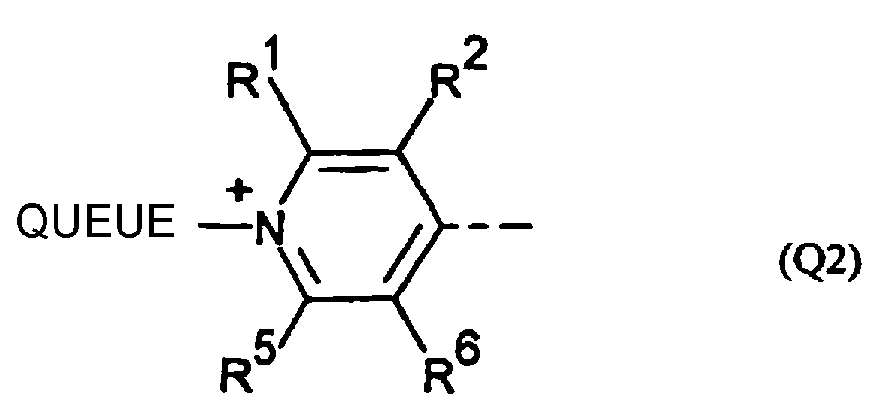
- the quaternized nitrogen heterocycle Q is typically a substituted or unsubstituted pyridinium, quinolinium, benzoquinolinium or benzazolium moiety having the formula or or the formula where the ring substituents R 1 , R 2 , R 3 and R 4 are optionally and independently H, Cl, F, C 1 -C 6 alkyl, C 1 -C 6 perfluoroalkyl, C 1 -C 6 alkoxy, amino, or amino substituted by 1-2 C 1 -C 6 alkyls.
- the ring substituents R 5 and R 6 are optionally and independently H, Cl, F, C 1 -C 6 alkyl, C 1 -C 6 perfluoroalkyl, C 1 -C 6 alkoxy, amino, amino substituted by 1-2 C 1 -C 6 alkyls, or phenyl.
- R 5 and R 6 when taken in combination, form a fused 6-membered aromatic ring (yielding a quinolinium moiety) that is optionally and independently substituted one or more times by Cl, F, C 1 -C 6 alkyl, C 1 -C 6 perfluoroalkyl, C 1 -C 6 alkoxy, amino, or amino substituted by 1-2 C 1 -C 6 alkyls.
- the quinolinium ring is optionally substituted by an additional fused 6-membered aromatic ring (yielding a naphtho-substituted pyridinium, or a benzoquinoline), that is also optionally and independently substituted one or more times by Cl, F, C 1 -C 6 alkyl, C 1 -C 6 perfluoroalkyl, C 1 -C 6 alkoxy, amino, or amino substituted by 1-2 C 1 -C 6 alkyls.
- R 5 and R 6 are H, or form a substituted or unsubstituted benzo moiety.
- R 5 and R 6 taken in combination, form a fused 6-membered substituted or unsubstituted benzo moiety yielding a quinolinium ring system.
- the ring fragment X is optionally -S-, -O-, -NR 7 -, or -CR 7 R 8 -, wherein R 7 and R 8 are as defined previously. Typically R 7 and R 8 methyls.
- X is O or S, more preferably X is S.
- TAIL The quaternizing moiety, is attached to the nitrogen atom of Q through a carbon atom and contains a total of 1-22 non-hydrogen atoms that are C, O N or S, such that within TAIL each heteroatom is separated from any adjacent heteroatoms by at least two carbon atoms.
- TAIL is composed of bonds that are selected from the group consisting of carbon-carbon bonds (C-C), ether bonds (C-O-C), thioether bonds (C-S-C) or amine bonds (C-NR 9 -C). Any carbon atom in TAIL is optionally further substituted by hydroxy, carboxy, sulfo, amino, or ammonium.
- any amine, amino or ammonium in TAIL is optionally substituted by an R 9 that is a C 2 -C 6 alkyl that is optionally further substituted by hydroxy, carboxy, sulfo, amino, amino substituted by 1-2 C 1 -C 6 alkyls or ammonium substituted by 1-3 C 1 -C 6 alkyls.
- the nitrogen atoms of TAIL form either one or two saturated 5- or 6-membered rings in combination with other C or N atoms in TAIL, such that the resulting rings are pyrrolidines, piperidines, piperazines or morpholines.
- the TAIL moiety includes at least one dialkylamino or a trialkylammonium substituent, where the alkyl substituents are methyl or ethyl.
- TAIL is -CH 3 or CH 2 CH 3 , or is a C 3 -C 22 alkyl that is optionally substituted one or more times by hydroxy, carboxy, sulfo, amino, amino substituted by 1-2 C 1 -C 6 alkyls, or ammonium substituted by 1-3 C 1 -C 6 alkyls.
- sulfo is meant sulfonic acid (-SO 3 H) or the common alkali metal salts of sulfonic acid.
- TAIL is a C 3 -C 12 alkyl that is linear and saturated, and substituted at its free terminus by hydroxy, carboxy, sulfo, amino, amino substituted by 1-2 C 1 -C 6 alkyls, or ammonium substituted by 1-3 C 1 -C 6 alkyls. Yet more preferably, TAIL is a C 3 -C 4 alkyl that is substituted once by sulfo or carboxy.
- R 11 and R 12 are independently H, F, Cl, or -CH 3 ; typically H.
- R 13 and R 14 are independently C 1 -C 18 alkyls that are linear, branched, saturated or unsaturated, and are optionally substituted one or more times by F, hydroxy or C 1 -C 6 alkoxy.
- R 13 and R 14 when taken in combination, form a 5- or 6-membered saturated ring that optionally contains an oxygen heteroatom.
- R 11 taken in combination with R 13 and R 10 taken in combination with R 12 are independently -(CH 2 ) 2 - or -(CH 2 ) 3 -, forming 5- or 6-membered rings.
- R 13 and R 14 are each linear alkyls, which may be the same or different, each having 4-8 carbon atoms, more preferably each having 5-7 carbon atoms.
- M has a formula as depicted in Table I, where R is H, phenyl, sulfophenyl, C 1 -C 6 alkyl or C 1 -C 6 alkyl substituted by carboxy.
- R is H, phenyl, sulfophenyl, C 1 -C 6 alkyl or C 1 -C 6 alkyl substituted by carboxy.
- Such dyes have been described, for example, by Brooker et al., J. AM. CHEM. SOC. 73, 5326 (1951 ) (incorporated by reference).
- any net positive or negative charges possessed by the dye are balanced by counterion(s), ⁇ .
- Any counterion currently used in conjunction with biomolecules is suitable.
- Preferred counterions include chloride, iodide, perchlorate, various sulfonates, alkali metal ions, alkaline earth metal ions, transition metal ions, ammonium or substituted ammonium ions.
- Table 2 Selected dyes useful for the present invention Dye no.
- merocyanine dyes including styryl dyes
- RH dyes styryl dyes
- Leslie Loew Loew et al., J. ORG. CHEM. 49, 2546 (1984 ), incorporated by reference
- Other merocyanine dyes are described by Brooker et al. (J. AM. CHEM. SOC. 73, 5326 (1951 ), incorporated by reference).
- Many dyes useful for the invention are available from Molecular Probes, Inc. (Eugene,
- the quaternizing moiety is typically obtained by quaternization of a methyl-substituted pyridine, quinoline or other nitrogen heterocycle by an alkylating agent that already contains other side chain substituents, or that is further reacted to yield the side chain substituents as described by Hildesheim (Grinvald et al. supra ).
- TAIL quaternizing moiety
- propane sultone or butane sultone is convenient and yields preferred dyes.
- Other substituents are usually added either before or after synthesis of the initial merocyanine dye via an intermediate haloalkyl quaternized heterocycle.
- addition of TAIL is also achieved subsequent to initial dye synthesis, which usually requires an azastilbene or fused analog thereof, as described by Loew et al. ( supra ).
- the styryl dyes of the present method are typically prepared by conversion of an aniline derivative that contains the desired R 1 , R 2 , R 3 , and R 4 substituents to a benzaldehyde, cinnamaldehyde or pentadienal derivative using methods well known in the art.
- the aldehyde derivative is then condensed with a quaternized pyridinium, quinolinium or benzazolium salt to give the useful dye.
- a staining mixture comprising one or more of the merocyanine dyes described above is combined with a sample mixture that is thought to contain poly(amino acids) for a sufficient time to form a dye-poly(amino acid) complex that gives a detectable colorimetric or fluorescent optical response upon illumination.
- the complex contains detergent molecules, as described above.
- the merocyanine dyes present in the complex interact non-covalently with either the poly(amino acid) itself, or with said detergent molecules present in the complex.
- the sample mixture or the combined mixture is optionally heated, typically to > 90 C. Additional steps are optionally and independently used, in any combination, to provide for separation or purification of the poly(amino acids), for enhancing the detection of the poly(amino acids), or for quantification of the poly(amino acids).
- the sample mixture is an aqueous or mostly aqueous solution, or is a solid, paste, emulsion or other solution that is combined with a mostly aqueous solution in the course of labeling.
- the sample mixture contains or is suspected to contain poly(amino acids).
- the concentration of poly(amino acids) is typically 10 ng/mL - 50 ⁇ g/mL, more preferably 50 ng/mL - 5 ⁇ g/mL.
- the concentration of poly(amino acids) is typically 1 ng/band - 4 ⁇ g/band.
- Poly(amino acids) suitable for the invention include both synthetic and naturally occurring poly(amino acids).
- the poly(amino acids) optionally incorporate non-peptide regions including lipid, phosphate, and/or carbohydrate regions.
- the poly(amino acids) are optionally relatively homogeneous or heterogeneous mixtures of poly(amino acids), or are multi-subunit complexes.
- the poly(amino acids) in the sample mixture are optionally covalently or non-covalently bound to a solid surface such as glass, plastic, or semiconductor material; or are unbound.
- the poly(amino acids) are optionally unmodified, or have been treated with a reagent so as to enhance or decrease the mobility of the poly(amino acid) in an electrophoretic gel, such as by complexing with the peptide (to decrease migration), by cleaving selected peptide bonds (to increase migration of the resulting fragments), by changing the relative charge on the protein (as by phosphorylation or dephosphorylation) or by covalent coupling of a constituent such as occurs during glycosylation. Such interactions are detected by the change in electrophoretic mobility of treated poly(amino acids), relative to untreated poly(amino acids).
- poly(amino acids) are peptides of mw > 500 daltons (more typically > 800 daltons) or are proteins (Tables 3 and 5). Large poly(amino acids) (> 200,000 daltons)are not well resolved in gels. Smaller poly(amino acids) ( ⁇ 1000 daltons) are difficult to detect on gels and filters, but are readily detected in solution.
- the poly(amino acids) present are a mixture of poly(amino acids) of different molecular weights (e.g. molecular weight standards).
- the sample mixture optionally contains discrete biological ingredients other than the desired poly(amino acids), including other poly(amino acids), amino acids, nucleic acids, carbohydrates, and lipids, which may or may not be removed in this method.
- the poly(amino acids) are separated from each other or from other ingredients in the sample by mobility, by size or by binding affinity in the course of the method.
- intact or fragmented biological membranes, liposomes or detergent micelles in the sample mixture are removed, destroyed or dispersed below the concentration at which they assemble into micelles (critical micelle concentration or CMC) prior to or in the course of labeling with this method.
- the sample mixture is essentially cell-free.
- the staining mixture is combined with the sample mixture so as to facilitate contact between any dye and any poly(amino acids) present in the combined mixture.
- the staining mixture is typically prepared by dissolving the dye in an organic solvent, such as DMSO, DMF or methanol; and then diluted with an aqueous solution according to the assay being performed.
- the dye is diluted into an aqueous solution, preferably buffered, that optionally contains a detergent.
- the dyes are diluted into water or buffer, optionally including acetic acid (typically 5%-7%).
- dye concentrations in the staining mixture are typically between 1 ⁇ M and 100 ⁇ M, preferably between about 5 ⁇ M and about 20 ⁇ M; more preferably at least 10-15 ⁇ M or higher.
- dye concentrations are typically greater than 0.10 ⁇ M and less than 10 ⁇ M; preferably greater than about 0.50 ⁇ M and less than or equal to about 5 ⁇ M; more preferably 1-3 ⁇ M. Concentrations below and above these values result in detectable staining for certain poly(amino acids), depending on the sensitivity of the detection method.
- a particular dye is generally selected using one or more of the following criteria: sensitivity to poly(amino acids), dynamic range, photostability, staining time, and insensitivity to the presence of nucleic acids.
- the sensitivity and dynamic range of the dyes is determined using the procedures of Examples 1 and 7.
- Preferred dyes have a sensitivity of 1-2 ng or less of poly(amino acid) per band in electrophoretic gels, or 10-30 ng or less of poly(amino acid) per mL of solution.
- the dyes have a dynamic range of about 3 or more orders of magnitude of poly(amino acid) concentration for solution assays.
- Preferred dyes in an aqueous solution in the absence of poly(amino acids), possess a quantum yield ⁇ 0.05, more preferably ⁇ 0.01.
- the dyes when combined with poly(amino acids), exhibit a fluorescence enhancement that is > 100-fold, more preferably > 300-fold relative to the dyes in the absence of poly(amino acids).
- Preferred dyes, when combined with poly(amino acids) have an absorption maximum within 10 nm of 488 nm, 514 nm, 543 nm, or 633 nm.
- the invention requires a detergent that is added simultaneously with or as part of the sample mixture or the staining mixture, or is added to the combined mixture as described below.
- the detergent is combined with the sample mixture before the staining mixture is added.
- the detergent is any amphiphilic surface active agent or surfactant that serves to coat the poly(amino acids), i.e. non-covalently associate with the poly(amino acid).
- Useful detergents include non-ionic, cationic, anionic, amphoteric and fluorinated surfactants; many are commercially available. Any detergent utilized in protein gel electrophoresis is preferred.
- the detergent is an anionic detergent, preferably an alkyl sulfate or alkyl sulfonate salt (typically 6-18 carbons); more preferably, sodium dodecyl sulfate (SDS), sodium octadecyl sulfate, or sodium decyl sulfate; most preferably, SDS.
- an alkyl sulfate or alkyl sulfonate salt typically 6-18 carbons
- SDS sodium dodecyl sulfate
- sodium octadecyl sulfate sodium octadecyl sulfate
- sodium decyl sulfate most preferably, SDS.
- detergent in the sample mixture, staining mixture, or combined mixture is present below the CMC for that detergent, in order to avoid poly(amino acid)-free micelle formation.
- the CMC is a function of the detergent being used and the ionic strength of the solution. For SDS solutions at moderate ionic strength, the CMC is about 0.1 % of the solution by weight.
- the concentration of the detergent is about 1-5% by weight, more typically about 2%.
- the concentration of SDS in the combined mixture is typically less than 1% by weight, preferably 0.05-0.1% by weight.
- the concentration of detergent in the combined mixture is preferably less than about 0.05% by weight.
- the combined mixture is a solution (e.g. Example 7; Table 3); typically, an aqueous solution (preferably buffered) that consists essentially of poly(amino acids), one or more merocyanine dyes, particularly preferred dyes, and a detergent.
- the aqueous solution is optionally used as a separation medium, such as within a sedimentation gradient (e.g. a sucrose gradient) or when performing capillary electrophoresis.
- the sample mixture or combined mixture is optionally heated and cooled before the detection or quantification of poly(amino acids).
- Table 3 Properties of selected dyes in solution assays Dye No.
- Fluorescence excitation and emission spectra are measured using 1 ⁇ M dye + 150 ⁇ g/mL BSA + 0.05% SDS in 2.0 mL volume.
- Fluorescence enhancements are calculated using I ⁇ M dye +/- 500 ⁇ g/mL BSA in 2.0 mL at fluorescence excitation and emission maxima.
- 4 Dynamic range limits are determined by titrating 1 ⁇ M dye with 0.05 to 50 ⁇ g protein in a 96-well microplate in a 200 ⁇ L volume. Three protein solutions are titrated separately for each dye: lysozyme, BSA and a protein mixture that contains equal weight concentrations of IgG, avidin, streptavidin, cellulase and ovalbumin.
- the lower limit of detection is the amount of protein required to produce fluorescence 10% above background.
- the upper limit is where the linearity of signal with respect to protein concentration starts to degrade for the protein type producing signal saturation at the lowest concentration.
- the merocyanine dyes are optionally used to prestain poly(amino acids) prior to separation; or are present as a component of the mobile phase during separation (e.g. Example 12) in gel or capillary electrophoresis.
- separated poly(amino acids) in electrophoretic (denaturing or non-denaturing) gels are post-stained using the staining mixture, or are transferred to a filter membrane or blot or other solid or semi-solid matrix before being combined with the staining mixture (e.g. Examples 1, 2, 3, and 5).
- polyacrylamide or agarose gels are used for electrophoresis.
- the gel is an iso-electric focusing gel, a gradient gel, a two-dimensional gel, or the gel is used for gel-mobility-shift analysis.
- the sample mixture or combined mixture is optionally heated before being applied to denaturing gels.
- the sensitivities of selected dyes of the present invention when used to post-stain electrophoretic gels are listed in Table 4. Table 4: Labeling sensitivity in gels of selected dyes 1 Dye No.
- Color 2 Sensitivity 3 (ng/band) Background Staining Coomassie Brilliant Blue blue* 30-60 (low) very high: requires destaining Nile red red/orange* 30-60 (low) low/med Silver staining tan to black* variable 2-10 (high) varies, low to very high even with destaining 101 faint greenish low med 102 light orange med med 103 red/orange low low/med 104 yellow med low/med 201 red/orange med low/med 301 barely visible low low 302 greenish/pink low low 303 pink/yellow to orange med med 304 bright orange high low/med 305 light orange low med 306 orange med low 307 bright orange high low/med 308 orange high med 309 pink/orange med low 310 red/orange high low/med 311 light orange low low/med 401 orange med med 402 red/orange high low 501 orange med low/med 601 red/orange high low 701 red/orange
- Sensitivity refers to the lower limit of protein detection, per band, using a 4X dilution series of BSA (using the procedure of Example 1).
- the sensitivity is defined to be the band containing the smallest amount of protein that is easily detectable by eye in a black and white Polaroid photograph taken at an optimal exposure.
- Low sensitivity means a detection limit comparable to that found with CBB and nile red
- high sensitivity means a detection limit comparable to that found with silver staining
- “medium” sensitivity is intermediate between the two ranges.
- destaining of stained gels is generally not necessary for either colorimetric or fluorescent detection. At very high dye concentrations, destaining is optionally used to improve visible color detection in gels. Gels are optionally washed briefly after staining to prevent transfer of dye to other surfaces.
- the method of the present invention optionally further comprises addition of an additional reagent, simultaneously or sequentially, to the sample mixture, the staining mixture, or the combined mixture.
- the additional reagent incorporates a means for producing a detectable response, including the change in, or appearance of, color, fluorescence, reflectance, pH, chemiluminescence, infrared spectra, magnetic properties, radioactivity, light scattering, x-ray scattering, or production of an electron-rich substrate.
- An additional reagent is optionally a detection reagent that colocalizes with poly(amino acids) in general or with specific poly(amino acids); or is a probe for a specific component of the sample mixture (e.g.
- the additional reagent is one or more additional merocyanine dyes, including preferred embodiments described above. Where the additional merocyanine dye(s) have overlapping spectral characteristics such that energy transfer occurs, the labeled poly(amino acids) exhibit an extended Stokes shift.
- the additional reagent is another protein stain (such as CBB or silver stain) such that labeling of the poly(amino acids) is enhanced by the colocalization of staining.
- the combined mixture of dye and sample is illuminated to give an optical response that is an absorption of visible light (colorimetric response), or is a fluorescence emission (fluorescence response), or both.
- Illumination is by a light source capable of exciting the dye-poly(amino acid) complex, typically at or near the wavelength of maximum absorption of the dye-poly(amino acid) complex, such as an ultraviolet (254-370 nm) or visible (490-550 nm) wavelength emission lamp, an arc lamp, a laser, or even sunlight or ordinary room light.
- the sample is excited with a wavelength within 20 nm of the maximum absorption of the dye-poly(amino acid) complex.
- the dye-poly(amino acid) complexes possess an absorption maximum between 480 and 650 nm, more preferably between 488 and 550 nm, most preferably matching the wavelength of a laser illumination source.
- the complexes also preferably excite in the UV at or near 300 nm.
- the optical response is detected qualitatively, or optionally quantitatively, by means that include visual inspection, CCD cameras, video cameras, photographic film, or the use of instrumentation such as laser scanning devices, fluorometers, photodiodes, quantum counters, epifluorescence microscopes, scanning microscopes, flow cytometers, fluorescence microplate readers, or by means for amplifying the signal such as photomultiplier tubes. Recording the optical response using POLAROID film results in enhanced sensitivity of signal.
- Quantification of the poly(amino acid) is typically performed by comparison of the optical response to a prepared standard or to a calibration curve, obtained from a standard dilution of a known concentration of a poly(amino acid) or poly(amino acid) mixture.
- the standard curve is generated by comparison with a reference dye or dyed particle that has been standardized versus the target dye-poly(amino acid) complex.
- said dyes are particularly useful in the formulation of a kit for the labeling of poly(amino acids), comprising one or more merocyanine dyes (preferably in a stock solution) and instructions for the use of the dye, optionally including a reference standard.
- the instructions specify that the sample mixture and staining mixtures are applied, either simultaneously or sequentially to a solid or semi-solid support, the resulting color or fluorescence of which is then compared to a calibrated standard to determine the presence, and optionally the concentration, of poly(amino acid) present in the sample mixture.
- the support, dye solution, calibration standard and instructions for use, in combination comprise a "dipstick" protein assay kit, allowing the rapid and convenient determination of protein concentration.
- the pure protein or mixture of proteins is prepared in Loading Buffer (50 mM Tris-HCl, pH 6.8, 2% SDS, 10% glycerol, and 0.015% bromophenol blue). Dithiothreitol is added to each sample, to 0.1 M. The samples are heated 4-5 minutes at 90-95 °C and loaded onto a 15% Tris-glycine polyacrylamide mini- or full-sized gel containing 0.05%-0.1% SDS, with a 4% stacking gel. The gel is electrophoresed under standard conditions, in Tris-glycine buffer containing 0.05%-0.1% SDS. The resulting gel is transferred to a staining dish containing 1-3 ⁇ M Dye 304 in 7.5% acetic acid (in water).
- Loading Buffer 50 mM Tris-HCl, pH 6.8, 2% SDS, 10% glycerol, and 0.015% bromophenol blue.
- Dithiothreitol is added to each sample, to 0.1
- the staining solution is gently agitated for 45-60 minutes. After 10 minutes, the protein bands are readily apparent, but sensitivity improves over about 30-40 minutes.
- the gel is removed from the staining dish, rinsed briefly in 7.5% acetic acid and transferred to a UV-transilluminator. The gel is photographed using 300 nm transillumination and black and white POLAROID 667 print film with a Wratten 9 gelatin filter, or using a CCD camera with UV illumination near 300 nm.
- the stained bands exhibit bright orange fluorescence. Under normal room lights, the stained bands appear as orange bands.
- Other dyes of the invention See Table 3 yield stained gels that possess bands having visible coloring from yellow to purple and fluorescence emission from green to red.
- a protein dilution series is prepared in Native Gel Loading Buffer (125 mM Tris-HCl, pH 6.8, 10% glycerol and 0.015% bromophenol blue). The samples are loaded onto a Tris-HCl non-denaturing polyacrylamide gel, and the gel is electrophoresed under standard conditions. The electrophoresed gel is stained and photographed as in Example 1. Staining sensitivity under these conditions is typically somewhat protein-selective.
- a dilution series of a short peptide is prepared in Loading Buffer. To each sample is added dithiothreitol to 0.1 M, the samples are heated 4-5 minutes at 90-95 °C then loaded onto 16.5% Tris-tricine gel. The gel is electrophoresed under standard conditions. The gel is stained and the protein bands are visualized as in Example 1. Polypeptides as small as 20 amino acids are readily detected using this method.
- Peptides such as a tetramer of the heptapeptide repeat found at the C-terminus of eukaryotic RNA polymerase II, tryptic peptides of trypsin and the small subunit of ⁇ -bungarotoxin, which are not stained with either CBB or silver staining, are readily detectable.
- a mouse cytosolic extract i.e., a whole cell lysate
- the gel is stained and visualized as in Example 1.
- An identical gel is stained in parallel with silver using a commercially available kit (Bio-Rad).
- the gel stained using the present invention exhibits more protein spots than the silver stained gel, indicating that the present invention is less protein-selective than silver staining.
- the signal intensity obtained with the instant method is more directly proportional to protein concentration than that obtained with silver staining.
- the sensitivity of staining using the present invention is superior to that of silver staining for detection of low molecular weight proteins (Table 3).
- the protein is diluted in TBS (20 mM Tris-HCl, pH 7.5, 500 mM NaCl), then applied to a nylon or nitrocellulose filter membrane. The membrane is washed with TBS, then incubated with 2 ⁇ M Dye 803 in 7.5% acetic acid or TBS.
- the proteins are first separated by gel electrophoresis and transferred to a nylon or nitrocellulose filter membrane using standard procedures. The blot is stained as above. The blot is illuminated at 300-365 nm. Protein spots or bands are fluorescent. Blots are photographed as in Example 1. Stained proteins appear as faint white spots or white bands. Staining under these conditions is somewhat protein selective. Proteins are optionally detected using laser illumination sources.
- a dilution series of bovine heart cytochrome oxidase complex is prepared in water. Samples are loaded onto a 12% polyacrylamide gel and the gel is electrophoresed under standard conditions. The resulting gel is cut in half, and one half is stained with Dye 801 in transfer buffer (20% MeOH, 25 mM Tris-glycine, pH 8.3, 192 mM glycine) and visualized as in Example 1 above. The proteins are electrophoretically transferred from both halves of the gel to a filter membrane, according to standard methods.
- the blot is blocked, probed with mouse monoclonal antibodies directed against specific COX subunits, and the resulting signals are visualized using nitroblue tetrazolium and 5-bromo-4-chloro-3-indolyl phosphate, using standard procedures.
- the two halves of the blot show equal sensitivity for detection of the COX subunits, indicating that staining proteins with the invention does not interfere with later immunological detection.
- a dilution series of proteins is prepared in 10 mM Tris-HCl, pH 7.5, containing 0.05% SDS. An equal volume of 2 ⁇ M Dye 307 is added to each sample. The samples are incubated 15 minutes at room temperature. The fluorescence intensity of each sample is measured using 490 nm excitation, using a fluorometer or fluorescence microplate reader. The fluorescence of Dye 307 in buffer alone (control) is subtracted from that of the protein-containing samples. Protein concentrations are determined by comparing the net fluorescence with that of a standard, such as bovine serum albumin, or protein mixture ( Figure 3 ).
- a standard such as bovine serum albumin, or protein mixture
- An extract from E. coli is prepared using standard methods, by suspending a bacterial cell pellet directly in Loading Buffer and heating it as in Example 1.
- the resulting sample is pipeted up and down repeatedly, through a fine bore syringe, to shear large DNA molecules.
- the sample is diluted serially and loaded onto a denaturing SDS gel in two duplicate dilution series and electrophoresed, as in Example 1.
- the electrophoresed gel is cut in half.
- One half is stained with Dye 304 according to Example 1.
- the other half is stained with silver according to standard methods.
- the silver-stained half shows staining of bands that correspond to nucleic acids as well as protein.
- the half stained with Dye 304 exhibits only those bands that result from protein staining.
- Gels are prepared and stained as in Examples 1, 2, 3 or 4.
- the stained gels are scanned using a laser-excited gel scanner.
- Stained proteins display fluorescence emission having a dynamic range of at least 3 orders of magnitude in protein concentration, with a sensitivity of a single ng per protein band ( Figure 4 , Table 2).
- Stained gels can also be analyzed using visible light lasers, or other scanners with light sources that overlap with the excitation spectra for the dye used.
- Dilution series of BSA, lysozyme and an equi-mass mixture of myosin, ⁇ -galactosidase, phosphorylase B, bovine serum albumin, ovalbumin, carbonic anhydrase, trypsin inhibitor, lysozyme and aprotinin are prepared in 10 mM Tris-HCl, pH 7.5, containing 0.05% SDS.
- An equal volume of Dye 803 in the same buffer is added to each and the samples are incubated for 15 minutes. A 1-5 ⁇ L aliquot of each mixture is spotted onto a thin layer chromatography plate or a nylon or nitrocellulose filter membrane.
- Protein-containing spots are detected colorimetrically under room light, or are detected by fluorescence following ultraviolet or visible illumination.
- the color intensity or fluorescence intensity of a given spot is indicative of the amount of protein present in that spot.
- the amount of protein in an unknown sample is determined by comparing either the colorimetric or fluorescence intensity with that of a standard of known concentration.
- Proteins are loaded and run on standard SDS gels, using standard methods, except that the running buffer contains 0.05% SDS and 1-3 ⁇ M Dye 801.
- the stained gels are photographed directly after electrophoresis (as in Example 1) or are destained in 7.5% acetic acid for 20-50 minutes prior to photography.
- the sensitivity obtained is about the same as obtained by staining gels after electrophoresis. Migration of protein bands can be monitored through the glass plates that support the gel, during electrophoresis.
- Proteins are diluted in Loading Buffer, heated to 90-95 °C for 4-5 minutes then cooled to room temperature.
- Dye 304 is added to 10 mM, and the samples are loaded onto a 12% polyacrylamide gel. The gel is electrophoresed and visualized using ultraviolet illumination as in Example 1, or as in Example 9, or with a CCD camera. The sensitivity of this method is somewhat less than in Examples 1 and 11.
Landscapes
- Life Sciences & Earth Sciences (AREA)
- Health & Medical Sciences (AREA)
- Molecular Biology (AREA)
- Chemical & Material Sciences (AREA)
- Engineering & Computer Science (AREA)
- Physics & Mathematics (AREA)
- Immunology (AREA)
- Urology & Nephrology (AREA)
- Biomedical Technology (AREA)
- Biochemistry (AREA)
- General Health & Medical Sciences (AREA)
- General Physics & Mathematics (AREA)
- Analytical Chemistry (AREA)
- Pathology (AREA)
- Hematology (AREA)
- Bioinformatics & Cheminformatics (AREA)
- Microbiology (AREA)
- Bioinformatics & Computational Biology (AREA)
- Biophysics (AREA)
- Proteomics, Peptides & Aminoacids (AREA)
- Chemical Kinetics & Catalysis (AREA)
- Biotechnology (AREA)
- Cell Biology (AREA)
- Spectroscopy & Molecular Physics (AREA)
- Electrochemistry (AREA)
- Food Science & Technology (AREA)
- Medicinal Chemistry (AREA)
- Investigating Or Analysing Biological Materials (AREA)
- Investigating Or Analysing Materials By The Use Of Chemical Reactions (AREA)
- Pyridine Compounds (AREA)
- Plural Heterocyclic Compounds (AREA)
- Indole Compounds (AREA)
- Sampling And Sample Adjustment (AREA)
- Detergent Compositions (AREA)
- Investigating, Analyzing Materials By Fluorescence Or Luminescence (AREA)
Claims (26)
- Procédé de détection pour un poly(acide aminé) dans un mélange échantillon, comprenant les étapes consistant à :a) préparer, à partir du mélange échantillon et d'un mélange colorant qui contient un ou plusieurs colorants mérocyanines, un mélange combiné exempt de cellules contenant le poly(acide aminé) (s'il est présent), le ou les colorants mérocyanines et un détergent ;b) faire incuber le mélange combiné pour amener le colorant dans le mélange colorant à s'associer avec tout poly(acide aminé) dans le mélange échantillon pour former un complexe colorant-poly(acide aminé) qui donne une réponse optique détectable lors de l'éclairement ;c) éclairer le produit obtenu à l'aide de l'étape (c), par exemple un complexe colorant-poly(acide aminé) ; etd) observer la réponse optique.
- Procédé selon la revendication 1, dans lequel, entre les étapes a) et b), le mélange échantillon ou le mélange combiné est chauffé.
- Procédé selon l'une des revendications 1 ou 2, dans lequel le détergent est ajouté simultanément à ou comme partie du mélange échantillon ou du mélange colorant, ou est ajouté après que les mélanges échantillon et colorant aient été combinés.
- Procédé, selon l'une de revendications 1, 2 ou 3, dans lequel chaque colorant mérocyanine a indépendamment la formule :
Q-B-M
où Q est :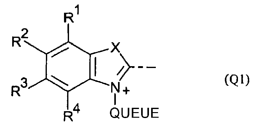
 - R1, R2, R3 et R4 représentent indépendamment H, Cl, F, alkyle en C1-C6, perfluoroalkyle en C1-C6, alcoxy en C1-C6, amino ou amino substitué par 1-2 alkyles en C1-C6 ;- R5 et R6 représentent indépendamment H, Cl, F, alkyle en C1-C6, perfluoroalkyle en C1-C6, alcoxy en C1-C6, amino ou amino substitué par 1-2 alkyles en C1-C6 ou phényle ; ou R5 et R6 pris en combinaison forment un noyau aromatique à 6 chaînons fusionné qui est facultativement substitué une ou plusieurs fois par des substituants qui peuvent être identiques ou différents et sont choisis parmi Cl, F, alkyle en C1-C6, perfluoroalkyle en C1-C6, alcoxy en C1-C6, amino et amino substitué par 1-2 alkyles en C1-C6,ou ledit noyau aromatique à 6 chaînons fusionné est facultativement substitué par un noyau aromatique à 6 chaînons fusionné supplémentaire qui est facultativement substitué par des substituants qui peuvent être identiques ou différents et sont choisis parmi Cl, F, alkyle en C1-C6, perfluoroalkyle en C1-C6, alcoxy en C1-C6, amino et amino substitué par 1-2 alkyles en C1-C6 ;- X représente -S-, -O-, -NR7- ou -CR7R8-, où R7 et R8 sont indépendamment H, alkyle en C1-C6 ou phényle ; ou R7 et R8 pris en combinaison complètent un noyau saturé à 5 ou 6 chaînons ;- QUEUE est attachée à Q par un atome de carbone et contient 1-22 atomes non hydrogène, qui sont C, 0, N ou S, de telle sorte que chaque hétéroatome est séparé de tous hétéroatomes adjacents par au moins deux atomes de carbone ; et en outre par le fait que QUEUE est composé de liaisons carbone-carbone (C-C), liaisons éther (C-OC), liaisons thioéther (C-S-C) ou liaisons amine (C-NR9-C) ; où tout atome de carbone dans QUEUE est facultativement encore substitué par hydroxy, carboxy, sulfo, amino ou ammonium ; et où toute liaison amine, amino ou ammonium dans QUEUE est facultativement substituée par R9 qui est un alkyle en C2-C6 qui est facultativement encore substitué par hydroxy, carboxy, sulfo, amino, amino substitué par 1-2 alkyles en C1-C6,ou ammonium substitué par 1-3 alkyles en C1-C6, ou lesdits atomes de N forment soit un soit deux cycles à 5 ou 6 chaînons saturés en combinaison avec des atomes de C ou N supplémentaires dans QUEUE ;- B représente
- (CR7=CR8)n-
où R7 et R8 sont tels que définis précédemment ;
n = 1, 2 ou 3 ;
et M représente :
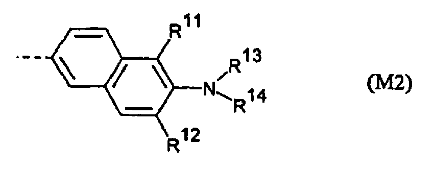 - R11 et R12 représentent indépendamment H, F, Cl ou -CH3 ;- R13 et R14 représentent indépendamment alkyle en C1-C18 qui est linéaire, ramifié, saturé ou insaturé, et est facultativement substitué une ou plusieurs fois par F, hydroxy ou alcoxy en C1-C6 ; ou R13 et R14 pris en combinaison forment un noyau saturé à 5 ou 6 chaînons contenant 0 ou 1 hétéroatome d'oxygène ; ou R13 pris en combinaison avec R11 et R14 pris en combinaison avec R12 représentent indépendamment -(CH2)2- ou -(CH2)3- ;
ou M représente :


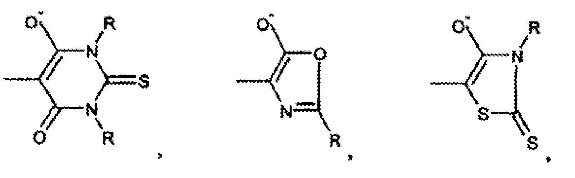
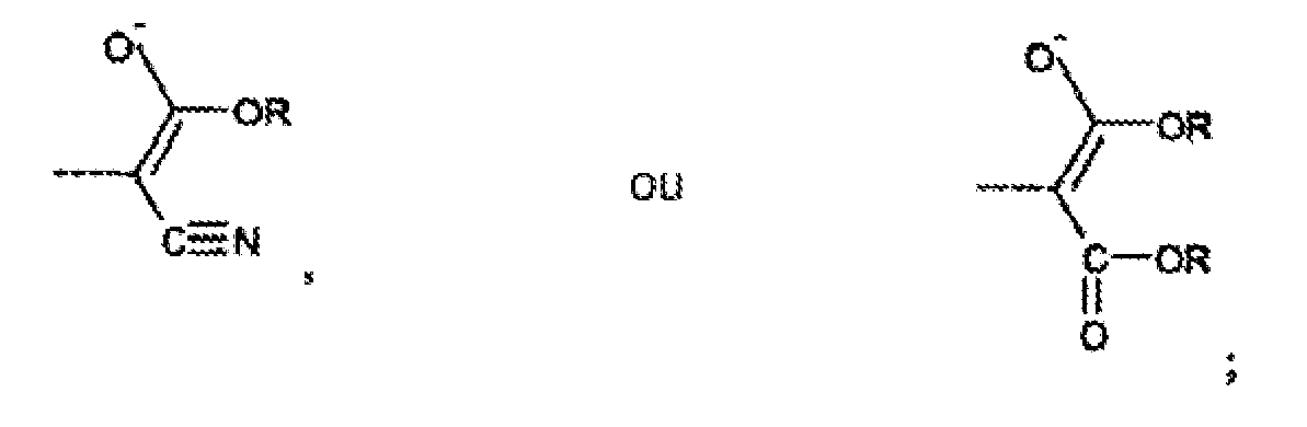
R représente indépendamment H, phényle, sulfophényle, alkyle en C1-C6 ou alkyle en C1-C6 substitué par carboxy. - Procédé selon l'une des revendications 1 ou 2, dans lequel le mélange colorant contient un ou plusieurs colorants styrylés ayant indépendamment la formule Q-B-M, où Q et B sont tels que définis dans la revendication 4 et M a la formule M1 ou M2 telle que définie dans la revendication 4.
- Procédé selon l'une des revendications 4 ou 5, dans lequel le mélange colorant contient un colorant dans lequel :(i) M a la formule M1 ; ou(ii) M a la formule M2.
- Procédé selon l'une des revendications 4 ou 5, dans lequel le mélange colorant contient un colorant qui a la formule :

- Procédé selon l'une des revendications 4 ou 5, dans lequel le mélange colorant contient un colorant qui a la formule :

- Procédé selon l'une des revendications 4 ou 5, dans lequel le mélange colorant contient un colorant qui a la formule :

- Procédé selon l'une des revendications 4 ou 5, dans lequel le mélange colorant contient un colorant qui a la formule :

- Procédé selon l'une quelconque des revendications 4 à 10, dans lequel le mélange colorant contient un colorant dans lequel :QUEUE représente -CH3 ou CH2CH3 ; ou QUEUE représente une chaîne alkyle en C3-C22 qui est linéaire ou ramifiée, saturée ou insaturée, qui est facultativement substituée une ou plusieurs fois par hydroxy, carboxy, sulfo, amino, amino substitué par 1-2 alkyles en C1-C6 ou ammonium substitué par 1-3 alkyles en C1-C6.
- Procédé selon l'une des revendications 4 ou 5, dans lequel le mélange colorant contient un colorant dans lequel :- M a la formule M1 ou M2 ;- X représente 0 ou S ;- B représente -(CH=CH)n- ;- n = 1 ou 2 ;- R11 et R12 représentent chacun H ;- R13 et R14 représentent indépendamment alkyle en C4-C8 ; et- QUEUE représente sulfopropyle, sulfobutyle, aminopropyleou aminobutyle, dont les amines sont substituées 1-3 fois par toute combinaison de H, méthyle ou éthyle.
- Procédé selon la revendication 12, dans lequel le mélange colorant contient un colorant dans lequel :- R1, R2, R3 et R4 représentent chacun H ;- R5 et R6 représentent chacun H ; et- ledit colorant est présent dans le mélange combiné à une concentration de 0,10 µM-10 µM pour une réponse optique fluorescente ou 10 µM-100 µM pour une réponse optique colorimétrique ; et- ledit poly(acide aminé) a une masse moléculaire de 500-200 000 daltons.
- Procédé selon l'une quelconque des revendications 4 à 6 ou 8 à 10, dans lequel :(i) le mélange colorant contient un colorant dans lequel Q représente Q1 ;(ii) le mélange colorant contient un colorant dans lequel Q représente Q2 ; ou(iii) le mélange colorant contient un colorant dans lequel Q représente Q3.
- Procédé selon l'une quelconque des revendications 1 à 14, comprenant en outre le transfert du mélange échantillon dans une matrice solide ou semi-solide avant ou après combinaison avec le mélange colorant.
- Procédé selon l'une quelconque des revendications 1 à 15, comprenant en outre les opérations consistant à retirer, détruire ou disperser au-dessous de la concentration de micelles critique toutes membranes biologiques qui sont présentes dans le mélange échantillon ; et comprenant en outre facultativement les opérations consistant à :(i) quantifier ledit poly(acide aminé) par mesure de ladite réponse optique détectable et comparer ladite mesure avec un étalon ; et/ou(ii) séparer par voie électrophorétique le mélange échantillon avant, après ou pendant qu'il est combiné avec le mélange colorant ; et/ou(iii) ajouter un réactif supplémentaire au mélange échantillon, au mélange colorant ou au mélange combiné.
- Procédé selon l'une quelconque des revendications 1 à 15, comprenant en outre les opérations consistant à chauffer le mélange échantillon ou le mélange combiné ; et facultativement comprenant en outre les opérations consistant à :(i) quantifier ledit poly(acide aminé) par mesure de ladite réponse optique détectable et comparer ladite mesure avec un étalon ; et/ou(ii) séparer par voie électrophorétique le mélange échantillon avant, après ou pendant qu'il est combiné avec le mélange colorant ; et/ou(iii) ajouter un réactif supplémentaire au mélange échantillon, au mélange colorant ou au mélange combiné.
- Procédé selon l'une quelconque des revendications 1 à 17, dans lequel ladite réponse optique détectable est une réponse de fluorescence.
- Procédé selon l'une quelconque des revendications 1 à 18, dans lequel le mélange échantillon contient un poly(acide aminé) et l'étape (c) est un éclairement d'un complexe formé par le poly(acide aminé) et le colorant à l'étape (b), l'étape (d) étant une observation de la réponse optique du complexe.
- Combinaison comprenant :a) un ou plusieurs colorants styrylés ayant la formule
Q-B-M
où Q représente Q1, Q2 ou Q3 tel que défini dans la revendication 4 ;
B est
-(CR7=CR8)n-
où R7 et R8 sont tels que définis dans la revendication 1
n = 1, 2 ou 3 ;
et M représente M1 ou M2 tel que défini dans la revendication 4 ;b) un détergent ; etc) un poly(acide aminé),
dans une solution aqueuse exempte de cellules où ledit détergent est présent à une concentration inférieure à la concentration critique de micelles pour ce détergent. - Utilisation d'un colorant mérocyanine en combinaison avec un détergent en tant que colorant de poly(acide aminé) dans un environnement exempt de cellules.
- Utilisation selon la revendication 21, dans laquelle le colorant mérocyanine est tel que défini à l'une quelconque des revendications 3 à 14 ou une combinaison appropriée de celui-ci.
- Utilisation selon la revendication 21, dans laquelle le colorant mérocyanine est tel que défini à la revendication 5.
- Utilisation selon la revendication 21, dans laquelle le colorant mérocyanine a la formule :

 - R5 et R6 représentent indépendamment H, Cl, F, alkyle en C1-C6, perfluoroalkyle en C1-C6, alcoxy en C1-C6, amino ou amino substitué par 1-2 alkyles en C1-C6 ; ou R5 et R6 pris en combinaison forment un noyau aromatique à 6 chaînons fusionné qui est facultativement substitué par un ou plusieurs substituants qui peuvent être identiquesou différents et sont choisis parmi Cl, F, alkyle en C1-C6, perfluoroalkyle en C1-C6, alcoxy en C1-C6, amino ou amino substitué par 1-2 alkyles en C1-C6 ;- QUEUE représente -CH3 ou CH2CH3 ; ou QUEUE représente une chaîne alkyle en C3-C22 qui est linéaire ou ramifié, saturé ou insaturé, qui est facultativement substitué uneou plusieurs fois par hydroxy, carboxy, sulfo, amino, amino substitué par 1-2 alkyles en C1-C6 ou ammonium substitué par 1-3 alkyles en C1-C6 ;- n = 1 ou 2 ; et- R13 et R14 représentent indépendamment alkyle en C1-C18 qui est linéaire, ramifié, saturé ou insaturé, et est facultativement substitué une ou plusieurs fois par F.
 - Utilisation selon l'une quelconque des revendications 21 à 24, dans laquelle le détergent est utilisé à une concentration au-dessous de sa concentration de micelles critique.
- Utilisation selon l'une quelconque des revendications 21 à 25, dans laquelle le colorant mérocyanine est utilisé comme colorant non sélectif pour colorer tous poly(acides aminés) dans l'échantillon qui est coloré, soumis à différentes sensibilités de différents poly(acides aminés).
Applications Claiming Priority (3)
| Application Number | Priority Date | Filing Date | Title |
|---|---|---|---|
| US444895 | 1995-05-19 | ||
| US08/444,895 US5616502A (en) | 1995-05-19 | 1995-05-19 | Non-specific protein staining using merocyanine dyes |
| PCT/US1996/007297 WO1996036882A1 (fr) | 1995-05-19 | 1996-05-20 | Coloration de proteines par colorant a base de merocyanine |
Publications (2)
| Publication Number | Publication Date |
|---|---|
| EP0774122A1 EP0774122A1 (fr) | 1997-05-21 |
| EP0774122B1 true EP0774122B1 (fr) | 2009-08-19 |
Family
ID=23766788
Family Applications (1)
| Application Number | Title | Priority Date | Filing Date |
|---|---|---|---|
| EP96920325A Expired - Lifetime EP0774122B1 (fr) | 1995-05-19 | 1996-05-20 | Coloration de proteines par colorant a base de merocyanine |
Country Status (8)
| Country | Link |
|---|---|
| US (1) | US5616502A (fr) |
| EP (1) | EP0774122B1 (fr) |
| JP (4) | JP4079381B2 (fr) |
| AT (1) | ATE440285T1 (fr) |
| AU (1) | AU699961B2 (fr) |
| CA (1) | CA2193705C (fr) |
| DE (1) | DE69638004D1 (fr) |
| WO (1) | WO1996036882A1 (fr) |
Families Citing this family (69)
| Publication number | Priority date | Publication date | Assignee | Title |
|---|---|---|---|---|
| US5912257A (en) * | 1995-09-06 | 1999-06-15 | The Research Foundation Of State University Of New York | Two-photon upconverting dyes and applications |
| US20030070927A1 (en) * | 1996-07-24 | 2003-04-17 | Merchant Mark E. | Reagents and methods for fluorescent analysis of serum proteins |
| JP2001512306A (ja) | 1997-01-08 | 2001-08-21 | ライフ テクノロジーズ,インコーポレイテッド | タンパク質の産生方法 |
| AU9340798A (en) * | 1997-08-12 | 1999-03-01 | Byk Gulden Lomberg Chemische Fabrik Gmbh | Method for assaying cellular protein |
| JP2002502980A (ja) * | 1998-02-10 | 2002-01-29 | イーワイ ラボラトリーズ インコーポレイテッド | 試料ホルダ起伏形状を補償し且つシステム・ノイズのロック除去を行う反射測定システム |
| US6485981B1 (en) * | 1998-07-29 | 2002-11-26 | Ciencia, Inc. | Method and apparatus for imaging and documenting fingerprints |
| US6372512B1 (en) | 1998-09-16 | 2002-04-16 | Resolution Sciences Corporation | Combined en bloc staining and embedding process |
| US6335446B1 (en) * | 1998-10-05 | 2002-01-01 | Oxford Glycosciences (Uk) Ltd. | Quinolinium- and pyridinium-based fluorescent dye compounds |
| ATE349700T1 (de) | 1998-10-27 | 2007-01-15 | Molecular Probes Inc | Übergangsmetallkomplex-haltige lumineszente proteinfarben |
| US20050027111A1 (en) * | 1999-02-02 | 2005-02-03 | Caliper Life Sciences, Inc. | Methods, devices and systems for characterizing proteins |
| EP1159605B1 (fr) * | 1999-02-02 | 2012-07-11 | Caliper Life Sciences, Inc. | Procedes de caracterisation de proteines |
| US6438398B1 (en) * | 1999-03-05 | 2002-08-20 | The University Of Miami | Process and device for measuring tear fluorescein clearance |
| US6664047B1 (en) | 1999-04-30 | 2003-12-16 | Molecular Probes, Inc. | Aza-benzazolium containing cyanine dyes |
| US6329205B1 (en) | 1999-08-31 | 2001-12-11 | Molecular Probes, Inc. | Detection method using luminescent europium-based protein stains |
| US6287758B1 (en) * | 2000-03-23 | 2001-09-11 | Axiom Biotechnologies, Inc. | Methods of registering trans-membrane electric potentials |
| US6579718B1 (en) | 2000-08-04 | 2003-06-17 | Molecular Probes, Inc. | Carbazolylvinyl dye protein stains |
| JP2004510767A (ja) | 2000-10-02 | 2004-04-08 | モレキュラー・プロウブズ・インコーポレーテッド | アルデヒド部分またはケトン部分を有する生物分子を標識するための試薬 |
| US7723123B1 (en) | 2001-06-05 | 2010-05-25 | Caliper Life Sciences, Inc. | Western blot by incorporating an affinity purification zone |
| ATE259801T1 (de) * | 2001-07-03 | 2004-03-15 | Dyomics Gmbh | Verbindungen auf der basis von 2-bzw. 4- chromenyliden-merocyaninen und ihre verwendung |
| WO2003090662A2 (fr) * | 2002-04-25 | 2003-11-06 | Rapidheal, Inc. | Systeme d'administration d'un facteur de croissance servant a la cicatrisation de plaies et a la prevention d'inflammations et de maladies |
| US20030203032A1 (en) * | 2002-04-25 | 2003-10-30 | Schultz Clyde L. | Growth factor delivery system for the healing of wounds and the prevention of inflammation and disease |
| CA2483868A1 (fr) * | 2002-05-03 | 2004-05-21 | Molecular Probes, Inc. | Compositions et methodes de detection et d'isolation de molecules phosphorylees |
| US20040171034A1 (en) * | 2002-05-03 | 2004-09-02 | Brian Agnew | Compositions and methods for detection and isolation of phosphorylated molecules |
| US7445894B2 (en) * | 2002-05-03 | 2008-11-04 | Molecular Probes, Inc. | Compositions and methods for detection and isolation of phosphorylated molecules |
| WO2004003510A2 (fr) * | 2002-07-01 | 2004-01-08 | Guava Technologies, Inc. | Colorants fluorescents, couples de transfert d'energie et procedes associes |
| EP1544618B1 (fr) * | 2002-07-25 | 2009-03-18 | Ajinomoto Co., Inc. | Methode d'analyse d'une proteine |
| US20050233307A1 (en) * | 2002-09-12 | 2005-10-20 | Kyle Gee | Site-specific labeling of affinity tags in fusion proteins |
| AU2003278832A1 (en) * | 2002-09-13 | 2004-04-30 | Carnegie Mellon University | Optical biosensors and methods of use thereof |
| US20050287661A1 (en) * | 2002-10-08 | 2005-12-29 | James Landers | Methods and systems for multiplexing ir-mediated heating on a microchip |
| WO2004038423A2 (fr) * | 2002-10-23 | 2004-05-06 | National Diagnostics, Inc. | Technique de coloration de proteines fluorescentes in-gel |
| US20050074497A1 (en) * | 2003-04-09 | 2005-04-07 | Schultz Clyde L. | Hydrogels used to deliver medicaments to the eye for the treatment of posterior segment diseases |
| US20050255144A1 (en) * | 2003-04-09 | 2005-11-17 | Directcontact Llc | Methods and articles for the delivery of medicaments to the eye for the treatment of posterior segment diseases |
| US9216106B2 (en) | 2003-04-09 | 2015-12-22 | Directcontact Llc | Device and method for the delivery of drugs for the treatment of posterior segment disease |
| US7446202B2 (en) | 2003-12-05 | 2008-11-04 | Molecular Probes, Inc. | Cyanine dye compounds |
| US7776529B2 (en) | 2003-12-05 | 2010-08-17 | Life Technologies Corporation | Methine-substituted cyanine dye compounds |
| US7625758B2 (en) * | 2005-01-26 | 2009-12-01 | Berkelman Thomas R | Coumarin-based cyanine dyes for non-specific protein binding |
| DE102005004745A1 (de) * | 2005-01-31 | 2006-08-10 | Dyomics Gmbh | Verfahren zu Nachweis und Quantifizierung von Proteinen |
| EP1846754A4 (fr) * | 2005-02-01 | 2008-10-08 | Sigma Aldrich Co | Detection de polyamino-acides au moyen de colorants de trimethincyanine |
| JP5306811B2 (ja) | 2005-05-11 | 2013-10-02 | ライフ テクノロジーズ コーポレーション | 2本鎖dnaへの高い選択性を有する蛍光化学物質及びそれらの使用 |
| JP4822409B2 (ja) * | 2005-10-19 | 2011-11-24 | 独立行政法人産業技術総合研究所 | タンパク質の分析方法 |
| WO2007087582A1 (fr) | 2006-01-24 | 2007-08-02 | Invitrogen Corporation | Dispositif et procédés pour quantifier des substances à analyser |
| EP1991876B1 (fr) | 2006-02-10 | 2010-11-10 | Agilent Technologies, Inc. | Analyse de protéines faisant appel à un colorant marqueur contenant une polyméthine |
| US8562802B1 (en) | 2006-02-13 | 2013-10-22 | Life Technologies Corporation | Transilluminator base and scanner for imaging fluorescent gels, charging devices and portable electrophoresis systems |
| GB0611116D0 (en) | 2006-06-06 | 2006-07-19 | Oxford Genome Sciences Uk Ltd | Proteins |
| US20090087873A1 (en) | 2006-07-21 | 2009-04-02 | Invitrogen Corporation | Sharply resolving labeled protein molecular weight standards |
| DE112006003967B4 (de) | 2006-07-28 | 2017-09-21 | Pierce Biotechnology, Inc. | Farbstoffe, Farbstoffzusammensetzungen und Verfahren zum Anfärben von Proteinen |
| US8155730B2 (en) | 2006-10-24 | 2012-04-10 | The Research Foundation Of State University Of New York | Composition, method, system, and kit for optical electrophysiology |
| US8664364B2 (en) * | 2007-01-24 | 2014-03-04 | Carnegie Mellon University | Optical biosensors |
| WO2008104803A2 (fr) | 2007-02-26 | 2008-09-04 | Oxford Genome Sciences (Uk) Limited | Protéines |
| EP2447719B1 (fr) | 2007-02-26 | 2016-08-24 | Oxford BioTherapeutics Ltd | Protéines |
| US8664396B2 (en) | 2007-11-21 | 2014-03-04 | Bio-Rad Laboratories, Inc. | Photoluminescent metal complexes for protein staining |
| US8426153B2 (en) | 2007-12-03 | 2013-04-23 | Carnegie Mellon University | Linked peptides fluorogenic biosensors |
| US20120282177A1 (en) | 2009-11-02 | 2012-11-08 | Christian Rohlff | ROR1 as Therapeutic and Diagnostic Target |
| KR101111351B1 (ko) * | 2009-12-01 | 2012-06-12 | 주식회사 인트론바이오테크놀로지 | 합성 비이온성의 친수성 고분자에 기반한 미리 염색된 단백질 크기 마커 |
| US8974651B2 (en) | 2010-04-17 | 2015-03-10 | C.C. Imex | Illuminator for visualization of fluorophores |
| JP2012031082A (ja) * | 2010-07-29 | 2012-02-16 | National Institute Of Advanced Industrial Science & Technology | タンパク質を検出するための分析試薬 |
| WO2013039883A1 (fr) | 2011-09-12 | 2013-03-21 | Abbvie Biotherapeutics Inc. | Cellules nk artificielles et utilisation de celles-ci |
| US10024863B2 (en) * | 2013-03-15 | 2018-07-17 | Life Technologies Corporation | Methods for dye selection for protein melt temperature determinations |
| US9835587B2 (en) | 2014-04-01 | 2017-12-05 | C.C. Imex | Electrophoresis running tank assembly |
| CN105062467B (zh) * | 2015-08-07 | 2017-04-26 | 山东大学 | 一种转子型的双红线粒体荧光探针及其应用 |
| US11698358B2 (en) * | 2017-09-07 | 2023-07-11 | Bristol-Myers Squibb Company | Method of purity determination by capillary electrophoresis |
| WO2019213543A1 (fr) | 2018-05-04 | 2019-11-07 | Li-Cor, Inc. | Colorants d'hémicyanine de 2-[2-[4-[bis(2-sulfoéthyl)amino]phényl]éthényl]-1-butyl-3,3-diméthyl-3h-indolium pour la détection d'anticorps et d'autres biomolécules |
| US11186719B2 (en) | 2018-05-04 | 2021-11-30 | Li-Cor, Inc. | Hemicyanine dyes |
| WO2020028639A1 (fr) | 2018-08-01 | 2020-02-06 | Essen Instruments, Inc. D/B/A Essen Bioscience, Inc. | Procédés, kits et compositions de coloration pour l'évaluation par cytométrie de flux de particules de taille virale non associées à l'aide de multiples colorants fluorogènes |
| US12455287B2 (en) | 2019-03-26 | 2025-10-28 | Sartorious BioAnalytical Instruments, Inc. | Flow cytometry evaluation for unassociated non-enveloped viral particles |
| US11709116B2 (en) | 2020-02-04 | 2023-07-25 | Sartorius Bioanalytical Instruments, Inc. | Liquid flourescent dye concentrate for flow cytometry evaluation of virus-size particles and related products and methods |
| WO2023079052A1 (fr) | 2021-11-07 | 2023-05-11 | Paia Biotech Gmbh | Procédé de détermination qualitative et quantitative d'agrégats de protéines |
| FR3130978B1 (fr) * | 2021-12-16 | 2024-08-23 | Commissariat Energie Atomique | Utilisation d’un compose specifique pour la detection de composes chimiques toxiques |
| FR3154185A1 (fr) * | 2023-10-16 | 2025-04-18 | Commissariat A L'energie Atomique Et Aux Energies Alternatives | Système colorimétrique et fluorogénique pour la détection d’une substance chimique et procédé de détection d’une substance chimique le mettant en œuvre |
Family Cites Families (21)
| Publication number | Priority date | Publication date | Assignee | Title |
|---|---|---|---|---|
| US4424201A (en) * | 1978-11-28 | 1984-01-03 | Rockefeller University | Employment of a mereyanine dye for the detection of malignant leukocytic cells |
| JPS55116259A (en) * | 1979-03-01 | 1980-09-06 | Fuji Photo Film Co Ltd | Microimmunoassay method |
| US4886744A (en) * | 1985-04-25 | 1989-12-12 | Polaroid Corporation | Fluorescent conjugates and biological diagnostic assay system |
| US5268486A (en) * | 1986-04-18 | 1993-12-07 | Carnegie-Mellon Unversity | Method for labeling and detecting materials employing arylsulfonate cyanine dyes |
| DE3912046B4 (de) * | 1988-09-02 | 2004-03-25 | Carnegie Mellon University | Verfahren zum Markieren einer Komponente einer wäßrigen Flüssigkeit und lumineszenzphotostabiles Reaktionsprodukt |
| US5122602A (en) * | 1989-02-13 | 1992-06-16 | Miles Inc. | Chromogenic merocyanine enzyme substrates |
| ATA194989A (de) * | 1989-08-17 | 1990-04-15 | Koller Ernst | Verfahren zum nachweis und zur bestimmung von humanserumalbumin in biologischen fluessigkeiten mit hilfe fluorogener cyanin-farbstoffe |
| JPH05281191A (ja) * | 1991-04-25 | 1993-10-29 | Toyobo Co Ltd | 多標識系2次元電気泳動法 |
| US5264589A (en) * | 1991-06-06 | 1993-11-23 | Miles Inc. | Merocyanine protein error indicators |
| ATE143144T1 (de) * | 1991-06-06 | 1996-10-15 | Bayer Ag | Teststreifen mit merocyanin und nitro- oder nitroso-substituierten polyhalogenierten phenolsulfonphthaleinen als protein-indikatoren |
| WO1993006482A1 (fr) * | 1991-09-16 | 1993-04-01 | Molecular Probes, Inc. | Dimeres de colorants de cyanine asymetriques |
| US5321130A (en) * | 1992-02-10 | 1994-06-14 | Molecular Probes, Inc. | Unsymmetrical cyanine dyes with a cationic side chain |
| JPH05322771A (ja) * | 1992-05-15 | 1993-12-07 | Toyobo Co Ltd | 多標識系電気泳動システム |
| JPH05322770A (ja) * | 1992-05-15 | 1993-12-07 | Toyobo Co Ltd | 多標識系電気泳動法 |
| JP2698730B2 (ja) * | 1992-08-20 | 1998-01-19 | 松下電器産業株式会社 | 標識色素とその前駆体、その合成方法および標識色素を用いたメタンフェタミンの検出方法 |
| JP2653615B2 (ja) * | 1992-11-16 | 1997-09-17 | 松下電器産業株式会社 | 標識色素とその前駆体化合物及び標識色素の製造方法並びに標識色素の使用方法 |
| JP3354975B2 (ja) * | 1992-10-06 | 2002-12-09 | イビデン株式会社 | 蛍光標識試薬および蛍光免疫測定法 |
| JPH06123740A (ja) * | 1992-10-12 | 1994-05-06 | Canon Inc | 蛋白質標識用(メロ)シアニン色素、及び色素標識蛋白質 |
| JP3260884B2 (ja) * | 1993-02-05 | 2002-02-25 | キヤノン株式会社 | 標識複合体、それを用いる分析法、生体関連物質の検出および定量の方法 |
| US5410030A (en) * | 1993-04-05 | 1995-04-25 | Molecular Probes, Inc. | Dimers of unsymmetrical cyanine dyes containing pyridinium moieties |
| JPH06313118A (ja) * | 1993-04-30 | 1994-11-08 | Matsushita Electric Ind Co Ltd | 標識色素及び標識色素の中間物 |
-
1995
- 1995-05-19 US US08/444,895 patent/US5616502A/en not_active Expired - Lifetime
-
1996
- 1996-05-20 EP EP96920325A patent/EP0774122B1/fr not_active Expired - Lifetime
- 1996-05-20 AU AU58669/96A patent/AU699961B2/en not_active Expired
- 1996-05-20 DE DE69638004T patent/DE69638004D1/de not_active Expired - Lifetime
- 1996-05-20 CA CA002193705A patent/CA2193705C/fr not_active Expired - Lifetime
- 1996-05-20 AT AT96920325T patent/ATE440285T1/de not_active IP Right Cessation
- 1996-05-20 WO PCT/US1996/007297 patent/WO1996036882A1/fr not_active Ceased
- 1996-05-20 JP JP53513296A patent/JP4079381B2/ja not_active Expired - Fee Related
-
2004
- 2004-04-21 JP JP2004126171A patent/JP2004271536A/ja not_active Withdrawn
- 2004-10-27 JP JP2004313169A patent/JP2005099031A/ja not_active Withdrawn
-
2007
- 2007-10-10 JP JP2007264888A patent/JP2008096437A/ja active Pending
Non-Patent Citations (1)
| Title |
|---|
| GAFFNEY ET AL., BIOCHIMICA ET BIOPHYSICA ACTA, vol. 1117, 1992, pages 321 - 325 * |
Also Published As
| Publication number | Publication date |
|---|---|
| JP2005099031A (ja) | 2005-04-14 |
| WO1996036882A1 (fr) | 1996-11-21 |
| AU5866996A (en) | 1996-11-29 |
| JP4079381B2 (ja) | 2008-04-23 |
| JP2004271536A (ja) | 2004-09-30 |
| AU699961B2 (en) | 1998-12-17 |
| DE69638004D1 (de) | 2009-10-01 |
| JPH10504108A (ja) | 1998-04-14 |
| US5616502A (en) | 1997-04-01 |
| ATE440285T1 (de) | 2009-09-15 |
| CA2193705A1 (fr) | 1996-11-21 |
| JP2008096437A (ja) | 2008-04-24 |
| CA2193705C (fr) | 2003-12-02 |
| EP0774122A1 (fr) | 1997-05-21 |
Similar Documents
| Publication | Publication Date | Title |
|---|---|---|
| EP0774122B1 (fr) | Coloration de proteines par colorant a base de merocyanine | |
| US6579718B1 (en) | Carbazolylvinyl dye protein stains | |
| US6316267B1 (en) | Luminescent protein stains an their method of use | |
| JPH10504108A5 (fr) | ||
| Jeon et al. | Amine-reactive activated esters of meso-CarboxyBODIPY: fluorogenic assays and labeling of amines, amino acids, and proteins | |
| US6329205B1 (en) | Detection method using luminescent europium-based protein stains | |
| CA2705848C (fr) | Complexes de metaux photoluminescents pour une coloration de proteines | |
| US20100041163A1 (en) | Coumarin-based cyanine dyes for non-specific protein binding | |
| KR20020079488A (ko) | 면역크로마토 장치 및 이를 사용한 샘플의 측정 방법 | |
| ES2355915T3 (es) | Reactivos de deteccion de proteinas y procedimientos con colorantes y dextrinas. | |
| JP2009186479A (ja) | ピロカテコールバイオレット−金属タンパク質分析 | |
| US8318498B2 (en) | Luminescent protein staining | |
| EP0558658A1 (fr) | Compositions et procedes de coloration de proteines | |
| JP4893964B2 (ja) | 新規化合物、該化合物を含むペプチド又はタンパク質の分析用試薬、及び該分析試薬を使用する分析方法 | |
| US8968541B2 (en) | Rapid electrophoresis binding method and related kits and compositions | |
| Yang et al. | OFF/ON red-emitting fluorescent probes for casein recognition and quantification based on indolium derivatives | |
| US12163957B2 (en) | Methods for detecting his-tagged proteins using NTA probes and polyacrylamide gel electrophoresis | |
| US11186719B2 (en) | Hemicyanine dyes | |
| WO2019213543A1 (fr) | Colorants d'hémicyanine de 2-[2-[4-[bis(2-sulfoéthyl)amino]phényl]éthényl]-1-butyl-3,3-diméthyl-3h-indolium pour la détection d'anticorps et d'autres biomolécules | |
| EP1788395A1 (fr) | Proteines luminescentes colorees contenant des complexes de metaux de transition | |
| Zhao et al. | Determination of proteins using the p-acetylchlorophosphonazo-barium (II) complex as a spectroprobe |
Legal Events
| Date | Code | Title | Description |
|---|---|---|---|
| PUAI | Public reference made under article 153(3) epc to a published international application that has entered the european phase |
Free format text: ORIGINAL CODE: 0009012 |
|
| AK | Designated contracting states |
Kind code of ref document: A1 Designated state(s): AT BE CH DE FR GB LI NL |
|
| 17P | Request for examination filed |
Effective date: 19970513 |
|
| RIN1 | Information on inventor provided before grant (corrected) |
Inventor name: STEINBERG, THOMAS, H. Inventor name: JONES, LAURIE, JEANNE Inventor name: SINGER, VICTORIA, L. Inventor name: HAUGLAND, RICHARD, P. |
|
| 17Q | First examination report despatched |
Effective date: 20000330 |
|
| RAP1 | Party data changed (applicant data changed or rights of an application transferred) |
Owner name: MOLECULAR PROBES, INC. |
|
| GRAP | Despatch of communication of intention to grant a patent |
Free format text: ORIGINAL CODE: EPIDOSNIGR1 |
|
| GRAC | Information related to communication of intention to grant a patent modified |
Free format text: ORIGINAL CODE: EPIDOSCIGR1 |
|
| GRAS | Grant fee paid |
Free format text: ORIGINAL CODE: EPIDOSNIGR3 |
|
| GRAA | (expected) grant |
Free format text: ORIGINAL CODE: 0009210 |
|
| AK | Designated contracting states |
Kind code of ref document: B1 Designated state(s): AT BE CH DE FR GB LI NL |
|
| REG | Reference to a national code |
Ref country code: GB Ref legal event code: FG4D |
|
| REG | Reference to a national code |
Ref country code: CH Ref legal event code: EP |
|
| REF | Corresponds to: |
Ref document number: 69638004 Country of ref document: DE Date of ref document: 20091001 Kind code of ref document: P |
|
| PLBE | No opposition filed within time limit |
Free format text: ORIGINAL CODE: 0009261 |
|
| STAA | Information on the status of an ep patent application or granted ep patent |
Free format text: STATUS: NO OPPOSITION FILED WITHIN TIME LIMIT |
|
| 26N | No opposition filed |
Effective date: 20100520 |
|
| BERE | Be: lapsed |
Owner name: MOLECULAR PROBES, INC. Effective date: 20100531 |
|
| PG25 | Lapsed in a contracting state [announced via postgrant information from national office to epo] |
Ref country code: BE Free format text: LAPSE BECAUSE OF NON-PAYMENT OF DUE FEES Effective date: 20100531 |
|
| PG25 | Lapsed in a contracting state [announced via postgrant information from national office to epo] |
Ref country code: AT Free format text: LAPSE BECAUSE OF FAILURE TO SUBMIT A TRANSLATION OF THE DESCRIPTION OR TO PAY THE FEE WITHIN THE PRESCRIBED TIME-LIMIT Effective date: 20090819 |
|
| REG | Reference to a national code |
Ref country code: FR Ref legal event code: PLFP Year of fee payment: 20 |
|
| PGFP | Annual fee paid to national office [announced via postgrant information from national office to epo] |
Ref country code: DE Payment date: 20150512 Year of fee payment: 20 Ref country code: CH Payment date: 20150512 Year of fee payment: 20 Ref country code: GB Payment date: 20150520 Year of fee payment: 20 |
|
| PGFP | Annual fee paid to national office [announced via postgrant information from national office to epo] |
Ref country code: NL Payment date: 20150510 Year of fee payment: 20 Ref country code: FR Payment date: 20150508 Year of fee payment: 20 |
|
| REG | Reference to a national code |
Ref country code: DE Ref legal event code: R071 Ref document number: 69638004 Country of ref document: DE |
|
| REG | Reference to a national code |
Ref country code: NL Ref legal event code: MK Effective date: 20160519 |
|
| REG | Reference to a national code |
Ref country code: CH Ref legal event code: PL |
|
| REG | Reference to a national code |
Ref country code: GB Ref legal event code: PE20 Expiry date: 20160519 |
|
| PG25 | Lapsed in a contracting state [announced via postgrant information from national office to epo] |
Ref country code: GB Free format text: LAPSE BECAUSE OF EXPIRATION OF PROTECTION Effective date: 20160519 |