EP0774655A2 - Fluide de calibration pour cytomètre de flux - Google Patents
Fluide de calibration pour cytomètre de flux Download PDFInfo
- Publication number
- EP0774655A2 EP0774655A2 EP96118445A EP96118445A EP0774655A2 EP 0774655 A2 EP0774655 A2 EP 0774655A2 EP 96118445 A EP96118445 A EP 96118445A EP 96118445 A EP96118445 A EP 96118445A EP 0774655 A2 EP0774655 A2 EP 0774655A2
- Authority
- EP
- European Patent Office
- Prior art keywords
- particles
- standard fluid
- cells
- flow cytometer
- scattered light
- Prior art date
- Legal status (The legal status is an assumption and is not a legal conclusion. Google has not performed a legal analysis and makes no representation as to the accuracy of the status listed.)
- Withdrawn
Links
- 239000012530 fluid Substances 0.000 title claims abstract description 62
- 238000010186 staining Methods 0.000 claims abstract description 16
- 239000002245 particle Substances 0.000 claims description 98
- VYPSYNLAJGMNEJ-UHFFFAOYSA-N Silicium dioxide Chemical compound O=[Si]=O VYPSYNLAJGMNEJ-UHFFFAOYSA-N 0.000 claims description 23
- 230000035945 sensitivity Effects 0.000 claims description 16
- 239000003381 stabilizer Substances 0.000 claims description 10
- 239000000463 material Substances 0.000 claims description 8
- 239000000377 silicon dioxide Substances 0.000 claims description 6
- 238000004811 liquid chromatography Methods 0.000 claims description 3
- 239000004973 liquid crystal related substance Substances 0.000 claims description 3
- 238000012856 packing Methods 0.000 claims description 3
- 125000006850 spacer group Chemical group 0.000 claims description 3
- 229920000936 Agarose Polymers 0.000 claims description 2
- 210000004027 cell Anatomy 0.000 description 39
- 210000003743 erythrocyte Anatomy 0.000 description 26
- 241000894006 Bacteria Species 0.000 description 24
- 210000002700 urine Anatomy 0.000 description 20
- 210000000265 leukocyte Anatomy 0.000 description 19
- 210000004369 blood Anatomy 0.000 description 14
- 239000008280 blood Substances 0.000 description 14
- 210000002919 epithelial cell Anatomy 0.000 description 14
- 239000012192 staining solution Substances 0.000 description 14
- 239000000975 dye Substances 0.000 description 13
- LYCAIKOWRPUZTN-UHFFFAOYSA-N Ethylene glycol Chemical compound OCCO LYCAIKOWRPUZTN-UHFFFAOYSA-N 0.000 description 9
- 238000003556 assay Methods 0.000 description 7
- LFQSCWFLJHTTHZ-UHFFFAOYSA-N Ethanol Chemical compound CCO LFQSCWFLJHTTHZ-UHFFFAOYSA-N 0.000 description 6
- OKKJLVBELUTLKV-UHFFFAOYSA-N Methanol Chemical compound OC OKKJLVBELUTLKV-UHFFFAOYSA-N 0.000 description 6
- MTHSVFCYNBDYFN-UHFFFAOYSA-N diethylene glycol Chemical compound OCCOCCO MTHSVFCYNBDYFN-UHFFFAOYSA-N 0.000 description 6
- 239000011148 porous material Substances 0.000 description 6
- 230000006399 behavior Effects 0.000 description 5
- 238000000034 method Methods 0.000 description 5
- 230000002485 urinary effect Effects 0.000 description 5
- FAPWRFPIFSIZLT-UHFFFAOYSA-M Sodium chloride Chemical compound [Na+].[Cl-] FAPWRFPIFSIZLT-UHFFFAOYSA-M 0.000 description 4
- 239000006185 dispersion Substances 0.000 description 4
- XMBWDFGMSWQBCA-UHFFFAOYSA-N hydrogen iodide Chemical compound I XMBWDFGMSWQBCA-UHFFFAOYSA-N 0.000 description 4
- 208000015181 infectious disease Diseases 0.000 description 4
- 238000003908 quality control method Methods 0.000 description 4
- HEMHJVSKTPXQMS-UHFFFAOYSA-M Sodium hydroxide Chemical compound [OH-].[Na+] HEMHJVSKTPXQMS-UHFFFAOYSA-M 0.000 description 3
- 238000004458 analytical method Methods 0.000 description 3
- 210000001772 blood platelet Anatomy 0.000 description 3
- 239000007850 fluorescent dye Substances 0.000 description 3
- 210000004276 hyalin Anatomy 0.000 description 3
- 239000004816 latex Substances 0.000 description 3
- 229920000126 latex Polymers 0.000 description 3
- 239000000203 mixture Substances 0.000 description 3
- GKLGUKVTTNJEBK-UHFFFAOYSA-N 3-hexyl-2-[3-(3-hexyl-1,3-benzoxazol-3-ium-2-yl)prop-2-enylidene]-1,3-benzoxazole Chemical compound O1C2=CC=CC=C2[N+](CCCCCC)=C1\C=C\C=C1/N(CCCCCC)C2=CC=CC=C2O1 GKLGUKVTTNJEBK-UHFFFAOYSA-N 0.000 description 2
- 108010010803 Gelatin Proteins 0.000 description 2
- VEXZGXHMUGYJMC-UHFFFAOYSA-N Hydrochloric acid Chemical compound Cl VEXZGXHMUGYJMC-UHFFFAOYSA-N 0.000 description 2
- KFZMGEQAYNKOFK-UHFFFAOYSA-N Isopropanol Chemical compound CC(C)O KFZMGEQAYNKOFK-UHFFFAOYSA-N 0.000 description 2
- WCUXLLCKKVVCTQ-UHFFFAOYSA-M Potassium chloride Chemical compound [Cl-].[K+] WCUXLLCKKVVCTQ-UHFFFAOYSA-M 0.000 description 2
- 229920002684 Sepharose Polymers 0.000 description 2
- PXIPVTKHYLBLMZ-UHFFFAOYSA-N Sodium azide Chemical compound [Na+].[N-]=[N+]=[N-] PXIPVTKHYLBLMZ-UHFFFAOYSA-N 0.000 description 2
- 229920002472 Starch Polymers 0.000 description 2
- 239000012736 aqueous medium Substances 0.000 description 2
- 210000000601 blood cell Anatomy 0.000 description 2
- 125000002091 cationic group Chemical group 0.000 description 2
- 210000003850 cellular structure Anatomy 0.000 description 2
- 230000001276 controlling effect Effects 0.000 description 2
- 239000003085 diluting agent Substances 0.000 description 2
- ZMMJGEGLRURXTF-UHFFFAOYSA-N ethidium bromide Chemical compound [Br-].C12=CC(N)=CC=C2C2=CC=C(N)C=C2[N+](CC)=C1C1=CC=CC=C1 ZMMJGEGLRURXTF-UHFFFAOYSA-N 0.000 description 2
- 229960005542 ethidium bromide Drugs 0.000 description 2
- 230000005284 excitation Effects 0.000 description 2
- 239000000499 gel Substances 0.000 description 2
- 239000008273 gelatin Substances 0.000 description 2
- 229920000159 gelatin Polymers 0.000 description 2
- 235000019322 gelatine Nutrition 0.000 description 2
- 235000011852 gelatine desserts Nutrition 0.000 description 2
- 210000003000 inclusion body Anatomy 0.000 description 2
- 238000005259 measurement Methods 0.000 description 2
- 239000003094 microcapsule Substances 0.000 description 2
- 230000003287 optical effect Effects 0.000 description 2
- BASFCYQUMIYNBI-UHFFFAOYSA-N platinum Chemical compound [Pt] BASFCYQUMIYNBI-UHFFFAOYSA-N 0.000 description 2
- 239000003755 preservative agent Substances 0.000 description 2
- 239000011780 sodium chloride Substances 0.000 description 2
- 239000012128 staining reagent Substances 0.000 description 2
- 239000008107 starch Substances 0.000 description 2
- 235000019698 starch Nutrition 0.000 description 2
- 239000000126 substance Substances 0.000 description 2
- XLYOFNOQVPJJNP-UHFFFAOYSA-N water Substances O XLYOFNOQVPJJNP-UHFFFAOYSA-N 0.000 description 2
- NWUYHJFMYQTDRP-UHFFFAOYSA-N 1,2-bis(ethenyl)benzene;1-ethenyl-2-ethylbenzene;styrene Chemical compound C=CC1=CC=CC=C1.CCC1=CC=CC=C1C=C.C=CC1=CC=CC=C1C=C NWUYHJFMYQTDRP-UHFFFAOYSA-N 0.000 description 1
- JKMHFZQWWAIEOD-UHFFFAOYSA-N 2-[4-(2-hydroxyethyl)piperazin-1-yl]ethanesulfonic acid Chemical compound OCC[NH+]1CCN(CCS([O-])(=O)=O)CC1 JKMHFZQWWAIEOD-UHFFFAOYSA-N 0.000 description 1
- ZLQJJALHJYCWSH-UHFFFAOYSA-N 3,3'-dipentyloxacarbocyanine Chemical compound O1C2=CC=CC=C2[N+](CCCCC)=C1C=CC=C1N(CCCCC)C2=CC=CC=C2O1 ZLQJJALHJYCWSH-UHFFFAOYSA-N 0.000 description 1
- GUUULVAMQJLDSY-UHFFFAOYSA-N 4,5-dihydro-1,2-thiazole Chemical compound C1CC=NS1 GUUULVAMQJLDSY-UHFFFAOYSA-N 0.000 description 1
- 102000009027 Albumins Human genes 0.000 description 1
- 108010088751 Albumins Proteins 0.000 description 1
- PTHCMJGKKRQCBF-UHFFFAOYSA-N Cellulose, microcrystalline Chemical compound OC1C(O)C(OC)OC(CO)C1OC1C(O)C(O)C(OC)C(CO)O1 PTHCMJGKKRQCBF-UHFFFAOYSA-N 0.000 description 1
- 241000233866 Fungi Species 0.000 description 1
- WQZGKKKJIJFFOK-GASJEMHNSA-N Glucose Natural products OC[C@H]1OC(O)[C@H](O)[C@@H](O)[C@@H]1O WQZGKKKJIJFFOK-GASJEMHNSA-N 0.000 description 1
- 239000007995 HEPES buffer Substances 0.000 description 1
- 102000001554 Hemoglobins Human genes 0.000 description 1
- 108010054147 Hemoglobins Proteins 0.000 description 1
- 208000001647 Renal Insufficiency Diseases 0.000 description 1
- 239000004280 Sodium formate Substances 0.000 description 1
- 239000011543 agarose gel Substances 0.000 description 1
- 150000001298 alcohols Chemical class 0.000 description 1
- 238000011955 best available control technology Methods 0.000 description 1
- 230000015572 biosynthetic process Effects 0.000 description 1
- 230000000903 blocking effect Effects 0.000 description 1
- 210000001124 body fluid Anatomy 0.000 description 1
- 239000010839 body fluid Substances 0.000 description 1
- 239000000872 buffer Substances 0.000 description 1
- 210000000170 cell membrane Anatomy 0.000 description 1
- VYXSBFYARXAAKO-WTKGSRSZSA-N chembl402140 Chemical compound Cl.C1=2C=C(C)C(NCC)=CC=2OC2=C\C(=N/CC)C(C)=CC2=C1C1=CC=CC=C1C(=O)OCC VYXSBFYARXAAKO-WTKGSRSZSA-N 0.000 description 1
- 238000007385 chemical modification Methods 0.000 description 1
- 239000007979 citrate buffer Substances 0.000 description 1
- 239000013078 crystal Substances 0.000 description 1
- 238000012217 deletion Methods 0.000 description 1
- 230000037430 deletion Effects 0.000 description 1
- 238000001514 detection method Methods 0.000 description 1
- 238000011161 development Methods 0.000 description 1
- 238000010586 diagram Methods 0.000 description 1
- 239000003814 drug Substances 0.000 description 1
- 229940079593 drug Drugs 0.000 description 1
- 239000012776 electronic material Substances 0.000 description 1
- 230000003028 elevating effect Effects 0.000 description 1
- 238000005516 engineering process Methods 0.000 description 1
- 239000003925 fat Substances 0.000 description 1
- 239000008103 glucose Substances 0.000 description 1
- 150000002334 glycols Chemical class 0.000 description 1
- 230000005764 inhibitory process Effects 0.000 description 1
- 239000003456 ion exchange resin Substances 0.000 description 1
- 229920003303 ion-exchange polymer Polymers 0.000 description 1
- 230000001678 irradiating effect Effects 0.000 description 1
- 201000006370 kidney failure Diseases 0.000 description 1
- 239000003550 marker Substances 0.000 description 1
- 244000005700 microbiome Species 0.000 description 1
- 238000002156 mixing Methods 0.000 description 1
- 150000002826 nitrites Chemical class 0.000 description 1
- 239000005304 optical glass Substances 0.000 description 1
- 239000003960 organic solvent Substances 0.000 description 1
- 244000045947 parasite Species 0.000 description 1
- 244000052769 pathogen Species 0.000 description 1
- 229910052697 platinum Inorganic materials 0.000 description 1
- 239000001103 potassium chloride Substances 0.000 description 1
- 235000011164 potassium chloride Nutrition 0.000 description 1
- 230000002335 preservative effect Effects 0.000 description 1
- 230000003449 preventive effect Effects 0.000 description 1
- BDERNNFJNOPAEC-UHFFFAOYSA-N propan-1-ol Chemical compound CCCO BDERNNFJNOPAEC-UHFFFAOYSA-N 0.000 description 1
- 239000008213 purified water Substances 0.000 description 1
- 238000011084 recovery Methods 0.000 description 1
- 230000001105 regulatory effect Effects 0.000 description 1
- 210000001995 reticulocyte Anatomy 0.000 description 1
- PYWVYCXTNDRMGF-UHFFFAOYSA-N rhodamine B Chemical compound [Cl-].C=12C=CC(=[N+](CC)CC)C=C2OC2=CC(N(CC)CC)=CC=C2C=1C1=CC=CC=C1C(O)=O PYWVYCXTNDRMGF-UHFFFAOYSA-N 0.000 description 1
- 229940043267 rhodamine b Drugs 0.000 description 1
- 239000001022 rhodamine dye Substances 0.000 description 1
- 150000003839 salts Chemical class 0.000 description 1
- HLBBKKJFGFRGMU-UHFFFAOYSA-M sodium formate Chemical compound [Na+].[O-]C=O HLBBKKJFGFRGMU-UHFFFAOYSA-M 0.000 description 1
- 235000019254 sodium formate Nutrition 0.000 description 1
- 239000000243 solution Substances 0.000 description 1
- 239000002904 solvent Substances 0.000 description 1
- 238000001179 sorption measurement Methods 0.000 description 1
- 239000012798 spherical particle Substances 0.000 description 1
- 235000000346 sugar Nutrition 0.000 description 1
- 150000008163 sugars Chemical class 0.000 description 1
- 230000009885 systemic effect Effects 0.000 description 1
- JZBRFIUYUGTUGG-UHFFFAOYSA-J tetrapotassium;2-[2-[bis(carboxylatomethyl)amino]ethyl-(carboxylatomethyl)amino]acetate Chemical compound [K+].[K+].[K+].[K+].[O-]C(=O)CN(CC([O-])=O)CCN(CC([O-])=O)CC([O-])=O JZBRFIUYUGTUGG-UHFFFAOYSA-J 0.000 description 1
- 230000000699 topical effect Effects 0.000 description 1
- 229920002554 vinyl polymer Polymers 0.000 description 1
- 239000002699 waste material Substances 0.000 description 1
Images


Classifications
-
- G—PHYSICS
- G01—MEASURING; TESTING
- G01N—INVESTIGATING OR ANALYSING MATERIALS BY DETERMINING THEIR CHEMICAL OR PHYSICAL PROPERTIES
- G01N15/00—Investigating characteristics of particles; Investigating permeability, pore-volume or surface-area of porous materials
- G01N15/10—Investigating individual particles
- G01N15/1012—Calibrating particle analysers; References therefor
-
- G—PHYSICS
- G01—MEASURING; TESTING
- G01N—INVESTIGATING OR ANALYSING MATERIALS BY DETERMINING THEIR CHEMICAL OR PHYSICAL PROPERTIES
- G01N15/00—Investigating characteristics of particles; Investigating permeability, pore-volume or surface-area of porous materials
- G01N15/10—Investigating individual particles
- G01N15/14—Optical investigation techniques, e.g. flow cytometry
- G01N15/1456—Optical investigation techniques, e.g. flow cytometry without spatial resolution of the texture or inner structure of the particle, e.g. processing of pulse signals
- G01N15/1459—Optical investigation techniques, e.g. flow cytometry without spatial resolution of the texture or inner structure of the particle, e.g. processing of pulse signals the analysis being performed on a sample stream
-
- Y—GENERAL TAGGING OF NEW TECHNOLOGICAL DEVELOPMENTS; GENERAL TAGGING OF CROSS-SECTIONAL TECHNOLOGIES SPANNING OVER SEVERAL SECTIONS OF THE IPC; TECHNICAL SUBJECTS COVERED BY FORMER USPC CROSS-REFERENCE ART COLLECTIONS [XRACs] AND DIGESTS
- Y10—TECHNICAL SUBJECTS COVERED BY FORMER USPC
- Y10T—TECHNICAL SUBJECTS COVERED BY FORMER US CLASSIFICATION
- Y10T436/00—Chemistry: analytical and immunological testing
- Y10T436/10—Composition for standardization, calibration, simulation, stabilization, preparation or preservation; processes of use in preparation for chemical testing
-
- Y—GENERAL TAGGING OF NEW TECHNOLOGICAL DEVELOPMENTS; GENERAL TAGGING OF CROSS-SECTIONAL TECHNOLOGIES SPANNING OVER SEVERAL SECTIONS OF THE IPC; TECHNICAL SUBJECTS COVERED BY FORMER USPC CROSS-REFERENCE ART COLLECTIONS [XRACs] AND DIGESTS
- Y10—TECHNICAL SUBJECTS COVERED BY FORMER USPC
- Y10T—TECHNICAL SUBJECTS COVERED BY FORMER US CLASSIFICATION
- Y10T436/00—Chemistry: analytical and immunological testing
- Y10T436/10—Composition for standardization, calibration, simulation, stabilization, preparation or preservation; processes of use in preparation for chemical testing
- Y10T436/101666—Particle count or volume standard or control [e.g., platelet count standards, etc.]
Definitions
- This invention relates to a standard fluid to be used in the quality control and calibration of a flow cytometer. More particularly, it relates to a standard fluid for a flow cytometer for analyzing cells contained in urine.
- This method comprises staining cells in a sample with a staining solution, passing the stained cells one by one through a flow cell under irradiating with excitation light beams and measuring the fluorescence or scattered light generated from the cells to thereby classify and count the cells.
- latex particles cannot be stained in general and, therefore, are not usable in a system of controlling a staining solution.
- Fluorescent particles bonded to a fluorescent dye are usable in an optical control system but not in a system of controlling staining wherein not only the staining solution per se but also the staining conditions (the amount of the staining solution to be pipetted, temperature, etc.) should be managed.
- microcapsules, starch grains, gelatin grains, etc. are sometimes different in staining modes from the cells to be assayed. This is seemingly because these particles differ from actual cells in the affinity for the dye contained in the staining solution. It is preferable that a calibration substance shows similar staining behaviors as those of the cells to be assayed, since the calibration and quality control can be facilitated thereby.
- the standard fluid disclosed in Japanese Laid-Open Patent Publication No. 6-102152 as cited above is one to be used in a device for photographing and analyzing particles from the image of each particle. That is, this patent discloses no standard fluid to be used in a device for analyzing scattered light intensity or fluorescence intensity.
- the present invention aims at providing a standard fluid for a flow cytometer which is free from the above-mentioned disadvantages encountering in the prior art, shows almost the same behaviors as those of the cells to be assayed and can be easily handled without any risk of infection.
- the present inventors have conducted extensive studies and, consequently, succeeded in the development of a standard fluid which contains no material with biological origin, can be stained with a staining solution and shows the same behaviors as those of the subject to be assayed.
- the present invention provides a standard fluid for a flow cytometer characterized by containing particles which show, after staining, almost the same fluorescence intensity and scattered light intensity as those of the cells to be assayed.
- Fig. 1 is a schematic diagram of a flow cytometer.
- Fig. 2 is a two-dimensional scattergram of the forward scattered light intensity and the fluorescence intensity obtained by using the standard fluid of the present invention by which particles corresponding to red blood cells, white blood cells and bacteria can be separated and counted.
- Fig. 3 is a two-dimensional scattergram of the fluorescence pulse width and the scattered light pulse width obtained by using the standard fluid of the present invention by which particles corresponding to epithelial cells and blood casts can be separated and counted.
- Fig. 4 is a two-dimensional scattergram of the forward scattered light intensity and the fluorescence intensity obtained by using the standard fluid of the present invention by which particles corresponding to red blood cells and bacteria can be separated and counted.
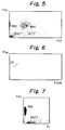
- Fig. 5 is a two-dimensional scattergram of the forward scattered light intensity and the fluorescence intensity obtained by using a urine sample in which red blood cells, white blood cells and bacteria are observed.
- Fig. 6 is a two-dimensional scattergram of the fluorescence pulse width and the scattered light pulse width obtained by using a urine sample in which epithelial cells are observed.
- Fig. 7 is a two-dimensional scattergram of the forward scattered light intensity and the fluorescence intensity obtained by using a urine sample in which red blood cells and bacteria are observed.
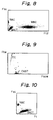
- Fig. 8 is a two-dimensional scattergram of the forward scattered light intensity and the fluorescence intensity obtained by using the standard fluid containing a fluorescence sensitivity stabilizer of the present invention by which particles corresponding to red blood cells, white blood cells and bacteria can be separated and counted.
- Fig. 9 is a two-dimensional scattergram of the fluorescence pulse width and the scattered light pulse width obtained by using the standard fluid containing a fluorescence sensitivity stabilizer of the present invention by which particles corresponding to epithelial cells and hyaline casts can be separated and counted.
- Fig. 10 is a two-dimensional scattergram of the forward scattered light intensity and the fluorescence intensity obtained by using the standard fluid containing a fluorescence sensitivity stabilizer of the present invention by which particles corresponding to red blood cells and bacteria can be separated and counted.
- the sample to be analyzed by using the standard fluid of the present invention are body fluids such as blood and urine containing red blood cells, white blood cells, platelets, etc. (in the case of blood) or red blood cells, white blood cells, epithelial cells, urinary casts, etc. (in the case of urine). Further, some urine samples contain microorganisms such as bacteria, fungi, protozoans and parasites due to topical or systemic infection. In the case of renal insufficiency, furthermore, urine samples contain chemical components and crystals composed of drugs and free fats. These components are the cells to be assayed in the present invention.
- the standard fluid of the present invention contains particles which show, after staining, almost the same fluorescence and scattered light intensities as those of the cells to be assayed.
- the particles to be used in the standard fluid of the present invention therefore, selection should be made of those capable of adsorbing a dye in such an amount as to give almost the same fluorescence intensity as that of the cells to be assayed.
- the size of the particles to be used in the present invention is not always the same as that of the cells to be assayed, since the scattered light intensity is affected not only by the particle size but also by the surface conditions of the particles.
- a fluorescence sensitivity stabilizer to the standard fluid of the present invention, since changes in the stained mode of the particles during storage (i.e., changes in the fluorescence sensitivity) can be prevented thereby for a long time.
- the stained particles in the standard fluid of the present invention can sustain the stable state at room temperature for about three months even though the standard fluid contains no fluorescence sensitivity stabilizer, the addition of the fluorescence sensitivity stabilizer can further prolong the shelf life of the standard fluid (for example, eight months or longer at room temperature).
- fluorescence sensitivity stabilizer water soluble organic solvents are suitable and alcohols and glycols may be appropriately employed therefor. More particularly speaking, methanol, ethanol, propanol, isopropanol, ethylene glycol, diethylene glycol, etc. are usable therefor and ethanol is the most desirable one.
- a fluorescence sensitivity stabilizer may be used preferably at a concentration of 5 to 15 w/w%, still preferably 6 to 14 w/w%. The addition of such a component also contributes to the inhibition of the growth of saprophytic bacteria.
- spherical silica particles still preferably particles of silica for liquid crystals spacers, in the standard fluid of the present invention.
- particles usable as packing materials in liquid chromatography in particular, crosslinked agarose particles and porous silica particles may be preferably employed in the standard fluid of the present invention.
- spherical silica particles of a high purity with a particle size of 3 to 15 ⁇ m, still preferably 5 to 10 ⁇ m. It is particularly preferable to use those employed in electronic materials such as liquid crystal spacers.
- HIPRESICA SPTM average particle size: 5.1 ⁇ m manufactured by Ube-Nitto Kasei Co., Ltd.
- porous silica with a particle size of 15 to 300 ⁇ m (still preferably 20 to 200 ⁇ m) and a pore size of 5 to 40 nm (still preferably 10 to 100 nm).
- NUCLEOPREPTM average particle size: 30 ⁇ m
- a cellulose gel for example, CellulofineTM manufactured by Chisso Corporation
- a hydrophilic vinyl polymer gel for example, TOYOPEAL HWTM manufactured by TOSOH Co., Ltd.
- a crosslinked agarose gel with a particle size of 25 to 300 ⁇ m, still preferably 40 to 200 ⁇ m.
- a crosslinked agarose gel with a particle size of 25 to 300 ⁇ m, still preferably 40 to 200 ⁇ m.
- Sepharose CL-2BTM particle size: 60 to 200 ⁇ m manufactured by Pharmacia Biotec Co., Ltd.
- spherical particles which are commonly employed as a packing material in liquid chromatography and those having no chemical modification are appropriate therefor.
- these materials are cited merely by way of example and the particles to be used in the standard fluid of the present invention are restricted neither in particle size nor in material, so long as they can achieve almost the same fluorescence and scattered light intensities after staining as those of the cells to be assayed.
- the particles cited above are those usable in the analysis of the cell components contained in urine.
- particles suitable for the analysis of platelets for example, totally porous, spherical silica having a particle size of 1 to 5 ⁇ m, still preferably 2 to 4 ⁇ m.
- the standard fluid for a flow cytometer of the present invention can be prepared by suspending the particles in an aqueous medium which is a buffer preferably having a pH value of 3 to 7, still preferably 4 to 5. If necessary, physiological salts (for example, sodium chloride or potassium chloride), sugars (for example, glucose), albumin, hemoglobin, nitrites, preservatives, etc. may be further added thereto.
- physiological salts for example, sodium chloride or potassium chloride
- sugars for example, glucose
- albumin for example, albumin
- hemoglobin hemoglobin
- preservatives preservatives, etc.
- the standard fluid for a flow cytometer of the present invention can be prepared by suspending particles of a single type in an aqueous medium. Alternatively, it may be prepared by mixing two or more types of particles different from each other depending on the sample to be assayed.
- the standard fluid for a flow cytometer of the present invention is mixed with a staining solution which has been prepared separately.
- the staining solution is prepared by dissolving one or more fluorescent dyes, selected depending on the cells to be assayed, in a solvent such as ethylene glycol, diethylene glycol or methanol.
- cationic dyes such as dicarbocyanine dyes (for example, 3,3'-dihexyl-2,2'-oxacarbocyanine iodide, 3,3'-dimethyl-2,2'-oxacarbocyanine iodide, 3,3'-dipropyl-2,2'-oxacarbocyanine iodide, 3,3'-dipentyl-2,2'-oxacarbocyanine iodide), Rhodamine dyes (for example, Rhodamine B, Rhodamine 6G) and dyes capable of specifically staining DNA (for example, ethidium bromide).
- dicarbocyanine dyes for example, 3,3'-dihexyl-2,2'-oxacarbocyanine iodide, 3,3'-dimethyl-2,2'-oxacarbocyanine iodide, 3,3'-dipropyl-2,2
- Quality control and calibration of a flow cytometer with the use of the standard fluid of the present invention may be performed by flowing a mixture of the flow cytometer standard fluid of the present invention with a staining solution through the flow cell of the flow cytometer.
- a flow cytometer it is possible to use, for example, a fully automated urine cell analyzer (Model UF-100, manufactured by TOA MEDICAL ELECTRONICS Co., Ltd.) for analyzing urine samples or automated reticulocyte analyzers (R-Series, manufactured by TOA MEDICAL ELECTRONICS Co., Ltd.) for analyzing blood samples.
- a method of the use of the standard fluid of the present invention will be illustrated by using a standard fluid for analyzing a urine sample.
- a mixture of the standard fluid with the staining solution (hereinafter referred to as the standard sample) is passed through the flow cell and irradiated with excitation light beams. Then the forward scattered light and fluorescence intensities from the formed components in the standard sample are measured. Thus, the formed components contained in the standard sample can be assayed.
- the electric resistance signal intensity i.e., volume information
- a flow cytometer by which electric resistance signal and optical information can be measured.
- Fig. 1 shows a flow cytometer which may be preferably employed in the assay with the use of the standard fluid of the present invention.
- valves 1 and 2 are opened for a definite period of time.
- these valves 1 and 2 are filled up with the sample fluid supplied from a suction nozzle 3 due to the negative pressure from the waste chamber.
- a syringe 4 presses out the fluid at a constant rate, the sample fluid is discharged from a sample nozzle 6.
- a valve 8 is opened and thus a sheath fluid is supplied into a chamber 7 of a flow cell 5.
- the sample is squeezed to fit to the inner diameter of the chamber 7 to thereby form a sheath flow, as shown in Fig. 7, which passes through an orifice 11.
- the orifice 11 is in the shape of a square tube of 100 to 300 ⁇ m in an inner side and made of an optical glass (including quartz glass).
- the formation of the sheath flow makes it possible to pass through the particles one by one in a line at the center of the orifice 11.
- the sample and sheath fluid passing through the orifice 11 are discharged via a recovery tube 14 connected to a chamber 25.
- a platinum electrode 13 located in the chamber 25 serves as an anode, while a stainless electrode 12 located in the chamber 7 serves as a cathode.
- the electric resistance between these electrodes 12 and 13 is determined depending on the electrical resistivity (conductivity) of the sheath fluid, the pore size (pore cross section) and pore length of the orifice, the electrical resistivity of the sample fluid and the diameter of the sample flow.
- An electrode 15 is a direct constant current source which supplies a constant current between the electrodes 12 and 13 so as to generate a current voltage depending on the electric resistivity and the current between the electrodes 12 and 13.
- the electric resistance varies at the both ends of the orifice 11. Namely, the electric resistance is elevated during the passage of the particle and thus the voltage between the electrodes 12 and 13 is varied. Further, there arises a pulse voltage in proportion to the size of the particle passing through the orifice 11. This voltage is added onto the current voltage as described above. The sum of these voltages thus observed between the electrodes 12 and 13 is detected with an amplifier 16 followed by the output of a resistance signal 29.
- the laser light is in the form of an ellipse having the minor width (for example, about 10 ⁇ m) similar to the blood cell size in the direction of the sample flow and the major width (for example, about 150 to 300 ⁇ m) much broader than the blood cell particle in the direction perpendicular to the flow and light irradiation.
- the minor width for example, about 10 ⁇ m
- the major width for example, about 150 to 300 ⁇ m
- the forward scattered light and forward fluorescence illuminating the cell (formed matter) and emitted at narrow angles are converged with a collector lens 20 and pass through a pinhole 21 of a blocking sheet 30.
- these beams pass through a dichroic mirror 22.
- the fluorescence is detected with a photomultiplier tube (PMT) 24 and converted into an electric signal 27 followed by output.
- the dichroic mirror reflects the scattered light which is then received by a photodiode 31 and converted into another electric signal 28 followed by output.
- the particles show almost the same behaviors as those of the cells to be assayed, which indicates that the standard fluid is usable in the calibration of the flow cytometer.
- the dye When the standard fluid for a flow cytometer of the present invention is mixed with a staining solution, the dye is adsorbed onto the particles and emits fluorescence depending on the adsorption level.
- the particles to be used in the standard fluid for a flow cytometer of the present invention are those capable of adsorbing a dye in such an amount as to achieve a fluorescence intensity similar to that of the cells to be assayed. Also, use can be made of particles as large as the cells to be assayed. However, the size of the particles to be used in the present invention may not always be the same as that of the cells to be assayed, since the scattered light intensity is affected not only by the particle size but also by the surface conditions of the particles.
- a cationic dye is used for staining cells.
- red blood cells have poor staining properties.
- the particles serving as a substitute for red blood cells would adsorb a dye not in a very large amount but to such an extent as to give a fluorescence intensity similar to that of the red blood cells, though particles adsorbing no dye cannot be employed therefor.
- the particles employed therefor have a size similar to the actual red blood cell size (3 to 10 ⁇ m). However, use can be also made of particles somewhat differing in size, so long as the scattered light intensity thereof is almost the same as that of the red blood cells.
- particles serving as a substitute for white blood cells or epithelial cells should adsorb a dye in such an amount as to achieve almost the same fluorescence intensity as those of these cells.
- the particles employed therefor have a size similar to the actual white blood cell size (3 to 15 ⁇ m) or epithelial cell size (15 to 150 ⁇ m), similar to the case of red blood cells.
- particles serving as a substitute for blood casts of either type are used in the standard fluid of the present invention. Similar to other cells, the particles to be used therefor are those which adsorb a dye in such an amount as to achieve a fluorescence intensity similar to that of blood casts containing inclusion bodies and have a size similar to the actual blood cast size (100 ⁇ m or above), though particles somewhat differing in size are also usable therefor, so long as they can provide a scattered light intensity almost the same as that of the blood casts.
- the particles to be used as a substitute for bacteria are those which adsorb a dye in such an amount as to achieve a fluorescence intensity similar to that of bacteria and give a scattered light intensity similar to that of bacteria.
- the present invention provides a standard fluid for a flow cytometer which shows almost the same behaviors as those of the cells to be assayed and can be easily handled without any risk of infection with bacteria, etc.
- the sensitivity of a flow cytometer can be precisely controlled by using particles of two or more types differing from each other in scattered light intensity and fluorescence intensity.
- the total sensitivity is roughly regulated by using particles having a higher sensitivity followed by fine adjustment with the use of particles having a lower sensitivity.
- Example 1 Assay of standard fluid for urine measurement
- HIPRESICA SPTM average particle size: 5.1 ⁇ m,for red blood cell, manufactured by Ube-Nitto Kasei Co., Ltd.
- 40/ ⁇ l Zorbax BP-SILTM particle size: 7 - 8 ⁇ m, for white blood cell, obtained from GL Science Co., Ltd.
- 40/ ⁇ l NUCLEOPREP 100-30TM average particle size: 30 ⁇ m, for epithelial cell, manufactured by MACHEREY-NAGEL
- Sepharose CL-2BTM particle size: 60 to 200 ⁇ m,for blood cast, manufactured by Pharmacia Biotec Co., Ltd.
- 2/ ⁇ l Pia Seed S-150TM average particle size: 2 ⁇ m) for bacteria, manufactured by Pia TEC Co., Ltd.
- a thick dispersion containing exclusively the particles was prepared. After counting the particles under the conditions as specified below, the dispersion was diluted and blended so as to give the above-mentioned particle concentrations. Thus the standard fluid was obtained.
- Staining reagent 3,3'-dihexyl-2,2'-oxacarbocyanine 0.144 w/w% ethidium bromide 0.036 w/w% ethylene glycol 99.820 w/w%.
- Diluent for staining HEPES 1.165 w/w% sodium hydroxide 0.036 w/w% EDTA-3K 0.391 w/w% purified water suffice for 100%.
- the standard fluid, the staining reagent and the diluent for staining were mixed together at a ratio of 400:40:1160 ( ⁇ l) and assay was performed at a reaction temperature of 35°C for a staining period of 10 seconds with the use of UF-100 manufactured by TOA MEDICAL ELECTRONICS Co., Ltd.
- Fig. 2 is a two-dimensional scattergram of the forward scattered light intensity and the fluorescence intensity by which particles corresponding to red blood cells, white blood cells and bacteria can be separated and counted.
- Fig. 3 is a two-dimensional scattergram of the fluorescence pulse width and the scattered light pulse width by which particles corresponding to epithelial cells and large particles corresponding to blood casts (upper) can be separated and counted.
- Fig. 4 is a two-dimensional scattergram of the forward scattered light intensity and the fluorescence intensity by which particles corresponding to red blood cells and bacteria can be separated and counted.
- Figs. 5 to 7 shows the results thus obtained.
- Fig. 5 is a scattergram corresponding to Fig. 2 wherein red blood cells and bacteria are observed at almost the same positions as observed in Fig. 2.
- a mass of white blood cells is observed at a somewhat upper part, compared with Fig. 2, and white blood cells having been damaged in cell membrane, etc. are scattered below. This part roughly corresponds to the white blood cells observed in Fig. 2.
- Fig. 6 is a scattergram corresponding to Fig. 3 wherein epithelial cells are observed at almost the same position as observed in Fig. 3. This urine sample showed no urinary cast.
- Fig. 7 is a scattergram corresponding to Fig. 4 wherein red blood cells are observed at almost the same position as observed in Fig. 4.
- Fsc stands for the forward scattered light intensity
- Fl stands for the fluorescence intensity
- Flw stands for the fluorescence pulse width
- Fscw stands for the forward scattered light pulse width
- RBC red blood cells
- WBC white blood cells
- BACT stands for bacteria
- EC epithelial cells
- CAST stands for blood cast.
- Example 2 Similar to Example 1, a thick dispersion containing exclusively the particles was first prepared. After counting the particles, the dispersion was diluted and blended so as to give the above-mentioned particle concentrations.
- Fig. 8 is a two-dimensional scattergram of the forward scattered light intensity and the fluorescence intensity by which particles corresponding to red blood cells, white blood cells and bacteria can be separated and counted.
- Fig. 9 is a two-dimensional scattergram of the fluorescence pulse width and the scattered light pulse width by which particles corresponding to epithelial cells and hyaline casts can be separated and counted.
- Fig. 10 is a two-dimensional scattergram of the forward scattered light intensity and the fluorescence intensity by which particles corresponding to red blood cells and bacteria can be separated and counted.
Landscapes
- Chemical & Material Sciences (AREA)
- Dispersion Chemistry (AREA)
- Physics & Mathematics (AREA)
- Health & Medical Sciences (AREA)
- Life Sciences & Earth Sciences (AREA)
- Analytical Chemistry (AREA)
- Biochemistry (AREA)
- General Health & Medical Sciences (AREA)
- General Physics & Mathematics (AREA)
- Immunology (AREA)
- Pathology (AREA)
- Investigating Or Analysing Biological Materials (AREA)
- Sampling And Sample Adjustment (AREA)
Applications Claiming Priority (4)
| Application Number | Priority Date | Filing Date | Title |
|---|---|---|---|
| JP29984595 | 1995-11-17 | ||
| JP299845/95 | 1995-11-17 | ||
| JP296654/96 | 1996-11-08 | ||
| JP29665496A JP3875754B2 (ja) | 1995-11-17 | 1996-11-08 | フローサイトメータ用標準液 |
Publications (2)
| Publication Number | Publication Date |
|---|---|
| EP0774655A2 true EP0774655A2 (fr) | 1997-05-21 |
| EP0774655A3 EP0774655A3 (fr) | 1997-10-08 |
Family
ID=26560787
Family Applications (1)
| Application Number | Title | Priority Date | Filing Date |
|---|---|---|---|
| EP96118445A Withdrawn EP0774655A3 (fr) | 1995-11-17 | 1996-11-18 | Fluide de calibration pour cytomètre de flux |
Country Status (5)
| Country | Link |
|---|---|
| US (1) | US5888823A (fr) |
| EP (1) | EP0774655A3 (fr) |
| JP (1) | JP3875754B2 (fr) |
| CN (1) | CN1100265C (fr) |
| TW (1) | TW517155B (fr) |
Cited By (3)
| Publication number | Priority date | Publication date | Assignee | Title |
|---|---|---|---|---|
| EP0965841A1 (fr) * | 1998-04-06 | 1999-12-22 | AVL Medical Instruments AG | Référance liquide de contrÔle ou d'étalonnage pour des appareils de mesure optique du taux d'hémoglobine dans des échantillons de sang |
| US6174728B1 (en) | 1998-04-03 | 2001-01-16 | Avl Medical Instruments Ag | Control or calibration standard for use with instruments for optical measurement of hemoglobin concentration in blood samples |
| EP1744145A2 (fr) | 2005-07-12 | 2007-01-17 | Sysmex Corporation | Matériel conforme pour analyseur de particules |
Families Citing this family (48)
| Publication number | Priority date | Publication date | Assignee | Title |
|---|---|---|---|---|
| US7282350B2 (en) | 1998-02-12 | 2007-10-16 | Immunivest Corporation | Labeled cell sets for use as functional controls in rare cell detection assays |
| US20010018192A1 (en) * | 1998-02-12 | 2001-08-30 | Terstappen Leon W.M.M. | Labeled cells for use as an internal functional control in rare cell detection assays |
| JP3662423B2 (ja) * | 1998-09-03 | 2005-06-22 | 株式会社ニレコ | 粒子解析装置 |
| JP2000214070A (ja) * | 1999-01-21 | 2000-08-04 | Sysmex Corp | シ―スフロ―セルとそれを用いた血液分析装置 |
| IL152714A (en) | 2000-05-09 | 2014-03-31 | Xy Llc | High-purity spermatozoa populations carrying chromosome-x and chromosome-y |
| US7016022B2 (en) * | 2000-08-02 | 2006-03-21 | Honeywell International Inc. | Dual use detectors for flow cytometry |
| US7630063B2 (en) * | 2000-08-02 | 2009-12-08 | Honeywell International Inc. | Miniaturized cytometer for detecting multiple species in a sample |
| US7277166B2 (en) * | 2000-08-02 | 2007-10-02 | Honeywell International Inc. | Cytometer analysis cartridge optical configuration |
| EP1378752A4 (fr) | 2001-04-13 | 2004-08-04 | Chugai Pharmaceutical Co Ltd | Procede permettant de quantifier l'expression d'antigenes |
| JP2003083869A (ja) * | 2001-09-10 | 2003-03-19 | Japan Water Research Center | クリプトスポリジウムパルバム疑似蛍光トレーサおよびその調製法 |
| EP1489093A4 (fr) * | 2002-03-19 | 2005-07-20 | Oncorex Inc | Peptides, compositions medicinales contenant ce peptide et compositions medicinales anticancereuses |
| FI112504B (fi) * | 2002-08-07 | 2003-12-15 | Cyflo Oy | Menetelmä bakteerien tunnistamiseksi |
| US20060118167A1 (en) * | 2004-12-03 | 2006-06-08 | Xy, Inc. | Pressure regulated continuously variable volume container for fluid delivery |
| JP4744132B2 (ja) * | 2004-12-09 | 2011-08-10 | シスメックス株式会社 | 粒子画像分析装置用標準液の表示値作成方法 |
| AU2006235230B8 (en) * | 2005-04-07 | 2010-08-26 | Chata Biosystems, Inc. | Flow path conditioner system |
| JP4953710B2 (ja) * | 2005-07-12 | 2012-06-13 | シスメックス株式会社 | 尿中有形成分分析装置用標準物質 |
| JP4946047B2 (ja) * | 2005-12-28 | 2012-06-06 | 東洋紡績株式会社 | 有形成分分類装置用粒子標準試薬 |
| JP4748719B2 (ja) * | 2006-03-02 | 2011-08-17 | 国立大学法人大阪大学 | 生細胞内の特定タンパク質の定量方法および標準蛍光マイクロビーズの作製方法 |
| JP4817450B2 (ja) * | 2007-01-31 | 2011-11-16 | シスメックス株式会社 | 血液分析装置、血液分析方法およびコンピュータプログラム |
| JP4926812B2 (ja) * | 2007-02-01 | 2012-05-09 | シスメックス株式会社 | 血球分析装置および体液分析方法 |
| US8119688B2 (en) * | 2007-09-19 | 2012-02-21 | Xy, Llc | Differential evaporation potentiated disinfectant system |
| US20100249166A1 (en) | 2007-09-19 | 2010-09-30 | Xy, Inc. | Differential evaporation potentiated disinfectant system |
| JP4580976B2 (ja) * | 2007-12-26 | 2010-11-17 | シスメックス株式会社 | 精度管理機能を備えた生体試料分析装置および精度管理測定結果の表示方法 |
| JP5380026B2 (ja) * | 2008-09-24 | 2014-01-08 | シスメックス株式会社 | 標本撮像装置 |
| JP5255498B2 (ja) * | 2009-03-30 | 2013-08-07 | シスメックス株式会社 | 試薬調製装置および検体処理システム |
| JP5304456B2 (ja) | 2009-06-10 | 2013-10-02 | ソニー株式会社 | 微小粒子測定装置 |
| CN101943663B (zh) * | 2010-07-09 | 2012-07-25 | 董珂 | 自动辨别微粒的衍射图像测量分析系统及方法 |
| JP2012047464A (ja) | 2010-08-24 | 2012-03-08 | Sony Corp | 微小粒子測定装置及び光軸補正方法 |
| ES2928973T3 (es) * | 2012-01-11 | 2022-11-23 | Takeda Pharmaceuticals Co | Caracterización de partículas subvisibles usando un analizador de partículas |
| JP5905317B2 (ja) * | 2012-03-30 | 2016-04-20 | ソニー株式会社 | 微小粒子分取装置におけるキャリブレーション方法及び該装置 |
| US20150177115A1 (en) | 2012-04-06 | 2015-06-25 | Slingshot Biosciences | Hydrogel particles with tunable optical properties |
| WO2013159296A1 (fr) * | 2012-04-25 | 2013-10-31 | 深圳联合医学科技有限公司 | Procédé de marquage d'un réactif à base de globules sanguins ou de maîtrise d'un processus d'essai |
| CN103540565A (zh) * | 2012-07-17 | 2014-01-29 | 苏州生物医学工程技术研究所 | 一种鸡红细胞的制备和储存方法 |
| JP6116502B2 (ja) * | 2014-02-28 | 2017-04-19 | シスメックス株式会社 | 検体分析装置および検体分析方法 |
| JP6228085B2 (ja) * | 2014-08-27 | 2017-11-08 | シスメックス株式会社 | 検体分析装置及び検体分析方法 |
| JP6692834B2 (ja) | 2015-02-09 | 2020-05-13 | スリングショット バイオサイエンシーズ, インコーポレイテッド | 調整可能な光学特性を有するヒドロゲル粒子およびその使用の方法 |
| JP6680492B2 (ja) * | 2015-09-11 | 2020-04-15 | シスメックス株式会社 | 細胞分析装置および細胞分析方法 |
| JP6731250B2 (ja) * | 2015-12-25 | 2020-07-29 | リオン株式会社 | 生物粒子計数器校正用の標準粒子懸濁液の製造方法、及び、生物粒子計数器の校正方法 |
| JP6237806B2 (ja) * | 2016-03-16 | 2017-11-29 | ソニー株式会社 | 微小粒子分取装置 |
| WO2021127424A1 (fr) | 2019-12-19 | 2021-06-24 | Roche Diagnostics Hematology, Inc. | Compositions et procédés d'étalonnage et de contrôle de qualité |
| KR20220129585A (ko) | 2020-01-24 | 2022-09-23 | 슬링샷 바이오사이언시즈 인코포레이티드 | 세포-유사 보정 입자를 위한 조성물 및 방법 |
| JP7807395B2 (ja) | 2020-05-04 | 2026-01-27 | スリングショット バイオサイエンシーズ, インコーポレイテッド | 多重化アッセイのためのパッシブ光学バーコード付与のための組成物および方法 |
| CN112730203B (zh) * | 2020-12-29 | 2023-06-16 | 深圳市科曼医疗设备有限公司 | 血球分析仪的光学系统、光学增益校准方法和存储介质 |
| AU2022376541A1 (en) | 2021-10-29 | 2024-04-18 | Slingshot Biosciences, Inc. | Hydrogel particles as feeder cells and as synthetic antigen presenting cells |
| CN119907921A (zh) | 2022-05-05 | 2025-04-29 | 弹弓生物科学公司 | 用于血液学的作为红细胞模拟物的工程化颗粒和含有工程化颗粒的组合物 |
| CA3257621A1 (fr) | 2022-06-02 | 2023-12-07 | Slingshot Biosciences, Inc. | Mimétique de cellule apoptotique |
| CN120712108A (zh) | 2022-10-26 | 2025-09-26 | 弹弓生物科学公司 | 具有可调整光学性质的大小可调整合成颗粒和用于使用其进行免疫细胞激活的方法 |
| WO2025049609A1 (fr) | 2023-08-29 | 2025-03-06 | Slingshot Biosciences, Inc. | Mimétiques de cellules souches cd34 |
Citations (3)
| Publication number | Priority date | Publication date | Assignee | Title |
|---|---|---|---|---|
| US4336173A (en) * | 1978-02-21 | 1982-06-22 | Sintef | Process for preparing an aqueous emulsion or dispersion of a partly water-soluble material, and optionally further conversion of the prepared dispersion or emulsion to a polymer dispersion when the partly water-soluble material is a polymerizable monomer |
| JPH04278460A (ja) | 1991-03-06 | 1992-10-05 | Shima Kenkyusho:Kk | 尿沈渣検査成績管理方法 |
| JPH06102152A (ja) | 1992-09-18 | 1994-04-15 | Hitachi Ltd | フロー式粒子画像解析装置用標準液 |
Family Cites Families (20)
| Publication number | Priority date | Publication date | Assignee | Title |
|---|---|---|---|---|
| FR2570081B1 (fr) * | 1984-09-10 | 1987-01-09 | Pf Medicament | Polysaccharides membranaires utiles notamment comme medicament et procede pour leur preparation |
| US4767206A (en) * | 1984-12-24 | 1988-08-30 | Flow Cytometry Standards Corporation | Calibration method for flow cytometry using fluorescent microbeads and synthesis thereof |
| US5084394A (en) * | 1984-12-24 | 1992-01-28 | Vogt Robert F | Method for corrective calibration of a flow cytometry using a mixture of fluorescent microbeads and cells |
| US4857451A (en) * | 1984-12-24 | 1989-08-15 | Flow Cytometry Standards Corporation | Method of compensating and calibrating a flow cytometer, and microbead standards kit therefor |
| US5093234A (en) * | 1984-12-24 | 1992-03-03 | Caribbean Microparticles Corporation | Method of aligning, compensating, and calibrating a flow cytometer for analysis of samples, and microbead standards kit therefor |
| US5380663A (en) * | 1984-12-24 | 1995-01-10 | Caribbean Microparticles Corporation | Automated system for performance analysis and fluorescence quantitation of samples |
| US5073498A (en) * | 1984-12-24 | 1991-12-17 | Caribbean Microparticles Corporation | Fluorescent alignment microbeads with broad excitation and emission spectra and its use |
| US4774189A (en) * | 1984-12-24 | 1988-09-27 | Flow Cytometry Standards Corp. | Fluorescent calibration microbeads simulating stained cells |
| US5073497A (en) * | 1989-06-30 | 1991-12-17 | Caribbean Microparticles Corporation | Microbead reference standard and method of adjusting a flow cytometer to obtain reproducible results using the microbeads |
| US4714682A (en) * | 1985-12-11 | 1987-12-22 | Flow Cytometry Standards Corporation | Fluorescent calibration microbeads simulating stained cells |
| US4868126A (en) * | 1985-12-11 | 1989-09-19 | Flow Cytometry Standards Corporation | Method of calibrating a fluorescent microscope using fluorescent calibration microbeads simulating stained cells |
| US4704891A (en) * | 1986-08-29 | 1987-11-10 | Becton, Dickinson And Company | Method and materials for calibrating flow cytometers and other analysis instruments |
| JPH01301166A (ja) * | 1988-05-30 | 1989-12-05 | Japan Synthetic Rubber Co Ltd | 血球計数器校正用標準液 |
| JP2958353B2 (ja) * | 1988-12-15 | 1999-10-06 | シスメックス株式会社 | 光学系調整用の標準物質およびその製造方法 |
| CA2053618C (fr) * | 1990-10-22 | 1999-10-05 | Toshihiko Kishimoto | Dosage de substances a l'aide de la cytometrie de flux |
| JPH0518061A (ja) * | 1991-07-08 | 1993-01-26 | Sanko Metal Ind Co Ltd | 外囲体 |
| US5456102A (en) * | 1993-03-19 | 1995-10-10 | Moorehead; Jack | Method and apparatus for particle counting and counter calibration |
| US5520952A (en) * | 1993-07-16 | 1996-05-28 | Tokyo Ohka Kogyo Co., Ltd. | Method for forming a protective coating film on electronic parts and devices |
| EP0678742B1 (fr) * | 1994-04-21 | 2002-07-24 | Hitachi, Ltd. | Procédé de surveillance d'une solution colorante pour l'analyse de particules et méthode d'étalonage pour une analyse de particules |
| US5620842A (en) * | 1995-03-29 | 1997-04-15 | Becton Dickinson And Company | Determination of the number of fluorescent molecules on calibration beads for flow cytometry |
-
1996
- 1996-11-08 JP JP29665496A patent/JP3875754B2/ja not_active Expired - Lifetime
- 1996-11-13 US US08/747,644 patent/US5888823A/en not_active Expired - Lifetime
- 1996-11-15 CN CN96114487A patent/CN1100265C/zh not_active Expired - Lifetime
- 1996-11-18 EP EP96118445A patent/EP0774655A3/fr not_active Withdrawn
- 1996-12-07 TW TW085115162A patent/TW517155B/zh not_active IP Right Cessation
Patent Citations (3)
| Publication number | Priority date | Publication date | Assignee | Title |
|---|---|---|---|---|
| US4336173A (en) * | 1978-02-21 | 1982-06-22 | Sintef | Process for preparing an aqueous emulsion or dispersion of a partly water-soluble material, and optionally further conversion of the prepared dispersion or emulsion to a polymer dispersion when the partly water-soluble material is a polymerizable monomer |
| JPH04278460A (ja) | 1991-03-06 | 1992-10-05 | Shima Kenkyusho:Kk | 尿沈渣検査成績管理方法 |
| JPH06102152A (ja) | 1992-09-18 | 1994-04-15 | Hitachi Ltd | フロー式粒子画像解析装置用標準液 |
Cited By (6)
| Publication number | Priority date | Publication date | Assignee | Title |
|---|---|---|---|---|
| US6174728B1 (en) | 1998-04-03 | 2001-01-16 | Avl Medical Instruments Ag | Control or calibration standard for use with instruments for optical measurement of hemoglobin concentration in blood samples |
| EP0965841A1 (fr) * | 1998-04-06 | 1999-12-22 | AVL Medical Instruments AG | Référance liquide de contrÔle ou d'étalonnage pour des appareils de mesure optique du taux d'hémoglobine dans des échantillons de sang |
| EP1744145A2 (fr) | 2005-07-12 | 2007-01-17 | Sysmex Corporation | Matériel conforme pour analyseur de particules |
| EP1744145A3 (fr) * | 2005-07-12 | 2008-12-31 | Sysmex Corporation | Matériel conforme pour analyseur de particules |
| US7601539B2 (en) | 2005-07-12 | 2009-10-13 | Sysmex Corporation | Standard material for particle analyzer |
| US9658197B2 (en) | 2005-07-12 | 2017-05-23 | Sysmex Corporation | Standard material for a particle analyzer |
Also Published As
| Publication number | Publication date |
|---|---|
| CN1158997A (zh) | 1997-09-10 |
| EP0774655A3 (fr) | 1997-10-08 |
| JPH09196916A (ja) | 1997-07-31 |
| CN1100265C (zh) | 2003-01-29 |
| JP3875754B2 (ja) | 2007-01-31 |
| TW517155B (en) | 2003-01-11 |
| US5888823A (en) | 1999-03-30 |
Similar Documents
| Publication | Publication Date | Title |
|---|---|---|
| US5888823A (en) | Standard fluid for flow cytometer | |
| EP1089078B1 (fr) | Réactif et méthode pour l'analyse des composants solides dans l'urine | |
| CN101008641B (zh) | 幼稚白细胞分析用试剂及试剂盒 | |
| US5747349A (en) | Fluorescent reporter beads for fluid analysis | |
| US9658197B2 (en) | Standard material for a particle analyzer | |
| EP1466985B1 (fr) | Méthodes et dispositifs pour la mésuration des bactéries, et supports de stockage de programmes d'ordinateur pour analyser les bactéries | |
| EP0398652B1 (fr) | Méthode de différentiation des leukocytes mettant en oeuvre un cytomètre de flux | |
| US10774359B2 (en) | Cellular analysis of body fluids | |
| US20120003730A1 (en) | Portable sample analyzer cartridge | |
| JP3096979B2 (ja) | 血液サンプルのヘモグロビン濃度を光学的に測定する装置のための品質管理及び較正用標準液 | |
| US6803208B2 (en) | Automated epifluorescence microscopy for detection of bacterial contamination in platelets | |
| CN101743469B (zh) | 试样分析用试剂盒以及试样分析方法 | |
| US4971917A (en) | Reagent for reticulocyte counting by flow cytometry | |
| US3616253A (en) | Method for determining bacterial populations | |
| JP3739482B2 (ja) | 尿中の有形成分分析方法および試薬 | |
| JP4953710B2 (ja) | 尿中有形成分分析装置用標準物質 | |
| JPH08170960A (ja) | 尿中有形成分分析用試薬及びその試薬を用いた有形成分分析方法 | |
| CA2109045A1 (fr) | Procede microphotolytique de mesure des caracteristiques cellulaires | |
| KR20240159630A (ko) | 생물학적 샘플을 분석하기 위한 큐벳 | |
| KR20240161181A (ko) | 큐벳을 코팅하기 위한 조성물 및 큐벳을 코팅하기 위한 조성물을 제조하는 방법 | |
| CN116973193A (zh) | 一种溶血剂及使用该溶血剂的糖化血红蛋白试剂盒 | |
| CN118937316A (zh) | 一种比色内毒素传感检测方法 |
Legal Events
| Date | Code | Title | Description |
|---|---|---|---|
| PUAI | Public reference made under article 153(3) epc to a published international application that has entered the european phase |
Free format text: ORIGINAL CODE: 0009012 |
|
| AK | Designated contracting states |
Kind code of ref document: A2 Designated state(s): DE FR GB IT |
|
| PUAL | Search report despatched |
Free format text: ORIGINAL CODE: 0009013 |
|
| AK | Designated contracting states |
Kind code of ref document: A3 Designated state(s): DE FR GB IT |
|
| 17P | Request for examination filed |
Effective date: 19980130 |
|
| RAP1 | Party data changed (applicant data changed or rights of an application transferred) |
Owner name: SYSMEX CORPORATION |
|
| 17Q | First examination report despatched |
Effective date: 20031121 |
|
| STAA | Information on the status of an ep patent application or granted ep patent |
Free format text: STATUS: THE APPLICATION HAS BEEN WITHDRAWN |
|
| 18W | Application withdrawn |
Effective date: 20110922 |