EP1132471A2 - TNF-bindende Proteine - Google Patents
TNF-bindende Proteine Download PDFInfo
- Publication number
- EP1132471A2 EP1132471A2 EP01108117A EP01108117A EP1132471A2 EP 1132471 A2 EP1132471 A2 EP 1132471A2 EP 01108117 A EP01108117 A EP 01108117A EP 01108117 A EP01108117 A EP 01108117A EP 1132471 A2 EP1132471 A2 EP 1132471A2
- Authority
- EP
- European Patent Office
- Prior art keywords
- tnf
- dna sequences
- sequences
- cells
- fragments
- Prior art date
- Legal status (The legal status is an assumption and is not a legal conclusion. Google has not performed a legal analysis and makes no representation as to the accuracy of the status listed.)
- Withdrawn
Links
Images



Classifications
-
- C—CHEMISTRY; METALLURGY
- C07—ORGANIC CHEMISTRY
- C07K—PEPTIDES
- C07K14/00—Peptides having more than 20 amino acids; Gastrins; Somatostatins; Melanotropins; Derivatives thereof
- C07K14/435—Peptides having more than 20 amino acids; Gastrins; Somatostatins; Melanotropins; Derivatives thereof from animals; from humans
- C07K14/705—Receptors; Cell surface antigens; Cell surface determinants
- C07K14/715—Receptors; Cell surface antigens; Cell surface determinants for cytokines; for lymphokines; for interferons
- C07K14/7151—Receptors; Cell surface antigens; Cell surface determinants for cytokines; for lymphokines; for interferons for tumor necrosis factor [TNF], for lymphotoxin [LT]
-
- A—HUMAN NECESSITIES
- A61—MEDICAL OR VETERINARY SCIENCE; HYGIENE
- A61P—SPECIFIC THERAPEUTIC ACTIVITY OF CHEMICAL COMPOUNDS OR MEDICINAL PREPARATIONS
- A61P29/00—Non-central analgesic, antipyretic or antiinflammatory agents, e.g. antirheumatic agents; Non-steroidal antiinflammatory drugs [NSAID]
-
- A—HUMAN NECESSITIES
- A61—MEDICAL OR VETERINARY SCIENCE; HYGIENE
- A61P—SPECIFIC THERAPEUTIC ACTIVITY OF CHEMICAL COMPOUNDS OR MEDICINAL PREPARATIONS
- A61P31/00—Antiinfectives, i.e. antibiotics, antiseptics, chemotherapeutics
- A61P31/04—Antibacterial agents
-
- A—HUMAN NECESSITIES
- A61—MEDICAL OR VETERINARY SCIENCE; HYGIENE
- A61P—SPECIFIC THERAPEUTIC ACTIVITY OF CHEMICAL COMPOUNDS OR MEDICINAL PREPARATIONS
- A61P37/00—Drugs for immunological or allergic disorders
- A61P37/02—Immunomodulators
- A61P37/06—Immunosuppressants, e.g. drugs for graft rejection
-
- C—CHEMISTRY; METALLURGY
- C07—ORGANIC CHEMISTRY
- C07K—PEPTIDES
- C07K16/00—Immunoglobulins [IG], e.g. monoclonal or polyclonal antibodies
- C07K16/18—Immunoglobulins [IG], e.g. monoclonal or polyclonal antibodies against material from animals or humans
- C07K16/28—Immunoglobulins [IG], e.g. monoclonal or polyclonal antibodies against material from animals or humans against receptors, cell surface antigens or cell surface determinants
- C07K16/2866—Immunoglobulins [IG], e.g. monoclonal or polyclonal antibodies against material from animals or humans against receptors, cell surface antigens or cell surface determinants against receptors for cytokines, lymphokines, interferons
-
- A—HUMAN NECESSITIES
- A61—MEDICAL OR VETERINARY SCIENCE; HYGIENE
- A61K—PREPARATIONS FOR MEDICAL, DENTAL OR TOILETRY PURPOSES
- A61K38/00—Medicinal preparations containing peptides
-
- C—CHEMISTRY; METALLURGY
- C12—BIOCHEMISTRY; BEER; SPIRITS; WINE; VINEGAR; MICROBIOLOGY; ENZYMOLOGY; MUTATION OR GENETIC ENGINEERING
- C12N—MICROORGANISMS OR ENZYMES; COMPOSITIONS THEREOF; PROPAGATING, PRESERVING, OR MAINTAINING MICROORGANISMS; MUTATION OR GENETIC ENGINEERING; CULTURE MEDIA
- C12N2799/00—Uses of viruses
- C12N2799/02—Uses of viruses as vector
- C12N2799/021—Uses of viruses as vector for the expression of a heterologous nucleic acid
- C12N2799/026—Uses of viruses as vector for the expression of a heterologous nucleic acid where the vector is derived from a baculovirus
-
- Y—GENERAL TAGGING OF NEW TECHNOLOGICAL DEVELOPMENTS; GENERAL TAGGING OF CROSS-SECTIONAL TECHNOLOGIES SPANNING OVER SEVERAL SECTIONS OF THE IPC; TECHNICAL SUBJECTS COVERED BY FORMER USPC CROSS-REFERENCE ART COLLECTIONS [XRACs] AND DIGESTS
- Y10—TECHNICAL SUBJECTS COVERED BY FORMER USPC
- Y10S—TECHNICAL SUBJECTS COVERED BY FORMER USPC CROSS-REFERENCE ART COLLECTIONS [XRACs] AND DIGESTS
- Y10S930/00—Peptide or protein sequence
- Y10S930/01—Peptide or protein sequence
- Y10S930/14—Lymphokine; related peptides
- Y10S930/144—Tumor necrosis factor
Definitions
- TNF ⁇ Tumor necrosis factor ⁇
- cachectin lymphotoxin
- Some of these TNF effects are through induction of other factors or by using synergistic effects other factors such as interferons or Interleukins achieved [13-16].
- TNF is in a number of pathological conditions, for example shock states in meningococcal sepsis [17], in the development of autoimmune glomerulonephritis in mice [18] or in cerebral malaria in mice [19] and involved in humans [41].
- shock states in meningococcal sepsis [17] in the development of autoimmune glomerulonephritis in mice [18] or in cerebral malaria in mice [19] and involved in humans [41].
- TNF can have a fever like Interleukin-1 trigger [39]. Due to the pleiotropic functional Properties of TNF can be assumed that TNF in Interaction with other cytokines in a whole range other pathological conditions as mediators of immune response, Inflammation or other processes is involved.
- TNF-BP TNF-binding proteins
- TNF / TNF-BP complex can be isolated [27].
- SDS-PAGE preparative SDS polyacrylamide gel electrophoresis
- the gel has a protein background smear in the range from 33 kD to 40 kD.
- the importance of the protein bands thus obtained was however with regard to the heterogeneity of the Starting material used (placenta tissue; united Material from several placentas) not clear.
- the present invention relates to non-soluble proteins, ie, for example, membrane proteins or so-called receptors, and soluble or insoluble fragments thereof, which bind TNF (TNF-BP), in homogeneous form, and their physiologically tolerable salts.
- TNF-BP TNF
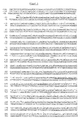
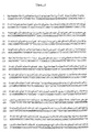
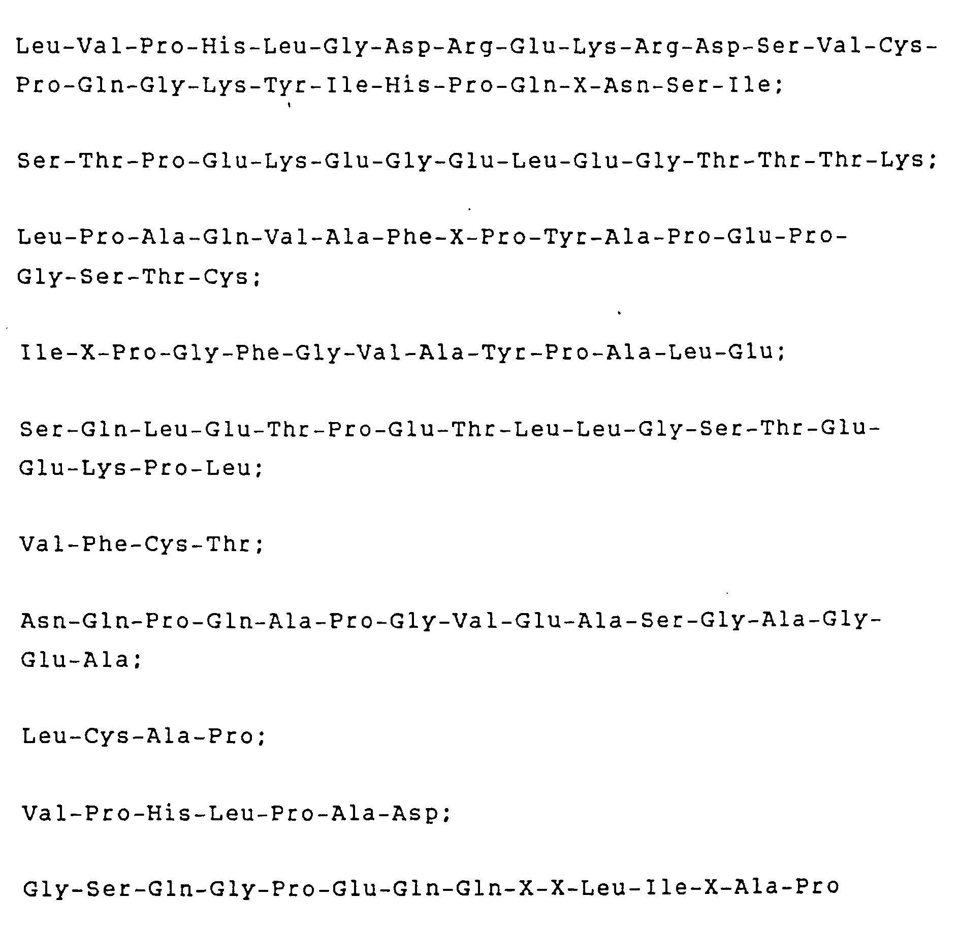
- those proteins which are characterized by at least one of the following partial amino acid sequences: where X stands for an amino acid residue that could not be clearly determined.
- TNF-BP are already characterized by one N-terminal partial sequence have been characterized [European Patent application with publication no. 308 378], where this sequence differs from the N-terminal according to the invention Partial sequence according to formula (IA) differs. Furthermore but it is the described in the prior art TNF binding proteins to isolate from urine, soluble, i.e. not membrane bound, TNF-BP and not around membrane-bound, i.e. insoluble, TNF-BP.
- the present application also relates to methods for isolating the TNF-BP according to the invention.
- This Procedures are characterized in that essentially carries out the following cleaning steps one after the other: Production of a cell or tissue extract, immunoaffinity chromatography and / or single or multiple Ligand affinity chromatography, high resolution liquid chromatography (HPLC) and preparative SDS polyacrylamide gel electrophoresis (SDS-PAGE).
- HPLC high resolution liquid chromatography
- SDS-PAGE preparative SDS polyacrylamide gel electrophoresis
- the present invention encompasses not only allelic variants but also those DNA sequences which result from deletions, substitutions and additions of one or more nucleotides of the sequences shown in FIGS. 1 and 4, where: the proteins encoded by it are still TNF-BP.
- a sequence resulting from such a deletion is described, for example, in Science 248, 1019-1023, (1990).
- DNA sequences which are suitable for such a protein with an apparent molecular weight of about 55 kD are particularly preferred, such as sequences those for both insoluble and soluble fragments of such Encode proteins.
- a DNA sequence, for example for encoding such a non-soluble protein fragment is sufficient from nucleotide -185 to 1122 of that shown in Figure 1 Sequence.
- DNA sequences for soluble protein fragments encode, for example, those of nucleotide -185 to 633 or from nucleotide -14 to 633 of that in Figure 1 sequence shown range.
- DNA sequences are the sequences of the open reading frame from nucleotide 2 to 1,177.
- the peptides will be IIA, IIC, IIE, IIF, IIG and IIH encoded by the partial cDNA sequence in Figure 4, wherein the slight deviations from the experimentally determined Amino acid sequences from that derived from the cDNA Sequence with the highest probability on the lower one Resolution based on gas phase sequencing.
- Prefers are also DNA sequences for both insoluble and soluble Fragments of TNF-binding proteins with an apparent Encode molecular weight of 75 kD / 65 kD. DNA sequences for such soluble fragments can be due to the hydrophilicity profiles that of those coding for such insoluble TNF-BP Nucleic acid sequences derived amino acid sequences be determined.
- the invention further relates to DNA sequences that a Include combination of two partial DNA sequences, the a partial sequence for such soluble fragments of non-soluble Proteins that bind TNF encodes (see above) and that different partial sequence, for all domains except the first Domain of the human heavy chain constant region Immunoglobulins, such as IgG, IgA, IgM or IgE, are encoded.
- the present invention naturally also relates to recombinant proteins encoded by such DNA sequences.
- proteins are also included here, in their amino acid sequences, for example by means of targeted mutagenesis, amino acids have been exchanged are that thereby the activity of TNF-BP or its Fragments, namely the binding of TNF or the interaction with others involved in signal transmission Membrane components, modified in a desired way or were obtained.
- Amino acid exchanges in proteins and Peptides which are generally the activity of such molecules do not change, are known in the art and for example by H. Neurath and R.L. Hill in "The Proteins "(Academic Press, New York, 1979, see especially Figure 6, page 14).
- the most common Exchanges that occur are: Ala / Ser, Val / Ile, Asp / Glu, Thr / Ser, Ala / Gly, Ala / Thr, Ser / Asn, Ala / Val, Ser / Gly, Tyr / Phe, Ala / Pro, Lys / Arg, Asp / Asn, Leu / Ile, Leu / Val, Ala / Glu, Asp / Gly, as well as those in reverse.
- the present invention further relates to vectors that DNA sequences according to the invention contain and for Transformation of suitable pro- and eukaryotic Host systems are suitable, with such vectors preferred are, their use for the expression of those of the invention DNA sequences encodes proteins.
- the present invention also relates transformed with such vectors pro- as eukaryotic Host systems, such as methods for producing inventive recombinant compounds by cultivation such host systems and subsequent isolation of them Connections from the host systems themselves or their culture supernatants.
- the present invention also relates to pharmaceuticals Preparations containing at least one of these TNF-BP or Fragments thereof, if desired in conjunction with others pharmaceutically active substances and / or non-toxic, inert, therapeutically compatible carrier materials contain.
- the present invention relates to Use of such TNF-BP on the one hand for the production of pharmaceuticals Preparations or on the other hand for the treatment of Diseases, preferably those in the course of which TNF is involved is.
- the starting material for the TNF-BP according to the invention are very generally cells which contain such TNF-BP in membrane-bound form and which are generally accessible to the person skilled in the art without restrictions, such as, for example, HL60- [ATCC No. CCL 240], U 937- [ATCC No. CRL 1593], SW 480- [ATCC No. CCL 228] and HEp2 cells [ATCC No. CCL 23].
- HL60- ATCC No. CCL 240
- U 937- [ATCC No. CRL 1593]
- SW 480- ATCC No. CCL 228
- HEp2 cells ATCC No. CCL 23.
- These cells can be cultured by known methods of the prior art [40] or to achieve high cell densities by the method already described in general and in detail for HL60 cells in Example 2.
- TNF-BP can then by known methods of the prior art using suitable detergents, for example Triton X-114, 1-0-n-octyl - ⁇ -D-glucopyranoside (octylglucoside), or 3 - [(3-cholylamidopropyl) - dimethylammonio] -1-propanesulfonate (CHAPS), in particular using Triton X-100, are extracted from the cells centrifuged and washed from the medium.
- suitable detergents for example Triton X-114, 1-0-n-octyl - ⁇ -D-glucopyranoside (octylglucoside), or 3 - [(3-cholylamidopropyl) - dimethylammonio] -1-propanesulfonate (CHAPS), in particular using Triton X-100, are extracted from the cells centrifuged and washed from the medium.
- Triton X-114 1-0-n
- TNF-BP polyethylene glycol-induced precipitation of the 125 I-TNF / TNF-BP complex [27], in particular filter binding tests with radioactively labeled TNF according to Example 1, can be used .
- the methods of the prior art generally used for the purification of proteins, in particular membrane proteins, such as ion exchange chromatography, gel filtration, affinity chromatography, HPLC and SDS-PAGE can be used to obtain the TNF-BP according to the invention.
- Particularly preferred methods for producing TNF-BP according to the invention are affinity chromatography, in particular using TNF- ⁇ as the ligand bound to the solid phase, and immunoaffinity chromatography, HPLC and SDS-PAGE.
- TNF-BP bands separated by SDS-PAGE can be eluted by known methods of protein chemistry, for example by electroelution according to Hunkapiller et al. [34], whereby according to the current state of knowledge, the electro-dialysis times given there are generally to be doubled. Any traces of SDS remaining after that can then, according to Bosserhoff et al. [50] can be removed.
- the TNF-BP cleaned in this way can be Methods of peptide chemistry known in the art, such as for example N-terminal amino acid sequencing or characterized enzymatic and chemical peptide cleavage become. Obtained by enzymatic or chemical cleavage Fragments can be made using common methods such as HPLC, separated and again sequenced N-terminal become. Such fragments that bind TNF themselves can using the above-mentioned detection methods for TNF-BP are identified and are also the subject of present invention.
- cDNA fragments are cloned by separating two relatively short sections of the amino acid sequence taking into account the genetic code completely degenerate and suitable in their complementarity Oligonucleotides are used as "primers", which means that fragment between these two sequences amplified and can be identified. The determination of A nucleotide sequence of such a fragment enables one independent determination of the amino acid sequence of the protein fragment, for which it encodes.
- PCR polymerase chain reaction
- nucleic acid sequences can then be sequenced using known methods [42]. Because of the specific as for certain receptors already known sequences, such partial sequences, that code for soluble TNF-BP fragments, determined and cut out of the overall sequence using known methods become [42].
- prokaryotes Suitable prokaryotic
- host organisms are gram-negative like gram-positive bacteria, such as E. coli strains such as E. coli HB 101 [ATCC No. 33 694] or E. coli W3110 [ATCC No. 27 325] or B. subtilis strains.
- nucleic acid sequences according to the invention which code for TNF-BP and for TNF-BP fragments, into suitable vectors for propagation such as expression in eukaryotic host cells, such as yeast, Insect and mammalian cells using known methods to get integrated. Such sequences are expressed preferred in mammalian and insect cells.
- a typical expression vector for mammalian cells contains an efficient promoter element to get a good transcription rate to achieve the DNA sequence to be expressed and Signals for efficient termination and polyadenylation of the transcript.
- Other elements that are used can be "enhancers", which are even more powerful Lead transcription and sequences which e.g. a longer one Half-life of the mRNA can cause.
- vectors can be used will be those suitable sequences that are for Encode signal peptides from other known proteins, contain. See, for example, that of Cullen, B.R. in Cell 46, 973-982 (1986) described vector pLJ268 or at Sharma, S. et al. in "Current Communications in Molecular Biology, "edt. By Gething, M.J., Cold Spring Harbor Lab. (1985), pages 73-78.
- a specific DNA sequence used in mammalian cells contain the origin of replication of the SV40 virus.
- cells that express the T antigen of the virus e.g. COS cells
- these vectors are proliferated.
- a but transient expression is not on COS cells limited.
- any transfectable mammalian cell line can be used for this.
- Signals that are strong Can cause transcription are e.g. the early and SV40 late promoters, promoters and enhancers of major immediate-early gene of the HCMV (human cytomegalovirus), the LTRs ("long terminal repeats") of retroviruses, such as RSV, HIV and MMTV. But it can also Signals from cellular genes, e.g. the promoters of Actin and collagenase genes can be used.
- telome chromosome
- a selectable marker e.g. Neomycin, hygromycin, Dihydrofolate reductase (dhfr) or hypoxanthine guanine phosphoribosyl transferase (hgpt) co-transfected.
- the stable ins Chromosome built-in DNA sequence can also be greatly increased become.
- a suitable selection marker for this is for example dihydrofolate reductase (dhfr).
- Mammalian cells e.g. CHO cells
- that do not have an intact dhfr gene are included after transfection increasing amounts of methotrexate incubated. In this way cell lines can be obtained which are more than a thousand Contain copies of the desired DNA sequence.
- Mammalian cells used for expression can be e.g. Cells of the human cell lines Hela [ATCC CCL2] and 293 [ATCC CRL 1573], and 3T3- [ATCC CCL 163] and L cells, e.g. [ATCC CCL 149], (CHO) cells [ATCC CCL 61], BHK [ATCC CCL 10] cells and CV 1 [ATCC CCL 70] - and the COS cell lines [ATCC CRL 1650, CRL 1651].
- Cells of the human cell lines Hela [ATCC CCL2] and 293 [ATCC CRL 1573] and 3T3- [ATCC CCL 163] and L cells, e.g. [ATCC CCL 149], (CHO) cells [ATCC CCL 61], BHK [ATCC CCL 10] cells and CV 1 [ATCC CCL 70] - and the COS cell lines [ATCC CRL 1650, CRL 1651].
- Suitable expression vectors include, for example Vectors such as pBC12MI [ATCC 67 109], pSV2dhfr [ATCC 37 146], pSVL [Pharmacia, Uppsala, Sweden], pRSVcat [ATCC 37 152] and pMSG [Pharmacia, Uppsala, Sweden].
- Particularly preferred Vectors are the vectors "pK19" used in Example 9 and "pN123". These can be transformed from those with them E. coli strains HB101 (pK19) and HB101 (pN123) according to known Methods are isolated [42]. These E.
- coli strains were on January 26, 1990 at the German Collection of Microorganisms and Zellkulturen GmbH (DSM) in Braunschweig, Germany under DSM 5761 for HB101 (pK19) and DMS 5764 for HB101 (pN123) deposited.
- DSM German Collection of Microorganisms and Zellkulturen GmbH
- pK19 HB101
- DMS 5764 for HB101 (pN123) deposited.
- pSV2 derived vectors such as von German, C. in "DNA Cloning" [Vol. II., Edt from Glover, D.M., IRL Press, Oxford, 1985].
- Especially preferred vectors are those of the German Collection of microorganisms and cell cultures GmbH (DSM) in Braunschweig, FRG deposited and in the European Patent Application No. 90107393.2 vectors described in detail pCD4-H ⁇ (DSM 5315), pCD4-H ⁇ l (DSM 5314) and pCD4-H ⁇ 3 (DSM 5523).
- Said European patent specification like that in Example 11 indicated equivalent applications included also information regarding the further use of this Vectors for the expression of chimeric proteins (see also Example 11) how to construct vectors for the Expression of such chimeric proteins with other immunoglobulin levels.
- the baculovirus expression system which has already been used successfully for the expression of a number of proteins (for an overview, see Luckow and Summers, Bio / Technology 6 , 47-55, 1988), can be used.
- Recombinant proteins can be produced authentically or as fusion proteins.
- the proteins thus produced can also be modified, for example glycosylated (Smith et al., Proc. Nat. Acad. Sci. USA 82 , 8404-8408, 1987).
- a so-called "transfer vector” is used for the production of a recombinant baculovirus which expresses the desired protein.
- This is understood to mean a plasmid which contains the heterologous DNA sequence under the control of a strong promoter, for example that of the polyhedral ring, which is surrounded on both sides by viral sequences.
- Particularly preferred vectors are the vectors "pN113", “pN119” and "pN124" used in Example 10. These can be isolated from the E. coli strains HB101 (pN113), HB101 (pN119) and HB101 (pN124) transformed with them by known methods [42].
- coli strains were obtained on January 26, 1990 at the German Collection of Microorganisms and Cell Cultures GmbH (DSM) in Braunschweig, FRG, under DSM 5762 for HB101 (pN113), DSM 5763 for HB101 (pN119) and DSM 5765 for HB101 (pN124).
- DSM German Collection of Microorganisms and Cell Cultures GmbH
- the transfer vector is then transfected into the insect cells together with DNA from the wild-type baculovirus.
- the recombinant viruses produced in the cells by homologous recombination can then be identified and isolated using known methods.
- An overview of the baculovirus expression system and the methods used can be found in Luckow and Summers [52].
- TNF-BP like their non-soluble or soluble ones Fragments can then be used in the prior art known methods of protein chemistry, such as the already described on pages 5-6, from which Cell mass or the culture supernatants are cleaned.
- the TNF-BP obtained according to the invention can also be used as Antigens for the generation of poly- and monoclonal antibodies according to known methods of technology [44,45] or used according to the method described in Example 3 become.
- Such antibodies, especially monoclonal antibodies against the 75 kD TNF-BP species are also subject of the present invention.
- Such against the 75 kD Antibodies directed to TNF-BP can be obtained by a person skilled in the art common modifications of the ones in Examples 4-6 in detail described cleaning process for the isolation of TNF-BP be used.
- TNF-BP Due to the high binding affinity of TNF-BP according to the invention for TNF (K d values in the order of magnitude of 10 -9-10 -10 M), these or fragments thereof can be used as diagnostics for the detection of TNF in serum or other body fluids according to the prior art known methods, for example in solid phase binding tests or in connection with anti-TNF-BP antibodies in so-called "sandwich" tests.
- TNF-BP can on the one hand to clean TNF and on the other hand to find TNF agonists and TNF antagonists according to the state of the art Techniques known in the art can be used.
- TNF-BP TNF-BP
- physiological compatible salts according to those known in the art Methods that can be made can also be used for making of pharmaceutical preparations, especially those for Treatment of diseases in the course of which TNF is involved is used.
- the dosage of such preparations can be considered the usual criteria in analogy to already used preparations of similar activity and structure respectively.
- TNF-BP was detected in a filter test with human radio-iodinated 125 I-TNF.
- TNF (46.47) was radioactively labeled with Na 125 I (IMS40, Amersham, Amersham, England) and iodo gene (# 28600, Pierce Eurochemie, Oud-Beijerland, The Netherlands) according to Fraker and Speck [48].
- IMS40 Amersham, Amersham, England
- iodo gene #1 28600, Pierce Eurochemie, Oud-Beijerland, The Netherlands
- To detect the TNF-BP isolated cell membranes or their solubilized, enriched and purified fractions were applied to moistened nitrocellulose filters (0.45 ⁇ , BioRad, Richmond, California, USA).
- the filters were then blocked in buffer solution with 1% defatted milk powder and then with 5 • 10 5 cpm / ml 125 I-TNF ⁇ (0.3-1.0 • 10 8 cpm / ⁇ g) in two batches with and without the addition of 5 ⁇ g / ml non- labeled TNF ⁇ incubated, washed and air dried.
- the bound radioactivity was detected semi-quantitatively by autoradiography or counted in a ⁇ -counter.
- the specific 125 I-TNF- ⁇ binding was determined after correction for non-specific binding in the presence of unlabelled TNF- ⁇ in excess.
- the specific TNF binding in the filter test was measured at various TNF concentrations and analyzed according to Scatchard [33], whereby a K d value of ⁇ 10 -9 -10 -10 M was determined.
- HL60 cells [ATCC no. CCL 240] were cultured on an experimental laboratory scale in an RPMI 1640 medium [GIBCO catalog No. 074-01800] which still contained 2 g / l NaHCO 3 and 5% fetal calf serum in a 5% CO 2 atmosphere and then centrifuged.
- the following procedure was used to achieve high cell densities on a technical scale.
- the cultivation was carried out in a 75 1 airlift fermenter (from Chemap, Switzerland) with 58 1 working volume.
- the cassette membrane system "PROSTAK” (Millipore, Switzerland) with a membrane area of 0.32 m 2 (1 cassette) was integrated into the outer circulation circuit.
- the culture medium (see Table 1) was run with a Watson-Marlow pump, type 603U, at 5 l / min. pumped around.
- the fermentation was started with growing HL-60 cells from a 20 l airlift fermenter (Chemap).
- the cells were grown in the inoculum fermenter in a conventional batch process in the medium according to Table 1 and a starting cell titer of 2 ⁇ 10 5 cells / ml. After 4 days, the HL60 batch was transferred into the 75 l fermenter with a titer of 4.9 ⁇ 10 6 cells / ml. The pH was kept at 7.1 and the pO 2 at 25% saturation, the oxygen being introduced through a microporous frit. After the initial batch fermentation, perfusion was started on the 2nd day with a cell titer of 4x10 6 cells / ml with 30 1 medium exchange per day. The conditioned medium was drawn off on the filtrate side of the membrane and replaced by the feed of fresh medium.
- the feed medium was strengthened as follows: Primatone from 0.25% to 0.35%, glutamine from 5 mM to 6 mM and glucose from 4 g / l to 6 g / l.
- the perfusion rate was then increased to 72 l medium / day on the 3rd and 4th day and to 100 l medium / day on the 5th day.
- the fermentation was stopped. Exponential cell growth up to 40x10 6 cells / ml took place under the given fermentation conditions.
- the doubling time of the cell population was up to 10x10 6 cells / ml 20-22 hours and then increased with increasing cell density to 30-36 hours.
- the proportion of living cells was 90-95% during the entire fermentation period.
- the HL-60 batch was then cooled down to approx.
- the centrifugate was washed with isotonic phosphate buffer (PBS: 0.2 g / l KCl. 0.2 g / l KH 2 PO 4 , 8.0 g / l NaCl, 2.16 g / l Na 2 HPO 4 • 7H 2 O ), which was mixed with 5% dimethylformamide, 10 mM benzamidine, 100 U / ml aprotinin, 10 ⁇ M leupeptin, 1 ⁇ M pepstatin, 1 mM o-phenanthroline, 5 mM iodoacetamide, 1 mM phenylmethylsulfonyl fluoride (hereinafter referred to as PBS-M) , washed.
- PBS-M isotonic phosphate buffer
- the washed cells were extracted at a density of 2.5 x 10 8 cells / ml in PBS-M with Triton X-100 (final concentration 1.0%).
- the cell extract was clarified by centrifugation (15,000 xg, 1 hour; 100,000 xg, 1 hour).
- a centrifugation supernatant obtained according to Example 2 from cultivation of HL60 cells on an experimental laboratory scale was diluted 1:10 with PBS.
- the diluted supernatant was applied at 4 ° C. to a column (flow rate: 0.2 ml / min.), which contained 2 ml Affigel 10 (Bio Rad catalog No. 153-6099), to which 20 mg recombinant human TNF- ⁇ [Pennica, D. et al. (1984) Nature 312 , 724; Shirai, T. et al. (1985) Nature 313 , 803; Wang, AM et al. (1985) Science 228 , 149] according to the manufacturer's recommendations.
- the column was washed at 4 ° C and a flow rate of 1 ml / min first with 20 ml PBS containing 0.1% Triton X 114 and then with 20 ml PBS.
- TNF-BP enriched in this way was eluted at 22 ° C. and a flow rate of 2 ml / min with 4 ml of 100 mM glycine, pH 2.8, 0.1% decyl maltoside.
- the eluate was concentrated in a Centricon 30 unit [Amicon] to 10 ⁇ l.
- 10 ul of this eluate was mixed with 20 ul complete Freund's adjuvant to form an emulsion.
- 10 ul of the emulsion were each according to the method of Holmdahl, R. et al. [(1985), J. Immunol. Methods 83 , 379], the methods described on days 0, 7 and 12 were injected into the rear foot paw of an anesthetized Balb / c mouse.
- the cells were mixed, collected by centrifugation, and resuspended by shaking gently in 2 ml of 50% (v / v) polyethylene glycol in IMEM at room temperature and diluted by slowly adding 10 ml of IMEM over 10 minutes of gentle shaking.
- the cells were collected by centrifugation and placed in 200 ml complete medium [IMEM + 20% fetal calf serum, glutamine (2.0 mM), 2-mercaptoethanol (100 ⁇ M), 100 ⁇ M hypoxanthine, 0.4 ⁇ M aminopterine and 16 ⁇ M thymidine ( HAT)] resuspended.
- the suspension was distributed into 10 tissue culture dishes, each containing 96 wells, and incubated without changing the medium at 37 ° C. in an atmosphere of 5% CO 2 and a relative atmospheric humidity of 98% for 11 days.
- the antibodies are distinguished by their inhibitory effect on TNF binding to HL60 cells or by their binding to antigen in the filter test according to Example 1.
- the following procedure was used to demonstrate the biological activity of anti (TNF-BP) antibodies: 5x10 6 HL60 or U937 cells were grown in complete RPMI 1640 medium together with affinity-purified monoclonal anti - (TNF-BP) antibodies or control antibodies (ie those that are not directed against TNF-BP) in a concentration range from 1 ng / ml to 10 ⁇ g / ml incubated. After an hour of incubation at 37 ° C, the cells were collected by centrifugation and washed with 4.5 ml of PBS at 0 ° C.
- Example 2 The specific radioactivity of the 125 I-TNF ⁇ was 700 Ci / mmol.
- the cells were incubated at 4 ° C for 2 hours, collected and washed 4 times with 4.5 ml of PBS containing 1% BSA and 0.001% Triton X 100 (Fluka) at 0 ° C. The radioactivity bound to the cells was measured in a ⁇ -scintillation counter. In a comparable experiment, the cell-bound radioactivity of cells that had not been treated with anti (TNF-BP) antibodies was determined (approximately 10,000 cpm / 5x10 6 cells).
- Example 3 For further cleaning, one according to Example 3 anti-(55 kD TNF-BP) monoclonal antibody obtained (2.8 mg / ml gel), TNF ⁇ (3.0 mg / ml gel) and bovine serum albumin (BSA, 8.5 mg / ml gel) according to the regulations of the manufacturer covalently to CNBr-activated Sepharose 4B (Pharmacia, Uppsala, Sweden).
- the according Cell extract obtained in Example 2 was over the thus prepared and cascaded in the following order Pillars headed: BSA-Sepharose voc column, immunoaffinity column [Anti (55 kD TNF-BP) antibody], TNF ⁇ ligand affinity column.
- these two pillars were included 40 ml each of (1) PBS, 1.0% Triton X-100, 10 mM benzamidine, 100 U / ml aprotinin, (2) PBS, 0.1% Triton X-100, 0.5M NaCl, 10 mM ATP, 10mM benzamidine, 100 U / ml aprotinin, (3) PBS, 0.1% Triton X-100, (4) 50mM Tris pH 7.5, 150mM NaCl, 1.0% NP-40, 1.0% deoxycholate, 0.1% SDS, (5) PBS, 0.2% decyl maltoside washed.
- Example 5 Those obtained according to Example 5 and according to the filter test (Example 1) active fractions were determined by SDS-PAGE [34] further separated.
- the samples were placed in SDS sample buffer heated to 95 ° C for 3 minutes and then on a 12% acrylamide separating gel with a 5% bulk gel separated electrophoretically.
- the following calibration proteins were used in SDS-PAGE gel: Phosphorylase B (97.4 kD), BSA (66.2 kD), ovalbumin (42.7 kD), carbonic anhydrase (31.0 kD), soybean trypsin inhibitor (21.5 kD) and lysozyme (14.4 kD).
- the PVDF membrane was then either stained for protein with 0.15% Serva Blue (Serva, Heidelberg, FRG) in methanol / water / glacial acetic acid (50/40/10 parts by volume) or blocked with degreased milk powder and then used to detect bands incubated with TNF-BP activity with 125 I - TNF ⁇ according to the filter test conditions described in Example 1. It was found that all the bands displayed in the protein staining specifically bound TNF ⁇ . All of these bands bound in the Western blot according to Towbin et al. [38] also the monoclonal anti-55kD TNF-BP antibody produced according to Example 3.
- Example 4 Samples according to Example 4 by two TNF- ⁇ ligand affinity chromatography the run of immunoaffinity chromatography obtained and according to HPLC Example 5 had further separated below the SDS-PAGE and blot transfer conditions specified above two additional bands of 75 kD and 65 kD, the both specifically bound TNF in the filter test (Example 1). in the Western blot according to Towbin et al. (see above) they reacted Proteins of these two bands do not match the example 3 produced anti (55 kD TNF-BP) antibodies. They responded however with a monoclonal antibody that starting from the 75 kD band (anti-75 kD TNF-BP antibody) was generated according to Example 3.
- oligonucleotides in suitable complementarity synthesized ("sense” and "antisense” oligonucleotides).
- Total cellular RNA was isolated from HL60 cells [42, 43], and the first strand of cDNA by oligo-dT priming or by priming with the "antisense” oligonucleotide a cDNA synthesis kit (RPN 1256, Amersham, Amersham, England) synthesized according to the manufacturer's instructions.
- This cDNA strand and the two synthesized degenerate "sense” and "anti-sense” oligonucleotides were found in a polymerase chain reaction (PCR, Perkin Elmer Cetus, Norwalk, CT, USA according to the manufacturer's instructions) used for amino acid residues 8-16 (Formula IA) To synthesize the coding base sequence as a cDNA fragment.
- the base sequence of this cDNA fragment is: 5'-AGGGAGAAGAGAGATAGTGTGTGTCCC-3 '.
- This cDNA fragment was used as a sample to determine a for the 55 kD TNF-BP coding cDNA clone in one Identify ⁇ gt11 cDNA library from human placenta (42.43).
- This clone was then made according to usual Methods cut from the ⁇ vector and into the plasmids pUC18 (Pharmacia, Uppsala, Sweden) and pUC19 (Pharmacia, Uppsala, Sweden) and in the M13mp18 / M13mp19 bacteriophages (Pharmacia, Uppsala, Sweden) cloned (42.43).
- the nucleotide sequence this cDNA clone was made using a Sequenase kit (U.S. Biochemical, Cleveland, Ohio, USA) according to the information of the manufacturer.
- nucleotide sequence and the derived amino acid sequence for the 55 kD TNF-BP and its signal peptide is in Figure 1 by means of in the prior art Common abbreviations for bases such as amino acids.
- Receptor protein sequences can be approximately 180 amino acids containing N-terminal such as 220 amino acid containing C-terminal domains from one to the Sequence comparisons of typical transmembrane region from 19 Amino acids (underlined in FIG. 1) are separated, determine.
- Hypothetical glycosylation sites are in Figure 1 by stars above the corresponding amino acid characterized.
- plasmid "pN11" contains the efficient promoter and enhancer of the "major immediate-early” gene of the human cytomegalo virus ("HCMV”; Boshart et al., Cell 41 , 521-530, 1985). Behind the promoter is a short DNA sequence, which contains several restriction sites that occur only once in the plasmid ("polylinker"), including the sites for HindIII, BalI, BamHI and PvuII (see sequence). Behind these interfaces are three translation stop codons in all three reading frames.
- the second intron and the polyadenylation signal of the preproinsulin gene of the rat are located behind the polylinker sequence (Lomedico et al., Cell 18 , 545-558, 1979).
- the plasmid also contains the origin of replication of the SV40 virus and a fragment from pBR322, which confers ampicillin resistance to E. coli bacteria and enables replication of the plasmid in E. coli.
- the expression vector "pN123” was constructed this plasmid "pN11" with the restriction endonuclease PvuII cut and then with alkaline phosphatase treated.
- the dephosphorylated vector was then made isolated with an agarose gel (V1).
- the 5 'overhanging nucleotides of the EcoRI-cut 1.3 kb fragment of 55 kD TNF-BP cDNA were generated using Klenow enzyme replenished. This fragment was then removed isolated from an agarose gel (F1). After that, V1 and F1 linked together using T4 ligase.
- E. coli HB101 cells were then known using this ligation approach Methods [42] transformed.
- transformants were identified using a plasmid which had transformed the 1.3kb EcoRI fragment the 55 kD TNF-BP cDNA in the for expression via the HCMV promoter contained correct orientation. This vector received the designation "pN123".
- the construction of the vector "pK19” was carried out as follows. A DNA fragment which only contains the cDNA coding for the extracellular part of the 55 kD TNF-BP (amino acids -28 to 182 according to FIG. 1) was obtained by means of PCR technology (Saiki et al., Science 230 , 1350-1354, 1985, see also Example 8). The following oligonucleotides were used to amplify the cDNA from "pN123" coding for the extracellular part of the 55 kD TNF-BP:
- oligonucleotides also made two Translation stop codons introduced after amino acid 182.
- the DNA fragment amplified in this way was treated with BamHI and Asp718 cut, the resulting protruding ends filled in with the help of the Klenow enzyme and this fragment then isolated from an agarose gel (F2).
- F2 was then ligated to V1 and the whole approach to transformation of E. coli HB101, as previously described.
- the plasmid isolated therefrom was given the designation "pK19".
- the culture supernatants of the cells transfected with "pK19" were examined in a "sandwich” test.
- PVC microtiter plates (Dynatech, Arlington, VA, USA) were sensitized with 100 ⁇ l / well of a rabbit anti-mouse immunoglobulin (10 ⁇ g / ml PBS). The plate was then washed and incubated with an anti-55 kD TNF-BP antibody, which was detected and isolated according to Example 3 by its antigen binding, but which did not inhibit TNF binding to cells (3 hours, 20 ° C.) . The plate was then washed again and overnight at 4 ° C.
- the plasmid "pVL941” (Luckow and Summers, 1989, "High Level Expression of Nonfused Foreign Genes with Autographa california Nuclear Polyhedrosis virus Expression Vectors", Virology 170 , 31-39) was used for the expression in a baculovirus expression system and modified it as follows. The only EcoRI restriction site in "pVL941" was removed by cutting the plasmid with EcoRI and filling in the protruding 5 'ends with Klenow enzyme. The plasmid pVL941 / E- obtained from this was digested with BamHI and Asp718 and the vector trunk was then isolated from an agarose gel. This fragment was ligated with a synthetic oligonucleotide of the following sequence:
- E. coli HB101 was transformed using the ligation approach and transformants containing a plasmid in which the oligonucleotide had been inserted correctly, were determined by restriction analysis and DNA sequencing known methods (see above) identified; this plasmid was called "pNR704".
- this plasmid "pNR704" was cut with EcoRI, treated with alkaline phosphatase and the so produced Vector fuselage (V2) then isolated from an agarose gel. The 1.3 kb fragment cut with EcoRI as above the 55 kD TNF-BP cDNA was ligated to fragment V2.
- V2 Vector fuselage
- the construction of the transfer vector "pN119" was carried out as follows.
- the 1.3 kb EcoRI / EcoRI fragment of the 55 kD TNF-BP cDNA in the "pUC19" plasmid was digested with BanI and ligated with the following synthetic oligonucleotide:
- the construction of the transfer vector "pN124" was proceeded as follows.
- the one described in Example 9 coding for the extracellular part of the 55 kD TNF-BP cDNA fragment was with the specified oligonucleotides Using the PCR technology as described in Example 9, amplified. This fragment was made with BamHI and Asp718 cut and isolated from an agarose gel (F4).
- the Plasmid "pNR704" was also used with BamHI and Asp718 cut and the vector body (V4) was isolated (see above). Fragments V4 and F4 were ligated, E. coli HB101 with them transformed and the recombinant transfer vector "pN124" was identified and isolated as described.
- the procedure for transfecting the insect cells was as follows. 3 ⁇ g of the transfer vector "pN113" were transfected with 1 ⁇ g DNA of the Autographa californica nuclear polyhedrosis virus (AcMNPV) (EP 127839) in Sf9 cells (ATCC CRL 1711). Polyhedrin negative viruses have been identified and purified from "plaques” [52]. Sf9 cells were again infected with these recombinant viruses as described in [52]. After 3 days in culture, the infected cells were examined for binding of TNF using 125 I-TNF ⁇ .
- AcMNPV Autographa californica nuclear polyhedrosis virus
- the transfected cells were washed off the cell culture dish with a Pasteur pipette and at a cell density of 5x10 6 cells / ml culture medium [52], which contained 10 ng / ml 125 I-TNF- ⁇ , both in the presence and in the absence of 5 ⁇ g / ml unlabeled TNF- ⁇ resuspended and incubated on ice for 2 hours.
- the cells were then washed with pure culture medium and the cell-bound radioactivity was counted in a ⁇ counter (see Table 2).
- Cells Cell-bound radioactivity per 10 6 cells uninfected cells 60 cpm infected cells 1600 ⁇ 330 cpm
Landscapes
- Health & Medical Sciences (AREA)
- Chemical & Material Sciences (AREA)
- Organic Chemistry (AREA)
- Life Sciences & Earth Sciences (AREA)
- Immunology (AREA)
- General Health & Medical Sciences (AREA)
- Medicinal Chemistry (AREA)
- Biophysics (AREA)
- Biochemistry (AREA)
- Proteomics, Peptides & Aminoacids (AREA)
- Molecular Biology (AREA)
- Genetics & Genomics (AREA)
- Veterinary Medicine (AREA)
- Animal Behavior & Ethology (AREA)
- Nuclear Medicine, Radiotherapy & Molecular Imaging (AREA)
- Pharmacology & Pharmacy (AREA)
- Public Health (AREA)
- Chemical Kinetics & Catalysis (AREA)
- General Chemical & Material Sciences (AREA)
- Engineering & Computer Science (AREA)
- Bioinformatics & Cheminformatics (AREA)
- Cell Biology (AREA)
- Toxicology (AREA)
- Zoology (AREA)
- Gastroenterology & Hepatology (AREA)
- Transplantation (AREA)
- Communicable Diseases (AREA)
- Oncology (AREA)
- Pain & Pain Management (AREA)
- Rheumatology (AREA)
- Peptides Or Proteins (AREA)
- Preparation Of Compounds By Using Micro-Organisms (AREA)
- Medicines That Contain Protein Lipid Enzymes And Other Medicines (AREA)
- Micro-Organisms Or Cultivation Processes Thereof (AREA)
Abstract
Description


| HL-60 Medium | |
| Komponenten | Konzentrationen mg/l |
| CaCl2 (wasserfrei) | 112,644 |
| Ca(NO3)2•4H2O | 20 |
| CUSO4•5H2O | 0,498•10-3 |
| Fe(NO3)3•9H2O | 0,02 |
| FeSO4•7H2O | 0,1668 |
| KCl | 336,72 |
| KNO3 | 0.0309 |
| MgCl2 (wasserfrei) | 11,444 |
| MgSO4 (wasserfrei) | 68,37 |
| NaCl | 5801,8 |
| Na2HPO4 (wasserfrei) | 188,408 |
| NaH2PO4•H2O | 75 |
| Na2SeO3•5H2O | 9,6•10-3 |
| ZnSO4•7H2O | 0,1726 |
| D-Glucose | 4000 |
| Glutathion (red.) | 0,2 |
| Hepes-Puffer | 2383,2 |
| Hypoxanthin | 0,954 |
| Linolsäure | 0,0168 |
| Liponsäure | 0,042 |
| Phenolrot | 10,24 |
| Putrescin 2HCl | 0,0322 |
| Na-Pyruvat | 88 |
| Thymidin | 0,146 |
| Biotin | 0,04666 |
| D-Ca-Pantothenat | 2,546 |
| Cholinchlorid | 5,792 |
| Folsäure | 2,86 |
| i-Inositol | 11,32 |
| Niacinamid | 2,6 |
| Nicotinamid | 0,0074 |
| para-Aminobenzoesäure | 0,2 |
| Pyridoxal HCl | 2,4124 |
| Pyridoxin HCl | 0,2 |
| Riboflavin | 0,2876 |
| Thiamin HCl | 2,668 |
| Vitamin B12 | 0,2782 |
| L-Alanin | 11,78 |
| L-Asparaginsäure | 10 |
| L-Asparagin H2O | 14,362 |
| L-Arginin | 40 |
| L-Arginin HCl | 92,6 |
| L-Aspartat | 33,32 |
| L-Cystin 2HCl | 62,04 |
| L-Cystein HCl•H2O | 7,024 |
| L-Glutaminsäure | 36,94 |
| L-Glutamin | 730 |
| L-Glycin | 21,5 |
| L-Histidin | 3 |
| L-Histidin HCl•H2O | 27,392 |
| L-Hydroxypyrolin | 4 |
| L-Isoleucin | 73,788 |
| L-Leucin | 75,62 |
| L-Lysin HCl | 102,9 |
| L-Methionin | 21,896 |
| L-Phenylalanin | 43,592 |
| L-Prolin | 26,9 |
| L-Serin | 31,3 |
| L-Threonin | 53 |
| L-Tryptophan | 11,008 |
| L-Tyrosin•2Na | 69,76 |
| L-Valin | 62,74 |
| Penicillin/Streptomycin | 100 U/ml |
| Insulin (human) | 5 µg/ml |
| Tranferrin (human) | 15 µg/ml |
| Rinderserumalbumin | 67 µg/ml |
| Primatone RL (Sheffield Products, Norwich NY, USA) | 0,25% |
| Pluronic F68 (Serva, Heidelberg, BRD) | 0,01% |
| Fötales Kälberserum | 0,3-3% |
Die Zellen wurden 2 Stunden bei 4°C inkubiert, gesammelt und 4 mal mit 4,5 ml PBS, das 1% BSA und 0,001% Triton X 100 (Fluka) enthielt, bei 0°C gewaschen. Die an die Zellen gebundene Radioaktivität wurde in einem γ-Scintillationszähler gemessen. In einem vergleichbaren Experiment wurde die zellgebundene Radioaktivität von Zellen, die nicht mit anti-(TNF-BP)-Antikörpern behandelt worden waren, bestimmt (ungefähr 10 000 cpm/5x106 Zellen).


Bei der N-terminalen Sequenzierung der 65 kD Bande wurden bis zum 15. Rest ohne Unterbrechung zwei parallele Sequenzen ermittelt. Da eine der beiden Sequenzen einer Teilsequenz des Ubiquitins [36,37] entsprach, wurde für die 65 kD-Bande die folgende Sequenz abgeleitet:



| Zellen | Zellgebundene Radioaktivität pro 106 Zellen |
| nichtinfizierte Zellen (Kontrolle) | 60 cpm |
| infizierte Zellen | 1600 ± 330 cpm |


- 1.
- G.E. Nedwin, S.L. Naylor, A.Y. Sakaguchi, D. Smith, J. Jarrett-Nedwin, D. Pennica, D.V. Goeddel and P.W. Gray: Nucl. Acids Res. 13, 6361, 1985
- 2.
- B. Beutler and A. Cerami: New England J. Med. 316, 379, 1987
- 3.
- L.J. Old: Science 230, 630, 1985
- 4.
- G. Trinchieri, M. Kobayashi, M. Rosen, R. Loudon, M. Murphy and B. Perussia: J. exp. Med. 164, 1206, 1986
- 5.
- J. Vilcek, V.J. Palombella, D. Henriksen-de Stefano, C. Swenson, R. Feinman, M. Hirai and M. Tsujimoto: J. exp. Med. 163, 632, 1986
- 6.
- B.J. Sugarman, B.B. Aggarwal, P.E. Hass, I.S. Figari, M.A. Palladino and H.M. Shepard: Science 230, 943, 1985
- 7.
- J.R. Gamble, J.M. Harlan, S.J. Klebanoff and M.A. Vadas: Proc. Natl. Acad. Sci. USA 82, 8667, 1985
- 8.
- N. Sato, T. Goto, K. Haranaka, N. Satomi, H. Nariuchi, Y. Mano and Y. Sawasaki: J. Natl. Cancer Inst. 76, 1113, 1986
- 9.
- A.H. Stolpen, E.C. Guinan, W. Fiers and J.S. Pober: Am. J. Pathol. 123, 16, 1986
- 10.
- J.S. Pober, L.A. Lapierre, A.H. Stolpen, T.A. Brock, T.A. Springer, W. Fiers, M.P. Bevilacqua, D.L. Mendrick and M.A Gimbrone: J. Immunol. 138, 3319, 1987
- 11.
- M. Kawakami, P. Pekala, M. Lane and A. Cerami: Proc. Natl. Acad. Sci. USA 79, 912, 1982
- 12.
- T. Collins, L.A. Lapierre, W. Fiers, J.L. Strominger and J.S Pober: Proc. Natl. Acad. Sci. USA 83, 446, 1986
- 13.
- G.H.W. Wong and D.V. Goeddel: Nature 323, 819, 1986
- 14.
- J.W. Lowenthal, D.W.Ballard, E. Böhnlein and W.C. Greene: Proc. Natl. Acad. Sci. USA 86, 2331, 1989
- 15.
- M.J. Lenardo, C.M. Fan, T. Maniatis and D. Baltimore: Cell 57, 287, 1989
- 16.
- A.E. Goldfeld and T. Maniatis: Proc. Natl. Acad. Sci. USA 86, 1490, 1989
- 17.
- A. Waage, A. Halsteuren and T. Espevik: Lancet, Febr. 14, 1987, 355,
- 18.
- C.O. Jacob and H.O. McDevitt: Nature 331, 356, 1988
- 19.
- G.E. Grau, L.F. Fajardo, P. Piguet. B. Allet, P. Lambert and P. Vassalli: Science 237, 1210, 1987
- 20.
- B. Beutler, I.W. Milsark and A.C. Cerami: Science 229, 869, 1985
- 21.
- B.B. Aggarwal, T.E. Eessalu and P.E. Hass: Nature 318, 665, 1985
- 22.
- M. Tsujimoto, Y.K. Yip and J. Vilcek: Proc. Natl. Acad. Sci. USA 82, 7626, 1985
- 23.
- C. Baglioni, S. McCandless, J. Tavernier and W. Fiers: J. Biol. Chem. 260, 13395, 1985
- 24.
- P. Hohmann, R. Remy, M. Brockhaus and A.P.G.M. van Loon: J. Biol. Chem., im Druck
- 25.
- F.C. Kull, S. Jacobs and P. Cuatrecasas: Proc. Natl. Acad. Sci. USA 82, 5756, 1985
- 26.
- A.A. Creasy, R. Yamamoto and Ch.R. Vitt: Proc. Natl. Acad. Sci. USA 84, 3293, 1987
- 27.
- G.B. Stauber, R.A. Aiyer and B.B. Aggarwal: J. Biol. Chem. 263, 19098, 1988
- 28.
- K. Hirano, K. Yamamoto, Y. Kobayashi and T. Osawa: J. Biochem. 105, 120, 1989
- 29.
- Y. Niitsu, N. Watanabe, H. Sone, H. Neda, N. Yamauchi, M. Maeda and I. Urushizaki: J. Biol. Resp. Modifiers 7, 276, 1988
- 30.
- I. Olsson, A. Grubb, U. Gullberg, M. Lantz, E. Nilsson, C. Peetre and H. Thysell: Abstract, 2nd Intern. Conference on Tumor Necrosis Factor and Related Cytokines, Napa, California, 15.-20. Januar 1989
- 31.
- H.R. Lötscher and M. Brockhaus: Abstract, 2nd Intern. Conference on Tumor Necrosis Factor and Related Cytokines, Napa, California, 15.-20. Januar 1989
- 32.
- M. Brockhaus, H. Lötscher, H.-P. Hohmann und W. Hunziker: Abstract, 2nd Intern. Conference on Tumor Necrosis Factor and Related Cytokines, Napa, California, 15.-20. Januar 1989
- 33.
- C.R. Cantor and P.R. Schimmel, in Biophysical Chemistry, W.H. Freeman, ed., San Francisco, 1980, p. 850
- 34.
- M.W. Hunkapiller, E. Lujan, F. Ostrander, L.E. Hood: Methods Enzymol. 91, 227, 1983
- 35.
- U.K. Lämmli: Nature 227, 680, 1970
- 36.
- T.St. John, W.M. Gallatin, M. Siegelman. H.T. Smith, V.A. Fried and I.L. Weissman: Science 231, 845, 1986
- 37.
- M. Siegelman, M.W. Bond, W.M. Gallatin, T.St. John, H.T. Smith, V.A. Fried and I.L. Weissman: Science 231, 823, 1986
- 38.
- H. Towbin, T. Staehelin and J. Gordon: Proc. Natl. Acad.Sci. USA 76, 4350, 1979
- 39.
- Dinarello, Ch.A., in Lymphokines, Vol. 14, E. Pick, ed., p. 1, Academic Press, London, 1987
- 40.
- D.J. Merchant, R.H. Kahn and W.H. Murphy: Handbook of Cell and Organ Culture, Burgess Publ. Co., Minneapolis, 1969
- 41.
- G.E. Grau, T.E. Taylor, M.E. Molyneux, J.J. Wirima, P. Vassalli, M. Hommel and P. Lambert: New Engl. J. Med. 320, 1586, 1989
- 42.
- J. Sambrook, E.F. Fritsch and T. Maniatis: Molecular Cloning, A Laboratory Manual, 2nd ed., Cold Spring Harbor Laboratory, Cold Spring Harbor, Cold Spring Harbor Laboratory Press, 1989
- 43.
- F.M. Ausubel, R. Brent, R.E. Kingston, D.D. Moore, J.A. Smith, J.G. Seidman and K. Struhl: Current Protocols in Molecular Biology 1987-1988, S. Wiley and Sons, New York, 1987
- 44.
- E. Harlow and D. Lane: Antibodies, A Laboratory Manual, Cold Spring Harbor Laboratory Publications, New York, 1988
- 45.
- S.Fazekas de St. Groth and D. Scheidegger: J. Immunol. Methods 35, 1, 1980
- 46.
- D. Pennica and D.V. Goeddel, in Lymphokines, Vol. 13, D.R. Webb and D.V. Goeddel, eds. p. 163, Academic Press. London, 1987
- 47.
- J. Tavernier, L. Franzen, A. Marmenout, J. van der Heyden, R. Muller, M. Ruysschaert, A. van Vliet, R. Banden and W. Fiers, in Lymphokines, Vol. 13, D.R. Webb and D.V. Goeddel, eds., p. 181, Academic Press, London
- 48.
- P.J. Fraker and J.C. Speck: Biochem. Biophys. Res. Commun. 80, 849, 1987
- 49.
- D.H. Erlich, D.H. Gelfand, R.K. Saiki: Nature 331, 61, 1988
- 50.
- Bosserhoff, J. Wallach and R.W. Frank: J. Chromatogr. 473, 71, 1989
- 51.
- R. Lathe: J. Mol. Biol. 183, 1, 1985
- 52.
- Luckow and Summers, "A Manual of Methods for Baculovirus Vectors and Insect Cell Culture Procedures", Texas Agricultural Experimental Station, Texas A & M University, Bulletin Nr. 1555, 2nd edition, 1988
Claims (19)
- Nichtlösliche Proteine und lösliche oder nichtlösliche Fragmente davon, die TNF binden, in homogener Form, sowie deren physiologisch verträgliche Salze.
- Verbindungen gemäss Anspruch 1, die durch Molekulargewichte gemäss SDS-PAGE unter nichtreduzierenden Bedingungen von etwa 55 kD und 75 kD charakterisiert sind.
- Verbindungen gemäss einem der Ansprüche 1 und 2, die wenigstens eine der folgenden Aminosäuresequenzen enthalten:wobei X für einen nicht bestimmten Aminosäurerest steht.
- DNA-Sequenzen, die für nichtlösliche Proteine oder lösliche wie nichtlösliche Fragmente davon, die TNF binden, kodieren, wobei solche DNA-Sequenzen aus den folgenden auswählbar sind:(a) DNA-Sequenzen, wie sie in Figur 1 oder Figur 4 dargestellt sind, wie deren komplementären Stränge, oder solche, die diese Sequenzen umfassen;(b) DNA-Sequenzen, die mit wie unter (a) definierten Sequenzen oder Fragmenten davon hybridisieren;(c) DNA-Sequenzen, die auf Grund der Entartung des genetischen Codes nicht mit Sequenzen, wie unter (a) und (b) definiert, hybridisieren, aber die für Polypeptide mit genau gleicher Aminosäuresequenz kodieren.
- DNA-Sequenzen gemäss Anspruch 4, die eine Kombination aus zwei Teil-DNA-Sequenzen umfassen, wobei die eine Teilsequenz für lösliche Fragmente von nichtlöslichen Proteinen, die TNF binden kodiert und die andere Teil-Sequenz, für alle Domänen ausser der ersten Domäne der konstanten Region der schweren Kette von humanen Immunglobulinen, wie IgG, IgA, IgM bzw. IgE, kodiert.
- DNA-Sequenzen gemäss Anspruch 5, wobei besagte humane Immunglobuline IgM bzw. solche der Klasse IgG sind.
- DNA-Sequenzen gemäss Anspruch 6, wobei besagte humane Immunglobuline solche vom Typ Ig1 bzw. Ig3 sind.
- Von DNA-Sequenzen gemäss einem der Ansprüche 4-7 kodierte rekombinante Proteine, wie allelische Varianten, oder Deletions-, Substitutions- oder Additionsanaloge davon.
- Vektoren, die DNA-Sequenzen gemäss einem der Ansprüche 4-7 enthalten und zur Expression der von diesen DNA-Sequenzen kodierten Proteinen in prokaryotischen- wie eukaryotischen Wirtssystemen geeignet sind.
- Prokaryotische- wie eukaryotische Wirtssysteme, die mit einem Vektor gemäss Anspruch 9 transformiert worden sind.
- Wirtssysteme gemäss Anspruch 10, wobei diese Säuger- oder Insektenzellen sind.
- Gegen eine Verbindung gemäss einem der Ansprüche 1-3 oder 8 gerichtete Antikörper.
- Ein Verfahren zur Isolierung einer Verbindung gemäss einem der Ansprüche 1-3, dadurch gekennzeichnet, dass man im wesentlichen die folgenden Reinigungsschritte nacheinander ausführt: Herstellung eines Zellextraktes, Immunaffinitätschromatographie und/oder ein- oder mehrfache Ligandenaffinitätschromatographie, HPLC und präparative SDS-PAGE und falls gewünscht, die so isolierten Verbindungen chemisch oder enzymatisch spaltet und/oder in geeignete Salze überführt.
- Ein Verfahren zur Herstellung einer Verbindung gemäss Anspruch 8, das dadurch gekennzeichnet ist, dass man ein wie in Anspruch 10 oder 11 beanspruchtes transformiertes Wirtssystem in einem geeigneten Medium kultiviert und aus dem Wirtssystem selbst oder dem Medium solche Verbindungen isoliert.
- Pharmazeutische Präparate, dadurch gekennzeichnet, dass sie eine oder mehrere Verbindung(en) gemäss einem der Ansprüche 1-3 oder 8, gewünschtenfalls in Kombination mit weiteren pharmazeutisch wirksamen Subtanzen und/oder nicht-toxischen, inerten, therapeutisch verträglichen Trägermaterialien enthalten.
- Pharmazeutische Präparate zur Behandlung von Krankheiten, bei denen TNF involviert ist, wobei solche Präparate dadurch gekennzeichnet sind, dass sie eine oder mehrere Verbindung(en) gemäss einem der Ansprüche 1-3 oder 8, gewünschtenfalls in Kombination mit weiteren pharmazeutisch wirksamen Subtanzen und/oder nicht-toxischen, inerten, therapeutisch verträglichen Trägermaterialien enthalten.
- Verwendung einer Verbindung gemäss einem der Ansprüche 1-3 oder 8 zur Behandlung von Krankheiten.
- Verwendung einer Verbindung gemäss einem der Ansprüche 1-3 oder 8 zur Behandlung von Krankheiten, bei denen TNF involviert ist.
- Eine wie in einem der Ansprüche 1-3 oder 8 beanspruchte Verbindung wann immer sie nach einem wie in Anspruch 13 oder 14 beanspruchten Verfahren hergestellt worden ist.
Applications Claiming Priority (8)
| Application Number | Priority Date | Filing Date | Title |
|---|---|---|---|
| CH331989 | 1989-09-12 | ||
| CH331989 | 1989-09-12 | ||
| CH74690 | 1990-03-08 | ||
| CH74690 | 1990-03-08 | ||
| CH134790 | 1990-04-20 | ||
| CH134790 | 1990-04-20 | ||
| EP99100703A EP0939121B2 (de) | 1989-09-12 | 1990-08-31 | TFN-bindende Proteine |
| EP90116707A EP0417563B1 (de) | 1989-09-12 | 1990-08-31 | TNF-bindende Proteine |
Related Parent Applications (1)
| Application Number | Title | Priority Date | Filing Date |
|---|---|---|---|
| EP99100703A Division EP0939121B2 (de) | 1989-09-12 | 1990-08-31 | TFN-bindende Proteine |
Publications (2)
| Publication Number | Publication Date |
|---|---|
| EP1132471A2 true EP1132471A2 (de) | 2001-09-12 |
| EP1132471A3 EP1132471A3 (de) | 2001-11-28 |
Family
ID=27172382
Family Applications (3)
| Application Number | Title | Priority Date | Filing Date |
|---|---|---|---|
| EP99100703A Expired - Lifetime EP0939121B2 (de) | 1989-09-12 | 1990-08-31 | TFN-bindende Proteine |
| EP90116707A Expired - Lifetime EP0417563B1 (de) | 1989-09-12 | 1990-08-31 | TNF-bindende Proteine |
| EP01108117A Withdrawn EP1132471A3 (de) | 1989-09-12 | 1990-08-31 | TNF-bindende Proteine |
Family Applications Before (2)
| Application Number | Title | Priority Date | Filing Date |
|---|---|---|---|
| EP99100703A Expired - Lifetime EP0939121B2 (de) | 1989-09-12 | 1990-08-31 | TFN-bindende Proteine |
| EP90116707A Expired - Lifetime EP0417563B1 (de) | 1989-09-12 | 1990-08-31 | TNF-bindende Proteine |
Country Status (8)
| Country | Link |
|---|---|
| US (4) | US5610279A (de) |
| EP (3) | EP0939121B2 (de) |
| JP (3) | JP2728968B2 (de) |
| AT (2) | ATE236249T1 (de) |
| BG (2) | BG64881B2 (de) |
| DE (3) | DE59010908D1 (de) |
| DK (2) | DK0417563T3 (de) |
| NL (1) | NL300129I2 (de) |
Families Citing this family (238)
| Publication number | Priority date | Publication date | Assignee | Title |
|---|---|---|---|---|
| US6479632B1 (en) * | 1988-09-12 | 2002-11-12 | Yeda Research And Development Co. Ltd. | Tumor necrosis factor inhibitory protein and its purification |
| IL83878A (en) * | 1987-09-13 | 1995-07-31 | Yeda Res & Dev | Soluble protein corresponding to tnf inhibitory protein its preparation and pharmaceutical compositions containing it |
| US6018026A (en) | 1988-01-22 | 2000-01-25 | Zymogenetics, Inc. | Biologically active dimerized and multimerized polypeptide fusions |
| US5811261A (en) * | 1988-09-12 | 1998-09-22 | Yeda Research And Development Co. Ltd. | Expression of the recombinant tumor necrosis factor binding protein I (TBP-I) |
| US6406697B1 (en) | 1989-02-23 | 2002-06-18 | Genentech, Inc. | Hybrid immunoglobulins |
| DK0393438T3 (da) * | 1989-04-21 | 2005-05-30 | Amgen Inc | TNF-receptor, TNF-bindende proteiner og DNAér, der koder herfor |
| US7264944B1 (en) | 1989-04-21 | 2007-09-04 | Amgen Inc. | TNF receptors, TNF binding proteins and DNAs coding for them |
| US6221675B1 (en) | 1989-04-21 | 2001-04-24 | Amgen, Inc. | TNF receptors, TNF binding proteins and DNAs coding for them |
| US6232446B1 (en) * | 1989-05-18 | 2001-05-15 | Yeda Research And Development Co. Ltd. | TNF ligands |
| ES2072330T5 (es) * | 1989-05-18 | 2013-06-24 | Yeda Research And Development Co., Ltd. | Proteínas de unión II al Factor de Necrosis Tumoral, su purificación y anticuerpos frente a los mismos |
| IL95031A (en) | 1989-07-18 | 2007-03-08 | Amgen Inc | Method for the production of a human recombinant tumor necrosis factor inhibitor |
| US6143866A (en) * | 1989-07-18 | 2000-11-07 | Amgen, Inc. | Tumor necrosis factor (TNF) inhibitor and method for obtaining the same |
| JP3443119B2 (ja) | 1989-08-07 | 2003-09-02 | ペプテック リミテッド | 腫瘍壊死因子結合リガンド |
| US5959087A (en) * | 1989-08-07 | 1999-09-28 | Peptide Technology, Ltd. | Tumour necrosis factor binding ligands |
| US20030225254A1 (en) * | 1989-08-07 | 2003-12-04 | Rathjen Deborah Ann | Tumour necrosis factor binding ligands |
| NZ235148A (en) | 1989-09-05 | 1991-12-23 | Immunex Corp | Tumour necrosis factor receptor protein and dna sequences |
| US5395760A (en) * | 1989-09-05 | 1995-03-07 | Immunex Corporation | DNA encoding tumor necrosis factor-α and -β receptors |
| US6541610B1 (en) | 1989-09-05 | 2003-04-01 | Immunex Corporation | Fusion proteins comprising tumor necrosis factor receptor |
| US5945397A (en) * | 1989-09-05 | 1999-08-31 | Immunex Corporation | Purified p75 (type II) tumor necrosis factor receptor polypeptides |
| DE59010908D1 (de) | 1989-09-12 | 2000-08-10 | Hoffmann La Roche | TNF-bindende Proteine |
| US6552170B1 (en) | 1990-04-06 | 2003-04-22 | Amgen Inc. | PEGylation reagents and compounds formed therewith |
| US20030064480A1 (en) * | 1990-06-28 | 2003-04-03 | Leander Lauffer | Fusion proteins with immunoglobulin portions, the preparation and use thereof |
| ATE169030T1 (de) * | 1990-06-28 | 1998-08-15 | Hoechst Ag | Fusionsproteine mit immunglobulinanteilen, ihre herstellung und verwendung |
| US7253264B1 (en) | 1990-06-28 | 2007-08-07 | Sanofi-Arentideutschland GmbH | Immunoglobulin fusion proteins, their production and use |
| EP0533006A1 (de) * | 1991-09-18 | 1993-03-24 | F.Hoffmann-La Roche & Co. Aktiengesellschaft | Chimäre Interleukin 5-Rezeptor/Immunoglobulin-Polypeptide |
| EP0670730B1 (de) * | 1992-03-30 | 2003-06-04 | Immunex Corporation | Fusionsprotein , das zwei rezeptoren des tumornekrose-faktors enthält |
| US5795975A (en) * | 1993-01-10 | 1998-08-18 | Yeda Research And Development Co. Ltd. | TNF receptor promoter |
| IL104355A (en) * | 1993-01-10 | 2006-08-20 | Yeda Res & Dev | Tnf receptor promoter |
| EP1004312A1 (de) * | 1993-07-30 | 2000-05-31 | Kennedy Institute Of Rheumatology | Verfahren zur Behandlung von multipler Sklerose |
| CA2139385C (en) * | 1994-02-04 | 2001-12-25 | Gottfried Alber | Products containing g-csf and tnf binding protein |
| US6074642A (en) | 1994-05-02 | 2000-06-13 | Alexion Pharmaceuticals, Inc. | Use of antibodies specific to human complement component C5 for the treatment of glomerulonephritis |
| AU747029B2 (en) * | 1994-05-11 | 2002-05-09 | Yeda Research And Development Co. Ltd. | Modulator of TNF/NGF superfamily receptors and soluble oligomeric TNF/NGF superfamily receptors |
| US6579697B1 (en) | 1995-05-11 | 2003-06-17 | Yeda Research And Development Co. Ltd. | Modulator of TNF/NGF superfamily receptors and soluble oligomeric TNF/NGF superfamily receptors |
| IL111125A0 (en) * | 1994-05-11 | 1994-12-29 | Yeda Res & Dev | Soluble oligomeric tnf/ngf super family ligand receptors and their use |
| US5741667A (en) * | 1994-05-27 | 1998-04-21 | Genentech, Inc. | Tumor necrosis factor receptor-associated factors |
| US5708142A (en) | 1994-05-27 | 1998-01-13 | Genentech, Inc. | Tumor necrosis factor receptor-associated factors |
| BR9508419A (pt) * | 1994-07-22 | 1997-11-18 | Hoffmann La Roche | Composições farmacêuticas compreendendo uma proteina de ligação de tnf quinerica |
| US7070783B1 (en) * | 1995-05-09 | 2006-07-04 | The Mathilda And Terence Kennedy Institute Of Rheumatology | Small molecular weight TNF receptor multimeric molecule |
| US5705364A (en) | 1995-06-06 | 1998-01-06 | Genentech, Inc. | Mammalian cell culture process |
| ZA966663B (en) * | 1995-08-17 | 1998-02-06 | Genentech Inc | Traf Inhibitors. |
| US20030040467A1 (en) | 1998-06-15 | 2003-02-27 | Mary Ann Pelleymounter | Ig/ob fusions and uses thereof. |
| US6936439B2 (en) | 1995-11-22 | 2005-08-30 | Amgen Inc. | OB fusion protein compositions and methods |
| US6030945A (en) * | 1996-01-09 | 2000-02-29 | Genentech, Inc. | Apo-2 ligand |
| US6998116B1 (en) | 1996-01-09 | 2006-02-14 | Genentech, Inc. | Apo-2 ligand |
| IL126383A0 (en) | 1996-04-01 | 1999-05-09 | Genentech Inc | Apo-2li and apo-3 apoptosis polypeptides |
| HRP970224A2 (en) * | 1996-05-08 | 1998-04-30 | Hoffmann La Roche | TREATMENT OF ASTHMA WITH TNFR-Ig |
| TW555765B (en) | 1996-07-09 | 2003-10-01 | Amgen Inc | Low molecular weight soluble tumor necrosis factor type-I and type-II proteins |
| US6462176B1 (en) | 1996-09-23 | 2002-10-08 | Genentech, Inc. | Apo-3 polypeptide |
| CA2273852C (en) | 1996-12-06 | 2009-09-29 | Amgen Inc. | Combination therapy using an il-1 inhibitor for treating il-1 mediated diseases |
| DK0942740T3 (da) | 1996-12-06 | 2003-12-15 | Amgen Inc | Kombinationsterapi under anvendelse af et TFN-bindende protein til behandling af TFN-medierede sygdomme |
| US6342369B1 (en) | 1997-05-15 | 2002-01-29 | Genentech, Inc. | Apo-2-receptor |
| JP2002508663A (ja) | 1997-06-18 | 2002-03-19 | ジェネンテク,インコーポレイテッド | Apo−2DcR |
| PT1015587E (pt) | 1997-09-18 | 2008-07-31 | Genentech Inc | Polipéptido dcr3, um homólogo de tnfr |
| EP1967587A1 (de) | 1997-10-10 | 2008-09-10 | Genentech, Inc. | APO-3-Liganden |
| EP1045906B1 (de) | 1998-01-15 | 2008-10-15 | Genentech, Inc. | Apo-2 ligand |
| NZ525914A (en) | 1998-03-10 | 2004-03-26 | Genentech Inc | Novel polypeptides and nucleic acids encoding the same |
| US6620382B1 (en) * | 1998-05-22 | 2003-09-16 | Biopheresis Technologies, Llc. | Method and compositions for treatment of cancers |
| US8197430B1 (en) | 1998-05-22 | 2012-06-12 | Biopheresis Technologies, Inc. | Method and system to remove cytokine inhibitor in patients |
| WO2000021981A2 (de) * | 1998-10-09 | 2000-04-20 | Bioserv Ag | TNFα BINDENDE PEPTIDE UND IHRE ANWENDUNG FÜR DIE DETEKTION, INAKTIVIERUNG UND/ODER ENTFERNUNG VON TNFα AUS BIOLOGISCHEN FLÜSSIGKEITEN |
| US6660843B1 (en) * | 1998-10-23 | 2003-12-09 | Amgen Inc. | Modified peptides as therapeutic agents |
| HK1042114B (zh) | 1998-10-23 | 2006-09-29 | Amgen K-A, Inc. | 与mp1受体结合并具有血小板生成活性的模拟二聚体血小板生成素肽 |
| AU2587500A (en) * | 1998-11-30 | 2000-06-19 | Drexel University | Methods and kits for identifying individuals at risk of developing osteoporosis |
| CA2365913C (en) | 1999-04-12 | 2015-03-03 | Genentech, Inc. | Tumor necrosis factor receptor homologs and nucleic acids encoding the same |
| US20040220103A1 (en) * | 1999-04-19 | 2004-11-04 | Immunex Corporation | Soluble tumor necrosis factor receptor treatment of medical disorders |
| AU5152700A (en) | 1999-06-15 | 2001-01-02 | Genentech Inc. | Secreted and transmembrane polypeptides and nucleic acids encoding the same |
| JP5118796B2 (ja) | 1999-06-28 | 2013-01-16 | ジェネンテック, インコーポレイテッド | 二価の金属イオンを利用したApo−2リガンドの製造方法 |
| SK782002A3 (en) | 1999-07-21 | 2003-08-05 | Lexigen Pharm Corp | FC fusion proteins for enhancing the immunogenicity of protein and peptide antigens |
| US6808902B1 (en) | 1999-11-12 | 2004-10-26 | Amgen Inc. | Process for correction of a disulfide misfold in IL-1Ra Fc fusion molecules |
| CA2399436A1 (en) * | 2000-02-10 | 2001-08-16 | Loran Marie Killar | Method of treating or inhibiting cellular injury or cell death |
| AU2048501A (en) | 2000-02-16 | 2001-08-27 | Genentech Inc. | Uses of agonists and antagonists to modulate activity of tnf-related molecules |
| AU4541101A (en) | 2000-03-02 | 2001-09-12 | Xencor Inc | Design and discovery of protein based tnf-alpha variants for the treatment of tnf-alpha related disorders |
| BR0001078A (pt) * | 2000-04-06 | 2003-05-13 | Maria Amalia Rotolo De Moraes | Método e aparelho de estìmulo positivo mental |
| AU2001247616B2 (en) | 2000-04-11 | 2007-06-14 | Genentech, Inc. | Multivalent antibodies and uses therefor |
| ATE415978T1 (de) | 2000-07-27 | 2008-12-15 | Genentech Inc | Sequentielle verabreichung von cpt-11 und apo-2l polypeptid |
| AU2002306651B2 (en) * | 2001-03-02 | 2007-12-13 | Medimmune, Llc | Methods of preventing or treating inflammatory or autoimmune disorders by administering integrin alphav Beta3 antagonists |
| WO2003002187A2 (en) * | 2001-06-26 | 2003-01-09 | Photomed Technologies, Inc. | Multiple wavelength illuminator |
| CA2451955C (en) | 2001-06-26 | 2015-09-29 | Abgenix, Inc. | Antibodies to opgl |
| US20050271663A1 (en) * | 2001-06-28 | 2005-12-08 | Domantis Limited | Compositions and methods for treating inflammatory disorders |
| US20060073141A1 (en) * | 2001-06-28 | 2006-04-06 | Domantis Limited | Compositions and methods for treating inflammatory disorders |
| PT1409544E (pt) | 2001-07-03 | 2009-09-16 | Genentech Inc | Anticorpos de dr4 humanos e suas utilizações |
| CA2453995A1 (en) * | 2001-08-03 | 2003-02-20 | Genentech, Inc. | Tacis and br3 polypeptides and uses thereof |
| WO2003029420A2 (en) | 2001-10-02 | 2003-04-10 | Genentech, Inc. | Apo-2 ligand variants and uses thereof |
| US7205275B2 (en) | 2001-10-11 | 2007-04-17 | Amgen Inc. | Methods of treatment using specific binding agents of human angiopoietin-2 |
| US7521053B2 (en) | 2001-10-11 | 2009-04-21 | Amgen Inc. | Angiopoietin-2 specific binding agents |
| US7138370B2 (en) | 2001-10-11 | 2006-11-21 | Amgen Inc. | Specific binding agents of human angiopoietin-2 |
| US7842668B1 (en) | 2001-11-13 | 2010-11-30 | Genentech, Inc. | Apo-2 ligand/trail formulations |
| EP1450847B1 (de) | 2001-11-13 | 2010-09-29 | Genentech, Inc. | Zusammensetzungen basierend auf APO2-Ligand/ TRAIL und ihre Verwendung |
| US7741285B2 (en) * | 2001-11-13 | 2010-06-22 | Genentech, Inc. | APO-2 ligand/trail formulations |
| PL374311A1 (en) * | 2001-12-21 | 2005-10-03 | Immunex Corporation | Methods for purifying protein |
| SI1478394T1 (sl) | 2002-02-27 | 2008-12-31 | Immunex Corp | STABILIZIRAN TNFR-Fc SESTAVEK Z ARGININOM |
| WO2003083066A2 (en) | 2002-03-27 | 2003-10-09 | Immunex Corporation | Methods for increasing polypeptide production |
| JP2005535572A (ja) * | 2002-04-12 | 2005-11-24 | メディミューン,インコーポレーテッド | 組換え抗インターロイキン−9抗体 |
| EP2500032A1 (de) | 2002-06-24 | 2012-09-19 | Genentech, Inc. | APO-2-Liganden-/Trailvarianten und Verwendungen dafür |
| US9028822B2 (en) | 2002-06-28 | 2015-05-12 | Domantis Limited | Antagonists against TNFR1 and methods of use therefor |
| US20080008713A1 (en) * | 2002-06-28 | 2008-01-10 | Domantis Limited | Single domain antibodies against tnfr1 and methods of use therefor |
| US20060002935A1 (en) * | 2002-06-28 | 2006-01-05 | Domantis Limited | Tumor Necrosis Factor Receptor 1 antagonists and methods of use therefor |
| US20050043516A1 (en) * | 2002-07-25 | 2005-02-24 | Genentech, Inc. | TACI antibodies and uses thereof |
| WO2004060911A2 (en) | 2002-12-30 | 2004-07-22 | Amgen Inc. | Combination therapy with co-stimulatory factors |
| US7101978B2 (en) * | 2003-01-08 | 2006-09-05 | Applied Molecular Evolution | TNF-α binding molecules |
| KR20110094361A (ko) | 2003-04-11 | 2011-08-23 | 메디뮨 엘엘씨 | 재조합 il9 항체 및 그의 용도 |
| US7348004B2 (en) | 2003-05-06 | 2008-03-25 | Syntonix Pharmaceuticals, Inc. | Immunoglobulin chimeric monomer-dimer hybrids |
| AU2004238263A1 (en) * | 2003-05-06 | 2004-11-25 | Syntonix Pharmaceuticals, Inc. | Inhibition of drug binding to serum albumin |
| TWI353991B (en) | 2003-05-06 | 2011-12-11 | Syntonix Pharmaceuticals Inc | Immunoglobulin chimeric monomer-dimer hybrids |
| ES2333598T5 (es) * | 2003-05-06 | 2013-09-04 | Biogen Idec Hemophilia Inc | Proteinas quimericas del factor de coagulacion fc para tratar la hemofilia. |
| PT2272868E (pt) * | 2003-06-05 | 2015-07-07 | Genentech Inc | Terapêutica de combinação para distúrbios de células b |
| US20050163775A1 (en) * | 2003-06-05 | 2005-07-28 | Genentech, Inc. | Combination therapy for B cell disorders |
| US20070027075A1 (en) * | 2003-06-09 | 2007-02-01 | Smithrud David B | Compositions and methods for targeted drug delivery |
| EP2385069A3 (de) | 2003-11-12 | 2012-05-30 | Biogen Idec MA Inc. | Neonataler Fc-Rezeptor (FcRn)-bindende Polypeptidvarianten, dimere Fc-Bindungsproteine und damit in Zusammenhang stehende Verfahren |
| US7435799B2 (en) * | 2004-01-08 | 2008-10-14 | Applied Molecular Evolution | TNF-α binding molecules |
| BRPI0507174A (pt) * | 2004-01-28 | 2008-04-01 | Syntonix Pharmaceuticals Inc | proteìnas de fusão hormÈnio-fc (fsh-fc) heterodiméricas estimuladoras de folìculo para o tratamento da infertilidade |
| JP2008511340A (ja) * | 2004-04-30 | 2008-04-17 | バイオフェレシス テクノロジーズ, インコーポレイテッド | 患者において可溶性tnfr1、可溶性tnfr2、および可溶性il2を除去する方法およびシステム |
| US7351739B2 (en) * | 2004-04-30 | 2008-04-01 | Wellgen, Inc. | Bioactive compounds and methods of uses thereof |
| CA2572765C (en) * | 2004-07-08 | 2013-05-21 | Amgen Inc. | Compound having improved bioefficiency when administered in a multidose regimen |
| DE602005027848D1 (de) | 2004-08-06 | 2011-06-16 | Genentech Inc | Tests und verfahren unter verwendung von biomarkern |
| RU2410438C2 (ru) | 2004-08-06 | 2011-01-27 | Дженентек, Инк. | Анализы и способы, использующие биомаркеры |
| TWI384069B (zh) | 2004-08-27 | 2013-02-01 | Pfizer Ireland Pharmaceuticals | 多胜肽之製法 |
| TWI374935B (en) | 2004-08-27 | 2012-10-21 | Pfizer Ireland Pharmaceuticals | Production of α-abeta |
| US7300773B2 (en) | 2004-08-27 | 2007-11-27 | Wyeth Research Ireland Limited | Production of TNFR-Ig |
| US7442778B2 (en) | 2004-09-24 | 2008-10-28 | Amgen Inc. | Modified Fc molecules |
| WO2006047639A2 (en) | 2004-10-27 | 2006-05-04 | Medimmune, Inc. | Modulation of antibody specificity by tailoring the affinity to cognate antigens |
| AU2006203889A1 (en) | 2005-01-05 | 2006-07-13 | Biogen Idec Ma Inc. | CRIPTO binding molecules |
| US8029783B2 (en) | 2005-02-02 | 2011-10-04 | Genentech, Inc. | DR5 antibodies and articles of manufacture containing same |
| WO2006138181A2 (en) | 2005-06-14 | 2006-12-28 | Amgen Inc. | Self-buffering protein formulations |
| US8008453B2 (en) | 2005-08-12 | 2011-08-30 | Amgen Inc. | Modified Fc molecules |
| AU2006279618A1 (en) | 2005-08-16 | 2007-02-22 | Genentech, Inc. | Apoptosis sensitivity to Apo2L/TRAIL by testing for 'GalNac-T14 expression in cells/tissues |
| US8741260B2 (en) * | 2005-10-07 | 2014-06-03 | Armagen Technologies, Inc. | Fusion proteins for delivery of GDNF to the CNS |
| AR058140A1 (es) * | 2005-10-24 | 2008-01-23 | Wyeth Corp | Metodo de produccion proteica utilizando compuestos anti-senescencia |
| US20070105807A1 (en) * | 2005-11-10 | 2007-05-10 | Sazani Peter L | Splice switch oligomers for TNF superfamily receptors and their use in treatment of disease |
| US7785834B2 (en) * | 2005-11-10 | 2010-08-31 | Ercole Biotech, Inc. | Soluble TNF receptors and their use in treatment of disease |
| CA2627446A1 (en) | 2005-11-21 | 2007-05-31 | Laboratoires Serono S.A. | Compositions and methods of producing hybrid antigen binding molecules and uses thereof |
| US9726673B2 (en) | 2005-11-23 | 2017-08-08 | Genentech, Inc. | Methods and compositions related to B cell assays |
| US9283260B2 (en) | 2006-04-21 | 2016-03-15 | Amgen Inc. | Lyophilized therapeutic peptibody formulations |
| CA2657581A1 (en) | 2006-07-11 | 2008-01-17 | Roy C. Levitt | Rhinosinusitis prevention and therapy with proinflammatory cytokine inhibitors |
| KR101495549B1 (ko) | 2006-07-13 | 2015-02-25 | 와이어쓰 엘엘씨 | 당단백질의 생산 |
| US8940683B2 (en) | 2006-08-10 | 2015-01-27 | Roy C. Levitt | Localized therapy of lower airways inflammatory disorders with proinflammatory cytokine inhibitors |
| JP5959795B2 (ja) | 2006-08-18 | 2016-08-02 | アーメイゲン・テクノロジーズ・インコーポレイテッドArmagen Technologies, Inc. | 血液脳関門送達のための物質 |
| US7833527B2 (en) | 2006-10-02 | 2010-11-16 | Amgen Inc. | Methods of treating psoriasis using IL-17 Receptor A antibodies |
| MX2009003542A (es) | 2006-10-12 | 2009-04-15 | Wyeth Corp | Metodos y composiciones con opalescencia reducida. |
| US20090264353A1 (en) * | 2007-10-19 | 2009-10-22 | Santaris Pharma A/S | Splice Switching Oligomers for TNF Superfamily Receptors and their Use in Treatment of Disease |
| US8192951B2 (en) | 2006-11-03 | 2012-06-05 | Wyeth Llc | Glycolysis-inhibiting substances in cell culture |
| BRPI0719459A2 (pt) * | 2006-12-22 | 2014-02-04 | Genentech Inc | "métodos para inibir a ligação do receptor de morte 6 (dr6) à proteína precursora amilóide (app), para o tratamento, para identificação de uma molécula de interesse que inibe a ligação do dr6 à app, para diagnosticar um paciente com, antagonista dr6 isolado, composição farmacêutica, artigo manufaturado e kit" |
| RU2009132986A (ru) | 2007-03-02 | 2011-04-10 | Уайт (Us) | Применение меди и глутамата в культуре клеток для производства полипептидов |
| TW200902708A (en) * | 2007-04-23 | 2009-01-16 | Wyeth Corp | Methods of protein production using anti-senescence compounds |
| EP2703011A3 (de) | 2007-05-07 | 2014-03-26 | MedImmune, LLC | Anti-Icos-Antikörper und ihre Verwendung bei der Behandlung von Krebs, Transplantationen und Autoimmunerkrankungen |
| NZ580530A (en) * | 2007-06-06 | 2012-04-27 | Domantis Ltd | Anti vegf polypeptides, antibody variable domains and antagonists |
| EP2997976A1 (de) | 2007-07-27 | 2016-03-23 | Armagen Technologies, Inc. | Verfahren und zusammensetzungen zur erhöhung der alpha-iduronidase-aktivität im zns |
| WO2009126564A1 (en) | 2008-04-07 | 2009-10-15 | Bayer Healthcare Llc | Methods of recombinant production of glycoproteins |
| US20110223630A1 (en) * | 2008-06-12 | 2011-09-15 | Anatoly Nikolaev | Method for screening for compounds that inhibit neurodegeneration |
| JP5933975B2 (ja) | 2008-11-12 | 2016-06-15 | メディミューン,エルエルシー | 抗体製剤 |
| MX2011005481A (es) | 2008-11-25 | 2011-08-17 | Biogen Idec Inc | Uso de antagonistas de receptor de muerte 6 (drg6) y de p75 para promover la supervivencia de células del sistema nervioso. |
| US9533055B2 (en) * | 2009-03-18 | 2017-01-03 | Armagen Technologies, Inc. | Compositions and methods for blood-brain barrier delivery of IgG-decoy receptor fusion proteins |
| CN102803292A (zh) | 2009-04-20 | 2012-11-28 | 辉瑞公司 | 蛋白质糖基化的控制及其相关组合物和方法 |
| EP3135294B1 (de) | 2009-08-14 | 2020-06-03 | The Government of the United States of America as represented by the Secretary of the Department of Health and Human Services | Verwendung von il-15 zur erhöhung der thymusleistung und zur behandlung von lymphopenie |
| SI2485761T1 (sl) | 2009-10-09 | 2019-05-31 | Armagen, Inc. | Postopki in sestavki za povečanje aktivnosti iduronat-2-sulfataze v CŽS |
| JP2013507124A (ja) * | 2009-10-09 | 2013-03-04 | アナフォア インコーポレイテッド | Il−23rを結合するポリペプチド |
| TW201117824A (en) | 2009-10-12 | 2011-06-01 | Amgen Inc | Use of IL-17 receptor a antigen binding proteins |
| AR078986A1 (es) * | 2009-11-12 | 2011-12-14 | Genentech Inc | Un metodo para promover la densidad de espinas dendriticas |
| CN102770458A (zh) * | 2009-12-02 | 2012-11-07 | 阿塞勒隆制药公司 | 用于增加Fc融合蛋白的血清半寿期的组合物和方法 |
| SI3295957T1 (sl) | 2010-01-15 | 2020-02-28 | Kirin-Amgen, Inc. | Formulacija protitelesa proti IL-17RA in terapevtski režim za zdravljenje luskavice |
| HRP20191305T1 (hr) | 2010-07-09 | 2019-10-18 | Bioverativ Therapeutics Inc. | Preradive jednolančane molekule i polipeptidi izrađeni koristeći iste |
| PT3552619T (pt) | 2010-07-09 | 2025-10-23 | Bioverativ Therapeutics Inc | Polipéptidos de fator ix e métodos de uso dos mesmos |
| EP3508573A1 (de) | 2010-07-09 | 2019-07-10 | Bioverativ Therapeutics Inc. | Systeme für faktor viii-verarbeitung und verfahren davon |
| EP2601287B1 (de) | 2010-08-05 | 2015-01-07 | Amgen Inc. | Dipeptide zur erhöhung des ertrags und der lebensfähigkeit von zellkulturen |
| WO2012023085A1 (en) | 2010-08-20 | 2012-02-23 | Wyeth Llc | Cell culture of growth factor-free adapted cells |
| ES2715204T3 (es) | 2010-10-30 | 2019-06-03 | Univ Oxford Innovation Ltd | Tratamiento de la enfermedad de Dupuytren |
| WO2012145682A1 (en) | 2011-04-21 | 2012-10-26 | Amgen Inc. | A method for culturing mammalian cells to improve recombinant protein production |
| EP2718328A4 (de) | 2011-06-08 | 2014-12-24 | Acceleron Pharma Inc | Zusammensetzungen und verfahren zur erhöhung der halbwertszeit von serum |
| KR101662412B1 (ko) | 2011-07-01 | 2016-10-04 | 암젠 인크 | 포유동물 세포 배양 |
| US9738707B2 (en) | 2011-07-15 | 2017-08-22 | Biogen Ma Inc. | Heterodimeric Fc regions, binding molecules comprising same, and methods relating thereto |
| BR112014001274A2 (pt) | 2011-07-18 | 2017-04-18 | Arts Biologics As | composto de hormônio luteinizante, e, composição farmacêutica |
| US20140234330A1 (en) | 2011-07-22 | 2014-08-21 | Amgen Inc. | Il-17 receptor a is required for il-17c biology |
| KR20140058669A (ko) | 2011-09-02 | 2014-05-14 | 암젠 인크 | 의약품 및 의약품의 광 노출을 분석하는 방법 |
| ES2942483T3 (es) | 2011-10-05 | 2023-06-01 | Oned Mat Inc | Materiales activos de nanoestructuras de silicio para baterías de iones de litio y procesos, composiciones, componentes y dispositivos relacionados con los mismos |
| CA2852021C (en) | 2011-10-21 | 2018-08-28 | Pfizer Inc. | Addition of iron to improve cell culture |
| RU2768492C2 (ru) | 2011-11-18 | 2022-03-24 | Ридженерон Фармасьютикалз, Инк. | Полимерные белковые микрочастицы |
| CN104093414A (zh) | 2011-11-29 | 2014-10-08 | 神经噬菌体制药股份有限公司 | 噬菌体的p3作为淀粉样蛋白结合剂的用途 |
| US8486399B2 (en) | 2011-12-02 | 2013-07-16 | Armagen Technologies, Inc. | Methods and compositions for increasing arylsulfatase A activity in the CNS |
| AR089231A1 (es) | 2011-12-15 | 2014-08-06 | Amgen Inc | Metodo de floculacion |
| HUE039033T2 (hu) | 2012-01-10 | 2018-12-28 | Biogen Ma Inc | Terápiás molekulák transzportjának fokozása a vér-agy gáton keresztül |
| US20150133383A1 (en) | 2012-05-11 | 2015-05-14 | Prorec Bio Ab | Method for diagnosis and treatment of prolactin associated disorders |
| AU2013315750B9 (en) | 2012-09-11 | 2018-11-15 | Coherus Biosciences, Inc. | Correctly folded etanercept in high purity and excellent yield |
| TWI613212B (zh) | 2012-10-02 | 2018-02-01 | 波克萊拉生物科技股份有限公司 | 作爲澱粉樣蛋白結合劑之g3p融合蛋白 |
| CN103509121B (zh) | 2012-11-22 | 2015-01-28 | 苏州康宁杰瑞生物科技有限公司 | 一种fsh融合蛋白及其制备方法和用途 |
| US20140199728A1 (en) | 2013-01-14 | 2014-07-17 | Amgen Inc. | Methods of using cell-cycle inhibitors to modulate one or more properties of a cell culture |
| WO2014123696A1 (en) | 2013-01-25 | 2014-08-14 | Thymon, Llc | Compositions for selective reduction of circulating bioactive soluble tnf and methods for treating tnf-mediated disease |
| AU2014240596B2 (en) | 2013-03-14 | 2018-09-27 | Amgen Inc. | Removal of leaked affinity purification ligand |
| TWI625390B (zh) | 2013-03-14 | 2018-06-01 | 安美基公司 | 用於增加重組蛋白質之甘露糖含量之方法 |
| TWI828269B (zh) | 2013-03-15 | 2024-01-01 | 美商百歐維拉提夫治療公司 | 因子ix多肽調配物 |
| AR095196A1 (es) | 2013-03-15 | 2015-09-30 | Regeneron Pharma | Medio de cultivo celular libre de suero |
| AU2014274253B2 (en) | 2013-05-28 | 2018-06-14 | Proclara Biosciences, Inc. | Polypeptides comprising a modified bacteriophage g3p amino acid sequence with reduced immunogenicity |
| US9481901B2 (en) | 2013-05-30 | 2016-11-01 | Amgen Inc. | Methods for increasing mannose content of recombinant proteins |
| ES3030960T3 (en) | 2013-10-31 | 2025-07-02 | Amgen Inc | Use of monensin to regulate glycosylation of recombinant proteins |
| MX376230B (es) | 2014-01-13 | 2025-03-07 | Amgen Inc | Regulación del metábolismo de ornitina para manipular el contenido de glicoformas ricas en manosa de proteínas recombinantes. |
| US10106829B2 (en) | 2014-01-29 | 2018-10-23 | Amgen Inc. | Overexpression of N-glycosylation pathway regulators to modulate glycosylation of recombinant proteins |
| BR112016017606A2 (pt) | 2014-01-29 | 2017-10-10 | Amgen Inc | superexpressão de reguladores de via de n-glicosilação para modular a glicosilação de proteínas recombinantes |
| WO2015114150A1 (en) | 2014-02-02 | 2015-08-06 | Medimmune Limited | Chimeric protein composed of ngf antagonist domain and a tnfa antagonist domain |
| JP6596014B2 (ja) | 2014-03-31 | 2019-10-23 | アムジェン ケー・エー,インコーポレイテッド | 爪および頭皮乾癬の治療方法 |
| SG10202113019XA (en) | 2014-06-04 | 2021-12-30 | Amgen Inc | Methods for harvesting mammalian cell cultures |
| US11275090B2 (en) | 2014-11-19 | 2022-03-15 | Amgen Inc. | Quantitation of glycan moiety in recombinant glycoproteins |
| PL3227454T3 (pl) | 2014-12-01 | 2020-07-27 | Amgen Inc. | Sposób manipulowania poziomem zawartości glikanów w glikoproteinie |
| EA201791212A1 (ru) | 2014-12-03 | 2018-01-31 | Проклара Байосайенсиз, Инк. | Полипептиды, содержащие аминокислотную последовательность модифицированного бактериофага g3p, в которой отсутствует сигнал гликозилирования |
| CA3008162A1 (en) | 2014-12-15 | 2016-06-23 | The Regents Of The University Of California | Bispecific or-gate chimeric antigen receptor responsive to cd19 and cd20 |
| US11384350B2 (en) | 2014-12-15 | 2022-07-12 | The Regents Of The University Of California | Cytotoxic molecules responsive to intracellular ligands for selective T cell mediated killing |
| WO2016100788A1 (en) | 2014-12-19 | 2016-06-23 | Alkermes, Inc. | Single chain fc fusion proteins |
| US10538589B2 (en) | 2015-01-14 | 2020-01-21 | Armagen Inc. | Methods and compositions for increasing N-acetylglucosaminidase (NAGLU) activity in the CNS using a fusion antibody comprising an anti-human insulin receptor antibody and NAGLU |
| EP3254104B1 (de) | 2015-02-04 | 2021-08-25 | Bristol-Myers Squibb Company | Verfahren zur auswahl therapeutischer moleküle |
| WO2016149139A1 (en) | 2015-03-13 | 2016-09-22 | Samsung Bioepis Co., Ltd. | Anti-tnf-alpha polypeptide composition and use thereof |
| TWI870789B (zh) | 2015-08-04 | 2025-01-21 | 美商再生元醫藥公司 | 補充牛磺酸之細胞培養基及用法 |
| DK3368571T5 (da) | 2015-10-30 | 2024-09-30 | Univ California | Transformerende, vækstfaktor-beta-responsive polypeptider og fremgangsmåder til anvendelse deraf |
| EA202192290A1 (ru) | 2015-12-16 | 2022-02-28 | Ридженерон Фармасьютикалз, Инк. | Композиции и способы получения белковых микрочастиц |
| CA3026393C (en) | 2016-06-22 | 2023-03-14 | Alkermes, Inc. | Compositions and methods for modulating il-10 immunostimulatory and anti-inflammatory properties |
| HUP1600456A2 (en) | 2016-07-19 | 2018-01-29 | Richter Gedeon Nyrt | Novel cell-based tnf-alpha binding assay |
| JP2019530440A (ja) | 2016-09-02 | 2019-10-24 | ザ リージェンツ オブ ザ ユニバーシティ オブ カリフォルニア | インターロイキン6受容体α結合単鎖可変断片を含む方法および組成物 |
| ES3064802T3 (en) | 2016-10-21 | 2026-04-29 | Amgen Inc | Pharmaceutical formulations and methods of making the same |
| WO2018107117A1 (en) | 2016-12-09 | 2018-06-14 | Akston Biosciences Corporation | Insulin-fc fusions and methods of use |
| BR112020000127A2 (pt) | 2017-07-06 | 2020-07-07 | Regeneron Pharmaceuticals, Inc. | método para selecionar um hidrolisado de soja, e, glicoproteína |
| US12582809B2 (en) | 2018-01-26 | 2026-03-24 | Bt Bidco, Inc. | Treatment of a disease of the gastrointestinal tract with a PDE4 inhibitor |
| CN110396133B (zh) | 2018-04-25 | 2021-07-23 | 免疫靶向有限公司 | 一种以白介素12为活性成分的融合蛋白型药物前体 |
| IL321253A (en) | 2018-05-01 | 2025-08-01 | Amgen Inc | Antibodies with a modulated glycan profile |
| EP3810085A1 (de) | 2018-06-20 | 2021-04-28 | Progenity, Inc. | Behandlung der erkrankung des gastrointestinaltraktes mit einem integrin-inhibitor |
| US20230312700A1 (en) | 2018-06-20 | 2023-10-05 | Progenity, Inc. | Treatment of a disease of the gastrointestinal tract with a tnf inhibitor |
| US12227565B2 (en) | 2018-06-20 | 2025-02-18 | Biora Therapeutics, Inc. | Method of formulating a pharmaceutical composition comprising administering an immune modulator to the small intestine |
| US12171764B2 (en) | 2018-06-20 | 2024-12-24 | Biora Therapeutics, Inc. | Treatment of a disease of the gastrointestinal tract with a JAK or other kinase inhibitor |
| EP3810268A1 (de) | 2018-06-20 | 2021-04-28 | Progenity, Inc. | Behandlung einer erkrankung des magen-darm-traktes mit einem il-12/il-23-inhibitor |
| WO2019246312A1 (en) | 2018-06-20 | 2019-12-26 | Progenity, Inc. | Treatment of a disease of the gastrointestinal tract with an immunomodulator |
| US11952399B2 (en) | 2018-12-18 | 2024-04-09 | Amgen Inc. | Methods for purifying proteins |
| US12297451B1 (en) | 2019-10-25 | 2025-05-13 | Regeneron Pharmaceuticals, Inc. | Cell culture medium |
| AU2020394444B2 (en) | 2019-11-25 | 2025-05-29 | Alkermes, Inc. | Substituted macrocyclic compounds and related methods of treatment |
| US11835527B2 (en) | 2020-01-21 | 2023-12-05 | Regeneron Pharmaceuticals, Inc. | Deglycosylation methods for electrophoresis of glycosylated proteins |
| US11760747B2 (en) | 2020-12-21 | 2023-09-19 | Alkermes, Inc. | Substituted piperidino compounds and related methods of treatment |
| JP2024503239A (ja) | 2020-12-22 | 2024-01-25 | アムジェン インコーポレイテッド | 細胞培養法 |
| US20250326850A1 (en) | 2021-07-27 | 2025-10-23 | Astrazeneca Ab | Treatment of lupus |
| JP2024538673A (ja) | 2021-10-07 | 2024-10-23 | リジェネロン・ファーマシューティカルズ・インコーポレイテッド | pHモデリング及び制御のシステム及び方法 |
| KR20240090312A (ko) | 2021-10-07 | 2024-06-21 | 리제너론 파아마슈티컬스, 인크. | Ph 미터 보정 및 교정 |
| EP4457342A1 (de) | 2021-12-29 | 2024-11-06 | Bristol-Myers Squibb Company | Erzeugung von landefeldzelllinien |
| WO2023154799A1 (en) | 2022-02-14 | 2023-08-17 | The United States Of America, As Represented By The Secretary, Department Of Health And Human Services | Combination immunotherapy for treating cancer |
| US20240245779A1 (en) | 2023-01-25 | 2024-07-25 | Regeneron Pharmaceuticals, Inc. | Methods of modeling liquid protein composition stability |
| KR20260058856A (ko) | 2023-08-25 | 2026-04-29 | 프로테올로직스 유에스 인크. | 항-il-13 다중특이적 항체 작제물 및 이의 용도 |
Family Cites Families (85)
| Publication number | Priority date | Publication date | Assignee | Title |
|---|---|---|---|---|
| US4769326A (en) | 1980-02-29 | 1988-09-06 | The Regents Of The University Of California | Expression linkers |
| US4593002A (en) | 1982-01-11 | 1986-06-03 | Salk Institute Biotechnology/Industrial Associates, Inc. | Viruses with recombinant surface proteins |
| GB8308235D0 (en) | 1983-03-25 | 1983-05-05 | Celltech Ltd | Polypeptides |
| US4816567A (en) * | 1983-04-08 | 1989-03-28 | Genentech, Inc. | Recombinant immunoglobin preparations |
| ATE37060T1 (de) | 1984-07-27 | 1988-09-15 | Abs Brandschutz | Begehbares schutzhaus. |
| US4675285A (en) | 1984-09-19 | 1987-06-23 | Genetics Institute, Inc. | Method for identification and isolation of DNA encoding a desired protein |
| JPS61293924A (ja) | 1985-06-23 | 1986-12-24 | Asahi Chem Ind Co Ltd | 生理活性物質の受容タンパク |
| US4770995A (en) * | 1985-08-29 | 1988-09-13 | New York Blood Center, Inc | Detection of the sensitivity of cells to the effects of tumor necrosis factor and lymphotoxin |
| DE3535987A1 (de) | 1985-10-09 | 1987-04-23 | Basf Ag | Verfahren zur herstellung von meophilen mikroorganismen, die eine bei hoeherer temperatur aktive d-hydantoinase enthalten |
| US4894439A (en) | 1986-05-22 | 1990-01-16 | Cetus Corporation | N-terminal derivatives of tumor necrosis factor purified by microporous PTFE membranes |
| US4935233A (en) | 1985-12-02 | 1990-06-19 | G. D. Searle And Company | Covalently linked polypeptide cell modulators |
| JPS62153300A (ja) | 1985-12-26 | 1987-07-08 | Teijin Ltd | ヒト免疫グロブリンGFc領域蛋白質およびその製造方法 |
| EP0230574A3 (de) * | 1986-01-31 | 1989-03-22 | Yale University | Pharmazeutische Zusammensetzungen gegen durch LAV/HTLV-III-Virus verursachte Infektionen und ihre Verwendung |
| US5098702A (en) | 1986-04-09 | 1992-03-24 | Cetus Corporation | Combination therapy using interleukin-2 and tumor necrosis factor |
| IL80005A (en) | 1986-09-10 | 1992-11-15 | Yeda Res & Dev | Compositions for modulating the effect of tnf and il-1 |
| EP0269455A3 (de) | 1986-11-28 | 1989-09-06 | Takeda Chemical Industries, Ltd. | Hochgereinigtes Fusionsprotein, das ein menschliches IgE Fc-Fragment enthält, sowie dessen Herstellung |
| JPH07106158B2 (ja) * | 1986-12-04 | 1995-11-15 | サントリー株式会社 | 抗腫瘍活性を有する新規ポリペプチドおよびその製造法 |
| US4965271A (en) | 1986-12-31 | 1990-10-23 | Hoechst Roussel Pharmaceuticals, Inc. | Method of inhibiting the activity of leukocyte derived cytokines |
| US4963354A (en) | 1987-01-21 | 1990-10-16 | Genentech, Inc. | Use of tumor necrosis factor (TNF) as an adjuvant |
| US5258498A (en) | 1987-05-21 | 1993-11-02 | Creative Biomolecules, Inc. | Polypeptide linkers for production of biosynthetic proteins |
| US5359032A (en) | 1987-08-26 | 1994-10-25 | Biogen Inc. | Interkeukin-1 inhibitor |
| IL83878A (en) * | 1987-09-13 | 1995-07-31 | Yeda Res & Dev | Soluble protein corresponding to tnf inhibitory protein its preparation and pharmaceutical compositions containing it |
| US5512544A (en) | 1987-09-13 | 1996-04-30 | Yeda Research And Development Co. Ltd. | Pharmaceutical compositions comprising an anticytokine |
| PT88641B (pt) | 1987-10-02 | 1993-04-30 | Genentech Inc | Metodo para a preparacao de uma variante de adesao |
| US5336603A (en) | 1987-10-02 | 1994-08-09 | Genentech, Inc. | CD4 adheson variants |
| US5204244A (en) | 1987-10-27 | 1993-04-20 | Oncogen | Production of chimeric antibodies by homologous recombination |
| PT89484B (pt) | 1988-01-22 | 1994-03-31 | Gen Hospital Corp | Genes clonados codificadores de proteinas de fusao ig-cd4 e sua utilizacao |
| EP0721983A1 (de) * | 1988-01-22 | 1996-07-17 | ZymoGenetics, Inc. | Verfahren zur herstellung von biologisch-aktive Dimerpeptiden |
| WO1989008114A1 (en) * | 1988-02-25 | 1989-09-08 | The General Hospital Corporation | Rapid immunoselection cloning method |
| GB8806339D0 (en) * | 1988-03-17 | 1988-04-13 | Hoffmann La Roche | Monoclonal antibodies |
| GB8807803D0 (en) * | 1988-03-31 | 1988-05-05 | Glaxo Group Ltd | Biochemical product |
| AU631545B2 (en) | 1988-04-15 | 1992-12-03 | Protein Design Labs, Inc. | Il-2 receptor-specific chimeric antibodies |
| US6858409B1 (en) | 1988-05-27 | 2005-02-22 | Amgen Inc. | Nucleic acids encoding interleukin-1 inhibitors and processes for preparing interleukin-1 inhibitors |
| US5075222A (en) | 1988-05-27 | 1991-12-24 | Synergen, Inc. | Interleukin-1 inhibitors |
| DE3817955A1 (de) | 1988-05-27 | 1989-11-30 | Hoechst Ag | Tnf-inhibitor enthaltendes arzneimittel |
| US5055447A (en) | 1988-07-28 | 1991-10-08 | Genentech, Inc. | Method and compositions for the treatment and prevention of septic shock |
| US5359037A (en) * | 1988-09-12 | 1994-10-25 | Yeda Research And Development Co. Ltd. | Antibodies to TNF binding protein I |
| US5811261A (en) | 1988-09-12 | 1998-09-22 | Yeda Research And Development Co. Ltd. | Expression of the recombinant tumor necrosis factor binding protein I (TBP-I) |
| IL94039A (en) * | 1989-08-06 | 2006-09-05 | Yeda Res & Dev | Antibodies to tbp - 1 and their use |
| WO1990005183A1 (en) | 1988-10-31 | 1990-05-17 | Immunex Corporation | Interleukin-4 receptors |
| US5223395A (en) | 1988-12-01 | 1993-06-29 | Centocor, Inc. | Immunometric assays for tumor necrosis factor-alpha and methods for preventing the loss of biological activity of tumor necrosis factor-alpha in biological samples |
| US5098833A (en) | 1989-02-23 | 1992-03-24 | Genentech, Inc. | DNA sequence encoding a functional domain of a lymphocyte homing receptor |
| US5225538A (en) | 1989-02-23 | 1993-07-06 | Genentech, Inc. | Lymphocyte homing receptor/immunoglobulin fusion proteins |
| US5116964A (en) * | 1989-02-23 | 1992-05-26 | Genentech, Inc. | Hybrid immunoglobulins |
| US6221675B1 (en) | 1989-04-21 | 2001-04-24 | Amgen, Inc. | TNF receptors, TNF binding proteins and DNAs coding for them |
| JPH03164179A (ja) * | 1989-04-21 | 1991-07-16 | Boehringer Ingelheim Internatl Gmbh | Tnfレセプター、tnf結合たん白質およびこれらをコードするdna |
| DK0393438T3 (da) | 1989-04-21 | 2005-05-30 | Amgen Inc | TNF-receptor, TNF-bindende proteiner og DNAér, der koder herfor |
| EP0394827A1 (de) | 1989-04-26 | 1990-10-31 | F. Hoffmann-La Roche Ag | Chimärische CD4-Immunoglobulin-Polypeptide |
| DE3922089A1 (de) * | 1989-05-09 | 1990-12-13 | Basf Ag | Neue proteine und ihre herstellung |
| ES2072330T5 (es) * | 1989-05-18 | 2013-06-24 | Yeda Research And Development Co., Ltd. | Proteínas de unión II al Factor de Necrosis Tumoral, su purificación y anticuerpos frente a los mismos |
| FI915911A0 (fi) | 1989-06-15 | 1991-12-16 | Immunex Corp | Interleukin-7-receptorer. |
| US6143866A (en) | 1989-07-18 | 2000-11-07 | Amgen, Inc. | Tumor necrosis factor (TNF) inhibitor and method for obtaining the same |
| IL95031A (en) | 1989-07-18 | 2007-03-08 | Amgen Inc | Method for the production of a human recombinant tumor necrosis factor inhibitor |
| JP3443119B2 (ja) | 1989-08-07 | 2003-09-02 | ペプテック リミテッド | 腫瘍壊死因子結合リガンド |
| US5073627A (en) | 1989-08-22 | 1991-12-17 | Immunex Corporation | Fusion proteins comprising GM-CSF and IL-3 |
| JPH05500611A (ja) | 1989-08-23 | 1993-02-12 | ザ・ジェネラル・ホスピタル・コーポレーション | 非ヒト霊長類cd4ポリペプチド、グリコシル化可能なヒトcd4分子、その断片、その融合タンパク質、その遺伝子配列および使用 |
| US5945397A (en) | 1989-09-05 | 1999-08-31 | Immunex Corporation | Purified p75 (type II) tumor necrosis factor receptor polypeptides |
| CA2485553A1 (en) * | 1989-09-05 | 1991-03-21 | Immunex Corporation | Tumor necrosis factor - .alpha. and - .beta. receptors |
| US6541610B1 (en) | 1989-09-05 | 2003-04-01 | Immunex Corporation | Fusion proteins comprising tumor necrosis factor receptor |
| NZ235148A (en) * | 1989-09-05 | 1991-12-23 | Immunex Corp | Tumour necrosis factor receptor protein and dna sequences |
| US5605690A (en) | 1989-09-05 | 1997-02-25 | Immunex Corporation | Methods of lowering active TNF-α levels in mammals using tumor necrosis factor receptor |
| US5395760A (en) | 1989-09-05 | 1995-03-07 | Immunex Corporation | DNA encoding tumor necrosis factor-α and -β receptors |
| DE59010908D1 (de) | 1989-09-12 | 2000-08-10 | Hoffmann La Roche | TNF-bindende Proteine |
| DK0433900T3 (da) * | 1989-12-13 | 1996-01-29 | Yeda Res & Dev | Ekspression af rekombinant tumornekrosefaktor bindingsprotein I (TBP-I) |
| US5136021A (en) | 1990-02-27 | 1992-08-04 | Health Research, Inc. | TNF-inhibitory protein and a method of production |
| AU7683391A (en) | 1990-04-27 | 1991-11-27 | Upjohn Company, The | Modified interleukin-1 inhibitors |
| WO1991018982A1 (en) | 1990-06-05 | 1991-12-12 | Immunex Corporation | Type ii interleukin-1 receptors |
| US5350683A (en) | 1990-06-05 | 1994-09-27 | Immunex Corporation | DNA encoding type II interleukin-1 receptors |
| GB2246569A (en) | 1990-06-15 | 1992-02-05 | Charing Cross Sunley Research | Tumour necrosis factor - alpha binding protein |
| ATE169030T1 (de) * | 1990-06-28 | 1998-08-15 | Hoechst Ag | Fusionsproteine mit immunglobulinanteilen, ihre herstellung und verwendung |
| US7253264B1 (en) | 1990-06-28 | 2007-08-07 | Sanofi-Arentideutschland GmbH | Immunoglobulin fusion proteins, their production and use |
| US20030064480A1 (en) | 1990-06-28 | 2003-04-03 | Leander Lauffer | Fusion proteins with immunoglobulin portions, the preparation and use thereof |
| GB9022648D0 (en) | 1990-10-18 | 1990-11-28 | Charing Cross Sunley Research | Polypeptide and its use |
| ATE218889T1 (de) | 1990-11-09 | 2002-06-15 | Stephen D Gillies | Cytokine immunokonjugate |
| DE4037837A1 (de) | 1990-11-28 | 1992-06-04 | Behringwerke Ag | Zellfreie rezeptorbindungsteste, ihre herstellung und verwendung |
| AU1235692A (en) | 1991-01-18 | 1992-08-27 | Synergen, Inc. | Methods for treating tumor necrosis factor mediated diseases |
| IL99120A0 (en) | 1991-08-07 | 1992-07-15 | Yeda Res & Dev | Multimers of the soluble forms of tnf receptors,their preparation and pharmaceutical compositions containing them |
| EP0611302B1 (de) | 1991-10-15 | 2000-03-29 | MULLARKEY, Michael F. | Rezeptoren zur Behandlung von Entzündungen vom verzögerten Typ |
| US5270038A (en) | 1992-01-23 | 1993-12-14 | Board Of Regents, The University Of Texas System | Tumor necrosis factor receptors on microorganisms |
| US5447851B1 (en) | 1992-04-02 | 1999-07-06 | Univ Texas System Board Of | Dna encoding a chimeric polypeptide comprising the extracellular domain of tnf receptor fused to igg vectors and host cells |
| IL101769A (en) | 1992-05-03 | 2007-02-11 | Yeda Res & Dev | Modulation of TNF receptor action |
| IL104355A (en) | 1993-01-10 | 2006-08-20 | Yeda Res & Dev | Tnf receptor promoter |
| US5721121A (en) | 1995-06-06 | 1998-02-24 | Genentech, Inc. | Mammalian cell culture process for producing a tumor necrosis factor receptor immunoglobulin chimeric protein |
| US5705364A (en) | 1995-06-06 | 1998-01-06 | Genentech, Inc. | Mammalian cell culture process |
| US6165476A (en) | 1997-07-10 | 2000-12-26 | Beth Israel Deaconess Medical Center | Fusion proteins with an immunoglobulin hinge region linker |
-
1990
- 1990-08-31 DE DE59010908T patent/DE59010908D1/de not_active Expired - Lifetime
- 1990-08-31 EP EP99100703A patent/EP0939121B2/de not_active Expired - Lifetime
- 1990-08-31 EP EP90116707A patent/EP0417563B1/de not_active Expired - Lifetime
- 1990-08-31 AT AT99100703T patent/ATE236249T1/de active
- 1990-08-31 DK DK90116707T patent/DK0417563T3/da not_active Application Discontinuation
- 1990-08-31 DE DE59010933T patent/DE59010933D1/de not_active Expired - Lifetime
- 1990-08-31 EP EP01108117A patent/EP1132471A3/de not_active Withdrawn
- 1990-08-31 DE DE2003199023 patent/DE10399023I2/de active Active
- 1990-08-31 DK DK99100703T patent/DK0939121T4/da active
- 1990-08-31 AT AT90116707T patent/ATE194384T1/de not_active IP Right Cessation
- 1990-09-12 JP JP2240176A patent/JP2728968B2/ja not_active Expired - Lifetime
-
1993
- 1993-07-21 US US08/095,640 patent/US5610279A/en not_active Expired - Lifetime
-
1994
- 1994-02-28 BG BG104893A patent/BG64881B2/en unknown
- 1994-02-28 BG BG098609A patent/BG63284B2/bg unknown
-
1995
- 1995-05-19 US US08/444,793 patent/US5808029A/en not_active Expired - Lifetime
- 1995-05-19 US US08/444,790 patent/US8063182B1/en active Active
- 1995-05-19 US US08/444,791 patent/US8163522B1/en active Active
-
1997
- 1997-08-18 JP JP9257433A patent/JPH1095800A/ja active Pending
- 1997-08-18 JP JP9257432A patent/JPH10114795A/ja active Pending
-
2003
- 2003-07-02 NL NL300129C patent/NL300129I2/nl unknown
Also Published As
| Publication number | Publication date |
|---|---|
| EP0417563A2 (de) | 1991-03-20 |
| ATE194384T1 (de) | 2000-07-15 |
| EP0939121B2 (de) | 2007-12-26 |
| JPH10114795A (ja) | 1998-05-06 |
| BG63284B2 (bg) | 2001-08-31 |
| DE10399023I2 (de) | 2006-11-23 |
| EP0417563B1 (de) | 2000-07-05 |
| US8163522B1 (en) | 2012-04-24 |
| EP0939121A2 (de) | 1999-09-01 |
| EP1132471A3 (de) | 2001-11-28 |
| US5610279A (en) | 1997-03-11 |
| EP0939121B1 (de) | 2003-04-02 |
| NL300129I2 (nl) | 2010-02-01 |
| JPH1095800A (ja) | 1998-04-14 |
| BG64881B2 (en) | 2006-07-31 |
| EP0939121A3 (de) | 1999-11-24 |
| DK0939121T3 (da) | 2003-06-30 |
| US8063182B1 (en) | 2011-11-22 |
| DK0939121T4 (da) | 2008-02-04 |
| DE59010908D1 (de) | 2000-08-10 |
| JPH04164099A (ja) | 1992-06-09 |
| NL300129I1 (nl) | 2003-09-01 |
| EP0417563A3 (en) | 1992-04-29 |
| JP2728968B2 (ja) | 1998-03-18 |
| US5808029A (en) | 1998-09-15 |
| DK0417563T3 (da) | 2000-11-06 |
| DE10399023I1 (de) | 2003-10-23 |
| ATE236249T1 (de) | 2003-04-15 |
| DE59010933D1 (de) | 2003-05-08 |
Similar Documents
| Publication | Publication Date | Title |
|---|---|---|
| EP0939121B1 (de) | TFN-bindende Proteine | |
| DE69132306T2 (de) | Menschlicher und Murin-Interleukin-5-Rezeptor | |
| DE69520088T3 (de) | Interferon-gamma-induzierendes Polypeptid monoklonal Antikörper, und Zusammensetzung für Interferon-gamma gebunden Krankheiten | |
| DE69322289T2 (de) | Behandlung von entzündlichen darmerkrankungen mit interferon-gamma-inhibitoren | |
| DE69633617T2 (de) | Il-17 receptor | |
| DE69017753T2 (de) | Tumor-Nekrosefaktor-Bindungsprotein II, seine Reinigung und spezifische Antikörper. | |
| DE69316948T2 (de) | Lösliche Liganden für CD40 | |
| DE69323765T2 (de) | Säugetier-rezeptoren für interleukin 10 (il-10) | |
| AU657788B2 (en) | Chimaeric interleukin 5-receptor/immunoglobulin polypeptides | |
| EP0790309B1 (de) | Zytotoxischer Lymphozyten-Reifefaktor 40kD Untereinheit und monoklonale Antikörper spezifisch dafür | |
| EP1017723A2 (de) | Ko-stimulierendes polypeptid von t-zellen, monoklonale antikörper sowie die herstellung und deren verwendung | |
| BG65519B1 (bg) | Белтъци, свързващи интерлевкин-18, тяхното получаване и приложение | |
| US6475983B1 (en) | Water-soluble polypeptides having a high affinity for α and β interferons | |
| EP0759466B1 (de) | Interleukin-12-beta-Rezeptoren mit niedriger Bindungsaffinität | |
| CA2134030C (en) | Soluble interferon .alpha.-receptor, its preparation and use | |
| DE69734141T2 (de) | PROTEIN, DAS FÜR MENSCHLICHE Th2-ZELLEN SPEZIFISCH IST, DAFÜR KODIERENDES GEN UND KORRESPONDIERENDE TRANSFORMANTE, REKOMBINANTER VEKTOR UND MONOKLONALER ANTKÖRPER | |
| DE69331977T2 (de) | Löslicher LDL-Rezeptor, seine Herstellung und Verwendung | |
| EP0492214A2 (de) | Menschlicher Interleukin 5 Rezeptor | |
| JP3104187B2 (ja) | インターフエロン―ガンマ結合蛋白 | |
| AU640686B2 (en) | A megakaryocytopoietic factor | |
| JP2005200422A (ja) | インターフェロンα/β結合タンパク質およびその製造法 | |
| DE69836226T2 (de) | Ifn rezeptor 1 bindende proteine, dafür kodierende dna und verfahren zur regulierung der zellulären antwort auf interferone | |
| Schlaeger | Brockhaus et al. | |
| KR970007862B1 (ko) | 인체 면역글로불린 e결합인자와 관련된 폴리 펩타이드를 코드화하는 유전자, 이러한 유전자가 삽입된 벡터 pcl2,이러한 벡터로 형질 전환된 에스케리키아콜라이숙주 및 이의 제조방법 |
Legal Events
| Date | Code | Title | Description |
|---|---|---|---|
| PUAI | Public reference made under article 153(3) epc to a published international application that has entered the european phase |
Free format text: ORIGINAL CODE: 0009012 |
|
| AC | Divisional application: reference to earlier application |
Ref document number: 939121 Country of ref document: EP Ref document number: 417563 Country of ref document: EP |
|
| AK | Designated contracting states |
Kind code of ref document: A2 Designated state(s): AT BE CH DE DK FR GB IT LI NL |
|
| RIN1 | Information on inventor provided before grant (corrected) |
Inventor name: SCHLAEGER, ERNST-JUERGEN, DR. Inventor name: LOETSCHER, HANSRUEDI, DR. Inventor name: LESSLAUER, WERNER, DR. Inventor name: GENTZ, REINER, DR. Inventor name: DEMBIC, ZLATKO, DR. Inventor name: BROCKHAUS, MANFRED, DR. |
|
| PUAL | Search report despatched |
Free format text: ORIGINAL CODE: 0009013 |
|
| RIN1 | Information on inventor provided before grant (corrected) |
Inventor name: SCHLAEGER, ERNST-JUERGEN, DR. Inventor name: LOETSCHER, HANSRUEDI, DR. Inventor name: LESSLAUER, WERNER, DR. Inventor name: GENTZ, REINER, DR. Inventor name: DEMBIC, ZLATKO, DR. Inventor name: BROCKHAUS, MANFRED, DR. |
|
| AK | Designated contracting states |
Kind code of ref document: A3 Designated state(s): AT BE CH DE DK FR GB IT LI NL |
|
| 17P | Request for examination filed |
Effective date: 20020404 |
|
| AKX | Designation fees paid |
Free format text: AT BE CH DE DK FR GB IT LI NL |
|
| RAP1 | Party data changed (applicant data changed or rights of an application transferred) |
Owner name: AHP MANUFACTURING B.V. |
|
| 17Q | First examination report despatched |
Effective date: 20070202 |
|
| STAA | Information on the status of an ep patent application or granted ep patent |
Free format text: STATUS: THE APPLICATION IS DEEMED TO BE WITHDRAWN |
|
| 18D | Application deemed to be withdrawn |
Effective date: 20100702 |