EP1281404A2 - Antikörper gegen den Urokinaserezeptor ind ihre Verwendung - Google Patents
Antikörper gegen den Urokinaserezeptor ind ihre Verwendung Download PDFInfo
- Publication number
- EP1281404A2 EP1281404A2 EP02013976A EP02013976A EP1281404A2 EP 1281404 A2 EP1281404 A2 EP 1281404A2 EP 02013976 A EP02013976 A EP 02013976A EP 02013976 A EP02013976 A EP 02013976A EP 1281404 A2 EP1281404 A2 EP 1281404A2
- Authority
- EP
- European Patent Office
- Prior art keywords
- par
- cells
- binding
- antibody
- substance
- Prior art date
- Legal status (The legal status is an assumption and is not a legal conclusion. Google has not performed a legal analysis and makes no representation as to the accuracy of the status listed.)
- Withdrawn
Links
Images


Classifications
-
- C—CHEMISTRY; METALLURGY
- C07—ORGANIC CHEMISTRY
- C07K—PEPTIDES
- C07K16/00—Immunoglobulins [IG], e.g. monoclonal or polyclonal antibodies
- C07K16/18—Immunoglobulins [IG], e.g. monoclonal or polyclonal antibodies against material from animals or humans
- C07K16/28—Immunoglobulins [IG], e.g. monoclonal or polyclonal antibodies against material from animals or humans against receptors, cell surface antigens or cell surface determinants
-
- A—HUMAN NECESSITIES
- A61—MEDICAL OR VETERINARY SCIENCE; HYGIENE
- A61K—PREPARATIONS FOR MEDICAL, DENTAL OR TOILETRY PURPOSES
- A61K39/00—Medicinal preparations containing antigens or antibodies
- A61K39/395—Antibodies; Immunoglobulins; Immune serum, e.g. antilymphocytic serum
- A61K39/39533—Antibodies; Immunoglobulins; Immune serum, e.g. antilymphocytic serum against materials from animals
- A61K39/3955—Antibodies; Immunoglobulins; Immune serum, e.g. antilymphocytic serum against materials from animals against proteinaceous materials, e.g. enzymes, hormones, lymphokines
-
- A—HUMAN NECESSITIES
- A61—MEDICAL OR VETERINARY SCIENCE; HYGIENE
- A61P—SPECIFIC THERAPEUTIC ACTIVITY OF CHEMICAL COMPOUNDS OR MEDICINAL PREPARATIONS
- A61P29/00—Non-central analgesic, antipyretic or antiinflammatory agents, e.g. antirheumatic agents; Non-steroidal antiinflammatory drugs [NSAID]
-
- A—HUMAN NECESSITIES
- A61—MEDICAL OR VETERINARY SCIENCE; HYGIENE
- A61P—SPECIFIC THERAPEUTIC ACTIVITY OF CHEMICAL COMPOUNDS OR MEDICINAL PREPARATIONS
- A61P43/00—Drugs for specific purposes, not provided for in groups A61P1/00-A61P41/00
-
- C—CHEMISTRY; METALLURGY
- C07—ORGANIC CHEMISTRY
- C07K—PEPTIDES
- C07K14/00—Peptides having more than 20 amino acids; Gastrins; Somatostatins; Melanotropins; Derivatives thereof
- C07K14/435—Peptides having more than 20 amino acids; Gastrins; Somatostatins; Melanotropins; Derivatives thereof from animals; from humans
- C07K14/705—Receptors; Cell surface antigens; Cell surface determinants
-
- G—PHYSICS
- G01—MEASURING; TESTING
- G01N—INVESTIGATING OR ANALYSING MATERIALS BY DETERMINING THEIR CHEMICAL OR PHYSICAL PROPERTIES
- G01N33/00—Investigating or analysing materials by specific methods not covered by groups G01N1/00 - G01N31/00
- G01N33/48—Biological material, e.g. blood, urine; Haemocytometers
- G01N33/50—Chemical analysis of biological material, e.g. blood, urine; Testing involving biospecific ligand binding methods; Immunological testing
- G01N33/53—Immunoassay; Biospecific binding assay; Materials therefor
- G01N33/566—Immunoassay; Biospecific binding assay; Materials therefor using specific carrier or receptor proteins as ligand binding reagents where possible specific carrier or receptor proteins are classified with their target compounds
-
- G—PHYSICS
- G01—MEASURING; TESTING
- G01N—INVESTIGATING OR ANALYSING MATERIALS BY DETERMINING THEIR CHEMICAL OR PHYSICAL PROPERTIES
- G01N33/00—Investigating or analysing materials by specific methods not covered by groups G01N1/00 - G01N31/00
- G01N33/48—Biological material, e.g. blood, urine; Haemocytometers
- G01N33/50—Chemical analysis of biological material, e.g. blood, urine; Testing involving biospecific ligand binding methods; Immunological testing
- G01N33/53—Immunoassay; Biospecific binding assay; Materials therefor
- G01N33/573—Immunoassay; Biospecific binding assay; Materials therefor for enzymes or isoenzymes
-
- G—PHYSICS
- G01—MEASURING; TESTING
- G01N—INVESTIGATING OR ANALYSING MATERIALS BY DETERMINING THEIR CHEMICAL OR PHYSICAL PROPERTIES
- G01N33/00—Investigating or analysing materials by specific methods not covered by groups G01N1/00 - G01N31/00
- G01N33/48—Biological material, e.g. blood, urine; Haemocytometers
- G01N33/50—Chemical analysis of biological material, e.g. blood, urine; Testing involving biospecific ligand binding methods; Immunological testing
- G01N33/86—Chemical analysis of biological material, e.g. blood, urine; Testing involving biospecific ligand binding methods; Immunological testing involving blood coagulating time or factors, or their receptors
-
- A—HUMAN NECESSITIES
- A61—MEDICAL OR VETERINARY SCIENCE; HYGIENE
- A61K—PREPARATIONS FOR MEDICAL, DENTAL OR TOILETRY PURPOSES
- A61K39/00—Medicinal preparations containing antigens or antibodies
- A61K2039/505—Medicinal preparations containing antigens or antibodies comprising antibodies
-
- A—HUMAN NECESSITIES
- A61—MEDICAL OR VETERINARY SCIENCE; HYGIENE
- A61K—PREPARATIONS FOR MEDICAL, DENTAL OR TOILETRY PURPOSES
- A61K38/00—Medicinal preparations containing peptides
-
- G—PHYSICS
- G01—MEASURING; TESTING
- G01N—INVESTIGATING OR ANALYSING MATERIALS BY DETERMINING THEIR CHEMICAL OR PHYSICAL PROPERTIES
- G01N2333/00—Assays involving biological materials from specific organisms or of a specific nature
- G01N2333/90—Enzymes; Proenzymes
- G01N2333/914—Hydrolases (3)
- G01N2333/948—Hydrolases (3) acting on peptide bonds (3.4)
- G01N2333/972—Plasminogen activators
- G01N2333/9723—Urokinase
-
- G—PHYSICS
- G01—MEASURING; TESTING
- G01N—INVESTIGATING OR ANALYSING MATERIALS BY DETERMINING THEIR CHEMICAL OR PHYSICAL PROPERTIES
- G01N2500/00—Screening for compounds of potential therapeutic value
- G01N2500/02—Screening involving studying the effect of compounds C on the interaction between interacting molecules A and B (e.g. A = enzyme and B = substrate for A, or A = receptor and B = ligand for the receptor)
Definitions
- the present invention relates to important further developments of certain aspects of the invention disclosed in International Patent Application No. PCT/DK90/0090, and US Patent Applications Serial Nos. 334,613 and 374,854, the developments of the present invention relating, in particular, to special types of antibodies, especially monoclonal antibodies, and the use of these special types of antibodies, in particular for detecting and quantitating u-PAR, for therapeutic use, and for drug screening.
- urokinase-type plasminogen activator (u-PA) has been found in all mammalian species so far investigated.
- u-PA urokinase-type plasminogen activator
- tissue degradation and/or cell migration presumably through a breakdown of the extracellular matrix, caused by plasmin together with other proteolytic enzymes.
- This relation has been most extensively studied in postlactational involution of the mammary and prostate gland and the early phase of trophoblast invasion after implantation of the fertilized egg in the uterus.
- u-PA plays a role in the degradative phase of inflammation, and there have also been reports that u-PA interferes with the lymphocyte-mediated cytotoxicity against a variety of cells, and a direct role of u-PA in the cytotoxic effect of natural killer cells has been proposed.
- a role of u-PA has been proposed in angiogenesis and in endothelial cell migration, a process important in tumor growth.
- u-PA is produced by many cultured cell types of neoplastic origin. It has been found that explants of tumor tissue released more u-PA than the corresponding normal tissue. u-PA has been identified in extracts from human lung, colon, endometrial, breast, prostate and renal carcinomas, human melanomas, murine mammary tumors, the murine Lewis lung tumor, and in ascites from human peritoneal carcinomatosis. An immunohistochemical study of invasively growing and metastasing Lewis lung carcinomas in mice consistently showed the presence of u-PA, but also a pronounced heterogenecity in the content of u-PA in different parts of the individual tumors. A high u-PA content was found in areas with invasive growth and degradation of surrounding normal tissue, while other areas were devoid of detectable u-PA. The u-PA was located in the cytoplasm of the tumor cells and extracellularly surrounding the tumor cells.
- Degradation of the surrounding normal tissue is a central feature of invasiveness of malignant tumors.
- the constant finding of u-PA in malignant tumors and the findings indicating that u-PA plays a role in tissue degradation in normal physiological events have led to the assumption that u-PA plays a similar role in cancer development.
- the hypothesis of u-PA playing a role in tissue destruction involves the assumption that plasmin, together with other proteolytic enzymes, degrades the extracellular matrix. It is noteworthy in this context that most components of the extracellular matrix can be degraded by plasmin. These include laminin, fibronectin, proteoglycans, and possibly some types of collagen, but not all.
- plasmin can activate latent collagenases which in turn can degrade the other types of collagen (see Dane et al., 1988, 1990).
- the extracellular matrix is composed of glycoproteins such as fibronectin and laminin, collagen and proteoglycans. Extracellular matrix becomes focally permeable to cell movement only during tissue healing and remodelling, inflammation, and neoplasia.
- Liotta (1986) has proposed a three-step hypothesis: The first step is tumor cell attachment via cell surface receptors. The anchored tumor cell next secretes hydrolytic enzymes (or induces host cells to secrete enzymes) which can degrade the matrix locally (including degradation of the attachment components).
- Matrix lysis most probably takes place in a highly localized region close to the tumor cell surface.
- the third step is tumor cell locomotion into the region of the matrix modified by proteolysis.
- invasion of the matrix is not merely due to passive growth pressure but requires active biochemical mechanisms.
- a requirement for the regulation of a proteolytic cascade system in extracellular processes is the precise localization of its initiation and progression.
- cellular receptors for various components are known and serve to localize reactions that either promote or terminate the reaction sequence (Müller-Eberhard, 1988, Mann et al., 1988).
- t-PA tissue-type plasminogen activator
- u-PA is located at the membrane of the tumor cells (Skriver et al., 1984), and recent findings indicate that at cell surfaces, u-PA is generally bound to a specific receptor and that this localization may be crucial for the regulation of u-PA catalyzed plasminogen activation in time and space (see Blasi et al., 1987, Dan ⁇ et al, 1990). Preliminary reports suggest that also t-PA may bind to cell surface receptors and retain its enzymatic activity (Beebe, 1987, Barnathan et al., 1988, Hajjar and Nachmann, 1988, Kuiper et al., 1988). This phenomenon, however, awaits further clarification concerning the nature of the binding sites.
- u-PAR The cellular receptor for u-PA (u-PAR) was originally identified in blood monocytes and in the monocyte-like U937 cell line (Vassalli et al., 1985), and its presence has been demonstrated on a variety of cultured cells, including several types of malignant cells (Stoppelli et al., 1985, Vassalli et al., 1985, Plow et al., 1986, Boyd et al., 1988a, Nielsen et al., 1988), human fibroblasts (Bajpai and Baker, 1985), and also in human breast carcinoma tissue (Needham et al., 1987).
- the receptor binds active 54 kD u-PA, its one-polypeptide chain proenzyme, pro-u-PA (see below), as well as 54 kD u-PA inhibited by the active site reagent DFP, but shows no binding of the low molecular weight (33 kD) form of active u-PA (Vassalli et al., 1985; Cubellis et al., 1986).
- binding to the receptor does not require the catalytic site of u-PA, and in agreement with these findings, the binding determinant of u-PA has been identified in the amino-terminal part of the enzyme, in a region which in the primary structure is remote from the catalytic site.
- the receptor binding domain is located in the 15 kD amino-terminal fragment (ATF, residues 1-135) of the u-PA molecule, more precisely within the cysteine-rich region termed the growth factor region as this region shows homologies to the part of epidermal growth factor (EGF) which is responsible for binding to the EGF receptor.
- the amino acid residues which appear to be critical for binding are located within the sequence 12-32 (Appella et al., 1987). Synthetic peptides have been constructed that inhibit the binding of very low (100 nM) concentrations. The lack of cross-reactivity between the murine and the human peptides indicates that the binding between u-PA and u-PAR is strongly species specific.
- Binding of u-PA to u-PAR is specific in the sense that as yet no other protein has been found to compete for binding to the receptor, though several proteins structurally related to u-PA, including t-PA and plasminogen, have been tested (Stoppelli et al., 1985, Vassalli et al., 1985, Nielsen et al., 1988). Fragments of u-PA containing only the receptor binding domain, e.g.
- ATF ensure specificity of the binding to the receptor, since other molecules that might bind u-PA (protease nexin and the specific plasminogen activator inhibitors PAI-1 and PAI-2) recognize the catalytically active region (Stoppelli et al., 1985; Nielsen et al., 1988). PAI-1 is able to form a covalent complex with u-PA but not with pro-u-PA (Andreasen et al., 1986).
- the number of receptors can be regulated by the addition of various agents such as phorbol myristate acetate (PMA) in U937 cells (Stoppelli et al., 1985, Nielsen et al., 1988), epidermal growth factor in A431 cells (Blasi et al., 1986) and HeLa cells (Estreicher et al., 1989) and dimethylformamide in colon carcinoma cells (Boyd et al., 1988b).
- PMA phorbol myristate acetate
- u-PA receptor The human u-PA receptor was recently purified and characterized (Behrendt et al. 1990) and its full length cDNA has been cloned (Roldan et al, 1990).
- u-PAR is a 55-60 kD glycoprotein, the molecular weight of which is unchanged after cleavage of disulfide bonds, suggesting that it consists of a single polypeptide chain.
- Several variants with different electrophoretic mobility identified appear to be glycosylation variants.
- the full length human u-PAR cDNA was 1363 bp long and the nucleotide sequence is shown below together with the deduced amino acid sequence of the 313 residues long u-PAR molecule.
- the signal peptide is underlined and the first 30 amino acids, the sequence of which was determined on the purified protein are overlined.
- a putative transmembrane domain is doubly underlined.
- the star symbols indicate the potential N-linked glycosy
- u-PA is released from many types of cultured cells as a single-chain proenzyme with little or no plasminogen activating capacity (Nielsen et al., 1982, Skriver et al., 1982, Eaton et al., 1984, Kasai et al., 1985, Pannell and Gurewich 1987).
- this proenzyme can be converted to its active two-chain counterpart.
- Such assays for pro-u-PA are self-activating and are strongly influenced by small amounts of contaminating or generated two-chain u-PA or plasmin. As discussed in detail elsewhere (Petersen et al., 1988), it is therefore possible that the high activity of one-chain u-PA found in these studies was apparent and not due to intrinsic activity of single-chain u-PA. Consistent with this interpretation is a report on a variant of recombinant single-chain u-PA which by site-directed mutagenesis was made partly resistant to plasmin cleavage. This variant of single-chain u-PA had an activity that in coupled assays was 200-fold lower than that of two-chain u-PA (Nelles et al., 1987).
- pro-u-PA is the predominant form of u-PA in intracellular stores, and it also constitutes a sizable fraction of the u-PA in extracellular fluids (Skriver et al., 1984, Kielberg et al., 1985). Extracellular activation of pro-u-PA may therefore be a crucial step in the physiological regulation of the u-PA pathway of plasminogen activation.
- the plasmin-catalyzed activation of pro-u-PA provides a positive feedback mechanism that accelerates and amplifies the effect of activation of a small amount of pro-u-PA.
- the initiation of the u-PA pathway of plasminogen activation under physiological conditions involves triggering factors that activate pro-u-PA as described herein.
- Mutants of human single-chain pro-u-PA in which lysine 158 is changed to another amino acid are not, or are only to a small extent, converted to active two-chain u-PA (Nelles et al., 1987).
- u-PA has been found to be unevenly distributed, distinctly located at cell-cell contact sites and at focal contacts that are the sites of closest apposition between the cells and the substratum (Pöllänen et al., 1987, 1988, Hébert and Baker 1988). u-PA was not detected in the two other types of cell-substratum contact, i.e. close contacts and fibronexuses, making it an intrinsic component at focal contact sites (Pöllänen et al., 1988). u-PA at the focal contact sites is receptor-bound (Hébert and Baker, 1988).
- the focal contact sites are located at the termini of actin-containing microfilament bundles, the socalled stress fibers or actin cables (Burridge, 1986). These sites contain several structural components (actin, talin) and regulatory factors (the tyrosine kinase protooncogene products P60 src , P120 gag-abl , P90 gag-yes , P80 gag-yes ), that are all located on the cytoplasmic side (see Burridge, 1986).
- Plasminogen as well as plasmin, binds to many types of cultured cells, including thrombocytes, endothelial cells and several cell types of neoplastic origin (Miles and Plow, 1985, Hajjar et al., 1986, Plow et al., 1986, Miles and Plow 1987, Burtin and Fondaneche, 1988).
- the binding is saturable with a rather low affinity for plasminogen (K D 1 ⁇ M).
- binding of plasmin appears to utilize the same site as plasminogen, but the binding parameters for plasmin indicate that more than one type of binding site for plasminogen and plasmin may exist.
- plasmin and plasminogen bind with almost equal affinity (Plow et al., 1986), while on others plasmin apparently binds with a higher affinity (K D 50 nM) than plasminogen (Burtin and Fondaneche, 1988).
- the binding is inhibited by low amounts of lysine and lysine analogues and appears to involve the kringle structure of the heavy chains of plasminogen and plasmin (Miles et al., 1988).
- the binding capacity varies between cell types and in many cell types is quite high (10 5 -10 7 binding sites per cell). The chemical nature of the binding sites are not known.
- a membrane protein, GPIIb/IIIa seems to be involved in the binding of plasminogen to thrombocytes (Miles et al., 1986) and, particularly on thrombin-stimulated thrombocytes, also fibrin may be involved in plasminogen binding (Miles et al., 1986).
- the thrombocyte protein thrombospondin forms complexes (K D 35 nM) with plasminogen (Silverstein et al., 1984).
- immobilized laminin (Salonen et al., 1984) and fibronectin (Salonen et al., 1985) bind plasminogen (K D 3 nM and 90 nM, respectively)
- Receptor-bound pro-u-PA can be activated by plasmin (Cubellis et al., 1986) and, at least in part, receptor-bound two-chain u-PA retains its ability to activate plasminogen (Vassalli et al., 1985).
- Ellis et al. (1989) havee published evidence indicating that the reactions leading to plasminogen activation can take place when pro-u-PA and plasminogen are added to U937 cells, and that they occur more efficiently when both plasminogen and pro-u-PA are bound to the surface.
- This experiment was performed in the absence of serum, i.e. under conditions where the plasminogen activation with the preparations used by Ellis et al. will also take place in solution (cf. Ellis et al., 1987), and these studies do not exclude the possibility that one or more of the processes involved (e.g. the plasminogen activation catalyzed by two-chain u-PA) actually occurred when the u-PA was not receptor-bound.
- the present invention relates to monoclonal and polyclonal antibodies to the u-PA receptor. Under conditions similar to those present extracellularly in the intact organism (i.e. in the presence of serum containing inhibitors of plasmin and of plasminogen activators), plasminogen activation initiated by endogenous pro-u-PA occurs virtually only when the pro-u-PA is receptor-bound. On the basis of this, new and potentially extremely valuable therapeutic and diagnostic methods and products are provided by these antibodies.
- pro-u-PA Binding of pro-u-PA to its receptor localizes pro-u-PA and u-PA not only to the cell surface, but focalizes them to distinct parts of the surface that at least in some cell types are the cell-cell and cell-substrate contact.
- the location of pro-u-PA and u-PA at the focal contact sites suggests that u-PA catalyzed plasminogen activation is involved in the breakdown of the contacts, e.g. during cell movement.
- a selective activation of pro-u-PA at these sites provides a means of might be intracellularly initiated and mediated by a transmembrane signal through the u-PA receptor.
- Human tumor cells are very commonly found to secrete plasminogen activator of the urokinase type (u-PA). By this means they are able to recruit the proteolytic potential available in the high concentration of plasminogen in plasma and other body fluids.
- the invasive properties of tumor cells may be at least partly dependent on their proteolytic capability mediated through the broad spectrum of activity of plasmin and including its indirect actions in activating other latent proteases, such as collagenases.
- the expression of protease activity by tumor cells facilitates their penetration of basement membranes, capillary walls and interstitial connective tissues, allowing spread to other sites and establishment of metastases.
- a stepwise pathway of pericellular proteolysis geared to cell migration can be envisaged: binding of u-PA and plasminogen to the cell surface will lead to extracellular proteolysis and to the local severing of cell-cell and cell-substrate connections. This region of the cell is therefore free to move and this will transpose u-PA to a region in which PAI-1 is present. PAI-1 will inactivate u-PA and in the absence of local proteolytic activity, the cell will form new connections with the matrix, a process required for further migration.
- Example 1 Further characterization of the interaction of u-PA and u-PAR required the purification of the u-PAR.
- the number of u-PAR produced by the monocyte-like cell U937 can be increased several fold by phorbol esters like PMA. This fact was used to produce sufficient quantities of the receptor for purification.
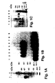
- Example 1 a complete purification of the u-PA receptor is described, involving temperature-induced phase separation of a detergent extract from cells, and affinity chromatography with immobilized DFP-inactivated u-PA. This resulted in a preparation that shows one band at approximately 55-60 kD after SDS-PAGE and silver staining, with a load of approximately 1 ⁇ g of the receptor.
- the purified protein could be chemically cross-linked with u-PA. Its amino acid composition and N-terminal sequence were determined (30 residues, some of which with some uncertainty). It was found to be heavily N-glycosylated, deglycosylation resulting in a protein with an apparent molecular weight of about 30-35 kD. The apparent molecular weight of u-PAR from different cell lines and from PMA-stimulated and non-stimulated U937 cells varied somewhat. This heterogeneity disappeared after deglycosylation and was thus due to differences in glycosylation of u-PAR from the various sources.
- Example 1 The modulation of the u-PAR molecules demonstrated in Example 1 may represent an important feature in the regulation of extracellular proteolysis and thus in the degradation of the extracellular matrix and basement membrane components, processes that are at the core of cell migration and invasiveness.
- Example 2 isolation of a ligand binding domain of u-PAR is identified and characterized. This provides potentially therapeutically valuable information on peptides that may inhibit the ligand binding.
- u-PAR is produced as a 313 residues long protein with a 282 residues long hydrophilic N terminal part (probably extracellular) followed by 21 rather hydrophobic amino acids (probably a trans-membrane domain).
- the potential extracellular part is organised in 3 repeats with striking homologies, particularly with respect to the pattern of cysteines. This may indicate the presence of distinct domains that may bind different ligands.
- u-PAR is at least in some cases terminally processed and anchored to the cell surface via a glycolipid anchor, and that the surface location can be regulated by the phospholipase PI-PLC, but not by the phospholipases A 2 and D (Example 3). Furthermore, it was found that also harvest fluid from cells that were not treated contain some free u-PAR, indicating release from the cells that may be mediated by an endogenous phospholipase. This may be a physiological mechanism and it is possible that measurement of free receptor, e.g. in serum, may be a diagnostically valuable indicator of some pathological processes.
- Example 4 In Example 4 is described the production of polyclonal mouse and rabbit antibodies against u-PAR as well as 4 monoclonal mouse antibodies against the receptor.
- the polyclonal antibodies precipited 125 I-labelled purified u-PAR in a dose-dependent manner, with a significant precipitation being obtained by the antiserum in a dilution of 1:7500.
- the antiserum was found to immunocapture radiolabelled u-PAR, and in an ELISA immobilized u-PAR in an amount of 1 ng was detected with the immune serum diluted 1:8000.
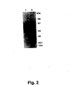
- the antibodies detected both purified u-PAR and u-PAR in the crude detergent phase of extracts of PMA-treated U937 cells.
- the polyclonal antibodies to u-PAR can be used to specifically prevent ligand binding. It is furthermore shown that the polyclonal u-PAR antibodies inhibit u-PA-catalyzed cell surface plasminogen activation.
- the monoclonal antibodies were selected by several procedures, including a screening ELISA with immobilized purified u-PAR, Western blotting experiments with phospholipase-solubilized u-PAR, inhibition of binding of 125 I-ATF (aminoterminal fragment of u-PA) to intact cells, and ability to stain cells in tissue sections positive for u-PAR mRNA.
- a screening ELISA with immobilized purified u-PAR Western blotting experiments with phospholipase-solubilized u-PAR, inhibition of binding of 125 I-ATF (aminoterminal fragment of u-PA) to intact cells, and ability to stain cells in tissue sections positive for u-PAR mRNA.
- a screening ELISA with immobilized purified u-PAR Western blotting experiments with phospholipase-solubilized u-PAR
- inhibition of binding of 125 I-ATF aminoterminal fragment of u-PA
- Purified monoclonal antibodies from these four hybridomas were characterized by their ability to immunoprecipitate intact purified u-PAR and u-PAR degraded by chymotrypsin to yield a Mr 16kD u-PA binding domain and an Mr 35-45kD non-u-PA-binding fragment. Also the ability to stain u-PAR and glycosylation variants thereof in cell extracts was assessed in Western blotting experiments.
- the antibodies could be used to quantitate u-PAR in double antibody sandwich type ELISAS, exemplified in an ELISA in which one of the monoclonal antibodies (4R) was used catching antibody, and another monoclonal antibody (2R) in a biotinylated form was used as detecting antibody.
- the polyclonal rabbit antibodies against u-PAR were used as catching antibodies, and a biotinylated monoclonal antibody (2R) as detecting antibody.
- the antibodies could be used in a drug screening scheme for identifying substances inhibiting u-PA/u-PAR interaction, cell surface plasmin generation and invasion and metastasis of human cancer cells inoculated into a substrain of nude mice.
- the antibodies of the invention together with commercially available reagents, provide means for selective quantitation of
- a procedure is described for preparation of monoclonal antibodies specific for the u-PA binding domain in u-PAR and the non-u-PA binding part of u-PAR, respectively; such antibodies are useful in quantitation of u-PAR in its different states.
- Example 7 it is demonstrated that one of the monoclonal antibodies (3R) strongly inhibits cell surface plasminogen activation, while this activation is not or only slightly affected by the three other antibodies.
- the monoclonal antibody 3R also efficiently inhibits binding of radiolabelled DFP-treated u-PA on the surface of U937 cells, while no inhibition was seen with 2R and 4R and only a slight inhibition with 1R.
- the antibodies 3R and 4R by flow cytometry using FITC-labelled antibodies to mouse IgG effectively binds to human monocytes. The binding of the 4R antibody is not affected by pre-treatment of the monocytes with u-PA, while the binding of the 3R antibody is completely inhibited by pre-treatment with u-PA.
- Example 8 it is demonstrated that two of the monoclonal antibodies (2R and 4R) in immunohistochemical studies specifically stain the same subpopulation of the cells, this subpopulation being characterized by containing mRNA for u-PAR as demonstrated by in situ hybridization.
- inhibition of receptor binding of u-PA is a means of inhibiting some of its physiological functions in relationship to therapeutic prevention of localized proteolytic activity, e.g. invasion and metastasis of cancer cells, inflammatory bowel disease, premalignant colonic adenomas, septic arthritis, osteoarthritis, rheumatoid arthritis (for which a direct involvement of excess u-PA production has been demonstrated), osteoporosis, cholesteatoma, and a number of skin and comeal diseases for which an excess plasminogen activation has been shown to be the pathogenetic cause, such as corneal ulcers, keratitis, epider-molysis bullosa, psoriasis, and pemphigus.
- localized proteolytic activity e.g. invasion and metastasis of cancer cells, inflammatory bowel disease, premalignant colonic adenomas, septic arthritis, osteoarthritis, rheumatoid arthritis (for which a direct involvement of excess
- u-PA receptors are present in several blood cells (neutrophilic granulocytes and monocytes) and endothelial cells, their regulation might also significantly affect intravascular fibrinolytic activity in physiological, pathological and pharmacological conditions.
- the above-mentioned diseases would be the first obvious targets for a therapy based on administration of substances that block or decrease cell surface plasminogen activation.
- a contraceptive effect is expected of measures that inhibit receptor binding.
- the therapy and prophylaxis will involve systemic or topical treatment with agents that block or reduce receptor bound plasminogen activator activity, such as will be explained below.
- a method for preventing or counteracting localized proteolytic activity in a mammal, in particular a human comprises inhibiting the activation of plasminogen to plasmin by preventing the binding pro-u-PA to a u-PA receptor (u-PAR) in the mammal and thereby preventing u-PA from converting plasminogen into plasmin.
- u-PAR u-PA receptor
- the term "localized proteolytic activity” is intended to designate a proteolytic activity which is located at one or several distinct regions in a human body, or at distinct cells, as opposed to an overall proteolytic activity exerting itself substantially anywhere in the body.
- the localized proteolytic activity can be inhibited generally in a mammal, in particular a human, or locally.
- the term "preventing or counteracting” is intended to designate a situation where the binding of u-PA to u-PAR is completely inhibited, or a situation where the binding is sufficiently inhibited so as to inhibit the undesired effect of the plasminogen activator.
- a u-PAR indicates that even though the polypeptide part of u-PAR in a species might be the same for all u-PARs, there is a plurality of u-PARs as for example the carbohydrate part or the mechanism of surface attachment of the u-PAR can be different. It may even be so that some cells, e.g. cancer cells, have substantially different u-PARs which might have important therapeutic significance as it might be possible, e.g.
- u-PA urokinase-type plasminogen activator
- plasminogen By cleavage at Arg 560 , plasminogen is activated to the broad spectrum protease plasmin.
- preventing u-PA from converting plasminogen into plasmin is therefore meant that this activation by u-PA is substantially inhibited or a situation where the activation is sufficiently inhibited so as to inhibit or reduce the undesired effect of the plasmin.
- the prevention of the binding of a receptor binding form of u-PA to a u-PAR is, e.g. suitably performed by blocking the u-PAR by administration, to the mammal, of a monoclonal antibody binding to the u-PAR, such as the 3R antibody of the present invention, so as to occupy a site of the receptor to which a receptor binding form of u-PA is normally bound, the monoclonal antibody being administered in an amount effective to reduce the binding of the receptor binding form of u-PA to the receptor.
- binding to a u-PAR so as to occupy a site of the receptor to which a receptor binding form of u-PA is normally bound is intended to mean that the antibody binds to the u-PAR so that a receptor binding form of u-PA can not be bound to the u-PAR.
- the antibody may be a monoclonal antibody that is reactive with non-carbohydrate moieties of the u-PAR, or it may be a monoclonal antibody that is reactive with carbohydrate moieties of the u-PAR, the latter permitting a valuable distinction between target cells where cells expressing distinct variants of u-PAR are the cells involved in the undesired proteolysis.
- the antibodies may be administered in various ways as described below.
- u-PA u-PA
- u-PA a role of u-PA (and therefore probably also of u-PAR) in thrombolysis under some conditions is suggested by the finding by inventors of the present invention of u-PA being present in endothelial cells during acute inflammation and in cancer. Under normal conditions, the endothelial cells contain t-PA, but no u-PA. It is furthermore interesting that the disease paroxysmal nocturnal hemo-globinuria is associated with an impaired ability to form glycerol-phosphoinositol anchors and impaired anchoring of u-PAR. This disease is often associated with thromboembolic disorders (See: Selvaraj et al., 1988, and references therein).
- Such other potential alternative ligands for u-PAR may be proteins located at cell-cell and focal cell-substratum contact sites such as various integrins.
- Antibodies to u-PAR that inhibit binding of such ligands may be valuable in inhibition of cell surface plasminogen activation, and prevention of binding of u-PAR to such alternative ligands may be functionally important and therapeutically valuable in a broad spectrum of diseases.
- u-PAR exists in various forms, such as the glycosylation variants described in Example 1, the variants with different sensitivity to the lipase PI-PLC suggested by the findings described in Example 3. In some diseases that involve increased u-PAR function, some of these forms may be preferentially changed. Monoclonal antibodies selectively directed against some distinct forms may therefore be particularly therapeutically valuable in such diseases.
- the administration of the various antibodies to a mammal, preferably a human being, may be performed by any administration method which is suitable for administering antibodies.
- Topical administration may be performed by formulating the antibody or derivative thereof in a salve, an ointment, a lotion, a creme, etc.
- compositions of the invention may for example include human monoclonal antibodies or derivatives thereof.
- Another strategy of treating the conditions and diseases mentioned above is to target a cell that contains a u-PAR on the surface by a medicament, comprising administering the medicament bound to an antibody against u-PAR such as a polyclonal or a monoclonal antibody, e.g. an antibody particularly directed to a variant of u-PAR present in a cancer cell type.
- an antibody against u-PAR such as a polyclonal or a monoclonal antibody, e.g. an antibody particularly directed to a variant of u-PAR present in a cancer cell type.
- the medicament may typically be an anti-cancer agent such as an alkylating agent, e.g. melphalan, chlorambucil, busulfan, cisplatin, thiotepa, an antimetabolite such as methotrexate, fluracil, azathioprin, an antimitoticum, typically vincristine, vinblastine, or an antibiotic such as doxorubicin, daunorubicin or bleomycin.
- the medicament may also comprise bacterial or other toxins.
- antibodies that distinguish between various forms of u-PAR.
- diagnostic kits, materials and methods based on antibodies is given further below.
- One aspect of the invention relates to a method of producing pure u-PAR, the method comprising subjecting a u-PAR-containing material to affinity chromatography with immobilized monoclonal antibodies according to the invention to u-PAR and eluting the u-PAR, e.g. under acidic conditions.
- analogue is used in the present context to indicate a protein or polypeptide of a similar amino acid composition or sequence as the characteristic amino acid sequence derived from the u-PAR, allowing for minor variations which do not have an adverse effect on the immunogenicity of the analogue.
- the analogous polypeptide or protein may be derived from mammals or may be partially or completely of synthetic origin.
- the monoclonal antibodies of the invention may also be used for the obtainment of a u-PAR containing fraction with high yield and purity.
- the procedure may be performed by immobilizing the specific monoclonal antibodies to a matrix, contacting said matrix with the preparation containing the released u-PAR compounds, washing, and finally treating the antigenantibody complex fixed to the matrix so as to release the u-PAR compounds in a purified form.
- a preferred way is to isolate the u-PAR compounds by means of column affinity chromatography involving antibodies fixed to the column matrix.
- the term "antibody” refers to a substance which is produced by a vertebrate or more precisely a cell of vertebrate origin belonging to the immune system as a response to exposure to the polypeptides of the invention.
- the variant domain of an antibody is composed of variable and constant sequences.
- the variant part of the domain is called the idiotype of the antibody. This part of the antibody is responsible for the interaction with the antigen, the antigen binding.
- the idiotypic structure is antigenic and can thus give rise to specific antibodies directed against the idiotypic structure. This has been done in mice.
- the antibodies raised against the idiotype, the anti-idiotypic antibodies may mimic the structure of the original antigen and therefore may function as the original antigen to raise antibodies reactive with the original antigen.
- This approach may be advantageous as it circumvents the problem associated with the characterization and synthesis of the important immunogenic parts of the protein in question. This is most important in the case of conformational epitopes, which might otherwise be difficult to identify. It has been shown for a number of organisms that protective immunity can be induced in this way (e.g. Trypanosoma druzei, Trypanosoma brucei, Hepatitis B virus, and Plasmodium knowlesii ).
- the antibodies of the present invention may be produced by a method which comprises administering in an immunogenic form at least a natural or synthetic part of the polypeptide of the invention to obtain cells producing antibodies reactive with said polypeptide and isolating the antibody containing material from the organism or the cells.
- the methods of producing antibodies of the invention will be explained further below.
- the antibody is preferably a monospecific antibody.
- the monospecific antibody may be prepared by injecting a suitable animal with a sub-stantially pure preparation of the polypeptide of the invention followed by one or more booster injections at suitable intervals (e.g. one or two weeks to a month) up to four or five months before the first bleeding.
- suitable intervals e.g. one or two weeks to a month
- the established immunization schedule is continued, and the animals are bled about one week after each booster immunization, and antibody is isolated from the serum in a suitable manner (cf. e.g. Harboe and Ingild, Scand. J. Immun. 2 (Suppl. 1), 1973, pp. 161-164.)
- the antibody may be a polyclonal antibody.
- Polyclonal antibodies may be obtained, e.g. as described in Harboe and Ingild, see above. More specifically, when polyclonal antibodies are to be obtained, the u-PAR compound preparation is, preferably after addition of a suitable adjuvant, such as Freund's incomplete or complete adjuvant, injected into an animal. When the immunogens are human u-PAR compounds, the animals may be rabbits.
- the animals are bled regularly, for instance at weekly intervals, and the blood obtained is separated into an antibody containing serum fraction, and optionally said fraction is subjected to further conventional procedures for antibody purification, and/or procedures involving use of purified u-PAR compounds.
- monoclonal antibodies are obtained.
- the monoclonal antibody may be raised against or directed substantially against an essential component of u-PAR compounds, i.e. an epitope.
- the monoclonal antibody may be produced by conventional techniques (e.g. as described by Köhler and Milstein, Nature 256, 1975, p. 495) e.g. by use of a hybridoma cell line, or by clones or subclones thereof or by cells carrying genetic information from the hybridoma cell line coding for said monoclonal antibody.
- the monoclonal antibody may be produced by fusing cells producing the monoclonal antibody with cells of a suitable cell line, and selecting and cloning the resulting hybridoma cells producing said monoclonal antibody.
- the monoclonal antibody may be produced by immortalizing an unfused cell line producing said monoclonal antibody, subsequently growing the cells in a suitable medium to produce said antibody, and harvesting the monoclonal antibody from the growth medium.
- the immunized animal used for the preparation of antibodies of the invention is preferably selected from the group consisting of rabbit, monkey, sheep, goat, mouse, rat, pig, horse and guinea pigs.
- the cells producing the antibodies of the invention may be spleen cells or lymph cells, e.g. peripheric lymphocytes.
- hybridoma cells When hybridoma cells are used in the production of antibodies of the invention, these may be grown in vitro or in a body cavity of an animal.
- the antibody-producing cell is injected into an animal such as a mouse resulting in the formation of an ascites tumor which releases high concentrations of the antibody in the ascites of the animal.
- the animals will also produce normal antibodies, these will only amount to a minor percentage of the monoclonal anti-bodies which may be purified from ascites by standard purification procedures such as centrifugation, filtration, precipitation, chromatography or a combination thereof.
- An example of a suitable manner in which the monoclonal antibody may be produced is as a result of fusing spleen cells from immunized mice (such as Balb/c mice) with myeloma cells using conventional techniques (e.g. as described by R. Dalchau, J. Kirkley, J.W. Fabre, "Monoclonal antibody to a human leukocyte-specific membrane glycoprotein probably homologous to the leukocyte-common (L-C) antigen of the rat", Eur. J. Immunol. 10 , 1980, pp. 737-744).
- the fusions obtained are screened by conventional techniques such as binding assays employing u-PAR compounds isolated by the above-described methods.
- the invention relates to a diagnostic agent comprising an antibody according to the invention capable of detecting and/or quantitating u-PAR or a derivative thereof in a sample.
- antibodies according to the invention are valuable in diagnosis of cancer and other disorders involving tissue invasion and tissue remodelling, considering the involvement of u-PAR in these processes.
- the finding that u-PAR mRNA and protein is consistently found in cells located in the the invasive front in a variety of different forms of cancer including colon adenocarcinomas, ductal mammary carcinomas and squamous skin carcinomas strongly supports this notion.
- a further support of this notion is the finding that u-PA mRNA and/or protein in all these cases are produced either by the u-PAR-containing cells or by cells located adjacent to the u-PAR-containing cells.
- a new aspect of the potential diagnostic and prognostic use of u-PAR determinations is the release of u-PAR from cultured cells (described in Example 3) that occurs even in the absence of exogeneously added phospholipase. This finding raises the possibility that u-PAR is also released into body fluids under some physiological and pathophysiological conditions and particularly in cancer. Determination of concentrations of u-PAR or degradation products thereof in body fluids, such as serum, urine, and ascites fluid may therefore prove to be diagnostically and/or prognostically valuable.
- the antibody is provided with a label for the detection of bound antibody or, alternatively (such as in a double antibody assay), a combination of labelled and unlabelled antibody may be employed.
- the substance used as label may be selected from any substance which is in itself detectable or which may be reacted with another substance to produce a detectable product.
- the label may be selected from radioactive isotopes, enzymes, chromophores, fluorescent or chemiluminescent substances, and complexing agents.
- enzymes useful as labels are ⁇ -galactosidase, urease, glucose oxidase, carbonic anhydrase, peroxidases (e.g. horseradish peroxidase), phosphatases (e.g. alkaline or acid phosphatase), glucose-6-phosphate dehydrogenase and ribonuclease.
- peroxidases e.g. horseradish peroxidase
- phosphatases e.g. alkaline or acid phosphatase
- glucose-6-phosphate dehydrogenase ribonuclease.
- Enzymes are not in themselves detectable, but must be combined with a substrate to catalyze a reaction the end product of which is detectable.
- a substrate may be added to the reaction mixture resulting in a coloured, fluorescent or chemiluminescent product or in a colour change or in a change in the intensity of the colour, fluorescence or chemiluminescence.
- substrates which are useful in the present method as substrates for the enzymes mentioned above are H 2 O 2 , p-nitrophenylphosphate, lactose, urea, ⁇ -D-glucose, CO 2 , RNA, starch, or malate.
- the substrate may be combined with, e.g. a chromophore which is either a donor or acceptor.
- Fluorescent substances which may be used as labels for the detection of the components as used according to the of invention may be 4-met-hylumbelliferyl-phosphate, 4-methylumbelliferyl-D-galactopyranoside, and 3-(p-hydroxyphenyl) propionic acid. These substances may be detected by means of a fluorescence spectrophotometer. Chemilumines-cent substances which may be peroxidase/eosin/EDTA, isoluminol/-EDTA/H 2 O 2 and a substrate therefor.
- Chromophores may be o-phenylenediamine or similar compounds. These substances may be detected by means of a spectrophotometer. Radioactive isotopes may be any detectable and in a laboratory acceptable isotope, e.g. 125 I, 131 I, 3 H, 35 P, 35 S or 14 C. The radioactivity may be measured in a ⁇ -counter or a scintillation counter or by radioautography followed by densitometry.
- Complexing agents may be Protein A, Protein G (which form a complex with immunoglobulins), biotin (which forms a complex with avidin and streptavidin), and lectin (which forms a complex with carbohydrate determinants, e.g. receptors).
- the complex is not in itself directly detectable, necessitating labelling of the substance with which the complexing agent forms a complex.
- the marking may be performed with any of the labelling substances described above.
- an antibody of the invention may be coupled to a bridging compound coupled to a solid support.
- the bridging compound which is designed to link the solid support and the antibody may be hydrazide, Protein A, glutaraldehyde, carbodiimide, or lysine.
- the solid support employed is e.g. a polymer or it may be a matrix coated with a polymer.
- the matrix may be of any suitable solid material, e.g. glass, paper or plastic.
- the polymer may be a plastic, cellulose such as specially treated paper, nitrocellulose paper or cyanogenbromide-activated paper.
- suitable plastics are latex, a polystyrene, polyvinylchloride, polyurethane, polyacrylamide, polyvinylacetate and any suitable copolymer thereof.
- silicone polymers include siloxane.
- the solid support may be in the form of a tray, a plate such as a mitrotiter plate, e.g. a thin layer or, preferably, strip, film, threads, solid particles such as beads, including Protein A-coated bacteria, or paper.
- a mitrotiter plate e.g. a thin layer or, preferably, strip, film, threads, solid particles such as beads, including Protein A-coated bacteria, or paper.
- the antibody of the invention may be used in an assay for the identification and/or quantification of at least a form and/or a part of said polypeptide present in a sample.
- the identification and/or quantification performed by the use according to the present invention may be any identification and/or quantification involving u-PAR compounds or a form of u-PAR compounds. Thus, both a qualitative and a quantitative determination of u-PAR compounds may be obtained according to the use of the present invention.
- the identification and/or quantification may be performed for both a scientific, a clinical and an industrial purpose. As will be further described below, it is especially important in clinical routine to identify or quantify u-PAR compounds.
- the sample may be a specimen obtained from a living organism such as a human or an animal.
- the specimen may be blood, e.g. an erythrocyte enriched fraction, or a tissue sample e.g. comprising liver cells.
- the specimen is urine.
- the antibody used in the method of the invention is a monoclonal antibody as this generally provides a higher precision and accuracy of the assay, at the same time possibly requiring less time to perform. Furthermore, a mixture of two or more different monoclonal antibodies may be employed as this may increase the detection limit and sensitivity of the test.
- the monoclonal antibody may be obtained by the method described below. Antibodies possessing high avidity may be selected for catching techniques.
- the antibody used in the present method is preferably in substantially pure form (purified according to suitable techniques or by the methods of the invention, see below) in order to improve the precision and/or accuracy of the assays of the invention.
- One aspect of the invention relates to a monoclonal antibody which reacts with u-PAR and thereby inhibits the binding of pro-u-PA and active u-PA, and cell surface plasminogen activation, such as explained herein.
- a monoclonal antibody having these properties is useful in a number of diagnostic and therapeutic utilities as explained in the following.
- this aspect evidently extends to functional equivalents to the monoclonal antibody, that is, any monoclonal antibody which reacts with u-PAR and thereby inhibits the binding of pro-u-PA and active u-PA and cell surface plasminogen activation.
- the monoclonal antibody according to this aspect of the invention can also be characterized as a monoclonal antibody which is a functional equivalent of the monoclonal antibody 3R produced by the clone 90101009 in that, like the antibody 3R, it reacts with u-PAR and thereby inhibits the binding between u-PAR and pro-u-PA or active u-PA, and cell surface activation of plasminogen to plasmin.
- the u-PA binding domain of u-PAR is located to the domain comprising the N-terminal 87 residues of u-PAR.
- another definition of the monoclonal antibody according to this aspect of the invention is a monoclonal antibody which reacts with the a polypeptide of the sequence shown as u-PAR 1 in Fig. 8 herein or any subsequence or analogue thereof which is capable of binding to u-PAR so as to inhibit the binding between u-PAR and u-PA.
- the reaction of the monoclonal antibody according to this aspect of the invention with u-PAR with the resulting inhibition of the binding of pro-u-PA and active u-PA to u-PAR and of cell surface plasminogen activation may be a reaction with the u-PA binding domain in u-PAR, or it may, as a special possibility, be a binding to another part of u-PAR than the u-PA binding domain, but, through an allosteric effect, causing a change in the binding domain.
- the reaction of the monoclonal antibody with u-PAR is a reaction with the u-PA binding domain of u-PAR (such as has been found to be the case for the monoclonal antibody 3R)
- the reaction may be a binding directly involving the binding site of u-PAR (which is contemplated to comprise an amino acid sequence of about 30 or less (or more), such as 15-30 or perhaps even less (but perhaps 30-50 or even more) or a binding which does not involve the binding site proper, but which binding nevertheless inhibits u-PA binding, such as antibody binding having an allosteric effect so as to inhibit u-PA binding.
- the monoclonal antibody binding to the u-PA binding domain of u-PAR so as to inhibit the binding of pro-u-PA or u-PA to u-PAR and cell surface plasminogen activation has, for therapeutical purposes, the same overall functionality and utility and constitutes one and the same embodiment of this aspect of the present invention.
- the monoclonal antibody according to this aspect of the invention may - be prepared by immunising an immunisable animal with u-PAR or a subsection or immunogenic variant thereof, e.g. the u-PA binding domain of u-PAR, fusing cells from the immunized animal with a myeloma cell line, and screening for clones producing an antibody which is capable of reacting with u-PAR with resulting inhibition of the binding of pro-u-PA and active u-PA to u-PAR and cell surface plasminogen activation.
- a subclass of this aspect of monoclonal antibodies are monoclonal antibodies which react with the u-PAR binding domain of u-PAR and thereby inhibit the binding of pro-u-PA and active u-PA and cell surface plasminogen activation.
- Methods for assessing that an antibody complies with the above definition comprise measuring the inhibition of surface plasminogen activation caused by reaction between the antibody and u-PAR or measuring the inhibition of binding of u-PA or pro-u-PA to u-PAR caused by reaction between the antibody and u-PAR.
- a special, narrow embodiment of this aspect of the invention is a monoclonal antibody which in competition experiments is capable of competing with the monoclonal antibody 3R produced by the hybridoma cell line deposited under the provisional accession number 90101009 in the European Collection of Animal Cell Cultures under the terms and conditions of the Budapest Treaty.
- the term "antibody” is intended to designate not only complete antibody molecules, but also binding fragments thereof such as the Fab fragment.
- the antibodies of the examples are produced by murine hybridomas, but antibodies used for therapeutic purposes would suitably be produced by human hybridomas.
- the establishment and use of human hybridomes is well-known in the art, cf., e.g., Glassy, M.C., Handley, H.H., Hagiwara, H. and Royston, I., A human lymphoblastoid B-cell line useful for generating antiobdy sectreting human/human hybridomas, Proc. Natl. Acad. Sci.
- antibodies of the invention are for diagnostic purposes, in particular in assays to detect of quantify the presence of u-PAR in a sample.
- assays in particular ELISAS (enzyme-linked immunosorbent assays), and monoclonal antibodies according to the invention are discussed in greater detail.
- polyclonal antibodies are discussed.
- a procedure for producing such specific polyclonal antibodies is described in Example 4, the antibodies being developed by immunisation with the 16kD u-PA-binding fragment of u-PAR obtained by degradation of intact u-PAR wich chymotrypsin.
- Another interesting specific polyclonal antibody can be obtained by immunisation with the 35-45kD non-u-PA-binding fragment of u-PAR.
- One aspect according to the invention of such use is a method for detecting or quantifying u-PAR in a sample, the detection of quantitation being substantially independent of whether the u-PAR has bound u-PA or not, comprising using, as a catching or detecting antibody or-both, an antibody capable of binding to u-PAR irrespective of whether u-PAR has bound to u-PA or not.
- Each antibody used in this method may be a monoclonal antibody according to the invention which reacts with u-PAR, but not with its u-PA binding domain, or a monoclonal antibody according to the invention which reacts both with free u-PAR and with complexes between u-PA and u-PAR.
- polyclonal antibodies which react with u-PAR, but not with its u-PA binding domain
- polyclonal antibodies which react both with free u-PAR and with complexes between u-PA and u-PAR.
- One useful embodiment of this method is a method wherein one monoclonal antibody which reacts with u-PAR, but not with its u-PA binding domain, or one monoclonal antibody which reacts both with free u-PAR and with complexes between u-PA and u-PAR, is used as catching antibody, and another monoclonal antibody which reacts with u-PAR, but not with its u-PA binding domain, or another monoclonal antibody which reacts both with free u-PAR and with complexes between u-PA and u-PAR, such other monoclonal antibody being directed against a different epitope, is used as detecting antibody.
- Another embodiment of such method is a method in which polyclonal antibodies which react with u-PAR, but not with its u-PA binding domain, or polyclonal antibodies which react both with free u-PAR and with complexes between u-PA and u-PAR, are used as catching antibodies, and a monoclonal antibody which reacts with u-PAR, but not with its u-PA binding domain, or a monoclonal antibody which reacts both with free u-PAR and with complexes between u-PA and u-PAR, is used as detecting antibody.
- Another embodiment is a method which a monoclonal antibody which reacts with u-PAR, but not with its u-PA binding domain, or a monoclonal antibody which reacts both with free u-PAR and with complexes between u-PA and u-PAR, is used as catching antibody, and polyclonal antibodies which react with u-PAR, but not with its u-PA binding domain, or polyclonal antibodies which react both with free u-PAR and with complexes between u-PA and u-PAR are used as detecting antibodies.
- a further embodiment is a method according in which polyclonal antibodies which react with u-PAR, but not with its u-PA binding domain, or polyclonal antibodies which react both with free u-PAR and with complexes between u-PA and u-PAR, are used as catching antibodies, and polyclonal antibodies which react with u-PAR, but not with its u-PA binding domain, or polyclonal antibodies which react both with free u-PAR and with complexes between u-PA and u-PAR, are used as detecting antibodies.
- Another method according to the invention is a method for detecting or quantifying complexes of u-PAR and u-PA in a sample, comprising using, as catching or detecting antibody, an antibody capable of binding to u-PAR which has bound u-PA or pro-u-PA, together with an antibody which detects bound u-PA or pro-u-PA as detecting or catching antibody, respectively.
- An embodiment hereof is a method in which a monoclonal antibody which reacts with u-PAR, but not with its u-PA binding domain, or a monoclonal antibody which reacts both with free u-PAR and with complexes between u-PA and u-PAR, is used as catching antibody or detecting antibody.
- Another embodiment hereof is a method in which polyclonal polyclonal antibodies which react with u-PAR, but not with its u-PA binding domain, og polyclonal antibodies which react both with free u-PAR and with complexes between u-PA and u-PAR, are used as catching antibodies or detecting antibodies.
- Another important method according to the invention is a method for detecting or quantitating u-PAR which has not bound u-PA, in a sample, comprising using, as catching or detecting antibody, an antibody capable of binding free u-PAR, but not complexes between u-PA and u-PAR.
- the antibody used in this method may be a monoclonal antibody which reacts with the u-PA binding domain of u-PAR, or a monoclonal antibody which reacts with free u-PAR, but not with complexes between u-PA and u-PAR, or polyclonal antibodies which react with the u-PA binding domain of u-PAR, but not with complexes between u-PA and u-PAR, or polyclonal antibodies which react with free u-PAR, but not with complexes between u-PA and u-PAR.
- One embodiment of this method is a method wherein a monoclonal antibody which reacts with the u-PA binding domain of u-PAR or a monoclonal antibody which reacts with free u-PAR, but not with complexes between u-PA and u-PAR, is used as catching antibody, and another monoclonal antibody which reacts with the u-PA binding domain of u-PAR or another monoclonal antibody which reacts with free u-PAR, but not with complexes between u-PA and u-PAR, such other antibody being directed against a different epitope, is used as detecting antibody.
- polyclonal antibodies which react with the u-PA binding domain of u-PAR, but not with complexes between u-PA and u-PAR, or polyclonal antibodies which react with free u-PAR, but not with complexes between u-PA and u-PAR are used as catching antibodies, and a monoclonal antibody which reacts with the u-PA binding domain of u-PAR or a monoclonal antibody which reacts with free u-PAR, but not with complexes between u-PA and u-PAR, is used as detecting antibody.
- a monoclonal antibody which reacts with the u-PA binding domain of u-PAR or a monoclonal antibody which reacts with free u-PAR, but not with complexes between u-PA and u-PAR is used as a catching antibody
- polyclonal antibodies which react with the u-PA binding domain of u-PAR, but not with complexes between u-PA and u-PAR, or polyclonal antibodies which react with free u-PAR, but not with complexes between u-PA and u-PAR is used as a detecting antibody.
- polyclonal antibodies which react with the u-PA binding domain of u-PAR, but not with complexes between u-PA and u-PAR, or polyclonal antibodies which react with free u-PAR, but not with complexes between u-PA and u-PAR are used as catching antibodies, and polyclonal antibodies which react with u-PAR, but not with its u-PA binding domain, or polyclonal antibodies which react both with free u-PAR and with complexes between u-PA and u-PAR, are used as detecting antibodies.
- Another method according to the present invention is a method for detecting or quantifying u-PAR which has not bound u-PA, in a sample, comprising using, as catching or detecting antibody, an antibody capable of binding free u-PAR, but not inhibiting u-PA binding, and using, as detecting or catching reagent, u-PA or a u-PAR binding variant thereof.
- Another method according to the invention is a method for immunohistochemical detection of u-PAR in tissue sections, using, as the detecting antibody, any monoclonal antibody which reacts with u-PAR, or any polyclonal antibody which reacts with u-PAR, including a monoclonal antibody which reacts with the u-PA binding domain of u-PAR, or a monoclonal antibody which reacts with free u-PAR, but not with complexes between u-PA and u-PAR, or a monoclonal antibody which reacts with u-PAR, but not with its u-PA binding domain, or a monoclonal antibody which reacts both with free u-PAR and with complexes between u-PA and u-PAR, or polyclonal antibodies which react with the u-PA binding domain of u-PAR, but not with complexes between u-PA and u-PAR, or polyclonal antibodies which react with u-PAR, but not with
- the antibody is one of the monoclonal antibodies defined above.
- this embodiment which detecting u-PAR independent of whether it has bound u-PA or not, comprising using a monoclonal antibody which reacts with u-PAR, but not with its u-PA binding domain, or a monoclonal antibody which reacts both with free u-PAR and with complexes between u-PA and u-PAR, or polyclonal antibodies which react with u-PAR, but not with its u-PA binding domain, or polyclonal antibodies which react both with free u-PAR'and with complexes between u-PA and u-PAR.
- Another embodiment of this method which embodiment detects free u-PAR, comprises using, as detecting antibody, an antibody capable of detecting free u-PAR, but not complexes between u-PA and u-PAR.
- the detecting antibody may be a monoclonal antibody which reacts with the u-PA binding domain of u-PAR or a monoclonal antibody which reacts with free u-PAR, but not with complexes between u-PA and u-PAR, or polyclonal antibodies which react with the u-PA binding domain of u-PAR, but not with complexes between u-PA and u-PAR, or polyclonal antibodies which react with free u-PAR, but not with complexes between u-PA and u-PAR.
- One example of a monoclonal antibody according to the invention which detects u-PAR independently of whether it has bound u-PA or not is the monoclonal antibody designated 4R and produced by the hybridoma cell line deposited under the provisional accession number 90101010.
- 1R and 2R are other monoclonal antibodies of the invention which are contemplated to be useful in this regard.
- each functional type of monoclonal antibody discussed herein is not limited to the specific products of the specific clones deposited, but is rather to be understood as the class of monoclonal antibodies showing the same functionality with respect to its utility as described in the present specification, including the examples and claims.
- One aspect of the invention relates to a method for the detection or quantitation of a glycosylations variant of u-PAR in a sample, comprising using, as detecting antibody, a monoclonal antibody which solely or preferentially binds to the variant.
- the glycosylation variant of u-PAR is typically a variant characteristic to a particular type of cancer cell.
- the sample may typically be serum, plasma or urine from a cancer patient or a suspected cancer patient, but it may, of course, also be another sample relevant in the particular case, such as, e.g. a faeces sample.
- the sample may also be an extract from a cancer tissue or a suspected cancer tissue.
- the sample may also be sample is serum, plasma or urine from a patient suffering from or suspected to suffer from a non-malignant disease involving tissue destruction, such as rheumatiod arthritis, collitis ulcerosa, or psoriasis.
- a non-malignant disease involving tissue destruction such as rheumatiod arthritis, collitis ulcerosa, or psoriasis.
- the detecting antibody is provided with a detectable label, and examples of these labels are given herein.
- the monoclonal antibodies according to the invention may also be used in a method for targeting a diagnostic to a cell that contains a u-PAR on the surface, the method comprising administering, to a mammal, in particular a human, in particular a mammal suffering from cancer or suspected to suffer from cancer, the diagnostic bound to a monoclonal antibody of the invention against u-PAR.
- the diagnostic may be a radioactive substance, such as technetium.
- the antibody according to the invention used for this purpose may be any of the monoclonal antibodies of the invention as mentioned above, that is, including a monoclonal antibody which solely or preferentially binds to a particular glycosylation variant of u-PAR.
- the monoclonal antibodies according to the invention can be characterized by one or several of a number of characteristic properties, that is, as monoclonal antibodies against u-PAR which
- One important use of the monoclonal antibodies according to the invention is for establishing tools for screening substances for their potential inhibition of u-PA/u-PAR interaction and, thus, for their potential anti-invasive and anti-metastatic effect in cancer as well as in other diseases where u-PA/u-PAR interaction is involved.
- One such use is a screening assay in which the possible inhibition of u-PA/u-PAR interaction by the substance is determined by adding the substance to a system comprising immobilized u-PAR and solubilized u-PA, u-PA bound to u-PAR being detected by being labelled or by means of a labelled anti-u-PA antibody, or adding the substance to a system comprising immobilized u-PA and solubilized u-PAR, u-PAR bound to u-PA being detected by being labelled or by means of a labelled anti-u-PAR antibody.
- an assay may be mentioned a very practical screening ELISA using immobilized monoclonal antibodies against u-PAR for catching u-PAR and subsequently measuring u-PA binding to u-PAR and the possible interference of candidate substances thereon, receptor-bound u-PA being detected by a labelled anti-u-PA antibody, the labelling being, e.g., biotin.
- a substance When a substance has been found positive in the above simple and fast screening, it can suitably be further tested in a much more laborious assay in which the possible inhibition of u-PA/u-PAR interaction by the substance is determined by adding the substance to a system comprising u-PAR and radiolabelled u-PA or a derivative thereof, cross-linking any u-PAR bound to u-PA and detecting any cross-linked product by SDS page and autoradiography. A positive result in this assay confirms that the substance does indeed inhibit the u-PA/u-PAR binding.
- the next step will be to subject substance which has been found, in the above assay, to positively inhibit u-PA/u-PAR binding, to an assay in which the possible inhibition of binding of u-PA to u-PAR on the surface of cultured cells is determined by adding the substance to a system comprising radiolabelled u-PA or a derivative thereof and cells carrying u-PAR and detecting any u-PA or derivative binding to u-PAR by gamma counting of the cells.
- a positive result in this assay shows that the inhibition of u-PA/u-PAR binding found in the previous assays is not an artefact related to the use of solubilized u-PAR, but is indeed also obtained when u-PAR is bound to cell surface, such as it will be in a clinical situation.
- the aim of inhibitin the u-PA/u-PAR interaction is to inhibit u-PA enzymatic activity in biological settings.
- This can be directly test in an assay in which the possible inhibition of cell surface plasminogen activation by receptor-bound exogeneous pro-u-PA is determined by adding the substance to cells carrying u-PAR and subsequently adding pro-u-PA, followed by measurement of plasmin generation on the cell surface.
- This situation with exogeneously added u-PA is similar to the situation in some types of cancer, such as, e.g., colon adenocarcinoma, in which cancer cells produce and contain u-PAR while u-PA is produced by adjacent non-malignant cells in the tumour stroma.
- the cancer cells themselves produce both u-PAR and u-PA.
- the inhibition of u-PA/u-PAR interaction will be more difficult than when the two components are produced by different cells.
- an assay is used in which the possible inhibition of cell surface plasminogen activation by receptor-bound endogeneous pro-u-PA is determined by incubating cells carrying u-PAR and producing pro-u-PA with the substance, followed by measurement of plasmin generation on the cell surface.
- Human cancer cells inoculated in conventional nude mice do not regularly invade and metastasize.
- a substrain of the nude mouse designated nu/nu META/Bom has been identified in which several cancer cell lines invade and metastasize in substantially all cases.
- the human cancer cells inoculated into the mouse have, prior to their inoculation, been transduced with the lacZ gene which encodes the enzyme ⁇ -D-galactosidase. This enzyme will give rise to a blue staining when subjected to the substrate X-gal.
- this system makes it possible to obtain a distinct colour difference between the human cancer cells and the mouse's own cells, thereby very considerably facilitating detection and quantitation of invading cells and metastases.
- cancer cells invading and metastasizing in this mouse model were found to produce both u-PA and u-PAR. Furthermore, it was found that their invasion and metastasis could be almost completely inhibited by administration of a monoclonal antibody against u-PA, inhibiting cell surface plasmin generation.
- a number of other models are also interesting, such as a model in which two types of cancer cells, one producing u-PA, the other producing u-PAR, are inoculated and therefore simulate the clinical situation occurring in some types of cancer where the two components are produced in two distinct cell types.
- human cancer cells producing u-PAR are inoculated together with human tumour-infiltrating fibroblasts producing u-PA.
- u-PA/u-PAR interaction-inhibiting substances found to inhibit invasion and metastasis in these nude mouse models are likely to be anti-invasive anti-metastatic in human cancer types in which u-PA/u-PAR interaction is believed to be crucial to the invasion and metastasis, such as colon adenocarcinoma, ductal mammary carcinoma and squamous skin carcinoma.
- Such compounds should therefore, after appropriate toxicological studies in animals, be further studied in phase 1 and phase 2 clinical trials, as they are strong candidates to be efficient anti-invasive and anti-metastatic drugs.
- one aspect of the invention relates to a substance for preventing or counteracting localized proteolytical activity in a mammal, in particular a human, by inhibiting the activation of plasminogen to plasmin by preventing the binding of a receptor-binding form of u-PA to a u-PAR in the mammal, when selected by the method described above.
- the invention relates to a substance for preventing or counteracting localized proteolytic activity in a various criteria as tested in one or several of the assays described above, in particular when the substance complies with the criteria shown in the assays 1)-4) as claimed in claim 75, as the substance is then likely to be active in clinical situations in which u-PA and u-PAR are produced by different cell types.
- suramin One chemical compound which has been found, using the methods of the invention, to prevent u-PA/u-PAR binding, is suramin. While suramin itself is likely to be too toxic to be acceptable as generally used cancer drug, the finding indicates that less toxic analogues or derivatives of suramin, still capable of inhibiting u-PA/u-PAR interaction, are suitable cancer drugs.
- SDS-PAGE SDS-PAGE .
- SDS-PAGE was performed according to Laemmli, U.K., "Cleavage of structural proteins during the assembly of the head of bacteriophage T4", Nature 227: 680-682, 1970, using 6-16% gradient slab gels. Pretreatment of samples under nonreducing conditions was performed without boiling. When reducing conditions were used, the samples were boiled for 5 minutes in the presence of 20 mM DTT.
- Phast-gel SDS-PAGE was performed on a Phast gel apparatus (Pharmacia), using ready-made 10-15% gradient gels. Electrophoresis was performed according to the recommendations of the manufacturer. Silver staining was performed according to Heukeshoven and Dernick, 1988.
- Tricine-SDS-PAGE of samples to be electroblotted for amino acid analysis or NH 2 -terminal amino-acid sequencing was performed in a Mini Protean II apparatus (BioRad) according to Shugger and von Jagow, 1987, on a 0.75 mm homogeneous 7.7% T, 3% C gel.
- the gel was pre-electrophoresed for 3 hours at 15 mA in the gel buffer with 12 mM 3-mercaptopropanoic acid added as a scavenger.
- the freeze-dried sample was dissolved directly in 50 ⁇ l of the sample buffer with 40 mM dithioerythritol as the reducing agent, and boiled for 2 minutes.
- the gel buffer used for pre-electrophoresis was replaced with electrophoresis buffer, after which electrophoresis was performed for 4 hours at 60 V.
- Electroblotting of samples for amino acid analysis or NH 2 -terminal amino acid sequencing was electroblotted onto a polyvinylidene difluoride (PVDF) membrane (Millipore), using a semi-dry electroblotting apparatus (JKA Instruments, Denmark). Electroblotting took place at pH 11.0 in 10 mM CAPS (3-(cyclohexylamino)-1-propanesulfonic acid), including 0.4 mM dithioerythritol and 10% methanol, and was performed at 0.8 mA/cm 2 for 2 hours. The protein was localized by staining with Coomassie R250 for 2 minutes and brief destaining, followed by wash in water (Matsudaira, 1987).
- PVDF polyvinylidene difluoride
- the Coomassie-stained protein band was cut out from the PVDF-membrane and treated with 25 mM iodoacetamide in 50 mM sodium borate, pH 8.0, for 1 hour in the dark at room temperature. After the reaction, it was washed extensively with water and dried under argon. The protein on the dried filter was sequenced on an Applied Biosystems protein sequencer, model 477A.
- the on-line HPLC identification system for the PTH amino acid derivatives included the derivative of carboxymethyl-cysteine (produced by deamidation of the amidomethyl derivative during conversion). The correct identification of this derivative was assured by a test-sequencing of chicken lysozyme (with cysteine at residue no. 6) after parallel preparative electrophoresis, electroblotting and alkylation.
- the following human cell lines were obtained from the indicated sources: the histiocytic lymphoma cell line U937 (E.K.O. Kruithof, University Hospital Center, Lausan-ne, Switzerland), a variant of this cell line, designated U937a (A. Fattorossi, Research Lab of Aeronautica Militare, Rome, Italy), the promyeloid leukemic cell line HL-60 (American Type Culture Collection (ATCC)), the bladder carcinoma cell line 5637 (ATCC), the larynx epidermoid carcinoma cell line HEp-2 (ATCC), the epidermoid carcinoma cell line A-431 (E.
- U937a The histiocytic lymphoma cell line U937 (E.K.O. Kruithof, University Hospital Center, Lausan-ne, Switzerland), a variant of this cell line, designated U937a (A. Fattorossi, Research Lab of Aeronautica Militare, Rome, Italy), the promyeloid leukemic cell line
- the U937 and U937a and HL-60 cells were grown in suspension, while all the other cell lines were grown as monolayers.
- the HT-1080 and A-431 cells were grown in Dulbecco's modified Eagle's medium with 10% heat-inactivated fetal calf serum.
- U937 cells Large-scale production of U937 cells.
- the U937 cells were grown in 1-liter spinner flasks to reach a density of 1.0-1.5 x 10 6 cells/ml in RPMI 1640 medium supplemented with 2 mM L-glutamine, 5% fetal calf serum (heat inactivated), 200 units/ml penicillin, 25 ⁇ g/ml streptomycin (or without antibiotics). Each flask contained 500 ml cell culture.
- Phorbol 12-myristate 13-acetate (PMA) induction and harvest of U937 cells The 500 ml cell suspension of one spinner flask was added to 1 liter of fresh medium without serum. 150 ⁇ l of PMA stock solution in dimethylsulfoxide (1 mg PMA/ml) was added, to reach a final concentration of 150 nM PMA. The cul- ture was transferred to a 10-layer cell factory (Nunc, Denmark) and grown for-3.5 days in the factory. Upon addition of the PMA solution, the cells stop dividing and attach to the surface.
- PMA Phorbol 12-myristate 13-acetate
- the clarified lysate was subjected to temperature-induced phase separation (Bordier, 1981) by incubation at 37°C for 10 minutes, after which the detergent phase was collected by centrifugation for 10 minutes at 20°C, 1,800 x g. The upper phase was discarded.
- the lower phase (approximately 2 ml) was washed by addition of 18 ml 0.1 M Tris/HCl, pH 8.1, at 0°C. followed by complete mixing to restore a clear, one-phase solution, and repeated phase separation by warming and centrifugation, as above.
- the lower phase was made up to 20 ml by addition of 0.1 M Tris/HCl, pH 8.1.
- 500 ⁇ l 10% w/v 3-((3-cholamidopropyl)dimethylammonio)-1-propanesul-fonate (CHAPS) was added to yield a clear, single-phase detergent fraction. Minor amounts of non-dissolved material were removed from this solution by centrifugation for 15 minutes at 4°C, 3,300 x g.
- Lysates and detergent phases from other cell types were prepared in the same manner, except that smaller amounts of cell material were used. The amounts of all reagents were reduced proportionally. In one experiment, 0.5% CHAPS was used as the lysis detergent instead of 1% Triton X114. In that experiment, no phase separation was performed.
- the reaction was stopped by extensive dialysis at 0°C against 0.25 M NaHCO 3 , 0.5 M NaCl, 0.1% Triton X-100, pH 8.5.
- the dialyzed material was coupled to 12.5 ml of CNBr-activated Sepharose (Pharmacia) that had been freshly equilibrated with 0.25 M NaHCO 3 , 0.5 M NaCl, pH 8.5 (coupling buffer).
- the reaction proceeded overnight at 4°C and was stopped by equilibration of the matrix with 1 M ethanolamine/HCl, pH 8.0 and incubation for 24 hours at 4°C.
- the matrix DFP-u-PA-Sepharose
- the clarified detergent fraction obtained from 6 x 10 9 U937 cells was diluted with 1 vol washing buffer-1 (10 mM sodium phosphate, 140 mM sodium chloride, 0.1% CHAPS, pH 7.4) and chromatographed on a column containing 8 ml DFP-u-PA-Sepharose, equilibrated with the same buffer. After application of the sample, the column was washed with washing buffer-1, followed by washing buffer-2 (10 mM sodium phosphate, 1 M sodium chloride, 0.1% CHAPS, pH 7.4). The column was eluted from below with elution buffer (0.1 M acetic acid, 0.5 M sodium chloride, 0.1% CHAPS, pH 2.5).
- Elution fractions were immediately titrated to pH 7.5 by addition of the appropriate volume of 0.1 M sodium phosphate, 1.0 M sodium carbonate, pH 9.0.
- u-PAR-containing fractions were identified by chemical cross-linking to the 125 I-labelled amino terminal (ATF) fragment of urokinase, followed by SDS-PAGE and autoradiography.
- Purified u-PAR samples for amino acid analysis or NH 2 -terminal amino acid sequencing were dialyzed against 0.1% acetic acid and lyophilized.
- Protein labelling with 125 I was performed as described previously (Nielsen et al., 1988), except that 0.1% Triton X100 was replaced by 0.01% Tween 80. Purified u-PAR, concentrated by freeze-drying after dialysis against 0.1% acetic acid, was iodinated in the same manner, except that 1.5 ⁇ g protein was treated with 250 ⁇ Ci 125 I in a volume of 25 ⁇ l.
- Lyophilized, purified u-PAR was radioiodinated directly.
- the samples were denatured under mildly reducing conditions by the addition of SDS and dithiothreitol to final concentrations of 0.5% and 1.6 mM, respectively, and boiling for 3 minutes. Aliquots of the denatured samples (10 ⁇ l) were adjusted to include 200 mM sodium phosphate, pH 8.6, 1.5% Triton X-100, 10 mM 1,10 phenanthroline (added from a methanol stock solution) and either 1 unit of peptide:N-glycosidase F (N-glycanase, Genzyme), or no enzyme, in a total volume 30 ⁇ l. Deglycosylation was performed at 37°C for 20 hours.
- lysate samples labelled by cross-linking to 125 I-ATF were made up to 200 ⁇ l with 0.05 M sodium acetate, pH 5.0. 90 ⁇ l aliquots of the mixture received either 14 ⁇ l of 33 ng/ ⁇ l neuraminidase (Boehringer-Mannheim) or no enzyme. Desialylation was performed overnight at 37°C.
- PMA-stimulated U937 cells were acid-treated to remove any surface-bound u-PA and lysed in a Triton X114 containing buffer.
- the detergent extract was subjected to temperature-induced phase separation, and the isolated detergent phase was used as the raw material for affinity chromatography.
- the acid eluates were neutralized and analyzed, either directly or after concentration by dialysis against 0.1% acetic acid and lyophilization.
- the electrophoretic appearance of the purified material is shown in Fig. 1.
- Fig. 1A After SDS-PAGE and silver staining (Fig. 1A), the eluted protein migrated as one broad band, covering the range from approximately 55 to 60 kDa. Outside this range, no protein material was detected. A single band with the same apparent molecular mass was also found when SDS-PAGE was performed under nonreducing conditions. (Fig. 1C, lane 5).
- Binding and cross-linking to 125 I-labelled ATF was specific and saturable. Thus, it could be competed for by an excess of unlabelled ATF, active u-PA or DFP-treated u-PA, while no competition was obtained with unrelated proteins such as, for example, bovine serum albumin, or with related proteins, such as t-PA, plasminogen or epidermal growth factor (Fig. 1B).
- unrelated proteins such as, for example, bovine serum albumin, or with related proteins, such as t-PA, plasminogen or epidermal growth factor (Fig. 1B).
- Quantification by amino acid analysis indicated a purification yield of 6-9 ⁇ g polypeptide (corresponding to about 10-15 ⁇ g u-PAR glycoprotein; see below) from 6 x 10 9 cells.
- Amino acid composition and NH 2 -terminal amino acid sequences are shown in table 1.
- This composition includes a strikingly high content of cysteine residues. Further, it is noted that rather few lysine residues are present.
- the analysis system employed allows the quantification of glucosamine and galactosamine in addition to the amino acids. Glucosamine was detected in an amount corresponding to approximately 30 mol of N-acetylglucosamine per mol protein, correcting for loss during hydrolysis. In contrast, no galactosamine was identified.
- the amino terminus like the amino acid composition of the entire protein, is rich in cysteine residues.
- Example 2 Data for probe construction (Example 2) were derived from the sequencing shown in Table 2A. For this construction, position 6 of the amino acid sequence was tentatively assigned Asn; see footnote a of Table 2A.
- Fig. 2 shows the electrophoretic appearance of the deglycosylated protein. The electrophoretic band observed after autoradiography of the 125 I-labelled protein was always slightly broader than that seen after direct protein staining.
- the reaction turned the heterogeneous 55-60 kDa receptor (lane 1) into a deglycosylated protein of only 35 kDa that migrated as a much sharper band (lane 2), thus further confirming that the initially heterogeneous material all represented variants of the same protein.
- the fibrosarcoma cell line HT-1080 contained a third u-PAR variant, giving rise to a single conjugate band of a slightly lower molecular weight (approximately 65 kDa; not shown).
- N-bound glycosylation was responsible, not only for molecular u-PAR heterogeneity within the PMA-stimulated U937 line and occurrence of two bands in the PMA-stimulated U937a line, but also for the electrophoretic difference between u-PARs from non-stimulated and PMA-stimulated U937 cells and for the variation among different cell lines (i.e. HT-1080 fibrosarcoma cells compared to the other cell lines tested).
- Purified u-PAR was obtained from phorbol-12-myristate-13-acetate treated U937 cells as described above. All the protein detectable in the purified preparation by SDS-PAGE and silver-staining was able to bind to non-labelled DFP-treated u-PA or ATF in a chemical cross-linking assay as described above.
- Chymotrypsin (67 u/mg) and tosylphenylchloromethylketone-treated trypsin (255 u/mg) were from Worthington Biochemical Corporation, Freehold, New Jersey. Endoproteinases Glu-C and Lys-C (sequencing grade) and bromelain were from Boehringer Mannheim, FRG.
- u-PAR purified u-PAR was concentrated by dialysis and lyophilization as described above and redissolved at 30 ⁇ g/ml final concentration in 0.05 M Tris/HCl, 0.05% CHAPS, pH 8.1, followed by addition of chymotrypsin (40 ng/ml, or as indicated). After incubation for 7 h at 37°C, the degradation was stopped by addition of 1 mM phenylmethylsulfonyl-fluoride (Merck, FRG), added in the form of a fresh 20 mM stock solution in dimethylsulfoxide.
- phenylmethylsulfonyl-fluoride Merck, FRG
- ATF amino acid residue no. 1-135 of human urokinase
- DSS N,N'-disuccinimidylsuberate
- non-labelled ATF was used as the ligand. These experiments were performed in the same manner, except that the protein concentrations were those indicated and that electrophoretic analysis was performed by Tricine-SDS-PAGE and silver-staining (see Example 4 below).
- Intact or chymotrypsin-treated u-PAR was diluted in 0.1 M Tris/HCl, 1% Triton X-114, pH 8.1, at 0°C and incubated for 5 min at 37°C.
- the resulting detergent and water phases, respectively, were separated by centrifugation. Each phase was made up to the starting volume by addition of 0.1 M Tris/HCl, pH 8.1, after which CHAPS (0.25% final concentration) was added in order to avoid renewed phase separation.
- the presence of ATF-binding components in each phase was analyzed by chemical cross-linking to 125 I-ATF (see above).
- samples cross-linked to 125 I-labelled ATF were 1.5-fold diluted and denatured by boiling for 3 min in the presence of 0.5% SDS and 1.7 mM dithiothreitol.
- the denatured samples were further 6-fold diluted by addition of a deglycosylation buffer, to include final concentrations of 0.12 M sodium phosphate, 0.9% Triton X-100, 5 mM 1,10 phenanthroline and 33 U/ml Peptide:N-glycosidase F (N-glycanase; Genzyme, Boston, Massachusetts), respectively, pH 8.6.
- 1,10 phenanthroline was added in the form of a 250 mM stock solution in methanol. Deglycosylation was performed by overnight incubation of the samples at 37°C.
- Samples of degraded u-PAR for amino acid sequence analysis were concentrated by lyophilization before electrophoresis. Samples for gels to be silver-stained were analyzed directly.
- Tricine-SDS-PAGE according to Schwarz and von Jagow (Anal. Biochem. 166 , 1987, 368-379) was performed as described above, except that 10% T, 3% C gels were used. Silver-staining was carried out using the reagent system of Heukeshoven and Dernick ( Electrophoresis 9 , 1988).
- PVDF polyvinylidene difluoride
- samples of the purified receptor were treated with dilution series of various proteases under non-denaturing conditions and subsequently analyzed for binding activity towards 125 I-labelled ATF, using the chemical cross-linking assay (L.S. Nielsen et al., J. Biol. Chem. 263 , 1988, 2358-2363).
- the electrophoretic patterns were identical, whether SDS-PAGE was performed with reduced or non-reduced samples (compare Fig. 4A and 4B); in particular, the conjugates formed between ATF and the chymotrypsin cleavage product of u-PAR migrated with the same apparent M r .
- the M r 32,000 conjugate was clearly detectable by silver staining, and since its formation was accompanied by consumption of the M r 16,000 fragment, this cleavage product was directly responsible for the ligand-binding activity.
- Approximately 20 ⁇ g of purified u-PAR was treated with chymotrypsin (40 ng/ml) under the same conditions as used above for the generation of the M r 16,000 fragment.
- the sample was concentrated by lyophilization and subjected to Tricine-SDS-PAGE, using the same system as employed in Fig. 5.
- the gel was electroblotted onto a PVDF membrane, and after Coomassie-staining, the following stained areas were excised from the membrane (see arrows in the parallel electrophoretic pattern shown in Fig. 5A): I) the M r 16,000 cleavage product; II) the stained area corresponding to the M r 35-45,000 region, i.e. the region containing the larger cleavage product, excluding the residual undegraded u-PAR.
- the excised polypeptides were subjected to NH 2 -terminal amino acid sequencing to yield the sequences shown in Table 3. Only one sequence was found in each case.
- the M r 16,000 frag ment had the same NH 2 -terminus as that of the intact u-PAR shown above, while the larger cleavage product had NH 2 -terminus at residue no. 88 of the intact protein, as identified by comparison with the complete amino acid sequence of u-PAR, deduced from cDNA sequencing (Roldan et al., 1990).
- u-PAR contains large amounts of N-linked carbohydrate which can be removed by treatment with the enzyme Peptide:N-glycosidase F (see above).
- Peptide:N-glycosidase F Peptide:N-glycosidase F
- the M r 32,000 conjugate was deglycosylated to yield a conjugate of approximately M r 25,000 (compare lanes A2 and B2).
- the conjugate formed with intact u-PAR (lane A1) was converted to the M r 50,000 product (lane B1), described above.
- the ligand-binding u-PAR fragment is glycosylated.
- an M r 16,000 chymotryptic fragment of u-PAR was generated which, at least qualitatively, retained the binding capability towards the ligand.
- Quantitative binding studies were not possible with the amounts available, but the activity was demonstrated by chemical cross-linking after incubation with the radiolabelled ligand present at 1 nM; i.e. using the same conditions as previously found optimal for the demonstration of the binding activity of the intact receptor (see above).
- the ligand-binding fragment probably covered the whole sequence 1-87, though additional cleavages close to the new COOH-terminus (which was not identified directly) cannot be excluded.
- the apparent molecular mass of this fragment when analyzed by SDS-PAGE i.e. M r 16,000 was somewhat higher than that expected for an 87 amino acid residue fragment, but this discrepancy could be explained by the presence of N-linked carbohydrate.
- M r 16,000 the conjugate of 125 I-ATF and the ligand-binding fragment migrated as an M r 25,000 conjugate in good agreement with the expected size for an adduct formed between ATF and an 87 amino acid residue component.
- the first repeat appears to be more distantly related (12 and 16% identity with the third and second repeats, respectively); seven of its eight cysteines align with those of the other two repeats.
- the u-PAR amino acid sequence deduced from cDNA sequencing contains five sites potential for N-linked glycosylation; however, it has not been determined which of these are actually glycosylated in the mature protein. Since only one of these sites, i.e. Asn 52, is situated within the region of residue no. 1-87, the deglycosylation experiment reported here identifies this asparagine as a glycosylated residue. However, the previously described carbohydrate-based difference, observed in the electrophoretic patterns of u-PAR when comparing two different strains of U937 cells after PMA stimulation (see above), was not connected to the ligand-binding domain.
- u-PAR preparations purified from either of the two strains of cells, in both cases upon chymotrypsin treatment yielded an M r 16,000 ligand-binding fragment which migrated as a sharp band in SDS-PAGE.
- the electrophoretic difference between the parent proteins was reflected in the larger (i.e. non-ligand-binding) product (results not shown), as was the heterogeneity within each preparation, discussed above.
- PVDF membranes (Immobilon-P) were from Millipore. N w ,N 'w -dimethyl-Arg was from Sigma, N w -monomethyl-Arg from Calbiochem, whereas N w ,N w -dimethyl-Arg was a kind gift from Dr. T. Ogawa (University of To-kushima, Japan). Ethanolamine was from Merck. Na 125 I, [9,10(n)- 3 H]-myristic acid (53 Ci/mmol). myo -[2- 3 H]inositol (18.3 Ci/mmol) and [1- 3 H]ethanolamine hydrochloride (19 Ci/mmol) were from Amersham.
- Acetylcholinesterases from human and bovine erythrocytes, phospholipase A 2 from bee venom and myelin basic protein from bovine brain were from Sigma.
- Phospholipase D from cabbage and phosphatidylinositol-specific phospholipase C from Bacillus cereus (PI-PLC) were from Boehringer Mannheim.
- u-PAR was purified from PMA-stimulated U937 cells as in Example 1. Active human u-PA was purchaged from Serono and was DFP-inactivated as described (Nielsen et al., 1988); the amino terminal fragment (ATF) of u-PA was a kind gift from Dr.
- ATF, u-PAR and DFP-inhibited u-PA were radiolabelled as described (Nielsen et al., 1988) except that 0.1% (v/v) Triton X-100 was replaced by 0.1% (w/v) CHAPS in the case of u-PAR and by 0,01% (v/v) Tween 80 in the case of ATF and DFP-u-PA.
- Preparation of polyclonal rabbit antibodies against human u-PAR was carried out as described in Example 11.
- exogenously added 125 I-labelled DFP-uPA (1 nM) were allowed to rebind to the unoccupied u-PAR by incubation for 2 hours at 4°C in buffer A followed by 3 x wash in the same buffer without added ligand. Incubation of these adherent U937 cells with the various phospholipases were performed in buffer A at 37°C on a shaking table.
- Both media were supplemented with: 2 mM L-glutamine, 5 mM Na-pyruvate, 200 units/ml penicillin, 25 ⁇ g/ml streptomycin, 25 mM HEPES (pH 7.4), 0.5 mg/ml defatted BSA and 4 x normal concentration of non-essential amino acids. All tracers were added from stock solutions in 25 mg/ml defatted BSA, 0.1 M HEPES (pH 7.4) to a final concentration of 0.1 mCi/ml in 10 ml media and metabolic labelling was allowed to proceed for 15 hours at 37°C.
- the supernatant was recovered by centrifugation (5 minutes at 5,000 x g) and incubation was proceeded overnight at 4°C after addition of 12 ⁇ g of polyclonal anti-u-PAR rabbit IgG and finally for an additional 3 hours with a new aliquote of Protein A Sepharose as above.
- the immobilized immunocomplexes were then extensively washed in 0.1 M Tris (pH 8.1)/0.1% CHAPS including either 0.1% (w/v) defatted BSA (once), 0.1% defatted BSA/1 M NaCl (once) or without further additions (twice).
- the Protein A Sepharose thus washed was collected by centrifugation and finally suspended in 50 ⁇ l of 0.1 M Tris (pH 6.8) containing 2% (w/v) SDS and boiled for 5 minutes before analysis by SDS-PAGE.
- Tricine-SDS-polyacrylamide gels were prepared according to Schwarz and von Jagow, 1987 in a Bio-Rad Mini-Protean II apparatus (8 cm x 7 cm x 0.75 mm). The homogenous gel (7.5% T and 3% C) was cast 1 day in advance and subjected to pre-electrophoresis at pH 8.45 with 0.5 M Tris, 0.1% (w/v) SDS and 12 mM 3-mercaptopropionic acid (added as scavenger) for 4 hours at 15 mA/gel.
- Purified, lyophilized u-PAR was reduced by boiling for 2 minutes in 4% (w/v) SDS, 12% (w/v) glycerol, 50 mM Tris and 40 mM dithiotreitol at pH 6.8.
- the gel buffer used for pre-electrophoresis was replaced with the original electrophoresis buffer (Schägger and von Janow, 1987) except that 1 mM 3-mercaptopropionic acid was included in the catode buffer. Electrophoresis was performed at 60 V for 4 hours.
- Electrotransfer onto a 0.45 ⁇ m PVDF-membrane was performed at pH 11 in 10 mM 3-(cyclohexylamino)-1-propane sulfonic acid, 10% v/v methanol and 0.4 mM dithiotreitol by the semi-dry approach at 0.8 mA/cm 2 for 2 hours as previously described (Ploug et al., 1989).
- the Coomassie stained u-PAR was prepared for amino acid analysis by acid hydrolysis directly on the excised PVDF-membrane at 110°C in 100 ⁇ l of redistilled 6M HCl including 0.05% (w/v) phenol and 5 ⁇ l of 1% (w/v) DTDPA in 2 M NaOH as published (Ploug et al., 1989). Amino acid analysis was performed on a Waters amino acid analyzer, equipped with o-phtaldialdehyde derivatization essentially as described (Barkholt and Jensen, 1989). However, the chromatographic system was modified slightly to increase resolution of basic amino acids.
- Elution was still performed by a pH-gradient resulting from mixing two non-halide buffers A and B (for composition see Barkholt and Jensen, 1989), but the gradient consisted of the following linear segments: initial eluant 100% A, 88% A and 12% B at 15 min, 60% A and 40% B at 24 min, 55% A and 45% B at 26 min, 50% A and 50% B at 36 min, 30% A and 70 B at 40 min, 25% A and 75% B at 64 min, 100% A at 65 min and 100% A from 65 to 70 min.
- Direct autoradiography 125 I
- fluorography 3 H
- X-ray film Kodak X-Omat
- intensifying screens Cronex
- the X-ray film was pre-exposed (0,2 - 0,3 A) and the polyacrylamide gels were impregnated with Amplify according to the manufacturer's instructions (Amersham).
- Acetylcholinesterases isolated from erythrocytes contain ethanolamine as a covalent constituent in a glycolipid membrane anchor, while myelin basic protein posseses a partly methylated arginine residue. It is therefore concluded that u-PAR does contain ethanolamine, covalently linked to the protein by acid labile bonds (e.g. ester or amide bonds). Quantitative analysis of the data in Fig. 9 shows that each u-PAR molecule contains 2-3 ethanolamine residues (see also Table 4).
- GPI glycosyl-phosphatidylinositol
- PI-PLC bacterial phosphatidylinositol-specific phospholipase C
- u-PA released to the medium by PI-PLC was essentially non-degraded and consisted primarily of intact two-chain u-PA (Mr 50,000) along with a smaller amount of its amino terminal fragment (ATF, Mr 17,000).
- the receptor-binding domain of u-PA resides in both of these components (Appella et al., 1987). Accordingly, these two molecular species did bind to the cell surface during preincubation with 125 I-labelled DFP-u-PA. In contrast, the low molecular weight form of u-PA (Mr 33,000), devoid of the receptor-binding domain, was eliminated by the washing procedures.
- a slow, endogenous release of u-PAR could, however, be detected after prolonged incubation in serum-free media without PI-PLC treatment (Fig. 11C); this finding may indicate that the cells either produce and secrete a soluble u-PAR or more likely, that they produce a GPI-specific phospholipase.
- Biosynthetic labelling of a component capable of binding to DFP-u-PA, was obtained after incubation of PMA-stimulated U937 cells with either [ 3 H]-ethanolanine, myo -[ 3 H]-inositol or [ 3 H]-myristic acid (data not shown).
- This protein was isolated from the detergent lysates of U937 cells by immunoprecipitation with specific polyclonal antibodies against u-PAR and analysed by SDS-PAGE and fluorography (see Materials and Methods).
- Samples of purified human u-PA receptor (Example 1) were subjected to SDS-polyacrylamide gel electrophoresis under non-reducing conditions on a 6-16% gradient gel. By the use of fluorescent molecular weight markers run in neighbouring lanes, the electrophoretic region corresponding to the antigen was excised. The gel piece was lyophilized and subsequently macerated in a Mikro-Dismembrator II apparatus (B. Braun AG, Federal Republic of Germany). The polyacrylamide powder was reconstituted in Tris-buffered saline, mixed with Freund's incomplete adjuvant and used for injection of a New Zealand white rabbit.
- the animal received 5 injections, each containing approximately 3 ⁇ g of the antigen, over a 10 week period, followed by a single 8 ⁇ g injection after an additional 7 weeks. Serum was drawn 1 week after the last injection, and IgG was prepared by Protein A-Sepharose chromatography. In order to remove antibodies against trace impurities in the injected antigen, the antibody was absorbed by consecutive passages through columns containing immobilized human u-PA and the protein mixture constituting the Triton X-114 detergent phase from PMA-stimulated U937 cells (see Example 1), respectively. The antibody preparation obtained did not inhibit the amidolytic or plasminogen activator activity of u-PA in solution.
- a similar procedure as the one described above for intact u-PAR is useful for the production of polyclonal antibodies specific for the 16 kD u-PA binding domain of u-PAR and the Mr 35-45 kD non-u-PA binding fragment of u-PAR obtained by chymotrypsin degradation as described in Example 2.
- these two fragments can be obtained separately by excission from SDS-PAGE gels as described above.
- Electrophoresis SDS-PAGE was carried out in slab gels with a linear 6-16% polyacrylamide concentration gradient according to Laemmli (supra). Samples were run under reducing conditions. The samples were reduced immediately before electrophoresis in Laemmli buffer except that 2-mercaptoethanol was replaced with dithiothreitol for 3 minutes at 100°C.
- molecular weight markers were used: phosphorylase b (molecular weight about 94,000), bovine serum albumin (molecular weight about 67,000), ovalbumin (molecular weight about 43,000), carbonic anhydrase (molecular weight about 30,000), soybean trypsin inhibitor (molecular weight about 20,100), and ⁇ -lactalbumin (molecular weight about 14,400).
- the sheets were rinsed, incubated with secondary antibody (alkaline phosphatase-conjugated rabbit anti-mouse Ig (Dakopatts, Copenhagen)), and developed with nitro blue tetrazolium/5-bromo-4-chloro-3-indolyl phosphate/Levamisol.
- secondary antibody alkaline phosphatase-conjugated rabbit anti-mouse Ig (Dakopatts, Copenhagen)
- Western blotting analysis of rabbit u-PAR antibody was performed in the same manner, except for the following modifications: SDS-PAGE was performed under non-reducing conditions. Newborn calf serum was used instead of fetal calf serum. Only 10% serum was included in the primary antibody incubation step. Alkaline phosphatase conjugated swine anti-rabbit Ig (Dakopatts code 306), 100-fold dilution, was used as the secondary antibody.
- the final concentration of 125 I-ATF was 2.2 nM, and the final dilutions of anti-u-PAR serum/control serum ranged from 1:300 to 1:153,600.
- the cells were then washed 3 times with 1 ml of PBS-bovine serum albumin, and the bound radioactivity was measured in a gamma counter. Under these conditions, 12% of the radioactivity became cell-bound when no antiserum had been added. 90% of the bound radioactivity was displaced when the cells were preincubated with 700 nM non-labelled u-PA.
- the antiserum was used to immunocapture 125 I-labelled purified u-PAR (Fig. 15).
- a 2-fold serial dilution of the anti-u-PAR serum 1:500-1:32000 showed that the same amount of 125 I-u-PAR (about 2% of total) was captured at a serum dilution up to 1:4000 and dropped to half the amount at 1:32000.
- the same serial dilution of non-immune serum and the other controls resulted in a capture of 125 I-u-PAR of about 0.5% of total.
- Fig. 16 The reaction of immune versus non-immune serum in an ELISA is shown in Fig. 16. 1 ng of purified u-PAR coated per well was sufficient to be detected with the immune serum diluted 1:8000. Both the non-immune serum at all dilutions and other controls gave reaction values at background level.
- the mouse antiserum against human u-PAR was used in a competition experiment in which U937 cells were preincubated with the antiserum followed by addition of 125 I-ATF. As shown in Fig. 17, the anti-u-PAR serum was able to completely inhibit the specific binding of 125 I-ATF to the cells. 50% inhibition was obtained at a 1:2400 dilution. Under the same conditions, a control serum showed only slight inhibition, i.e. about 20% at the highest concentration used (a 1:300 dilution). In Western blotting, the u-PAR contained within the detergent phase from PMA-treated U937 cells, as well as the purified u-PAR, were detected by the anti-u-PAR serum (Fig. 17, inset, lanes 1 and 2). The control immune serum gave no reaction with the same preparations (lanes 3 and 4).
- Rabbit polyclonal antibodies were prepared by immunizing a rabbit with polyacrylamide gel material containing affinity-purified u-PAR that had subsequently been subjected to preparative SDS-PAGE.
- the IgG fraction was isolated from the obtained antiserum and absorbed by passage through columns with immobilized human u-PA and immobilized membrane-protein mixture derived from PMA-stimulated U937 cells, respectively.
- the antibody recognized u-PAR in the Triton X-114 detergent phase from PMA-stimulated U937 cells (Fig. 18) (A).
- a protein in the 50-65 kD range was recognized (lanes 1 and 2) which could be identified as being u-PAR by the ability to form a 100-110 kD conjugate with DFP-treated u-PA after the performance of chemical cross-linking (see Example 1 for methods) (lane 3).
- No staining was obtained with DFP-treated u-PA alone (lanes 5 and 6), and the cross-linking procedure did not alter the electrophoretic appearance of u-PAR when no DFP-treated u-PA was added (lane 2).
- none of the samples was any band stained with the pre-immune IgG from the same rabbit, prepared in the same manner (B).
- the effect of the rabbit antibody on the ligand binding capability of u-PAR was studied in a different experiment (not shown) in which a purified sample of u-PAR (Example 1; approximately 20 ng/ml) was pre-incubated with the purified and absorbed IgG from the rabbit anti-u-PAR serum (final IgG concentration 90 ⁇ g/ml during preincubation). This treatment completely hindered the subsequent formation of cross-linked conjugates with 125 I-ATF. The IgG from the pre-immune serum had no effect on the cross-linking assay at the same concentration.
- mice of the BALB/c strain were immunized with u-PAR purified from U937a cells on a diisopropylfluoride urokinase-type plasminogen activator (DFP-u-PA) ligand affinity column (see Example 1).
- DFP-u-PA diisopropylfluoride urokinase-type plasminogen activator
- the mice were given three intraperitoneal injections with 5 ⁇ g of u-PAR with 3 week intervals. 8-10 days after the last injection, serum was tested in both ELISA and Western blotting for reactivity against u-PAR. When positive reaction was detected, a final booster injection of 10-15 ⁇ g of u-PAR was given intraperitoneally.
- Enzyme-linked Immunosorbent Assay used for screening
- the Protein G Sepharose 4 FF column was opened by removing the top cap first. This will avoid air bubbles being drawn into the gel.
- the 20% ethanol storage solution was poured off and the Protein G Sepha- rose 4 FF column was equilibrated by filling it to the top with Binding Buffer (-30 ml) whereafter the column was allowed to drain. The column will stop flowing automatically as the meniscus reaches the top frit, preventing the column from drying out.
- the culture supernatants were centrifuged, 150 x g, and filtered (0.20 ⁇ m). 50150 ml of the prepared sample was applied and allowed to absorb into the gel.
- Unbound proteins were washed away by filling the columns to the top with Binding Buffer (-30 ml) and the buffer was allowed to pass through the column, eluting unbound materials.
- the bound IgG was eluted by filling the column with Elution Buffer (-15 ml) on the column.
- 1 ml fractions of eluted antibodies were collected in mini sorb tubes containing 40-80 ⁇ l neutralizing buffer, and the purity of the elution fractions were checked on a 8-25% gradient gel employing Phast gel System (Pharmacia) followed by silver staining (see Example 1).
- the wells were coated with 100 ⁇ l of purified u-PAR diluted in 0.1 M Na 2 CO 3 , pH 9,8, to a concentration of 20 ng/ml, and incubated over-night at 4°C
- the wells were then washed 4 x in PBS/Tween 20, and the remaining active sites in the wells were blocked with 1% SMP/PBS, 200 ⁇ l/well, for 1/2 hour at room temperature with gentle shaking. They were again washed as above, and 100 ⁇ l/well of spent media from the hybridomas secreting the monoclonal antibodies was added followed by incubation for 1 hour at 37°C with gentle shaking. After washing as above, 100 ⁇ l/well of undiluted solution of Sub-Isotyping antibodies were added.
- conditioned medium from the stably ELISA positive hybridomas was tested for (1) its ability to stain PI-PLC solubilized u-PAR (see Example 3) in Western blotting (see Example 4), (2) inhibition of binding of the ATF part of u-PA in a radiolabelled form to U937 cells (see Example 1), and (3) staining of paraffin sections of colon adenoarcinonas, which by in situ hybridization were shown to contain cells with expression of mRNA for u-PAR (see Example 8).
- Radioimmunoprecipitation assay (RIPA). This assay was developed with the aim to detect specifically the intact u-PAR (I) or a chymotrypsin degraded form of u-PAR (D) which comprises an M r ⁇ 16,000 u-PA binding domain and an M r 35-45,000 fragment which does not bind u-PA (described in Example 2).
- the 4 monoclonal antibodies were tested in a radioimmunoprecipitation assay for binding to purified u-PAR in an intact form and a degraded form.
- the chymotrypsin digestion resulted in liberation of a u-PA binding fragment of Mr 16,000 and a heterogeneous component in the Mr 30-50,000 range, which represents the rest of the molecule.
- the intact u-PAR is a heterogeneous component, with Mr 50-65,000.
- Both preparations of u-PAR, u-PAR(I) and u-PAR(D), were radiolabelled with 125 I-iodine as described in Example 1 and subjected to immunoprecipitation. Fig.
- 19A shows that the monoclonal antibodies (1R, 2R, 3R, 4R in lanes 7, 5, 6, 8, respectively) precipitate the u-PAR(I) in the Mr 50-65,000 range with different efficiency. This difference may reflect different affinities for the purified u-PAR. 2R and 4R are equally efficient in precipitating u-PAR(I), whereas 3R is less efficient. The amount of u-PAR(I) precipitated by 1R is very small. This is in accordance with the ELISA results showing a relatively weak raction of this antibody with purified u-PAR (see above). A pool of the 4 monoclonal antibodies reflect the combined effect of the individual antibodies.
- Electrophoresis SDS-PAGE was carried out in slab gels with a linear 6-16% polyacrylamide concentration gradient according to Laemmli (supra). Samples were run under reducing conditions. The samples were reduced immediately before electrophoresis in Laemmli buffer except that 2-mercaptoethanol was replaced with dithiothreitol for 3 minutes at 100°C.
- the strips were rinsed in 0.05% Tween in blocking solution (as above), incubated with secondary antibody [alkaline phosphatase-conjugated rabbit anti-mouse Ig (Dakopatts, Copenhagen)], and developed with nitroblue tetrazolium/5-bromo-4-chloro-3-indotyl phosphatase.
- the U937 cells express a u-PAR protein in the 50-65 kD range and the U937a cells express in addition an u-PAR variant around 40-45 kD.
- the monoclonal antibody 1R (lane 1) did not show any reaction in Western blotting, whereas 2R, 3R and 4R (lanes 2, 3 and 4, respectively) recognized a protein with the expected molecular weight for u-PAR. 4R gave a very weak reaction.
- 20B shows the pattern of reactivity with u-PAR from U937a cells.
- 1R (lane 1) showed no reaction with u-PAR.
- 2R and 3R (lanes 2 and 3, respectively) recognized both the high and low molecular weight form of u-PAR whereas 4R (lane 4) predominantly and strongly recognized the low molecular weight variant of u-PAR.
- Biological material u-PAR affinity purified as described in Example 1.
- the quantification of purified u-PAR was analyzed by a two antibody sandwich ELISA.
- the monoclonal antibody 4R was used as the catching antibody, the purified u-PAR could be measured in the 1:500 to 1:4000 dilution range under the conditions above.
- the irrelevant monoclonal antibody binds no u-PAR (Fig. 21).
- the affinity purified polyclonal rabbit antibody to u-PAR was employed, the u-PAR could be measured in the 1:1000 to 1:4000 dilution range.
- the preimmune rabbit serum showed no binding of u-PAR (Fig. 21).
- the sensitivity of a quantitative ELISA is dependent on the slope of the curve. In the example with 4R, the steepest slope is obtained; thus, this will give the most accurate quantification.
- the 3R monoclonal antibody can be substituted by polyclonal antibodies developed by immunization with the 16 kD u-PA binding fragment of u-PAR.
- the 1R, 2R and 4R monoclonal antibodies can be substituted by polyclonal antibodies developed by immunization with the Mr 35-45 kD non-u-PA binding fragment of u-PAR as described in Example 4.
- Plasminogen activation by u-PAR-bound u-PA on U937 cells was determined as described in detail previously (Ellis et al., 1990). Briefly, varying concentrations of plasminogen (0.09 uM and 2.26 ⁇ M) were incubated with U937 cells (pre-incubated with active u-PA and subsequently washed) in the presence of the plasmin specific fluorogenic substrate Val-Leu-Lys-AMC (Bachem, Switzerland). Plasmin generation was determined from the rate of change of the increase in fluorescence due to substrate hydrolysis, measured at excitation and emission wavelengths of 380 nm and 480 nm, respectively.
- the concentration of u-PA bound to u-PAR on U937 cells was determined using 125 I-u-PA (prepared as described in Ellis et al., 1990) which was incubated with the cells in parallel incubations to the kinetic experiments and treated identically. 125 I-u-PA bound to u-PAR was then quantitated using standard gamma-counting techniques.
- u-PA bound to u-PAR on U937 cells was found to activate its natural substrate plasminogen with different kinetic characteristics from those displayed in the absence of u-PAR.
- the activation of Glu-plasminogen by u-PAR-bound u-PA followed an apparently Michaelis-Menton type kinetic mechanism. This was characterized by a K m of 0.67 ⁇ M and a k cat of 5.6 min -1 (Fig. 22). Both of these constants were different from those obtained with u-PA in solution, i.e. the absence of U937 cells.
- plasminogen As plasminogen (and plasmin) is known to bind to U937 cells, as well as a wide variety of other cells (Ellis et al., 1989; Plow et al., 1986), these constants measure plasminogen activation taking place at the surface of cells possessing u-PAR, i.e. cell-surface plasminogen activation.
- Table 5 also shows similar data for plasminogen activation by u-PA bound to u-PAR on PMA-stimulated U937 cells.
- the K m for plasminogen activation is now 1.43 ⁇ M, still much lower than for the reaction in solution.
- the k cat also falls from 5.6 min -1 to 1.23 min -1 , resulting in an overall reduction in plasminogen activation (k cat /K m ) of approximately 10-fold when compared to unstimulated cells.
- the polyclonal rabbit antibody raised against purified u-PAR was used to demonstrate that the cell-surface plasminogen activating activity of u-PA demonstrated in the previous section was indeed due to u-PA binding to u-PAR, and also to demonstrate that this antibody did block binding of u-PA to u-PAR in solution.
- the anti-u-PAR antibody When pre-incubated with U937 cells at a concentration of 25 ⁇ g/ml for 30 minutes, the anti-u-PAR antibody resulted in a decrease in the subsequent plasminogen activating activity of 76% (mean of three experiments, range 66% - 82%). In contrast the preimmune IgG gave ⁇ 1% inhibition, whilst DFP-u-PA gave 90% inhibition (range 74% - 100% in three experiments). Therefore the anti-u-PAR polyclonal antibody effectively inhibits the cell-surface plasminogen activation.
- monoclonal antibodies 1R, 2R, 3R and 4R 3 were found to be without effect in inhibiting cell-surface plasminogen activation at the concentrations tested (up to 50 ⁇ g/ml) by the procedure described above. These were monoclonal antibodies 1R, 2R and 4R. However, monoclonal antibody 3R strongly inhibited plasminogen activation as shown in Fig. 23. This inhibition was half-maximal at an antibody concentration of approximately 0.07 ⁇ g/ml.
- the monoclonal antibody 3R also efficiently inhibited binding of 125 I labelled DFP-treated u-PA to u-PAR on the U937 cells as tested by cross linking experiments performed as described in Example 1.
- the antibodies 1R, 2R and 4R did not inhibit the DFP-u-PA binding.
- the 3R antibody thus inhibits u-PA binding to its specific cellular receptor, an interaction which is essential for the generation of plasmin proteolytic activity on the cell surface.
- the cells were then incubated at a final concentration of 2 x 10 6 cells/ml in 0.05 M Tris-HCl pH 7.4, 0.1 M NaCl with pro-u-PA (1.2 nM), plasminogen 2 (0.14 ⁇ M) and 0.2 mM H-D-Val-Leu-Lys-7-amido-4-methylcoumarin (Bachem), a plasmin specific fluorogenic peptide substrate.
- pro-u-PA 1.2 nM
- plasminogen 2 (0.14 ⁇ M
- Bachem H-D-Val-Leu-Lys-7-amido-4-methylcoumarin
- Plasmin generation was determined from the rate of change in the fluorescence intensity (Ellis et al., 1990). In control experiments the anti-u-PAR antibodies were shown to have no effect on plasminogen activation in the absence of cells at antibody concentrations up to 50 ⁇ g/ml.
- T7 and T3 polymerase pBluescriptKS(+) plasmid vector (Stratagene; CA, USA); RNasin and DNase I (Promega, WI, USA); [35]S-UTP (1300 Ci/mmol) (NEN Dupont, MA, USA); Dithiothretiol and restriction endonucleases (Boehringer Mannheim, Mannheim, FRG); K5 autoradiographic emulsion (Ilford, Cheshire, England); Formamide (Fluka, Buchs, Switzerland); Salmon Sperm DNA (Type III, Sigma, MO, USA). All other materials were as described previously (Kristensen et al., 1984; Kristensen et al., 1990), or of the best commercially available grade.
- Tissue preparation Following surgery, tissue specimens from 13 patients with adenocarcinoma of the colon were dissected and placed in 4 % or 10 % (wt/vol) formalin - 0.9 % NaCl solution for 24-48 hours before embedding in paraffin wax.
- RNA probes Preparation of RNA probes . Fragments of the complete human u-PAR cDNA (see Example 3) were subcloned using standard techniques (Maniatis et al., 1982), and two subclones were prepared: pHUR04: PstI(184)-PstI(451) fragment and pHUR06: BamHI(497)-BamHI(1081) fragment in pBluescriptKS(+), base pair numbers corresponding to sequence as listed in Example 3: Pure plasmid preparations were prepared by banding in CsCl gradients and the plasmids were linearized for transcription using SmaI restriction endonuclease (pHUR04) or SpeI and EcoRI (pHUR06).
- the pHUR04 template was transcribed with the T3 polymerase and the pHUR06 template linearized with EcoRI was transcribed with T7 polymerase, yielding antisense transcripts.
- the pHUR06 template linearized by digestion with SpeI was transcribed with the T3 polymerase yielding sense transcripts.
- RNA probe was redissolved in 10 mM DTT and radioactivity measured using scintillation counting. Probe preparations always contained more than 4 x 10 6 cpm/ ⁇ l, and the amount of TCA precipitable material was usually above 90%.
- the two corresponding RNA probes transcribed from the opposite strands of the pHUR06 plasmid template were adjusted to the same radioactivity concentration by addition of 10 mM DTT, and deionized formamide was added to a final concentration of 50%. Probes were stored at -20°C until use.
- In situ hybridization was performed using a method adapted from a number of published procedures (e.g. Cox et al., 1984; Angerer et al., 1987). Slides were dipped in 0.5% gelatin, 0.5% chrome-alum, dried at room temperature, baked at 180°C for 3 hours and stored dust-free at room temperature. Paraffin sections were cut, placed on slides, heated to 60°C for 30 minutes, deparaffinized in xylen and rehydrated through graded alcohols to PBS (0.01 M sodium phosphate buffer pH 7.4, containing 0.14 M NaCl). The slides were then washed twice in PBS, acid treated in 0.2 M HCl for 20 minutes and washed for 5 minutes in PBS.
- PBS sodium phosphate buffer pH 7.4, containing 0.14 M NaCl
- the probe was heated to 80°C for 3 min and allowed to cool before addition to the hybridization mix.
- the final hybridization solution contained RNA probe (80 pg/ ⁇ l), deionized formamide (50%), dextran sulphate (10%), t-RNA (1 ⁇ g/ ⁇ l), Ficoll 400 (0.02% (wt/vol), polyvinylpyrrolidone (0.02% (wt/vol)), BSA Fraction V (0.02% (wt/vol)), 10 mM DTT, 0.3 M NaCl, 0.5 mM EDTA, 10 mM Tris-Cl and 10 mM NaPO4 (pH 6.8).
- the hybridization solution was applied to the slides (approx. 20 ⁇ l pr.
- Sections were hybridized at 47°C overnight (16 - 18 hours) in a chamber humidified with 10 ml of a mixture similar to the hybridization solution, except for probe, dextran sulphate, DTT and t-RNA (washing mix). After hybridization, the position of air bubbles occasionally formed over the section was marked, and coverslips were removed by incubation in washing mix for 1 hour at 50°C. The washing mix was changed, and washing continued for 1 hour at 50°C.
- Sections were washed in 0.5 M NaCl, 1 mM EDTA, 10 nM Tris-Cl (pH 7.2, NTE) with 10 mM DTT at 37°C for 15 min, and treated with RNase A (20 ⁇ g/ml) in NTE at 37°C for 30 min. This was followed by washing in NTE at 37°C (2 x 30 min), and washing in 2 liters of 15 mM sodium chloride, 1.5 mM sodium citrate, pH 7.0 with 1 mM DTT for 30 min at room temperature with stirring. Sections were then dehydrated in grading solutions of ethanol, all containing 300 mM ammonium acetate until 99% ethanol, and air-dried. Finally, autoradiographic emulsion was applied following the manufacturer's recommendations, and sections were stored in black airtight boxes with dessicant at 4°C until developed after 1-2 weeks of exposure.
- Tissues were analyzed with antisense transcripts from the two nonoverlapping clones pHUR04 and pHUR06 and with sense transcripts from pHUR06.
- hybridization signal was consistently seen when using pHUR06 antisense transcripts.
- a particularly prominent hybridization signal was found above cells at the leading edge of disrupted tumor glands in areas with clear signs of inflammation and degradation of surrounding mesenchymal tissue (Fig. 25a-b).
- hybridization signal was located above cells closely associated with coherent strands of tumor cells (Fig. 25-d) or above cells integrated at the serosal surface of the neoplastic epithelium itself (Fig. 25-c).
- the hybridization signals obtained with pHUR06 antisense transcripts were confirmed on adjacent sections using antisense transcripts from pHUR04 (not shown). Unspecific binding of radioactive probe was demonstrated using sense transcripts from pHUR06 and in all tumors analyzed gave rise to a signal uniformly distributed above tissue sections and with an intensity comparable to that obtained with pHUR06 antisense transcripts in areas of no hybridization (e.g. normally appearing mucosa) (not shown).
- Antibodies Monoclonal antibodies against u-PAR, clones 2R and 4R, purified IgG.
- Tissue-sections Routinely processed, formalin-fixed and paraffin-embedded tissue blocks from 3 cases of colon adenocarcinomas and 1 case of lung squamous cell carcinoma were used. Thin sections (3-5 microns) were cut on a microtome.
- Sections were deparaffinized in xylene for 2 x 5 min, rehydrated through graded ethanol baths to TBS-Triton and treated with a 0.01% trypsin solution in 50 mM Tris/HCl with 0.1 % CaCl 2 for 10 min at room temperature (by immersion). Sections were then washed for 5 min in running deionized water, then blocked in 5% rabbit serum-TBS-BSA for 15 min at room temperature. After a brief (5-10 sec) wash in TBS-Triton, primary antibody was applied to the sections in a concentration of 1-10 ⁇ g/ml diluted in TBS-BSA.
- the primary antibodies were m-anti-u-PAR clones 2R and 4R and, as a negative control, a m-anti-TNP antibody. Incubation was overnight at 4°C or at 37°C for 1 hour in a humidified chamber to prevent drying out of sections. After washing sections for 5 x 5 min in TBS-Triton, a secondary rabbitanti-mouse antibody was applied (DAKOPATTS Z259, diluted 1:50 in 5%rabbit serum-TBS-BSA) for 30 min at room temperature, followed by 5 x 5 min washing in TBS-Triton.
- DAKOPATTS Z259 diluted 1:50 in 5%rabbit serum-TBS-BSA
- Alkaline phosphatase-conjugated mouseanti-alkaline phosphatase was then applied to sections (DAKOPATTS D651, diluted 1:20 in 5% rabbit serum-TBS-BSA) for 30 min at room temperature. A final wash was performed in TBS-Triton for 5 x 5 min, then 2 x 5 min in AP-buffer. Development with the chromogenic substrate NBT/BCIP was for 30 min in the dark at room temperature as described (Kristensen et al., 1990, Histochemistry, 93, 559-562). Sections were contrast-stained with Kernechtrot, 0.025% before being coverslipped with Eukitt.
- TBS 50 mM Tris/HCl, 150 mM NaClpH 7.4
- TBS-Triton TBS with 1% Triton X-100
- TBS-BSA TBS with 0.25 % bovine serum albumin 5%
- rabbit serum-TBS-BSA TBS-BSA with 5% normal rabbit serum AP-buffer: 100 mM Tris/HCl, pH 9.5 with 5 mM MgCl 2 , 100 mM NaCl
- the antibodies 2R and 4R reacted with the same tissue components in all 4 specimens, allthough the antibody 2R showed a considerable higher staining intensity than 4R when compared at the same immunoglobulin concentration.
- Immunoreactive material was consistently found in neutrophils, in a subset of histiocytes and in cells at the tumoral-stromal interface at invasive foci. No staining was seen on sections incubated with the m-anti-TNP antibody used as control. Tissue sections from the same specimens were on previously tested by in situ hybridization for the localization of specific u-PAR mRNA (see above).
- the immunohistochemical localization of u-PAR as seen with antibodies 2R and 4R in these tumors was virtually identical to the distribution of u-PAR mRNA (detected by in situ hybridization) as regards positive cells at invasive foci, histiocytic cells and neutrophils that are found in the interstitial tissue or are present in granulation tissue. As far as neutrophils contained in vessels are concerned, a heavy u-PAR immunoreactivity could be seen, but no u-PAR mRNA could be found in these cells.
- the following substance screening scheme comprising various successive steps has been established to identify substances which can be used to inhibit the interaction between u-PA and u-PAR and thereby be used as drugs to inhibit the invasive and metastatic process.
- the initial screening of the substances is performed using the below described substance screening ELISA and if the substances show inhibitory effect, the further screening of substances is performed using the next steps in the scheme.
- Substance screening ELISA for use in the screening of substances capable of inhibiting u-PA/u-PAR interaction
- a two antibody sandwich ELISA assay has been developed for screening substances for their capability to inhibit the interaction between u-PA and u-PAR in solution.
- the effect of the compound or antibody to be screened to inhibit the interaction can be tested at various concentration. In the present case, the compound Suramin tested and the effect of Suramin on the interaction is examined.
- the drug screening assay constructed as a two antibody sandwich ELISA was used to examine the effect of Suramin on the interaction between u-PA and u-PAR (Fig. 27). shows that the Suramin blocks the binding reaction in a dose-dependent manner and that Suramin added in serial dilution with a fixed concentration of DFP-u-PA to purified u-PAR has an increasing inhibitory effect in the screening ELISA with increasing concentration of Suramin.
- HEp-2 larynx epidermoid carcinoma cell line containing u-PAR (Behrendt et al ., 1990) was purchased from ATCC. A clarified lysate of HEp-2 cells was produced as described above for U937 cells except that 1% CHAPS was used instead of 1 % Triton X-114. Suramin was used in various concentrations.
- the HEp-2 cell lysate was diluted 40-fold and incubated in the presence of 125 I-labeled ATF and varying concentration of suramin followed by chemical cross-linking and analysis by SDS-PAGE and autoradiography.
- the cross-linking and analysis procedure was performed as described in Example 1.
- Suramin was found to inhibit the binding reaction between u-PAR and 125 I-labeled ATF in a dose-dependent manner as appears from Fig. 28. No binding activity was detected in the presence of a concentration of 1 mg/ml suramin and more than 50% inhibition of the binding activity was obtained in the presence of a concentration of 0.1 mg/ml suramin.
- the binding activity was defined as the formation of radiolabeled covalent conjugate.
- screening ELISA can be used to identifying a substance capable of inhibiting the binding between u-PA and u-PAR.
- the capability of a compound or antibody to inhibit the bindning of u-PA to u-PAR is suitably examined as a step of the scheme using the method described in Example 4.
- the effect of the inhibtion of the binding by the polyclonal antibodies is examined. From Fig. 17 it can be seen that polyclonal antibodies inhibit the binding of u-PA to u-PAR.
- the capability of the compound or the antibodies to inhibit the binding of u-PA to u-PAR on the cell-surface can be evaluated.
- the effect of compounds or antibodies in the inhibition of the plasminogen activation is examined for different types of cancer cells as various types of cancer cells are known to differ with respect to the expression of u-PAR and u-PA.
- the effect is examined for cancer cell types expressing u-PAR but not u-PA and therefore not possessing endogeneous pro-u-PA and for cancer cell types expressing both u-PAR and u-PA and therefore possessing endogeneous pro-u-PA.
- cancer cell types from different sources are used in the assay.
- This screening assay is performed with the cancer cell type MDA-MB-435 (Price J. et al ., 1987) expressing u-PAR but not expressing u-PA essentially as described in Example 7.
- the monoclonal antibody R3 but not R1, R2 and R4 is capable of inhibiting the activation.
- the capability of the compound or the antibodies with respect to inhibiting of the activation can be examined.
- This screening assay should be performed on cancer cells expressing both u-PAR and u-PA and thereby containing both u-PAR and pro-u-PA and below is described an example in which the effect of the monoclonal antibody R3 to inhibit the activation is shown.
- MDA-MD-231 cells obtained from ATCC, Maryland, USA, were seeded into 24 well culture plates at a density of 0.25 x 10 6 cells/ml in DMEM (Flow Laboratories, Scotland) supplemented with 5% foetal calf serum, and in the presence or absence of 100 ⁇ g/ml of the monoclonal antibody R3.
- the cells were cultured for 5 days i.e. to confluence, without change of medium. Counting of the cells after this period demonstrated that the growth of the cells was unaffected by the presence of the antibody.
- the fluorescence due to cleavage of the substrate by the generated plasmin activity was measured in a Perkin-Elmer LS5 spectrofluorometer using excitation and emission wavelengths of 380 and 480 nm, respec-. tively.
- the generated plasmin was quantified by determining the rate of change in fluorescence over each 3 minute time interval, and comparing this to calibration curves made using active-site titrated plasmin (Ellis et al., 1989).
- Fig. 24 is shown the effect of culturing MDA-MB-231 cells in the presence of monoclonal antibody 3R on the plasmin generation.
- Antibody 3R reduces the rate of plasmin generation to 20% of that of the control cells cultured in the absence of the antibody. It is thus demonstrated that 3R is effective in blocking the binding of endogenously secreted pro-u-PA to PAR and thereby the activation of plasminogen.
- the last step in the screening of substances which have been shown in the above described previous steps of the substance screening scheme to inhibit the interaction between u-PA and u-PAR and which thereby are potential substances to be used as drugs to inhibit the invasive and metastatic process is to evaluate the effect of the substance to inhibit the invasive and metastatic process in vivo .
- a mouse model in which the invasive and metastatic process of human cancer cells can be measured has been developed to evaluate this effect.
- Nude female mice 6-8 weeks-old nu/nu-META/Bom (Bomholtgaard, Denmark) are inoculated with the below transduced human cancer cells.
- the following types types of human cancer cells can be used: MDA-MB-231, MDA-MB-435, HT1080 (Andreasen et al, 1986), U937 and MIII (Clark R et al , 1989)
- the human cancer cell type to be used in the assay is labeled by transducing the cells with a LacZ gene wereby the cells can be anized by X-gal staining.
- This transduction has previously been performed by Lin et al. (1990) using ras-transduced mouse 3T3 cells transfected with the LacZ gene whereas in the present example, a human cancer cell line is used.
- the lacZ gene codes for ⁇ -D-galactosidase, the activity of which can be detected by staining with the chromogenic substrate 5-bromo-4-chloro-3-indoyl- ⁇ -D-galactopyronoside (X-gal), which gives a dark blue reaction.
- X-gal 5-bromo-4-chloro-3-indoyl- ⁇ -D-galactopyronoside
- the transduction of the human cancer cells chosen can be performed as described below.
- the pharmacokinetic characteristics of the compound or antibody to be screened should be clarified in order to administer and retain the compound or antibody in an effective concentration in the mouse.
- the administration of the compound or antibody to be screened depends on the chemical nature of the compound or antibody and may be performed by interperitoneal injection or subcutaneous injection, by oral administration or local application. Below is described an example in which the pharmacokinetic characteristics of a monoclonal antibody has been examined.
- the transduced cancer cells to be injected into the mouse may be preincubated in a solution containing the compound or antibody in order to ensure a contact between the cells and the compound or antibody. Normally several experiments in which this pretreatment is varied or excluded should be performed in order to clarify and predict the overall effect of the compound or the antibody.
- the transduced cancer cells can be inoculated either subcutanously or interperitonally.
- the growth of the tumors should be measured in order to elucidate whether the compound or antibody under test affects the growth of the tumor in addition to the possible effect on the invasive and metastatic process.
- the growth is measured as described below.
- the duration of the assay depends on the nature of the compound or antibody and on the effect of the compound or antibody on the invasive and metastatic process.
- the effect of the compound or antibody on the invasive and metastatic process and on the growth of the tumor can be evaluated locally and/or in the lungs, the lymphe nodes and in the interperitonal cavity utilizing the very distinct differention between human cells and mouse cells rendering possible by the staining.
- An example of such an evaluation is given below under section 6).
- the human breast cancer cell lines MDA-MB-231 and MDA-MB-435 were routinely propagated in DMEM with 10% Fetal Calf Serum (D10). Cells for nude mouse experiments were harvested using a cell scraper instead of using enzymes. The cell lines were tested and found free from Mycoplasma contamination.
- mice 6-8 weeks-old nu/nu-META/Bom (Bomholtgaard, Denmark) were used. The mice were kept in laminar flow clean benches and all equipment used was autoclaved.
- Viral stocks of the BAG vector (containing LTR - LAC-Z - SV 40 PROMO-TOR - NEO® - LTR) (Price J et al, 1987) packed in PA317 cells (Miller A.D. et al, 1986) containing the LacZ gene was used to transduce the cells.
- the neomycin resistance gene were used for selection of the transduced cells.
- the human cancer cells were plated one day before infection at a 1:10 split ratio. To infect the cells, the culture medium was replaced with 5 ml of viral supernatant containing 4 ⁇ g/ml of polyprene and incubated for 2 hours at 37°C in a 5% CO 2 incubator.
- the infected cell population were grown in medium containing 500 ⁇ g/ml G418.
- the selected cells did not all espress ⁇ -galactosidase as determined by X-gal staining as described above. Therefore, cells were subjected to fluorescein-di- ⁇ -D-galactopyranoside (FDG)-FACS selection as described by Nolan et al . (1988). In this procedure, the release of fluoreoscein from non-fluorescent substrate by cells expressing the lacZ gene product, ⁇ -galactosidase, allows cell separation in a flow cytometer.
- FDG fluorescein-di- ⁇ -D-galactopyranoside
- a confluent 100 mm dish was trypsinized and the single cell suspension was adjusted to 1-5 x 10 7 cells/ml in D10.
- 100 ⁇ l of the cell suspension were incubated at 37°C in a Falcon 2058 tube. Uptake of the substrate FDG by the cells was accomplished by hypotonic shock with the addition of 100 ⁇ l of 2 mM FDG in dH 2 O. Following a 1 minute incubation at 37°C, the substrate was trapped within the cells by the addition of 1.8 ml ice cold D10. The cells were incubated on ice for 1 hour and sorted on a Becton Dickinson dual laser FACStar Plus flow cytometer set to 488 nm wavelength.
- a total number of 2 x 10 6 tumor cells were inoculated subcutanous into each of the flanks.
- the tumor cells were always placed inferior to the thoracic wall.
- One day before inoculation with the cells 500 ⁇ g of antibodies were injected into the mice and 21 days after another 500 ⁇ g was injected.
- the mice were sacrified 6 weeks after inoculation.
- An-u-PA-antibodies clone 5 (Nielsen et al., 1986) was used to inhibit the enzymatic activity and as irrelevant control antibodies was used an IgG1 mouse monoclonal antibody against a barley protein.
- the transduced cell line was examined for the presence of mRNA coding for both u-PA and u-PAR by Nothern blotting.
- the tumor growth was measured in two dimensions three times weekly and tumor area calcutaled. Using the transformed Gompertz function (Rygaard K and Spang-Thomsen M, 1989), the growth rate (slope of the transformed Gompertz growth curve) of the individual tumors was calculated.
- MDA-MB-435 BAG cells The capacity of MDA-MB-435 BAG cells to activate plasminogen was determined by a modification of the method previously described for use with suspension-growing cells (Ellis et al ., 1990). Briefly, the MDA-MB-231 cells were grown to confluency in 24 well Costar trays maintained in serum-free DMEM (Flow Laboratories, Scotland) in the presence of the plasmin inhibitor aprotinin (10 ⁇ g/ml) (Bayer, Germany). Prior to assay the cells were washed 3 times i Hepes-buffered DMEM, followed by incubation for 15 minutes at room temperature in the presence or absence of 10 ⁇ g/ml of a monoclonal antibody to u-PA (clone 5).
- the cells were incubated with plasminogen (20 ⁇ g/ml) and the specific fluorogenic plasmin specific substrate H-D-Val-Leu-Lys-AMC (0.2 mH) in PBS containing 0.2% BSA (200 ⁇ l in each of 12 wells per incubation) at 37°C.
- plasminogen 20 ⁇ g/ml
- specific fluorogenic plasmin specific substrate H-D-Val-Leu-Lys-AMC 0.2 mH
- PBS containing 0.2% BSA 200 ⁇ l in each of 12 wells per incubation
- 150 ⁇ l of the cell supernatant was removed, diluted with 150 ⁇ l 0.05 M Tris pH 7.4, 0.1 NaCl and the fluoresence intensity measured using micro-cuvettes in a Perkin Elmer LS-5 luminesence spectrometer with excitation end emission wavelengths of 380 and 480 nm, respectively.
- Plasmin concentrations were then determined by calculation of substrate hydrolysis (dF/
- mice Seven groups each of 5 nude mice were injected intraperitoneally with a single dose of ⁇ g anti-u-PA-antibodies clone 5. At 0, 1, 2, 4, 8, 16 and 32 days after this injection a group of 5 mice were bleed and serum were pooled. Furthermore, in the experiment with tumor-bearing animals, serum was obtained from all anti-uPA-antibody-treated animals day 14 after antibody injection.
- Nunc Immunoplates (Flat bottom Maxisorb, Nunc A/S, Denmark) were coated over-night at 4°C with 1 ⁇ g/ml of u-PA (UKIDAN Leo) in 0.1 M Na 2 CO 3 , pH 9.8, Excess u-PA was washed away and additional protein binding sites were blocked by a one hour incubation with 1% skimmed powder in PBS. After washing the plates were incubated for 1 hour at 37°C with standard dilutions of anti-u-PA antibody clone 5 in mouse serum or sample to be tested. The plates were washed again and then incubated for another hour with biotinylated rabbit anti-mouse IgG (Dako).
- u-PA UKIDAN Leo
- MDA-MB-231 and MDA-MB-435 cells do not stain with X-gal unless they have been infected with the BAG vector and have received the LacZ gene as the cells shown in Fig. 30. Because of low viral titre (-5 x 10 4 cfu/ml), only about 1% of either cell line was initially found to stain positive with X-gal. Following G418 selection, approximately 60-70% of the G418-resistant cells expressed the lacZ gene as based upon X-gal staining. In order to enrich for lacZ-expressing cells, both breast cancer cell lines were subjected to FDG-FACS selection. This enriched both cell population to more than 95% X-gal positivity.
- the purpose of transducing the human tumor cells with the LacZ gene was to be able to localize the cells after their dissemination in the nude mouse.
- Primary subcutanous tumors of either of the transduced cell lines demonstrated highly specific X-gal staining, whereas tumors from non-tranduced cells did not stain blue. This is shown for MDA-MB-231 and MDA-MB-231 BAG tumors in Fig. 30 (C and D respectively). Identical results were seen following staining of MDA-MB-435 and MDA-MB-435 BAG.
- MDA-MB-231 BAG tumors in mice not injected with anti-u-PA antibodies growed invasively into the peritoneal muscle layer of the mice.
- the cancer cells expand into the muscle fibers, and often tumor cells can belocated at the peritoneal side of the muscles.
- the irrelevant antibody had no effect on local tumor invasion as appears from Table 6.
- the anti-u-PA antibodies inhibited the localspread of the tumor cells.
- 5 out of 8 mice had metastatic spread of the cancer cells to intraperitoneal organs. Most commonly, tumor cells were found in the portal tract of the liver and in pancreas. The irrelevant antibody had no effect on the metastatic formation to the interperitonal cavity.
- a single interperitonal injection of 100 ⁇ g anti-u-PA antibody resulted in a serum concentration of the antibody of approximately 25 ng/ml.
- the serum concentration only declines slowly with an estimated T 1/2 of about 21 days.
- the present invention in summary, relates to the following points:
Landscapes
- Health & Medical Sciences (AREA)
- Life Sciences & Earth Sciences (AREA)
- Chemical & Material Sciences (AREA)
- Immunology (AREA)
- Engineering & Computer Science (AREA)
- Medicinal Chemistry (AREA)
- Molecular Biology (AREA)
- General Health & Medical Sciences (AREA)
- Hematology (AREA)
- Urology & Nephrology (AREA)
- Organic Chemistry (AREA)
- Biomedical Technology (AREA)
- Biochemistry (AREA)
- Microbiology (AREA)
- Cell Biology (AREA)
- Animal Behavior & Ethology (AREA)
- Analytical Chemistry (AREA)
- Biotechnology (AREA)
- Public Health (AREA)
- Pharmacology & Pharmacy (AREA)
- Veterinary Medicine (AREA)
- Pathology (AREA)
- General Physics & Mathematics (AREA)
- Food Science & Technology (AREA)
- Physics & Mathematics (AREA)
- Proteomics, Peptides & Aminoacids (AREA)
- Bioinformatics & Cheminformatics (AREA)
- Chemical Kinetics & Catalysis (AREA)
- General Chemical & Material Sciences (AREA)
- Nuclear Medicine, Radiotherapy & Molecular Imaging (AREA)
- Genetics & Genomics (AREA)
- Biophysics (AREA)
- Gastroenterology & Hepatology (AREA)
- Zoology (AREA)
- Toxicology (AREA)
- Epidemiology (AREA)
- Mycology (AREA)
- Endocrinology (AREA)
- Pain & Pain Management (AREA)
- Rheumatology (AREA)
Applications Claiming Priority (3)
| Application Number | Priority Date | Filing Date | Title |
|---|---|---|---|
| DKPCTDK9000270 | 1990-10-18 | ||
| WOPCT/DK90/00270 | 1990-10-18 | ||
| EP91918632A EP0574391B1 (de) | 1990-10-18 | 1991-10-18 | Antikörper gegen den urokinaserezeptor und ihre verwendung |
Related Parent Applications (1)
| Application Number | Title | Priority Date | Filing Date |
|---|---|---|---|
| EP91918632.0 Division | 1992-05-05 |
Publications (2)
| Publication Number | Publication Date |
|---|---|
| EP1281404A2 true EP1281404A2 (de) | 2003-02-05 |
| EP1281404A3 EP1281404A3 (de) | 2004-04-07 |
Family
ID=8153643
Family Applications (2)
| Application Number | Title | Priority Date | Filing Date |
|---|---|---|---|
| EP02013976A Withdrawn EP1281404A3 (de) | 1990-10-18 | 1991-10-18 | Antikörper gegen den urokinaserezeptor ind ihre verwendung |
| EP91918632A Expired - Lifetime EP0574391B1 (de) | 1990-10-18 | 1991-10-18 | Antikörper gegen den urokinaserezeptor und ihre verwendung |
Family Applications After (1)
| Application Number | Title | Priority Date | Filing Date |
|---|---|---|---|
| EP91918632A Expired - Lifetime EP0574391B1 (de) | 1990-10-18 | 1991-10-18 | Antikörper gegen den urokinaserezeptor und ihre verwendung |
Country Status (8)
| Country | Link |
|---|---|
| EP (2) | EP1281404A3 (de) |
| JP (1) | JPH07500486A (de) |
| AT (1) | ATE219791T1 (de) |
| AU (1) | AU661978B2 (de) |
| CA (1) | CA2094276C (de) |
| DE (1) | DE69133049T2 (de) |
| ES (1) | ES2179036T3 (de) |
| WO (1) | WO1992007083A1 (de) |
Families Citing this family (17)
| Publication number | Priority date | Publication date | Assignee | Title |
|---|---|---|---|---|
| US6248715B1 (en) | 1993-06-01 | 2001-06-19 | Chiron Corporation | Method of treating a urokinase-type plasminogen activator-mediated disorder |
| DE69432608T2 (de) | 1993-06-01 | 2004-02-26 | Chiron Corp. (N.D.Ges.D. Staates Delaware), Emeryville | Expression von inhibitoren vom plasminogenaktivator vom urokinasetyp |
| JP2852192B2 (ja) * | 1994-07-08 | 1999-01-27 | カンセアフォースクニングスフォンデン・アフ・1989(フォンデン・チル・フレメ・アフ・エクスペリメンテル・カンセアフォースクニング) | uPARのドメイン2+3のuPA結合部位および抗体 |
| US6077508A (en) * | 1998-03-23 | 2000-06-20 | American Diagnostica Inc. | Urokinase plasminogen activator receptor as a target for diagnosis of metastases |
| ES2274781T3 (es) * | 1999-04-13 | 2007-06-01 | Wilex Ag | Empleo diagnostico y terapeutico de anticuerpos contra el receptor de la urocinasa. |
| US20030096733A1 (en) * | 2001-07-10 | 2003-05-22 | Omnio Ab | Novel drug targets for arthritis |
| EP1413629A1 (de) * | 2001-08-03 | 2004-04-28 | ASAHI MEDICAL Co., Ltd. | Verfahren zur bewertung der biokompatibilität |
| JP2004137151A (ja) * | 2002-10-15 | 2004-05-13 | Keio Gijuku | 脳腫瘍の治療・診断薬 |
| TW200813091A (en) * | 2006-04-10 | 2008-03-16 | Amgen Fremont Inc | Targeted binding agents directed to uPAR and uses thereof |
| US20100075372A1 (en) * | 2006-09-28 | 2010-03-25 | Taka-Aki Sato | Method for deparaffinization of paraffin-embedded specimen and method for analysis of paraffin-embedded specimen |
| PL2117571T3 (pl) | 2006-12-08 | 2017-08-31 | Monopar Therapeutics Inc. | Epitop receptora aktywatora plazminogenu typu urokinazy |
| CA2757517A1 (en) * | 2009-04-07 | 2010-10-21 | Velin-Pharma A/S | Method and device for treatment of conditions associated with inflammation or undesirable activation of the immune system |
| US10539487B2 (en) | 2010-03-04 | 2020-01-21 | Ventana Medical Systems, Inc. | Systems and methods for monitoring tissue sample processing |
| US10126216B2 (en) | 2011-02-17 | 2018-11-13 | Ventana Medical Systems, Inc. | Method for tissue sample fixation |
| WO2011109769A1 (en) | 2010-03-04 | 2011-09-09 | Ventana Medical Systems, Inc. | Processing system for processing specimens using acoustic energy |
| MA41842A (fr) * | 2015-03-31 | 2018-02-06 | Oblique Therapeutics Ab | Nouveaux procédés de sélection d'épitope |
| GB201617002D0 (en) | 2016-10-06 | 2016-11-23 | Oblique Therapeutics Ab | Multi-protease method |
Family Cites Families (3)
| Publication number | Priority date | Publication date | Assignee | Title |
|---|---|---|---|---|
| US4923807A (en) * | 1984-05-18 | 1990-05-08 | New England Medical Center Hospitals Inc. | Arg-Serpin human plasminogen activator inhibitor designated PAI-2 |
| DE3902021A1 (de) * | 1989-01-20 | 1990-07-26 | Entec | Verwendung von antipeptidergen mitteln, gegebenenfalls in kombination mit anti-androgenen mitteln zur behandlung des humanen prostatakarzinoms |
| DE69033653T2 (de) * | 1989-04-07 | 2001-06-07 | Cancerforskningsfonden Af 1989 (Fonden Til Fremme Af Eksperimental Cancerforskning), Kopenhagen | Plasminogen-aktivator-rezeptor vom urokinasetyp |
-
1991
- 1991-10-18 CA CA002094276A patent/CA2094276C/en not_active Expired - Lifetime
- 1991-10-18 ES ES91918632T patent/ES2179036T3/es not_active Expired - Lifetime
- 1991-10-18 AT AT91918632T patent/ATE219791T1/de active
- 1991-10-18 WO PCT/DK1991/000319 patent/WO1992007083A1/en not_active Ceased
- 1991-10-18 AU AU87572/91A patent/AU661978B2/en not_active Ceased
- 1991-10-18 EP EP02013976A patent/EP1281404A3/de not_active Withdrawn
- 1991-10-18 EP EP91918632A patent/EP0574391B1/de not_active Expired - Lifetime
- 1991-10-18 DE DE69133049T patent/DE69133049T2/de not_active Expired - Lifetime
- 1991-10-18 JP JP3517643A patent/JPH07500486A/ja active Pending
Also Published As
| Publication number | Publication date |
|---|---|
| DE69133049D1 (de) | 2002-08-01 |
| CA2094276A1 (en) | 1992-04-19 |
| EP0574391B1 (de) | 2002-06-26 |
| DE69133049T2 (de) | 2003-01-02 |
| AU8757291A (en) | 1992-05-20 |
| WO1992007083A1 (en) | 1992-04-30 |
| CA2094276C (en) | 2005-08-23 |
| ATE219791T1 (de) | 2002-07-15 |
| EP1281404A3 (de) | 2004-04-07 |
| EP0574391A1 (de) | 1993-12-22 |
| JPH07500486A (ja) | 1995-01-19 |
| AU661978B2 (en) | 1995-08-17 |
| ES2179036T3 (es) | 2003-01-16 |
Similar Documents
| Publication | Publication Date | Title |
|---|---|---|
| US5519120A (en) | Urokinase-type plasminogen activator receptor antibodies | |
| EP0467932B1 (de) | Plasminogen-aktivator-rezeptor vom urokinasetyp | |
| EP0574391B1 (de) | Antikörper gegen den urokinaserezeptor und ihre verwendung | |
| Hajjar | The endothelial cell tissue plasminogen activator receptor. Specific interaction with plasminogen. | |
| Ossowski et al. | In vivo paracrine interaction between urokinase and its receptor: effect on tumor cell invasion. | |
| US6265551B1 (en) | Form of dipeptidylpeptidase IV (CD26) found in human serum, antibodies thereto, and uses thereof | |
| Le Gall et al. | Regulated cell surface pro-EGF ectodomain shedding is a zinc metalloprotease-dependent process | |
| Luther et al. | Epitope-mapped monoclonal antibodies as tools for functional and morphological analyses of the human urokinase receptor in tumor tissue | |
| US5891664A (en) | Vectors and methods for recombinant production of uPA-binding fragments of the human urokinase-type plasminogen receptor (uPAR) | |
| US20040213789A1 (en) | Heparanase activity neutralizing anti-heparanase monoclonal antibody and other anti-heparanase antibodies | |
| Nielsen et al. | Urokinase receptor‐associated protein (uPARAP) is expressed in connection with malignant as well as benign lesions of the human breast and occurs in specific populations of stromal cells | |
| Radtke et al. | Protein C inhibitor is expressed in tubular cells of human kidney. | |
| KR101154550B1 (ko) | 폰 빌레브란트 인자 특이적 절단 효소에 대한 항체 및그것을 이용한 분석계 | |
| EP1064005B1 (de) | Verwendung des urokinase plasminogen aktivator rezeptors als zielscheibe in der diagnose von micrometastasen | |
| Costantini et al. | Occurrence of components of fibrinolysis pathways in situ in neoplastic and nonneoplastic human breast tissue | |
| EP0691350B1 (de) | uPA-Bindungstelle auf 2+3 Domäne von uPAR und damit reagierende Antikörper | |
| US20060269552A1 (en) | Heparanase activity neutralizing anti-heparanase monclonal antibody and other anti-heparanase antibodies | |
| EP1856155B1 (de) | Monoklonale antikörper und immundetektionsprüfung spezifisch für das chemotaktische epitop des urokinase-plasminogenaktivator-rezeptors | |
| JPH07501524A (ja) | uPA−Rの検出・分離方法及びuPAのuPA−Rへの結合阻止方法 | |
| JP2003021632A (ja) | ウロキナーゼレセプターに対する抗体とその用途 | |
| US20040063605A1 (en) | Composition and method for the treatment or prevention of hiv infection | |
| Sidenius | Urokinase Receptor Cleavage and Shedding: Occurrence and Consequences | |
| Stump | Occurrence of Components of Fibrinolysis Pathways in Situ in Neoplastic and Nonneoplastic Human Breast Tissue' | |
| Stoppelli | The Plasminogen Activation System |
Legal Events
| Date | Code | Title | Description |
|---|---|---|---|
| PUAI | Public reference made under article 153(3) epc to a published international application that has entered the european phase |
Free format text: ORIGINAL CODE: 0009012 |
|
| AC | Divisional application: reference to earlier application |
Ref document number: 0574391 Country of ref document: EP Kind code of ref document: P |
|
| AK | Designated contracting states |
Designated state(s): AT BE CH DE DK ES FR GB GR IT LI LU NL SE |
|
| PUAL | Search report despatched |
Free format text: ORIGINAL CODE: 0009013 |
|
| AK | Designated contracting states |
Kind code of ref document: A3 Designated state(s): AT BE CH DE DK ES FR GB GR IT LI LU NL SE |
|
| 17P | Request for examination filed |
Effective date: 20040429 |
|
| 17Q | First examination report despatched |
Effective date: 20040608 |
|
| AKX | Designation fees paid |
Designated state(s): AT BE CH DE DK ES FR GB GR IT LI LU NL SE |
|
| STAA | Information on the status of an ep patent application or granted ep patent |
Free format text: STATUS: THE APPLICATION IS DEEMED TO BE WITHDRAWN |
|
| 18D | Application deemed to be withdrawn |
Effective date: 20050606 |