EP1360962A2 - Composition contenant l'Interferon-tau et Méthode pour son utilisation - Google Patents
Composition contenant l'Interferon-tau et Méthode pour son utilisation Download PDFInfo
- Publication number
- EP1360962A2 EP1360962A2 EP03006878A EP03006878A EP1360962A2 EP 1360962 A2 EP1360962 A2 EP 1360962A2 EP 03006878 A EP03006878 A EP 03006878A EP 03006878 A EP03006878 A EP 03006878A EP 1360962 A2 EP1360962 A2 EP 1360962A2
- Authority
- EP
- European Patent Office
- Prior art keywords
- ifnτ
- ovifnτ
- interferon
- cells
- human
- Prior art date
- Legal status (The legal status is an assumption and is not a legal conclusion. Google has not performed a legal analysis and makes no representation as to the accuracy of the status listed.)
- Withdrawn
Links
Images


Classifications
-
- C—CHEMISTRY; METALLURGY
- C07—ORGANIC CHEMISTRY
- C07K—PEPTIDES
- C07K14/00—Peptides having more than 20 amino acids; Gastrins; Somatostatins; Melanotropins; Derivatives thereof
- C07K14/435—Peptides having more than 20 amino acids; Gastrins; Somatostatins; Melanotropins; Derivatives thereof from animals; from humans
- C07K14/52—Cytokines; Lymphokines; Interferons
- C07K14/555—Interferons [IFN]
-
- C—CHEMISTRY; METALLURGY
- C07—ORGANIC CHEMISTRY
- C07K—PEPTIDES
- C07K16/00—Immunoglobulins [IG], e.g. monoclonal or polyclonal antibodies
- C07K16/18—Immunoglobulins [IG], e.g. monoclonal or polyclonal antibodies against material from animals or humans
- C07K16/24—Immunoglobulins [IG], e.g. monoclonal or polyclonal antibodies against material from animals or humans against cytokines, lymphokines or interferons
- C07K16/249—Interferons
-
- C—CHEMISTRY; METALLURGY
- C12—BIOCHEMISTRY; BEER; SPIRITS; WINE; VINEGAR; MICROBIOLOGY; ENZYMOLOGY; MUTATION OR GENETIC ENGINEERING
- C12N—MICROORGANISMS OR ENZYMES; COMPOSITIONS THEREOF; PROPAGATING, PRESERVING, OR MAINTAINING MICROORGANISMS; MUTATION OR GENETIC ENGINEERING; CULTURE MEDIA
- C12N15/00—Mutation or genetic engineering; DNA or RNA concerning genetic engineering, vectors, e.g. plasmids, or their isolation, preparation or purification; Use of hosts therefor
- C12N15/09—Recombinant DNA-technology
- C12N15/11—DNA or RNA fragments; Modified forms thereof; Non-coding nucleic acids having a biological activity
-
- G—PHYSICS
- G01—MEASURING; TESTING
- G01N—INVESTIGATING OR ANALYSING MATERIALS BY DETERMINING THEIR CHEMICAL OR PHYSICAL PROPERTIES
- G01N33/00—Investigating or analysing materials by specific methods not covered by groups G01N1/00 - G01N31/00
- G01N33/48—Biological material, e.g. blood, urine; Haemocytometers
- G01N33/50—Chemical analysis of biological material, e.g. blood, urine; Testing involving biospecific ligand binding methods; Immunological testing
- G01N33/68—Chemical analysis of biological material, e.g. blood, urine; Testing involving biospecific ligand binding methods; Immunological testing involving proteins, peptides or amino acids
-
- A—HUMAN NECESSITIES
- A61—MEDICAL OR VETERINARY SCIENCE; HYGIENE
- A61K—PREPARATIONS FOR MEDICAL, DENTAL OR TOILETRY PURPOSES
- A61K38/00—Medicinal preparations containing peptides
-
- C—CHEMISTRY; METALLURGY
- C07—ORGANIC CHEMISTRY
- C07K—PEPTIDES
- C07K2319/00—Fusion polypeptide
-
- C—CHEMISTRY; METALLURGY
- C07—ORGANIC CHEMISTRY
- C07K—PEPTIDES
- C07K2319/00—Fusion polypeptide
- C07K2319/01—Fusion polypeptide containing a localisation/targetting motif
- C07K2319/02—Fusion polypeptide containing a localisation/targetting motif containing a signal sequence
-
- C—CHEMISTRY; METALLURGY
- C07—ORGANIC CHEMISTRY
- C07K—PEPTIDES
- C07K2319/00—Fusion polypeptide
- C07K2319/61—Fusion polypeptide containing an enzyme fusion for detection (lacZ, luciferase)
-
- C—CHEMISTRY; METALLURGY
- C07—ORGANIC CHEMISTRY
- C07K—PEPTIDES
- C07K2319/00—Fusion polypeptide
- C07K2319/70—Fusion polypeptide containing domain for protein-protein interaction
- C07K2319/74—Fusion polypeptide containing domain for protein-protein interaction containing a fusion for binding to a cell surface receptor
- C07K2319/75—Fusion polypeptide containing domain for protein-protein interaction containing a fusion for binding to a cell surface receptor containing a fusion for activation of a cell surface receptor, e.g. thrombopoeitin, NPY and other peptide hormones
Definitions
- the present invention relates to interferon- ⁇ compositions and methods of use.
- oTP-1 ovine trophoblast protein-one
- sheep conceptuses between days 10 and 21 of pregnancy
- the protein oTP-1 was shown to inhibit uterine secretion of prostaglandin F 2 -alpha, which causes the corpus luteum on the ovary to undergo physiological and endocrinological demise in nonpregnant sheep (Bazer, et al., 1986). Accordingly, oTP-1 has antiluteolytic biological activity. The primary role of oTP-1 was assumed to be associated with the establishment of pregnancy.
- oTP-1 was subsequently found to (i) exhibit limited homology (50-70%) with interferon alphas (IFN ⁇ ) of various species (Imakawa, et al., 1987), and (ii) bind to a Type I interferon receptor (Stewart, et al ., 1987).
- IFN ⁇ interferon alphas
- oTP-1 has several features that distinguish it from IFN ⁇ including the following: oTP-1's role in reproductive biochemistry (other interferons are not known to have any role in the biochemical regulation of reproductive cycles), oTP-1's cellular source -- trophoblast cells (IFN ⁇ is derived from lymphocytes cells), oTP-1's size -- 172 amino acids (IFN ⁇ is typically about 166 amino acids), and oTP-1 is weakly inducible by viruses (IFN ⁇ is highly inducible by viruses).
- IFN ⁇ interferon- tau
- the Greek letter ⁇ stands for trophoblast.
- interferons have been classified into two distinct groups: type I interferons, including IFN ⁇ , IFN ⁇ , and IFN ⁇ (also known as IFN ⁇ II); and type II interferons, represented by IFN ⁇ (reviewed by DeMaeyer, et al .).
- type I interferons including IFN ⁇ , IFN ⁇ , and IFN ⁇ (also known as IFN ⁇ II); and type II interferons, represented by IFN ⁇ (reviewed by DeMaeyer, et al .).
- IFN ⁇ 's has been shown to inhibit various types of cellular proliferation. IFN ⁇ 's are especially useful against hematologic malignancies such as hairy-cell leukemia (Quesada, et al., 1984). Further, these proteins have also shown activity against multiple myeloma, chronic lymphocytic leukemia, low-grade lymphoma, Kaposi's sarcoma, chronic myelogenous leukemia, renal-cell carcinoma, urinary bladder tumors and ovarian cancers (Bonnem, et al., 1984; Oldham, 1985). The role of interferons and interferon receptors in the pathogenesis of certain autoimmune and inflammatory diseases has also been investigated (Benoit, et al., 1993).
- IFN ⁇ 's are also useful against various types of viral infections (Finter, et al., 1991).
- Alpha interferons have shown activity against human papillomavirus infection, Hepatitis B, and Hepatitis C infections (Finter, et al., 1991; Kashima, et al., 1988; Dusheiko, et al., 1986; Davis, et al., 1989).
- the usefulness of IFN ⁇ 's has been limited by their toxicity: use of interferons in the treatment of cancer and viral disease has resulted in serious side effects, such as fever, chills, anorexia, weight loss, and fatigue (Pontzer, et al., 1991; Oldham, 1985).
- the present invention includes a method of inhibiting tumor cell growth.
- cells are contacted with interferon- ⁇ (IFN ⁇ ) at a concentration effective to inhibit growth of the tumor cells.
- IFN ⁇ can be obtained from a number of sources including cows, sheep, and humans. Two embodiments include the IFN ⁇ presented as either SEQ ID NO:2 or SEQ ID NO:4.
- a number-of tumor cells can be targeted for growth inhibition by IFN ⁇ , including but not limited to the following: human carcinoma cells and steroid-affected tumor cells (e.g ., mammary tumor cells).
- the present invention also includes a method of inhibiting viral replication in a cell.
- cells infected with a virus are contacted with interferon- ⁇ at a concentration effective to inhibit viral replication within the cells.
- the IFN ⁇ molecules described above are also useful in this method of the present invention.
- the replication of a number of viruses can be inhibited in cells, these viruses include RNA (e.g ., feline leukemia virus, human immunodeficiency virus, or Hepatitis C Virus) and DNA (e.g., Hepatitis B Virus) viruses.
- the human IFN ⁇ molecules of the present invention can also be used in a method of enhancing fertility in a female mammal.
- an amount of human IFN ⁇ effective to enhance fertility of a female mammal is administered, typically in a pharmaceutically acceptable carrier.
- Exemplary of such human IFN ⁇ molecules are the protein sequences presented as SEQ ID NO:4 and SEQ ID NO:12.
- the present invention also includes an isolated nucleic acid which encodes a human interferon- ⁇ .
- exemplary of such nucleic acid molecules are SEQ ID NO:3 and SEQ ID NO:11.
- the invention includes expression vectors for the expression of human IFN ⁇ .
- the expression vector includes (a) a nucleic acid containing an open reading frame that encodes human interferon- ⁇ ; and (b) regulatory sequences effective to express said open reading frame in a host cell.
- the regulatory sequence may include sequences useful for targeting or secretion of the IFN ⁇ polypeptide: such sequences may be endogenous (such as the normally occurring IFN ⁇ leader sequences, see SEQ ID NO:11) or heterologous (such as a secretory signal recognized in yeast or bacterial expression systems).
- regulatory sequences may also include, 5' to said nucleic acid sequence, a promoter region and an ATG start codon in-frame with the interferon- ⁇ coding sequence, and 3' to said coding sequence, a translation termination signal followed by a transcription termination signal.
- the nucleic acid in the expression vector may be selected from, for example, SEQ ID NO:1, SEQ ID NO:3 and SEQ ID NO:11.
- the present invention includes a recombinantly produced human interferon- ⁇ protein.
- One exemplary sequence for such a protein is given as SEQ ID NO:4.
- Human IFN ⁇ may also contain a carboxy terminal extension (such as the sequence presented as SEQ ID NO:12).
- the invention includes a method of recombinantly producing interferon- ⁇ .
- a recombinant expression system containing an open reading frame (ORF) having a polynucleotide sequence which encodes a human interferon- ⁇ polypeptide, where the vector is designed to express the ORF in the host is introduced into suitable host cells.
- the host is then cultured under conditions resulting in the expression of the ORF sequence.
- the human IFN ⁇ sequences discussed above are examples of suitable human IFN ⁇ polypeptides.
- polynucleotide coding sequences are SEQ ID NO:3 and SEQ ID NO:11. Numerous vectors and their corresponding hosts are useful in the practice of this method of the invention, including, lambda gtl1 phage vector and E. coli cells. Other host cells include, yeast and insect cell expression systems.
- the invention further includes expression systems useful for the expression of interferon- ⁇ polypeptides.
- these systems include a host capable of supporting expression of an open reading frame in a selected expression vector, and the selected expression vector containing an open reading frame (ORF) having a polynucleotide sequence which encodes a human interferon- ⁇ polypeptide.
- ORF open reading frame
- Exemplary of sequences that can be utilized in such expression systems are SEQ ID NO:4, SEQ ID NO:15, SEQ ID NO:16, SEQ ID NO:17, SEQ ID NO:18, SEQ ID NO:19 and SEQ ID NO:20.
- the invention also includes isolated interferon- ⁇ polypeptides. These polypeptides are derived from the interferon- ⁇ amino acid coding sequence and are between about 15 and 172 amino acids in length. Such polypeptides can be selected, for example, from the sequences presented as SEQ ID NO:2 and SEQ ID NO:4. Exemplary IFN ⁇ -derived polypeptides include, but are not limited to, the following: SEQ ID NO:5, SEQ ID NO:7, SEQ ID NO:10, SEQ ID NO:15, SEQ ID NO:17, and SEQ ID NO:20.
- the invention includes a method of blocking the binding of alpha-interferon to a cell having an alpha-interferon receptor.
- the cell is contacted with an interferon- ⁇ polypeptide at a concentration effective to allow the binding of interferon- ⁇ to each alpha-interferon receptor.
- the cells, having the IFN ⁇ polypeptide bound to the receptor are then exposed to alpha-interferon (IFN ⁇ ).
- IFN ⁇ alpha-interferon
- the invention includes a method of blocking the binding of interferon- ⁇ to a cell having a interferon- ⁇ receptor.
- the cell is contacted with an interferon- ⁇ -derived polypeptide (e.g. , SEQ ID NO:5, SEQ ID NO:7, SEQ ID NO:10, SEQ ID NO:15, SEQ ID NO:17 or SEQ ID NO:20), where the polypeptide is at a concentration effective to allow the binding of the polypeptide to each interferon- ⁇ -receptor.
- the cells are then exposed to interferon- ⁇ .
- the invention also includes purified antibodies that are immunoreactive with human interferon- ⁇ .
- the antibodies may be polyclonal or monoclonal.
- Exemplary IFN ⁇ polypeptide antigens include, but are not limited, to the following: SEQ ID NO:4, SEQ ID NO:15, SEQ ID NO:17 and SEQ ID NO:20.
- the present invention also includes the following: interferon- ⁇ -derived polypeptides that have anti-tumor (i.e ., anti-proliferative) activity; interferon- ⁇ -derived polypeptides that have anti-viral activity; and hybrid ⁇ -interferon molecules in which the toxicity portion of native IFN ⁇ has been replaced by analogous sequences from IFN ⁇ .
- SEQ ID NO:1 is the nucleotide sequence of a synthetic gene encoding ovine interferon- ⁇ (OvIFN ⁇ ). Also shown is the encoded amino acid sequence.
- SEQ ID NO:2 is an amino acid sequence of a mature OvIFN ⁇ protein.
- SEQ ID NO:3 is a synthetic nucleotide sequence encoding a mature human interferon- ⁇ (HuIFN ⁇ ) protein.
- SEQ ID NO:4 is an amino acid sequence for a mature HuIFN ⁇ protein.
- SEQ ID NO:5 is the amino acid sequence of fragment 1-37 of SEQ ID NO:2.
- SEQ ID NO:6 is the amino acid sequence of fragment 34-64 of SEQ ID NO:2.
- SEQ ID NO:7 is the amino acid sequence of fragment 62-92 of SEQ ID NO:2.
- SEQ ID NO:8 is the amino acid sequence of fragment 90-122 of SEQ ID NO:2.
- SEQ ID NO:9 is the amino acid sequence of fragment 119-150 of SEQ ID NO:2.
- SEQ ID NO:10 is the amino acid sequence of fragment 139-172 of SEQ ID NO:2.
- SEQ ID NO:11 is the nucleotide sequence of a natural HuIFN ⁇ gene with a leader sequence.
- SEQ ID NO:12 is the predicted amino acid coding sequence of the SEQ ID NO:11.
- SEQ ID NO:13 is a 25-mer synthetic oligonucleotide according to the subject invention.
- SEQ ID NO:14 is a 25-mer synthetic oligonucleotide according the subject invention.
- SEQ ID NO:15 is the amino acid sequence of fragment 1-37 of SEQ ID NO:4.
- SEQ ID NO:16 is the amino acid sequence of fragment 34-64 of SEQ ID NO:4.
- SEQ ID NO:17 is the amino acid sequence of fragment 62-92 of SEQ ID NO:4.
- SEQ ID NO:18 is the amino acid sequence of fragment 90-122 of SEQ ID NO:4.
- SEQ ID NO:19 is the amino acid sequence of fragment 119-150 of SEQ ID NO:4.
- SEQ ID NO:20 is the amino acid sequence of fragment 139-172 of SEQ ID NO:4.
- Interferon- ⁇ refers to any one of a family of interferon proteins having the following characteristics: (i) anti-luteolytic properties; (ii) anti-viral properties; (iii) anticellular proliferation properties; (iv) 45-68% amino acid homology with ⁇ -Interferons and greater than 70% amino acid homology to the sequence presented as SEQ ID NO:2. Interferon- ⁇ can be isolated from a number of mammalian sources as described below.
- An interferon- ⁇ polypeptide is a polypeptide having between about 15 and 172 amino acids derived from an interferon- ⁇ amino acid coding sequence, where said 15 to 172 amino acids are contiguous in native interferon- ⁇ . Such 15-172 amino acid regions can also be assembled into polypeptides where two or more such interferon- ⁇ regions are joined that are normally discontinuous in the native protein.
- Ovine interferon- ⁇ (OvIFN ⁇ ) is a major conceptus secretory protein produced by the embryonic trophectoderm during the critical period of maternal recognition in sheep.
- One isolate of mature OvIFN ⁇ is 172 amino acids in length (SEQ ID NO:2).
- the cDNA coding sequence contains an additional 23 amino acids at the amino-terminal end of the mature protein (Imakawa, et al., 1987).
- the coding sequence of this OvIFN ⁇ isolate is presented as Figure 7.
- OvIFN ⁇ protein For the isolation of OvIFN ⁇ protein, conceptuses were collected from pregnant sheep and cultured in vitro in a modified Minimum Essential Medium as described previously (Godkin, et al., 1982). Conceptuses were collected on various days of pregnancy with the first day of mating being described as Day 0. IFN ⁇ was purified from conceptus culture medium essentially as described by Vallet, et al., (1987) and Godkin, et al. (1982).
- IFN ⁇ The homogeneity of IFN ⁇ was assessed by sodium dodecyl sulfate polyacrylamide gel electrophoresis (SDS-PAGE; Maniatis, et al.; Ausubel, et al .). Determination of protein concentration in purified IFN ⁇ samples was performed using the bicinchoninic (BCA) assay (Pierce Chemical Co., Rockford, IL; Smith, et al., 1985).
- BCA bicinchoninic
- OvIFN ⁇ and BoIFN ⁇ have similar functions in maternal recognition of pregnancy, and (ii) share a high degree of amino acid and nucleotide sequence homology between mature proteins.
- the nucleic acid sequence homology between OvIFN ⁇ and BoIFN ⁇ is 76.3% for the 5' non-coding region, 89.7% for the coding region, and 91.9% for the 3' non-coding region.
- the amino acid sequence homology is 80.4%.
- Example 1 describes the reproductive functions of OvIFN ⁇ .
- OvIFN ⁇ and recombinant human ⁇ -2-Interferon (rHuIFN ⁇ 2 ) were infused into uterine lumen of ewes at a variety of concentrations.
- the life span of the corpus luteum was assessed by examination of interestrous intervals, maintenance of progesterone secretion, and inhibition of prostaglandin secretion (Davis, et al ., 1992 ). Comparison of the data resulting from these examinations demonstrated a considerable lengthening of the interestrous interval when IFN ⁇ is administered at 100 ⁇ g/day and no meaningful effect when rHuIFN ⁇ is administered. These data support the conclusion that IFN ⁇ significantly influences the biochemical events of the estrous cycle.
- Example 2 The antiviral properties of interferon- ⁇ at various stages of the reproductive cycle were also examined (Example 2).
- Conceptus cultures were established using conceptus obtained from sheep at days 12 through 16 of the estrus cycle. Antiviral activity of supernatant from each conceptus culture was assessed. Culture supernatants had increasing antiviral activity associated with advancing development of the conceptus up to Day 16 post estrus.
- Recombinant IFN ⁇ was produced using bacterial and yeast cells.
- the amino acid coding sequence for OvIFN ⁇ was used to generate a corresponding DNA coding sequence with codon usage optimized for expression in E. coli (Example 3).
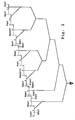
- the DNA coding sequence was synthetically constructed by sequential addition of oligonucleotides. Cloned oligonucleotides were fused into a single polynucleotide using the restriction digestions and ligations outlined in Figure 2.
- the polynucleotide coding sequence had the sequence presented as SEQ ID NO:1.
- this synthetic coding sequence can be placed in a number of bacterial expression vectors: for example, lambda gtl1 (Promega, Madison WI); pGEX (Smith, et al.); pNH vectors (Stratagene, La Jolla CA). Cloning of the IFN ⁇ synthetic polynucleotide into a modified pIN III omp-A expression vector is described in Example 3. Production of the IFN ⁇ protein was induced by the addition of IPTG. Soluble recombinant IFN ⁇ was liberated from the cells by sonication or osmotic fractionation.
- the protein can be further purified by standard methods, including size fractionation (column chromatography or preoperative gel electrophoresis) or affinity chromatography (using, for example, anti-IFN ⁇ antibodies (solid support available from Pharmacia, Piscataway NJ). Protein preparations can also be concentrated by, for example, filtration (Amicon, Danvers, Mass.).
- the synthetic IFN ⁇ gene was also cloned into the yeast cloning vector pBS24Ub (Example 4; Sabin, et al.; Ecker, et al.). Synthetic linkers were constructed to permit in-frame fusion of the IFN ⁇ coding sequences with the ubiquitin coding sequences in the vector. The resulting junction allowed in vivo cleavage of the ubiquitin sequences from the IFN ⁇ sequences.
- the recombinant plasmid pBS24Ub-IFN ⁇ was transformed into the yeast S. cerevisiae.
- Transformed yeast cells were cultured, lysed and the recombinant IFN ⁇ (r-IFN ⁇ ) protein isolated from the cell lysates.
- the amount of r-IFN ⁇ was quantified by radioimmunoassay. Microsequencing of the purified r-IFN ⁇ was carried out. The results demonstrated identity with native IFN ⁇ through the first 15 amino acids. The results also confirmed that the ubiquitin/IFN ⁇ fusion protein was correctly processed in vivo.
- Recombinant IFN ⁇ obtained by this method exhibited antiviral activity similar to the antiviral activity of IFN ⁇ purified from conceptus-conditioned culture medium.
- yeast vectors can be used in the practice of the present invention including, but are not limited to, vectors with regulatable expression (Hitzeman, et al.; Rutter, et al.; Oeda, et al .).
- the yeast transformation host is typically Saccharomyces cerevisiae, however, other yeast suitable for transformation can be used as well ( e.g., Schizosaccharomyces pombe ).
- the DNA encoding the IFN ⁇ polypeptide can be cloned into any number of commercially available vectors to generate expression of the polypeptide in the appropriate host system.
- These systems include the above described bacterial and yeast expression systems as well as the following: baculovirus expression (Reilly, et al .; Beames, et al .; Clontech, Palo Alto CA); and expression in mammalian cells (Clontech, Palo Alto CA; Gibco-BRL, Gaithersburg MD).
- baculovirus expression Reilly, et al .; Beames, et al .; Clontech, Palo Alto CA
- mammalian cells Clontech, Palo Alto CA; Gibco-BRL, Gaithersburg MD.
- These recombinant polypeptides can be expressed as fusion proteins or as native proteins.
- a number of features can be engineered into the expression vectors, such as leader sequences which promote the secretion of the expressed sequences into
- the recombinantly produced polypeptides are typically isolated from lysed cells or culture media. Purification can be carried out by methods known in the art including salt fractionation, ion exchange chromatography, and affinity chromatography. Immunoaffinity chromatography can be employed, as described above, using antibodies generated based on the IFN ⁇ polypeptides.
- DNA was screened for sequences homologous to interferon- ⁇ (Example 5). Several sequences that hybridized with the OvIFN ⁇ cDNA probe were identified. Several clones containing partial sequences of human interferon- ⁇ were then isolated (Example 6). Two synthetic 25-mer oligonucleotides, corresponding to sequences from the OvIFN ⁇ cDNA (Imakawa, et al., 1987) were synthesized. These primers were employed in amplification reactions using DNA derived from the following two cDNA libraries: human term placenta and human term cytotrophoblast. The resulting amplified DNA fragments were electrophoretically separated and a band containing an IFN ⁇ amplification product was isolated. The product was subcloned and the inserted amplification product sequenced using the dideoxy termination method.
- Example 7 describes the isolation of a full-length human IFN ⁇ gene.
- High molecular weight DNA was isolated from peripheral blood mononuclear cells (PBMCs) and size-fractionated. Fractions were tested for the presence of IFN ⁇ sequences using polymerase chain reaction: DNA molecules from fractions that tested amplification positive were used to generate a subgenomic library in ⁇ gt11.
- PBMCs peripheral blood mononuclear cells
- This subgenomic library was plated and hybridized with an OvIFN ⁇ cDNA probe (Example 7A). Approximately 20 clones were identified that hybridized to the probe. Plaques corresponding to the positive clones were passaged, DNA isolated and analyzed by amplification reactions using OvIFN ⁇ primers. Of these twenty plaques, six plaques generated positive PCR signals. The phage from these six clones were purified and the inserts sequenced. One of the inserts from one of these six clones was used as a hybridization probe in the following screening.
- Recombinant phage from the ⁇ gt11 subgenomic library were screened using the hybridization probe just described (Example 7B). Three clones giving positive hybridization signals were isolated and the inserts sequenced. The resulting nucleic acid sequence information is presented as SEQ ID NO:11 and the predicted protein coding sequence is presented as SEQ ID NO:12. The predicted mature protein coding sequence is presented as SEQ ID NO:4.
- the human IFN ⁇ sequences presented as SEQ ID NO:12 and SEQ ID NO:11, and primers and probes derived therefrom, can be used as specific probes to detect isolates of further human IFN ⁇ coding sequences and/or pseudogenes. Further, there may be more than one isoform of the IFN ⁇ protein and more than one coding sequence per species.
- the specific nucleic acid probes used in the practice of the present invention and antibodies reactive with the IFN ⁇ polypeptides of the present invention may be useful to isolate unidentified variants of interferon- ⁇ in mammals, according to the methods of the invention disclosed herein.
- Human placental cDNA libraries and an ovine cDNA library were analyzed by hybridization to the OvIFN ⁇ cDNA probe (Example 8). This DNA hybridization analysis suggested that the IFN ⁇ -signals from human cDNA libraries were approximately 1/100 of the signal obtained using the ovine cDNA library. OvIFN ⁇ cDNAs constitute around 0.4% of the ovine cDNA library. Accordingly, the abundance of human cDNAs responding to the OvIFN ⁇ probe appears to be low, at least in the term placenta from which the cDNA libraries were generated.
- interferon- ⁇ in human tissue was also examined using in situ hybridization (Example 9). Sections from four healthy, different term and first trimester human placentas were examined. This analysis employed a cDNA probe derived from the OvIFN ⁇ cDNA sequences (Example 9B). In situ hybridization was performed using an anti-sense RNA probe. In three separate experiments, specific hybridization was observed in all term and first trimester placental tissues.
- First trimester placental villi (composed of an outer layer of syncytiotrophoblast, an underlying layer of cytotrophoblast, and a central stromal region with various types of mesenchymal cells) displayed the highest transcript level of IFN ⁇ in the cytotrophoblast cells. Less intense but detectable levels were present in both the syncytiotrophoblast and stromal cells. A similar pattern of transcript expression was demonstrated in the placental villi of term tissue but the level of signal detection was low. First trimester extravillous trophoblasts displayed the highest amount of message and stained positive when present in the maternal blood spaces.
- the present results demonstrate that the human IFN ⁇ gene is highly expressed in early placental tissues by migrating extravillous trophoblasts, but is also expressed in villous syncytiotrophoblasts, cytotrophoblasts, and various stromal cells. These results demonstrate the detection of IFN ⁇ transcripts in human pregnancy tissues, and IFN ⁇ expression in the villous cytotrophoblasts as well as the extravillous trophoblast of first trimester placenta.
- IFN ⁇ The antiviral activity of IFN ⁇ has been evaluated against a number of viruses, including both RNA and DNA viruses.
- IFN ⁇ has potent antiviral activity with limited cytotoxic effects.
- Highly purified OvIFN ⁇ was tested for anti-retroviral and cytotoxic effects on peripheral blood lymphocytes exposed to feline AIDS and human AIDS retroviruses (Bazer, F.W., et al., (1989)).
- This feline AIDS lentivirus produces a chronic AIDS-like syndrome in cats and is a model for human AIDS (Pederson, et al., 1987).
- Replication of either virus in peripheral blood lymphocytes (PBL) was monitored by reverse transcriptase (RT) activity in culture supernatants over time.
- RT reverse transcriptase
- RNA-dependent DNA polymerase RT activity was assayed in FIV- and HIV-infected feline and human PBL cultures treated with IFN ⁇ (Example 11). Replication of FIV was reduced to about one-third of control values when cells were cultured in the presence of IFN ⁇ .
- Addition of OvIFN ⁇ produced a rapid, dose-dependent decrease in reverse transcriptase (RT) activity (Example 11, Table 4). While concentrations as low as 0.62 ng/ml of IFN ⁇ inhibited viral replication, much higher concentrations (40 ng/ml) having greater effects on RT-activity were without toxic effects on the cells.
- the results suggest that replication of the feline immunodeficiency virus was reduced significantly compared to control values when cells were cultured in the presence of OvIFN ⁇ .
- IFN ⁇ appeared to exert no cytotoxic effect on the cells hosting the retrovirus. This was true even when IFN ⁇ was present at 40 ng per ml of culture medium. This concentration of IFN ⁇ is equivalent to about 8,000 anti-viral units of alpha interferon, when IFN ⁇ is assayed for its ability to protect Madin-Darby bovine kidney cells from lysis by vesicular stomatitis virus as described by Pontzer, et al. (1988).
- IFN ⁇ was also tested for activity against HIV replication in human cells.
- Human peripheral lymphocytes, which had been infected with HIV were treated with varying concentrations of IFN ⁇ (Example 12).
- Replication of HIV in peripheral blood lymphocytes was monitored by reverse transcriptase activity in culture supernatants over time.
- Over a range of concentrations of IFN ⁇ produced significant anti-HIV effects (Example 12, Table 5).
- a concentration of only 10 ng/ml resulted in over a 50% reduction in RT activity after only six days.
- a concentration of 500 ng/ml resulted in a 90% reduction in RT activity within 10 days.
- there was no evidence of any cytotoxic effects attributable to the administration of IFN ⁇ (Example 12, Table 5).
- IFN ⁇ antiviral effects of IFN ⁇ against HIV were evaluated by treating human PBMC cells with various amounts of either recombinant IFN ⁇ or recombinant human IFN ⁇ 2 at the time of infection with HIV (Example 18).
- the data from these experiments support the conclusion that, at similar concentrations, IFN ⁇ and IFN ⁇ are effective in reducing the replication of HIV in human lymphocytes.
- treatment of cells with IFN ⁇ 2 resulted in cytoxicity, whereas no such cytotoxity was observed with treatment using IFN ⁇ , even when IFN ⁇ was used at much higher concentrations. No cytotoxicity was observed using IFN ⁇ , even when IFN ⁇ was used at 200 times the dosage of interferon-alpha II.
- Interferon- ⁇ has also been shown to inhibit Hepatitis B Virus DNA replication in hepatocytes (Example 18).
- a human cell derived from liver cells transfected with Hepatitis B Virus (HBV) was used to test the antiviral effects of IFN ⁇ .
- the cells were treated with both the IFN ⁇ and IFN ⁇ over a range of concentrations. Both IFN ⁇ and IFN ⁇ reduced DNA production by approximately two-fold compared to the no interferon control.
- the hepatocyte was examined for the effects of IFN ⁇ and IFN ⁇ on hepatospecific mRNA production (Example 18).
- Two hepatocyte specific proteins, Apo E and Apo A1 were detected by hybridization analysis. There was no apparent reduction of mRNA production for either hepatospecific mRNA at concentrations up to 40,000 units/ml of either IFN ⁇ or IFN ⁇ . Further, no evidence for hepatotoxicity with IFN ⁇ was seen in this assay.
- IFN ⁇ is an effective antiviral agent against a wide variety of viruses, including both RNA and DNA viruses.
- IFN ⁇ anti-cellular growth activity was examined using a colony inhibition assay (Example 13).
- Human amnion (WISH) or MDBK cells were plated at low cell densities to form colonies originating from single cells. Dilutions of interferons were added to triplicate wells and the plates were incubated to allow colony formation. IFN ⁇ inhibited both colony size and number in these assays. IFN ⁇ was more effective at inhibiting cell proliferation of the human cell line (WISH) than human IFN ⁇ .
- the antiproliferative activity of IFN ⁇ was dose-dependent. High concentrations of IFN ⁇ stopped proliferation, while cell viability was not impaired.
- IFN ⁇ appears to inhibit progress of cells through S phase.
- the antiproliferative effects of IFN ⁇ were also studied for rat and bovine cell lines (Example 14).
- the rate of 3 H-thymidine incorporation was used to assess the rate of cellular proliferation.
- the data obtained demonstrate that IFN ⁇ drastically reduced the rate of cellular proliferation (Example 14, Table 7) for each tested cell line.
- Example 15 The antiproliferative activity and lack of toxicity of IFN ⁇ was further examined using a series of human tumor cell lines (Example 15).
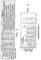
- a variety of human tumor cell lines were selected from the standard lines used in NIH screening procedure for antineoplastic agents (Pontzer, C.H., et al. , (1991)). At least one cell line from each major neoplastic category was examined. The following cell lines were obtained from American Type Culture Collection (12301 Parklawn Dr., Rockville MD 20852) :
- the antiproliferative activity was evaluated by measuring the rate of 3 H-thymidine incorporation into cells which have been treated with IFN ⁇ . Significant differences between treatments were assessed by an analysis of variance followed by Scheffe's F-test. Cell cycle analysis was performed by flow cytometry.
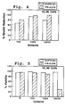
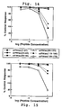
- HUT 78 The human cutaneous T cell lymphoma, HUT 78, responded similarly to HL-60 when treated with IFN ⁇ (Example 15, Figure 9). Both OvIFN ⁇ and rHuIFN ⁇ reduce HUT 78 cell growth, but IFN ⁇ demonstrated adverse effects on cell viability.
- the T cell lymphoma H9 was less sensitive to the antiproliferative effects of IFN ⁇ than the tumor cell lines described above. While IFN ⁇ was not toxic to the H9 cells, it failed to inhibit cell division significantly at any of the concentrations examined (Example 15, Figure 10). In contrast, IFN ⁇ was observed to reduce H9 growth by approximately 60%. Thus, only OvIFN ⁇ is an effective growth inhibitor of this T cell lymphoma.
- IFN ⁇ and IFN ⁇ were equally efficacious antitumor agents.
- SK-MEL-28 inhibition of proliferation by IFN ⁇ was accomplished by a 13% drop in viability, while IFN ⁇ was not cytotoxic.
- IFN ⁇ is equal or preferable to IFN ⁇ as an antineoplastic agent against human tumors.
- IFN ⁇ exhibits antiproliferative activity against human tumor cells without toxicity and is as potent or more potent than human IFN ⁇ . Clinical trials of the IFN ⁇ 2s have shown them to be effective antitumor agents (Dianzani, F., 1992; Krown, 1987).
- One therapeutic advantage of IFN ⁇ as a therapeutic is the elimination of toxic effects seen with high doses IFN ⁇ s.
- IFN ⁇ immunoglobulin-like tumors like Kaposi's sarcoma (associated with HIV infection) where the antineoplastic effects of IFN ⁇ are coupled with IFN ⁇ ability to inhibit retroviral growth.
- B16-F10 is a syngeneic mouse transplantable tumor selected because of its high incidence of pulmonary metastases (Poste, et al., 1981). Interferon treatment was initiated 3 days after the introduction of the tumor cells. The in vivo administration of IFN ⁇ dramatically reduced B16-F10 pulmonary tumors. Thus, IFN ⁇ appears to be an efficacious antineoplastic agent in vivo as well as in vitro.
- IFN ⁇ antiviral activity also blocked the antiviral activity of recombinant bovine IFN ⁇ (rBoIFN ⁇ ); recombinant bovine IFN ⁇ was unaffected by the peptides.
- rBoIFN ⁇ recombinant bovine IFN ⁇
- IFN ⁇ peptides may represent common receptor binding regions for IFN ⁇ and various IFN ⁇ s.
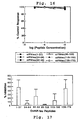
- OvIFN ⁇ (1-37) and OvIFN ⁇ (139-172) also blocked OvIFN ⁇ anti-FIV and anti-HIV activity (Example 17; Figures 11A and 11B). While both peptides blocked FIV RT activity, only the C-terminal peptide, OvIFN ⁇ (139-172), appeared to be an efficient inhibitor of vesicular stomatitis virus activity on the feline cell line, Fc9.
- portions of the IFN ⁇ interferon molecule may be used to substitute regions of interferon alpha molecules.
- the region of an interferon alpha molecule that is responsible for increased cytotoxicity, relative to IFN ⁇ treatment can be identified by substituting polypeptide regions derived from IFN ⁇ for regions of an interferon alpha molecule.
- substitutions can be carried out by manipulation of synthetic genes (see below) encoding the selected IFN ⁇ and interferon alpha molecules, coupled to the functional assays described herein (such as, antiviral, antiproliferative and cytoxicity assays).
- IFN ⁇ The antiproliferative activity of IFN ⁇ (Example 17, Table 11) involved a further region of the molecule, since IFN ⁇ (119-150) was the most effective inhibitor of OvIFN ⁇ -induced reduction of cell proliferation. This results suggests that the region of the molecule primarily responsible for inhibition of cell growth is the IFN ⁇ (119-150) region.
- This region of the IFN ⁇ molecule may be useful alone or fused to other proteins (such as serum albumin, an antibody or an interferon alpha polypeptide) as an antineoplastic agent.
- serum albumin an antibody or an interferon alpha polypeptide
- a conjugated protein between an N-terminal peptide derived from human interferon- ⁇ and serum albumin was shown to have anticellular proliferation activity (Ruegg, et al., 1990).
- binding of 125 I-OvIFN ⁇ to its receptor on MDBK cells could be blocked by antisera to 4 of the 6 peptides; the 4 polypeptides representing amino acids 1-37, 62-92, 119-150 and 139-172 of OvIFN ⁇ .
- This reflects the multiple binding domains as well as the functional significance of these regions. Since different regions of IFN ⁇ are involved in elicitation of different functions, modification of selected amino acids could potentially result in IFN ⁇ -like interferons with selective biological activity.
- the final modeling step was to apply the IFN ⁇ x-ray crystallographic coordinates of the IFN ⁇ carbon backbone to the IFN ⁇ sequence.
- the functionally active domains of IFN ⁇ , identified above, were localized to one side of the molecule and found to be in close spatial proximity. This is consistent with multiple binding sites on IFN ⁇ interacting simultaneously with the type I IFN receptor.
- the three dimensional modeling data coupled with the function data described above provides the ability to introduce sequence variations into specific regions of IFN ⁇ to generate enhancement of selected functions (e.g., antiviral or anticellular proliferation) or the ability to substitute a region(s) of selected function into other interferon molecules (e.g., antiviral, antineoplastic, or reduced cytotoxicity).
- selected functions e.g., antiviral or anticellular proliferation
- other interferon molecules e.g., antiviral, antineoplastic, or reduced cytotoxicity
- Example 3 The construction of a synthetic gene for OvIFN ⁇ is described in Example 3. Briefly, a consensus amino sequence was back-translated using optimal codon usage for E. coli. The sequence was edited to include 20, unique, restriction sites spaced throughout the length of the construct. This 540 base pair synthetic gene sequence was divided into 11 oligonucleotide fragments. Individual fragments were synthesized and cloned, either single or double stranded, into either pTZ 19R, pTZ 18R or pBluescript, amplified and fused. The synthetic OvIFN ⁇ construct was then cloned into a modified pIN-III-ompA expression vector for expression in bacteria and also cloned into a yeast expression plasmid. A similarly constructed human IFN ⁇ synthetic gene (SEQ ID NO:3) has been designed, constructed and expressed in yeast cells.
- SEQ ID NO:3 A similarly constructed human IFN ⁇ synthetic gene (SEQ ID NO:3) has been designed, constructed and expressed in yeast cells.
- the synthetic gene construct facilitates introduction of mutations for possible enhancement of antitumor (anticellular proliferative) and antiviral activities. Further, the disparate regions of the molecule responsible for different functions allows for separate manipulation of different functions. For example, two deletion mutants, OvIFN ⁇ (1-155) and OvIFN ⁇ (1-166), have been constructed to examine the role of carboxy terminal sequences in IFN ⁇ molecules.
- Tyr 123 has been implicated in the anticellular proliferative activity of IFN ⁇ (McInnes, et al., 1989).
- the equivalent of Tyr 123 in IFN ⁇ is contained within peptide OvIFN ⁇ (119-150): this polypeptide inhibits OvIFN ⁇ and human IFN ⁇ antiproliferative activity.
- Mutations converting Tyr 123 to conservative (Trp) and nonconservative (Asp) substitutions have been generated, as well as mutant sequences having deletion of this residue.
- the codon for Tyr 123 is located within an SspI site; elimination of this site has been used for screening. The antiproliferative activity of these mutant IFN ⁇ is evaluated as described herein.
- Synthetic peptides can be generated which correspond to the IFN ⁇ polypeptides of the present invention. Synthetic peptides can be commercially synthesized or prepared using standard methods and apparatus in the art (Applied Biosystems, Foster City CA).
- oligonucleotide sequences encoding peptides can be either synthesized directly by standard methods of oligonucleotide synthesis, or, in the case of large coding sequences, synthesized by a series of cloning steps involving a tandem array of multiple oligonucleotide fragments corresponding to the coding sequence (Crea; Yoshio et al.; Eaton et al.). Oligonucleotide coding sequences can be expressed by standard recombinant procedures (Maniatis et al.; Ausubel et al.).
- the present invention includes interferon- ⁇ or interferon- ⁇ -derived polypeptides covalently attached to a second polypeptide to form a fused, or hybrid, protein.
- the interferon- ⁇ sequences making up such fused proteins can be recombinantly produced interferon- ⁇ or a bioactive portion thereof, as described above.
- the polypeptides presented as SEQ ID NO:10 and SEQ ID NO:20 may be advantageously fused with a soluble peptide, such as, serum albumin, an antibody ( e.g ., specific against an virus-specific cell surface antigen), or an interferon alpha polypeptide.
- a soluble peptide such as, serum albumin, an antibody (e.g ., specific against an virus-specific cell surface antigen), or an interferon alpha polypeptide.
- fusion proteins include (i) replacing toxicity-associated regions of interferon- ⁇ with the interferon- ⁇ regions SEQ ID NO:5 and SEQ ID NO:15, and (ii) fusion proteins containing the interferon- ⁇ regions SEQ ID NO:9 and SEQ ID NO:19 as anticellular proliferation agents.
- the fused proteins of the present invention may be formed by chemical conjugation or by recombinant techniques.
- the interferon- ⁇ and second selected polypeptide are modified by conventional coupling agents for covalent attachment.
- serum albumin is derivatized with N-succinimidyl-S-acetyl thioacetate (Duncan), yielding thiolated serum albumin.
- the activated serum albumin polypeptide is then reacted with interferon- ⁇ derivatized with N-succinimidyl 3-(2-pyridyldithio) propionate (Cumber), to produce the fused protein joined through a disulfide linkage.
- recombinant interferon- ⁇ may be prepared with a cysteine residue to allow disulfide coupling of the interferon- ⁇ to an activated ligand, thus simplifying the coupling reaction.
- An interferon- ⁇ expression vector, used for production of recombinant interferon- ⁇ , can be modified for insertion of an internal or a terminal cysteine codon according to standard methods of site-directed mutagenesis (Ausubel, et al .).
- a fused protein is prepared recombinantly using an expression vector in which the coding sequence of a second selected polypeptide is joined to the interferon- ⁇ coding sequence.
- human serum albumin coding sequences can be fused in-frame to the coding sequence of an interferon- ⁇ polypeptide, such as, SEQ ID NO:9.
- the fused protein is then expressed using a suitable host cell.
- the fusion protein may be purified by molecular-sieve and ion-exchange chromatography methods, with additional purification by polyacrylamide gel electrophoretic separation and/or HPLC chromatography, if necessary.
- interferon- ⁇ -containing fusion proteins may be prepared.
- One variation on the above fusion is to exchange positions of the interferon- ⁇ and selected second protein molecules in the fusion protein (e.g. , carboxy terminal versus amino terminal fusions).
- internal portions of a native interferon- ⁇ polypeptide for example, amino acid regions of between 15 and 172 amino acids
- two or more such interferon- ⁇ portions are contiguous that are normally discontinuous in the native protein.
- Fusion proteins containing the polypeptide antigens of the present invention fused with the glutathione-S-transferase (Sj26) protein can be expressed using the pGEX-GLI vector system in E. coli JM101 cells.
- the fused Sj26 protein can be isolated readily by glutathione substrate affinity chromatography (Smith). Expression and partial purification of IFN ⁇ proteins is described in (Example 20), and is applicable to any of the other soluble, induced polypeptides coded by sequences described by the present invention.
- Insoluble GST (sj26) fusion proteins can be purified by preparative gel electrophoresis.
- IFN ⁇ - ⁇ -galactosidase fusion proteins can be isolated as described in Example 19.
- an expression vector such as the lambda gtl1 or pGEX vectors described above, containing IFN ⁇ coding sequences and expression control elements which allow expression of the coding regions in a suitable host.
- the control elements generally include a promoter, translation initiation codon, and translation and transcription termination sequences, and an insertion site for introducing the insert into the vector.
- the DNA encoding the desired polypeptide can be cloned into any number of vectors (discussed above) to generate expression of the polypeptide in the appropriate host system. These recombinant polypeptides can be expressed as fusion proteins or as native proteins. A number of features can be engineered into the expression vectors, such as leader sequences which promote the secretion of the expressed sequences into culture medium. Recombinantly produced IFN ⁇ , and polypeptides derived therefrom, are typically isolated from lysed cells or culture media. Purification can be carried out by methods known in the art including salt fractionation, ion exchange chromatography, and affinity chromatography. Immunoaffinity chromatography can be employed using antibodies generated against selected IFN ⁇ antigens.
- the invention includes specific antibodies directed against the polypeptides of the present invention.
- a host animal such as a rabbit
- the purified antigen or fused protein antigen may be generated using a variety of coding sequences derived from other proteins, such as ⁇ -galactosidase or glutathione-S-transferase.
- the host serum or plasma is collected following an appropriate time interval, and this serum is tested for antibodies specific against the antigen.
- Example 20 describes the production of rabbit serum antibodies which are specific against the IFN ⁇ antigens in a Sj26/IFN ⁇ hybrid protein. These techniques can be applied to the all of the IFN ⁇ molecules and polypeptides derived therefrom.
- the gamma globulin fraction or the IgG antibodies of immunized animals can be obtained, for example, by use of saturated ammonium sulfate or DEAE Sephadex, or other techniques known to those skilled in the art for producing polyclonal antibodies.
- purified protein or fused protein may be used for producing monoclonal antibodies.
- spleen or lymphocytes from a animal immunized with the selected polypeptide antigen are removed and immortalized or used to prepare hybridomas by methods known to those skilled in the art (Harlow, et al.).
- Lymphocytes can be isolated from a peripheral blood sample.
- Epstein-Barr virus (EBV) can be used to immortalize human lymphocytes or a fusion partner can be used to produce hybridomas.
- Antibodies secreted by the immortalized cells are screened to determine the clones that secrete antibodies of the desired specificity, for example, by using the ELISA or Western blot method (Ausubel et al .). Experiments performed in support of the present invention have yielded four hybridomas producing monoclonal antibodies specific for ovine IFN ⁇ have been isolated.
- Antigenic regions of polypeptides are generally relatively small, typically 7 to 10 amino acids in length. Smaller fragments have been identified as antigenic regions. Interferon- ⁇ polypeptide antigens are identified as described above. The resulting DNA coding regions can be expressed recombinantly either as fusion proteins or isolated polypeptides.
- amino acid sequences can be conveniently chemically synthesized (Applied Biosystems, Foster City CA).
- Antigens obtained by any of these methods may be directly used for the generation of antibodies or they may be coupled to appropriate carrier molecules.
- Many such carriers are known in the art and are commercially available (e.g ., Pierce, Rockford IL).
- Antibodies reactive with IFN ⁇ are useful, for example, in the analysis of structure/function relationships.
- IFN ⁇ bears some similarity to the IFN ⁇ family based on structure and its potent antiviral properties, the IFN ⁇ s do not possess the reproductive properties associated with IFN ⁇ . Also, recombinant bovine IFN ⁇ has little or no effect on interestrous interval compared to IFN ⁇ (Davis, et al., 1992).
- IFN ⁇ has some structural similarities to other interferons, it has very distinctive properties of its own: for example, the capability of significantly influencing the biochemical events of the estrous cycle.
- the human IFN ⁇ of the present invention can be used in methods of enhancing fertility and prolonging the life span of the corpus luteum in female mammals as generally described in Hansen, et al., herein incorporated by reference. Further, the human interferon- ⁇ of the present invention could be used to regulate growth and development of uterine and/or fetal-placental tissues. The human IFN ⁇ is particularly useful for treatment of humans, since potential antigenic responses are less likely using such a same-species protein.
- IFN ⁇ The antiviral activity of IFN ⁇ has broad therapeutic applications without the toxic effects that are usually associated with IFN ⁇ s. Although the presence of IFN ⁇ in culture medium inhibited reverse transcriptase activity of the feline immunodeficiency virus (Example 11), this is not due to a direct effect of IFN ⁇ on the FIV. Rather, IFN ⁇ appears to induce the host cell to produce a factor(s) which is inhibitory to the reverse transcriptase of the virus.
- IFN ⁇ was found to exert its antiviral activity without adverse effects on the cells: no evidence of cytotoxic effects attributable to the administration of IFN ⁇ was observed. It is the lack of cytotoxicity of IFN ⁇ which makes it extremely valuable as an in vivo therapeutic agent. This lack of cytotoxicity sets IFN ⁇ apart from most other known antiviral agents and all other known interferons.
- Formulations comprising the IFN ⁇ compounds of the present invention can be used to inhibit viral replication.
- the human IFN ⁇ of the present invention can be employed in methods for affecting the immune relationship between fetus and mother, for example, in preventing transmission of maternal viruses (e.g ., HIV) to the developing fetus.
- the human interferon- ⁇ is particularly useful for treatment of humans, since potential antigenic responses are less likely using a homologous protein.
- IFN ⁇ exhibits potent anticellular activity. IFN ⁇ can also be used to inhibit cellular growth without the negative side effects associated with other interferons which are currently known. Formulations comprising the IFN ⁇ compounds of the subject invention can be used to inhibit, prevent, or slow tumor growth.
- IFN ⁇ can suppress estrogen receptor numbers. Therefore, IFN ⁇ can be used in the treatment or prevention of estrogen-dependent tumors.
- IFN ⁇ appears to interact with the Type I IFN receptor via several epitopes on the molecule, and these regions either separately or in combination may affect distinct functions of IFN ⁇ differently.
- polypeptides of the present invention are useful for the selective inhibition of binding of interferons to the interferon receptor. Specifically, as described herein, certain of the disclosed peptides selectively inhibit the antiviral activity of IFN ⁇ while others inhibit the antiproliferative activity. Combinations of these peptides could be used to inhibit both activities.
- these peptides despite binding to the interferon receptor and blocking IFN ⁇ activity, these peptides do not, themselves, elicit the antiviral or antiproliferative activity.
- polypeptides can be used as immunoregulatory molecules when it is desired to prevent immune responses triggered by interferon molecules.
- These peptides could be used as immunosuppressants to prevent, for example, interferon-mediated immune responses to tissue transplants.
- Other types of interferon mediated responses may also be blocked, such as the cytotoxic effects of alpha interferon.
- IFN ⁇ proteins can be formulated according to known methods for preparing pharmaceutically useful compositions. Formulations comprising interferons or interferon-like compounds have been previously described (for example, Martin, 1976). In general, the compositions of the subject invention will be formulated such that an effective amount of the IFN ⁇ is combined with a suitable carrier in order to facilitate effective administration of the composition.
- compositions used in these therapies may also be in a variety of forms. These include, for example, solid, semi-solid, and liquid dosage forms, such as tablets, pills, powders, liquid solutions or suspensions, liposomes, suppositories, injectable, and infusible solutions. The preferred form depends on the intended mode of administration and therapeutic application.
- the compositions also preferably include conventional pharmaceutically acceptable carriers and adjuvants which are known to those of skill in the art.
- the compositions of the invention are in the form of a unit dose and will usually be administered to the patient one or more times a day.
- IFN ⁇ may be administered to a patient in any pharmaceutically acceptable dosage form, including intravenous, intramuscular, intralesional, or subcutaneous injection.
- any pharmaceutically acceptable dosage form including intravenous, intramuscular, intralesional, or subcutaneous injection.
- compositions and methods used for other interferon compounds can be used for the delivery of these compounds.
- IFN ⁇ interferon-associated cytotoxicity
- concentrations which are greater than those which can generally be utilized for other interferon (e.g ., IFN ⁇ ) compounds.
- IFN ⁇ can be administered at rates from about 5 ⁇ 10 4 to 20 ⁇ 10 6 units/day to about 500 ⁇ 10 6 units/day or more. In a preferred embodiment, the dosage is about 10 6 units/day. High doses are preferred for systemic administration. It should, of course, be understood that the compositions and methods of this invention may be used in combination with other therapies.
- a maintenance dose is administered if necessary. Subsequently, the dosage or the frequency of administration, or both, may be reduced, as a function of the symptoms, to a level at which the improved condition is retained. When the symptoms have been alleviated to the desired level, treatment should cease. Patients may, however, require intermittent treatment on a long-term basis upon any recurrence of disease symptoms.
- compositions of the subject invention can be administered through standard procedures to treat a variety of cancers and viral diseases including those for which other interferons have previously shown activity. See, for example, Finter, et al . (1991); Dianzani, et al . (1992); Francis, et al . (1992) and U.S. Patent Nos. 4,885,166 and 4,975,276.
- the compositions of the subject invention have unique features and advantages, including their ability to treat these conditions without toxicity.
- disorders of the skin can be treated intralesionally using IFN ⁇ , wherein formulation and dose will depend on the method of administration and on the size and severity of the lesion to be treated.
- Preferred methods include intradermal and subcutaneous injection. Multiple injections into large lesions may be possible, and several lesions on the skin of a single patient may be treated at one time.
- the schedule for administration can be determined by a person skilled in the art. Formulations designed for sustained release can reduce the frequency of administration.
- Systemic treatment is essentially equivalent for all applications. Multiple intravenous or subcutaneous doses are possible, and in the case of implantable methods for treatment, formulations designed for sustained release are particularly useful. Patients may also be treated using implantable subcutaneous portals, reservoirs, or pumps.
- Regional treatment with the IFN ⁇ polypeptides of the present invention is useful for treatment of cancers in specific organs.
- Treatment can be accomplished by intraarterial infusion.
- a catheter can be surgically or angiographically implanted to direct treatment to the affected organ.
- a subcutaneous portal, connected to the catheter, can be used for chronic treatment, or an implantable, refillable pump may also be employed.
- T4 DNA ligase T4 polynucleotide kinase
- Taq DNA polymerase calf intestinal phosphatase
- calf intestinal phosphatase purchased from New England Biolabs (Beverly, MA) or Promega Biotech (Madison, WI): these reagents were used according to the manufacturer's instruction.
- a "SEQUENASE DNA II” sequencing kit was used (United States Biochemical Corporation, Cleveland OH).
- Immunoblotting and other reagents were from Sigma Chemical Co. (St. Louis, MO) or Fisher Scientific (Needham, MA). Nitrocellulose filters are obtained from Schleicher and Schuell (Keene, NH).
- Synthetic oligonucleotide linkers and primers are prepared using commercially available automated oligonucleotide synthesizers (e.g ., an ABI model 380B-02 DNA synthesizer (Applied Biosystems, Foster City, CA)). Alternatively, custom designed synthetic oligonucleotides may be purchased, for example, from Synthetic Genetics (San Diego, CA). cDNA synthesis kit and random priming labeling kits are obtained from Boehringer-Mannheim Biochemical (BMB, Indianapolis, IN).
- Oligonucleotide sequences encoding polypeptides can be either synthesized directly by standard methods of oligonucleotide synthesis, or, in the case of large coding sequences, synthesized by a series of cloning steps involving a tandem array of multiple oligonucleotide fragments corresponding to the coding sequence (Crea; Yoshio et al.; Eaton et al.). Oligonucleotide coding sequences can be expressed by standard recombinant procedures (Maniatis et al.; Ausubel et al.).
- peptides can be synthesized directly by standard in vitro techniques (Applied Biosystems, Foster City CA).
- Recombinant human IFN ⁇ (rHuIFN ⁇ ) and rBoIFN ⁇ was obtained from Genentech Inc. (South San Francisco, CA).
- the reference preparation of recombinant human IFN ⁇ (rHuIFN ⁇ ) was obtained from the National Institutes of Health: rHuIFN ⁇ is commercially available from Lee Biomolecular (San Diego, CA).
- Antisera were diluted in 0.1 M PBS, pH 7.2. The desired dilution(s) of antisera (0.1 mL) were added to each well and the plate incubated 1 hours at 37°C. The plates was then washed 5 times with PBS 0.5% "TWEEN-20".
- HRP Horseradish peroxidase conjugated goat antihuman antiserum
- the reagent consists of 50 mL 0.05 M citric acid, pH 4.2, 0.078 mL 30% hydrogen peroxide solution and 15 mg ABTS. 0.1 mL of the substrate was added to each well, then incubated for 30 min at room temperature. The reaction was stopped with the addition of 0.050 mL 5% SDS (w/v). The relative absorbance is determined at 410 nm.
- IFN ⁇ was infused into uterine lumen of ewes at the concentrations given in Table 1.
- Recombinant human IFN ⁇ (rHuIFN ⁇ ) was infused at similar concentrations.
- control animals which received control proteins, were also used.
- the life span of the corpus luteum was assessed by examination of interestrous intervals, maintenance of progesterone secretion, and inhibition of prostaglandin secretion (Davis, et al ., 1992 ).
- VSV vesicular stomatitis virus
- One antiviral unit caused a 50% reduction in destruction of the monolayer, relative to untreated MDBK cells infected with VSV (control plates). Specific activities were further evaluated using normal ovine fibroblasts (Shnf) in a plaque inhibition assay (Langford, et al., 1981). A minimum of three samples were examined at each time point, and each sample was assayed in triplicate. The results presented in Table 2 are expressed as mean units/ml.
- the amino acid coding sequence for OvIFN ⁇ (Imakawa, et al., 1987) was used to generate a corresponding DNA coding sequence with codon usage optimized for expression in E. coli.
- Linker sequences were added to the 5' and 3' ends to facilitate cloning in bacterial expression vectors.
- the nucleotide sequence was designed to include 19 unique restriction enzyme sites spaced evenly throughout the coding sequence ( Figure 1).
- the nucleotide sequence was divided into eleven oligonucleotide fragments ranging in sizes of 33 to 75 bases.
- Each of the eleven oligonucleotides were synthesized on a 380-B 2-column DNA synthesizer (Applied Biosytems) and cloned single- or double-stranded into one of the following vectors: "pBLUESCRIPT + (KS)" (Stratagene, LaJolla, CA), pTZ18R (Pharmacia, Piscataway, NJ), or PTZ19R (Pharmacia, Piscataway, NJ) cloning vectors.
- KS 380-B 2-column DNA synthesizer
- the vectors were transformed into E. coli K. strain "XL1-BLUE" (recA1 endA1 gyrA96 thi hsdR17 (r k - , m k +) supE44 relA1 ⁇ - (lac), ⁇ F', proAB, lac q Z ⁇ M15, Tn10(tet R ⁇ ) which is commercially available from Stratagene (LaJolla, CA). Transformed cells were grown in L broth supplemented with ampicillin (50 ⁇ g/ml). Oligonucleotide cloning and fusion was performed using standard recombinant DNA techniques.
- Cloning vectors were cut with the appropriate restriction enzymes to insert the synthetic oligonucleotides.
- the vectors were treated with calf intestine alkaline phosphatase (CIP) to remove terminal phosphate groups.
- Oligonucleotides were phosphorylated and cloned, as either single- or double-stranded molecules, into the appropriate vector using T4 DNA ligase. When single-strands were introduced into cloning vectors, the second strand was completed by the bacterial host following transfection.
- CIP calf intestine alkaline phosphatase
- oligonucleotides were first annealed with their synthetic complementary strand then ligated into the cloning vector.
- E. coli K12 strains SB221 or NM522 were then transformed with the ligation.
- E. coli strain GM119 was used for cloning when the methylation-sensitive Stu I and Cla I restriction sites were involved. Restriction analyses were performed on isolated DNA at each stage of the cloning procedure.
- Oligonucleotides were fused into a single polynucleotide using the restriction digestions and ligations outlined in Figure 2. Oligonucleotide-containing-DNA fragments were typically isolated after electrophoretic size fractionation on low-melting point agarose gels (Maniatis, et al .; Sambrook, et al .; Ausubel, et al .). The resulting IFN ⁇ polynucleotide coding sequence spans position 16 through 531: a coding sequence of 172 amino acids.
- the nucleotide sequence of the final polynucleotide was confirmed by DNA sequencing using the dideoxy chain termination method.
- the full length Stu I/ Sst I fragment (540 bp; Figure 2) was cloned into a modified pIN III omp-A expression vector and transformed into a competent SB221 strain of E. coli.
- IFN ⁇ protein For expression of the IFN ⁇ protein, cells carrying the expression vector were grown in L-broth containing ampicillin to an OD (550 nm) of 0.1-1, induced with IPTG for 3 hours and harvested by centrifugation. Soluble recombinant IFN ⁇ was liberated from the cells by sonication or osmotic fractionation.
- the synthetic IFN ⁇ gene synthesized in Example 3, was flanked at the 5' end by an StuI restriction site and at the 3' end by a SacI restriction site.
- Two oligonucleotide primers (SEQ ID NO:13 and SEQ ID NO:14) were used to attach linkers to the synthetic IFN ⁇ gene using polymerase chain reaction.
- the linker at the 5' end allowed the placement of the synthetic IFN ⁇ gene in correct reading with the ubiquitin coding sequence present in the yeast cloning vector pBS24Ub (Chiron Corp., Emeryville, CA).
- the linker also constructed a ubiquitin-IFN ⁇ junction region that allowed in vivo cleavage of the ubiquitin sequences from the IFN ⁇ sequences.
- the 5' oligonucleotide also encoded a Sac II restriction endonuclease cleavage site.
- the 3' oligonucleotide contained a Stu I cleavage site.
- the vector carrying the synthetic IFN ⁇ gene (Example 3) was isolated from E. coli strain "XLI-BLUE" by the alkaline lysis method. Isolated vector was diluted 500-fold in 10 mM Tris, pH 8.0/1 mM EDTA/10 mM NaCl. The PCR reaction was performed in a 100 ⁇ l volume using Taq DNA polymerase and primers SEQ ID NO:13/SEQ ID NO:14. The amplified fragments were digested with StuI and Sac II. These digested fragments were ligated into the Sac II and Sma I sites of "pBLUESCRIPT+(KS)."
- the resulting plasmid was named pBSY-IFN ⁇ .
- the DNA sequence was verified using double stranded DNA as the template.
- Plasmid pBSY-IFN ⁇ was digested with Sac II and Eco RV and the fragment containing the synthetic IFN ⁇ gene was isolated.
- the yeast expression vector pBS24Ub (Sabin, et al .; Ecker, et al .) was digested with Sal I. Blunt ends were generated using T4 DNA polymerase.
- the vector DNA was extracted with phenol and ethanol precipitated (Sambrook, et al., 1989).
- the recovered linearized plasmid was digested with Sac II, purified by agarose gel electrophoresis, and ligated to the Sac II- Eco RV fragment isolated from pBSY-IFN ⁇ . The resulting recombinant plasmid was designated pBS24Ub-IFN ⁇ .
- the recombinant plasmid pBS24Ub-IFN ⁇ was transformed into E. coli.
- Recombinant clones containing the IFN ⁇ insert were isolated and identified by restriction enzyme analysis. Plasmid DNA from clones containing IFN ⁇ coding sequences was used for transformation of S. cerevisiae (Rothstein, 1986). Transformation mixtures were plated on uracil omission medium and incubated for 3-5 days at 30°C. Colonies were then streaked and maintained on uracil and leucine omission medium (Rothstein, 1986).
- pBS24-IFN ⁇ was grown for 24 hours at 30°C in 5 ⁇ uracil and leucine omission medium containing 8% glucose. This culture was then diluted 20-fold in YEP medium containing 1% glucose and further incubated for another 24-36 hours.
- Cells were harvested by centrifugation, washed in 50 mM Tris, pH 7.6,/1 mM EDTA and resuspended in wash buffer containing 1 mM PMSF. The cells were lysed using a Bead-beater apparatus (Biospec Products, Bartlesville, OK). The lysate was spun at 43,000 ⁇ g for 20 minutes. The supernatant fraction was recovered and subjected to the purification protocol described below.
- the supernatant was loaded on a 1 ⁇ 10 cm DEAE column and washed with 10 mM Tris, pH 8.0. Retained proteins were eluted with a 300 ml, 0 to 0.5 M NaCl gradient in 10 mM Tris, pH 8.0. Three-milliliter fractions were collected. Ten-microliter samples of fractions 17-26 containing the recombinant (r-IFN ⁇ ) were electrophorectically separated on 15% SDS-polyacrylamide gels. The gels were stained with Coomassie blue.
- Fractions 18, 19, and 20 contained largest amount of r-IFN ⁇ . These fractions were loaded individually on a 1.5 ⁇ 90 cm Sephadex S-200 column and proteins were resolved in two peaks. Aliquots of each protein peak (25 ⁇ l) were electrophoretically separated on 15% SDS-polyacrylamide gels and the proteins visualized with Coomassie staining.
- r-IFN ⁇ -containing fractions were combined and the amount of r-IFN ⁇ quantified by radioimmunoassay (Vallet, et al ., 1988). Total protein concentration was determined by using the Lowry protein assay (Lowry, et al. , 1951).
- Microsequencing of purified r-IFN ⁇ demonstrated identity with native IFN ⁇ through the first 15 amino acids, confirming that the ubiquitin/r-IFN ⁇ fusion protein was correctly processed in vivo .
- the membrane was baked at 80°C for 2 hours and incubated at 42°C for 4 hours in the following prehybridization solution: 5 ⁇ SSC (1 ⁇ SSC is 0.15 M NaCl and 0.15 M sodium citrate), 50% vol/vol formamide, 0.6% (wt/vol) SDS, 0.5% (wt/vol) nonfat dry milk, 20 mM Tris-HCl (pH 7.5), 4 mM EDTA, and 0.5 mg/ml single stranded herring sperm DNA (Promega).
- the filter was then incubated in a hybridization solution (5 ⁇ SSC, 20% vol/vol formamide, 0.6% (wt/vol) SDS, 0.5% (wt/vol) nonfat dry milk, 20 mM Tris-HCl (pH 7.5), 4 mM EDTA, and 2 ⁇ 10 8 cpm/ml 32 P-labelled OvIFN ⁇ cDNA (Imakawa, et al ., 1987)) for 18 hours at 42°C.
- the filter was washed at 42°C for 15 minutes with 2 ⁇ SSC and 0.1% (wt/vol) SDS and exposed to X-ray film (XAR, Eastman Kodak, Rochester, NY) at -80°C for 48 hours in the presence of an intensifying screen.
- Two synthetic oligonucleotides (each 25-mer), corresponding to the sequence 231 to 255 (contained in SEQ ID NO:13) and 566 to 590 (contained in SEQ ID NO:14) of OvIFN ⁇ cDNA (numbering relative to the cap site, Imakawa, et al., 1987) were synthesized. These primers contained, respectively, cleavage sites for the restriction endonucleases PstI and Eco RI. SEQ ID NO:13 was modified to contain the EcoRI site, which begins at position 569.
- DNA was isolated from approximately 1 ⁇ 10 5 plaque forming units (pfu) of the following two cDNA libraries: human term placenta (Clontech, Inc., Palo Alto, CA) and human term cytotrophoblast (Dr. J.F. Strauss, University of Pennsylvania, Philadelphia PA).
- the DNA was employed in polymerase chain reaction (PCR) amplifications (Mullis; Mullis, et al.; Pekin Elmer Cetus Corp. Norwalk CT). Amplification reactions were carried out for 30 cycles (45°C, 1m; 72°C, 2m; 94°C, lm) (thermal cycler and reagents, Perkin Elmer Cetus) using primers SEQ ID NO:13/SEQ ID NO:14.
- PCR polymerase chain reaction
- Amplification products were electrophoretically separated (100 volts in a 1.5% agarose gel (Bio-Rad)) and transferred onto a nylon membrane (IBI).
- the membrane was baked at 80°C for 2 hours and prehybridized and hybridized with 32 P-labelled OvIFN ⁇ cDNA as described above.
- the membrane was washed in 5 ⁇ SSC/0.1% (wt/vol) SDS for 5 minutes at 42°C and in 2 ⁇ SSC/0.1% (wt/vol) SDS for 2 minutes at 42°C. It was then exposed at -80°C to "XAR" (Eastman Kodak) X-ray film for 24 hours in the presence of an intensifying screen. An amplification product that hybridized with the labelled probe DNA was detected.
- PCR was performed again as directed above. Amplified products were digested with the restriction endonucleases Eco RI and PstI (Promega) for 90 minutes at 37°C. The resulting DNA fragments were electrophoretically separated as described above and the band containing the IFN ⁇ amplification product was excised from the gel. DNA fragments were recovered by electroelution, subcloned into Eco RI/ Pst I digested-dephosphorylated plasmid pUC19 and transformed into E. coli strain JM101 (Promega) by calcium chloride method (Sambrook, et al., 1989).
- the plasmids were isolated and the inserted amplification product sequenced using the dideoxy termination method (Sanger, et al., 1977; "SEQUENASE” reactions, United States Biochemical, Cleveland, OH). Nucleotide sequences were determined, and comparison of these as well as the deduced amino acid sequences to other IFN sequences were performed using DNA Star Software (Madison, WI).
- PBMC HMW DNA was digested with restriction endonuclease Eco RI and subjected to electrophoretic analysis in a 0.8% agarose gel.
- a series of samples containing ranges of DNA fragments sized 1.5 to 10 kb e.g., 1.5 to 2.5 kb, 2.5 kb to 3 kb
- the DNAs were electroeluted and purified.
- Each DNA sample was amplified as described above using the OvIFN ⁇ primers.
- the DNA molecules of any sample that yielded a positive PCR signal were cloned into ⁇ gt11 (the subgenomic ⁇ gt11 library).
- the ⁇ gt11 phage were then plated for plaques and plaque-lift hybridization performed using the 32 P-labelled OvIFN ⁇ cDNA probe. Approximately 20 clones were identified that hybridized to the probe.
- Plaques that hybridized to the probe were further analyzed by PCR using the OvIFN ⁇ primers described above. Six plaques which generated positive PCR signals were purified. The phage DNA from these clones was isolated and digested with Eco RI restriction endonuclease. The DNA inserts were subcloned into pUC19 vectors and their nucleotide sequences determined by dideoxy nucleotide sequencings.
- Recombinant phage from the ⁇ gt11 subgenomic library were propagated in E. coli Y1080 and plated with E. coli Y1090 at a density of about 20,000 plaques/150 mm plate.
- the plates were overlaid with duplicate nitrocellulose filters, which were hybridized with a 32 P-labelled probe from one of the six human IFN ⁇ cDNA clones isolated above. Three clones giving positive hybridization signals were further screened and purified.
- the phage DNAs were isolated, digested with Eco RI, subcloned into pUC19 vector and sequenced. The three clones yielded sequence information for over 800 bases relative to cap site (clones were sequenced in both orientations).
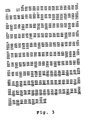
- the nucleic acid sequence information is presented as SEQ ID NO:11 and the predicted protein coding sequence is presented as SEQ ID NO:12. Comparison of the predicted mature protein sequence (SEQ ID NO:12) of this gene to the predicted protein sequence of OvIFN ⁇ is shown in Figure 3.
- HuIFN ⁇ mRNA in human term placenta and amniocytes was analyzed by using reverse transcriptase-PCR (RT-PCR) method (Clontech Laboratories, Palo Alto CA).
- tcRNA Total cellular RNA isolated from human placenta, amniocytes and ovine conceptuses were reverse transcribed using the primer SEQ ID NO:14.
- the primer SEQ ID NO:13 was then added to the reaction and polymerase chain reaction carried out for 40 cycles.
- the PCR products were size fractionated on agarose gels and transferred to filters.
- the DNA on the filters was hybridized with 32 P-labelled OvIFN ⁇ and HuIFN ⁇ cDNAs. The results of these analyses demonstrate the presence of human IFN ⁇ mRNA in the feto-placental annex.
- the aminocytes also expressed the messages corresponding to OvIFN ⁇ primers and human probe.
- an antisense cRNA probe was synthesized by in vitro transcription (Sambrook, et al., 1989) using T 7 RNA polymerase (Stratagene). A trace amount of 3 H-CTP (NEN-DuPont, Cambridge, MA) was used in the transcription reaction. dUTP labeled with digoxigenin (Boehringer-Mannheim, Indianapolis, IN) was incorporated into the cRNA and yield was estimated through TCA precipitation and scintillation counting.
- In situ hybridization was performed using the anti-sense RNA probe, as described by Lawrence, et al . (1985) with the following modifications.
- Deparaffinized and hydrated sections were prehybridized for 10 minutes at room temperature in phosphate buffered saline (PBS) containing 5 mM MgCl 2 .
- Nucleic acids in the sections were denatured for 10 minutes at 65°C in 50% formamide/2 ⁇ SSC.
- Sections were incubated overnight at 37°C with a hybridization cocktail (30 ⁇ l/slide) containing 0.3 ⁇ g/ml digoxigenin-labelled cRNA probe and then washed for 30 minutes each at 37°C in 50 formamide/1 ⁇ SSC. Final washes were performed for 30 minutes each at room temperature in 1 ⁇ SSC and 0.1 ⁇ SSC.
- the sections were blocked for 30 minutes with 0.5% Triton X-100 (Sigma) and 0.5% non-fat dry milk.
- Hybridization signal was detected using purified sheep antidioxigenin Fab fragments conjugated to alkaline phosphatase (Boehringer-Mannheim). After unbound antibody was removed, nitroblue tetrazolium/5-bromo-4-chloro-3-indolyl-phosphate substrate (Promega) and levamisole (Bector Laboratories, Burlingame, CA) were added for signal detection via colorimetric substrate generation. The tissues were counterstained in methyl green (Sigma), dehydrated, and mounted.
- tissue sections were pretreated with 100 ⁇ g/ml of pancreatic RNaseA (Sigma) for 30 minutes at 37°C.
- the RNase was inactivated on the slide with 400 units of RNase inhibitor (Promega).
- the slides were then washed twice in 250 ml of PBS/5 mM MgCl 2 .
- tRNA (Sigma) was substituted for the digoxigenin probes.
- First trimester placental villi composed of an outer layer of syncytiotrophoblast, an underlying layer of cytotrophoblast, and a central stromal region with various types of mesenchymal cells, displayed the highest transcript level of IFN ⁇ in the cytotrophoblast cells. Less intense but detectable levels were present in both the syncytiotrophoblast and stromal cells. A similar pattern of transcript expression was demonstrated in the placental villi of term tissue but the level of signal detection was low. First trimester extravillous trophoblast displayed the highest amount of message and stained positive when present in the maternal blood spaces.
- the antiviral assays were performed essentially as described above in Example 2. Specific activities are expressed in antiviral units/mg protein obtained from antiviral assays using either Madin-Darby bovine kidney (MDBK) cells or sheep normal fibroblasts (Shnf). All samples were assayed simultaneously to eliminate interassay variability.
- MDBK Madin-Darby bovine kidney
- Scnf sheep normal fibroblasts
- IFN ⁇ had a higher specific activity than either rBoIFN ⁇ or rBoIFN ⁇ (Table 3).
- the NIH standard preparation of rHuIFN ⁇ had a similar specific activity, while a commercial preparation of rHuIFN ⁇ exhibited low specific antiviral activity. Comparable relative antiviral activity was demonstrated using either bovine or ovine cells.
- IFN ⁇ appeared to exert no cytotoxic effect on the cells hosting the retrovirus. This was true even when IFN ⁇ was present at 40 ng per ml of culture medium.
- IFN ⁇ was also tested for activity against HIV infection in human cells.
- Human peripheral blood lymphocytes which had been infected with HIV (Crowe, et al .), were treated with varying concentrations of OvIFN ⁇ . Replication of HIV in peripheral blood lymphocytes was monitored by reverse transcriptase activity in culture supernatants over time. Reverse transcriptase activity was measured essentially by the method of Hoffman, et al . The data from these assays are presented in Table 5.
- concentrations of OvIFN ⁇ produced significant antiviral effects.
- a concentration of only 10 ng/ml resulted in over a 50% reduction in RT activity after only six days.
- a concentration of 500 ng/ml resulted in a 90% reduction in RT activity within 10 days.
- IFN ⁇ activity For examination of IFN ⁇ activity, cells were replated at 2.5 ⁇ 10 5 cells/well in HMEM with 10% FBS in 6 well plates. Various dilutions of IFN ⁇ alone or in combination with peptides were added to achieve a final volume of 1 ml. Plates were incubated at 37°C in 5% Co 2 for 12, 15, 18, 24, or 48 hours. Cells were treated with trypsin, collected by low speed centrifugation and washed. The cell pellet was blotted dry and 250 ⁇ l of nuclear staining solution (5 mg propidium iodide, 0.3 ml NP40 and 0.1 gm sodium citrate i 100 ml distilled H 2 O) was added to each tube. The tubes were incubated at room temperature.
- nuclear staining solution 5 mg propidium iodide, 0.3 ml NP40 and 0.1 gm sodium citrate i 100 ml distilled H 2 O
- OvIFN ⁇ inhibited both colony size and number.
- Ovine IFN ⁇ was more effective than human IFN ⁇ on the human cell line; thus, it is very potent in cross-species activity. Its activity was dose-dependent, and inhibition of proliferation could be observed at concentrations as low as 1 unit/ml. Concentrations as high as 50,000 units/ml (units of antiviral activity/ml) stopped proliferation, while cell viability was not impaired.
- OvIFN ⁇ The antiproliferative effects of OvIFN ⁇ were studied for a rat cell line and a bovine cell line. The rate of 3 H-thymidine incorporation was used to assess the rate of cellular proliferation.
- Rat (MtBr7 .c5) or bovine kidney (MDBK) cells were seeded in phenol red-free DME-F12 medium supplemented with 3% dextran-coated charcoal stripped Controlled Process Serum Replacement 2 (CPSR 2, Sigma) and 5% dextran-coated charcoal stripped fetaL bovine serum (FBS). After attaching for approximately 15-18 hours, the cells were washed once with serum-free DME-F12 medium.
- CPSR 2 Controlled Process Serum Replacement 2
- FBS fetaL bovine serum
- the medium was replaced with phenol red-free DME-F12 medium supplemented with 3% stripped CPSR2, 1% stripped FBS ("3/1" medium) or 3/1 medium containing OvIFN ⁇ at various units of antiviral activity as determined in the vesicular stomatitis virus challenge assay for interferons (Example 2).
- OvIFN ⁇ drastically reduced the rate of cellular proliferation (based on thymidine incorporation) for each of the cell lines tested.
- the antiproliferative activity of OvIFN ⁇ on human tumor cell lines was evaluated by measuring the rate of 3 H-thymidine incorporation into cells which have been treated with OvIFN ⁇ .
- Adherent tumor lines were plated at 2.5 X 10 5 cells/well in 1 ml in 6-well plates. They received interferon treatments as just described, but were trypsinized prior to counting.
- Human MCF7 breast adenocarcinoma cells were seeded from logarithmically growing cultures in phenol red-free DME-F12 medium supplemented with 3% dextran-coated charcoal stripped CPSR and 5% dextran-coated FBS. After attaching for approximately 15-18 hours, the cells were washed once with serum-free DME-F12 medium. The medium was replaced with phenol red-free DME-F12 medium supplemented with 3% stripped CPSR2, 1% stripped FBS ("3/1" medium) or 3/1 medium containing OvIFN ⁇ at the indicated number of units of antiviral activity as determined in the vesicular stomatitis virus challenge assay for interferons.
- TCA trichloroacetic acid
- OvIFN ⁇ was able to substantially reduce the rate of 3 H-thymidine incorporation in the human carcinoma cell line. This demonstrates the efficacy of OvIFN ⁇ in inhibiting tumor cell proliferation, in particular, mammary tumor cell proliferation.
- Figure 5 presents data demonstrating that rHuIFN ⁇ is cytotoxic.
- results of one of three replicate experiments are presented as mean % viability ⁇ SD.
- the T cell lymphoma cell line H9 was slightly less sensitive to the antiproliferative effects of the IFNs than the tumor cell lines described above. Results of one of three replicate experiments are presented in Figure 10 as mean % growth reduction ⁇ SD. While rHuIFN ⁇ was not toxic to the H9 cells, it failed to inhibit cell division significantly at any of the concentrations examined. In contrast, OvIFN ⁇ was observed to reduce H9 growth by approximately 60% ( Figure 10). Thus, only OvIFN ⁇ is an effective growth inhibitor of this T cell lymphoma.
- B16-F10 is a syngeneic mouse transplantable tumor selected because of its high incidence of pulmonary metastases (Poste, et al ., 1981). Interferon treatment was initiated 3 days after the introduction of the tumor cells. Each mouse received 100 ⁇ l of either PBS alone, PBS containing 1 X 10 5 units of OvIFN ⁇ , or PBS containing 1 X 10 5 units of recombinant murine IFN ⁇ (MuIFN ⁇ ), i.v. per day for 3 consecutive days.
- mice were sacrificed at 21 days and the lungs were preserved in 10% buffered formalin.
- the frequency of pulmonary metastases were compared between control mice (PBS), OvIFN ⁇ -treated mice, and MulFN ⁇ -treated mice.
- the results of these in vivo administrations demonstrated that OvIFN ⁇ dramatically reduced B16-F10 pulmonary tumors. These results support the use of IFN ⁇ as an efficacious antineoplastic agent in vivo.
- peptides were of approximately the same molecular weight but differed slightly in overall hydrophilicity. Despite this difference, all peptides were antigenic as demonstrated by the production of rabbit antisera with titers greater than 1:3,000 as assessed by ELISA (Harlow, et al .).
- the peptides were used to inhibit the antiviral activity (Example 2) of OvIFN ⁇ and rBoIFN ⁇ .
- the results of this analysis are presented in Figure 12: 1 mM N- and C-terminal peptides both effectively blocked the antiviral activity of OvIFN ⁇ using MDBK cells.
- the peptide OvIFN ⁇ (119-150) showed minimal inhibitory activity.
- the OvIFN ⁇ (34-64) and (90-122) peptides had no apparent inhibitory activity.
- OvIFN ⁇ antiviral activity was also examined as follows. Monolayers of Madin Darby bovine kidney cells were incubated with 40 units/ml OvIFN ⁇ in the presence or absence of various concentrations of OvIFN ⁇ peptides (see Figure 13). Results in Figure 13 are expressed as the percent of control antiviral activity: that is, in the absence of any competing peptide. Data presented are the means of 6 replicate experiments. The data demonstrate that inhibition by OvIFN ⁇ (1-37), (62-92), (119-150), and (139-172) were significantly different than OvIFN ⁇ (34-64) and (90-122) at 10 -3 M and 3 x 10 -3 M.
- OvIFN ⁇ (139-172) was significantly different than all other peptides at 10 -3 M. Significance was assessed by analysis of variance followed by Scheffe's F test at p ⁇ 0.05. Thus, OvIFN ⁇ (1-37) (62-92), (119-150), and (139-172), in particular (139-172), may represent receptor binding regions for IFN ⁇ .
- the ability of the OvIFN ⁇ peptides to inhibit bovine IFN ⁇ (BoIFN ⁇ ) antiviral activity was examined as follows. Monolayers of Madin Darby bovine kidney cells were incubated with 40 units/ml bovine IFN ⁇ in the presence or absence of various concentrations of OvIFN ⁇ peptides. The results are presented in Figure 14 and are expressed as the percent of control antiviral activity in the absence of OvIFN ⁇ peptides. The data presented are the means of 4 replicate experiments. The results indicate that inhibition by OvIFN ⁇ (62-92), (119-150), and (139-172) were significantly different from OvIFN ⁇ (1-37), (34-64) and (90-122) at 10 -3 M.
- OvIFN ⁇ (139-172) was significantly different than OvIFN ⁇ (1-37), (34-64) and (90-122) at 3 x 10 -3 M. Significance was assessed by analysis of variance followed by Scheffe's F test at p ⁇ 0.05. Thus, OvIFN ⁇ (62-92), (119-150), and (139-172), in particular (139-172), may represent common receptor binding regions for IFN ⁇ and bovine IFN ⁇ .
- OvIFN ⁇ (139-172) was significantly different from all other peptides at 10 -3 M. Significance was assessed by analysis of variance followed by Scheffe's F test at p ⁇ 0.05. Thus, OvIFN ⁇ (139-172) may represent a common receptor binding region for IFN ⁇ and various IFN ⁇ (s).
- OvIFN ⁇ peptides described above appear to have no effect on the antiviral activity of IFN ⁇ .
- Peptide inhibition of bovine IFN ⁇ antiviral activity was evaluated as follows. Monolayers of Madin Darby bovine kidney cells were incubated with 40 units/ml bovine IFN gamma in the presence or absence of various concentrations of OvIFN ⁇ peptides. Results are expressed as the percent of control antiviral activity in the absence of OvIFN ⁇ peptides. The data are presented in Figure 16 and are the means of 3 replicate experiments. There were no significant differences among peptides as assessed by analysis of variance followed by Scheffe's F test at p ⁇ 0.05.
- OvIFN ⁇ (1-37) and OvIFN ⁇ (139-172) also blocked OvIFN ⁇ anti-FIV and anti-HIV activity.
- Reverse transcriptase (RT) activity (Examples 12 and 13) was monitored over a 14 day period in FIV-infected FET-1 cells (1 X 10 6 /ml) and HIV-infected HPBL (1 X 10 6 /ml). Control cultures received no OvIFN ⁇ .
- OvIFN ⁇ was used at 100 ng/ml, and peptides were used at 200 ⁇ M.
- Antipeptide antisera inhibition of OvIFN ⁇ antiviral activity was evaluated as follows. Monolayers of MDBK cells were incubated with 20 units/ml of OvIFN ⁇ in the presence a 1:30 dilution of either preimmune sera or antisera to each of the OvIFN ⁇ peptides described above. In Figure 17 the data from duplicate experiments are presented as the mean percent inhibition of OvIFN ⁇ antiviral activity produced by antipeptide antisera relative to the appropriate preimmune sera ⁇ standard error. Significant differences were assessed by analysis of variance followed by Scheffe's F test at p ⁇ 0.05.
- the IFN ⁇ binding assay was carried out as follows. Five ⁇ g of IFN ⁇ was iodinated for 2 minutes with 500 ⁇ Ci of Na 125 I (15 mCi/ ⁇ g; Amersham Corporation, Arlington Heights, IL) in 25 ⁇ l of 0.5 M potassium phosphate buffer, pH 7.4, and 10 ⁇ l of chloramine-T (5 mg/ml) (Griggs, et al., 1992). The specific activity of the iodinated protein was 137 ⁇ Ci/ ⁇ g. For binding assays, monolayers of MDBK cells were fixed with paraformaldehyde and blocked with 5% nonfat dry milk.
- Antisera to IFN ⁇ (119-150), which inhibited binding of OvIFN ⁇ to receptor, also reversed the OvIFN ⁇ antiproliferative effect.
- IFN ⁇ Several other peptides, notably IFN ⁇ (139-172), reversed the OvIFN ⁇ antiproliferative effect, but to a lesser extent.
- the antiviral effects of IFN ⁇ against HIV were evaluated by treating human PBMC cells with various amounts of either recombinant ovine IFN ⁇ (r-OvIFN ⁇ ) or recombinant human IFN ⁇ 2 at the time of infection with HIV. Drug was present throughout the experiment. At day 7 and day 14, p24 production was determined (by ELISA (Wang, et al ., 1988, 1989) and compared to a zero drug control. The results of this analysis are presented in Table 12.
- Human PBMC's were seeded at 5 ⁇ 10 5 cells/ml. Cells were stimulated at day 0 with 3 ⁇ g/ml PHA. Cells were treated with recombinant human IFN ⁇ 2A (at concentrations of 10, 100, 1,000 and 10,000 units/ml) and IFN ⁇ (at concentrations of 2.6, 26, 260, 2,600, 26,000, 260,000, and 2,600,000 units/ml) in 200 ⁇ l/wells (4 replicates of each concentration using 96 well flat bottom plates). Control cultures were given no interferons. After 4 days of incubation, cells were pulsed for 9 hours using 3 H-thymidine at 1 uCi/well. The cells were harvested and the incorporation of labeled thymidine into DNA was determined (Figure 8).
- the cell line used is a human cell that was derived from liver cells transfected with Hepatitis B Virus (HBV).
- HBV Hepatitis B Virus
- the cell line semi-stably produces HBV virus: over time the cell line's production of HBV intracellular DNA and secreted virus decreases.
- the cells are pre-treated with deAZA-C (5-azacytidine; Miyoshi, et al.) to induce production of the virus. Treatment was for 2-3 days and the amount of induction was about a factor of two.
- the cells were then treated with either the IFN ⁇ and IFN ⁇ at levels of 0, 5,000, 10,000, 20,000 and 40,000 units per ml.
- the hepatocyte cell line HepG2-T14 (described above) was examined for the effects of IFN ⁇ and IFN ⁇ on hepatospecific mRNA production.
- Cells were incubated in concentrations of IFN ⁇ or IFN ⁇ at 0, 5,000, 10,000, 20,000, and 40,000 units per ml.
- the messenger RNAs for the hepatocyte specific proteins Apo E and Apo A1 were detected by hybridization analysis (Sambrook, et al.; Maniatis, et al.) using probes specific for these two mRNA's (Shoulders, et al., and Wallis, et al .).
- Sepharose 4B beads conjugated with anti-beta galactosidase is purchased from Promega. The beads are packed in 2 ml column and washed successively with phosphate-buffered saline with 0.02% sodium azide and 10 ml TX buffer (10 mM Tris buffer, pH 7.4, 1% aprotinin).
- the IFN ⁇ coding sequence (e.g ., Figure 7) is cloned into the polylinker site of lambda gt11.
- the IFN ⁇ coding sequence is placed in-frame with the amino terminal ⁇ -galactosidase coding sequences in lambda gt11.
- Lysogens infected with gt11/IFN ⁇ are used to inoculate 500 ml of NZYDT broth.
- the culture is incubated at 32°C with aeration to an O.D. of about 0.2 to 0.4, then brought to 43°C quickly in a 43°C water bath for 15 minutes to induce gtl1 peptide synthesis, and incubated further at 37°C for 1 hour.
- the cells are pelleted by centrifugation, suspended in 10 ml of lysis buffer (10 mM Tris, pH 7.4 containing 2% "TRITON X-100" and 1% aprotinin added just before use.
- the resuspended cells are frozen in liquid nitrogen then thawed, resulting in substantially complete cell lysis.
- the lysate is treated with DNaseI to digest bacterial and phage DNA, as evidenced by a gradual loss of viscosity in the lysate.
- Non-solubilized material is removed by centrifugation.
- the clarified lysate material is loaded on the Sepharose column, the ends of the column closed, and the column placed on a rotary shaker for 2 hrs. at room temperature and 16 hours at 4°C. After the column settles, it is washed with 10 ml of TX buffer.
- the fused protein is eluted with 0.1 M carbonate/bicarbonate buffer, pH10. Typically, 14 ml of the elution buffer is passed through the column, and the fusion protein is eluted in the first 4-6 ml of eluate.
- the eluate containing the fusion protein is concentrated in "CENTRICON-30" cartridges (Amicon, Danvers, Mass.).
- the final protein concentrate is resuspended in, for example, 400 ⁇ l PBS buffer. Protein purity is analyzed by SDS-PAGE.
- the purified fused protein is injected subcutaneously in Freund's adjuvant in a rabbit. Approximately 1 mg of fused protein is injected at days 0 and 21, and rabbit serum is typically collected at 6 and 8 weeks.
- the IFN ⁇ coding sequence (e.g ., Figure 7) is cloned into the pGEX vector (Boyer, et al .; Frangioni, et al .; Guan, et al .; Hakes, et al .; Smith, et al., 1988).
- the pGEX vector (Smith, et al .) was modified by insertion of a thrombin cleavage sequence in-frame with the glutathione-S-transferase protein (GST -- sj26 coding sequence). This vector is designated pGEXthr.
- the IFN ⁇ coding sequence is placed in-frame with the sj26-thrombin coding sequences (Guan, et al .; Hakes, et al .).
- the IFN ⁇ coding sequence insert can be generated by the polymerase chain reaction using PCR primers specific for the insert.
- the IFN ⁇ fragment is ligated to the linearized pGEXthr vector.
- the ligation mixture is transformed into E. coli and ampicillin resistant colonies are selected. Plasmids are isolated from the ampicillin resistant colonies and analyzed by restriction enzyme digestion to identify clones containing the IFN ⁇ insert (vector designated pGEXthr-IFN ⁇ ).
- E. coli strain XL-I Blue is transformed with pGEXthr-IFN ⁇ and is grown at 37°C overnight. DNA is prepared from randomly-picked colonies. The presence of the insert coding sequence is typically confirmed by (i) restriction digest mapping, (ii) hybridization screening using labelled IFN ⁇ probes ( i.e ., Southern analysis), or (iii) direct DNA sequence analysis.
- a pGEXthr-IFN ⁇ clone is grown overnight.
- the overnight culture is diluted 1:10 with LB medium containing ampicillin and grown for one hour at 37°C.
- the overnight culture is diluted 1:100 and grown to OD of 0.5-1.0 before addition of IPTG (isopropylthio- ⁇ -galactoside).
- IPTG isopropylthio- ⁇ -galactoside.
- IPTG GENE-BRL, Gaithersburg MD
- IPTG is added to a final concentration of 0.2-0.5 mM for the induction of protein expression and the incubation is typically continued for 2-5 hours, preferably 3.5 hours.
- Bacterial cells are harvested by centrifugation and resuspended in 1/100 culture volume of MTPBS (150 mM NaCl, 16 mM Na 2 HPO 4 , 4 mM NaH 2 PO 4 ). Cells are lysed by lysozyme, sonication or French press, and lysates cleared of cellular debris by centrifugation.
- MTPBS 150 mM NaCl, 16 mM Na 2 HPO 4 , 4 mM NaH 2 PO 4 .
- the extracts can be concentrated by ultrafiltration using, for example, a "CENTRICON 10" filter.
- the fusion proteins are partially purified over a glutathione agarose affinity column as described in detail by Smith, et al .
- 100 ml cultures are grown overnight.
- the cultures are diluted to 1 liter, and the cells grown another hour at 37°C.
- Expression of the fusion proteins is induced using IPTG.
- the induced cultures are grown at 37°C for 3.5 hours.
- Cells are harvested and a sonicator used to lyse the cells.
- Cellular debris is pelleted and the clear lysate loaded onto a glutathione "SEPHAROSE" column.
- the column is washed with several column volumes.
- the fusion protein is eluted from the affinity column with reduced glutathione and dialyzed.
- the IFN ⁇ can be liberated from the hybrid protein by treatment with thrombin.
- the sj26 and IFN ⁇ fragments of the hybrid protein can then be separated by size fractionation over columns or on gels.
- the IFN ⁇ portion of the hybrid protein is released from the column by treatment with thrombin (Guan, et al .; Hakes, et al .).
- the purified Sj26/IFN ⁇ fused protein is injected subcutaneously in Freund's adjuvant in a rabbit. Approximately 1 mg of fused protein is injected at days 0 and 21, and rabbit serum is typically collected at 6 and 8 weeks. A second rabbit is similarly immunized with purified Sj26 protein obtained from control bacterial lysate.
- Minilysates from the following bacterial cultures are prepared: (1) KM392 cells infected with pGEXthr and pGEXthr containing the IFN ⁇ insert; and (2) cells infected with lambda gtl1 containing the IFN ⁇ insert.
- the minilysates and a commercial source ⁇ -galactosidase are fractionated by SDS-PAGE, and the bands transferred to nitrocellulose filters for Western blotting (Sambrook, et al .; Ausubel, et al .).
- serum from control (Sj26) rabbits is immunoreactive with each of the Sj26 and Sj26 fused protein antigens.
- Serum from the animal immunized with Sj26/IFN ⁇ fused protein is reactive with all Sj-26 and beta-gal fusion proteins containing IFN ⁇ coding sequences, indicating the presence of specific immunoreaction with the IFN ⁇ antigen. None of the sera are expected to be immunoreactive with beta-galactosidase.
- Anti-IFN ⁇ antibody present in the sera from the animal immunized with the Sj26/IFN ⁇ is purified by affinity chromatography (using immobilized recombinantly produced IFN ⁇ as ligand, essentially as described above in Example 12 for the anti-beta-galactosidase antibody).
Landscapes
- Health & Medical Sciences (AREA)
- Life Sciences & Earth Sciences (AREA)
- Chemical & Material Sciences (AREA)
- Organic Chemistry (AREA)
- Genetics & Genomics (AREA)
- Molecular Biology (AREA)
- Engineering & Computer Science (AREA)
- Biochemistry (AREA)
- General Health & Medical Sciences (AREA)
- Biomedical Technology (AREA)
- Biophysics (AREA)
- Medicinal Chemistry (AREA)
- Proteomics, Peptides & Aminoacids (AREA)
- Zoology (AREA)
- Biotechnology (AREA)
- Immunology (AREA)
- Wood Science & Technology (AREA)
- Bioinformatics & Cheminformatics (AREA)
- Hematology (AREA)
- Gastroenterology & Hepatology (AREA)
- Microbiology (AREA)
- Urology & Nephrology (AREA)
- General Engineering & Computer Science (AREA)
- Physics & Mathematics (AREA)
- Toxicology (AREA)
- Pathology (AREA)
- Cell Biology (AREA)
- Food Science & Technology (AREA)
- Plant Pathology (AREA)
- Analytical Chemistry (AREA)
- General Physics & Mathematics (AREA)
- Medicines That Contain Protein Lipid Enzymes And Other Medicines (AREA)
- Peptides Or Proteins (AREA)
- Preparation Of Compounds By Using Micro-Organisms (AREA)
- Micro-Organisms Or Cultivation Processes Thereof (AREA)
Applications Claiming Priority (3)
| Application Number | Priority Date | Filing Date | Title |
|---|---|---|---|
| US96989092A | 1992-10-30 | 1992-10-30 | |
| US969890 | 1992-10-30 | ||
| EP93924958A EP0669981A1 (fr) | 1992-10-30 | 1993-10-19 | Compositions d'interferon tau et leurs procedes d'utilisation |
Related Parent Applications (1)
| Application Number | Title | Priority Date | Filing Date |
|---|---|---|---|
| EP93924958A Division EP0669981A1 (fr) | 1992-10-30 | 1993-10-19 | Compositions d'interferon tau et leurs procedes d'utilisation |
Publications (2)
| Publication Number | Publication Date |
|---|---|
| EP1360962A2 true EP1360962A2 (fr) | 2003-11-12 |
| EP1360962A3 EP1360962A3 (fr) | 2004-02-04 |
Family
ID=25516125
Family Applications (2)
| Application Number | Title | Priority Date | Filing Date |
|---|---|---|---|
| EP03006878A Withdrawn EP1360962A3 (fr) | 1992-10-30 | 1993-10-19 | Composition contenant l'Interferon-tau et Méthode pour son utilisation |
| EP93924958A Withdrawn EP0669981A1 (fr) | 1992-10-30 | 1993-10-19 | Compositions d'interferon tau et leurs procedes d'utilisation |
Family Applications After (1)
| Application Number | Title | Priority Date | Filing Date |
|---|---|---|---|
| EP93924958A Withdrawn EP0669981A1 (fr) | 1992-10-30 | 1993-10-19 | Compositions d'interferon tau et leurs procedes d'utilisation |
Country Status (8)
| Country | Link |
|---|---|
| EP (2) | EP1360962A3 (fr) |
| JP (1) | JPH08505047A (fr) |
| KR (1) | KR100357768B1 (fr) |
| CN (1) | CN1090510A (fr) |
| AU (1) | AU689450B2 (fr) |
| CA (1) | CA2148119A1 (fr) |
| TW (2) | TW391983B (fr) |
| WO (1) | WO1994010313A2 (fr) |
Cited By (2)
| Publication number | Priority date | Publication date | Assignee | Title |
|---|---|---|---|---|
| WO2007018846A3 (fr) * | 2005-07-27 | 2007-06-21 | Pepgen Coporation | Procedes permettant de reduire la formation de tissus cicatriciels |
| WO2008021487A1 (fr) * | 2006-08-18 | 2008-02-21 | Pepgen Corporation | Méthode de traitement combiné utilisant l'interféron-tau |
Families Citing this family (22)
| Publication number | Priority date | Publication date | Assignee | Title |
|---|---|---|---|---|
| US5906816A (en) * | 1995-03-16 | 1999-05-25 | University Of Florida | Method for treatment of autoimmune diseases |
| US6372206B1 (en) | 1989-03-02 | 2002-04-16 | University Of Florida | Orally-administered interferon-TAU compositions and methods |
| US5705363A (en) | 1989-03-02 | 1998-01-06 | The Women's Research Institute | Recombinant production of human interferon τ polypeptides and nucleic acids |
| US5648241A (en) * | 1989-09-15 | 1997-07-15 | The General Hospital Corporation | Conjugate vaccine against group B streptococcus |
| US5939286A (en) * | 1995-05-10 | 1999-08-17 | University Of Florida | Hybrid interferon tau/alpha polypeptides, their recombinant production, and methods using them |
| KR100464531B1 (ko) * | 1996-04-12 | 2005-03-07 | 유니버시티 오브 플로리다 | 잡종인터페론조성물및그것을사용하는방법 |
| US6204022B1 (en) * | 1996-04-12 | 2001-03-20 | Pepgen Corporation And University Of Florida | Low-toxicity human interferon-alpha analogs |
| FI104363B (fi) * | 1997-05-19 | 2000-01-14 | Timo Kalevi Korpela | Immunosuppressanttien ja interferonien farmakologisten keskinäisten vaikutusten parantaminen lisäaineilla |
| RU2150291C1 (ru) * | 1999-05-17 | 2000-06-10 | Гапонюк Петр Яковлевич | Противовирусное средство на основе рекомбинантного интерферона в виде мази, геля, суппозитория, крема, линимента |
| CA2382425A1 (fr) * | 1999-06-22 | 2000-12-28 | Carol H. Pontzer | Mutants d'interferon tau et leurs techniques de preparation |
| US6833256B1 (en) | 1999-06-22 | 2004-12-21 | University Of Maryland | Interferon tau mutants and methods for making them |
| AU781377B2 (en) * | 1999-08-27 | 2005-05-19 | University Of Florida Research Foundation, Inc. | Materials and methods for inhibition of IgE production |
| US7105154B2 (en) * | 2000-07-19 | 2006-09-12 | Pepgen Corporation | Method of treatment using interferon-tau |
| JP2006213597A (ja) | 2000-07-19 | 2006-08-17 | Pepgen Corp | インターフェロン−タウを用いるc型肝炎ウイルスの処置のための組成物およびモニタリングの方法 |
| AU2001277055A1 (en) * | 2000-07-19 | 2002-01-30 | Pepgen Corporation | Composition for treatment of and method of monitoring hepatitis c virus using interferon-tau |
| US7083782B2 (en) | 2000-07-19 | 2006-08-01 | Pepgen Corporation | Method of treatment using interferon-tau |
| US7431920B2 (en) | 2000-07-19 | 2008-10-07 | Pepgen Corporation | Method of treating IL-10 deficiency |
| KR20040022244A (ko) | 2001-08-12 | 2004-03-11 | 펩젠 코포레이션 | 하이브리드 인터페론/인터페론 타우 단백질, 조성물 및사용방법 |
| TW200518768A (en) * | 2003-11-17 | 2005-06-16 | Pepgen Corp | Methods for treatment of obesity and for promotion of weight loss |
| EP1946766B1 (fr) | 2005-09-27 | 2010-08-25 | Kabushiki Kaisha Hayashibara Seibutsu Kagaku Kenkyujo | Remede contre un trouble de l'appareil reproducteur |
| CN101244261B (zh) * | 2008-03-10 | 2010-09-15 | 山东大学 | 一种含未复性重组蛋白的生物制剂及其制备方法与应用 |
| CN113969285B (zh) * | 2021-11-09 | 2023-05-23 | 上海市农业科学院 | 重组表达的羊干扰素-τBB8基因及其制备方法 |
Family Cites Families (3)
| Publication number | Priority date | Publication date | Assignee | Title |
|---|---|---|---|---|
| WO1989008706A1 (fr) * | 1988-03-18 | 1989-09-21 | Institut National De La Recherche Agronomique (Inr | Isoformes de la trophoblastine, nouveaux interferons constitues par lesdites isoformes, leurs procedes d'obtention et leurs applications |
| EP0367063A1 (fr) * | 1988-10-26 | 1990-05-09 | The Curators Of The University Of Missouri | Isolation et clonage d'ADNc pour le gène codant la protéine traphoblastique-1 bovine |
| WO1990009806A2 (fr) * | 1989-03-02 | 1990-09-07 | University Of Florida | Composition d'inhibition de tumeurs et d'inhibition non cytotoxique de la replication de virus |
-
1993
- 1993-10-18 TW TW082108707A patent/TW391983B/zh not_active IP Right Cessation
- 1993-10-18 TW TW089102648A patent/TW585911B/zh not_active IP Right Cessation
- 1993-10-18 CN CN93119213A patent/CN1090510A/zh active Pending
- 1993-10-19 WO PCT/US1993/010016 patent/WO1994010313A2/fr not_active Ceased
- 1993-10-19 CA CA002148119A patent/CA2148119A1/fr not_active Abandoned
- 1993-10-19 JP JP6511137A patent/JPH08505047A/ja active Pending
- 1993-10-19 KR KR1019950701721A patent/KR100357768B1/ko not_active Expired - Fee Related
- 1993-10-19 AU AU54449/94A patent/AU689450B2/en not_active Ceased
- 1993-10-19 EP EP03006878A patent/EP1360962A3/fr not_active Withdrawn
- 1993-10-19 EP EP93924958A patent/EP0669981A1/fr not_active Withdrawn
Cited By (2)
| Publication number | Priority date | Publication date | Assignee | Title |
|---|---|---|---|---|
| WO2007018846A3 (fr) * | 2005-07-27 | 2007-06-21 | Pepgen Coporation | Procedes permettant de reduire la formation de tissus cicatriciels |
| WO2008021487A1 (fr) * | 2006-08-18 | 2008-02-21 | Pepgen Corporation | Méthode de traitement combiné utilisant l'interféron-tau |
Also Published As
| Publication number | Publication date |
|---|---|
| CA2148119A1 (fr) | 1994-05-11 |
| EP0669981A1 (fr) | 1995-09-06 |
| KR100357768B1 (ko) | 2003-04-07 |
| JPH08505047A (ja) | 1996-06-04 |
| AU5444994A (en) | 1994-05-24 |
| WO1994010313A3 (fr) | 1994-10-27 |
| KR950704489A (ko) | 1995-11-20 |
| CN1090510A (zh) | 1994-08-10 |
| WO1994010313A2 (fr) | 1994-05-11 |
| AU689450B2 (en) | 1998-04-02 |
| EP1360962A3 (fr) | 2004-02-04 |
| TW585911B (en) | 2004-05-01 |
| TW391983B (en) | 2000-06-01 |
Similar Documents
| Publication | Publication Date | Title |
|---|---|---|
| US6174996B1 (en) | Hybrid interferon τ/type I interferon polypeptides | |
| EP0914435B1 (fr) | Compositions d'interferon hybrides et leurs procedes d'utilisation | |
| EP1360962A2 (fr) | Composition contenant l'Interferon-tau et Méthode pour son utilisation | |
| US6204022B1 (en) | Low-toxicity human interferon-alpha analogs | |
| KR100223255B1 (ko) | 개량된 알파 인터페론 조성물 및 인간 말초혈 백혈구로 부터의 제조 방법 | |
| JP3507507B2 (ja) | 非免疫原性ペプチドを介して結合されたインターフェロン―αと免疫グロブリンとのハイブリッド | |
| FI100972B (fi) | Pesäkkeitä stimuloivaa tekijä-1:tä (CSF-1) koodaava DNA-sekvenssi ja m enetelmä CSF-1-proteiinin valmistamiseksi | |
| WO1997033607A1 (fr) | Compositions d'interferon-tau administrees oralement et procedes associes | |
| WO1997033607A9 (fr) | Compositions d'interferon-tau administrees oralement et procedes associes | |
| Pontzer et al. | Structure/function studies with interferon tau: evidence for multiple active sites | |
| JP3784078B2 (ja) | 可溶性ldlリセプター | |
| WO1990009806A2 (fr) | Composition d'inhibition de tumeurs et d'inhibition non cytotoxique de la replication de virus | |
| WO2004022088A1 (fr) | Hybride d'interféron et de fragment fc d'immunoglobuline | |
| AU706762B2 (en) | Interferon tau compositions and methods of use | |
| HK1059034A (en) | Interferon tau composition and methods of use | |
| KR100464531B1 (ko) | 잡종인터페론조성물및그것을사용하는방법 | |
| WO2000006596A2 (fr) | HYBRIDES D'INTERFERON- alpha | |
| US7235232B2 (en) | Interferon alpha hybrids | |
| CA2495480A1 (fr) | Hybride d'interferon et de fragment fc d'immunoglobuline |
Legal Events
| Date | Code | Title | Description |
|---|---|---|---|
| PUAI | Public reference made under article 153(3) epc to a published international application that has entered the european phase |
Free format text: ORIGINAL CODE: 0009012 |
|
| AC | Divisional application: reference to earlier application |
Ref document number: 0669981 Country of ref document: EP Kind code of ref document: P |
|
| AK | Designated contracting states |
Kind code of ref document: A2 Designated state(s): AT BE CH DE DK ES FR GB GR IE IT LI LU NL PT SE |
|
| PUAL | Search report despatched |
Free format text: ORIGINAL CODE: 0009013 |
|
| AK | Designated contracting states |
Kind code of ref document: A3 Designated state(s): AT BE CH DE DK ES FR GB GR IE IT LI LU NL PT SE |
|
| REG | Reference to a national code |
Ref country code: HK Ref legal event code: DE Ref document number: 1059034 Country of ref document: HK |
|
| 17P | Request for examination filed |
Effective date: 20040726 |
|
| AKX | Designation fees paid |
Designated state(s): AT BE CH DE DK ES FR GB GR IE IT LI LU NL PT SE |
|
| STAA | Information on the status of an ep patent application or granted ep patent |
Free format text: STATUS: THE APPLICATION HAS BEEN WITHDRAWN |
|
| 18W | Application withdrawn |
Effective date: 20051019 |
|
| REG | Reference to a national code |
Ref country code: HK Ref legal event code: WD Ref document number: 1059034 Country of ref document: HK |