EP2277913A2 - Domaines se liant à l'antigène issus de poisson - Google Patents
Domaines se liant à l'antigène issus de poisson Download PDFInfo
- Publication number
- EP2277913A2 EP2277913A2 EP10185409A EP10185409A EP2277913A2 EP 2277913 A2 EP2277913 A2 EP 2277913A2 EP 10185409 A EP10185409 A EP 10185409A EP 10185409 A EP10185409 A EP 10185409A EP 2277913 A2 EP2277913 A2 EP 2277913A2
- Authority
- EP
- European Patent Office
- Prior art keywords
- antigen binding
- binding domain
- process according
- specific antigen
- antigen specific
- Prior art date
- Legal status (The legal status is an assumption and is not a legal conclusion. Google has not performed a legal analysis and makes no representation as to the accuracy of the status listed.)
- Withdrawn
Links
Images







Classifications
-
- C—CHEMISTRY; METALLURGY
- C07—ORGANIC CHEMISTRY
- C07K—PEPTIDES
- C07K16/00—Immunoglobulins [IG], e.g. monoclonal or polyclonal antibodies
- C07K16/005—Immunoglobulins [IG], e.g. monoclonal or polyclonal antibodies constructed by phage libraries
-
- A—HUMAN NECESSITIES
- A61—MEDICAL OR VETERINARY SCIENCE; HYGIENE
- A61P—SPECIFIC THERAPEUTIC ACTIVITY OF CHEMICAL COMPOUNDS OR MEDICINAL PREPARATIONS
- A61P37/00—Drugs for immunological or allergic disorders
- A61P37/02—Immunomodulators
-
- A—HUMAN NECESSITIES
- A61—MEDICAL OR VETERINARY SCIENCE; HYGIENE
- A61P—SPECIFIC THERAPEUTIC ACTIVITY OF CHEMICAL COMPOUNDS OR MEDICINAL PREPARATIONS
- A61P43/00—Drugs for specific purposes, not provided for in groups A61P1/00-A61P41/00
-
- C—CHEMISTRY; METALLURGY
- C07—ORGANIC CHEMISTRY
- C07K—PEPTIDES
- C07K16/00—Immunoglobulins [IG], e.g. monoclonal or polyclonal antibodies
- C07K16/40—Immunoglobulins [IG], e.g. monoclonal or polyclonal antibodies against enzymes
-
- C—CHEMISTRY; METALLURGY
- C07—ORGANIC CHEMISTRY
- C07K—PEPTIDES
- C07K2317/00—Immunoglobulins specific features
- C07K2317/20—Immunoglobulins specific features characterized by taxonomic origin
-
- C—CHEMISTRY; METALLURGY
- C07—ORGANIC CHEMISTRY
- C07K—PEPTIDES
- C07K2317/00—Immunoglobulins specific features
- C07K2317/50—Immunoglobulins specific features characterized by immunoglobulin fragments
- C07K2317/56—Immunoglobulins specific features characterized by immunoglobulin fragments variable (Fv) region, i.e. VH and/or VL
-
- C—CHEMISTRY; METALLURGY
- C07—ORGANIC CHEMISTRY
- C07K—PEPTIDES
- C07K2317/00—Immunoglobulins specific features
- C07K2317/50—Immunoglobulins specific features characterized by immunoglobulin fragments
- C07K2317/56—Immunoglobulins specific features characterized by immunoglobulin fragments variable (Fv) region, i.e. VH and/or VL
- C07K2317/565—Complementarity determining region [CDR]
-
- C—CHEMISTRY; METALLURGY
- C07—ORGANIC CHEMISTRY
- C07K—PEPTIDES
- C07K2317/00—Immunoglobulins specific features
- C07K2317/50—Immunoglobulins specific features characterized by immunoglobulin fragments
- C07K2317/56—Immunoglobulins specific features characterized by immunoglobulin fragments variable (Fv) region, i.e. VH and/or VL
- C07K2317/569—Single domain, e.g. dAb, sdAb, VHH, VNAR or nanobody®
Definitions
- the present invention relates to the production of antigen specific antigen binding domains (single domain antibodies) from fish, where the term fish encompasses both cartilaginous (subclass Elasmobranchii) and bony fish(class Osteichthyes).
- antigen specific antigen binding domains we mean the variable region of a Novel Antigen Receptor (NAR).
- Antibodies are useful in, among other things, molecular diagnostics and therapeutics because of their high affinity binding and specificity.
- monoclonal antibodies are produced using animal models the production of human monoclonal antibodies remains difficult.
- the body mounts an immune response because the monoclonal antibody is foreign to the human system.
- Single domain antibodies are also important as they can penetrate tissues taking with them any linked compounds. In addition, they can bind within cavities on the surface of proteins, for example within enzyme binding sites thus disrupting function.
- Single domain antibodies produced from Camelidae have been shown to recognize protein cavities and as such have the ability to inhibit enzymes ( Lauwereys et al., EMBO 17 pp3512-3520 1998 ).
- WO94/25591 and European patent application number 99200439.0 relate to the production of single domain antibodies from Camelidae heavy chain antibodies.
- Single domain antibodies produced from Camelidae heavy chain antibodies are more stable than mouse single domain antibodies and can be produced in larger quantities.
- the smaller members of the Camelidae family, for example llama are to be kept in humane conditions they require significant areas of land to live upon.
- An object of the present invention is to provide a process for the production of antigen specific antigen binding domains which seeks to alleviate the above problems.
- a further object of the present invention is to provide a composition comprising antigen specific antigen binding domains for the inhibition of protein activity which seeks to alleviate the above problems.
- an antigen specific antigen binding domain using a transformed host containing an expressible DNA sequence encoding the antigen specific antigen binding domain, wherein the antigen specific antigen binding domain is derived from a variable region of NAR found in fish.
- antigen specific antigen binding domains produced from the variable region of NAR found in fish are as stable as single domain antibodies produced from members of the Camelidae family.
- the immunoglobulin isotype now known as NAR Novel Antigen Receptor
- NAR Novel Antigen Receptor
- teleost fish which are much more advanced evolutionary than sharks, appear to rely solely upon the production of a low affinity, non-specific IgM type response ( Watts et al., Aust Vet J 79 pp570-574 2001 ).
- a defining characteristic of teleost IgM is their low affinity and ability to non-specifically bind multiple antigens.
- IgM neutralisation is through non-specific multiple binding, resulting mainly in agglutination, etc..
- Neutralisation without complement is usually associated with specific, high affinity binding and had not until this invention been seen in fish species.
- the antigen specific antigen binding domains of the present invention have been shown to neutralise activity of an enzyme immunogen directly without calling upon other components of the immune system.
- the NAR variable (V) region conforms to the model of typical Ig superfamily domains with the predicted canonical, intradomain disulphide bond. However, whilst camelid VHH regions have up to 75% sequence identity with other mammalian VH regions, the identity between NAR V and conventional VH domains is as low as 25% ( Roux et al., Proceedings of the National Academy of Sciences. USA 95 ppll804-11809 1998 ).
- NAR V region sequences Due to this low identity and lack of NAR sequences in the Kabat database, the amino acids of NAR V regions have previously been numbered sequentially ( Roux et al., Proceedings of the National Academy of Sciences. USA 95 ppll804-11809 1998 ). To enable easy comparison of residues in different NAR V molecules, or NAR V region sequences with those of other species during this work, a numbering system was derived for NAR V region based upon that of Kabat et al., (1991) (Sequences of Proteins of immunological Interest, 5th Edition. National Institutes of Health, Betheseda, USA ). (Note: this numbering system is used in the Figures attached hereto).
- NAR V domains were classified into two closely related classes (type I or II), both being constructed from one V, three D and one J segment.
- Type I regions have non-canonical cys residues in Fr2 (C35) and Fr4 (C103), which likely form a domain-stabilising disulphide bond.
- C35 Fr2
- Fr4 C103
- additional cysteine residue pairs have been observed and almost certainly form disulphide bridges within the CDR, as is found in some cattle VH domains with an unusually long CDR3.
- Type II regions are very similar in overall structure to type I but instead have non-canonical cysteine residues located in Fr1 (C29) and CDR3, which are proposed to form a constraining disulphide bond like that observed in camelid VHH domains.
- the presence of cysteines within each NAR type is shown in schematic form in Figure 1 .
- NAR type has been identified as the predominant expressed form in nurse shark pups (Type III) but due to its germline joined state displays no junctional diversity.
- type I and II NAR the DNA encoding the V region is generated by the physical joining of DNA segments which are spatially separate in the genome. This joining process occurs in B-cells and helps generate the diversity of sequence seen for these NAR types. For type III these DNA segments are already physically joined in the DNA of all cells, hence the term germline joined.
- NAR possesses the cluster type genomic organisation usually observed for Ig receptors in cartilaginous fish, but less than five NAR loci are thought to exist, with only two or three being capable of functional rearrangement and expression.
- the diversity observed in the primary repertoire is generated through recombination mechanisms and, although extensive (due to the presence of three D segments), is localised to CDR3. On encounter with antigen this repertoire is rapidly expanded by extensive mutation.
- the pattern of mutation in NAR is unlike that observed in shark IgM, which shows low levels of mutation and poor clustering to CDRs, but rather bears the hallmarks of mammalian-like somatic mutation.
- NAR V is similar to VH, VL and TCR V but distinct from all three, hence its "unique domain architecture".
- the VH name has been used in the past because the constant portion of NAR is a heavy chain but the V region is actually more like VL/TCR V than VH (i.e. groups closer on a phylogenetic tree).
- NAR V is not like the camel VHH domains which are derived from bona fide heavy chain V regions.
- the antigen binding domain of the NAR is closer to a VL domain naturally lacking VH rather than the other way round.
- NAR V and camel VHH clearly shows the huge difference in sequence. If NAR V and camel VHH have the same physical structure (which has been implied but not proven) they achieve this using completely different amino acid sequences, and one would not be able to amplify a NAR V region library using camel VHH library primers. In addition, the ways in which the NAR V and camel VHH gene repertoires are created during VDJ joining are different due to the organisation of the immunoglobulin genes ( Schluter et al Immunol Today 18 pp543-549 1997 ).
- the transformed host is a prokaryote or a lower eukaryote.
- the prokaryote host is Escherichia coli.
- the expressible DNA sequence is in the form of a phagemid vector.
- Phagemid expression has advantages over phage genome expression in that it results in greater genetic stability and the bacterial transformation efficiency is higher thus enabling the construction of potentially larger and more diverse libraries.
- the gene encoding the variable region of the antibody can be fused to that of a phage surface protein, usually gene III or VIII.
- Gene III fusion is favoured due to its limited copy number (3-5 copies) on the tip of each phage, minimising possible avidity effects which are undesirable when trying to isolate binders of high affinity.
- the antibody fragment genes can be cloned directly into the phage genome or fused to gene segments present within phagemid plasmids.
- the fish is a member of the Elasmobranchii subclass, for example, a shark or a dogfish.
- a greater number of smaller members of the Elasmobranchii subclass can be kept in tanks which are smaller in unit area than the grazing area required for the same number of members of the Camelidae family. As the members of the Elasmobranchii subclass are kept in tanks they can easily be caught for extraction of their blood.
- the shark is a nurse shark, Ginglymostoma cirratum.
- the antigen specific antigen binding domain has a specific specificity. Accordingly, the antigen specific antigen binding domain can be targeted to a specific antigen(s).
- the antigen specific antigen binding domain is monoclonal.
- the antigen specific antigen binding domain is raised to a single antigen.
- the specificity of the antigen specific antigen binding domain is determined by an antigen which is introduced into the chosen fish.
- Screening of displayed libraries for specific binding sites involves repeated cycles of selection with the desired antigen in the process of biopanning.
- the library of phage displayed antigen binding domains is incubated with immobilised antigen, unbound phage are washed out and bound phage eluted.
- This selected population is expanded by bacterial infection and put through further rounds of selection.
- Such cyclic panning has thus proven able to enrich for clones of high affinity, much like in vivo antibody selection.
- step d) the cDNA of the antigen binding domains is generated.
- restriction enzymes are used to digest the amplified DNA sequences encoding the antigen specific antigen binding domain.
- the restrictions enzymes can be chosen depending upon, for example, the handle of the primers used in the above process.
- restriction enzymes are NcoI and NotI.
- the display vector is any phagemid vector, for example, pHEN2.
- the expression vector is a soluble expression vector such as pIMS100.
- vectors are merely examples of the vectors which can be used. It is common general knowledge to those skilled in the art which vectors can be used.
- an antigen specific antigen binding domain produced by the process as defined above.
- composition for the inhibition of protein activity comprising antigen specific antigen binding domains derived from a variable region of the immunoglobulin isotype NAR found in fish.
- NAR V region is 12 kDa which is 20% smaller than any 15 kDa single domain antibody derived from Camelidae, it was still possible to alter protein activity therewith. Size is a significant factor in the therapeutic applications of antigen specific antigen binding domains and other single domain antibodies, with therapeutic benefits of increased tissue penetration, better access to protein clefts for neutralisation via steric hindrance and reduced immunogenicity, resulting from the use of antigen specific antigen binding domains of the present invention.
- Antigen specific antigen binding domains derived from NAR therefore have a wider target population than single domain antibodies derived from Camelidae by virtue of their smaller size.
- the potential for immunogenicity is also reduced since in general the smaller the size of a protein the less the immunogenicity.
- NAR sequences have, in work previous to that of the inventors, been identified at the DNA level, there has been no clue from the DNA evidence that a somatically maturable repertoire, capable of selecting high affinity, specific binders could be a characteristic of the NAR response.
- a somatically maturable repertoire capable of selecting high affinity, specific binders could be a characteristic of the NAR response.
- Sequencing of these genes confirms that an atypical (for fish and organisms of this evolutionary lineage) somatically-maturable (showing mutation from the germ line repertoire) response occurs within the NAR repertoire, driven by the immunisation process. This has resulted in the selection of highly specific, high affinity antigen binding domains capable of antigen neutralisation in isolation and not the expected non-specific, low affinity IgM like response typically found in fish and sharks.
- the inventors have been able to isolate NAR antigen specific antigen binding domains and demonstrate for the first time that the NAR V is able to fold and function in isolation from the rest of the molecule (and in a non-shark environment), that the antigen specific antigen binding domain matures from the germ line genes to become specific for antigen (only possible with a library derived from mRNA and not DNA) and that the antigen specific antigen binding domain is able to bind specifically to the immunising antigen.
- the inventors have been able to immunize a shark and derive from this immunization a specific, somatically matured antigen specific antigen binding domain that is of high affinity and specific for the immunogen.
- the antigen specific antigen binding domain is able to neutralise the activity of the immunogen directly, without calling upon other components of the immune system. According to previous understandings, this should not have been possible for a primitive species such as sharks.
- composition wherein the antigen specific antigen binding domain is a product of the process as defined above.
- inhibition of protein activity is in a concentration dependent manner.
- the composition further comprises a pharmaceutical carrier or diluent therefor.
- Such pharmaceutical carriers are well known in the art.
- an antigen specific antigen binding domain produced from a variable region of NAR.
- RNA from bleeds 4 and 5 for each of the three sharks was pooled and stored at -80 °C until required for cDNA synthesis.
- ready-to-go RT-PCR beads 200 ⁇ M each dNTP, 10 mM Tris-HCl buffer, 60 mM KCl, 1.5 mM MgCl 2 , M-MuLV reverse transcriptase, RNAguard7, RNase/DNase free BSA and 2 U Taq DNA polymerase (APB Ltd.) were reconstituted in 45 ⁇ l of DEPC treated H 2 O by incubating on ice for 5 min or until the beads were completely dissolved.
- To each tube 2 ⁇ l of nurse shark tRNA at 2 ⁇ g/ ⁇ l and 2 ⁇ l of NAR F4 For primer or F4 For2 primer at 25 pM/ ⁇ l were added.
- Both of these primers are specific for NAR framework region 4 and have a Not I site incorporated in the handle to allow subsequent cloning into the phagemid vector. Tubes were flicked gently to mix contents and incubated on a PCR block pre-warmed to 46 °C for 30 min. Following cDNA synthesis, tubes were incubated at 95 °C for 7 min to inactivate the reverse transcriptase and denature the template.
- tubes were pre-heated to 95°C and 1 ⁇ l of Taq DNA polymerase at 1 U/ ⁇ l added to each prior to cycling 32 times at 95 °C for 2 min, 55 °C for 1 min and 72 °C for 1 min 30 s.
- PAGE-purified PCR product was digested with NcoI and NotI restriction enzymes, at the sites incorporated by the handled primers used for amplification, to allow cloning into the phagemid vector pHEN2. Restricted DNA was purified on a 1.5% agarose gel and the DNA excised and cleaned.
- Plasmid DNA harvested from an overnight culture of E. coli XL1-Blue and phenol:chloroform treated, was similarly cut with NcoI and NotI restriction enzymes. Double-cut vector was purified on a 0.7% agarose gel and DNA extracted. For library construction digested vector was not treated with calf alkaline phosphatase.
- Ligated DNA was aliquotted into chilled electroporation cuvettes and to each 40 ⁇ l of freshly thawed electroporation-competent XL1-Blue cells was added. Cells were electroporated and resuspended in 100 ⁇ l ice-cold 2xTY media with 1% glucose (w/v) added. Dilutions at 10 -2 , 10 -4 and 10 -6 were performed for each transformation and plated on TYE agar containing 100 ⁇ g/ml ampicillin and 1-2% glucose (w/v). The remaining bacterial suspension was plated straight onto 140 mm petri-dishes containing TYE with ampicillin and glucose (as above). All plates were grown overnight at 37 °C.
- PCR was undertaken as follows; 1 cycle at 95 °C for 3 minutes (to lyse bacteria) and 20 cycles of 95 °C for 1 min, 55 °C for 1 min and 72 °C for 1 min.
- PCR product was run on a 1.5% agarose gel containing EtBr against molecular weight marker VI (Boehringer Ltd.) to evaluate the percentage of the library carrying NAR V region insert. Using this method, 75% of the library was observed to be carrying an insert approximating that expected for the NAR V region, giving a functional library size of 3.75 x 10 6 members. Fifty clones, established in this way to be carrying correctly sized inserts, were then sequenced to evaluate library diversity.
- a further 15 clones carried frameshift mutations leading to the production of non-sense or truncated proteins.
- the frameshift occurred within CDR3, possibly as a consequence of nucleotide addition or deletion during the recombination process.
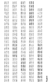
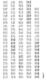
- the frameshift mutation arose within Fr2 (position 41 according to Figure 3 ) and Fr3 (position 67) respectively and are more likely due to polymerase errors during library construction (the frameshift in clone 14 occurs immediately after a long poly-A tract in the DNA sequence).
- NAR types were represented, with approximately 80% being type I and 20% type II, however a number of clones proved difficult to assign to an NAR type.
- clone 33 has a type II Fr1 but type I CDR3 and Fr4, whilst clones 06, 40 and 46 have a type I Fr1 and CDR3 but a type II Fr2 and Fr4. This finding suggests the possibility that gene conversion may be occurring between the NAR genes.
- Clones 24 and 36 are both assigned as type I on the basis of other sequence characteristics but do not carry the pair of cysteine residues normally observed in the type I CDR3.
- the clones 06, 40, 46 and 48 all encode an uneven number of cysteine residues. As mentioned previously in the case of 06, this may be due to gene conversion. Very few clones bearing an uneven number of cysteines have been observed previously and so it is thought that the V region must be under considerable pressure to maintain an even number of cysteine residues, enabling formation of disulphide bonds. The consequence of unpaired cysteines within the NAR V region is, as yet, unknown but may be detrimental to domain folding. If this is indeed the case then such clones will probably be eliminated from the library during early pans due to their toxicity to the expressing bacteria.
- Clone 02 encodes 4 cysteine residues in its CDR3, giving this V region a total of 8 cysteine residues and the potential to form 4 disulphide bonds.
- Such type I domains carrying 4, or occasionally 6 or more, cysteine residues have been previously encountered.
- the ability to form these additional disulphide bonds, combined with the small size of the NAR V region, may provide an additional source for highly stable antibody fragments.
- Colonies which were not sequenced, were scraped from the library plates with a sterile spreader into a final volume of 10 ml 2xTY medium containing 100 ⁇ g/ml ampicillin and 2% glucose. Cells were combined with sterile glycerol to 20% (v/v), and following thorough mixing aliquotted as 500 ⁇ l shots and flash-frozen prior to storage at -80 °C.
- a single aliquot of library stock was added to 200 ml of pre-warmed 2xTY medium containing ampicillin at 100 ⁇ g/ml and 1-2% glucose (w/v) and grown at 37 °C/ 250 rpm until log phase (OD 600 of 0.4 - 0.8) was reached.
- log phase OD 600 of 0.4 - 0.8
- the culture was spun at 3.5K rpm/ 4 °C for 10 min and the cell pellet re-suspended in 100 ml of 2xTY containing 100 ⁇ g/ml ampicillin, 50 ⁇ g/ml kanamycin and 0.1-0.25% glucose and incubated overnight at 30 °C/ 250 rpm to allow library expression and rescue.
- the overnight culture was spun at 12K rpm/ 4 °C for 20 min, 80 ml of supernatant was removed and added to 20 ml of PEG/NaCl, mixed well and incubated on ice for at least 1 h.
- the precipitated phage was pelleted at 12K rpm/ 4 °C and resuspended in 2 ml PBS.
- the phage suspension was spun at 13K rpm for 10 min to remove any remaining bacterial debris and the phage supernatant stored at 4 °C.
- the phage stock was titrated by performing serial dilutions in PBS and the addition of 900 ⁇ l of a log phase culture to 100 ⁇ l of each dilution.
- Nunc Maxisorp Immuno test tubes (Gibco BRL, Life technologies Ltd.) were coated with either HEL or Ova in 4 ml of PBS overnight at 4 °C. The tube was then washed 3 times with PBS before being blocked with 2% Marvel in PBS (MPBS) for 2 h at room temperature, following which it was washed a further 3 times with PBS. Selection was conducted by incubating the coated immunotube for 1 h at room temperature with 1 ml of phage stock in 3 ml of 2% MPBS on an over-and-under tumbler. A further hour of stationary incubation was allowed before the supernatant containing unbound phage was discarded and bound phage eluted as described below.
- MPBS 2% Marvel in PBS
- the immunotube was washed 20 times with PBST, excess liquid drained off and 1 ml of 100 mM triethylamine added. The tube was then rotated for a maximum of 10 min at room temperature to elute bound phage. Following incubation the phage solution was neutralized by mixing with 500 ⁇ l of 1 M Tris-HCl. In this state the phage solution was stored at 4 °C for further use (or long-term at -20 °C if glycerol added at 15% v/v).
- the culture was spun at 3.5 K rpm for 10 min and the resultant cell pellet re-suspended in 2xTY containing 100 ⁇ g/ml ampicillin, 50 ⁇ g/ml kanamycin and 0.1-0.25% glucose and incubated overnight at 30 °C/250 rpm.
- the overnight culture was spun at 12K rpm for 10 min and 40 ml of supernatant added to 10 ml of PEG/NaCl, and mixed well prior to incubation on ice for at least 1 h.
- the phage pellet was again re-suspended in 2 ml of PBS and spun for 10 min at 13K rpm to remove any remaining bacterial debris and the phage stored at 4 °C for the short term.
- the immune library was subject to five rounds of panning against the protein antigens Hen egg white lysozyme (HEL) and Chicken ovalbumin, (Ova), independently, using M13K07 helper phage and triethylamine elution.
- HEL Hen egg white lysozyme
- Ova Chicken ovalbumin,
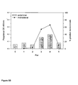
- the antigen coating density was kept constant at 100 ⁇ g/ml for pans 1 and 2. Following the first round of panning approximately 10 6 phage were eluted from both the HEL and Ova coated immunotubes, increasing 10-fold following pan 2. For pans 3 and 4 the antigen coating density was reduced for each pan in an attempt to select higher affinity binders. Whilst the number of phage eluted following HEL selection remained constant at ⁇ 10 6 for both pans that for Ova selection dropped to 10 3 in pan 3, rising back to 10 6 following pan 4. For pan 5 the antigen coating concentration was further reduced and selection was accompanied by a significant drop in the number of phage eluted. Due to this reduction in the number of phage eluted polyclonal and monoclonal phage ELISAs were conducted to determine if enrichment of HEL or Ova binders was occurring ( Figure 5 ).
- Monoclonal phage ELISAs show an increase in the number of positive phage for both sets of selection over pans 1 to 4. In the case of HEL selection this increase was from less than 1% to approximately 80% following pan 4. For Ova selected clones the numbers of positives was slightly lower but regardless increased from less than 1% to approximately 66% after the fourth pan. Following pan 5 the number of HEL-positive clones remained constant at 80% but the number of Ova-positive monoclonals dropped back to the levels observed in earlier pans (-10%).
- a 96-well Immulon 4 ELISA plate (Dynatech Laboratories Ltd.) was coated with 100 ⁇ l of antigen at 10 ⁇ g/ml for 1 h at 37°C. Following three washes with PBST the wells were blocked with 300 ⁇ l of 2% MPBS (PBS with 2% w/v marvel added) for a further hour at room temperature of overnight at 4°C. Wells were washed 3 times with PBST and to individual wells 10 ⁇ l of PEG precipitated phage from each pan, in 100 ⁇ l of 2% MPBS, was added and the plate incubated for 1 h at room temperature. The phage solution was discarded and the plate washed with PBST 3 times.
- the overnight plate was spun at 2K rpm for 10 min to give a supernatant containing monoclonal phage supernatant.
- 50 ⁇ l of this phage supernatant in 50 ⁇ l of MPBS was added per well and the plate incubated at room temperature for 1 h. Following incubation the plate was incubated with anti-M13 HRP conjugated antibody and developed as normal.
- V region fragments were then ligated into similarly cut, alkaline phosphatase treated and cleaned pIMS100 expression vector. Following overnight incubation at 15°C the resultant vector, harbouring the NAR V insert fused upstream of the HuCk domain and 6His tail, was transformed into electroporation-competent E. coli XL1-Blue cells. Colonies were picked, grown as overnight cultures in 5 ml TB (containing 2% glucose (v/v), 100 ⁇ g/ml ampicillin, 25 ⁇ g/ml tetracycline) and glycerol stocks and plasmid prepared.
- Inserts were sequenced from plasmid using the M13 reverse (5' TTCACACAGGAAACAG 3')(SEQ ID. 67) and HuCk forward (5' GAAGATGAAGACAGATGGTGC 3')(SEQ ID. 68) primer. Once sequence data had been generated the clone was given a unique name to enable identification.
- the clones 5A7 and 4F11 were chosen to represent the two different amino acid sequences found within the HEL-selected positive clones ( Figures 6 and 7 ).
- the two clones are both conventional NAR type I, and so are illustrated aligned against a typical type I clone in Figure 8 .
- the two clones differ from one another at only two positions (43 & 44), both lying within Fr2 and carry identical CDR3 regions.
- the clones 4H11 and 3E4 were chosen to represent the two different amino acid sequences found within the Ova-selected positive clones ( Figures 9 and 10 ). Again these clones were both conventional NAR type I and as such are shown aligned against a typical type I clone in Figure 11 . These clones differ at 6 amino acids; three within Fr1 (positions 13, 14 & 30), two within Fr2 (positions 46 & 47) and one within CDR3 (position 101).
- a single colony of transformed E. coli was used to inoculate 5 ml LB containing 1% glucose (v/v), 12.5 ⁇ g/ml tetracycline and 50 ⁇ g/ml ampicillin and grown up at 37°C /250 rpm overnight.
- This culture was used to seed 50 ml TB medium containing 1% glucose (v/v), 12.5 ⁇ g/ml tetracycline and 50 ⁇ g/ml ampicillin in 250 ml baffled flasks, at 1% v/v.
- the 50 ml cultures were grown over a period of 24 hours at 25°C /250 rpm, with one change of media after approximately 10 hours growth. Growth of all the cultures was good with the overnight OD 600 being in the order of 10-20 OD units.
- the cell pellet resulting from centrifugation was resuspended in 10% of the original culture volume of fractionation buffer (100 ml 200 mM Tris-HC1, 20% sucrose, pH 7.5, 1 ml 100 mM EDTA/L of culture) .
- the suspension was incubated on ice with gentle shaking for 15 min following which an equal volume of ice-cold sterile H 2 O was added and incubation continued for a further 15 min (method modified from French et al., Enzyme & Microbial Technology 19 pp332-338 1996 ).
- the suspension was spun at 13K rpm / 4°C for 20 min, the supernatant containing the periplasmic fraction harvested and passed through a 0.22 ⁇ m filter (Sartorius Instruments Ltd.).
- An Immulon 4 96-well flat bottomed ELISA plate was coated with a suitable concentration of the desired antigen at 100 ⁇ l per well and the plate incubated at 37°C for 1 h. The plate was washed 3 times with PBST prior to blocking with 200 ⁇ l per well of PBS containing 2% Marvel (w/v) for 1 h at 37°C. Wells were washed a further three times with PBST before addition of samples.
- a 1 in 5 dilution of crude periplasmic release solution was prepared, added to the top wells of the plate at 200 ⁇ l per well and doubling dilutions in PBS performed. Plates were then incubated at 4°C for 1 h. Each plate was washed a further 5 times with PBST. Goat anti-HuCk peroxidase conjugate antibody was diluted 1:1000 in PBS and 100 ⁇ l added to wells containing antigen binding domains. Plates were incubated for 1 h at 4 °C and following 6 washes with PBST the ELISA was developed as described previously and the plate read at 450 nm.
- the HEL-selected clone 5A7 shows good binding to HEL at the top dilution applied and as the sample is serially diluted binding reduces accordingly. Limited binding to the highly related protein turkey egg-white lysozyme (TEL) is observed at the highest dilution but no binding is observed to the proteins Chicken ovalbumin (Ova), Bovine serum albumin (BSA), Keyhole limpet haemocyanin (KLH) or the blocking agent Marvel. An identical pattern of protein binding is also observed for the HEL-selected clone 4F11 ( Figure 13 ), which is not surprising considering the high degree of amino acid sequence similarity between these two clones (111/113 aa identical). The OD 450 signals obtained for 3F11 are slightly higher than those for 5A7, but this may simply be due to small differences in the amount of protein present in the samples.
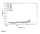
- the Ova-selected clone 4H11 ( Figure 14 ) showed no binding to any of the proteins tested, including Ova, the antigen it was selected against. To ensure that this was not simply a consequence of there being too little protein present in the assay, a binding assay was performed with undiluted periplasmic release solution. In this instance some binding to all of the proteins was observed for the wells containing the top dilutions of 4H11 protein. This binding was immediately lost once the sample was diluted and so is likely to be non-specific, no doubt resulting from very high concentrations of protein being present. This data supported the initial finding that the 4H11 clone does not bind significantly to Ova.
- the 3E4 clone like 4H11, does not show binding to the proteins HEL, BSA, KLH, TEL or the blocking agent Marvel, however low level binding is observed for this clone to the selection antigen Ova.
- the pattern of binding by this clone to Ova is unusual in that binding at the highest protein concentration is low and shows no significant drop on dilution of the sample. When the protein concentration was increased by repeating the assay with undiluted periplasmic solution a similar pattern of binding was observed, thus negating the possibility that the protein concentration was initially too low. The reason for this unusual binding is as yet unknown, but may be due to 3E4 binding only with low affinity to Ova.
- NAR clones capable of binding antigen in a library previously constructed from material from a naive animal and the isolation of HEL-binding, but not Ova-binding clones, from the library constructed from the HEL immunised animals illustrates the highly specific nature of the NAR response following antigen challenge. In other words, antigen specific antigen binding domains with a specific specificity are produced.
- clones 5A7 and 4F11 were shown to be capable of binding HEL in the antigen binding ELISA it was possible to test the stability of these clones to thermal denaturation.
- Subsaturating dilutions of both of the clones, ascertained from the antigen binding curves, were prepared and incubated at a range of temperatures for 3 h prior to their addition to a HEL coated ELISA plate.
- the samples were then incubated on the ELISA plate for an hour at 4 °C and binding detected with an anti-HuCk HRP conjugated antibody. Stability of the antigen binding domains was plotted as a percentage of that obtained for a control sample which had not been heat treated ( Figure 15 ).
- Both clone 5A7 and clone 4F11 show considerable resistance to irreversible denaturation losing 50% functionality at approximately 85°C and retaining approximately 30% functionality after 3 h at 95°C. This high stability is probably a consequence of the additional, non-canonical cysteine residues found within the NAR V domain. Both clones encode 6 cysteine residues and therefore are capable of forming 3 intradomain disulphide bonds, which (if formed) would contribute greatly to the high stability of these domains. The shape of the stability curves for both of the clones is almost identical and the minor difference in stability between the clones may be simply due to assay variability.
- HEL-5A7 The ability of HEL-5A7 to inhibit the enzymatic activity of HEL was tested by mixing 12.5 ⁇ l of HEL with 12.5 ⁇ l of purified HEL-5A7 protein in a sterile 96 well tissue culture plate, to give a final HEL concentration of 10 ⁇ g/ml and HEL-5A7 concentrations of 2500 nM, 250 nM and 25 nM.
- the control well was set up with buffer replacing HEL-5A7.
- a sample of freeze dried Micrococcus lysodeikticus was reconstituted in 0.1 M phosphate/citrate buffer (pH 5.8) containing 0.09%NaCl, mixed thoroughly and 175 ⁇ l added to the prepared wells. The plate was read over a period of 30 min (at 1 min intervals) at 450 nm. Enzymatic activity was plotted as percentage initial absorbance against time for each sample.
- HEL-5A7 protein reduced the rate of cell lysis in a concentration dependent manner with respect to the control ( Figure 16 ).
- HEL-5A7 protein at a final concentration of 2500 nM the rate of cell lysis (9.3x10- 3 OD units/min) is almost halved when compared to the control (17x10 -3 OD units/min) indicating that the HEL-5A7 region binds within or adjacent to the lysozyme active site cavity.
- a similarly prepared antigen specific antigen binding domain raised against an unrelated antigen showed no effect upon the rate of cell lysis when introduced to the assay at the same concentrations.
Landscapes
- Chemical & Material Sciences (AREA)
- Health & Medical Sciences (AREA)
- Organic Chemistry (AREA)
- Life Sciences & Earth Sciences (AREA)
- General Health & Medical Sciences (AREA)
- Medicinal Chemistry (AREA)
- Immunology (AREA)
- Biophysics (AREA)
- Genetics & Genomics (AREA)
- Molecular Biology (AREA)
- Proteomics, Peptides & Aminoacids (AREA)
- Biochemistry (AREA)
- Virology (AREA)
- Engineering & Computer Science (AREA)
- Veterinary Medicine (AREA)
- General Chemical & Material Sciences (AREA)
- Nuclear Medicine, Radiotherapy & Molecular Imaging (AREA)
- Pharmacology & Pharmacy (AREA)
- Animal Behavior & Ethology (AREA)
- Public Health (AREA)
- Chemical Kinetics & Catalysis (AREA)
- Bioinformatics & Cheminformatics (AREA)
- Peptides Or Proteins (AREA)
- Micro-Organisms Or Cultivation Processes Thereof (AREA)
- Preparation Of Compounds By Using Micro-Organisms (AREA)
- Medicines Containing Antibodies Or Antigens For Use As Internal Diagnostic Agents (AREA)
- Transition And Organic Metals Composition Catalysts For Addition Polymerization (AREA)
Applications Claiming Priority (4)
| Application Number | Priority Date | Filing Date | Title |
|---|---|---|---|
| GB0119553A GB0119553D0 (en) | 2001-08-10 | 2001-08-10 | Antibodies |
| GB0210508A GB0210508D0 (en) | 2002-05-08 | 2002-05-08 | Antibodies |
| EP10155260A EP2202243A3 (fr) | 2001-08-10 | 2002-08-12 | Domaines liant un antigène prévenant de poissons |
| EP02749132A EP1419179B1 (fr) | 2001-08-10 | 2002-08-12 | Domaines de liaison d'antigenes issus de poissons |
Related Parent Applications (3)
| Application Number | Title | Priority Date | Filing Date |
|---|---|---|---|
| EP02749132.3 Division | 2002-08-12 | ||
| EP10155260 Previously-Filed-Application | 2002-08-12 | ||
| EP10155260.2 Division | 2010-03-02 |
Publications (2)
| Publication Number | Publication Date |
|---|---|
| EP2277913A2 true EP2277913A2 (fr) | 2011-01-26 |
| EP2277913A3 EP2277913A3 (fr) | 2012-08-08 |
Family
ID=26246430
Family Applications (5)
| Application Number | Title | Priority Date | Filing Date |
|---|---|---|---|
| EP10185416.4A Expired - Lifetime EP2281837B1 (fr) | 2001-08-10 | 2002-08-12 | Domaines se liant à l'antigène issus de poisson |
| EP02749132A Expired - Lifetime EP1419179B1 (fr) | 2001-08-10 | 2002-08-12 | Domaines de liaison d'antigenes issus de poissons |
| EP10185409A Withdrawn EP2277913A3 (fr) | 2001-08-10 | 2002-08-12 | Domaines se liant à l'antigène issus de poisson |
| EP10185470A Withdrawn EP2277914A3 (fr) | 2001-08-10 | 2002-08-12 | Domaines se liant à l'antigène issus de poisson |
| EP10155260A Withdrawn EP2202243A3 (fr) | 2001-08-10 | 2002-08-12 | Domaines liant un antigène prévenant de poissons |
Family Applications Before (2)
| Application Number | Title | Priority Date | Filing Date |
|---|---|---|---|
| EP10185416.4A Expired - Lifetime EP2281837B1 (fr) | 2001-08-10 | 2002-08-12 | Domaines se liant à l'antigène issus de poisson |
| EP02749132A Expired - Lifetime EP1419179B1 (fr) | 2001-08-10 | 2002-08-12 | Domaines de liaison d'antigenes issus de poissons |
Family Applications After (2)
| Application Number | Title | Priority Date | Filing Date |
|---|---|---|---|
| EP10185470A Withdrawn EP2277914A3 (fr) | 2001-08-10 | 2002-08-12 | Domaines se liant à l'antigène issus de poisson |
| EP10155260A Withdrawn EP2202243A3 (fr) | 2001-08-10 | 2002-08-12 | Domaines liant un antigène prévenant de poissons |
Country Status (11)
| Country | Link |
|---|---|
| US (2) | US20050043519A1 (fr) |
| EP (5) | EP2281837B1 (fr) |
| JP (1) | JP5133494B2 (fr) |
| AT (1) | ATE459651T1 (fr) |
| AU (2) | AU2002319544B2 (fr) |
| CA (2) | CA2457636C (fr) |
| DE (1) | DE60235553D1 (fr) |
| DK (1) | DK1419179T3 (fr) |
| ES (1) | ES2337986T3 (fr) |
| PT (1) | PT1419179E (fr) |
| WO (1) | WO2003014161A2 (fr) |
Cited By (2)
| Publication number | Priority date | Publication date | Assignee | Title |
|---|---|---|---|---|
| US9475870B2 (en) | 2012-05-07 | 2016-10-25 | The University Court Of The University Of Aberdeen | Single domain binding molecule |
| US10202438B2 (en) | 2013-04-23 | 2019-02-12 | The University Court Of The University Of Aberdeen | Synthetic library of specific binding molecules |
Families Citing this family (152)
| Publication number | Priority date | Publication date | Assignee | Title |
|---|---|---|---|---|
| EP1600459A3 (fr) * | 2002-06-28 | 2005-12-07 | Domantis Limited | Ligand |
| DK2330121T3 (en) * | 2004-06-02 | 2014-12-15 | Adalta Pty Ltd | Binding moieties based on shark IgNar domains |
| US7563443B2 (en) * | 2004-09-17 | 2009-07-21 | Domantis Limited | Monovalent anti-CD40L antibody polypeptides and compositions thereof |
| US20090017050A1 (en) | 2007-07-13 | 2009-01-15 | Ventana Medical Systems, Inc. | Egfr antigen-binding molecules and uses thereof |
| PE20091163A1 (es) | 2007-11-01 | 2009-08-09 | Wyeth Corp | Anticuerpos para gdf8 |
| EP2217716A4 (fr) | 2007-11-09 | 2011-02-09 | Salk Inst For Biological Studi | Utilisation d'inhibiteurs de récepteurs tam en tant qu'antimicrobiens |
| EP2222709B1 (fr) | 2007-11-30 | 2016-11-23 | Glaxo Group Limited | Produits de construction de liaison à un antigène |
| JP2011527572A (ja) | 2008-07-09 | 2011-11-04 | バイオジェン・アイデック・エムエイ・インコーポレイテッド | Lingo抗体または断片を含む組成物 |
| EP2700651B1 (fr) | 2008-07-18 | 2019-04-10 | Bristol-Myers Squibb Company | Compositions monovalentes pour la liaison au CD28 et procédés d'utilisation |
| US20100075329A1 (en) | 2008-09-23 | 2010-03-25 | O'toole Margot | Methods for predicting production of activating signals by cross-linked binding proteins |
| CA2739352C (fr) | 2008-10-29 | 2021-07-13 | Wyeth Llc | Procedes de purification de molecules de liaison d'antigene monodomaines |
| KR101593285B1 (ko) | 2008-10-29 | 2016-02-11 | 아블린쓰 엔.브이. | 단일 도메인 항원 결합 분자의 제형 |
| US9085795B2 (en) | 2009-02-04 | 2015-07-21 | Molecular Innovations, Inc. | Methods for screening candidate agents for modulating prorenin and renin, assays for detecting prorenin and antibodies |
| CA2753263A1 (fr) | 2009-02-24 | 2010-09-02 | Glaxo Group Limited | Constructions multivalentes et/ou multispecifiques de liaison au rankl |
| US20110305692A1 (en) | 2009-02-24 | 2011-12-15 | Glaxo Group Limited | Antigen-binding contructs |
| EP2401297A1 (fr) | 2009-02-24 | 2012-01-04 | Glaxo Group Limited | Constructions de liaison d'antigène |
| MX366890B (es) | 2009-10-23 | 2019-07-30 | Millennium Pharm Inc | Moléculas de anticuerpo anti - gcc y composiciones y métodos relacionados. |
| US8496933B2 (en) | 2009-11-04 | 2013-07-30 | Laboratorios Silanes, S.A. De C.V. | VHNAR anti-cytokine domains |
| US20150231215A1 (en) | 2012-06-22 | 2015-08-20 | Randolph J. Noelle | VISTA Antagonist and Methods of Use |
| MX342017B (es) | 2010-03-26 | 2016-09-09 | Dartmouth College | Proteina mediadora de celula t regulatoria vista, agentes de enlace de vista y uso de los mismos. |
| US10745467B2 (en) | 2010-03-26 | 2020-08-18 | The Trustees Of Dartmouth College | VISTA-Ig for treatment of autoimmune, allergic and inflammatory disorders |
| WO2012007880A2 (fr) | 2010-07-16 | 2012-01-19 | Ablynx Nv | Molécules de liaison à un antigène à domaine unique modifiées et leurs utilisations |
| EP2643352B1 (fr) | 2010-11-23 | 2018-05-02 | Glaxo Group Limited | Protéines de liaison à l'antigène oncostatin m (osm) |
| SG190362A1 (en) | 2010-11-24 | 2013-06-28 | Glaxo Group Ltd | Multispecific antigen binding proteins targeting hgf |
| WO2012103240A2 (fr) | 2011-01-25 | 2012-08-02 | Eleven Biotherapeutics, Inc. | Agents de liaison à un récepteur |
| JP6147726B2 (ja) | 2011-03-29 | 2017-06-14 | グラクソスミスクライン・リミテッド・ライアビリティ・カンパニーGlaxoSmithKline LLC | タンパク質精製のためのバッファー系 |
| WO2012138958A1 (fr) | 2011-04-07 | 2012-10-11 | Glaxosmithkline Llc | Formulations ayant une viscosité réduite |
| US9409987B2 (en) | 2011-04-15 | 2016-08-09 | Compugen Ltd | Polypeptides and polynucleotides, and uses thereof for treatment of immune related disorders and cancer |
| SI3415531T1 (sl) | 2011-05-27 | 2023-12-29 | Glaxo Group Limited | Bcma (cd269/tnfrsf17) - vezni proteini |
| US9435812B2 (en) | 2011-08-31 | 2016-09-06 | Ventana Medical Systems, Inc. | Expression of ETS related gene (ERG) and phosphatase and tensin homolog (PTEN) correlates with prostate cancer capsular penetration |
| GB2502127A (en) | 2012-05-17 | 2013-11-20 | Kymab Ltd | Multivalent antibodies and in vivo methods for their production |
| US9156915B2 (en) | 2012-04-26 | 2015-10-13 | Thomas Jefferson University | Anti-GCC antibody molecules |
| MX381731B (es) | 2012-05-14 | 2025-03-13 | Biogen Ma Inc | Antagonistas de proteina que interactua con el receptor nogo 2 que contiene repeticion rica en leucina y dominio de inmunoglobulina (lingo-2) para el tratamiento de afecciones que involucran neuronas motoras. |
| CN104619722B (zh) | 2012-06-22 | 2022-10-04 | 达特茅斯大学理事会 | 新型vista-ig构建体和vista-ig用于治疗自身免疫性、过敏性和炎性疾病的用途 |
| US9890215B2 (en) | 2012-06-22 | 2018-02-13 | King's College London | Vista modulators for diagnosis and treatment of cancer |
| WO2014018535A1 (fr) | 2012-07-25 | 2014-01-30 | Salk Institute For Biological Studies | Régulation de l'interaction entre les ligands de tam et les membranes lipidiques contenant de la phosphatidylsérine |
| CA2884704C (fr) | 2012-09-07 | 2023-04-04 | Randolph J. Noelle | Modulateurs vista de diagnostic et de traitement de cancer |
| CN104870014A (zh) | 2012-10-09 | 2015-08-26 | 比奥根Ma公司 | 联合治疗及用于治疗脱髓鞘病症的用途 |
| WO2014120916A1 (fr) | 2013-02-01 | 2014-08-07 | Bristol-Myers Squibb Company | Anticorps à domaines pegylés monovalents pour la liaison à cd28 et leurs méthodes d'utilisation |
| US9963513B2 (en) | 2013-02-05 | 2018-05-08 | Engmab Sàrl | Method for the selection of antibodies against BCMA |
| EP2762496A1 (fr) | 2013-02-05 | 2014-08-06 | EngMab AG | Procédé pour la sélection d'anticorps contre BCMA |
| JP6499087B2 (ja) | 2013-02-26 | 2019-04-10 | ロシュ グリクアート アーゲー | 二重特異性t細胞活性化抗原結合分子 |
| US10450354B2 (en) | 2013-03-12 | 2019-10-22 | Molecular Templates, Inc. | CD20-binding immunotoxins for inducing cellular internalization and methods using same |
| TWI625335B (zh) | 2013-03-15 | 2018-06-01 | Glaxosmithkline Intellectual Property (No. 2) Limited | 純化抗體的方法 |
| WO2014141149A1 (fr) | 2013-03-15 | 2014-09-18 | Glaxosmithkline Intellectual Property (No.2) Limited | Formulations présentant une viscosité réduite |
| KR20150132332A (ko) | 2013-03-15 | 2015-11-25 | 글락소스미스클라인 인털렉츄얼 프로퍼티 (넘버 2) 리미티드 | 저농도 항체 제형 |
| CA2902288A1 (fr) | 2013-03-15 | 2014-09-18 | Glaxosmithkline Intellectual Property (No.2) Limited | Utilisation d'intermediaires de l'acide tricarboxylique (atc) en vue de la regulation de la generation d'ammoniac dans des cultures cellulaires |
| EP2970426B1 (fr) | 2013-03-15 | 2019-08-28 | Michael C. Milone | Ciblage de cellules cytotoxiques par des récepteurs chimériques pour une immunothérapie adoptive |
| EP2789630A1 (fr) | 2013-04-09 | 2014-10-15 | EngMab AG | Anticorps bispécifiques contre le CD3e et ROR1 |
| NL1040254C2 (en) | 2013-05-17 | 2014-11-24 | Ablynx Nv | Stable formulations of immunoglobulin single variable domains and uses thereof. |
| CA2931684C (fr) | 2013-12-19 | 2024-02-20 | Novartis Ag | Recepteurs antigeniques chimeriques de la mesotheline humaine et leurs utilisations |
| EP3087101B1 (fr) | 2013-12-20 | 2024-06-05 | Novartis AG | Récepteur d'antigène chimérique régulable |
| EP4043493A1 (fr) | 2013-12-24 | 2022-08-17 | Janssen Pharmaceutica NV | Anticorps anti-vista et fragments |
| WO2015100246A1 (fr) | 2013-12-24 | 2015-07-02 | Ossianix, Inc. | Composés à liaison sélective baff et procédés associés |
| US11014987B2 (en) | 2013-12-24 | 2021-05-25 | Janssen Pharmaceutics Nv | Anti-vista antibodies and fragments, uses thereof, and methods of identifying same |
| US20170081411A1 (en) | 2014-03-15 | 2017-03-23 | Novartis Ag | Regulatable chimeric antigen receptor |
| EP3593812A3 (fr) | 2014-03-15 | 2020-05-27 | Novartis AG | Traitement du cancer à l'aide d'un récepteur d'antigène chimérique |
| TWI694836B (zh) | 2014-05-16 | 2020-06-01 | 英商葛蘭素史克智慧財產管理有限公司 | 抗體調配物 |
| EP3154585B1 (fr) | 2014-06-11 | 2022-02-23 | Kathy A. Green | Utilisation d'antagonistes et d'agonistes vista pour supprimer ou améliorer l'immunité humorale |
| EP3160995A2 (fr) | 2014-06-26 | 2017-05-03 | Ossianix Inc. | Banques de vnar de requin-nourrice semi-synthétiques pour la préparation et l'utilisation de composés de liaison sélectifs |
| WO2016014530A1 (fr) | 2014-07-21 | 2016-01-28 | Novartis Ag | Combinaisons de faibles doses renforçant l'immunité d'inhibiteurs de mtor et car |
| US11542488B2 (en) | 2014-07-21 | 2023-01-03 | Novartis Ag | Sortase synthesized chimeric antigen receptors |
| MY181834A (en) | 2014-07-21 | 2021-01-08 | Novartis Ag | Treatment of cancer using humanized anti-bcma chimeric antigen receptor |
| US9777061B2 (en) | 2014-07-21 | 2017-10-03 | Novartis Ag | Treatment of cancer using a CD33 chimeric antigen receptor |
| US20170209492A1 (en) | 2014-07-31 | 2017-07-27 | Novartis Ag | Subset-optimized chimeric antigen receptor-containing t-cells |
| SG11201700770PA (en) | 2014-08-19 | 2017-03-30 | Novartis Ag | Anti-cd123 chimeric antigen receptor (car) for use in cancer treatment |
| ES2891332T3 (es) | 2014-09-17 | 2022-01-27 | Novartis Ag | Direccionamiento a células citotóxicas con receptores quiméricos para la inmunoterapia adoptiva |
| CA2963692A1 (fr) | 2014-10-09 | 2016-04-14 | Engmab Ag | Anticorps bispecifiques contre cd3epsilon et ror1 |
| WO2016059602A2 (fr) | 2014-10-16 | 2016-04-21 | Glaxo Group Limited | Méthodes de traitement du cancer et compositions associées |
| WO2016077840A2 (fr) | 2014-11-14 | 2016-05-19 | Ossianix, Inc. | Composés de liaison sélectifs de tfr et procédés associés |
| US20180334490A1 (en) | 2014-12-03 | 2018-11-22 | Qilong H. Wu | Methods for b cell preconditioning in car therapy |
| JP2018505911A (ja) | 2014-12-05 | 2018-03-01 | イミュネクスト,インコーポレーテッド | 推定上のvista受容体としてのvsig8の同定と、vista/vsig8調節剤を産生するためのその使用 |
| CN107567461A (zh) | 2014-12-29 | 2018-01-09 | 诺华股份有限公司 | 制备嵌合抗原受体表达细胞的方法 |
| JP2018504400A (ja) | 2015-01-08 | 2018-02-15 | バイオジェン・エムエイ・インコーポレイテッドBiogen MA Inc. | Lingo‐1拮抗薬及び脱髄障害の治療のための使用 |
| US11161907B2 (en) | 2015-02-02 | 2021-11-02 | Novartis Ag | Car-expressing cells against multiple tumor antigens and uses thereof |
| EP3280795B1 (fr) | 2015-04-07 | 2021-03-24 | Novartis AG | Combinaison de traitement à l'aide du récepteur antigénique chimérique et de derivés de l'amino pyrimidine |
| AU2016249005B2 (en) | 2015-04-17 | 2022-06-16 | Novartis Ag | Methods for improving the efficacy and expansion of chimeric antigen receptor-expressing cells |
| EP3286211A1 (fr) | 2015-04-23 | 2018-02-28 | Novartis AG | Traitement du cancer à l'aide de protéine récepteur antigénique chimérique et un inhibiteur de protéine kinase |
| EP3722314A1 (fr) | 2015-06-24 | 2020-10-14 | Janssen Pharmaceutica NV | Anticorps anti-vista et fragments |
| WO2017015427A1 (fr) | 2015-07-21 | 2017-01-26 | Novartis Ag | Méthodes pour améliorer l'efficacité et l'expansion de cellules immunitaires |
| AU2016302881B2 (en) | 2015-08-03 | 2022-09-15 | Bristol-Myers Squibb Company | Monoclonal antibodies against BCMA |
| CN105384825B (zh) | 2015-08-11 | 2018-06-01 | 南京传奇生物科技有限公司 | 一种基于单域抗体的双特异性嵌合抗原受体及其应用 |
| CN116334205A (zh) | 2015-09-03 | 2023-06-27 | 诺华股份有限公司 | 预测细胞因子释放综合征的生物标志物 |
| KR20180077193A (ko) | 2015-10-30 | 2018-07-06 | 글락소스미스클라인 인털렉츄얼 프로퍼티 디벨로프먼트 리미티드 | 예후 방법 |
| PT3393504T (pt) | 2015-12-22 | 2025-12-30 | Novartis Ag | Recetor de antigénio quimérico (car) de mesotelina e anticorpo contra o inibidor de pd-l1 para utilização combinada em terapia anticancerígena |
| CA3009852A1 (fr) | 2015-12-28 | 2017-07-06 | Novartis Ag | Methodes de production de cellules d'expression de recepteur d'antigene chimerique |
| TW202216201A (zh) | 2016-02-12 | 2022-05-01 | 比利時商楊森製藥公司 | 抗vista抗體及片段、其用途及鑑定其之方法 |
| CA3016287A1 (fr) | 2016-03-04 | 2017-09-08 | Novartis Ag | Cellules exprimant de multiples molecules de recepteur d'antigene chimere (car) et leurs utilisations |
| JP7184751B2 (ja) | 2016-04-15 | 2022-12-06 | イミュネクスト インコーポレイテッド | 抗ヒトvista抗体およびその使用 |
| EP3443096B1 (fr) | 2016-04-15 | 2023-03-01 | Novartis AG | Compositions et méthodes pour l'expression sélective des récepteurs antigéniques chimériques |
| US11197902B2 (en) | 2016-06-24 | 2021-12-14 | Ig Biosciences Corporation | Prebiotic neutraceutical compositions and methods of treatment using the same |
| US20200031924A1 (en) | 2016-07-13 | 2020-01-30 | Biogen Ma Inc. | Dosage Regimens of Lingo-1 Antagonists and Uses for Treatment of Demyelinating Disorders |
| SG11201900344YA (en) | 2016-07-15 | 2019-02-27 | Novartis Ag | Treatment and prevention of cytokine release syndrome using a chimeric antigen receptor in combination with a kinase inhibitor |
| WO2018014260A1 (fr) | 2016-07-20 | 2018-01-25 | Nanjing Legend Biotech Co., Ltd. | Protéines de liaison antigènes multi-spécifiques et leurs procédés d'utilisation |
| CN110214150A (zh) | 2016-07-28 | 2019-09-06 | 诺华股份有限公司 | 嵌合抗原受体和pd-1抑制剂的组合疗法 |
| EP3490590A2 (fr) | 2016-08-01 | 2019-06-05 | Novartis AG | Traitement du cancer au moyen d'un récepteur d'antigène chimérique en combinaison avec un inhibiteur d'une molécule de macrophage pro-m2 |
| KR102512934B1 (ko) | 2016-08-10 | 2023-03-23 | 레전드 바이오테크 아일랜드 리미티드 | Bcma를 표적으로 하는 키메라 항원 수용체 및 이들의 이용 방법 |
| RU2759909C2 (ru) | 2016-09-07 | 2021-11-18 | Глэксосмитклайн Интеллекчуал Проперти Дивелопмент Лимитед | Способы очистки антител |
| JP2020501508A (ja) | 2016-09-15 | 2020-01-23 | クアドルセプト バイオ リミテッド | 多量体、四量体および八量体 |
| WO2018067992A1 (fr) | 2016-10-07 | 2018-04-12 | Novartis Ag | Récepteurs antigéniques chimériques pour le traitement du cancer |
| WO2018068201A1 (fr) | 2016-10-11 | 2018-04-19 | Nanjing Legend Biotech Co., Ltd. | Anticorps à domaine unique et ses variants contre ctla-4 |
| EP3535295A1 (fr) | 2016-11-02 | 2019-09-11 | EngMab Sàrl | Anticorps bispécifique contre bcma et cd3 et médicament immunologique pour une utilisation combinée dans le traitement du myélome multiple |
| JP7258762B2 (ja) * | 2016-11-07 | 2023-04-17 | デーゲンエルイックス べスローテン フェンノートシャップ | 新規アミロイドベータオリゴマー特異的結合分子 |
| EP3574005B1 (fr) | 2017-01-26 | 2021-12-15 | Novartis AG | Compositions de cd28 et procédés pour une thérapie à base de récepteur antigénique chimérique |
| US20200048359A1 (en) | 2017-02-28 | 2020-02-13 | Novartis Ag | Shp inhibitor compositions and uses for chimeric antigen receptor therapy |
| MX2019012566A (es) | 2017-04-21 | 2019-12-02 | Centro De Investig Cientifica Y De Educacion Superior De Ensenada Baja California | Vnar con actividad anti-angiogenica en tumores solidos de animales de compa?ia. |
| US20200055948A1 (en) | 2017-04-28 | 2020-02-20 | Novartis Ag | Cells expressing a bcma-targeting chimeric antigen receptor, and combination therapy with a gamma secretase inhibitor |
| WO2019000223A1 (fr) | 2017-06-27 | 2019-01-03 | Nanjing Legend Biotech Co., Ltd. | Activateurs de cellules effectrices immunitaires d'anticorps chimériques et leurs procédés d'utilisation |
| CA3075367A1 (fr) | 2017-09-27 | 2019-04-04 | Elasmogen Ltd | Molecules de liaison specifiques |
| WO2019084288A1 (fr) | 2017-10-25 | 2019-05-02 | Novartis Ag | Procédés de conception de cellules d'expression de récepteur antigénique chimérique |
| WO2019089798A1 (fr) | 2017-10-31 | 2019-05-09 | Novartis Ag | Compositions anti-car et procédés |
| GB201721802D0 (en) | 2017-12-22 | 2018-02-07 | Almac Discovery Ltd | Ror1-specific antigen binding molecules |
| TW201930358A (zh) | 2017-12-28 | 2019-08-01 | 大陸商南京傳奇生物科技有限公司 | 針對tigit之單域抗體及其變異體 |
| WO2019139987A1 (fr) | 2018-01-09 | 2019-07-18 | Elstar Therapeutics, Inc. | Constructions de liaison à la calréticuline et lymphocytes t modifiés pour le traitement de maladies |
| KR102839330B1 (ko) | 2018-01-15 | 2025-07-30 | 난징 레전드 바이오테크 씨오., 엘티디. | Pd-1에 대한 단일-도메인 항체 및 이의 변이체 |
| AU2019215031C1 (en) | 2018-01-31 | 2026-02-26 | Novartis Ag | Combination therapy using a chimeric antigen receptor |
| TW202000891A (zh) | 2018-03-07 | 2020-01-01 | 英商葛蘭素史克智慧財產發展有限公司 | 純化抗體之方法 |
| TW202003555A (zh) | 2018-03-07 | 2020-01-16 | 英商葛蘭素史克智慧財產發展有限公司 | 用於純化重組多肽之方法 |
| US12152073B2 (en) | 2018-03-14 | 2024-11-26 | Marengo Therapeutics, Inc. | Multifunctional molecules that bind to calreticulin and uses thereof |
| JP7346790B2 (ja) | 2018-03-30 | 2023-09-20 | ナンジン レジェンド バイオテック カンパニー,リミテッド | Lag‐3に対する単一ドメイン抗体およびその使用 |
| EP3784351A1 (fr) | 2018-04-27 | 2021-03-03 | Novartis AG | Thérapies reposant sur des cellules car-t présentant une efficacité améliorée |
| AU2019284911B2 (en) | 2018-06-13 | 2026-04-09 | Novartis Ag | BCMA chimeric antigen receptors and uses thereof |
| US12258414B2 (en) * | 2018-06-22 | 2025-03-25 | Ossianix, Inc. | Anti-CD98hc VNARs for crossing the blood brain barrier and type IV VNAR libraries |
| WO2020010250A2 (fr) | 2018-07-03 | 2020-01-09 | Elstar Therapeutics, Inc. | Molécules d'anticorps anti-tcr et leurs utilisations |
| PT3844267T (pt) | 2018-08-31 | 2025-09-30 | Novartis Ag | Métodos de preparação de células que expressam recetor de antigénio quimérico |
| WO2020047449A2 (fr) | 2018-08-31 | 2020-03-05 | Novartis Ag | Procédés de fabrication de cellules exprimant un récepteur d'antigène chimère |
| US12351642B2 (en) | 2018-09-14 | 2025-07-08 | Ossianix, Inc. | TFR-specific binding moieties and transcytosis method to select VNARs that cross cellular barriers |
| US12016885B2 (en) | 2018-11-06 | 2024-06-25 | Alsatech, Inc. | Method of solubilizing TDP-43 protein aggregates in amyotrophic lateral sclerosis using a microvascular endothelial cell expressing an anti-TDP-43 Fab |
| AU2020226893B2 (en) | 2019-02-21 | 2025-02-27 | Marengo Therapeutics, Inc. | Multifunctional molecules that bind to T cell related cancer cells and uses thereof |
| GB2597851B (en) | 2019-02-21 | 2024-05-29 | Marengo Therapeutics Inc | Antibody molecules that bind to NKP30 and uses thereof |
| US20220088075A1 (en) | 2019-02-22 | 2022-03-24 | The Trustees Of The University Of Pennsylvania | Combination therapies of egfrviii chimeric antigen receptors and pd-1 inhibitors |
| GB201903767D0 (en) | 2019-03-19 | 2019-05-01 | Quadrucept Bio Ltd | Multimers, tetramers & octamers |
| US20220168389A1 (en) | 2019-04-12 | 2022-06-02 | Novartis Ag | Methods of making chimeric antigen receptor-expressing cells |
| EP4065158A2 (fr) | 2019-11-26 | 2022-10-05 | Novartis AG | Récepteurs antigéniques chimériques et leurs utilisations |
| AU2020416273A1 (en) | 2020-01-03 | 2022-07-28 | Marengo Therapeutics, Inc. | Anti-TCR antibody molecules and uses thereof |
| WO2021163618A1 (fr) | 2020-02-14 | 2021-08-19 | Novartis Ag | Procédé de prédiction de réponse à une thérapie de récepteur antigénique chimérique |
| CN115175695A (zh) | 2020-02-27 | 2022-10-11 | 诺华股份有限公司 | 制备表达嵌合抗原受体的细胞的方法 |
| MX2022010685A (es) | 2020-02-27 | 2022-09-23 | Novartis Ag | Metodos de produccion de celulas que expresan receptores de antigeno quimericos. |
| WO2021190980A1 (fr) | 2020-03-22 | 2021-09-30 | Quadrucept Bio Limited | Multimères pour l'évolution d'une souche virale |
| CN116157510A (zh) | 2020-06-17 | 2023-05-23 | 詹森生物科技公司 | 用于制造多能干细胞的材料和方法 |
| WO2022012680A1 (fr) | 2020-07-16 | 2022-01-20 | Nanjing Legend Biotech Co., Ltd. | Molécules de liaison à cd20 et leurs utilisations |
| CN112301431B (zh) * | 2020-08-12 | 2023-01-31 | 华东理工大学 | 基于鲨鱼抗体可变区v-nar的噬菌体文库及其构建方法 |
| GB202020152D0 (en) | 2020-12-18 | 2021-02-03 | Elasmogen Ltd | Fast-track humanisation of specific binding molecules |
| WO2022184659A1 (fr) | 2021-03-01 | 2022-09-09 | Quadrucept Bio Limited | Domaines d'anticorps et multimères |
| WO2022269473A1 (fr) | 2021-06-23 | 2022-12-29 | Janssen Biotech, Inc. | Matériaux et procédés pour régions charnières dans des récepteurs exogènes fonctionnels |
| US20250188492A1 (en) | 2022-07-22 | 2025-06-12 | Janssen Biotech, Inc. | Enhanced Transfer of Genetic Instructions to Effector Immune Cells |
| IL319225A (en) | 2022-09-15 | 2025-04-01 | Novartis Ag | Treatment of autoimmune disorders using antigen receptor chemotherapeutics |
| GB202216503D0 (en) | 2022-11-05 | 2022-12-21 | Quadrucept Bio Ltd | Non-human vertebrates & cells |
| WO2024200846A1 (fr) | 2023-03-30 | 2024-10-03 | 272BIO Limited | Polypeptides se liant à la gnrh et leurs utilisations |
| WO2025034806A1 (fr) | 2023-08-08 | 2025-02-13 | Wisconsin Alumni Research Foundation | Anticorps à domaine unique et variants de ceux-ci ciblant la protéine d'activation des fibroblastes |
| WO2025178959A1 (fr) | 2024-02-20 | 2025-08-28 | University Of Georgia Research Foundation, Inc. | Anticorps à domaine unique et variants de ceux-ci contre tab1 |
| CN121896173A (zh) | 2024-10-21 | 2026-04-21 | 深圳市湾岛细胞科技有限公司 | 表达外源肌苷单磷酸脱氢酶的真核细胞 |
Family Cites Families (3)
| Publication number | Priority date | Publication date | Assignee | Title |
|---|---|---|---|---|
| ES2052027T5 (es) | 1988-11-11 | 2005-04-16 | Medical Research Council | Clonacion de secuencias de dominio variable de inmunoglobulina. |
| US6838254B1 (en) | 1993-04-29 | 2005-01-04 | Conopco, Inc. | Production of antibodies or (functionalized) fragments thereof derived from heavy chain immunoglobulins of camelidae |
| EP0954978B1 (fr) | 1998-03-12 | 2011-11-30 | VHsquared Limited | Produits contenant des levures ou des moisissures inactivées, ayant sur leur surface externe des anticorps actifs |
-
2002
- 2002-08-12 CA CA2457636A patent/CA2457636C/fr not_active Expired - Lifetime
- 2002-08-12 EP EP10185416.4A patent/EP2281837B1/fr not_active Expired - Lifetime
- 2002-08-12 EP EP02749132A patent/EP1419179B1/fr not_active Expired - Lifetime
- 2002-08-12 EP EP10185409A patent/EP2277913A3/fr not_active Withdrawn
- 2002-08-12 DK DK02749132.3T patent/DK1419179T3/da active
- 2002-08-12 EP EP10185470A patent/EP2277914A3/fr not_active Withdrawn
- 2002-08-12 CA CA2763913A patent/CA2763913C/fr not_active Expired - Fee Related
- 2002-08-12 AT AT02749132T patent/ATE459651T1/de active
- 2002-08-12 US US10/486,608 patent/US20050043519A1/en not_active Abandoned
- 2002-08-12 WO PCT/GB2002/003714 patent/WO2003014161A2/fr not_active Ceased
- 2002-08-12 ES ES02749132T patent/ES2337986T3/es not_active Expired - Lifetime
- 2002-08-12 EP EP10155260A patent/EP2202243A3/fr not_active Withdrawn
- 2002-08-12 DE DE60235553T patent/DE60235553D1/de not_active Expired - Lifetime
- 2002-08-12 AU AU2002319544A patent/AU2002319544B2/en not_active Ceased
- 2002-08-12 JP JP2003519110A patent/JP5133494B2/ja not_active Expired - Fee Related
- 2002-08-12 PT PT02749132T patent/PT1419179E/pt unknown
-
2008
- 2008-10-10 AU AU2008229871A patent/AU2008229871B2/en not_active Ceased
-
2011
- 2011-10-25 US US13/280,837 patent/US8865431B2/en not_active Expired - Lifetime
Non-Patent Citations (9)
| Title |
|---|
| DIAZ M ET AL: "Evolution and the molecular basis of somatic hypermutation of antigen receptor genes.", PHILOSOPHICAL TRANSACTIONS OF THE ROYAL SOCIETY OF LONDON. SERIES B: BIOLOGICAL SCIENCES. ENGLAND 29 JAN 2001, vol. 356, no. 1405, 29 January 2001 (2001-01-29), pages 67-72, XP008017869, ISSN: 0962-8436 * |
| DIAZ M ET AL: "Mutational pattern of the nurse shark antigen receptor gene (NAR) is similar to that of mammalian lg genes and to spontaneous mutations in evolution: the translesion synthesis model of somatic hypermutation", INTERNATIONAL IMMUNOLOGY, OXFORD UNIVERSITY PRESS, GB, vol. 11, no. 5, 1 January 1999 (1999-01-01), pages 825-833, XP002988301, ISSN: 0953-8178, DOI: 10.1093/INTIMM/11.5.825 * |
| DIAZ M ET AL: "Somatic hypermutation of the new antigen receptor gene (NAR) in the nurse shark does not generate the repertoire: possible role in antigen-driven reactions in the absence of germinal centers.", PROCEEDINGS OF THE NATIONAL ACADEMY OF SCIENCES OF THE UNITED STATES OF AMERICA. UNITED STATES 24 NOV 1998, vol. 95, no. 24, 24 November 1998 (1998-11-24), pages 14343-14348, XP002245380, ISSN: 0027-8424 * |
| DOOLEY H ET AL: "Phage display as a tool to investigate antibody repertoire diversity: The novel antigen receptor (NAR) of the nurse shark.", DEVELOPMENTAL & COMPARATIVE IMMUNOLOGY, vol. 24, no. Supplement 1, 2000, pages S37-S38, XP008018904, 8th Congress of the International Society of Developmental and Comparative Immunology;Cairns, Australia; July 03-06, 2000 ISSN: 0145-305X * |
| GREENBERG A S ET AL: "A new antigen receptor gene family that undergoes rearrangement and extensive somatic diversification in sharks.", NATURE. ENGLAND 9 MAR 1995, vol. 374, no. 6518, 9 March 1995 (1995-03-09), pages 168-173, XP002245381, ISSN: 0028-0836 * |
| KRISHNAN U V ET AL: "Isolation of the new antigen receptor from wobbegong sharks and expression of the VH domain in E.coli (presentation)", GENOME CONFERENCE, XX, XX, 11 February 2001 (2001-02-11), page 7PAGES, XP002988302, * |
| NUTTALL S D ET AL: "Isolation of the new antigen receptor from wobbegong sharks, and use as a scaffold for the display of protein loop libraries.", MOLECULAR IMMUNOLOGY. ENGLAND AUG 2001, vol. 38, no. 4, August 2001 (2001-08), pages 313-326, XP001157582, ISSN: 0161-5890 * |
| NUTTALL STEWART D ET AL: "A naturally occurring NAR variable domain binds the Kgp protease from Porphyromonas gingivalis.", FEBS LETTERS. NETHERLANDS 10 APR 2002, vol. 516, no. 1-3, 10 April 2002 (2002-04-10), pages 80-86, XP004349053, ISSN: 0014-5793 * |
| ROUX K H ET AL: "Structural analysis of the nurse shark (new) antigen receptor (NAR): Molecular convergence of NAR and unusual mammalian immunoglobulins", PROCEEDINGS OF THE NATIONAL ACADEMY OF SCIENCES, NATIONAL ACADEMY OF SCIENCES, US, vol. 95, 1 September 1998 (1998-09-01), pages 11804-11809, XP002249618, ISSN: 0027-8424, DOI: 10.1073/PNAS.95.20.11804 * |
Cited By (7)
| Publication number | Priority date | Publication date | Assignee | Title |
|---|---|---|---|---|
| US9475870B2 (en) | 2012-05-07 | 2016-10-25 | The University Court Of The University Of Aberdeen | Single domain binding molecule |
| US10287341B2 (en) | 2012-05-07 | 2019-05-14 | The University Court Of The University Of Aberdeen | Single domain binding molecule |
| US11034756B2 (en) | 2012-05-07 | 2021-06-15 | Elasmogen Limited | Single domain binding molecule |
| US11692025B2 (en) | 2012-05-07 | 2023-07-04 | Elasmogen Limited | Single domain binding molecule |
| US10202438B2 (en) | 2013-04-23 | 2019-02-12 | The University Court Of The University Of Aberdeen | Synthetic library of specific binding molecules |
| US10472410B2 (en) | 2013-04-23 | 2019-11-12 | The University Court Of The University Of Aberdeen | Isolation of therapeutic target specific VNAR domains to ICOSL |
| US11459377B2 (en) | 2013-04-23 | 2022-10-04 | Elasmogen Limited | Synthetic library of specific binding molecules |
Also Published As
| Publication number | Publication date |
|---|---|
| DK1419179T3 (da) | 2010-06-21 |
| AU2008229871A1 (en) | 2008-11-06 |
| EP2202243A3 (fr) | 2012-08-08 |
| EP2202243A2 (fr) | 2010-06-30 |
| CA2457636A1 (fr) | 2003-02-20 |
| WO2003014161A2 (fr) | 2003-02-20 |
| AU2008229871B2 (en) | 2011-10-06 |
| US20120064074A1 (en) | 2012-03-15 |
| US8865431B2 (en) | 2014-10-21 |
| DE60235553D1 (de) | 2010-04-15 |
| EP1419179A2 (fr) | 2004-05-19 |
| US20050043519A1 (en) | 2005-02-24 |
| EP2281837A2 (fr) | 2011-02-09 |
| JP2005515165A (ja) | 2005-05-26 |
| JP5133494B2 (ja) | 2013-01-30 |
| EP2277914A3 (fr) | 2012-08-08 |
| WO2003014161A9 (fr) | 2004-04-01 |
| EP2281837A3 (fr) | 2012-08-08 |
| EP2277913A3 (fr) | 2012-08-08 |
| ATE459651T1 (de) | 2010-03-15 |
| PT1419179E (pt) | 2010-03-16 |
| EP2281837B1 (fr) | 2016-10-05 |
| ES2337986T3 (es) | 2010-05-03 |
| AU2002319544B2 (en) | 2008-07-10 |
| CA2457636C (fr) | 2012-01-03 |
| EP1419179B1 (fr) | 2010-03-03 |
| CA2763913A1 (fr) | 2003-02-20 |
| WO2003014161A3 (fr) | 2003-08-28 |
| EP2277914A2 (fr) | 2011-01-26 |
| CA2763913C (fr) | 2014-10-28 |
Similar Documents
| Publication | Publication Date | Title |
|---|---|---|
| EP2281837B1 (fr) | Domaines se liant à l'antigène issus de poisson | |
| AU2002319544A1 (en) | Antigen binding domains from fish | |
| JP4323317B2 (ja) | 可変領域配列のクローニング方法 | |
| US11685773B2 (en) | Method for mass humanization of rabbit antibodies | |
| JP4213586B2 (ja) | ラクダ抗体ライブラリーの作製方法 | |
| Figini et al. | In vitro assembly of repertoires of antibody chains on the surface of phage by renaturation | |
| CA2258518C (fr) | Molecules de reconnaissance ayant une interaction specifique avec le site actif ou la fissure d'une molecule cible | |
| US20240076732A1 (en) | High-throughput nucleotide library sequencing | |
| JP3298879B2 (ja) | 結合タンパク質の最適化 | |
| US20050214857A1 (en) | Method for displaying loops from immunoglobulin domains in different contexts | |
| JPH06510671A (ja) | キメラ抗体の製造−組合せアプローチ | |
| JP2012500634A (ja) | 鳥類から得られた抗体のクローニング方法 | |
| HK1154026A (en) | Antigen binding domains from fish | |
| Wang et al. | Construction and characterization of phage display library: recognition of mouse serologically detected male (SDM) antigen | |
| Dooley | Characterisation of single domain antibody fragments from the nurse shark Ginglymostoma cirratum, using phage display | |
| Hulin | Characterisation of Protein and DNA Binding Specificity of Human VH Homodimers (homo-VHDs) |
Legal Events
| Date | Code | Title | Description |
|---|---|---|---|
| PUAI | Public reference made under article 153(3) epc to a published international application that has entered the european phase |
Free format text: ORIGINAL CODE: 0009012 |
|
| AC | Divisional application: reference to earlier application |
Ref document number: 1419179 Country of ref document: EP Kind code of ref document: P Ref document number: 2202243 Country of ref document: EP Kind code of ref document: P |
|
| AK | Designated contracting states |
Kind code of ref document: A2 Designated state(s): AT BE BG CH CY CZ DE DK EE ES FI FR GB GR IE IT LI LU MC NL PT SE SK TR |
|
| PUAL | Search report despatched |
Free format text: ORIGINAL CODE: 0009013 |
|
| AK | Designated contracting states |
Kind code of ref document: A3 Designated state(s): AT BE BG CH CY CZ DE DK EE ES FI FR GB GR IE IT LI LU MC NL PT SE SK TR |
|
| RIC1 | Information provided on ipc code assigned before grant |
Ipc: A61K 39/395 20060101ALI20120702BHEP Ipc: C07K 16/00 20060101AFI20120702BHEP Ipc: C07K 16/40 20060101ALI20120702BHEP |
|
| STAA | Information on the status of an ep patent application or granted ep patent |
Free format text: STATUS: THE APPLICATION IS DEEMED TO BE WITHDRAWN |
|
| 18D | Application deemed to be withdrawn |
Effective date: 20130409 |