EP2617811A1 - Procédé de fabrication de feuillet cellulaire multicouche, feuillet cellulaire multicouche ayant un réseau vasculaire obtenu par le procédé, procédé d'utilisation de celui-ci - Google Patents
Procédé de fabrication de feuillet cellulaire multicouche, feuillet cellulaire multicouche ayant un réseau vasculaire obtenu par le procédé, procédé d'utilisation de celui-ci Download PDFInfo
- Publication number
- EP2617811A1 EP2617811A1 EP11825225.3A EP11825225A EP2617811A1 EP 2617811 A1 EP2617811 A1 EP 2617811A1 EP 11825225 A EP11825225 A EP 11825225A EP 2617811 A1 EP2617811 A1 EP 2617811A1
- Authority
- EP
- European Patent Office
- Prior art keywords
- cell sheet
- cells
- multilayered
- vascular
- gel
- Prior art date
- Legal status (The legal status is an assumption and is not a legal conclusion. Google has not performed a legal analysis and makes no representation as to the accuracy of the status listed.)
- Granted
Links
Images




Classifications
-
- C—CHEMISTRY; METALLURGY
- C12—BIOCHEMISTRY; BEER; SPIRITS; WINE; VINEGAR; MICROBIOLOGY; ENZYMOLOGY; MUTATION OR GENETIC ENGINEERING
- C12N—MICROORGANISMS OR ENZYMES; COMPOSITIONS THEREOF; PROPAGATING, PRESERVING, OR MAINTAINING MICROORGANISMS; MUTATION OR GENETIC ENGINEERING; CULTURE MEDIA
- C12N5/00—Undifferentiated human, animal or plant cells, e.g. cell lines; Tissues; Cultivation or maintenance thereof; Culture media therefor
- C12N5/06—Animal cells or tissues; Human cells or tissues
- C12N5/0602—Vertebrate cells
- C12N5/069—Vascular Endothelial cells
- C12N5/0691—Vascular smooth muscle cells; 3D culture thereof, e.g. models of blood vessels
-
- A—HUMAN NECESSITIES
- A61—MEDICAL OR VETERINARY SCIENCE; HYGIENE
- A61F—FILTERS IMPLANTABLE INTO BLOOD VESSELS; PROSTHESES; DEVICES PROVIDING PATENCY TO, OR PREVENTING COLLAPSING OF, TUBULAR STRUCTURES OF THE BODY, e.g. STENTS; ORTHOPAEDIC, NURSING OR CONTRACEPTIVE DEVICES; FOMENTATION; TREATMENT OR PROTECTION OF EYES OR EARS; BANDAGES, DRESSINGS OR ABSORBENT PADS; FIRST-AID KITS
- A61F2/00—Filters implantable into blood vessels; Prostheses, i.e. artificial substitutes or replacements for parts of the body; Appliances for connecting them with the body; Devices providing patency to, or preventing collapsing of, tubular structures of the body, e.g. stents
- A61F2/02—Prostheses implantable into the body
-
- A—HUMAN NECESSITIES
- A61—MEDICAL OR VETERINARY SCIENCE; HYGIENE
- A61P—SPECIFIC THERAPEUTIC ACTIVITY OF CHEMICAL COMPOUNDS OR MEDICINAL PREPARATIONS
- A61P9/00—Drugs for disorders of the cardiovascular system
-
- A—HUMAN NECESSITIES
- A61—MEDICAL OR VETERINARY SCIENCE; HYGIENE
- A61P—SPECIFIC THERAPEUTIC ACTIVITY OF CHEMICAL COMPOUNDS OR MEDICINAL PREPARATIONS
- A61P9/00—Drugs for disorders of the cardiovascular system
- A61P9/04—Inotropic agents, i.e. stimulants of cardiac contraction; Drugs for heart failure
-
- A—HUMAN NECESSITIES
- A61—MEDICAL OR VETERINARY SCIENCE; HYGIENE
- A61P—SPECIFIC THERAPEUTIC ACTIVITY OF CHEMICAL COMPOUNDS OR MEDICINAL PREPARATIONS
- A61P9/00—Drugs for disorders of the cardiovascular system
- A61P9/10—Drugs for disorders of the cardiovascular system for treating ischaemic or atherosclerotic diseases, e.g. antianginal drugs, coronary vasodilators, drugs for myocardial infarction, retinopathy, cerebrovascula insufficiency, renal arteriosclerosis
-
- C—CHEMISTRY; METALLURGY
- C12—BIOCHEMISTRY; BEER; SPIRITS; WINE; VINEGAR; MICROBIOLOGY; ENZYMOLOGY; MUTATION OR GENETIC ENGINEERING
- C12M—APPARATUS FOR ENZYMOLOGY OR MICROBIOLOGY; APPARATUS FOR CULTURING MICROORGANISMS FOR PRODUCING BIOMASS, FOR GROWING CELLS OR FOR OBTAINING FERMENTATION OR METABOLIC PRODUCTS, i.e. BIOREACTORS OR FERMENTERS
- C12M21/00—Bioreactors or fermenters specially adapted for specific uses
- C12M21/08—Bioreactors or fermenters specially adapted for specific uses for producing artificial tissue or for ex-vivo cultivation of tissue
-
- C—CHEMISTRY; METALLURGY
- C12—BIOCHEMISTRY; BEER; SPIRITS; WINE; VINEGAR; MICROBIOLOGY; ENZYMOLOGY; MUTATION OR GENETIC ENGINEERING
- C12M—APPARATUS FOR ENZYMOLOGY OR MICROBIOLOGY; APPARATUS FOR CULTURING MICROORGANISMS FOR PRODUCING BIOMASS, FOR GROWING CELLS OR FOR OBTAINING FERMENTATION OR METABOLIC PRODUCTS, i.e. BIOREACTORS OR FERMENTERS
- C12M25/00—Means for supporting, enclosing or fixing the microorganisms, e.g. immunocoatings
- C12M25/02—Membranes; Filters
-
- C—CHEMISTRY; METALLURGY
- C12—BIOCHEMISTRY; BEER; SPIRITS; WINE; VINEGAR; MICROBIOLOGY; ENZYMOLOGY; MUTATION OR GENETIC ENGINEERING
- C12N—MICROORGANISMS OR ENZYMES; COMPOSITIONS THEREOF; PROPAGATING, PRESERVING, OR MAINTAINING MICROORGANISMS; MUTATION OR GENETIC ENGINEERING; CULTURE MEDIA
- C12N5/00—Undifferentiated human, animal or plant cells, e.g. cell lines; Tissues; Cultivation or maintenance thereof; Culture media therefor
- C12N5/06—Animal cells or tissues; Human cells or tissues
- C12N5/0602—Vertebrate cells
- C12N5/0652—Cells of skeletal and connective tissues; Mesenchyme
- C12N5/0657—Cardiomyocytes; Heart cells
-
- C—CHEMISTRY; METALLURGY
- C12—BIOCHEMISTRY; BEER; SPIRITS; WINE; VINEGAR; MICROBIOLOGY; ENZYMOLOGY; MUTATION OR GENETIC ENGINEERING
- C12N—MICROORGANISMS OR ENZYMES; COMPOSITIONS THEREOF; PROPAGATING, PRESERVING, OR MAINTAINING MICROORGANISMS; MUTATION OR GENETIC ENGINEERING; CULTURE MEDIA
- C12N2502/00—Coculture with; Conditioned medium produced by
- C12N2502/28—Vascular endothelial cells
-
- C—CHEMISTRY; METALLURGY
- C12—BIOCHEMISTRY; BEER; SPIRITS; WINE; VINEGAR; MICROBIOLOGY; ENZYMOLOGY; MUTATION OR GENETIC ENGINEERING
- C12N—MICROORGANISMS OR ENZYMES; COMPOSITIONS THEREOF; PROPAGATING, PRESERVING, OR MAINTAINING MICROORGANISMS; MUTATION OR GENETIC ENGINEERING; CULTURE MEDIA
- C12N2513/00—3D culture
-
- C—CHEMISTRY; METALLURGY
- C12—BIOCHEMISTRY; BEER; SPIRITS; WINE; VINEGAR; MICROBIOLOGY; ENZYMOLOGY; MUTATION OR GENETIC ENGINEERING
- C12N—MICROORGANISMS OR ENZYMES; COMPOSITIONS THEREOF; PROPAGATING, PRESERVING, OR MAINTAINING MICROORGANISMS; MUTATION OR GENETIC ENGINEERING; CULTURE MEDIA
- C12N2533/00—Supports or coatings for cell culture, characterised by material
- C12N2533/50—Proteins
- C12N2533/54—Collagen; Gelatin
-
- C—CHEMISTRY; METALLURGY
- C12—BIOCHEMISTRY; BEER; SPIRITS; WINE; VINEGAR; MICROBIOLOGY; ENZYMOLOGY; MUTATION OR GENETIC ENGINEERING
- C12N—MICROORGANISMS OR ENZYMES; COMPOSITIONS THEREOF; PROPAGATING, PRESERVING, OR MAINTAINING MICROORGANISMS; MUTATION OR GENETIC ENGINEERING; CULTURE MEDIA
- C12N2533/00—Supports or coatings for cell culture, characterised by material
- C12N2533/50—Proteins
- C12N2533/56—Fibrin; Thrombin
Definitions
- the present invention relates to a method for producing a multilayered cell sheet useful in the field of medicine, biology, drug discovery, pharmacology, etc., a multilayered cell sheet having a vascular network obtained thereby, and a method of use thereof.
- the present application claims priority to a Japanese application (Japanese Patent Application No. 2010-225201 ) filed on September, 14, 2010.
- animal cell culture techniques have been remarkably developed, and also research and development related to animal cells have been carried out in a wide variety of fields.
- animal cells of interest not only the originally developed animal cells per se as well as the products thereof have been commercialized, but the cells or their cell surface proteins have come to be analyzed to design useful pharmaceuticals, regenerate patient's cells in vitro, or return them after enhancing the function to the patient's body for therapy.
- the technology of culturing, as well as evaluating, analyzing and using the animal cells are a field that are attracting researchers' attention.
- Many of the animal cells including human cells are attachment-dependent.
- animal cells when animal cells are intended to be cultured in vitro, first they must be attached to the surface of a substrate. Sometimes it is necessary to detach the cultured cells, without separating into pieces, while retaining the form as they are cultured on the surface of the substrate.
- organ transplantation which intends to replace the incurable organ with another person's organ has become popular in recent years.
- Targets include a wide variety of organs such as the skin, the cornea, the kidney, the liver, and the heart, and prognosis after surgery has become extremely well, indicating that the technology is being established as one medical technology.
- Eye Bank was established in Japan about 50 years ago, along with the start of transplantation activities. However, the number of donors is still small, and while patients who require corneal transplant is about 20,000 in Japan alone, the actual number of patients who can receive the transplantation is said to be one tenth, or about 2000.
- corneal transplantation is an mostly established technology, a further improved medical technology is being sought after, due to shortage of donors. Under these circumstances, a technique of culturing the patient's normal cells to the desired size for transplantation was developed.
- Patent Document 1 discloses a novel method of culturing cells on a cell culture support, where the surface of the substrate is coated with a polymer having an upper or lower critical solution temperature of 0 to 80°C in water, the cells are cultured at a temperature not exceeding the upper critical solution temperature or not falling below the lower critical solution temperature, and the cultured cells are detached by increasing or decreasing the temperature of the substrate to exceed the upper critical solution temperature or fall below the lower critical solution temperature, without treatment with an enzyme.
- Patent Document 2 also describes a method of culturing skin cells using this temperature-responsive cell culture substrate at a temperature not exceeding the upper critical solution temperature or not falling below the lower critical solution temperature and then detaching the cultured skin cells with low damage by increasing or decreasing the temperature of the substrate to exceed the upper critical solution temperature or fall below the lower critical solution temperature.
- Use of the temperature-responsive cell culture substrate has led to a variety of new developments on known culture techniques. Furthermore, in Japanese Unexamined Patent Re-publication (Saikohyo) No.
- Patent Document 3 it was found that by culturing myocardial cells on a cell culture support the surface of which being coated with a temperature-responsive polymer, obtaining a myocardial cell-like sheet, then allowing the cultured layered cell sheet to adhere to a polymer membrane at a medium temperature not exceeding the upper critical solution temperature or not falling below the lower critical solution temperature, detaching it as it is together with the polymer membrane, and changing it into a three dimensional structure by a given method, a cell sheet having few structural defects and several functions as a myocardium-like tissue in vitro and a three-dimensional structure can be obtained.
- the myocardium-like cell sheet cannot be infinitely layered, with about three layers being the limit, and thus there has been a strong need for a technology that easily permits layering for a plurality of times.
- FASEB. J., 20(6), 708-710 (2006 ) attempted to multilayer cell sheets in vivo, and obtained a multilayered myocardial sheet with a thickness of 1 mm. It was found that in order to obtain a thick multilayered cell sheet among them, nutrients and oxygen must be supplied to each multilayered cell and each cell sheet.
- FASEB. J., 20(6), 708-710 (2006 ) (Non-patent document 1), cell sheets must be repeatedly transplanted in vivo, and thus the implanted site must be opened each time, which poses a great burden to the recipient of the transplant.
- Non-patent document 1 FASEB. J., 20(6), 708-710 (2006 )
- the present invention is intended to solve the problems on layering of cell sheet described above.
- the present invention provides a novel method for producing a multilayered cell sheet developed based on a concept entirely different from the conventional art, a multilayered cell sheet having a vascular network obtained thereby, and a method for using it.
- the present inventors have continued research from various viewpoints. As a result, it was found that by providing a channel in a gel for perfusing a medium, inducing vascular endothelial cells, fabricating a vascular bed having a vascular network constructed in the gel, and layering a cell sheet on the vascular bed, the vascular network can be constructed in the cell sheet, and cell sheets can be simply layered to a thick multilayer sheet.
- the present invention was made based on such a finding.
- the present invention provides a method for producing a multilayered cell sheet, which comprises fabricating a vascular bed having a vascular network constructed from a channel provided in the gel for perfusing the medium to the surface, and layering a cell sheet on the vascular bed to construct a vascular network in the cell sheet. Furthermore, the present invention provides a multilayered cell sheet obtained thereby. The present invention further provides a method for using the multilayered cell sheet.
- the present invention is believed to be an extremely important invention that was realized for the first time using a cell structure based on an unprecedentedly novel concept that by using a vascular bed, a thick multilayered cell sheet can be fabricated in vitro.
- a vascular network can be constructed in a cell sheet, and by layering the cell sheet, a thick multilayered cell sheet can be simply fabricated.
- a thick multilayered cell sheet is expected to be useful as an in-vivo tissue substitute in regenerative medicine for various tissues.
- the purpose of the present invention is to fabricate a thick multilayered cell sheet in a simple manner. For that purpose, it is necessary to construct a vascular network in the multilayered cell sheet as described above. It was found in the present invention that by providing a channel in the gel and perfusing a medium to the channel, a vascular network can be simply formed between the channel in the gel and the surface. In one such technology, for example, a cell sheet containing vascular endothelial cells is allowed to adhere to the gel surface, and to perfuse a medium to the channel in the gel, so that vascular endothelial cells in the cell sheet are arranged in the gel toward the channel, resulting in the construction of a vascular network in the gel.
- vascular endothelial cells in a medium to be perfused to the channel in the gel, a vascular network can be constructed in between the channel in the gel and the surface.
- a channel is to be provided in the gel, it is only required to create a region in which no gel has been filled, and any material can be used for that purpose without limitation.
- any material can be used for that purpose without limitation.
- the vascular bed refers to a structure that is found in a biological tissue and organ and that has combined capillary vessels and peripheral tissues.
- the vascular bed plays a function of exchanging oxygen, glucose and other nutrients in the biological tissue via thin walls of capillary vessels that run throughout the vascular bed. Waste products such as carbon dioxide are exudated into the capillary vessels in the vascular bed. It is because of this material exchange that tissue fluids in the biological tissues are maintained at relatively constant levels.
- a vascular bed that performs the function of efficiently exchanging oxygen, glucose and other nutrients in the cell sheet was artificially fabricated in vitro and used.
- the gel When a vascular bed having a channel for perfusing a medium, etc., into the gel is fabricated, the gel may be mixed with the cells before fabrication.
- the cell type to be mixed with the gel, the number of cells, the ratio, etc. are not limited, and may be selected or adjusted as appropriate depending on the biological tissue to be transplanted, the organ, the site, use, etc.
- cells used include one type or combinations of two types or more of myocardial cells, cardiac myoblasts, myoblasts, mesenchymal stem cells, vascular endothelial cells, vascular endothelial progenitor cells, fibroblasts, bone marrow-derived cells, and adipose-derived cells, and their types are not limited in any way.
- cells that can be used include, for example, one type or combinations of two types or more of hepatic parenchymal cells, sinusoidal endothelial cells, Kupffer cells, stellate cells, pit cells, biliary epithelial cells, vascular endothelial cells, vascular endothelial progenitor cells, fibroblasts, bone marrow-derived cells, adipose-derived cells, and mesenchymal stem cells, and their types are not limited in any way.
- cells that can be used include, for example, one type or combinations of two types or more of kidney cells, granular cells, collecting duct epithelial cells, parietal epithelial cells, podocytes, mesangial cells, smooth muscle cells, renal tubular cells, interstitial cells, glomerular cells, vascular endothelial cells, vascular endothelial progenitor cells, fibroblasts, bone marrow-derived cells, adipose-derived cells, and mesenchymal stem cells, and their types are not limited in any way.
- cells that can be used include, for example, one type or combinations of two types or more of adrenomedullary cells, adrenal cortical cells, spherical layer cells, zonal fasciculate cells, network layer cells, vascular endothelial cells, vascular endothelial progenitor cells, fibroblasts, bone marrow-derived cells, adipose-derived cells, and mesenchymal stem cells, and their types are not limited in any way.
- cells that can be used include, for example, one type or combinations of two types or more of epidermal keratinocytes, melanocytes, piloerection muscle cells, hair follicle cells, vascular endothelial cells, vascular endothelial progenitor cells, fibroblasts, bone marrow-derived cells, adipose-derived cells, and mesenchymal stem cells, and their types are not limited in any way.
- cells that can be used include cells harvested from tissues constituting the mucus.
- mucous membrane As the type of the mucous membrane, there can be mentioned buccal mucosa, gastric mucosa, intestinal mucosa, olfactory epithelium, oral mucosa, uterine mucous membrane, and the like.
- the cells harvested from the mucous tissue one type or combinations of two types or more of the cells can be mentioned, and their types are not limited in any way.
- a gel comprising a biodegradable polymer is most suitable when the object is the use of a multilayered cell sheet obtained in transplant treatment into a living body.
- the biodegradable polymer part disappears in vivo, and the multilayered cell sheet of the present invention comes to be conjugated to the living body via blood vessels.
- the type of such biodegradable polymers includes, but not limited to, one type or combinations of two types or more of collagen, fibrin, gelatin, polysaccharide, elastin, fibronectin, laminin, chitin, chitosan and the like.
- the present invention intends to use a gel having a vascular network thus obtained and to construct a vascular network in a cell sheet engrafted as a vascular bed on the gel.
- the present invention provides a technology for constructing a vascular network in a cell sheet by using an artificial product of a gel.
- those circulated in the vascular bed may be, but not limited to, any of a culture medium, blood, serum, etc.
- a culture medium as one that is easy to handle.
- the type of the medium is not specifically limited, it may preferably be suitable for the cells constituting the cell sheet to be cultured on the vascular bed.
- the M199 medium for culturing myocardial cells may be preferred.
- the type of the cells to be used in the cell sheet of the present invention is not specifically limited, and cells of the site of interest to be transplanted using the multilayered cell sheet obtained, or cells derived from the desired organ or tissue to be evaluated may be used.
- cells used include one type or combinations of two types or more of myocardial cells, cardiac myoblasts, myoblasts, mesenchymal stem cells, vascular endothelial cells, vascular endothelial progenitor cells, fibroblasts, bone marrow-derived cells, and adipose-derived cells, and their types are not limited in any way.
- cells that can be used include, for example, one type or combinations of two types or more of hepatic parenchymal cells, sinusoidal endothelial cells, Kupffer cells, stellate cells, pit cells, biliary epithelial cells, vascular endothelial cells, vascular endothelial progenitor cells, fibroblasts, bone marrow-derived cells, adipose-derived cells, and mesenchymal stem cells, and their types are not limited in any way.
- cells that can be used include, for example, one type or combinations of two types or more of kidney cells, granular cells, collecting duct epithelial cells, parietal epithelial cells, podocytes, mesangial cells, smooth muscle cells, renal tubular cells, interstitial cells, glomerular cells, vascular endothelial cells, vascular endothelial progenitor cells, fibroblasts, bone marrow-derived cells, adipose-derived cells, and mesenchymal stem cells, and their types are not limited in any way.
- cells that can be used include, for example, one type or combinations of two types or more of adrenomedullary cells, adrenal cortical cells, spherical layer cells, zonal fasciculate cells, network layer cells, vascular endothelial cells, vascular endothelial progenitor cells, fibroblasts, bone marrow-derived cells, adipose-derived cells, and mesenchymal stem cells, and their types are not limited in any way.
- cells that can be used include, for example, one type or combinations of two types or more of epidermal keratinocytes, melanocytes, piloerection muscle cells, hair follicle cells, vascular endothelial cells, vascular endothelial progenitor cells, fibroblasts, bone marrow-derived cells, adipose-derived cells, and mesenchymal stem cells, and their types are not limited in any way.
- cells that can be used include buccal mucosa, gastric mucosa, intestinal mucosa, olfactory epithelium, oral mucosa, uterine mucous membrane, and the like.
- the cells harvested from the mucous tissue one type or combinations of two types or more of the cells can be mentioned, and their types are not limited in any way.
- the content ratio of the above cells is not specifically limited.
- the cell sheet contains vascular endothelial cells, vascular endothelial progenitor cells, etc., the vascular network could be conveniently constructed in the cell sheet in an efficient manner.
- Cells as used herein include, but not limited to, cells directly harvested from biological tissues, cells directly harvested and differentiated in an culture system, or cell lines. While the origin of these cells is not specifically limited, there can be mentioned, for example, humans, rats, mice, guinea pigs, marmosets, rabbits, dogs, cats, sheep, pigs, goats, monkeys, chimpanzees, or immunodeficient animals thereof.
- cells derived from humans, pigs, monkeys, or chimpanzees may preferably be used.
- cells as used herein are not specifically limited, they may be cells fluorescently or chromogenically stained using at least one method with a reagent, a protein, a gene, etc.
- reporter gene-introduced cells the activity of a cell sheet or a multilayered cell sheet can be known by detecting fluorescence derived from a reporter protein obtained by the expression of a reporter gene, or fluorescence emitted by the reaction of a reporter protein and a specific substrate thereof.
- the reporter gene or reporter protein used include, but not limited to, for example, green fluorescent protein (GFP), chloramphenicol acetyltransferase (CAT), DsReD, ⁇ -glucronidase, LacZ, Kaede, luciferase, alkaline phosphatase and the like.
- a method for introducing a gene into the cell may be any conventional method, and include, but not limited to, for example, a lipofection method, a viral vector method, a calcium phosphate method, an electroporation method, a DEAE dextran method, and a microinjection method.
- Cells derived from a gene-transduced animal (transgenic animal) in which a reporter gene has been introduced into the host's genome using these gene transduction methods may be used.
- the promoter sequence controlling the expression of the reporter gene is not specifically limited, and may be selected as appropriate depending on the purpose of detecting the expression of the reporter gene.
- the cell sheet of the present invention can be obtained by culturing cells at a temperature zone in which the hydration force of a polymer, of which hydration force varies in the temperature range of 0-80°C, is weak on a cell culture support the surface of which is coated with the polymer, and then detaching the cultured cells in a sheet form by varying the temperature of the culture medium to a temperature in which the hydration force of the polymer is strong.
- the cells may be cultured at a temperature zone in which the hydration force of a polymer, of which hydration force varies in the temperature range of 0-80°C, is weak on a cell culture support the surface of which is coated with the polymer.
- the temperature may preferably be 37°C which is a temperature for culturing cells.
- the temperature-responsive polymer for use in the present invention may be any of a homopolymer or copolymer.
- polymers there can be mentioned polymers described in Japanese Unexamined Patent Publication (Kokai) No. 2-211865 . Specifically, it can be obtained by monopolymerization or copolymerization of the following monomers.
- Monomers that can be used include, for example, a (meth)acrylamide compound, a N-(or N,N-di)alkyl-substituted (meth)acrylamide derivative, or a vinylether derivative, and in the case of a copolymer, any two or more of these can be used.
- a copolymer with monomers other than the above monomers, a graft polymer or copolymer with each other, or a mixture of polymers and copolymers can be used. Also, crosslinking can be performed as long as it does not impair the inherent property of the polymer.
- temperature-responsive polymers include, but not limited to, poly-N-n-propyl acrylamide (the lower critical dissolution temperature of the monomer alone is 21°C), poly-N-n-propyl methacrylamide (the lower critical dissolution temperature of the monomer alone is 27°C), poly-N-isopropyl acrylamide (the lower critical dissolution temperature of the monomer alone is 32°C), poly-N-isopropyl methacrylamide (the lower critical dissolution temperature of the monomer alone is 43°C), poly-N-cyclopropyl acrylamide (the lower critical dissolution temperature of the monomer alone is 45°C), poly-N-ethoxyethyl acrylamide (the lower critical dissolution temperature of the monomer alone is 35°C), poly-N-ethoxyethyl methacrylamide (the lower critical dissolution temperature of the monomer alone is 45°C), poly-N-t
- Monomers as used herein for copolymerization include, but not limited to, polyacrylamide, poly-N,N-diethyl acrylamide, poly-N,N-dimethyl acrylamide, polyethylene oxide, polyacrylic acid and a salt thereof, hydrated polymers such as polyhydroxyethyl methacrylate, polyhydroxyethyl acrylate, polyvinyl alcohol, polyvinyl pyrrolidone, cellulose, and carboxymethyl cellulose.
- a method of coating the surface of the substrate with each of the above polymer as used herein include, but not limited to, subjecting the substrate and the above monomer or polymer to an electron beam irradiation (EB), gamma-ray irradiation, ultraviolet ray irradiation, plasma treatment, corona treatment, and an organic polymerization reaction, or physical adsorption such as coating and kneading.
- EB electron beam irradiation
- gamma-ray irradiation gamma-ray irradiation
- ultraviolet ray irradiation ultraviolet ray irradiation
- plasma treatment corona treatment
- an organic polymerization reaction or physical adsorption such as coating and kneading.
- the amount coated of a temperature-responsive polymer on the surface of the culture substrate may be in the range of 1.1-2.3 ⁇ g/cm 2 , preferably 1.4-1.9 ⁇ g/cm 2 , and more preferably 1.5-1.8
- the amount of coating is less than 1.1 ⁇ g/cm 2 , the cells on the polymer cannot be easily detached even if stimulated, and inconveniently deteriorates work efficiency. Conversely, if the amount of coating exceeds 2.3 ⁇ g/cm 2 , the cells cannot easily attach to the region, and thus the cells cannot be fully attached.
- the amount of the temperature-responsive polymer coating on the substrate surface may be 2.3 ⁇ g/cm 2 or more, and the amount coated of the temperature-responsive polymer may preferably be 9.0 ⁇ g/cm 2 or less, preferably 8.0 ⁇ g/cm 2 or less, and suitably 7.0 ⁇ g/cm 2 or less.
- the amount coated of the temperature-responsive polymer is 9.0 ⁇ g/cm 2 or more, it makes the attachment of cells difficult even if a cell-adhering protein is further coated on the temperature-responsive polymer coating layer, and thus is not desirable.
- the type of such a cell-adhering protein includes, but not limited to, one type or a mixture of two types or more of, for example, collagen, laminin, laminin 5, fibronectin, matrigel, etc.
- the method for coating these cell-adhering proteins may follow any standard method, and usually a method of applying an aqueous solution of a cell-adhering protein to the substrate surface, and then removing the aqueous solution and rinsing is used.
- the present invention is a technology of using the cell sheet per se as much as possible using a temperature-responsive culture dish. Thus, an extremely large amount of a cell-adhering protein coated on the temperature-responsive polymer is not preferred.
- the measurement of the amount coated of a temperature-responsive polymer and the amount coated of a cell-attaching protein may follow any standard method, and there can be mentioned a method of measuring directly the cell-attached part using FT-IR-ATR, and a method of immobilizing a previously labelled polymer in a similar method and then estimating from the amount of the labelled polymer immobilized to the cell-attachment part, and any of the two methods can be used.
- the number of cells inoculated at the time of culturing may vary depending on the animal species used, but may generally be 0.4 ⁇ 10 6 to 2.5 ⁇ 10 6 cells/cm 2 , preferably 0.5 ⁇ 10 6 to 2.1 ⁇ 10 6 cells/cm 2 , and more preferably 0.6 ⁇ 10 6 to 1.7 ⁇ 10 6 cells/cm 2 .
- the inoculum concentration is 0.4 ⁇ 10 6 cell/cm 2 or less, generally cells do not grow properly, the function of the cell sheet obtained cannot be expressed properly, and thus is not preferred in working the present invention.
- the temperature of the culture substrate to which the cultured cells are attached can be varied to higher than the upper critical dissolution temperature or lower than the lower critical dissolution temperature of the coating polymer on the culture substrate for detaching. At this time, this can be performed in the culture medium or in another isotonic solution, which can be selected depending on the purpose.
- a method of lightly tapping or shaking the substrate and furthermore a method of stirring the culture medium using a pipet, alone or in combination.
- the culture conditions other than the temperature may follow any standard method, and is not specifically limited.
- the medium used may be one to which a known serum such as fetal calf serum (FCS) has been added or a serum-free medium that contains no such serum.
- FCS fetal calf serum
- poly(N-isopropylacrylamide) is known to be a polymer having a lower critical dissolution temperature of 31°C. Therefore, in a free state, the polymer chain dehydrates at a temperature of 31°C or higher in water, thereby aggregating and becoming cloudy. Conversely, at a temperature of 31°C or lower, the polymer chain hydrates and becomes dissolved in water. According to the present invention, this polymer has been coated and immobilized on the surface of the substrate such as a petri dish.
- the polymer on the substrate surface may dehydrate, but since the polymer chain is coated and immobilized on the substrate surface, the substrate surface comes to exhibit hydrophobicity.
- the polymer on the substrate surface may hydrate, but since the polymer chain is coated and immobilized on the substrate surface, the substrate surface comes to exhibit hydrophilicity.
- the hydrophobic surface at this time is the surface suitable for cell's attachment and growth, and the hydrophilic surface becomes a surface to which cells cannot attach, and thus the cells in culture or the cell sheet can be easily detached only by cooling.
- a substrate that is coated those commonly used in cell culture such as glass, reformed glass, polystyrene, a compound such as polymethylmethacrylate may be used, and a substance that can be shaped such as a polymer compound other than the above and ceramics can be used.
- the shape of the culture substrate for use in the present invention is not specifically limited, and, for example, a dish, a multiplate, a flask, a cell insert cultured on a porous membrane, or a flat membrane may be mentioned.
- a substrate that is coated those commonly used in cell culture such as glass, reformed glass, polystyrene, a compound such as polymethylmethacrylate, and a substance that can be shaped such as a polymer compound other than the above and ceramics can be used.
- the cell sheet for use in the present invention is free of damage by proteolytic enzymes as represented by dispase and trypsin during culture.
- the cell sheet detached from the substrate has an adhesive protein, and when the cells are detached in a sheet form, the cell-cell desmosome structure can be maintained to a certain degree. This permits a favorable adhesion to the vascular bed when placed thereon, and permits an efficient engraftment.
- dispase that is a proteolytic enzyme
- it is generally known that a cell-cell desmosome structure can be detached while maintaining it for 10-40%.
- the basal membrane-like proteins in between the cell-substrate are destroyed, the cell sheet obtained has a weak strength.
- the cell sheet of the present invention maintains 60% or more of both of the desmosome structure and the basal membrane-like protein, and thus various effects mentioned above can be obtained.
- a method for fabricating a multilayered cell sheet of the present invention is not specifically limited, and the multilayered cell sheet can be obtained by detaching the cultured cells in a sheet-like form and layering the cultured cell sheets with each other using, as needed, a device for moving cultured cells.
- the temperature of the culture medium is not specifically limited, as long as it is lower than the upper critical dissolution temperature when the polymer coated on the surface of the culture substrate has the temperature, or higher than the temperature of the lower critical dissolution temperature when the polymer has the temperature.
- a low temperature range in which cultured cells cannot grow or a high temperature range in which cultured cells die is obviously not suitable for culturing.
- Culture conditions other than the temperature may follow any standard method, and is not specifically limited.
- the medium used may be one to which a known serum such as fetal calf serum (FCS) has been added or a serum-free medium that contains no such serum.
- FCS fetal calf serum
- a device for moving a cell sheet can be used. Such a device is not specifically limited with regard to the material or shape, as long as it can capture the detached cell sheet.
- a material such as polyvinylidene difluoride (PVDF), silicone, polyvinyl alcohol, urethane, cellulose and a derivative thereof, chitin, chitosan, collagen, gelatin, or fibrin glue may be used in the form of a membrane, a porous membrane, an nonwoven fabric, or a woven fabric by being contacted with the cell sheet.
- PVDF polyvinylidene difluoride
- a thick multilayered cell sheet can be obtained.
- a vascular network as in the present invention was not constructed, three layers was the most in which the multilayered cell sheet could survive.
- cell sheets of four layers or more can be layered.
- the layering method is not specifically limited, but layering cell sheets of 3 layers or less for a plurality of times may be preferred to layering the cell sheet at one time.
- the timing of layering may preferably be when a vascular network connected to the vascular bed has been fully constructed.
- the number of layering times may be appropriately matched with the purpose of use of the multilayered cell sheet, and may preferably be, but not limited to, 5 layers or more, more preferably 10 layers or more, and still more preferably 15 layers or more.
- the thickness of the multilayered cell sheet is increased, the effects of the present invention can be markedly received, and a larger amount of cells an be conveniently transplanted to the transplant recipient.
- a structure having a channel on both of the upper surface and the lower surface of the multilayered cell sheet can be obtained, and by connecting the channels to a living body, nutrients and oxygen can be efficiently introduced into the multilayered cell sheet.
- a structure having a channel on both of the upper surface and the lower surface of the multilayered cell sheet can be obtained.
- the multilayered cell sheet fabricated has a channel of a blood vessel-like structure connected to the inside of the cell sheet on both of the upper surface and the lower surface of the multilayered cell sheet.
- the method for constructing the vascular network is not specifically limited.
- the flow rate of the culture medium perfused in the gel is not specifically limited, and the maximum flow rate may preferably be the largest flow rate at which the channel in the gel is not destroyed, and the minimum flow rate may preferably be the smallest flow rate at which the culture medium can reach the gel surface.
- the numerical values cannot be specifically indicated since it is greatly influenced by such factors as the size of the channel and the property of the gel.
- the flow rate of the culture medium may be varied for perfusion in the upper and lower channels of the multilayered cell sheet.
- perfusion culture was performed by using different flow rates of the culture medium at the upper and lower channels of the multilayered cell sheet, a phenomenon in which vascular endothelial cells migrate toward the channel in which the culture medium is perfused faster can be seen.
- vascular endothelial cells migrate toward the channel in which the culture medium is perfused faster can be seen.
- vascular endothelial cells In order to allow vascular endothelial cells to migrate into both of the upper and lower channels of the multilayered cell sheet, a method of switching the flow rate of the upper channel and the flow rate of the lower channel with each other of the culture medium perfused at a constant time interval can be used.
- the temperature, humidity, and atmosphere in the perfusion culture of a multilayered cell sheet may follow a standard cell culture method, and is not specifically limited.
- the culturing method may be one that comprises culturing the multilayered cell sheet layered on the vascular bed by perfusing a culture medium from a channel formed in the gel of the vascular bed, one that comprises culturing by immersing the multilayered cell sheet in a culture medium filled in a sealed container that can maintain a constant temperature, humidity, and atmosphere, or one that comprises culturing by perfusing a culture medium from the channel formed in the gel in the vascular bed while being immersed in the culture medium.
- Culturing by perfusing the culture medium from the channel formed in the gel in the vascular bed while being immersed in the culture medium can promote vascular formation in the vascular bed and vascular formation in the multilayered cell sheet, and thus may be preferred.
- Culturing the multilayered cell sheet layered on the vascular bed while applying pressure to a sealed container that can maintain a constant temperature, humidity, and atmosphere can increase the concentration of dissolved oxygen in the culture medium and can promote vascular formation, and thus may be preferred.
- the strength of pressure may be optimized as appropriate, since it is greatly influenced by a variety of factors depending on the size of the container, the size of the multilayered cell sheet to be cultured, and the amount of the culture medium. As used herein, it may be 5 mmHg or more, preferably 10 mmHg or more, and most preferably 15 mmHg or more.
- a factor to be added herein may be any factor that induces neovascular formation, and is not specifically limited.
- VEGF vascular endothelial growth factor
- HGF hepatocyte growth factor
- FGF fibroblast growth factor
- EGF epidermal growth factor
- PDGF platelet-derived growth factor
- IGF insulin-like growth factor
- TGF- ⁇ transforming growth factor- ⁇
- PlGF placental growth factor
- MMP family proteins of the above-mentioned factors, or the like.
- One or combinations of two or more of the above may be selected as a factor to be added to the culture medium for promoting neovascular formation.
- the concentration of a neovascular formation-promoting factor to be added to the culture medium may be optimized as appropriate, since it is greatly influenced by a variety of factors such as the type and number of cells included in the cell sheet, the size of the cell sheet, and the type of the vascular bed, and cannot be specifically described herein.
- the temperature, humidity, and atmosphere during the perfusion culture of a multilayered cell sheet may follow a standard cell culture method used, and is not specifically limited.
- the multilayered cell sheet may be cultured by perfusing the culture medium from a channel formed in the gel in the vascular bed, or may be cultured by immersing the multilayered cell sheet in a culture medium filled in a sealed container that can maintain a constant temperature, humidity, and atmosphere.
- the most preferred method is culturing by perfusing a culture medium from a channel formed in the gel in the vascular bed, while immersing the multilayered cell sheet in a culture container filled with a culture medium in a sealed container that can maintain a constant temperature, humidity, and atmosphere.
- a method of constructing a mature vascular network in the multilayered cell sheet prepared according to the present invention there can be mentioned a method of detaching a multilayered cell sheet layered on the vascular bed made of the gel from the vascular bed, and retransplanting on a vascular bed derived from a biological tissue segment removed from a living body of an animal including a human, or on an artificial vascular bed equipped with a flap or an artery and a vein.
- a vascular bed derived from a biological tissue and an artificial vascular bed equipped with a flap or an artery and a vein derived from a biological tissue segment retains an artery and a vein derived from a living body, and since the multilayered cell sheet obtained according to the present invention is connected to the capillary vascular network via these arteries and veins, a multilayered cell sheet capable of being transplanted to a given site in a living body can be obtained.
- a method for preparing a vascular bed derived from a living body is not specifically limited, there can be mentioned a method in which a biological tissue segment to which an artery and a vein is connected is excised with an electric knife to create a state in which one each of an artery and a vein is connected to the biological tissue segment, and while maintaining the state, it is retained in the living body for about one week.
- An artificial vascular bed refers to a vascular bed-like structure in which a capillary vascular network has been constructed and the gel and/or cells have been filled in the interstatium of the capillary vascular network.
- an artificial vascular bed is not specifically limited, there can be mentioned a method in which an artery-vein loop in which an artery and a vein in the living body has been anastomosed is included in a region partitioned in a sterile container, and filling the gel in the partitioned region, which is dwelled in the living body to construct a capillary vascular network.
- Cells contained in the artificial vascular bed are not specifically limited, and may be selected as appropriate depending on the site to be transplanted and the use thereof.
- a multilayered cell sheet obtained according to the present invention can be transplanted to a given site of a living body.
- the method includes, but not limited to, a method of connecting a channel provided in the multilayered cell sheet of the present invention and the blood vessel of a living body and a method of allowing the surface of a multilayered cell sheet to attach to a living body, whereupon blood vessel induction may have been carried out in advance at the transplanted site, and is not specifically limited.
- a method of inducing blood vessels includes, but not limited to, for example, a method of embedding FGF, a blood vessel growth factor, into microspheres, and allowing it to act on a living body for 8-10 days while varying the composition, size, and injection range of the microspheres, a method of cutting a polyethylene terephthalate mesh into an arbitrary size, preparing a bag-like product, placing FGF dissolved in a high concentration agarose solution in the bag, and removing the bag 8-10 days later to fabricate a space in which blood vessels were induced, and the like.
- the transplanted multilayered cell sheet can express its function in a human body for a long time.
- the amount expressed can be controlled by the size and shape of the detached multilayered cell sheet, or by both of them.
- the constituent cells of such a multilayered cell sheet are myocardial cells, cardiac myoblasts, myoblasts, or mesenchymal stem cells, it can be used for the purpose of treating diseases accompanied by a disease or disorder selected from the group consisting of cardiac failure, ischemic heart disease, myocardial infarction, myocardial disease, myocarditis, hypertrophic cardiomyopathy, dilated phase of hypertrophic cardiomyopathy, and dilated cardiomyopathy, but the transplant recipient may be identified, as appropriate, depending on the cell type used, and is not specifically limited.
- the cell sheet of the present invention or a three dimensional structure can be transplanted to animals, they would be animals for evaluating pharmaceuticals. And the amount expressed of function can be controlled in the size and shape of the detached multilayered cell sheet.
- Animals used herein include, but not limited to, rats, mice, guinea pigs, marmosets, rabbits, dogs, pigs, chimpanzees, or immunodeficient animals thereof.
- such animals may be used in, but not limited to, a system for evaluating cardiac function in which a test substance can be administered to these animals to judge the effect of the test substance on the cardiac function.
- myocardial cells isolated from the heart of SD rats on day 0 after birth and cultured were used. After removing the heart from a rat pup, myocardial cells were isolated using collagenase Type II (manufactured by Worthington), an enzyme that decomposes collagen which is a major tissue component. The isolated myocardial cells were inoculated at a concentration of 320 ⁇ 10 4 cells/dish to a temperature-responsive culture dish ( ⁇ 35mm, Dish Upcell Type-E manufactured by CellSeed). Four days later when the myocardial cells reached confluence on the surface of the culture dish, the temperature was reduced to 20°C to recover the sheet-like myocardial cell groups. In this experiment, the tissue layered in 3 layers by pipetting was used.
- a stainless steel wire was provided on an acrylic frame, and into the frame, a collagen gel solution was run, cured and gelled. Then, the stainless steel wire was removed to obtain a gel having a microchannel.
- E of Fig. 1 shows the photograph of a corresponding apparatus
- Fig. 2 shows a schematic diagram explaining a method for constructing a vascular network inside the cell sheet using the apparatus.
- Fig. 3 shows a diagram in which a channel has been formed with the collagen gel.
- a schematic diagram (left) of the outline of an apparatus for perfusing the culture medium into the collagen gel and a photograph (right) of an example of the apparatus are shown in Fig. 4 .
- the arrow indicated in Fig. 4 (left) shows the direction in which the culture medium flows.
- a multitude of cell sheets were layered and were allowed to adhere to the collagen gel.
- 2-6 layers of myocardial cell sheets was suspended on a culture dish, and after the culture medium was taken in and out with a pipet aid to layer the sheets on the culture tray at one time, it was incubated for 30 minutes so as to be adhered and integrated. Then, it was adhered onto the collagen gel.
- a perfusion culture bioreactor shown in Fig. 5 (right) the multilayered cell sheet layered on a vascular bed was cultured by perfusing the culture medium from the channel while immersing it in the culture medium.
- vascular endothelial cells contained in the cells isolated from the rat's heart migrated toward the channel, thereby forming a capillary vascular network in between the vascular bed and the cell sheet ( Fig. 5A ).
- An acrylic subject was run into the channel and cured to observe neovascular formation. The result revealed the formation of numerous capillary vascular networks in between the vascular bed and the cell sheet ( Fig. 5B ).
- Fig. 5(A) The dark arrowhead shown in Fig. 5(A) indicates a capillary vessel formed by the migration of vascular endothelial cells contained in the cell sheet toward the channel in the vascular bed of the gel.
- Fig. 5(B) shows that an acrylic resin was run into the channel of the gel, and after curing, the gel and the cell sheet were removed.
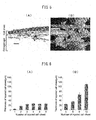
- the HE stain image was digitized and the thickness at 10 sites was measured.
- the thickness of second and third layers increased as the number of layers increased, whereas for a 4th layer or more, no changes in the thickness were noted.
- the measured values were about 7 ⁇ m for the first layer, about 18 ⁇ m for the second layer, about 24 ⁇ m for the 3rd layer, about 23 ⁇ m for the 4th layer, about 26 ⁇ m for the 5th layer, and about 23 ⁇ m for the 6th layer.
- the thickness of the tissue increased as the number of layers increased up to the third layer, the thickness of layers greater than the third layer was almost equal to that of the third layer ( Fig. 6A ).
- the reason for this may be that the supply of oxygen and nutrients and the removal of wastes due to diffusion in the cell sheet may reach only up to the third layer, and thus the cell sheet on the 4th layer or more may become necrotic.
- a vascular network was formed by an artificial channel in the collagen gel and the cell sheet. Since the myocardial cell sheet of the primary isolate contains fibroblasts, endothelial cells, and smooth muscles in addition to myocardial cells, neovascular formation is expected to occur by the presence of blood vessels in the cell sheet and by performing a gel culture. Thus, it was assumed that by preparing a channel for perfusing a culture medium into the collagen gel and culturing the myocardial cell sheet of the primary isolate on the collagen gel channel, a fresh culture medium could be supplied to the myocardial cells even in a thick tissue if blood vessels are newly formed from the cell sheet and connected to the artificial channel.
- the thicknesses of tissues prepared by: (1) culturing 6 layers (3 layers + 3 layers) for a total of 10 days, (2) perfusion culturing 8 layers (3 layers + 3 layers + 2 layers) for a total of 15 days, and (3) perfusion culturing 9 layers (3 layers + 3 layers + 3 layers) for a total of 15 days were compared to a tissue prepared by single layering (3 layers, 5 days) to see changes in thickness.
- the sample was fixed in 4% paraformaldehyde, and the paraffin sections were HE-stained.
- Each of the stained tissues is shown in Fig. 7 . 3 layers + 3 layers + 3 layers were repeatedly layered with an interval culturing period of 5 days for a total of 15 days.
- a vascular bed-forming container was fabricated that was designed to generate channels on and under the multilayered cell sheet.
- a stainless steel wire was provided on an acrylic frame, and into the frame, a collagen gel solution was run, cured and gelled. Then, the stainless steel wire at the lower part was removed to obtain a gel having a microchannel. Thereafter a three-layer myocardial cell sheet was layered. Then, at the upper part of the multilayered cell sheet, a stainless steel wire was mounted, and into the frame, a collagen gel solution was run and gelled. Then, the stainless steel wire at the upper part was removed to generate a channel at the upper part as well.
- culturing was performed at a flow rate of the lower channel of 500 ⁇ l/min ( Fig. 8 (left), Microchannel A) and a flow rate of the upper channel of 250 ⁇ l/min ( Fig. 8 (left), Microchannel B), and a perfusion culture was performed for 5 days in the bioreactor shown in Fig. 4 .
- blood was perfused from the lower channel in order to confirm the connection of the capillary vascular network in the cell sheet and the channel in the vascular bed ( Fig. 9A ).
- the color of the upper part of the cell sheet was found to be changed, indicating that a channel has been formed in between the gel and the multilayered cell sheet.
- vascular endothelial cells migrated toward the lower channel where the flow rate is faster, forming a vascular network ( Fig. 9B ).
- vascular endothelial cells migrate toward the upper channel where the flow rate is faster.
- the vascular network is constructed by the effect of the physical characteristics of the flow rate of the culture medium.
- the distance between the cell sheet and the channel is 100 ⁇ m or less, vascular endothelial cells migrate efficiently, and blood vessel-like structures are formed.
- Fig. 10 is a diagram showing the outline of vascular network construction induced during the fabrication of a multilayered cell sheet using a vascular bed. From a cell sheet ( Fig. 10A ) placed on the collagen gel, vascular endothelial cells migrate toward the channel ( Fig. 10B ), and coat the inner wall of the channel, forming a lumen structure ( Fig. 10C ). Thereafter, further transplantation of the multilayered cell sheet on the upper layer ( Fig. 10D ) causes the connection of the lumen structure connected to the gel channel and the vascular endothelial cells in the newly layered cell sheet, leading to vascular formation ( Fig. 10E ). By repeating this process, a thick multilayered cell sheet can be fabricated ( Fig. 10E ).
- a vascular network can be constructed in a cell sheet, and by layering the sheet, a thick multilayered cell sheet can be easily fabricated.
- a thick multilayered cell sheet is useful as an in-vivo tissue substitute in regenerative medicine for various tissues.
Landscapes
- Health & Medical Sciences (AREA)
- Engineering & Computer Science (AREA)
- Life Sciences & Earth Sciences (AREA)
- Bioinformatics & Cheminformatics (AREA)
- Chemical & Material Sciences (AREA)
- Organic Chemistry (AREA)
- Biomedical Technology (AREA)
- Wood Science & Technology (AREA)
- Zoology (AREA)
- Genetics & Genomics (AREA)
- General Health & Medical Sciences (AREA)
- Biotechnology (AREA)
- Microbiology (AREA)
- Biochemistry (AREA)
- General Engineering & Computer Science (AREA)
- Cardiology (AREA)
- Veterinary Medicine (AREA)
- Animal Behavior & Ethology (AREA)
- Sustainable Development (AREA)
- Heart & Thoracic Surgery (AREA)
- Public Health (AREA)
- Vascular Medicine (AREA)
- Cell Biology (AREA)
- Chemical Kinetics & Catalysis (AREA)
- Medicinal Chemistry (AREA)
- General Chemical & Material Sciences (AREA)
- Pharmacology & Pharmacy (AREA)
- Nuclear Medicine, Radiotherapy & Molecular Imaging (AREA)
- Molecular Biology (AREA)
- Rheumatology (AREA)
- Immunology (AREA)
- Transplantation (AREA)
- Oral & Maxillofacial Surgery (AREA)
- Urology & Nephrology (AREA)
- Hospice & Palliative Care (AREA)
- Micro-Organisms Or Cultivation Processes Thereof (AREA)
- Materials For Medical Uses (AREA)
- Medicines Containing Material From Animals Or Micro-Organisms (AREA)
- Apparatus Associated With Microorganisms And Enzymes (AREA)
Applications Claiming Priority (2)
| Application Number | Priority Date | Filing Date | Title |
|---|---|---|---|
| JP2010225201 | 2010-09-14 | ||
| PCT/JP2011/071056 WO2012036225A1 (fr) | 2010-09-14 | 2011-09-14 | Procédé de fabrication de feuillet cellulaire multicouche, feuillet cellulaire multicouche ayant un réseau vasculaire obtenu par le procédé, procédé d'utilisation de celui-ci |
Publications (3)
| Publication Number | Publication Date |
|---|---|
| EP2617811A1 true EP2617811A1 (fr) | 2013-07-24 |
| EP2617811A4 EP2617811A4 (fr) | 2014-03-26 |
| EP2617811B1 EP2617811B1 (fr) | 2016-06-15 |
Family
ID=45831681
Family Applications (1)
| Application Number | Title | Priority Date | Filing Date |
|---|---|---|---|
| EP11825225.3A Not-in-force EP2617811B1 (fr) | 2010-09-14 | 2011-09-14 | Procédé de fabrication de feuillet cellulaire multicouche, feuillet cellulaire multicouche ayant un réseau vasculaire obtenu par le procédé, procédé d'utilisation de celui-ci |
Country Status (5)
| Country | Link |
|---|---|
| US (1) | US9617519B2 (fr) |
| EP (1) | EP2617811B1 (fr) |
| JP (1) | JP5322333B2 (fr) |
| CN (1) | CN103097518B (fr) |
| WO (1) | WO2012036225A1 (fr) |
Cited By (2)
| Publication number | Priority date | Publication date | Assignee | Title |
|---|---|---|---|---|
| EP3199622A4 (fr) * | 2014-09-23 | 2018-08-08 | The University of Tokyo | Tissu artificiel tridimensionnel, son procédé de production, dispositif de perfusion de type tissu artificiel tridimensionnel, et méthode d'évaluation des médicaments à l'aide dudit tissu artificiel tridimensionnel |
| EP3375860A4 (fr) * | 2015-11-10 | 2019-07-03 | Nikkiso Co., Ltd. | Composite support de cellules et procédé de production de composite support de cellules |
Families Citing this family (24)
| Publication number | Priority date | Publication date | Assignee | Title |
|---|---|---|---|---|
| WO2013069661A1 (fr) * | 2011-11-08 | 2013-05-16 | 国立大学法人名古屋大学 | Feuillet de cellules progénitrices vasculaires issu de cellules souches pluripotentes induites, et son procédé de production |
| CN103100112B (zh) * | 2012-12-07 | 2014-04-16 | 中国人民解放军第四军医大学 | 一种双膜结构移植材料的制备方法及用途 |
| JP6047000B2 (ja) * | 2012-12-12 | 2016-12-21 | 日本メナード化粧品株式会社 | 細胞を含む支持体層に培養液を灌流させることを特徴とする3次元培養組織 |
| JP6341553B2 (ja) * | 2013-11-27 | 2018-06-13 | 国立大学法人大阪大学 | 三次元組織体及びその製造方法 |
| JP6452304B2 (ja) * | 2014-03-31 | 2019-01-16 | 学校法人東京女子医科大学 | 細胞シート培養基材、細胞シート培養基材複合物、及び細胞シート/培養基材複合の製造方法 |
| CN104225681B (zh) * | 2014-09-19 | 2016-03-02 | 浙江大学 | 一种组织工程骨及其制备方法 |
| JP6480192B2 (ja) * | 2015-01-22 | 2019-03-06 | 学校法人東京女子医科大学 | 血管床における流体の灌流方法並びに該方法の実施装置 |
| EP3265104A4 (fr) | 2015-03-03 | 2018-11-07 | President and Fellows of Harvard College | Procédés de génération de tissu humain fonctionnel |
| WO2017003546A2 (fr) * | 2015-04-24 | 2017-01-05 | President And Fellows Of Harvard College | Dispositifs de simulation d'une fonction d'un tissu et leurs procédés d'utilisation et fabrication |
| US10883080B2 (en) * | 2015-06-25 | 2021-01-05 | Tokyo Women's Medical University | Method for culturing animal cell composition, method for producing animal cell composition using same, and animal cell composition |
| GB2561312B (en) | 2015-10-19 | 2021-03-24 | Emulate Inc | Microfluidic model of the blood brain barrier |
| JP6792777B2 (ja) | 2016-02-22 | 2020-12-02 | 凸版印刷株式会社 | スフェロイド形成促進方法 |
| JP6782002B2 (ja) * | 2016-06-09 | 2020-11-11 | 国立大学法人 鹿児島大学 | 毛細血管網様の微小流路を有する微小流路構造体の製造方法 |
| EP3593816A4 (fr) | 2017-03-06 | 2020-12-30 | Tokyo Women's Medical University | Inhibiteur de lypd1 et procédé de production de tissu biologique l'utilisant |
| JP7130200B2 (ja) | 2017-10-25 | 2022-09-05 | 日本光電工業株式会社 | 心筋細胞を含むシート状組織の張力測定デバイス、システム及びキット |
| CN108485975A (zh) * | 2018-06-29 | 2018-09-04 | 大连医科大学附属第医院 | 仿生芯片肾脏 |
| US20210386790A1 (en) * | 2019-01-16 | 2021-12-16 | University Of Utah Research Foundation | Use of mesenchymal stem cell sheets for preventing uterine scar formation |
| JP2020141661A (ja) | 2019-02-28 | 2020-09-10 | 日本光電工業株式会社 | 筋細胞を含む細胞構造体の張力測定デバイス、システム及びキット |
| JP7033095B2 (ja) * | 2019-03-04 | 2022-03-09 | 日清食品ホールディングス株式会社 | 三次元筋組織とその製造方法 |
| WO2021132478A1 (fr) * | 2019-12-26 | 2021-07-01 | 国立大学法人東京大学 | Complexe de tissu tridimensionnel et procédé de fabrication de complexe de tissu tridimensionnel |
| US20230293771A1 (en) * | 2020-07-16 | 2023-09-21 | University Of Utah Research Foundation | Chondrocyte cell sheets and methods for their production and use |
| JP7793141B2 (ja) * | 2021-09-07 | 2026-01-05 | Ckd株式会社 | 血管網の製造方法、及び血管網の製造装置 |
| US20250034503A1 (en) * | 2021-12-22 | 2025-01-30 | Cellular Agriculture Ltd | Cell culture construct |
| WO2023228255A1 (fr) * | 2022-05-23 | 2023-11-30 | 学校法人早稲田大学 | Modèle de lésion associé à un organe tubulaire, son procédé de fabrication et similaire |
Family Cites Families (8)
| Publication number | Priority date | Publication date | Assignee | Title |
|---|---|---|---|---|
| JPH06104061B2 (ja) | 1989-02-10 | 1994-12-21 | 花王株式会社 | 細胞培養支持体材料 |
| JPH05192138A (ja) | 1992-01-22 | 1993-08-03 | Kao Corp | 皮膚細胞培養法及び培養皮膚 |
| EP2275532B1 (fr) | 2000-07-21 | 2012-06-06 | Cellseed Inc. | Feuille cellulaire du type muscle cardiaque et procédé de production associé |
| WO2002078439A2 (fr) | 2001-03-30 | 2002-10-10 | The Arizona Board Of Regents On Behalf Of The University Of Arizona | Constructions prevascularisees d'implantation destinees a fournir une perfusion sanguine |
| US20080131476A1 (en) | 2005-02-28 | 2008-06-05 | Masato Kanzaki | Cultured Cell Sheet, Production Method and Tissue Repair Method Using Thereof |
| US7622298B2 (en) | 2006-03-24 | 2009-11-24 | Norits, Inc. | Method for creating perfusable microvessel systems |
| WO2010065957A2 (fr) | 2008-12-05 | 2010-06-10 | Trustees Of Tufts College | Constructions cutanées vivantes vascularisées et leurs procédés d'utilisation |
| US8669086B2 (en) * | 2010-04-29 | 2014-03-11 | The United States Of America, As Represented By The Secretary Of The Navy | Cell and biofactor printable biopapers |
-
2011
- 2011-09-14 CN CN201180044349.XA patent/CN103097518B/zh not_active Expired - Fee Related
- 2011-09-14 EP EP11825225.3A patent/EP2617811B1/fr not_active Not-in-force
- 2011-09-14 US US13/823,007 patent/US9617519B2/en active Active
- 2011-09-14 WO PCT/JP2011/071056 patent/WO2012036225A1/fr not_active Ceased
- 2011-09-14 JP JP2012534047A patent/JP5322333B2/ja active Active
Cited By (4)
| Publication number | Priority date | Publication date | Assignee | Title |
|---|---|---|---|---|
| EP3199622A4 (fr) * | 2014-09-23 | 2018-08-08 | The University of Tokyo | Tissu artificiel tridimensionnel, son procédé de production, dispositif de perfusion de type tissu artificiel tridimensionnel, et méthode d'évaluation des médicaments à l'aide dudit tissu artificiel tridimensionnel |
| US10624991B2 (en) | 2014-09-23 | 2020-04-21 | The University Of Tokyo | Three-dimensional artificial tissue, method for producing the same, three-dimensional artificial tissue perfusion device, and drug evaluation method using three-dimensional artificial tissue |
| EP3375860A4 (fr) * | 2015-11-10 | 2019-07-03 | Nikkiso Co., Ltd. | Composite support de cellules et procédé de production de composite support de cellules |
| US10870824B2 (en) | 2015-11-10 | 2020-12-22 | Nikkiso Co., Ltd. | Cell support composite and method for producing cell support composite |
Also Published As
| Publication number | Publication date |
|---|---|
| EP2617811A4 (fr) | 2014-03-26 |
| US20130173018A1 (en) | 2013-07-04 |
| WO2012036225A1 (fr) | 2012-03-22 |
| CN103097518A (zh) | 2013-05-08 |
| US9617519B2 (en) | 2017-04-11 |
| JPWO2012036225A1 (ja) | 2014-02-03 |
| JP5322333B2 (ja) | 2013-10-23 |
| CN103097518B (zh) | 2015-10-14 |
| EP2617811B1 (fr) | 2016-06-15 |
Similar Documents
| Publication | Publication Date | Title |
|---|---|---|
| US9617519B2 (en) | Method for manufacturing multilayered cell sheet, multilayered cell sheet having vascular network obtained thereby, and method of use thereof | |
| EP2617810B1 (fr) | Procédé de production de feuillets de cellules superposées, feuillets de cellules superposées dotés d'un réseau vasculaire obtenu par ce procédé, et procédé d'utilisation associé | |
| JP2024015176A (ja) | 臓器および組織を脱細胞化および再細胞化する方法 | |
| CN102858380B (zh) | 胰岛细胞片、制造方法及其利用方法 | |
| JP5725560B2 (ja) | 細胞シートを利用した細胞評価システム及びその利用方法 | |
| JP2016039806A (ja) | サイトカイン産生細胞シートとその利用方法 | |
| JP6452304B2 (ja) | 細胞シート培養基材、細胞シート培養基材複合物、及び細胞シート/培養基材複合の製造方法 | |
| JP2017074050A (ja) | 肝組織細胞機能の長期維持方法 | |
| JP6468714B2 (ja) | 培養容器及びそれを利用した複数の積層細胞シートの作製方法 | |
| EP2130905A1 (fr) | Procédé de culture de cellules eucaryotes | |
| KR20170121205A (ko) | 줄기세포 배양상청의 제조방법 | |
| US10883080B2 (en) | Method for culturing animal cell composition, method for producing animal cell composition using same, and animal cell composition | |
| Hou et al. | Construction of a three-dimensional urothelium on-chip with barrier function based on urinary flow microenvironment | |
| JP6480192B2 (ja) | 血管床における流体の灌流方法並びに該方法の実施装置 | |
| JPWO2005047496A1 (ja) | 細胞培養法、細胞の三次元培養法、三次元組織、人工臓器、及び組織移植方法 | |
| JP5252828B2 (ja) | 上皮系細胞培養方法 | |
| JP2023070669A (ja) | 人工血管床、人工三次元生体組織及び血管網を備えた人工三次元生体組織の作製方法 | |
| JP2021023826A (ja) | 創傷被覆用シート、及び創傷被覆方法 | |
| Tajima et al. | Construction method of cellular structure using cell-sheet with biocompatible rigging |
Legal Events
| Date | Code | Title | Description |
|---|---|---|---|
| PUAI | Public reference made under article 153(3) epc to a published international application that has entered the european phase |
Free format text: ORIGINAL CODE: 0009012 |
|
| 17P | Request for examination filed |
Effective date: 20130410 |
|
| AK | Designated contracting states |
Kind code of ref document: A1 Designated state(s): AL AT BE BG CH CY CZ DE DK EE ES FI FR GB GR HR HU IE IS IT LI LT LU LV MC MK MT NL NO PL PT RO RS SE SI SK SM TR |
|
| DAX | Request for extension of the european patent (deleted) | ||
| A4 | Supplementary search report drawn up and despatched |
Effective date: 20140225 |
|
| RIC1 | Information provided on ipc code assigned before grant |
Ipc: A61K 35/12 20060101ALI20140219BHEP Ipc: A61P 9/00 20060101ALI20140219BHEP Ipc: A61P 9/10 20060101ALI20140219BHEP Ipc: C12M 3/04 20060101ALI20140219BHEP Ipc: A61P 9/04 20060101ALI20140219BHEP Ipc: A61K 35/34 20060101ALI20140219BHEP Ipc: C12N 5/071 20100101AFI20140219BHEP |
|
| 17Q | First examination report despatched |
Effective date: 20150108 |
|
| GRAP | Despatch of communication of intention to grant a patent |
Free format text: ORIGINAL CODE: EPIDOSNIGR1 |
|
| INTG | Intention to grant announced |
Effective date: 20160307 |
|
| GRAS | Grant fee paid |
Free format text: ORIGINAL CODE: EPIDOSNIGR3 |
|
| GRAA | (expected) grant |
Free format text: ORIGINAL CODE: 0009210 |
|
| AK | Designated contracting states |
Kind code of ref document: B1 Designated state(s): AL AT BE BG CH CY CZ DE DK EE ES FI FR GB GR HR HU IE IS IT LI LT LU LV MC MK MT NL NO PL PT RO RS SE SI SK SM TR |
|
| REG | Reference to a national code |
Ref country code: CH Ref legal event code: EP Ref country code: GB Ref legal event code: FG4D |
|
| REG | Reference to a national code |
Ref country code: IE Ref legal event code: FG4D |
|
| REG | Reference to a national code |
Ref country code: AT Ref legal event code: REF Ref document number: 806534 Country of ref document: AT Kind code of ref document: T Effective date: 20160715 |
|
| REG | Reference to a national code |
Ref country code: DE Ref legal event code: R096 Ref document number: 602011027484 Country of ref document: DE Ref country code: FR Ref legal event code: PLFP Year of fee payment: 6 |
|
| REG | Reference to a national code |
Ref country code: LT Ref legal event code: MG4D |
|
| REG | Reference to a national code |
Ref country code: NL Ref legal event code: MP Effective date: 20160615 |
|
| PG25 | Lapsed in a contracting state [announced via postgrant information from national office to epo] |
Ref country code: FI Free format text: LAPSE BECAUSE OF FAILURE TO SUBMIT A TRANSLATION OF THE DESCRIPTION OR TO PAY THE FEE WITHIN THE PRESCRIBED TIME-LIMIT Effective date: 20160615 Ref country code: LT Free format text: LAPSE BECAUSE OF FAILURE TO SUBMIT A TRANSLATION OF THE DESCRIPTION OR TO PAY THE FEE WITHIN THE PRESCRIBED TIME-LIMIT Effective date: 20160615 Ref country code: NO Free format text: LAPSE BECAUSE OF FAILURE TO SUBMIT A TRANSLATION OF THE DESCRIPTION OR TO PAY THE FEE WITHIN THE PRESCRIBED TIME-LIMIT Effective date: 20160915 |
|
| REG | Reference to a national code |
Ref country code: AT Ref legal event code: MK05 Ref document number: 806534 Country of ref document: AT Kind code of ref document: T Effective date: 20160615 |
|
| PG25 | Lapsed in a contracting state [announced via postgrant information from national office to epo] |
Ref country code: SE Free format text: LAPSE BECAUSE OF FAILURE TO SUBMIT A TRANSLATION OF THE DESCRIPTION OR TO PAY THE FEE WITHIN THE PRESCRIBED TIME-LIMIT Effective date: 20160615 Ref country code: RS Free format text: LAPSE BECAUSE OF FAILURE TO SUBMIT A TRANSLATION OF THE DESCRIPTION OR TO PAY THE FEE WITHIN THE PRESCRIBED TIME-LIMIT Effective date: 20160615 Ref country code: NL Free format text: LAPSE BECAUSE OF FAILURE TO SUBMIT A TRANSLATION OF THE DESCRIPTION OR TO PAY THE FEE WITHIN THE PRESCRIBED TIME-LIMIT Effective date: 20160615 Ref country code: GR Free format text: LAPSE BECAUSE OF FAILURE TO SUBMIT A TRANSLATION OF THE DESCRIPTION OR TO PAY THE FEE WITHIN THE PRESCRIBED TIME-LIMIT Effective date: 20160916 Ref country code: LV Free format text: LAPSE BECAUSE OF FAILURE TO SUBMIT A TRANSLATION OF THE DESCRIPTION OR TO PAY THE FEE WITHIN THE PRESCRIBED TIME-LIMIT Effective date: 20160615 |
|
| PG25 | Lapsed in a contracting state [announced via postgrant information from national office to epo] |
Ref country code: CZ Free format text: LAPSE BECAUSE OF FAILURE TO SUBMIT A TRANSLATION OF THE DESCRIPTION OR TO PAY THE FEE WITHIN THE PRESCRIBED TIME-LIMIT Effective date: 20160615 Ref country code: IS Free format text: LAPSE BECAUSE OF FAILURE TO SUBMIT A TRANSLATION OF THE DESCRIPTION OR TO PAY THE FEE WITHIN THE PRESCRIBED TIME-LIMIT Effective date: 20161015 Ref country code: RO Free format text: LAPSE BECAUSE OF FAILURE TO SUBMIT A TRANSLATION OF THE DESCRIPTION OR TO PAY THE FEE WITHIN THE PRESCRIBED TIME-LIMIT Effective date: 20160615 Ref country code: IT Free format text: LAPSE BECAUSE OF FAILURE TO SUBMIT A TRANSLATION OF THE DESCRIPTION OR TO PAY THE FEE WITHIN THE PRESCRIBED TIME-LIMIT Effective date: 20160615 Ref country code: EE Free format text: LAPSE BECAUSE OF FAILURE TO SUBMIT A TRANSLATION OF THE DESCRIPTION OR TO PAY THE FEE WITHIN THE PRESCRIBED TIME-LIMIT Effective date: 20160615 Ref country code: SK Free format text: LAPSE BECAUSE OF FAILURE TO SUBMIT A TRANSLATION OF THE DESCRIPTION OR TO PAY THE FEE WITHIN THE PRESCRIBED TIME-LIMIT Effective date: 20160615 |
|
| PG25 | Lapsed in a contracting state [announced via postgrant information from national office to epo] |
Ref country code: ES Free format text: LAPSE BECAUSE OF FAILURE TO SUBMIT A TRANSLATION OF THE DESCRIPTION OR TO PAY THE FEE WITHIN THE PRESCRIBED TIME-LIMIT Effective date: 20160615 Ref country code: AT Free format text: LAPSE BECAUSE OF FAILURE TO SUBMIT A TRANSLATION OF THE DESCRIPTION OR TO PAY THE FEE WITHIN THE PRESCRIBED TIME-LIMIT Effective date: 20160615 Ref country code: BE Free format text: LAPSE BECAUSE OF NON-PAYMENT OF DUE FEES Effective date: 20160615 Ref country code: SM Free format text: LAPSE BECAUSE OF FAILURE TO SUBMIT A TRANSLATION OF THE DESCRIPTION OR TO PAY THE FEE WITHIN THE PRESCRIBED TIME-LIMIT Effective date: 20160615 Ref country code: PL Free format text: LAPSE BECAUSE OF FAILURE TO SUBMIT A TRANSLATION OF THE DESCRIPTION OR TO PAY THE FEE WITHIN THE PRESCRIBED TIME-LIMIT Effective date: 20160615 Ref country code: PT Free format text: LAPSE BECAUSE OF FAILURE TO SUBMIT A TRANSLATION OF THE DESCRIPTION OR TO PAY THE FEE WITHIN THE PRESCRIBED TIME-LIMIT Effective date: 20161017 |
|
| REG | Reference to a national code |
Ref country code: DE Ref legal event code: R097 Ref document number: 602011027484 Country of ref document: DE |
|
| PLBE | No opposition filed within time limit |
Free format text: ORIGINAL CODE: 0009261 |
|
| STAA | Information on the status of an ep patent application or granted ep patent |
Free format text: STATUS: NO OPPOSITION FILED WITHIN TIME LIMIT |
|
| PG25 | Lapsed in a contracting state [announced via postgrant information from national office to epo] |
Ref country code: MC Free format text: LAPSE BECAUSE OF FAILURE TO SUBMIT A TRANSLATION OF THE DESCRIPTION OR TO PAY THE FEE WITHIN THE PRESCRIBED TIME-LIMIT Effective date: 20160615 |
|
| REG | Reference to a national code |
Ref country code: CH Ref legal event code: PL |
|
| 26N | No opposition filed |
Effective date: 20170316 |
|
| PG25 | Lapsed in a contracting state [announced via postgrant information from national office to epo] |
Ref country code: DK Free format text: LAPSE BECAUSE OF FAILURE TO SUBMIT A TRANSLATION OF THE DESCRIPTION OR TO PAY THE FEE WITHIN THE PRESCRIBED TIME-LIMIT Effective date: 20160615 |
|
| REG | Reference to a national code |
Ref country code: IE Ref legal event code: MM4A |
|
| PG25 | Lapsed in a contracting state [announced via postgrant information from national office to epo] |
Ref country code: IE Free format text: LAPSE BECAUSE OF NON-PAYMENT OF DUE FEES Effective date: 20160914 Ref country code: LI Free format text: LAPSE BECAUSE OF NON-PAYMENT OF DUE FEES Effective date: 20160930 Ref country code: CH Free format text: LAPSE BECAUSE OF NON-PAYMENT OF DUE FEES Effective date: 20160930 |
|
| REG | Reference to a national code |
Ref country code: FR Ref legal event code: PLFP Year of fee payment: 7 |
|
| PG25 | Lapsed in a contracting state [announced via postgrant information from national office to epo] |
Ref country code: SI Free format text: LAPSE BECAUSE OF FAILURE TO SUBMIT A TRANSLATION OF THE DESCRIPTION OR TO PAY THE FEE WITHIN THE PRESCRIBED TIME-LIMIT Effective date: 20160615 Ref country code: LU Free format text: LAPSE BECAUSE OF NON-PAYMENT OF DUE FEES Effective date: 20160914 |
|
| PG25 | Lapsed in a contracting state [announced via postgrant information from national office to epo] |
Ref country code: HU Free format text: LAPSE BECAUSE OF FAILURE TO SUBMIT A TRANSLATION OF THE DESCRIPTION OR TO PAY THE FEE WITHIN THE PRESCRIBED TIME-LIMIT; INVALID AB INITIO Effective date: 20110914 Ref country code: CY Free format text: LAPSE BECAUSE OF FAILURE TO SUBMIT A TRANSLATION OF THE DESCRIPTION OR TO PAY THE FEE WITHIN THE PRESCRIBED TIME-LIMIT Effective date: 20160615 |
|
| PG25 | Lapsed in a contracting state [announced via postgrant information from national office to epo] |
Ref country code: TR Free format text: LAPSE BECAUSE OF FAILURE TO SUBMIT A TRANSLATION OF THE DESCRIPTION OR TO PAY THE FEE WITHIN THE PRESCRIBED TIME-LIMIT Effective date: 20160615 Ref country code: HR Free format text: LAPSE BECAUSE OF FAILURE TO SUBMIT A TRANSLATION OF THE DESCRIPTION OR TO PAY THE FEE WITHIN THE PRESCRIBED TIME-LIMIT Effective date: 20160615 Ref country code: MT Free format text: LAPSE BECAUSE OF NON-PAYMENT OF DUE FEES Effective date: 20160930 Ref country code: MK Free format text: LAPSE BECAUSE OF FAILURE TO SUBMIT A TRANSLATION OF THE DESCRIPTION OR TO PAY THE FEE WITHIN THE PRESCRIBED TIME-LIMIT Effective date: 20160615 |
|
| PG25 | Lapsed in a contracting state [announced via postgrant information from national office to epo] |
Ref country code: BG Free format text: LAPSE BECAUSE OF FAILURE TO SUBMIT A TRANSLATION OF THE DESCRIPTION OR TO PAY THE FEE WITHIN THE PRESCRIBED TIME-LIMIT Effective date: 20160615 |
|
| REG | Reference to a national code |
Ref country code: FR Ref legal event code: PLFP Year of fee payment: 8 |
|
| PG25 | Lapsed in a contracting state [announced via postgrant information from national office to epo] |
Ref country code: AL Free format text: LAPSE BECAUSE OF FAILURE TO SUBMIT A TRANSLATION OF THE DESCRIPTION OR TO PAY THE FEE WITHIN THE PRESCRIBED TIME-LIMIT Effective date: 20160615 |
|
| PGFP | Annual fee paid to national office [announced via postgrant information from national office to epo] |
Ref country code: GB Payment date: 20220915 Year of fee payment: 12 Ref country code: DE Payment date: 20220920 Year of fee payment: 12 |
|
| PGFP | Annual fee paid to national office [announced via postgrant information from national office to epo] |
Ref country code: FR Payment date: 20220928 Year of fee payment: 12 |
|
| REG | Reference to a national code |
Ref country code: DE Ref legal event code: R119 Ref document number: 602011027484 Country of ref document: DE |
|
| GBPC | Gb: european patent ceased through non-payment of renewal fee |
Effective date: 20230914 |
|
| PG25 | Lapsed in a contracting state [announced via postgrant information from national office to epo] |
Ref country code: GB Free format text: LAPSE BECAUSE OF NON-PAYMENT OF DUE FEES Effective date: 20230914 |
|
| PG25 | Lapsed in a contracting state [announced via postgrant information from national office to epo] |
Ref country code: GB Free format text: LAPSE BECAUSE OF NON-PAYMENT OF DUE FEES Effective date: 20230914 Ref country code: FR Free format text: LAPSE BECAUSE OF NON-PAYMENT OF DUE FEES Effective date: 20230930 Ref country code: DE Free format text: LAPSE BECAUSE OF NON-PAYMENT OF DUE FEES Effective date: 20240403 |