EP2840552A2 - Procédé et système pour traiter des séries multiples d'images biologiques obtenues d'un patient - Google Patents
Procédé et système pour traiter des séries multiples d'images biologiques obtenues d'un patient Download PDFInfo
- Publication number
- EP2840552A2 EP2840552A2 EP20140189106 EP14189106A EP2840552A2 EP 2840552 A2 EP2840552 A2 EP 2840552A2 EP 20140189106 EP20140189106 EP 20140189106 EP 14189106 A EP14189106 A EP 14189106A EP 2840552 A2 EP2840552 A2 EP 2840552A2
- Authority
- EP
- European Patent Office
- Prior art keywords
- image
- series
- images
- segmentation mask
- method defined
- Prior art date
- Legal status (The legal status is an assumption and is not a legal conclusion. Google has not performed a legal analysis and makes no representation as to the accuracy of the status listed.)
- Ceased
Links
Images



Classifications
-
- G—PHYSICS
- G06—COMPUTING OR CALCULATING; COUNTING
- G06T—IMAGE DATA PROCESSING OR GENERATION, IN GENERAL
- G06T1/00—General purpose image data processing
-
- G—PHYSICS
- G06—COMPUTING OR CALCULATING; COUNTING
- G06T—IMAGE DATA PROCESSING OR GENERATION, IN GENERAL
- G06T7/00—Image analysis
- G06T7/0002—Inspection of images, e.g. flaw detection
- G06T7/0012—Biomedical image inspection
- G06T7/0014—Biomedical image inspection using an image reference approach
-
- A—HUMAN NECESSITIES
- A61—MEDICAL OR VETERINARY SCIENCE; HYGIENE
- A61B—DIAGNOSIS; SURGERY; IDENTIFICATION
- A61B6/00—Apparatus or devices for radiation diagnosis; Apparatus or devices for radiation diagnosis combined with radiation therapy equipment
- A61B6/48—Diagnostic techniques
- A61B6/481—Diagnostic techniques involving the use of contrast agents
-
- A—HUMAN NECESSITIES
- A61—MEDICAL OR VETERINARY SCIENCE; HYGIENE
- A61B—DIAGNOSIS; SURGERY; IDENTIFICATION
- A61B6/00—Apparatus or devices for radiation diagnosis; Apparatus or devices for radiation diagnosis combined with radiation therapy equipment
- A61B6/48—Diagnostic techniques
- A61B6/486—Diagnostic techniques involving generating temporal series of image data
-
- A—HUMAN NECESSITIES
- A61—MEDICAL OR VETERINARY SCIENCE; HYGIENE
- A61B—DIAGNOSIS; SURGERY; IDENTIFICATION
- A61B6/00—Apparatus or devices for radiation diagnosis; Apparatus or devices for radiation diagnosis combined with radiation therapy equipment
- A61B6/50—Apparatus or devices for radiation diagnosis; Apparatus or devices for radiation diagnosis combined with radiation therapy equipment specially adapted for specific body parts; specially adapted for specific clinical applications
- A61B6/507—Apparatus or devices for radiation diagnosis; Apparatus or devices for radiation diagnosis combined with radiation therapy equipment specially adapted for specific body parts; specially adapted for specific clinical applications for determination of haemodynamic parameters, e.g. perfusion CT
-
- A—HUMAN NECESSITIES
- A61—MEDICAL OR VETERINARY SCIENCE; HYGIENE
- A61B—DIAGNOSIS; SURGERY; IDENTIFICATION
- A61B6/00—Apparatus or devices for radiation diagnosis; Apparatus or devices for radiation diagnosis combined with radiation therapy equipment
- A61B6/52—Devices using data or image processing specially adapted for radiation diagnosis
- A61B6/5211—Devices using data or image processing specially adapted for radiation diagnosis involving processing of medical diagnostic data
- A61B6/5229—Devices using data or image processing specially adapted for radiation diagnosis involving processing of medical diagnostic data combining image data of a patient, e.g. combining a functional image with an anatomical image
-
- G—PHYSICS
- G06—COMPUTING OR CALCULATING; COUNTING
- G06T—IMAGE DATA PROCESSING OR GENERATION, IN GENERAL
- G06T7/00—Image analysis
- G06T7/40—Analysis of texture
- G06T7/41—Analysis of texture based on statistical description of texture
- G06T7/44—Analysis of texture based on statistical description of texture using image operators, e.g. filters, edge density metrics or local histograms
-
- G—PHYSICS
- G06—COMPUTING OR CALCULATING; COUNTING
- G06T—IMAGE DATA PROCESSING OR GENERATION, IN GENERAL
- G06T2200/00—Indexing scheme for image data processing or generation, in general
- G06T2200/24—Indexing scheme for image data processing or generation, in general involving graphical user interfaces [GUIs]
-
- G—PHYSICS
- G06—COMPUTING OR CALCULATING; COUNTING
- G06T—IMAGE DATA PROCESSING OR GENERATION, IN GENERAL
- G06T2207/00—Indexing scheme for image analysis or image enhancement
- G06T2207/10—Image acquisition modality
- G06T2207/10016—Video; Image sequence
-
- G—PHYSICS
- G06—COMPUTING OR CALCULATING; COUNTING
- G06T—IMAGE DATA PROCESSING OR GENERATION, IN GENERAL
- G06T2207/00—Indexing scheme for image analysis or image enhancement
- G06T2207/20—Special algorithmic details
- G06T2207/20092—Interactive image processing based on input by user
- G06T2207/20108—Interactive selection of 2D slice in a 3D data set
-
- G—PHYSICS
- G06—COMPUTING OR CALCULATING; COUNTING
- G06T—IMAGE DATA PROCESSING OR GENERATION, IN GENERAL
- G06T2207/00—Indexing scheme for image analysis or image enhancement
- G06T2207/30—Subject of image; Context of image processing
- G06T2207/30004—Biomedical image processing
- G06T2207/30016—Brain
Definitions
- the present invention relates generally to the processing of biological images and, more particularly, to the processing of multiple series of biological images obtained from a patient affected by an ischemic stroke.
- Stroke is one of the leading causes of morbidity and mortality in developed countries.
- a stroke occurs when blood vessels in the brain are compromised. It has been defined as a “sudden neurological shortage presumed to be of vascular origin” (translated from Bousser MG: Classification et orientation generates du diagnostic. In “Accidents vas Diagrams cerebraux”: Bogousslavsky J, Bousser MG, Mas JL, 1993, 95-103, Doin editeurs .)
- Hemorrhagic stroke is relatively infrequent, and occurs when a blood vessel ruptures, flooding a portion of the brain with blood.
- ischemic (or nonhemorrhagic) stroke is much more common and occurs when a blood vessel is blocked (e.g., due to a clot), causing cerebral nervous tissue to be deprived of oxygen, ultimately leading to necrosis if left untreated.
- Ischemic stroke is typically treated by administration of a thrombolytic, namely, a drug that aims to dissolve the clot that caused obstruction of the vessel in question.
- a thrombolytic namely, a drug that aims to dissolve the clot that caused obstruction of the vessel in question.
- This technique restores blood flow to hypoperfused areas, potentially salvaging those portions of the affected cerebral tissue that have not yet been irreversibly damaged because they have been receiving blood flow from collateral arteries anastomosing with branches of the occluded vascular tree. These portions are known as the "ischemic penumbra", while portions of the cerebral tissue that have been irreversibly damaged due to oxygen deprivation are known as the "core ischemic zone”. Further information can be found on the Internet by visiting the Internet Stroke Center at Washington University in St. Louis (http://www.strokecenter.org/education/ais_pathogenesis/22_ischemicjpenumbra.htm).
- thrombolysis i.e., treatment with a thrombolytic
- cerebral tissue being depleted at a rate of several (approximately 4) million neurons per minute
- clinicians find themselves operating within a narrow time window (typically considered to be only a few hours) from occurrence of a stroke if there is to be any hope of success.
- a thrombolytic is administered to a patient within this time window, then it may be possible to salvage the ischemic penumbra.
- reperfusion of severely hypoperfused areas can result in hemorrhage with its associated complications. If it turns out that the patient had insignificant salvageable cerebral tissue to begin with (i.e., with dim prospects for a positive outcome from the outset), then administering the thrombolytic will unnecessarily raise the risk of harming the patient.
- clinicians must typically assess the size of the ischemic penumbra and core ischemic zone by viewing and considering numerous images obtained from radiological instruments such as magnetic resonance imaging (MRI) machines. Due to the disparity in image type and presentation, it is only the most highly experienced clinicians who are able to extract useful diagnostic information from these images towards the decision to administer a thrombolytic. However, the number of clinicians having the requisite level of experience is expected to become inadequate to meet the needs of a growing and aging population that naturally brings about an increase in ischemic stroke cases.
- MRI magnetic resonance imaging
- a first broad aspect of the present invention seeks to provide a computer-readable storage medium comprising computer-readable program code stored thereon which, when interpreted by a computing apparatus, causes the computing apparatus to implement an image processing tool for processing a plurality of biological images arranged in a plurality of image series wherein certain biological images across different image series have a predefined correspondence with one another.
- the computer-readable program code comprises computer-readable program code for causing the computing apparatus to be attentive to receipt of an indication of a selected biological image from the plurality of biological images and belonging to a first one of the image series; computer-readable program code for causing the computing apparatus to be attentive to receipt of an indication of a segmentation mask created based on the selected biological image; computer-readable program code for causing the computing apparatus to apply the segmentation mask to a second biological image from the plurality of biological images, the second biological image belonging to a second one of the image series that is different from the first one of the image series, the second biological image having a predefined correspondence with the selected biological image; and computer-readable program code for causing the computing apparatus to display the second biological image after application of the segmentation mask.
- a second broad aspect of the present invention seeks to provide a method, which comprises obtaining a plurality of series of biological images including a first series and a second series, wherein certain ones of the images in the first series and certain ones of the images in the second series are corresponding; creating a respective segmentation mask for each of at least one selected image in the first series; applying each respective segmentation mask to the corresponding image in the second series; and selecting a set of the images in the second series on which to effect a volumetric computation, the selected set of images in the second series including at least one image to which a respective segmentation mask has been applied.
- a third broad aspect of the present invention seeks to provide a system, which comprises means for obtaining a plurality of series of biological images including a first series and a second series, wherein certain ones of the images in the first series and certain ones of the images in the second series are corresponding; means for creating a respective segmentation mask for each of at least one selected image in the first series; means for applying each respective segmentation mask to the corresponding image in the second series; and means for selecting a set of the images in the second series on which to effect a volumetric computation, the selected set of images in the second series including at least one image to which a respective segmentation mask has been applied.
- a fourth broad aspect of the present invention seeks to provide an image processing system for processing a plurality of biological images arranged in a plurality of image series wherein certain biological images across different image series have a predefined correspondence with one another.
- the image processing system comprises an input configured to receive an indication of a selected biological image from the plurality of biological images and belonging to a first one of the image series and receive an indication of a segmentation mask created based on the selected biological image; a processing entity configured to apply the segmentation mask to a second biological image from the plurality of biological images, the second biological image belonging to a second one of the image series that is different from the first one of the image series, the second biological image having a predefined correspondence with the selected biological image; and a display entity configured to cause display of the second biological image.
- a fifth broad aspect of the present invention seeks to provide a method of processing images that are arranged in a first series of biological images and a second series of biological images, each image being associated with a respective axial slice height.
- the method comprises:
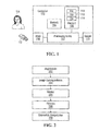
- Non-limiting embodiments of the present invention provide an image processing tool that implements methods of processing biological images. These methods may be performed, at least in part, by a computing apparatus such as a computer shown in Fig. 1 as 100.
- the computer 100 has a processing entity 102 communicatively coupled to a first memory 104, a second memory 106, an input 108 and an output 110.
- the processing entity 102 may include one or more processors for processing computer-executable instructions and data. It will be understood by those of ordinary skill in the art that the computer 100 may also include other components not shown in Fig. 1 .
- the computer 100 may communicate with other apparatuses and systems (not shown) over a network (not shown).
- such other apparatuses and systems may include a PACS (picture archiving and communications system) commonly used to store radiological and other biological images of patients in a healthcare setting.
- PACS picture archiving and communications system
- the first memory 104 can be an electronic storage comprising a computer-readable medium storing computer-executable instructions and/or data.
- the first memory 104 is readily accessible by the processing entity 102 at runtime and may include a volatile memory (e.g., random access memory - RAM) for storing computer-executable instructions and/or data.
- the second memory 106 can be an electronic storage comprising a computer-readable medium storing computer-executable instructions and/or data.
- the second memory 106 may include a non-volatile memory (e.g., flash EEPROM) for storing computer-executable instructions and data in a persistent manner.
- the second memory 106 may store electronic files 116.
- the files 116 may encode images such as radiological images (specifically, MRI images) and/or other types of biological images of a patient.
- the biological images can be two-dimensional and each may comprise a plurality of picture elements (pixels) having respective values.
- the biological images can be three-dimensional and each may comprise a plurality of volume elements (voxels) having respective values.
- the input 108 may be used to receive commands and other input from a user 114 such as a clinician who is attempting to determine whether to administer a thrombolytic to a patient in real time, or a student who is participating in a simulation exercise.
- the input 108 may include one or more input devices, examples of which include but are not limited to a keyboard, a mouse, a microphone, a touch screen and an image acquisition apparatus (e.g., a scanner, a camera, an x-ray machine, MRI machine, etc.).
- the one or more input devices may also include a computer-readable medium such as a removable memory 112 as well as any requisite device for accessing such medium.
- the one or more input devices may be locally or remotely connected to the processing entity 102, either physically or by way of a communication connection.
- the output 110 may include one or more output devices, which may include a display device, such as a screen/monitor.
- output devices include, without limitation, a printer, a speaker, as well as a computer-writable medium and any requisite device for writing to such medium.
- the one or more output devices may be locally or remotely connected to processing entity 102, either physically or by way of a communication connection.
- the processing entity 102 executes computer-executable instructions stored by one or more of the memories 104,106,112, the computer 100 can be caused to carry out one or more of the methods described herein.
- the methods described herein may also be carried out using a hardware device having circuits for performing one or more of the calculations or functions described herein.
- Other configurations for a computing apparatus are also within the scope of the present invention.
- Certain non-limiting embodiments of the present invention have application to diagnosis and treatment of ischemic stroke.
- certain classes of biological images are of particular significance.
- Biological images in a first class are obtained by diffusion-weighted magnetic resonance imaging and are hereinafter referred to as DWI images.
- DWI images can bring to light a variety of parameters, which include, without limitation:
- Biological images in a second class are obtained by perfusion-weighted magnetic resonance imaging and are hereinafter referred to as PWI images.
- Perfusion is the steady state delivery of blood to tissue parenchyma through the capillaries, representing the microscopic coherent motion of water and cellular material.
- PWI images can bring to light a variety of hemodynamic parameters, which include, without limitation:
- PWI images can be obtained by performing computations on a time series of raw images.
- Tmax time to peak of the residue function
- biological images including DWI and PWI images
- biological images that evidence at least some of the above parameters are assumed to be available to the computer 100 for processing and/or display. Such availability can arise by virtue of the biological images being stored as files 116 in the memory 106 or by the PACS, etc.
- the set of images that evidences a particular parameter are hereinafter referred to as forming a "series" of images.
- One or more of the series may include DWI images, while another one or more of the series may include PWI images.
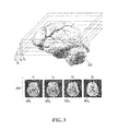
- each biological image 304i, 3042, 3043, 3044 in a given series 306 is a two-dimensional axial image of a patient's brain 302 at a particular axial slice height Hi, H2, H3, H4.
- the axial slice height of an image may be indicated in a header portion of a. file that encodes the image, such as one of the files 116.
- a common format for such a file is DICOM (abbreviation of "Digital Imaging and Communications in Medicine”), which includes a DICOM header that specifies the height of the axial slice at which the accompanying image was taken.
- file formats other than DICOM may be used, including proprietary file formats that may not include a header, whereby the height of an axial slice is recorded in a different fashion.
- views other than axial e.g., sagittal or coronal
- FIG. 2 A non-limiting description of an example method that may be performed by the computer 100 when executing computer-readable instructions stored in one or more of the memories 104,106,112 is now provided with reference to the flowchart in Fig. 2 .
- the method includes the steps of registration 202, image correspondence 204, display 206, filtering 208 and volumetric computation 210. It should be appreciated that not all of these steps are essential in all embodiments. In the description to follow, reference will be made to pixels for reasons of simplicity, with the understanding that reference could also have been made to voxels.
- Step 202 Registration
- the step of registration 202 generally includes ensuring that all images in all of the series have the same pixel spacing, origin and orientation. This can be achieved using operations of zoom, translation and rotation. Specifically:
- Step 204 Image Correspondence
- the step of image correspondence 204 generally includes attempting to identify a group of images, one from each series, that have comparable axial slice heights.
- the information regarding the axial slice height of a particular image may be available in a header that accompanies the image.
- An ideal example of two axial slice heights that are comparable includes two axial slice heights that are identical.
- an ima'ge in series A with a particular axial slice height H, and an image in series B also with an axial slice height H can be said to exhibit image correspondence.
- one non-limiting methodology for executing the step of image correspondence 204 between pairs of images in two series A and B is as follows (assuming that the various images have already been registered in accordance with the registration step 202).
- the tolerable axial slice height difference AH is increased (e.g., to one unit of height) and step 502 is repeated. That is to say, from the remaining images in series A and series B, select all pairs of images having an axial slice height that differs only by one unit of height.
- the tolerable axial slice height difference is increased again at step 504 and the process continues until a maximum tolerable height difference AH max is reached (step 506).
- the maximum tolerable height difference AH max represents a height difference beyond which two images are simply too distant in the Z-direction (i.e., axially) that they cannot be said to correspond to one another.
- any image in series A that was not paired with an image in series B by virtue of step 502 create a blank corresponding B-series image
- any image in series B that was not paired with an image in series A by virtue of step 502 create a blank corresponding A-series image.
- the images in each series are ordered in accordance with their axial slice height (or in the case of a blank image, the axial slice height of the corresponding image in the other series).
- an index e.g., "j” is given to corresponding images in each series. In this way, images denoted A(j) will correspond to images denoted B(j), for any value of j, recalling that the "image" in question may be blank.
- Step 206 Display
- the step of display 206 generally includes providing a scrolling functionality to view several images in a single series, and/or corresponding images in two or more series simultaneously (such as images A(j) and B(j) for one or more values of j). It is recalled that corresponding images are those images that were found to have a comparable axial slice height.
- a table 402 of M rows and N columns can be presented.
- M i.e., the number of rows, that is, series
- N i.e., the number of columns, that is, images per series
- M 2 and, specifically, images from series A and B are displayed.
- N 4 and, specifically, images having indexes 1, 2, 7 and 8 are displayed.
- scrolling functionality can be provided by, for example, a horizontal navigation bar 404, allowing the user 114 to manipulate which N indexes to display (the N indexes can be contiguous or individually selected).
- scrolling functionality can be provided by, for example, a vertical navigation bar 406, allowing the user 114 to indicate from which M series the images for display will be taken from.
- a key consideration is how to display the images in the table 402.
- one option is to display the raw image content of the files 116, which may provide grayscale pixel values having an 8- or 16-bit dynamic range.
- a color scheme is utilized, the value of a pixel is mapped to a color.
- Several standard color schemes can be provided, including rainbow, gray, etc., as well as customized color schemes.
- the same or different color schemes can be used for different image series.
- a touch-up panel 408A, 408B can be provided for each series whereby the color scheme can be selected for the particular image series.
- the touch-up panel 408A, 408B can also be used to manually apply other image processing features such as contrast and brightness.
- an autolevel graphical element (such as a button) 410A, 41OB allows the user 114 to select an "autolevel" function.
- the autolevel function adjusts the contrast and brightness for the images of the particular series, by constraining the values of the pixels in the images of the particular series to within m - s and m + s , where m is the average pixel value and s is the standard deviation of pixel values either (i) within each individual image or (ii) among all images of the series.
- m is the average pixel value
- s is the standard deviation of pixel values either (i) within each individual image or (ii) among all images of the series.
- a "red" pixel in image A(p) will correspond a value that is the same as the pixel value of a "red” pixel in image A(q) for any/? and any q (provided of course that there are "red” pixels in images A(p) and A(q)) .
- an image of interest e.g., a DICOM image
- an image of interest has pixel values that are stored in one of the files 116 in memory, say file 702.
- These pixel values are processed as per the registration step 202, thereby to yield a new set of pixel values that are stored in a second file 704 in memory.
- the contents of the second file 704 are copied into a third file 706.
- the contents of the second file 704 are also used to generate a "segmentation mask" by the filtering step 208 (see below).
- the segmentation mask is then applied to the contents of the second file 704, thereby to yield a new set of pixel values which are stored in a third file 706 in memory.
- the contents of the third file 706 are used to calculate the optimized color scheme when the autolevel button 410A, 410B is activated.
- the selected color scheme (whether optimized or not) as well as other functions (such as brightness and contrast, if applicable) are applied to the contents of the third file 706, which yields a new set of pixel values (which are high-resolution since they contain color information) that are stored in a fourth file 708 in memory.
- the fourth file 708 thus contains the actual color values that are rendered by the display device.
- Step 208 Filtering
- the step of filtering 208 is performed based on segmentation masks that can be created via the input 108.
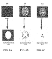
- a segmentation mask defines a set of pixels to be eliminated from an image by bringing the value of those pixels captured by the segmentation mask to a baseline value (such as zero).
- a segmentation mask can be created according to various techniques, including but not limited to:
- the segmentation mask is applied to an image.
- Application of the segmentation mask brings the value of pixels captured by the segmentation mask to the baseline value, leaving the value of the other pixels intact.
- the result is the creation of a filtered image.
- a segmentation mask can be applied to the image from which it was created, but also to images appearing in other series but corresponding to the image from which it was created (where correspondence is established in accordance with step 204 described above). Since corresponding images have the same pixel spacing, origin and orientation, the segmentation mask geometrically "fits" over all images corresponding to the image from which the segmentation mask was created. In some cases, the same segmentation mask can be used for images appearing at different axial slice heights, but it may be preferable to create different segmentation masks for different axial slice heights due to physiological dimensionality variations between adjacent axial slices.
- segmentation masks that can be created, along with some possible applications, are provided below:
- segmentation masks described above can be applied one after the other on the same image (or series of images), resulting in application of a "compound” segmentation mask, which has the features of removing noise, skull tissue, cerebrospinal fluid and large vessels, thus providing improved visibility of key features of interest in the cerebral tissue.
- compound segmentation mask which has the features of removing noise, skull tissue, cerebrospinal fluid and large vessels, thus providing improved visibility of key features of interest in the cerebral tissue.
- multiple segmentation masks derived from multiple series can be applied to the same series.
- the same original segmentation mask can be applied to images in multiple series, including the series containing the image from which the segmentation mask was derived.
- segmentation masks can be created to remove the skull and cerebrospinal fluid. Thereafter, the B1000 or ADC series can be displayed. From there, it may be possible to identify a region representing the core ischemic zone by creating and applying one or more further segmentation masks. A first such further segmentation mask can be used to remove pixels whose values fall below a threshold. A second such further segmentation mask can be created by absorbing remaining isolated pixels. Finally, the pixels that have not been captured by the compound segmentation mask represent areas of the cerebral tissue in the core ischemic zone.
- a next step is therefore to perform a volumetric computation.
- Step 210 Volumetric Computation
- the step of volumetric computation 210 is performed on a plurality of images of a given series.
- One specific volumetric computation of interest seeks to estimate the volume of the core ischemic zone.
- the computer 100 can estimate the total volume of the core ischemic zone. This is basically a computation of the area of the identified region times inter-slice axial distance, but interpolated (e.g., in linear fashion) to account for variability in the area of the region of interest between neighbouring axial slices.
- the computer 100 automatically performs this calculation for those images in the selected series (e.g., B1000 or ADC) where the region of interest appears.
- the user 114 can select via the input 108 a specific set of images on which the volumetric computation will be performed, which can possibly reduce artifacts at the extreme axial slices.
- the output is thus a volume in an appropriate unit (e.g., CC).
- the images under consideration can be PWI images that can allow isolation of a poorly irrigated area of the brain that is nevertheless salvageable/viable.
- Suitable image series are the TTP or MTT or Tmax series, for example, which can be filtered using a compound segmentation mask to arrive at a region on each image in the chosen series which represents the ischemic penumbra.
- the computer 100 can then effect a computation of the surface area of the region times inter-slice axial distance, but interpolated (e.g., in linear fashion) to account for variability in the area of the region of interest between axial slices.
- the computer 100 automatically performs this calculation for those images in the selected series (e.g., TTP or MTT or . Tmax) where the region of interest appears, whereas in other embodiments, the user 114 can select via the input 108 the images on which the volumetric computation will be performed, which can possibly reduce, artifacts at the extreme axial slices.
- the output is thus a volume in an appropriate unit (e.g., CC).
- the user 114 can have access to two important volumetric computations, namely that of the core ischemic zone and that of the ischemic penumbra. Then, based on the absolute and/or relative volumes of each, as well as other factors, the user 114 can be in a position to perform a more informed cost/benefit analysis regarding the option of administering a thrombolytic. This can be useful in both real-life and simulated environments. Further information on the clinical value of this approach can be found in Optimal Definition for PWI / DWI Mismatch in Acute Ischemic Stroke Patients, W Kakuda et al, Journal of Cerebral Blood Flow & Metabolism (2008), 1-5.
- embodiments of the present invention are applicable to other areas of neuromedicine (such as diagnosis and/or treatment of epilepsy, tumors, Alzheimer's, etc.) as well as potentially other areas of medicine in general.
- the biological images of particular significance may be different, but the principles set forth herein are clearly applicable thereto.
- inputs received from the user 114 can be recorded (and restored) by the computer 100 so as to serve for traceability and training purposes. More specifically, the computer 100 can record the values of the pixels that were displayed (based on user selections of contrast, brightness, color scheme, etc.), the validity intervals [x,y] used in the creation segmentation masks, the various parameters of other segmentation masks used for isolating the core ischemic zone and the ischemic penumbra, the parameters used to calculate the perfusion images, and so on.
Landscapes
- Engineering & Computer Science (AREA)
- Health & Medical Sciences (AREA)
- Life Sciences & Earth Sciences (AREA)
- Physics & Mathematics (AREA)
- Medical Informatics (AREA)
- Radiology & Medical Imaging (AREA)
- Nuclear Medicine, Radiotherapy & Molecular Imaging (AREA)
- General Health & Medical Sciences (AREA)
- Computer Vision & Pattern Recognition (AREA)
- General Physics & Mathematics (AREA)
- Theoretical Computer Science (AREA)
- Heart & Thoracic Surgery (AREA)
- Biophysics (AREA)
- Pathology (AREA)
- Veterinary Medicine (AREA)
- Biomedical Technology (AREA)
- High Energy & Nuclear Physics (AREA)
- Molecular Biology (AREA)
- Surgery (AREA)
- Animal Behavior & Ethology (AREA)
- Optics & Photonics (AREA)
- Public Health (AREA)
- Quality & Reliability (AREA)
- Probability & Statistics with Applications (AREA)
- Dentistry (AREA)
- Oral & Maxillofacial Surgery (AREA)
- Magnetic Resonance Imaging Apparatus (AREA)
- Apparatus For Radiation Diagnosis (AREA)
- Measuring And Recording Apparatus For Diagnosis (AREA)
Applications Claiming Priority (2)
| Application Number | Priority Date | Filing Date | Title |
|---|---|---|---|
| US99650907P | 2007-11-20 | 2007-11-20 | |
| EP08762935A EP2232441A2 (fr) | 2007-11-20 | 2008-03-27 | Procédé et système pour traiter de multiples séries d'images biologiques obtenues à partir d'un patient |
Related Parent Applications (1)
| Application Number | Title | Priority Date | Filing Date |
|---|---|---|---|
| EP08762935A Division EP2232441A2 (fr) | 2007-11-20 | 2008-03-27 | Procédé et système pour traiter de multiples séries d'images biologiques obtenues à partir d'un patient |
Publications (2)
| Publication Number | Publication Date |
|---|---|
| EP2840552A2 true EP2840552A2 (fr) | 2015-02-25 |
| EP2840552A3 EP2840552A3 (fr) | 2015-06-10 |
Family
ID=40019234
Family Applications (2)
| Application Number | Title | Priority Date | Filing Date |
|---|---|---|---|
| EP08762935A Ceased EP2232441A2 (fr) | 2007-11-20 | 2008-03-27 | Procédé et système pour traiter de multiples séries d'images biologiques obtenues à partir d'un patient |
| EP14189106.9A Ceased EP2840552A3 (fr) | 2007-11-20 | 2008-03-27 | Procédé et système pour traiter des séries multiples d'images biologiques obtenues d'un patient |
Family Applications Before (1)
| Application Number | Title | Priority Date | Filing Date |
|---|---|---|---|
| EP08762935A Ceased EP2232441A2 (fr) | 2007-11-20 | 2008-03-27 | Procédé et système pour traiter de multiples séries d'images biologiques obtenues à partir d'un patient |
Country Status (5)
| Country | Link |
|---|---|
| US (2) | US8320647B2 (fr) |
| EP (2) | EP2232441A2 (fr) |
| JP (2) | JP5481386B2 (fr) |
| KR (2) | KR101331583B1 (fr) |
| WO (1) | WO2009066136A2 (fr) |
Families Citing this family (35)
| Publication number | Priority date | Publication date | Assignee | Title |
|---|---|---|---|---|
| US8320647B2 (en) * | 2007-11-20 | 2012-11-27 | Olea Medical | Method and system for processing multiple series of biological images obtained from a patient |
| CN101332088B (zh) * | 2008-07-21 | 2011-05-11 | 深圳先进技术研究院 | 一种获取脑部特征参数的系统及溶栓决策系统 |
| JP5322548B2 (ja) * | 2008-09-17 | 2013-10-23 | 株式会社東芝 | X線ct装置、医用画像処理装置および医用画像処理プログラム |
| JP5366577B2 (ja) * | 2009-02-04 | 2013-12-11 | 株式会社東芝 | 医用画像処理装置 |
| US8588486B2 (en) * | 2009-06-18 | 2013-11-19 | General Electric Company | Apparatus and method for isolating a region in an image |
| JP5229175B2 (ja) * | 2009-09-25 | 2013-07-03 | 大日本印刷株式会社 | 医用画像表示処理方法、装置およびプログラム |
| GB0917154D0 (en) * | 2009-09-30 | 2009-11-11 | Imp Innovations Ltd | Method and apparatus for processing medical images |
| US8768029B2 (en) * | 2010-10-20 | 2014-07-01 | Medtronic Navigation, Inc. | Selected image acquisition technique to optimize patient model construction |
| US9262444B2 (en) * | 2010-11-24 | 2016-02-16 | General Electric Company | Systems and methods for applying series level operations and comparing images using a thumbnail navigator |
| JP5921132B2 (ja) * | 2011-10-17 | 2016-05-24 | 株式会社東芝 | 医用画像処理システム |
| US8837800B1 (en) | 2011-10-28 | 2014-09-16 | The Board Of Trustees Of The Leland Stanford Junior University | Automated detection of arterial input function and/or venous output function voxels in medical imaging |
| US8532353B2 (en) * | 2011-11-23 | 2013-09-10 | Vital Images, Inc. | Synthetic visualization and quantification of perfusion-related tissue viability |
| US9081487B2 (en) | 2012-11-22 | 2015-07-14 | Agfa Healthcare Inc. | System and method for manipulating an image |
| US20140324400A1 (en) * | 2013-04-30 | 2014-10-30 | Marquette University | Gesture-Based Visualization System for Biomedical Imaging and Scientific Datasets |
| US9081876B2 (en) | 2013-07-23 | 2015-07-14 | Agfa Healthcare Inc. | Methods and systems for navigating image series |
| DE102013224264B4 (de) * | 2013-11-27 | 2018-12-27 | Siemens Healthcare Gmbh | Verfahren zur Verarbeitung von Magnetresonanz-Diffusionsbilddaten |
| JP6554717B2 (ja) * | 2014-04-24 | 2019-08-07 | ジーイー・メディカル・システムズ・グローバル・テクノロジー・カンパニー・エルエルシー | 磁気共鳴装置およびプログラム |
| WO2016001825A1 (fr) * | 2014-06-30 | 2016-01-07 | Universität Bern | Procédé de segmentation et de prédiction de régions de tissu chez des patients atteints d'ischémie cérébrale aigüe |
| EP2988270A1 (fr) * | 2014-08-22 | 2016-02-24 | Siemens Aktiengesellschaft | Procédé d'évaluation de volume du mésappariement de pénombre dans l'accident vasculaire cérébral ischémique aigu et système associé |
| US10373718B2 (en) | 2014-12-01 | 2019-08-06 | Bijoy Menon Professional Corporation | Decision support tool for stroke patients |
| CN107454960A (zh) * | 2015-02-09 | 2017-12-08 | 三星电子株式会社 | 用于处理磁共振图像的方法和设备 |
| KR101628726B1 (ko) * | 2015-06-11 | 2016-06-10 | 재단법인 아산사회복지재단 | 컴퓨터를 이용한 복셀 내 비결집운동 분석방법 및 프로그램 |
| EP3446141B1 (fr) | 2016-04-21 | 2024-11-20 | Koninklijke Philips N.V. | Imagerie par résonance magnétique de structures artérielles |
| EP3446142A1 (fr) * | 2016-04-21 | 2019-02-27 | Koninklijke Philips N.V. | Imagerie par résonance magnétique de structures artérielles |
| KR101740464B1 (ko) | 2016-10-20 | 2017-06-08 | (주)제이엘케이인스펙션 | 뇌졸중 진단 및 예후 예측 방법 및 시스템 |
| AU2018288766B2 (en) | 2017-06-19 | 2021-01-21 | Viz.ai, Inc. | A method and system for computer-aided triage |
| US10733730B2 (en) | 2017-06-19 | 2020-08-04 | Viz.ai Inc. | Method and system for computer-aided triage |
| EP3847951A4 (fr) * | 2018-09-04 | 2022-04-20 | Bonewise Inc. | Dispositif d'évaluation de l'âge osseux, procédé et support d'enregistrement pour un programme d'enregistrement |
| WO2020264355A1 (fr) | 2019-06-27 | 2020-12-30 | Viz.ai Inc. | Procédé et système de triage assisté par ordinateur d'un accident vasculaire cérébral |
| US10902602B1 (en) | 2019-07-30 | 2021-01-26 | Viz.ai Inc. | Method and system for computer-aided triage of stroke |
| CN111340821B (zh) * | 2020-02-20 | 2020-12-08 | 太原理工大学 | 一种基于模块连接的大脑结构网络的偏测性检测方法 |
| JP7572187B2 (ja) * | 2020-08-31 | 2024-10-23 | 富士フイルム株式会社 | 磁気共鳴イメージング装置 |
| EP3985605A1 (fr) * | 2020-10-15 | 2022-04-20 | Koninklijke Philips N.V. | Procédés et systèmes de segmentation d'images |
| US11694807B2 (en) | 2021-06-17 | 2023-07-04 | Viz.ai Inc. | Method and system for computer-aided decision guidance |
| US11526994B1 (en) * | 2021-09-10 | 2022-12-13 | Neosoma, Inc. | Labeling, visualization, and volumetric quantification of high-grade brain glioma from MRI images |
Citations (2)
| Publication number | Priority date | Publication date | Assignee | Title |
|---|---|---|---|---|
| US20030045791A1 (en) * | 2001-06-25 | 2003-03-06 | Carroll Timothy J. | Determination of the arterial input function in dynamic contrast-enhanced mri |
| EP1860453A1 (fr) * | 2006-05-26 | 2007-11-28 | Kabushiki Kaisha Toshiba | Imagerie par résonance magnétique pondérée par diffusion |
Family Cites Families (175)
| Publication number | Priority date | Publication date | Assignee | Title |
|---|---|---|---|---|
| US4335427A (en) * | 1980-04-21 | 1982-06-15 | Technicare Corporation | Method of selecting a preferred difference image |
| JPH0614746B2 (ja) * | 1984-09-13 | 1994-02-23 | 株式会社東芝 | X線画像処理装置 |
| JP2960419B2 (ja) | 1988-08-10 | 1999-10-06 | 株式会社日立製作所 | 磁気共鳴イメージング方法 |
| US5603322A (en) | 1993-01-19 | 1997-02-18 | Mcw Research Foundation | Time course MRI imaging of brain functions |
| US5425368A (en) * | 1993-03-17 | 1995-06-20 | Board Of Regents, The University Of Texas System | Method and apparatus for estimating tissue volumes in magnetic resonance images |
| US6294914B1 (en) | 1993-06-02 | 2001-09-25 | The Board Of Trustees Of The University Of Illinois | Method of enhancing an MRI signal |
| US5537491A (en) | 1993-11-24 | 1996-07-16 | Xerox Corporation | Analyzing an image or other data to obtain a stable number of groups |
| JP2779125B2 (ja) | 1993-12-08 | 1998-07-23 | 科学技術振興事業団 | Mriによる脳機能可視化方法 |
| US5416415A (en) | 1994-08-05 | 1995-05-16 | General Electric Company | Over-shoulder MRI magnet for human brain imaging |
| US5396207A (en) | 1994-08-05 | 1995-03-07 | General Electric Company | On-shoulder MRI magnet for human brain imaging |
| US5690106A (en) * | 1995-06-30 | 1997-11-25 | Siemens Corporate Research, Inc. | Flexible image registration for rotational angiography |
| US5647360A (en) * | 1995-06-30 | 1997-07-15 | Siemens Corporate Research, Inc. | Digital subtraction angiography for 3D diagnostic imaging |
| WO1997009690A1 (fr) | 1995-09-05 | 1997-03-13 | Northrop Grumman Corporation | Criblage dimensionnel de donnees et connectivite floue pour analyse d'image d'irm |
| DE19628951C2 (de) | 1996-07-18 | 2000-08-31 | Juergen Hennig | Verfahren der Kernspintomographie zur zeitaufgelösten Darstellung pulsatiler Gefäße (Projektionsangiographie) |
| US5848121A (en) * | 1996-10-28 | 1998-12-08 | General Electric Company | Method and apparatus for digital subtraction angiography |
| US6078176A (en) | 1996-11-08 | 2000-06-20 | General Electric Company | Fast spin echo pulse sequence for diffusion weighted imaging |
| JP4040742B2 (ja) | 1997-03-28 | 2008-01-30 | 株式会社東芝 | Mri装置 |
| US6031374A (en) | 1997-04-11 | 2000-02-29 | Epstein; Frederick H. | Method for extracting deformations from velocity-encoded magnetic resonance images of the heart |
| JP2000512533A (ja) | 1997-04-17 | 2000-09-26 | コーニンクレッカ フィリップス エレクトロニクス エヌ ヴィ | 拡散重み付けmri方法 |
| EP0910804A1 (fr) | 1997-04-17 | 1999-04-28 | Koninklijke Philips Electronics N.V. | Procede d'imagerie par resonance magnetique a diffusion ponderee |
| JP3120224B2 (ja) | 1997-08-27 | 2000-12-25 | 技術研究組合医療福祉機器研究所 | Mri装置 |
| WO1999030486A2 (fr) * | 1997-12-10 | 1999-06-17 | Koninklijke Philips Electronics N.V. | Formation d'une image d'assemblage provenant d'images radiologiques successives |
| US5999839A (en) | 1998-01-30 | 1999-12-07 | General Electric Company | Arterial MRI with chemical-shift nulling |
| WO1999052429A1 (fr) | 1998-04-13 | 1999-10-21 | The Trustees Of The University Of Pennsylvania | Formation d'images multicoupe de flux sanguin cerebral par imagerie par resonance magnetique de marquage de spin arteriel en continu |
| US6980845B1 (en) | 1998-04-13 | 2005-12-27 | The Trustees Of The University Of Pennsylvania | Multi-slice cerebral blood flow imaging with continuous arterial spin labeling MRI |
| US6782286B2 (en) | 1998-04-20 | 2004-08-24 | Kabushiki Kaisha Toshiba | MRI system and MR imaging method |
| JPH11332847A (ja) | 1998-05-22 | 1999-12-07 | Masayuki Fujioka | Mriを用いた選択的神経細胞死の非侵襲的判定方法 |
| US6078175A (en) | 1998-10-26 | 2000-06-20 | General Electric Company | Acquistion of segmented cardiac gated MRI perfusion images |
| US6342598B1 (en) | 1998-11-26 | 2002-01-29 | Bracco International B.V. | Amphipatic polycarboxylic chelates and complexes with paramagnetic metals as MRI contrast agents |
| GB2345139A (en) | 1998-12-24 | 2000-06-28 | Marconi Electronic Syst Ltd | MRI apparatus with continuous movement of patient |
| US6757423B1 (en) | 1999-02-19 | 2004-06-29 | Barnes-Jewish Hospital | Methods of processing tagged MRI data indicative of tissue motion including 4-D LV tissue tracking |
| US8617514B2 (en) | 1999-02-22 | 2013-12-31 | Georgetown University | Tumor-targeted nanodelivery systems to improve early MRI detection of cancer |
| US6904163B1 (en) | 1999-03-19 | 2005-06-07 | Nippon Telegraph And Telephone Corporation | Tomographic image reading method, automatic alignment method, apparatus and computer readable medium |
| US6307368B1 (en) | 1999-05-14 | 2001-10-23 | Board Of Trustees Of The Leland Stanford Junior University | Linear combination steady-state free precession MRI |
| US6265875B1 (en) | 1999-05-17 | 2001-07-24 | General Electric Company | Method and apparatus for efficient MRI tissue differentiation |
| US6490472B1 (en) | 1999-09-03 | 2002-12-03 | The Mcw Research Foundation, Inc. | MRI system and method for producing an index indicative of alzheimer's disease |
| JP3668076B2 (ja) | 1999-10-18 | 2005-07-06 | ジーイー横河メディカルシステム株式会社 | Mri装置 |
| US6430431B1 (en) | 1999-11-11 | 2002-08-06 | Mcw Research Foundation, Inc. | MRI system for measuring vision capabilities |
| GB2356458A (en) | 1999-11-16 | 2001-05-23 | Marconi Electronic Syst Ltd | MRI using modified FLAIR sequence |
| JP2001187037A (ja) | 1999-12-27 | 2001-07-10 | Ge Medical Systems Global Technology Co Llc | 拡散運動検出用勾配磁場印加方向決定方法、拡散係数測定方法およびmri装置 |
| JP3254451B2 (ja) | 2000-03-06 | 2002-02-04 | 経済産業省産業技術総合研究所長 | 多チャンネルmri画像処理によるカラー化方法及び装置 |
| US20010056231A1 (en) | 2000-03-13 | 2001-12-27 | Andrzej Jesmanowicz | High resolution MRI imaging of brain functions |
| US6560353B1 (en) | 2000-03-21 | 2003-05-06 | Magnetic Resonance Innovations, Inc. | Method of MRI image reconstruction from partially acquired data in two or more dimensions using a multidimensional inverse transform technique |
| US6751495B2 (en) * | 2000-03-31 | 2004-06-15 | Brigham & Womens' Hospital | Method of fast and reliable tissue differentiation using diffusion-weighted magnetic resonance imaging |
| JP4008193B2 (ja) | 2000-08-29 | 2007-11-14 | ジーイー・メディカル・システムズ・グローバル・テクノロジー・カンパニー・エルエルシー | 画像処理方法、画像処理装置、mri装置および記録媒体 |
| WO2002018967A1 (fr) | 2000-08-30 | 2002-03-07 | Koninklijke Philips Electronics N.V. | Acquisition d'images d'un plan en mouvement par irm |
| JP3853585B2 (ja) | 2000-10-11 | 2006-12-06 | ジーイー・メディカル・システムズ・グローバル・テクノロジー・カンパニー・エルエルシー | Mri装置 |
| EP1334467A2 (fr) | 2000-10-31 | 2003-08-13 | Koninklijke Philips Electronics N.V. | Procede et systeme permettant la detection et le suivi de marqueurs dans des images a marqueurs irm |
| US6445181B1 (en) | 2000-11-09 | 2002-09-03 | The Board Of Trustees Of The Leland Stanford Junior University | MRI method apparatus for imaging a field of view which is larger than a magnetic field |
| US6591128B1 (en) | 2000-11-09 | 2003-07-08 | Koninklijke Philips Electronics, N.V. | MRI RF coil systems having detachable, relocatable, and or interchangeable sections and MRI imaging systems and methods employing the same |
| US6542769B2 (en) | 2000-12-18 | 2003-04-01 | The General Hospital Corporation | Imaging system for obtaining quantative perfusion indices |
| JP2002209867A (ja) | 2001-01-10 | 2002-07-30 | Sogen Boku | 脳の磁気共鳴映像から白質と灰白質および脳脊髄液を分離し体積を算出する方法 |
| US6420873B1 (en) | 2001-02-15 | 2002-07-16 | Netcom, Inc. | MRI noise detector and suppressor |
| US6829509B1 (en) | 2001-02-20 | 2004-12-07 | Biophan Technologies, Inc. | Electromagnetic interference immune tissue invasive system |
| US6792302B2 (en) | 2001-02-21 | 2004-09-14 | Universite De Lausanne | Method and apparatus for determining treatment for stroke |
| AUPR358701A0 (en) * | 2001-03-07 | 2001-04-05 | University Of Queensland, The | Method of predicting stroke evolution |
| US6373920B1 (en) * | 2001-03-16 | 2002-04-16 | Ge Medical Systems Global Technology Company, Llc | Method and apparatus for acquiring CT perfusion images |
| WO2002082376A2 (fr) | 2001-04-06 | 2002-10-17 | Regents Of The University Of California | Procede d'analyse de donnees de diffusion d'imagerie a resonance magnetique |
| WO2002086530A1 (fr) | 2001-04-20 | 2002-10-31 | Wisconsin Alumni Research Foundation | Determination de la fonction d'entree arterielle en irm de contraste dynamique |
| DE10121802B4 (de) | 2001-05-04 | 2004-09-30 | Siemens Ag | Verfahren zur Bilderzeugung bei einem Kernspintomographie-Gerät und Kernspintomographie-Gerät sowie Datenverarbeitungsanlage |
| US6597938B2 (en) | 2001-08-16 | 2003-07-22 | Koninklijke Philips Electronics, N.V. | System for assistance of parameter determination and diagnosis in MRI dynamic uptake studies |
| US6754522B2 (en) * | 2001-09-05 | 2004-06-22 | Medimag C.V.I., Inc. | Imaging methods and apparatus particularly useful for two and three-dimensional angiography |
| GB0122405D0 (en) | 2001-09-17 | 2001-11-07 | Royal Brompton & Harefield Nhs | Magnetic resonance imaging |
| US6445184B1 (en) | 2001-11-20 | 2002-09-03 | Koninklijke Philips Electronics N.V. | Multiple gradient echo type projection reconstruction sequence for MRI especially for diffusion weighted MRI |
| US6972565B2 (en) | 2001-12-27 | 2005-12-06 | Kabushiki Kaisha Toshiba | System, method and apparatus for MRI maintenance and support |
| US6956373B1 (en) | 2002-01-02 | 2005-10-18 | Hugh Keith Brown | Opposed orthogonal fusion system and method for generating color segmented MRI voxel matrices |
| DE10256208B4 (de) | 2002-12-02 | 2008-05-15 | Siemens Ag | Verfahren zur verbesserten Flussmessung in der Magnetresonanz-Tomographie |
| JP3980374B2 (ja) | 2002-02-20 | 2007-09-26 | 株式会社東芝 | Mri装置 |
| JP2003250775A (ja) | 2002-02-25 | 2003-09-09 | Ge Medical Systems Global Technology Co Llc | Mri装置およびmra撮影方法 |
| US7558612B2 (en) | 2002-04-16 | 2009-07-07 | The Board Of Trustees Of The Leland Stanford Junior University | Motion compensated spiral FISP MRI |
| JP3920140B2 (ja) | 2002-05-13 | 2007-05-30 | 株式会社東芝 | Mri装置及びフロー定量化装置 |
| US6963768B2 (en) | 2002-05-16 | 2005-11-08 | General Electric Company | Whole body MRI scanning with moving table and interactive control |
| US6707300B2 (en) | 2002-05-17 | 2004-03-16 | Ge Medical Systems Global Technology Co., Llc | Gradient non-linearity compensation in moving table MRI |
| US6922580B2 (en) | 2002-06-04 | 2005-07-26 | Koninklijke Philips Electronics N.V. | Blood flow gated MRI |
| JP4030805B2 (ja) | 2002-06-07 | 2008-01-09 | ジーイー・メディカル・システムズ・グローバル・テクノロジー・カンパニー・エルエルシー | コイル感度マップ作成方法およびmri装置 |
| JP4073269B2 (ja) | 2002-07-26 | 2008-04-09 | 株式会社東芝 | Mri装置及び画像のww/wlの設定方法 |
| US7215231B1 (en) | 2002-08-16 | 2007-05-08 | Fonar Corporation | MRI system |
| US7009396B2 (en) | 2002-09-12 | 2006-03-07 | General Electric Company | Method and system for extended volume imaging using MRI with parallel reception |
| JP4013128B2 (ja) * | 2002-09-12 | 2007-11-28 | 株式会社日立メディコ | 血流動態解析装置、方法、及び画像診断装置 |
| US7545967B1 (en) * | 2002-09-18 | 2009-06-09 | Cornell Research Foundation Inc. | System and method for generating composite subtraction images for magnetic resonance imaging |
| US7106892B2 (en) * | 2002-09-19 | 2006-09-12 | Koninklijke Philips Electronics, N.V. | Display of image data information |
| US6894494B2 (en) | 2002-11-25 | 2005-05-17 | Her Majesty The Queen In Right Of Canada, As Represented By The Minister Of National Defence Of Her Majesty's Canadian Government | Method and device for correcting organ motion artifacts in MRI systems |
| CA2506849A1 (fr) | 2002-12-04 | 2004-06-17 | Konstantinos Tsougarakis | Fusion de plans d'imagerie multiples pour imagerie isotrope en irm et analyse d'image quantitative utilisant l'imagerie isotrope ou quasi isotrope |
| US7202662B2 (en) | 2002-12-11 | 2007-04-10 | The Board Of Trustees Of The Leland Stanford Junior University | Correction of the effect of gradient field non-linearities in phase contrast MRI |
| DE10301497B4 (de) | 2003-01-16 | 2013-12-24 | Siemens Aktiengesellschaft | Magnetresonanz-Projektionsangiographie-Verfahren mit kontinuierlicher Tischverschiebung sowie MRT-Gerät zur Durchführung des Verfahrens |
| US7405564B2 (en) | 2003-01-20 | 2008-07-29 | Koninklijke Philips Electronics N.V. | Sub-sampled moving table MRI for at least two adjacent fields of view |
| JP4342809B2 (ja) | 2003-02-14 | 2009-10-14 | 株式会社東芝 | Mri装置 |
| WO2004077086A1 (fr) | 2003-02-28 | 2004-09-10 | Koninklijke Philips Electronics N.V. | Irm a table mobile et a sous-echantillonnage |
| US7450983B2 (en) | 2003-03-18 | 2008-11-11 | University Of Cincinnati | Automated brain MRI and CT prescriptions in Talairach space |
| US20040218794A1 (en) * | 2003-05-01 | 2004-11-04 | Yi-Hsuan Kao | Method for processing perfusion images |
| WO2004104611A2 (fr) | 2003-05-05 | 2004-12-02 | Case Western Reserve University | Conception et suivi d'une sonde pour irm, et reconstruction et correction efficaces d'images brouillees d'irm |
| US7596402B2 (en) | 2003-05-05 | 2009-09-29 | Case Western Reserve University | MRI probe designs for minimally invasive intravascular tracking and imaging applications |
| KR100517891B1 (ko) | 2003-05-13 | 2005-09-30 | 허순녕 | 뇌정위 방사선 치료용 프레임 |
| JP2007525250A (ja) | 2003-06-02 | 2007-09-06 | ザ・ジェネラル・ホスピタル・コーポレイション | 組織血流の遅延−補償型算出方法 |
| WO2004113945A2 (fr) | 2003-06-24 | 2004-12-29 | Koninklijke Philips Electronics N.V. | Systeme de bobines a radio-frequences pour systemes d'imagerie a resonance magnetique |
| WO2005012926A2 (fr) | 2003-07-08 | 2005-02-10 | The Government Of The United States Of America, Represented By The Secretary Of The Department Of Health And Human Services | Caracterisation d'echantillon par irm du tenseur de diffusion et irm q-space |
| US7251520B2 (en) | 2003-07-08 | 2007-07-31 | General Electric Company | Method and apparatus of slice selective magnetization preparation for moving table MRI |
| US6882148B2 (en) | 2003-07-09 | 2005-04-19 | Catholic Healthcare West | Split-blade data collection for propeller MRI |
| US20050154291A1 (en) | 2003-09-19 | 2005-07-14 | Lei Zhao | Method of using a small MRI scanner |
| CN1879033A (zh) | 2003-11-05 | 2006-12-13 | 皇家飞利浦电子股份有限公司 | 具有减小的声学噪音的mri系统 |
| CN100578250C (zh) | 2003-11-18 | 2010-01-06 | 皇家飞利浦电子股份有限公司 | 用于mri的混合tem/鸟笼型线圈 |
| FR2862786B1 (fr) * | 2003-11-21 | 2006-01-06 | Ge Med Sys Global Tech Co Llc | Procede de recalage elastique d'une image applique a l'angiographie soustraite numerisee |
| US7304477B2 (en) | 2003-11-24 | 2007-12-04 | Koninklijke Philips Electronics N.V. | MRI apparatus with means for noise reduction |
| US6975113B1 (en) | 2003-11-25 | 2005-12-13 | General Electric Company | Method and system for moving table MRI with partial fourier imaging |
| DE102004003381B4 (de) | 2004-01-22 | 2007-02-01 | Siemens Ag | Verfahren zur Bestimmung der Lage einer Schicht in einem Untersuchungsgebiet, in welcher Schicht eine Schichtbildaufnahme erfolgen soll |
| DE102004005122A1 (de) * | 2004-02-02 | 2005-08-18 | Siemens Ag | Verfahren und Vorrichtung zur verbesserten Auswertung von Magnetresonanzbildern |
| US20080252291A1 (en) | 2004-02-06 | 2008-10-16 | Koninklijke Philips Electronics N.V. | High Angular Resolution Diffusion Weighted Mri |
| US20050215884A1 (en) | 2004-02-27 | 2005-09-29 | Greicius Michael D | Evaluation of Alzheimer's disease using an independent component analysis of an individual's resting-state functional MRI |
| CN1977182A (zh) | 2004-04-28 | 2007-06-06 | 皇家飞利浦电子股份有限公司 | 涉及对比度处理和扫描参数更新的连续移动平台的磁共振成像 |
| EP1751573A1 (fr) | 2004-05-14 | 2007-02-14 | Koninklijke Philips Electronics N.V. | Imagerie par resonance utilisant une table mobile |
| US9638770B2 (en) | 2004-05-21 | 2017-05-02 | Devicor Medical Products, Inc. | MRI biopsy apparatus incorporating an imageable penetrating portion |
| US20050273001A1 (en) | 2004-06-04 | 2005-12-08 | The Mcw Research Foundation | MRI display interface for medical diagnostics and planning |
| US8160314B2 (en) * | 2004-06-18 | 2012-04-17 | Siemens Aktiengesellschaft | System and method for linking VOIs across timepoints for analysis of disease progression or response to therapy |
| CN1993357B (zh) | 2004-07-02 | 2011-10-19 | 伯拉考成像股份公司 | 用于磁共振成像(mri)的含有具有多羟基化取代基的螯合部分的高弛豫造影剂 |
| CN101031237B (zh) | 2004-07-27 | 2012-11-14 | 外科视象公司 | 具有协同式mri天线探头的mri兼容通用传递插管的mri系统及相关系统和方法 |
| GB2434648A (en) | 2004-08-19 | 2007-08-01 | Gen Electric | Mri magnet producing an asymmetrical stray field, with compensation coils |
| EP1788939A4 (fr) | 2004-09-08 | 2009-11-25 | Univ Ramot | Methode de contraste et d'irm |
| US7317821B2 (en) | 2004-11-22 | 2008-01-08 | Carestream Health, Inc. | Automatic abnormal tissue detection in MRI images |
| JP4717422B2 (ja) | 2004-11-26 | 2011-07-06 | ジーイー・メディカル・システムズ・グローバル・テクノロジー・カンパニー・エルエルシー | Mr画像生成方法、mri装置、およびその方法をコンピュータに実行させるプログラム |
| JP4721693B2 (ja) * | 2004-12-09 | 2011-07-13 | 富士フイルムRiファーマ株式会社 | 頭蓋内容積および局所脳構造物解析プログラム、記録媒体および頭蓋内容積および局所脳構造物解析方法 |
| FI20045484A0 (fi) | 2004-12-15 | 2004-12-15 | Nomir Oy | Menetelmä MRI-kuvantamisessa ja MRI-laite, jossa on laukaisin |
| US7899231B2 (en) * | 2005-01-28 | 2011-03-01 | Siemens Medical Solutions Usa, Inc. | System and method for splicing medical image datasets |
| US20060184001A1 (en) | 2005-02-02 | 2006-08-17 | Minkoff Lawrence A | Method for MRI scanning of animals for transmissible spongiform encephalopathies |
| WO2006088453A1 (fr) | 2005-02-15 | 2006-08-24 | Zhao, Lei | Procede d’utilisation d’un petit scanner irm |
| US20060206024A1 (en) | 2005-03-09 | 2006-09-14 | Invivo Corporation | Wireless in-bore patient monitor for MRI |
| CN1663527A (zh) | 2005-03-16 | 2005-09-07 | 南京航空航天大学 | 一种脑功能磁共振图像运动校正方法 |
| US7577472B2 (en) | 2005-03-18 | 2009-08-18 | The Mcw Research Foundation Inc. | MRI method for producing an index indicative of brain disorders |
| WO2006102370A2 (fr) | 2005-03-22 | 2006-09-28 | University Of Massachusetts | Cartographie irm fonctionnelle cerebrale utilisee en tant que marqueur dans des maladies et des troubles du snc |
| FR2884340B1 (fr) * | 2005-04-11 | 2012-03-02 | Gen Electric | Procede et dispositif de traitement d'images en angiographie soustraite |
| WO2006119259A2 (fr) | 2005-04-29 | 2006-11-09 | Beth Israel Deaconess Medical Center, Inc. | Systemes irm et procedes associes |
| WO2006119085A2 (fr) | 2005-04-29 | 2006-11-09 | The Regents Of The University Of California | Scanner tep-irm integre |
| WO2006118549A1 (fr) * | 2005-05-02 | 2006-11-09 | Agency For Science, Technology And Research | Procede et appareil d'interpretation assistee par atlas d'images de diffusion et de perfusion obtenues par resonance magnetique |
| WO2006119164A2 (fr) | 2005-05-04 | 2006-11-09 | Mayo Foundation For Medical Education And Research | Acquisition d'irm par sense 2d et echantillonnage d'espace de fourier partiel |
| EP1880345A4 (fr) | 2005-05-06 | 2012-11-28 | Icad Inc | Procede de suivi de structure ameliorant le contraste pour analyse pharmacocinetique et parametriques de tissus ameliore au moyen d'un irm a haute resolution |
| EP1886254A4 (fr) | 2005-05-20 | 2011-04-27 | Advanced Mri Technologies Llc | Visualisation efficace des territoires vasculaires corticaux et irm fonctionnelle au moyen d'une irm a marquage des spins arteriels cycle |
| WO2007022499A2 (fr) | 2005-08-18 | 2007-02-22 | Conolly Steven M | Reduction d'artefacts de susceptibilite dans une irm au moyen de materiaux composites |
| US8218835B2 (en) * | 2005-08-22 | 2012-07-10 | Dai Nippon Printing Co., Ltd. | Method for assisting in diagnosis of cerebral diseases and apparatus thereof |
| US20070047786A1 (en) | 2005-08-25 | 2007-03-01 | Lenovo (Singapore) Pte. Ltd. | System and method for creating robust training data from MRI images |
| JP2007068796A (ja) | 2005-09-08 | 2007-03-22 | Ge Medical Systems Global Technology Co Llc | Rfパルス印加方法およびmri装置 |
| JP2007068798A (ja) | 2005-09-08 | 2007-03-22 | Ge Medical Systems Global Technology Co Llc | Rfパルス印加方法およびmri装置 |
| JP2007075387A (ja) | 2005-09-15 | 2007-03-29 | Ge Medical Systems Global Technology Co Llc | 息止めmr撮影方法、mri装置、息止め断層像撮影方法および断層像撮影装置 |
| DE102005045679B3 (de) | 2005-09-24 | 2007-03-01 | Bruker Biospin Mri Gmbh | Anordnung der bildgebenden Kernspinresonanz (MRI) mit MRI-Phantom und Verfahren zur Bestimmung der Homogenität des Magnetfelds |
| US7920730B2 (en) | 2005-10-07 | 2011-04-05 | Siemens Medical Solutions Usa, Inc. | Automatic bone detection in MRI images |
| JP5334582B2 (ja) | 2005-10-17 | 2013-11-06 | アルバータ ヘルス サービシズ | 外部ビーム放射線治療とmriとの統合システム |
| WO2007047915A2 (fr) | 2005-10-18 | 2007-04-26 | 3Tp Llc | Procedes automatises destines a preselectionner des voxels et mettre en oeuvre une analyse pharmacocinetique et parametrique destinee a un irm et a une tomographie par ordinateur a contraste dynamique ameliore |
| CN101351225B (zh) | 2005-10-20 | 2013-09-25 | 乔治敦大学 | 提高癌症早期mri检测的肿瘤靶向纳米传送系统 |
| US7627155B2 (en) | 2005-10-26 | 2009-12-01 | Siemens Medical Solutions Usa, Inc. | Fast geometric flows based white matter fiber tract segmentation in DT-MRI |
| US7642782B2 (en) | 2005-10-27 | 2010-01-05 | Koninklijke Philips Electronics N.V. | Active decoupling of transmitters in MRI |
| EP1977264A2 (fr) | 2005-10-28 | 2008-10-08 | Koninklijke Philips Electronics N.V. | Bobines radiofrequence specifiques de regions d'imagerie pour l'imagerie par resonance magnetique |
| GB2435327B (en) | 2005-11-01 | 2008-01-09 | Siemens Magnet Technology Ltd | Transportable magnetic resonance imaging (MRI) system |
| GB2431999B (en) | 2005-11-04 | 2008-01-16 | Siemens Magnet Technology Ltd | Switching circuit for controlling multiple heating elements |
| US8255054B2 (en) | 2005-11-04 | 2012-08-28 | Kenergy, Inc. | MRI compatible implanted electronic medical device |
| US7514923B2 (en) | 2005-11-07 | 2009-04-07 | General Electric Company | Method for localized excitation for accurate center frequency measurements with MRI |
| JP5085031B2 (ja) * | 2005-11-10 | 2012-11-28 | 株式会社東芝 | X線アンギオ撮影装置 |
| EP1952340B1 (fr) | 2005-11-21 | 2012-10-24 | Agency for Science, Technology and Research | Superposition des images de l'atlas du cerveau et des images du cerveau avec delimitation des regions d'infarctus et de penombre dans le diagnostic d'un accident cerebrovasculaire |
| US7767196B2 (en) | 2005-11-21 | 2010-08-03 | The University Of Hong Kong | Optimized relaxivity and specificity hepatobiliary MRI contrast agent |
| US7996117B2 (en) | 2005-11-25 | 2011-08-09 | Hitachi Medical Corporation | MRI system employing superconducting magnet and its maintenance method |
| CN101322042B (zh) | 2005-12-08 | 2012-12-26 | 皇家飞利浦电子股份有限公司 | 用于接收和/或发射rf信号、尤其是在mri系统中接收和/或发射rf信号的装置 |
| US7696751B2 (en) | 2005-12-08 | 2010-04-13 | Koninklijke Philips Electronics N. V. | Method and apparatus for ferrous object and/or magnetic field detection for MRI safety |
| US20070156042A1 (en) | 2005-12-30 | 2007-07-05 | Orhan Unal | Medical device system and method for tracking and visualizing a medical device system under MR guidance |
| WO2007081730A1 (fr) | 2006-01-04 | 2007-07-19 | California Institute Of Technology | Imagerie a resonance magnetique en deux et en trois dimensions mettant en oeuvre une sphere ferromagnetique fixée fermement à l'échantillon |
| US8379957B2 (en) | 2006-01-12 | 2013-02-19 | Siemens Corporation | System and method for segmentation of anatomical structures in MRI volumes using graph cuts |
| US7309988B2 (en) | 2006-01-12 | 2007-12-18 | General Electric Company | Methods and systems for reducing acoustic noise in a magnetic resonance imaging (MRI) system |
| US7345485B2 (en) | 2006-01-18 | 2008-03-18 | Koninklijke Philips Electronics N.V. | Optical interface for local MRI coils |
| WO2007084747A2 (fr) | 2006-01-19 | 2007-07-26 | The Johns Hopkins University | Procede d’evaluation de la rigidite arterielle centrale par irm, et de la fonction vasculaire comprenant la rigidite arterielle, programme d'applications et supports permettant la mise en œuvre desdits procedes |
| WO2007082495A1 (fr) | 2006-01-19 | 2007-07-26 | Medsonic Ltd | Système de positionnement irm pour une chirurgie du cerveau par ultrasons |
| WO2007085342A1 (fr) | 2006-01-27 | 2007-08-02 | Julius-Maximilians-Universität Würzburg | Irm parallèle impliquant une pondération de densité ou une pondération d'acquisition |
| JP5105848B2 (ja) | 2006-02-06 | 2012-12-26 | 株式会社東芝 | 磁気共鳴イメージング装置および磁気共鳴イメージング装置における撮影条件設定方法 |
| JP4745080B2 (ja) * | 2006-02-20 | 2011-08-10 | 猛 中浦 | X線診断装置、画像処理装置及びプログラム |
| JP4820680B2 (ja) | 2006-04-12 | 2011-11-24 | 株式会社東芝 | 医用画像表示装置 |
| CN100411587C (zh) | 2006-07-06 | 2008-08-20 | 上海交通大学 | 基于机器学习的立体核磁共振脑图像弹性配准方法 |
| US8285014B2 (en) * | 2007-04-06 | 2012-10-09 | Siemens Aktiengesellschaft | Measuring blood volume with C-arm computed tomography |
| JP5794752B2 (ja) * | 2007-07-24 | 2015-10-14 | 株式会社東芝 | X線コンピュータ断層撮影装置及び画像処理装置 |
| US8320647B2 (en) * | 2007-11-20 | 2012-11-27 | Olea Medical | Method and system for processing multiple series of biological images obtained from a patient |
| US8588498B2 (en) * | 2011-09-30 | 2013-11-19 | General Electric Company | System and method for segmenting bones on MR images |
-
2008
- 2008-03-26 US US12/055,751 patent/US8320647B2/en active Active
- 2008-03-27 KR KR1020137001324A patent/KR101331583B1/ko active Active
- 2008-03-27 JP JP2010534555A patent/JP5481386B2/ja active Active
- 2008-03-27 WO PCT/IB2008/001621 patent/WO2009066136A2/fr not_active Ceased
- 2008-03-27 KR KR1020107013408A patent/KR101262338B1/ko not_active Expired - Fee Related
- 2008-03-27 EP EP08762935A patent/EP2232441A2/fr not_active Ceased
- 2008-03-27 EP EP14189106.9A patent/EP2840552A3/fr not_active Ceased
-
2012
- 2012-11-01 US US13/666,367 patent/US9123100B2/en active Active
-
2013
- 2013-09-04 JP JP2013183471A patent/JP5986049B2/ja active Active
Patent Citations (2)
| Publication number | Priority date | Publication date | Assignee | Title |
|---|---|---|---|---|
| US20030045791A1 (en) * | 2001-06-25 | 2003-03-06 | Carroll Timothy J. | Determination of the arterial input function in dynamic contrast-enhanced mri |
| EP1860453A1 (fr) * | 2006-05-26 | 2007-11-28 | Kabushiki Kaisha Toshiba | Imagerie par résonance magnétique pondérée par diffusion |
Non-Patent Citations (6)
Also Published As
| Publication number | Publication date |
|---|---|
| WO2009066136A2 (fr) | 2009-05-28 |
| US9123100B2 (en) | 2015-09-01 |
| EP2840552A3 (fr) | 2015-06-10 |
| EP2232441A2 (fr) | 2010-09-29 |
| KR20130021450A (ko) | 2013-03-05 |
| US20130058549A1 (en) | 2013-03-07 |
| KR101262338B1 (ko) | 2013-05-08 |
| JP5986049B2 (ja) | 2016-09-06 |
| US8320647B2 (en) | 2012-11-27 |
| KR101331583B1 (ko) | 2013-11-20 |
| WO2009066136A3 (fr) | 2009-07-09 |
| KR20100098645A (ko) | 2010-09-08 |
| JP5481386B2 (ja) | 2014-04-23 |
| JP2014000455A (ja) | 2014-01-09 |
| JP2011502729A (ja) | 2011-01-27 |
| US20090129649A1 (en) | 2009-05-21 |
Similar Documents
| Publication | Publication Date | Title |
|---|---|---|
| US8320647B2 (en) | Method and system for processing multiple series of biological images obtained from a patient | |
| US11783484B2 (en) | Protocol-aware tissue segmentation in medical imaging | |
| Greenspan | Super-resolution in medical imaging | |
| Aubert-Broche et al. | A new improved version of the realistic digital brain phantom | |
| Kauffmann et al. | Spatial frequency processing in scene-selective cortical regions | |
| Bône et al. | Contrast-enhanced brain MRI synthesis with deep learning: key input modalities and asymptotic performance | |
| EP4264308B1 (fr) | Procédés permettant d'entraîner un cnn et de traiter une séquence de perfusion entrée en utilisant ledit cnn | |
| Heinone et al. | Semi-automatic tool for segmentation and volumetric analysis of medical images | |
| Itti et al. | Robust multimodality registration for brain mapping | |
| Hastreiter et al. | Analysis and 3-dimensional visualization of neurovascular compression syndromes1 | |
| Dong et al. | High-resolution magnetic resonance spectroscopic imaging using a multi-encoder attention u-net with structural and adversarial loss | |
| US10229500B2 (en) | Incorporation of statistical strength into voxels in an fMRI image | |
| Seibert et al. | Default network correlations analyzed on native surfaces | |
| DE102015200850B4 (de) | Verfahren zur Auswertung von medizinischen Bilddaten | |
| Zhao et al. | Using anatomic magnetic resonance image information to enhance visualization and interpretation of functional images: A comparison of methods applied to clinical arterial spin labeling images | |
| Morell-Ortega et al. | Robust deep MRI contrast synthesis using a prior-based and task-oriented 3D network | |
| CN113243929B (zh) | 可视化方法和设备 | |
| US20060173276A1 (en) | Contrast-enhanced moving table mr arteriography with stochastic sampling of central k-space | |
| Prabakar et al. | An investigation of volumetric and corpus callosum dimension to detect brain disorders | |
| Matsumoto et al. | A fast Way to visualize the brain surface with volume rendering of MRI data | |
| JP2025004671A (ja) | 分析システムおよびプログラム | |
| Rezk-Salama et al. | Interactive Direct Volume Rendering of Dural Arteriovenous Fistulae in MR-CISS Data | |
| Lewis et al. | Optimization of high resolution PET iterative reconstruction with resolution modeling for image derived input function | |
| Cash et al. | A comparison of techniques for quantifying amyloid burden on a combined PET/MR scanner | |
| Hwang | Diffusion tensor tractography: Visualization and quantitation with applications to Alzheimer Disease and traumatic brain injury |
Legal Events
| Date | Code | Title | Description |
|---|---|---|---|
| PUAI | Public reference made under article 153(3) epc to a published international application that has entered the european phase |
Free format text: ORIGINAL CODE: 0009012 |
|
| 17P | Request for examination filed |
Effective date: 20141015 |
|
| AC | Divisional application: reference to earlier application |
Ref document number: 2232441 Country of ref document: EP Kind code of ref document: P |
|
| AK | Designated contracting states |
Kind code of ref document: A2 Designated state(s): AT BE BG CH CY CZ DE DK EE ES FI FR GB GR HR HU IE IS IT LI LT LU LV MC MT NL NO PL PT RO SE SI SK TR |
|
| PUAL | Search report despatched |
Free format text: ORIGINAL CODE: 0009013 |
|
| AK | Designated contracting states |
Kind code of ref document: A3 Designated state(s): AT BE BG CH CY CZ DE DK EE ES FI FR GB GR HR HU IE IS IT LI LT LU LV MC MT NL NO PL PT RO SE SI SK TR |
|
| RIC1 | Information provided on ipc code assigned before grant |
Ipc: G06T 7/00 20060101AFI20150505BHEP |
|
| R17P | Request for examination filed (corrected) |
Effective date: 20151210 |
|
| RBV | Designated contracting states (corrected) |
Designated state(s): AT BE BG CH CY CZ DE DK EE ES FI FR GB GR HR HU IE IS IT LI LT LU LV MC MT NL NO PL PT RO SE SI SK TR |
|
| STAA | Information on the status of an ep patent application or granted ep patent |
Free format text: STATUS: EXAMINATION IS IN PROGRESS |
|
| 17Q | First examination report despatched |
Effective date: 20180312 |
|
| APBK | Appeal reference recorded |
Free format text: ORIGINAL CODE: EPIDOSNREFNE |
|
| APBN | Date of receipt of notice of appeal recorded |
Free format text: ORIGINAL CODE: EPIDOSNNOA2E |
|
| APBR | Date of receipt of statement of grounds of appeal recorded |
Free format text: ORIGINAL CODE: EPIDOSNNOA3E |
|
| APAF | Appeal reference modified |
Free format text: ORIGINAL CODE: EPIDOSCREFNE |
|
| P01 | Opt-out of the competence of the unified patent court (upc) registered |
Effective date: 20231221 |
|
| APBT | Appeal procedure closed |
Free format text: ORIGINAL CODE: EPIDOSNNOA9E |
|
| STAA | Information on the status of an ep patent application or granted ep patent |
Free format text: STATUS: THE APPLICATION HAS BEEN REFUSED |
|
| 18R | Application refused |
Effective date: 20241007 |