EP3221355B1 - Kombinationstherapie aus t-zellaktivierenden bispezifischen antigenbindenden molekülen cd3 und folatrezeptor 1 (folr1) mit pd-1-achsenbindenden antagonisten - Google Patents
Kombinationstherapie aus t-zellaktivierenden bispezifischen antigenbindenden molekülen cd3 und folatrezeptor 1 (folr1) mit pd-1-achsenbindenden antagonisten Download PDFInfo
- Publication number
- EP3221355B1 EP3221355B1 EP15797954.3A EP15797954A EP3221355B1 EP 3221355 B1 EP3221355 B1 EP 3221355B1 EP 15797954 A EP15797954 A EP 15797954A EP 3221355 B1 EP3221355 B1 EP 3221355B1
- Authority
- EP
- European Patent Office
- Prior art keywords
- seq
- antigen binding
- antibody
- folr1
- amino acid
- Prior art date
- Legal status (The legal status is an assumption and is not a legal conclusion. Google has not performed a legal analysis and makes no representation as to the accuracy of the status listed.)
- Active
Links
Images































Classifications
-
- A—HUMAN NECESSITIES
- A61—MEDICAL OR VETERINARY SCIENCE; HYGIENE
- A61K—PREPARATIONS FOR MEDICAL, DENTAL OR TOILETRY PURPOSES
- A61K47/00—Medicinal preparations characterised by the non-active ingredients used, e.g. carriers or inert additives; Targeting or modifying agents chemically bound to the active ingredient
- A61K47/50—Medicinal preparations characterised by the non-active ingredients used, e.g. carriers or inert additives; Targeting or modifying agents chemically bound to the active ingredient the non-active ingredient being chemically bound to the active ingredient, e.g. polymer-drug conjugates
- A61K47/51—Medicinal preparations characterised by the non-active ingredients used, e.g. carriers or inert additives; Targeting or modifying agents chemically bound to the active ingredient the non-active ingredient being chemically bound to the active ingredient, e.g. polymer-drug conjugates the non-active ingredient being a modifying agent
- A61K47/68—Medicinal preparations characterised by the non-active ingredients used, e.g. carriers or inert additives; Targeting or modifying agents chemically bound to the active ingredient the non-active ingredient being chemically bound to the active ingredient, e.g. polymer-drug conjugates the non-active ingredient being a modifying agent the modifying agent being an antibody, an immunoglobulin or a fragment thereof, e.g. an Fc-fragment
- A61K47/6835—Medicinal preparations characterised by the non-active ingredients used, e.g. carriers or inert additives; Targeting or modifying agents chemically bound to the active ingredient the non-active ingredient being chemically bound to the active ingredient, e.g. polymer-drug conjugates the non-active ingredient being a modifying agent the modifying agent being an antibody, an immunoglobulin or a fragment thereof, e.g. an Fc-fragment the modifying agent being an antibody or an immunoglobulin bearing at least one antigen-binding site
- A61K47/6849—Medicinal preparations characterised by the non-active ingredients used, e.g. carriers or inert additives; Targeting or modifying agents chemically bound to the active ingredient the non-active ingredient being chemically bound to the active ingredient, e.g. polymer-drug conjugates the non-active ingredient being a modifying agent the modifying agent being an antibody, an immunoglobulin or a fragment thereof, e.g. an Fc-fragment the modifying agent being an antibody or an immunoglobulin bearing at least one antigen-binding site the antibody targeting a receptor, a cell surface antigen or a cell surface determinant
-
- A—HUMAN NECESSITIES
- A61—MEDICAL OR VETERINARY SCIENCE; HYGIENE
- A61K—PREPARATIONS FOR MEDICAL, DENTAL OR TOILETRY PURPOSES
- A61K47/00—Medicinal preparations characterised by the non-active ingredients used, e.g. carriers or inert additives; Targeting or modifying agents chemically bound to the active ingredient
- A61K47/50—Medicinal preparations characterised by the non-active ingredients used, e.g. carriers or inert additives; Targeting or modifying agents chemically bound to the active ingredient the non-active ingredient being chemically bound to the active ingredient, e.g. polymer-drug conjugates
- A61K47/51—Medicinal preparations characterised by the non-active ingredients used, e.g. carriers or inert additives; Targeting or modifying agents chemically bound to the active ingredient the non-active ingredient being chemically bound to the active ingredient, e.g. polymer-drug conjugates the non-active ingredient being a modifying agent
- A61K47/68—Medicinal preparations characterised by the non-active ingredients used, e.g. carriers or inert additives; Targeting or modifying agents chemically bound to the active ingredient the non-active ingredient being chemically bound to the active ingredient, e.g. polymer-drug conjugates the non-active ingredient being a modifying agent the modifying agent being an antibody, an immunoglobulin or a fragment thereof, e.g. an Fc-fragment
- A61K47/6801—Drug-antibody or immunoglobulin conjugates defined by the pharmacologically or therapeutically active agent
- A61K47/6803—Drugs conjugated to an antibody or immunoglobulin, e.g. cisplatin-antibody conjugates
- A61K47/6811—Drugs conjugated to an antibody or immunoglobulin, e.g. cisplatin-antibody conjugates the drug being a protein or peptide, e.g. transferrin or bleomycin
- A61K47/6817—Toxins
- A61K47/6829—Bacterial toxins, e.g. diphteria toxins or Pseudomonas exotoxin A
-
- A—HUMAN NECESSITIES
- A61—MEDICAL OR VETERINARY SCIENCE; HYGIENE
- A61P—SPECIFIC THERAPEUTIC ACTIVITY OF CHEMICAL COMPOUNDS OR MEDICINAL PREPARATIONS
- A61P1/00—Drugs for disorders of the alimentary tract or the digestive system
- A61P1/04—Drugs for disorders of the alimentary tract or the digestive system for ulcers, gastritis or reflux esophagitis, e.g. antacids, inhibitors of acid secretion, mucosal protectants
-
- A—HUMAN NECESSITIES
- A61—MEDICAL OR VETERINARY SCIENCE; HYGIENE
- A61P—SPECIFIC THERAPEUTIC ACTIVITY OF CHEMICAL COMPOUNDS OR MEDICINAL PREPARATIONS
- A61P11/00—Drugs for disorders of the respiratory system
-
- A—HUMAN NECESSITIES
- A61—MEDICAL OR VETERINARY SCIENCE; HYGIENE
- A61P—SPECIFIC THERAPEUTIC ACTIVITY OF CHEMICAL COMPOUNDS OR MEDICINAL PREPARATIONS
- A61P13/00—Drugs for disorders of the urinary system
- A61P13/12—Drugs for disorders of the urinary system of the kidneys
-
- A—HUMAN NECESSITIES
- A61—MEDICAL OR VETERINARY SCIENCE; HYGIENE
- A61P—SPECIFIC THERAPEUTIC ACTIVITY OF CHEMICAL COMPOUNDS OR MEDICINAL PREPARATIONS
- A61P15/00—Drugs for genital or sexual disorders; Contraceptives
-
- A—HUMAN NECESSITIES
- A61—MEDICAL OR VETERINARY SCIENCE; HYGIENE
- A61P—SPECIFIC THERAPEUTIC ACTIVITY OF CHEMICAL COMPOUNDS OR MEDICINAL PREPARATIONS
- A61P35/00—Antineoplastic agents
-
- A—HUMAN NECESSITIES
- A61—MEDICAL OR VETERINARY SCIENCE; HYGIENE
- A61P—SPECIFIC THERAPEUTIC ACTIVITY OF CHEMICAL COMPOUNDS OR MEDICINAL PREPARATIONS
- A61P43/00—Drugs for specific purposes, not provided for in groups A61P1/00-A61P41/00
-
- C—CHEMISTRY; METALLURGY
- C07—ORGANIC CHEMISTRY
- C07K—PEPTIDES
- C07K16/00—Immunoglobulins [IG], e.g. monoclonal or polyclonal antibodies
- C07K16/18—Immunoglobulins [IG], e.g. monoclonal or polyclonal antibodies against material from animals or humans
- C07K16/28—Immunoglobulins [IG], e.g. monoclonal or polyclonal antibodies against material from animals or humans against receptors, cell surface antigens or cell surface determinants
-
- C—CHEMISTRY; METALLURGY
- C07—ORGANIC CHEMISTRY
- C07K—PEPTIDES
- C07K16/00—Immunoglobulins [IG], e.g. monoclonal or polyclonal antibodies
- C07K16/18—Immunoglobulins [IG], e.g. monoclonal or polyclonal antibodies against material from animals or humans
- C07K16/28—Immunoglobulins [IG], e.g. monoclonal or polyclonal antibodies against material from animals or humans against receptors, cell surface antigens or cell surface determinants
- C07K16/2803—Immunoglobulins [IG], e.g. monoclonal or polyclonal antibodies against material from animals or humans against receptors, cell surface antigens or cell surface determinants against the immunoglobulin superfamily
-
- C—CHEMISTRY; METALLURGY
- C07—ORGANIC CHEMISTRY
- C07K—PEPTIDES
- C07K16/00—Immunoglobulins [IG], e.g. monoclonal or polyclonal antibodies
- C07K16/18—Immunoglobulins [IG], e.g. monoclonal or polyclonal antibodies against material from animals or humans
- C07K16/28—Immunoglobulins [IG], e.g. monoclonal or polyclonal antibodies against material from animals or humans against receptors, cell surface antigens or cell surface determinants
- C07K16/2803—Immunoglobulins [IG], e.g. monoclonal or polyclonal antibodies against material from animals or humans against receptors, cell surface antigens or cell surface determinants against the immunoglobulin superfamily
- C07K16/2809—Immunoglobulins [IG], e.g. monoclonal or polyclonal antibodies against material from animals or humans against receptors, cell surface antigens or cell surface determinants against the immunoglobulin superfamily against the T-cell receptor (TcR)-CD3 complex
-
- C—CHEMISTRY; METALLURGY
- C07—ORGANIC CHEMISTRY
- C07K—PEPTIDES
- C07K16/00—Immunoglobulins [IG], e.g. monoclonal or polyclonal antibodies
- C07K16/18—Immunoglobulins [IG], e.g. monoclonal or polyclonal antibodies against material from animals or humans
- C07K16/28—Immunoglobulins [IG], e.g. monoclonal or polyclonal antibodies against material from animals or humans against receptors, cell surface antigens or cell surface determinants
- C07K16/2803—Immunoglobulins [IG], e.g. monoclonal or polyclonal antibodies against material from animals or humans against receptors, cell surface antigens or cell surface determinants against the immunoglobulin superfamily
- C07K16/2818—Immunoglobulins [IG], e.g. monoclonal or polyclonal antibodies against material from animals or humans against receptors, cell surface antigens or cell surface determinants against the immunoglobulin superfamily against CD28 or CD152
-
- C—CHEMISTRY; METALLURGY
- C07—ORGANIC CHEMISTRY
- C07K—PEPTIDES
- C07K16/00—Immunoglobulins [IG], e.g. monoclonal or polyclonal antibodies
- C07K16/18—Immunoglobulins [IG], e.g. monoclonal or polyclonal antibodies against material from animals or humans
- C07K16/28—Immunoglobulins [IG], e.g. monoclonal or polyclonal antibodies against material from animals or humans against receptors, cell surface antigens or cell surface determinants
- C07K16/2803—Immunoglobulins [IG], e.g. monoclonal or polyclonal antibodies against material from animals or humans against receptors, cell surface antigens or cell surface determinants against the immunoglobulin superfamily
- C07K16/2827—Immunoglobulins [IG], e.g. monoclonal or polyclonal antibodies against material from animals or humans against receptors, cell surface antigens or cell surface determinants against the immunoglobulin superfamily against B7 molecules, e.g. CD80, CD86
-
- C—CHEMISTRY; METALLURGY
- C07—ORGANIC CHEMISTRY
- C07K—PEPTIDES
- C07K16/00—Immunoglobulins [IG], e.g. monoclonal or polyclonal antibodies
- C07K16/18—Immunoglobulins [IG], e.g. monoclonal or polyclonal antibodies against material from animals or humans
- C07K16/28—Immunoglobulins [IG], e.g. monoclonal or polyclonal antibodies against material from animals or humans against receptors, cell surface antigens or cell surface determinants
- C07K16/30—Immunoglobulins [IG], e.g. monoclonal or polyclonal antibodies against material from animals or humans against receptors, cell surface antigens or cell surface determinants from tumour cells
-
- C—CHEMISTRY; METALLURGY
- C07—ORGANIC CHEMISTRY
- C07K—PEPTIDES
- C07K16/00—Immunoglobulins [IG], e.g. monoclonal or polyclonal antibodies
- C07K16/18—Immunoglobulins [IG], e.g. monoclonal or polyclonal antibodies against material from animals or humans
- C07K16/28—Immunoglobulins [IG], e.g. monoclonal or polyclonal antibodies against material from animals or humans against receptors, cell surface antigens or cell surface determinants
- C07K16/30—Immunoglobulins [IG], e.g. monoclonal or polyclonal antibodies against material from animals or humans against receptors, cell surface antigens or cell surface determinants from tumour cells
- C07K16/3023—Lung
-
- C—CHEMISTRY; METALLURGY
- C07—ORGANIC CHEMISTRY
- C07K—PEPTIDES
- C07K16/00—Immunoglobulins [IG], e.g. monoclonal or polyclonal antibodies
- C07K16/18—Immunoglobulins [IG], e.g. monoclonal or polyclonal antibodies against material from animals or humans
- C07K16/28—Immunoglobulins [IG], e.g. monoclonal or polyclonal antibodies against material from animals or humans against receptors, cell surface antigens or cell surface determinants
- C07K16/30—Immunoglobulins [IG], e.g. monoclonal or polyclonal antibodies against material from animals or humans against receptors, cell surface antigens or cell surface determinants from tumour cells
- C07K16/3046—Stomach, Intestines
-
- C—CHEMISTRY; METALLURGY
- C07—ORGANIC CHEMISTRY
- C07K—PEPTIDES
- C07K16/00—Immunoglobulins [IG], e.g. monoclonal or polyclonal antibodies
- C07K16/18—Immunoglobulins [IG], e.g. monoclonal or polyclonal antibodies against material from animals or humans
- C07K16/28—Immunoglobulins [IG], e.g. monoclonal or polyclonal antibodies against material from animals or humans against receptors, cell surface antigens or cell surface determinants
- C07K16/30—Immunoglobulins [IG], e.g. monoclonal or polyclonal antibodies against material from animals or humans against receptors, cell surface antigens or cell surface determinants from tumour cells
- C07K16/3069—Reproductive system, e.g. ovaria, uterus, testes, prostate
-
- C—CHEMISTRY; METALLURGY
- C07—ORGANIC CHEMISTRY
- C07K—PEPTIDES
- C07K16/00—Immunoglobulins [IG], e.g. monoclonal or polyclonal antibodies
- C07K16/46—Hybrid immunoglobulins
- C07K16/468—Immunoglobulins having two or more different antigen binding sites, e.g. multifunctional antibodies
-
- A—HUMAN NECESSITIES
- A61—MEDICAL OR VETERINARY SCIENCE; HYGIENE
- A61K—PREPARATIONS FOR MEDICAL, DENTAL OR TOILETRY PURPOSES
- A61K39/00—Medicinal preparations containing antigens or antibodies
- A61K2039/505—Medicinal preparations containing antigens or antibodies comprising antibodies
-
- A—HUMAN NECESSITIES
- A61—MEDICAL OR VETERINARY SCIENCE; HYGIENE
- A61K—PREPARATIONS FOR MEDICAL, DENTAL OR TOILETRY PURPOSES
- A61K39/00—Medicinal preparations containing antigens or antibodies
- A61K2039/505—Medicinal preparations containing antigens or antibodies comprising antibodies
- A61K2039/507—Comprising a combination of two or more separate antibodies
-
- C—CHEMISTRY; METALLURGY
- C07—ORGANIC CHEMISTRY
- C07K—PEPTIDES
- C07K2317/00—Immunoglobulins specific features
- C07K2317/20—Immunoglobulins specific features characterized by taxonomic origin
- C07K2317/21—Immunoglobulins specific features characterized by taxonomic origin from primates, e.g. man
-
- C—CHEMISTRY; METALLURGY
- C07—ORGANIC CHEMISTRY
- C07K—PEPTIDES
- C07K2317/00—Immunoglobulins specific features
- C07K2317/20—Immunoglobulins specific features characterized by taxonomic origin
- C07K2317/24—Immunoglobulins specific features characterized by taxonomic origin containing regions, domains or residues from different species, e.g. chimeric, humanized or veneered
-
- C—CHEMISTRY; METALLURGY
- C07—ORGANIC CHEMISTRY
- C07K—PEPTIDES
- C07K2317/00—Immunoglobulins specific features
- C07K2317/30—Immunoglobulins specific features characterized by aspects of specificity or valency
- C07K2317/31—Immunoglobulins specific features characterized by aspects of specificity or valency multispecific
-
- C—CHEMISTRY; METALLURGY
- C07—ORGANIC CHEMISTRY
- C07K—PEPTIDES
- C07K2317/00—Immunoglobulins specific features
- C07K2317/30—Immunoglobulins specific features characterized by aspects of specificity or valency
- C07K2317/34—Identification of a linear epitope shorter than 20 amino acid residues or of a conformational epitope defined by amino acid residues
-
- C—CHEMISTRY; METALLURGY
- C07—ORGANIC CHEMISTRY
- C07K—PEPTIDES
- C07K2317/00—Immunoglobulins specific features
- C07K2317/30—Immunoglobulins specific features characterized by aspects of specificity or valency
- C07K2317/35—Valency
-
- C—CHEMISTRY; METALLURGY
- C07—ORGANIC CHEMISTRY
- C07K—PEPTIDES
- C07K2317/00—Immunoglobulins specific features
- C07K2317/50—Immunoglobulins specific features characterized by immunoglobulin fragments
- C07K2317/55—Fab or Fab'
-
- C—CHEMISTRY; METALLURGY
- C07—ORGANIC CHEMISTRY
- C07K—PEPTIDES
- C07K2317/00—Immunoglobulins specific features
- C07K2317/50—Immunoglobulins specific features characterized by immunoglobulin fragments
- C07K2317/56—Immunoglobulins specific features characterized by immunoglobulin fragments variable (Fv) region, i.e. VH and/or VL
-
- C—CHEMISTRY; METALLURGY
- C07—ORGANIC CHEMISTRY
- C07K—PEPTIDES
- C07K2317/00—Immunoglobulins specific features
- C07K2317/50—Immunoglobulins specific features characterized by immunoglobulin fragments
- C07K2317/56—Immunoglobulins specific features characterized by immunoglobulin fragments variable (Fv) region, i.e. VH and/or VL
- C07K2317/565—Complementarity determining region [CDR]
-
- C—CHEMISTRY; METALLURGY
- C07—ORGANIC CHEMISTRY
- C07K—PEPTIDES
- C07K2317/00—Immunoglobulins specific features
- C07K2317/70—Immunoglobulins specific features characterized by effect upon binding to a cell or to an antigen
- C07K2317/73—Inducing cell death, e.g. apoptosis, necrosis or inhibition of cell proliferation
-
- C—CHEMISTRY; METALLURGY
- C07—ORGANIC CHEMISTRY
- C07K—PEPTIDES
- C07K2317/00—Immunoglobulins specific features
- C07K2317/70—Immunoglobulins specific features characterized by effect upon binding to a cell or to an antigen
- C07K2317/73—Inducing cell death, e.g. apoptosis, necrosis or inhibition of cell proliferation
- C07K2317/732—Antibody-dependent cellular cytotoxicity [ADCC]
-
- C—CHEMISTRY; METALLURGY
- C07—ORGANIC CHEMISTRY
- C07K—PEPTIDES
- C07K2317/00—Immunoglobulins specific features
- C07K2317/70—Immunoglobulins specific features characterized by effect upon binding to a cell or to an antigen
- C07K2317/76—Antagonist effect on antigen, e.g. neutralization or inhibition of binding
-
- C—CHEMISTRY; METALLURGY
- C07—ORGANIC CHEMISTRY
- C07K—PEPTIDES
- C07K2317/00—Immunoglobulins specific features
- C07K2317/70—Immunoglobulins specific features characterized by effect upon binding to a cell or to an antigen
- C07K2317/77—Internalization into the cell
-
- C—CHEMISTRY; METALLURGY
- C07—ORGANIC CHEMISTRY
- C07K—PEPTIDES
- C07K2317/00—Immunoglobulins specific features
- C07K2317/90—Immunoglobulins specific features characterized by (pharmaco)kinetic aspects or by stability of the immunoglobulin
- C07K2317/92—Affinity (KD), association rate (Ka), dissociation rate (Kd) or EC50 value
-
- C—CHEMISTRY; METALLURGY
- C07—ORGANIC CHEMISTRY
- C07K—PEPTIDES
- C07K2319/00—Fusion polypeptide
- C07K2319/55—Fusion polypeptide containing a fusion with a toxin, e.g. diphteria toxin
Definitions
- the present invention relates to combination therapies employing T cell activating bispecific antigen binding molecule and a PD-1 axis binding antagonist, and, optionally, a TIM3 antagonist, and the use of these combination therapies for the treatment of cancer.
- Monoclonal antibodies are powerful therapeutic agents for the treatment of cancer that selectively target antigens which are differentially expressed on cancer cells.
- Bispecific antibodies designed to bind with one antigen binding moiety to a surface antigen on target cells, and with the second antigen binding moiety to an activating, invariant component of the T cell receptor (TCR) complex, have become of interest in recent years.
- TCR T cell receptor
- the simultaneous binding of such an antibody to both of its targets will force a temporary interaction between target cell and T cell, causing activation of any cytotoxic T cell and subsequent lysis of the target cell.
- the immune response is redirected to the target cells and is independent of peptide antigen presentation by the target cell or the specificity of the T cell as would be relevant for normal MHC-restricted activation of CTLs.
- CTLs are only activated when a target cell is presenting the bispecific antibody to them, i.e., the immunological synapse is mimicked.
- bispecific antibodies that do not require lymphocyte preconditioning or co-stimulation in order to elicit efficient lysis of target cells. It is not well understood how TCBs affect the T cell itself beyond activation of certain effector function.
- T cells Activation of resting T lymphocytes, or T cells, by antigen-presenting cells (APCs) appears to require two signal inputs.
- the primary, or antigen specific, signal is transduced through the T-cell receptor (TCR) following recognition of foreign antigen peptide presented in the context of the major histocompatibility-complex (MHC).
- MHC major histocompatibility-complex
- the second, or co-stimulatory, signal is delivered to T-cells by co-stimulatory molecules expressed on antigen-presenting cells (APCs), and promotes T-cell clonal expansion, cytokine secretion and effector function.
- APCs antigen-presenting cells
- T cells can receive both positive and negative secondary co- stimulatory signals.
- the balance of positive and negative signals is important to elicit effective immune responses, while maintaining immune tolerance and preventing autoimmunity.
- Negative secondary signals appear necessary for induction of T-cell tolerance, while positive signals promote T cell activation.
- T cell dysfunction or anergy occurs concurrently with an induced and sustained expression of the inhibitory receptor, programmed death 1 polypeptide (PD-1).
- PD-L1 programmed death 1 polypeptide
- One of its ligands, PD-L1 is overexpressed in many cancers and is often associated with poor prognosis ( Okazaki T et al., Intern. Immun. 2007 19(7):813 ) ( Thompson RH et al., Cancer Res 2006, 66(7):3381 ).
- T cell Immunoglobulin- and Mucin domain-containing molecule 3 (TIM3), is important in immune regulation.
- This cell surface protein is expressed, preferentially, by type 1 T helper cells and has been implicated in the regulation of macrophage activation, inflammatory conditions and cancer ( Majeti R et al., PNAS, 106 (2009) 3396-3401 and WO2009/091547 ).
- Binding of TIM-3 to one of its ligands e.g., galectin-9 can suppress the Th1 response by inducing programmed cell death, thereby supporting peripheral tolerance.
- Treatment with TIM-3 siRNA or with an anti-TIM-3 antagonist antibody increases secretion of interferon alpha from CD4 positive T-cells, supporting the inhibitory role of TIM-3 in human T cells.
- anti-TIM-3 monoclonal antibodies include are disclosed in WO2013/06490 and US2012/189617 ( Ngiow et al., Cancer Res 7:6567 (2011 )).
- FOLR1 is expressed on tumor cells of various origins, e.g., ovarian and lung cancer.
- Several approaches to target FOLR1 with therapeutic antibodies, such as farletuzumab, antibody drug conjugates, or adoptive T cell therapy for imaging of tumors have been described ( Kandalaft et al., J Transl Med. 2012 Aug 3;10:157. doi: 10.1186/1479-5876-10-157 ; van Dam et al., Nat Med. 2011 Sep 18;17(10):1315-9. doi: 10.1038/nm.2472 ; Clifton et al., Hum Vaccin. 2011 Feb;7(2):183-90 . Epub 2011 Feb 1; Kelemen et al., Int J Cancer.
- T cell activating bispecific antigen binding molecule and a PD-1 axis binding antagonist for use in combination in the treatment of cancer, wherein the T cell activating bispecific antigen binding molecule comprises a first Fab fragment capable of specific binding to CD3, a second Fab fragment capable of specific binding to Folate Receptor 1 (FolR1) and an Fc domain, wherein the PD-1 axis binding antagonist inhibits the binding of PD-1 to its ligand binding partners, wherein the PD-1 axis binding antagonist is selected from the group consisting of an anti-PD-1 antibody, an anti-PD-Ll antibody and an anti-PD-L2 antibody.
- the T cell activating bispecific antigen binding molecule comprises a first Fab fragment capable of specific binding to CD3, a second Fab fragment capable of specific binding to Folate Receptor 1 (FolR1) and an Fc domain
- the PD-1 axis binding antagonist inhibits the binding of PD-1 to its ligand binding partners
- the PD-1 axis binding antagonist is
- the first Fab fragment comprises at least one heavy chain complementarity determining region (CDR) amino acid sequence selected from the group consisting of SEQ ID NO: 37, SEQ ID NO: 38 and SEQ ID NO: 39 and at least one light chain CDR selected from the group of SEQ ID NO: 32, SEQ ID NO: 33, SEQ ID NO: 34.
- CDR complementarity determining region
- the first Fab fragment comprises a variable heavy chain comprising an amino acid sequence of SEQ ID NO: 36 and a variable light chain comprising an amino acid sequence of SEQ ID NO: 31.
- the T cell activating bispecific antigen binding molecule further comprises a third Fab fragment capable of specific binding to FolR1.
- the second and third Fab fragment capable of specific binding to FolR1 comprise identical heavy chain complementarity determining region (CDR) and light chain CDR sequences.
- the third Fab fragment is identical to the second Fab fragment.
- the Fab fragment capable of specific binding to Folate Receptor 1 comprises at least one heavy chain complementarity determining region (CDR) amino acid sequence selected from the group consisting of SEQ ID NO: 16, SEQ ID NO: 17 and SEQ ID NO: 18 and at least one light chain CDR selected from the group of SEQ ID NO: 32, SEQ ID NO: 33, SEQ ID NO: 34.
- CDR complementarity determining region
- the Fab fragment capable of specific binding to Folate Receptor 1 comprises a variable heavy chain comprising an amino acid sequence of SEQ ID NO: 15 and a variable light chain comprising an amino acid sequence of SEQ ID NO: 31.
- the Fab fragment capable of specific binding to Folate Receptor 1 comprises at least one heavy chain complementarity determining region (CDR) amino acid sequence selected from the group consisting of SEQ ID NO: 8, SEQ ID NO: 56 and SEQ ID NO: 57 and at least one light chain CDR selected from the group of SEQ ID NO: 59, SEQ ID NO: 60, SEQ ID NO: 65.
- CDR heavy chain complementarity determining region
- the Fab fragment capable of specific binding to Folate Receptor 1 comprises a variable heavy chain comprising an amino acid sequence of SEQ ID NO: 55 and a variable light chain comprising an amino acid sequence of SEQ ID NO: 64.
- the Fab fragment capable of specific binding to Folate Receptor 1 comprises at least one heavy chain complementarity determining region (CDR) amino acid sequence selected from the group consisting of SEQ ID NO:16, SEQ ID NO: 402 and SEQ ID NO: 400 and at least one light chain CDR selected from the group of SEQ ID NO: 32, SEQ ID NO: 33, SEQ ID NO: 34.
- CDR complementarity determining region
- the Fab fragment capable of specific binding to FolR1 comprises:
- the Fab fragment capable of specific binding to FolR1 comprises a variable heavy chain comprising an amino acid sequence of SEQ ID NO: 401 and a variable light chain comprising an amino acid sequence of SEQ ID NO: 31.
- the anti-PDL-1 antibody is a monoclonal antibody
- the anti-PDLl antibody is an antibody fragment selected from the group consisting of Fab, Fab'-SH, Fv, scFv, and (Fab')2 fragments
- the anti-PDL1 antibody is a humanized antibody or a human antibody.
- the anti-PD-L1 antibody comprises a heavy chain comprising HVR-H1 sequence of SEQ ID NO:289, HVR-H2 sequence of SEQ ID NO:290, and HVR-H3 sequence of SEQ ID NO:291; and a light chain comprising HVR-L1 sequence of SEQ ID NO: 292, HVR-L2 sequence of SEQ ID NO: 293, and HVR-L3 sequence of SEQ ID NO: 294, or (ii) the antibody comprises a heavy chain variable region comprising the amino acid sequence of SEQ ID NO:280 or SEQ ID NO:281 and a light chain variable region comprising the amino acid sequence of SEQ ID NO:383.
- a T cell activating bispecific antigen binding molecule and the PD-1 axis binding antagonist for use according to the above embodiments, further comprising administering to the individual a T cell immunoglobulin mucin 3 (TIM3) antagonist, wherein the TIM3 antagonist is an anti-TIM3 antibody.
- TIM3 T cell immunoglobulin mucin 3
- the anti-TIM3 antibody is a monoclonal antibody, or (ii) the anti-TIM3 antibody is a human, humanized, or chimeric antibody, or (iii) the anti-TIM3 antibody is an antibody fragment that binds to TIM3, or (iv) the anti-TIM3 antibody is Fab fragment.
- the PD-1 axis binding antagonist is administered before the FolR1 TCB, or (ii) the PD-1 axis binding antagonist is administered simultaneous with the FolR1 TCB, or (iii) the PD-1 axis binding antagonist is administered after the FolR1 TCB.
- the cancer is selected from the group consisting of ovarian cancer, lung cancer, breast cancer, renal cancer, colorectal cancer, endometrial cancer.
- At least one of the T cell activating bispecific antigen binding molecule and the PD-1 axis binding antagonist is administered intravenously, intramuscularly, subcutaneously, topically, orally, transdermally, intraperitoneally, intraorbitally, by implantation, by inhalation, intrathecally, intraventricularly, or intranasally.
- kits comprising a T cell activating bispecific antigen binding molecule specific for Folate Receptor 1 (FolR1) and CD3, and a package insert comprising instructions for using the T cell activating bispecific antigen binding molecule with a PD-1 axis binding antagonist to treat or delay progression of cancer in an individual, wherein the PD-1 axis binding antagonist is an anti-PD-1 antibody or an anti-PDL-1 antibody.
- Folate Receptor 1 Folate Receptor 1
- package insert comprising instructions for using the T cell activating bispecific antigen binding molecule with a PD-1 axis binding antagonist to treat or delay progression of cancer in an individual, wherein the PD-1 axis binding antagonist is an anti-PD-1 antibody or an anti-PDL-1 antibody.
- the kit further comprises instructions for using the T cell activating bispecific antigen binding molecule with a TIM3 antagonist.
- kits comprising a T cell activating bispecific antigen binding molecule specific for Folate Receptor 1 (FolR1) and CD3 and a PD-1 axis binding antagonist, and a package insert comprising instructions for using the T cell activating bispecific antigen binding molecule and the PD-1 axis binding antagonist to treat or delay progression of cancer in an individual, wherein the PD-1 axis binding antagonist is an anti-PD-1 antibody or an anti-PDL-1 antibody.
- Folate Receptor 1 Folate Receptor 1
- package insert comprising instructions for using the T cell activating bispecific antigen binding molecule and the PD-1 axis binding antagonist to treat or delay progression of cancer in an individual, wherein the PD-1 axis binding antagonist is an anti-PD-1 antibody or an anti-PDL-1 antibody.
- the kit further comprises a TIM3 antagonist, wherein the TIM3 antagonist is an anti-TIM3 antibody.
- the present invention relates to bispecific antibodies combining a Folate Receptor 1 (FolR1) targeting antigen binding site with a second antigen binding site that targets CD3 and their use in combination with a PD-1 axis binding antagonist, e.g., for the treatment of cancer.
- the combination further comprises a TIM3 antagonist.
- the methods and combinations of the present invention enable enhanced immunotherapy.
- the advantage over conventional treatment is the specificity of inducing T cell activation only at the site where FolR1 is expressed as well as the reduction and/or reversal of low T cell mediated activity also termed T cell exhaustion due to the combination with a PD-1 axis binding antagonist, and, optionally, a TIM3 antagonist.
- the present invention provides a method for treating or delaying progression of a cancer in an individual comprising administering to the individual an effective amount of a T cell activating bispecific antigen binding molecule and a PD-1 axis binding antagonist.
- the T cell activating bispecific antigen binding molecule comprises a first antigen binding moiety capable of specific binding to CD3 and a second antigen binding moiety capable of specific binding to Folate Receptor 1 (FolR1).
- the first antigen binding moiety comprises at least one heavy chain complementarity determining region (CDR) amino acid sequence selected from the group consisting of SEQ ID NO: 37, SEQ ID NO: 38 and SEQ ID NO: 39 and at least one light chain CDR selected from the group of SEQ ID NO: 32, SEQ ID NO: 33, SEQ ID NO: 34.
- the first antigen binding moiety comprises a variable heavy chain comprising an amino acid sequence of SEQ ID NO: 36 and a variable light chain comprising an amino acid sequence of SEQ ID NO: 31.
- the T cell activating bispecific antigen binding molecule further comprises a third antigen binding moiety capable of specific binding to FolR1.
- the second and third antigen binding moiety capable of specific binding to FolR1 comprise identical heavy chain complementarity determining region (CDR) and light chain CDR sequences.
- the third antigen binding moiety is identical to the second antigen binding moiety.
- at least one of the first, second and third antigen binding moiety is a Fab molecule.
- the antigen binding moiety capable of specific binding to Folate Receptor 1 comprises at least one heavy chain complementarity determining region (CDR) amino acid sequence selected from the group consisting of SEQ ID NO: 16, SEQ ID NO: 17 and SEQ ID NO: 18 and at least one light chain CDR selected from the group of SEQ ID NO: 32, SEQ ID NO: 33, SEQ ID NO: 34.
- the antigen binding moiety capable of specific binding to Folate Receptor 1 (FolR1) comprises a variable heavy chain comprising an amino acid sequence of SEQ ID NO: 15 and a variable light chain comprising an amino acid sequence of SEQ ID NO: 31.
- the antigen binding moiety capable of specific binding to Folate Receptor 1 comprises at least one heavy chain complementarity determining region (CDR) amino acid sequence selected from the group consisting of SEQ ID NO: 8, SEQ ID NO: 56 and SEQ ID NO: 57 and at least one light chain CDR selected from the group of SEQ ID NO: 59, SEQ ID NO: 60, SEQ ID NO: 65.
- the antigen binding moiety capable of specific binding to Folate Receptor 1 (FolR1) comprises a variable heavy chain comprising an amino acid sequence of SEQ ID NO: 55 and a variable light chain comprising an amino acid sequence of SEQ ID NO: 64.
- the antigen binding moiety capable of specific binding to Folate Receptor 1 comprises at least one heavy chain complementarity determining region (CDR) amino acid sequence selected from the group consisting of SEQ ID NO: 8, SEQ ID NO: 9 and SEQ ID NO: 50 and at least one light chain CDR selected from the group of SEQ ID NO: 52, SEQ ID NO: 53, SEQ ID NO: 54.
- the antigen binding moiety capable of specific binding to FolR1 comprises:
- the antigen binding moiety capable of specific binding to FolR1 comprises a variable heavy chain comprising an amino acid sequence of SEQ ID NO: 49 and a variable light chain comprising an amino acid sequence of SEQ ID NO: 51.
- the T cell activating bispecific antigen binding molecule binds to a human FolR1, a cynomolgus monkey FolR1 and a murine FolR1. In one embodiment, the T cell activating bispecific antigen binding molecule induces proliferation of a human CD3 positive T cell in vitro.
- the T cell activating bispecific antigen binding molecule induces human peripheral blood mononuclear cell mediated killing of a FolR1-expressing human tumor cell in vitro. In one embodiment, the T cell activating bispecific antigen binding molecule induces T cell mediated killing of a FolR1-expressing human tumor cell in vitro. In one embodiment, the T cell activating bispecific antigen binding molecule induces T cell mediated killing of the FolR1-expressing human tumor cell in vitro with an EC50 of between about 36 pM and about 39573 pM after 24 hours. In one embodiment, the T cell activating bispecific antigen binding molecule induces upregulation of cell surface expression of at least one of CD25 and CD69 on the T cell as measured by flow cytometry.
- the T cell activating bispecific antigen binding molecule binds human FolR1 with an apparent K D of about 5.36 pM to about 4 nM. In one embodiment, the T cell activating bispecific antigen binding molecule binds human and cynomolgus FolR1 with an apparent K D of about 4 nM. In one embodiment, the T cell activating bispecific antigen binding molecule binds murine FolR1 with an apparent K D of about 1.5 nM. In one embodiment, the T cell activating bispecific antigen binding molecule binds human FolR1 with a monovalent binding K D of at least about 1000 nM.
- the T cell activating bispecific antigen binding molecule binds to FolR1 expressed on a human tumor cell. In one embodiment, the T cell activating bispecific antigen binding molecule binds to a conformational epitope on human FolR1. In one embodiment, the T cell activating bispecific antigen binding molecule does not bind to human Folate Receptor 2 (FolR2) or to human Folate Receptor 3 (FolR3). In one embodiment, the antigen binding moiety binds to a FolR1 polypeptide comprising the amino acids 25 to 234 of human FolR1 (SEQ ID NO:227).
- the FolR1 antigen binding moiety binds to a FolR1 polypeptide comprising the amino acid sequence of SEQ ID NOs:227, 230 and 231, and wherein the FolR1 antigen binding moiety does not bind to a FolR polypeptide comprising the amino acid sequence of SEQ ID NOs:228 and 229.
- the T cell activating bispecific antigen binding molecule comprises a) a first antigen-binding site that competes for binding to human FolR1 with a reference antibody comprising a variable heavy chain domain (VH) of SEQ ID NO: 49 and a variable light chain domain of SEQ ID NO: 51; and b) a second antigen-binding site that competes for binding to human CD3 with a reference antibody comprising a variable heavy chain domain (VH) of SEQ ID NO: 36 and a variable light chain domain of SEQ ID NO: 31, wherein binding competition is measured using a surface plasmon resonance assay.
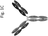
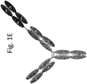
- the T cell activating bispecific antigen binding molecule comprises a first, a second, a third, a fourth and a fifth polypeptide chain that form a first, a second and a third antigen binding moiety, wherein the first antigen binding moiety is capable of binding CD3 and the second and the third antigen binding moiety each are capable of binding Folate Receptor 1 (FolR1), wherein a) the first and the second polypeptide chain comprise, in amino (N)-terminal to carboxyl (C)-terminal direction, VLD1 and CLD1; b) the third polypeptide chain comprises, in N-terminal to C-terminal direction, VLD2 and CH1D2; c) the fourth polypeptide chain comprises, in N-terminal to C-terminal direction, VHD1, CH1D1, CH2D1 and CH3D1; d) the fifth polypeptide chain comprises VHD1, CH1D1, VHD2, CLD2, CH2D2 and CH3D2; wherein
- the bispecific antibody is bivalent both for FolR1 and CD3.
- the bispecific antibody comprises one or more Fab fragment(s) comprising an antigen binding site specific for CD3, wherein the variable regions or the constant regions of the heavy and light chain are exchanged.
- the bispecific antibody comprises an Fc domain, at least one Fab fragment comprising the antigen binding site specific for FolR1, and at least one Fab fragment comprising the antigen binding site specific for CD3 wherein either the variable regions or the constant regions of the heavy and light chain of at least one Fab fragment are exchanged.
- the bispecific antibody comprises:
- At least one of said Fab fragments is connected to the Fc domain via a peptide linker.
- said bispecific antibody comprises an Fc domain, which comprises one or more amino acid substitution that reduces binding to Fc receptors and/or effector function.
- said one or more amino acid substitution is at one or more positions selected from the group of L234, L235, and P329.
- each subunit of the Fc domain comprises three amino acid substitutions that abolish binding to an activating or inhibitory Fc receptor and/or effector function wherein said amino acid substitutions are L234A, L235A and P329G.
- the PD-1 axis binding antagonist is selected from the group consisting of a PD-1 binding antagonist, a PDL1 binding antagonist and a PDL2 binding antagonist. In some embodiments, the PD-1 axis binding antagonist is a PD-1 binding antagonist. In some embodiments, the PD-1 binding antagonist inhibits the binding of PD-1 to its ligand binding partners. In some embodiments, the PD-1 binding antagonist inhibits the binding of PD-1 to PDL1. In some embodiments, the PD-1 binding antagonist inhibits the binding of PD-1 to PDL2. In some embodiments, the PD-1 binding antagonist inhibits the binding of PD-1 to both PDL1 and PDL2. In some embodiments, PD-1 binding antagonist is an antibody.
- the anti-PD-1 antibody is a monoclonal antibody. In some embodiments, the anti-PD-1 antibody is an antibody fragment selected from the group consisting of Fab, Fab'-SH, Fv, scFv, and (Fab')2 fragments.
- PD-1 binding antagonist is nivolumab, pembrolizumab, CT-011, or AMP-224. In some embodiments, the PD-1 axis binding antagonist is a PDL1 binding antagonist. In some embodiments, the PDL1 binding antagonist inhibits the binding of PDL1 to PD-1. In some embodiments, the PDL1 binding antagonist inhibits the binding of PDL1 to B7-1.
- the PDL1 binding antagonist inhibits the binding of PDL1 to both PD-1 and B7-1.
- the PDL1 binding antagonist is an anti-PDLl antibody.
- the anti-PDLl antibody is a monoclonal antibody.
- the anti-PDLl antibody is an antibody fragment selected from the group consisting of Fab, Fab'-SH, Fv, scFv, and (Fab')2 fragments.
- the anti-PDLl antibody is a humanized antibody or a human antibody.
- the PDL1 binding antagonist is selected from the group consisting of: YW243.55.S70, MPDL3280A, MDX-1105, and MEDI4736.
- the anti-PDLl antibody comprises a heavy chain comprising HVR-H1 sequence of SEQ ID NO:289, HVR-H2 sequence of SEQ ID NO:290, and HVR-H3 sequence of SEQ ID NO:291; and a light chain comprising HVR-L1 sequence of SEQ ID NO:292, HVR-L2 sequence of SEQ ID NO:293, and HVR-L3 sequence of SEQ ID NO:294.
- anti-PDLl antibody comprises a heavy chain variable region comprising the amino acid sequence of SEQ ID NO:280 or SEQ ID NO:281 and a light chain variable region comprising the amino acid sequence of SEQ ID NO:383.
- the anti-PDLl antibody comprises a heavy chain comprising the amino acid sequence of SEQ ID NO:278 and/or a light chain comprising the amino acid sequence of SEQ ID NO:279.
- the PD-1 axis binding antagonist is a PDL2 binding antagonist.
- PDL2 binding antagonist is an antibody.
- the anti-PDL2 antibody is a monoclonal antibody.
- the anti-PDL2 antibody is an antibody fragment selected from the group consisting of Fab, Fab'-SH, Fv, scFv, and (Fab')2 fragments.
- PDL2 binding antagonist is an immunoadhesin.
- the medthod of any of the above embodiments further comprises administering to the individual a T cell immunoglobulin mucin 3 (TIM3) antagonist.
- TIM3 antagonist is an anti-TIM3 antibody.
- the anti-TIM3 antibody induces internalization of TIM3 on a TIM3 expressing cell of at least 45% after 120 Minutes at 37 °C wherein internalization is measured by FACS analysis.
- the anti-TIM3 antibody has one or more of the following properties:
- T cells in the individual have enhanced activation, proliferation and/or effector function relative to prior to the administration of the combination. In one embodiment, T cells in the individual have enhanced activation, proliferation and/or effector function relative to administration of the T cell activating bispecific antigen binding molecule alone. In one embodiment, T cell effector function is secretion of at least one of IL-2, IFN- ⁇ and TNF- ⁇ . In one embodiment, the individual comprises less than about 15% PD-1 hi expressing tumor-infiltrating T cells.
- the invention provides for a method of enhancing immune function in an individual having a FolR1 positive cancer comprising administering to the individual an effective amount of a combination of a T cell activating bispecific antigen binding molecule specific for Folate Receptor 1 (FolR1) and CD3, and a PD-1 axis binding antagonist.
- T cells in the individual have enhanced activation, proliferation and/or effector function relative to prior to the administration of the combination.
- T cells in the individual have enhanced activation, proliferation and/or effector function relative to administration of the T cell activating bispecific antigen binding molecule alone.
- T cell effector function is secretion of at least one of IL-2, IFN- ⁇ and TNF- ⁇ .
- the individual comprises less than about 15% PD-1 hi expressing tumor-infiltrating T cells.
- the invention provides for a method for selecting a patient for treatment with a combination of a T cell activating bispecific antigen binding molecule specific for Folate Receptor 1 (FolR1) and CD3, and a PD-1 axis binding antagonist comprising measuring the level of PD-1 expression, wherein a patient having less than about 15% PD-1 hi expressing T cells is selected for treatment with the combination.
- a T cell activating bispecific antigen binding molecule specific for Folate Receptor 1 (FolR1) and CD3, and a PD-1 axis binding antagonist comprising measuring the level of PD-1 expression, wherein a patient having less than about 15% PD-1 hi expressing T cells is selected for treatment with the combination.
- the invention provides for a kit comprising a T cell activating bispecific antigen binding molecule specific for Folate Receptor 1 (FolR1) and CD3, and a package insert comprising instructions for using the T cell activating bispecific antigen binding molecule with a PD-1 axis binding antagonist to treat or delay progression of cancer in an individual.
- the kit further comprises instructions for using the T cell activating bispecific antigen binding molecule with a TIM3 antagonist.
- the invention provides for a kit comprising a T cell activating bispecific antigen binding molecule specific for Folate Receptor 1 (FolR1) and CD3 and a PD-1 axis binding antagonist, and a package insert comprising instructions for using the T cell activating bispecific antigen binding molecule and the PD-1 axis binding antagonist to treat or delay progression of cancer in an individual.
- the kit further comprises a TIM3 antagonist.
- the PD-1 axis binding antagonist is an anti-PD-1 antibody or an anti-PDL-1 antibody.
- the PD-1 axis binding antagonist is an anti-PD-1 immunoadhesin.
- the invention provides for a pharmaceutical composition comprising a T cell activating bispecific antigen binding molecule specific for Folate Receptor 1 (FolR1) and CD3, a PD-1 axis binding antagonist and a pharmaceutically acceptable carrier.
- the pharmaceutical composition further comprises a TIM3 antagonist.
- the invention provides for a use of a combination of a T cell activating bispecific antigen binding molecule specific for Folate Receptor 1 (FolR1) and CD3 and a PD-1 axis binding antagonist in the manufacture of a medicament for the treatment of cancer.
- the medicament is for treatment of ovarian cancer, lung cancer, breast cancer, renal cancer, colorectal cancer, endometrial cancer.
- advantageously said T cell activating bispecific antigen binding molecule and/or PD-1 axis binding antagonist is human or humanized.
- the bispecific antibody comprises an Fc domain, at least one Fab fragment comprising the antigen binding site specific for FolR1, and at least one Fab fragment comprising the antigen binding site specific for CD3.
- the invention provides for a method for treating or delaying progression of a cancer in an individual comprising administering to the individual an effective amount of a T cell activating bispecific antigen binding molecule and a TIM3 antagonist.
- the T cell activating bispecific antigen binding molecule comprises an Fc domain, two Fab fragments comprising each an antigen binding site specific for FolR1, and one Fab fragment comprising an antigen binding site specific for CD3.
- the present invention provides the use of a combination of a T cell activating bispecific antigen binding molecule that binds to FolR1 and CD3, and a PD-1 axis binding antagonist in the manufacture of a medicament for the treatment of cancer.
- the present invention provides the use of a combination of a T cell activating bispecific antigen binding molecule that binds to FolR1 and CD3, a PD-1 axis binding antagonist and a TIM3 antagonist in the manufacture of a medicament for the treatment of cancer.
- acceptor human framework for the purposes herein is a framework comprising the amino acid sequence of a light chain variable domain (VL) framework or a heavy chain variable domain (VH) framework derived from a human immunoglobulin framework or a human consensus framework, as defined below.
- An acceptor human framework "derived from” a human immunoglobulin framework or a human consensus framework may comprise the same amino acid sequence thereof, or it may contain amino acid sequence changes. In some embodiments, the number of amino acid changes are 10 or less, 9 or less, 8 or less, 7 or less, 6 or less, 5 or less, 4 or less, 3 or less, or 2 or less.
- the VL acceptor human framework is identical in sequence to the VL human immunoglobulin framework sequence or human consensus framework sequence.
- Bind refers to the strength of the sum total of noncovalent interactions between a single binding site of a molecule (e.g., an antibody) and its binding partner (e.g., an antigen).
- binding affinity refers to intrinsic binding affinity which reflects a 1:1 interaction between members of a binding pair (e.g., antibody and antigen).
- the affinity of a molecule X for its partner Y can generally be represented by the dissociation constant (Kd). Affinity can be measured by common methods known in the art, including those described herein. Specific illustrative and exemplary embodiments for measuring binding affinity are described in the following.
- an “affinity matured” antibody refers to an antibody with one or more alterations in one or more hypervariable regions (HVRs), compared to a parent antibody which does not possess such alterations, such alterations resulting in an improvement in the affinity of the antibody for antigen.
- HVRs hypervariable regions
- a bispecific antibody that specifically binds Folate Receptor1 (FolR1) and CD3 is used interchangeably herein and refer to a bispecific antibody that is capable of binding FolR1 and CD3 with sufficient affinity such that the antibody is useful as a diagnostic and/or therapeutic agent in targeting CD3 + T cells to FolR2 + target cells.
- anti-TIM3 antibody and "TIM3 antibody” are used synonymously herein to refer to an antibody that specifically binds to TIM3.
- An anti-TIM3 antibody described herein refers to an antibody that is capable of binding TIM3, especially a TIM3 polypeptide expressed on a cell surface, with sufficient affinity such that the antibody is useful as a diagnostic and/or therapeutic agent.
- the extent of binding of an antibody that specifically binds TIM3 to an unrelated non-TIM3 protein is less than about 10% of the binding of the antibody to TIM3 as measured, e.g., by a radioimmunoassay (RIA) or flow cytometry (FACS).
- RIA radioimmunoassay
- FACS flow cytometry
- an antibody that specifically binds TIM3 has a dissociation constant (Kd) of ⁇ 1 ⁇ M, ⁇ 100 nM, ⁇ 10 nM, ⁇ 1 nM, ⁇ 0.1 nM, ⁇ 0.01 nM, or ⁇ 0.001 nM (e.g., 10 -8 M or less, e.g. from 10 -8 M to 10 -13 M, e.g., from 10 -9 M to 10 -13 M).
- an antibody that specifically binds TIM3 binds to an epitope of TIM3 that is conserved among DR5 from different species.
- said antibody binds to human and cynomolgous monkey TIM3.
- the term "An antibody that specifically binds TIM3" also encompasses bispecific antibodies that are capable of binding TIM3 and a second antigen.
- antibody herein is used in the broadest sense and encompasses various antibody structures, including but not limited to monoclonal antibodies, polyclonal antibodies, multispecific antibodies (e.g., bispecific antibodies), and antibody fragments so long as they exhibit the desired antigen-binding activity.
- antibody fragment refers to a molecule other than an intact antibody that comprises a portion of an intact antibody that binds the antigen to which the intact antibody binds.
- antibody fragments include but are not limited to Fv, Fab, Fab', Fab'-SH, F(ab') 2 ; diabodies, cross-Fab fragments; linear antibodies; single-chain antibody molecules (e.g. scFv); and multispecific antibodies formed from antibody fragments.
- scFv antibodies are, e.g. described in Houston, J.S., Methods in Enzymol. 203 (1991) 46-96 ).
- antibody fragments comprise single chain polypeptides having the characteristics of a VH domain, namely being able to assemble together with a VL domain, or of a VL domain, namely being able to assemble together with a VH domain to a functional antigen binding site and thereby providing the antigen binding property of full length antibodies.
- Fab fragment refers to an antibody fragment comprising a light chain fragment comprising a VL domain and a constant domain of a light chain (CL), and a VH domain and a first constant domain (CH1) of a heavy chain.
- the bispecific antibodies of the invention comprise at least one Fab fragment, wherein either the variable regions or the constant regions of the heavy and light chain are exchanged. Due to the exchange of either the variable regions or the constant regions, said Fab fragment is also referred to as "cross-Fab fragment” or "xFab fragment” or "crossover Fab fragment”.
- crossover Fab molecule comprises a peptide chain composed of the light chain variable region (VL) and the heavy chain constant region (CH1), and a peptide chain composed of the heavy chain variable region (VH) and the light chain constant region (CL).
- VL variable region
- CH1 heavy chain constant region
- VH heavy chain variable region
- CL light chain constant region
- the crossover Fab molecule comprises a peptide chain composed of the heavy chain variable region (VH) and the light chain constant region (CL), and a peptide chain composed of the light chain variable region (VL) and the heavy chain constant region (CH1).
- This crossover Fab molecule is also referred to as CrossFab (CLCH1) .
- Bispecific antibody formats comprising crossover Fab fragments have been described, for example, in WO 2009/080252 , WO 2009/080253 , WO 2009/080251 , WO 2009/080254 , WO 2010/136172 , WO 2010/145792 and WO 2013/026831 .
- a “single chain Fab fragment” or “scFab” is a polypeptide consisting of an antibody heavy chain variable domain (VH), an antibody constant domain 1 (CH1), an antibody light chain variable domain (VL), an antibody light chain constant domain (CL) and a linker, wherein said antibody domains and said linker have one of the following orders in N-terminal to C-terminal direction: a) VH-CH1-linker-VL-CL, b) VL-CL-linker-VH-CH1, c) VH-CL-linker-VL-CH1 or d) VL-CH1-linker-VH-CL; and wherein said linker is a polypeptide of at least 30 amino acids, preferably between 32 and 50 amino acids.
- Said single chain Fab fragments a) VH-CH1-linker-VL-CL, b) VL-CL-linker-VH-CH1, c) VH-CL-linker-VL-CH1 and d) VL-CH1-linker-VH-CL, are stabilized via the natural disulfide bond between the CL domain and the CH1 domain.
- these single chain Fab molecules might be further stabilized by generation of interchain disulfide bonds via insertion of cysteine residues (e.g. position 44 in the variable heavy chain and positionn 100 in the variable light chain according to Kabat numbering).
- cysteine residues e.g. position 44 in the variable heavy chain and positionn 100 in the variable light chain according to Kabat numbering.
- N-terminus denotes the last amino acid of the N-terminus.
- C-terminus denotes the last amino acid of the C-terminus.
- fused or “connected” is meant that the components (e.g. a Fab molecule and an Fc domain subunit) are linked by peptide bonds, either directly or via one or more peptide linkers.
- linker refers to a peptide linker and is preferably a peptide with an amino acid sequence with a length of at least 5 amino acids, preferably with a length of 5 to 100, more preferably of 10 to 50 amino acids.
- said peptide linker is (G 4 S) 2 (SEQ ID NO: 386).
- immunoglobulin molecule refers to a protein having the structure of a naturally occurring antibody.
- immunoglobulins of the IgG class are heterotetrameric glycoproteins of about 150,000 daltons, composed of two light chains and two heavy chains that are disulfide-bonded. From N- to C-terminus, each heavy chain has a variable region (VH), also called a variable heavy domain or a heavy chain variable domain, followed by three constant domains (CH1, CH2, and CH3), also called a heavy chain constant region.
- VH variable region
- CH1, CH2, and CH3 constant domains
- each light chain has a variable region (VL), also called a variable light domain or a light chain variable domain, followed by a constant light (CL) domain, also called a light chain constant region.
- VL variable region
- the heavy chain of an immunoglobulin may be assigned to one of five types, called ⁇ (IgA), ⁇ (IgD), ⁇ (IgE), ⁇ (IgG), or ⁇ (IgM), some of which may be further divided into subtypes, e.g. ⁇ 1 (IgG 1 ), ⁇ 2 (IgG 2 ), ⁇ 3 (IgG 3 ), ⁇ 4 (IgG 4 ), ⁇ 1 (IgA 1 ) and ⁇ 2 (IgA 2 ).
- the light chain of an immunoglobulin may be assigned to one of two types, called kappa ( ⁇ ) and lambda ( ⁇ ), based on the amino acid sequence of its constant domain.
- An immunoglobulin essentially consists of two Fab molecules and an Fc domain, linked via the immunoglobulin hinge region.
- an "antibody that binds to the same epitope” as a reference antibody refers to an antibody that blocks binding of the reference antibody to its antigen in a competition assay by 50% or more, and conversely, the reference antibody blocks binding of the antibody to its antigen in a competition assay by 50% or more.
- An exemplary competition assay is provided herein.
- an antigen binding domain refers to the part of an antigen binding molecule that comprises the area which specifically binds to and is complementary to part or all of an antigen. Where an antigen is large, an antigen binding molecule may only bind to a particular part of the antigen, which part is termed an epitope.
- An antigen binding domain may be provided by, for example, one or more antibody variable domains (also called antibody variable regions).
- an antigen binding domain comprises an antibody light chain variable region (VL) and an antibody heavy chain variable region (VH).
- chimeric antibody refers to an antibody in which a portion of the heavy and/or light chain is derived from a particular source or species, while the remainder of the heavy and/or light chain is derived from a different source or species, usually prepared by recombinant DNA techniques. Chimeric antibodies comprising a rabbit variable region and a human constant region are preferred. Other preferred forms of “chimeric antibodies” encompassed by the present invention are those in which the constant region has been modified or changed from that of the original antibody to generate the properties according to the invention, especially in regard to C1q binding and/or Fc receptor (FcR) binding. Such chimeric antibodies are also referred to as "class-switched antibodies”.
- Chimeric antibodies are the product of expressed immunoglobulin genes comprising DNA segments encoding immunoglobulin variable regions and DNA segments encoding immunoglobulin constant regions. Methods for producing chimeric antibodies involve conventional recombinant DNA and gene transfection techniques are well known in the art. See e.g. Morrison, S.L., et al., Proc. Natl. Acad. Sci. USA 81 (1984) 6851-6855 ; US Patent Nos. 5,202,238 and 5,204,244 .
- cytotoxic agent refers to a substance that inhibits or prevents a cellular function and/or causes cell death or destruction.
- Cytotoxic agents include, but are not limited to, radioactive isotopes (e.g., At 211 , I 131 , I 125 , Y 90 , Re 186 , Re 188 , Sm 153 , Bi 212 , P 32 , Pb 212 and radioactive isotopes of Lu); chemotherapeutic agents or drugs (e.g., methotrexate, adriamicin, vinca alkaloids (vincristine, vinblastine, etoposide), doxorubicin, melphalan, mitomycin C, chlorambucil, daunorubicin or other intercalating agents); growth inhibitory agents; enzymes and fragments thereof such as nucleolytic enzymes; antibiotics; toxins such as small molecule toxins or enzymatically active toxins of bacterial, fungal
- Antibody effector functions refer to those biological activities attributable to the Fc region of an antibody, which vary with the antibody isotype. Examples of antibody effector functions include: C1q binding and complement dependent cytotoxicity (CDC); Fc receptor binding; antibody-dependent cell-mediated cytotoxicity (ADCC); antibody-dependent cellular phagocytosis (ADCP), cytokine secretion, immune complex-mediated antigen uptake by antigen presenting cells; down regulation of cell surface receptors (e.g. B cell receptor); and B cell activation.
- CDC complement dependent cytotoxicity
- ADCC antibody-dependent cell-mediated cytotoxicity
- ADCP antibody-dependent cellular phagocytosis
- cytokine secretion immune complex-mediated antigen uptake by antigen presenting cells
- down regulation of cell surface receptors e.g. B cell receptor
- B cell activation e.g. B cell activation
- engine engineered, engineering
- engineering includes modifications of the amino acid sequence, of the glycosylation pattern, or of the side chain group of individual amino acids, as well as combinations of these approaches.
- amino acid mutation as used herein is meant to encompass amino acid substitutions, deletions, insertions, and modifications. Any combination of substitution, deletion, insertion, and modification can be made to arrive at the final construct, provided that the final construct possesses the desired characteristics, e.g., reduced binding to an Fc receptor, or increased association with another peptide.
- Amino acid sequence deletions and insertions include amino- and/or carboxy-terminal deletions and insertions of amino acids.
- Particular amino acid mutations are amino acid substitutions.
- non-conservative amino acid substitutions i.e. replacing one amino acid with another amino acid having different structural and/or chemical properties, are particularly preferred.
- Amino acid substitutions include replacement by non-naturally occurring amino acids or by naturally occurring amino acid derivatives of the twenty standard amino acids (e.g. 4-hydroxyproline, 3-methylhistidine, ornithine, homoserine, 5-hydroxylysine).
- Amino acid mutations can be generated using genetic or chemical methods well known in the art. Genetic methods may include site-directed mutagenesis, PCR, gene synthesis and the like. It is contemplated that methods of altering the side chain group of an amino acid by methods other than genetic engineering, such as chemical modification, may also be useful. Various designations may be used herein to indicate the same amino acid mutation. For example, a substitution from proline at position 329 of the Fc domain to glycine can be indicated as 329G, G329, G 329 , P329G, or Pro329Gly.
- an "effective amount" of an agent refers to an amount effective, at dosages and for periods of time necessary, to achieve the desired therapeutic or prophylactic result.
- Fc domain or "Fc region” herein is used to define a C-terminal region of an immunoglobulin heavy chain that contains at least a portion of the constant region.
- the term includes native sequence Fc regions and variant Fc regions.
- the boundaries of the Fc region of an IgG heavy chain might vary slightly, the human IgG heavy chain Fc region is usually defined to extend from Cys226, or from Pro230, to the carboxyl-terminus of the heavy chain.
- the C-terminal lysine (Lys447) of the Fc region may or may not be present.
- a "subunit" of an Fc domain as used herein refers to one of the two polypeptides forming the dimeric Fc domain, i.e. a polypeptide comprising C-terminal constant regions of an immunoglobulin heavy chain, capable of stable self-association.
- a subunit of an IgG Fc domain comprises an IgG CH2 and an IgG CH3 constant domain.
- a "modification promoting the association of the first and the second subunit of the Fc domain” is a manipulation of the peptide backbone or the post-translational modifications of an Fc domain subunit that reduces or prevents the association of a polypeptide comprising the Fc domain subunit with an identical polypeptide to form a homodimer.
- a modification promoting association as used herein particularly includes separate modifications made to each of the two Fc domain subunits desired to associate (i.e. the first and the second subunit of the Fc domain), wherein the modifications are complementary to each other so as to promote association of the two Fc domain subunits.
- a modification promoting association may alter the structure or charge of one or both of the Fc domain subunits so as to make their association sterically or electrostatically favorable, respectively.
- (hetero)dimerization occurs between a polypeptide comprising the first Fc domain subunit and a polypeptide comprising the second Fc domain subunit, which might be non-identical in the sense that further components fused to each of the subunits (e.g. antigen binding moieties) are not the same.
- the modification promoting association comprises an amino acid mutation in the Fc domain, specifically an amino acid substitution.
- the modification promoting association comprises a separate amino acid mutation, specifically an amino acid substitution, in each of the two subunits of the Fc domain.
- FR Framework or "FR” refers to variable domain residues other than hypervariable region (HVR) residues.
- the FR of a variable domain generally consists of four FR domains: FR1, FR2, FR3, and FR4. Accordingly, the HVR and FR sequences generally appear in the following sequence in VH (or VL): FR1-H1(L1)-FR2-H2(L2)-FR3-H3(L3)-FR4.
- full length antibody “intact antibody,” and “whole antibody” are used herein interchangeably to refer to an antibody having a structure substantially similar to a native antibody structure or having heavy chains that contain an Fc region as defined herein.
- host cell refers to cells into which exogenous nucleic acid has been introduced, including the progeny of such cells.
- Host cells include “transformants” and “transformed cells,” which include the primary transformed cell and progeny derived therefrom without regard to the number of passages. Progeny may not be completely identical in nucleic acid content to a parent cell, but may contain mutations. Mutant progeny that have the same function or biological activity as screened or selected for in the originally transformed cell are included herein.
- a “human antibody” is one which possesses an amino acid sequence which corresponds to that of an antibody produced by a human or a human cell or derived from a non-human source that utilizes human antibody repertoires or other human antibody-encoding sequences. This definition of a human antibody specifically excludes a humanized antibody comprising non-human antigen-binding residues.
- the term "human antibody” as used herein also comprises such antibodies which are modified in the constant region to generate the properties according to the invention, especially in regard to C1q binding and/or FcR binding, e.g. by "class switching” i.e. change or mutation of Fc parts (e.g. from IgG1 to IgG4 and/or IgG1/IgG4 mutation.)
- recombinant human antibody is intended to include all human antibodies that are prepared, expressed, created or isolated by recombinant means, such as antibodies isolated from a host cell such as a NS0 or CHO cell or from an animal (e.g. a mouse) that is transgenic for human immunoglobulin genes or antibodies expressed using a recombinant expression vector transfected into a host cell.
- recombinant human antibodies have variable and constant regions in a rearranged form.
- the recombinant human antibodies according to the invention have been subjected to in vivo somatic hypermutation.
- the amino acid sequences of the VH and VL regions of the recombinant antibodies are sequences that, while derived from and related to human germ line VH and VL sequences, may not naturally exist within the human antibody germ line repertoire in vivo.
- a "human consensus framework” is a framework which represents the most commonly occurring amino acid residues in a selection of human immunoglobulin VL or VH framework sequences.
- the selection of human immunoglobulin VL or VH sequences is from a subgroup of variable domain sequences.
- the subgroup of sequences is a subgroup as in Kabat et al., Sequences of Proteins of Immunological Interest, Fifth Edition, NIH Publication 91-3242, Bethesda MD (1991), vols. 1-3 .
- the subgroup is subgroup kappa I as in Kabat et al., supra.
- the subgroup is subgroup III as in Kabat et al., supra.
- a “humanized” antibody refers to a chimeric antibody comprising amino acid residues from non-human HVRs and amino acid residues from human FRs.
- a humanized antibody will comprise substantially all of at least one, and typically two, variable domains, in which all or substantially all of the HVRs (e.g., CDRs) correspond to those of a non-human antibody, and all or substantially all of the FRs correspond to those of a human antibody.
- a humanized antibody optionally may comprise at least a portion of an antibody constant region derived from a human antibody.
- a "humanized form" of an antibody, e.g., a non-human antibody refers to an antibody that has undergone humanization.
- humanized antibodies encompassed by the present invention are those in which the constant region has been additionally modified or changed from that of the original antibody to generate the properties according to the invention, especially in regard to C1q binding and/or Fc receptor (FcR) binding.
- FcR Fc receptor
- hypervariable region refers to each of the regions of an antibody variable domain which are hypervariable in sequence and/or form structurally defined loops ("hypervariable loops").
- native four-chain antibodies comprise six HVRs; three in the VH (H1, H2, H3), and three in the VL (L1, L2, L3).
- HVRs generally comprise amino acid residues from the hypervariable loops and/or from the "complementarity determining regions" (CDRs), the latter being of highest sequence variability and/or involved in antigen recognition.
- CDRs complementarity determining regions
- Exemplary hypervariable loops occur at amino acid residues 26-32 (L1), 50-52 (L2), 91-96 (L3), 26-32 (HI), 53-55 (H2), and 96-101 (H3).
- Exemplary CDRs CDR-L1, CDR-L2, CDR-L3, CDR-H1, CDR-H2, and CDR-H3 occur at amino acid residues 24-34 of L1, 50-56 of L2, 89-97 of L3, 31-35B of H1, 50-65 of H2, and 95-102 of H3.
- Hypervariable regions are also referred to as complementarity determining regions (CDRs), and these terms are used herein interchangeably in reference to portions of the variable region that form the antigen binding regions.
- CDRs complementarity determining regions
- CDR Definitions 1 CDR Kabat Chothia AbM 2 V H CDR1 31-35 26-32 26-35 V H CDR2 50-65 52-58 50-58 V H CDR3 95-102 95-102 95-102 V L CDR1 24-34 26-32 24-34 V L CDR2 50-56 50-52 50-56 V L CDR3 89-97 91-96 89-97 1 Numbering of all CDR definitions in Table A is according to the numbering conventions set forth by Kabat et al. (see below). 2 "AbM” with a lowercase “b” as used in Table A refers to the CDRs as defined by Oxford Molecular's "AbM” antibody modeling software.
- Kabat et al. also defined a numbering system for variable region sequences that is applicable to any antibody.
- One of ordinary skill in the art can unambiguously assign this system of "Kabat numbering" to any variable region sequence, without reliance on any experimental data beyond the sequence itself.
- Kabat numbering refers to the numbering system set forth by Kabat et al., U.S. Dept. of Health and Human Services, "Sequence of Proteins of Immunological Interest" (1983 ). Unless otherwise specified, references to the numbering of specific amino acid residue positions in an antibody variable region are according to the Kabat numbering system.
- CDRs generally comprise the amino acid residues that form the hypervariable loops.
- CDRs also comprise "specificity determining residues," or "SDRs,” which are residues that contact antigen. SDRs are contained within regions of the CDRs called abbreviated-CDRs, or a-CDRs.
- Exemplary a-CDRs (a-CDR-L1, a-CDR-L2, a-CDR-L3, a-CDR-H1, a-CDR-H2, and a-CDR-H3) occur at amino acid residues 31-34 of L1, 50-55 of L2, 89-96 of L3, 31-35B of H1, 50-58 of H2, and 95-102 of H3.
- HVR residues and other residues in the variable domain are numbered herein according to Kabat et al., supra.
- an “immunoconjugate” is an antibody conjugated to one or more heterologous molecule(s), including but not limited to a cytotoxic agent.
- mammals include, but are not limited to, domesticated animals (e.g., cows, sheep, cats, dogs, and horses), primates (e.g., humans and non-human primates such as monkeys), rabbits, and rodents (e.g., mice and rats).
- domesticated animals e.g., cows, sheep, cats, dogs, and horses
- primates e.g., humans and non-human primates such as monkeys
- rabbits e.g., mice and rats
- rodents e.g., mice and rats.
- the individual or subject is a human.
- an “isolated” antibody is one which has been separated from a component of its natural environment.
- an antibody is purified to greater than 95% or 99% purity as determined by, for example, electrophoretic (e.g., SDS-PAGE, isoelectric focusing (IEF), capillary electrophoresis) or chromatographic (e.g., ion exchange or reverse phase HPLC).
- electrophoretic e.g., SDS-PAGE, isoelectric focusing (IEF), capillary electrophoresis
- chromatographic e.g., ion exchange or reverse phase HPLC
- nucleic acid refers to a nucleic acid molecule that has been separated from a component of its natural environment.
- An isolated nucleic acid includes a nucleic acid molecule contained in cells that ordinarily contain the nucleic acid molecule, but the nucleic acid molecule is present extrachromosomally or at a chromosomal location that is different from its natural chromosomal location.
- isolated nucleic acid encoding a bispecific antibody that specifically binds DR5 and FAP antibody refers to one or more nucleic acid molecules encoding antibody heavy and light chains (or fragments thereof), including such nucleic acid molecule(s) in a single vector or separate vectors, and such nucleic acid molecule(s) present at one or more locations in a host cell.
- monoclonal antibody refers to an antibody obtained from a population of substantially homogeneous antibodies, i.e., the individual antibodies comprising the population are identical and/or bind the same epitope, except for possible variant antibodies, e.g., containing naturally occurring mutations or arising during production of a monoclonal antibody preparation, such variants generally being present in minor amounts.
- polyclonal antibody preparations typically include different antibodies directed against different determinants (epitopes)
- each monoclonal antibody of a monoclonal antibody preparation is directed against a single determinant on an antigen.
- the modifier "monoclonal” indicates the character of the antibody as being obtained from a substantially homogeneous population of antibodies, and is not to be construed as requiring production of the antibody by any particular method.
- the monoclonal antibodies to be used in accordance with the present invention may be made by a variety of techniques, including but not limited to the hybridoma method, recombinant DNA methods, phage-display methods, and methods utilizing transgenic animals containing all or part of the human immunoglobulin loci, such methods and other exemplary methods for making monoclonal antibodies being described herein.
- naked antibody refers to an antibody that is not conjugated to a heterologous moiety (e.g., a cytotoxic moiety) or radiolabel.
- the naked antibody may be present in a pharmaceutical formulation.
- Native antibodies refer to naturally occurring immunoglobulin molecules with varying structures.
- native IgG antibodies are heterotetrameric glycoproteins of about 150,000 daltons, composed of two identical light chains and two identical heavy chains that are disulfide-bonded. From N- to C-terminus, each heavy chain has a variable region (VH), also called a variable heavy domain or a heavy chain variable domain, followed by three constant domains (CH1, CH2, and CH3).
- VH variable region
- VL variable region
- the light chain of an antibody may be assigned to one of two types, called kappa ( ⁇ ) and lambda ( ⁇ ), based on the amino acid sequence of its constant domain.
- blocking antibody or an “antagonist” antibody is one that inhibits or reduces a biological activity of the antigen it binds.
- blocking antibodies or antagonist antibodies substantially or completely inhibit the biological activity of the antigen.
- the anti-PD-Ll antibodies of the invention block the signaling through PD- 1 so as to restore a functional response by T-cells (e.g., proliferation, cytokine production, target cell killing) from a dysfunctional state to antigen stimulation.
- agonist or activating antibody is one that enhances or initiates signaling by the antigen to which it binds.
- agonist antibodies cause or activate signaling without the presence of the natural ligand.
- package insert is used to refer to instructions customarily included in commercial packages of therapeutic products, that contain information about the indications, usage, dosage, administration, combination therapy, contraindications and/or warnings concerning the use of such therapeutic products.
- No substantial cross-reactivity means that a molecule (e.g. , an antibody) does not recognize or specifically bind an antigen different from the actual target antigen of the molecule (e.g. an antigen closely related to the target antigen), particularly when compared to that target antigen.
- an antibody may bind less than about 10% to less than about 5% to an antigen different from the actual target antigen, or may bind said antigen different from the actual target antigen at an amount consisting of less than about 10%, 9%, 8% 7%, 6%, 5%, 4%, 3%, 2%, 1%, 0.5%, 0.2%, or 0.1%, preferably less than about 2%, 1%, or 0.5%, and most preferably less than about 0.2% or 0.1% antigen different from the actual target antigen.
- Percent (%) amino acid sequence identity with respect to a reference polypeptide sequence is defined as the percentage of amino acid residues in a candidate sequence that are identical with the amino acid residues in the reference polypeptide sequence, after aligning the sequences and introducing gaps, if necessary, to achieve the maximum percent sequence identity, and not considering any conservative substitutions as part of the sequence identity. Alignment for purposes of determining percent amino acid sequence identity can be achieved in various ways that are within the skill in the art, for instance, using publicly available computer software such as BLAST, BLAST-2, ALIGN or Megalign (DNASTAR) software. Those skilled in the art can determine appropriate parameters for aligning sequences, including any algorithms needed to achieve maximal alignment over the full length of the sequences being compared.
- % amino acid sequence identity values are generated using the sequence comparison computer program ALIGN-2.
- the ALIGN-2 sequence comparison computer program was authored by Genentech, Inc., and the source code has been filed with user documentation in the U.S. Copyright Office, Washington D.C., 20559, where it is registered under U.S. Copyright Registration No. TXU510087.
- the ALIGN-2 program is publicly available from Genentech, Inc., South San Francisco, California, or may be compiled from the source code.
- the ALIGN-2 program should be compiled for use on a UNIX operating system, including digital UNIX V4.0D. All sequence comparison parameters are set by the ALIGN-2 program and do not vary.
- the % amino acid sequence identity of a given amino acid sequence A to, with, or against a given amino acid sequence B is calculated as follows: 100 times the fraction X/Y where X is the number of amino acid residues scored as identical matches by the sequence alignment program ALIGN-2 in that program's alignment of A and B, and where Y is the total number of amino acid residues in B.
- pharmaceutical formulation refers to a preparation which is in such form as to permit the biological activity of an active ingredient contained therein to be effective, and which contains no additional components which are unacceptably toxic to a subject to which the formulation would be administered.
- a “pharmaceutically acceptable carrier” refers to an ingredient in a pharmaceutical formulation, other than an active ingredient, which is nontoxic to a subject.
- a pharmaceutically acceptable carrier includes, but is not limited to, a buffer, excipient, stabilizer, or preservative.
- PD-1 axis binding antagonist is a molecule that inhibits the interaction of a PD- 1 axis binding partner with either one or more of its binding partner, so as to remove T-cell dysfunction resulting from signaling on the PD- 1 signaling axis - with a result being to restore or enhance T-cell function ⁇ e.g., proliferation, cytokine production, target cell killing).
- a PD- 1 axis binding antagonist includes a PD- 1 binding antagonist, a PD-L1 binding antagonist and a PD-L2 binding antagonist.
- PD-1 binding antagonists is a molecule that decreases, blocks, inhibits, abrogates or interferes with signal transduction resulting from the interaction of PD-1 with one or more of its binding partners, such as PD-L1, PD-L2.
- the PD-1 binding antagonist is a molecule that inhibits the binding of PD-1 to its binding partners.
- the PD-1 binding antagonist inhibits the binding of PD-1 to PD-L1 and/or PD-L2.
- PD-1 binding antagonists include anti-PD-1 antibodies, antigen binding fragments thereof, immunoadhesins, fusion proteins, oligopeptides and other molecules that decrease, block, inhibit, abrogate or interfere with signal transduction resulting from the interaction of PD-1 with PD-L1 and/or PD-L2.
- a PD-1 binding antagonist reduces the negative co-stimulatory signal mediated by or through cell surface proteins expressed on T lymphocytes mediated signaling through PD-1 so as render a dysfunctional T-cell less dysfunctional (e.g. , enhancing effector responses to antigen recognition).
- the PD-1 binding antagonist is an anti-PD-1 antibody.
- a PD- 1 binding antagonist is MDX- 1106 described herein.
- a PD-1 binding antagonist is Merck 3745 described herein.
- a PD-1 binding antagonist is CT-01 1 described herein.
- PD-L1 binding antagonists is a molecule that decreases, blocks, inhibits, abrogates or interferes with signal transduction resulting from the interaction of PD-L1 with either one or more of its binding partners, such as PD-1 , B7-1 .
- a PD-L1 binding antagonist is a molecule that inhibits the binding of PD-L 1 to its binding partners.
- the PD-L1 binding antagonist inhibits binding of PD-L1 to PD-1 and/or B7-1.
- the PD-L1 binding antagonists include anti-PD-Ll antibodies, antigen binding fragments thereof, immunoadhesins, fusion proteins, oligopeptides and other molecules that decrease, block, inhibit, abrogate or interfere with signal transduction resulting from the interaction of PD-L1 with one or more of its binding partners, such as PD-1, B7- 1 .
- a PD-L1 binding antagonist reduces the negative co-stimulatory signal mediated by or through cell surface proteins expressed on T lymphocytes mediated signaling through PD-L 1 so as to render a dysfunctional T-cell less dysfunctional (e.g. , enhancing effector responses to antigen recognition).
- a PD-L1 binding antagonist is an anti-PD-L1 antibody.
- an anti-PD-Ll antibody is YW243.55.S70 described herein.
- an anti-PD-Ll antibody is MDX- 1 105 described herein.
- an anti-PD-Ll antibody is MPDL3280A described herein.
- PD-L2 binding antagonists is a molecule that decreases, blocks, inhibits, abrogates or interferes with signal transduction resulting from the interaction of PD-L2 with either one or more of its binding partners, such as PD- 1.
- a PD-L2 binding antagonist is a molecule that inhibits the binding of PD-L2 to its binding partners.
- the PD-L2 binding antagonist inhibits binding of PD-L2 to PD- 1.
- the PD-L2 antagonists include anti-PD-L2 antibodies, antigen binding fragments thereof, immunoadhesins, fusion proteins, oligopeptides and other molecules that decrease, block, inhibit, abrogate or interfere with signal transduction resulting from the interaction of PD- L2 with either one or more of its binding partners, such as PD-1.
- a PD-L2 binding antagonist reduces the negative co-stimulatory signal mediated by or through cell surface proteins expressed on T lymphocytes mediated signaling through PD-L2 so as render a dysfunctional T-cell less dysfunctional (e.g. , enhancing effector responses to antigen recognition).
- a PD-L2 binding antagonist is an immunoadhesin.
- a "PD-1 oligopeptide " "PD-L1 oligopeptide " or “PD-L2 oligopeptide” is an oligopeptide that binds, preferably specifically, to a PD-1, PD-L1 or PD-L2 negative costimulatory polypeptide, respectively, including a receptor, ligand or signaling component, respectively, as described herein.
- Such oligopeptides may be chemically synthesized using known oligopeptide synthesis methodology or may be prepared and purified using recombinant technology.
- Such oligopeptides are usually at least about 5 amino acids in length, alternatively at least about 6, 7, 8, 9, 10, 1 1 , 12, 13, 14, 15, 16, 17, 18, 19, 20, 21 , 22, 23, 24, 25, 26, 27, 28, 29, 30, 31 , 32, 33, 34, 35, 36, 37, 38, 39, 40, 41, 42, 43, 44, 45, 46, 47, 48, 49, 50, 51 , 52, 53, 54, 55, 56, 57, 58, 59, 60, 61 , 62, 63, 64, 65, 66, 67, 68, 69, 70, 71 , 72, 73, 74, 75, 76, 77, 78, 79, 80, 81 , 82, 83, 84, 85, 86, 87, 88, 89, 90, 91 , 92, 93, 94, 95, 96, 97, 98, 99, or 100 amino acids in length or more.
- oligopeptides may be identified using well known techniques.
- techniques for screening oligopeptide libraries for oligopeptides that are capable of specifically binding to a polypeptide target are well known in the art (see, e.g., U.S. Patent Nos. 5,556,762 , 5,750,373 , 4,708,871 , 4,833,092 , 5,223,409 , 5,403,484 , 5,571 ,689 , 5,663, 143 ; PCT Publication Nos. WO 84/03506 and WO84/03564 ; Geysen et al., Proc. Natl. Acad. Sci.
- the term "anergy” refers to the state of unresponsiveness to antigen stimulation resulting from incomplete or insufficient signals delivered through the T-cell receptor (e.g. increase in intracellular Ca +2 in the absence of ras-activation). T cell anergy can also result upon stimulation with antigen in the absence of co-stimulation, resulting in the cell becoming refractory to subsequent activation by the antigen even in the context of costimulation.
- the unresponsive state can often be overriden by the presence of lnterleukin-2. Anergic T-cells do not undergo clonal expansion and/or acquire effector functions.
- exhaustion refers to T cell exhaustion as a state of T cell dysfunction that arises from sustained TCR signaling that occurs during many chronic infections and cancer. It is distinguished from anergy in that it arises not through incomplete or deficient signaling, but from sustained signaling. It is defined by poor effector function, sustained expression of inhibitory receptors and a transcriptional state distinct from that of functional effector or memory T cells. Exhaustion prevents optimal control of infection and tumors. Exhaustion can result from both extrinsic negative regulatory pathways (e.g., immunoregulatory cytokines) as well as cell intrinsic negative regulatory (costimulatory) pathways (PD-1 , B7-H3, B7-H4, etc.).
- extrinsic negative regulatory pathways e.g., immunoregulatory cytokines
- costimulatory costimulatory
- Enhancing T-cell function means to induce, cause or stimulate a T-cell to have a sustained or amplified biological function, or renew or reactivate exhausted or inactive T-cells.
- Examples of enhancing T-cell function include: increased secretion of y-interferon from CD8 + T-cells, increased proliferation, increased antigen responsiveness (e.g. , viral, pathogen, or tumor clearance) relative to such levels before the intervention.
- the level of. enhancement is as least 50%, alternatively 60%, 70%, 80%, 90%, 100%, 1 20%, 150%, 200%. The manner of measuring this enhancement is known to one of ordinary skill in the art.
- Tumor immunity refers to the process in which tumors evade immune recognition and clearance. Thus, as a therapeutic concept, tumor immunity is “treated” when such evasion is attenuated, and the tumors are recognized and attacked by the immune system. Examples of tumor recognition include tumor binding, tumor shrinkage and tumor clearance.
- Immunogenecity refers to the ability of a particular substance to provoke an immune response. Tumors are immunogenic and enhancing tumor immunogenicity aids in the clearance of the tumor cells by the immune response. Examples of enhancing tumor immunogenicity include treatment with anti-PDL antibodies and a ME inhibitor.
- sustained response refers to the sustained effect on reducing tumor growth after cessation of a treatment.
- the tumor size may remain to be the same or smaller as compared to the size at the beginning of the administration phase.
- the sustained response has a duration at least the same as the treatment duration, at least 1.5X, 2. OX, 2.5X, or 3. OX length of the treatment duration.
- FAP Fibroblast activation protein
- FAP Fibroblast activation protein
- the term encompasses "full-length,” unprocessed FAP as well as any form of FAP that results from processing in the cell.
- the term also encompasses naturally occurring variants of FAP, e.g., splice variants or allelic variants.
- an anti-FAP antibody of the invention binds to the extracellular domain of FAP.
- the amino acid sequence of exemplary FAP polypeptide sequences, including the sequence of human FAP are disclosed in WO 2012/020006 .
- treatment refers to clinical intervention in an attempt to alter the natural course of the individual being treated, and can be performed either for prophylaxis or during the course of clinical pathology. Desirable effects of treatment include, but are not limited to, preventing occurrence or recurrence of disease, alleviation of symptoms, diminishment of any direct or indirect pathological consequences of the disease, preventing metastasis, decreasing the rate of disease progression, amelioration or palliation of the disease state, and remission or improved prognosis.
- antibodies of the invention are used to delay development of a disease or to slow the progression of a disease.
- cancer refers to proliferative diseases, such as the cancer is colorectal cancer, sarcoma, head and neck cancer, squamous cell carcinoma, breast cancer, pancreatic cancer, gastric cancer, non-small-cell lung carcinoma, small-cell lung cancer and mesothelioma, including refractory versions of any of the above cancers, or a combination of one or more of the above cancers.
- the cancer is colorectal cancer and optionally the chemotherapeutic agent is Irinotecan.
- the sarcoma is chondrosarcoma, leiomyosarcoma, gastrointestinal stromal tumours, fibrosarcoma, osteosarcoma. liposarcoma or maligant fibrous histiocytoma.
- variable region refers to the domain of an antibody heavy or light chain that is involved in binding the antibody to antigen.
- the variable domains of the heavy chain and light chain (VH and VL, respectively) of a native antibody generally have similar structures, with each domain comprising four conserved framework regions (FRs) and three hypervariable regions (HVRs).
- FRs conserved framework regions
- HVRs hypervariable regions
- antibodies that bind a particular antigen may be isolated using a VH or VL domain from an antibody that binds the antigen to screen a library of complementary VL or VH domains, respectively.
- VH or VL domain e.g., Portolano et al., J. Immunol. 150:880-887 (1993 ); Clarkson et al., Nature 352:624-628 (1991 ).
- antigen binding molecule refers in its broadest sense to a molecule that specifically binds an antigenic determinant. Examples of antigen binding molecules are immunoglobulins and derivatives, e.g. fragments, thereof.
- antigen-binding site of an antibody when used herein refer to the amino acid residues of an antibody which are responsible for antigen-binding.
- the antigen-binding portion of an antibody comprises amino acid residues from the "complementary determining regions" or "CDRs".
- CDRs complementary determining regions
- FR Framework regions are those variable domain regions other than the hypervariable region residues as herein defined. Therefore, the light and heavy chain variable domains of an antibody comprise from N- to C-terminus the domains FR1, CDR1, FR2, CDR2, FR3, CDR3, and FR4.
- CDR3 of the heavy chain is the region which contributes most to antigen binding and defines the antibody's properties.
- CDR and FR regions are determined according to the standard definition of Kabat et al., Sequences of Proteins of Immunological Interest, 5th ed., Public Health Service, National Institutes of Health, Bethesda, MD (1991 ) and/or those residues from a "hypervariable loop".
- Antibody specificity refers to selective recognition of the antibody for a particular epitope of an antigen. Natural antibodies, for example, are monospecific. "Bispecific antibodies” according to the invention are antibodies which have two different antigen-binding specificities. Antibodies of the present invention are specific for two different antigens, i.e. DR5 as first antigen and FAP as second antigen.
- monospecific antibody denotes an antibody that has one or more binding sites each of which bind to the same epitope of the same antigen.
- bispecific means that the antigen binding molecule is able to specifically bind to at least two distinct antigenic determinants.
- a bispecific antigen binding molecule comprises at least two antigen binding sites, each of which is specific for a different antigenic determinant.
- the bispecific antigen binding molecule is capable of simultaneously binding two antigenic determinants, particularly two antigenic determinants expressed on two distinct cells.
- the antibody provided herein is a multispecific antibody, e.g. a bispecific antibody.
- Multispecific antibodies are monoclonal antibodies that have binding specificities for at least two different sites.
- a bispecific antibody with binding specificities for FAP and DR5.
- bispecific antibodies may bind to two different epitopes of DR5.
- Bispecific antibodies may also be used to localize cytotoxic agents to cells which express DR5.
- Bispecific antibodies can be prepared as full length antibodies or antibody fragments.
- Multispecific antibodies include, but are not limited to, recombinant co-expression of two immunoglobulin heavy chain-light chain pairs having different specificities (see Milstein and Cuello, Nature 305: 537 (1983 )), WO 93/08829 , and Traunecker et al., EMBO J. 10: 3655 (1991 )), and "knob-in-hole” engineering (see, e.g., U.S. Patent No. 5,731,168 ). Multi-specific antibodies may also be made by engineering electrostatic steering effects for making antibody Fc-heterodimeric molecules ( WO 2009/089004 ); cross-linking two or more antibodies or fragments (see, e.g., US Patent No.
- the antibody or fragment herein also includes a "Dual Acting FAb” or “DAF” comprising at least one antigen binding site that binds to FAP or DR5 as well as another, different antigen (see, US 2008/0069820 , for example).
- a "Dual Acting FAb” or “DAF” comprising at least one antigen binding site that binds to FAP or DR5 as well as another, different antigen (see, US 2008/0069820 , for example).
- bispecific antibodies as used within the current application denotes the presence of a specified number of binding sites in an antibody molecule.
- the terms “bivalent”, “tetravalent”, and “hexavalent” denote the presence of two binding sites, four binding sites, and six binding sites, respectively, in an antibody molecule.
- the bispecific antibodies according to the invention are at least “bivalent” and may be “trivalent” or “multivalent” (e.g.”tetravalent” or "hexavalent”).
- Antibodies of the present invention have two or more binding sites and are bispecific. That is, the antibodies may be bispecific even in cases where there are more than two binding sites (i.e. that the antibody is trivalent or multivalent).
- Bispecific antibodies of the invention include, for example, multivalent single chain antibodies, diabodies and triabodies, as well as antibodies having the constant domain structure of full length antibodies to which further antigen-binding sites (e.g., single chain Fv, a VH domain and/or a VL domain, Fab, or (Fab)2) are linked via one or more peptide-linkers.
- the antibodies can be full length from a single species, or be chimerized or humanized.
- vector refers to a nucleic acid molecule capable of propagating another nucleic acid to which it is linked.
- the term includes the vector as a self-replicating nucleic acid structure as well as the vector incorporated into the genome of a host cell into which it has been introduced.
- Certain vectors are capable of directing the expression of nucleic acids to which they are operatively linked. Such vectors are referred to herein as "expression vectors.”
- amino acid denotes the group of naturally occurring carboxy ⁇ -amino acids comprising alanine (three letter code: ala, one letter code: A), arginine (arg, R), asparagine (asn, N), aspartic acid (asp, D), cysteine (cys, C), glutamine (gln, Q), glutamic acid (glu, E), glycine (gly, G), histidine (his, H), isoleucine (ile, 1), leucine (leu, L), lysine (lys, K), methionine (met, M), phenylalanine (phe, F), proline (pro, P), serine (ser, S), threonine (thr, T), tryptophan (trp, W), tyrosine (tyr, Y), and valine (val, V).
- the expressions "cell”, “cell line”, and “cell culture” are used interchangeably and all such designations include progeny.
- the words “transfectants” and “transfected cells” include the primary subject cell and cultures derived there from without regard for the number of transfers. It is also understood that all progeny may not be precisely identical in DNA content, due to deliberate or inadvertent mutations. Variant progeny that have the same function or biological activity as screened for in the originally transformed cell are included.
- Bind refers to the strength of the sum total of noncovalent interactions between a single binding site of a molecule (e.g., an antibody) and its binding partner (e.g., an antigen).
- binding affinity refers to intrinsic binding affinity which reflects a 1:1 interaction between members of a binding pair (e.g., antibody and antigen).
- the affinity of a molecule X for its partner Y can generally be represented by the dissociation constant (Kd). Affinity can be measured by common methods known in the art, including those described herein. Specific illustrative and exemplary embodiments for measuring binding affinity are described in the following.
- binding refers to the binding of the antibody to an epitope of the antigen in an in-vitro assay, preferably in a surface plasmon resonance assay (SPR, BIAcore, GE-Healthcare Uppsala, Sweden).
- the affinity of the binding is defined by the terms ka (rate constant for the association of the antibody from the antibody/antigen complex), kD (dissociation constant), and KD (kD/ka).
- Binding or specifically binding means a binding affinity (KD) of 10 -8 mol/l or less, preferably 10 -9 M to 10 -13 mol/l.
- Binding of the antibody to the death receptor can be investigated by a BIAcore assay (GE-Healthcare Uppsala, Sweden).
- the affinity of the binding is defined by the terms ka (rate constant for the association of the antibody from the antibody/antigen complex), kD (dissociation constant), and KD (kD/ka)
- Reduced binding for example reduced binding to an Fc receptor, refers to a decrease in affinity for the respective interaction, as measured for example by SPR.
- the term includes also reduction of the affinity to zero (or below the detection limit of the analytic method), i.e. complete abolishment of the interaction.
- “increased binding” refers to an increase in binding affinity for the respective interaction.
- T cell activation refers to one or more cellular response of a T lymphocyte, particularly a cytotoxic T lymphocyte, selected from: proliferation, differentiation, cytokine secretion, cytotoxic effector molecule release, cytotoxic activity, and expression of activation markers.
- the T cell activating bispecific antigen binding molecules of the invention are capable of inducing T cell activation. Suitable assays to measure T cell activation are known in the art described herein.
- a “target cell antigen” as used herein refers to an antigenic determinant presented on the surface of a target cell, for example a cell in a tumor such as a cancer cell or a cell of the tumor stroma. In particular "target cell antigen” refers to Folate Receptor 1.
- first and second with respect to antigen binding moieties etc., are used for convenience of distinguishing when there is more than one of each type of moiety. Use of these terms is not intended to confer a specific order or orientation of the T cell activating bispecific antigen binding molecule unless explicitly so stated.
- epitope includes any polypeptide determinant capable of specific binding to an antibody.
- epitope determinant include chemically active surface groupings of molecules such as amino acids, sugar side chains, phosphoryl, or sulfonyl, and, in certain embodiments, may have specific three dimensional structural characteristics, and or specific charge characteristics.
- An epitope is a region of an antigen that is bound by an antibody.
- antigenic determinant is synonymous with “antigen” and “epitope,” and refers to a site (e.g. a contiguous stretch of amino acids or a conformational configuration made up of different regions of non-contiguous amino acids) on a polypeptide macromolecule to which an antigen binding moiety binds, forming an antigen binding moiety-antigen complex.
- Useful antigenic determinants can be found, for example, on the surfaces of tumor cells, on the surfaces of virus-infected cells, on the surfaces of other diseased cells, on the surface of immune cells, free in blood serum, and/or in the extracellular matrix (ECM).
- ECM extracellular matrix
- the proteins referred to as antigens herein can be any native form the proteins from any vertebrate source, including mammals such as primates (e.g. humans) and rodents (e.g. mice and rats), unless otherwise indicated.
- the antigen is a human protein.
- the term encompasses the "full-length", unprocessed protein as well as any form of the protein that results from processing in the cell.
- the term also encompasses naturally occurring variants of the protein, e.g. splice variants or allelic variants.
- Exemplary human proteins useful as antigens include, but are not limited to: FolR1 (Folate receptor alpha (FRA); Folate binding protein (FBP); human FolR1 UniProt no.: P15328; murine FolR1 UniProt no.: P35846; cynomolgus FolR1 UniProt no.: G7PR14) and CD3, particularly the epsilon subunit of CD3 (see UniProt no. P07766 (version 130), NCBI RefSeq no. NP_000724.1, SEQ ID NO:150 for the human sequence; or UniProt no. Q95LI5 (version 49), NCBI GenBank no.
- the T cell activating bispecific antigen binding molecule of the invention binds to an epitope of CD3 or a target cell antigen that is conserved among the CD3 or target antigen from different species. In certain embodiments the T cell activating bispecific antigen binding molecule of the invention binds to CD3 and FolR1, but does not bind to FolR2 (Folate receptor beta; FRB; human FolR2 UniProt no.: P14207) or FolR3 (Folate receptor gamma; human FolR3 UniProt no.: P41439).
- FolR2 FolR2
- FolR3 FolR3 (Folate receptor gamma; human FolR3 UniProt no.: P41439).
- glycosylation engineering includes metabolic engineering of the glycosylation machinery of a cell, including genetic manipulations of the oligosaccharide synthesis pathways to achieve altered glycosylation of glycoproteins expressed in cells.
- glycosylation engineering includes the effects of mutations and cell environment on glycosylation.
- the glycosylation engineering is an alteration in glycosyltransferase activity.
- the engineering results in altered glucosaminyltransferase activity and/or fucosyltransferase activity.
- the invention is based on the use of a therapeutic combination of a T cell activating bispecific antigen binding molecule, e.g., a T cell activating bispecific antigen binding molecule comprising a first antigen binding site specific for Folate Receptor 1 (FolR1) and a second antigen binding site specific for CD3, and a PD-1 axis binding antagonist, e.g., for the treatment of cancer.
- a T cell activating bispecific antigen binding molecule e.g., a T cell activating bispecific antigen binding molecule comprising a first antigen binding site specific for Folate Receptor 1 (FolR1) and a second antigen binding site specific for CD3, and a PD-1 axis binding antagonist, e.g., for the treatment of cancer.
- the therapeutic combination further includes a TIM3 antagonist.
- the present invention relates to T cell activating bispecific antigen binding molecules and their use in combination with a PD-1 axis binding antagonists.
- the advantage of the combination over monotherapy is that the T cell activating bispecific antigen binding molecules used in the present invention enable re-direction and activation of T cells to the targeted cell while the PD-1 axis binding antagonist enhances T cell function by reducing T cell exhaustion.
- a method for treating or delaying progression of cancer in an individual comprising administering to the individual an effective amount of a T cell activating bispecific antigen binding molecules, e.g., a FolR1-TCB, and a PD-1 axis binding antagonist.
- a T cell activating bispecific antigen binding molecules e.g., a FolR1-TCB
- a PD-1 axis binding antagonist e.g., a PD-1 axis binding antagonist
- the treatment results in sustained response in the individual after cessation of the treatment.
- the methods of this invention may find use in treating conditions where enhanced immunogenicity is desired such as increasing tumor immunogenicity for the treatment of cancer.
- a variety of cancers may be treated, or their progression may be delayed, including but are not limited to a cancer that may contain a BRAF V600E mutation, a cancer that may contain a BRAF wildtype, a cancer that may contain a KRAS wildtype, or a cancer that may contain an activating KRAS mutation.
- the individual has endometrial cancer.
- the endometrial cancer may be at early stage or late state.
- the individual has melanoma.
- the melanoma may be at early stage or at late stage.
- the individual has colorectal cancer.
- the colorectal cancer may be at early stage or at late stage.
- the individual has lung cancer, e.g., non-small cell lung cancer.
- the non-small cell lung cancer may be at early stage or at late stage.
- the individual has pancreatic cancer.
- the pancreatice cancer may be at early stage or late state.
- the individual has a hematological malignancy.
- the hematological malignancy may be early stage or late stage.
- the individual has ovarian cancer.
- the ovarian cancer may be at early stage or at late stage.
- the individual has breast cancer.
- the breast cancer may be at early stage or at late stage.
- the individual has renal cell carcinoma.
- the renal cell carcinoma may be at early stage or at late stage.
- the individual is a mammal, such as domesticated animals (e.g., cows, sheep, cats, dogs, and horses), primates (e.g., humans and non-human primates such as monkeys), rabbits, and rodents (e.g., mice and rats).
- domesticated animals e.g., cows, sheep, cats, dogs, and horses
- primates e.g., humans and non-human primates such as monkeys
- rabbits e.g., mice and rats
- rodents e.g., mice and rats.
- a method of enhancing immune function in an individual having cancer comprising administering an effective amount of a T cell activating bispecific antigen binding molecules, specifically, a FolR1-TCB, and a PD-1 axis binding antagonist.
- the T cells in the individual have enhanced priming, activation, proliferation and/or effector function relative to prior to the administration of the T cell activating bispecific antigen binding molecules and the PD-1 pathway antagonist.
- the T cell effector function is secretion of at least one of IL-2, IFN- ⁇ and TNF- ⁇ .
- administering a FolR1-TCB and an anti-PDL-1 antibody results in increased T cell secretion of IL-2, IFN- ⁇ and TNF- ⁇ .
- the T cell is a CD8+ T cell.
- the T cell priming is characterized by elevated CD44 expression and/or enhanced cytolytic activity in CD8 T cells.
- the CD8 T cell activation is characterized by an elevated frequency of ⁇ - IFT ⁇ T CD8 T cells.
- the CD8 T cell is an antigen-specific T-cell.
- the immune evasion by signaling through PD-L1 surface expression is inhibited.
- the cancer has elevated levels of T-cell infiltration.
- the combination therapy of the invention comprises administration of a FolR1-TCB and a PD-1 axis binding antagonist.
- the FolR1-TCB and a PD-1 axis binding antagonist may be administered in any suitable manner known in the art.
- FolR1-TCB and a PD-1 axis binding antagonist may be administered sequentially (at different times) or concurrently (at the same time).
- the FolR1-TCB is administered continuously.
- the FolR1-TCB is administered intermittently.
- the FolR1-TCB is administered before administration of the PD-1 axis binding antagonist.
- the FolR1-TCB is administered simultaneously with administration of the PD-1 axis binding antagonist.
- the FolR1-TCB is administered after administration of the PD-1 axis binding antagonist.
- a method for treating or delaying progression of cancer in an individual comprising administering to the individual an effective amount of a T cell activating bispecific antigen binding molecules, e.g., a FolR1-TCB, and a PD-1 axis binding antagonist, further comprising administering an additional therapy.
- the additional therapy comprises a TIM-3 antagonist.
- a method for treating or delaying progression of cancer in an individual comprising administering to the individual an effective amount of a T cell activating bispecific antigen binding molecules, specifically, a FolR1-TCB, a PD-1 axis binding antagonist, and a TIM-3 antagonist.
- the additional therapy may also be radiation therapy, surgery (e.g., lumpectomy and a mastectomy), chemotherapy, gene therapy, DNA therapy, viral therapy, R A therapy, immunotherapy, bone marrow transplantation, nanotherapy, monoclonal antibody therapy, or a combination of the foregoing.
- the additional therapy may be in the form of adjuvant or neoadjuvant therapy.
- the additional therapy is the administration of small molecule enzymatic inhibitor or anti-metastatic agent.
- the additional therapy is the administration of side-effect limiting agents (e.g., agents intended to lessen the occurrence and/or severity of side effects of treatment, such as anti-nausea agents, etc.).
- the additional therapy is radiation therapy.
- the additional therapy is surgery.
- the additional therapy is a combination of radiation therapy and surgery.
- the additional therapy is gamma irradiation.
- the additional therapy is therapy targeting P13K/A T/mTOR pathway, HSP90 inhibitor, tubulin inhibitor, apoptosis inhibitor, and/or chemopreventative agent.
- the additional therapy may be one or more of the chemotherapeutic agents described hereabove.
- T cell activating bispecific antigen binding molecules e.g., a FolR1-TCB
- the PD-1 axis binding antagonist may be administered by the same route of administration or by different routes of administration.
- T cell activating bispecific antigen binding molecules e.g., a FolR1-TCB is administered intravenously, intramuscularly, subcutaneously, topically, orally, transdermally, intraperitoneally, intraprbitally, by implantation, by inhalation, intrathecally, intraventricularly, or intranasally.
- the PD-1 axis binding antagonist is administered intravenously, intramuscularly, subcutaneously, topically, orally, transdermally, intraperitoneally, intraorbitally, by implantation, by inhalation, intrathecally, intraventricularly, or intranasally.
- An effective amount of the T cell activating bispecific antigen binding molecules and the PD-1 axis binding antagonist may be administered for prevention or treatment of disease.
- the appropriate dosage of the T cell activating bispecific antigen binding molecules and/or the PD-1 axis binding antagonist may be deterimined based on the type of disease to be treated, the type of the T cell activating bispecific antigen binding molecules and the PD-1 axis binding antagonist, the severity and course of the disease, the clinical condition of the individual, the individual's clinical history and response to the treatment, and the discretion of the attending physician.
- the present invention provides a pharmaceutical composition comprising a T cell activating bispecific antigen binding molecules as described herein, a PD-1 axis binding antagonists as described herein and a pharmaceutically acceptable carrier.
- the pharmaceutical composition further comprises a TIM3 antagonist.
- the invention provides for a kit comprising a T cell activating bispecific antigen binding molecule specific for Folate Receptor 1 (FolR1) and CD3, and a package insert comprising instructions for using the T cell activating bispecific antigen binding molecule with a PD-1 axis binding antagonist to treat or delay progression of cancer in an individual.
- the kit further comprises instructions for using the T cell activating bispecific antigen binding molecule with a TIM3 antagonist.
- the invention provides for a kit comprising a T cell activating bispecific antigen binding molecule specific for Folate Receptor 1 (FolR1) and CD3 and a PD-1 axis binding antagonist, and a package insert comprising instructions for using the T cell activating bispecific antigen binding molecule and the PD-1 axis binding antagonist to treat or delay progression of cancer in an individual.
- the kit further comprises a TIM3 antagonist.
- the PD-1 axis binding antagonist is an anti-PD-1 antibody or an anti-PDL-1 antibody.
- the PD-1 axis binding antagonist is an anti-PD-1 immunoadhesin.
- the invention provides a kit comprising:
- T cell activating bispecific antigen binding molecule for use in the invention
- the T cell activating bispecific antigen binding molecule of the invention is bispecific, i.e. it comprises at least two antigen binding moieties capable of specific binding to two distinct antigenic determinants, i.e. to CD3 and to FolR1.
- the antigen binding moieties are Fab molecules (i.e. antigen binding domains composed of a heavy and a light chain, each comprising a variable and a constant region).
- said Fab molecules are human.
- said Fab molecules are humanized.
- said Fab molecules comprise human heavy and light chain constant regions.
- the T cell activating bispecific antigen binding molecule of the invention is capable of simultaneous binding to the target cell antigen FolR1 and CD3.
- the T cell activating bispecific antigen binding molecule is capable of crosslinking a T cell and a FolR1 expressing target cell by simultaneous binding to the target cell antigen FolR1 and CD3.
- simultaneous binding results in lysis of the FolR1 expressing target cell, particularly a FolR1 expressing tumor cell.
- simultaneous binding results in activation of the T cell.
- simultaneous binding results in a cellular response of a T lymphocyte, particularly a cytotoxic T lymphocyte, selected from the group of: proliferation, differentiation, cytokine secretion, cytotoxic effector molecule release, cytotoxic activity, and expression of activation markers.
- binding of the T cell activating bispecific antigen binding molecule to CD3 without simultaneous binding to the target cell antigen FolR1 does not result in T cell activation.
- the T cell activating bispecific antigen binding molecule is capable of re-directing cytotoxic activity of a T cell to a FolR1 expressing target cell.
- said re-direction is independent of MHC-mediated peptide antigen presentation by the target cell and and/or specificity of the T cell.
- a T cell according to some of the embodiments of the invention is a cytotoxic T cell.
- the T cell is a CD4 + or a CD8 + T cell, particularly a CD8 + T cell.
- the T cell activating bispecific antigen binding molecule of the invention comprises at least one antigen binding moiety capable of binding to CD3 (also referred to herein as an "CD3 antigen binding moiety" or "first antigen binding moiety”).
- the T cell activating bispecific antigen binding molecule comprises not more than one antigen binding moiety capable of specific binding to CD3.
- the T cell activating bispecific antigen binding molecule provides monovalent binding to CD3.
- CD3 is human CD3 or cynomolgus CD3, most particularly human CD3.
- the CD3 antigen binding moiety is cross-reactive for (i.e. specifically binds to) human and cynomolgus CD3.
- the first antigen binding moiety is capable of specific binding to the epsilon subunit of CD3 (see UniProt no. P07766 (version 130), NCBI RefSeq no. NP_000724.1, SEQ ID NO:150 for the human sequence; UniProt no. Q95LI5 (version 49), NCBI GenBank no. BAB71849.1, for the cynomolgus [Macaca fascicularis] sequence).
- the CD3 antigen binding moiety comprises at least one heavy chain complementarity determining region (CDR) selected from the group consisting of SEQ ID NO: 37, SEQ ID NO: 38 and SEQ ID NO: 39 and at least one light chain CDR selected from the group of SEQ ID NO: 32, SEQ ID NO: 33, SEQ ID NO: 34.
- the CD3 antigen binding moiety comprises the heavy chain CDR1 of SEQ ID NO: 37, the heavy chain CDR2 of SEQ ID NO: 38, the heavy chain CDR3 of SEQ ID NO:39, the light chain CDR1 of SEQ ID NO: 32, the light chain CDR2 of SEQ ID NO: 33, and the light chain CDR3 of SEQ ID NO:34.
- the CD3 antigen binding moiety comprises a variable heavy chain comprising an amino acid sequence of: SEQ ID NO: 36 and a variable light chain comprising an amino acid sequence of: SEQ ID NO: 31.
- the CD3 antigen binding moiety comprises a heavy chain variable region sequence that is at least about 95%, 96%, 97%, 98%, 99% or 100% identical to SEQ ID NO: 36 and a light chain variable region sequence that is at least about 95%, 96%, 97%, 98%, 99% or 100% identical to SEQ ID NO: 31.
- the T cell activating bispecific antigen binding molecule of the invention comprises at least one antigen binding moiety capable of binding to the target cell antigen FolR1 (also referred to herein as an "FolR1 binding moiety” or "second” or “third” antigen binding moiety).
- the antigen binding moiety capable of binding to the target cell antigen FolR1 does not bind to FolR2 or FolR3.
- the FolR1 antigen binding moiety is cross-reactive for (i.e. specifically binds to) human and cynomolgus FolR1.
- the T cell activating bispecific antigen binding molecule comprises two antigen binding moieties capable of binding to the target cell antigen FolR1.
- each of these antigen binding moieties specifically binds to the same antigenic determinant. In an even more particular embodiment, all of these antigen binding moieties are identical. In one embodiment the T cell activating bispecific antigen binding molecule comprises not more than two antigen binding moieties capable of binding to FolR1.
- the FolR1 binding moiety is generally a Fab molecule that specifically binds to FolR1 and is able to direct the T cell activating bispecific antigen binding molecule to which it is connected to a target site, for example to a specific type of tumor cell that expresses FolR1.
- the present invention provides a T cell activating bispecific antigen binding molecule comprising
- the T cell activating bispecific antigen binding molecule additionally comprises (iii) a third antigen binding moiety which is a Fab molecule capable of specific binding to FolR1.
- the second and third antigen binding moiety capable of specific binding to FolR1 comprise identical heavy chain complementarity determining region (CDR) and light chain CDR sequences.
- the third antigen binding moiety is identical to the second antigen binding moiety.
- the T cell activating bispecific antigen binding molecule of any of the above embodiments additionally comprises an Fc domain composed of a first and a second subunit capable of stable association.
- first antigen binding moiety and the second antigen binding moiety are each fused at the C-terminus of the Fab heavy chain to the N-terminus of the first or second subunit of the Fc domain.
- the third antigen binding moiety is fused at the C-terminus of the Fab heavy chain to the N-terminus of the Fab heavy chain of the first antigen binding moiety, optionally via a peptide linker.
- not more than one antigen binding moiety capable of specific binding to CD3 is present in the T cell activating bispecific antigen binding molecule (i.e. the T cell activating bispecific antigen binding molecule provides monovalent binding to CD3).
- the inventors of the present invention generated a bispecific antibody wherein the binding moieties share a common light chain that retains the specificity and efficacy of the parent monospecific antibody for CD3 and can bind a second antigen (e.g., FolR1) using the same light chain.
- a bispecific molecule with a common light chain that retains the binding properties of the parent antibody is not straight-forward as the common CDRs of the hybrid light chain have to effectuate the binding specificity for both targets.
- the present invention provides a T cell activating bispecific antigen binding molecule comprising a first and a second antigen binding moiety, one of which is a Fab molecule capable of specific binding to CD3 and the other one of which is a Fab molecule capable of specific binding to FolR1, wherein the first and the second Fab molecule have identical VLCL light chains.
- said identical light chain (VLCL) comprises the light chain CDRs of SEQ ID NO: 32, SEQ ID NO: 33 and SEQ ID NO: 34.
- said identical light chain (VLCL) comprises SEQ ID NO. 35.
- the CD3 antigen binding moiety comprises the heavy chain CDR1 of SEQ ID NO: 37, the heavy chain CDR2 of SEQ ID NO: 38, the heavy chain CDR3 of SEQ ID NO:39, the light chain CDR1 of SEQ ID NO: 32, the light chain CDR2 of SEQ ID NO: 33, and the light chain CDR3 of SEQ ID NO:34 and the FolR1 antigen binding moiety comprises the heavy chain CDR1 of SEQ ID NO: 16, the heavy chain CDR2 of SEQ ID NO: 17, the heavy chain CDR3 of SEQ ID NO:18, the light chain CDR1 of SEQ ID NO: 32, the light chain CDR2 of SEQ ID NO: 33, and the light chain CDR3 of SEQ ID NO:34.
- the antigen binding moiety that is specific for FolR1 comprises a heavy chain variable region sequence that is at least about 95%, 96%, 97%, 98%, 99% or 100% identical to SEQ ID NO:15 and a light chain variable region sequence that is at least about 95%, 96%, 97%, 98%, 99% or 100% identical to SEQ ID NO: 31 or variants thereof that retain functionality.
- the T cell activating bispecific antigen binding molecule comprises a polypeptide sequence that is at least about 95%, 96%, 97%, 98%, 99% or 100% identical to SEQ ID NO: 36, a polypeptide sequence that is at least about 95%, 96%, 97%, 98%, 99% or 100% identical to SEQ ID NO:15, and a polypeptide sequence that is at least about 95%, 96%, 97%, 98%, 99% or 100% identical to SEQ ID NO: 31.
- the T cell activating bispecific antigen binding molecule additionally comprises (iii) a third antigen binding moiety (which is a Fab molecule) capable of specific binding to FolR1.
- the second and third antigen binding moiety capable of specific binding to FolR1 comprise identical heavy chain complementarity determining region (CDR) and light chain CDR sequences.
- the third antigen binding moiety is identical to the second antigen binding moiety.
- the CD3 antigen binding moiety comprises the heavy chain CDR1 of SEQ ID NO: 37, the heavy chain CDR2 of SEQ ID NO: 38, the heavy chain CDR3 of SEQ ID NO:39, the light chain CDR1 of SEQ ID NO: 32, the light chain CDR2 of SEQ ID NO: 33, and the light chain CDR3 of SEQ ID NO:34 and the FolR1 antigen binding moiety comprises the heavy chain CDR1 of SEQ ID NO: 16, the heavy chain CDR2 of SEQ ID NO: 17, the heavy chain CDR3 of SEQ ID NO:18, the light chain CDR1 of SEQ ID NO: 32, the light chain CDR2 of SEQ ID NO: 33, and the light chain CDR3 of SEQ ID NO:34.
- the CD3 antigen binding moiety comprises the heavy chain CDR1 of SEQ ID NO: 37, the heavy chain CDR2 of SEQ ID NO: 38, the heavy chain CDR3 of SEQ ID NO:39, the light chain CDR1 of SEQ ID NO: 32, the light chain CDR2 of SEQ ID NO: 33, and the light chain CDR3 of SEQ ID NO:34
- the FolR1 antigen binding moiety comprises the heavy chain CDR1 of SEQ ID NO:16, the heavy chain CDR2 of SEQ ID NO:402, the heavy chain CDR3 of SEQ ID NO:400, the light chain CDR1 of SEQ ID NO: 32, the light chain CDR2 of SEQ ID NO: 33, and the light chain CDR3 of SEQ ID NO:34.
- the antigen binding moiety that is specific for FolR1 comprises a heavy chain variable region sequence that is at least about 95%, 96%, 97%, 98%, 99% or 100% identical to SEQ ID NO:401 and a light chain variable region sequence that is at least about 95%, 96%, 97%, 98%, 99% or 100% identical to SEQ ID NO: 31 or variants thereof that retain functionality.
- the T cell activating bispecific antigen binding molecule comprises a polypeptide sequence that is at least about 95%, 96%, 97%, 98%, 99% or 100% identical to SEQ ID NO: 36, a polypeptide sequence that is at least about 95%, 96%, 97%, 98%, 99% or 100% identical to SEQ ID NO:401, and a polypeptide sequence that is at least about 95%, 96%, 97%, 98%, 99% or 100% identical to SEQ ID NO: 31.
- the T cell activating bispecific antigen binding molecule additionally comprises (iii) a third antigen binding moiety (which is a Fab molecule) capable of specific binding to FolR1.
- the second and third antigen binding moiety capable of specific binding to FolR1 comprise identical heavy chain complementarity determining region (CDR) and light chain CDR sequences.
- the third antigen binding moiety is identical to the second antigen binding moiety.
- the CD3 antigen binding moiety comprises the heavy chain CDR1 of SEQ ID NO: 37, the heavy chain CDR2 of SEQ ID NO: 38, the heavy chain CDR3 of SEQ ID NO:39, the light chain CDR1 of SEQ ID NO: 32, the light chain CDR2 of SEQ ID NO: 33, and the light chain CDR3 of SEQ ID NO:34 and the FolR1 antigen binding moiety comprises the heavy chain CDR1 of SEQ ID NO:16, the heavy chain CDR2 of SEQ ID NO:402, the heavy chain CDR3 of SEQ ID NO:400, the light chain CDR1 of SEQ ID NO: 32, the light chain CDR2 of SEQ ID NO: 33, and the light chain CDR3 of SEQ ID NO:34.
- the invention relates to bispecific molecules wherein at least two binding moieties have identical light chains and corresponding remodeled heavy chains that confer the specific binding to the T cell activating antigen CD3 and the target cell antigen FolR1, respectively.
- 'common light chain' principle i.e. combining two binders that share one light chain but still have separate specificities, prevents light chain mispairing.
- there are less side products during production facilitating the homogenous preparation of T cell activating bispecific antigen binding molecules.
- T cell activating bispecific antigen binding molecule can be fused to each other in a variety of configurations. Exemplary configurations are depicted in Figures 1A-I and are further described below.
- said T cell activating bispecific antigen binding molecule further comprises an Fc domain composed of a first and a second subunit capable of stable association.
- Fc domain composed of a first and a second subunit capable of stable association.
- the inventors of the present invention generated a second bispecific antibody format wherein one of the binding moieties is a crossover Fab fragment.
- a monovalent bispecific antibody is provided, wherein one of the Fab fragments of an IgG molecule is replaced by a crossover Fab fragment.
- Crossover Fab fragments are Fab fragments wherein either the variable regions or the constant regions of the heavy and light chain are exchanged.
- Bispecific antibody formats comprising crossover Fab fragments have been described, for example, in WO2009080252 , WO2009080253 , WO2009080251 , WO2009080254 , WO2010/136172 , WO2010/145792 and WO2013/026831 .
- the first antigen binding moiety is a crossover Fab molecule wherein either the variable or the constant regions of the Fab light chain and the Fab heavy chain are exchanged.
- Such modification prevent mispairing of heavy and light chains from different Fab molecules, thereby improving the yield and purity of the T cell activating bispecific antigen binding molecule of the invention in recombinant production.
- the variable regions of the Fab light chain and the Fab heavy chain are exchanged.
- the constant regions of the Fab light chain and the Fab heavy chain are exchanged.
- the CD3 antigen binding moiety comprises the heavy chain CDR1 of SEQ ID NO: 37, the heavy chain CDR2 of SEQ ID NO: 38, the heavy chain CDR3 of SEQ ID NO:39, the light chain CDR1 of SEQ ID NO: 32, the light chain CDR2 of SEQ ID NO: 33, and the light chain CDR3 of SEQ ID NO:34 and the FolR1 antigen binding moiety comprises the heavy chain CDR1 of SEQ ID NO: 8, the heavy chain CDR2 of SEQ ID NO: 56, the heavy chain CDR3 of SEQ ID NO:57, the light chain CDR1 of SEQ ID NO: 59, the light chain CDR2 of SEQ ID NO: 60, and the light chain CDR3 of SEQ ID NO:65.
- the second antigen binding moiety is a conventional Fab molecule.
- the second antigen binding moiety is a conventional Fab molecule.
- the antigen binding moiety that is specific for FolR1 comprises a heavy chain variable region sequence that is at least about 95%, 96%, 97%, 98%, 99% or 100% identical to SEQ ID NO:55 and a light chain variable region sequence that is at least about 95%, 96%, 97%, 98%, 99% or 100% identical to SEQ ID NO: 64 or variants thereof that retain functionality.
- the T cell activating bispecific antigen binding molecule comprises a polypeptide sequence that is at least about 95%, 96%, 97%, 98%, 99% or 100% identical to SEQ ID NO: 36, a polypeptide sequence that is at least about 95%, 96%, 97%, 98%, 99% or 100% identical to SEQ ID NO: 31, a polypeptide sequence that is at least about 95%, 96%, 97%, 98%, 99% or 100% identical to SEQ ID NO:55, and a polypeptide sequence that is at least about 95%, 96%, 97%, 98%, 99% or 100% identical to SEQ ID NO: 64.
- the T cell activating bispecific antigen binding molecule additionally comprises (iii) a third antigen binding moiety capable of specific binding to FolR1.
- the third antigen binding moiety is a conventional Fab molecule. In one embodiment, the third antigen binding moiety is a crossover Fab molecule.
- the second and third antigen binding moiety capable of specific binding to FolR1 comprise identical heavy chain complementarity determining region (CDR) and light chain CDR sequences.
- the third antigen binding moiety is identical to the second antigen binding moiety.
- the CD3 antigen binding moiety comprises the heavy chain CDR1 of SEQ ID NO: 37, the heavy chain CDR2 of SEQ ID NO: 38, the heavy chain CDR3 of SEQ ID NO:39, the light chain CDR1 of SEQ ID NO: 32, the light chain CDR2 of SEQ ID NO: 33, and the light chain CDR3 of SEQ ID NO:34 and the FolR1 antigen binding moiety comprises the heavy chain CDR1 of SEQ ID NO: 8, the heavy chain CDR2 of SEQ ID NO: 56, the heavy chain CDR3 of SEQ ID NO:57, the light chain CDR1 of SEQ ID NO: 59, the light chain CDR2 of SEQ ID NO: 60, and the light chain CDR3 of SEQ ID NO:65.
- the second antigen binding moiety and the third antigen binding moiety are both a conventional Fab molecule.
- the second antigen binding moiety and the third antigen binding moiety are both a conventional Fab molecule.
- the T cell activating bispecific antigen binding molecule comprises
- the CD3 antigen binding moiety comprises the heavy chain CDR1 of SEQ ID NO: 37, the heavy chain CDR2 of SEQ ID NO: 38, the heavy chain CDR3 of SEQ ID NO:39, the light chain CDR1 of SEQ ID NO: 32, the light chain CDR2 of SEQ ID NO: 33, and the light chain CDR3 of SEQ ID NO:34 and the FolR1 antigen binding moiety comprises the heavy chain CDR1 of SEQ ID NO: 8, the heavy chain CDR2 of SEQ ID NO: 9, the heavy chain CDR3 of SEQ ID NO:50, the light chain CDR1 of SEQ ID NO: 52, the light chain CDR2 of SEQ ID NO: 53, and the light chain CDR3 of SEQ ID NO:54.
- the second antigen binding moiety is a conventional Fab molecule. In one embodiment, the second antigen binding moiety is a crossover Fab molecule.
- the second antigen binding moiety is a conventional Fab molecule. In one embodiment, the second antigen binding moiety is a crossover Fab molecule.
- the antigen binding moiety that is specific for FolR1 comprises a heavy chain variable region sequence that is at least about 95%, 96%, 97%, 98%, 99% or 100% identical to SEQ ID NO:49 and a light chain variable region sequence that is at least about 95%, 96%, 97%, 98%, 99% or 100% identical to SEQ ID NO: 51 or variants thereof that retain functionality.
- the T cell activating bispecific antigen binding molecule comprises a polypeptide sequence that is at least about 95%, 96%, 97%, 98%, 99% or 100% identical to SEQ ID NO: 36, a polypeptide sequence that is at least about 95%, 96%, 97%, 98%, 99% or 100% identical to SEQ ID NO: 31, a polypeptide sequence that is at least about 95%, 96%, 97%, 98%, 99% or 100% identical to SEQ ID NO:49, and a polypeptide sequence that is at least about 95%, 96%, 97%, 98%, 99% or 100% identical to SEQ ID NO: 51.
- the T cell activating bispecific antigen binding molecule additionally comprises (iii) a third antigen binding moiety capable of specific binding to FolR1.
- the third antigen binding moiety is a conventional Fab molecule. In one embodiment, the second antigen binding moiety is a crossover Fab molecule.
- the second and third antigen binding moiety capable of specific binding to FolR1 comprise identical heavy chain complementarity determining region (CDR) and light chain CDR sequences.
- the third antigen binding moiety is identical to the second antigen binding moiety.
- the CD3 antigen binding moiety comprises the heavy chain CDR1 of SEQ ID NO: 37, the heavy chain CDR2 of SEQ ID NO: 38, the heavy chain CDR3 of SEQ ID NO:39, the light chain CDR1 of SEQ ID NO: 32, the light chain CDR2 of SEQ ID NO: 33, and the light chain CDR3 of SEQ ID NO:34 and the FolR1 antigen binding moiety comprises the heavy chain CDR1 of SEQ ID NO: 8, the heavy chain CDR2 of SEQ ID NO: 9, the heavy chain CDR3 of SEQ ID NO:50, the light chain CDR1 of SEQ ID NO: 52, the light chain CDR2 of SEQ ID NO: 53, and the light chain CDR3 of SEQ ID NO:54.
- the second antigen binding moiety and the third antigen binding moiety are both a conventional Fab molecule.
- the second antigen binding moiety and the third antigen binding moiety are both a conventional Fab molecule.
- the invention relates to bispecific molecules wherein two binding moieties confer specific binding to FolR1 and one binding moiety confers specificity to the T cell activating antigen CD3.
- One of the heavy chains is modified to ensure proper pairing of the heavy and light chains, thus eliminating the need for a common light chain approach.
- the presence of two FolR1 binding sites enables appropriate engagement with the target antigen FolR1 and the activation of T cells.
- the components of the T cell activating bispecific antigen binding molecule can be fused to each other in a variety of configurations. Exemplary configurations are depicted in Figures 1A-I and are further described below.
- said T cell activating bispecific antigen binding molecule further comprises an Fc domain composed of a first and a second subunit capable of stable association.
- Fc domain composed of a first and a second subunit capable of stable association.
- the T cell activating bispecific antigen binding molecules comprise at least two Fab fragments having identical light chains (VLCL) and having different heavy chains (VHCL) which confer the specificities to two different antigens, i.e. one Fab fragment is capable of specific binding to a T cell activating antigen CD3 and the other Fab fragment is capable of specific binding to the target cell antigen FolR1.
- the T cell activating bispecific antigen binding molecule comprises at least two antigen binding moieties (Fab molecules), one of which is a crossover Fab molecule and one of which is a conventional Fab molecule.
- the first antigen binding moiety capable of specific binding to CD3 is a crossover Fab molecule and the second antigen binding moiety capable of specific binding to FolR is a conventional Fab molecule.
- T cell activating bispecific antigen binding molecule can be fused to each other in a variety of configurations. Exemplary configurations are depicted in Figures 1A-I .
- the first and second antigen binding moiety are each fused at the C-terminus of the Fab heavy chain to the N-terminus of the first or the second subunit of the Fc domain.
- the T cell activating bispecific antigen binding molecule essentially consists of a first and a second antigen binding moiety, an Fc domain composed of a first and a second subunit, and optionally one or more peptide linkers, wherein the first and second antigen binding moiety are each fused at the C-terminus of the Fab heavy chain to the N-terminus of the first or the second subunit of the Fc domain.
- the first and second antigen binding moiety both are Fab fragments and have identical light chains (VLCL).
- the first antigen binding moiety capable of specific binding to CD3 is a crossover Fab molecule and the second antigen binding moiety capable of specific binding to FolR is a conventional Fab molecule.
- the second antigen binding moiety is fused at the C-terminus of the Fab heavy chain to the N-terminus of the first or the second subunit of the Fc domain and the first antigen binding moiety is fused at the C-terminus of the Fab heavy chain to the N-terminus of the Fab heavy chain of the second antigen binding moiety.
- the T cell activating bispecific antigen binding molecule essentially consists of a first and a second antigen binding moiety, an Fc domain composed of a first and a second subunit, and optionally one or more peptide linkers, wherein the first antigen binding moiety is fused at the C-terminus of the Fab heavy chain to the N-terminus of the Fab heavy chain of the second antigen binding moiety, and the second antigen binding moiety is fused at the C-terminus of the Fab heavy chain to the N-terminus of the first or the second subunit of the Fc domain.
- the first and second antigen binding moiety both are Fab fragments and have identical light chains (VLCL).
- the first antigen binding moiety capable of specific binding to CD3 is a crossover Fab molecule and the second antigen binding moiety capable of specific binding to FolR is a conventional Fab molecule.
- the Fab light chain of the first antigen binding moiety and the Fab light chain of the second antigen binding moiety may additionally be fused to each other.
- the first antigen binding moiety is fused at the C-terminus of the Fab heavy chain to the N-terminus of the first or second subunit of the Fc domain.
- the second antigen binding moiety is fused at the C-terminus of the Fab heavy chain to the N-terminus of the Fab heavy chain of the first antigen binding moiety.
- the T cell activating bispecific antigen binding molecule essentially consists of a first and a second antigen binding moiety, an Fc domain composed of a first and a second subunit, and optionally one or more peptide linkers, wherein the second antigen binding moiety is fused at the C-terminus of the Fab heavy chain to the N-terminus of the Fab heavy chain of the first antigen binding moiety, and the first antigen binding moiety is fused at the C-terminus of the Fab heavy chain to the N-terminus of the first or the second subunit of the Fc domain.
- the first and second antigen binding moiety both are Fab fragments and have identical light chains (VLCL).
- the first antigen binding moiety capable of specific binding to CD3 is a crossover Fab molecule and the second antigen binding moiety capable of specific binding to FolR is a conventional Fab molecule.
- the Fab light chain of the first antigen binding moiety and the Fab light chain of the second antigen binding moiety may additionally be fused to each other.
- the antigen binding moieties may be fused to the Fc domain or to each other directly or through a peptide linker, comprising one or more amino acids, typically about 2-20 amino acids.
- Peptide linkers are known in the art and are described herein. Suitable, non-immunogenic peptide linkers include, for example, (G 4 S) n (SEQ ID NO: 387), (SG 4 ) n (SEQ ID NO: 388), (G 4 S) n (SEQ ID NO: 387) or G 4 (SG 4 ) n (SEQ ID NO: 389) peptide linkers.
- "n" is generally a number between 1 and 10, typically between 2 and 4.
- a particularly suitable peptide linker for fusing the Fab light chains of the first and the second antigen binding moiety to each other is (G 4 S) 2 (SEQ ID NO: 386).
- An exemplary peptide linker suitable for connecting the Fab heavy chains of the first and the second antigen binding moiety is EPKSC(D)-(G 4 S) 2 (SEQ ID NOS 390 and 391).
- linkers may comprise (a portion of) an immunoglobulin hinge region. Particularly where an antigen binding moiety is fused to the N-terminus of an Fc domain subunit, it may be fused via an immunoglobulin hinge region or a portion thereof, with or without an additional peptide linker.
- T cell activating bispecific antigen binding molecule comprising two binding moieties specific for the target cell antigen FolR have superior characteristics compared to T cell activating bispecific antigen binding molecule comprising only one binding moiety specific for the target cell antigen FolR.
- the T cell activating bispecific antigen binding molecule of the invention further comprises a third antigen binding moiety which is a Fab molecule capable of specific binding to FolR.
- the second and third antigen binding moiety capable of specific binding to FolR1 comprise identical heavy chain complementarity determining region (CDR) and light chain CDR sequences, i.e., the heavy chain CDR sequences of the second antigen binding moiety are the same as the heavy chain CDR sequences of the third antigen binding moiety, and the light chain CDR sequences of the second antigen binding moiety are the same as the light chain CDR sequences of the third antigen binding moiety.
- the third antigen binding moiety is identical to the second antigen binding moiety (i.e. they comprise the same amino acid sequences).
- the first and second antigen binding moiety are each fused at the C-terminus of the Fab heavy chain to the N-terminus of the first or second subunit of the Fc domain and the third antigen binding moiety is fused at the C-terminus of the Fab heavy chain to the N-terminus to the N-terminus of the Fab heavy chain of the first antigen binding moiety.
- the T cell activating bispecific antigen binding molecule essentially consists of a first, a second and a third antigen binding moiety, an Fc domain composed of a first and a second subunit, and optionally one or more peptide linkers, wherein the first and second antigen binding moiety are each fused at the C-terminus of the Fab heavy chain to the N-terminus of the first subunit of the Fc domain and the third antigen binding moiety is fused at the C-terminus of the Fab heavy chain to the N-terminus of the Fab heavy chain of the first antigen binding moiety.
- the first, second and third antigen binding moiety are conventional Fab fragments and have identical light chains (VLCL).
- the first antigen binding moiety capable of specific binding to CD3 is a crossover Fab molecule and the second and third antigen binding moiety capable of specific binding to FolR is a conventional Fab molecule.
- the Fab light chain of the first antigen binding moiety and the Fab light chain of the third antigen binding moiety may additionally be fused to each other.
- the invention provides for a bispecific antibody comprising a) a first antigen-binding site that competes for binding to human FolR1 with a reference antibody comprising a variable heavy chain domain (VH) of SEQ ID NO: 49 and a variable light chain domain of SEQ ID NO: 51; and b) a second antigen-binding site that competes for binding to human CD3 with a reference antibody comprising a variable heavy chain domain (VH) of SEQ ID NO: 36 and a variable light chain domain of SEQ ID NO: 31, wherein binding competition is measured using a surface plasmon resonance assay.
- the invention provides for a T cell activating bispecific antigen binding molecule comprising a first antigen binding moiety capable of specific binding to CD3, and a second antigen binding moiety capable of specific binding to Folate Receptor 1 (FolR1), wherein the T cell activating bispecific antigen binding molecule binds to the same epitope on human FolR1 as a first reference antibody comprising a variable heavy chain domain (VH) of SEQ ID NO: 49 and a variable light chain domain of SEQ ID NO: 51; and wherein the T cell activating bispecific antigen binding molecule binds to the same epitope on human CD3 as a second reference antibody comprising a variable heavy chain domain (VH) of SEQ ID NO: 36 and a variable light chain domain of SEQ ID NO: 31.
- VH variable heavy chain domain
- the invention provides for a T cell activating bispecific antigen binding molecule that comprises a first, second, third, fourth and fifth polypeptide chain that form a first, a second and a third antigen binding moiety wherein the first antigen binding moiety is capable of binding CD3 and the second and the third antigen binding moiety each are capable of binding Folate Receptor 1 (FolR1).
- the first and the second polypeptide chain comprise, in amino (N)-terminal to carboxyl (C)-terminal direction, a first light chain variable domain (VLD1) and a first light chain constant domain (CLD1).
- the third polypeptide chain comprises, in N-terminal to C-terminal direction, second light chain variable domain (VLD2) and a second heavy chain constant domain 1 (CH1D2).
- the fourth polypeptide chain comprises, in N-terminal to C-terminal direction, a first heavy chain variable domain (VHD1), a first heavy chain constant domain 1 (CH1D1), a first heavy chain constant domain 2 (CH2D1) and a first heavy chain constant domain 3 (CH3D1).
- the fifth polypeptide chain comprises VHD1, CH1D1, a second heavy chain variable domain (VHD2), a second light chain constant domain (CLD2), a second heavy chain constant domain 2 (CH2D2) and a second heavy chain constant domain 3 (CH3D2).
- the third polypeptide chain and VHD2 and CLD2 of the fifth polypeptide chain form the first antigen binding moiety capable of binding CD3.
- the second polypeptide chain and VHD1 and CH1D1 of the fifth polypeptide chain form the third binding moiety capable of binding to FolR1.
- the first polypeptide chain and VHD1 and CH1D1 of the fourth polypeptide chain form the second binding moiety capable of binding to FolR1.
- the second and the third antigen binding moiety are each fused at the C-terminus of the Fab heavy chain to the N-terminus of the first or second subunit of the Fc domain, and the first antigen binding moiety is fused at the C-terminus of the Fab heavy chain to the N-terminus of the Fab heavy chain of the second antigen binding moiety.
- the T cell activating bispecific antigen binding molecule essentially consists of a first, a second and a third antigen binding moiety, an Fc domain composed of a first and a second subunit, and optionally one or more peptide linkers, wherein the second and third antigen binding moiety are each fused at the C-terminus of the Fab heavy chain to the N-terminus of the first subunit of the Fc domain and the first antigen binding moiety is fused at the C-terminus of the Fab heavy chain to the N-terminus of the Fab heavy chain of the third antigen binding moiety.
- the first, second and third antigen binding moiety are conventional Fab fragments and have identical light chains (VLCL).
- the first antigen binding moiety capable of specific binding to CD3 is a crossover Fab molecule and the second and third antigen binding moiety capable of specific binding to FolR is a conventional Fab molecule.
- the Fab light chain of the first antigen binding moiety and the Fab light chain of the second antigen binding moiety may additionally be fused to each other.
- the antigen binding moieties may be fused to the Fc domain directly or through a peptide linker.
- the antigen binding moieties are each fused to the Fc domain through an immunoglobulin hinge region.
- the immunoglobulin hinge region is a human IgG 1 hinge region.
- the first and the second antigen binding moiety and the Fc domain are part of an immunoglobulin molecule.
- the immunoglobulin molecule is an IgG class immunoglobulin.
- the immunoglobulin is an IgG 1 subclass immunoglobulin.
- the immunoglobulin is an IgG 4 subclass immunoglobulin.
- the immunoglobulin is a human immunoglobulin.
- the immunoglobulin is a chimeric immunoglobulin or a humanized immunoglobulin.
- said T cell activating bispecific antigen binding molecule the first and the second antigen binding moiety and the Fc domain are part of an immunoglobulin molecule, and the third antigen binding moiety is fused at the C-terminus of the Fab heavy chain to the N-terminus of the Fab heavy chain of the first antigen binding moiety, wherein the first, second and third antigen binding moiety are conventional Fab fragments and have identical light chains (VLCL), wherein the first antigen binding moiety capable of specific binding to CD3 comprises at least one heavy chain complementarity determining region (CDR) selected from the group consisting of SEQ ID NO: 37, SEQ ID NO: 38 and SEQ ID NO: 39 and at least one light chain CDR selected from the group of SEQ ID NO: 32, SEQ ID NO: 33 and SEQ ID NO: 34; and the second and the third antigen binding moiety capable of specific binding to FolR1 comprise at least one heavy chain complementarity determining region (CDR) selected from the group consisting of SEQ ID NO:
- said T cell activating bispecific antigen binding molecule the first and the second antigen binding moiety and the Fc domain are part of an immunoglobulin molecule, and the third antigen binding moiety is fused at the C-terminus of the Fab heavy chain to the N-terminus of the Fab heavy chain of the first antigen binding moiety, wherein the first, second and third antigen binding moiety are conventional Fab fragments and have identical light chains (VLCL), wherein the first antigen binding moiety capable of specific binding to CD3 comprises a variable heavy chain comprising a sequence of SEQ ID NO: 36, a variable light chain comprising a sequence of SEQ ID NO: 31; and the second and the third antigen binding moiety capable of specific binding to FolR1 comprise a variable heavy chain comprising a sequence of SEQ ID NO: 15, a variable light chain comprising a sequence of SEQ ID NO: 31.
- said T cell activating bispecific antigen binding molecule the first and the second antigen binding moiety and the Fc domain are part of an immunoglobulin molecule, and the third antigen binding moiety is fused at the C-terminus of the Fab heavy chain to the N-terminus of the Fab heavy chain of the first antigen binding moiety and the first antigen binding moiety capable of specific binding to CD3 is a crossover Fab molecule wherein either the variable or the constant regions of the Fab light chain and the Fab heavy chain are exchanged, comprising at least one heavy chain complementarity determining region (CDR) selected from the group consisting of SEQ ID NO: 37, SEQ ID NO: 38 and SEQ ID NO: 39 and at least one light chain CDR selected from the group of SEQ ID NO: 32, SEQ ID NO: 33 and SEQ ID NO: 34; and the second and the third antigen binding moiety capable of specific binding to FolR1 comprise at least one heavy chain complementarity determining region (CDR) selected from the group consisting of SDR) selected from
- said T cell activating bispecific antigen binding molecule the first and the second antigen binding moiety and the Fc domain are part of an immunoglobulin molecule, and the third antigen binding moiety is fused at the C-terminus of the Fab heavy chain to the N-terminus of the Fab heavy chain of the first antigen binding moiety and the first antigen binding moiety capable of specific binding to CD3 is a crossover Fab molecule wherein either the variable or the constant regions of the Fab light chain and the Fab heavy chain are exchanged, wherein the first antigen binding moiety capable of specific binding to CD3 comprises a variable heavy chain comprising a sequence of SEQ ID NO: 36, a variable light chain comprising a sequence of SEQ ID NO: 31; and the second and the third antigen binding moiety capable of specific binding to FolR1 comprise a variable heavy chain comprising a sequence of SEQ ID NO: 55, a variable light chain comprising a sequence of SEQ ID NO: 65.
- the T cell activating bispecific antigen binding molecule is monovalent for each antigen.
- the T cell activating bispecific antigen binding molecule can bind to human CD3 and human folate receptor alpha (FolR1) and was made without employing a hetero-dimerization approach, such as, e.g., knob-into-hole technology.
- the molecule can be produced by employing a common light chain library and CrossMab technology.
- the variable region of the CD3 binder is fused to the CH1 domain of a standard human IgG1 antibody to form the VLVH crossed molecule (fused to Fc) which is common for both specificities.
- VHCL To generate the crossed counterparts (VHCL), a CD3 specific variable heavy chain domain is fused to a constant human ⁇ light chain whereas a variable heavy chain domain specific for human FolR1 (e.g., isolated from a common light chain library) is fused to a constant human ⁇ light chain.
- the resulting desired molecule with correctly paired chains comprises both kappa and lambda light chains or fragments thereof. Consequently, this desired bispecific molecule species can be purified from mispaired or homodimeric species with sequential purification steps selecting for kappa and lambda light chain, in either sequence.
- purification of the desired bispecific antibody employs subsequent purification steps with KappaSelect and LambdaFabSelect columns (GE Healthcare) to remove undesired homodimeric antibodies.
- the Fc domain of the T cell activating bispecific antigen binding molecule consists of a pair of polypeptide chains comprising heavy chain domains of an immunoglobulin molecule.
- the Fc domain of an immunoglobulin G (IgG) molecule is a dimer, each subunit of which comprises the CH2 and CH3 IgG heavy chain constant domains. The two subunits of the Fc domain are capable of stable association with each other.
- the T cell activating bispecific antigen binding molecule of the invention comprises not more than one Fc domain.
- the Fc domain of the T cell activating bispecific antigen binding molecule is an IgG Fc domain.
- the Fc domain is an IgG 1 Fc domain.
- the Fc domain is an IgG 4 Fc domain.
- the Fc domain is an IgG 4 Fc domain comprising an amino acid substitution at position S228 (Kabat numbering), particularly the amino acid substitution S228P. This amino acid substitution reduces in vivo Fab arm exchange of IgG 4 antibodies (see Stubenrauch et al., Drug Metabolism and Disposition 38, 84-91 (2010 )).
- the Fc domain is human.
- T cell activating bispecific antigen binding molecules comprise different antigen binding moieties, fused to one or the other of the two subunits of the Fc domain, thus the two subunits of the Fc domain are typically comprised in two non-identical polypeptide chains. Recombinant co-expression of these polypeptides and subsequent dimerization leads to several possible combinations of the two polypeptides. To improve the yield and purity of T cell activating bispecific antigen binding molecules in recombinant production, it will thus be advantageous to introduce in the Fc domain of the T cell activating bispecific antigen binding molecule a modification promoting the association of the desired polypeptides.
- the Fc domain of the T cell activating bispecific antigen binding molecule comprises a modification promoting the association of the first and the second subunit of the Fc domain.
- the site of most extensive protein-protein interaction between the two subunits of a human IgG Fc domain is in the CH3 domain of the Fc domain.
- said modification is in the CH3 domain of the Fc domain.
- said modification is a so-called "knob-into-hole” modification, comprising a "knob” modification in one of the two subunits of the Fc domain and a "hole” modification in the other one of the two subunits of the Fc domain.
- the knob-into-hole technology is described e.g.
- the method involves introducing a protuberance ("knob") at the interface of a first polypeptide and a corresponding cavity ("hole") in the interface of a second polypeptide, such that the protuberance can be positioned in the cavity so as to promote heterodimer formation and hinder homodimer formation.
- Protuberances are constructed by replacing small amino acid side chains from the interface of the first polypeptide with larger side chains (e.g. tyrosine or tryptophan).
- Compensatory cavities of identical or similar size to the protuberances are created in the interface of the second polypeptide by replacing large amino acid side chains with smaller ones (e.g. alanine or threonine).
- an amino acid residue is replaced with an amino acid residue having a larger side chain volume, thereby generating a protuberance within the CH3 domain of the first subunit which is positionable in a cavity within the CH3 domain of the second subunit, and in the CH3 domain of the second subunit of the Fc domain an amino acid residue is replaced with an amino acid residue having a smaller side chain volume, thereby generating a cavity within the CH3 domain of the second subunit within which the protuberance within the CH3 domain of the first subunit is positionable.
- the protuberance and cavity can be made by altering the nucleic acid encoding the polypeptides, e.g. by site-specific mutagenesis, or by peptide synthesis.
- the threonine residue at position 366 is replaced with a tryptophan residue (T366W)
- T366W tryptophan residue
- Y407V valine residue
- the threonine residue at position 366 is replaced with a serine residue (T366S) and the leucine residue at position 368 is replaced with an alanine residue (L368A).
- the serine residue at position 354 is replaced with a cysteine residue (S354C)
- the tyrosine residue at position 349 is replaced by a cysteine residue (Y349C).
- the antigen binding moiety capable of binding to CD3 is fused (optionally via the antigen binding moiety capable of binding to a target cell antigen) to the first subunit of the Fc domain (comprising the "knob" modification).
- fusion of the antigen binding moiety capable of binding to CD3 to the knob-containing subunit of the Fc domain will (further) minimize the generation of antigen binding molecules comprising two antigen binding moieties capable of binding to CD3 (steric clash of two knob-containing polypeptides).
- a modification promoting association of the first and the second subunit of the Fc domain comprises a modification mediating electrostatic steering effects, e.g. as described in PCT publication WO 2009/089004 .
- this method involves replacement of one or more amino acid residues at the interface of the two Fc domain subunits by charged amino acid residues so that homodimer formation becomes electrostatically unfavorable but heterodimerization electrostatically favorable.
- the Fc domain confers to the T cell activating bispecific antigen binding molecule favorable pharmacokinetic properties, including a long serum half-life which contributes to good accumulation in the target tissue and a favorable tissue-blood distribution ratio. At the same time it may, however, lead to undesirable targeting of the T cell activating bispecific antigen binding molecule to cells expressing Fc receptors rather than to the preferred antigen-bearing cells. Moreover, the co-activation of Fc receptor signaling pathways may lead to cytokine release which, in combination with the T cell activating properties and the long half-life of the antigen binding molecule, results in excessive activation of cytokine receptors and severe side effects upon systemic administration.
- Activation of (Fc receptor-bearing) immune cells other than T cells may even reduce efficacy of the T cell activating bispecific antigen binding molecule due to the potential destruction of T cells e.g. by NK cells.
- the Fc domain of the T cell activating bispecific antigen binding molecules according to the invention exhibits reduced binding affinity to an Fc receptor and/or reduced effector function, as compared to a native IgG 1 Fc domain.
- the Fc domain (or the T cell activating bispecific antigen binding molecule comprising said Fc domain) exhibits less than 50%, preferably less than 20%, more preferably less than 10% and most preferably less than 5% of the binding affinity to an Fc receptor, as compared to a native IgG 1 Fc domain (or a T cell activating bispecific antigen binding molecule comprising a native IgG 1 Fc domain), and/or less than 50%, preferably less than 20%, more preferably less than 10% and most preferably less than 5% of the effector function, as compared to a native IgG 1 Fc domain domain (or a T cell activating bispecific antigen binding molecule comprising a native IgG 1 Fc domain).
- the Fc domain domain (or the T cell activating bispecific antigen binding molecule comprising said Fc domain) does not substantially bind to an Fc receptor and/or induce effector function.
- the Fc receptor is an Fc ⁇ receptor.
- the Fc receptor is a human Fc receptor.
- the Fc receptor is an activating Fc receptor.
- the Fc receptor is an activating human Fc ⁇ receptor, more specifically human Fc ⁇ RIIIa, Fc ⁇ RI or Fc ⁇ RIIa, most specifically human Fc ⁇ RIIIa.
- the effector function is one or more selected from the group of CDC, ADCC, ADCP, and cytokine secretion. In a particular embodiment the effector function is ADCC.
- the Fc domain domain exhibits substantially similar binding affinity to neonatal Fc receptor (FcRn), as compared to a native IgG 1 Fc domain domain.
- FcRn neonatal Fc receptor
- Substantially similar binding to FcRn is achieved when the Fc domain (or the T cell activating bispecific antigen binding molecule comprising said Fc domain) exhibits greater than about 70%, particularly greater than about 80%, more particularly greater than about 90% of the binding affinity of a native IgG 1 Fc domain (or the T cell activating bispecific antigen binding molecule comprising a native IgG 1 Fc domain) to FcRn.
- the Fc domain is engineered to have reduced binding affinity to an Fc receptor and/or reduced effector function, as compared to a non-engineered Fc domain.
- the Fc domain of the T cell activating bispecific antigen binding molecule comprises one or more amino acid mutation that reduces the binding affinity of the Fc domain to an Fc receptor and/or effector function. Typically, the same one or more amino acid mutation is present in each of the two subunits of the Fc domain.
- the amino acid mutation reduces the binding affinity of the Fc domain to an Fc receptor.
- the amino acid mutation reduces the binding affinity of the Fc domain to an Fc receptor by at least 2-fold, at least 5-fold, or at least 10-fold.
- the combination of these amino acid mutations may reduce the binding affinity of the Fc domain to an Fc receptor by at least 10-fold, at least 20-fold, or even at least 50-fold.
- the T cell activating bispecific antigen binding molecule comprising an engineered Fc domain exhibits less than 20%, particularly less than 10%, more particularly less than 5% of the binding affinity to an Fc receptor as compared to a T cell activating bispecific antigen binding molecule comprising a non-engineered Fc domain.
- the Fc receptor is an Fc ⁇ receptor.
- the Fc receptor is a human Fc receptor.
- the Fc receptor is an activating Fc receptor.
- the Fc receptor is an activating human Fc ⁇ receptor, more specifically human Fc ⁇ RIIIa, Fc ⁇ RI or Fc ⁇ RIIa, most specifically human Fc ⁇ RIIIa.
- binding to each of these receptors is reduced.
- binding affinity to a complement component, specifically binding affinity to C1q is also reduced.
- binding affinity to neonatal Fc receptor (FcRn) is not reduced. Substantially similar binding to FcRn, i.e.
- the Fc domain (or the T cell activating bispecific antigen binding molecule comprising said Fc domain) exhibits greater than about 70% of the binding affinity of a non-engineered form of the Fc domain (or the T cell activating bispecific antigen binding molecule comprising said non-engineered form of the Fc domain) to FcRn.
- the Fc domain, or T cell activating bispecific antigen binding molecules of the invention comprising said Fc domain may exhibit greater than about 80% and even greater than about 90% of such affinity.
- the Fc domain of the T cell activating bispecific antigen binding molecule is engineered to have reduced effector function, as compared to a non-engineered Fc domain.
- the reduced effector function can include, but is not limited to, one or more of the following: reduced complement dependent cytotoxicity (CDC), reduced antibody-dependent cell-mediated cytotoxicity (ADCC), reduced antibody-dependent cellular phagocytosis (ADCP), reduced cytokine secretion, reduced immune complex-mediated antigen uptake by antigen-presenting cells, reduced binding to NK cells, reduced binding to macrophages, reduced binding to monocytes, reduced binding to polymorphonuclear cells, reduced direct signaling inducing apoptosis, reduced crosslinking of target-bound antibodies, reduced dendritic cell maturation, or reduced T cell priming.
- CDC complement dependent cytotoxicity
- ADCC reduced antibody-dependent cell-mediated cytotoxicity
- ADCP reduced antibody-dependent cellular phagocytosis
- reduced immune complex-mediated antigen uptake by antigen-presenting cells reduced binding to NK cells, reduced binding to macrophages, reduced binding to monocytes, reduced binding to polymorphonuclear cells, reduced direct signaling inducing
- the reduced effector function is one or more selected from the group of reduced CDC, reduced ADCC, reduced ADCP, and reduced cytokine secretion. In a particular embodiment the reduced effector function is reduced ADCC. In one embodiment the reduced ADCC is less than 20% of the ADCC induced by a non-engineered Fc domain (or a T cell activating bispecific antigen binding molecule comprising a non-engineered Fc domain).
- the amino acid mutation that reduces the binding affinity of the Fc domain to an Fc receptor and/or effector function is an amino acid substitution. In one embodiment the Fc domain comprises an amino acid substitution at a position selected from the group of E233, L234, L235, N297, P331 and P329.
- the Fc domain comprises an amino acid substitution at a position selected from the group of L234, L235 and P329. In some embodiments the Fc domain comprises the amino acid substitutions L234A and L235A. In one such embodiment, the Fc domain is an IgG 1 Fc domain, particularly a human IgG 1 Fc domain. In one embodiment the Fc domain comprises an amino acid substitution at position P329. In a more specific embodiment the amino acid substitution is P329A or P329G, particularly P329G. In one embodiment the Fc domain comprises an amino acid substitution at position P329 and a further amino acid substitution at a position selected from E233, L234, L235, N297 and P331.
- the further amino acid substitution is E233P, L234A, L235A, L235E, N297A, N297D or P331S.
- the Fc domain comprises amino acid substitutions at positions P329, L234 and L235.
- the Fc domain comprises the amino acid mutations L234A, L235A and P329G ("P329G LALA").
- the Fc domain is an IgG 1 Fc domain, particularly a human IgG 1 Fc domain.
- the "P329G LALA" combination of amino acid substitutions almost completely abolishes Fc ⁇ receptor binding of a human IgG 1 Fc domain, as described in PCT publication no. WO 2012/130831 .
- WO 2012/130831 also describes methods of preparing such mutant Fc domains and methods for determining its properties such as Fc receptor binding or effector functions.
- IgG 4 antibodies exhibit reduced binding affinity to Fc receptors and reduced effector functions as compared to IgG 1 antibodies.
- the Fc domain of the T cell activating bispecific antigen binding molecules of the invention is an IgG 4 Fc domain, particularly a human IgG 4 Fc domain.
- the IgG 4 Fc domain comprises amino acid substitutions at position S228, specifically the amino acid substitution S228P.
- the IgG 4 Fc domain comprises an amino acid substitution at position L235, specifically the amino acid substitution L235E.
- the IgG 4 Fc domain comprises an amino acid substitution at position P329, specifically the amino acid substitution P329G.
- the IgG 4 Fc domain comprises amino acid substitutions at positions S228, L235 and P329, specifically amino acid substitutions S228P, L235E and P329G.
- the Fc domain exhibiting reduced binding affinity to an Fc receptor and/or reduced effector function, as compared to a native IgG 1 Fc domain is a human IgG 1 Fc domain comprising the amino acid substitutions L234A, L235A and optionally P329G, or a human IgG 4 Fc domain comprising the amino acid substitutions S228P, L235E and optionally P329G.
- N-glycosylation of the Fc domain has been eliminated.
- the Fc domain comprises an amino acid mutation at position N297, particularly an amino acid substitution replacing asparagine by alanine (N297A) or aspartic acid (N297D).
- Fc domains with reduced Fc receptor binding and/or effector function also include those with substitution of one or more of Fc domain residues 238, 265, 269, 270, 297, 327 and 329 ( U.S. Patent No. 6,737,056 ).
- Such Fc mutants include Fc mutants with substitutions at two or more of amino acid positions 265, 269, 270, 297 and 327, including the so-called "DANA" Fc mutant with substitution of residues 265 and 297 to alanine ( US Patent No. 7,332,581 ).
- Mutant Fc domains can be prepared by amino acid deletion, substitution, insertion or modification using genetic or chemical methods well known in the art. Genetic methods may include site-specific mutagenesis of the encoding DNA sequence, PCR, gene synthesis, and the like. The correct nucleotide changes can be verified for example by sequencing. Binding to Fc receptors can be easily determined e.g. by ELISA, or by Surface Plasmon Resonance (SPR) using standard instrumentation such as a BIAcore instrument (GE Healthcare), and Fc receptors such as may be obtained by recombinant expression. A suitable such binding assay is described herein.
- binding affinity of Fc domains or cell activating bispecific antigen binding molecules comprising an Fc domain for Fc receptors may be evaluated using cell lines known to express particular Fc receptors, such as human NK cells expressing Fc ⁇ IIIa receptor. Effector function of an Fc domain, or a T cell activating bispecific antigen binding molecule comprising an Fc domain, can be measured by methods known in the art.
- a suitable assay for measuring ADCC is described herein.
- Other examples of in vitro assays to assess ADCC activity of a molecule of interest are described in U.S. Patent No. 5,500,362 ; Hellstrom et al.
- non-radioactive assays methods may be employed (see, for example, ACTITM non-radioactive cytotoxicity assay for flow cytometry (CellTechnology, Inc. Mountain View, CA); and CytoTox 96® non-radioactive cytotoxicity assay (Promega, Madison, WI)).
- PBMC peripheral blood mononuclear cells
- NK Natural Killer
- ADCC activity of the molecule of interest may be assessed in vivo, e.g. in a animal model such as that disclosed in Clynes et al., Proc Natl Acad Sci USA 95, 652-656 (1998 ).
- binding of the Fc domain to a complement component, specifically to C1q is reduced.
- said reduced effector function includes reduced CDC.
- C1q binding assays may be carried out to determine whether the T cell activating bispecific antigen binding molecule is able to bind C1q and hence has CDC activity. See e.g., C1q and C3c binding ELISA in WO 2006/029879 and WO 2005/100402 .
- a CDC assay may be performed (see, for example, Gazzano-Santoro et al., J Immunol Methods 202, 163 (1996 ); Cragg et al., Blood 101, 1045-1052 (2003 ); and Cragg and Glennie, Blood 103, 2738-2743 (2004 )).
- the T cell activating bispecific antigen binding molecule of the invention comprise different antigen binding moieties, some of which are fused to one or the other of the two subunits of the Fc domain, thus the two subunits of the Fc domain are typically comprised in two non-identical polypeptide chains. Recombinant co-expression of these polypeptides and subsequent dimerization leads to several possible combinations of the two polypeptides. To improve the yield and purity of the bispecific antibodies of the invention in recombinant production, it will thus be advantageous to introduce in the Fc domain of the bispecific antibodies of the invention a modification promoting the association of the desired polypeptides.
- the Fc domain of the bispecific antibodies of the invention comprises a modification promoting the association of the first and the second subunit of the Fc domain.
- the site of most extensive protein-protein interaction between the two subunits of a human IgG Fc domain is in the CH3 domain of the Fc domain.
- said modification is in the CH3 domain of the Fc domain.
- said modification is a so-called "knob-into-hole” modification, comprising a "knob” modification in one of the two subunits of the Fc domain and a "hole” modification in the other one of the two subunits of the Fc domain.
- the knob-into-hole technology is described e.g. in US 5,731,168 ; US 7,695,936 ; Ridgway et al., Prot Eng 9, 617-621 (1996 ) and Carter, J Immunol Meth 248, 7-15 (2001 ).
- the method involves introducing a protuberance ("knob”) at the interface of a first polypeptide and a corresponding cavity ("hole”) in the interface of a second polypeptide, such that the protuberance can be positioned in the cavity so as to promote heterodimer formation and hinder homodimer formation.
- Protuberances are constructed by replacing small amino acid side chains from the interface of the first polypeptide with larger side chains (e.g. tyrosine or tryptophan).
- Compensatory cavities of identical or similar size to the protuberances are created in the interface of the second polypeptide by replacing large amino acid side chains with smaller ones (e.g. alanine or threonine).
- an amino acid residue is replaced with an amino acid residue having a larger side chain volume, thereby generating a protuberance within the CH3 domain of the first subunit which is positionable in a cavity within the CH3 domain of the second subunit, and in the CH3 domain of the second subunit of the Fc domain an amino acid residue is replaced with an amino acid residue having a smaller side chain volume, thereby generating a cavity within the CH3 domain of the second subunit within which the protuberance within the CH3 domain of the first subunit is positionable.
- the protuberance and cavity can be made by altering the nucleic acid encoding the polypeptides, e.g. by site-specific mutagenesis, or by peptide synthesis.
- the threonine residue at position 366 in the CH3 domain of the first subunit of the Fc domain the threonine residue at position 366 is replaced with a tryptophan residue (T366W), and in the CH3 domain of the second subunit of the Fc domain the tyrosine residue at position 407 is replaced with a valine residue (Y407V).
- the threonine residue at position 366 in the second subunit of the Fc domain additionally the threonine residue at position 366 is replaced with a serine residue (T366S) and the leucine residue at position 368 is replaced with an alanine residue (L368A).
- the serine residue at position 354 is replaced with a cysteine residue (S354C)
- the tyrosine residue at position 349 is replaced by a cysteine residue (Y349C).
- a modification promoting association of the first and the second subunit of the Fc domain comprises a modification mediating electrostatic steering effects, e.g. as described in WO 2009/089004 .
- this method involves replacement of one or more amino acid residues at the interface of the two Fc domain subunits by charged amino acid residues so that homodimer formation becomes electrostatically unfavorable but heterodimerization electrostatically favorable.
- a T cell activating bispecific antigen binding molecule that binds to FolR1 and CD3 comprises an Immunoglobulin G (IgG) molecule with two binding sites specific for FolR1, wherein the Fc part of the first heavy chain comprises a first dimerization module and the Fc part of the second heavy chain comprises a second dimerization module allowing a heterodimerization of the two heavy chains of the IgG molecule.
- IgG Immunoglobulin G
- the first dimerization module comprises knobs and the second dimerization module comprises holes according to the knobs into holes strategy (see Carter P.; Ridgway J.B.B.; Presta L.G.: Immunotechnology, Volume 2, Number 1, February 1996 , pp. 73-73 (1)).
- One of skill in the art can appreciate the advantageous efficiency of a molecule that selectively distinguishes between cancerous and non-cancerous, healthy cells.
- One way to accomplish this goal is by appropriate target selection. Markers expressed exclusively on tumor cells can be employed to selectively target effector molecules or cells to tumor cells while sparing normal cells that do not express such marker. However, in some instances, so called tumor cell markers are also expressed in normal tissue, albeit at lower levels. This expression in normal tissue raises the possibility of toxicity. Thus, there was a need in the art for molecules that can more selectively target tumor cells.
- the invention described herein provides for T cell activating bispecific antigen binding molecules that selectively target FolR1-positive tumor cells and not normal, non-cancerous cells that express FolR1 at low levels or not at all.
- the T cell activating bispecific antigen binding molecule comprises at least two, preferably two, FolR1 binding moieties of relatively low affinity that confer an avidity effect which allows for differentiation between high and low FolR1 expressing cells. Because tumor cells express FolR1 at high or intermediate levels, this embodiment of the invention selectively binds to, and/or induces killing of, tumor cells and not normal, non-cancerous cells that express FolR1 at low levels or not at all. In one embodiment, the T cell activating bispecific antigen binding molecule is in the 2+1 inverted format.
- the T cell activating bispecific antigen binding molecule induces T cell mediated killing of FolR1-positive tumor cells and not non-tumor cells and comprises a CD3 antigen binding moiety that comprises the heavy chain CDR1 of SEQ ID NO: 37, the heavy chain CDR2 of SEQ ID NO: 38, the heavy chain CDR3 of SEQ ID NO:39, the light chain CDR1 of SEQ ID NO: 32, the light chain CDR2 of SEQ ID NO: 33, and the light chain CDR3 of SEQ ID NO:34 and two FolR1 antigen binding moieties that each comprise the heavy chain CDR1 of SEQ ID NO: 8, the heavy chain CDR2 of SEQ ID NO: 9, the heavy chain CDR3 of SEQ ID NO:50, the light chain CDR1 of SEQ ID NO: 52, the light chain CDR2 of SEQ ID NO: 53, and the light chain CDR3 of SEQ ID NO:54.
- a CD3 antigen binding moiety that comprises the heavy chain CDR1 of SEQ ID NO:
- the T cell activating bispecific antigen binding molecule does not induce killing of a normal cells having less than about 1000 copies of FolR1 its surface.
- one embodiment of the invention does not require chemical cross linking or a hybrid approach to be produced.
- the invention provides for T cell activating bispecific antigen binding molecule capable of production in CHO cells.
- the T cell activating bispecific antigen binding molecule comprises humanized and human polypeptides.
- the T cell activating bispecific antigen binding molecule does not cause FcgR crosslinking.
- the T cell activating bispecific antigen binding molecule is capable of production in CHO cells and comprises a CD3 antigen binding moiety that comprises the heavy chain CDR1 of SEQ ID NO: 37, the heavy chain CDR2 of SEQ ID NO: 38, the heavy chain CDR3 of SEQ ID NO:39, the light chain CDR1 of SEQ ID NO: 32, the light chain CDR2 of SEQ ID NO: 33, and the light chain CDR3 of SEQ ID NO:34 and two FolR1 antigen binding moieties that each comprise the heavy chain CDR1 of SEQ ID NO: 8, the heavy chain CDR2 of SEQ ID NO: 9, the heavy chain CDR3 of SEQ ID NO:50, the light chain CDR1 of SEQ ID NO: 52, the light chain CDR2 of SEQ ID NO: 53, and the light chain CDR3 of SEQ ID NO:54.
- a CD3 antigen binding moiety that comprises the heavy chain CDR1 of SEQ ID NO: 37, the heavy chain CDR2 of SEQ ID NO: 38
- some embodiments contemplated herein include T cell activating bispecific antigen binding molecules having two binding moieties that confer specific binding to FolR1 and one binding moiety that confers specificity to the T cell activating antigen CD3, wherein each individual FolR1 binding moiety engages the antigen with low affinity. Because the molecule comprises two antigen binding moieties that confer binding to FolR1, the overall avidity of the molecule, nevertheless, provides effective binding to FolR1-expressing target cells and activation of T cells to induce T cell effector function.
- the T cell activating bispecific antigen binding molecule binds human FolR1 with an apparent K D of about 5.36 pM to about 4 nM.
- the T cell activating bispecific antigen binding molecule binds human and cynomolgus FolR1 with an apparent K D of about 4 nM. In one embodiment, the T cell activating bispecific antigen binding molecule binds murine FolR1 with an apparent K D of about 1.5 nM. In one embodiment, the T cell activating bispecific antigen binding molecule binds human FolR1 with a monovalent binding K D of at least about 1000 nM.
- the T cell activating bispecific antigen binding molecule binds human and cynomolgus FolR1 with an apparent K D of about 4 nM, binds murine FolR1 with an apparent K D of about 1.5 nM, and comprises a CD3 antigen binding moiety that comprises the heavy chain CDR1 of SEQ ID NO: 37, the heavy chain CDR2 of SEQ ID NO: 38, the heavy chain CDR3 of SEQ ID NO:39, the light chain CDR1 of SEQ ID NO: 32, the light chain CDR2 of SEQ ID NO: 33, and the light chain CDR3 of SEQ ID NO:34 and two FolR1 antigen binding moieties that each comprise the heavy chain CDR1 of SEQ ID NO: 8, the heavy chain CDR2 of SEQ ID NO: 9, the heavy chain CDR3 of SEQ ID NO:50, the light chain CDR1 of SEQ ID NO: 52, the light chain CDR2 of SEQ ID NO: 53, and the light chain CDR3 of SEQ
- the T cell activating bispecific antigen binding molecule binds human FolR1 with a monovalent binding K D of at least about 1000 nM and comprises a CD3 antigen binding moiety that comprises the heavy chain CDR1 of SEQ ID NO: 37, the heavy chain CDR2 of SEQ ID NO: 38, the heavy chain CDR3 of SEQ ID NO:39, the light chain CDR1 of SEQ ID NO: 32, the light chain CDR2 of SEQ ID NO: 33, and the light chain CDR3 of SEQ ID NO:34 and two FolR1 antigen binding moieties that each comprise the heavy chain CDR1 of SEQ ID NO: 8, the heavy chain CDR2 of SEQ ID NO: 9, the heavy chain CDR3 of SEQ ID NO:50, the light chain CDR1 of SEQ ID NO: 52, the light chain CDR2 of SEQ ID NO: 53, and the light chain CDR3 of SEQ ID NO:54.
- a CD3 antigen binding moiety that comprises the heavy chain CDR1 of SEQ ID NO:
- the T cell activating bispecific antigen binding molecules contemplated herein can induce T cell effector function, e.g., cell surface marker expression, cytokine production, T cell mediated killing.
- the T cell activating bispecific antigen binding molecule induces T cell mediated killing of the FolR1-expressing target cell, such as a human tumor cell, in vitro.
- the T cell is a CD8 positive T cell.
- FolR1-expressing human tumor cells include but are not limited to Hela, Skov-3, HT-29, and HRCEpiC cells. Other FolR1 positive human cancer cells that can be used for in vitro testing are readily available to the skilled artisan.
- the T cell activating bispecific antigen binding molecule induces T cell mediated killing of the FolR1-expressing human tumor cell in vitro with an EC50 of between about 36 pM and about 39573 pM after 24 hours.
- T cell activating bispecific antigen binding molecules that induce T cell mediated killing of the FolR1-expressing tumor cell in vitro with an EC50 of about 36 pM after 24 hours.
- the T cell activating bispecific antigen binding molecule induces T cell mediated killing of the FolRl-expressing tumor cell in vitro with an EC50 of about 178.4 pM after 24 hours.
- the T cell activating bispecific antigen binding molecule induces T cell mediated killing of the FolR1-expressing tumor cell in vitro with an EC50 of about 134.5 pM or greater after 48 hours.
- the EC50 can be measure by methods known in the art, for example by methods disclosed herein by the examples.
- the T cell activating bispecific antigen binding molecule of any of the above embodiments induces upregulation of cell surface expression of at least one of CD25 and CD69 on the T cell as measured by flow cytometry.
- the T cell is a CD4 positive T cell or a CD8 positive T cell.
- the T cell activating bispecific antigen binding molecule of any of the above embodiments binds to FolR1 expressed on a human tumor cell. In one embodiment, the T cell activating bispecific antigen binding molecule of any of the above embodiments binds to a conformational epitope on human FolR1. In one embodiment, the T cell activating bispecific antigen binding molecule of any of the above embodiments does not bind to human Folate Receptor 2 (FolR2) or to human Folate Receptor 3 (FolR3).
- the antigen binding moiety binds to a FolR1 polypeptide comprising the amino acids 25 to 234 of human FolR1 (SEQ ID NO:227).
- the FolR1 antigen binding moiety binds to a FolR1 polypeptide comprising the amino acid sequence of SEQ ID NOs:227, 230 and 231, and wherein the FolR1 antigen binding moiety does not bind to a FolR polypeptide comprising the amino acid sequence of SEQ ID NOs:228 and 229.
- the T cell activating bispecific antigen binding molecule comprises a FolR1 antigen binding moiety that binds to a FolR1 polypeptide comprising the amino acid sequence of SEQ ID NOs:227, 230 and 231, and wherein the FolR1 antigen binding moiety does not bind to a FolR polypeptide comprising the amino acid sequence of SEQ ID NOs:228 and 229, and comprises a CD3 antigen binding moiety that comprises the heavy chain CDR1 of SEQ ID NO: 37, the heavy chain CDR2 of SEQ ID NO: 38, the heavy chain CDR3 of SEQ ID NO:39, the light chain CDR1 of SEQ ID NO: 32, the light chain CDR2 of SEQ ID NO: 33, and the light chain CDR3 of SEQ ID NO:34 and two FolR1 antigen binding moieties that each comprise the heavy chain CDR1 of SEQ ID NO: 8, the heavy chain CDR2 of SEQ ID NO: 9, the heavy chain CDR3 of SEQ ID NO:50
- the T cell activating bispecific antigen binding molecules contemplated herein can have agonist, antagonist or neutral effect.
- agonist effect include induction or enhancement of signaling through the FolR1 upon engagement by the FolR1 binding moiety with the FolR1 receptor on the target cell.
- antagonist activity include abrogation or reduction of signaling through the FolR1 upon engagement by the FolR1 binding moiety with the FolR1 receptor on the target cell. This can, for example, occur by blocking or reducing the interaction between folate with FolR1.
- a PD-1 axis binding antagonist includes a PD-1 binding antagonist, a PDL1 binding antagonist and a PDL2 binding antagonist.
- Alternative names for "PD-1” include CD279 and SLEB2.
- Alternative names for "PDL1” include B7-H1, B7-4, CD274, and B7-H.
- Alternative names for "PDL2” include B7-DC, Btdc, and CD273.
- PD-1, PDL1, and PDL2 are human PD-1, PDL1 and PDL2.
- the PD-1 binding antagonist is a molecule that inhibits the binding of PD-1 to its ligand binding partners.
- the PD-1 ligand binding partners are PDL1 and/or PDL2.
- a PDL1 binding antagonist is a molecule that inhibits the binding of PDL1 to its binding partners.
- PDL1 binding partners are PD-1 and/or B7-1.
- the PDL2 binding antagonist is a molecule that inhibits the binding of PDL2 to its binding partners.
- a PDL2 binding partner is PD-1.
- the antagonist may be an antibody, an antigen binding fragment thereof, an immunoadhesin, a fusion protein, or oligopeptide.
- the PD-1 binding antagonist is an anti-PD-1 antibody (e.g., a human antibody, a humanized antibody, or a chimeric antibody).
- the anti-PD-1 antibody is selected from the group consisting of nivolumab, pembrolizumab, and CT-011.
- the PD-1 binding antagonist is an immunoadhesin (e.g., an immunoadhesin comprising an extracellular or PD-1 binding portion of PDL1 or PDL2 fused to a constant region (e.g., an Fc region of an immunoglobulin sequence).
- the PD-1 binding antagonist is AMP-224.
- Nivolumab also known as MDX-1106-04, MDX-1106, ONO-4538, BMS-936558, and OPDIVO®, is an anti-PD-1 antibody described in WO2006/121168 .
- Pembrolizumab also known as MK-3475, Merck 3475, lambrolizumab, KEYTRUDA®, and SCH-900475, is an anti-PD-1 antibody described in WO2009/114335 .
- CT-011 also known as hBAT or hBAT-1, is an anti-PD-1 antibody described in WO2009/101611 .
- AMP-224 also known as B7-DCIg, is a PDL2-Fc fusion soluble receptor described in WO2010/027827 and WO2011/066342 .
- the anti-PD-1 antibody is nivolumab ( CAS Registry Number:946414-94-4 ).
- an isolated anti-PD-1 antibody comprising a heavy chain variable region comprising the heavy chain variable region amino acid sequence from SEQ ID NO:274 and/or a light chain variable region comprising the light chain variable region amino acid sequence from SEQ ID NO:275.
- an isolated anti-PD-1 antibody comprising a heavy chain and/or a light chain sequence, wherein:
- the heavy chain variable region comprises one or more framework sequences juxtaposed between the HVRs as: (HC-FR1)-(HVR-H1)-(HC-FR2)-(HVR-H2)-(HCFR3)-(HVR-H3)-(HC-FR4), and the light chain variable regions comprises one or more framework sequences juxtaposed between the HVRs as: (LC-FR1)-(HVR-L1)-(LC-FR2)-(HVRL2)-(LC-FR3)-(HVR-L3)-(LC-FR4).
- the framework sequences are derived from human consensus framework sequences.
- the heavy chain framework sequences are derived from a Kabat subgroup I, II, or III sequence.
- the heavy chain framework sequence is a VH subgroup III consensus framework.
- one or more of the heavy chain framework sequences is the following:
- an anti-PDLl antibody comprising a heavy chain and a light chain variable region sequence, wherein:
- the antibody further comprises a human or murine constant region.
- the human constant region is selected from the group consisting of IgG1, IgG2, IgG2, IgG3, IgG4.
- the human constant region is IgG1.
- the murine constant region is selected from the group consisting of IgG1, IgG2A, IgG2B, IgG3.
- the murine constant region if IgG2A.
- the antibody has reduced or minimal effector function.
- the minimal effector function results from an "effectorless Fc mutation" or aglycosylation.
- the effector-less Fc mutation is an N297A or D265A/N297A substitution in the constant region.
- an isolated anti-PDL1 antibody comprising a heavy chain and a light chain variable region sequence, wherein:
- one or more of the heavy chain framework sequences is the following:
- one or more of the heavy chain framework sequences is the following:
- the anti-PDL1 antibody described herein is in a formulation comprising the antibody at an amount of about 60 mg/mL, histidine acetate in a concentration of about 20 mM, sucrose in a concentration of about 120 mM, and polysorbate (e.g., polysorbate 20) in a concentration of 0.04% (w/v), and the formulation has a pH of about 5.8.
- the anti-PDL1 antibody described herein is in a formulation comprising the antibody in an amount of about 125 mg/mL, histidine acetate in a concentration of about 20 mM, sucrose is in a concentration of about 240 mM, and polysorbate (e.g., polysorbate 20) in a concentration of 0.02% (w/v), and the formulation has a pH of about 5.5.
- TIM3 Antagonists for use in the invention
- a T cell activating bispecific antigen binding molecule a PD-1 axis binding antagonist, and a TIM-3 antagonist.
- the TIM-3 antagonist is an anti-TIM-3 antibody.
- the anti-TIM3 induces internalization of TIM3 expressed on a cell of at least 45% after 120 Minutes at 37 °C as determined by FACS analysis.
- the cell is, e.g., a RPMI8226 cells (ATCC ® CCL-155TM).
- the antibody induces internalization of TIM3 on TIM3 expressing RPMI8226 cells (ATCC ® CCL-155TM) of at least 55% after 120 Minutes at 37 °C as determined by FACS analysis. In one embodiment, the antibody induces internalization of TIM3 on TIM3 expressing RPMI8226 cells (ATCC ® CCL-155TM) of at least 60% after 240 Minutes at 37 °C as determined by FACS analysis. In one embodiment, the antibody induces internalization of TIM3 on TIM3 expressing RPMI8226 cells (ATCC ® CCL-155TM) of at least 65% after 240 Minutes at 37 °C as determined by FACS analysis.
- RPMI8226 cells ATCC ® CCL-155TM
- the anti-TIM3 antibody competes for binding to TIM3 with an anti-Tim3 antibody comprising the VH and VL of Tim3_0016. In some embodiments, the anti-TIM3 antibody binds to a human and cynomolgoues TIM3. In some embodiments, the anti-TIM3 antibody shows as a immunoconjugate a cytotoxic activity on TIM3 expressing cells. In one such embodiment, the immunoconjugate has a relative IC50 value of the cytotoxic activity as Pseudomonas exotoxin A conjugate on RPMI-8226 cells of 0.1 or lower. In one embodiment, the anti-TIM3 antibody induces interferon-gamma release as determined by MLR assay.
- the anti-TIM3 antibody binds to a human and cynomolgoues TIM3 and induces interferon-gamma release as determined by a MLR assay.
- the anti-TIM3 antibody comprises at least one, two, three, four, five, or six HVRs selected from (a) HVR-H1 comprising the amino acid sequence of SEQ ID NO:304; (b) HVR-H2 comprising the amino acid sequence of SEQ ID NO:305; (c) HVR-H3 comprising the amino acid sequence of SEQ ID NO:306; (d) HVR-L1 comprising the amino acid sequence of SEQ ID NO:307; or HVR-L1 comprising the amino acid sequence of SEQ ID NO:314; HVR-L1 comprising the amino acid sequence of SEQ ID NO:315; (e) HVR-L2 comprising the amino acid sequence of SEQ ID NO:308; and (f) HVR-L3 comprising the amino acid sequence of SEQ
- the anti-TIM3 antibody comprises (a) HVR-H1 comprising the amino acid sequence of SEQ ID NO:304; (b) HVR-H2 comprising the amino acid sequence of SEQ ID NO:305; (c) HVR-H3 comprising the amino acid sequence of SEQ ID NO:306; (d) HVR-L1 comprising the amino acid sequence of SEQ ID NO:307; or HVR-L1 comprising the amino acid sequence of SEQ ID NO:314; or HVR-L1 comprising the amino acid sequence of SEQ ID NO:315; (e) HVR-L2 comprising the amino acid sequence of SEQ ID NO:308; and (f) HVR-L3 comprising the amino acid sequence of SEQ ID NO:309.
- the anti-TIM3 antibody comprises (a) HVR-H1 comprising the amino acid sequence of SEQ ID NO:304; (b) HVR-H2 comprising the amino acid sequence of SEQ ID NO:305; (c) HVR-H3 comprising the amino acid sequence of SEQ ID NO:306; (d) HVR-L1 comprising the amino acid sequence of SEQ ID NO:307; (e) HVR-L2 comprising the amino acid sequence of SEQ ID NO:308; and (f) HVR-L3 comprising the amino acid sequence of SEQ ID NO:309.
- the anti-TIM3 antibody comprises (a) HVR-H1 comprising the amino acid sequence of SEQ ID NO:304; (b) HVR-H2 comprising the amino acid sequence of SEQ ID NO:305; (c) HVR-H3 comprising the amino acid sequence of SEQ ID NO:306; (d) HVR-L1 comprising the amino acid sequence of SEQ ID NO:314; (e) HVR-L2 comprising the amino acid sequence of SEQ ID NO:308; and (f) HVR-L3 comprising the amino acid sequence of SEQ ID NO:309.
- the anti-TIM3 antibody comprises (a) HVR-H1 comprising the amino acid sequence of SEQ ID NO:304; (b) HVR-H2 comprising the amino acid sequence of SEQ ID NO:305; (c) HVR-H3 comprising the amino acid sequence of SEQ ID NO:306; (d) HVR-L1 comprising the amino acid sequence of SEQ ID NO:315; (e) HVR-L2 comprising the amino acid sequence of SEQ ID NO:308; and (f) HVR-L3 comprising the amino acid sequence of SEQ ID NO:309.
- the anti-TIM3 antibody comprises (a) a VH domain comprising at least one, at least two, or all three VH HVR sequences selected from (i) HVR-H1 comprising the amino acid sequence of SEQ ID NO:304, (ii) HVR-H2 comprising the amino acid sequence of SEQ ID NO:305, and (iii) HVR-H3 comprising an amino acid sequence selected from SEQ ID NO:306; and (b) a VL domain comprising at least one, at least two, or all three VL HVR sequences selected from (i) HVR-L1 comprising the amino acid sequence of SEQ ID NO:307; or HVR-L1 comprising the amino acid sequence of SEQ ID NO:314; or HVR-L1 comprising the amino acid sequence of SEQ ID NO:315; (ii) HVR-L2 comprising the amino acid sequence of SEQ ID NO:308 and (c) HVR-L3 comprising the amino acid sequence of SEQ ID NO:309.
- the anti-TIM3 antibody comprises (a) a VH domain comprising (i) HVR-H1 comprising the amino acid sequence of SEQ ID NO:304, (ii) HVR-H2 comprising the amino acid sequence of SEQ ID NO:305, and (iii) HVR-H3 comprising an amino acid sequence selected from SEQ ID NO:306; and (b) a VL domain comprising (i) HVR-L1 comprising the amino acid sequence of SEQ ID NO:307; or HVR-L1 comprising the amino acid sequence of SEQ ID NO:314; or HVR-L1 comprising the amino acid sequence of SEQ ID NO:315; (ii) HVR-L2 comprising the amino acid sequence of SEQ ID NO:308 and (iii) HVR-L3 comprising the amino acid sequence of SEQ ID NO:309.
- the anti-TIM3 antibody comprises (a) a VH domain comprising (i) HVR-H1 comprising the amino acid sequence of SEQ ID NO:304, (ii) HVR-H2 comprising the amino acid sequence of SEQ ID NO:305, and (iii) HVR-H3 comprising an amino acid sequence selected from SEQ ID NO:306; and (b) a VL domain comprising (i) HVR-L1 comprising the amino acid sequence of SEQ ID NO:307; (ii) HVR-L2 comprising the amino acid sequence of SEQ ID NO:308 and (iii) HVR-L3 comprising the amino acid sequence of SEQ ID NO:309.
- the anti-TIM3 antibody comprises (a) a VH domain comprising (i) HVR-H1 comprising the amino acid sequence of SEQ ID NO:304, (ii) HVR-H2 comprising the amino acid sequence of SEQ ID NO:305, and (iii) HVR-H3 comprising an amino acid sequence selected from SEQ ID NO:306; and (b) a VL domain comprising (i) HVR-L1 comprising the amino acid sequence of SEQ ID NO:314; (ii) HVR-L2 comprising the amino acid sequence of SEQ ID NO:308 and (iii) HVR-L3 comprising the amino acid sequence of SEQ ID NO:309.
- the anti-TIM3 antibody comprises (a) a VH domain comprising (i) HVR-H1 comprising the amino acid sequence of SEQ ID NO:304, (ii) HVR-H2 comprising the amino acid sequence of SEQ ID NO:305, and (iii) HVR-H3 comprising an amino acid sequence selected from SEQ ID NO:306; and (b) a VL domain comprising (i) HVR-L1 comprising the amino acid sequence of SEQ ID NO:315; (ii) HVR-L2 comprising the amino acid sequence of SEQ ID NO:308 and (iii) HVR-L3 comprising the amino acid sequence of SEQ ID NO:309.
- such anti-TIM3 antibody comprises
- the anti-TIM3 antibody according to any of the above embodiments is a monoclonal antibody, including a chimeric, humanized or human antibody.
- an anti-TIM3antibody is an antibody fragment, e.g., a Fv, Fab, Fab', scFv, diabody, or F(ab') 2 fragment.
- the antibody is a full length antibody, e.g., an intact IgG1 or IgG4 antibody or other antibody class or isotype as defined herein.
- an anti-TIM3 antibody according to any of the above embodiments may incorporate any of the features, singly or in combination, as described herein.
- the anti-TIM3 antibody is any of the antibodies described in WO 2011/155607 , WO 2013/006490 , WO 03/063792 , WO 2009/097394 , or WO 2011/159877 .
- the anti-TIM3 antibody is F38-2E2.
- the anti-TIM-3 antibodies are antibodies from hybridomas 8B.2C12 and 25F.1D6 and prepared as disclosed in U. S. Patent application Nos: 2004/0005322 and 2005/0191721 , Sabatos, C. A. et al., Nature Immunol. 4:1102-1110, 2003 , and Sanchez-Fueyo, A. et al., Nature Immunol.
- TIM-3 antibodies to TIM-3 are specifically contemplated and can be produced, e.g., with the methods disclosed herein.
- the nucleotide and protein sequences of TIM3 human sequences can be found at Genbank accession number AF251707.1 and Uniprot accession number Q8TDQ0.
- An exemplary human TIM3 amino acid sequence is set forth at SEQ ID NO:380; an exemplary human TIM3 extracellular domain amino acid sequence is set forth at SEQ ID NO:381.
- the PD-1 binding antagonist is an antibody (e.g., an anti-PD-1 antibody, an anti-PDLl antibody, or an anti-PDL2 antibody).
- the TIM3 antagonist is an antibody (e.g., an anti-TIM3 antagonist antibody).
- the antibodies described herein may be prepared using techniques available in the art for generating antibodies, exemplary methods of which are described in more detail in the following sections.
- the antibody is directed against an antigen of interest.
- the antibody may be directed against PD-1 (such as human PD-1), PDL1 (such as human PDL1), PDL2 (such as human PDL2), an TIM3 (such as human TIM3).
- the antigen is a biologically important polypeptide and administration of the antibody to a mammal suffering from a disorder can result in a therapeutic benefit in that mammal.
- an antibody described herein has a dissociation constant (Kd) of 1 ⁇ M, 150 nM, 100 nM, 50 nM, 10 nM, 1 nM, 0.1 nM, 0.01 nM, or 0.001 nM (e.g. 10-8 M or less, e.g. from 10-8 M to 10-13 M, e.g., from 10-9 M to 10-13 M).
- Kd is measured by a radiolabeled antigen binding assay (RIA) performed with the Fab version of an antibody of interest and its antigen as described by the following assay.
- Solution binding affinity of Fabs for antigen is measured by equilibrating Fab with a minimal concentration of (125I)-labeled antigen in the presence of a titration series of unlabeled antigen, then capturing bound antigen with an anti-Fab antibody-coated plate (see, e.g., Chen et al., J. Mol. Biol. 293:865-881(1999 )).
- MICROTITER® multi-well plates (Thermo Scientific) are coated overnight with 5 ⁇ g/ml of a capturing anti-Fab antibody (Cappel Labs) in 50 mM sodium carbonate (pH 9.6), and subsequently blocked with 2% (w/v) bovine serum albumin in PBS for two to five hours at room temperature (approximately 23°C).
- a non-adsorbent plate (Nunc #269620)
- 100 pM or 26 pM [125I]-antigen are mixed with serial dilutions of a Fab of interest.
- the Fab of interest is then incubated overnight; however, the incubation may continue for a longer period (e.g.
- Kd is measured using surface plasmon resonance assays using a BIACORE®-2000 or a BIACORE®-3000 (BIAcore, Inc., Piscataway, NJ) at 25°C with immobilized antigen CM5 chips at ⁇ 10 response units (RU).
- CM5 carboxymethylated dextran biosensor chips
- EDC N- ethyl- N '-(3-dimethylaminopropyl)-carbodiimide hydrochloride
- NHS N-hydroxysuccinimide
- Antigen is diluted with 10 mM sodium acetate, pH 4.8, to 5 ⁇ g/ml ( ⁇ 0.2 ⁇ M) before injection at a flow rate of 5 ⁇ l/minute to achieve approximately 10 response units (RU) of coupled protein. Following the injection of antigen, 1 M ethanolamine is injected to block unreacted groups. For kinetics measurements, two-fold serial dilutions of Fab (0.78 nM to 500 nM) are injected in PBS with 0.05% polysorbate 20 (TWEEN-20TM) surfactant (PBST) at 25°C at a flow rate of approximately 25 ⁇ l/min.
- TWEEN-20TM polysorbate 20
- association rates (kon) and dissociation rates (koff) are calculated using a simple one-to-one Langmuir binding model (BIACORE® Evaluation Software version 3.2) by simultaneously fitting the association and dissociation sensorgrams.
- the equilibrium dissociation constant (Kd) is calculated as the ratio koff/kon. See, e.g., Chen et al., J. Mol. Biol. 293:865-881 (1999 ).
- an anti-TIM3 antibody as described herein exhibits a binding affinity of at least 100 pM or less against human TIM3, a binding affinity of at least 300 pM or less against human TIM3, a binding affinity of at least 400 pM or less against human TIM3, a neutralizing ability of at least 40 nM or less against the human TIM3, a neutralizing ability of at least 120 nM or less against the human TIM3, and a neutralizing ability of at least 31 nM or less against the human TIM3.
- binding affinity may be measured by surface plasmon resonance as described in U.S. Patent No. 8,771,697 ,
- an antibody described herein is an antibody fragment.
- Antibody fragments include, but are not limited to, Fab, Fab', Fab'-SH, F(ab')2, Fv, and scFv fragments, and other fragments described below.
- Fab fragment antigen
- Fab' fragment antigen binding domain
- Diabodies are antibody fragments with two antigen-binding sites that may be bivalent or bispecific. See, for example, EP 404,097 ; WO 1993/01161 ; Hudson et al., Nat. Med. 9:129-134 (2003 ); and Hollinger et al., Proc. Natl. Acad. Sci. USA 90: 6444-6448 (1993 ). Triabodies and tetrabodies are also described in Hudson et al., Nat. Med.
- Single-domain antibodies are antibody fragments comprising all or a portion of the heavy chain variable domain or all or a portion of the light chain variable domain of an antibody.
- a single-domain antibody is a human single-domain antibody (Domantis, Inc., Waltham, MA; see, e.g., U.S. Patent No. 6,248,516 B1 ).
- Antibody fragments can be made by various techniques, including but not limited to proteolytic digestion of an intact antibody as well as production by recombinant host cells (e.g. E. coli or phage), as described herein.
- an antibody described herein is a chimeric antibody.
- Certain chimeric antibodies are described, e.g., in U.S. Patent No. 4,816,567 ; and Morrison et al., Proc. Natl. Acad. Sci. USA, 81:6851-6855 (1984 )).
- a chimeric antibody comprises a non-human variable region (e.g., a variable region derived from a mouse, rat, hamster, rabbit, or non-human primate, such as a monkey) and a human constant region.
- a chimeric antibody is a "class switched" antibody in which the class or subclass has been changed from that of the parent antibody.
- Chimeric antibodies include antigen-binding fragments thereof.
- a chimeric antibody is a humanized antibody.
- a non-human antibody is humanized to reduce immunogenicity to humans, while retaining the specificity and affinity of the parental non-human antibody.
- a humanized antibody comprises one or more variable domains in which HVRs, e.g., CDRs, (or portions thereof) are derived from a non-human antibody, and FRs (or portions thereof) are derived from human antibody sequences.
- a humanized antibody optionally will also comprise at least a portion of a human constant region.
- some FR residues in a humanized antibody are substituted with corresponding residues from a non-human antibody (e.g., the antibody from which the HVR residues are derived), e.g., to restore or improve antibody specificity or affinity.
- a non-human antibody e.g., the antibody from which the HVR residues are derived
- Humanized antibodies and methods of making them are reviewed, e.g., in Almagro and Fransson, Front. Biosci. 13:1619-1633 (2008 ), and are further described, e.g., in Riechmann et al., Nature 332:323-329 (1988 ); Queen et al., Proc. Nat'l Acad. Sci. USA 86:10029-10033 (1989 ); US Patent Nos.
- Human framework regions that may be used for humanization include but are not limited to: framework regions selected using the "best-fit" method (see, e.g., Sims et al. J. Immunol. 151:2296 (1993 )); framework regions derived from the consensus sequence of human antibodies of a particular subgroup of light or heavy chain variable regions (see, e.g., Carter et al. Proc. Natl. Acad. Sci. USA, 89:4285 (1992 ); and Presta et al. J. Immunol., 151:2623 (1993 )); human mature (somatically mutated) framework regions or human germline framework regions (see, e.g., Almagro and Fransson, Front. Biosci.
- an antibody described herein is a human antibody.
- Human antibodies can be produced using various techniques known in the art. Human antibodies are described generally in van Dijk and van de Winkel, Curr. Opin. Pharmacol. 5: 368-74 (2001 ) and Lonberg, Curr. Opin. Immunol. 20:450-459 (2008 ). Human antibodies may be prepared by administering an immunogen to a transgenic animal that has been modified to produce intact human antibodies or intact antibodies with human variable regions in response to antigenic challenge. Such animals typically contain all or a portion of the human immunoglobulin loci, which replace the endogenous immunoglobulin loci, or which are present extrachromosomally or integrated randomly into the animal's chromosomes.
- Human variable regions from intact antibodies generated by such animals may be further modified, e.g., by combining with a different human constant region.
- Human antibodies can also be made by hybridoma-based methods. Human myeloma and mouse-human heteromyeloma cell lines for the production of human monoclonal antibodies have been described. (See, e.g., Kozbor J. Immunol., 133: 3001 (1984 ); Brodeur et al., Monoclonal Antibody Production Techniques and Applications, pp. 51-63 (Marcel Dekker, Inc., New York, 1987 ); and Boerner et al., J.
- Human hybridoma technology (Trioma technology) is also described in Vollmers and Brandlein, Histology and Histopathology, 20(3):927-937 (2005 ) and Vollmers and Brandlein, Methods and Findings in Experimental and Clinical Pharmacology, 27(3):185-91 (2005 ).
- Human antibodies may also be generated by isolating Fv clone variable domain sequences selected from human-derived phage display libraries. Such variable domain sequences may then be combined with a desired human constant domain. Techniques for selecting human antibodies from antibody libraries are described below.
- Antibodies may be isolated by screening combinatorial libraries for antibodies with the desired activity or activities. For example, a variety of methods are known in the art for generating phage display libraries and screening such libraries for antibodies possessing the desired binding characteristics. Such methods are reviewed, e.g., in Hoogenboom et al. in Methods in Molecular Biology 178:1-37 (O'Brien et al., ed., Human Press, Totowa, NJ, 2001 ) and further described, e.g., in the McCafferty et al., Nature 348:552-554 ; Clackson et al., Nature 352: 624-628 (1991 ); Marks et al., J. Mol. Biol.
- repertoires of VH and VL genes are separately cloned by polymerase chain reaction (PCR) and recombined randomly in phage libraries, which can then be screened for antigen-binding phage as described in Winter et al., Ann. Rev. Immunol., 12: 433-455 (1994 ).
- Phage typically display antibody fragments, either as singlechain Fv (scFv) fragments or as Fab fragments.
- scFv singlechain Fv
- Libraries from immunized sources provide highaffinity antibodies to the immunogen without the requirement of constructing hybridomas.
- naive repertoire can be cloned (e.g., from human) to provide a single source of antibodies to a wide range of non-self and also self antigens without any immunization as described by Griffiths et al., EMBO J, 12: 725-734 (1993 ).
- naive libraries can also be made synthetically by cloning unrearranged V-gene segments from stem cells, and using PCR primers containing random sequence to encode the highly variable CDR3 regions and to accomplish rearrangement in vitro, as described by Hoogenboom and Winter, J. Mol. Biol., 227:381-388 (1992 ).
- Patent publications describing human antibody phage libraries include, for example: US Patent No.
- Antibodies or antibody fragments isolated from human antibody libraries are considered human antibodies or human antibody fragments herein.
- an antibody described herein is a multispecific antibody, e.g. a bispecific antibody.
- Multispecific antibodies are monoclonal antibodies that have binding specificities for at least two different sites. Examples of T cell activating bispecific antigen binding molecules specific for FolR1 and CD3 are described herein.
- the PD1 axis component antagonist is multispecific. In one of the binding specificities is for a PD-1 axis component (e.g., PD-1, PDL1, or PDL2) and the other is for any other antigen. In some embodiments, one of the binding specificities is for IL-17 or IL-17R and the other is for any other antigen.
- bispecific antibodies may bind to two different epitopes of a PD-1 axis component (e.g., PD-1, PDL1, or PDL2), IL-17, or IL-17R.
- a PD-1 axis component e.g., PD-1, PDL1, or PDL2
- IL-17 IL-17R
- Bispecific antibodies can be prepared as full length antibodies or antibody fragments.
- one of the binding specificities is for a PD-1 axis component (e.g., PD-1, PDL1, or PDL2) and the other is for IL-17 or IL-17R.
- a multispecific antibody comprises a first binding specificity for a PD-1 axis component (e.g., PD-1, PDL1, or PDL2) and a second binding specificity for IL-17 or IL-17R.
- a multispecific antibody may be made by any of the techniques described herein and below.
- Multispecific antibodies include, but are not limited to, recombinant co-expression of two immunoglobulin heavy chain-light chain pairs having different specificities (see Milstein and Cuello, Nature 305: 537 (1983 )), WO 93/08829 , and Traunecker et al., EMBO J. 10: 3655 (1991 )), and "knob-in-hole” engineering (see, e.g., U.S. Patent No. 5,731,168 ). Multi-specific antibodies may also be made by engineering electrostatic steering effects for making antibody Fc-heterodimeric molecules ( WO 2009/089004A1 ); crosslinking two or more antibodies or fragments (see, e.g., US Patent No.
- T cell activating bispecific antigen binding molecule e.g., a T cell activating bispecific antigen binding molecule comprising a first antigen binding site specific for Folate Receptor 1 (FolR1) and a second antigen binding site specific for CD3, and antibodies may be used for production of the T cell activating bispecific antigen binding molecule and antibodies described herein.
- the T cell activating bispecific antigen binding molecule and antibodies of the invention may be expressed as a single polynucleotide that encodes the entire bispecific antigen binding molecule or as multiple (e.g., two or more) polynucleotides that are co-expressed.
- Polypeptides encoded by polynucleotides that are co-expressed may associate through, e.g., disulfide bonds or other means to form a functional T cell activating bispecific antigen binding molecule and antibody.
- the light chain portion of a Fab fragment may be encoded by a separate polynucleotide from the portion of the bispecific antibody or the antibody binding to FolR1 comprising the heavy chain portion of the Fab fragment, an Fc domain subunit and optionally (part of) another Fab fragment.
- the heavy chain polypeptides will associate with the light chain polypeptides to form the Fab fragment.
- the portion of the T cell activating bispecific antigen binding molecule or the FolR1 antigen binding portion provided therein comprising one of the two Fc domain subunits and optionally (part of) one or more Fab fragments could be encoded by a separate polynucleotide from the portion of the bispecific antibody or the antibody binding to FolR1 provided therein comprising the other of the two Fc domain subunits and optionally (part of) a Fab fragment. When co-expressed, the Fc domain subunits will associate to form the Fc domain.
- RNA for example, in the form of messenger RNA (mRNA).
- mRNA messenger RNA
- RNA of the present invention may be single stranded or double stranded.
- amino acid sequence variants of the T cell activating bispecific antigen binding molecule specific for FolR1 and CD3 provided herein and antibodies are contemplated, in addition to those described above. For example, it may be desirable to improve the binding affinity and/or other biological properties of the T cell activating bispecific antigen binding molecule.
- Amino acid sequence variants of a T cell activating bispecific antigen binding molecule and antibody may be prepared by introducing appropriate modifications into the nucleotide sequence encoding the T cell activating bispecific antigen binding molecule or antibody, or by peptide synthesis. Such modifications include, for example, deletions from, and/or insertions into and/or substitutions of residues within the amino acid sequences of the antibody. Any combination of deletion, insertion, and substitution can be made to arrive at the final construct, provided that the final construct possesses the desired characteristics, e.g., antigen-binding.
- variants having one or more amino acid substitutions are provided.
- Sites of interest for substitutional mutagenesis include the HVRs and FRs.
- Conservative substitutions are shown in Table B under the heading of "conservative substitutions.” More substantial changes are provided in Table B under the heading of "exemplary substitutions,” and as further described below in reference to amino acid side chain classes.
- Amino acid substitutions may be introduced into an antibody of interest and the products screened for a desired activity, e.g., retained/improved antigen binding, decreased immunogenicity, or improved ADCC or CDC.
- Amino acids may be grouped according to common side-chain properties:
- Non-conservative substitutions will entail exchanging a member of one of these classes for another class.
- substitutional variant involves substituting one or more hypervariable region residues of a parent antibody (e.g. a humanized or human antibody).
- a parent antibody e.g. a humanized or human antibody
- the resulting variant(s) selected for further study will have modifications (e.g., improvements) in certain biological properties (e.g., increased affinity, reduced immunogenicity) relative to the parent antibody and/or will have substantially retained certain biological properties of the parent antibody.
- An exemplary substitutional variant is an affinity matured antibody, which may be conveniently generated, e.g., using phage display-based affinity maturation techniques such as those described herein. Briefly, one or more HVR residues are mutated and the variant antibodies displayed on phage and screened for a particular biological activity (e.g. binding affinity).
- Alterations may be made in HVRs, e.g., to improve antibody affinity. Such alterations may be made in HVR "hotspots," i.e., residues encoded by codons that undergo mutation at high frequency during the somatic maturation process (see, e.g., Chowdhury, Methods Mol. Biol. 207:179-196 (2008 )), and/or SDRs (a-CDRs), with the resulting variant VH or VL being tested for binding affinity.
- HVR "hotspots” i.e., residues encoded by codons that undergo mutation at high frequency during the somatic maturation process (see, e.g., Chowdhury, Methods Mol. Biol. 207:179-196 (2008 )
- SDRs a-CDRs
- affinity maturation diversity is introduced into the variable genes chosen for maturation by any of a variety of methods (e.g., error-prone PCR, chain shuffling, or oligonucleotide-directed mutagenesis).
- a secondary library is then created. The library is then screened to identify any antibody variants with the desired affinity.
- Another method to introduce diversity involves HVR-directed approaches, in which several HVR residues (e.g., 4-6 residues at a time) are randomized. HVR residues involved in antigen binding may be specifically identified, e.g., using alanine scanning mutagenesis or modeling. CDR-H3 and CDR-L3 in particular are often targeted.
- substitutions, insertions, or deletions may occur within one or more HVRs so long as such alterations do not substantially reduce the ability of the antibody to bind antigen.
- conservative alterations e.g., conservative substitutions as provided herein
- Such alterations may be outside of HVR "hotspots" or SDRs.
- each HVR either is unaltered, or contains no more than one, two or three amino acid substitutions.
- a useful method for identification of residues or regions of an antibody that may be targeted for mutagenesis is called "alanine scanning mutagenesis" as described by Cunningham and Wells (1989) Science, 244:1081-1085 .
- a residue or group of target residues e.g., charged residues such as arg, asp, his, lys, and glu
- a neutral or negatively charged amino acid e.g., alanine or polyalanine
- Further substitutions may be introduced at the amino acid locations demonstrating functional sensitivity to the initial substitutions.
- a crystal structure of an antigen-antibody complex to identify contact points between the antibody and antigen. Such contact residues and neighboring residues may be targeted or eliminated as candidates for substitution.
- Variants may be screened to determine whether they contain the desired properties.
- Amino acid sequence insertions include amino- and/or carboxyl-terminal fusions ranging in length from one residue to polypeptides containing a hundred or more residues, as well as intrasequence insertions of single or multiple amino acid residues.
- terminal insertions include an antibody with an N-terminal methionyl residue.
- Other insertional variants of the antibody molecule include the fusion to the N- or C-terminus of the antibody to an enzyme (e.g. for ADEPT) or a polypeptide which increases the serum half-life of the antibody.
- a T cell activating bispecific antigen binding molecule or an antibody provided herein is altered to increase or decrease the extent to which the antibody is glycosylated. Addition or deletion of glycosylation sites to an antibody may be conveniently accomplished by altering the amino acid sequence such that one or more glycosylation sites is created or removed.
- the carbohydrate attached thereto may be altered.
- Native antibodies produced by mammalian cells typically comprise a branched, biantennary oligosaccharide that is generally attached by an N-linkage to Asn297 of the CH2 domain of the Fc region. See, e.g., Wright et al. TIBTECH 15:26-32 (1997 ).
- the oligosaccharide may include various carbohydrates, e.g., mannose, N-acetyl glucosamine (GlcNAc), galactose, and sialic acid, as well as a fucose attached to a GlcNAc in the "stem" of the biantennary oligosaccharide structure.
- modifications of the oligosaccharide in a bispecific antibody or an antibody binding to DR5 of the invention may be made in order to create antibody variants with certain improved properties.
- bispecific antibody variants or variants of antibodies having a carbohydrate structure that lacks fucose attached (directly or indirectly) to an Fc region.
- the amount of fucose in such antibody may be from 1% to 80%, from 1% to 65%, from 5% to 65% or from 20% to 40%.
- the amount of fucose is determined by calculating the average amount of fucose within the sugar chain at Asn297, relative to the sum of all glycostructures attached to Asn 297 (e. g. complex, hybrid and high mannose structures) as measured by MALDI-TOF mass spectrometry, as described in WO 2008/077546 , for example.
- Asn297 refers to the asparagine residue located at about position 297 in the Fc region (Eu numbering of Fc region residues); however, Asn297 may also be located about ⁇ 3 amino acids upstream or downstream of position 297, i.e., between positions 294 and 300, due to minor sequence variations in antibodies. Such fucosylation variants may have improved ADCC function. See, e.g., US Patent Publication Nos. US 2003/0157108 (Presta, L. ); US 2004/0093621 (Kyowa Hakko Kogyo Co., Ltd).
- Examples of publications related to "defucosylated” or “fucose-deficient” antibody variants include: US 2003/0157108 ; WO 2000/61739 ; WO 2001/29246 ; US 2003/0115614 ; US 2002/0164328 ; US 2004/0093621 ; US 2004/0132140 ; US 2004/0110704 ; US 2004/0110282 ; US 2004/0109865 ; WO 2003/085119 ; WO 2003/084570 ; WO 2005/035586 ; WO 2005/035778 ; WO2005/053742 ; WO2002/031140 ; Okazaki et al. J. Mol. Biol.
- Examples of cell lines capable of producing defucosylated antibodies include Lec13 CHO cells deficient in protein fucosylation ( Ripka et al. Arch. Biochem. Biophys. 249:533-545 (1986 ); US Pat Appl No US 2003/0157108 A1, Presta, L ; and WO 2004/056312 A1, Adams et al. , especially at Example 11), and knockout cell lines, such as alpha-1,6-fucosyltransferase gene, FUT8, knockout CHO cells (see, e.g., Yamane-Ohnuki et al. Biotech. Bioeng. 87: 614 (2004 ); Kanda, Y. et al., Biotechnol. Bioeng., 94(4):680-688 (2006 ); and WO2003/085107 ).
- T cell activating bispecific antigen binding molecule variants and antibody variants are further provided with bisected oligosaccharides, e.g., in which a biantennary oligosaccharide attached to the Fc region of the T cell activating bispecific antigen binding molecule binding to FolR1 is bisected by GlcNAc.
- Such T cell activating bispecific antigen binding molecule variants may have reduced fucosylation and/or improved ADCC function. Examples of such antibody variants are described, e.g., in WO 2003/011878 (Jean-Mairet et al. ); US Patent No. 6,602,684 (Umana et al. ); and US 2005/0123546 (Umana et al. ).
- Antibody variants with at least one galactose residue in the oligosaccharide attached to the Fc region are also provided. Such antibody variants may have improved CDC function. Such antibody variants are described, e.g., in WO 1997/30087 (Patel et al. ); WO 1998/58964 (Raju, S. ); and WO 1999/22764 (Raju, S. ).
- cysteine engineered T cell activating bispecific antigen binding molecule and antibodies e.g., THIOMABS
- one or more residues of the T cell activating bispecific antigen binding molecule are substituted with cysteine residues.
- the substituted residues occur at accessible sites of the T cell activating bispecific antigen binding molecule.
- reactive thiol groups are thereby positioned at accessible sites of the antibody and may be used to conjugate the antibody to other moieties, such as drug moieties or linker-drug moieties, to create an immunoconjugate.
- any one or more of the following residues may be substituted with cysteine: V205 (Kabat numbering) of the light chain; A118 (EU numbering) of the heavy chain; and S400 (EU numbering) of the heavy chain Fc region.
- Cysteine engineered antibodies may be generated as described, e.g., in U.S. Patent No. 7,521,541 .
- T cell activating bispecific antigen binding molecule and antibodies of the invention may be obtained, for example, by solid-state peptide synthesis (e.g. Merrifield solid phase synthesis) or recombinant production.
- solid-state peptide synthesis e.g. Merrifield solid phase synthesis
- polynucleotide encoding the T cell activating bispecific antigen binding molecule or antibodies (or fragments), e.g., as described above is isolated and inserted into one or more vectors for further cloning and/or expression in a host cell.
- Such polynucleotide may be readily isolated and sequenced using conventional procedures.
- a vector, preferably an expression vector, comprising one or more of the polynucleotides of the invention is provided.
- Methods which are well known to those skilled in the art can be used to construct expression vectors containing the coding sequence of a T cell activating bispecific antigen binding molecule or an antibody along with appropriate transcriptional/translational control signals. These methods include in vitro recombinant DNA techniques, synthetic techniques and in vivo recombination/genetic recombination. See, for example, the techniques described in Maniatis et al., MOLECULAR CLONING: A LABORATORY MANUAL, Cold Spring Harbor Laboratory, N.Y. (1989 ); and Ausubel et al., CURRENT PROTOCOLS IN MOLECULAR BIOLOGY, Greene Publishing Associates and Wiley Interscience, N.Y (1989 ).
- the expression vector can be part of a plasmid, virus, or may be a nucleic acid fragment.
- the expression vector includes an expression cassette into which the polynucleotide encoding T cell activating bispecific antigen binding molecule (fragment) or an antibody (fragment) (i.e. the coding region) is cloned in operable association with a promoter and/or other transcription or translation control elements.
- a "coding region” is a portion of nucleic acid which consists of codons translated into amino acids.
- a "stop codon" (TAG, TGA, or TAA) is not translated into an amino acid, it may be considered to be part of a coding region, if present, but any flanking sequences, for example promoters, ribosome binding sites, transcriptional terminators, introns, 5' and 3' untranslated regions, and the like, are not part of a coding region.
- Two or more coding regions can be present in a single polynucleotide construct, e.g. on a single vector, or in separate polynucleotide constructs, e.g. on separate (different) vectors.
- any vector may contain a single coding region, or may comprise two or more coding regions, e.g.
- a vector of the present invention may encode one or more polypeptides, which are post- or co-translationally separated into the final proteins via proteolytic cleavage.
- a vector, polynucleotide, or nucleic acid of the invention may encode heterologous coding regions, either fused or unfused to a polynucleotide encoding the T cell activating bispecific antigen binding molecule (fragment) or an antibody, or variant or derivative thereof.
- Heterologous coding regions include without limitation specialized elements or motifs, such as a secretory signal peptide or a heterologous functional domain. An operable association is when a coding region for a gene product, e.g.
- a polypeptide is associated with one or more regulatory sequences in such a way as to place expression of the gene product under the influence or control of the regulatory sequence(s).
- Two DNA fragments (such as a polypeptide coding region and a promoter associated therewith) are "operably associated” if induction of promoter function results in the transcription of mRNA encoding the desired gene product and if the nature of the linkage between the two DNA fragments does not interfere with the ability of the expression regulatory sequences to direct the expression of the gene product or interfere with the ability of the DNA template to be transcribed.
- a promoter region would be operably associated with a nucleic acid encoding a polypeptide if the promoter was capable of effecting transcription of that nucleic acid.
- the promoter may be a cell-specific promoter that directs substantial transcription of the DNA only in predetermined cells.
- transcription control elements besides a promoter, for example enhancers, operators, repressors, and transcription termination signals, can be operably associated with the polynucleotide to direct cell-specific transcription.
- Suitable promoters and other transcription control regions are disclosed herein.
- a variety of transcription control regions are known to those skilled in the art. These include, without limitation, transcription control regions, which function in vertebrate cells, such as, but not limited to, promoter and enhancer segments from cytomegaloviruses (e.g. the immediate early promoter, in conjunction with intron-A), simian virus 40 (e.g. the early promoter), and retroviruses (such as, e.g. Rous sarcoma virus).
- transcription control regions include those derived from vertebrate genes such as actin, heat shock protein, bovine growth hormone and rabbit â-globin, as well as other sequences capable of controlling gene expression in eukaryotic cells. Additional suitable transcription control regions include tissue-specific promoters and enhancers as well as inducible promoters (e.g. promoters inducible tetracyclins). Similarly, a variety of translation control elements are known to those of ordinary skill in the art. These include, but are not limited to ribosome binding sites, translation initiation and termination codons, and elements derived from viral systems (particularly an internal ribosome entry site, or IRES, also referred to as a CITE sequence). The expression cassette may also include other features such as an origin of replication, and/or chromosome integration elements such as retroviral long terminal repeats (LTRs), or adeno-associated viral (AAV) inverted terminal repeats (ITRs).
- LTRs retroviral long terminal repeats
- AAV adeno
- Polynucleotide and nucleic acid coding regions of the present invention may be associated with additional coding regions which encode secretory or signal peptides, which direct the secretion of a polypeptide encoded by a polynucleotide of the present invention.
- additional coding regions which encode secretory or signal peptides, which direct the secretion of a polypeptide encoded by a polynucleotide of the present invention.
- DNA encoding a signal sequence may be placed upstream of the nucleic acid encoding a bispecific antibody of the invention or the antibody binding to DR5 of the invention or a fragment thereof.
- proteins secreted by mammalian cells have a signal peptide or secretory leader sequence which is cleaved from the mature protein once export of the growing protein chain across the rough endoplasmic reticulum has been initiated.
- polypeptides secreted by vertebrate cells generally have a signal peptide fused to the N-terminus of the polypeptide, which is cleaved from the translated polypeptide to produce a secreted or "mature" form of the polypeptide.
- the native signal peptide e.g.
- an immunoglobulin heavy chain or light chain signal peptide is used, or a functional derivative of that sequence that retains the ability to direct the secretion of the polypeptide that is operably associated with it.
- a heterologous mammalian signal peptide, or a functional derivative thereof may be used.
- the wild-type leader sequence may be substituted with the leader sequence of human tissue plasminogen activator (TPA) or mouse ⁇ -glucuronidase.
- DNA encoding a short protein sequence that could be used to facilitate later purification (e.g. a histidine tag) or assist in labeling the T cell activating bispecific antigen binding molecule may be included within or at the ends of the T cell activating bispecific antigen binding molecule (fragment) or the antibody (fragment) encoding polynucleotide.
- a host cell comprising one or more polynucleotides of the invention is provided.
- a host cell comprising one or more vectors of the invention is provided.
- the polynucleotides and vectors may incorporate any of the features, singly or in combination, described herein in relation to polynucleotides and vectors, respectively.
- a host cell comprises (e.g. has been transformed or transfected with) a vector comprising a polynucleotide that encodes a T cell activating bispecific antigen binding molecule or an antibody of the invention or a part thereof.
- the term "host cell” refers to any kind of cellular system which can be engineered to generate the T cell activating bispecific antigen binding molecule, e.g., the FolR1 T cell activating bispecific antigen binding molecules disclosed herein, or antibody, e.g., anti-PD-1 antibodies, anti-PD-L1 antibodies, and anti-TIM3 antibodies of the invention or fragments thereof.
- Host cells suitable for replicating and for supporting expression of T cell activating bispecific antigen binding molecule and antibodies of the invention are well known in the art. Such cells may be transfected or transduced as appropriate with the particular expression vector and large quantities of vector containing cells can be grown for seeding large scale fermenters to obtain sufficient quantities of the T cell activating bispecific antigen binding molecule and antibodies for clinical applications.
- Suitable host cells include prokaryotic microorganisms, such as E. coli, or various eukaryotic cells, such as Chinese hamster ovary cells (CHO), insect cells, or the like. For example, polypeptides may be produced in bacteria in particular when glycosylation is not needed.
- the polypeptide may be isolated from the bacterial cell paste in a soluble fraction and can be further purified.
- eukaryotic microbes such as filamentous fungi or yeast are suitable cloning or expression hosts for polypeptide-encoding vectors, including fungi and yeast strains whose glycosylation pathways have been "humanized", resulting in the production of a polypeptide with a partially or fully human glycosylation pattern. See Gerngross, Nat Biotech 22, 1409-1414 (2004 ), and Li et al., Nat Biotech 24, 210-215 (2006 ).
- Suitable host cells for the expression of (glycosylated) polypeptides are also derived from multicellular organisms (invertebrates and vertebrates).
- invertebrate cells include plant and insect cells. Numerous baculoviral strains have been identified which may be used in conjunction with insect cells, particularly for transfection of Spodoptera frugiperda cells. Plant cell cultures can also be utilized as hosts. See e.g. US Patent Nos. 5,959,177 , 6,040,498 , 6,420,548 , 7,125,978 , and 6,417,429 (describing PLANTIBODIESTM technology for producing antibodies in transgenic plants). Vertebrate cells may also be used as hosts.
- mammalian cell lines that are adapted to grow in suspension may be useful.
- useful mammalian host cell lines are monkey kidney CV1 line transformed by SV40 (COS-7); human embryonic kidney line (293 or 293T cells as described, e.g., in Graham et al., J Gen Virol 36, 59 (1977 )), baby hamster kidney cells (BHK), mouse sertoli cells (TM4 cells as described, e.g., in Mather, Biol Reprod 23, 243-251 (1980 )), monkey kidney cells (CV1), African green monkey kidney cells (VERO-76), human cervical carcinoma cells (HELA), canine kidney cells (MDCK), buffalo rat liver cells (BRL 3A), human lung cells (W138), human liver cells (Hep G2), mouse mammary tumor cells (MMT 060562), TRI cells (as described, e.g., in Mather et al., Annals N.Y.
- CHO Chinese hamster ovary
- dhfr - CHO cells Urlaub et al., Proc Natl Acad Sci USA 77, 4216 (1980 )
- myeloma cell lines such as YO, NS0, P3X63 and Sp2/0.
- Yazaki and Wu Methods in Molecular Biology, Vol. 248 (B.K.C. Lo, ed., Humana Press, Totowa, NJ), pp. 255-268 (2003 ).
- Host cells include cultured cells, e.g., mammalian cultured cells, yeast cells, insect cells, bacterial cells and plant cells, to name only a few, but also cells comprised within a transgenic animal, transgenic plant or cultured plant or animal tissue.
- the host cell is a eukaryotic cell, preferably a mammalian cell, such as a Chinese Hamster Ovary (CHO) cell, a human embryonic kidney (HEK) cell or a lymphoid cell (e.g., Y0, NS0, Sp20 cell).
- CHO Chinese Hamster Ovary
- HEK human embryonic kidney
- a lymphoid cell e.g., Y0, NS0, Sp20 cell.
- Cells expressing a polypeptide comprising either the heavy or the light chain of an antigen binding domain such as an antibody may be engineered so as to also express the other of the antibody chains such that the expressed product is an antibody that has both a heavy and a light chain.
- Any animal species of antibody, antibody fragment, antigen binding domain or variable region can be used in the T cell activating bispecific antigen binding molecules of the invention.
- Non-limiting antibodies, antibody fragments, antigen binding domains or variable regions useful in the present invention can be of murine, primate, or human origin.
- a chimeric form of antibody may be used wherein the constant regions of the antibody are from a human.
- a humanized or fully human form of the antibody can also be prepared in accordance with methods well known in the art (see e. g. U.S. Patent No. 5,565,332 to Winter ). Humanization may be achieved by various methods including, but not limited to (a) grafting the non-human (e.g., donor antibody) CDRs onto human (e.g. recipient antibody) framework and constant regions with or without retention of critical framework residues (e.g.
- Human antibodies and human variable regions can be produced using various techniques known in the art. Human antibodies are described generally in van Dijk and van de Winkel, Curr Opin Pharmacol 5, 368-74 (2001 ) and Lonberg, Curr Opin Immunol 20, 450-459 (2008 ). Human variable regions can form part of and be derived from human monoclonal antibodies made by the hybridoma method (see e.g. Monoclonal Antibody Production Techniques and Applications, pp. 51-63 (Marcel Dekker, Inc., New York, 1987 )). Human antibodies and human variable regions may also be prepared by administering an immunogen to a transgenic animal that has been modified to produce intact human antibodies or intact antibodies with human variable regions in response to antigenic challenge (see e.g.
- Human antibodies and human variable regions may also be generated by isolating Fv clone variable region sequences selected from human-derived phage display libraries (see e.g., Hoogenboom et al. in Methods in Molecular Biology 178, 1-37 (O'Brien et al., ed., Human Press, Totowa, NJ, 2001 ); and McCafferty et al., Nature 348, 552-554 ; Clackson et al., Nature 352, 624-628 (1991 )). Phage typically display antibody fragments, either as singlechain Fv (scFv) fragments or as Fab fragments.
- scFv singlechain Fv
- the antigen binding moieties useful in the present invention are engineered to have enhanced binding affinity according to, for example, the methods disclosed in U.S. Pat. Appl. Publ. No. 2004/0132066 .
- the ability of the T cell activating bispecific antigen binding molecule of the invention to bind to a specific antigenic determinant can be measured either through an enzyme-linked immunosorbent assay (ELISA) or other techniques familiar to one of skill in the art, e.g. surface plasmon resonance technique (analyzed on a BIACORE T100 system) ( Liljeblad, et al., Glyco J 17, 323-329 (2000 )), and traditional binding assays ( Heeley, Endocr Res 28, 217-229 (2002 )).
- ELISA enzyme-linked immunosorbent assay
- Competition assays may be used to identify an antibody, antibody fragment, antigen binding domain or variable domain that competes with a reference antibody for binding to a particular antigen, e.g. an antibody that competes with the V9 antibody for binding to CD3.
- a competing antibody binds to the same epitope (e.g. a linear or a conformational epitope) that is bound by the reference antibody.
- epitope e.g. a linear or a conformational epitope
- Detailed exemplary methods for mapping an epitope to which an antibody binds are provided in Morris (1996) "Epitope Mapping Protocols," in Methods in Molecular Biology vol. 66 (Humana Press, Totowa, NJ ).
- immobilized antigen e.g.
- CD3 is incubated in a solution comprising a first labeled antibody that binds to the antigen (e.g. V9 antibody, described in US 6,054,297 ) and a second unlabeled antibody that is being tested for its ability to compete with the first antibody for binding to the antigen.
- the second antibody may be present in a hybridoma supernatant.
- immobilized antigen is incubated in a solution comprising the first labeled antibody but not the second unlabeled antibody. After incubation under conditions permissive for binding of the first antibody to the antigen, excess unbound antibody is removed, and the amount of label associated with immobilized antigen is measured.
- the antigen binding moieties useful in the present invention are engineered to have enhanced binding affinity according to, for example, the methods disclosed in U.S. Pat. Appl. Publ. No. 2004/0132066 .
- the ability of the T cell activating bispecific antigen binding molecule or the antibody of the invention to bind to a specific antigenic determinant can be measured either through an enzyme-linked immunosorbent assay (ELISA) or other techniques familiar to one of skill in the art, e.g.
- Competition assays may be used to identify an antibody, antibody fragment, antigen binding domain or variable domain that competes with a reference antibody for binding to a particular antigen.
- a competing antibody binds to the same epitope (e.g. a linear or a conformational epitope) that is bound by the reference antibody.
- immobilized antigen is incubated in a solution comprising a first labeled antibody that binds to the antigen and a second unlabeled antibody that is being tested for its ability to compete with the first antibody for binding to the antigen.
- the second antibody may be present in a hybridoma supernatant.
- immobilized antigen is incubated in a solution comprising the first labeled antibody but not the second unlabeled antibody.
- T cell activating bispecific antigen binding molecules and antibodies prepared as described herein may be purified by art-known techniques such as high performance liquid chromatography, ion exchange chromatography, gel electrophoresis, affinity chromatography, size exclusion chromatography, and the like.
- the actual conditions used to purify a particular protein will depend, in part, on factors such as net charge, hydrophobicity, hydrophilicity etc., and will be apparent to those having skill in the art.
- affinity chromatography purification an antibody, ligand, receptor or antigen can be used to which the bispecific antibody or the antibody binding to DR5 binds.
- a matrix with protein A or protein G may be used for affinity chromatography purification of bispecific antibodies of the invention.
- Sequential Protein A or G affinity chromatography and size exclusion chromatography can be used to isolate a bispecific antibody essentially as described in the Examples.
- the purity of the bispecific antibody or the antibody binding to DR5 can be determined by any of a variety of well-known analytical methods including gel electrophoresis, high pressure liquid chromatography, and the like.
- T cell activating bispecific antigen binding molecules e.g., a T cell activating bispecific antigen binding molecules comprising a first antigen binding site specific for Folate Receptor 1 (FolR1) and a second antigen binding site specific for CD3, and antibodies, e.g., anti-PD-1 axis binding antagonist antibodies and anti-TIM3 antagonist antibodies provided herein may be identified, screened for, or characterized for their physical/chemical properties and/or biological activities by various assays known in the art.
- Folate Receptor 1 Folate Receptor 1
- the affinity of the T cell activating bispecific antigen binding molecules e.g., a T cell activating bispecific antigen binding molecules comprising a first antigen binding site specific for Folate Receptor 1 (FolR1) and a second antigen binding site specific for CD3, and antibodies, e.g., anti-PD-1 axis binding antagonist antibodies and anti-TIM3 antagonist antibodies provided herein for their respective antigen, e.g., FolR1, PD-1, PD-L1, TIM3, can be determined in accordance with the methods set forth in the Examples by surface plasmon resonance (SPR), using standard instrumentation such as a BIAcore instrument (GE Healthcare), and receptors or target proteins such as may be obtained by recombinant expression.
- binding of T cell activating bispecific antigen binding molecules and antibodies provided therein to their respective antigen may be evaluated using cell lines expressing the particular receptor or target antigen, for example by flow cytometry (FACS).
- FACS flow cytometry
- K D may be measured by surface plasmon resonance using a BIACORE® T100 machine (GE Healthcare) at 25°C.
- His-tagged recombinant Fc-receptor is captured by an anti-Penta His antibody (Qiagen) ("Penta His” disclosed as SEQ ID NO: 392) immobilized on CM5 chips and the bispecific constructs are used as analytes.
- carboxymethylated dextran biosensor chips (CM5, GE Healthcare) are activated with N-ethyl-N'-(3-dimethylaminopropyl)-carbodiimide hydrochloride (EDC) and N-hydroxysuccinimide (NHS) according to the supplier's instructions.
- EDC N-ethyl-N'-(3-dimethylaminopropyl)-carbodiimide hydrochloride
- NHS N-hydroxysuccinimide
- Anti Penta-His antibody (“Penta His" disclosed as SEQ ID NO: 392) is diluted with 10 mM sodium acetate, pH 5.0, to 40 ⁇ g/ml before injection at a flow rate of 5 ⁇ l/min to achieve approximately 6500 response units (RU) of coupled protein.
- 1 M ethanolamine is injected to block unreacted groups.
- the Fc-receptor is captured for 60 s at 4 or 10 nM.
- four-fold serial dilutions of the bispecific construct (range between 500 nM and 4000 nM) are injected in HBS-EP (GE Healthcare, 10 mM HEPES, 150 mM NaCl, 3 mM EDTA, 0.05 % Surfactant P20, pH 7.4) at 25 °C at a flow rate of 30 ⁇ l/min for 120 s.
- bispecific constructs are captured by an anti human Fab specific antibody (GE Healthcare) that is immobilized on an activated CM5-sensor chip surface as described for the anti Penta-His antibody ("Penta His" disclosed as SEQ ID NO: 392).
- the final amount of coupled protein is approximately 12000 RU.
- the bispecific constructs are captured for 90 s at 300 nM.
- the target antigens are passed through the flow cells for 180 s at a concentration range from 250 to 1000 nM with a flowrate of 30 ⁇ l/min. The dissociation is monitored for 180 s.
- a T cell activating bispecific antigen binding molecules e.g., a T cell activating bispecific antigen binding molecules comprising a first antigen binding site specific for Folate Receptor 1 (FolR1) and a second antigen binding site specific for CD3, and antibodies, e.g., anti-PD-1 axis binding antagonist antibodies and anti-TIM3 antagonist antibodies of the invention is tested for its antigen binding activity, e.g., by known methods such as ELISA, Western blot, etc.
- Folate Receptor 1 Folate Receptor 1
- competition assays may be used to identify an antibody or fragment that competes with a specific reference antibody for binding to the respective antigens.
- a competing antibody binds to the same epitope (e.g., a linear or a conformational epitope) that is bound by a specific reference antibody.
- epitope e.g., a linear or a conformational epitope
- Detailed exemplary methods for mapping an epitope to which an antibody binds are provided in Morris (1996) "Epitope Mapping Protocols," in Methods in Molecular Biology vol. 66 (Humana Press, Totowa, NJ ). Further methods are described in the example section.
- assays are provided for identifying T cell activating bispecific antigen binding molecules, e.g., a T cell activating bispecific antigen binding molecules comprising a first antigen binding site specific for Folate Receptor 1 (FolR1) and a second antigen binding site specific for CD3, and antibodies, e.g., anti-PD-1 axis binding antagonist antibodies and anti-TIM3 antagonist antibodies provided herein having biological activity.
- Biological activity may include, e.g., inducing DNA fragmentation, induction of apoptosis and lysis of targeted cells.
- Antibodies having such biological activity in vivo and/or in vitro are also provided.
- T cell activating antigen binding molecule and antibody of the invention is tested for such biological activity.
- Assays for detecting cell lysis e.g. by measurement of LDH release
- apoptosis e.g. using the TUNEL assay
- Assays for measuring ADCC or CDC are also described in WO 2004/065540 (see Example 1 therein).
- T cell activating bispecific antigen binding molecules e.g., a T cell activating bispecific antigen binding molecule comprising a first antigen binding site specific for Folate Receptor 1 (FolR1) and a second antigen binding site specific for CD3, and antibodies, e.g., anti-PD-1 axis binding antagonist antibodies and anti-TIM3 antagonist antibodies as described herein are prepared by mixing such T cell activating bispecific antigen binding molecules or antibody having the desired degree of purity with one or more optional pharmaceutically acceptable carriers ( Remington's Pharmaceutical Sciences 16th edition, Osol, A. Ed. (1980 )), in the form of lyophilized formulations or aqueous solutions.
- optional pharmaceutically acceptable carriers Remington's Pharmaceutical Sciences 16th edition, Osol, A. Ed. (1980 )
- Pharmaceutically acceptable carriers are generally nontoxic to recipients at the dosages and concentrations employed, and include, but are not limited to: buffers such as phosphate, citrate, and other organic acids; antioxidants including ascorbic acid and methionine; preservatives (such as octadecyldimethylbenzyl ammonium chloride; hexamethonium chloride; benzalkonium chloride; benzethonium chloride; phenol, butyl or benzyl alcohol; alkyl parabens such as methyl or propyl paraben; catechol; resorcinol; cyclohexanol; 3-pentanol; and m-cresol); low molecular weight (less than about 10 residues) polypeptides; proteins, such as serum albumin, gelatin, or immunoglobulins; hydrophilic polymers such as polyvinylpyrrolidone; amino acids such as glycine, glutamine, asparagine, histidine, arg
- sHASEGP soluble neutral-active hyaluronidase glycoproteins
- rHuPH20 HYLENEX®, Baxter International, Inc.
- Certain exemplary sHASEGPs and methods of use, including rHuPH20, are described in US Patent Publication Nos. 2005/0260186 and 2006/0104968 .
- a sHASEGP is combined with one or more additional glycosaminoglycanases such as chondroitinases.
- Exemplary lyophilized antibody formulations are described in US Patent No. 6,267,958 .
- Aqueous antibody formulations include those described in US Patent No. 6,171,586 and WO2006/044908 , the latter formulations including a histidine-acetate buffer.
- the formulation herein may also contain more than one active ingredients as necessary for the particular indication being treated, preferably those with complementary activities that do not adversely affect each other.
- active ingredients are suitably present in combination in amounts that are effective for the purpose intended.
- Active ingredients may be entrapped in microcapsules prepared, for example, by coacervation techniques or by interfacial polymerization, for example, hydroxymethylcellulose or gelatin-microcapsules and poly-(methylmethacylate) microcapsules, respectively, in colloidal drug delivery systems (for example, liposomes, albumin microspheres, microemulsions, nano-particles and nanocapsules) or in macroemulsions.
- colloidal drug delivery systems for example, liposomes, albumin microspheres, microemulsions, nano-particles and nanocapsules
- Sustained-release preparations may be prepared. Suitable examples of sustained-release preparations include semipermeable matrices of solid hydrophobic polymers containing the antibody, which matrices are in the form of shaped articles, e.g. films, or microcapsules.
- the formulations to be used for in vivo administration are generally sterile. Sterility may be readily accomplished, e.g., by filtration through sterile filtration membranes.
- the therapeutic combinations comprising one or more of the T cell activating bispecific antigen binding molecules and the anti-PD-1 axis binding antagonist antibody and, optionally, the TIM3 antagonist provided herein may be used in therapeutic methods.
- a T cell activating bispecific antigen binding molecules that binds to Folate Receptor 1 (FolR1) and CD3 for use as a medicament is provided for use in combination with an anti-PD-1 axis binding antagonist antibody.
- a T cell activating bispecific antigen binding molecules that binds to FolR1 and CD3 for use in combination with an anti-PD-1 axis binding antagonist antibody is provided for use in a method of treatment.
- the combination further comprises a TIM3 antagonist, e.g., an anti-TIM3 antagonist antibody.
- the invention provides a T cell activating bispecific antigen binding molecules that binds to FolR1 and CD3 and an anti-PD-1 axis binding antagonist antibody for use in a method of treating an individual having cancer comprising administering to the individual an effective amount of the T cell activating bispecific antigen binding molecules that binds to FolR1 and CD3 and the anti-PD-1 axis binding antagonist antibody.
- the method further comprises administering to the individual an effective amount of at least one TIM3 antagonist, e.g., as described below.
- An "individual" according to any of the above embodiments is preferably a human.
- said cancer is pancreatic cancer, sarcoma or colorectal carcinoma.
- the cancer is colorectal cancer, sarcoma, head and neck cancers, squamous cell carcinomas, breast cancer, pancreatic cancer, gastric cancer, non-small-cell lung carcinoma, small-cell lung cancer or mesothelioma.
- the breast cancer may be triple negative breast cancer.
- the invention provides the use of a therapeutic combination comprising a T cell activating bispecific antigen binding molecules that binds to FolR1 and CD3 and an anti-PD-1 axis binding antagonist antibody in the manufacture or preparation of a medicament.
- the combination further comprises a TIM3 antagonist.
- the medicament is for treatment of cancer.
- the medicament is for use in a method of treating cancer comprising administering to an individual having cancer an effective amount of the medicament.
- the method further comprises administering to the individual an effective amount of at least one additional therapeutic agent, e.g., as described below.
- An "individual" according to any of the above embodiments may be a human.
- the invention provides a method for treating cancer.
- the method comprises administering to an individual having cancer an effective amount of a therapeutic combination comprising a T cell activating bispecific antigen binding molecules that binds to FolR1 and CD3 and an anti-PD-1 axis binding antagonist antibody.
- the method further comprises administering to the individual an effective amount of at least one additional therapeutic agent, as described below.
- the at least one additional therapeutic agent is an anti-TIM3 antagonist antibody.
- An "individual" according to any of the above embodiments may be a human.
- said cancer is pancreatic cancer, sarcoma or colorectal carcinoma.
- the cancer is colorectal cancer, sarcoma, head and neck cancers, squamous cell carcinomas, breast cancer, pancreatic cancer, gastric cancer, non-small-cell lung carcinoma, small-cell lung cancer or mesothelioma.
- the invention provides pharmaceutical formulations comprising any of the T cell activating bispecific antigen binding molecules that binds to FolR1 and CD3 provided herein, e.g., for use in any of the above therapeutic methods, and an anti-PD-1 axis binding antagonist antibody.
- a pharmaceutical formulation comprises any of the T cell activating bispecific antigen binding molecules that binds to FolR1 provided herein and a pharmaceutically acceptable carrier.
- a pharmaceutical formulation comprises any of T cell activating bispecific antigen binding molecules that binds to FolR1 and CD3 and an anti-PD-1 axis binding antagonist antibody provided herein and at least one additional therapeutic agent, e.g., as described below.
- a bispecific antibody can be administered by any suitable means, including parenteral, intrapulmonary, and intranasal, and, if desired for local treatment, intralesional administration.
- Parenteral infusions include intramuscular, intravenous, intraarterial, intraperitoneal, or subcutaneous administration. Dosing can be by any suitable route, e.g. by injections, such as intravenous or subcutaneous injections, depending in part on whether the administration is brief or chronic.
- Various dosing schedules including but not limited to single or multiple administrations over various time-points, bolus administration, and pulse infusion are contemplated herein.
- Bispecific antibodies may be be formulated, dosed, and administered in a fashion consistent with good medical practice. Factors for consideration in this context include the particular disorder being treated, the particular mammal being treated, the clinical condition of the individual patient, the cause of the disorder, the site of delivery of the agent, the method of administration, the scheduling of administration, and other factors known to medical practitioners.
- the bispecific antibody need not be, but is optionally formulated with one or more agents currently used to prevent or treat the disorder in question. The effective amount of such other agents depends on the amount of antibody present in the formulation, the type of disorder or treatment, and other factors discussed above. These are generally used in the same dosages and with administration routes as described herein, or about from 1 to 99% of the dosages described herein, or in any dosage and by any route that is empirically/clinically determined to be appropriate.
- the appropriate dosage of a bispecific antibody will depend on the type of disease to be treated, the type of antibody, the severity and course of the disease, whether the bispecific antibody is administered for preventive or therapeutic purposes, previous therapy, the patient's clinical history and response to the bispecific antibody and the discretion of the attending physician.
- the bispecific antibody is suitably administered to the patient at one time or over a series of treatments.
- about 1 ⁇ g/kg to 15 mg/kg (e.g. 0.1mg/kg-10mg/kg) of the bispecific antibody or the novel antibody binding to DR5 can be an initial candidate dosage for administration to the patient, whether, for example, by one or more separate administrations, or by continuous infusion.
- One typical daily dosage might range from about 1 ⁇ g/kg to 100 mg/kg or more, depending on the factors mentioned above. For repeated administrations over several days or longer, depending on the condition, the treatment would generally be sustained until a desired suppression of disease symptoms occurs.
- One exemplary dosage of the bispecific would be in the range from about 0.05 mg/kg to about 10 mg/kg.
- one or more doses of about 0.5 mg/kg, 2.0 mg/kg, 4.0 mg/kg or 10 mg/kg may be administered to the patient.
- Such doses may be administered intermittently, e.g. every week or every three weeks (e.g. such that the patient receives from about two to about twenty, or e.g. about six doses of the bispecific antibody).
- An initial higher loading dose, followed by one or more lower doses may be administered.
- other dosage regimens may be useful. The progress of this therapy is easily monitored by conventional techniques and assays.
- any of the above formulations or therapeutic methods may be carried out using an immunoconjugate of the invention in place of or in addition to the T cell activating bispecific antigen binding molecules that binds to FolR1 and CD3 and the anti-PD-1 axis binding antagonist antibody, and, optionally, the anti-TIM3 antagonist antibody.
- an article of manufacture containing materials useful for the treatment, prevention and/or diagnosis of the disorders described above comprises a container and a label or package insert on or associated with the container.
- Suitable containers include, for example, bottles, vials, syringes, IV solution bags, etc.
- the containers may be formed from a variety of materials such as glass or plastic.
- the container holds a composition which is by itself or combined with another composition effective for treating, preventing and/or diagnosing the condition and may have a sterile access port (for example the container may be an intravenous solution bag or a vial having a stopper pierceable by a hypodermic injection needle).
- At least one active agent in the composition is a bispecific antibody and an additional active agent is the further chemotherapeutic agent as described herein.
- the label or package insert indicates that the composition is used for treating the condition of choice.
- the article of manufacture may comprise (a) a first container with a composition contained therein, wherein the composition comprises a bispecific antibody; and (b) a second container with a composition contained therein, wherein the composition comprises a further cytotoxic or otherwise therapeutic agent.
- the article of manufacture in this embodiment of the invention may further comprise a package insert indicating that the compositions can be used to treat a particular condition.
- the article of manufacture may further comprise a second (or third) container comprising a pharmaceutically-acceptable buffer, such as bacteriostatic water for injection (BWFI), phosphate-buffered saline, Ringer's solution and dextrose solution. It may further include other materials desirable from a commercial and user standpoint, including other buffers, diluents, filters, needles, and syringes.
- BWFI bacteriostatic water for injection
- phosphate-buffered saline such as bacteriostatic water for injection (BWFI), phosphate-buffered saline, Ringer's solution and dextrose solution.
- BWFI bacteriostatic water for injection
- phosphate-buffered saline such as bacteriostatic water for injection (BWFI), phosphate-buffered saline, Ringer's solution and dextrose solution.
- BWFI bacteriostatic water for injection
- Ringer's solution such as phosphate
- any of the above articles of manufacture may include an immunoconjugate of the invention in place of or in addition to the T cell activating bispecific antigen binding molecules that binds to FolR1 and CD3 and the anti-PD-1 axis binding antagonist antibody and, optionally, the anti-TIM3 antagonist antibody.
- DNA sequences were determined by double strand sequencing.
- Desired gene segments where required were either generated by PCR using appropriate templates or were synthesized by Geneart AG (Regensburg, Germany) from synthetic oligonucleotides and PCR products by automated gene synthesis. In cases where no exact gene sequence was available, oligonucleotide primers were designed based on sequences from closest homologues and the genes were isolated by RT-PCR from RNA originating from the appropriate tissue. The gene segments flanked by singular restriction endonuclease cleavage sites were cloned into standard cloning / sequencing vectors. The plasmid DNA was purified from transformed bacteria and concentration determined by UV spectroscopy. The DNA sequence of the subcloned gene fragments was confirmed by DNA sequencing. Gene segments were designed with suitable restriction sites to allow sub-cloning into the respective expression vectors. All constructs were designed with a 5'-end DNA sequence coding for a leader peptide which targets proteins for secretion in eukaryotic cells.
- PBMCs Peripheral blood mononuclear cells
- enriched lymphocyte preparations obtained from local blood banks or from fresh blood from healthy human donors. Briefly, blood was diluted with sterile PBS and carefully layered over a Histopaque gradient (Sigma, H8889). After centrifugation for 30 minutes at 450 x g at room temperature (brake switched off), part of the plasma above the PBMC containing interphase was discarded. The PBMCs were transferred into new 50 ml Falcon tubes and tubes were filled up with PBS to a total volume of 50 ml. The mixture was centrifuged at room temperature for 10 minutes at 400 x g (brake switched on).
- PBMC pellet washed twice with sterile PBS (centrifugation steps at 4°C for 10 minutes at 350 x g).
- the resulting PBMC population was counted automatically (ViCell) and stored in RPMI1640 medium, containing 10% FCS and 1% L-alanyl-L-glutamine (Biochrom, K0302) at 37°C, 5% CO 2 in the incubator until assay start.
- T cell enrichment from PBMCs was performed using the Pan T Cell Isolation Kit II (Miltenyi Biotec #130-091-156), according to the manufacturer's instructions.
- the cell pellets were diluted in 40 ⁇ l cold buffer per 10 million cells (PBS with 0.5% BSA, 2 mM EDTA, sterile filtered) and incubated with 10 ⁇ l Biotin-Antibody Cocktail per 10 million cells for 10 min at 4°C. 30 ⁇ l cold buffer and 20 ⁇ l Anti-Biotin magnetic beads per 10 million cells were added, and the mixture incubated for another 15 min at 4°C. Cells were washed by adding 10-20x the current volume and a subsequent centrifugation step at 300 x g for 10 min. Up to 100 million cells were resuspended in 500 ⁇ l buffer.
- Magnetic separation of unlabeled human pan T cells was performed using LS columns (Miltenyi Biotec #130-042-401) according to the manufacturer's instructions. The resulting T cell population was counted automatically (ViCell) and stored in AIM-V medium at 37°C, 5% CO 2 in the incubator until assay start (not longer than 24 h).
- PBMCs Peripheral blood mononuclar cells
- enriched lymphocyte preparations obtained from local blood banks or from fresh blood from healthy human donors.
- T-cell enrichment from PBMCs was performed using the Naive CD8 + T cell isolation Kit from Miltenyi Biotec (#130-093-244), according to the manufacturer's instructions, but skipping the last isolation step of CD8 + T cells (also see description for the isolation of primary human pan T cells).
- Spleens were isolated from C57BL/6 mice, transferred into a GentleMACS C-tube (Miltenyi Biotech #130-093-237) containing MACS buffer (PBS + 0.5% BSA + 2 mM EDTA) and dissociated with the GentleMACS Dissociator to obtain single-cell suspensions according to the manufacturer's instructions.
- the cell suspension was passed through a pre-separation filter to remove remaining undissociated tissue particles. After centrifugation at 400 x g for 4 min at 4°C, ACK Lysis Buffer was added to lyse red blood cells (incubation for 5 min at room temperature). The remaining cells were washed with MACS buffer twice, counted and used for the isolation of murine pan T cells.
- the negative (magnetic) selection was performed using the Pan T Cell Isolation Kit from Miltenyi Biotec (#130-090-861), following the manufacturer's instructions.
- the resulting T cell population was automatically counted (ViCell) and immediately used for further assays.
- PBMCs Peripheral blood mononuclar cells
- Heparinized blood was diluted 1:3 with sterile PBS
- Lymphoprep medium (Axon Lab #1114545) was diluted to 90% with sterile PBS.
- Two volumes of the diluted blood were layered over one volume of the diluted density gradient and the PBMC fraction was separated by centrifugation for 30 min at 520 x g, without brake, at room temperature.
- the PBMC band was transferred into a fresh 50 ml Falcon tube and washed with sterile PBS by centrifugation for 10 min at 400 x g at 4°C.
- One low-speed centrifugation was performed to remove the platelets (15 min at 150 x g, 4°C), and the resulting PBMC population was automatically counted (ViCell) and immediately used for further assays.
- the following antigens and screening tools were generated as monovalent Fc fusion proteins (the extracellular domain of the antigen linked to the hinge region of Fc-knob which is co-expressed with an Fc-hole molecule).
- the antigen genes were synthesized (Geneart, Regensburg, Germany) based on sequences obtained from GenBank or SwissProt and inserted into expression vectors to generate fusion proteins with Fc-knob with a C-terminal Avi-tag for in vivo or in vitro biotinylation. In vivo biotinylation was achieved by co-expression of the bacterial birA gene encoding a bacterial biotin ligase during production.
- HEK293 EBNA cells were seeded 24 hours before transfection.
- transfection cells were centrifuged for 5 minutes at 210 g, and supernatant was replaced by pre-warmed CD CHO medium.
- Expression vectors were resuspended in 20 mL of CD CHO medium containing 200 ⁇ g of vector DNA. After addition of 540 ⁇ L of polyethylenimine (PEI), the solution was mixed for 15 seconds and incubated for 10 minutes at room temperature. Afterwards, cells were mixed with the DNA/PEI solution, transferred to a 500 mL shake flask and incubated for 3 hours at 37°C in an incubator with a 5% CO 2 atmosphere.
- PEI polyethylenimine
- F17 medium After the incubation, 160 mL of F17 medium was added and cells were cultured for 24 hours. One day after transfection, 1 mM valproic acid and 7% Feed 1 (Lonza) were added to the culture. The production medium was also supplemented with 100 ⁇ M biotin. After 7 days of culturing, the cell supernatant was collected by spinning down cells for 15 min at 210 g. The solution was sterile filtered (0.22 ⁇ m filter), supplemented with sodium azide to a final concentration of 0.01 % (w/v), and kept at 4°C. Secreted proteins were purified from cell culture supernatants by affinity chromatography using Protein A, followed by size exclusion chromatography.
- pH of collected fractions was adjusted by adding 1/10 (v/v) of 0.5 M sodium phosphate, pH 8.0.
- the protein was concentrated and filtered prior to loading on a HiLoad Superdex 200 column (GE Healthcare) equilibrated with 20 mM histidine, 140 mM sodium chloride, pH 6.0.
- the protein concentration was determined by measuring the optical density (OD) at 280 nm, using the molar extinction coefficient calculated on the basis of the amino acid sequence.
- Purity and molecular weight of the FolR1-Fc-fusion was analyzed by SDS capillary electrophoresis in the presence and absence of a reducing agent following the manufacturer instructions (instrument Caliper LabChipGX, Perkin Elmer).
- the aggregate content of samples was analyzed using a TSKgel G3000 SW XL analytical size-exclusion column (Tosoh) equilibrated in 25 mM K2HPO4, 125 mM NaCl, 200 mM L-arginine monohydrochloride, 0.02 % (w/v) NaN3, pH 6.7 running buffer at 25°C.
- Purified antigen-Fc-fusion proteins were analyzed by surface plasmon resonance assays using commercially available antibodies to confirm correct and natural conformation of the antigens (data not shown).
- Table 1 Antigens produced for isolation, selection and counter selection of human FolR1 antibodies
- Antigen ECD (aa) Accession number Sequence Seq ID No human FolR1 25 - 234 P15328 227 human FolR2 17 - 230 P14207 228 human FolR3 24 - 243 P41439 229 murine FolR1 25 - 232 P35846 230 cynomolg us FolR1 25 - 234 G7PR14 231
- Table 2 Summary of the yield and final monomer content of the FolR- Fc- fusions.
- the T cell activating bispecific molecules described herein comprise at least one CD3 binding moiety.
- This moiety can be generated by immunizing laboratory animals, screening phage library or using known anti-CD3 antibodies.
- the common light chain with CD3 ⁇ specificity was generated by humanizing the light chain of a murine parental anti-CD3 ⁇ antibody (CH2527).
- CH2527 murine parental anti-CD3 ⁇ antibody
- acceptor framework sequences are selected by aligning the sequence of the donor to a collection of potential acceptor sequences and choosing one that has either reasonable homology to the donor, or shows similar amino acids at some positions critical for structure and activity.
- the search for the antibody acceptor framework was performed by aligning the mouse VL-domain sequence of the parental antibody to a collection of human germline sequences and choosing the human sequence that showed high sequence identity.
- a good match in terms of framework sequence homology was found in a rather infrequent human light chain belonging to the V-domain family 7 of the lambda type, more precisely, hVL_7_46 (IMGT nomenclature, GenBank Acc No. Z73674).
- This infrequent human light chain was subsequently chosen as acceptor framework for humanization of the light chain of CH2527.
- the three complementarity determining regions (CDRs) of the mouse light chain variable domain were grafted onto this acceptor framework.
- the framework 4 region is not part of the variable region of the germline V-gene, the alignment for this region (J-element) was done individually. Hence the IGLJ3-02 sequence was chosen for humanization of this light chain. Thirteen humanized variants were generated (CH2527-VL7_46-1 to VL7_46-10, VL7_46-12 to VL7_46-14). These differ in framework residues (and combinations thereof) that were back-mutated to the murine V-domain sequence or in CDR-residues (Kabat definition) that could be kept identical to the human germline sequence.
- the following framework residues outside the CDRs were back-mutated to the murine residues in the final humanized VL-domain variant VL7_46-13 (murine residues listed): V36, E38, F44, G46, G49, and G57, respectively.
- the human J-element IGLJ3-02 was 100% identical to the J-element of the murine parental antibody.
- Humanized VL variants were assessed as chimera in a 2+1 TCB format, i.e. humanized light chain V-domains were paired with murine heavy chain V-domains. SPR assessment was carried out on a ProteOn XPR36 instrument (Bio-Rad). More precisely, the variants were captured directly from the culture supernatant on an anti-Fab derivatized GLM sensorchip (Goat Anti-Human IgG, F(ab')2 Fragment Specific, Jackson ImmunoResearch) in vertical orientation.
- binding responses were qualitatively compared to binding of the murine control construct and graded + (comparable binding observed), +/- (reduced binding observed) and - (no binding observed).
- the capture antibody was regenerated after each cycle of ligand capture and analyte binding and the murine construct was re-injected at the end of the study to confirm the activity of the capture surface.
- the results are summarized in Table 3.
- Table 3 Qualitative binding assessment based on SPR for the humanized light chain variants combined with the murine heavy chain of CH2527. Only the humanized light chain variant that was finally chosen, CH2527-VL7_46-13, highlighted in bold letters, exhibited comparable binding to human and cynomolgus CD3 ⁇ .
- the light chain V-domain variant that was chosen for the humanized lead molecule is VL7_46-13.
- the degree of humanness i.e. the sequence homology of the humanized V-domain to the human germline V-domain sequence was determined.
- the overall sequence identity with the closest human germline homolog is 65% before humanization and 80% afterwards. Omitting the CDR regions, the sequence identity is 92% to the closest human germline homolog.
- VL7_46-13 is the only humanized VL variant out of a panel of 13 variants that showed comparable binding to the parental murine antibody and also retained its cross-reactivity to cynomolgus CD3 ⁇ .
- the humanized VL-domain VL7_46-13 based on an infrequent human germline belonging to the V-domain family 7 of the lambda type and retaining affinity and specificity for CD3 ⁇ , is also suitable to be used as a common light chain in phage-displayed antibody libraries of the Fab-format and enables successful selection for novel specificities which greatly facilitates the generation and production of bispecific molecules binding to CD3 ⁇ and e.g. a tumor target and sharing the same 'common' light chain.
- LC common light chain
- the light chain is derived from the human sequence hVK1-39, and is used in an unmodified and non-randomized fashion. This will ensure that the same light chain can be used for other projects without additional modifications.
- Elution of phage particles is done by addition of 1ml 100mM TEA (triethylamine) for 10 min and neutralization by addition of 500ul 1M Tris/HCl pH 7.4 and 5.
- Re-infection of exponentially growing E. coli TG1 bacteria, infection with helper phage VCSM13 and subsequent PEG/NaCl precipitation of phagemid particles is applied in subsequent selection rounds. Selections are carried out over 3-5 rounds using either constant or decreasing (from 10 ⁇ -7M to 2x10 ⁇ -9M) antigen concentrations.
- capture of antigen/phage complexes is performed using neutravidin plates instead of streptavidin beads.
- binding reactions are supplemented either with 100 nM bovine serum albumin, or with non-fat milk powder in order to compete for unwanted clones arising from mere sticky binding of the antibodies to the plastic support. Selections are being carried out over three or four rounds using decreasing antigen concentrations of the antigen starting from 100nM and going down to 5nM in the final selection round.
- Specific binders are defined as signals ca. 5 ⁇ higher than background and are identified by ELISA.
- Specific binders are identified by ELISA as follows: 100 ⁇ l of 10nM biotinylated antigen per well are coated on neutravidin plates. Fab-containing bacterial supernatants are added and binding Fabs are detected via their Flag-tags by using an anti-Flag/HRP secondary antibody.
- ELISA-positive clones are bacterially expressed as soluble Fab fragments in 96-well format and supernatants are subjected to a kinetic screening experiment by SPR-analysis using ProteOn XPR36 (BioRad).
- Clones expressing Fabs with the highest affinity constants are identified and the corresponding phagemids are sequenced.
- the Fab sequences are amplified via PCR from the phagemid and cloned via appropriate restriction sites into human IgG1 expression vectors for mammalian production.
- an antibody library for the display on M13 phage is described.
- a multi framework library for the heavy chain with one constant (or "common") light chain was designed for the generation of Fc-containing, but FcgR binding inactive T cell bispecific antibodies of IgG1 P329G LALA or IgG4 SPLE PG isotype in which one or two Fab recognize a tumor surface antigen expressed on a tumor cell whereas the remaining Fab arm of the antibody recognizes CD3e on a T cell.
- an antibody library for the display on M13 phage is described.
- This library is designed solely for the generation of Fc-containing, but FcgR binding inactive T cell bispecific antibodies of IgG1 P329G LALA or IgG4 SPLE PG isotype. Diversity was introduced via randomization oligonucleotides only in the CDR3 of the different heavy chains.
- Methods for generation of antibody libraries are well known in the art and are described in ( Hoogenboom et al., Nucleic Acids Res. 1991, 19, 4133-413 ; or in: Lee et., al J. Mol. Biol. (2004) 340, 1073-1093 ). We used these heavy chains in the library:
- the antibodies 16A3, 15A1, 18D3, 19E5, 19A4, 15H7, 15B6, 16D5, 15E12, 21D1, 16F12, 21A5, 21G8, 19H3, 20G6, and 20H7 comprising the common light chain VL7_46-13 with CD3 ⁇ specificity were obtained by phage display selections against different species (human, cynomolgus and murine) of FolR1.
- Clones 16A3, 15A1, 18D3, 19E5, 19A4, 15H7, 15B6, 21D1, 16F12, 19H3, 20G6, and 20H7 were selected from a sub-library in which the common light chain was paired with a heavy chain repertoire based on the human germline VH1_46.
- CDR3 of VH1_46 has been randomized based on 6 different CDR3 lengths.
- Clones 16D5, 15E12, 21A5, and 21G8 were selected from a sub-library in which the common light chain was paired with a heavy chain repertoire based on the human germline VH3_15.
- CDR3 of VH3_15 has been randomized based on 6 different CDR3 lengths.
- Clones exhibiting signals on human FolR1 and being negative on human IgG were short-listed for further analyses and were also tested in a similar fashion against the remaining two species of FolR1. They were bacterially expressed in a 0.5 liter culture volume, affinity purified and further characterized by SPR-analysis using BioRad's ProteOn XPR36 biosensor.
- K D Affinities (K D ) of selected clones were measured by surface plasmon resonance (SPR) using a ProteOn XPR36 instrument (Biorad) at 25 °C with biotinylated human, cynomolgus, and murine FolR1 as well as human FolR2 and FolR3 (negative controls) immobilized on NLC chips by neutravidin capture.
- Immobilization of antigens (ligand) Recombinant antigens were diluted with PBST (10 mM phosphate, 150 mM sodium chloride pH 7.4, 0.005% Tween 20) to 10 ⁇ g/ml, then injected at 30 ⁇ l/minute in vertical orientation.
- Injection of analytes For 'one-shot kinetics' measurements, injection direction was changed to horizontal orientation, two-fold dilution series of purified Fab (varying concentration ranges) were injected simultaneously along separate channels 1-5, with association times of 200 s, and dissociation times of 600 s. Buffer (PBST) was injected along the sixth channel to provide an "in-line" blank for referencing. Association rate constants (k on ) and dissociation rate constants (k off ) were calculated using a simple one-to-one Langmuir binding model in ProteOn Manager v3.1 software by simultaneously fitting the association and dissociation sensorgrams. The equilibrium dissociation constant (K D ) was calculated as the ratio k off /k on .
- Table 4 lists the equilibrium dissociation constants (K D ) of the selected clones specific for FolR1.
- K D Equilibrium dissociation constants (KD) for anti-FolR1 antibodies (Fab-format) selected by phage display from common light chain sub-libraries comprising VL7_46-13, a humanized light chain specific for CD3 ⁇ . KD in nM.
- the antibodies 11F8, 36F2, 9D11, 5D9, 6B6, and 14E4 were obtained by phage display selections based on generic multi-framework sub-libraries against different species (human, cynomolgus and murine) of FolR1.
- different VL-domains with randomized CDR3 (3 different lengths) are paired with different VH-domains with randomized CDR3 (6 different lengths).
- the selected clones are of the following VL/VH pairings: 11F8 (Vk_1_5/VH_1_69), 36F2 (Vk_3_20/VH_1_46), 9D11 (Vk2D_28/VH1_46), 5D9 (Vk3_20/VH1_46), 6B6 (Vk3_20/VH1_46), and 14E4 (Vk3_20/VH3_23).
- Clones exhibiting signals on human FolR1 and being negative on human IgG were short-listed for further analyses and were also tested in a similar fashion against the remaining two species of FolR1. They were bacterially expressed in a 0.5 liter culture volume, affinity purified and further characterized by SPR-analysis using BioRad's ProteOn XPR36 biosensor.
- K D Affinities (K D ) of selected clones were measured by surface plasmon resonance (SPR) using a ProteOn XPR36 instrument (Biorad) at 25 °C with biotinylated human, cynomolgus, and murine FolR1 as well as human FolR2 and FolR3 (negative controls) immobilized on NLC chips by neutravidin capture.
- Immobilization of antigens (ligand) Recombinant antigens were diluted with PBST (10 mM phosphate, 150 mM sodium chloride pH 7.4, 0.005% Tween 20) to 10 ⁇ g/ml, then injected at 30 ⁇ l/minute in vertical orientation.
- Injection of analytes For 'one-shot kinetics' measurements, injection direction was changed to horizontal orientation, two-fold dilution series of purified Fab (varying concentration ranges) were injected simultaneously along separate channels 1-5, with association times of 150 or 200 s, and dissociation times of 200 or 600 s, respectively. Buffer (PBST) was injected along the sixth channel to provide an "in-line" blank for referencing. Association rate constants (k on ) and dissociation rate constants (k off ) were calculated using a simple one-to-one Langmuir binding model in ProteOn Manager v3.1 software by simultaneously fitting the association and dissociation sensorgrams.
- PBST Buffer
- the equilibrium dissociation constant (K D ) was calculated as the ratio k off /k on .
- Table 5 lists the equilibrium dissociation constants (K D ) of the selected clones specific for FolR1.
- Table 5 Equilibrium dissociation constants (K D ) for anti-FolR1 antibodies (Fab-format) selected by phage display from generic multi-framework sub-libraries. K D in nM.
- the antibodies isolated from a common light chain- or Fab-library were converted into the corresponding human IgG1 format.
- the variable heavy and variable light chains of unique FolR1 binders from phage display were amplified by standard PCR reactions using the Fab clones as the template.
- the PCR products were purified and inserted (either by restriction endonuclease and ligase based cloning, or by 'recombineering' using the InFusion kit from Invitrogen) into suitable expression vectors in which they are fused to the appropriate human constant heavy or human constant light chain.
- the expression cassettes in these vectors consist of a chimeric MPSV promoter and a synthetic polyadenylation site.
- the plasmids contain the oriP region from the Epstein Barr virus for the stable maintenance of the plasmids in HEK293 cells harboring the EBV nuclear antigen (EBNA).
- EBNA EBV nuclear antigen
- HEK293 EBNA cells are cultivated in suspension serum free in CD CHO culture medium.
- HEK293 EBNA cells are seeded 24 hours before transfection (for alternative scales all amounts were adjusted accordingly).
- transfection cells are centrifuged for 5 min by 210 x g, supernatant is replaced by pre-warmed 20 ml CD CHO medium.
- Expression vectors are mixed in 20 ml CD CHO medium to a final amount of 200 ⁇ g DNA.
- PEI solution After addition of 540 ⁇ l PEI solution is vortexed for 15 s and subsequently incubated for 10 min at room temperature. Afterwards cells are mixed with the DNA/PEI solution, transferred to a 500 ml shake flask and incubated for 3 hours by 37 °C in an incubator with a 5 % CO2 atmosphere. After incubation time 160 ml F17 medium is added and cell are cultivated for 24 hours. One day after transfection 1 mM valporic acid and 7 % Feed 1 is added.
- the protein concentration was determined by measuring the optical density (OD) at 280 nm, using the molar extinction coefficient calculated on the basis of the amino acid sequence.
- Purity and molecular weight of the constructs were analyzed by SDS capillary electrophoresis in the presence and absence of a reducing agent following the manufacturer instructions (instrument Caliper LabChipGX, Perkin Elmer). Purified proteins were frozen in liquid N 2 and stored at -80 °C. Based on in vitro characterization results selected binders were converted into a T-cell bispecific format. In these molecules the FolR1:CD3 binding moieties are arranged in a 2:1 order with the FolR1 Fabs being located at the N-terminus.
- T-cell bispecific formats were prepared for one common light chain binder (16D5) and three formats for one binder from the Fab library (9D11) to compare their killing properties in vitro.
- the standard format is the 2+1 inverted format as already described (FolR1:CD3 binding moieties arranged in a 2:1 order with the FolR1 Fabs located at the N-terminus).
- the FolR1:CD3 binding moieties are arranged in a 2:1 order with the CD3 Fab being located at the N-terminus.
- Two monovalent formats were also prepared.
- the 1+1 head-to-tail has the FolR1 :CD3 binding moieties arranged in a 1:1 order on the same arm of the molecule with the FolR1 Fab located at the N-terminus.
- the FolR1:CD3 binding moieties are present once, each on one arm of the molecule.
- the IgGs were injected for 60 seconds at a concentration of 500 nM. IgGs binding to huFolR2 and huFolR3 were rejected for lack of specificity. Most of the binders are only crossreactive between human and cyno FolR1, additional crossreactivity to murine FolR1 went most of the time hand in hand with loss of specificity. Table 8: Crossreactivity and specificity of 25 new folate receptor 1 binders (as IgGs) as well as of two control IgGs (Mov19 and Farletuzumab). + means binding, - means no binding, +/- means weak binding.
- the avidity of the interaction between the anti-FolR1 IgGs or T cell bispecifics and the recombinant folate receptors was determined as described below (Table 9).
- Recombinant biotinylated monomeric Fc fusions of human, cynomolgus and murine Folate Receptor 1 (FolR1-Fc) were directly coupled on a SA chip using the standard coupling instruction (Biacore, Freiburg/Germany).
- the immobilization level was about 300-400 RU.
- the anti-FolR1 IgGs or T cell bispecifics were passed at a concentration range from 2.1 to 500 nM with a flow of 30 ⁇ L/minutes through the flow cells over 180 seconds.
- the dissociation was monitored for 600 seconds. Bulk refractive index differences were corrected for by subtracting the response obtained on reference flow cell immobilized with recombinant biotinylated IL2 receptor Fc fusion.
- folate Sigma F7876
- the binding curves resulting from the bivalent binding of the IgGs or T cell bispecifics were approximated to a 1:1 Langmuir binding and fitted with that model (which is not correct, but gives an idea of the avidity).
- the apparent avidity constants for the interactions were derived from the rate constants of the fitting using the Bia Evaluation software (GE Healthcare).
- Table 9 Bivalent binding (avidity with apparent KD) of selected FolR1 binders as IgGs or as T-cell bispecifics (TCB) on human and cyno FolR1.
- Analyte Ligand ka (1/Ms) kd (1/s) Apparent KD (M) 16D5 TCB huFolR1 8.31E+04 3.53E-04 4.24E-09 cyFolR1 1.07E+05 3.70E-04 3.45E-09 9D11 TCB huFolR1 1.83E+05 9.83E-05 5.36E-10 cyFolR1 2.90E+05 6.80E-05 2.35E-10 21A5 TCB huFolR1 2.43E+05 2.64E-04 1.09E-09 cyFolR1 2.96E+05 2.76E-04 9.32E-10 36F2 IgG huFolR1 2.62E+06 1.51E-02 5.74
- the dissociation phase was monitored for 240 s and triggered by switching from the sample solution to HBS-EP.
- the chip surface was regenerated after every cycle using a double injection of 60 sec 10 mM Glycine-HCl pH 2.1 or pH 1.5. Bulk refractive index differences were corrected for by subtracting the response obtained on the reference flow cell 1.
- the affinity constants for the interactions were derived from the rate constants by fitting to a 1:1 Langmuir binding using the Bia Evaluation software (GE Healthcare).
- Table 10 Monovalent binding (affinity) of selected FolR1 binders as IgGs or as T-cell bispecifics (TCB) on human and cyno FolR1.
- the affinity of the interaction between the anti-FolR1 T cell bispecifics and the recombinant human CD3 ⁇ -Fc was determined as described below (Table 11).
- affinity measurement direct coupling of around 9000 resonance units (RU) of the anti-human Fab specific antibody (Fab capture kit, GE Healthcare) was performed on a CM5 chip at pH 5.0 using the standard amine coupling kit (GE Healthcare).
- Anti-FolR1 T cell bispecifics were captured at 20 nM with a flow rate of 10 ⁇ l/min for 40 sec, the reference flow cell was left without capture.
- Dilution series (6.17 to 500 nM) of human CD3 ⁇ -Fc fusion were passed on all flow cells at 30 ⁇ l/min for 240 sec to record the association phase.
- the dissociation phase was monitored for 240 s and triggered by switching from the sample solution to HBS-EP.
- the chip surface was regenerated after every cycle using a double injection of 60 sec 10 mM Glycine-HCl pH 2.1. Bulk refractive index differences were corrected for by subtracting the response obtained on the reference flow cell 1.
- the affinity constants for the interactions were derived from the rate constants by fitting to a 1:1 Langmuir binding using the Bia Evaluation software (GE Healthcare).
- Table 11 Monovalent binding (affinity) of selected FolR1 T-cell bispecifics (TCB) on human CD3-Fc.
- the CD3 binding part is identical for all constructs and the affinity is similar for the measured T cell bispecifics (KD range between 60 and 90 nM).
- the anti-FolR1 T cell bispecifics were injected for 60 s at 500 nM with a flow of 30 ⁇ L/minutes through the flow cells, followed by an injection of hu CD ⁇ -Fc for 60 s at 500 nM. Bulk refractive index differences were corrected for by subtracting the response obtained on reference flow cell immobilized with recombinant biotinylated IL2 receptor Fc fusion.
- the four T cell bispecifics tested (16D5 TCB, 21A5 TCB, 51C7 TCB and 45D2 TCB) were able to bind simultaneously to Folate Receptor 1 and human CD3 as expected.
- the anti-FolR1 IgGs or T cell bispecifics were directly immobilized on a CM5 chip at pH 5.0 using the standard amine coupling kit (GE Healthcare), with a final response around 700 RU.
- 500 nM huFolR1-Fc was then captured for 60 s, followed by 500 nM of the different binders for 30 s.
- the surface was regenerated with two injections of 10 mM glycine pH 2 for 30 s each. It is assessed if the different binders can bind to huFolR1 captured on immobilized binders (Table 12).
- 36F2 binds to a different epitope than Mov 19 and Farletuzumab as it binds to human, cynomous and murine FolR1.
- Table 13 Epitope grouping of selected FolR1 binders as IgGs or as T-cell bispecifics (TCB) on human FolR1 Epitope 1 Epitope 2 Epitope 3 16D5 9D11 Mov19 21A5 Farletuzumab 36F2
- FolR1 binders in the IgG formats were screened by surface plasmon resonance (SPR) and by in vitro assay on cells to select the best candidates.
- SPR surface plasmon resonance
- the anti-FolR1 IgGs were analyzed by SPR to characterize their crossreactivity (to human, murine and cynomolgus FolR1) and specificity (to human FolR1, human FolR2, human FolR3). Unspecific binding to human FolR2 and 3 was considered an exclusion factor. Binding and specificity to human FolR1 was confirmed on cells. Some binders did not bind on cells expressing FolR1 even though they recognized the recombinant human FolR1 in SPR. Aggregation temperature was determined but was not an exclusion factor because the selected binders were all stable.
- binders were tested in a polyreactivity ELISA to check for unspecific binding, which led to the exclusion of four binders. This process resulted in an initial selection of three binders: 36F2 (Fab library), 9D11 (Fab library) and 16D5 (common light chain). 36F2 dissociated rapidly from huFolR1 and was, therefore, initially not favored.
- New FolR1 binders were generated via Phage Display using either a Fab library or a common light chain library using the CD3 light chain. The identified binders were converted into a human IgG1 format and binding to FolR1 high expressing HeLa cells was addressed. As reference molecule the human FolR1 binder Mov19 was included. Most of the binders tested in this assay showed intermediate to good binding to FolR1 with some clones binding equally well as Mov19 (see Figure 2 ). The clones 16A3, 18D3, 15H7, 15B6, 21D1, 14E4 and 16F12 were excluded because binding to FolR1 on cells could not be confirmed by flow cytometry.
- HEK cells were transiently transfected with either human FolR1 or human FolR2 to address specificity.
- the clones 36F2 and 9D11 derived from the Fab library and the clones 16D5 and 21A5 derived from the CLC library bind specifically to human FolR1 and not to human FolR2 (see Figures 3A-B ). All the other tested clones showed at least some binding to human FolR2 (see Figures 3A-B ). Therefore these clones were excluded from further characterization.
- Mov19 FolR1 TCB was included in all studies. These FolR1 TCBs were then used to compare induction of internalization after binding to FolR1 on HeLa cells. All three tested clones are internalized upon binding to FolR1 comparable to internalization upon binding of Mov19 FoLR1 TCB ( Figure 5 ). 21A5 FolR1 TCB was discontinued due to signs of polyreactivity.
- the FolR1 TCBs were used to determine T cell mediated killing of tumor cells expressing FoLR1.
- a panel of potential target cell lines was used to determine FoLR1 binding sites by Qifikit analysis.
- the used panel of tumor cells contains FolR1 high, intermediate and low expressing tumor cells and a FolR1 negative cell line.
- Table 14 FolR1 binding sites on tumor cells Cell line Origin FolR1 binding sites Hela Cervix adenocarcinoma 2'240'716 Skov3 Ovarian adenocarcinoma 91'510 OVCAR5 Ovarian adenocarcinoma 22'077 HT29 Colorectal adenocarcinoma 10'135 MKN45 Gastric adenocarcinoma 54
- Binding of the three different FoLR1 TCBs (containing 9D11, 16D5 and Mov19 binders) to this panel of tumor cell lines was determined showing that the FoLR1 TCBs bind specifically to FolR1 expressing tumor cells and not to a FoLR1 negative tumor cell line.
- the amount of bound construct is proportional to the FolR1 expression level and there is still good binding of the constructs to the FolR1 low cell line HT-29 detectable.
- DP47 TCB is an untargeted TCB and was prepared as described in WO2014/131712 .
- the intermediate expressing cell line SKOV3 and the low expressing cell line HT-29 were further on used to test T cell mediated killing and T cell activation using 16D5 TCB and 9D11 TCB; DP47 TCB was included as negative control. Both cell lines were killed in the presence of already very low levels of 16D5 TCB and 9D11 TCB and there was no difference in activity between both TCBs even though 9D11 TCB binds stronger to FolR1 than 16D5 TCB. Overall killing of SKOV3 cells was higher compared to HT-29 which reflects the higher expression levels of FolR1 on SKOV3 cells ( Figures 7A-D ).
- Table 15 EC50 values of tumor cell killing and T cell activation with SKOV3 cells Construct Killing 24 h (pM) Killing 48 h (pM) CD4+ CD69+ (%) CD4+ CD25+ (%) CD8+ CD69+ (%) CD8+ CD25+ (%) 9D11 FolR1 TCB 1.1 0.03 0.51 0.46 0.019 0.03 16D5 FolR1 TCB 0.7 0.04 0.34 0.33 0.025 0.031
- Table 16 EC50 values of tumor cell killing and T cell activation with HT-29 cells Construct Killing 24 h (pM) Killing 48 h (pM) CD4+ CD69+ (%) CD4+ CD25+ (%) CD8+ CD69+ (%) CD8+ CD25+ (%) 9D11 FolR1 TCB 2.3 0.1 1.22 1.11 0.071 0.084 16D5 FolR1 TCB 2.8 0.1 0.69 0.62 0.021 0.028
- T-cell killing mediated by 36F2 TCB, 16D5 TCB, 16D5 TCB classical, 16D5 TCB 1+1 and 16D5 TCB HT antibodies of Hela, Skov-3 (medium FolR1, about 70000-90000 copies) and HT-29 (low FolR1, about 10000) human tumor cells was assessed.
- DP47 TCB antibody was included as negative control.
- Human PBMCs were used as effectors and the killing was detected at 24 h of incubation with the bispecific antibody. Briefly, target cells were harvested with Trypsin/EDTA, washed, and plated at density of 25 000 cells/well using flat-bottom 96-well plates. Cells were left to adhere overnight.
- PBMCs Peripheral blood mononuclear cells
- enriched lymphocyte preparations obtained from healthy human donors.
- Fresh blood was diluted with sterile PBS and layered over Histopaque gradient (Sigma, #H8889). After centrifugation (450 x g, 30 minutes, room temperature), the plasma above the PBMC-containing interphase was discarded and PBMCs transferred in a new falcon tube subsequently filled with 50 ml of PBS. The mixture was centrifuged (400 x g, 10 minutes, room temperature), the supernatant discarded and the PBMC pellet washed twice with sterile PBS (centrifugation steps 350 x g, 10 minutes).
- the resulting PBMC population was counted automatically (ViCell) and stored in RPMI1640 medium containing 10% FCS and 1% L-alanyl-L-glutamine (Biochrom, K0302) at 37°C, 5% CO2 in cell incubator until further use (no longer than 24 h).
- the antibody was added at the indicated concentrations (range of 0.01 pM - 100 nM in triplicates).
- PBMCs were added to target cells at final E:T ratio of 10:1.
- mice were immunized genetically, using a plasmid expression vector coding for full-length human Tim-3 by intradermal application of 100 ug vector DNA (plasmid 15304_hTIM3-fl), followed by Electroporation (2 square pulses of 1000 V/cm, duration 0.1 ms, interval 0.125 s; followed by 4 square pulses of 287.5 V/cm, duration 10 ms, interval 0.125 s.
- Mice received either 6 consecutive immunizations at days 0, 14, 28, 42, 56, 70, and 84. Blood was taken at days 36, 78 and 92 and serum prepared, which was used for titer determination by ELISA (see below).
- Animals with highest titers were selected for boosting at day 96, by intravenous injection of 50 ug of recombinant human Tim-3 human Fc chimera, and monoclonal antibodies were isolated by hybridoma technology, by fusion of splenocytes to myeloma cell line 3 days after boost.
- ELISA for Tim3 Nunc-Maxi Sorp Streptavidine plates (MicroCoat #11974998/MC1099) were coated by 25 ⁇ l/well with Tim3-ECD-His-Biotin (biotinylated with BirA Ligase) and incubated at RT for 1 h while shaking at 400 rpm rotation. After washing (3x90 ⁇ l/well with PBST-buffer) 25 ⁇ l aTim3 samples or diluted (1:2 steps) reference antibody aTim3 F38-2E2 (Biolegend) was added and incubated 1h at RT.
- Tim3 Adherent CHO-K1 cell line stably transfected with plasmid 15312_hTIM3-fl_pUC_Neo coding for full-length human Tim3 and selection with G418 (Neomycin restistance marker on plasmid) were seeded at a concentration of 1.2x10E6 cells/ml into 384-well flat bottom plates and grown over night. At the next day 25 ⁇ l/well Tim3 sample or aTim3 reference antibody F38-2E2 Azide free (Biolegend, 354004) was added and incubated for 2h at 4°C (to avoid internalization). After washing (3x90 ⁇ l/well PBST (BIOTEK Washer: Prog.
- Biacore characzterization of the Tim3 ABs A surface plasmon resonance (SPR) based assay has been used to determine the kinetic parameters of the binding between several murine Tim3 binders as well as commercial human Tim3 binding references. Therefore, an anti-mouse IgG was immobilized by amine coupling to the surface of a (Biacore) CM5 sensor chip. The samples were then captured and hu/cy Tim3-ECD was bound to them. The sensor chip surface was regenerated after each analysis cycle. The equilibrium constant K D was finally gained by fitting the data to a 1:1 langmuir interaction model.
- SPR surface plasmon resonance
- TIM3-0030 3.11E-09 1.28E-09 n.f. TIM3-0033 n.f. 3.05E-09 n.f. TIM3-0038 7.56E-09 5.69E-10 n.f. Reference antibody Biolegend F38-2E2 1.36E-08 7.50E-09 1.68E-07 Reference antibody USB 11E365 1.34E-08 7.73E-09 1.41E-07
- Chimeric antibodies derivatives Chimeric Tim3 antibodies were generated by amplifying the variable heavy and light chain regions of the anti-TIM3 mouse antibodies Tim3-0016, Tim3-0016 variant (0018), Tim3-0021, Tim3-0022, Tim3-0026, Tim3-0028, Tim3-0030, and Tim3-0033, Tim3-0038 from via PCR and cloning them into heavy chain expression vectors as fusion proteins with human IgG1 backbones / human CH1-Hinge-CH2-CH3 with LALA and PG mutations (Leucine 234 to Alanine, Leucine 235 to Alanine, Proline 329 to Glycine) abrogating effector functions and light chain expression vectors as fusion proteins to human C-kappa.
- LC and HC Plasmids were then cotransfected into HEK293 and purified after 7 days from supertnatants by standard methods for antibody purification.
- Removal of glycosylation site NYT Modifying 1 HVR-L1 position in Tim3-0016, Tim3_0016 variant (named 0018 or Tim3_0018) by substitution of N by Q or S Mutations within the variable light vchain region of Tim3_0016 and Tim3_0016 variant (0018) were generated by in vitro mutagenesis using Agilent "Quick Change Lightning Site-directed Mutagenesis Kit" according manufacturer's instructions.
- the fluorescent labeling of the hybridoma derived monoclonal antibody was carried out by using Alexa Fluor 488 Monoclonal Antibody Labeling Kit (manufactured by Invitrogen) according to the manufacturer's instructions. After the labeling, each antibody was confirmed to be positively labeled with Alexa Fluor 488 (hereinafter referred to as "Alexa-488") by FACSCalibur (manufactured by BD Biosciences) analysis for TIM-3 expressing RPMI-8226 and Pfeiffer cells.
- Alexa-488 Alexa Fluor 488
- the relation of epitopes between generated anti-TIM3 antibodies and six anti-TIM3 reference antibodies was analyzed by a FACS based binding competition assay.
- the TIM3 reference antibodies were the following: antibodies 4177 and 8213 as described in US2012/189617 , antibodies 1.7E10 and 27.12E12 as described in WO2013/06490 ; antibody 344823 (Clone 344823, manufactured by R&D Systems) and antibody F38-2E2 (Clone F38-2E2, manufactured by BioLegend and R&D Systems).
- test antibody was allowed to interact and bind with the TIM-3 expressing RPMI-8226 cells (ATCC ® CCL-155TM) and then it was evaluated by flow cytometry method whether another anti-TIM-3 antibody could also bind to TIM-3 expressing cells.
- Tim3_0016 and Tim3_0016 variant Tim3_0018 show no binding competition with any tested anti-TIM-3 reference antibodies and it was suggested that these Abs recognized the new epitope different from the epitopes to which all previous described TIM3 reference antibodies recognized whereas Tim3_0022, Tim3_0026, Tim3_0028 and Tim3_0038 compete to different extend for binding to surface expressed TIM3 on JRPMI-8226 cells with various competitors.
- a mixed lymphocyte reaction was used to demonstrate the effect of blocking ther TIM-3 pathway to lymphocyte effector cells.
- T cells in the assay were tested for activation and theier IFN-gamma secretion in the presence or absence of an anti-TIM-3 mAbs.
- Human Lymphocytes were isolated from peripheral blood of healthy donor by density gradient centrifugation using Leukosep (Greiner Bio One, 227 288). Briefly, heparinized blood were diluted with the three fold volume of PBS and 25 ml aliquots of the diluted blood were layered in 50 ml Leukosep tubes.
- lymphocyte containing fractions were harvested, washed in PBS and used directly in functional assay or resuspended in freezing medium (10% DMSO, 90 %FCS) at 1.0E+07 cells/ml and stored in liquid nitrogen. 1:1 target/responder cell ratio was used in MLR assay (i.e. each MLR culture contained - 2.0E+05 PBMCs from each donor in a total volume of 200 ⁇ l.
- Tim3_0016, Tim3_0016 variant (Tim3_0018), Tim3_0021, Tim3_0022, Tim3_0026, Tim3_0028, Tim3_0030, Tim3_0033, Tim3_0038 and F38-2E2 were added to each culture at different antibody concentrations. Either no antibody or an isotype control antibody was used as a negative control and rec hu IL-2 (20 EU/ml) was used as positive control. The cells were cultured for 6 days at 37°C. After day 6 100 ⁇ l of medium was taken from each culture for cytokine measurement. The levels of IFN-gamma were measured using OptEIA ELISA kit (BD Biosciences).
- the results are shown in Table 21 (IFN-g secretion/release).
- the anti-TIM-3 monoclonal antibodies promoted T cell activation and IFN-gamma secretion in concentration dependent manner.
- the anti-TIM3 antibodies Tim3_0021, Tim3_0022, Tim3_0028, and Tim3_0038 reduce release of the inflammatory cytokine IFN-gamma) more than the F38-2E2 antibody.
- Tim3_0016, Tim3_0016 variant (Tim3_0018), Tim3_0033 and Tim3_0038 showed a similar release when compared the F38-2E2 antibody. In contrast, cultures containing the isotype control antibody did not show an increase in IFN-gamma secretion.
- TIM-3-specific antibodies described herein can be internalized into TIM-3-expressing cells, including TIM-3 expressing lymphoma, multiple myeloma and AML cells.
- the disclosed TIM-3 specific antibodies and fragments thereof are shown to be internalized into rec TIM3 CHO cells stabile expressing human TIM-3 as evaluated by cell based ELISA, flow cytometry (FACS) and confocal microscopy.
- Stable Tim3-transfected CHO-K1 cells (clone 8) (4x10⁴ cells/well/100gl) were seeded into 98 well-MTP using fresh culture medium.
- test antibodies were added to the cells (10 ⁇ g/ml in cell culture medium) and incubated for 0.5 hour at 4°C.
- a commercial mouse-anti-human antibody (TIM3 MAB 11E365 (US Biological, T5469-92P) was used. After washing (2x with cell culture medium) and centrifugation cells were incubated for 3 h at a) 4°C or b) 37°C in 200 ⁇ l cell culture medium. Internalization typically occurs at 37°C, but not at 4°C, which provides another control for the reaction.
- Fig. 29A and B The results are shown in Fig. 29A and B for (Internalization). Almost all tested anti-TIM-3 monoclonal antibodies were similar well internalized into stable Tim3-transfected CHO-K1 cells after 3h incubation at 37°C (not all data shown). The determination of EC50 internalizing values (time dependency) as well as comparison of the kinetics of the internalization depending on mono- vs. bivalency was estimated by FACS for selected candidates. In short, human TIM3 stable expressing CHO-K1 cells were seeded (4x10 5 cells/well/50 ⁇ l) into 98 well-v bottom MTP using fresh culture medium and incubated with Redimune® NF Liquid for 10 min at RT to block unspecific binding.
- TIM3 expressing RPMI-8226 cells ATCC ® CCL-155TM
- results are shown in the Table 22.
- the following antibodies were used as TIM3 reference antibodies: antibody 8213 as described in US2012/189617 , antibody 27.12E12 as described in WO2013/06490 .
- Tim3_0016, Tim3_0016 variant (Tim3_0018), Tim3_0038 were used as human IgG1 chimeric versions.
- CD14+ Monocytes were isolated from anticoagulated peripheral blood of healphy donors by density gradient centrifugation using Ficoll-Paque (GE Healthcare) (see General Protocols in the User Manuals or visit www.miltenyibiotec.com/protocols) and subsequent positiv selection via CD14 MicroBeads.
- CD14+ cells are magnetically labeled with CD14 MicroBeads.
- the cell suspension is loaded onto a MACS® Column which is placed in the magnetic field of a MACS Separator.
- the magnetically labeled CD14+ cells are retained in the column.
- the unlabeled cells run through, this cell fraction is depleted of CD 14+ cells.
- the magnetically retained CD14+ cells can be eluted as the positively selected cell fraction.
- the monocytes were harvested and and used directly in binding assay or resuspended in freezing medium (10% DMSO, 90 %FCS) at 1.0E+07 cells/ml and stored in liquid nitrogen.
- freezing medium 10% DMSO, 90 %FCS
- Monocytes express constitutively TIM3 on their surface. 1x10⁵ CD14+ isolated human monocytes (50 ⁇ l/well) were put into 98 well-v bottom MTP in fresh culture medium and incubated with Redimune® NF Liquid for 15 min at RT to block unspecific binding.
- CD14+ Monocytes were isolated from cynomolgus monkey anticoagulated peripheral blood (Covance) by density gradient centrifugation using Ficoll-Paque (GE Healthcare) (see General Protocols in the User Manuals or visit www.miltenyibiotec.com/protocols) and subsequent positiv selection via NHP CD14 MicroBeads.
- CD14+ cells are magnetically labeled with CD14 MicroBeads. Then the cell suspension is loaded onto a MACS® Column which is placed in the magnetic field of a MACS Separator. The magnetically labeled CD14+ cells are retained in the column. The unlabeled cells run through, this cell fraction is depleted of CD 14+ cells.
- the magnetically retained CD14+ cells can be eluted as the positively selected cell fraction.
- the monocytes were harvested and and used directly in binding assay or resuspended in freezing medium (10% DMSO, 90 %FCS) at 1.0E+07 cells/ml and stored in liquid nitrogen.
- freezing medium 10% DMSO, 90 %FCS
- Monocytes express constitutively TIM3 on their surface. 1x10⁵ CD14+ isolated cyno monocytes (50 ⁇ l/well) were put into 98 well-v bottom MTP in fresh culture medium and incubated with Redimune® NF Liquid for 15 min at RT to block unspecific binding.
- the binding capacity of disclosed anti-TIM3 antibodies and two anti-TIM3 reference antibodies clones (1) 4177 and (2) 8213 (Kyowa) was analyzed by a FACS.
- human TIM3 expressing B cell lymphoma cells (exemplified as Pfeiffer cells) and multiple myeloma cells (exemplified as RPMI-8226 cells) were incubated with BD human Fc Block for 10 min at RT to block unspecific binding. Then 2x10 5 cells (50 ⁇ l/well) were put into 98 well-v bottom MTP and 50 ⁇ l/well of Alexa488 labeled anti-TIM3 (10 ⁇ g/ml in BD Staining buffer) were added and incubated for 1h at 4oC.
- cytotoxic activity of disclosed anti-TIM3 antibodies and one commercial available anti-TIM3 reference antibody clone 11E365 was analysed with Promega CellTiter-Glo Luminescent Cell Viability Assay.
- recombinant CHO K1 stabile expressing human TIM-3 or 2 x10⁴ cells 50 ⁇ l/well in 98 well MTP, in triplicate
- human TIM3 expressing B cell lymphoma cells (exemplified as Pfeiffer cells) or multiple myeloma cells (exemplified as RPMI-8226 cells) were added 25 ⁇ l/well 1:5 serial dilution of disclosed anti-TIM-3 antibodies with the highest concentration of 10 ⁇ g/ml or appropriate media to untreated cells or Isotype control to untargeted treated cells.
- Treatment ranges from 10 ⁇ g/ml to 1ng/ml in triplicate. All antibodies were used as full length mouse Fc ⁇ versions .
- 10 ⁇ g/ml of mouse Fc ⁇ fragment specific Fabs conjugated with PE 24 were added and incubated for 3 days at 37°C.
- Cycloheximide as a known protein synthesis inhibitor in eukaryotes was used as positive control.
- Table 25 Cytotoxic activity of anti-TIM3 mAbs on TIM-3 expressing recombinant, NHL and MM cell lines in sandwich format.
- Antibodies and references (all anti TIM3 antibodies conjugated to a deimunized Pseudomonas exotoxin A) IC50 [nM] recTIM-3 CHO cells Pfeiffer cells RPMI-8226 Tim3_ 0016 0,04 0,09 0,55 Tim3_0016 variant 1 (Tim3 0018) 0,05 0,10 0,66 Tim3_ 0020 0,07 0,11 >64 Tim3_ 0021 0,04 0,10 5,9 Tim3_ 0022 0,02 0,07 0,36 Tim3_ 0023 0,03 0,08 >64 Tim3_ 0026 0,03 0,08 >64 Tim3_ 0030 0,03 0,10 >64 Tim3_ 0033 0,11 0,20 0,79 Tim3_ 0038 0,01 ⁇ 0.002 0,16 US Biol.
- TIM3 clones are highly potent (IC50 range 0,01-0,2 nM) on recombinant CHO K1 stabile expressing human TIM-3 and Pfeiffer cells expressing high and moderate levels of TIM-3 and even more potent in their cytotoxic activity than the strong internalizing reference anti-TIM-3 Ab clone 11E365, US Biological.
- TIM3 clones 0016, 0018, 0021, 0022, 0033 and 0038 are also potent on RPMI-8226 cells expressing 5 fold lower TIM-3 level compare to recombinant CHO TIM-3 cells .
- Example 28 Comparison of the cytotoxic activity of disclosed anti-TIM3 antibodies vs.
- Table 26 Comparison of cytotoxic activity of anti-TIM3 mAbs on TIM-3 expressing NHL and MM cell lines. Pfeiffer cells RPMI-8226 cells Antibodies and references (all anti TIM3 antibodies conjugated to a deimunized Pseudomonas exotoxin A) Max. killing Rel. IC50 [nM] Max. killing Rel.
- TIM3 clones are highly active (IC50 range 0,02-0,08 nM) on Pfeiffer and RPMI-8226 cells expressing TIM-3 and even more potent in their cytotoxic activity than the strong internalizing reference anti-TIM-3 Ab clone 27-12E12. All antibodies were compared as Pseudomonas exotoxin (PE24) conjugates using the same Pseudomonas exotoxin under the same conditions.
- cytotoxic activity was analysed with Promega CellTiter-Glo Luminescent Cell Viability Assay as described above. 1:5 serial dilutions of Fab-fragments of disclosed anti-TIM3 antibodies directly conjugated to PE24 with the highest concentration of 50 ⁇ g/ml or appropriate media to untreated cells or non-binding anti-PSMA Fab-PE24 control to untargeted treated cells were incubated with 7,5x10 3 Pfeiffer cells or 2x10 3 RPMI-8226 cells (50 ⁇ l/well in 98 well MTP) for 4 days at 37°C. Treatment ranges from 50 ⁇ g/ml to 8ng/ml in triplicate. Cycloheximide was used as positive control. The results are shown in Table 27.
- Table 27 Cytotoxic activity of Fab-PE24 constructs of disclosed anti-TIM3 antibodies on MM, NHL and AML cell lines. RPMI-8226 Karpas-299 CMK TF-1 MOLM-13 Antibodies and references (all anti TIM3 antibodies conjugated to a deimunized Pseudomonas exotoxin A) Max. killing IC50 [nM] Max. killing IC50 [nM] Max. killing IC50 [nM] Max. killing IC50 [nM] Max. killing IC50 [nM] Cycloheximide 100 [%] 281 100 [%] 113 100 [%] 149,0 100 [%] 207 100 [%] 156 Anti_PSMA 10.5 [%] N.A.
- Fab-PE24 constructs of disclosed anti-TIM3 antibodies are highly potent (IC50 range 1-10 nM) on MM (RPMI-8226) and NHL (Karpas-299) cells expressing moderate level of TIM-3 and demonstrate significant cytotoxic activity on AML cell lines (CMK, TF-1, MOLM-13) expressing very low levels of TIM-3.
- CD34+ cells from peripheral blood of relapsed/refractory patients were obtained from AllCells, LLC, Alameda, CA. After confirmation of purity and viability of all samples (purity range 84-94% and viability range 95-99%) the expression level of TIM-3 was evaluated by FACS as described in Example 7 using anti-TIM-3 mAbs 344823 (R&D). (see Fig. 31 ). All tested (4/4) primary leukemic stem/progenitor (CD34+) AML samples from relapsed/refractory patients demonstrate homogeneous expression of TIM-3 at different levels.
- cytotoxic activity of Fab-PE24 constructs of disclosed anti-TIM3 clones 0016 and 0022 on primary CD34+ AML cells 1 x10 4 cells (50 ⁇ l/well in 98 well MTP, in triplicate) were incubated with 1:5 serial dilutions of Fab-fragments with the highest concentration of 50 ⁇ g/ml or appropriate media to untreated cells or non-binding anti-PSMA Fab-PE24 control to untargeted treated cells for 3 days at 37°C. Cycloheximide was used as positive control. Cytotoxic activity was analysed with Promega CellTiter-Glo Luminescent Cell Viability Assay as described above in Example 28. The results are shown in Table 28.
- Tim3_0016 and Tim3_0022 are highly potent on (2/4) primary AML samples (PB0142 and PB0135) (IC50 range 30-116 nM) and demonstrate significant cytotoxic activity on all (4/4) primary leukemic stem/progenitor (CD34+) AML cells expressing different levels of TIM-3.
- Fab-PE24 constructs of all selected disclosed anti-TIM3 antibodies (IC50 range 0,3-5 nM) on NHL (Pfeiffer) and MM (RPMI-8226) cells expressing moderate level of TIM-3.
- the highest cytotoxic activity was observed with Fab-PE24 constructs of disclosed anti-TIM3 antibodies Tim3_0016 and Tim3_0038.
- Cytotoxic activity of Amanitin-conugated anti-TIM-3 clone 0016 (IC50 0,8 nM) is comparable with cytotoxic activity of Fab-PE24 construct of the same clone (IC50 0,3 nM) on NHL (Pfeiffer) cells expressing moderate level of TIM-3.
- Freshly excised solid tumor lesions and malignant effusions were collected from 34 patients with non-small cell lung cancer, 7 patients with ovarian cancer and 1 patient with renal cell carcinoma (RCC) between.
- the solid tumor lesions were dissociated mechanically and digested using accutase (PAA), collagenase IV (Worthington), hyaluronidase (Sigma), and DNAse type IV (Sigma) directly after excision. Single-cell suspensions were prepared.
- the cellular fraction of malignant effusions was isolated by density gradient centrifugation using Histopaque-1119 (Sigma). All samples were stored in liquid nitrogen until further usage. The study was approved by the local Ethical Review Board (Ethikkommission Nordwestschweiz).
- All tumor samples were comprehensively characterized by multicolor flow cytometry.
- the following antibodies were used for flow cytometric analysis: ⁇ -CD4-PE, ⁇ -CD8-PE-Cy7, ⁇ -CD11b-PerCP-eFluor710, ⁇ -CD45-PE-Cy7, ⁇ -CD45-PerCP-Cy5.5, ⁇ -CD137-FITC, ⁇ -BTLA-Biotin, ⁇ -CTLA-4-PE, ⁇ -ICOS-FITC, ⁇ -IPN- ⁇ -FITC, ⁇ -Lag-3-APC (all eBioscience), ⁇ -CD3-PECF594, ⁇ -CD25-BV605, ⁇ -CD69-FITC, ⁇ -Epcam-FITC, ⁇ -granzyme B-PE, ⁇ -active caspase 3-PE, ⁇ -PD-1-BV605, Steptavidin-BV711 (all BD Bioscience), ⁇ -CD45RA-BV421, ⁇ -CCR
- Dead cells were stained with LIVE/DEAD® Fixable Near-IR Dead Cell Stain Kit or LIVE/DEAD® Fixable Blue Dead Cell Stain Kit (Invitrogen).
- Fixation and Permabilization Buffers from eBioscience were used. Samples were acquired for flow cytometric analysis on a BD LSR Fortessa. The human IL-2, IFN- ⁇ and TNF ELISA sets were all obtained from BD Bioscience.
- CD8 + and CD4+ T cells (CD45 + CD3 + ) were characterized for the expression of the surface markers PD-1, Tim-3, CTLA-4, Lag-3, BTLA, CD25, CD69, CD137, ICOS, CD45RA and CCR7.
- Tumor cells (CD4S - Epcam + ) were characterized for the expression of FolR1 comparing the binding of a FolR1 specific antibody with its matched isotype control. Only samples that were positive for FolR1 expression were used for treatment with FolR1-TCB, and samples expressing EpCAM for treatment with catumaxomab, respectively.
- FolR1 positive tumor digests or malignant effusions were thawed, washed and plated in 96-well flat bottom cell culture plates (BD Falcon) with a density of 3x10 5 cells/200 ⁇ l/well in complete medium (DMEM + Sodium Pyruvate (1mM) + MEM non essential AA (1x) + L-Glutamin (2mM) + Penicillin/Streptomycin (100ng/ml) + 2-Mercaptoethanol (50nM) + Ciproxin (1mg/ml) + 10% human Serum).
- the samples were cultured in the presence or absence of FolR1-TCB or DP47 TCB at a concentration of 2 nM
- CD8 + and CD4+ T cells (CD45 + CD3 + ) upon FolR1-TCB treatment was determined by multicolor flow cytometry by measuring the expression of the cell surface markers CD25, CD69, CD137, ICOS, PD-1 and Tim-3. Furthermore the expression of granzyme B and IFN- ⁇ was determined by intracellular staining. The concentration of IL-2 in the cell culture supernatants was measured by ELISA (human IL-2 ELISA set, BD OptEIA) following the instructions of the manufacturer.
- ELISA human IL-2 ELISA set, BD OptEIA
- the trifunctional TCB catumaxomab (Removab®) was obtained from Fresenius. The experimental conditions were similar as indicated above for FolR1-TCB. Briefly, EpCAM positive tumor digests or malignant effusions were cultured in the presence or absence of catumaxomab at a concentration of 10 ng/ml for 24h. Analysis of CD8 + and CD4+ positive T cells (CD45 + CD3 + ) was performed as described above.
- % of specific killing 100 - [(% of Skov3 live cells in FolR1-TCB treated sample / % of Skov3 live cells in untreated sample) x 100].
- a 96-well flat-bottom plate was precoated with 0.5 ug/ml anti-CD3 ⁇ (clone OKT3, Biolegend) for 2 hrs at 37°C. Afterwards, the antibody solution was removed and the plate washed extensively. Frozen tumor suspensions were thawed, washed and cultured at 3x10 5 cells/200 ⁇ L/well in complete medium with 2 ⁇ g/ml anti-CD28 antibody (clone 28.2, eBioscience) for 24 hrs. After 24 hrs of incubation cells were collected, washed and analyzed by flow cytometry for expression of activation markers e.g. CD25 and T cell effector functions e.g. granzyme B and IFN- ⁇ on CD8 + T cells. Supernatants were collected for IL-2, IFN- ⁇ and TNF- ⁇ ELISA which was performed according to the manufacturer's instructions.
- Tumor digests were stimulated by agonistic anti-CD3 and anti-CD28 antibodies as described above in the presence or absence of 10 ⁇ g/ml anti-PD-1 antibody (MDX5C4) per well and incubated for 24 hrs. After 24 hrs cells were collected, washed and analyzed by flow cytometry. Supernatants were collected for IL-2, IFN- ⁇ and TNF- ⁇ ELISA which was performed according to the manufacturer's instructions.
- the T cell bispecific antibodies engaging CD3 and folate receptor 1 (Mov19 based FolR1-TCB and the control antibody DP47-TCB were provided by Roche Glycart.
- the anti-PD-1 antibody 5C4 is described in US Pat. No. 8,008,449 .
- the anti-Tim3 antibody F38-2EL was used.
- For flow cytometric characterization of FolR1 expression the antibody anti-FolR1-APC (aa25-233) from LifeSpanBiosciences and its matched isotype control (Biolegend) were used. Tumor lesions from 15 patients with FolR1 + tumors were characterized for T cell activation induced by FolR1 TCB.
- the amount of CD3 + T cells and of FolR1 + tumor cells was highly variable between patients (CD3 + : mean 33.9% ⁇ standard deviation of 16.6%, FolR1 + : 17.1% ⁇ 16.8%).
- Characterization of the expression of the inhibitory receptors PD-1, Tim-3, CTLA-4, Lag- and BTLA on T cells revealed a large heterogeneity among patients ( Fig. 11A-B ).
- Fig. 12A-O reveals a large heterogeneity in FolR1-TCB induced T cell activation between patients.
- upregulation of CD25, CD137, and ICOS varying from 9 - 80%, 2.5 - 50% and 3.5 - 71%, respectively was observed.
- Acquisition of effector functions such as IFN- ⁇ secretion, CD107 degranulation and expression of granzyme B was observed, ranging from 3.7 - 59%, a fold change of 1 - 7 or 1.3 - 64, respectively ( Fig. 12A-I ).
- FolR1-TCB-induced perforin secretion was highly variable in TILs, and severely impaired in a subset of patients ( Fig. 12N ).
- Fig. 25A-I FolR1-TCB induced activation and acquisition of effector functions of CD4 + T cells
- E effector CD45 + CD3 + T cells
- T FolR1 + cells
- the latter was determined by the mean fluorescence intensity of FolR1 on tumor cells (CD45 - EpCAM + ) using flow cytometry ( Fig. 13C ).
- T cell activation i.e., even low amounts of FolR1 + cells, high E:T ratios, or poor T-cell infiltration have been sufficient for an efficient upregulation of activation and functional markers.
- the presence of potentially immune-suppressive cell populations such as regulatory T-cells or immature myeloid cells did not influence T-cell activation or T-cell function.
- FolR1-TCB induced T cell activation inversely correlates with expression of PD-1 and Tim-3
- CD8 + T cells with a high expression of PD-1 or Tim-3 showed a marginal effect upon FolR1-TCB treatment, while T cells with a low expression of these inhibitory receptors could be strongly activated upon treatment with FolR1-TCB ( Fig. 14A-I ).
- Measurement of FolR1-TCB induced IL-2 secretion normalized to the content of T cells in the samples revealed the same dependencies on PD-1 and Tim-3 expression ( Fig. 15A-C ), while FolR1-TCB induced upregulation of granzyme B was less dependent on prior expression of these inhibitory receptors ( Fig. 14J-L ).
- FolR1-TCB induced tumor cell killing inversely correlates with expression of PD-1 and Tim-3
- FolR1-TCB CFSE-labelled Skov3 cells were exogenously added to the tumor digests which contain a previously determined amount of CD3 + T cells using multicolor flow cytometry.
- FolR1-TCB induced killing of Skov3 cells was determined by measuring activated caspase 3 and a live/dead marker.
- the specific killing upon FolR1-TCB exposure negatively correlated with single or co-expression of PD-1 and Tim-3 on CD8 + T cells.
- FolR1-TCB induced killing was also influenced by the baseline expression of CTLA-4 and the co-expression of PD-1 and CTLA-4.
- CTLA-4 expression was less pronounced compared to PD-1 and Tim-3 expression.
- polyclonal stimulation was used in a sub-optimal dose to assess the impact of inhibitory receptors on T cell function.
- the effect of stimulation with agonistic anti-CD3 and anti-CD28 antibodies on T cell activation, as exemplified by CD25 expression, and on T cell effector function as analyzed by IFN- ⁇ , TNF- ⁇ and IL-2 production as well as granzyme B expression varied substantially between patients as determined by flow cytometry ( Figure 20A-B ) and ELISA ( Fig. 20C-E ).
- T cell function varies from T cell populations that exhibit a largely preserved T cell function (i.e., sustained CD25 and granzyme B expression, as well as IL-2, IFN- ⁇ and TNF- ⁇ production) to those with abrogated T cell function (loss of CD25 and granzyme B expression and of cytokine production).
- inhibitory receptor (iR) score as a marker for the cumulative expression of inhibitory receptors on T cells.
- iR inhibitory receptor
- the percentage of expression of PD-1, Tim-3, CTLA-4, Lag-3 and BTLA was analysed in all NSCLC samples and a score based on the median and interquartile ranges of each expressed receptor was defined and calculated for each sample (e.g., Fig. 21F ).
- T cells expressing a high iR score indicating expression of multiple inhibitory receptors showed a marginal effect upon polyclonal stimulation, correlating with their highly dysfunctional state, whereas T cells with a low iR score could be strongly activated upon polyclonal stimulation ( Fig. 21A-E ).
- Upregulation of T cell effector functions, indicated by IL-2, IFN- ⁇ and TNF- ⁇ production not only correlated with the cumulative expression of inhibitory receptors but similarly with PD-1 and Tim-3 expression as well with the co-expression of both receptors ( Fig. 22A-I ), indicating a significant contribution of PD-1 and Tim-3 to T cell dysfunction.
- Inhibitory receptors are gradually expressed on tumor-infiltrating T cells.
- the concomitant expression of these receptors in CD8 + T cells ( Figs. 32, 33 ) relative to the expression of any of the five analyzed receptors was analyzed.
- Expression is shown as heat map, displaying the percentage of expression for the individual patients ( Fig. 32 ) or as a radar plot, which shows the expression as mean and standard deviation of the four respective receptors on CD8 + T cells, pregated for the fifth, indicated immune checkpoint ( Fig. 33 ).
- CD8 + PD-1 + T cells on average expressed the lowest percentages of other inhibitory receptors, whereas CD8 + BTLA + T cells expressed all of the four other inhibitory receptors at high levels, indicating that BTLA marks a particularly exhausted T cell subset ( Figs. 32, 33 ).
- An increase in the number of co-expressed inhibitory receptors was observed from CD8 + Tim-3 + T cells over CD8 + CTLA-4 + T cells to CD8 + LAG-3 + T cells ( Figs. 32, 33 ).
- Blockade of PD-1 can partially restore T cell function
- T cell function by PD-1 blocking antibodies depends on the level of PD-1 expression. As we found a clear correlation between the expression of inhibitory receptors, particularly PD-1 and Tim-3, and T cell activation upon polyclonal stimulation, blockade of the PD-1 or PD-1/Tim-3 pathways might restore T cell function. However, addition of a blocking antibody to PD-1 (5C4) or combined blockade of PD-1 and Tim-3 upon stimulation with agonistic anti-CD3 and anti-CD28 antibodies could restore T cell effector function such as production and secretion of IL-2, IFN- ⁇ and TNF- ⁇ only in some patients whereas in other patients only a marginal effect was seen ( Fig. 23A-D ).
- PD-1 hi and PD-1 int subsets in tumor-infiltrating CD8 + T cells from NSCLC patients.
- PD-1 hi , PD-1 int , and PD-1 neg subsets could be identified based on their measured fluorescence intensity.
- Cells from 33 patients were analysed for PD-1 expression to define uniform parameters for reproducible discernment of the three subsets.
- the analysis covered the whole spectrum of PD-1 expression levels and included tumor samples with clearly distinguished PD-1 neg or PD-1 hi populations. This allowed to set the gates for this analysis, which was then applied to all samples.
- T cells activated by FolR1 TCB T cells were stimulated with FolR1 as described above. Blockade of PD-1 further strengthened FolR1-TCB induced T cell activation of T cells from a subset of patients.
- Measurement of FolR1-TCB induced IFN- ⁇ , TNF and IL-2 secretion normalized to the content of T cells in the samples revealed that in patient cell populations with a substantial amount of PD-1 hi expressing (approximately > 15%) cells were not able to secrete these cytokines. In contrast, cytokine secretion could be induced in most patient cell populations with a lower amount of PD-1 hi expressing (approximately ⁇ 15%) cells ( Figure 27A-C ).
- a blocking antibody to PD-1 or combined blockade of PD-1 and Tim-3 upon stimulation by FolR1-TCB stimulation increased production of IL-2, IFN- ⁇ and TNF- ⁇ ( Figure 28A-F ).
- the PD-1 hi expressing subset therefore may exhibit a more exhausted phenotype which appears to be resistant to PD-1 blockade alone.
- T cell effector functions such as production of IL-2, IFN- ⁇ and TNF- ⁇ could be restored in TILs from some NSCLC patients, whereas in other patients only a marginal recovery of T cell functions could be achieved.
- the increase in cytokine production upon exposure to anti-CD3/CD28 stimulation in combination with the PD-1 blocking antibody was compared to the percentage of PD-1 hi CD8 + T cells from the PD-1 positive population per patient.
- the increase in cytokine expression upon PD-1 blockade inversely correlated with the percentage of PD-1 hi T cells, indicating that patients expressing larger numbers of PD-1 hi T cells respond poorly to PD-1 blockade alone ( FIG. 24A-C ).
- T cell dysfunction correlates with the expression of multiple inhibitory receptors (i.e., patients with a high iR score ) and response to a PD-1 directed therapy correlates with the expression levels of PD-1 on CD8 + T cells
- PD-1 hi T cells expressed significantly higher levels of additional receptors compared to PD-1 int subsets ( Fig. 34 ).
- PD-1 hi and PD-1 int may identify two distinct T cell populations where PD-1 hi T cells may exhibit a more exhausted phenotype, which cannot be recovered by PD-1 blockade alone.
- the data presented herein for the first time provides a comprehensive phenotypical and functional analysis of tumor-infiltrating CD8 + T cells from patients with NSCLC.
- the data shows that these cells mainly possess an effector memory phenotype (CCR7-CD45RAlow) and show large heterogeneity in expression of inhibitory receptors such as PD-1, Tim-3, CTLA-4, LAG-3 and BTLA.
- PD-1 effector memory phenotype
- CTLA-4 CTLA-4
- LAG-3 LAG-3
- BTLA tumor-infiltrating lymphocytes
- T cell function in a clinically relevant setting we combined polyclonal T cell stimulation with antibody-mediated inhibition of PD-1.
- the effect of PD-1 blockade on T cell functionality varied between TILs from different patients, but could be predicted by assessing the percentage of CD8 + T cells expressing PD-1 at high levels.
- TILs can be correlated with and is largely affected by the number and expression level of inhibitory receptors.
- T cells expressing low levels of inhibitory receptors showed some degree of impaired functionality, as the secretion of IL-2 was impaired in the vast majority of patients.
- the effect of PD-1 blockade correlated with the levels of PD-1 expression, with minimal effects on responsiveness of TILs with high proportions of PD-1hi subpopulations.
- These findings are in line with experiments in a murine, chronic LCMV infection model where the subset of PD-1 int DbGP33-specific CD8 + T cells could be restored upon PD-1 blockade.
- the PD-1 hi subset appeared more "exhausted," i.e., exhibited signs of functional exhaustion, and responded poorly to PD-1 blockade.
- the level of PD-1 expression may represent a novel marker to define distinct T cell subsets in human cancers and, may serve as a predictor of responses to treatment with PD-1 blocking antibodies.
- PBMCs peripheral blood mononuclear cells
- Inhibitory receptor expression is highly diverse in tumor-infiltrating CD8 + T-cells
- T-cell activation among different patients after FolR1-TCB stimulation may be due to an impaired TIL functionality.
- a hallmark of dysfunctional T-cells in both chronic viral infections and in tumors is the overexpression of inhibitory receptors.
- PD-1 was found to be the most prominent inhibitory receptor with the highest percentage of expression (60.2 ⁇ 30%), followed by Tim-3 (29.5 ⁇ 24.4%), CTLA-4 (24.6 ⁇ 17.6%), Lag-3 (7.0 ⁇ 5.9%), and BTLA (3.9 ⁇ 2.6%) ( Fig. 35F ).
- Tim-3 (29.5 ⁇ 24.4%)
- CTLA-4 (24.6 ⁇ 17.6%
- Lag-3 7.0 ⁇ 5.9%
- BTLA 3.9 ⁇ 2.6%)
- Fig. 35F As described previously in a murine chronic viral infection model ( Blackburn et al., Proc Natl Acad Sci U S A 2008;105(39):15016-21 ) and, as shown herein, in human tumors, the PD-1 + population could be divided into a PD-1 hi and a PD-1 int expressing subpopulation ( Fig. 35A ).
- a cut-off value of 30% PD-1 hi expression was chosen to separate the two groups.
- the percentage of PD-1 hi cells ranged from 39.1-60.5% in the PD-1 hi abundant (49.5 ⁇ 7.9%) and from 2.65-19.5% in the PD-1 hi scarce group (8.4 ⁇ 5.7%; Fig. 36E ).
- the cut-off value was validated in a second cohort of 14 NSCLC and 2 ovarian cancer patients with a similar distribution in the frequency of PD-1 hi cells, where we observed comparable results upon polyclonal stimulation by anti-CD3/anti-CD28 antibodies ( Fig. 39 ).
- PD-1 blockade restores FolR1-TCB-induced T-cell function only in PD-1 hi scarce tumors
- cytotoxic tumor cell killing could neither be improved in T-cells from PD-1 hi scarce nor from PD-1 hi abundant tumors by the additional PD-1 blockade ( Fig. 38E ).
- the examples set forth herein describe the immuno-modulatory capacity of a CD3 x FolR1-specific TCB in primary cancer lesions from patients with non-small cell lung cancer (NSCLC), epithelial ovarian carcinoma (EOC) and renal cell carcinoma (RCC).
- NSCLC non-small cell lung cancer
- EOC epithelial ovarian carcinoma
- RRCC renal cell carcinoma
- T-cell activation markers we observed a strong upregulation of T-cell activation markers, effector cytokine secretion and tumor cell killing upon FolR1-TCB stimulation in PBMCs from healthy donors ( Fig. 40 ).
- T-cell effector functions largely varied and were generally diminished in intratumoral T-cells.
- killing capacity and effector cytokine production was significantly lower in TILs with complete loss of IL-2 production and severely impaired TNF and IFN- ⁇ secretion in the majority of tumors.
- PD-1 displayed the broadest expression of all analyzed inhibitory receptors.
- PD-1 hi T-cell subsets displayed a high co-expression of Tim-3 and CTLA-4 and to a lesser extent of Lag-3 and BTLA, while their PD-1 int counterparts expressed only low levels of other inhibitory receptors, comparable to PD-1 neg T-cells.
- the frequency of PD-1 hi CD8 + T-cells differed largely between patients and allowed us to discriminate between PD-1 hi abundant and scarce tumors.
- the frequency of PD-1 hi T-cells may therefore be useful as a surrogate marker for the functionality of TILs upon TCB activation as well as serve as a predictive marker for the therapeutic responses to TCB treatment.
- This immune profile could guide the selection of patients who are likely to respond to immunotherapy such as TCBs. Its correlation with clinical benefits remains to be determined in prospective clinical interventions.
- a promising avenue to improve the therapeutic efficacy of TCBs lies in the blockade of inhibitory signals on T-cells. As PD-1 was the most prominently expressed inhibitory receptor in all tumors analyzed we assessed whether PD-1 blockade could enhance T-cell effector functions upon TCB activation.
- TCB-induced T-cell activation has been shown to upregulate PD-1 expression, which may lead to secondary resistance in the presence of PD-L1 expressed on both tumor cells and infiltrating immune cells as recently demonstrated both with a Her2-specific TCB and with a carcinoembryonic antigen-(CEA) specific TCB ( Junttila et al., Cancer Res 2014;74(19):5561-71 ; Osada et al., Cancer Immunol Immunother 2015 ).
- blockade of the PD-1/PD-L1 axis could completely restore TCB-induced T-cell function both in vitro and in a mouse tumor model.
- Exemplary light chain variants with reduced affinity Name Sequence Seq ID No K53A aa 113 K53A_VL _CDR1 GSSTGAVTTSNYAN 32 K53A_VL _CDR2 GTNARAP 396 K53A_VL _CDR3 ALWYSNLWV 34 S93A aa 114 S93A_VL CDR1 GSSTGAVTTSNYAN 32 S93A_VL_ CDR2 GTNKRAP 33 S93A_VL_ CDR3 ALWYANLWV 397 b.
Landscapes
- Health & Medical Sciences (AREA)
- Chemical & Material Sciences (AREA)
- Immunology (AREA)
- Organic Chemistry (AREA)
- Life Sciences & Earth Sciences (AREA)
- General Health & Medical Sciences (AREA)
- Medicinal Chemistry (AREA)
- Molecular Biology (AREA)
- Biochemistry (AREA)
- Biophysics (AREA)
- Genetics & Genomics (AREA)
- Proteomics, Peptides & Aminoacids (AREA)
- Bioinformatics & Cheminformatics (AREA)
- Engineering & Computer Science (AREA)
- Pharmacology & Pharmacy (AREA)
- Animal Behavior & Ethology (AREA)
- Public Health (AREA)
- Veterinary Medicine (AREA)
- Cell Biology (AREA)
- General Chemical & Material Sciences (AREA)
- Nuclear Medicine, Radiotherapy & Molecular Imaging (AREA)
- Chemical Kinetics & Catalysis (AREA)
- Epidemiology (AREA)
- Toxicology (AREA)
- Reproductive Health (AREA)
- Gynecology & Obstetrics (AREA)
- Pregnancy & Childbirth (AREA)
- Endocrinology (AREA)
- Urology & Nephrology (AREA)
- Pulmonology (AREA)
- Peptides Or Proteins (AREA)
- Medicines Containing Antibodies Or Antigens For Use As Internal Diagnostic Agents (AREA)
- Medicines That Contain Protein Lipid Enzymes And Other Medicines (AREA)
- Medicines Containing Material From Animals Or Micro-Organisms (AREA)
- Pharmaceuticals Containing Other Organic And Inorganic Compounds (AREA)
- Micro-Organisms Or Cultivation Processes Thereof (AREA)
Claims (24)
- T-zellaktivierendes bispezifisches antigenbindendes Molekül und PD-1-achsenbindender Antagonist zur Verwendung in Kombination bei der Behandlung von Krebs, wobei das T-zellaktivierende bispezifische antigenbindende Molekül ein erstes Fab-Fragment, das spezifisch an CD3 binden kann, ein zweites Fab-Fragment, das spezifisch an Folatrezeptor 1 (FolR1) binden kann, und eine Fc-Domäne umfasst, wobei der PD-1-achsenbindende Antagonist das Binden von PD-1 an seine ligandenbindende Partner inhibiert, wobei der PD-1-achsenbindende Antagonist aus der Gruppe, bestehend aus einem Anti-PD-1-Antikörper, einem Anti-PD-L1-Antikörper und einem Anti-PD-L2-Antikörper, ausgewählt ist.
- T-zellaktivierendes bispezifisches antigenbindendes Molekül und PD-1-achsenbindender Antagonist zur Verwendung nach Anspruch 1, wobei das erste Fab-Fragment mindestens eine Aminosäuresequenz der komplementaritätsbestimmenden Region (CDR) der schweren Kette, ausgewählt aus der Gruppe, bestehend aus SEQ ID NO:37, SEQ ID NO:38 und SEQ ID NO:39, und mindestens eine CDR der leichten Kette, ausgewählt aus der Gruppe von SEQ ID NO:32, SEQ ID NO:33, SEQ ID NO:34, umfasst.
- T-zellaktivierendes bispezifisches antigenbindendes Molekül und PD-1-achsenbindender Antagonist zur Verwendung nach einem der Ansprüche 1 oder 2, wobei das erste Fab-Fragment eine variable schwere Kette, umfassend eine Aminosäuresequenz von SEQ ID NO:36, und eine variable leichte Kette, umfassend eine Aminosäuresequenz von SEQ ID NO:31, umfasst.
- T-zellaktivierendes bispezifisches antigenbindendes Molekül und PD-1-achsenbindender Antagonist zur Verwendung nach einem von Anspruch 1-3, wobei das T-zellaktivierende bispezifische antigenbindende Molekül ferner ein drittes Fab-Fragment, das spezifisch an FolR1 binden kann, umfasst.
- T-zellaktivierendes bispezifisches antigenbindendes Molekül und PD-1-achsenbindender Antagonist zur Verwendung nach Anspruch 4, wobei das zweite und dritte Fab-Fragment, die spezifisch an FolR1 binden können, identische Sequenzen der komplementaritätsbestimmenden Region (CDR) der schweren Kette und der CDR der leichten Kette umfassen.
- T-zellaktivierendes bispezifisches antigenbindendes Molekül und PD-1-achsenbindender Antagonist zur Verwendung nach Anspruch 5, wobei das dritte Fab-Fragment mit dem zweiten Fab-Fragment identisch ist.
- T-zellaktivierendes bispezifisches antigenbindendes Molekül und PD-1-achsenbindender Antagonist zur Verwendung nach einem der vorstehenden Ansprüche, wobei das Fab-Fragment, das spezifisch an Folatrezeptor 1 (FolR1) binden kann, mindestens eine Aminosäuresequenz der komplementaritätsbestimmenden Region (CDR) der schweren Kette, ausgewählt aus der Gruppe, bestehend aus SEQ ID NO:16, SEQ ID NO:17 und SEQ ID NO:18, und mindestens eine CDR der leichten Kette, ausgewählt aus der Gruppe von SEQ ID NO:32, SEQ ID NO:33, SEQ ID NO:34, umfasst.
- T-zellaktivierendes bispezifisches antigenbindendes Molekül und PD-1-achsenbindender Antagonist zur Verwendung nach Anspruch 7, wobei das Fab-Fragment, das spezifisch an Folatrezeptor 1 (FolR1) binden kann, eine variable schwere Kette, umfassend eine Aminosäuresequenz von SEQ ID NO:15, und eine variable leichte Kette, umfassend eine Aminosäuresequenz von SEQ ID NO:31, umfasst.
- T-zellaktivierendes bispezifisches antigenbindendes Molekül und PD-1-achsenbindender Antagonist zur Verwendung nach einem der Ansprüche 1-6, wobei das Fab-Fragment, das spezifisch an Folatrezeptor 1 (FolR1) binden kann, mindestens eine Aminosäuresequenz der komplementaritätsbestimmenden Region (CDR) der schweren Kette, ausgewählt aus der Gruppe, bestehend aus SEQ ID NO:8, SEQ ID NO:56 und SEQ ID NO:57, und mindestens eine CDR der leichten Kette, ausgewählt aus der Gruppe von SEQ ID NO:59, SEQ ID NO:60, SEQ ID NO:65, umfasst.
- T-zellaktivierendes bispezifisches antigenbindendes Molekül und PD-1-achsenbindender Antagonist zur Verwendung nach Anspruch 9, wobei das Fab-Fragment, das spezifisch an Folatrezeptor 1 (FolR1) binden kann, eine variable schwere Kette, umfassend eine Aminosäuresequenz von SEQ ID NO:55, und eine variable leichte Kette, umfassend eine Aminosäuresequenz von SEQ ID NO:64, umfasst.
- T-zellaktivierendes bispezifisches antigenbindendes Molekül und PD-1-achsenbindender Antagonist zur Verwendung nach einem der Ansprüche 1-6, wobei das Fab-Fragment, das spezifisch an Folatrezeptor 1 (FolR1) binden kann, mindestens eine Aminosäuresequenz der komplementaritätsbestimmenden Region (CDR) der schweren Kette, ausgewählt aus der Gruppe, bestehend aus SEQ ID NO:16, SEQ ID NO:402 und SEQ ID NO:400, und mindestens eine CDR der leichten Kette, ausgewählt aus der Gruppe von SEQ ID NO:32, SEQ ID NO:33, SEQ ID NO:34, umfasst.
- T-zellaktivierendes bispezifisches antigenbindendes Molekül und PD-1-achsenbindender Antagonist zur Verwendung nach Anspruch 11, wobei das Fab-Fragment, das spezifisch an FolR1 binden kann, Folgendes umfasst:(a) eine Aminosäuresequenz der komplementaritätsbestimmenden Region der schweren Kette 1 (CDR-H1) von SEQ ID NO:16;(b) eine Aminosäuresequenz der CDR-H2 von SEQ ID NO:402;(c) eine Aminosäuresequenz der CDR-H3 von SEQ ID NO:400;(d) eine Aminosäuresequenz der komplementaritätsbestimmenden Region der leichten Kette 1 (CDR-L1) von SEQ ID NO:32;(e) eine Aminosäuresequenz der CDR-L2 von SEQ ID NO:33 und(f) eine Aminosäuresequenz der CDR-L3 von SEQ ID NO:34.
- T-zellaktivierendes bispezifisches antigenbindendes Molekül und PD-1-achsenbindender Antagonist zur Verwendung nach Anspruch 12, wobei das Fab-Fragment, das spezifisch an FolR1 binden kann, eine variable schwere Kette, umfassend eine Aminosäuresequenz von SEQ ID NO:401, und eine variable leichte Kette, umfassend eine Aminosäuresequenz von SEQ ID NO:31, umfasst.
- T-zellaktivierendes bispezifisches antigenbindendes Molekül und PD-1-achsenbindender Antagonist zur Verwendung nach einem der vorstehenden Ansprüche, wobei (i) der Anti-PD-L1-Antikörper ein monoklonaler Antikörper ist oder (ii) der Anti-PD-L1-Antikörper ein Antikörperfragment, ausgewählt aus der Gruppe, bestehend aus Fab-, Fab'-SH-, Fv-, scFv- und (Fab')2-Fragment, ist oder (iii) der Anti-PD-Ll-Antikörper ein humanisierter Antikörper oder ein humaner Antikörper ist.
- T-zellaktivierendes bispezifisches antigenbindendes Molekül und PD-1-achsenbindender Antagonist zur Verwendung nach einem der vorstehenden Ansprüche, wobei (i) der Anti-PD-L1-Antikörper eine schwere Kette, umfassend die HVR-H1-Sequenz von SEQ ID NO:289, die HVR-H2-Sequenz von SEQ ID NO:290 und die HVR-H3-Sequenz von SEQ ID NO:291; und eine leichte Kette, umfassend die HVR-L1-Sequenz von SEQ ID NO:292, die HVR-L2-Sequenz von SEQ ID NO:293 und die HVR-L3-Sequenz von SEQ ID NO:294, umfasst oder (ii) der Antikörper eine variable Region der schweren Kette, umfassend die Aminosäuresequenz von SEQ ID NO:280 oder SEQ ID NO:281, und eine variable Region der leichten Kette, umfassend die Aminosäuresequenz von SEQ ID NO:383, umfasst.
- T-zellaktivierendes bispezifisches antigenbindendes Molekül und PD-1-achsenbindender Antagonist zur Verwendung nach einem der Ansprüche 1-15, ferner umfassend das Verabreichen eines T-Zell-Immunoglobulin-Mucin-3(TIM3)-Antagonisten an das Individuum, wobei der TIM3-Antagonist ein Anti-TIM3-Antikörper ist.
- T-zellaktivierendes bispezifisches antigenbindendes Molekül und PD-1-achsenbindender Antagonist zur Verwendung nach Anspruch 16, wobei (i) der Anti-TIM3-Antikörper ein monoklonaler Antikörper ist oder (ii) der Anti-TIM3-Antikörper ein humaner, humanisierter oder chimärer Antikörper ist oder (iii) der Anti-TIM3-Antikörper ein Antikörperfragment ist, das an TIM3 bindet, oder (iv) der Anti-TIM3-Antikörper ein Fab-Fragment ist.
- T-zellaktivierendes bispezifisches antigenbindendes Molekül und PD-1-achsenbindender Antagonist zur Verwendung nach einem der Ansprüche 1-17, wobei (i) der PD-1-achsenbindende Antagonist vor dem FolR1-TCB verabreicht wird oder (ii) der PD-1-achsenbindende Antagonist gleichzeitig mit dem FolR1-TCB verabreicht wird oder (iii) der PD-1-achsenbindende Antagonist nach dem FolR1-TCB verabreicht wird.
- T-zellaktivierendes bispezifisches antigenbindendes Molekül und PD-1-achsenbindender Antagonist zur Verwendung nach einem der Ansprüche 1-18, wobei der Krebs aus der Gruppe, bestehend aus Eierstockkrebs, Lungenkrebs, Brustkrebs, renalem Krebs, Dickdarmkrebs, Gebärmutterschleimhautkrebs, ausgewählt ist.
- T-zellaktivierendes bispezifisches antigenbindendes Molekül und PD-1-achsenbindender Antagonist zur Verwendung nach einem der Ansprüche 1-19, wobei mindestens eines von dem T-zellaktivierenden bispezifischen antigenbindenden Molekül und dem PD-1-achsenbindenden Antagonisten intravenös, intramuskulär, subkutan, topisch, oral, transdermal, intraperitoneal, intraorbital, durch Implantation, durch Inhalation, intrathekal, intraventrikulär oder intranasal verabreicht wird.
- Kit, umfassend ein T-zellaktivierendes bispezifisches antigenbindendes Molekül, das für Folatrezeptor 1 (FolR1) und CD3 spezifisch ist, zum Verwenden des T-zellaktivierenden bispezifischen antigenbindenden Moleküls mit einem PD-1-achsenbindenden Antagonisten zur Verwendung bei der Behandlung oder Verzögerung des Fortschreitens von Krebs bei einem Individuum, wobei der PD-1-achsenbindende Antagonist ein Anti-PD-1-Antikörper oder ein Anti-PD-L1-Antikörper ist.
- Kit zur Verwendung nach Anspruch 21 zum Verwenden des T-zellaktivierenden bispezifischen antigenbindenden Moleküls mit einem TIM3-Antagonisten.
- Kit, umfassend ein T-zellaktivierendes bispezifisches antigenbindendes Molekül, das für Folatrezeptor 1 (FolR1) und CD3 spezifisch ist, und einen PD-1-achsenbindenden Antagonisten, zum Verwenden des T-zellaktivierenden bispezifischen antigenbindenden Moleküls und des PD-1-achsenbindenden Antagonisten zur Verwendung bei der Behandlung oder Verzögerung des Fortschreitens von Krebs bei einem Individuum, wobei der PD-1-achsenbindende Antagonist ein Anti-PD-1-Antikörper oder ein Anti-PD-L1-Antikörper ist.
- Kit zur Verwendung nach Anspruch 23, ferner umfassend einen TIM3-Antagonisten, wobei der TIM3-Antagonist ein Anti-TIM3-Antikörper ist.
Priority Applications (7)
| Application Number | Priority Date | Filing Date | Title |
|---|---|---|---|
| HRP20201928TT HRP20201928T1 (hr) | 2014-11-20 | 2015-11-16 | Kombinirana terapija bispecifičnom antigen vezujućom molekulom koja aktivira t-stanice, za cd3 i folatni receptor 1 (folr1), i antagonistom vezivanja osi pd-1 |
| PL15797954T PL3221355T3 (pl) | 2014-11-20 | 2015-11-16 | Terapia skojarzona składająca się z dwuswoistych aktywujących limfocyty T cząsteczek wiążących antygen CD3 i receptor folianowy 1 (FolR1) oraz antagonistów wiązania osi PD-1 |
| EP22183523.4A EP4141032B1 (de) | 2014-11-20 | 2015-11-16 | Kombinationstherapie von t-zell-aktivierenden bispezifischen antigenbindenden molekülen und pd-1-achsen-bindenden antagonisten |
| DK20181448.0T DK3789402T3 (da) | 2014-11-20 | 2015-11-16 | Kombinationsbehandling med T-celleaktiverende bispecifikke antigenbindende molekyler og PD-1-aksebindende antagonister |
| SI201531446T SI3221355T1 (sl) | 2014-11-20 | 2015-11-16 | Kombinirano zdravljenje z bispecifičnimi molekulami CD3, ki aktivirajo celice T in vežejo antigene in folatni receptor 1 (FoIR1) ter antagonisti za vezavo osi PD-1 |
| RS20201436A RS61134B1 (sr) | 2014-11-20 | 2015-11-16 | Kombinovana terapija bispecifičnim antigen vezujućim molekulima koji aktiviraju t ćelije za cd3 i folatni receptor 1 (folr1), i antagonistima vezivanja ose pd-1 |
| EP20181448.0A EP3789402B1 (de) | 2014-11-20 | 2015-11-16 | Kombinationstherapie von t-zell-aktivierenden bispezifischen antigenbindenden molekülen und pd-1-achsen-bindenden antagonisten |
Applications Claiming Priority (4)
| Application Number | Priority Date | Filing Date | Title |
|---|---|---|---|
| EP14194136 | 2014-11-20 | ||
| EP15152141 | 2015-01-22 | ||
| EP15167173 | 2015-05-11 | ||
| PCT/EP2015/076682 WO2016079050A1 (en) | 2014-11-20 | 2015-11-16 | Combination therapy of t cell activating bispecific antigen binding molecules cd3 abd folate receptor 1 (folr1) and pd-1 axis binding antagonists |
Related Child Applications (3)
| Application Number | Title | Priority Date | Filing Date |
|---|---|---|---|
| EP22183523.4A Division EP4141032B1 (de) | 2014-11-20 | 2015-11-16 | Kombinationstherapie von t-zell-aktivierenden bispezifischen antigenbindenden molekülen und pd-1-achsen-bindenden antagonisten |
| EP20181448.0A Division EP3789402B1 (de) | 2014-11-20 | 2015-11-16 | Kombinationstherapie von t-zell-aktivierenden bispezifischen antigenbindenden molekülen und pd-1-achsen-bindenden antagonisten |
| EP20181448.0A Division-Into EP3789402B1 (de) | 2014-11-20 | 2015-11-16 | Kombinationstherapie von t-zell-aktivierenden bispezifischen antigenbindenden molekülen und pd-1-achsen-bindenden antagonisten |
Publications (2)
| Publication Number | Publication Date |
|---|---|
| EP3221355A1 EP3221355A1 (de) | 2017-09-27 |
| EP3221355B1 true EP3221355B1 (de) | 2020-10-07 |
Family
ID=54695692
Family Applications (3)
| Application Number | Title | Priority Date | Filing Date |
|---|---|---|---|
| EP22183523.4A Active EP4141032B1 (de) | 2014-11-20 | 2015-11-16 | Kombinationstherapie von t-zell-aktivierenden bispezifischen antigenbindenden molekülen und pd-1-achsen-bindenden antagonisten |
| EP15797954.3A Active EP3221355B1 (de) | 2014-11-20 | 2015-11-16 | Kombinationstherapie aus t-zellaktivierenden bispezifischen antigenbindenden molekülen cd3 und folatrezeptor 1 (folr1) mit pd-1-achsenbindenden antagonisten |
| EP20181448.0A Active EP3789402B1 (de) | 2014-11-20 | 2015-11-16 | Kombinationstherapie von t-zell-aktivierenden bispezifischen antigenbindenden molekülen und pd-1-achsen-bindenden antagonisten |
Family Applications Before (1)
| Application Number | Title | Priority Date | Filing Date |
|---|---|---|---|
| EP22183523.4A Active EP4141032B1 (de) | 2014-11-20 | 2015-11-16 | Kombinationstherapie von t-zell-aktivierenden bispezifischen antigenbindenden molekülen und pd-1-achsen-bindenden antagonisten |
Family Applications After (1)
| Application Number | Title | Priority Date | Filing Date |
|---|---|---|---|
| EP20181448.0A Active EP3789402B1 (de) | 2014-11-20 | 2015-11-16 | Kombinationstherapie von t-zell-aktivierenden bispezifischen antigenbindenden molekülen und pd-1-achsen-bindenden antagonisten |
Country Status (23)
| Country | Link |
|---|---|
| US (2) | US10781262B2 (de) |
| EP (3) | EP4141032B1 (de) |
| JP (2) | JP6944369B2 (de) |
| KR (2) | KR102665542B1 (de) |
| CN (1) | CN107206072B (de) |
| AU (1) | AU2015348657B2 (de) |
| CA (1) | CA2966566C (de) |
| DK (3) | DK3789402T3 (de) |
| ES (3) | ES2926673T3 (de) |
| FI (1) | FI4141032T3 (de) |
| HR (3) | HRP20201928T1 (de) |
| HU (3) | HUE059632T2 (de) |
| IL (1) | IL251961B (de) |
| LT (3) | LT3789402T (de) |
| MX (1) | MX387463B (de) |
| MY (1) | MY192999A (de) |
| PL (3) | PL4141032T3 (de) |
| RS (3) | RS65752B1 (de) |
| RU (1) | RU2753902C2 (de) |
| SG (1) | SG11201704056XA (de) |
| SI (3) | SI3221355T1 (de) |
| WO (1) | WO2016079050A1 (de) |
| ZA (1) | ZA201703121B (de) |
Families Citing this family (85)
| Publication number | Priority date | Publication date | Assignee | Title |
|---|---|---|---|---|
| US12466897B2 (en) | 2011-10-10 | 2025-11-11 | Xencor, Inc. | Heterodimeric human IgG1 polypeptides with isoelectric point modifications |
| US11053316B2 (en) | 2013-01-14 | 2021-07-06 | Xencor, Inc. | Optimized antibody variable regions |
| US9605084B2 (en) | 2013-03-15 | 2017-03-28 | Xencor, Inc. | Heterodimeric proteins |
| US10487155B2 (en) | 2013-01-14 | 2019-11-26 | Xencor, Inc. | Heterodimeric proteins |
| US10968276B2 (en) | 2013-03-12 | 2021-04-06 | Xencor, Inc. | Optimized anti-CD3 variable regions |
| KR102211837B1 (ko) | 2013-01-14 | 2021-02-03 | 젠코어 인코포레이티드 | 신규한 이형이량체 단백질 |
| US10858417B2 (en) | 2013-03-15 | 2020-12-08 | Xencor, Inc. | Heterodimeric proteins |
| US10519242B2 (en) | 2013-03-15 | 2019-12-31 | Xencor, Inc. | Targeting regulatory T cells with heterodimeric proteins |
| SG10201804945WA (en) | 2013-12-12 | 2018-07-30 | Shanghai hengrui pharmaceutical co ltd | Pd-1 antibody, antigen-binding fragment thereof, and medical application thereof |
| EP3699195A3 (de) | 2014-03-28 | 2020-11-04 | Xencor, Inc. | Bispezifische, an cd38 und cd3 bindende antikörper |
| EP4141032B1 (de) | 2014-11-20 | 2024-05-29 | F. Hoffmann-La Roche AG | Kombinationstherapie von t-zell-aktivierenden bispezifischen antigenbindenden molekülen und pd-1-achsen-bindenden antagonisten |
| EP3223907A2 (de) | 2014-11-26 | 2017-10-04 | Xencor, Inc. | Cd3 und cd38 bindende heterodimere antikörper |
| PE20171324A1 (es) | 2014-11-26 | 2017-09-11 | Xencor Inc | Anticuerpos heterodimericos que se unen a cd3 y a antigenos tumorales |
| US10259887B2 (en) | 2014-11-26 | 2019-04-16 | Xencor, Inc. | Heterodimeric antibodies that bind CD3 and tumor antigens |
| WO2016105450A2 (en) | 2014-12-22 | 2016-06-30 | Xencor, Inc. | Trispecific antibodies |
| US10227411B2 (en) | 2015-03-05 | 2019-03-12 | Xencor, Inc. | Modulation of T cells with bispecific antibodies and FC fusions |
| US10426847B2 (en) | 2015-03-26 | 2019-10-01 | Oncosec Medical Incorporated | Method for the treatment of malignancies |
| WO2017055404A1 (en) | 2015-10-02 | 2017-04-06 | F. Hoffmann-La Roche Ag | Bispecific antibodies specific for pd1 and tim3 |
| CA3007030A1 (en) | 2015-12-07 | 2017-06-15 | Xencor, Inc. | Heterodimeric antibodies that bind cd3 and psma |
| IL260021B (en) | 2015-12-14 | 2022-09-01 | Macrogenics Inc | Bispecific molecules that are immunoreactive for pd1 and ctla4 and methods for using them |
| KR20250071286A (ko) | 2016-01-10 | 2025-05-21 | 네오티엑스 테라퓨틱스 엘티디. | 면역강화제에 의해 증진되는 초항원 매개된 암 면역요법 |
| EP3464360B1 (de) | 2016-05-27 | 2025-11-12 | Agenus Inc. | Anti-tim-3-antikörper und verfahren zur verwendung davon |
| MX2018014950A (es) * | 2016-06-07 | 2019-04-25 | Macrogenics Inc | Terapia de combinacion. |
| JP7010854B2 (ja) | 2016-06-14 | 2022-01-26 | ゼンコア インコーポレイテッド | 二重特異性チェックポイント阻害剤抗体 |
| RU2769282C2 (ru) | 2016-06-20 | 2022-03-30 | Кимаб Лимитед | Анти-PD-L1 и IL-2 цитокины |
| CN109715663B (zh) | 2016-06-28 | 2022-11-25 | Xencor股份有限公司 | 结合生长抑素受体2的异源二聚抗体 |
| US10793632B2 (en) | 2016-08-30 | 2020-10-06 | Xencor, Inc. | Bispecific immunomodulatory antibodies that bind costimulatory and checkpoint receptors |
| DK3523416T3 (da) * | 2016-10-05 | 2025-03-03 | Univ Central Florida Res Found Inc | Fremgangsmåder og sammensætninger relateret til nk-celle- og anti-pdl1-kræftbehandlinger |
| MY203000A (en) | 2016-10-14 | 2024-06-01 | Xencor Inc | Il15/il15r� heterodimeric fc-fusion proteins |
| WO2018108759A1 (en) | 2016-12-13 | 2018-06-21 | F. Hoffmann-La Roche Ag | Method to determine the presence of a target antigen in a tumor sample |
| MY201482A (en) | 2017-04-03 | 2024-02-26 | Hoffmann La Roche | Immunoconjugates of an anti-pd-1 antibody with a mutant il-2 or with il-15 |
| JP6997209B2 (ja) | 2017-04-04 | 2022-02-04 | エフ・ホフマン-ラ・ロシュ・アクチェンゲゼルシャフト | Cd40およびfapに特異的に結合することができる新規な二重特異性抗原結合分子 |
| EP4516809A3 (de) | 2017-04-05 | 2025-09-03 | F. Hoffmann-La Roche AG | Bispezifische antikörper mit spezifischer bindung an pd1 und lag3 |
| KR102294136B1 (ko) * | 2017-04-05 | 2021-08-26 | 에프. 호프만-라 로슈 아게 | 항-lag3 항체 |
| US20180333503A1 (en) * | 2017-05-16 | 2018-11-22 | Immunogen, Inc. | Anti-folr1 immunoconjugates and anti-pd-1 antibody combinations |
| CN111051346A (zh) * | 2017-05-31 | 2020-04-21 | 斯特库伯株式会社 | 使用免疫特异性结合btn1a1的抗体和分子治疗癌症的方法 |
| KR20200041834A (ko) * | 2017-06-01 | 2020-04-22 | 젠코어 인코포레이티드 | Cd123 및 cd3에 결합하는 이중특이성 항체 |
| EP3645738A4 (de) * | 2017-06-25 | 2021-08-18 | Systimmune, Inc. | Anti-pd-l1-antikörper und verfahren zu ihrer herstellung und verwendung |
| WO2019006472A1 (en) | 2017-06-30 | 2019-01-03 | Xencor, Inc. | TARGETED HETETRODIMERIC FUSION PROTEINS CONTAINING IL-15 / IL-15RA AND ANTIGEN-BINDING DOMAINS |
| BR112020007630A2 (pt) * | 2017-11-01 | 2020-11-17 | F. Hoffmann-La Roche Ag | anticorpo biespecífico ox40, produto farmacêutico, composição farmacêutica e anticorpos biespecíficos anti-fap/ anti-ox40 |
| JP2021502100A (ja) | 2017-11-08 | 2021-01-28 | ゼンコア インコーポレイテッド | 新規抗pd−1配列を用いた二重特異性および単一特異性抗体 |
| US10981992B2 (en) | 2017-11-08 | 2021-04-20 | Xencor, Inc. | Bispecific immunomodulatory antibodies that bind costimulatory and checkpoint receptors |
| EP3710482A4 (de) * | 2017-11-14 | 2021-08-18 | University Of Virginia Patent Foundation | Zusammensetzungen und verfahren zur herstellung und verwendung von bispezifischen antikörpern |
| KR20200099135A (ko) * | 2017-12-14 | 2020-08-21 | 에프. 호프만-라 로슈 아게 | Cea cd3 이중특이성 항체 및 pd-1 축 결합 길항제의 암 치료를 위한 투약 요법에서의 용도 |
| IL275426B2 (en) | 2017-12-19 | 2025-03-01 | Xencor Inc | Engineered il-2 fc fusion proteins |
| CN109971724B (zh) * | 2017-12-28 | 2023-10-31 | 上海细胞治疗研究院 | 靶向ErbB受体家族且自表达PD-1抗体的CAR-T细胞及其用途 |
| IL325995A (en) * | 2018-02-08 | 2026-03-01 | Genentech Inc | Bispecific antigen binding molecules and methods of use |
| WO2019153200A1 (zh) * | 2018-02-08 | 2019-08-15 | 北京韩美药品有限公司 | 抗pd-1/抗her2天然抗体结构样异源二聚体形式双特异抗体及其制备 |
| AU2019236091B2 (en) * | 2018-03-13 | 2025-06-05 | Phanes Therapeutics, Inc. | Anti-folate receptor 1 antibodies and uses thereof |
| US10752691B2 (en) * | 2018-03-13 | 2020-08-25 | Tusk Therapeutics Ltd. | Anti-CD25 antibody agents |
| KR102275930B1 (ko) * | 2018-03-14 | 2021-07-12 | (주)알테오젠 | Folr1에 특이적으로 결합하는 항체 및 그의 용도 |
| PE20210377A1 (es) * | 2018-03-29 | 2021-03-02 | I Mab Biopharma Us Ltd | Anticuerpos anti-pd-l1 y usos de los mismos |
| CA3096052A1 (en) | 2018-04-04 | 2019-10-10 | Xencor, Inc. | Heterodimeric antibodies that bind fibroblast activation protein |
| SG11202007961QA (en) | 2018-04-13 | 2020-09-29 | Hoffmann La Roche | Her2-targeting antigen binding molecules comprising 4-1bbl |
| CN112384243A (zh) * | 2018-04-17 | 2021-02-19 | 英温拉公司 | 三价三特异性抗体构建体 |
| SG11202010163QA (en) | 2018-04-18 | 2020-11-27 | Xencor Inc | Pd-1 targeted heterodimeric fusion proteins containing il-15/il-15ra fc-fusion proteins and pd-1 antigen binding domains and uses thereof |
| EP3781598A1 (de) | 2018-04-18 | 2021-02-24 | Xencor, Inc. | Auf tim-3 abzielende heterodimere fusionsproteine mit il-15-/il-15ra-fc-fusionsproteinen und tim-3-antigen-bindenden domänen |
| AU2019355971B2 (en) | 2018-10-03 | 2025-05-08 | Xencor, Inc. | IL-12 heterodimeric Fc-fusion proteins |
| KR20210091714A (ko) * | 2018-10-17 | 2021-07-22 | 이뮤놈 인코포레이티드 | 엑소좀-표적 이중 특이적 항체 |
| CA3118789A1 (en) * | 2018-11-06 | 2020-05-14 | Genmab A/S | Antibody formulation |
| CR20210330A (es) * | 2018-12-21 | 2021-07-20 | Hoffmann La Roche | ANTICUERPOS QUE SE UNEN A CD3 (Divisional 2021-0325) |
| UA128001C2 (uk) | 2018-12-21 | 2024-03-06 | Ф. Хоффманн-Ля Рош Аг | Націлені на пухлину агоністичні cd28-антигензв'язувальні молекули |
| CN114409798A (zh) * | 2019-02-14 | 2022-04-29 | 美勒斯公司 | 制备包含两种或更多种抗体的组合物 |
| JP2022520632A (ja) * | 2019-02-15 | 2022-03-31 | インテグラル・モレキュラー・インコーポレイテッド | 共通軽鎖を含む抗体及びその使用 |
| CA3132185A1 (en) | 2019-03-01 | 2020-09-10 | Xencor, Inc. | Heterodimeric antibodies that bind enpp3 and cd3 |
| CN111714618B (zh) * | 2019-03-22 | 2024-07-12 | 香雪生命科学技术(广东)有限公司 | T细胞和高亲和力pd-1融合蛋白的组合 |
| NZ781538A (en) | 2019-04-24 | 2025-12-19 | Heidelberg Pharma Res Gmbh | Amatoxin antibody-drug conjugates and uses thereof |
| SE544000C2 (en) * | 2020-03-17 | 2021-10-26 | Xbrane Biopharma Ab | Novel combination of tis sequence, signal peptide sequence and nucleic acid sequence encoding a recombinant protein |
| SE544001C2 (en) * | 2020-03-17 | 2021-10-26 | Xbrane Biopharma Ab | Novel combination of tis sequences, signal peptide sequences and nucleic acid sequences encoding heavy and light chains of an antibody |
| US20230090156A1 (en) * | 2020-04-27 | 2023-03-23 | Nanyang Technological University | HUMANIZED MONOCLONAL ANTIBODY AGAINST cANGPTL4 |
| WO2021231976A1 (en) | 2020-05-14 | 2021-11-18 | Xencor, Inc. | Heterodimeric antibodies that bind prostate specific membrane antigen (psma) and cd3 |
| CN116102644B (zh) * | 2020-05-19 | 2025-06-03 | 益科思特(北京)医药科技发展有限公司 | 抗新型冠状病毒Spike蛋白抗体及其应用 |
| CN115776898A (zh) * | 2020-07-07 | 2023-03-10 | 百奥泰生物制药股份有限公司 | 双特异性抗体及其应用 |
| MX2023000662A (es) * | 2020-07-17 | 2023-02-27 | Pfizer | Anticuerpos terapeuticos y sus usos. |
| CA3192204A1 (en) | 2020-08-19 | 2022-02-24 | Xencor, Inc. | Anti-cd28 and/or anti-b7h3 compositions |
| US20220133902A1 (en) * | 2020-11-04 | 2022-05-05 | Heidelberg Pharma Research Gmbh | Composition Comprising a Combination of Immune Checkpoint Inhibitor and Antibody-Amatoxin Conjugate for Use in Cancer Therapy |
| TW202233684A (zh) * | 2020-11-18 | 2022-09-01 | 美商泰尼歐生物公司 | 結合於葉酸受體α之重鏈抗體 |
| US11739144B2 (en) | 2021-03-09 | 2023-08-29 | Xencor, Inc. | Heterodimeric antibodies that bind CD3 and CLDN6 |
| EP4305065A1 (de) | 2021-03-10 | 2024-01-17 | Xencor, Inc. | Cd3 und gpc3 bindende heterodimere antikörper |
| CN117241832A (zh) | 2021-03-19 | 2023-12-15 | 海德堡医药研究有限责任公司 | B淋巴细胞特异性的鹅膏毒素抗体缀合物 |
| CN114181311B (zh) * | 2021-12-20 | 2023-06-20 | 华东师范大学 | 一种全人源抗DLL3 scFv及其在CART细胞治疗中的应用 |
| CN114807031A (zh) * | 2022-05-13 | 2022-07-29 | 山东赛恩福干细胞工程集团有限公司 | 一种人外周血免疫细胞库和干细胞库的构建方法 |
| CN121851177A (zh) * | 2023-12-11 | 2026-04-14 | 合肥天港免疫药物有限公司 | 双特异性抗体及其应用 |
| WO2025181189A1 (en) | 2024-03-01 | 2025-09-04 | F. Hoffmann-La Roche Ag | Antibodies binding to cd3 |
| CN119264262B (zh) * | 2024-09-19 | 2025-09-02 | 中山大学 | 一种双特异性抗体及其应用 |
Family Cites Families (231)
| Publication number | Priority date | Publication date | Assignee | Title |
|---|---|---|---|---|
| CA1247080A (en) | 1983-03-08 | 1988-12-20 | Commonwealth Serum Laboratories Commission | Antigenically active amino acid sequences |
| NZ207394A (en) | 1983-03-08 | 1987-03-06 | Commw Serum Lab Commission | Detecting or determining sequence of amino acids |
| WO1984003506A1 (en) | 1983-03-08 | 1984-09-13 | Commw Serum Lab Commission | Antigenically active amino acid sequences |
| US4816567A (en) | 1983-04-08 | 1989-03-28 | Genentech, Inc. | Recombinant immunoglobin preparations |
| NZ215865A (en) | 1985-04-22 | 1988-10-28 | Commw Serum Lab Commission | Method of determining the active site of a receptor-binding analogue |
| US4676980A (en) | 1985-09-23 | 1987-06-30 | The United States Of America As Represented By The Secretary Of The Department Of Health And Human Services | Target specific cross-linked heteroantibodies |
| US6548640B1 (en) | 1986-03-27 | 2003-04-15 | Btg International Limited | Altered antibodies |
| IL85035A0 (en) | 1987-01-08 | 1988-06-30 | Int Genetic Eng | Polynucleotide molecule,a chimeric antibody with specificity for human b cell surface antigen,a process for the preparation and methods utilizing the same |
| US5202238A (en) | 1987-10-27 | 1993-04-13 | Oncogen | Production of chimeric antibodies by homologous recombination |
| US5204244A (en) | 1987-10-27 | 1993-04-20 | Oncogen | Production of chimeric antibodies by homologous recombination |
| US5571689A (en) | 1988-06-16 | 1996-11-05 | Washington University | Method of N-acylating peptide and proteins with diheteroatom substituted analogs of myristic acid |
| US5223409A (en) | 1988-09-02 | 1993-06-29 | Protein Engineering Corp. | Directed evolution of novel binding proteins |
| US5663143A (en) | 1988-09-02 | 1997-09-02 | Dyax Corp. | Engineered human-derived kunitz domains that inhibit human neutrophil elastase |
| EP0368684B2 (de) | 1988-11-11 | 2004-09-29 | Medical Research Council | Klonierung von Immunglobulin sequenzen aus den variabelen Domänen. |
| DE3920358A1 (de) | 1989-06-22 | 1991-01-17 | Behringwerke Ag | Bispezifische und oligospezifische, mono- und oligovalente antikoerperkonstrukte, ihre herstellung und verwendung |
| WO1991003493A1 (en) | 1989-08-29 | 1991-03-21 | The University Of Southampton | Bi-or trispecific (fab)3 or (fab)4 conjugates |
| US5959177A (en) | 1989-10-27 | 1999-09-28 | The Scripps Research Institute | Transgenic plants expressing assembled secretory antibodies |
| US6150584A (en) | 1990-01-12 | 2000-11-21 | Abgenix, Inc. | Human antibodies derived from immunized xenomice |
| US6075181A (en) | 1990-01-12 | 2000-06-13 | Abgenix, Inc. | Human antibodies derived from immunized xenomice |
| US5770429A (en) | 1990-08-29 | 1998-06-23 | Genpharm International, Inc. | Transgenic non-human animals capable of producing heterologous antibodies |
| AU668347B2 (en) | 1990-11-21 | 1996-05-02 | Torrey Pines Institute For Molecular Studies | Synthesis of equimolar multiple oligomer mixtures, especially of oligopeptide mixtures |
| CA2095633C (en) | 1990-12-03 | 2003-02-04 | Lisa J. Garrard | Enrichment method for variant proteins with altered binding properties |
| US5571894A (en) | 1991-02-05 | 1996-11-05 | Ciba-Geigy Corporation | Recombinant antibodies specific for a growth factor receptor |
| EP1400536A1 (de) | 1991-06-14 | 2004-03-24 | Genentech Inc. | Verfahren zur Herstellung humanisierter Antikörper |
| WO1994004679A1 (en) | 1991-06-14 | 1994-03-03 | Genentech, Inc. | Method for making humanized antibodies |
| GB9114948D0 (en) | 1991-07-11 | 1991-08-28 | Pfizer Ltd | Process for preparing sertraline intermediates |
| US5565332A (en) | 1991-09-23 | 1996-10-15 | Medical Research Council | Production of chimeric antibodies - a combinatorial approach |
| FI941572A7 (fi) | 1991-10-07 | 1994-05-27 | Oncologix Inc | Anti-erbB-2-monoklonaalisten vasta-aineiden yhdistelmä ja käyttömenete lmä |
| WO1993008829A1 (en) | 1991-11-04 | 1993-05-13 | The Regents Of The University Of California | Compositions that mediate killing of hiv-infected cells |
| CA2372813A1 (en) | 1992-02-06 | 1993-08-19 | L.L. Houston | Biosynthetic binding protein for cancer marker |
| US5798100A (en) | 1994-07-06 | 1998-08-25 | Immunomedics, Inc. | Multi-stage cascade boosting vaccine |
| US5731168A (en) | 1995-03-01 | 1998-03-24 | Genentech, Inc. | Method for making heteromultimeric polypeptides |
| US5869046A (en) | 1995-04-14 | 1999-02-09 | Genentech, Inc. | Altered polypeptides with increased half-life |
| US5547668A (en) | 1995-05-05 | 1996-08-20 | The Board Of Trustees Of The University Of Illinois | Conjugates of folate anti-effector cell antibodies |
| JPH11507535A (ja) | 1995-06-07 | 1999-07-06 | イムクローン システムズ インコーポレイテッド | 腫瘍の成長を抑制する抗体および抗体フラグメント類 |
| US6267958B1 (en) | 1995-07-27 | 2001-07-31 | Genentech, Inc. | Protein formulation |
| US7423125B2 (en) | 1995-08-01 | 2008-09-09 | Vegenics Limited | Antibodies to VEGF-C |
| GB9603256D0 (en) | 1996-02-16 | 1996-04-17 | Wellcome Found | Antibodies |
| ATE299938T1 (de) | 1997-05-02 | 2005-08-15 | Genentech Inc | Ein verfahren zur herstellung multispezifischer antikörper die heteromultimere und gemeinsame komponenten besitzen |
| US6171586B1 (en) | 1997-06-13 | 2001-01-09 | Genentech, Inc. | Antibody formulation |
| ES2244066T3 (es) | 1997-06-24 | 2005-12-01 | Genentech, Inc. | Procedimiento y composiciones de glicoproteinas galactosiladas. |
| US6040498A (en) | 1998-08-11 | 2000-03-21 | North Caroline State University | Genetically engineered duckweed |
| AU759779B2 (en) | 1997-10-31 | 2003-05-01 | Genentech Inc. | Methods and compositions comprising glycoprotein glycoforms |
| US6610833B1 (en) | 1997-11-24 | 2003-08-26 | The Institute For Human Genetics And Biochemistry | Monoclonal human natural antibodies |
| DK1034298T3 (da) | 1997-12-05 | 2012-01-30 | Scripps Research Inst | Humanisering af murint antistof |
| PT1049787E (pt) | 1998-01-23 | 2005-04-29 | Vlaams Interuniv Inst Biotech | Derivados de anticorpos multipropositos |
| DK1071700T3 (da) | 1998-04-20 | 2010-06-07 | Glycart Biotechnology Ag | Glykosylerings-modifikation af antistoffer til forbedring af antistofafhængig cellulær cytotoksicitet |
| US6737056B1 (en) | 1999-01-15 | 2004-05-18 | Genentech, Inc. | Polypeptide variants with altered effector function |
| CA2704600C (en) | 1999-04-09 | 2016-10-25 | Kyowa Kirin Co., Ltd. | A method for producing antibodies with increased adcc activity |
| US7125978B1 (en) | 1999-10-04 | 2006-10-24 | Medicago Inc. | Promoter for regulating expression of foreign genes |
| ES2248127T3 (es) | 1999-10-04 | 2006-03-16 | Medicago Inc. | Metodo para regular la transcripcion de genes foraneos en presencia de nigtrogeno. |
| EP1229125A4 (de) | 1999-10-19 | 2005-06-01 | Kyowa Hakko Kogyo Kk | Verfahren zur herstellung eines polypeptids |
| EP1240319A1 (de) | 1999-12-15 | 2002-09-18 | Genentech, Inc. | "shotgun scanning", eine methode zur kartierung von funktionellen protein epitopen |
| JP2003531588A (ja) | 2000-04-11 | 2003-10-28 | ジェネンテック・インコーポレーテッド | 多価抗体とその用途 |
| US6773883B2 (en) | 2000-07-31 | 2004-08-10 | The Brigham & Women's Hospital, Inc. | Prognostic classification of endometrial cancer |
| CA2953239A1 (en) | 2000-10-06 | 2002-04-18 | Kyowa Hakko Kirin Co., Ltd. | Antibody composition-producing cell |
| US6946292B2 (en) | 2000-10-06 | 2005-09-20 | Kyowa Hakko Kogyo Co., Ltd. | Cells producing antibody compositions with increased antibody dependent cytotoxic activity |
| US7064191B2 (en) | 2000-10-06 | 2006-06-20 | Kyowa Hakko Kogyo Co., Ltd. | Process for purifying antibody |
| US6596541B2 (en) | 2000-10-31 | 2003-07-22 | Regeneron Pharmaceuticals, Inc. | Methods of modifying eukaryotic cells |
| CA2430013C (en) | 2000-11-30 | 2011-11-22 | Medarex, Inc. | Transgenic transchromosomal rodents for making human antibodies |
| NZ571596A (en) | 2001-08-03 | 2010-11-26 | Glycart Biotechnology Ag | Antibody glycosylation variants having increased antibody-dependent cellular cytotoxicity |
| CA2463879C (en) | 2001-10-25 | 2012-12-04 | Genentech, Inc. | Glycoprotein compositions |
| US20040093621A1 (en) | 2001-12-25 | 2004-05-13 | Kyowa Hakko Kogyo Co., Ltd | Antibody composition which specifically binds to CD20 |
| US7470428B2 (en) | 2002-01-30 | 2008-12-30 | The Brigham And Women's Hospital, Inc. | Compositions and methods related to TIM-3, a Th1-specific cell surface molecule |
| US7432063B2 (en) | 2002-02-14 | 2008-10-07 | Kalobios Pharmaceuticals, Inc. | Methods for affinity maturation |
| EP1498490A4 (de) | 2002-04-09 | 2006-11-29 | Kyowa Hakko Kogyo Kk | Verfahren zur herstellung einer antikörperzusammensetzung |
| CA2481658A1 (en) | 2002-04-09 | 2003-10-16 | Kyowa Hakko Kogyo Co., Ltd. | Method of enhancing of binding activity of antibody composition to fcy receptor iiia |
| ATE503829T1 (de) | 2002-04-09 | 2011-04-15 | Kyowa Hakko Kirin Co Ltd | Zelle mit erniedrigter oder deletierter aktivität eines am gdp-fucosetransport beteiligten proteins |
| EP1500400A4 (de) | 2002-04-09 | 2006-10-11 | Kyowa Hakko Kogyo Kk | Arzneimittel mit antikörper-zusammensetzung |
| CA2481925A1 (en) | 2002-04-09 | 2003-10-16 | Kyowa Hakko Kogyo Co., Ltd. | Therapeutic agent for patients having human fc.gamma.riiia |
| BR0309145A (pt) | 2002-04-09 | 2005-02-01 | Kyowa Hakko Kogyo Kk | Células das quais o genoma é modificado |
| CA2488441C (en) | 2002-06-03 | 2015-01-27 | Genentech, Inc. | Synthetic antibody phage libraries |
| TWI335821B (en) | 2002-12-16 | 2011-01-11 | Genentech Inc | Immunoglobulin variants and uses thereof |
| WO2004065416A2 (en) | 2003-01-16 | 2004-08-05 | Genentech, Inc. | Synthetic antibody phage libraries |
| EP2248892B1 (de) | 2003-01-22 | 2015-04-22 | Roche Glycart AG | Fusionskonstruktionen und deren Verwendung zur Herstellung von Antikörpern mit erhöhter Fc-Rezeptor-Bindeaffinität und Effektorfunktion |
| US7871607B2 (en) | 2003-03-05 | 2011-01-18 | Halozyme, Inc. | Soluble glycosaminoglycanases and methods of preparing and using soluble glycosaminoglycanases |
| US20060104968A1 (en) | 2003-03-05 | 2006-05-18 | Halozyme, Inc. | Soluble glycosaminoglycanases and methods of preparing and using soluble glycosaminogly ycanases |
| AU2004249673A1 (en) | 2003-05-23 | 2004-12-29 | Morphotek, Inc. | Anti alpha - folate - receptor - tetramer antibodies |
| CA2541006C (en) | 2003-10-03 | 2015-02-17 | Brigham And Women's Hospital | Tim-3 polypeptides |
| JPWO2005035586A1 (ja) | 2003-10-08 | 2007-11-22 | 協和醗酵工業株式会社 | 融合蛋白質組成物 |
| JPWO2005035778A1 (ja) | 2003-10-09 | 2006-12-21 | 協和醗酵工業株式会社 | α1,6−フコシルトランスフェラーゼの機能を抑制するRNAを用いた抗体組成物の製造法 |
| US9296820B2 (en) | 2003-11-05 | 2016-03-29 | Roche Glycart Ag | Polynucleotides encoding anti-CD20 antigen binding molecules with increased Fc receptor binding affinity and effector function |
| WO2005053742A1 (ja) | 2003-12-04 | 2005-06-16 | Kyowa Hakko Kogyo Co., Ltd. | 抗体組成物を含有する医薬 |
| EP3101034A1 (de) | 2004-02-12 | 2016-12-07 | Morphotek, Inc. | Monoklonale, spezifisch an folatrezeptor-alpha bindende antikörper |
| EP1740615B1 (de) | 2004-03-31 | 2014-11-05 | Genentech, Inc. | Humanisierte anti-tgf-beta-antikörper |
| US7785903B2 (en) | 2004-04-09 | 2010-08-31 | Genentech, Inc. | Variable domain library and uses |
| PT1737891E (pt) | 2004-04-13 | 2013-04-16 | Hoffmann La Roche | Anticorpos anti p-selectina |
| TWI380996B (zh) | 2004-09-17 | 2013-01-01 | Hoffmann La Roche | 抗ox40l抗體 |
| DK1791565T3 (en) | 2004-09-23 | 2016-08-01 | Genentech Inc | Cysteingensplejsede antibodies and conjugates |
| JO3000B1 (ar) | 2004-10-20 | 2016-09-05 | Genentech Inc | مركبات أجسام مضادة . |
| PL1871805T3 (pl) | 2005-02-07 | 2020-03-31 | Roche Glycart Ag | Cząsteczki wiążące antygen, które wiążą egfr, wektory kodujące te cząsteczki oraz ich zastosowania |
| US7608413B1 (en) | 2005-03-25 | 2009-10-27 | Celera Corporation | Kidney disease targets and uses thereof |
| AU2006232287B2 (en) | 2005-03-31 | 2011-10-06 | Chugai Seiyaku Kabushiki Kaisha | Methods for producing polypeptides by regulating polypeptide association |
| CA2607444C (en) | 2005-04-22 | 2015-03-10 | Morphotek, Inc. | Antibodies with immune effector activity and that internalize in folate receptor alpha-positive cells |
| PL1874821T3 (pl) | 2005-04-26 | 2013-09-30 | Trion Pharma Gmbh | Kombinacja przeciwciał i glikokortykoidów do leczenia raka |
| DK2439273T3 (da) | 2005-05-09 | 2019-06-03 | Ono Pharmaceutical Co | Humane monoklonale antistoffer til programmeret død-1(pd-1) og fremgangsmåder til behandling af cancer ved anvendelse af anti-pd-1- antistoffer alene eller i kombination med andre immunterapeutika |
| BRPI0611445A2 (pt) | 2005-05-09 | 2010-09-08 | Glycart Biotechnology Ag | molécula de ligação a antìgeno glicomanipulada, polinucleotìdeo, polipeptìdeo, vetor, célula hospedeira, método para produção, uso e composição farmacêutica |
| PT1907424E (pt) | 2005-07-01 | 2015-10-09 | Squibb & Sons Llc | Anticorpos monoclonais humanos para o ligando 1 de morte programada (pd-l1) |
| NZ612578A (en) | 2005-08-19 | 2014-11-28 | Abbvie Inc | Dual variable domain immunoglobin and uses thereof |
| EP1940881B1 (de) | 2005-10-11 | 2016-11-30 | Amgen Research (Munich) GmbH | Zusammensetzungen mit spezies-kreuzspezifischen antikörpern und verwendungen davon |
| ES2577292T3 (es) | 2005-11-07 | 2016-07-14 | Genentech, Inc. | Polipéptidos de unión con secuencias hipervariables de VH/VL diversificadas y consenso |
| EP1973951A2 (de) | 2005-12-02 | 2008-10-01 | Genentech, Inc. | Bindende polypeptide mit eingeschränkten diversitätssequenzen |
| SG153825A1 (en) | 2005-12-16 | 2009-07-29 | Ibc Pharmaceuticals Inc | Multivalent immunoglobulin-based bioactive assemblies |
| PT1999154E (pt) | 2006-03-24 | 2013-01-24 | Merck Patent Gmbh | Domínios proteicos heterodiméricos modificados |
| TW200812616A (en) | 2006-05-09 | 2008-03-16 | Genentech Inc | Binding polypeptides with optimized scaffolds |
| NZ573646A (en) | 2006-06-12 | 2012-04-27 | Wyeth Llc | Single-chain multivalent binding proteins with effector function |
| JP2009541275A (ja) | 2006-06-22 | 2009-11-26 | ノボ・ノルデイスク・エー/エス | 二重特異性抗体の生産 |
| WO2008027236A2 (en) | 2006-08-30 | 2008-03-06 | Genentech, Inc. | Multispecific antibodies |
| EP1900752A1 (de) | 2006-09-15 | 2008-03-19 | DOMPE' pha.r.ma s.p.a. | Humane anti-Folatrezeptor-alpha Antikörper und -fragmente für die Radioimmuntherapie von Eierstockkrebs |
| US20080226635A1 (en) | 2006-12-22 | 2008-09-18 | Hans Koll | Antibodies against insulin-like growth factor I receptor and uses thereof |
| MX2009010611A (es) | 2007-04-03 | 2010-03-26 | Micromet Ag | Enlazadores biespecificos, especificos para especies. |
| RS53008B2 (sr) | 2007-04-03 | 2022-12-30 | Amgen Res Munich Gmbh | Interspecijski specifičan cd3-epsilon vezujući domen |
| RU2769948C2 (ru) | 2007-04-03 | 2022-04-11 | Эмджен Рисерч (Мьюник) Гмбх | CD3-Эпсилон-связывающий домен с межвидовой специфичностью |
| CN100592373C (zh) | 2007-05-25 | 2010-02-24 | 群康科技(深圳)有限公司 | 液晶显示面板驱动装置及其驱动方法 |
| WO2009070642A1 (en) | 2007-11-28 | 2009-06-04 | Medimmune, Llc | Protein formulation |
| US20090162359A1 (en) | 2007-12-21 | 2009-06-25 | Christian Klein | Bivalent, bispecific antibodies |
| US9266967B2 (en) | 2007-12-21 | 2016-02-23 | Hoffmann-La Roche, Inc. | Bivalent, bispecific antibodies |
| US8242247B2 (en) | 2007-12-21 | 2012-08-14 | Hoffmann-La Roche Inc. | Bivalent, bispecific antibodies |
| US8227577B2 (en) | 2007-12-21 | 2012-07-24 | Hoffman-La Roche Inc. | Bivalent, bispecific antibodies |
| JP6157046B2 (ja) | 2008-01-07 | 2017-07-05 | アムジェン インコーポレイテッド | 静電的ステアリング(electrostaticsteering)効果を用いた抗体Fcヘテロ二量体分子を作製するための方法 |
| EP3043181B1 (de) | 2008-01-15 | 2020-04-08 | The Board of Trustees of the Leland Stanford Junior University | Marker für stammzellen von akuter myeloischer leukämie |
| WO2009097394A2 (en) | 2008-01-29 | 2009-08-06 | The Brigham And Women's Hospital, Inc. | Methods for modulating a population of myeloid-derived suppressor cells and uses thereof |
| CN101970499B (zh) | 2008-02-11 | 2014-12-31 | 治疗科技公司 | 用于肿瘤治疗的单克隆抗体 |
| EP2262837A4 (de) | 2008-03-12 | 2011-04-06 | Merck Sharp & Dohme | Pd-1-bindende proteine |
| RU2474588C2 (ru) | 2008-05-05 | 2013-02-10 | Новиммун Са | Перекрестно-реактивные антитела анти-il-17a/il-17f и способы их применения |
| JP2012500855A (ja) | 2008-08-25 | 2012-01-12 | アンプリミューン、インコーポレーテッド | Pd−1アンタゴニストおよび感染性疾患を処置するための方法 |
| EP3255060A1 (de) | 2008-12-09 | 2017-12-13 | F. Hoffmann-La Roche AG | Anti-pd-l1-antikörper und deren verwendung zur verbesserung der t-zell-funktion |
| CA2756244A1 (en) | 2009-04-02 | 2010-10-07 | Roche Glycart Ag | Multispecific antibodies comprising full length antibodies and single chain fab fragments |
| EP2417164A1 (de) | 2009-04-07 | 2012-02-15 | Roche Glycart AG | Bispezifische anti-erbb-2/anti-c-met-antikörper |
| SG175077A1 (en) | 2009-04-07 | 2011-11-28 | Roche Glycart Ag | Trivalent, bispecific antibodies |
| WO2010129304A2 (en) | 2009-04-27 | 2010-11-11 | Oncomed Pharmaceuticals, Inc. | Method for making heteromultimeric molecules |
| PE20120540A1 (es) | 2009-05-27 | 2012-05-09 | Hoffmann La Roche | Anticuerpos triespecificos o tetraespecificos |
| US9676845B2 (en) | 2009-06-16 | 2017-06-13 | Hoffmann-La Roche, Inc. | Bispecific antigen binding proteins |
| US8703132B2 (en) | 2009-06-18 | 2014-04-22 | Hoffmann-La Roche, Inc. | Bispecific, tetravalent antigen binding proteins |
| KR101528013B1 (ko) | 2009-08-31 | 2015-06-16 | 로슈 글리카트 아게 | 친화성-성숙된 인간화된 항-cea 단일클론 항체 |
| WO2011028952A1 (en) | 2009-09-02 | 2011-03-10 | Xencor, Inc. | Compositions and methods for simultaneous bivalent and monovalent co-engagement of antigens |
| WO2011066342A2 (en) | 2009-11-24 | 2011-06-03 | Amplimmune, Inc. | Simultaneous inhibition of pd-l1/pd-l2 |
| PL2504364T3 (pl) | 2009-11-24 | 2017-12-29 | Medimmune Limited | Ukierunkowane środki wiążące przeciwko B7-H1 |
| CA2785907A1 (en) | 2009-12-29 | 2011-07-28 | Emergent Product Development Seattle, Llc | Ron binding constructs and methods of use thereof |
| US20130045492A1 (en) | 2010-02-08 | 2013-02-21 | Regeneron Pharmaceuticals, Inc. | Methods For Making Fully Human Bispecific Antibodies Using A Common Light Chain |
| TWI622402B (zh) * | 2010-02-24 | 2018-05-01 | 免疫遺傳股份有限公司 | 葉酸受體1抗體類和免疫共軛物類及彼等之用途 |
| TW201138821A (en) | 2010-03-26 | 2011-11-16 | Roche Glycart Ag | Bispecific antibodies |
| US9527926B2 (en) | 2010-05-14 | 2016-12-27 | Rinat Neuroscience Corp. | Heterodimeric proteins and methods for producing and purifying them |
| AU2011262758B8 (en) | 2010-06-11 | 2014-09-04 | Kyowa Kirin Co., Ltd. | Anti-tim-3 antibody |
| WO2011159877A2 (en) * | 2010-06-18 | 2011-12-22 | The Brigham And Women's Hospital, Inc. | Bi-specific antibodies against tim-3 and pd-1 for immunotherapy in chronic immune conditions |
| CA2806021C (en) | 2010-08-13 | 2019-05-21 | Roche Glycart Ag | Anti-fap antibodies and methods of use |
| ES2402254T3 (es) | 2010-09-30 | 2013-04-30 | Heidelberg Pharma Ag | Conjugados de amatoxinas con ligadores mejorados |
| CN103347537A (zh) | 2010-10-20 | 2013-10-09 | 莫弗泰克股份有限公司 | 抗叶酸受体α抗体糖型 |
| ES2758994T3 (es) | 2010-11-05 | 2020-05-07 | Zymeworks Inc | Diseño anticuerpo heterodimérico estable con mutaciones en el dominio Fc |
| MX343607B (es) | 2010-11-05 | 2016-11-11 | Morphotek Inc | Receptor alfa de folato como marcador de diagnóstico y pronóstico para cánceres que expresan receptor alfa de folato. |
| PT3434767T (pt) | 2010-11-30 | 2026-01-23 | Chugai Pharmaceutical Co Ltd | Agente terapêutico indutor de citotoxicidade |
| US20120189620A1 (en) | 2011-01-21 | 2012-07-26 | Emory University | Methods for treating non-functioning pituitary adenoma |
| SG192673A1 (en) | 2011-02-10 | 2013-09-30 | Roche Glycart Ag | Mutant interleukin-2 polypeptides |
| ES2692268T5 (en) | 2011-03-29 | 2025-02-26 | Roche Glycart Ag | Antibody fc variants |
| BR112013025415B1 (pt) | 2011-04-01 | 2021-03-16 | Immunogen, Inc | uso de um imunoconjugado antirreceptor de folato 1 (folr1) |
| EA201892619A1 (ru) | 2011-04-29 | 2019-04-30 | Роше Гликарт Аг | Иммуноконъюгаты, содержащие мутантные полипептиды интерлейкина-2 |
| WO2012158818A2 (en) | 2011-05-16 | 2012-11-22 | Fabion Pharmaceuticals, Inc. | Multi-specific fab fusion proteins and methods of use |
| KR102060389B1 (ko) | 2011-05-21 | 2019-12-31 | 마크로제닉스, 인크. | 사람 및 비-사람 cd3에 결합할 수 있는 cd3-결합 분자 |
| WO2013006490A2 (en) | 2011-07-01 | 2013-01-10 | Cellerant Therapeutics, Inc. | Antibodies that specifically bind to tim3 |
| SI2731972T1 (en) | 2011-07-15 | 2018-04-30 | Eisai R&D Management Co., Ltd. | Anti-folate receptor alpha antibodies and uses thereof |
| MX368257B (es) * | 2011-08-01 | 2019-09-26 | Genentech Inc | Antagonistas de unión al eje pd-1e inhibidores de mek y sus usos en el tratamiento de cáncer. |
| NO2748201T3 (de) | 2011-08-23 | 2018-05-12 | ||
| WO2013026837A1 (en) | 2011-08-23 | 2013-02-28 | Roche Glycart Ag | Bispecific t cell activating antigen binding molecules |
| WO2013026839A1 (en) | 2011-08-23 | 2013-02-28 | Roche Glycart Ag | Bispecific antibodies specific for t-cell activating antigens and a tumor antigen and methods of use |
| JP6060162B2 (ja) | 2011-08-23 | 2017-01-11 | ロシュ グリクアート アーゲー | 2つのFabフラグメントを含むFc不含抗体および使用方法 |
| EP2748202B1 (de) | 2011-08-23 | 2018-07-04 | Roche Glycart AG | Bispezifische antigenbindende moleküle |
| EP2578230A1 (de) | 2011-10-04 | 2013-04-10 | Trion Pharma Gmbh | Entfernung von Tumorzellen aus einer intraoperativen autologen Blutsammlung |
| US10851178B2 (en) | 2011-10-10 | 2020-12-01 | Xencor, Inc. | Heterodimeric human IgG1 polypeptides with isoelectric point modifications |
| RS60499B1 (sr) | 2011-12-20 | 2020-08-31 | Medimmune Llc | Modifikovani polipeptidi za bispecifične skelete antitela |
| US9248181B2 (en) | 2012-04-20 | 2016-02-02 | Merus B.V. | Methods and means for the production of Ig-like molecules |
| BR112014028306A2 (pt) | 2012-05-15 | 2018-04-17 | Morphotek, Inc. | métodos para tratamento de câncer gástrico. |
| US20150203591A1 (en) | 2012-08-02 | 2015-07-23 | Regeneron Pharmaceuticals, Inc. | Mutivalent antigen-binding proteins |
| MY175687A (en) | 2012-08-07 | 2020-07-06 | Roche Glycart Ag | Composition comprising two antibodies engineered to have reduced and increased effector function |
| US9682143B2 (en) * | 2012-08-14 | 2017-06-20 | Ibc Pharmaceuticals, Inc. | Combination therapy for inducing immune response to disease |
| CN104379169A (zh) | 2012-08-14 | 2015-02-25 | Ibc药品公司 | 用于治疗疾病的t-细胞重定向双特异性抗体 |
| JOP20200236A1 (ar) | 2012-09-21 | 2017-06-16 | Regeneron Pharma | الأجسام المضادة لمضاد cd3 وجزيئات ربط الأنتيجين ثنائية التحديد التي تربط cd3 وcd20 واستخداماتها |
| HUE042040T2 (hu) | 2012-09-27 | 2019-06-28 | Merus Nv | Bispecifikus IGG antitestek mint T-sejt kapcsolók |
| RU2015117393A (ru) | 2012-10-08 | 2016-12-10 | Роше Гликарт Аг | Лишенные fc антитела, содержащие два Fab-фрагмента, и способы их применения |
| UY35148A (es) | 2012-11-21 | 2014-05-30 | Amgen Inc | Immunoglobulinas heterodiméricas |
| US9207238B2 (en) | 2012-12-07 | 2015-12-08 | Kyowa Hakko Kirin Co., Ltd. | Anti-FOLR1 antibody |
| US9522196B2 (en) | 2012-12-25 | 2016-12-20 | Kagoshima University | Antibody recognizing folate receptors α and β |
| US10968276B2 (en) | 2013-03-12 | 2021-04-06 | Xencor, Inc. | Optimized anti-CD3 variable regions |
| US10131710B2 (en) | 2013-01-14 | 2018-11-20 | Xencor, Inc. | Optimized antibody variable regions |
| US10487155B2 (en) | 2013-01-14 | 2019-11-26 | Xencor, Inc. | Heterodimeric proteins |
| US9605084B2 (en) | 2013-03-15 | 2017-03-28 | Xencor, Inc. | Heterodimeric proteins |
| KR102211837B1 (ko) * | 2013-01-14 | 2021-02-03 | 젠코어 인코포레이티드 | 신규한 이형이량체 단백질 |
| US9701759B2 (en) | 2013-01-14 | 2017-07-11 | Xencor, Inc. | Heterodimeric proteins |
| WO2014122143A1 (en) | 2013-02-05 | 2014-08-14 | Engmab Ag | Method for the selection of antibodies against bcma |
| JO3529B1 (ar) | 2013-02-08 | 2020-07-05 | Amgen Res Munich Gmbh | مضاد التصاق خلايا الدم البيض من أجل التخفيف من الاثار السلبية الممكنة الناتجة عن مجالات ارتباط cd3- المحدد |
| EP2961770A1 (de) | 2013-02-26 | 2016-01-06 | Roche Glycart AG | Bispezifische antigenbindende moleküle zur t-zellen-aktivierung |
| EP2961773B1 (de) | 2013-02-26 | 2019-03-20 | Roche Glycart AG | Bispezifische antigenbindende moleküle zur t-zellen-aktivierung |
| LT2961771T (lt) * | 2013-02-26 | 2020-03-10 | Roche Glycart Ag | Bispecifinės t ląstelę aktyvinančios antigeną surišančios molekulės, specifinės cd3 ir cea antigenams |
| CN105283201B (zh) | 2013-03-14 | 2019-08-02 | 斯克利普斯研究所 | 靶向剂抗体偶联物及其用途 |
| US10858417B2 (en) | 2013-03-15 | 2020-12-08 | Xencor, Inc. | Heterodimeric proteins |
| JP2016514676A (ja) | 2013-03-15 | 2016-05-23 | メルク パテント ゲゼルシャフト ミット ベシュレンクテル ハフツングMerck Patent Gesellschaft mit beschraenkter Haftung | 四価二重特異性抗体 |
| US20140308285A1 (en) | 2013-03-15 | 2014-10-16 | Amgen Inc. | Heterodimeric bispecific antibodies |
| US11634502B2 (en) | 2013-03-15 | 2023-04-25 | Amgen Inc. | Heterodimeric bispecific antibodies |
| AU2014229282B2 (en) | 2013-03-15 | 2017-02-02 | Glaxosmithkline Intellectual Property (No.2) Limited | Low concentration antibody formulations |
| US20140302037A1 (en) | 2013-03-15 | 2014-10-09 | Amgen Inc. | BISPECIFIC-Fc MOLECULES |
| WO2014163684A1 (en) * | 2013-04-03 | 2014-10-09 | Ibc Pharmaceuticals, Inc. | Combination therapy for inducing immune response to disease |
| UA118028C2 (uk) | 2013-04-03 | 2018-11-12 | Рош Глікарт Аг | Біспецифічне антитіло, специфічне щодо fap і dr5, антитіло, специфічне щодо dr5, і спосіб їх застосування |
| EP2789630A1 (de) | 2013-04-09 | 2014-10-15 | EngMab AG | Bispezifische Antikörper gegen CD3e und ROR1 |
| SG11201509361TA (en) | 2013-05-28 | 2015-12-30 | Numab Ag | Novel antibodies |
| WO2015033223A2 (en) | 2013-09-03 | 2015-03-12 | Novimmune S.A. | Readily isolated bispecific binding molecules with native format having mutated constant regions |
| US20160257748A1 (en) | 2013-09-25 | 2016-09-08 | Amgen Inc. | V-c-fc-v-c antibody |
| IL263466B2 (en) | 2013-12-17 | 2023-10-01 | Genentech Inc | Anti-CD3 antibodies and methods of using them |
| JP2017507900A (ja) | 2013-12-17 | 2017-03-23 | ジェネンテック, インコーポレイテッド | Pd−1軸結合アンタゴニスト及び抗her2抗体を使用してher2陽性がんを治療する方法 |
| HUE045065T2 (hu) * | 2014-01-31 | 2019-12-30 | Novartis Ag | TIM-3 antitest molekulák és felhasználásaik |
| UA117289C2 (uk) | 2014-04-02 | 2018-07-10 | Ф. Хоффманн-Ля Рош Аг | Мультиспецифічне антитіло |
| EP2982692A1 (de) | 2014-08-04 | 2016-02-10 | EngMab AG | Bispezifische Antikörper gegen CD3-Epsilon und BCMA |
| EP3177643B1 (de) | 2014-08-04 | 2019-05-08 | F.Hoffmann-La Roche Ag | Bispezifische antigenbindende moleküle zur t-zellen-aktivierung |
| WO2016019969A1 (en) | 2014-08-08 | 2016-02-11 | Ludwig-Maximilians-Universität München | Subcutaneously administered bispecific antibodies for use in the treatment of cancer |
| DK3186283T3 (da) | 2014-08-29 | 2020-03-02 | Hoffmann La Roche | Kombinationsbehandling med tumormålrettede IL-2- immuncytokinervarianter og antistoffer mod humant PD-L1 |
| WO2016036678A1 (en) | 2014-09-02 | 2016-03-10 | Medimmune, Llc | Formulations of bispecific antibodies |
| MA40764A (fr) | 2014-09-26 | 2017-08-01 | Chugai Pharmaceutical Co Ltd | Agent thérapeutique induisant une cytotoxicité |
| EP3204415B1 (de) | 2014-10-09 | 2020-06-17 | EngMab Sàrl | Bispezifische antikörper gegen cd3epsilon und ror1 |
| CA2963696A1 (en) | 2014-10-09 | 2016-04-14 | Engmab Ag | Bispecific antibodies against cd3epsilon and ror1 for use in the treatment of ovarian cancer |
| DK3221357T3 (da) | 2014-11-20 | 2020-08-10 | Hoffmann La Roche | Fælles letkæder og fremgangsmåder til anvendelse |
| PT3221356T (pt) | 2014-11-20 | 2020-10-29 | Hoffmann La Roche | Moléculas de ligação ao antigénio biespecíficas ativadoras das células t contra folr1 e cd3 |
| EP4141032B1 (de) | 2014-11-20 | 2024-05-29 | F. Hoffmann-La Roche AG | Kombinationstherapie von t-zell-aktivierenden bispezifischen antigenbindenden molekülen und pd-1-achsen-bindenden antagonisten |
| EP3023437A1 (de) | 2014-11-20 | 2016-05-25 | EngMab AG | Bispezifische Antikörper gegen CD3epsilon und BCMA |
| EP3029068A1 (de) | 2014-12-03 | 2016-06-08 | EngMab AG | Bispezifische Antikörper gegen CD3-Epsilon-BCMA und zur Verwendung bei der Behandlung von Krankheiten |
| EP3288981A1 (de) | 2015-05-01 | 2018-03-07 | Genentech, Inc. | Maskierte anti-cd3-antikörper und verfahren zur verwendung |
| DK3310814T5 (da) | 2015-06-16 | 2024-10-07 | Hoffmann La Roche | Humaniserede og affinitetsmodnede antistoffer mod FcRH5 og fremgangsmåder til anvendelse |
| SI3331910T1 (sl) | 2015-08-03 | 2020-07-31 | Engmab Sarl | Monoklonska protitelesa proti humani antigen dozorevanja limfocitov B (BCMA) |
| PE20180484A1 (es) | 2015-10-02 | 2018-03-07 | Hoffmann La Roche | Moleculas biespecificas de union a antigeno activadoras de celulas t |
| JP2018536389A (ja) | 2015-10-02 | 2018-12-13 | エフ・ホフマン−ラ・ロシュ・アクチェンゲゼルシャフト | メソテリンとcd3に結合する二重特異性細胞活性化抗原結合分子 |
| WO2017055314A1 (en) | 2015-10-02 | 2017-04-06 | F. Hoffmann-La Roche Ag | Bispecific anti-cd19xcd3 t cell activating antigen binding molecules |
| JP2018533930A (ja) | 2015-10-02 | 2018-11-22 | エフ・ホフマン−ラ・ロシュ・アクチェンゲゼルシャフト | 二重特異性t細胞活性化抗原結合分子 |
| WO2017055318A1 (en) | 2015-10-02 | 2017-04-06 | F. Hoffmann-La Roche Ag | Cd33xcd3 bispecific t cell activating antigen binding molecules |
| IL313608A (en) | 2015-12-09 | 2024-08-01 | Hoffmann La Roche | Antibody against CD20 type II to reduce the formation of antibodies against drugs |
-
2015
- 2015-11-16 EP EP22183523.4A patent/EP4141032B1/de active Active
- 2015-11-16 HU HUE20181448A patent/HUE059632T2/hu unknown
- 2015-11-16 MY MYPI2017701821A patent/MY192999A/en unknown
- 2015-11-16 SI SI201531446T patent/SI3221355T1/sl unknown
- 2015-11-16 HR HRP20201928TT patent/HRP20201928T1/hr unknown
- 2015-11-16 HU HUE22183523A patent/HUE067988T2/hu unknown
- 2015-11-16 RS RS20240809A patent/RS65752B1/sr unknown
- 2015-11-16 HR HRP20221083TT patent/HRP20221083T1/hr unknown
- 2015-11-16 LT LTEP20181448.0T patent/LT3789402T/lt unknown
- 2015-11-16 HU HUE15797954A patent/HUE052831T2/hu unknown
- 2015-11-16 MX MX2017006610A patent/MX387463B/es unknown
- 2015-11-16 DK DK20181448.0T patent/DK3789402T3/da active
- 2015-11-16 LT LTEP22183523.4T patent/LT4141032T/lt unknown
- 2015-11-16 EP EP15797954.3A patent/EP3221355B1/de active Active
- 2015-11-16 WO PCT/EP2015/076682 patent/WO2016079050A1/en not_active Ceased
- 2015-11-16 PL PL22183523.4T patent/PL4141032T3/pl unknown
- 2015-11-16 LT LTEP15797954.3T patent/LT3221355T/lt unknown
- 2015-11-16 DK DK15797954.3T patent/DK3221355T3/da active
- 2015-11-16 EP EP20181448.0A patent/EP3789402B1/de active Active
- 2015-11-16 PL PL20181448.0T patent/PL3789402T3/pl unknown
- 2015-11-16 KR KR1020177016745A patent/KR102665542B1/ko active Active
- 2015-11-16 ES ES20181448T patent/ES2926673T3/es active Active
- 2015-11-16 FI FIEP22183523.4T patent/FI4141032T3/fi active
- 2015-11-16 HR HRP20240959TT patent/HRP20240959T1/hr unknown
- 2015-11-16 ES ES15797954T patent/ES2835823T3/es active Active
- 2015-11-16 RU RU2017121327A patent/RU2753902C2/ru active
- 2015-11-16 DK DK22183523.4T patent/DK4141032T3/da active
- 2015-11-16 ES ES22183523T patent/ES2984937T3/es active Active
- 2015-11-16 JP JP2017527231A patent/JP6944369B2/ja active Active
- 2015-11-16 KR KR1020247004449A patent/KR20240024318A/ko active Pending
- 2015-11-16 SI SI201531881T patent/SI3789402T1/sl unknown
- 2015-11-16 PL PL15797954T patent/PL3221355T3/pl unknown
- 2015-11-16 RS RS20220833A patent/RS63564B1/sr unknown
- 2015-11-16 RS RS20201436A patent/RS61134B1/sr unknown
- 2015-11-16 SG SG11201704056XA patent/SG11201704056XA/en unknown
- 2015-11-16 CA CA2966566A patent/CA2966566C/en active Active
- 2015-11-16 SI SI201532023T patent/SI4141032T1/sl unknown
- 2015-11-16 AU AU2015348657A patent/AU2015348657B2/en active Active
- 2015-11-16 CN CN201580073062.8A patent/CN107206072B/zh active Active
-
2017
- 2017-04-27 IL IL251961A patent/IL251961B/en active IP Right Grant
- 2017-05-05 ZA ZA2017/03121A patent/ZA201703121B/en unknown
- 2017-05-19 US US15/600,011 patent/US10781262B2/en active Active
-
2020
- 2020-07-06 JP JP2020116674A patent/JP2020183403A/ja active Pending
- 2020-08-07 US US16/988,540 patent/US11613587B2/en active Active
Non-Patent Citations (1)
| Title |
|---|
| None * |
Also Published As
Similar Documents
| Publication | Publication Date | Title |
|---|---|---|
| US11613587B2 (en) | Combination therapy of T cell activating bispecific antigen binding molecules and PD-1 axis binding antagonists | |
| US11214622B2 (en) | Antibodies specific for DR5 and methods of use | |
| EP3221356B1 (de) | T-zellen-aktivierende bispezifische antigenbindungsmoleküle gegen folr1 und cd3 | |
| EP3221357B1 (de) | Gemeinsame leichte ketten und verfahren zur verwendung | |
| TW202144437A (zh) | 靶向ox40及fap之雙特異性抗原結合分子 | |
| TW202233686A (zh) | 抗hla-g抗體及其用途 | |
| HK40114510A (zh) | 激动性ltbr抗体以及包含它们的双特异性抗体 | |
| HK40114235A (zh) | 激动性ltbr抗体以及包含它们的双特异性抗体 | |
| HK40129589A (zh) | 靶向bcma和cd28的双特异性抗体 | |
| CN121219321A (zh) | 与bcma特异性结合的免疫刺激性抗原结合分子 | |
| HK40089973A (zh) | 靶向ox40和fap的双特异性抗原结合分子 | |
| BR112017010324B1 (pt) | Uso de uma molécula de ligação ao antígeno biespecífica que ativa células t, uso de um antagonista de ligação do eixo pd-1, kits e uso de uma combinação de uma molécula | |
| HK1214604B (zh) | 对fap和dr5特异性的双特异性抗体、对dr5特异性的抗体及使用方法 |
Legal Events
| Date | Code | Title | Description |
|---|---|---|---|
| STAA | Information on the status of an ep patent application or granted ep patent |
Free format text: STATUS: THE INTERNATIONAL PUBLICATION HAS BEEN MADE |
|
| PUAI | Public reference made under article 153(3) epc to a published international application that has entered the european phase |
Free format text: ORIGINAL CODE: 0009012 |
|
| STAA | Information on the status of an ep patent application or granted ep patent |
Free format text: STATUS: REQUEST FOR EXAMINATION WAS MADE |
|
| 17P | Request for examination filed |
Effective date: 20170620 |
|
| AK | Designated contracting states |
Kind code of ref document: A1 Designated state(s): AL AT BE BG CH CY CZ DE DK EE ES FI FR GB GR HR HU IE IS IT LI LT LU LV MC MK MT NL NO PL PT RO RS SE SI SK SM TR |
|
| AX | Request for extension of the european patent |
Extension state: BA ME |
|
| DAV | Request for validation of the european patent (deleted) | ||
| DAX | Request for extension of the european patent (deleted) | ||
| STAA | Information on the status of an ep patent application or granted ep patent |
Free format text: STATUS: EXAMINATION IS IN PROGRESS |
|
| 17Q | First examination report despatched |
Effective date: 20180420 |
|
| GRAP | Despatch of communication of intention to grant a patent |
Free format text: ORIGINAL CODE: EPIDOSNIGR1 |
|
| STAA | Information on the status of an ep patent application or granted ep patent |
Free format text: STATUS: GRANT OF PATENT IS INTENDED |
|
| RIC1 | Information provided on ipc code assigned before grant |
Ipc: A61K 39/00 20060101ALI20200110BHEP Ipc: A61K 47/68 20170101ALI20200110BHEP Ipc: C07K 16/28 20060101AFI20200110BHEP |
|
| INTG | Intention to grant announced |
Effective date: 20200205 |
|
| GRAJ | Information related to disapproval of communication of intention to grant by the applicant or resumption of examination proceedings by the epo deleted |
Free format text: ORIGINAL CODE: EPIDOSDIGR1 |
|
| STAA | Information on the status of an ep patent application or granted ep patent |
Free format text: STATUS: EXAMINATION IS IN PROGRESS |
|
| GRAP | Despatch of communication of intention to grant a patent |
Free format text: ORIGINAL CODE: EPIDOSNIGR1 |
|
| STAA | Information on the status of an ep patent application or granted ep patent |
Free format text: STATUS: GRANT OF PATENT IS INTENDED |
|
| INTC | Intention to grant announced (deleted) | ||
| RIC1 | Information provided on ipc code assigned before grant |
Ipc: A61P 13/12 20060101ALI20200603BHEP Ipc: A61P 1/04 20060101ALI20200603BHEP Ipc: A61P 43/00 20060101ALI20200603BHEP Ipc: C07K 16/28 20060101AFI20200603BHEP Ipc: A61P 11/00 20060101ALI20200603BHEP Ipc: A61K 39/00 20060101ALI20200603BHEP Ipc: A61K 47/68 20170101ALI20200603BHEP Ipc: A61P 15/00 20060101ALI20200603BHEP Ipc: A61P 35/00 20060101ALI20200603BHEP |
|
| INTG | Intention to grant announced |
Effective date: 20200616 |
|
| GRAS | Grant fee paid |
Free format text: ORIGINAL CODE: EPIDOSNIGR3 |
|
| GRAA | (expected) grant |
Free format text: ORIGINAL CODE: 0009210 |
|
| STAA | Information on the status of an ep patent application or granted ep patent |
Free format text: STATUS: THE PATENT HAS BEEN GRANTED |
|
| AK | Designated contracting states |
Kind code of ref document: B1 Designated state(s): AL AT BE BG CH CY CZ DE DK EE ES FI FR GB GR HR HU IE IS IT LI LT LU LV MC MK MT NL NO PL PT RO RS SE SI SK SM TR |
|
| REG | Reference to a national code |
Ref country code: GB Ref legal event code: FG4D |
|
| REG | Reference to a national code |
Ref country code: CH Ref legal event code: EP Ref country code: AT Ref legal event code: REF Ref document number: 1321069 Country of ref document: AT Kind code of ref document: T Effective date: 20201015 |
|
| REG | Reference to a national code |
Ref country code: IE Ref legal event code: FG4D |
|
| REG | Reference to a national code |
Ref country code: DE Ref legal event code: R096 Ref document number: 602015060212 Country of ref document: DE |
|
| REG | Reference to a national code |
Ref country code: HR Ref legal event code: TUEP Ref document number: P20201928T Country of ref document: HR |
|
| REG | Reference to a national code |
Ref country code: PT Ref legal event code: SC4A Ref document number: 3221355 Country of ref document: PT Date of ref document: 20201204 Kind code of ref document: T Free format text: AVAILABILITY OF NATIONAL TRANSLATION Effective date: 20201125 |
|
| REG | Reference to a national code |
Ref country code: DK Ref legal event code: T3 Effective date: 20201203 Ref country code: RO Ref legal event code: EPE |
|
| REG | Reference to a national code |
Ref country code: FI Ref legal event code: FGE |
|
| REG | Reference to a national code |
Ref country code: NO Ref legal event code: T2 Effective date: 20201007 |
|
| REG | Reference to a national code |
Ref country code: NL Ref legal event code: FP |
|
| REG | Reference to a national code |
Ref country code: SE Ref legal event code: TRGR |
|
| REG | Reference to a national code |
Ref country code: HR Ref legal event code: ODRP Ref document number: P20201928T Country of ref document: HR Payment date: 20201207 Year of fee payment: 6 |
|
| REG | Reference to a national code |
Ref country code: SK Ref legal event code: T3 Ref document number: E 35896 Country of ref document: SK |
|
| REG | Reference to a national code |
Ref country code: HR Ref legal event code: T1PR Ref document number: P20201928 Country of ref document: HR |
|
| REG | Reference to a national code |
Ref country code: HU Ref legal event code: AG4A Ref document number: E052831 Country of ref document: HU |
|
| PG25 | Lapsed in a contracting state [announced via postgrant information from national office to epo] |
Ref country code: LV Free format text: LAPSE BECAUSE OF FAILURE TO SUBMIT A TRANSLATION OF THE DESCRIPTION OR TO PAY THE FEE WITHIN THE PRESCRIBED TIME-LIMIT Effective date: 20201007 Ref country code: IS Free format text: LAPSE BECAUSE OF FAILURE TO SUBMIT A TRANSLATION OF THE DESCRIPTION OR TO PAY THE FEE WITHIN THE PRESCRIBED TIME-LIMIT Effective date: 20210207 |
|
| REG | Reference to a national code |
Ref country code: ES Ref legal event code: FG2A Ref document number: 2835823 Country of ref document: ES Kind code of ref document: T3 Effective date: 20210623 |
|
| REG | Reference to a national code |
Ref country code: DE Ref legal event code: R097 Ref document number: 602015060212 Country of ref document: DE |
|
| PG25 | Lapsed in a contracting state [announced via postgrant information from national office to epo] |
Ref country code: MC Free format text: LAPSE BECAUSE OF FAILURE TO SUBMIT A TRANSLATION OF THE DESCRIPTION OR TO PAY THE FEE WITHIN THE PRESCRIBED TIME-LIMIT Effective date: 20201007 Ref country code: LU Free format text: LAPSE BECAUSE OF NON-PAYMENT OF DUE FEES Effective date: 20201116 Ref country code: EE Free format text: LAPSE BECAUSE OF FAILURE TO SUBMIT A TRANSLATION OF THE DESCRIPTION OR TO PAY THE FEE WITHIN THE PRESCRIBED TIME-LIMIT Effective date: 20201007 Ref country code: SM Free format text: LAPSE BECAUSE OF FAILURE TO SUBMIT A TRANSLATION OF THE DESCRIPTION OR TO PAY THE FEE WITHIN THE PRESCRIBED TIME-LIMIT Effective date: 20201007 |
|
| PLBE | No opposition filed within time limit |
Free format text: ORIGINAL CODE: 0009261 |
|
| STAA | Information on the status of an ep patent application or granted ep patent |
Free format text: STATUS: NO OPPOSITION FILED WITHIN TIME LIMIT |
|
| 26N | No opposition filed |
Effective date: 20210708 |
|
| PG25 | Lapsed in a contracting state [announced via postgrant information from national office to epo] |
Ref country code: AL Free format text: LAPSE BECAUSE OF FAILURE TO SUBMIT A TRANSLATION OF THE DESCRIPTION OR TO PAY THE FEE WITHIN THE PRESCRIBED TIME-LIMIT Effective date: 20201007 |
|
| REG | Reference to a national code |
Ref country code: HR Ref legal event code: ODRP Ref document number: P20201928 Country of ref document: HR Payment date: 20211111 Year of fee payment: 7 |
|
| PG25 | Lapsed in a contracting state [announced via postgrant information from national office to epo] |
Ref country code: MT Free format text: LAPSE BECAUSE OF FAILURE TO SUBMIT A TRANSLATION OF THE DESCRIPTION OR TO PAY THE FEE WITHIN THE PRESCRIBED TIME-LIMIT Effective date: 20201007 Ref country code: CY Free format text: LAPSE BECAUSE OF FAILURE TO SUBMIT A TRANSLATION OF THE DESCRIPTION OR TO PAY THE FEE WITHIN THE PRESCRIBED TIME-LIMIT Effective date: 20201007 |
|
| REG | Reference to a national code |
Ref country code: AT Ref legal event code: UEP Ref document number: 1321069 Country of ref document: AT Kind code of ref document: T Effective date: 20201007 |
|
| PG25 | Lapsed in a contracting state [announced via postgrant information from national office to epo] |
Ref country code: MK Free format text: LAPSE BECAUSE OF FAILURE TO SUBMIT A TRANSLATION OF THE DESCRIPTION OR TO PAY THE FEE WITHIN THE PRESCRIBED TIME-LIMIT Effective date: 20201007 |
|
| REG | Reference to a national code |
Ref country code: HR Ref legal event code: ODRP Ref document number: P20201928 Country of ref document: HR Payment date: 20221110 Year of fee payment: 8 |
|
| REG | Reference to a national code |
Ref country code: HR Ref legal event code: ODRP Ref document number: P20201928 Country of ref document: HR Payment date: 20231102 Year of fee payment: 9 |
|
| REG | Reference to a national code |
Ref country code: DE Ref legal event code: R082 Ref document number: 602015060212 Country of ref document: DE Representative=s name: KRAUS & LEDERER PARTGMBB, DE |
|
| REG | Reference to a national code |
Ref country code: HR Ref legal event code: ODRP Ref document number: P20201928 Country of ref document: HR Payment date: 20241106 Year of fee payment: 10 |
|
| PGFP | Annual fee paid to national office [announced via postgrant information from national office to epo] |
Ref country code: NL Payment date: 20251022 Year of fee payment: 11 |
|
| REG | Reference to a national code |
Ref country code: HR Ref legal event code: ODRP Ref document number: P20201928 Country of ref document: HR Payment date: 20251027 Year of fee payment: 11 |
|
| REG | Reference to a national code |
Ref country code: CH Ref legal event code: U11 Free format text: ST27 STATUS EVENT CODE: U-0-0-U10-U11 (AS PROVIDED BY THE NATIONAL OFFICE) Effective date: 20251201 |
|
| PGFP | Annual fee paid to national office [announced via postgrant information from national office to epo] |
Ref country code: PT Payment date: 20251024 Year of fee payment: 11 |
|
| PGFP | Annual fee paid to national office [announced via postgrant information from national office to epo] |
Ref country code: HU Payment date: 20251117 Year of fee payment: 11 |
|
| PGFP | Annual fee paid to national office [announced via postgrant information from national office to epo] |
Ref country code: DE Payment date: 20251022 Year of fee payment: 11 |
|
| PGFP | Annual fee paid to national office [announced via postgrant information from national office to epo] |
Ref country code: LT Payment date: 20251022 Year of fee payment: 11 Ref country code: GB Payment date: 20251022 Year of fee payment: 11 |
|
| PGFP | Annual fee paid to national office [announced via postgrant information from national office to epo] |
Ref country code: NO Payment date: 20251024 Year of fee payment: 11 |
|
| PGFP | Annual fee paid to national office [announced via postgrant information from national office to epo] |
Ref country code: AT Payment date: 20251024 Year of fee payment: 11 |
|
| PGFP | Annual fee paid to national office [announced via postgrant information from national office to epo] |
Ref country code: FI Payment date: 20251022 Year of fee payment: 11 Ref country code: DK Payment date: 20251022 Year of fee payment: 11 Ref country code: IT Payment date: 20251022 Year of fee payment: 11 |
|
| PGFP | Annual fee paid to national office [announced via postgrant information from national office to epo] |
Ref country code: HR Payment date: 20251027 Year of fee payment: 11 Ref country code: FR Payment date: 20251022 Year of fee payment: 11 |
|
| PGFP | Annual fee paid to national office [announced via postgrant information from national office to epo] |
Ref country code: GR Payment date: 20251024 Year of fee payment: 11 Ref country code: BE Payment date: 20251022 Year of fee payment: 11 Ref country code: TR Payment date: 20251027 Year of fee payment: 11 |
|
| PGFP | Annual fee paid to national office [announced via postgrant information from national office to epo] |
Ref country code: SE Payment date: 20251022 Year of fee payment: 11 Ref country code: CH Payment date: 20251201 Year of fee payment: 11 |
|
| PGFP | Annual fee paid to national office [announced via postgrant information from national office to epo] |
Ref country code: IE Payment date: 20251024 Year of fee payment: 11 Ref country code: CZ Payment date: 20251104 Year of fee payment: 11 |
|
| PGFP | Annual fee paid to national office [announced via postgrant information from national office to epo] |
Ref country code: BG Payment date: 20251028 Year of fee payment: 11 Ref country code: PL Payment date: 20251023 Year of fee payment: 11 |
|
| PGFP | Annual fee paid to national office [announced via postgrant information from national office to epo] |
Ref country code: RO Payment date: 20251103 Year of fee payment: 11 Ref country code: SK Payment date: 20251029 Year of fee payment: 11 |
|
| PGFP | Annual fee paid to national office [announced via postgrant information from national office to epo] |
Ref country code: RS Payment date: 20251031 Year of fee payment: 11 |
|
| PGFP | Annual fee paid to national office [announced via postgrant information from national office to epo] |
Ref country code: SI Payment date: 20251029 Year of fee payment: 11 Ref country code: ES Payment date: 20251201 Year of fee payment: 11 |