EP3659524A1 - System zur minimalinvasiven operativen gastrointestinalen behandlung - Google Patents
System zur minimalinvasiven operativen gastrointestinalen behandlung Download PDFInfo
- Publication number
- EP3659524A1 EP3659524A1 EP19217066.0A EP19217066A EP3659524A1 EP 3659524 A1 EP3659524 A1 EP 3659524A1 EP 19217066 A EP19217066 A EP 19217066A EP 3659524 A1 EP3659524 A1 EP 3659524A1
- Authority
- EP
- European Patent Office
- Prior art keywords
- flexible
- retractor
- catheter
- elements
- distal
- Prior art date
- Legal status (The legal status is an assumption and is not a legal conclusion. Google has not performed a legal analysis and makes no representation as to the accuracy of the status listed.)
- Withdrawn
Links
Images



















Classifications
-
- A—HUMAN NECESSITIES
- A61—MEDICAL OR VETERINARY SCIENCE; HYGIENE
- A61B—DIAGNOSIS; SURGERY; IDENTIFICATION
- A61B1/00—Instruments for performing medical examinations of the interior of cavities or tubes of the body by visual or photographical inspection, e.g. endoscopes; Illuminating arrangements therefor
- A61B1/00064—Constructional details of the endoscope body
- A61B1/00066—Proximal part of endoscope body, e.g. handles
-
- A—HUMAN NECESSITIES
- A61—MEDICAL OR VETERINARY SCIENCE; HYGIENE
- A61B—DIAGNOSIS; SURGERY; IDENTIFICATION
- A61B1/00—Instruments for performing medical examinations of the interior of cavities or tubes of the body by visual or photographical inspection, e.g. endoscopes; Illuminating arrangements therefor
- A61B1/00064—Constructional details of the endoscope body
- A61B1/00071—Insertion part of the endoscope body
- A61B1/0008—Insertion part of the endoscope body characterised by distal tip features
- A61B1/00082—Balloons
-
- A—HUMAN NECESSITIES
- A61—MEDICAL OR VETERINARY SCIENCE; HYGIENE
- A61B—DIAGNOSIS; SURGERY; IDENTIFICATION
- A61B1/00—Instruments for performing medical examinations of the interior of cavities or tubes of the body by visual or photographical inspection, e.g. endoscopes; Illuminating arrangements therefor
- A61B1/00064—Constructional details of the endoscope body
- A61B1/00071—Insertion part of the endoscope body
- A61B1/0008—Insertion part of the endoscope body characterised by distal tip features
- A61B1/00085—Baskets
-
- A—HUMAN NECESSITIES
- A61—MEDICAL OR VETERINARY SCIENCE; HYGIENE
- A61B—DIAGNOSIS; SURGERY; IDENTIFICATION
- A61B1/00—Instruments for performing medical examinations of the interior of cavities or tubes of the body by visual or photographical inspection, e.g. endoscopes; Illuminating arrangements therefor
- A61B1/00064—Constructional details of the endoscope body
- A61B1/00071—Insertion part of the endoscope body
- A61B1/0008—Insertion part of the endoscope body characterised by distal tip features
- A61B1/00087—Tools
-
- A—HUMAN NECESSITIES
- A61—MEDICAL OR VETERINARY SCIENCE; HYGIENE
- A61B—DIAGNOSIS; SURGERY; IDENTIFICATION
- A61B1/00—Instruments for performing medical examinations of the interior of cavities or tubes of the body by visual or photographical inspection, e.g. endoscopes; Illuminating arrangements therefor
- A61B1/00131—Accessories for endoscopes
- A61B1/00135—Oversleeves mounted on the endoscope prior to insertion
-
- A—HUMAN NECESSITIES
- A61—MEDICAL OR VETERINARY SCIENCE; HYGIENE
- A61B—DIAGNOSIS; SURGERY; IDENTIFICATION
- A61B1/00—Instruments for performing medical examinations of the interior of cavities or tubes of the body by visual or photographical inspection, e.g. endoscopes; Illuminating arrangements therefor
- A61B1/00147—Holding or positioning arrangements
- A61B1/00154—Holding or positioning arrangements using guiding arrangements for insertion
-
- A—HUMAN NECESSITIES
- A61—MEDICAL OR VETERINARY SCIENCE; HYGIENE
- A61B—DIAGNOSIS; SURGERY; IDENTIFICATION
- A61B1/00—Instruments for performing medical examinations of the interior of cavities or tubes of the body by visual or photographical inspection, e.g. endoscopes; Illuminating arrangements therefor
- A61B1/012—Instruments for performing medical examinations of the interior of cavities or tubes of the body by visual or photographical inspection, e.g. endoscopes; Illuminating arrangements therefor characterised by internal passages or accessories therefor
- A61B1/018—Instruments for performing medical examinations of the interior of cavities or tubes of the body by visual or photographical inspection, e.g. endoscopes; Illuminating arrangements therefor characterised by internal passages or accessories therefor for receiving instruments
-
- A—HUMAN NECESSITIES
- A61—MEDICAL OR VETERINARY SCIENCE; HYGIENE
- A61B—DIAGNOSIS; SURGERY; IDENTIFICATION
- A61B1/00—Instruments for performing medical examinations of the interior of cavities or tubes of the body by visual or photographical inspection, e.g. endoscopes; Illuminating arrangements therefor
- A61B1/31—Instruments for performing medical examinations of the interior of cavities or tubes of the body by visual or photographical inspection, e.g. endoscopes; Illuminating arrangements therefor for the rectum, e.g. proctoscopes, sigmoidoscopes, colonoscopes
-
- A—HUMAN NECESSITIES
- A61—MEDICAL OR VETERINARY SCIENCE; HYGIENE
- A61B—DIAGNOSIS; SURGERY; IDENTIFICATION
- A61B1/00—Instruments for performing medical examinations of the interior of cavities or tubes of the body by visual or photographical inspection, e.g. endoscopes; Illuminating arrangements therefor
- A61B1/32—Devices for opening or enlarging the visual field, e.g. of a tube of the body
-
- A—HUMAN NECESSITIES
- A61—MEDICAL OR VETERINARY SCIENCE; HYGIENE
- A61B—DIAGNOSIS; SURGERY; IDENTIFICATION
- A61B17/00—Surgical instruments, devices or methods
- A61B17/00234—Surgical instruments, devices or methods for minimally invasive surgery
-
- A—HUMAN NECESSITIES
- A61—MEDICAL OR VETERINARY SCIENCE; HYGIENE
- A61B—DIAGNOSIS; SURGERY; IDENTIFICATION
- A61B17/00—Surgical instruments, devices or methods
- A61B17/02—Surgical instruments, devices or methods for holding wounds open, e.g. retractors; Tractors
- A61B17/0218—Surgical instruments, devices or methods for holding wounds open, e.g. retractors; Tractors for minimally invasive surgery
-
- A—HUMAN NECESSITIES
- A61—MEDICAL OR VETERINARY SCIENCE; HYGIENE
- A61B—DIAGNOSIS; SURGERY; IDENTIFICATION
- A61B17/00—Surgical instruments, devices or methods
- A61B17/12—Surgical instruments, devices or methods for ligaturing or otherwise compressing tubular parts of the body, e.g. blood vessels or umbilical cord
- A61B17/128—Surgical instruments, devices or methods for ligaturing or otherwise compressing tubular parts of the body, e.g. blood vessels or umbilical cord for applying or removing clamps or clips
- A61B17/1285—Surgical instruments, devices or methods for ligaturing or otherwise compressing tubular parts of the body, e.g. blood vessels or umbilical cord for applying or removing clamps or clips for minimally invasive surgery
-
- A—HUMAN NECESSITIES
- A61—MEDICAL OR VETERINARY SCIENCE; HYGIENE
- A61B—DIAGNOSIS; SURGERY; IDENTIFICATION
- A61B17/00—Surgical instruments, devices or methods
- A61B17/34—Trocars; Puncturing needles
- A61B17/3417—Details of tips or shafts, e.g. grooves, expandable, bendable; Multiple coaxial sliding cannulas, e.g. for dilating
- A61B17/3421—Cannulas
-
- A—HUMAN NECESSITIES
- A61—MEDICAL OR VETERINARY SCIENCE; HYGIENE
- A61B—DIAGNOSIS; SURGERY; IDENTIFICATION
- A61B17/00—Surgical instruments, devices or methods
- A61B17/00234—Surgical instruments, devices or methods for minimally invasive surgery
- A61B2017/00238—Type of minimally invasive operation
- A61B2017/00269—Type of minimally invasive operation endoscopic mucosal resection EMR
-
- A—HUMAN NECESSITIES
- A61—MEDICAL OR VETERINARY SCIENCE; HYGIENE
- A61B—DIAGNOSIS; SURGERY; IDENTIFICATION
- A61B17/00—Surgical instruments, devices or methods
- A61B17/00234—Surgical instruments, devices or methods for minimally invasive surgery
- A61B2017/00292—Surgical instruments, devices or methods for minimally invasive surgery mounted on or guided by flexible, e.g. catheter-like, means
- A61B2017/003—Steerable
-
- A—HUMAN NECESSITIES
- A61—MEDICAL OR VETERINARY SCIENCE; HYGIENE
- A61B—DIAGNOSIS; SURGERY; IDENTIFICATION
- A61B17/00—Surgical instruments, devices or methods
- A61B2017/00535—Surgical instruments, devices or methods pneumatically or hydraulically operated
- A61B2017/00557—Surgical instruments, devices or methods pneumatically or hydraulically operated inflatable
-
- A—HUMAN NECESSITIES
- A61—MEDICAL OR VETERINARY SCIENCE; HYGIENE
- A61B—DIAGNOSIS; SURGERY; IDENTIFICATION
- A61B17/00—Surgical instruments, devices or methods
- A61B2017/00743—Type of operation; Specification of treatment sites
- A61B2017/00818—Treatment of the gastro-intestinal system
-
- A—HUMAN NECESSITIES
- A61—MEDICAL OR VETERINARY SCIENCE; HYGIENE
- A61B—DIAGNOSIS; SURGERY; IDENTIFICATION
- A61B17/00—Surgical instruments, devices or methods
- A61B17/02—Surgical instruments, devices or methods for holding wounds open, e.g. retractors; Tractors
- A61B17/0218—Surgical instruments, devices or methods for holding wounds open, e.g. retractors; Tractors for minimally invasive surgery
- A61B2017/0225—Surgical instruments, devices or methods for holding wounds open, e.g. retractors; Tractors for minimally invasive surgery flexible, e.g. fabrics, meshes, or membranes
-
- A—HUMAN NECESSITIES
- A61—MEDICAL OR VETERINARY SCIENCE; HYGIENE
- A61B—DIAGNOSIS; SURGERY; IDENTIFICATION
- A61B17/00—Surgical instruments, devices or methods
- A61B17/34—Trocars; Puncturing needles
- A61B17/3417—Details of tips or shafts, e.g. grooves, expandable, bendable; Multiple coaxial sliding cannulas, e.g. for dilating
- A61B17/3421—Cannulas
- A61B2017/345—Cannulas for introduction into a natural body opening
Definitions
- the teachings provided herein are generally directed to improved methods and devices for operatively treating gastrointestinal disorders endoscopically in a stable, yet dynamic operative environment, and in a minimally-invasive manner.
- One current problem includes a lack of technology for an optimal minimally-invasive expansion of a stable, working space adjacent to the target tissues that could otherwise collapse around the target lesion or defect during an operative treatment. Having the ability to effectively expand and optimally reconfigure (reshape) the working space could markedly facilitate an intra-luminal operation. A better expanded, stable and optimally configured working space allows the instruments and endoscope to be independently manipulated and properly visualized around the target tissue.
- One of skill would appreciate having the ability to see and approach both the target tissue and the surrounding anatomy for reference, orientation, and surgical maneuvering.
- Another current problem includes a lack of an endoscopic technology for organizing the endoscope, instruments, and working space in a manner that can maximize the working space for the treatment.
- the larger working space can improve the ability to manipulate the instruments and endoscope in a minimally-invasive manner from outside the body.
- one of skill would like to have a working space that has a point of entry for the instruments that is as far as practical from the target tissue to provide additional flexibility in approaching and visualizing the target tissue, perhaps providing more operating room for selecting a trajectory of the instruments toward the target tissue that is, for example, at least substantially perpendicular to the plane of dissection of the target tissue.
- Having a technology to overcome this problem would provide the person of skill with a system and procedure that is more desirable for a removal of tissue.
- the system includes a proximal coupler to retain a proximal portion of the first and second flexible elements and a distal coupler to retain a distal portion of the first and second elements, wherein the proximal and distal couplers include a lumen dimensioned to receive the endoscope therethrough when the catheter is backloaded over the endoscope.
- the system further includes a second flexible tube positioned within the first lumen, the second flexible tube floating within the single lumen of the tube such that at least an intermediate portion of the second flexible tube moves radially within the first lumen and a second flexible guide is slidably positioned within the second flexible tube.
- the second flexible guide is configured and dimensioned to receive a second instrument for axial movement therein, the second flexible guide having a longitudinal axis and a tube distal portion movable to an angled position with respect to the longitudinal axis.
- the distal tips of the flexible guides can be substantially aligned with the longitudinal axis when positioned within the catheter and return to the angled position when exposed from the first and second flexible tubes.
- the system in some embodiments includes a proximal coupler to retain a proximal portion of the first and second elements of the reshaping system and a distal coupler to retain a distal portion of the first and second elements, wherein the proximal and distal couplers can include an opening dimensioned to receive the endoscope therethrough when the catheter is backloaded over the endoscope.
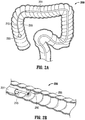
- the systems disclosed herein include a retractor which creates an asymmetric working space within the body lumen. More particularly, when working in a confined body lumen, such as the colon, expansion of the lumen is limited because it is undesirable to over-expand which could stretch the lumen beyond its ability to return to its normal state or more dangerously could rupture the lumen.
- the asymmetric working spaces disclosed herein are designed to reconfigure or reshape the body lumen-transform the cylindrical space within the body lumen to a non-cylindrical asymmetrical space (i.e., changing the geometry) to shift the space around the target tissue to create more working space around the target tissue to provide both visual and mechanical improvements. Stated another way, in a cylindrical working space, there is a lot of area of unused space while in the reshaping of the embodiments disclosed herein the space is moved or shifted to reduce the unused space and create a larger area for tissue access and treatment.
- the systems taught herein can include dynamically reconfigurable, asymmetric retractor structures on the distal end of a flexible and torque-able multi-channel shaft having a handle that allows for control over both the stiffness and geometry of the working space formed by the expansion of the retractor.
- the retractor can include a stabilizer subsystem having 2-8, 3-5, 4-6, or any range therein, flexible retractor elements.
- the retractor elements can be aligned at least substantially parallel to each other when fully collapsed for positioning in the patient. In some embodiments, the retractor elements are aligned on planes that are within about 5-30 degrees, about 10-25 degrees, about 15-20 degrees, about 15 degrees, or any range therein, of each other.
- Tissue triangulation wherein the tissue is triangulated between two endoscopic instruments, enhances access and maneuverability.
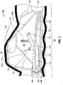
- FIG. 1 illustrates a system for operatively treating gastrointestinal disorders endoscopically in a stable, yet dynamic operative environment, and in a minimally-invasive manner, according to some embodiments.
- the system 100 can include a multi-lumen-catheter retractor system for ease of positioning in a subject, and such systems can be designed to provide a minimally invasive treatment of the subject.
- the system 100 can have a flexible outer tube 105 configured for guiding one or more channels 110 and an endoscope 115 within the system 100.
- the flexible outer tube 105 can have a lumen (not shown), a proximal end (not shown), and a distal end 108 to house, for example, the channel(s) and the endoscope during use of the system 100.
- the system can comprise a stable, yet dynamic operative environment in that it can include a reversibly-expandable retractor 150, as shown in Figure 1 , that expands to form a treatment space or working chamber 160 in the subject.
- the retractor 150 can be configured, for example, for the expansion to occur distal to the distal end 108 of the outer tube 105.
- the retractor can at least substantially render the target tissue 190 aperistaltic for the treatment.
- the retractor 150 can have a variety of configurations to serve, for example, as a scaffolding within the gastrointestinal tract 195.
- the retractor 150 can be a reversibly-stabilized and reversibly-expandable retractor, the retractor 150 forming an asymmetrical treatment space 160 upon the expansion. And, the retractor 150 can be configured to reversibly stiffen an otherwise flexible arrangement of the retractor 150, the arrangement designed to facilitate ease of positioning of the system 100 in the subject and to reversibly stiffen for the expansion of the retractor 150.
- the stabilization of the retractor 150 can, in some embodiments, include a stabilizer subsystem for stabilizing the retractor 150 as taught herein, the stabilizer having, for example, an at least substantially-rigid beam 175 to support the expanded retractor 150.
- the beam 175 is at the base of the chamber formed by the retractor elements, with the retractor elements extending radially (laterally) away from the beam 175.
- the beam 175 can be formed by the more rigid element exposed when the retractor elements are exposed from the outer tube for expansion, or alternatively, can be advanced independently from within the outer tube as in some of the embodiments described in more detail below.
- the outer tube can be manufactured from any materials know to be useful to one of skill for the purposes taught herein.
- the outer tube can comprise a polymer, or perhaps a polymer having an embedded wire reinforcement.
- the wire reinforcement can be a mesh, a braid, a helical coil or any combination thereof.
- the wire reinforcement can include any material believed by one of skill to be useful for the purposes set-forth herein.
- wire reinforcement can comprise a material having an elastic modulus that is about 1-3 orders of magnitude higher than the polymer tube.
- the working space being (i) at least substantially non-collapsible, (ii) at least substantially aperistaltic; and, (iii) at least substantially affixed at a particular point in the abdominal cavity in relation to any fixed body point, like a hip, for example.
- This is a significant improvement over existing systems, as existing systems have not addressed many existing problems including, for example, bowel collapse that can result from an air leak from the working space; peristalsis that is normal, even in a sedated patient; and, additional undesired bowel movements caused by the patient's breathing, movement of the scope or other instrument manipulation, or perhaps even by a surrounding peristalsis causing movement at a treatment area.
- the systems taught herein can provide for organizing the orientation of the floating channels, in order to further facilitate improving the flexibility of the system.
- the proximal coupler, the ring that can be attached to the distal end of the outer tube can be used to organize the tools and endoscope in a particular arrangement to facilitate a particular positioning of the tools as they emerge from the shaft into the working space created by the retractor.
- the tool channels can be placed further than the endoscope from the retractor elements that expand the most.
- the amount or change can include an increase or decrease relative to the reference parameter, can be compared to a reference point for the parameter.
- the deviation from the reference point can be, for example, in an amount of at least 1%, at least 2%, at least 3%, at least 5%, at least 10%, at least 15%, at least 20%, at least 25%, at least 30%, at least 35%, at least 40%, at least 45%, or any 1% increment therein.
- a "substantial [function]” or “substantially [functioning]” limitation can serve as a comparison to a reference function parameter, to indicate a deviation that will still provide the intended function.
- Reference functions can include, for example, floating, aperistalsis, attaching, flexing, rigidity, a position or positioning relative to another object, and the like.
- the deviation from the reference point can be, for example, in an amount of less than 1%, less than 3%, less than 5%, less than 10%, less than 15%, less than 20%, less than 25%, less than 30%, less than 35%, less than 40%, less than 45%, or any 0.1% increment therein.
- a component can have an acceptable, substantial [function] when it deviates from the reference by less than the acceptable deviation.
- the retractor can be a reversibly-stabilized and reversibly-expandable retractor, the retractor forming an asymmetrical treatment space upon the expansion.
- the retractor can be configured to reversibly stiffen an otherwise flexible arrangement of the retractor, the flexible arrangement designed to facilitate the ease of positioning of the system in the subject and to reversibly stiffen for the expansion of the retractor.
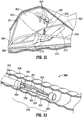
- the endoscope 315 can be articulated in the working space 360 toward the target lesion 390 to improve visibility.
- the system can comprise a stable, yet dynamic operative environment in that it can include a reversibly-expandable retractor 350 that expands to form a treatment space 360 in the subject.
- the retractor 350 can be configured, for example, for the expansion to occur distal to the distal end 308 of the outer tube (catheter) 305.
- the retractor can at least substantially render the target tissue 390 aperistaltic for the treatment.
- the retractor 350 can have a variety of configurations to serve, for example, as a scaffolding within the gastrointestinal tract 395.
- the extent of outward expansion of the retractor elements can be controlled by controlling the distance between the proximal and distal couplers 398, 399, The retractor can be repeatedly moved between expanded and retracted positions as desired by adjusting the distance between the coupler 398, 399. Such controlled expansion of the retractor elements can also be achieved by operatively coupling the proximal end of the retractor elements to an actuator as in the embodiment of FIG. 11 .
- the retractor elements can be composed of a material, e.g., shape memory material, to automatically expand when exposed from a catheter or sheath.
- retractor element 351 is a flexible element having a proximal portion 351a extending from the proximal coupler 398 at a first angle, a distal portion 351b extending from the distal hub or coupler 399 preferably at a second angle different from the first angle, and an engaging portion 351c, which engages the tissue, extending between the proximal and distal portions 351a, 351b.
- portion 351a extends at a greater angle to the longitudinal axis than distal portion 351b providing an asymmetric expansion of the retractor element itself.
- the length of the distal portion 351b exceeds the length of portion 351a.
- the beam 375 is at the base of the chamber formed by the retractor elements, with the retractor elements extending radially (laterally) away from the beam 375.
- the beam 375 can be formed by the more rigid element exposed when the retractor elements are exposed from the outer tube for expansion, or alternatively, can be advanced independently from the outer tube or formed by advancement of a rigidifying structure as in some of the embodiments described in more detail below.
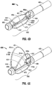
- FIGS. 4A-4E illustrate details of an alternate system as taught herein, in side, axial, and oblique views of expanded and collapsed configurations, and including a stabilizer subsystem, according to some embodiments.
- the figures illustrate an example of a multi-lumen catheter system which is similar to the system of FIGS. 3A-3K in that it has a reversibly-stabilized and reversibly-expandable retractor for a minimally invasive treatment of a subject.
- FIGs 4A-4C illustrate side and axial views that show that the system 400 can comprise a flexible outer tube (or catheter) 405 for guiding a tool channel (not shown) and an endoscope (not shown) within the system 400 in the same manner as system 300.
- the lumens 406b, 406c can receive the first and second tools directly therein, or alternatively, receive tool channels (flexible guides) 410b, 410c like tool channels 310 described above for angling the endoscopic tools slidably positioned therein.
- FIG. 4D illustrates the system in the collapsed configuration
- FIG. 4E illustrates the system in the expanded configuration.
- the tool channels (flexible guides) 410b and 410c are shown exposed from the catheter 405 so their distal ends are in a curved position. The tool channels can be further advanced axially to align the curved distal ends with the target tissue.
- FIGS. 5A-5D illustrate side and top views of a system as taught herein, having side views and top views of expanded and collapsed configurations, according to some embodiments.
- the tool channels and tools are omitted for clarity, being similar to those described herein.
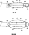
- FIGS. 5A and 5B illustrates side views of system 500 in collapsed and expanded configurations showing an example of an asymmetric work space that can be formed during an endoscopic procedure using the system 500. And, as shown in FIG.
- the retractor elements can alternatively be fixedly attached at their distal ends, e.g., to distal coupler 599, and operatively connected at proximal ends to an actuator or made of self-expanding material such as shape memory material as in the various embodiments described herein.
- Each retractor element 551, 552 in the embodiment of FIG. 5B expands to a substantially uniform (symmetric) arcuate shape, although alternatively they each can be configured to expand to a non-uniform (non-symmetric) shape as in the embodiments described above.
- the rigid beam 575 can be provided in a permanently stiffened condition as beam 175 of FIG. 1 , or alternatively can be formed by advancement of a rigidifying (stabilizing) structure over a flexible element or into the lumen of the flexible tubular member by an actuator as described above. In either case, the beam rigidifies the retractor system in the asymmetrical configuration creating the stable asymmetrical working space to facilitate access and manipulation of the target tissue.
- the flexible outer tube 605 has a lumen, a proximal end extending into the handle 680, and a distal end 608.
- Each tool channel (serves as a guide through which a tool (not shown) can be manipulated in a treatment of a target tissue in a subject. That is, the tool channels are configured to receive and reorient the tools inserted therethrough as in the embodiments described above.
- the retractor 650 can be a reversibly-stabilized and reversibly-expandable retractor 650 forming a treatment space 660 upon expansion and configured for the expansion to occur distal to the distal end 608 of the outer tube 605.
- the retractor 650 can be designed to reversibly-stiffen an otherwise flexible arrangement of the retractor 650, the flexible arrangement designed to facilitate the positioning of the system in the subject and to reversibly-stiffen for the expansion of the retractor 650.
- the reversibly-stiffened arrangement of the retractor 650 can form an at least substantially-rigid beam 675 from an otherwise flexible beam 670 as a structural support for the expansion of the retractor 650.
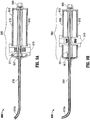
- FIGS. 6C and 6D illustrate oblique views of the system 600 in expanded configurations.
- the expanded configurations have a rigid beam 675 that was formed from the flexible beam that is typically present in the collapsed state for positioning in the subject.
- the rigid beam 675 can be formed from a flexible beam, in some embodiments, by slidably inserting a rigid member (e.g., a rod) either over or alternatively into a flexible member that composes the flexible beam to transform the flexible beam into a stiffer, more rigid beam.
- a rigid member e.g., a rod
- the stabilizer actuator 612 is operably connected to the rigid member (stabilizing structure) such as rigid rod 672 through a rod coupler 613.
- the stabilizer actuator 612 and/or the retractor actuator 614 can have a plurality of ratchet teeth 616 to provide a plurality of positions for reversibly-fixing the stabilizer and/or for reversibly fixing the retractor in position during expansion or collapse of the retractor.
- coupler 611 is in the proximal position and the retractor elements are in the non-expanded position.
- retractor actuator 614 is slid distally to move the attached coupler 611 distally, as shown in FIG. 6D , thereby causing the attached elements 651, 652 to bend outwardly due to their fixed connection at their distal end to the distal coupler 699.
- the handle can be any of a variety of shapes to provide a desired or ergonomic position for operation of the system.
- the retractor actuator can be configured as a finger-activated button on the handle 680 that slides back and forth through a slot in the handle 680 to expand or collapse the retractor elements.
- a means for dynamically adjusting or ratcheting the retractor position can be provided along the handle slot to lock the position of the retractor elements in place when the retractor actuator button is not pressed.
- a button on the opposite side of the handle can be operatively connected to the stabilizer subsystem to convert the flexible beam into a rigid beam, or convert the rigid beam into a flexible beam.
- the covering extends over an intermediate portion of the elements 651, 652 and can be in the form of a heat shrink tubing.
- the covering helps control expansion by providing a less flexible region. This covering is similar to covering 451a and 452a of the embodiment of FIGS. 4D, 4E .
- a lumen 806c can be provided to contain a working channel 810c which receives a tool channel to guide a working instrument or alternatively directly receives a working instrument.
- the lumen 806 of the outer tube 805 is used to guide an endoscope 815.
- Only a portion of the retractor components 851, 854 are shown to partially describe an embodiment of the relation between the outer tube 805 and the retractor.
- the retractor can be configured, for example, for the expansion to occur distal to the distal end 808 of the outer tube 805.
- the retractor can include four retractor elements in the same manner as described above, only two of which are shown (elements 851 and 854).
- the working channel 810c can be a floating channel that is (i) at least substantially attached to the lumen of the outer tube at a first proximal location (not shown) and a first distal location 806c and (ii) at least substantially floating in the lumen 806 of the outer tube 805 between the first proximal location (not shown) and the first distal location 806c.
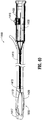
- FIG. 9A shows the tip 910a of the tool channel 910 in a substantially extended position
- FIG. 9B shows the tip 910a of the tool channel 910 in a substantially bent position, such that the distal tip 910a is deflected substantially normal to the central axis of the tool channel 910.
- a system 900 consistent with other systems taught herein, for example, can include an entry port 909, a tool channel 91 inserted through entry port 909, a wire coupler 911, ratchet teeth 916, a pull wire 917 for flexing or extending the tip 910a of the working channel 910, and wire actuator 919.
- one of skill can insert the endoscope through the endoscope channel of the system and extend the distal end of the endoscope distal to the distal end of the retractor to form an assembly.
- the assembly can then be inserted into a body lumen or orifice, such as the colon, and advanced orally until the distal end of the scope or the lens is in proximity to the target tissue (lesion or defect) to be treated.
- the system can then be advanced forward over the scope until the retractor is positioned over the distal end of the endoscope while observing the image from the endoscope.
- FIGS. 10A-10E illustrate a retractor sheath covering a retractor of a system as taught herein, according to some embodiments.
- FIGS. 10A-10C show top, oblique, and side-views showing a flexible, clear sheath 1000 that covers a collapsed configuration of the retractor 1050 to render an at least substantially smooth and/or atraumatic surface 1005 for a delivery of the retractor 1050 to a target site (not shown) for a treatment of a target tissue (not shown).
- the cover is in a closed configuration that can be sustained until the expansion of the retractor 1050 for the treatment, or it can be reversibly-obtained following the treatment.
- FIGS. 10D and 10E show a top-view and side-view of an expanded configuration of the retractor with the cover in an open configuration for the treatment.
- Retainers can be used at any position around the retractor to facilitate a retention of the configuration of the working space 1060, for example, to retain the configuration under forces of the expansion of the retractor 1050.
- the sheath 1000 can also prevent or inhibit tissue from entering the retractor 1050 until desired.
- the sheath 1000 can also act as a collection means for entrapping and/or pulling out a resected tissue, which can be particularly desirable in the resection of cancerous tissue in some embodiments.
- the sheath 1000 can be at least substantially closed around the retractor 1050 during delivery, and can be designed to open as the retractor 1050 is expanded to create the working space 1060 for the treatment. Alternatively, the expansion of the retractor elements and the sheath can be independent.
- valves 1121A, 1121B are proximal of luer fittings 1127', 1129'.
- the tool channels 1124', 1126' are identical to tool channels 1124, 1126 in all other respects.
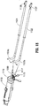
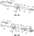
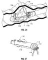
- catheter 1110 includes a handle housing 1130 at the proximal portion 1113 which contains two actuators: actuator 1132 for controlling movement of the retractor system 1150 and actuator 1134 for controlling movement of the rigidifying (stabilizing) structure. These are discussed in more detail below.
- Catheter 1110 also includes tubing 1139 having a luer coupling 1137 and a control switch 1175 (see FIGS. 31A, 31B ) for closing off an internal gasket 1176.
- the string, e.g., suture 1172 for closing covering 1170 is secured by the elastomeric gasket 1176 as the switch 1174 is moved from the position of FIG. 31A to the position of FIG. 31B . More particularly, in the initial position of FIG.
- the elements 1156, 1158 do not extend at all beyond the transverse dimension of the catheter 1110.
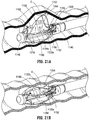
- the retractor system 1150 i.e., the retractor elements 1152, 1154, expand to only one side of a plane passing through a longitudinal axis of the catheter 1110, thereby creating the asymmetric working space 1151 (and asymmetric cage), with its attendant advantages described herein.
- retractor elements can be utilized, including for example providing relatively movable couplers 1140, 1148 to expand the retractor elements 1152, 1154 (and optionally 1156, 1158) in the same manner as the couplers described above, e.g., couplers 198, 199.
- the retractor elements can also alternatively be made of self-expanding material, such as shape memory material, which expand when exposed from the catheter or sheath.
- the cover 1170 can be configured to have an opening in the collapsed position, or, alternatively, it can provided with a slit which can be opened due to stretching when the retractor elements 1152, 1154 are moved to the expanded position.
- the retractor elements 1152, 1154 When the retractor elements 1152, 1154 are expanded, they move past the cover 1170 toward the target tissue.
- the edges of the cover 1170 can be attached to the retractor elements 1152, 1154 and thereby move with the retractor elements.
- the target tissue is removed by the endoscopic instruments described herein, the removed tissue is placed within the cover 1170, and the cover 1170 is closed, e.g., by a string or suture 1172 shown in FIG. 29 to encapsulate the tissue and prevent leakage and seeding during removal from the body lumen.
- the suture 1172 can be embedded in a wall of the cover 1170 or in pockets or channels formed in the cover 1170, where it is permanently fixed at a distal anchor point, and pulled proximally to tension the su
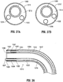
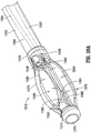
- FIGS 32-42 illustrate alternative embodiments of the system of the present invention.
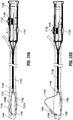
- the system includes floating (flexible) channels within the outer tube.
- the floating channels are fixed at their proximal and distal ends; in another embodiment the floating channels are fixed at their proximal ends but are unattached at their distal ends.
- the floating channels reduce the overall stiffness of the catheter (outer tube) which would otherwise be stiffer if the channels were fixed along their entire length and did not float within the catheter.
- the floating channels also reduce kinking of the tool channels (flexible guides) inserted through the floating channels and reduce kinking of the tools inserted through the tool channels (or inserted directly through the floating channels in the embodiments where the tool channels are not utilized).
- Actuator 1256 on shell 1251a provides for rigidifying the cage by rigidifying a flexible beam.
- a separate slidable beam for rigidifying the cage is not provided as alternative rigidifying structure is provided.
- a stiffener member in the form of a rigid beam is operatively connected to actuator 1256 so that distal movement of the actuator 1256 advances the stiffener distally either within a lumen of the flexible element or over the outer surface of the flexible element to provide a stiffer structure.
- Actuator 1256 can be moved proximally to unstiffen the flexible element to facilitate collapse of the retractor system.
- the outer tube 1212 has a length, measured from the distal end of handle 1251 to a distal edge of end cap 1218 of about 10cm to about 200cm, and more preferably about 60cm to about 90cm.
- the floating channels 1222, 1224 have a length of about 10.1 cm to about 204cm, and preferably about 60.5cm to about 91cm, thereby exceeding the length of the outer tube 1212. Other dimensions are also contemplated. This greater length of the floating channels 1222, 1224 in the embodiments where they are fixed at both the proximal and distal ends enables the floating movement.
- the first and second proximal tubes 1228, 1232 are preferably attached to an inner wall of the outer tube 1212 or handle housing 1251 by bonding or welding or other attachment methods.
- the first and second distal tubes 1226, 1230 are preferably attached to an inner wall of the outer tube 1212 by bonding or welding or other attachment methods.
- the fixed proximal tubes 1228 or 1232 are shown cutaway (into a half cylinder) for clarity, it being understood that the tubes can be cylindrical in configuration like the distal fixed tubes 1226, 1230. Other configurations for the fixed distal and proximal tubes are contemplated.
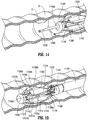
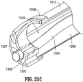
- Figures 35A-35C illustrate a cutaway view of the system so that only one of the floating channels, the second floating channel 1324, is illustrated.
- the first floating channel is attached and configured in a similar fashion as second floating channel 1324.
- Second floating channel 1324 is attached at its proximal end in the same manner as floating channel 1224, i.e., attached within a fixed proximal tube.
- the fixed distal tubes 1326, 1330 which form pockets for the respective floating channels 1322, 1324 are dimensioned so their length exceeds the largest extent of movement in response to the greatest curvature of the outer tube 1312 as a result of bending of the outer tube 1312 during use. This ensures that the floating channels 1322, 1324 will not retract out of the proximal end of the respective fixed distal tubes 1326, 1330,
- the length of the distal tubes 1326 and 1330 are between about 1.5cm to about 3cm, and preferably about 2cm. Other dimensions are also contemplated.
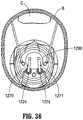
- Flexible guides identical to flexible guides 1270, 1271 of Figure 33 and/or flexible guides 1122, 1124 of Figures 19-25 are inserted through the floating channels 1322 and 1324 in the same manner as described above so that endoscopic working instruments can be inserted into the chamber formed by the flexible elements for performing the procedure.
- endoscopic working instruments can be inserted directly through the floating channels of any of the embodiments herein without the intermediary flexible guides.
- Such direct insertion of instrumentation without flexible guides (tool channels) is also described above as an alternative system and method.
- the flexible guides described herein can be color coded to improve the system's usability.
- flexible guide 1270 can be of a first color, such as red
- flexible guide 1271 can be of a second color, such as black.
- first color such as red
- second color such as black
- the entire flexible guide can have the same color or alternatively the matching color can be only at the proximal end visible to the user and the distal end visible by the endoscope.
- other indicia can be provided so the user can match the proximal end of the flexible tube with the distal end within the chamber.
- Retractor system 1410 also has flexible elements 1418, 1420 forming the base of the cage and identical to retractor elements 1384, 1386 described above.
- Retractor system 1415 differs from retractor system 1370 (and 1150) in that a beam 1422 is provided which itself has sufficient rigidity to maintain the overall stiffness of the expanded cage when fairly mild forces are applied (e.g., weight of small portion of the intestinal wall, minor external intra-abdominal pressure, etc.) and limit bending of the cage with respect to the outer tube 1422 during use when fairly mild forces are applied.
- fairly mild forces e.g., weight of small portion of the intestinal wall, minor external intra-abdominal pressure, etc.
Landscapes
- Health & Medical Sciences (AREA)
- Life Sciences & Earth Sciences (AREA)
- Surgery (AREA)
- General Health & Medical Sciences (AREA)
- Public Health (AREA)
- Veterinary Medicine (AREA)
- Nuclear Medicine, Radiotherapy & Molecular Imaging (AREA)
- Animal Behavior & Ethology (AREA)
- Molecular Biology (AREA)
- Engineering & Computer Science (AREA)
- Biomedical Technology (AREA)
- Heart & Thoracic Surgery (AREA)
- Medical Informatics (AREA)
- Pathology (AREA)
- Biophysics (AREA)
- Radiology & Medical Imaging (AREA)
- Physics & Mathematics (AREA)
- Optics & Photonics (AREA)
- Reproductive Health (AREA)
- Vascular Medicine (AREA)
- Surgical Instruments (AREA)
- Endoscopes (AREA)
Applications Claiming Priority (3)
| Application Number | Priority Date | Filing Date | Title |
|---|---|---|---|
| US14/622,831 US10595711B2 (en) | 2009-12-16 | 2015-02-14 | System for a minimally-invasive, operative gastrointestinal treatment |
| PCT/US2016/016911 WO2016130442A1 (en) | 2009-12-16 | 2016-02-06 | System for a minimally-invasive, operative gastrointestinal treatment |
| EP16707301.4A EP3256052B1 (de) | 2015-02-14 | 2016-02-06 | System zur minimalinvasiven operativen gastrointestinalen behandlung |
Related Parent Applications (2)
| Application Number | Title | Priority Date | Filing Date |
|---|---|---|---|
| EP16707301.4A Division EP3256052B1 (de) | 2015-02-14 | 2016-02-06 | System zur minimalinvasiven operativen gastrointestinalen behandlung |
| EP16707301.4A Division-Into EP3256052B1 (de) | 2015-02-14 | 2016-02-06 | System zur minimalinvasiven operativen gastrointestinalen behandlung |
Publications (1)
| Publication Number | Publication Date |
|---|---|
| EP3659524A1 true EP3659524A1 (de) | 2020-06-03 |
Family
ID=55447129
Family Applications (2)
| Application Number | Title | Priority Date | Filing Date |
|---|---|---|---|
| EP16707301.4A Active EP3256052B1 (de) | 2015-02-14 | 2016-02-06 | System zur minimalinvasiven operativen gastrointestinalen behandlung |
| EP19217066.0A Withdrawn EP3659524A1 (de) | 2015-02-14 | 2016-02-06 | System zur minimalinvasiven operativen gastrointestinalen behandlung |
Family Applications Before (1)
| Application Number | Title | Priority Date | Filing Date |
|---|---|---|---|
| EP16707301.4A Active EP3256052B1 (de) | 2015-02-14 | 2016-02-06 | System zur minimalinvasiven operativen gastrointestinalen behandlung |
Country Status (6)
| Country | Link |
|---|---|
| US (2) | US10595711B2 (de) |
| EP (2) | EP3256052B1 (de) |
| JP (1) | JP6457100B2 (de) |
| CN (1) | CN107995856B (de) |
| AU (1) | AU2016218285B2 (de) |
| WO (1) | WO2016130442A1 (de) |
Families Citing this family (74)
| Publication number | Priority date | Publication date | Assignee | Title |
|---|---|---|---|---|
| US8100822B2 (en) | 2004-03-16 | 2012-01-24 | Macroplata Systems, Llc | Anoscope for treating hemorrhoids without the trauma of cutting or the use of an endoscope |
| US12121209B2 (en) | 2014-02-11 | 2024-10-22 | Cornell University | Method and apparatus for providing increased visualization and manipulation of a body side wall |
| US11986150B2 (en) | 2009-12-15 | 2024-05-21 | Lumendi Ltd. | Method and apparatus for manipulating the side wall of a body lumen or body cavity so as to provide increased visualization of the same and/or increased access to the same, and/or for stabilizing instruments relative to the same |
| US9986893B2 (en) | 2009-12-15 | 2018-06-05 | Cornell University | Method and apparatus for manipulating the side wall of a body lumen or body cavity so as to provide increased visualization of the same and/or increased access to the same, and/or for stabilizing instruments relative to the same |
| US10485401B2 (en) | 2009-12-15 | 2019-11-26 | Lumendi Ltd. | Method and apparatus for manipulating the side wall of a body lumen or body cavity so as to provide increased visualization of the same and/or increased access to the same, and/or for stabilizing instruments relative to the same |
| WO2011084490A1 (en) | 2009-12-15 | 2011-07-14 | Cornell University | Method and apparatus for stabilizing, straightening, or expanding the wall of a lumen or cavity |
| US10149601B2 (en) | 2009-12-15 | 2018-12-11 | Lumendi Ltd. | Method and apparatus for manipulating the side wall of a body lumen or body cavity so as to provide increased visualization of the same and/or increased access to the same, and/or for stabilizing instruments relative to the same |
| US11877722B2 (en) | 2009-12-15 | 2024-01-23 | Cornell University | Method and apparatus for manipulating the side wall of a body lumen or body cavity |
| ES2874194T3 (es) | 2009-12-16 | 2021-11-04 | Boston Scient Scimed Inc | Disposiciones para realizar una estructura anatómica endoluminal |
| US8932211B2 (en) | 2012-06-22 | 2015-01-13 | Macroplata, Inc. | Floating, multi-lumen-catheter retractor system for a minimally-invasive, operative gastrointestinal treatment |
| US11344285B2 (en) | 2009-12-16 | 2022-05-31 | Boston Scientific Scimed, Inc. | Multi-lumen-catheter retractor system for a minimally-invasive, operative gastrointestinal treatment |
| US9186131B2 (en) | 2009-12-16 | 2015-11-17 | Macroplata, Inc. | Multi-lumen-catheter retractor system for a minimally-invasive, operative gastrointestinal treatment |
| US10758116B2 (en) | 2009-12-16 | 2020-09-01 | Boston Scientific Scimed, Inc. | System for a minimally-invasive, operative gastrointestinal treatment |
| USRE48850E1 (en) | 2009-12-16 | 2021-12-14 | Boston Scientific Scimed, Inc. | Multi-lumen-catheter retractor system for a minimally-invasive, operative gastrointestinal treatment |
| US9565998B2 (en) | 2009-12-16 | 2017-02-14 | Boston Scientific Scimed, Inc. | Multi-lumen-catheter retractor system for a minimally-invasive, operative gastrointestinal treatment |
| US10531869B2 (en) | 2009-12-16 | 2020-01-14 | Boston Scientific Scimed, Inc. | Tissue retractor for minimally invasive surgery |
| US10966701B2 (en) | 2009-12-16 | 2021-04-06 | Boston Scientific Scimed, Inc. | Tissue retractor for minimally invasive surgery |
| US10595711B2 (en) | 2009-12-16 | 2020-03-24 | Boston Scientific Scimed, Inc. | System for a minimally-invasive, operative gastrointestinal treatment |
| US12376737B1 (en) | 2009-12-16 | 2025-08-05 | Boston Scientific Scimed, Inc. | Tissue retractor for minimally invasive surgery |
| WO2012047939A2 (en) * | 2010-10-04 | 2012-04-12 | Ind Platforms Llc | Expandable devices, rail systems, and motorized devices |
| JP7082052B2 (ja) | 2015-09-03 | 2022-06-07 | ネプチューン メディカル インク. | 小腸内での内視鏡前進の為の器具 |
| US10328271B2 (en) * | 2015-11-12 | 2019-06-25 | Medtronic, Inc. | Implantable electrical stimulator with deflecting tip lead |
| EP3500151A4 (de) | 2016-08-18 | 2020-03-25 | Neptune Medical Inc. | Vorrichtung und verfahren zur verbesserten visualisierung des dünndarms |
| WO2019232354A1 (en) | 2018-05-31 | 2019-12-05 | Neptune Medical Inc. | Device and method for enhanced visualization of the small intestine |
| AU2017335562A1 (en) * | 2016-09-10 | 2019-01-03 | Boston Scientific Scimed, Inc. | System for a minimally-invasive, operative gastrointestinal treatment |
| JP7048628B2 (ja) | 2016-11-28 | 2022-04-05 | アダプティブエンドウ エルエルシー | 分離可能使い捨てシャフト付き内視鏡 |
| EP3562374A1 (de) * | 2016-12-30 | 2019-11-06 | Boston Scientific Scimed, Inc. | System zur minimal-invasiven behandlung in einem körperlumen |
| EP3525652A1 (de) * | 2017-01-20 | 2019-08-21 | Boston Scientific Scimed, Inc. | System für eine minimalinvasive operative behandlung |
| CN116327271A (zh) | 2017-03-18 | 2023-06-27 | 波士顿科学国际有限公司 | 用于体腔内的微创治疗的系统 |
| KR102026938B1 (ko) * | 2017-06-13 | 2019-09-30 | 주식회사 파인메딕스 | 내시경용 하이브리드 나이프 |
| WO2019018682A1 (en) | 2017-07-20 | 2019-01-24 | Neptune Medical Inc. | DYNAMICALLY RIGIDING SURFACE |
| SE541401C2 (en) * | 2017-08-16 | 2019-09-17 | Implantica Patent Ltd | Abdominal instrument |
| CN111278497B (zh) * | 2017-11-28 | 2024-01-12 | 圣犹达医疗用品心脏病学部门有限公司 | 管腔控制导管 |
| US10933222B2 (en) * | 2017-12-05 | 2021-03-02 | Boston Scientific Scimed, Inc. | Tissue retraction system for performing minimally invasive procedures |
| CN108261174B (zh) | 2018-03-13 | 2024-06-07 | 南微医学科技股份有限公司 | 一种内窥镜端帽 |
| CN112020333B (zh) | 2018-04-26 | 2024-06-25 | 奥林巴斯株式会社 | 处置系统和扩张设备 |
| WO2019207706A1 (ja) | 2018-04-26 | 2019-10-31 | オリンパス株式会社 | 処置システム |
| DE102018208564A1 (de) | 2018-05-30 | 2019-12-05 | Kardion Gmbh | Steuerbare Einführungshülse |
| DE102018208537A1 (de) | 2018-05-30 | 2019-12-05 | Kardion Gmbh | Vorrichtung zum Anbinden eines Herzunterstützungssystems an eine Einführeinrichtung und Verfahren zum Herstellen einer Vorrichtung zum Anbinden eines Herzunterstützungssystems an eine Einführeinrichtung |
| DE102018208555A1 (de) | 2018-05-30 | 2019-12-05 | Kardion Gmbh | Vorrichtung zum Verankern eines Herzunterstützungssystems in einem Blutgefäß, Verfahren zum Betreiben und Herstellverfahren zum Herstellen einer Vorrichtung und Herzunterstützungssystem |
| DE102018211297A1 (de) | 2018-07-09 | 2020-01-09 | Kardion Gmbh | Herzunterstützungssystem und Verfahren zur Überwachung der Integrität einer Haltestruktur eines Herzunterstützungssystems |
| WO2020018934A1 (en) | 2018-07-19 | 2020-01-23 | Nep Tune Medical Inc. | Dynamically rigidizing composite medical structures |
| CN209236231U (zh) * | 2018-09-05 | 2019-08-13 | 山东冠龙医疗用品有限公司 | 一种多通道工作套管 |
| CN109512476B (zh) * | 2018-12-19 | 2020-08-28 | 四川大学华西医院 | 一种消化道早期癌症的术中牵拉装置 |
| US11793392B2 (en) | 2019-04-17 | 2023-10-24 | Neptune Medical Inc. | External working channels |
| JP2022529936A (ja) | 2019-04-17 | 2022-06-27 | ネプチューン メディカル インク. | 医療用動的硬化複合構造 |
| CN114025653B (zh) * | 2019-05-01 | 2025-12-05 | 波士顿科学国际有限公司 | 用于铰接线引导的系统和方法 |
| JP7253063B2 (ja) * | 2019-09-06 | 2023-04-05 | 富士フイルム株式会社 | 内視鏡処置具 |
| CN110613489B (zh) * | 2019-09-23 | 2020-08-21 | 宋勇 | 一种内窥镜手术器械 |
| AU2020376754B2 (en) * | 2019-10-30 | 2023-02-23 | Boston Scientific Scimed, Inc. | Endoscopic catheter device |
| WO2021118958A1 (en) * | 2019-12-13 | 2021-06-17 | Boston Scientific Scimed, Inc. | Devices, systems, and methods for minimally invasive surgery in a body lumen |
| USD1018844S1 (en) | 2020-01-09 | 2024-03-19 | Adaptivendo Llc | Endoscope handle |
| US11957323B2 (en) * | 2020-02-18 | 2024-04-16 | Boston Scientific Medical Device Limited | Medical device handle assemblies and methods of using the same |
| CN121943406A (zh) * | 2020-03-23 | 2026-05-01 | 恩德威视基金会 | 用于至少部分闭塞身体内腔的装置 |
| AU2021245989A1 (en) | 2020-03-30 | 2022-10-27 | Neptune Medical Inc. | Layered walls for rigidizing devices |
| CN111287988B (zh) * | 2020-04-17 | 2022-05-24 | 三门核电有限公司 | 一种屏蔽主泵锁紧杯就地视频检查方法 |
| CN111436893A (zh) * | 2020-05-14 | 2020-07-24 | 江苏工大博实医用机器人研究发展有限公司 | 一种医用柔性管状工具 |
| WO2022104262A1 (en) | 2020-11-16 | 2022-05-19 | Lumendi Ltd. | Methods and apparatus for inverting a hollow sleeve and thereafter reverting an inverted hollow sleeve |
| USD1051380S1 (en) | 2020-11-17 | 2024-11-12 | Adaptivendo Llc | Endoscope handle |
| US12589237B2 (en) | 2020-11-20 | 2026-03-31 | Kardion Gmbh | Mechanical circulatory support system with guidewire aid |
| JP2024505529A (ja) | 2021-01-29 | 2024-02-06 | ネプチューン メディカル インク. | 動的に剛体化する装置の意図しない運動を防止するデバイスおよび方法 |
| CA3211622A1 (en) | 2021-03-10 | 2022-09-15 | Neal Tanner | Control of robotic dynamically rigidizing composite medical structures |
| AU2022239357A1 (en) * | 2021-03-16 | 2023-09-21 | Atricure, Inc. | Delivery devices and related methods |
| USD1031035S1 (en) | 2021-04-29 | 2024-06-11 | Adaptivendo Llc | Endoscope handle |
| USD1070082S1 (en) | 2021-04-29 | 2025-04-08 | Adaptivendo Llc | Endoscope handle |
| CN113288439B (zh) * | 2021-06-30 | 2022-05-03 | 中国人民解放军陆军特色医学中心 | 一种机器人臂直肠镜系统 |
| USD1066659S1 (en) | 2021-09-24 | 2025-03-11 | Adaptivendo Llc | Endoscope handle |
| CA3249667A1 (en) | 2022-04-19 | 2023-10-26 | Neptune Medical Inc. | MANAGEMENT AND HANDLING OF A LONG-LENGTH ROBOTIC ENDOSCOPE |
| CN119233783A (zh) | 2022-04-27 | 2024-12-31 | 海王星医疗公司 | 用于内窥镜检查的卫生护套 |
| CN116211222B (zh) * | 2023-01-31 | 2025-01-28 | 湖南省华芯医疗器械有限公司 | 一种内窥镜的引导装置及内窥镜 |
| US12471900B2 (en) | 2023-06-01 | 2025-11-18 | Thompson Surgical Instruments, Inc. | Surgical retractor and bidirectional ratchet |
| AU2024338348A1 (en) | 2023-09-07 | 2026-03-19 | Neptune Medical Inc. | Pressure rigidization apparatuses and methods |
| AU2024353942A1 (en) | 2023-09-28 | 2026-03-19 | Neptune Medical Inc. | Telescoping robot |
| CN119366842A (zh) * | 2025-01-02 | 2025-01-28 | 湖南省华芯医疗器械有限公司 | 内窥镜及其插入装置 |
Citations (4)
| Publication number | Priority date | Publication date | Assignee | Title |
|---|---|---|---|---|
| JP2005046274A (ja) * | 2003-07-31 | 2005-02-24 | Olympus Corp | 内視鏡挿入用補助具 |
| US20080188868A1 (en) * | 2006-12-01 | 2008-08-07 | Barry Weitzner | Direct drive endoscopy systems and methods |
| US8506479B2 (en) | 2009-12-16 | 2013-08-13 | Macroplata, Inc. | Substantially rigid and stable endoluminal surgical suite for treating a gastrointestinal lesion |
| WO2013192116A1 (en) * | 2012-06-22 | 2013-12-27 | Macroplata Inc. | Multi-lumen-catheter retractor system for a minimally-invasive, operative gastrointestinal treatment |
Family Cites Families (194)
| Publication number | Priority date | Publication date | Assignee | Title |
|---|---|---|---|---|
| US457787A (en) | 1891-08-18 | Rectal | ||
| US827193A (en) | 1905-09-08 | 1906-07-31 | Arthur N Penland | Surgical instrument. |
| US1621159A (en) | 1925-11-27 | 1927-03-15 | Robert T Evans | Abdominoscope |
| US2072346A (en) | 1934-10-04 | 1937-03-02 | Ward R Smith | Drainage tube |
| US2434167A (en) | 1945-05-23 | 1948-01-06 | Ernest O Knoblauch | Valved coupling |
| DE1466889B1 (de) | 1965-07-28 | 1970-04-23 | Eberhard Dr Regenbogen | Rektoskop zur Endoskopie des als Sigma bezeichneten Bereichs des menschlichen Darmes |
| US3517128A (en) | 1968-02-08 | 1970-06-23 | James R Hines | Surgical expanding arm dilator |
| US3557794A (en) | 1968-07-30 | 1971-01-26 | Us Air Force | Arterial dilation device |
| US4040413A (en) | 1974-07-18 | 1977-08-09 | Fuji Photo Optical Co. Ltd. | Endoscope |
| US4083369A (en) | 1976-07-02 | 1978-04-11 | Manfred Sinnreich | Surgical instruments |
| US4224929A (en) | 1977-11-08 | 1980-09-30 | Olympus Optical Co., Ltd. | Endoscope with expansible cuff member and operation section |
| US4295464A (en) | 1980-03-21 | 1981-10-20 | Shihata Alfred A | Ureteric stone extractor with two ballooned catheters |
| US4718406A (en) | 1982-06-28 | 1988-01-12 | The United States Of America As Represented By The Secretary Of The Navy | Fiber optics image scope (micro-endoscope), ureteroscope |
| US4519403A (en) | 1983-04-29 | 1985-05-28 | Medtronic, Inc. | Balloon lead and inflator |
| US4611594A (en) | 1984-04-11 | 1986-09-16 | Northwestern University | Medical instrument for containment and removal of calculi |
| EP0163502B1 (de) | 1984-05-30 | 1990-07-18 | Devices For Vascular Intervention Inc. | Vorrichtung für Atherektomie |
| US4710171A (en) | 1986-06-09 | 1987-12-01 | The Kendall Company | Needle depth setting sheath assembly and needle stop |
| JPS63292935A (ja) | 1987-05-26 | 1988-11-30 | Olympus Optical Co Ltd | 内視鏡用処置具案内装置 |
| US4836204A (en) | 1987-07-06 | 1989-06-06 | Landymore Roderick W | Method for effecting closure of a perforation in the septum of the heart |
| US5211638A (en) | 1988-01-25 | 1993-05-18 | Baxter International Inc. | Pre-slit injection site |
| US4966596A (en) | 1988-08-08 | 1990-10-30 | The Beth Israel Hospital Association | Laser atherectomy catheter |
| US5087265A (en) | 1989-02-17 | 1992-02-11 | American Biomed, Inc. | Distal atherectomy catheter |
| US5059199A (en) | 1989-04-12 | 1991-10-22 | Olympus Optical Co., Ltd. | Treating device for endoscopes |
| DE69023515T2 (de) | 1989-06-28 | 1996-04-04 | David S Zimmon | Aufblasbare tamponadevorrichtungen. |
| AU6042690A (en) | 1989-08-01 | 1991-03-11 | Claudio D'orazi | Percutaneous mechanical dilating catheter for cardiac valves and blood vessels |
| US5345927A (en) | 1990-03-02 | 1994-09-13 | Bonutti Peter M | Arthroscopic retractors |
| US5454365A (en) | 1990-11-05 | 1995-10-03 | Bonutti; Peter M. | Mechanically expandable arthroscopic retractors |
| US5197971A (en) | 1990-03-02 | 1993-03-30 | Bonutti Peter M | Arthroscopic retractor and method of using the same |
| US5025778A (en) | 1990-03-26 | 1991-06-25 | Opielab, Inc. | Endoscope with potential channels and method of using the same |
| JP2533732Y2 (ja) | 1990-11-29 | 1997-04-23 | 株式会社トップ | 内視鏡装着用バルーン |
| US5112310A (en) | 1991-02-06 | 1992-05-12 | Grobe James L | Apparatus and methods for percutaneous endoscopic gastrostomy |
| US5167239A (en) | 1991-05-30 | 1992-12-01 | Endomedix Corporation | Anchorable guidewire |
| US5386817A (en) | 1991-06-10 | 1995-02-07 | Endomedical Technologies, Inc. | Endoscope sheath and valve system |
| US5766151A (en) | 1991-07-16 | 1998-06-16 | Heartport, Inc. | Endovascular system for arresting the heart |
| US5304123A (en) | 1991-10-24 | 1994-04-19 | Children's Medical Center Corporation | Detachable balloon catheter for endoscopic treatment of vesicoureteral reflux |
| US5197649A (en) | 1991-10-29 | 1993-03-30 | The Trustees Of Columbia University In The City Of New York | Gastrointestinal endoscoptic stapler |
| US5336227A (en) | 1991-11-05 | 1994-08-09 | Wilk & Nakao Medical Technology Incorporated | Surgical cauterization snare with polyp capturing web net |
| US5997547A (en) | 1991-11-05 | 1999-12-07 | Nakao; Naomi L. | Surgical retrieval assembly and associated method |
| US5211624A (en) | 1991-12-09 | 1993-05-18 | Cinberg James Z | Surgical closure device method |
| US5259366A (en) | 1992-11-03 | 1993-11-09 | Boris Reydel | Method of using a catheter-sleeve assembly for an endoscope |
| JPH06296617A (ja) | 1993-04-16 | 1994-10-25 | Olympus Optical Co Ltd | バスケット鉗子 |
| US5840031A (en) | 1993-07-01 | 1998-11-24 | Boston Scientific Corporation | Catheters for imaging, sensing electrical potentials and ablating tissue |
| WO1995001751A1 (en) | 1993-07-01 | 1995-01-19 | Boston Scientific Corporation | Imaging, electrical potential sensing, and ablation catheters |
| US5423830A (en) | 1993-07-07 | 1995-06-13 | Schneebaum; Cary W. | Polyp retrieval method and associated instrument assembly |
| US6405732B1 (en) | 1994-06-24 | 2002-06-18 | Curon Medical, Inc. | Method to treat gastric reflux via the detection and ablation of gastro-esophageal nerves and receptors |
| US6009877A (en) | 1994-06-24 | 2000-01-04 | Edwards; Stuart D. | Method for treating a sphincter |
| US5551622A (en) | 1994-07-13 | 1996-09-03 | Yoon; Inbae | Surgical stapler |
| US5868760A (en) | 1994-12-07 | 1999-02-09 | Mcguckin, Jr.; James F. | Method and apparatus for endolumenally resectioning tissue |
| US5665062A (en) | 1995-01-23 | 1997-09-09 | Houser; Russell A. | Atherectomy catheter and RF cutting method |
| CA2220689A1 (en) | 1995-05-10 | 1996-11-14 | Cardiogenesis Corporation | System for treating or diagnosing heart tissue |
| JPH08317928A (ja) | 1995-05-26 | 1996-12-03 | Olympus Optical Co Ltd | 医療用処置具 |
| JPH08336538A (ja) | 1995-06-12 | 1996-12-24 | Olympus Optical Co Ltd | 生体操作器具 |
| US5716321A (en) | 1995-10-10 | 1998-02-10 | Conceptus, Inc. | Method for maintaining separation between a falloposcope and a tubal wall |
| US5722103A (en) | 1996-02-01 | 1998-03-03 | International Shoe Machine Corporation | Toe and side and heel lasting machine and method of lasting |
| US6119913A (en) | 1996-06-14 | 2000-09-19 | Boston Scientific Corporation | Endoscopic stapler |
| JPH1028691A (ja) | 1996-07-18 | 1998-02-03 | Olympus Optical Co Ltd | 圧排子 |
| US5776097A (en) | 1996-12-19 | 1998-07-07 | University Of California At Los Angeles | Method and device for treating intracranial vascular aneurysms |
| US6626903B2 (en) | 1997-07-24 | 2003-09-30 | Rex Medical, L.P. | Surgical biopsy device |
| US5954731A (en) | 1997-07-29 | 1999-09-21 | Yoon; Inbae | Surgical instrument with multiple rotatably mounted spreadable end effectors |
| US6494881B1 (en) | 1997-09-30 | 2002-12-17 | Scimed Life Systems, Inc. | Apparatus and method for electrode-surgical tissue removal having a selectively insulated electrode |
| JP3429685B2 (ja) | 1997-10-06 | 2003-07-22 | オリンパス光学工業株式会社 | 内視鏡案内管 |
| US6159178A (en) | 1998-01-23 | 2000-12-12 | Heartport, Inc. | Methods and devices for occluding the ascending aorta and maintaining circulation of oxygenated blood in the patient when the patient's heart is arrested |
| US6423058B1 (en) | 1998-02-19 | 2002-07-23 | Curon Medical, Inc. | Assemblies to visualize and treat sphincters and adjoining tissue regions |
| AU753618B2 (en) * | 1998-03-06 | 2002-10-24 | Curon Medical, Inc. | Apparatus to electrosurgically treat esophageal sphincters |
| US5947983A (en) | 1998-03-16 | 1999-09-07 | Boston Scientific Corporation | Tissue cutting and stitching device and method |
| ATE271351T1 (de) | 1998-03-20 | 2004-08-15 | Cook Urological Inc | Minimal invasive medizinische fassvorrichtung |
| US6478210B2 (en) | 2000-10-25 | 2002-11-12 | Scimed Life Systems, Inc. | Method and device for full thickness resectioning of an organ |
| US6585144B2 (en) | 1998-06-19 | 2003-07-01 | Acimed Life Systems, Inc. | Integrated surgical staple retainer for a full thickness resectioning device |
| US6126058A (en) | 1998-06-19 | 2000-10-03 | Scimed Life Systems, Inc. | Method and device for full thickness resectioning of an organ |
| US6406425B1 (en) | 1998-06-22 | 2002-06-18 | Origin Medasystems | Cannula-based irrigation system and method |
| US6142933A (en) | 1998-11-23 | 2000-11-07 | Ethicon Endo-Surgery, Inc. | Anoscope for hemorrhoidal surgery |
| JP4242491B2 (ja) | 1998-12-09 | 2009-03-25 | オリンパス株式会社 | 内視鏡治療装置 |
| CA2261192A1 (fr) | 1999-02-16 | 2000-08-16 | Rd-Chus Inc. | Extracteur chirurgical |
| JP4503725B2 (ja) | 1999-05-17 | 2010-07-14 | オリンパス株式会社 | 内視鏡治療装置 |
| US6428473B1 (en) | 2000-02-18 | 2002-08-06 | Genzyme Corporation | Illuminated rectal retractor |
| ES2204749T3 (es) | 2000-02-18 | 2004-05-01 | E.V.R. Endovascular Researches S.A. | Dispositivo endoluminal para el suministro y despliegue de una protesis endoluminal expansionable. |
| WO2001074260A1 (en) | 2000-03-24 | 2001-10-11 | Johns Hopkins University | Peritoneal cavity device and method |
| US20020107514A1 (en) | 2000-04-27 | 2002-08-08 | Hooven Michael D. | Transmural ablation device with parallel jaws |
| GB2365340A (en) | 2000-05-23 | 2002-02-20 | Queen Mary & Westfield College | Medical tissue resection device for removing tissue comprising a cavity of variable volume for receiving tissue & means for removing the tissue thus received |
| US6805273B2 (en) | 2002-11-04 | 2004-10-19 | Federico Bilotti | Surgical stapling instrument |
| IT1316922B1 (it) | 2000-09-01 | 2003-05-13 | Paolo Fontana | Anoscopio operatore. |
| JP3758962B2 (ja) | 2000-10-11 | 2006-03-22 | ペンタックス株式会社 | 内視鏡用バスケット型回収具 |
| US6398795B1 (en) | 2000-11-30 | 2002-06-04 | Scimed Life Systems, Inc. | Stapling and cutting in resectioning for full thickness resection devices |
| US6808491B2 (en) | 2001-05-21 | 2004-10-26 | Syntheon, Llc | Methods and apparatus for on-endoscope instruments having end effectors and combinations of on-endoscope and through-endoscope instruments |
| JP2002345724A (ja) | 2001-05-22 | 2002-12-03 | Asahi Optical Co Ltd | 内視鏡 |
| US20030045859A1 (en) | 2001-06-11 | 2003-03-06 | Larry Dominguez | Delivery system using balloon catheter |
| US6659941B2 (en) | 2001-06-19 | 2003-12-09 | Mallinckrodt, Inc. | Balloon assisted endoscope for viewing a fetus during delivery |
| CA2455385C (en) | 2001-06-26 | 2011-01-25 | Tyco Healthcare Group, Lp | Conduit harvesting instrument and method |
| CA2452024C (en) | 2001-06-28 | 2010-09-14 | Lithotech Medical Ltd. | Foreign body retrieval device |
| US6632227B2 (en) | 2001-08-24 | 2003-10-14 | Scimed Life Systems, Inc. | Endoscopic resection devices |
| US6913610B2 (en) | 2001-10-16 | 2005-07-05 | Granit Medical Innovations, Inc. | Endoscopic retractor instrument and associated method |
| US6605078B2 (en) | 2001-11-26 | 2003-08-12 | Scimed Life Systems, Inc. | Full thickness resection device |
| US7306574B2 (en) | 2002-01-17 | 2007-12-11 | Optivia Medical, Llc | Steerable dilatation system, dilator, and related methods for stepped dilatation |
| US7749193B2 (en) | 2002-03-26 | 2010-07-06 | Activein Ltd. | Vascular coupling device |
| ES2333215T3 (es) | 2002-05-09 | 2010-02-18 | Tyco Healthcare Group Lp | Retractor de organos endoscopico. |
| US6685079B2 (en) | 2002-05-24 | 2004-02-03 | Scimed Life Systems, Inc. | Full thickness resectioning device |
| US20030225432A1 (en) | 2002-05-31 | 2003-12-04 | Baptiste Reginald C. | Soft tissue retraction device for an endoscopic instrument |
| JP4488280B2 (ja) | 2002-06-07 | 2010-06-23 | オリンパス株式会社 | 内視鏡用処置具および内視鏡装置 |
| US20040082859A1 (en) | 2002-07-01 | 2004-04-29 | Alan Schaer | Method and apparatus employing ultrasound energy to treat body sphincters |
| DE10231004A1 (de) | 2002-07-09 | 2004-01-22 | Dwl Elektronische Systeme Gmbh | Proktoskop |
| JP3960904B2 (ja) | 2002-11-08 | 2007-08-15 | オリンパスメディカルシステムズ株式会社 | 経内視鏡的医療具 |
| US20040186349A1 (en) | 2002-12-24 | 2004-09-23 | Usgi Medical Corp. | Apparatus and methods for achieving endoluminal access |
| US20040249367A1 (en) | 2003-01-15 | 2004-12-09 | Usgi Medical Corp. | Endoluminal tool deployment system |
| US7122003B2 (en) | 2003-04-16 | 2006-10-17 | Granit Medical Innovations, Llc | Endoscopic retractor instrument and associated method |
| JP2007511247A (ja) | 2003-05-19 | 2007-05-10 | ユーエスジーアイ メディカル, インコーポレイテッド | 管腔ツール展開システム |
| DE112004001398T5 (de) | 2003-07-29 | 2006-06-29 | Pentax Corp. | Gerät zur inneren Behandlung eines Patienten und System zur inneren Behandlung eines Patienten |
| JP2005046247A (ja) | 2003-07-31 | 2005-02-24 | Topcon Corp | レーザ手術装置 |
| US7169115B2 (en) | 2003-09-29 | 2007-01-30 | Ethicon Endo-Surgery, Inc. | Endoscopic mucosal resection device with overtube and method of use |
| US7276066B2 (en) | 2003-10-29 | 2007-10-02 | Pentax Corporation | Medical instrument for endoscope |
| ITMI20032278A1 (it) | 2003-11-21 | 2005-05-22 | Ethicon Endo Surgery Inc | Dispositivo di diagnosi |
| JPWO2005056101A1 (ja) | 2003-12-11 | 2007-12-06 | 正紀 板谷 | バルーンカテーテル |
| US20080228209A1 (en) | 2004-03-08 | 2008-09-18 | Demello Richard M | System and method for removal of material from a blood vessel using a small diameter catheter |
| US8100822B2 (en) | 2004-03-16 | 2012-01-24 | Macroplata Systems, Llc | Anoscope for treating hemorrhoids without the trauma of cutting or the use of an endoscope |
| US8277373B2 (en) | 2004-04-14 | 2012-10-02 | Usgi Medical, Inc. | Methods and apparaus for off-axis visualization |
| US20050272977A1 (en) | 2004-04-14 | 2005-12-08 | Usgi Medical Inc. | Methods and apparatus for performing endoluminal procedures |
| JP4923231B2 (ja) | 2004-04-15 | 2012-04-25 | クック メディカル テクノロジーズ エルエルシー | 内視鏡手術用アクセス器具及び外付け式付属品チャネルを関節運動させる方法 |
| US7654997B2 (en) | 2004-04-21 | 2010-02-02 | Acclarent, Inc. | Devices, systems and methods for diagnosing and treating sinusitus and other disorders of the ears, nose and/or throat |
| US8444657B2 (en) | 2004-05-07 | 2013-05-21 | Usgi Medical, Inc. | Apparatus and methods for rapid deployment of tissue anchors |
| CN101048102B (zh) | 2004-06-07 | 2012-04-04 | 诺瓦尔外科系统公司 | 用于远程操纵外科或诊断工具的链节系统和铰接机构 |
| US7678117B2 (en) | 2004-06-07 | 2010-03-16 | Novare Surgical Systems, Inc. | Articulating mechanism with flex-hinged links |
| JP3768222B1 (ja) | 2004-10-06 | 2006-04-19 | 有限会社エムテック ジャパン | レトラクター |
| US7918787B2 (en) | 2005-02-02 | 2011-04-05 | Voyage Medical, Inc. | Tissue visualization and manipulation systems |
| US7860555B2 (en) | 2005-02-02 | 2010-12-28 | Voyage Medical, Inc. | Tissue visualization and manipulation system |
| US10064540B2 (en) | 2005-02-02 | 2018-09-04 | Intuitive Surgical Operations, Inc. | Visualization apparatus for transseptal access |
| CN101541227B (zh) | 2005-02-10 | 2013-06-05 | G.I.视频有限公司 | 用于带导向元件的胃肠工具的前进技术 |
| JP2006244071A (ja) | 2005-03-02 | 2006-09-14 | Toshiba Corp | 情報提供方法、ポータルサイトシステム、及びプログラム |
| JP2008536552A (ja) | 2005-04-11 | 2008-09-11 | ユーエスジーアイ メディカル インク. | 軸外可視化のための方法および装置 |
| US7914540B2 (en) | 2005-04-27 | 2011-03-29 | Board Of Trustees Of Southern Illinois University | Material retrieval device and method of using |
| EP1886711B1 (de) | 2005-06-02 | 2018-04-04 | Olympus Corporation | Ballonkatheter |
| US8926633B2 (en) | 2005-06-24 | 2015-01-06 | Abbott Laboratories | Apparatus and method for delivering a closure element |
| US8007508B2 (en) | 2005-07-01 | 2011-08-30 | Cox John A | System for tissue dissection and retraction |
| EP1943938B9 (de) | 2005-11-04 | 2016-09-21 | Olympus Corporation | Endoskopsystem |
| WO2007081601A2 (en) | 2006-01-10 | 2007-07-19 | Southern Illinois University | Material retrieval device and method of using |
| US7959559B2 (en) | 2006-03-29 | 2011-06-14 | Olympus Medical Systems Corp. | Endoscope insertion assisting device, endoscope apparatus, medical treatment device and endoscope insertion method |
| US8517933B2 (en) | 2006-06-13 | 2013-08-27 | Intuitive Surgical Operations, Inc. | Retraction of tissue for single port entry, robotically assisted medical procedures |
| WO2008011163A2 (en) | 2006-07-21 | 2008-01-24 | Prescient Medical, Inc. | Conformable tissue contact catheter |
| US9138137B2 (en) | 2006-08-31 | 2015-09-22 | Dignity Health | Inflatable surgical retractor |
| US20080058590A1 (en) | 2006-09-01 | 2008-03-06 | Nidus Medical, Llc. | Tissue visualization device having multi-segmented frame |
| US9259233B2 (en) | 2007-04-06 | 2016-02-16 | Hologic, Inc. | Method and device for distending a gynecological cavity |
| US20080262492A1 (en) | 2007-04-11 | 2008-10-23 | Cambridge Endoscopic Devices, Inc. | Surgical Instrument |
| US8591399B2 (en) | 2007-04-25 | 2013-11-26 | Karl Storz Endovision, Inc. | Surgical method utilizing transluminal endoscope and instruments |
| WO2008134457A1 (en) | 2007-04-27 | 2008-11-06 | Voyage Medical, Inc. | Complex shape steerable tissue visualization and manipulation catheter |
| JP2008295728A (ja) | 2007-05-31 | 2008-12-11 | Olympus Medical Systems Corp | 処置具 |
| US8460184B2 (en) | 2009-12-11 | 2013-06-11 | University Hospitals Of Cleveland | Airway management |
| EP2170452B1 (de) | 2007-07-09 | 2013-02-13 | Cook Medical Technologies LLC | Ballonkatheter mit deflationsmechanismus |
| US8430890B2 (en) | 2007-11-02 | 2013-04-30 | Jeffrey Milsom | Method and apparatus for endoscopically treating rectal prolapse |
| WO2009076176A1 (en) | 2007-12-07 | 2009-06-18 | Edward Diao | Endoscopic system for accessing constrained surgical spaces |
| CA2715740C (en) | 2008-02-18 | 2014-05-27 | Polytouch Medical Ltd. | A device and method for deploying and attaching a patch to a biological tissue |
| WO2009117696A1 (en) | 2008-03-21 | 2009-09-24 | Usgi Medical, Inc. | Steerable tool guide for use with flexible endoscopic medical devices |
| CN201200436Y (zh) | 2008-04-17 | 2009-03-04 | 丁国平 | 腹腔镜标本旋切器 |
| US8764630B2 (en) | 2008-05-19 | 2014-07-01 | Olympus Medical Systems Corp. | Endoscopic surgical procedure and surgical apparatus |
| CA2727706A1 (en) | 2008-06-10 | 2009-12-17 | Cornell University | Method and apparatus for repairing vacular abnormalties and/or other body lumen abnormalties using an endoluminal approach and a flowable forming material |
| US20090312645A1 (en) | 2008-06-16 | 2009-12-17 | Boston Scientific Scimed, Inc. | Methods and Devices for Accessing Anatomic Structures |
| ES2782331T3 (es) | 2008-07-30 | 2020-09-14 | Univ Cornell | Un aparato para enderezar y aplanar la pared lateral de un lumen corporal o una cavidad corporal, a fin de proporcionar la exposición tridimensional de una lesión o anormalidad dentro del lumen corporal o la cavidad corporal, y/o para estabilizar un instrumento en relación con los mismos |
| US20100049137A1 (en) | 2008-08-20 | 2010-02-25 | Cook Incorporated | Device for Crossing Occlusions and Method of Use Thereof |
| US8470013B2 (en) | 2008-10-20 | 2013-06-25 | Imds Corporation | Systems and methods for aneurysm treatment and vessel occlusion |
| EP2358278B1 (de) | 2008-12-08 | 2021-05-12 | Acist Medical Systems, Inc. | System und katheter zur bildführung und verfahren dafür |
| DE102009041510A1 (de) | 2009-09-14 | 2011-03-31 | Richard Wolf Gmbh | Endoskopisches Instrument |
| US9101733B2 (en) | 2009-09-29 | 2015-08-11 | Biosense Webster, Inc. | Catheter with biased planar deflection |
| WO2011041578A2 (en) | 2009-10-01 | 2011-04-07 | Macroplata, Inc. | Detachable balloon catheter |
| WO2011044084A1 (en) | 2009-10-05 | 2011-04-14 | Osman Said G | Endoscopic soft tissue working space creation |
| US8608652B2 (en) | 2009-11-05 | 2013-12-17 | Ethicon Endo-Surgery, Inc. | Vaginal entry surgical devices, kit, system, and method |
| WO2011084490A1 (en) | 2009-12-15 | 2011-07-14 | Cornell University | Method and apparatus for stabilizing, straightening, or expanding the wall of a lumen or cavity |
| US9986893B2 (en) | 2009-12-15 | 2018-06-05 | Cornell University | Method and apparatus for manipulating the side wall of a body lumen or body cavity so as to provide increased visualization of the same and/or increased access to the same, and/or for stabilizing instruments relative to the same |
| US10758116B2 (en) | 2009-12-16 | 2020-09-01 | Boston Scientific Scimed, Inc. | System for a minimally-invasive, operative gastrointestinal treatment |
| US10531869B2 (en) | 2009-12-16 | 2020-01-14 | Boston Scientific Scimed, Inc. | Tissue retractor for minimally invasive surgery |
| US9186131B2 (en) | 2009-12-16 | 2015-11-17 | Macroplata, Inc. | Multi-lumen-catheter retractor system for a minimally-invasive, operative gastrointestinal treatment |
| US10595711B2 (en) | 2009-12-16 | 2020-03-24 | Boston Scientific Scimed, Inc. | System for a minimally-invasive, operative gastrointestinal treatment |
| US9565998B2 (en) | 2009-12-16 | 2017-02-14 | Boston Scientific Scimed, Inc. | Multi-lumen-catheter retractor system for a minimally-invasive, operative gastrointestinal treatment |
| US9326757B2 (en) | 2009-12-31 | 2016-05-03 | Teleflex Medical Incorporated | Surgical instruments for laparoscopic aspiration and retraction |
| HUE026680T2 (en) | 2010-05-25 | 2016-07-28 | Arc Medical Design Ltd | Covering for a medical scoping device |
| US8602980B2 (en) | 2010-06-11 | 2013-12-10 | The Hospital For Sick Children | Folding endoscope and method of using the same |
| NL2005359C2 (nl) | 2010-08-22 | 2012-02-27 | Anton Jansen | Inrichting voor het wegnemen van weefsel. |
| US20120053485A1 (en) | 2010-09-01 | 2012-03-01 | Salient Surgical Technologies, Inc. | Catheter Having Needle And Expandable Support Member And Methods Of Use |
| US8777961B2 (en) | 2010-10-04 | 2014-07-15 | Covidien Lp | Surgical retrieval apparatus |
| US20120095498A1 (en) | 2010-10-13 | 2012-04-19 | Ethicon Endo-Surgery, Inc. | Methods and devices for mechanical space creation at a surgical site |
| US9545323B2 (en) | 2010-11-16 | 2017-01-17 | W. L. Gore & Associates, Inc. | Fenestration devices, systems, and methods |
| US20120165842A1 (en) | 2010-12-22 | 2012-06-28 | Ethicon Endo-Surgery, Inc. | Endoluminal fold creation |
| EP2679169A4 (de) | 2011-02-25 | 2016-12-28 | Tokusen Kogyo Kk | Dreidimensionaler retraktor |
| US10149697B2 (en) | 2011-10-04 | 2018-12-11 | Angioworks Medical, B.V. | Devices and methods for percutaneous tissue removal |
| CA3066835C (en) | 2011-12-07 | 2021-09-14 | Becton, Dickinson And Company | Needle shielding assemblies and infusion devices for use therewith |
| US9345574B2 (en) | 2011-12-09 | 2016-05-24 | Edwards Lifesciences Corporation | Force-based heart valve sizer |
| US20130172828A1 (en) | 2011-12-28 | 2013-07-04 | Gary S. Kappel | Endoscopic guide wire track |
| US8881442B2 (en) | 2012-01-28 | 2014-11-11 | Arthur Joseph Elftmann, JR. | Dual trigger for semi-automatic rifle |
| WO2014164661A1 (en) | 2013-03-13 | 2014-10-09 | Vantage Surgical Systems, Inc. | A merged trocar-obturator device for optical-entry in minimally invasive surgery |
| KR20160088849A (ko) | 2013-06-09 | 2016-07-26 | 매크로프라타, 아이엔씨. | 최소-침습성의 수술 위장 치료를 위한 다-내강-카테터 견인기 시스템 |
| JP2015000280A (ja) | 2013-06-18 | 2015-01-05 | コニカミノルタ株式会社 | 管路拡張装置及び内視鏡システム |
| CN103340679B (zh) | 2013-07-16 | 2015-07-08 | 东南大学 | 一种新型妇科手术辅助系统 |
| WO2015026968A1 (en) | 2013-08-20 | 2015-02-26 | Brigham Young University | Surgical retractor |
| EP3104925B1 (de) | 2014-02-11 | 2019-01-30 | Cornell University | Vorrichtung zur manipulation der seitenwand eines körperlumens oder körperhohlraums zur verbesserten visualisierung davon und/oder für verbesserten zugang dazu und/oder zur stabilisierung von instrumenten in relation dazu |
| WO2015164437A1 (en) | 2014-04-21 | 2015-10-29 | Clph, Llc | Imaging apparatus and systems and methods for using them |
| US9440043B2 (en) | 2014-06-13 | 2016-09-13 | Leading Age Supplies LLC | Catheter having a tapered structure and balloon formed above a lower drainage hole |
| CN116672004A (zh) | 2014-09-19 | 2023-09-01 | 恩多巧爱思股份有限公司 | 将网附接到外科勒除器装置的带涂层的环构件的方法 |
| GB201421588D0 (en) | 2014-12-04 | 2015-01-21 | Gyrus Medical Ltd | Tissue bag and method of morcellating tissue |
-
2015
- 2015-02-14 US US14/622,831 patent/US10595711B2/en active Active
-
2016
- 2016-02-06 AU AU2016218285A patent/AU2016218285B2/en not_active Ceased
- 2016-02-06 EP EP16707301.4A patent/EP3256052B1/de active Active
- 2016-02-06 EP EP19217066.0A patent/EP3659524A1/de not_active Withdrawn
- 2016-02-06 JP JP2017540237A patent/JP6457100B2/ja not_active Expired - Fee Related
- 2016-02-06 WO PCT/US2016/016911 patent/WO2016130442A1/en not_active Ceased
- 2016-02-06 CN CN201680008105.9A patent/CN107995856B/zh not_active Expired - Fee Related
-
2019
- 2019-11-19 US US16/688,453 patent/US11471026B2/en active Active
Patent Citations (4)
| Publication number | Priority date | Publication date | Assignee | Title |
|---|---|---|---|---|
| JP2005046274A (ja) * | 2003-07-31 | 2005-02-24 | Olympus Corp | 内視鏡挿入用補助具 |
| US20080188868A1 (en) * | 2006-12-01 | 2008-08-07 | Barry Weitzner | Direct drive endoscopy systems and methods |
| US8506479B2 (en) | 2009-12-16 | 2013-08-13 | Macroplata, Inc. | Substantially rigid and stable endoluminal surgical suite for treating a gastrointestinal lesion |
| WO2013192116A1 (en) * | 2012-06-22 | 2013-12-27 | Macroplata Inc. | Multi-lumen-catheter retractor system for a minimally-invasive, operative gastrointestinal treatment |
Also Published As
| Publication number | Publication date |
|---|---|
| WO2016130442A1 (en) | 2016-08-18 |
| JP6457100B2 (ja) | 2019-01-23 |
| CN107995856A (zh) | 2018-05-04 |
| JP2018506350A (ja) | 2018-03-08 |
| CN107995856B (zh) | 2021-04-16 |
| US10595711B2 (en) | 2020-03-24 |
| US11471026B2 (en) | 2022-10-18 |
| AU2016218285A1 (en) | 2017-07-27 |
| AU2016218285B2 (en) | 2018-05-31 |
| EP3256052A1 (de) | 2017-12-20 |
| US20150157192A1 (en) | 2015-06-11 |
| US20200085284A1 (en) | 2020-03-19 |
| EP3256052B1 (de) | 2021-05-19 |
Similar Documents
| Publication | Publication Date | Title |
|---|---|---|
| US11471026B2 (en) | System for a minimally-invasive, operative gastrointestinal treatment | |
| US12059138B2 (en) | System for a minimally-invasive, operative gastrointestinal treatment background | |
| US10595841B2 (en) | Multi-lumen-catheter retractor system for a minimally-invasive, operative gastrointestinal treatment | |
| EP2863791B1 (de) | Retraktorsystem für multilumen-katheter für minimal invasive chirurgische gastrointestinale behandlung | |
| US20160338572A1 (en) | Endoluminal system and method for gastrointestinal treatment | |
| US20150223798A1 (en) | Floating, multi-lumen-catheter retractor system for a minimally-invasive, operative gastrointestinal treatment | |
| US12089830B2 (en) | Multi-lumen-catheter retractor system for a minimally-invasive, operative gastrointestinal treatment | |
| USRE48850E1 (en) | Multi-lumen-catheter retractor system for a minimally-invasive, operative gastrointestinal treatment | |
| EP3457908B1 (de) | System zur minimalinvasiven operativen gastrointestinalen behandlung | |
| US12376737B1 (en) | Tissue retractor for minimally invasive surgery |
Legal Events
| Date | Code | Title | Description |
|---|---|---|---|
| PUAI | Public reference made under article 153(3) epc to a published international application that has entered the european phase |
Free format text: ORIGINAL CODE: 0009012 |
|
| STAA | Information on the status of an ep patent application or granted ep patent |
Free format text: STATUS: REQUEST FOR EXAMINATION WAS MADE |
|
| 17P | Request for examination filed |
Effective date: 20200117 |
|
| AC | Divisional application: reference to earlier application |
Ref document number: 3256052 Country of ref document: EP Kind code of ref document: P |
|
| AK | Designated contracting states |
Kind code of ref document: A1 Designated state(s): AL AT BE BG CH CY CZ DE DK EE ES FI FR GB GR HR HU IE IS IT LI LT LU LV MC MK MT NL NO PL PT RO RS SE SI SK SM TR |
|
| STAA | Information on the status of an ep patent application or granted ep patent |
Free format text: STATUS: EXAMINATION IS IN PROGRESS |
|
| 17Q | First examination report despatched |
Effective date: 20230124 |
|
| STAA | Information on the status of an ep patent application or granted ep patent |
Free format text: STATUS: THE APPLICATION IS DEEMED TO BE WITHDRAWN |
|
| 18D | Application deemed to be withdrawn |
Effective date: 20241204 |