EP3852010B1 - Système et procédé d'affichage de parties d'images in vivo - Google Patents
Système et procédé d'affichage de parties d'images in vivo Download PDFInfo
- Publication number
- EP3852010B1 EP3852010B1 EP21153056.3A EP21153056A EP3852010B1 EP 3852010 B1 EP3852010 B1 EP 3852010B1 EP 21153056 A EP21153056 A EP 21153056A EP 3852010 B1 EP3852010 B1 EP 3852010B1
- Authority
- EP
- European Patent Office
- Prior art keywords
- images
- image
- image portions
- portions
- layout
- Prior art date
- Legal status (The legal status is an assumption and is not a legal conclusion. Google has not performed a legal analysis and makes no representation as to the accuracy of the status listed.)
- Active
Links
Images



Classifications
-
- A—HUMAN NECESSITIES
- A61—MEDICAL OR VETERINARY SCIENCE; HYGIENE
- A61B—DIAGNOSIS; SURGERY; IDENTIFICATION
- A61B1/00—Instruments for performing medical examinations of the interior of cavities or tubes of the body by visual or photographical inspection, e.g. endoscopes; Illuminating arrangements therefor
- A61B1/04—Instruments for performing medical examinations of the interior of cavities or tubes of the body by visual or photographical inspection, e.g. endoscopes; Illuminating arrangements therefor combined with photographic or television appliances
- A61B1/041—Capsule endoscopes for imaging
-
- A—HUMAN NECESSITIES
- A61—MEDICAL OR VETERINARY SCIENCE; HYGIENE
- A61B—DIAGNOSIS; SURGERY; IDENTIFICATION
- A61B1/00—Instruments for performing medical examinations of the interior of cavities or tubes of the body by visual or photographical inspection, e.g. endoscopes; Illuminating arrangements therefor
- A61B1/00002—Operational features of endoscopes
- A61B1/00004—Operational features of endoscopes characterised by electronic signal processing
- A61B1/00009—Operational features of endoscopes characterised by electronic signal processing of image signals during a use of endoscope
-
- A—HUMAN NECESSITIES
- A61—MEDICAL OR VETERINARY SCIENCE; HYGIENE
- A61B—DIAGNOSIS; SURGERY; IDENTIFICATION
- A61B1/00—Instruments for performing medical examinations of the interior of cavities or tubes of the body by visual or photographical inspection, e.g. endoscopes; Illuminating arrangements therefor
- A61B1/00002—Operational features of endoscopes
- A61B1/00004—Operational features of endoscopes characterised by electronic signal processing
- A61B1/00009—Operational features of endoscopes characterised by electronic signal processing of image signals during a use of endoscope
- A61B1/000094—Operational features of endoscopes characterised by electronic signal processing of image signals during a use of endoscope extracting biological structures
-
- A—HUMAN NECESSITIES
- A61—MEDICAL OR VETERINARY SCIENCE; HYGIENE
- A61B—DIAGNOSIS; SURGERY; IDENTIFICATION
- A61B1/00—Instruments for performing medical examinations of the interior of cavities or tubes of the body by visual or photographical inspection, e.g. endoscopes; Illuminating arrangements therefor
- A61B1/00002—Operational features of endoscopes
- A61B1/00043—Operational features of endoscopes provided with output arrangements
- A61B1/00045—Display arrangement
-
- A—HUMAN NECESSITIES
- A61—MEDICAL OR VETERINARY SCIENCE; HYGIENE
- A61B—DIAGNOSIS; SURGERY; IDENTIFICATION
- A61B1/00—Instruments for performing medical examinations of the interior of cavities or tubes of the body by visual or photographical inspection, e.g. endoscopes; Illuminating arrangements therefor
- A61B1/00002—Operational features of endoscopes
- A61B1/00043—Operational features of endoscopes provided with output arrangements
- A61B1/00045—Display arrangement
- A61B1/0005—Display arrangement combining images e.g. side-by-side, superimposed or tiled
-
- A—HUMAN NECESSITIES
- A61—MEDICAL OR VETERINARY SCIENCE; HYGIENE
- A61B—DIAGNOSIS; SURGERY; IDENTIFICATION
- A61B1/00—Instruments for performing medical examinations of the interior of cavities or tubes of the body by visual or photographical inspection, e.g. endoscopes; Illuminating arrangements therefor
- A61B1/04—Instruments for performing medical examinations of the interior of cavities or tubes of the body by visual or photographical inspection, e.g. endoscopes; Illuminating arrangements therefor combined with photographic or television appliances
- A61B1/045—Control thereof
-
- G—PHYSICS
- G06—COMPUTING OR CALCULATING; COUNTING
- G06F—ELECTRIC DIGITAL DATA PROCESSING
- G06F16/00—Information retrieval; Database structures therefor; File system structures therefor
- G06F16/50—Information retrieval; Database structures therefor; File system structures therefor of still image data
- G06F16/58—Retrieval characterised by using metadata, e.g. metadata not derived from the content or metadata generated manually
- G06F16/583—Retrieval characterised by using metadata, e.g. metadata not derived from the content or metadata generated manually using metadata automatically derived from the content
- G06F16/5838—Retrieval characterised by using metadata, e.g. metadata not derived from the content or metadata generated manually using metadata automatically derived from the content using colour
-
- G—PHYSICS
- G06—COMPUTING OR CALCULATING; COUNTING
- G06F—ELECTRIC DIGITAL DATA PROCESSING
- G06F3/00—Input arrangements for transferring data to be processed into a form capable of being handled by the computer; Output arrangements for transferring data from processing unit to output unit, e.g. interface arrangements
- G06F3/01—Input arrangements or combined input and output arrangements for interaction between user and computer
- G06F3/048—Interaction techniques based on graphical user interfaces [GUI]
- G06F3/0481—Interaction techniques based on graphical user interfaces [GUI] based on specific properties of the displayed interaction object or a metaphor-based environment, e.g. interaction with desktop elements like windows or icons, or assisted by a cursor's changing behaviour or appearance
- G06F3/0482—Interaction with lists of selectable items, e.g. menus
-
- G—PHYSICS
- G06—COMPUTING OR CALCULATING; COUNTING
- G06F—ELECTRIC DIGITAL DATA PROCESSING
- G06F3/00—Input arrangements for transferring data to be processed into a form capable of being handled by the computer; Output arrangements for transferring data from processing unit to output unit, e.g. interface arrangements
- G06F3/01—Input arrangements or combined input and output arrangements for interaction between user and computer
- G06F3/048—Interaction techniques based on graphical user interfaces [GUI]
- G06F3/0484—Interaction techniques based on graphical user interfaces [GUI] for the control of specific functions or operations, e.g. selecting or manipulating an object, an image or a displayed text element, setting a parameter value or selecting a range
- G06F3/04842—Selection of displayed objects or displayed text elements
-
- G—PHYSICS
- G06—COMPUTING OR CALCULATING; COUNTING
- G06F—ELECTRIC DIGITAL DATA PROCESSING
- G06F3/00—Input arrangements for transferring data to be processed into a form capable of being handled by the computer; Output arrangements for transferring data from processing unit to output unit, e.g. interface arrangements
- G06F3/01—Input arrangements or combined input and output arrangements for interaction between user and computer
- G06F3/048—Interaction techniques based on graphical user interfaces [GUI]
- G06F3/0484—Interaction techniques based on graphical user interfaces [GUI] for the control of specific functions or operations, e.g. selecting or manipulating an object, an image or a displayed text element, setting a parameter value or selecting a range
- G06F3/04845—Interaction techniques based on graphical user interfaces [GUI] for the control of specific functions or operations, e.g. selecting or manipulating an object, an image or a displayed text element, setting a parameter value or selecting a range for image manipulation, e.g. dragging, rotation, expansion or change of colour
-
- G—PHYSICS
- G06—COMPUTING OR CALCULATING; COUNTING
- G06T—IMAGE DATA PROCESSING OR GENERATION, IN GENERAL
- G06T11/00—Two-dimensional [2D] image generation
- G06T11/60—Creating or editing images; Combining images with text
-
- G—PHYSICS
- G06—COMPUTING OR CALCULATING; COUNTING
- G06T—IMAGE DATA PROCESSING OR GENERATION, IN GENERAL
- G06T3/00—Geometric image transformations in the plane of the image
- G06T3/40—Scaling of whole images or parts thereof, e.g. expanding or contracting
- G06T3/4038—Image mosaicing, e.g. composing plane images from plane sub-images
-
- G—PHYSICS
- G06—COMPUTING OR CALCULATING; COUNTING
- G06T—IMAGE DATA PROCESSING OR GENERATION, IN GENERAL
- G06T7/00—Image analysis
- G06T7/0002—Inspection of images, e.g. flaw detection
- G06T7/0012—Biomedical image inspection
-
- G—PHYSICS
- G06—COMPUTING OR CALCULATING; COUNTING
- G06V—IMAGE OR VIDEO RECOGNITION OR UNDERSTANDING
- G06V10/00—Arrangements for image or video recognition or understanding
- G06V10/20—Image preprocessing
-
- G—PHYSICS
- G06—COMPUTING OR CALCULATING; COUNTING
- G06V—IMAGE OR VIDEO RECOGNITION OR UNDERSTANDING
- G06V10/00—Arrangements for image or video recognition or understanding
- G06V10/94—Hardware or software architectures specially adapted for image or video understanding
- G06V10/945—User interactive design; Environments; Toolboxes
Definitions
- the present invention relates to a method and system for displaying an image stream captured in-vivo. More specifically, the present invention relates to systems and methods for arranging and displaying image portions, for example in the form of an array.
- a user may view a complete image stream, e.g., the original stream of images captured by the in-vivo device, or a slightly reduced stream of images (e.g. in which similar images are merged or removed), and substantially all images which differ from each other are presented.
- a complete image stream e.g., the original stream of images captured by the in-vivo device, or a slightly reduced stream of images (e.g. in which similar images are merged or removed), and substantially all images which differ from each other are presented.
- the user may typically use a relatively fast display rate of, for example, 20-30 frames per second.
- a user may want to reduce viewing time of the image stream.
- Known methods for reducing a viewing time exist. For example, a summary movie of the entire image stream may be generated, based on editing methods. Editing methods may include, for example, selecting images which follow predetermined criteria. A shortened movie may be created, assisting the physician to reduce viewing time.
- substantially identical images may contain pathologies. Redundant images may be filtered from the summary movie, and in some cases only one or a few representative image frames which contain the pathology may be presented in the summary movie.
- the number of image portions to be displayed in the rectangular array layout may be predetermined, set by a user, or adjusted based on the number of suspected pathological image portions that are found in the image stream.
- a spatial arrangement of the image portions in the layout may be determined or selected, and a time bar with a cursor indicating the capture time of the image portions displayed in the current layout may be displayed.
- Some embodiments include determining a similarity between the selected image portions which are to be displayed.
- the similar image portions may be arranged in a chronological sequence in the layout.
- all the suspected pathological image portions may be arranged in chronological sequence in the layout.
- the complete image frame corresponding to an image portion marked by a user in the layout may be displayed.
- the selection criteria may be determined, for example by a user or predetermined and stored in the system.
- Some embodiments of the present invention are directed to a swallowable in-vivo device, such as an autonomous swallowable capsule. Other embodiments need not be swallowable or autonomous, and may have other shapes or configurations.
- Devices according to embodiments of the present invention including imaging, receiving, processing, storage and/or display units suitable for use with embodiments of the present invention, may be similar to embodiments described in US Patent Application Publication Number 2006/0074275 , entitled “SYSTEM AND METHOD FOR EDITING AN IMAGE STREAM CAPTURED IN-VIVO", U.S. Pat. No. 5,604,531 to Iddan et al ., entitled “In-vivo Video Camera System", and/or in U.S. Pat. No.
- devices and systems as described herein may have other configurations and other sets of components.
- Devices, systems and methods according to some embodiments of the present invention may be similar to the commercial PillCam ® SB2 or PillCam ® Colon capsules and the associated data recorders and RAPID ® workstation and software provided by Given Imaging, Ltd.
- a user may view a layout of images or image portions, arranged for example in tiled or grid array.
- the layout contains selected portions of a subset of images from an input image stream captured during an imaging procedure.
- the array or grid may be, for example, a square or rectangular or hexagonal array, and the selected portions or images may be arranged to improve the efficiency of the review by a user.
- the images or portions thereof may be selected based on one or more predetermined criteria.
- the images are selected based on criteria that indicate possible pathology in the images. Additionally, the images may be selected based on criteria that indicates possible landmarks in the imaged body lumen, such as entrance to the small bowel, entrance to the colon, etc.
- the array may include a predetermined or a varying amount of images or portions thereof, and a plurality of arrays may be generated and displayed to the user, for example in accordance with the total number of images which were selected from the original input image stream.
- An abnormality of the tissue may be prominent or may catch the eye of a user looking at multiple images or portions thereof which contain normal tissue as well as pathology. Similarly, if only normal tissue is presented in the array of images or image portions, the user may quickly determine that no pathology is present in the current layout of images, and may scroll (e.g., use an input device to cause the display to redisplay or move) to the next array for its inspection. The user may scroll through one or more arrays of images or image portions, which may be spatially arranged on the display device in order to allow easier or quicker detection of abnormal tissue or tissue suspected as being pathological.
- this review method may allow each suspected portion a longer relatively longer display time and the user may have to actively scroll to the next array. Additionally, in a video or image stream display, the viewer is exposed to entire frames such that the user is required to analyze the content of suspected and non-suspected portions alike, whereas the a review method based on arrays of suspected portions and images, may remove non-suspected image portions to save time required for a full frame analysis. Such a review method may save the viewing physician time, while enabling a reliable diagnosis with increased probability of finding a pathological, suspicious or abnormal image.
- An exemplary embodiment of the present invention provides a system and method for displaying an image stream, the image stream preferably being produced by an in-vivo imaging device such as an ingestible or swallowable capsule.
- a workstation may accept the in vivo images and display selected portions of a subset of the image frames, for example as a plurality of layouts such as rectangular tiled arrays or grids on a display device or monitor.
- the image stream may be edited and reduced, or a subset of images to be viewed may be selected for initial viewing or preview, using editing methods which may include any number of methods based on pre-determined frame selection and frame skipping, or methods based on algorithmic recognition of pre-determined criteria, e.g. images pertaining to a known symptom, such as polyps, bleeding and/or ulcers, etc.
- Editing methods used by embodiments of the invention may be similar to and/or may include methods described in US Patent Publication Number 2006/0074275 , titled “System and Method for Editing an Image Stream Captured In Vivo” and/or US Patent Publication Number 2007/0078335 , titled “System and Method for In Vivo Feature Detection.”
- an embodiment corresponding to Figure 2 of US Patent Publication Number 2007/0078335 teaches the identification of a portion of an image suspected of depicting pathological tissue, such as a polyp.
- One or more arrays or grids of portions of images are generated and displayed in a specific spatial layout according to embodiments of the present invention.
- the images or portions thereof may include selected images for display, for example images or portions of images corresponding to one or more predetermined criteria.
- the selection of images or portions thereof for display includes selected images detected or filtered automatically by a processor or filter, according to one or more predetermined criteria.
- Selected images or selected portions of images may include, for example: pathological images (or portions) such as portions of images detected by a polyp detector and indicated as possibly containing a polyp, portions of images detected by a blood detector as suspicious of containing blood, images detected by a lesion detector as potentially containing a lesion, and portions of images detected by an abnormality detector as possibly containing abnormal tissue instead of expected normal tissue.
- Such portions may include tissue having higher level of redness than surrounding tissue and/or shapes, patterns or texture typical for certain gastrointestinal disease or pathologies such as edema, erosion, erythema, inflammation, neoplasia, or polyps.
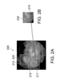
- a selected portion of an image may contain, for example, substantially the portion of the image which contains the detected pathology or the suspected abnormality, or a frame or cropped version of the image where a greater portion of the frame or cropped version of the image contains the suspected abnormality or pathology than the original image.
- a suspected polyp which was identified by a polyp detector may be included in a selected portion of an image.
- some surrounding background pixels which do not make up the suspected pathology may also be included in the selected portion of the image. This is primarily due to the need of a reviewer to see the suspected pathology in the context of the tissue area, and adding some background pixels, may allow the user to identify the suspected pathology better.
- the selected portion of the image is typically rectangular, square or hexagonal, but the suspected pathologies boundaries may be formed in a variety of different shapes.
- the selected portion of an image may contain the portion of a frame which was detected by a blood detector as depicting or corresponding to suspected bleeding.
- the selected portion of an image may contain a suspected lesion, ulcer, inflammation, etc.
- the frame or cropped version may omit a portion of the image not containing the detected abnormality, but since the selected portion or cropped version is typically in a square, rectangular or hexagonal form, some sections or pixels of the selected portion may not include an abnormality, for example pixels which include tissue nearby the suspected pathology may be included in the cropped version of the image.
- the system may include a device, for example, a capsule 40.
- Capsule 40 may be a swallowable in-vivo capsule, but other sorts of devices or suitable implementations may be used.
- capsule 40 may communicate with an external receiving and display system to provide display of data, control, or other functions.
- power may be provided by an internal battery 45 or a wireless receiving system.
- Other embodiments may have other configurations and capabilities.
- Capsule 40 may include one or more imagers 46, for capturing images, one or more illumination sources 42, for illuminating the body lumen, and a transmitter 41, for transmitting image data and possibly other information to a receiving device such as receiver 12.
- Transmitter 41 may include receiver capability, for example, to receive control information.
- the receiver capcbility may be included in a separate component.
- An optical system including, for example, lenses 49, lensholders 44 or mirrors, may aid in focusing reflected light onto the imagers 46.
- the lenholders 44, illumination units 42, and imagers 46 may be mounted on a substrate 56.
- An imaging head 57 and/or 58 may include the optical system, optical dome 54, imager 46, illumination units 42, and substrate 56.
- an image receiver 12 located outside the patient's body in one or more locations, are an image receiver 12, preferably including an antenna or antenna array, an image receiver storage unit 16, a data processor 14, a data processor storage unit 19, and an image monitor 18, for displaying, for example, the images recorded by the capsule 40.
- the image receiver 12 and image receiver storage unit 16 are small and portable, and are worn on the patient's body during recording of the images.
- the data processor 14, data processor storage unit 19, and image monitor 18 may be included in a computer or workstation 11.
- data processor storage unit 19 may include an image database 10 and a logical editing database 20.
- Logical editing database 20 may include, for example, pre-defined criteria and rules for selecting images or portions thereof, stored in the image database 10, to be displayed to the viewer (e.g., in viewing window 200 of Fig. 2 ).
- a list of the pre-defined criteria and rules may be displayed for selection by the viewer (e.g., associated with select button 331 of Fig. 3 ).
- rules or criteria need not be selectable by a user.
- selection criteria may include, but are not limited to: average intensity of the image, average value of the R, B, or G pixels in the image, median value of the pixel intensity, criteria based on HSV color space, B/R, G/R, STD (standard deviation) values of the previous criteria, differences between images, etc.
- a plurality of certain criteria may be associated to a rule or detector, for example, a polyp detector may use several criteria to determine whether a candidate polyp is present in the image. Similarly, a bleeding or redness detector may use different criteria to determine whether the image includes suspected bleeding or pathological tissue having an abnormal level of redness.
- the user may decide which rules and/or detectors to activate.
- Data processor 14 may include one or more standard data processors, such as a microprocessor, multiprocessor, accelerator board, or any other serial or parallel high performance data processor.
- Image monitor 18 may be a computer screen, a conventional video display, or any other device capable of providing image or other data.
- the imager 46 is a suitable ccomplementary metal-oxide-semiconductor (CMOS) camera, such as a "camera on a chip" type CMOS imager specified by Given Imaging Ltd. of Israel and designed by Aptina Corporation of California, USA.
- CMOS ccomplementary metal-oxide-semiconductor
- the imager 46 may be another device, for example, a charge-coupled device (CCD).
- the illumination source 42 may be, for example, one or more light emitting diodes, or another suitable light source.
- imager 46 may capture images and send data representing the images to transmitter 41, which transmits images to image receiver 12 using, for example, electromagnetic radio waves. Other signal transmission methods are possible and, alternatively, data may be dowloaded from capsule 40 after the procedure.
- Image receiver 12 may transfer the image data to image receiver storage unit 16. After a certain period of time of data collection, the image data stored in storage unit 16 may be sent to the data processor 14 or the data processor storage unit 19. For example, the image receiver storage unit 16 may be taken off the patient's body and connected to the personal computer or workstation which includes the data processor 14 and data processor storage unit 19 via a standard data link, e.g., a serial or parallel interface of known construction.
- Data processor 14 may analyze and edit the data, for example, according to the logical editing database 20, and provide the analyzed and edited data to the image monitor 18, where for example a health professional views the image data.
- Data processor 14 may operate software which, in conjunction with basic operating software such as an operating system and device drivers, controls the operation of data processor 14.
- the software controlling data processor 14 may include code written, for example, in the C++ language and possibly alternative or additional languages, and may be implemented in a variety of known methods.
- the image data collected and stored may be stored indefinitely, transferred to other locations, manipulated or analyzed.
- a health professional may use the images to diagnose pathological conditions of, for example, the GI tract, and in addition, the system may provide information about the location of these pathologies.
- the data processor storage unit 19 first collects data and then transfers data to the data processor 14, the image data is not viewed in real time, other configurations allow for real time or quasi-real time viewing.
- the capsule 40 may collect a series of still images as it traverses the GI tract.
- the images may be later presented as, for example, a stream of images or a moving image of the traverse of the GI tract.
- One or more in-vivo imager systems may collect a large volume of data, as the capsule 40 may take several hours to traverse the GI tract.
- the imagers 46 may record images at a rate of, for example, two to forty images per second (other rates, such as four frames per minute, may be used).
- the imagers 46 may have a fixed or variable frame capture and/or transmission rate.
- the imagers 46 may switch back and forth between frame rates, for example, based on parameters, such as the capsule 40 speed, its estimated location, similarity between consecutive images, or other criteria.
- a total of thousands of images, for example, over 300,000 images, may be recorded.
- the image recordation rate, the frame capture rate, the total number of images captured, the total number of images selected for the edited moving image, and the view time of the edited moving image may each be fixed or varied.
- each frame of image data includes 256 rows of 256 pixels each, each pixel including bytes for color and brightness, according to known methods.
- color may be represented by a mosaic of four sub-pixels, each sub-pixel corresponding to primaries such as red, green, or blue (where one primary is represented twice).
- the brightness of the overall pixel may be recorded by a one byte (i.e., 0-255) brightness value.
- images may be stored sequentially in data processor storage unit 19.
- the stored data may include one or more pixel properties, including color and brightness.
- the system and method of the present invention may be practiced with alternate configurations.
- the components gathering image information need not be contained in a capsule, but may be contained in any other vehicle suitable for traversing a lumen in a human body, such as an endoscope, stent, catheter, needle, etc.
- Data processor storage unit 19 may store a series of images recorded by a capsule 40.
- the images the capsule 40 records as it moves through a patient's GI tract may be combined consecutively to form a moving image stream or movie.
- the data processor 14 may include an editing filter 22 for editing a moving image stream.
- Editing filter 22 may be an editing filter processor and may be implemented by data processor 14. While the editing filter is shown in Fig. 1 as being separate from and connected to processor 14, in some embodiments editing filter may be a set of code or instructions executed by, for example, processor 14. Editing filter 22 may be or include one or more dedicated processors. The editing filter 22 may generate a subset of the original input set of images (the remaining images may be removed, or hidden from view). The editing filter 22 may evaluate the degree or occurrence in each frame of each of a plurality of pre-defined criteria from logical database 20.
- the editing filter 22 may select only a subset of images according to the predefined criteria, constraints, and rules provided by the logical database 20, to form a subset of images of interest.
- the editing filter 22 may select for display only a portion of some images, for example a portion of an image which matches a predefined criteria, e.g. the portion of the image which received a high score according to the one or more rules or criteria provided in logical database 20.
- the portion may be made to fit a frame, and thus the portion may include non-selected image data.
- editing filter 22 may select images or portions of images from one or more image streams captured by one or more imagers 46.
- the image streams may be processed separately, for example, each stream may be processed as a separate stream and images may be independently selected from each stream captured by a single imager 46.
- streams may be merged, for example images from two or more streams may be sorted chronologically according to the capture time of the images and merged into a single stream. Other sorting methods are possible, for example based on different image parameters such as similarity between images, or based on the score assigned to the image portions by the pathology or abnormality detectors.
- the merged stream may be processed as one stream (e.g., editing filter 22 may select images from the merged stream instead of separately from each stream).
- the set of displayed images includes as many images as possible, which may be relevant to generate a correct diagnosis of the patient's condition by a health professional. It may be less desirable to omit certain highly informative images from the set of displayed images, to ensure correct diagnosis. Pathologies or abnormalities in human tissue have a very wide range of manifestation, making them in some cases difficult to detect. Accordingly, the editing filter 22 may select frames or portions of frames based on a specific predetermined criterion, or on a combination of a plurality of pre-determined criteria.
- the pre-determined criteria may include, for example, a measure or score of one or more pathology detections and/or anatomical landmark detections (e.g., polyp detector, blood detector, ulcer detector, anomaly detector, duodenum detector, detectors of splenic and/or hepatic flexures in the colon, etc., which are determined based on color, texture, structure or pattern recognition analysis of pixels in the frames), a measure or score of visibility or field of view in the frame of biological tissue which may be distorted or obscured by features such as shadows or residue, the estimated location or region of the capsule (e.g., a higher priority may be assigned to frames estimated to have been captured in a particular region of interest), the probability that capsule is in preferred region (e.g., the colon in a colon imaging procedure, or the small bowel in a small bowel imaging procedure), secondary (non-image) sensor information (e.g., pH, pressure, electrical induction of sensors to determine the proximity of the capsule to the walls of the intestinal passages), capsule motion or
- the editing filter 22 may compute and assign one or more measures, ratings or scores or numbers to each frame based on one or more pre-determined criteria.
- a single criterion is used to select a subset of images for display containing only image portions pertaining to the selected criterion. For example, each image may be scanned for polyps by a polyp detector. The polyp detector may produce a score of the probability of a polyp existing in the image, and may also provide estimated boundaries of that polyp in the image. Based on the estimated boundaries, only the relevant portion of the image may be extracted into the subset of selected images for display (see Fig. 2 for example).
- one subset of images may include all images or portions of images associated with a high score or probability of polyp existance, while another subset of images may present all image or portions thereof relevant to or associated with blood or redness detection in the images.
- the same image may be a part of two or more subsets of different criteria. It may be beneficial for a health care professional to view a subset of images including all image portions pertaining to the same symptom or pathology, since such view increases the chance of correct diagnosis, e.g. quickly finding the true positives (e.g.
- a score, rating, or measure may be a simplified representation (e.g., a derived value or rating, such as an integer 0-100) of more complex characteristics of an image or a portion of an image (e.g., criteria, such as, color variation, appearance of certain textural or structural patterns, light intensity of the image or portions thereof, blood detection, etc.).
- a score may include any rating, rank, hierarchy, scale or relative values of features or criteria. Typically a score is a numerical value, for example, a number from 1 to 10, but need not be limited as such.
- scores may include, for example, letter (A, B, C, ...), signs or symbols (+, -), computer bit values (0, 1), the results of one or more decisions or conditions (yes no), for example, indicated by the status of one or more computing flags.
- Scores may be discrete (non-continuous) values, for example, integers, a, b, c, etc., or may be continuous, for example, having any real value between 0 and 1 (subject to the precision of computer representation of numbers). Any interval between consecutive scores may be set (e.g., 0.1, 0.2, ..., or 1, 2, ..., etc.) and scores may or may not be normalized.
- Scores for each frame or portion thereof may be stored with the frames in the same database (e.g., image database 10).
- the scores may be defined, e.g., in a header or summary frame information package, with the data in an initial image stream or with frames copied to a second edited image stream.
- the scores may be stored in a database separate from the images (e.g., logical database 20) with pointers pointing to the images.
- the scores in separate database may be stored with associated predefined criteria, constraints, and rules to form a subset of selected image portions.
- the editing filter 22 may attempt to determine if a criterion or feature is more visible in a portion of image A than in a portion of image B and then if the criterion or feature is more visible in a portion of image B than in a portion of image C.
- the content of image B may be evaluated twice, once for comparison with image A and then again for comparison with image C.
- scores according to embodiments of the invention, the content of each image need only be evaluated once with respect to each criterion to determine the score of the image.
- a simple numerical comparison of scores may be executed to compare the image frame with both images A and C.
- Using a score to compare and select images may greatly reduce at least the number of times the content of an image is evaluated and thus the computational effort of image comparisons.
- the editing filter 22 may assign a single combined score, e.g., a scalar value, rating each frame or group of frames based on combined frame properties associated with two or more of the plurality of pre-determined criteria.
- the scores may be, for example, a normal or weighted average of frame values for each of the two or more pre-determined criteria.
- each frame may have a score, s1,s2,s3,..., assigned for each pre-determined criteria, 1, 2, 3, ...
- the editing filter 22 may store each score individually for each individual criterion.
- a score vector may refer to any representation or storage that separates individual scores for each criterion, for example, such as a table or data array.
- the scores may be all in the same units (e.g., a number), but need not be.
- the editing filter 22 may assign frames weighted scores, in which larger weights may be assigned for some pre-defined criteria than others. For example, since a large polyp (e.g., at least 6 mm in diameter) is more significant for diagnosis than a small polyp (e.g., 1mm in diameter), the weight assigned to the large polyp score may be greater than the weight assigned to the small polyp score. While in some embodiments polyps are discussed, other pathologies, and other features, may be detected, rated, or scored. The score for each criterion may be weighted or combined in any suitable manner. In one embodiment, the weight of one score may affect the weight(s) of one or more other scores.
- the weights of other scores may be changed in the combined score or the score may be added (e.g., the weight being changed from zero to one or more) or removed (e.g., the weight being changed from one to zero) from the combined score.
- different weights for one or more scores may be used for different respective regions of the GI tract. For example, when a capsule is in (or is estimated to be) the colon (e.g., indicated by the location score or probability of being in the colon), a score indicating the tissue visibility may be given less weight because the relatively wide passage of the colon rarely obscures tissue visibility, thereby making the score less of a defining feature than other scores.
- the scores or measures may be absolute or relative to each other.
- the absolute score(s) for each frame or portion of frame may be a value associated with the criteria for the single frame.
- the relative score(s) for each frame or for a portion of frame may be a change in the value associated with the criteria relative to the value associated with the criteria for a previous or adjacent frame.
- Both absolute and relative scores may or may not be scaled (normalized). Scores may be scaled with a different scaling factor, for example, for images captured or estimated to be captured within each region of the GI tract, each segment of the image stream or for each different frame capture and/or transmission rate.
- the particular pre-determined criteria and their measures, ratings or scores used for selecting a subset of images for display in a two-dimensional tiled array layout may be preset (e.g., by a programmer or at a factory), automatically selected by the data processor 14 or the editing filter 22 itself and/or manually selected by a user (e.g., using input device 24).
- the editing filter 22 may always use one or more default criteria, for example, unless modified by a user.
- An editing graphical user interface may enable a user to select from a plurality of possible criteria (e.g., select button 331 of Fig. 3 ), from which a user may choose one or more.
- the pre-determined criteria may be semiautomatically selected by a processor and/or semi-manually selected by a user.
- the user may indirectly select pre-determined criteria by selecting the desired properties or constraints associated with the movie, such as a maximum movie length (e.g., 45 minutes or 9000 images), a review mode (e.g., preview movie, quick view mode, pathology detection mode, colon analysis mode, small bowel analysis mode, etc.), or other editing constraints. These parameters may in turn trigger the automatic selection of pre-determined criteria by a processor that meet the user-selected constraints.
- the editing filter 22 may determine whether a frame or a portion of a frame corresponds to the selection criteria, and assign a score based on the level of correspondence.
- the editing filter 22 may compare the scores of each image portion to a predetermined threshold value or range.
- the editing filter may select for display each frame with a score exceeding (or lower than) the predetermined value or within the predetermined range for display. Accordingly, the editing filter 22 may not select for display (or may select for deletion) each frame with a score below the predetermined value or outside the predetermined range.
- the score threshold may not be predetermined, but instead may be automatically calculated by editing filter 22 and/or data processor 14.
- the scores may be calculated, for example, based on the number of images in the original image stream (so that a predetermined number of input images satisfy the threshold or a predetermined percentage of input images satisfy the threshold), based on the number of images required in the selected set of images (so that a predetermined number of selected images satisfy the threshold), or based on a time limit for display of the selected set of images (so that the number of images that satisfy the threshold form a selected set of images with a viewing time of less than or equal to a predetermined time, for example when viewing the selected set of images in a standard or average display rate).
- a user may set these parameters, while in other embodiments the parameters may be predetermined or automatically generated by editing filter 22.
- a layout unit 28 may determine the arrangement of the image portions selected by editing filter 22 on the screen or display 18. While the layout unit 28 is shown in Fig. 1 as being separate from and connected to processor 14, in some embodiments layout unit 28 may be a set of code or instructions executed by processor 14. Layout unit 28 may be or include one or more dedicated processors. Layout unit 28 selects or generates a spatial arrangement of a subset of the original image stream, including selected images or portions thereof. The spatial arrangement of the subset of image portions on the display 18 are predetermined, or are selected by a user, for example from a list of possible layout arrangements using select button 332 of Fig. 3 .
- the complete image frame 312 and the previous and next frames 311 and 313 may be automatically displayed to the user, for example upon movement of an input device (such as a mouse) over one of the image portions in the layout.
- a selection of an image portion or a double click with the mouse may open a segment of the image stream which includes the selected image portion (e.g., a few images before and a few images after the selected image or the image from which the portion was extracted).
- Select criteria button 331 may include a list of selection criteria or rules, which a user may select for determining which image portions are selected for display in the layout pages.
- the select criteria button 331 may include high-level detection options, such as Polyp Detection, Lesion Detection, Inflammation Detection, Bleeding Detection, etc.
- one or more rules/criteria may be selected by the user, while other embodiments allow criteria to be predetermined or preprogrammed in advance. The user may decide, for example, to choose only image portions indicated as suspected bleeding images, or may choose to view all images detected as suspected pathologies by at least one of the available detectors.
- Select arrangement button 332 enables the user to select the specific spatial arrangement of image portions in the layout. Several spatial arrangements of image portions are described in Figs. 4A - 4C hereinbelow.
- Select layout array button 333 enables the user to select the number and/or size of image portions that will appear in the rows and columns of the array.
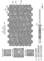
- Figs. 4A , 4B and 4C are different spatial arrangements of display layouts according to embodiments of the present invention.
- Different parameters may be used for positioning the image portions on the screen.
- the image portions may be sorted according to illumination intensity of the image (or portion), according to the chronological time of capturing the image, according to the degree of similarity between the image portions, according to color parameters (e.g. in the RGB portion, R pixels' median value of the portion) or based on other features, for example scores of criteria or features which were used for selection of the image portions from the original captured image stream.
- the different spatial layout arrangements may provide a better viewing experience for the user, and may reduce overall viewing time of the image portions layouts, and/or the image stream.
- Another parameter for positioning the frame portions may be the degree of similarity between chronologically sequential images, which may be preferably kept continuous or in the same area of the grid or array rather than separated in the layout.
- the images may be spatially ordered based on the scores, time, or other criteria.
- One advantage of arranging the selected portions according to their chronological order of capture is that similar image portions may be displayed next to each other, since sequences of several images captured while the capsule was in the same position are displayed in the sequential order of their capture timestamp.
- the sequences 420 and 421 are examples of similar image portions which are kept together due to the chronological spatial arrangement.
- Embodiments of the present invention may include apparatuses for performing the operations herein.
- Such apparatuses may be specially constructed for the desired purposes, or may comprise general purpose computers selectively activated or reconfigured by a computer program stored in the computers.
- Such computer programs may be stored in a computer readable storage medium, such as, but is not limited to, any type of disk including floppy disks, optical disks, CD-ROMs, magnetic-optical disks, read-only memories (ROMs), random access memories (RAMs) electrically programmable read-only memories (EPROMs), electrically erasable and programmable read only memories (EEPROMs), magnetic or optical cards, or any other type of media suitable for storing electronic instructions, and capable of being coupled to a computer system bus.
Landscapes
- Engineering & Computer Science (AREA)
- Health & Medical Sciences (AREA)
- Life Sciences & Earth Sciences (AREA)
- Physics & Mathematics (AREA)
- Surgery (AREA)
- Theoretical Computer Science (AREA)
- General Physics & Mathematics (AREA)
- General Health & Medical Sciences (AREA)
- Nuclear Medicine, Radiotherapy & Molecular Imaging (AREA)
- Medical Informatics (AREA)
- Radiology & Medical Imaging (AREA)
- Animal Behavior & Ethology (AREA)
- Public Health (AREA)
- Heart & Thoracic Surgery (AREA)
- Pathology (AREA)
- Molecular Biology (AREA)
- Optics & Photonics (AREA)
- Biophysics (AREA)
- Biomedical Technology (AREA)
- Veterinary Medicine (AREA)
- General Engineering & Computer Science (AREA)
- Human Computer Interaction (AREA)
- Library & Information Science (AREA)
- Multimedia (AREA)
- Signal Processing (AREA)
- Data Mining & Analysis (AREA)
- Software Systems (AREA)
- Databases & Information Systems (AREA)
- Quality & Reliability (AREA)
- Computer Vision & Pattern Recognition (AREA)
- Endoscopes (AREA)
- Measuring And Recording Apparatus For Diagnosis (AREA)
- Studio Devices (AREA)
- Image Processing (AREA)
- Measurement Of The Respiration, Hearing Ability, Form, And Blood Characteristics Of Living Organisms (AREA)
Claims (15)
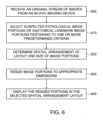
- Procédé d'affichage de parties d'images in vivo, le procédé comprenant :la réception (600) d'un flux d'images in vivo (200) capturées dans une lumière corporelle ;la réalisation d'une reconnaissance algorithmique de critères prédéterminés de façon à sélectionner automatiquement (610), au moyen d'un processeur (14) ou d'un filtre (22), des images (200) correspondant à une anomalie détectée ou à une pathologie suspectée à partir du flux, la sélection étant basée sur un ou plusieurs critères de sélection, les critères comprenant la détection d'une anomalie ou d'une pathologie suspectée, et lesdites images sélectionnées (200) comprenant l'anomalie détectée ou la pathologie suspectée, comprenant en outre la division des images sélectionnées (200) en différents sous-ensembles d'images, les sous-ensembles se rapportant à différents symptômes ou pathologies ;le recadrage automatique desdites images sélectionnées (200) pour obtenir des parties d'images sélectionnées (211, 212) en omettant les parties des images sélectionnées (200) ne contenant pas l'anomalie détectée ou la pathologie suspectée ;la réception (620) d'un réseau bidimensionnel pour l'affichage des parties d'images sélectionnées (211, 212) ;la réception (620) d'un agencement spatial pour l'affichage des parties d'images sélectionnées (211, 212) pour déterminer une distribution desdites parties d'images sélectionnées (211, 212) dans ledit réseau bidimensionnel ;le redimensionnement automatique (630) des parties d'images sélectionnées (211, 212) aux dimensions appropriées sur la base du réseau bidimensionnel reçu ; etl'affichage (640) des parties d'images automatiquement sélectionnées, recadrées et redimensionnées (211, 212) selon le réseau bidimensionnel et l'agencement spatial reçus ;les parties d'images sélectionnées (211, 212) étant affichées dans une disposition en mosaïque bidimensionnelle dans le réseau bidimensionnel sans espaces, arrière-plan ou bordures entre les parties d'images adjacentes (211, 212) ; etchaque disposition en mosaïque bidimensionnelle comprenant uniquement des parties d'images provenant du même sous-ensemble d'images.
- Procédé selon la revendication 1, comprenant en outre la détermination d'au moins l'un des éléments suivants : le nombre de parties d'images (211, 212) à afficher dans le réseau bidimensionnel et les dimensions appropriées des parties d'images sélectionnées (211, 212) dans le réseau bidimensionnel.
- Procédé selon la revendication 1, ledit réseau bidimensionnel et l'agencement spatial étant reçus d'un utilisateur.
- Procédé selon la revendication 1, comprenant en outre :la détermination de la similarité entre des parties d'images séquentielles (211, 212) ; etl'agencement des parties d'images séquentielles similaires (211, 212) dans une séquence chronologique dans le réseau bidimensionnel.
- Procédé selon la revendication 1, comprenant en outre :la réception d'une indication utilisateur d'une partie d'image sélectionnée (211, 212) dans le réseau bidimensionnel ; etl'affichage de l'image complète (312) correspondant à la partie d'image sélectionnée (211, 212).
- Procédé selon la revendication 1, comprenant en outre la détermination du ou des critères de sélection ; et comprenant facultativement l'acceptation d'une indication utilisateur du ou des critères de sélection.
- Procédé selon la revendication 1, l'agencement spatial étant sélectionné parmi : l'agencement de parties d'images (211, 212) dans un ordre chronologique dans le réseau bidimensionnel, l'agencement de parties d'images (211, 212) représentant des zones de lumière intestinale au centre du réseau bidimensionnel et des parties d'images (211, 212) qui contiennent des parois tissulaires autour de la périphérie du réseau, et l'agencement de parties d'images (211, 212) représentant des zones de lumière intestinale dans les rangées supérieures du réseau bidimensionnel et des parties d'images (211, 212) qui contiennent des parois tissulaires dans les rangées inférieures du réseau bidimensionnel.
- Procédé selon la revendication 1, le réseau bidimensionnel comprenant un réseau d'hexagones (2032).
- Procédé selon la revendication 1,les étapes de sélection automatique (610) des images (200) et de recadrage des images sélectionnées comprenant l'édition du flux d'images utilisant un filtre d'édition, pour obtenir un sous-ensemble d'images correspondant à un ou plusieurs critères d'édition, et le recadrage, à partir de chaque image du sous-ensemble d'images, de parties d'images (211, 212) correspondant aux critères d'édition, lesdites parties d'images (211, 212) comprenant l'anomalie détectée ou la pathologie suspectée ;les étapes de réception (620) d'un réseau bidimensionnel et d'un agencement spatial comprenant la réception d'un agencement spatial sélectionné pour les parties d'images (211, 212) dans une disposition en grille ;l'étape de redimensionnement automatique (630) des parties d'images sélectionnées (211, 212) comprenant le redimensionnement des parties d'images recadrées (211, 212) aux dimensions appropriées basées sur l'agencement spatial sélectionné ; etl'étape d'affichage (640) comprenant l'affichage des parties d'images redimensionnées (211, 212) agencées spatialement dans la disposition en grille ;la disposition en grille comprenant facultativement une grille d'images à bordures hexagonales.
- Procédé selon la revendication 1, un filtre d'homogénéisation étant appliqué aux parties d'images (211, 212) de façon à supprimer les caractéristiques qui créent une disposition irrégulière, hétérogène ou bruitée, ou qui ont un effet perturbateur sur l'œil d'un utilisateur.
- Procédé selon la revendication 1, une plus grande partie de chaque partie d'image sélectionnée (211, 212) contenant l'anomalie détectée ou la pathologie suspectée que l'image sélectionnée correspondante (200).
- Procédé selon la revendication 1, les images sélectionnées (200) étant recadrées de sorte que les parties d'images (211, 212) comprennent en outre des pixels d'arrière-plan environnants qui ne constituent pas l'anomalie détectée ou la pathologie suspectée dans les parties d'images (211, 212).
- Système d'affichage de parties d'images in vivo comprenant :
une unité de traitement (11, 12) pour recevoir un flux d'images in vivo (200) capturées dans une lumière corporelle par une capsule d'imagerie (40), l'unité de traitement (11, 12) comprenant :un filtre d'édition (22) pour sélectionner automatiquement dans le flux des images (200) correspondant à une anomalie détectée ou une pathologie suspectée, la sélection étant basée sur un ou plusieurs critères de sélection, les critères de sélection comprenant la détection d'une anomalie ou d'une pathologie suspectée, et pour recadrer automatiquement les images sélectionnées (200) afin d'obtenir des parties d'images sélectionnées (211, 212) en omettant les parties des images sélectionnées (200) ne contenant pas l'anomalie ou la pathologie suspectée, et le filtre d'édition étant configuré pour diviser les images sélectionnées (200) en différents sous-ensembles d'images, les sous-ensembles se rapportant à différents symptômes ou pathologies ; etune unité de disposition (28) pour recevoir un réseau bidimensionnel pour l'affichage des parties d'images sélectionnées (211, 212), pour recevoir un agencement spatial pour l'affichage des parties d'images sélectionnées (211, 212) afin de déterminer la distribution desdites parties d'images sélectionnées (211, 212) dans ledit réseau bidimensionnel, et pour redimensionner automatiquement les parties d'images sélectionnées (211, 212) de sorte que les parties d'images (211, 212) soient de taille uniforme et de dimensions appropriées basées sur le réseau bidimensionnel reçu ; etun dispositif d'affichage (18) pour afficher les parties d'images automatiquement sélectionnées, recadrées et redimensionnées (211, 212) dans une disposition en mosaïque bidimensionnelle selon le réseau bidimensionnel et l'agencement spatial reçus sans sélection utilisateur des images (200) à partir d'un flux d'images en mouvement ;le système étant configuré pour afficher les parties d'images sélectionnées (211, 212) dans le réseau bidimensionnel, qui se touchent pratiquement, sans espace, arrière-plan ou bordure entre des parties d'images adjacentes (211, 212) ; etchaque disposition en mosaïque bidimensionnelle comprenant uniquement des parties d'images provenant des mêmes images de sous-ensemble d'images. - Système selon la revendication 13, l'unité de disposition (28) étant destinée à déterminer le nombre de parties d'images sélectionnées (211, 212) à afficher dans le réseau bidimensionnel ; et/ou l'unité de disposition (28) étant destinée à déterminer la similarité entre des parties d'images sélectionnées séquentielles (211, 212), et à agencer les parties d'images similaires (211, 212) dans une séquence chronologique dans le réseau bidimensionnel.
- Système selon la revendication 13, le filtre d'édition (22) étant en outre configuré pour appliquer un filtre d'homogénéisation aux parties d'images (211, 212) de façon à supprimer les caractéristiques qui créent une disposition irrégulière, hétérogène ou bruitée, ou qui ont un effet perturbateur sur l'œil d'un utilisateur.
Applications Claiming Priority (3)
| Application Number | Priority Date | Filing Date | Title |
|---|---|---|---|
| US32870510P | 2010-04-28 | 2010-04-28 | |
| EP11774537.2A EP2567347A4 (fr) | 2010-04-28 | 2011-04-28 | Système et procédé d'affichage de parties d'images in vivo |
| PCT/IL2011/000345 WO2011135573A1 (fr) | 2010-04-28 | 2011-04-28 | Système et procédé d'affichage de parties d'images in vivo |
Related Parent Applications (1)
| Application Number | Title | Priority Date | Filing Date |
|---|---|---|---|
| EP11774537.2A Division EP2567347A4 (fr) | 2010-04-28 | 2011-04-28 | Système et procédé d'affichage de parties d'images in vivo |
Publications (2)
| Publication Number | Publication Date |
|---|---|
| EP3852010A1 EP3852010A1 (fr) | 2021-07-21 |
| EP3852010B1 true EP3852010B1 (fr) | 2025-04-09 |
Family
ID=44860958
Family Applications (2)
| Application Number | Title | Priority Date | Filing Date |
|---|---|---|---|
| EP21153056.3A Active EP3852010B1 (fr) | 2010-04-28 | 2011-04-28 | Système et procédé d'affichage de parties d'images in vivo |
| EP11774537.2A Ceased EP2567347A4 (fr) | 2010-04-28 | 2011-04-28 | Système et procédé d'affichage de parties d'images in vivo |
Family Applications After (1)
| Application Number | Title | Priority Date | Filing Date |
|---|---|---|---|
| EP11774537.2A Ceased EP2567347A4 (fr) | 2010-04-28 | 2011-04-28 | Système et procédé d'affichage de parties d'images in vivo |
Country Status (6)
| Country | Link |
|---|---|
| US (2) | US9060673B2 (fr) |
| EP (2) | EP3852010B1 (fr) |
| JP (1) | JP5972865B2 (fr) |
| KR (1) | KR101708726B1 (fr) |
| CN (2) | CN106923779A (fr) |
| WO (1) | WO2011135573A1 (fr) |
Families Citing this family (68)
| Publication number | Priority date | Publication date | Assignee | Title |
|---|---|---|---|---|
| CN106923779A (zh) | 2010-04-28 | 2017-07-07 | 基文影像公司 | 用于显示体内图像部分的系统和方法 |
| US8913807B1 (en) * | 2010-12-30 | 2014-12-16 | Given Imaging Ltd. | System and method for detecting anomalies in a tissue imaged in-vivo |
| JP5580765B2 (ja) * | 2011-03-03 | 2014-08-27 | オリンパスメディカルシステムズ株式会社 | 画像処理装置、画像処理方法、及び画像処理プログラム |
| US8855413B2 (en) * | 2011-05-13 | 2014-10-07 | Abbyy Development Llc | Image reflow at word boundaries |
| EP2570986A1 (fr) * | 2011-09-13 | 2013-03-20 | Alcatel Lucent | Procédé pour la création d'un couvercle pour un dispositif électronique et dispositif électronique |
| US20130167072A1 (en) * | 2011-12-22 | 2013-06-27 | Sap Portals Israel Ltd. | Smart and Flexible Layout Context Manager |
| CN104115149B (zh) * | 2012-02-07 | 2017-11-17 | 皇家飞利浦有限公司 | 用于统计学测试的扫描数据库的交互式优化 |
| US9697598B2 (en) | 2012-11-23 | 2017-07-04 | Koninklijke Philips N.V. | Generating a key-image from a medical image |
| US9854958B1 (en) * | 2013-03-23 | 2018-01-02 | Garini Technologies Corporation | System and method for automatic processing of images from an autonomous endoscopic capsule |
| US20140378810A1 (en) | 2013-04-18 | 2014-12-25 | Digimarc Corporation | Physiologic data acquisition and analysis |
| RU2676432C2 (ru) | 2013-04-18 | 2018-12-28 | Конинклейке Филипс Н.В. | Получение цервикальных изображений |
| EP2987106A4 (fr) * | 2013-04-18 | 2016-12-14 | Digimarc Corp | Acquisition et analyse de données physiologiques |
| US20140321770A1 (en) * | 2013-04-24 | 2014-10-30 | Nvidia Corporation | System, method, and computer program product for generating an image thumbnail |
| US9743824B2 (en) * | 2013-09-04 | 2017-08-29 | Siemens Medical Solutions Usa, Inc. | Accurate and efficient polyp detection in wireless capsule endoscopy images |
| WO2015049962A1 (fr) | 2013-10-02 | 2015-04-09 | オリンパスメディカルシステムズ株式会社 | Système d'endoscope |
| US9430706B1 (en) | 2013-10-02 | 2016-08-30 | Given Imaging Ltd. | System and method for detection of in-vivo pathology sequences |
| JP5865557B2 (ja) * | 2013-11-21 | 2016-02-17 | オリンパス株式会社 | 内視鏡画像表示装置 |
| US9349160B1 (en) * | 2013-12-20 | 2016-05-24 | Google Inc. | Method, apparatus and system for enhancing a display of video data |
| US9342881B1 (en) * | 2013-12-31 | 2016-05-17 | Given Imaging Ltd. | System and method for automatic detection of in vivo polyps in video sequences |
| US10117563B2 (en) * | 2014-01-09 | 2018-11-06 | Gyrus Acmi, Inc. | Polyp detection from an image |
| US10993837B2 (en) * | 2014-04-23 | 2021-05-04 | Johnson & Johnson Surgical Vision, Inc. | Medical device data filtering for real time display |
| US11120547B2 (en) * | 2014-06-01 | 2021-09-14 | CapsoVision, Inc. | Reconstruction of images from an in vivo multi-camera capsule with two-stage confidence matching |
| US9633276B2 (en) * | 2014-07-14 | 2017-04-25 | Sony Corporation | Blood detection system with real-time capability and method of operation thereof |
| JP6425931B2 (ja) * | 2014-07-16 | 2018-11-21 | オリンパス株式会社 | カプセル型内視鏡及び内視鏡システム |
| US10660533B2 (en) | 2014-09-30 | 2020-05-26 | Rapsodo Pte. Ltd. | Remote heart rate monitoring based on imaging for moving subjects |
| US9727949B1 (en) * | 2014-11-19 | 2017-08-08 | Google Inc. | Providing secure digital images |
| WO2016089413A1 (fr) * | 2014-12-04 | 2016-06-09 | Capso Vision, Inc. | Enrobage de capsule pour commande de capture d'image |
| JP6569687B2 (ja) * | 2014-12-15 | 2019-09-04 | ソニー株式会社 | 情報処理方法、映像処理装置及びプログラム |
| USD776713S1 (en) * | 2014-12-17 | 2017-01-17 | Rgi Informatics, Llc | Display device with a timeline graphical user interface |
| CN107735136B (zh) | 2015-06-30 | 2021-11-02 | 瑞思迈私人有限公司 | 使用移动应用的面罩大小调整工具 |
| JP6269610B2 (ja) * | 2015-07-28 | 2018-01-31 | 株式会社リコー | 情報処理装置、画像表示方法、通信システム、プログラム |
| US10861151B2 (en) | 2015-08-07 | 2020-12-08 | The Arizona Board Of Regents On Behalf Of Arizona State University | Methods, systems, and media for simultaneously monitoring colonoscopic video quality and detecting polyps in colonoscopy |
| US10572997B2 (en) * | 2015-12-18 | 2020-02-25 | Given Imaging Ltd. | System and method for detecting anomalies in an image captured in-vivo using color histogram association |
| DE112017000025B4 (de) * | 2016-03-03 | 2025-02-20 | Hoya Corporation | Verfahren zur Erzeugung von Korrekturdaten und Vorrichtung zur Erzeugung von Korrekturdaten |
| CN105955738A (zh) * | 2016-04-28 | 2016-09-21 | 乐视控股(北京)有限公司 | 一种3d列表数据对应的用户界面显示方法及装置 |
| CN109328028B (zh) * | 2016-05-18 | 2021-11-23 | 基文影像公司 | 用于选择体内捕获到的图像以用于显示的系统和方法 |
| EP3267394A1 (fr) * | 2016-07-04 | 2018-01-10 | Centre National De La Recherche Scientifique | Procédé et appareil pour la détection en temps réel de polypes dans une coloscopie optique |
| WO2018211674A1 (fr) * | 2017-05-18 | 2018-11-22 | オリンパス株式会社 | Dispositif de traitement d'image, procédé de traitement d'image et programme |
| US10671349B2 (en) | 2017-07-24 | 2020-06-02 | Tesla, Inc. | Accelerated mathematical engine |
| US11409692B2 (en) | 2017-07-24 | 2022-08-09 | Tesla, Inc. | Vector computational unit |
| US20190139643A1 (en) * | 2017-11-08 | 2019-05-09 | International Business Machines Corporation | Facilitating medical diagnostics with a prediction model |
| CN108024061A (zh) * | 2017-12-08 | 2018-05-11 | 合肥工业大学 | 医用内窥镜人工智能系统的硬件架构及图像处理方法 |
| CN108055454B (zh) * | 2017-12-08 | 2020-07-28 | 合肥工业大学 | 医用内窥镜人工智能芯片的体系架构及图像处理方法 |
| CN108042090B (zh) * | 2017-12-08 | 2020-12-01 | 合肥工业大学 | 配置人工智能芯片的医用内窥镜系统及图像处理方法 |
| EP3539455A1 (fr) * | 2018-03-14 | 2019-09-18 | Sorbonne Université | Procédé pour déterminer automatiquement la qualité de visualisation des images en vidéocapsule endoscopique |
| US11112952B2 (en) * | 2018-03-26 | 2021-09-07 | Microscopes International, Llc | Interface for display of multi-layer images in digital microscopy |
| US11100633B2 (en) * | 2018-06-13 | 2021-08-24 | Cosmo Artificial Intelligence—Al Limited | Systems and methods for processing real-time video from a medical image device and detecting objects in the video |
| JP7163079B2 (ja) * | 2018-06-22 | 2022-10-31 | 株式会社エビデント | 撮像システム、画像構築方法 |
| GB2570743B (en) * | 2018-06-29 | 2021-06-23 | Imagination Tech Ltd | Tile assignment to processing cores within a graphics processing unit |
| KR102275760B1 (ko) * | 2018-07-09 | 2021-07-09 | 주식회사 셀바스에이아이 | 조기 위암의 예측 방법 및 이를 이용한 디바이스 |
| US11562231B2 (en) | 2018-09-03 | 2023-01-24 | Tesla, Inc. | Neural networks for embedded devices |
| JP7752529B2 (ja) * | 2018-10-19 | 2025-10-10 | ギブン イメージング リミテッド | 生体内画像ストリームの精査用情報を生成及び表示するためのシステム並びに方法 |
| US11816585B2 (en) | 2018-12-03 | 2023-11-14 | Tesla, Inc. | Machine learning models operating at different frequencies for autonomous vehicles |
| US12311073B2 (en) * | 2018-12-05 | 2025-05-27 | Moraine Corporation | Hygiene evaluating device, hygiene evaluation imaging system, imaging stand, and hygiene evaluation method |
| US11694335B2 (en) * | 2018-12-05 | 2023-07-04 | Stryker Corporation | Systems and methods for displaying medical imaging data |
| US11610117B2 (en) | 2018-12-27 | 2023-03-21 | Tesla, Inc. | System and method for adapting a neural network model on a hardware platform |
| CN109903269B (zh) * | 2019-01-24 | 2021-05-04 | 刘星宇 | 确定脊柱横断面图像的异常类型的方法及计算设备 |
| CN109934276B (zh) * | 2019-03-05 | 2020-11-17 | 安翰科技(武汉)股份有限公司 | 基于迁移学习的胶囊内窥镜图像分类系统及方法 |
| CN109965829B (zh) * | 2019-03-06 | 2022-05-06 | 重庆金山医疗技术研究院有限公司 | 成像优化方法、图像处理装置、成像装置及内窥镜系统 |
| TWI737318B (zh) * | 2020-05-27 | 2021-08-21 | 禾瑞亞科技股份有限公司 | 觸控處理方法、裝置與觸控系統 |
| KR102193302B1 (ko) * | 2020-07-06 | 2020-12-22 | 주식회사 스마트 잭 | 영상을 이용한 시약 정보 수집 방법 및 장치 |
| CN112863700A (zh) * | 2021-01-28 | 2021-05-28 | 青岛海信医疗设备股份有限公司 | 医疗影像协同方法、装置、设备及计算机存储介质 |
| WO2022195696A1 (fr) * | 2021-03-16 | 2022-09-22 | オリンパスメディカルシステムズ株式会社 | Système et méthode d'assistance médicale |
| KR102641492B1 (ko) * | 2022-01-05 | 2024-02-27 | 주식회사 아이넥스코퍼레이션 | 내시경 영상을 이용한 용종 검출 방법, 장치 및 프로그램 |
| KR102641489B1 (ko) * | 2022-01-05 | 2024-02-27 | 주식회사 아이넥스코퍼레이션 | 내시경 영상을 이용한 용종 검출 방법, 장치 및 프로그램 |
| CN116932193B (zh) * | 2022-04-07 | 2024-08-20 | 华为技术有限公司 | 一种显示子系统的通道分配方法、装置及存储介质 |
| JP2025031279A (ja) * | 2023-08-25 | 2025-03-07 | キヤノン株式会社 | 情報処理装置、情報処理装置の制御方法及びプログラム |
| DE102023133361A1 (de) * | 2023-11-29 | 2025-06-05 | Resolve Biosciences Gmbh | Optisches system zur darstellung eines objekts |
Family Cites Families (113)
| Publication number | Priority date | Publication date | Assignee | Title |
|---|---|---|---|---|
| CS175782B1 (fr) | 1974-10-16 | 1977-05-31 | ||
| US4243652A (en) | 1978-07-14 | 1981-01-06 | The Procter & Gamble Company | Gastrointestinal scanning agent |
| JPS5519124A (en) | 1978-07-27 | 1980-02-09 | Olympus Optical Co | Camera system for medical treatment |
| JPS5697872A (en) | 1980-01-07 | 1981-08-06 | Fuji Photo Film Co Ltd | Measuring material for concentration of hemoglobin in blood |
| JPS5745833A (en) | 1980-09-01 | 1982-03-16 | Taeko Nakagawa | Stomack camera |
| US5993378A (en) | 1980-10-28 | 1999-11-30 | Lemelson; Jerome H. | Electro-optical instruments and methods for treating disease |
| DE3440177A1 (de) | 1984-11-02 | 1986-05-15 | Friedrich Dipl.-Ing. 8031 Eichenau Hilliges | Fernseh-aufnahme- und -wiedergabeeinrichtung zur endoskopie an menschlichen und tierischen koerpern |
| JPS61143876A (ja) | 1984-12-17 | 1986-07-01 | Hitachi Medical Corp | 複数画像表示の画像処理装置 |
| US4698664A (en) | 1985-03-04 | 1987-10-06 | Apert-Herzog Corporation | Audio-visual monitoring system |
| JPH0761309B2 (ja) | 1987-03-03 | 1995-07-05 | オリンパス光学工業株式会社 | 内視鏡装置 |
| JPH0684970B2 (ja) | 1987-03-31 | 1994-10-26 | 株式会社京都医科学研究所 | 糞便中の潜血検出方法 |
| US4907095A (en) | 1987-04-16 | 1990-03-06 | Fuji Photo Film Co., Ltd. | Still image trimming apparatus |
| US4938823A (en) | 1988-10-07 | 1990-07-03 | The Pultrusions Corporation | Pultrusion/extrusion method |
| US5697885A (en) | 1989-01-30 | 1997-12-16 | Olympus Optical Co., Ltd. | Endoscope for recording and displaying time-serial images |
| JP2750151B2 (ja) | 1989-04-27 | 1998-05-13 | オリンパス光学工業株式会社 | 画像記録検索表示装置 |
| JPH04109927A (ja) | 1990-08-31 | 1992-04-10 | Toshiba Corp | 電子内視鏡装置 |
| JPH04144533A (ja) | 1990-10-05 | 1992-05-19 | Olympus Optical Co Ltd | 内視鏡 |
| JPH04357927A (ja) | 1991-01-14 | 1992-12-10 | Olympus Optical Co Ltd | 画像表示装置 |
| DE69227545T2 (de) | 1991-07-12 | 1999-04-29 | Robinson, Mark R., Albuquerque, N.Mex. | Oximeter zur zuverlässigen klinischen Bestimmung der Blutsauerstoffsättigung in einem Fötus |
| JP3237900B2 (ja) | 1992-06-19 | 2001-12-10 | 株式会社東芝 | 画像表示システム |
| JPH0638219A (ja) | 1992-07-20 | 1994-02-10 | Olympus Optical Co Ltd | 映像表示装置 |
| US5381784A (en) | 1992-09-30 | 1995-01-17 | Adair; Edwin L. | Stereoscopic endoscope |
| US5603687A (en) | 1992-10-28 | 1997-02-18 | Oktas General Partnership | Asymmetric stereo-optic endoscope |
| EP0596651A1 (fr) | 1992-11-02 | 1994-05-11 | National Semiconductor Corporation | Réseau de données avec capacité de communication isochrone |
| JP3020376B2 (ja) | 1993-03-26 | 2000-03-15 | サージミヤワキ株式会社 | 動物用体内型個体識別器具 |
| US5642157A (en) | 1993-11-29 | 1997-06-24 | Kabushiki Kaisha Toshiba | Electronic endoscope apparatus usable in combination with medical diagnosis apparatus |
| IL108352A (en) | 1994-01-17 | 2000-02-29 | Given Imaging Ltd | In vivo video camera system |
| US5880777A (en) | 1996-04-15 | 1999-03-09 | Massachusetts Institute Of Technology | Low-light-level imaging and image processing |
| US7194117B2 (en) | 1999-06-29 | 2007-03-20 | The Research Foundation Of State University Of New York | System and method for performing a three-dimensional virtual examination of objects, such as internal organs |
| JP3250467B2 (ja) | 1996-10-04 | 2002-01-28 | 松下電器産業株式会社 | 映像要約方法および映像表示方法 |
| US6173317B1 (en) | 1997-03-14 | 2001-01-09 | Microsoft Corporation | Streaming and displaying a video stream with synchronized annotations over a computer network |
| US6240312B1 (en) | 1997-10-23 | 2001-05-29 | Robert R. Alfano | Remote-controllable, micro-scale device for use in in vivo medical diagnosis and/or treatment |
| IL122602A0 (en) | 1997-12-15 | 1998-08-16 | Tally Eitan Zeev Pearl And Co | Energy management of a video capsule |
| GB9802415D0 (en) | 1998-02-05 | 1998-04-01 | Danmere Limited | Storage tape control system |
| JP4074366B2 (ja) | 1998-02-24 | 2008-04-09 | コニカミノルタビジネステクノロジーズ株式会社 | 画像検索装置及び方法並びに画像検索プログラムを記録した記録媒体 |
| JP2000047651A (ja) | 1998-07-29 | 2000-02-18 | Mitsubishi Electric Corp | 映像再生装置 |
| IL126727A (en) | 1998-10-22 | 2006-12-31 | Given Imaging Ltd | A method of bringing a device to the goal |
| US6654539B1 (en) | 1998-10-26 | 2003-11-25 | Sony Corporation | Trick playback of digital video data |
| US6208354B1 (en) | 1998-11-03 | 2001-03-27 | Ati International Srl | Method and apparatus for displaying multiple graphics images in a mixed video graphics display |
| US6408128B1 (en) | 1998-11-12 | 2002-06-18 | Max Abecassis | Replaying with supplementary information a segment of a video |
| US6504990B1 (en) | 1998-11-12 | 2003-01-07 | Max Abecassis | Randomly and continuously playing fragments of a video segment |
| JP3826598B2 (ja) | 1999-01-29 | 2006-09-27 | 株式会社日立製作所 | 画像監視装置及び記録媒体 |
| US8636648B2 (en) | 1999-03-01 | 2014-01-28 | West View Research, Llc | Endoscopic smart probe |
| US6563959B1 (en) | 1999-07-30 | 2003-05-13 | Pixlogic Llc | Perceptual similarity image retrieval method |
| US6791601B1 (en) | 1999-11-11 | 2004-09-14 | Stryker Corporation | Multi-function image and video capture device for use in an endoscopic camera system |
| JP2001209785A (ja) | 1999-11-19 | 2001-08-03 | Fujitsu Ltd | 画像処理装置、画像処理方法、および画像処理プログラム記憶媒体 |
| JP2001167248A (ja) | 1999-12-06 | 2001-06-22 | Canon Inc | 画像処理装置、画像処理システム、画像処理方法、及び記憶媒体 |
| US6976229B1 (en) | 1999-12-16 | 2005-12-13 | Ricoh Co., Ltd. | Method and apparatus for storytelling with digital photographs |
| JP4573393B2 (ja) | 2000-01-06 | 2010-11-04 | オリンパス株式会社 | 画像表示装置 |
| IL134017A (en) | 2000-01-13 | 2008-04-13 | Capsule View Inc | Camera for photography inside the intestines |
| US20010015753A1 (en) | 2000-01-13 | 2001-08-23 | Myers Kenneth J. | Split image stereoscopic system and method |
| US7039453B2 (en) | 2000-02-08 | 2006-05-02 | Tarun Mullick | Miniature ingestible capsule |
| CN1307938C (zh) | 2000-03-08 | 2007-04-04 | 吉温成象有限公司 | 一个供体内成像的设备和系统 |
| JP3810268B2 (ja) | 2000-04-07 | 2006-08-16 | シャープ株式会社 | オーディオビジュアルシステム |
| US6709387B1 (en) | 2000-05-15 | 2004-03-23 | Given Imaging Ltd. | System and method for controlling in vivo camera capture and display rate |
| US8538770B2 (en) | 2000-08-01 | 2013-09-17 | Logical Images, Inc. | System and method to aid diagnoses using cross-referenced knowledge and image databases |
| US7260777B2 (en) | 2001-08-17 | 2007-08-21 | Desknet Inc. | Apparatus, method and system for transforming data |
| JP2004533272A (ja) | 2000-10-24 | 2004-11-04 | ハウメディカ・オステオニクス・コーポレイション | 癒合用インプラント装置 |
| US7027633B2 (en) | 2000-11-30 | 2006-04-11 | Foran David J | Collaborative diagnostic systems |
| WO2002054932A2 (fr) | 2001-01-16 | 2002-07-18 | Given Imaging Ltd. | Systeme et methode d'imagerie a grand champ de lumieres corporelles |
| US6891570B2 (en) | 2001-01-31 | 2005-05-10 | Itt Manufacturing Enterprises Inc. | Method and adaptively deriving exposure time and frame rate from image motion |
| AU2002241215A1 (en) | 2001-03-14 | 2002-09-24 | Given Imaging Ltd. | Method and system for detecting colorimetric abnormalities |
| US6697123B2 (en) | 2001-03-30 | 2004-02-24 | Koninklijke Philips Electronics N.V. | Adaptive picture-in-picture |
| US7119814B2 (en) | 2001-05-18 | 2006-10-10 | Given Imaging Ltd. | System and method for annotation on a moving image |
| IL143260A (en) | 2001-05-20 | 2006-09-05 | Given Imaging Ltd | Array and method for locating an intra-body signal source |
| IL159451A0 (en) | 2001-06-20 | 2004-06-01 | Given Imaging Ltd | Motility analysis within a gastrointestinal tract |
| US7272657B2 (en) | 2001-07-30 | 2007-09-18 | Digeo, Inc. | System and method for displaying video streams ranked by user-specified criteria |
| JP4744026B2 (ja) | 2001-07-30 | 2011-08-10 | オリンパス株式会社 | カプセル内視鏡およびカプセル内視鏡システム |
| US7347817B2 (en) | 2001-08-02 | 2008-03-25 | Given Imaging Ltd. | Polarized in vivo imaging device, system and method |
| US20030086596A1 (en) | 2001-11-07 | 2003-05-08 | Medical Metrics, Inc. | Method, computer software, and system for tracking, stabilizing, and reporting motion between vertebrae |
| US20030174208A1 (en) | 2001-12-18 | 2003-09-18 | Arkady Glukhovsky | Device, system and method for capturing in-vivo images with three-dimensional aspects |
| JP4234605B2 (ja) | 2002-02-12 | 2009-03-04 | ギブン イメージング リミテッド | 画像ストリームを表示するためのシステムおよび方法 |
| US7474327B2 (en) | 2002-02-12 | 2009-01-06 | Given Imaging Ltd. | System and method for displaying an image stream |
| US8022980B2 (en) * | 2002-02-12 | 2011-09-20 | Given Imaging Ltd. | System and method for displaying an image stream |
| JP2004023458A (ja) | 2002-06-17 | 2004-01-22 | Toshiba Corp | 動画像符号化/復号化方法及び装置 |
| JP4328125B2 (ja) | 2003-04-25 | 2009-09-09 | オリンパス株式会社 | カプセル型内視鏡装置およびカプセル型内視鏡システム |
| JP3810381B2 (ja) * | 2003-04-25 | 2006-08-16 | オリンパス株式会社 | 画像表示装置、画像表示方法および画像表示プログラム |
| WO2004100776A1 (fr) | 2003-05-14 | 2004-11-25 | Olympus Corporation | Dispositif medical a capsule |
| US7215338B2 (en) | 2003-10-02 | 2007-05-08 | Given Imaging Ltd. | System and method for presentation of data streams |
| US20050075537A1 (en) | 2003-10-06 | 2005-04-07 | Eastman Kodak Company | Method and system for real-time automatic abnormality detection for in vivo images |
| US7319781B2 (en) | 2003-10-06 | 2008-01-15 | Carestream Health, Inc. | Method and system for multiple passes diagnostic alignment for in vivo images |
| US7510283B2 (en) | 2003-11-20 | 2009-03-31 | Heidelberg Engineering Optische Messsysteme Gmbh | High resolution imaging for diagnostic evaluation of the fundus of the human eye |
| EP1699530B1 (fr) | 2003-12-31 | 2012-07-04 | Given Imaging Ltd. | Systeme et procede pour afficher un flux d'images |
| EP1790277B1 (fr) * | 2004-08-23 | 2018-11-21 | Olympus Corporation | Dispositif d'affichage d'image et programme d'affichage d'image |
| JP4133981B2 (ja) * | 2004-09-09 | 2008-08-13 | 株式会社東芝 | メタデータと動画像の再生装置 |
| US7986337B2 (en) | 2004-09-27 | 2011-07-26 | Given Imaging Ltd. | System and method for editing an image stream captured in vivo |
| US7558413B2 (en) * | 2004-10-12 | 2009-07-07 | Siemens Medical Solutions USA, Inc, | Method for detecting polyps in a three dimensional image volume |
| JP2008518544A (ja) | 2004-10-29 | 2008-05-29 | コーニンクレッカ フィリップス エレクトロニクス エヌ ヴィ | Mpegストリームのスライシング |
| US7486981B2 (en) | 2004-11-15 | 2009-02-03 | Given Imaging Ltd. | System and method for displaying an image stream |
| KR100633288B1 (ko) | 2004-12-27 | 2006-10-11 | 삼성토탈 주식회사 | 스티렌을 제조하는 방법 |
| JP4547293B2 (ja) * | 2005-04-04 | 2010-09-22 | オリンパス株式会社 | 画像表示装置 |
| US8423123B2 (en) * | 2005-09-30 | 2013-04-16 | Given Imaging Ltd. | System and method for in-vivo feature detection |
| US7577283B2 (en) | 2005-09-30 | 2009-08-18 | Given Imaging Ltd. | System and method for detecting content in-vivo |
| KR20070078335A (ko) | 2006-01-26 | 2007-07-31 | 엘지전자 주식회사 | 프로세서에서의 온칩메모리 제어장치 |
| JP5005981B2 (ja) * | 2006-08-03 | 2012-08-22 | オリンパスメディカルシステムズ株式会社 | 画像表示装置 |
| US8900124B2 (en) * | 2006-08-03 | 2014-12-02 | Olympus Medical Systems Corp. | Image display device |
| JP2008109637A (ja) | 2006-09-25 | 2008-05-08 | Toshiba Corp | 動画像符号化装置及びその方法 |
| JP2009018048A (ja) * | 2007-07-12 | 2009-01-29 | Fujifilm Corp | 医用画像表示装置、方法及びプログラム |
| EP2174578A4 (fr) * | 2007-07-12 | 2013-07-03 | Olympus Medical Systems Corp | Dispositif de traitement d'image, son procédé de fonctionnement et son programme |
| US20090074265A1 (en) | 2007-09-17 | 2009-03-19 | Capsovision Inc. | Imaging review and navigation workstation system |
| US8660175B2 (en) | 2007-12-10 | 2014-02-25 | Qualcomm Incorporated | Selective display of interpolated or extrapolated video units |
| US20090228834A1 (en) * | 2008-02-29 | 2009-09-10 | Christopher Reynolds | Flow radiology user interface |
| JP2009226170A (ja) * | 2008-03-25 | 2009-10-08 | Olympus Medical Systems Corp | 医療用画像処理装置 |
| JP5374078B2 (ja) | 2008-06-16 | 2013-12-25 | オリンパス株式会社 | 画像処理装置、画像処理方法および画像処理プログラム |
| US20100014738A1 (en) * | 2008-07-21 | 2010-01-21 | Tomtec Imaging Systems Gmbh | Method and system for breast cancer screening |
| US8628015B2 (en) | 2008-10-31 | 2014-01-14 | Hand Held Products, Inc. | Indicia reading terminal including frame quality evaluation processing |
| US8150124B2 (en) * | 2009-10-12 | 2012-04-03 | Capso Vision Inc. | System and method for multiple viewing-window display of capsule images |
| US8446465B2 (en) | 2010-01-05 | 2013-05-21 | Given Imaging Ltd. | System and method for displaying an image stream captured in-vivo |
| US8682142B1 (en) | 2010-03-18 | 2014-03-25 | Given Imaging Ltd. | System and method for editing an image stream captured in-vivo |
| CN106923779A (zh) | 2010-04-28 | 2017-07-07 | 基文影像公司 | 用于显示体内图像部分的系统和方法 |
| WO2012037419A2 (fr) * | 2010-09-16 | 2012-03-22 | Omnyx, LLC | Traitement d'image numérique de pathologie |
| US20120113239A1 (en) | 2010-11-08 | 2012-05-10 | Hagai Krupnik | System and method for displaying an image stream |
| JP5784404B2 (ja) * | 2011-07-29 | 2015-09-24 | オリンパス株式会社 | 画像処理装置、画像処理方法、及び画像処理プログラム |
-
2011
- 2011-04-28 CN CN201610932584.5A patent/CN106923779A/zh active Pending
- 2011-04-28 KR KR1020127028118A patent/KR101708726B1/ko not_active Expired - Fee Related
- 2011-04-28 EP EP21153056.3A patent/EP3852010B1/fr active Active
- 2011-04-28 US US13/643,483 patent/US9060673B2/en active Active
- 2011-04-28 CN CN201180021287.0A patent/CN102934127B/zh active Active
- 2011-04-28 JP JP2013506809A patent/JP5972865B2/ja not_active Expired - Fee Related
- 2011-04-28 EP EP11774537.2A patent/EP2567347A4/fr not_active Ceased
- 2011-04-28 WO PCT/IL2011/000345 patent/WO2011135573A1/fr not_active Ceased
-
2015
- 2015-05-15 US US14/713,016 patent/US10101890B2/en active Active
Also Published As
| Publication number | Publication date |
|---|---|
| JP2013524988A (ja) | 2013-06-20 |
| US20130109915A1 (en) | 2013-05-02 |
| EP2567347A4 (fr) | 2014-08-27 |
| EP3852010A1 (fr) | 2021-07-21 |
| CN102934127B (zh) | 2016-11-16 |
| CN106923779A (zh) | 2017-07-07 |
| EP2567347A1 (fr) | 2013-03-13 |
| US9060673B2 (en) | 2015-06-23 |
| US10101890B2 (en) | 2018-10-16 |
| US20150248223A1 (en) | 2015-09-03 |
| KR101708726B1 (ko) | 2017-02-21 |
| CN102934127A (zh) | 2013-02-13 |
| WO2011135573A1 (fr) | 2011-11-03 |
| KR20130067257A (ko) | 2013-06-21 |
| JP5972865B2 (ja) | 2016-08-17 |
Similar Documents
| Publication | Publication Date | Title |
|---|---|---|
| US10101890B2 (en) | System and method for displaying portions of in-vivo images | |
| US9072442B2 (en) | System and method for displaying an image stream | |
| US12059125B2 (en) | Systems and methods for generating and displaying a study of a stream of in-vivo images | |
| US8682142B1 (en) | System and method for editing an image stream captured in-vivo | |
| EP1769729B1 (fr) | Système et procédé de détection de caractéristiques in-vivo | |
| US9324145B1 (en) | System and method for detection of transitions in an image stream of the gastrointestinal tract | |
| JP4971615B2 (ja) | 生体内でキャプチャされた画像ストリームを編集するシステムおよび方法 | |
| US20120113239A1 (en) | System and method for displaying an image stream | |
| US8768024B1 (en) | System and method for real time detection of villi texture in an image stream of the gastrointestinal tract | |
| CN104350742B (zh) | 用于显示图像流的系统和方法 | |
| US9342881B1 (en) | System and method for automatic detection of in vivo polyps in video sequences | |
| US7567692B2 (en) | System and method for detecting content in-vivo | |
| US7577283B2 (en) | System and method for detecting content in-vivo | |
| WO2015170319A2 (fr) | Système et procédé permettant une analyse séquentielle d'images d'un flux d'images in vivo | |
| US8929629B1 (en) | Method and system for image-based ulcer detection | |
| US8022980B2 (en) | System and method for displaying an image stream | |
| KR100942997B1 (ko) | 캡슐 내시경 영상의 디스플레이 시스템 및 그 방법 |
Legal Events
| Date | Code | Title | Description |
|---|---|---|---|
| PUAI | Public reference made under article 153(3) epc to a published international application that has entered the european phase |
Free format text: ORIGINAL CODE: 0009012 |
|
| STAA | Information on the status of an ep patent application or granted ep patent |
Free format text: STATUS: THE APPLICATION HAS BEEN PUBLISHED |
|
| AC | Divisional application: reference to earlier application |
Ref document number: 2567347 Country of ref document: EP Kind code of ref document: P |
|
| AK | Designated contracting states |
Kind code of ref document: A1 Designated state(s): AL AT BE BG CH CY CZ DE DK EE ES FI FR GB GR HR HU IE IS IT LI LT LU LV MC MK MT NL NO PL PT RO RS SE SI SK SM TR |
|
| STAA | Information on the status of an ep patent application or granted ep patent |
Free format text: STATUS: REQUEST FOR EXAMINATION WAS MADE |
|
| 17P | Request for examination filed |
Effective date: 20211101 |
|
| RBV | Designated contracting states (corrected) |
Designated state(s): AL AT BE BG CH CY CZ DE DK EE ES FI FR GB GR HR HU IE IS IT LI LT LU LV MC MK MT NL NO PL PT RO RS SE SI SK SM TR |
|
| STAA | Information on the status of an ep patent application or granted ep patent |
Free format text: STATUS: EXAMINATION IS IN PROGRESS |
|
| 17Q | First examination report despatched |
Effective date: 20230214 |
|
| REG | Reference to a national code |
Ref legal event code: R079 Free format text: PREVIOUS MAIN CLASS: G06K0009360000 Ipc: G06V0010940000 Ref country code: DE Ref legal event code: R079 Ref document number: 602011075299 Country of ref document: DE Free format text: PREVIOUS MAIN CLASS: G06K0009360000 Ipc: G06V0010940000 |
|
| GRAP | Despatch of communication of intention to grant a patent |
Free format text: ORIGINAL CODE: EPIDOSNIGR1 |
|
| STAA | Information on the status of an ep patent application or granted ep patent |
Free format text: STATUS: GRANT OF PATENT IS INTENDED |
|
| RIC1 | Information provided on ipc code assigned before grant |
Ipc: G06T 11/60 20060101ALI20241029BHEP Ipc: G06T 3/4038 20240101ALI20241029BHEP Ipc: G06F 16/583 20190101ALI20241029BHEP Ipc: A61B 1/04 20060101ALI20241029BHEP Ipc: A61B 1/00 20060101ALI20241029BHEP Ipc: G06V 10/94 20220101AFI20241029BHEP |
|
| INTG | Intention to grant announced |
Effective date: 20241108 |
|
| GRAS | Grant fee paid |
Free format text: ORIGINAL CODE: EPIDOSNIGR3 |
|
| GRAA | (expected) grant |
Free format text: ORIGINAL CODE: 0009210 |
|
| STAA | Information on the status of an ep patent application or granted ep patent |
Free format text: STATUS: THE PATENT HAS BEEN GRANTED |
|
| AC | Divisional application: reference to earlier application |
Ref document number: 2567347 Country of ref document: EP Kind code of ref document: P |
|
| AK | Designated contracting states |
Kind code of ref document: B1 Designated state(s): AL AT BE BG CH CY CZ DE DK EE ES FI FR GB GR HR HU IE IS IT LI LT LU LV MC MK MT NL NO PL PT RO RS SE SI SK SM TR |
|
| REG | Reference to a national code |
Ref country code: GB Ref legal event code: FG4D |
|
| REG | Reference to a national code |
Ref country code: CH Ref legal event code: EP |
|
| REG | Reference to a national code |
Ref country code: DE Ref legal event code: R096 Ref document number: 602011075299 Country of ref document: DE |
|
| REG | Reference to a national code |
Ref country code: IE Ref legal event code: FG4D |
|
| REG | Reference to a national code |
Ref country code: NL Ref legal event code: MP Effective date: 20250409 |
|
| PG25 | Lapsed in a contracting state [announced via postgrant information from national office to epo] |
Ref country code: NL Free format text: LAPSE BECAUSE OF FAILURE TO SUBMIT A TRANSLATION OF THE DESCRIPTION OR TO PAY THE FEE WITHIN THE PRESCRIBED TIME-LIMIT Effective date: 20250409 |
|
| REG | Reference to a national code |
Ref country code: AT Ref legal event code: MK05 Ref document number: 1784248 Country of ref document: AT Kind code of ref document: T Effective date: 20250409 |
|
| PG25 | Lapsed in a contracting state [announced via postgrant information from national office to epo] |
Ref country code: ES Free format text: LAPSE BECAUSE OF FAILURE TO SUBMIT A TRANSLATION OF THE DESCRIPTION OR TO PAY THE FEE WITHIN THE PRESCRIBED TIME-LIMIT Effective date: 20250409 Ref country code: FI Free format text: LAPSE BECAUSE OF FAILURE TO SUBMIT A TRANSLATION OF THE DESCRIPTION OR TO PAY THE FEE WITHIN THE PRESCRIBED TIME-LIMIT Effective date: 20250409 Ref country code: PT Free format text: LAPSE BECAUSE OF FAILURE TO SUBMIT A TRANSLATION OF THE DESCRIPTION OR TO PAY THE FEE WITHIN THE PRESCRIBED TIME-LIMIT Effective date: 20250811 |
|
| REG | Reference to a national code |
Ref country code: LT Ref legal event code: MG9D |
|
| PG25 | Lapsed in a contracting state [announced via postgrant information from national office to epo] |
Ref country code: GR Free format text: LAPSE BECAUSE OF FAILURE TO SUBMIT A TRANSLATION OF THE DESCRIPTION OR TO PAY THE FEE WITHIN THE PRESCRIBED TIME-LIMIT Effective date: 20250710 Ref country code: NO Free format text: LAPSE BECAUSE OF FAILURE TO SUBMIT A TRANSLATION OF THE DESCRIPTION OR TO PAY THE FEE WITHIN THE PRESCRIBED TIME-LIMIT Effective date: 20250709 |
|
| PG25 | Lapsed in a contracting state [announced via postgrant information from national office to epo] |
Ref country code: PL Free format text: LAPSE BECAUSE OF FAILURE TO SUBMIT A TRANSLATION OF THE DESCRIPTION OR TO PAY THE FEE WITHIN THE PRESCRIBED TIME-LIMIT Effective date: 20250409 |
|
| PG25 | Lapsed in a contracting state [announced via postgrant information from national office to epo] |
Ref country code: BG Free format text: LAPSE BECAUSE OF FAILURE TO SUBMIT A TRANSLATION OF THE DESCRIPTION OR TO PAY THE FEE WITHIN THE PRESCRIBED TIME-LIMIT Effective date: 20250409 |
|
| PG25 | Lapsed in a contracting state [announced via postgrant information from national office to epo] |
Ref country code: HR Free format text: LAPSE BECAUSE OF FAILURE TO SUBMIT A TRANSLATION OF THE DESCRIPTION OR TO PAY THE FEE WITHIN THE PRESCRIBED TIME-LIMIT Effective date: 20250409 |
|
| PG25 | Lapsed in a contracting state [announced via postgrant information from national office to epo] |
Ref country code: AT Free format text: LAPSE BECAUSE OF FAILURE TO SUBMIT A TRANSLATION OF THE DESCRIPTION OR TO PAY THE FEE WITHIN THE PRESCRIBED TIME-LIMIT Effective date: 20250409 |
|
| PG25 | Lapsed in a contracting state [announced via postgrant information from national office to epo] |
Ref country code: RS Free format text: LAPSE BECAUSE OF FAILURE TO SUBMIT A TRANSLATION OF THE DESCRIPTION OR TO PAY THE FEE WITHIN THE PRESCRIBED TIME-LIMIT Effective date: 20250709 |
|
| PG25 | Lapsed in a contracting state [announced via postgrant information from national office to epo] |
Ref country code: IS Free format text: LAPSE BECAUSE OF FAILURE TO SUBMIT A TRANSLATION OF THE DESCRIPTION OR TO PAY THE FEE WITHIN THE PRESCRIBED TIME-LIMIT Effective date: 20250809 |
|
| PG25 | Lapsed in a contracting state [announced via postgrant information from national office to epo] |
Ref country code: LV Free format text: LAPSE BECAUSE OF FAILURE TO SUBMIT A TRANSLATION OF THE DESCRIPTION OR TO PAY THE FEE WITHIN THE PRESCRIBED TIME-LIMIT Effective date: 20250409 |
|
| REG | Reference to a national code |
Ref country code: CH Ref legal event code: H13 Free format text: ST27 STATUS EVENT CODE: U-0-0-H10-H13 (AS PROVIDED BY THE NATIONAL OFFICE) Effective date: 20251125 |
|
| PG25 | Lapsed in a contracting state [announced via postgrant information from national office to epo] |
Ref country code: LU Free format text: LAPSE BECAUSE OF NON-PAYMENT OF DUE FEES Effective date: 20250428 |
|
| REG | Reference to a national code |
Ref country code: BE Ref legal event code: MM Effective date: 20250430 |
|
| REG | Reference to a national code |
Ref country code: DE Ref legal event code: R097 Ref document number: 602011075299 Country of ref document: DE |
|
| PG25 | Lapsed in a contracting state [announced via postgrant information from national office to epo] |
Ref country code: SM Free format text: LAPSE BECAUSE OF FAILURE TO SUBMIT A TRANSLATION OF THE DESCRIPTION OR TO PAY THE FEE WITHIN THE PRESCRIBED TIME-LIMIT Effective date: 20250409 Ref country code: DK Free format text: LAPSE BECAUSE OF FAILURE TO SUBMIT A TRANSLATION OF THE DESCRIPTION OR TO PAY THE FEE WITHIN THE PRESCRIBED TIME-LIMIT Effective date: 20250409 |
|
| PG25 | Lapsed in a contracting state [announced via postgrant information from national office to epo] |
Ref country code: BE Free format text: LAPSE BECAUSE OF NON-PAYMENT OF DUE FEES Effective date: 20250430 |
|
| PG25 | Lapsed in a contracting state [announced via postgrant information from national office to epo] |
Ref country code: CH Free format text: LAPSE BECAUSE OF NON-PAYMENT OF DUE FEES Effective date: 20250430 |
|
| PG25 | Lapsed in a contracting state [announced via postgrant information from national office to epo] |
Ref country code: CZ Free format text: LAPSE BECAUSE OF FAILURE TO SUBMIT A TRANSLATION OF THE DESCRIPTION OR TO PAY THE FEE WITHIN THE PRESCRIBED TIME-LIMIT Effective date: 20250409 |
|
| PG25 | Lapsed in a contracting state [announced via postgrant information from national office to epo] |
Ref country code: EE Free format text: LAPSE BECAUSE OF FAILURE TO SUBMIT A TRANSLATION OF THE DESCRIPTION OR TO PAY THE FEE WITHIN THE PRESCRIBED TIME-LIMIT Effective date: 20250409 |
|
| PG25 | Lapsed in a contracting state [announced via postgrant information from national office to epo] |
Ref country code: RO Free format text: LAPSE BECAUSE OF FAILURE TO SUBMIT A TRANSLATION OF THE DESCRIPTION OR TO PAY THE FEE WITHIN THE PRESCRIBED TIME-LIMIT Effective date: 20250409 Ref country code: SK Free format text: LAPSE BECAUSE OF FAILURE TO SUBMIT A TRANSLATION OF THE DESCRIPTION OR TO PAY THE FEE WITHIN THE PRESCRIBED TIME-LIMIT Effective date: 20250409 |
|
| PG25 | Lapsed in a contracting state [announced via postgrant information from national office to epo] |
Ref country code: IT Free format text: LAPSE BECAUSE OF FAILURE TO SUBMIT A TRANSLATION OF THE DESCRIPTION OR TO PAY THE FEE WITHIN THE PRESCRIBED TIME-LIMIT Effective date: 20250409 |
|
| PG25 | Lapsed in a contracting state [announced via postgrant information from national office to epo] |
Ref country code: MC Free format text: LAPSE BECAUSE OF FAILURE TO SUBMIT A TRANSLATION OF THE DESCRIPTION OR TO PAY THE FEE WITHIN THE PRESCRIBED TIME-LIMIT Effective date: 20250409 |
|
| PLBE | No opposition filed within time limit |
Free format text: ORIGINAL CODE: 0009261 |
|
| STAA | Information on the status of an ep patent application or granted ep patent |
Free format text: STATUS: NO OPPOSITION FILED WITHIN TIME LIMIT |
|
| REG | Reference to a national code |
Ref country code: CH Ref legal event code: L10 Free format text: ST27 STATUS EVENT CODE: U-0-0-L10-L00 (AS PROVIDED BY THE NATIONAL OFFICE) Effective date: 20260218 |
|
| 26N | No opposition filed |
Effective date: 20260112 |
|
| PGFP | Annual fee paid to national office [announced via postgrant information from national office to epo] |
Ref country code: GB Payment date: 20260319 Year of fee payment: 16 |
|
| PG25 | Lapsed in a contracting state [announced via postgrant information from national office to epo] |
Ref country code: IE Free format text: LAPSE BECAUSE OF NON-PAYMENT OF DUE FEES Effective date: 20250428 |
|
| PG25 | Lapsed in a contracting state [announced via postgrant information from national office to epo] |
Ref country code: FR Free format text: LAPSE BECAUSE OF NON-PAYMENT OF DUE FEES Effective date: 20250609 |
|
| PG25 | Lapsed in a contracting state [announced via postgrant information from national office to epo] |
Ref country code: CY Free format text: LAPSE BECAUSE OF FAILURE TO SUBMIT A TRANSLATION OF THE DESCRIPTION OR TO PAY THE FEE WITHIN THE PRESCRIBED TIME-LIMIT Effective date: 20250409 |
|
| PG25 | Lapsed in a contracting state [announced via postgrant information from national office to epo] |
Ref country code: LT Free format text: LAPSE BECAUSE OF FAILURE TO SUBMIT A TRANSLATION OF THE DESCRIPTION OR TO PAY THE FEE WITHIN THE PRESCRIBED TIME-LIMIT Effective date: 20250409 |
|
| PGFP | Annual fee paid to national office [announced via postgrant information from national office to epo] |
Ref country code: DE Payment date: 20260319 Year of fee payment: 16 |
|
| PG25 | Lapsed in a contracting state [announced via postgrant information from national office to epo] |
Ref country code: SE Free format text: LAPSE BECAUSE OF FAILURE TO SUBMIT A TRANSLATION OF THE DESCRIPTION OR TO PAY THE FEE WITHIN THE PRESCRIBED TIME-LIMIT Effective date: 20250409 |