EP4301870B1 - Multiplex-erfassung von gen- und proteinexpression aus einer biologischen probe - Google Patents
Multiplex-erfassung von gen- und proteinexpression aus einer biologischen probe Download PDFInfo
- Publication number
- EP4301870B1 EP4301870B1 EP22715466.3A EP22715466A EP4301870B1 EP 4301870 B1 EP4301870 B1 EP 4301870B1 EP 22715466 A EP22715466 A EP 22715466A EP 4301870 B1 EP4301870 B1 EP 4301870B1
- Authority
- EP
- European Patent Office
- Prior art keywords
- capture
- analyte
- spatial
- probes
- biological sample
- Prior art date
- Legal status (The legal status is an assumption and is not a legal conclusion. Google has not performed a legal analysis and makes no representation as to the accuracy of the status listed.)
- Active
Links
Images

















Classifications
-
- C—CHEMISTRY; METALLURGY
- C12—BIOCHEMISTRY; BEER; SPIRITS; WINE; VINEGAR; MICROBIOLOGY; ENZYMOLOGY; MUTATION OR GENETIC ENGINEERING
- C12Q—MEASURING OR TESTING PROCESSES INVOLVING ENZYMES, NUCLEIC ACIDS OR MICROORGANISMS; COMPOSITIONS OR TEST PAPERS THEREFOR; PROCESSES OF PREPARING SUCH COMPOSITIONS; CONDITION-RESPONSIVE CONTROL IN MICROBIOLOGICAL OR ENZYMOLOGICAL PROCESSES
- C12Q1/00—Measuring or testing processes involving enzymes, nucleic acids or microorganisms; Compositions therefor; Processes of preparing such compositions
- C12Q1/68—Measuring or testing processes involving enzymes, nucleic acids or microorganisms; Compositions therefor; Processes of preparing such compositions involving nucleic acids
- C12Q1/6813—Hybridisation assays
- C12Q1/6841—In situ hybridisation
-
- C—CHEMISTRY; METALLURGY
- C12—BIOCHEMISTRY; BEER; SPIRITS; WINE; VINEGAR; MICROBIOLOGY; ENZYMOLOGY; MUTATION OR GENETIC ENGINEERING
- C12Q—MEASURING OR TESTING PROCESSES INVOLVING ENZYMES, NUCLEIC ACIDS OR MICROORGANISMS; COMPOSITIONS OR TEST PAPERS THEREFOR; PROCESSES OF PREPARING SUCH COMPOSITIONS; CONDITION-RESPONSIVE CONTROL IN MICROBIOLOGICAL OR ENZYMOLOGICAL PROCESSES
- C12Q1/00—Measuring or testing processes involving enzymes, nucleic acids or microorganisms; Compositions therefor; Processes of preparing such compositions
- C12Q1/68—Measuring or testing processes involving enzymes, nucleic acids or microorganisms; Compositions therefor; Processes of preparing such compositions involving nucleic acids
- C12Q1/6869—Methods for sequencing
- C12Q1/6874—Methods for sequencing involving nucleic acid arrays, e.g. sequencing by hybridisation
-
- C—CHEMISTRY; METALLURGY
- C12—BIOCHEMISTRY; BEER; SPIRITS; WINE; VINEGAR; MICROBIOLOGY; ENZYMOLOGY; MUTATION OR GENETIC ENGINEERING
- C12N—MICROORGANISMS OR ENZYMES; COMPOSITIONS THEREOF; PROPAGATING, PRESERVING, OR MAINTAINING MICROORGANISMS; MUTATION OR GENETIC ENGINEERING; CULTURE MEDIA
- C12N15/00—Mutation or genetic engineering; DNA or RNA concerning genetic engineering, vectors, e.g. plasmids, or their isolation, preparation or purification; Use of hosts therefor
- C12N15/09—Recombinant DNA-technology
- C12N15/10—Processes for the isolation, preparation or purification of DNA or RNA
- C12N15/1034—Isolating an individual clone by screening libraries
- C12N15/1065—Preparation or screening of tagged libraries, e.g. tagged microorganisms by STM-mutagenesis, tagged polynucleotides, gene tags
-
- C—CHEMISTRY; METALLURGY
- C12—BIOCHEMISTRY; BEER; SPIRITS; WINE; VINEGAR; MICROBIOLOGY; ENZYMOLOGY; MUTATION OR GENETIC ENGINEERING
- C12Q—MEASURING OR TESTING PROCESSES INVOLVING ENZYMES, NUCLEIC ACIDS OR MICROORGANISMS; COMPOSITIONS OR TEST PAPERS THEREFOR; PROCESSES OF PREPARING SUCH COMPOSITIONS; CONDITION-RESPONSIVE CONTROL IN MICROBIOLOGICAL OR ENZYMOLOGICAL PROCESSES
- C12Q1/00—Measuring or testing processes involving enzymes, nucleic acids or microorganisms; Compositions therefor; Processes of preparing such compositions
- C12Q1/68—Measuring or testing processes involving enzymes, nucleic acids or microorganisms; Compositions therefor; Processes of preparing such compositions involving nucleic acids
- C12Q1/6813—Hybridisation assays
- C12Q1/6834—Enzymatic or biochemical coupling of nucleic acids to a solid phase
- C12Q1/6837—Enzymatic or biochemical coupling of nucleic acids to a solid phase using probe arrays or probe chips
Definitions
- kits for preparing biological samples for multiplex spatial gene expression and proteomic analysis such as determining a location of a nucleic acid analyte and a protein analyte in a biological sample.
- Cells within a tissue of a subject have differences in cell morphology and/or function due to varied analyte levels (e.g., gene and/or protein expression) within the different cells.
- the specific position of a cell within a tissue e.g., the cell's position relative to neighboring cells or the cell's position relative to the tissue microenvironment
- analytes e.g., nucleic acid, protein, etc.
- current methods include transcriptome wide spatial detection or various protein detection methods, including immunofluorescence, however, methods that detect both spatial gene expression and spatial protein expression within the spatial context of a biological sample simultaneously are still needed.
- determining the spatial location of a nucleic acid and a protein from a biological sample including: a) providing a spatial array including a first and second plurality of capture probes where each plurality includes a spatial barcode and a capture domain, b) contacting the spatial array with a biological sample, c) contacting the biological sample with (i) a plurality of analyte capture agents, where an analyte capture agent includes an analyte binding moiety and an oligonucleotide including an analyte binding moiety barcode and an analyte capture sequence, where the analyte capture sequence includes a sequence complementary to a second plurality of capture domains, and (ii) a plurality of templated ligation probes, where one of the templated ligation probes includes a sequence complementary a first plurality of capture domains, d) binding the analyte binding moiety of the analyte capture agent to a target protein, e
- the capture domains of the first plurality of capture probes are defined non-homopolymeric capture sequences or homopolymeric sequences.
- the capture domains of the second plurality of capture probes are defined non-homopolymeric sequences or homopolymeric capture sequences.
- capture domains of the first plurality of capture probes are different from the capture domains of the second plurality of capture probes.
- homopolymeric sequence includes a polyT sequence and the non-homopolymeric sequence includes a fixed sequence or a degenerate sequence.
- the fixed sequence includes at least one sequence selected from SEQ ID NO: 1 through SEQ ID NO: 11.
- the nucleic acid is a RNA or DNA. In some embodiments, the RNA is a mRNA.
- the biological sample before step (b) the biological sample is deparaffinized and decrosslinked.
- the decrosslinking includes the use of a buffer.
- the buffer includes Tris-EDTA buffer at a pH from about 8 to about 10 and a temperature from about 60°C to about 80°C.
- the buffer includes citrate buffer at a pH from about 5 to about 7 and a temperature from about 70°C to about 100°C.
- the hybridizing the templated ligation products and the analyte capture sequences of the bound analyte capture agents includes permeabilizing the biological sample.
- the analyte capture sequence of the oligonucleotide is blocked prior to binding to the target protein.
- the oligonucleotide of the analyte capture agent is blocked by a blocking probe.
- the blocking probe is removed prior to hybridizing the analyte capture sequence of the oligonucleotide of the analyte capture sequence to the capture domain of the capture probe.
- the determining in step (g) includes: a) extending the captured ligation products and the captured oligonucleotides of the analyte capture agents, where the extension products include the spatial barcode or a complement thereof, b) releasing the extension products, or complements thereof, from the spatial array, c) producing a library from the released extension products or complements thereof, and d) sequencing the library.
- the method prior to step (c) the method includes pre-amplifying the extension products, or complements thereof.
- the complement of the oligonucleotide of the analyte capture agent includes an analyte binding moiety barcode specific to the analyte binding moiety of the analyte capture agent.
- the method includes imaging the biological sample.
- the imaging includes one or more of expansion microscopy, bright field microscopy, dark field microscopy, phase contrast microscopy, electron microscopy, fluorescence microscopy, reflection microscopy, interference microscopy and confocal microscopy.
- the method includes staining the biological sample.
- the staining includes hematoxylin and eosin.
- the staining includes the use of a detectable label selected from the group consisting of a radioisotope, a fluorophore, a chemiluminescent compound, a bioluminescent compound, or a combination thereof.
- the spatial array includes one or more protein dilution series.
- spatial array including: a) a plurality of capture probes including spatial barcodes and a first plurality of capture domains hybridized to a plurality of templated ligation products, and b) a plurality of capture probes including spatial barcodes and a second plurality of capture domains hybridized to a plurality of oligonucleotides from analyte capture agents, where the oligonucleotides include an analyte capture sequence and an analyte binding moiety barcode.
- the capture probes include cleavage domains, unique molecular identifiers, one or more functional sequences, or a combination thereof.
- the first plurality of capture domains are homopolymeric sequences or defined non-homopolymeric sequences.
- the second plurality of capture domains are homopolymeric sequences or defined non-homopolymeric sequences.
- the first plurality of capture domains are poly(T) sequences.
- the spatial array includes one or more protein dilution series.
- kit including: a) a spatial array including a plurality of capture probes, where the capture probes include spatial barcodes and where the plurality of capture probes include a first plurality of first capture domains and a second plurality of second capture domains, b) one or more analyte capture agents, c) one or more nucleic acid templated ligation probe pairs, and d) one or more enzymes and buffers for practicing any of the methods described herein.
- Also provided herein are methods for determining the spatial location of a nucleic acid and a protein in a diseased biological sample including: a) providing a spatial array including a first and a second plurality of capture probes where each plurality includes a spatial barcode and a capture domain, b) contacting the diseased biological sample with the spatial array, c) contacting the diseased biological sample with: (i) a plurality of analyte capture agents, where an analyte capture agent includes an analyte binding moiety and an oligonucleotide including an analyte binding moiety barcode and an analyte capture sequence, where the analyte capture sequence includes a sequence complementary to a second plurality of capture domains, and (ii) a plurality of templated ligation probes, where one of the templated ligation probes includes a sequence complementary a first plurality of capture domains, d) binding the analyte binding moiety of the analyte capture
- the diseased biological sample is a cancerous biological sample.
- the cancerous biological sample is an ovarian cancer biological sample, a breast cancer biological sample, a lung cancer biological sample, or a melanoma.
- the breast cancer sample is triple positive breast cancer or a ductal cell invasive carcinoma sample.
- each when used in reference to a collection of items, is intended to identify an individual item in the collection but does not necessarily refer to every item in the collection, unless expressly stated otherwise, or unless the context of the usage clearly indicates otherwise.
- the present disclosure features methods, compositions, and kits for spatial analysis of biological samples. More specifically, the present disclosure features methods, compositions, and kits for both spatial gene expression and spatial protein expression in a biological sample.
- Spatial analysis methodologies and compositions described herein can provide a vast amount of analyte and/or expression data for a variety of analytes within a biological sample at high spatial resolution, while retaining native spatial context.
- Spatial analysis methods and compositions can include, e.g., the use of a capture probe including a spatial barcode (e.g., a nucleic acid sequence that provides information as to the location or position of an analyte within a cell or a tissue sample (e.g., mammalian cell or a mammalian tissue sample) and a capture domain that is capable of binding to an analyte (e.g., a protein and/or a nucleic acid) produced by and/or present in a cell.
- a spatial barcode e.g., a nucleic acid sequence that provides information as to the location or position of an analyte within a cell or a tissue sample
- a capture domain that is capable of binding to an analyte (
- Spatial analysis methods and compositions can also include the use of a capture probe having a capture domain that captures an intermediate agent for indirect detection of an analyte.
- the intermediate agent can include a nucleic acid sequence (e.g., a barcode) associated with the intermediate agent. Detection of the intermediate agent is therefore indicative of the analyte in the cell or tissue sample.
- a "barcode” is a label, or identifier, that conveys or is capable of conveying information (e.g., information about an analyte in a sample, a bead, and/or a capture probe).
- a barcode can be part of an analyte, or independent of an analyte.
- a barcode can be attached to an analyte.
- a particular barcode can be unique relative to other barcodes.
- an "analyte” can include any biological substance, structure, moiety, or component to be analyzed.
- target can similarly refer to an analyte of interest.
- Analytes can be broadly classified into one of two groups: nucleic acid analytes, and non-nucleic acid analytes.
- non-nucleic acid analytes include, but are not limited to, lipids, carbohydrates, peptides, proteins, glycoproteins (N-linked or O-linked), lipoproteins, phosphoproteins, specific phosphorylated or acetylated variants of proteins, amidation variants of proteins, hydroxylation variants of proteins, methylation variants of proteins, ubiquitylation variants of proteins, sulfation variants of proteins, viral proteins (e.g., viral capsid, viral envelope, viral coat, viral accessory, viral glycoproteins, viral spike, etc.), extracellular and intracellular proteins, antibodies, and antigen binding fragments.
- viral proteins e.g., viral capsid, viral envelope, viral coat, viral accessory, viral glycoproteins, viral spike, etc.
- the analyte(s) can be localized to subcellular location(s), including, for example, organelles, e.g., mitochondria, Golgi apparatus, endoplasmic reticulum, chloroplasts, endocytic vesicles, exocytic vesicles, vacuoles, lysosomes, etc.
- organelles e.g., mitochondria, Golgi apparatus, endoplasmic reticulum, chloroplasts, endocytic vesicles, exocytic vesicles, vacuoles, lysosomes, etc.
- analyte(s) can be peptides or proteins, including without limitation antibodies and enzymes. Additional examples of analytes can be found in Section (I)(c) of WO 2020/176788 and/or U.S. Patent Application Publication No. 2020/0277663 .
- an analyte can be detected indirectly, such as through detection of an intermediate agent, for example, a ligation product or an analyte capture agent (e.g., an oligonucleotide-conjugated antibody), such as those described herein.
- an intermediate agent for example, a ligation product or an analyte capture agent (e.g., an oligonucleotide-conjugated antibody), such as those described herein.
- a “biological sample” is typically obtained from the subject for analysis using any of a variety of techniques including, but not limited to, biopsy, surgery, and laser capture microscopy (LCM), and generally includes cells and/or other biological material from the subject.
- a biological sample can be a tissue section.
- a biological sample can be a fixed and/or stained biological sample (e.g., a fixed and/or stained tissue section).
- stains include histological stains (e.g., hematoxylin and/or eosin) and immunological stains (e.g., fluorescent stains).
- a biological sample e.g., a fixed and/or stained biological sample
- Biological samples are also described in Section (I)(d) of WO 2020/176788 and/or U.S. Patent Application Publication No. 2020/0277663 .
- a biological sample is permeabilized with one or more permeabilization reagents.
- permeabilization of a biological sample can facilitate analyte capture.
- Exemplary permeabilization agents and conditions are described in Section (I)(d)(ii)(13) or the Exemplary Embodiments Section of WO 2020/176788 and/or U.S. Patent Application Publication No. 2020/0277663 .
- Array-based spatial analysis methods involve the transfer of one or more analytes from a biological sample to an array of features on a substrate, where each feature is associated with a unique spatial location on the array. Subsequent analysis of the transferred analytes includes determining the identity of the analytes and the spatial location of the analytes within the biological sample. The spatial location of an analyte within the biological sample is determined based on the feature to which the analyte is bound (e.g., directly or indirectly) on the array, and the feature's relative spatial location within the array.
- a “capture probe” refers to any molecule capable of capturing (directly or indirectly) and/or labelling an analyte (e.g., an analyte of interest) in a biological sample.
- the capture probe is a nucleic acid or a polypeptide.
- the capture probe includes a barcode (e.g., a spatial barcode and/or a unique molecular identifier (UMI)) and a capture domain).
- UMI unique molecular identifier
- a capture probe can include a cleavage domain and/or a functional domain (e.g., a primer-binding site, such as for next-generation sequencing (NGS)).
- NGS next-generation sequencing
- Section (II)(b) e.g., subsections (i)-(vi)) of WO 2020/176788 and/or U.S. Patent Application Publication No. 2020/0277663 .
- Generation of capture probes can be achieved by any appropriate method, including those described in Section (II)(d)(ii) of WO 2020/176788 and/or U.S. Patent Application Publication No. 2020/0277663 .
- FIG. 1 is a schematic diagram showing an exemplary capture probe, as described herein.
- the capture probe 102 is optionally coupled to a feature 101 by a cleavage domain 103, such as a disulfide linker.
- the capture probe can include a functional sequence 104 that are useful for subsequent processing.
- the functional sequence 104 can include all or a part of sequencer specific flow cell attachment sequence (e.g., a P5 or P7 sequence), all or a part of a sequencing primer sequence, (e.g., a R1 primer binding site, a R2 primer binding site), or combinations thereof.
- the capture probe can also include a spatial barcode 105.
- the capture probe can also include a unique molecular identifier (UMI) sequence 106. While FIG.
- UMI unique molecular identifier
- the capture probe can also include a capture domain 107 to facilitate capture of a target analyte.
- the capture probe comprises one or more additional functional sequences that can be located, for example between the spatial barcode 105 and the UMI sequence 106, between the UMI sequence 106 and the capture domain 107, or following the capture domain 107.
- the capture domain can have a sequence complementary to a sequence of a nucleic acid analyte.
- the capture domain can have a sequence complementary to a connected probe described herein.
- the capture domain can have a sequence complementary to a capture handle sequence present in an analyte capture agent.
- the capture domain can have a sequence complementary to a splint oligonucleotide.
- Such splint oligonucleotide in addition to having a sequence complementary to a capture domain of a capture probe, can have a sequence of a nucleic acid analyte, a sequence complementary to a portion of a connected probe described herein, and/or a capture handle sequence described herein.
- the functional sequences can generally be selected for compatibility with any of a variety of different sequencing systems, e.g., Ion Torrent Proton or PGM, Illumina sequencing instruments, PacBio, Oxford Nanopore, etc., and the requirements thereof.
- functional sequences can be selected for compatibility with non-commercialized sequencing systems. Examples of such sequencing systems and techniques, for which suitable functional sequences can be used, include (but are not limited to) Ion Torrent Proton or PGM sequencing, Illumina sequencing, PacBio SMRT sequencing, and Oxford Nanopore sequencing.
- functional sequences can be selected for compatibility with other sequencing systems, including non-commercialized sequencing systems.
- the spatial barcode 105 and functional sequences 104 is common to all of the probes attached to a given feature.
- the UMI sequence 106 of a capture probe attached to a given feature is different from the UMI sequence of a different capture probe attached to the given feature.
- an analyte capture agent refers to an agent that interacts with an analyte (e.g., an analyte in a biological sample) and with a capture probe (e.g., a capture probe attached to a substrate or a feature) to identify the analyte.
- the analyte capture agent includes: (i) an analyte binding moiety (e.g., that binds to an analyte), for example, an antibody or antigen-binding fragment thereof; (ii) analyte binding moiety barcode; and (iii) an analyte capture sequence.
- the analyte capture agent includes a capture agent barcode domain that is conjugated or otherwise attached to the analyte binding moiety.
- the capture agent barcode domain is covalently-linked to the analyte binding moiety.
- a capture agent barcode domain is a nucleic acid sequence.
- a capture agent barcode domain includes an analyte binding moiety barcode and an analyte capture sequence.
- the analyte capture agents are capable of binding to cell surface analytes that are post-translationally modified.
- analyte capture agents can be specific for cell surface analytes based on a given state of posttranslational modification (e.g., phosphorylation, glycosylation, ubiquitination, nitrosylation, methylation, acetylation or lipidation), such that a cell surface analyte profile can include posttranslational modification information of one or more analytes.
- the analyte binding moiety of the analyte capture agent that binds to a biological analyte can include, but is not limited to, an antibody, or an epitope binding fragment thereof, a cell surface receptor binding molecule, a receptor ligand, a small molecule, a bi-specific antibody, a bi-specific T-cell engager, a T-cell receptor engager, a B-cell receptor engager, a pro-body, an aptamer, a monobody, an affimer, a darpin, and a protein scaffold, or any combination thereof.
- the analyte binding moiety can bind to the macromolecular constituent (e.g., analyte) with high affinity and/or with high specificity.
- the analyte binding moiety can include a nucleotide sequence (e.g., an oligonucleotide), which can correspond to at least a portion or an entirety of the analyte binding moiety.
- the analyte binding moiety can include a polypeptide and/or an aptamer (e.g., a polypeptide and/or an aptamer that binds to a specific target molecule, e.g., an analyte).
- the analyte binding moiety can include an antibody or antibody fragment (e.g., an antigen-binding fragment) that binds to a specific analyte (e.g., a polypeptide).
- the plurality of analytes includes a single species of analyte (e.g., a single species of polypeptide). In some embodiments in which the plurality of analytes includes a single species of analyte, the analyte binding moieties of the plurality of analyte capture agents are the same.
- An analyte binding moiety barcode can be a nucleic acid sequence of a given length and/or sequence that is associated with the analyte binding moiety.
- An analyte binding moiety barcode can generally include any of the variety of aspects of barcodes described herein.
- an analyte capture agent that is specific to one type of analyte can have coupled thereto a first capture agent barcode domain (e.g., that includes a first analyte binding moiety barcode), while an analyte capture agent that is specific to a different analyte can have a different capture agent barcode domain (e.g., that includes a second barcode analyte binding moiety barcode) coupled thereto.
- such a capture agent barcode domain can include an analyte binding moiety barcode that permits identification of the analyte binding moiety to which the capture agent barcode domain is coupled.
- the selection of the capture agent barcode domain can allow significant diversity in terms of sequence, while also being readily attachable to most analyte binding moieties (e.g., antibodies or aptamers) as well as being readily detected, (e.g., using sequencing or array technologies). Additional description of analyte capture agents can be found in Section (II)(b)(ix) of WO 2020/176788 and/or Section (II)(b)(viii) U.S. Patent Application Publication No. 2020/0277663 .
- an analyte capture sequence comprises a poly(T) nucleic acid sequence that hybridizes to a capture domain that comprises a poly(A) nucleic acid sequence. In some embodiments, an analyte capture sequence comprises a non-homopolymeric nucleic acid sequence that hybridizes to a capture domain that comprises a non-homopolymeric nucleic acid sequence that is complementary (or substantially complementary) to the non-homopolymeric nucleic acid sequence of the analyte capture region.
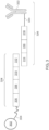
- FIG. 2 is a schematic diagram of an exemplary analyte capture agent 202 comprised of an analyte binding moiety 204 and a capture agent barcode domain 208.
- An analyte binding moiety 204 is a molecule capable of binding to an analyte 206 and interacting with a spatially-barcoded capture probe.
- the analyte binding moiety can bind to the analyte 206 with high affinity and/or with high specificity.
- the analyte capture agent can include a capture agent barcode domain 208, a nucleotide sequence (e.g., an oligonucleotide), which can hybridize to at least a portion or an entirety of a capture domain of a capture probe.
- the analyte binding moiety 204 can include a polypeptide and/or an aptamer (e.g., an oligonucleotide or peptide molecule that binds to a specific target analyte).
- the analyte binding moiety 204 can include an antibody or antibody fragment (e.g., an antigen-binding fragment).
- a spatial barcode with one or more neighboring cells, such that the spatial barcode identifies the one or more cells, and/or contents of the one or more cells, as associated with a particular spatial location.
- One method is to promote analytes or analyte proxies (e.g., intermediate agents) out of a cell and towards a spatially-barcoded array (e.g., including spatially-barcoded capture probes).
- Another method is to cleave spatially-barcoded capture probes from an array and promote the spatially-barcoded capture probes towards and/or into or onto the biological sample.
- capture probes may be configured to form ligation products with a template (e.g., a DNA or RNA template, such as an analyte or an intermediate agent, or portion thereof), thereby creating ligations products that serve as proxies for a template.
- a template e.g., a DNA or RNA template, such as an analyte or an intermediate agent, or portion thereof
- an "extended capture probe” refers to a capture probe having additional nucleotides added to the terminus (e.g., 3' or 5' end) of the capture probe thereby extending the overall length of the capture probe.
- an "extended 3' end” indicates additional nucleotides were added to the most 3' nucleotide of the capture probe to extend the length of the capture probe, for example, by polymerization reactions used to extend nucleic acid molecules including templated polymerization catalyzed by a polymerase (e.g., a DNA polymerase or a reverse transcriptase).
- a polymerase e.g., a DNA polymerase or a reverse transcriptase
- extending the capture probe includes adding to a 3' end of a capture probe a nucleic acid sequence that is complementary to a nucleic acid sequence of an analyte or intermediate agent specifically bound to the capture domain of the capture probe.
- the capture probe is extended using reverse transcription.
- the capture probe is extended using one or more DNA polymerases. The extended capture probes include the sequence of the capture probe and the sequence of the spatial barcode of the capture probe.
- extended capture probes are amplified (e.g., in bulk solution or on the array) to yield quantities that are sufficient for downstream analysis, e.g., via DNA sequencing.
- extended capture probes e.g., DNA molecules
- act as templates for an amplification reaction e.g., a polymerase chain reaction.
- Section (II)(a) of WO 2020/176788 and/or U.S. Patent Application Publication No. 2020/0277663 Analysis of captured analytes (and/or intermediate agents or portions thereof), for example, including sample removal, extension of capture probes, sequencing (e.g., of a cleaved extended capture probe and/or a cDNA molecule complementary to an extended capture probe), sequencing on the array (e.g., using, for example, in situ hybridization or in situ ligation approaches), temporal analysis, and/or proximity capture, is described in Section (II)(g) of WO 2020/176788 and/or U.S. Patent Application Publication No. 2020/0277663 . Some quality control measures are described in Section (II)(h) of WO 2020/176788 and/or U.S. Patent Application Publication No. 2020/0277663 .
- Exemplary methods for identifying spatial information of biological and/or medical importance can be found in U.S. Patent Application Publication No. 2021/0140982A1 , U.S. Patent Application No. 2021/0198741A1 , and/or U.S. Patent Application No. 2021/0199660 .
- Spatial information can provide information of biological importance.
- the methods and compositions described herein can allow for: identification of transcriptome and/or proteome expression profiles (e.g., in healthy and/or diseased tissue); identification of multiple analyte types in close proximity (e.g., nearest neighbor analysis); determination of up- and/or down-regulated genes and/or proteins in diseased tissue; characterization of tumor microenvironments; characterization of tumor immune responses; characterization of cells types and their co-localization in tissue; and identification of genetic variants within tissues (e.g., based on gene and/or protein expression profiles associated with specific disease or disorder biomarkers).
- a substrate functions as a support for direct or indirect attachment of capture probes to features of the array.
- a "feature” is an entity that acts as a support or repository for various molecular entities used in spatial analysis.
- some or all of the features in an array are functionalized for analyte capture.
- Exemplary substrates are described in Section (II)(c) of WO 2020/176788 and/or U.S. Patent Application Publication No. 2020/0277663 .
- Exemplary features and geometric attributes of an array can be found in Sections (II)(d)(i), (II)(d)(iii), and (II)(d)(iv) of WO 2020/176788 and/or U.S. Patent Application Publication No. 2020/0277663 .
- Capture can be achieved actively (e.g., using electrophoresis) or passively (e.g., using diffusion). Analyte capture is further described in Section (II)(e) of WO 2020/176788 and/or U.S. Patent Application Publication No. 2020/0277663 .
- spatial analysis can be performed by attaching and/or introducing a molecule (e.g., a peptide, a lipid, or a nucleic acid molecule) having a barcode (e.g., a spatial barcode) to a biological sample (e.g., to a cell in a biological sample).
- a plurality of molecules e.g., a plurality of nucleic acid molecules
- a plurality of barcodes e.g., a plurality of spatial barcodes
- a biological sample e.g., to a plurality of cells in a biological sample for use in spatial analysis.
- the biological sample after attaching and/or introducing a molecule having a barcode to a biological sample, the biological sample can be physically separated (e.g., dissociated) into single cells or cell groups for analysis.
- Some such methods of spatial analysis are described in Section (III) of WO 2020/176788 and/or U.S. Patent Application Publication No. 2020/0277663 .
- spatial analysis can be performed by detecting multiple oligonucleotides that hybridize to an analyte.
- spatial analysis can be performed using RNA-templated ligation (RTL).
- RTL RNA-templated ligation
- Methods of RTL have been described previously. See, e.g., Credle et al., Nucleic Acids Res. 2017 Aug 21; 45(14):e128 .
- RTL includes hybridization of two oligonucleotides to adjacent sequences on an analyte (e.g., an RNA molecule, such as an mRNA molecule).
- the oligonucleotides are DNA molecules.
- one of the oligonucleotides includes at least two ribonucleic acid bases at the 3' end and/or the other oligonucleotide includes a phosphorylated nucleotide at the 5' end.
- one of the two oligonucleotides includes a capture domain (e.g., a poly(A) sequence, a non-homopolymeric sequence).
- a ligase e.g., SplintR ligase
- the two oligonucleotides hybridize to sequences that are not adjacent to one another.
- hybridization of the two oligonucleotides creates a gap between the hybridized oligonucleotides.
- a polymerase e.g., a DNA polymerase
- the ligation product is released from the analyte.
- the ligation product is released using an endonuclease (e.g., RNAse H).
- the released ligation product can then be captured by capture probes (e.g., instead of direct capture of an analyte) on an array, optionally amplified, and sequenced, thus determining the location and optionally the abundance of the analyte in the biological sample.
- capture probes e.g., instead of direct capture of an analyte
- sequence information for a spatial barcode associated with an analyte is obtained, and the sequence information can be used to provide information about the spatial distribution of the analyte in the biological sample.
- Various methods can be used to obtain the spatial information.
- specific capture probes and the analytes they capture are associated with specific locations in an array of features on a substrate.
- specific spatial barcodes can be associated with specific array locations prior to array fabrication, and the sequences of the spatial barcodes can be stored (e.g., in a database) along with specific array location information, so that each spatial barcode uniquely maps to a particular array location.
- specific spatial barcodes can be deposited at predetermined locations in an array of features during fabrication such that at each location, only one type of spatial barcode is present so that spatial barcodes are uniquely associated with a single feature of the array.
- the arrays can be decoded using any of the methods described herein so that spatial barcodes are uniquely associated with array feature locations, and this mapping can be stored as described above.
- each array feature location represents a position relative to a coordinate reference point (e.g., an array location, a fiducial marker) for the array. Accordingly, each feature location has an "address" or location in the coordinate space of the array.
- Some exemplary spatial analysis workflows are described in the Exemplary Embodiments section of WO 2020/176788 and/or U.S. Patent Application Publication No. 2020/0277663 . See, for example, the Exemplary embodiment starting with "In some non-limiting examples of the workflows described herein, the sample can be immersed" of WO 2020/176788 and/or U.S. Patent Application Publication No. 2020/0277663 . See also, e.g., the Visium Spatial Gene Expression Reagent Kits User Guide (e.g., Rev C, dated June 2020 ), and/or the Visium Spatial Tissue Optimization Reagent Kits User Guide (e.g., Rev C, dated July 2020 ).
- the Visium Spatial Gene Expression Reagent Kits User Guide e.g., Rev C, dated June 2020
- the Visium Spatial Tissue Optimization Reagent Kits User Guide e.g., Rev C, dated July 2020 .
- spatial analysis can be performed using dedicated hardware and/or software, such as any of the systems described in Sections (II)(e)(ii) and/or (V) of WO 2020/176788 and/or U.S. Patent Application Publication No. 2020/0277663 , or any of one or more of the devices or methods described in Sections Control Slide for Imaging, Methods of Using Control Slides and Substrates for, Systems of Using Control Slides and Substrates for Imaging, and/or Sample and Array Alignment Devices and Methods, Informational labels of WO 2020/123320 .
- the systems can optionally include a control unit that includes one or more electronic processors, an input interface, an output interface (such as a display), and a storage unit (e.g., a solid state storage medium such as, but not limited to, a magnetic, optical, or other solid state, persistent, writeable and/or re-writeable storage medium).
- the control unit can optionally be connected to one or more remote devices via a network.
- the control unit (and components thereof) can generally perform any of the steps and functions described herein. Where the system is connected to a remote device, the remote device (or devices) can perform any of the steps or features described herein.
- the systems can optionally include one or more detectors (e.g., CCD, CMOS) used to capture images.
- the systems can optionally include software instructions encoded and/or implemented in one or more of tangible storage media and hardware components such as application specific integrated circuits.
- the software instructions when executed by a control unit (and in particular, an electronic processor) or an integrated circuit, can cause the control unit, integrated circuit, or other component executing the software instructions to perform any of the method steps or functions described herein.
- the systems described herein can detect (e.g., register an image) the biological sample on the array.
- Exemplary methods to detect the biological sample on an array are described in WO 2021/102003 and/or U.S. Patent Application No. US 2021/0150707 A1 .
- a map of analyte presence and/or level can be aligned to an image of a biological sample using one or more fiducial markers, e.g., objects placed in the field of view of an imaging system which appear in the image produced, as described in the Substrate Attributes Section, Control Slide for Imaging Section of WO 2020/123320 , WO 2021/102005 , and/or U.S. Patent Application No. US 2021/0158522 A1 .
- fiducial markers e.g., objects placed in the field of view of an imaging system which appear in the image produced, as described in the Substrate Attributes Section, Control Slide for Imaging Section of WO 2020/123320 , WO 2021/102005 , and/or U.S. Patent Application No. US 2021/0158522 A1 .
- Fiducial markers can be used as a point of reference or measurement scale for alignment (e.g., to align a sample and an array, to align two substrates, to determine a location of a sample or array on a substrate relative to a fiducial marker) and/or for quantitative measurements of sizes and/or distances.
- RNA-seq single cell RNA-seq
- mRNA messenger RNA
- multi-omics approaches that can provide a powerful complement to traditional methodologies, enabling a greater understanding of cellular heterogeneity and organization within biological samples.
- the combination of protein detection using analyte capture agents on a spatial array allows for the simultaneous examination of protein and gene expression from the same biological sample (e.g., tissue section).
- an array comprising capture probes e.g., any of the capture probes described herein
- the plurality of templated ligation probes include a pair of probes for a target nucleic acid (e.g., DNA, RNA).
- the probes are complementary to portions of the target nucleic acid, however, when both probes hybridize to the target nucleic acid a gap is present between the two probes.
- the gap is ligated, thereby generating a templated ligation product (e.g., DNA or RNA templated ligation product).
- one of the pair of probes includes a flanking sequence complementary to a capture domain of the array.
- the sequence complementary to the capture domain of the templated ligation product hybridizes to the capture domain of the capture probe.
- analyte capture agents as described herein, can also be contacted with the biological sample.
- the analyte capture agents are contacted with the biological sample before the biological sample is contacted with an array.
- the analyte capture agents are contacted with the biological sample after the biological sample is contacted with the array.
- the analyte binding moiety of the analyte capture agent interacts (e.g., binds) to an analyte (e.g., protein) in a biological sample.
- the analyte binding moiety is an antibody or antigen-binding fragment.
- Analyte capture agents can also include a conjugated oligonucleotide that can comprise one or more domains.
- the conjugated oligonucleotide can include an analyte binding moiety barcode and an analyte capture sequence.
- the analyte binding moiety barcode, or a complement thereof refers to (e.g., identifies) a barcode that is associated with or otherwise identifies the analyte binding moiety.
- the conjugated oligonucleotide can include an analyte capture sequence.
- the analyte capture sequence is capable of interacting with (e.g., hybridizing) to a capture domain of a capture probe on a substrate.
- the templated ligation probes are allowed to bind the target nucleic acid before the analyte capture agents are delivered to the biological sample. In some embodiments, the templated ligation probes can be ligated together before, concurrently, or after the analyte capture agents are delivered to the biological sample. In some embodiments, the analyte capture agents are delivered to the biological sample and the analyte binding moiety is allowed to bind the target analyte (e.g., protein) before the templated ligation probes are delivered. In some embodiments, the analyte capture agents are delivered to the biological sample and the analyte capture sequence is blocked (e.g., blocked by any of the methods described herein).
- target analyte e.g., protein
- the analyte capture sequence of the analyte capture agents is unblocked (e.g., unblocked by any of the methods described herein) before, concurrently, or after the templated ligation probes (e.g., RNA templated ligation probes) are delivered and/or before, concurrently, or after the templated ligation probes are ligated together.
- the templated ligation probes e.g., RNA templated ligation probes
- determining the spatial location of a nucleic acid and a protein from a biological sample including: a) providing a spatial array including a first and second plurality of capture probes where each plurality includes a spatial barcode and a capture domain, b) contacting the spatial array with a biological sample, c) contacting the biological sample with (i) a plurality of analyte capture agents, where an analyte capture agent includes an analyte binding moiety and an oligonucleotide including an analyte binding moiety barcode and an analyte capture sequence, where the analyte capture sequence includes a sequence complementary to a second plurality of capture domains, and (ii) a plurality of templated ligation probes, where one of the templated ligation probes includes a sequence complementary a first plurality of capture domains, d) binding the analyte binding moiety of the analyte capture agent to a target protein,
- Also provided herein are methods for determining the spatial location of a nucleic acid and a protein in a diseased biological sample including: a) providing a spatial array including a first and a second plurality of capture probes where each plurality includes a spatial barcode and a capture domain, b) contacting the diseased biological sample with the spatial array, c) contacting the diseased biological sample with: (i) a plurality of analyte capture agents, where an analyte capture agent includes an analyte binding moiety and an oligonucleotide including an analyte binding moiety barcode and an analyte capture sequence, where the analyte capture sequence includes a sequence complementary to a second plurality of capture domains, and (ii) a plurality of templated ligation probes, where one of the templated ligation probes includes a sequence complementary a first plurality of capture domains, d) binding the analyte binding moiety of the analyte capture
- the nucleic acid is RNA. In some embodiments, the RNA is mRNA. In some embodiments, the nucleic acid is DNA.
- the diseased biological sample is a cancerous biological sample.
- the cancerous biological sample is an ovarian cancer biological sample or a breast cancer biological sample.
- the breast cancer sample is triple positive breast cancer.
- the breast cancer is invasive ductal cell carcinoma breast cancer.
- the invasive ductal cell carcinoma is grade II invasive ductal carcinoma.
- the invasive ductal cell carcinoma is grade III invasive ductal carcinoma.
- the breast cancer is invasive lobular carcinoma.
- the cancerous biological sample is lung cancer.
- the cancerous biological sample is melanoma.
- the cancerous biological sample is colon cancer.
- the cancerous biological sample is glioblastoma.
- the cancerous biological sample is prostate cancer.
- the capture probes include unique molecular identifiers, functional sequences, or combinations thereof.
- the first plurality of capture domains are homopolymeric sequences. In some embodiments, the first plurality of capture domains comprise poly(T) sequences. In some embodiments, the first plurality of capture domains are defined non-homopolymeric sequences. In some embodiments, the first plurality of capture domains includes a degenerate sequence. In some embodiments, the first plurality of capture domains includes a fixed sequence. For example, the first plurality of capture domains can comprises one of SEQ ID NO: 1, SEQ ID NO: 2, SEQ ID NO: 3, SEQ ID NO: 4, SEQ ID NO: 5, SEQ ID NO: 6, SEQ ID NO: 7, SEQ ID NO: 8, SEQ ID NO: 9, SEQ ID NO: 10, or SEQ ID NO: 11.
- the second plurality of capture domains are homopolymeric sequences. In some embodiments, the second plurality of capture domains are defined non-homopolymeric sequences. In some embodiments, the second plurality of capture domains comprise poly(T) sequences. In some embodiments, the second plurality of capture domains includes a degenerate sequence. In some embodiments, the second plurality of capture domains includes a fixed sequence. For example, the second plurality of capture domains can comprise one of SEQ ID NO: 1, SEQ ID NO: 2, SEQ ID NO: 3, SEQ ID NO: 4, SEQ ID NO: 5, SEQ ID NO: 6, SEQ ID NO: 7, SEQ ID NO: 8, SEQ ID NO: 9, SEQ ID NO: 10, or SEQ ID NO: 11.
- the first plurality of capture domains and the second plurality of capture domains include the same sequence. In some embodiments, the first plurality of capture domains and the second plurality of capture domains include different sequences.
- the methods of the present disclosure can be used with any biological sample (e.g., any biological sample described herein).
- the biological sample is a tissue section.
- the biological sample is a tissue sample.
- the biological sample is a fresh-frozen biological sample.
- the biological sample is a fixed biological sample (e.g., formalin-fixed paraffin embedded (FFPE), paraformaldehyde, acetone, or methanol).
- the biological sample is an FFPE sample.
- the biological sample is an FFPE tissue section.
- the tissue sample is a tumor sample.
- the tissue section is a tumor tissue section.
- the tumor tissue section is a fixed tumor tissue section (e.g., a formal-fixed paraffin-embedded tumor tissue section).
- the tumor sample comprises one or more cancer tumors. Numerous types of cancer are known in the art.
- the tissue sample is derived from a biopsy sample. In some embodiments, the tissue sample is derived from a whole rodent embryo. In some embodiments, the tissue is selected from, but not limited to, brain tissue, breast tissue, colon tissue, heart tissue, lung tissue, spleen tissue, testes tissue, inflamed tonsil tissue, cervix tissue, and lymph node tissue.
- an FFPE sample is deparaffinized and decrosslinked prior to delivering a plurality of templated ligation probes (e.g., RNA templated ligation probes) and analyte capture agents.
- an FFPE biological sample is deparaffinized and decrosslinked before step (b).
- the paraffin-embedding material can be removed (e.g., deparaffinization) from the biological sample (e.g., tissue section) by incubating the biological sample in an appropriate solvent (e.g., xylene), followed by a series of rinses (e.g., ethanol of varying concentrations), and rehydration in water.
- the biological sample can be dried following deparaffinization.
- the biological sample after the step of drying the biological sample, the biological sample can be stained (e.g., H&E stain, any of the variety of stains described herein).
- the method includes staining the biological sample.
- the staining includes the use of hematoxylin and eosin.
- a biological sample can be stained using any number of biological stains, including but not limited to, acridine orange, Bismarck brown, carmine, coomassie blue, cresyl violet, DAPI, eosin, ethidium bromide, acid fuchsine, hematoxylin, Hoechst stains, iodine, methyl green, methylene blue, neutral red, Nile blue, Nile red, osmium tetroxide, propidium iodide, rhodamine, or safranin.
- the biological sample can be stained using known staining techniques, including Can-Grunwald, Giemsa, hematoxylin and eosin (H&E), Jenner's, Leishman, Masson's trichrome, Papanicolaou, Romanowsky, silver, Sudan, Wright's, and/or Periodic Acid Schiff (PAS) staining techniques.
- PAS staining is typically performed after formalin or acetone fixation.
- the staining includes the use of a detectable label selected from the group consisting of a radioisotope, a fluorophore, a chemiluminescent compound, a bioluminescent compound, or a combination thereof.
- the biological sample is imaged after staining the biological sample. In some embodiments, the biological sample is imaged prior to staining the biological sample. In some embodiments, the biological sample is visualized or imaged using bright field microscopy. In some embodiments, the biological sample is visualized or imaged using fluorescence microscopy. Additional methods of visualization and imaging are known in the art. Non-limiting examples of visualization and imaging include expansion microscopy, bright field microscopy, dark field microscopy, phase contrast microscopy, electron microscopy, fluorescence microscopy, reflection microscopy, interference microscopy and confocal microscopy. In some embodiments, the sample is stained and imaged prior to adding the first and/or second primer to the biological sample on the array.
- the fixed biological sample can be further processed.
- fixed biological samples can be treated to remove crosslinks (e.g., formaldehyde-induced crosslinks (e.g., decrosslinking)).
- decrosslinking the crosslinks (e.g., formaldehyde-induced crosslinks) in the fixed (e.g., FFPE, PFA) biological sample can include treating the sample with heat.
- decrosslinking the formaldehyde-induced crosslinks can include performing a chemical reaction. In some embodiments, decrosslinking the formaldehyde-induced crosslinks, can include treating the sample with a permeabilization reagent. In some embodiments, decrosslinking the formaldehyde-induced crosslinks can include heat, a chemical reaction, and/or permeabilization reagents.
- decrosslinking crosslinks can be performed in the presence of a buffer.
- the buffer is Tris-EDTA (TE) buffer (e.g., TE buffer for FFPE biological samples).
- the buffer is citrate buffer (e.g., citrate buffer for FFPE biological samples).
- the buffer is Tris-HCl buffer (e.g., Tris-HCl buffer for PFA fixed biological samples).
- the buffer e.g., TE buffer, Tris-HCl buffer
- the biological sample is permeabilized (e.g., permeabilized by any of the methods described herein). In some embodiments, the permeabilization is an enzymatic permeabilization. In some embodiments, the permeabilization is a chemical permeabilization. In some embodiments, the biological sample is permeabilized before delivering the RNA templated ligation probes and analyte capture agents to the biological sample. In some embodiments, the biological sample is permeabilized at the same time as the RNA templated ligation probes and analyte capture agents are delivered to the biological sample. In some embodiments, the biological sample is permeabilized after the RNA templated ligation probes and analyte capture agents are delivered to the biological sample. In some embodiments, hybridizing the RNA templated ligation products to the second capture domains and the analyte capture sequences of the bound analyte capture agents to the first capture domains further comprises permeabilizing the biological sample.
- the biological sample is permeabilized from about 30 to about 120 minutes, from about 40 to about 110 minutes, from about 50 to about 100 minutes, from about 60 to about 90 minutes, or from about 70 to 80 minutes. In some embodiments, the biological samples is permeabilized about 30, about 35, about 40, about 45, about 50, about 55, about 60, about 65, about 70, about 75, about 80, about 90, about 95, about 100, about 105, about 110, about 115, or about 130 minutes.
- the permeabilization buffer comprises urea.
- the urea is at a concentration of about 0.5M to 3.0M. In some embodiments, the concentration of the urea is about 0.5, 1.0, 1.5, 2.0, 2.5, or about 3.0M.
- the permeabilization buffer includes a detergent. In some embodiments, the detergent is sarkosyl. In some embodiments, the sarkosyl is present at about 2% to about 10% (v/v). In some embodiments, the sarkosyl is present at about 3%, 4%, 5%, 6%, 7%, 8%, or 9% (v/v).
- the permeabilization buffer comprises polyethylene glycol (PEG).
- the PEG is from about PEG 2K to about PEG 16K. In some embodiments, the PEG is PEG 2K, 3K, 4K, 5K, 6K, 7K, 8K, 9K, 10K, 11K, 12K, 13K, 14K, 15K, or 16K. In some embodiments, the PEG is present at a concentration from about 2% to 25%, from about 4% to about 23%, from about 6% to about 21%, or from about 8% to about 20% (v/v).
- the method includes a step of permeabilizing the biological sample (e.g., a tissue section).
- the biological sample can be permeabilized to facilitate transfer of the extended products to the capture probes on the array.
- the permeabilizing includes the use of an organic solvent (e.g., acetone, ethanol, and methanol), a detergent (e.g., saponin, Triton X-100 TM , Tween-20 TM , or sodium dodecyl sulfate (SDS)), and an enzyme (an endopeptidase, an exopeptidase, a protease), or combinations thereof.
- an organic solvent e.g., acetone, ethanol, and methanol
- a detergent e.g., saponin, Triton X-100 TM , Tween-20 TM , or sodium dodecyl sulfate (SDS)
- an enzyme an endopeptidase, an exopeptidase,
- the permeabilizing includes the use of an endopeptidase, a protease, SDS, polyethylene glycol tert-octylphenyl ether, polysorbate 80, and polysorbate 20, N-lauroylsarcosine sodium salt solution, saponin, Triton X-100 TM , Tween-20 TM , or combinations thereof.
- the endopeptidase is pepsin.
- the endopeptidase is Proteinase K. Additional methods for sample permeabilization are described, for example, in Jamur et al., Method Mol. Biol. 588:63-66, 2010 .
- antibody staining includes the use of an antibody staining buffer.
- the antibody staining buffer e.g., a PBS-based buffer
- the antibody staining buffer includes a detergent (e.g., Tween-20, SDS, sarkosyl).
- the antibody staining buffer includes a serum, such as for example, a goat serum.
- the goat serum is from about 1% to about 10% (v/v), from about 2% to about 9% (v/v), from about 3% to about 8% (v/v), or about 4% to about 7% (v/v).
- the antibody staining buffer includes dextran sulfate.
- the dextran sulfate is at a concentration of about 1 mg/ml to about 20 mg/ml, from about 5 mg/ml to about 15 mg/ml, or from about 8 mg/ml to about 12 mg/ml.
- the methods provided herein can also utilize blocking probes to block the non-specific binding (e.g., hybridization) of the analyte capture sequence and the capture domain of a capture probe on an array.
- the biological sample is contacted with a plurality of analyte capture agents, where an analyte capture agent includes an analyte capture sequence that is reversibly blocked with a blocking probe.
- the analyte capture sequence is reversibly blocked with more than one blocking probe (e.g., 2, 3, 4, or more blocking probes).
- the analyte capture agent is blocked prior to binding the target analyte (e.g., a target protein).
- the oligonucleotide of the analyte capture agent (e.g., analyte capture sequence) is blocked by a blocking probe.
- blocking probes are hybridized to the analyte capture sequence of the analyte capture agents before introducing the analyte capture agents to a biological sample.
- blocking probes are hybridized to the analyte capture sequence of the analyte capture agents after introducing the analyte capture agents to the biological sample.
- the capture domain can also be blocked to prevent non-specific binding, and/or to control the time of binding, between the analyte capture sequence and the capture domain.
- the blocking probes can be alternatively or additionally introduced during staining of the biological sample.
- the analyte capture sequence is blocked prior to binding to the capture domain, where the blocking probe includes a sequence complementary or substantially complementary to the analyte capture sequence.
- the analyte capture sequence is blocked with one blocking probe. In some embodiments, the analyte capture sequence is blocked with two blocking probes. In some embodiments, the analyte capture sequence is blocked with more than two blocking probes (e.g., 3, 4, 5, or more blocking probes). In some embodiments, a blocking probe is used to block the free 3' end of the analyte capture sequence. In some embodiments, a blocking probe is used to block the 5' end of the analyte capture sequence. In some embodiments, two blocking probes are used to block both 5' and 3' ends of the analyte capture sequence. In some embodiments, both the analyte capture sequence and the capture probe domain are blocked.
- a blocking probe is used to block the free 3' end of the analyte capture sequence. In some embodiments, a blocking probe is used to block the 5' end of the analyte capture sequence. In some embodiments, two blocking probes are used to block both 5' and 3' ends of the
- the blocking probes can differ in length and/or complexity.
- the blocking probe can include a nucleotide sequence of about 8 to about 24 nucleotides in length (e.g., about 8 to about 22, about 8 to about 20, about 8 to about 18, about 8 to about 16, about 8 to about 14, about 8 to about 12, about 8 to about 10, about 10 to about 24, about 10 to about 22, about 10 to about 20, about 10 to about 18, about 10 to about 16, about 10 to about 14, about 10 to about 12, about 12 to about 24, about 12 to about 22, about 12 to about 20, about 12 to about 18, about 12 to about 16, about 12 to about 14, about 14 to about 24, about 14 to about 22, about 14 to about 20, about 14 to about 18, about 14 to about 16, about 16 to about 24, about 16 to about 22, about 16 to about 20, about 16 to about 18, about 18 to about 24, about 18 to about 22, about 18 to about 20, about 20 to about 24, about 20 to about 22, or about 22 to about 24 nucleotides in length).
- the blocking probe is removed prior to hybridizing the analyte capture sequence of the oligonucleotide of the analyte capture sequence to the first capture domain. For example, once the blocking probe is released from the analyte capture sequence, the analyte capture sequence can bind to the first capture domain on the array. In some embodiments, blocking the analyte capture sequence reduces non-specific background staining. In some embodiments, blocking the analyte capture sequence allows for control over when to allow the binding of the analyte capture sequence to the capture domain of a capture probe during a spatial workflow, thereby controlling the time of capture of the analyte capture sequence on the array.
- the blocking probes are reversibly bound, such that the blocking probes can be removed from the analyte capture sequence during or after the time that analyte capture agents are in contact with the biological sample.
- the blocking probe can be removed with RNAse treatment (e.g., RNAse H treatment).
- the blocking probes are removed by increasing the temperature (e.g., heating) the biological sample.
- the blocking probes are removed enzymatically (e.g., cleaved).
- the blocking probes are removed by a USER enzyme.
- the blocking probes are removed by an endonuclease.
- the endonuclease is endonuclease IV.
- the endonuclease is endonuclease V.
- the determining in step (g) includes a) extending the captured ligation products and the captured oligonucleotides of the analyte capture agents, wherein the extension products comprise the spatial barcode or a complement thereof, b) releasing the extension products, or complements thereof, from the spatial array, c) producing a library from the released extension products or complements thereof, and d) sequencing the library.
- extension e.g., extension of captured nucleic acid ligation products and the captured oligonucleotides of the analyte capture agents and/or extension of the plurality of captures probes
- a polymerase e.g., any suitable polymerase, e.g., T4 polymerase.
- the released extension products can be prepared for downstream applications, such as generation of a sequencing library and next-generation sequencing.
- Producing sequencing libraries are known in the art.
- the released extension products can be purified and collected for downstream amplification steps.
- the released extension products can be amplified using PCR, where primer binding sites flank the spatial barcode and ligation product or analyte binding moiety barcode, or complements thereof, generating a library associated with a particular spatial barcode.
- the library preparation can be quantitated and/or quality controlled to verify the success of the library preparation steps.
- the library amplicons are sequenced and analyzed to decode spatial information and the ligation product or analyte binding moiety barcode, or complements thereof.
- the amplicons can then be enzymatically fragmented and/or size-selected in order to provide for desired amplicon size.
- sequences can be added to the amplicons thereby allowing for capture of the library preparation on a sequencing flowcell (e.g., on Illumina sequencing instruments).
- i7 and i5 can index sequences be added as sample indexes if multiple libraries are to be pooled and sequenced together.
- Read 1 and Read 2 sequences can be added to the library preparation for sequencing purposes.
- the aforementioned sequences can be added to a library preparation sample, for example, via End Repair, A-tailing, Adaptor Ligation, and/or PCR.
- the cDNA fragments can then be sequenced using, for example, paired-end sequencing using TruSeq Read 1 and TruSeq Read 2 as sequencing primer sites, although other methods are known in the art.
- the determining in step (g) can include a pre-amplification step.
- a complementary strand to the extended RNA ligation products and/or the extension product of the captured oligonucleotides of the analyte capture agents the step can be generated and further include a pre-amplification step of the extension products or complements thereof (e.g., extended products) prior to library production (e.g., RTL library production; captured oligonucleotide of the analyte capture agent production).
- library production e.g., RTL library production; captured oligonucleotide of the analyte capture agent production.
- spatial arrays including spatial arrays described in the methods herein, that include a dilution series of protein standards directly on the array.
- protein quantification with antibodies can be difficult due to the varying affinity antibodies have for their protein targets.
- standard curves with the protein of interest e.g., similar to an ELISA assay
- a protein standard is spotted on the array (e.g., on top of the features of the array).
- more than one protein standard is spotted on the array (e.g., 2, 3, 4, 5, 6, 7, 8, 9, 10, 11, 12, 13, 14, 15, 16, 17, 18, 19, 20, or more protein standards are spotted on the array).
- Readout from the internal protein standard series allows for quantification of proteins of interest in parallel from the biological sample (e.g., a tissue section) directly on the array which can lead to increased accuracy when determining protein concentration.
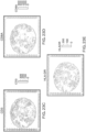
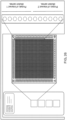
- FIG. 26 shows an exemplary substrate with an exemplary configuration that includes a dilution series for two different proteins spotted on the margins of a spatial array.
- FIG. 26 shows a dilution series for two proteins of interest, however, as described herein more than two protein dilution series can be spotted on the spatial array.
- compositions such as spatial arrays including a) a plurality of capture probes including spatial barcodes and a first plurality of capture domains hybridized to a plurality of templated ligation products, and b) a plurality of capture probes comprising spatial barcodes and a second plurality of capture domains hybridized to a plurality of oligonucleotides from analyte capture agents, wherein the oligonucleotides comprise an analyte capture sequence and an analyte binding moiety barcode.
- the analyte capture sequences of the oligonucleotides are hybridized to the second plurality of capture domains.
- the capture probes include cleavage domains, unique molecular identifiers, functional sequences, or combinations thereof.
- the first plurality of capture domains are homopolymeric sequences.
- the first plurality of capture domains comprise poly(T) sequences.
- the first plurality of capture domains are defined non-homopolymeric sequences.
- the first plurality of capture domains comprise a degenerate sequence.
- the first plurality of capture domains comprise a fixed sequence.
- the second plurality of capture domains are homopolymeric sequences. In some compositions, the second plurality of capture domains are defined non-homopolymeric sequences. In some compositions, the second plurality of capture domains comprise poly(T) sequences. In some compositions the second plurality of capture domains comprise a degenerate sequence. In some compositions, the second plurality of capture domains comprise a fixed sequence. For example, the second plurality of capture domains can comprise one of SEQ ID NO: 1, SEQ ID NO: 2, SEQ ID NO: 3, SEQ ID NO: 4, SEQ ID NO: 5, SEQ ID NO: 6, SEQ ID NO: 7, SEQ ID NO: 8, SEQ ID NO: 9, SEQ ID NO: 10, or SEQ ID NO: 11.
- the spatial array comprises one or more protein dilution series.
- kits including a) a spatial array comprising a plurality of capture probes, where the capture probes include spatial barcodes and wherein the plurality of capture probes comprise a first plurality of first capture domains and a second plurality of second capture domains, b) one or more analyte capture agents, c) one or more RNA templated ligation probe pairs, and d) one or more enzymes and buffers for practicing any of the methods described herein.
- one or more enzymes includes polymerases, RNases, DNases, proteases, lipases, or combinations thereof.
- the analyte capture agent includes an antibody as the analyte binding moiety and the analyte capture sequence that comprises a barcode sequence that identifies the antibody as well as the capture sequence that is complementary to the associated capture probe capture domain on the array.
- Templated ligation probes were allowed to hybridize to their target mRNAs and antibody-oligonucleotide conjugates were incubated with the samples wherein the antibodies were allowed to bind to their protein targets (as described in PCT/US2020/66720 ). Briefly, FFPE human lymph nodes tissues were sectioned, mounted on spatial array slides and deparaffinized using a series of xylene and ethanol washes prior to brightfield imaging. Tissues were washed and decrosslinked by incubating the tissues in TE (pH 9.0) buffer for 1 hr at 70°C. After decrosslinking the tissues, the targeted templated ligation probes were added to the tissues and probe hybridization ran overnight at 50°C.
- the probes were ligated together at 37°C for 1 hr.
- the analyte capture agents were added to the tissues, which were incubated in an antibody staining buffer with the tissues overnight at room temperature. The tissue samples were washed four times with the antibody staining buffer without antibodies.
- Tissues were permeabilized and the ligated probe products, or ligation products, were allowed to migrate for hybridization to the capture domains of the capture probes on the spatial array surface.
- the oligonucleotides of the analyte capture agents also migrated in parallel and were captured via their capture sequences that hybridized to capture probe capture domains on the spatial array that are complementary to the capture sequences.
- the ligation products representing the target mRNAs and the oligonucleotides of the analyte capture agents representing the binding of the antibodies to the targeted proteins were concurrently captured on the array surface.
- the tissues were incubated with RNAse H and an associated buffer for 30 min at 37°C, tissues permeabilized using a protease for an additional 40 min., followed by washing to remove the enzymes from the tissues.
- the captured ligation products and the analyte binding agent oligonucleotides were extended to create extended products of the captured molecules including the spatial barcode or a complement thereof, the analyte binding moiety barcode if present and other functional sequences from the capture probe.
- Library preparations were made, the libraries sequenced on an Illumina sequencing instrument, and spatial locations determined using Space Ranger and Loupe Browser (10X Genomics).
- the antibody sequences e.g., the complement of the captured oligonucleotide from the analyte binding agents
- sequences relating to the analyte binding moiety barcode were used to determine abundance and location of the labeled protein by the analyte binding agents. Spatial expression patterns were determined using SpaceRanger data analysis software and Loupe browser visualization software (10X Genomics).
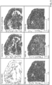
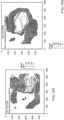
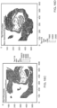
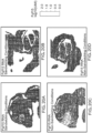
- FIGs. 4A-B demonstrates the results of an experiment where analyte capture agents were combined to determine whether multiple targets could be identified concurrently in a tissue.
- eight different antibodies were conjugated to oligonucleotides comprising analyte binding moiety barcodes and capture sequences targeting eight proteins: NCAM1, Ki67, CD8A, PDL1, CD20, CD11B, CD45RA and CD66B.
- FIG. 4A (RNA) shows the spatial gene expression clustering of the associated protein in the lymph node tissue sample
- FIG. 4B protein shows the spatial protein expression clustering of the eight targets. This experiment demonstrates that it is feasible to multiplex different analyte capture agents to concurrently identify the spatial gene and protein expression patterns of multiplex targets in a tissue sample.
- Additional experimental results from the multiplexed experiment shown in FIG. 4A included targeting CD20 and CD20 mRNA when only one protein is targeted with an analyte capture agent. These results (data not shown) show a lymph node tissue sample where an antibody directed to CD20 was conjugated to an oligonucleotide for subsequent capture on a spatial array. mRNA from the tissue section was successfully captured and extended thereby providing CD20 spatial gene expression analysis (data not shown).
- spatially presented protein array demonstrates that the CD20 antibody of the analyte capture agent was able to bind to the CD20 protein followed by oligonucleotide capture on the spatial array, extension and spatial determination of the location of the CD20 protein via the analyte binding moiety barcode (data not shown).
- the methods demonstrate the utility of the analyte capture agents to target a protein of interest and simultaneously detect spatial gene expression and protein expression in a tissue sample.
- FFPE human tonsil tissues were sectioned, mounted on spatial array slides and deparaffinized using a series of xylene and ethanol washes prior to H&E staining and brightfield imaging. Tissues were washed and decrosslinked by incubating the tissues in citrate buffer (pH 6.0) for 1 hour at 90°C. After decrosslinking the tissues, the targeted ligation probes were applied to the tissues and probe hybridization ran overnight at 50°C. Post hybridization, the probes were ligated together at 37°C for 1 hour to generate ligation products.
- analyte capture agents were applied to the tissues, which were incubated in an antibody staining buffer (PBS-based buffer, 5% goat serum, salmon sperm DNA and dextran sulfate) with the tissues overnight at 4°C. The tissue samples were washed four times with antibody-free antibody staining buffer.
- an antibody staining buffer PBS-based buffer, 5% goat serum, salmon sperm DNA and dextran sulfate
- Tissues were permeabilized and the ligation products were allowed to migrate for capture by hybridization to the capture domains of the capture probes on the spatial array surface.
- the oligonucleotides of the analyte capture agents complementary to the alternative capture sequences of the second set of capture probes on the array were also captured by hybridization.
- both ligation products representing the mRNA of the targeted protein and the oligonucleotide of the analyte capture agent representing the binding of the antibody to the targeted protein were concurrently captured on the array surface.
- the tissues were incubated with RNase H and an associated buffer, and polyethylene glycol (PEG) for 30 minutes at 37°C.
- Tissues were permeabilized using a permeabilization buffer comprising a protease (e.g., Proteinase K), PEG, 1M urea, for an additional 60 minutes, followed by washing to remove the enzymes from the tissues.
- the captured ligation products and the analyte binding agent oligonucleotides were extended to include the spatial barcode or a complement thereof, the analyte binding moiety barcode or a complement thereof if present and other functional sequences from the capture probe. Additionally, said products were pre-amplified prior to library preparation.
- Library preparations were made from the extended products, the libraries sequenced on an Illumina sequencing instrument, and spatial locations determined and visualized.
- the antibody sequences e.g., the complement of the captured oligonucleotide from the analyte binding agents
- Truseq_pR1 and Truseq_pR2 were amplified with Truseq_pR1 and Truseq_pR2.
- sequences relating to the analyte binding moiety barcode were used to determine abundance and location of the labeled protein by the analyte binding agents.
- Spatial expression patterns were determined using SpaceRanger data analysis software and Loupe browser visualization software (10X Genomics).
- FFPE tonsil tissue sections exemplary spatial gene and protein expression for a 20-plex antibody-oligonucleotide conjugate targeting proteins of interest and RNA templated probes targeting 18,000 mRNA targets was performed.
- the FFPE human tonsil tissue section was H&E stained which identified where follicles containing maturing immune cells and the epithelial layer can be seen (data not shown).
- Representative gene expression cluster data and representative protein expression cluster data align with the macroscopic structures visible in the H&E image shown (data not shown).
- the data demonstrate the feasibility of multiplexing different analyte capture agents to concurrently identify the spatial gene and protein expression patterns of multiplex targets in a tissue sample.
- FIG. 6 shows an exemplary H&E stained FFPE tonsil tissue section and spatial and gene expression data (top).
- FIG. 6 also shows an expanded view of the H&E stained FFPE tonsil tissue section and clustering of gene expression and protein expression of CD8a and CD4 superimposed on the H&E FFPE tonsil tissue section (bottom). Visualization of the CD8a and CD4 markers coincide with the follicles shown in the H&E staining where CD8a localizes to the edge of the follicles and CD4 localizes outside the follicles.
- FIG. 7 shows a panel of 18 of the 21 targeted proteins including specifically genes: Ki67, NCAM1, BCL, CD138, CD21, CD268, CD11b, LAG3, CD4, CD20, PD-L1, CD45RA, CD68, PanCK, CD66b, PCNA, CD235a/b and CD79a.
- the top row shows protein expression and the bottom panel shows RNA expression.
- FIG. 8 shows expanded views of exemplary clustering data for FFPE human tonsil tissue using a 12-plex antibody-oligonucleotide conjugate test panel of nine genes shown in FIG. 7 , including specifically: CD4, PD-L1, CD20, CD11b, BCL-2, CD21, CD791, CD45RA, PCNA, Ki67, PanCK, and CD235a/b.
- FFPE human triple positive breast cancer tissues were sectioned, mounted on spatial array slides and deparaffinized using a series of xylene and ethanol washes prior to drying at room temperature. Next, the slides were heated at 37°C for 15 minutes, followed by a series of ethanol washes (100%, 96%, 96%, and 70% ethanol). Next, the tissues were H&E stained and brightfield imaged. Alternatively, tissues can be stained (e.g., immunofluorescence stained) instead of H&E staining.
- Tissues were washed and decrosslinked by incubating the tissues in Tris-EDTA (TE) buffer (pH 9.0) for 1 hour at 95°C followed by a series of washes with 0.1 N HCl. After decrosslinking the tissues, the targeted ligation probes were added to the tissues and probe hybridization ran overnight at 50°C. The tissues were washed in a post-hybridization buffer including 3x SSC, Baker's yeast tRNA, and nuclease free water and followed by a 2x SSC buffer wash. Post-hybridization, the probes were ligated together at 37°C for 1 hour.
- TE Tris-EDTA
- the tissues were incubated in an antibody blocking buffer (PBS-based buffer (pH 7.4), goat serum, salmon sperm DNA, Tween-20, an RNase inhibitor, and dextran sulfate) at room temperature for 60 minutes.
- PBS-based buffer pH 7.4
- goat serum serum
- salmon sperm DNA Tween-20
- RNase inhibitor an RNase inhibitor
- dextran sulfate dextran sulfate
- the blocking buffer was removed from the tissues and the tissues were incubated overnight at 4°C with the analyte capture agents in an antibody staining mixture (PBS-based buffer (pH 7.4), 5% goat serum, 0.1 ⁇ g/ ⁇ L salmon sperm DNA, 0.1% Tween-20, 1 U/ ⁇ L RNase inhibitor, blocking oligonucleotides, analyte capture agents (e.g., antibodies with a conjugated oligonucleotide) and 10 mg/mL dextran sulfate)).
- the tissue samples were washed several times with antibody staining buffer without antibodies.
- Tissues were permeabilized and the ligated probes were released for capture by hybridization to the capture domains of the capture probes on the spatial array surface.
- the oligonucleotides of the analyte capture agents complementary to the alternative capture sequences of the second set of capture probes on the array were also captured by hybridization.
- both ligation products representing the target mRNA and the oligonucleotide of the analyte capture agent representing the binding of the antibody to the targeted protein were concurrently captured on the array surface.
- the tissues were incubated with an RNase (e.g., RNase H), an associated buffer, and polyethylene glycol (PEG) for 30 minutes at 37°C.
- RNase e.g., RNase H
- PEG polyethylene glycol
- Tissues were permeabilized using a permeabilization buffer comprising a protease (e.g., Proteinase K), PEG, 3M urea, for an additional 60 minutes, followed by washing to remove the enzymes from the tissues. After permeabilization the tissues were washed in 2x SSC several times.
- a protease e.g., Proteinase K
- PEG polyglycerol
- 3M urea urea
- the captured ligation products and the analyte binding agent oligonucleotides were extended to create extended products of the captured molecules including the complement of the spatial barcode, the analyte binding moiety barcode if present and other functional sequences from the capture probe. Additionally, said products were pre-amplified prior to library preparation.
- Library preparations were made from the extended products, sequenced on an Illumina sequencing instrument, and spatial locations were determined and visualized.
- the antibody sequences e.g., the complement of the captured oligonucleotide from the analyte binding agents
- Truseq_pR1 and Truseq_pR2 were amplified with Truseq_pR1 and Truseq_pR2.
- sequences relating to the analyte binding moiety barcode were used to determine abundance and location of the labeled protein by the analyte binding agents.
- Spatial expression patterns were determined using SpaceRanger data analysis software and Loupe browser visualization software (10X Genomics).
- FIGs. 9A-C demonstrate the results of an experiment where analyte capture agents were combined to determine whether targets could be identified concurrently in grade II FFPE triple positive invasive ductal carcinoma breast cancer tissue.
- 11 different antibodies were conjugated to oligonucleotides comprising analyte binding moiety barcodes and capture sequences (e.g., analyte capture sequences) targeting eleven proteins: Her2, EpCAM, PanCK, N-Cadherin, PCNA, AlphaSMA, Vimentin, CD8a, CD4, CD68, CD20, and HLA-DR.
- FIG. 9B shows the spatial gene expression clustering
- FIG. 9C protein shows clustering of the associated protein in the FFPE triple positive breast cancer tissue sample.
- the data demonstrate the feasibility of multiplexing different analyte capture agents to concurrently identify the spatial gene and protein expression clusters patterns of multiplex targets in triple positive breast cancer tissue samples.
- a panel of 11 targeted proteins including specifically genes: Her2, EpCAM, PanCK, N-Cadherin, PCNA, AlphaSMA, Vimentin, CD8a, CD4, CD68, CD20, and HLA-DR was generated where for each target RNA expression and protein expression was generated (data not shown).
- Exemplary spatial gene and protein expression for KRT10, KRT18, and PanCK Ab was also generated and unbiased clustering of gene and protein expression superimposed on the H&E image demonstrate similar patterns (data not shown).
- An expanded view of Her2 and Vimentin RNA expression and protein expression is shown in FIG. 10 . Adjacent sections were immunofluorescently stained for HER2 (top) and Vimentin (bottom) demonstrating strong signal within the dashed box region of the biopsy sample.
- RNA and protein expression for Her2, EpCAM, PanCK, N-Cadherin, PCNA, AlphaSMA, Vimentin, CD8a, CD4, CD68, CD20, and HLA-DR also overlap (data not shown) demonstrating the utility of the analyte capture agents to target a protein of interest and simultaneously detect spatial gene expression in triple positive breast cancer tissue samples.
- FIGs. 11A-C show H&E-stained human invasive ductal carcinoma tissue ( FIG. 11A).
- FIGs. 11B and 11C show gene expression clustering and protein expression clustering, respectively, superimposed on the H&E image with similar patterns.
- FIG. 12 shows additional examination of different biomarkers: CD11b, PD-L1, and alpha-SMA exhibit regional variation within the tissue sample. These protein biomarkers were used to define the region of interest (e.g., shown as region 1 and region 2). The identified regions of interest specific gene expression patterns were identified with the top 50 highly expressed genes. The data demonstrate the feasibility of multiplexing different analyte capture agents to concurrently identify the spatial gene and protein expression cluster patterns of multiplex targets in invasive ductal carcinoma tissue samples.
- FIG. 13 shows exemplary spatial gene and protein expression for Acta2 (top) and Epcam (bottom). Unbiased clustering of gene and protein expression superimposed on an H&E image of the same tissue sample demonstrate similar patterns. Plots showing the correlation between Acta2 gene expression and Alpha-SMA protein expression (encoded by the Acta2 gene) and Epcam gene expression and EPCAM protein expression (encoded by the EPCAM gene) were also generated (data not shown). A similar experiment was performed as described in FIG. 13 on a different FFPE invasive ductal carcinoma breast cancer tissue and the exemplary spatial gene and protein expression for Acta2 and separately HLA-DRA (gene) and HLA-DR (protein) correlated with one another (data not shown).
- HLA-DRA gene
- HLA-DR protein
- Example 4 Spatial proteomics and gene expression on FFPE mouse spleen, brain, head, and torso sections in a mouse embryo sample
- FFPE mouse spleen samples FFPE mouse samples, FFPE mouse embryo torso samples, FFPE mouse embryo head samples were placed onto standard slides (for sandwich conditions) or spatial expression (GEx) slides (as non-sandwich conditions).
- GEx slides include an array of spatially barcoded capture probes. Briefly, tissues were sectioned and mounted on slides and dried overnight in a desiccator. The following day, the tissues were heated to 60°C, followed by deparaffinization and rehydration. Tissues were H&E stained and bright-field imaged. Tissues were destained using HCl and decrosslinked for 1 hour in citrate buffer (pH 6.0) at 95°C.
- tissues were incubated overnight with whole mouse transcriptome (RNA templated ligation) probe sets at 50°C. The following day, tissues were washed to remove un-hybridized probes, then treated with ligase to ligate together the RTL probes. After another wash step, the tissues were blocked with antibody blocking buffer. Tissues were incubated overnight with a library of conjugated antibodies (e.g., a library comprising a plurality of analyte capture agents, each comprising an antigen specific antibody conjugated to an oligonucleotide). The following day, tissues were subjected to sandwiching or non-sandwich conditions as follows.
- a library of conjugated antibodies e.g., a library comprising a plurality of analyte capture agents, each comprising an antigen specific antibody conjugated to an oligonucleotide.
- tissues placed on standard slides for the sandwiching conditions were washed with PBS-T, subjected to an eosin stain, and washed with SSC.
- the tissues were subjected to sandwiching conditions. Briefly, the tissue slides were mounted in a sandwiching instrument along with a GEx slide and a reagent solution including an RNAse and Proteinase K. Upon closure in the instrument, the tissue sections were permeabilized for 30 min. allowing the ligation products and the oligonucleotides from the analyte capture agents to migrate to the GEx slide for capture by the capture probes. Following permeabilization and capture, the GEx slides were removed from the instrument.
- Tissues placed on GEx slides for non-sandwiching conditions were washed with PBS-T and SSC.
- the tissues were subjected to a 30 min probe release step with an RNase, followed by permeabilization with a permeabilization buffer including Proteinase K. Accordingly, the ligation products and analyte capture agents were captured by the capture probes on the GEx slide.
- GEx slides were washed twice with 2x SSC, and subjected to probe extension, denaturation, and pre-amplification followed by amplification and sequencing of the templated ligation and analyte capture agent libraries.
- FIG. 14A sandwiching conditions
- FIG. 14B non-sandwiching conditions
- tyrosine phosphatase receptor type C Ptprc; e.g., Ensembl: ENSMUSG00000026395
- mRNA expression was detected in a spleen tissue sample.
- gene product e.g., protein
- FIG. 14D gene product CD45R was also detected both in sandwiching conditions
- CD45R is the protein name of Ptprc, and it was determined whether there was overlap of mRNA expression of Ptprc and protein expression of CD45R. As shown in FIGs. 14A-14D , both sandwiching (77% correlation) and non-sandwiching conditions (76% correlation), respectively, demonstrates overlap of transcript and protein, indicating that transcript (i.e., mRNA) and protein detection (1) was identified in similar areas of the tissues and (2) was comparable across the sandwich and non-sandwich conditions in mouse spleen samples.
- transcript i.e., mRNA
- the quality, sensitivity, and detection under each condition were evaluated globally in mouse brain samples. As shown in Table 2, the quality, sensitivity, and detection of globally-detected transcripts (i.e., mRNA) and proteins were comparable across the sandwich and non-sandwich conditions in mouse brain samples. Table 2.
- FIG. 15A sandwiching conditions
- FIG. 15B non-sandwiching conditions
- tyrosine hydroxylase Th; e.g., Ensembl: ENSMUSG00000000214
- mRNA expression was detected in brain.
- the gene product e.g., protein
- FIG. 15C sandwiching conditions
- FIG. 15D non-sandwiching conditions
- protein was also detected using analyte capture agent methods as previously described. The quality, sensitivity, and detection under each condition (sandwiching versus non-sandwiching conditions) were evaluated in mouse embryo torso and head samples.
- FIGs. 16A and 16C show mRNA ( FIG. 16A ) and protein ( FIG. 16C ) detection of lymphocyte antigen 76 (Ter119) (e.g., NCBI Gene ID: 104231), of mouse embryo torso samples in Condition 1 (i.e., in sandwiching conditions).
- FIGs. 16B and 16D show mRNA ( FIG. 16B ) and protein ( FIG.
- FIGs. 17A and 17C show mRNA ( FIG. 17A ) and protein ( FIG. 17C ) detection of Ter119 of mouse embryo head samples in Condition 1 (i.e., in sandwiching conditions).
- FIGs. 17B and 17D show mRNA ( FIG. 17B ) and protein ( FIG. 17D ) detection of Ter119 of mouse embryo head samples in Condition 2 (i.e., in non-sandwiching conditions).
- FIGs. 17A-17D Ter119 mRNA and protein was readily detected with considerable overlap of mRNA and protein detection in both sandwiching conditions and non-sandwiching conditions, demonstrating the adaptability and reproducibility of the methods regardless of condition.
- FIGs. 18A-18I metallophosphoesterase domain containing 1 (Mppedl; Ensembl: ENSMUSG00000041708) mRNA and protein was analyzed. Mppedl was readily detected in the brain region of mouse embryo head samples.
- FIG. 18A shows detection of Mppedl RNA in a mouse embryo head sample using sandwiching conditions (Condition 1 of Tables 3 and 4 ).
- FIG. 18E shows detection of Mppedl protein in a mouse embryo head sample using sandwiching conditions (Condition 1 of Tables 3 and 4 ).
- FIG. 18B shows detection of Mppedl protein in a mouse embryo head sample using sandwiching conditions.
- Mppedl has metallophosphoesterase activity, which could have a role in brain development, as such was expected to be present in the embryo head sample and not in the embryo torso sample.
- Mppedl mRNA and protein was detected in non-sandwiching conditions in the head ( FIG. 18C (mRNA) and FIG. 18F (protein)) but not in the torso ( FIG. 18D (mRNA) and FIG. 18G (protein)).
- FIG. 18H mRNA
- FIG. 18I RNA was detected in the head
- both gene and protein expression down to the single biomarker level, can be detected concurrently in the same sample.
- biomarkers-troponin C1 slow skeletal and cardiac type
- Tnnc1 e.g., Ensembl: ENSMUSG00000091898
- Fgf15 fibroblast growth factor 15
- epiphycan Epyc e.g., Ensembl: ENSMUSG00000019936
- serine (or cysteine) peptidase inhibitor, clade A, member 1E Serine (or cysteine) peptidase inhibitor, clade A, member 1E
- Serpinale e.g., Ensembl: ENSMUSG00000072849
- Tnnc1 is involved in muscle contraction regulation and its expression was readily detected in all samples under each Condition. See FIGs. 19A-19I .
- Fgf15 functions in retinal neurogenesis and as a cell fate determination factor. Indeed, its expression was found in the eye of the embryo in each head sample, but was not detected in the torso.
- Epyc functions in bone formation and in cartilage structure and was detected in each sample (head and torso).
- Serpinale is active in the liver as it functions in alpha-1 antitrypsin protein production. As shown in FIGs. 22A-22I , Serpinale was detected in the torso of mouse embryos but was not detected in the head/upper torso samples. Consistent among each of these biomarker images, the non-sandwiching methods readily detected individual mRNA and protein biomarkers compared to sandwiching methods.
- both gene and protein expression down to the single biomarker level, can be detected concurrently across multiple tissue types using the methods described herein.
- FIGs. 23A-E a 25-plex antibody panel was used to study the tumor microenvironment and show spatial immune cell infiltration in a breast cancer FFPE tissue section.
- the data shown in FIGs. 23A-E are from cored samples from larger tissue sections which demonstrated consistent spatial gene and spatial protein expression as shown in FIGs. 23C-E and described herein (data not shown).
- FIG. 23A shows an H&E stained human invasive ductal carcinoma grade III breast cancer FFPE tissue section
- FIG. 23B shows the H&E image of FIG. 23A annotated by a pathologist.
- the pathologist annotations are outlined in the image and correspond to either Blood Vessel, DCIS (ductal carcinoma in situ), Immune Cells, Invasive Carcinoma, Necrosis, or Normal Gland.
- the pathologist identified ductal carcinoma in situ as well as areas of tissue with immune cell infiltrates.
- FIG. 23C shows spatial protein expression of CD3 (e.g., a marker for T cells)
- FIG. 23D shows spatial protein expression of CD8A (e.g., a marker for cytotoxic T cells).
- FIG. 23E shows an accumulation of cytotoxic T cells expressing human leukocyte antigen - DR isotype (HLA-DR) (e.g., a marker of T cell activation) were observed.
- HLA-DR human leukocyte antigen - DR isotype
- SMA alpha-smooth muscle actin
- tissue section was H&E stained and spatial protein expression of PanCK and HLA-DR within the tissue section. Manual selection of the HLA-DR and PanCK positive regions was performed with the 10x Loupe Browser showing contrasting regions within the tissue section. Local differential expression analysis of both regions generated the top 50 genes associated with expression in the HLA-DR or PanCK selected regions identified and are shown in Table 5. Table 5.
- the data demonstrate that spatial protein expression can identify immune cell infiltration in breast cancer FFPE tissue sections and moreover the spatial protein expression correlates with annotations by a pathologist which demonstrates the utility of the methods described herein in identifying immune cells within a tumor microenvironment can be used as a diagnostic tool.
- FIGs. 24A-D a 25-plex antibody panel was used to study an FFPE ovarian cancer (e.g., carcinoma) tissue section and show spatial immune cell infiltration.
- the antibody panel included antibodies for both intracellular and extracellular markers.
- FIG. 24A shows a pathologist's annotation for invasive carcinoma and immune cells in an H&E stained ovarian cancer FFPE tissue section.
- the pathologist annotations are outlined in the image and correspond to either Blood Vessel, DCIS (ductal carcinoma in situ), Immune Cells, Invasive Carcinoma, or Necrosis.
- the 25-plex antibody panel included antibodies for protein immune markers which confirmed the presence of immune cells within the ovarian cancer FFPE tissue section. Further, the antibody panel distinguished (e.g., subtyped) the immune cells based on their characteristic surface markers. For example, FIG. 24B shows spatial protein expression of CD20 (e.g., a marker for B cells) and FIG. 24C shows spatial protein expression of CD68 (e.g., a marker for monocytes). FIG. 24D shows spatial protein expression of CD8A (e.g., a marker for cytotoxic T cells) and a small region of the ovarian carcinoma includes cytotoxic T cell infiltration (arrow) while the larger carcinoma does not.
- CD20 e.g., a marker for B cells
- FIG. 24C shows spatial protein expression of CD68 (e.g., a marker for monocytes).
- FIG. 24D shows spatial protein expression of CD8A (e.g., a marker for cytotoxic T cells) and a small region of the ovarian carcinoma includes
- FIGs. 25A-D show differential spatial cytotoxic T cell infiltration within different regions of an ovarian cancer FFPE tissue section. Gene expression profiles of the large and small carcinoma regions were compared to find differences that may be correlated to varying immune cell infiltration observed in the protein expression data.
- FIG. 25A shows spatial protein expression of CD8A (e.g., a marker for cytotoxic T cells) and FIG. 25B shows highly infiltrated ("hot") and non-infiltrated (“cold”) areas of the ovarian cancer FFPE tissue section.
- the infiltration mapping is based on protein detection of CD3 protein expression for the "hot” or high cytotoxic T cell infiltration, and CD8 protein expression for the "cold” or minimal to no cytotoxic T cell infiltration.
- FIG. 25C and 25D show spatial gene expression of immune response genes.
- FIG. 25C shows spatial gene expression of HLA class I histocompatibility antigen, alpha chain G (HLA-G; also known as human leukocyte antigen G) in the small carcinoma which correlates with protein expression data
- FIG. 25D shows spatial gene expression in the large carcinoma of interphotoreceptor matrix proteoglycan 2 (IMPG2), which is known to be involved in tumor growth, which also correlates with protein expression data.
- IMPG2 interphotoreceptor matrix proteoglycan 2
- the data demonstrate that spatial protein expression can identify immune cell infiltration in ovarian cancer FFPE tissues sections and moreover the spatial protein expression correlates with annotations by a pathologist and with gene expression data which demonstrates the utility of the methods described herein in identifying immune cells within a tumor microenvironment.
- the methods described herein can also be used as a diagnostic tool and/or screening method. Additionally, spatial gene expression correlating to different ovarian carcinomas within a tissue section are distinguish
- tissue sections were prepared and analysis was performed by the methods described in Example 4.
- FIGs. 27A-D spatial gene and spatial protein expression analysis in a FFPE lung cancer tissue section.
- FIG. 27A shows an H&E stained lung cancer FFPE tissue section and
- FIGs. 27B and 27C show spatial gene expression clustering ( FIG. 27B ) and spatial protein expression clustering ( FIG. 27C ) , respectively.
- FIG. 27D shows a HLA-DR protein spatial UMI plot. As described herein, HLA-DR is a marker of T cell activation and FIG. 27D
- FIGs. 28A-D spatial gene and spatial protein expression analysis in a FFPE melanoma tissue section.
- FIG. 28A shows an H&E stained lung cancer FFPE tissue section and FIGs. 28B and 28C , show spatial gene expression clustering ( FIG. 28B ) and spatial protein expression clustering ( FIG. 28C ), respectively.
- FIG. 28D shows HLA-DR protein spatial UMI plot. As described herein, HLA-DR is a marker of T cell activation and FIG. 28D
- tissue types were assayed including: healthy brain tissue, breast cancer invasive lobular carcinoma tissue, healthy breast tissue, colon cancer tissue, healthy colon tissue, glioblastoma tissue, heart tissue, healthy lung tissue, prostate cancer tissue, healthy spleen tissue, testes tissue, inflamed tonsil tissue, cervix tissue, and lymph node tissue (data not shown).
- FIGs. 29A-C show Vimentin (VIM) antibody immunofluorescence staining ( FIG. 29A ) and DAPI staining in a grade II invasive ductal carcinoma FFPE breast cancer tissue section.
- FIG. 29B shows spatial protein expression superimposed on the fluorescent image in FIG. 29A.
- FIG. 29C shows spatial protein expression with a VIM specific analyte capture agent in the same tissue section.
- the data demonstrate a similar spatial distribution between Vimentin antibody immunofluorescent staining ( FIG. 29A ) and spatial protein expression with a VIM specific analyte capture agent ( FIG. 29C ) and demonstrates the utility of the methods described herein to recapitulate spatial protein expression with analyte capture agents relative to immunofluorescent antibody staining.
Landscapes
- Life Sciences & Earth Sciences (AREA)
- Chemical & Material Sciences (AREA)
- Organic Chemistry (AREA)
- Health & Medical Sciences (AREA)
- Engineering & Computer Science (AREA)
- Proteomics, Peptides & Aminoacids (AREA)
- Zoology (AREA)
- Wood Science & Technology (AREA)
- Genetics & Genomics (AREA)
- Biotechnology (AREA)
- General Engineering & Computer Science (AREA)
- Bioinformatics & Cheminformatics (AREA)
- Physics & Mathematics (AREA)
- General Health & Medical Sciences (AREA)
- Biophysics (AREA)
- Biochemistry (AREA)
- Molecular Biology (AREA)
- Microbiology (AREA)
- Analytical Chemistry (AREA)
- Immunology (AREA)
- Biomedical Technology (AREA)
- Bioinformatics & Computational Biology (AREA)
- Crystallography & Structural Chemistry (AREA)
- Plant Pathology (AREA)
- Measuring Or Testing Involving Enzymes Or Micro-Organisms (AREA)
- Peptides Or Proteins (AREA)
Claims (17)
- Verfahren zur Bestimmung des räumlichen Orts einer Nukleinsäure und eines Proteins in einer biologischen Probe, umfassend:a) Bereitstellen einer räumlichen Anordnung, die eine erste und zweite Vielzahl von Erfassungssonden umfasst, wobei jede aus der ersten und zweiten Vielzahl von Erfassungssonden einen räumlichen Barcode und eine Erfassungsdomäne umfasst,b) Inkontaktbringen der räumlichen Anordnung mit der biologischen Probe,c) Inkontaktbringen der biologischen Probe mit:(i) einer Vielzahl von Analyterfassungsmitteln, wobei ein Analyterfassungsmittel aus der Vielzahl von Analyterfassungsmitteln eine Proteinbindungseinheit und ein Oligonukleotid umfasst, das einen Proteinbindungseinheits-Barcode und eine Analyterfassungssequenz umfasst, wobei die Analyterfassungssequenz eine Sequenz umfasst, die zu der Erfassungsdomäne der zweiten Vielzahl von Erfassungssonden komplementär ist, und(ii) einer Vielzahl von Matrizenligationssonden, wobei eine der Matrizenligationssonden eine Sequenz umfasst, die komplementär zu der Erfassungsdomäne der ersten Vielzahl von Erfassungssonden ist,d) Binden der Proteinbindungseinheit des Analyterfassungsmittels an ein Zielprotein,e) Hybridisieren der Vielzahl von Matrizenligationssonden an eine Zielnukleinsäure und Ligieren der hybridisierten Matrizenligationssonden, um ein Matrizenligationsprodukt herzustellen,f) Hybridisieren: (i) des Matrizenligationsprodukts an die Erfassungsdomäne der ersten Vielzahl von Erfassungssonden und (ii) der Analyterfassungssequenz des gebundenen Analyterfassungsmittels an die Erfassungsdomäne der zweiten Vielzahl von Erfassungssonden auf der räumlichen Anordnung, undg) Bestimmen: (i) der Sequenz oder eines Abschnitts davon des erfassten Matrizenligationsprodukts oder eines Komplements davon und der Sequenz des räumlichen Barcodes der ersten Vielzahl von Erfassungssonden oder eines Komplements davon, das mit dem Matrizenligationsprodukt verbunden ist, und (ii) der Sequenz des Proteinbindungseinheits-Barcodes oder eines Komplements davon des gebundenen Analyterfassungsmittels und der Sequenz des räumlichen Barcodes der zweiten Vielzahl von Erfassungssonden oder eines Komplements davon, das mit dem Analyterfassungsmittel verbunden ist, wodurch der räumliche Ort der Nukleinsäure und des Proteins in der biologischen Probe bestimmt wird.
- Verfahren nach Anspruch 1, wobei die Erfassungsdomänen der ersten Vielzahl von Erfassungssonden definierte nicht homopolymere Erfassungssequenzen oder homopolymere Sequenzen sind, wobei die Erfassungsdomänen der zweiten Vielzahl von Erfassungssonden optional definierte nicht homopolymere Sequenzen oder homopolymere Erfassungssequenzen sind, wobei die Erfassungsdomänen der ersten Vielzahl von Erfassungssonden optional von den Erfassungsdomänen der zweiten Vielzahl von Erfassungssonden verschieden sind.
- Verfahren nach Anspruch 2, wobei die homopolymeren Sequenzen PolyT-Sequenzen umfassen und die nicht homopolymeren Sequenzen fixierte Sequenzen oder degenerierte Sequenzen umfassen, wobei die fixierten Sequenzen bevorzugt zumindest eine Sequenz umfassen, die aus SEQ-ID-NR.: 1 bis SEQ-ID-NR.: 11 ausgewählt ist.
- Verfahren nach einem der Ansprüche 1-3, wobei die Nukleinsäure RNA oder DNA ist, wobei die RNA bevorzugt mRNA ist.
- Verfahren nach einem der Ansprüche 1-4, wobei die biologische Probe eine Gewebeprobe ist, wobei die Gewebeprobe bevorzugt eine frisch eingefrorene Gewebeprobe oder eine fixierte Gewebeprobe ist, wobei die fixierte Gewebeprobe eine formalinfixierte Gewebeprobe, eine acetonfixierte Gewebeprobe, eine paraformaldehydische Gewebeprobe oder eine methanolfixierte Gewebeprobe ist.
- Verfahren nach einem der Ansprüche 1-4, wobei die biologische Probe ein Gewebeschnitt ist, wobei der Gewebeschnitt ein frisch eingefrorener Gewebeschnitt oder ein fixierter Gewebeschnitt ist, und wobei optional der fixierte Gewebeschnitt ein formalinfixierter in Paraffin eingebetteter Gewebeschnitt, ein acetonfixierter Gewebeschnitt, ein paraformaldehydischer Gewebeschnitt oder ein methanolfixierter Gewebeschnitt ist.
- Verfahren nach Anspruch 5, wobei die Gewebeprobe aus einer Biopsieprobe oder einem ganzen Nagetierembryo stammt.
- Verfahren nach einem der Ansprüche 1-7, wobei die biologische Probe vor (b) entparaffiniert und entnetzt wird, wobei das Entnetzen bevorzugt die Verwendung eines Puffers umfasst, wobei optional:(i) der Puffer Tris-EDTA-Puffer bei einem pH-Wert von etwa 8 bis etwa 10 und einer Temperatur von etwa 60 °C bis etwa 80 °C umfasst; oder(ii) der Puffer Citratpuffer bei einem pH-Wert von etwa 5 bis etwa 7 und einer Temperatur von etwa 70 °C bis etwa 100 °C umfasst.
- Verfahren nach Anspruch 1, wobei das Hybridisieren des Matrizenligationsprodukts und der Analyterfassungssequenzen der gebundenen Analyterfassungsmittel in (f) das Permeabilisieren der biologischen Probe umfasst.
- Verfahren nach Anspruch 1, wobei die
Analyterfassungssequenz des Oligonukleotids vor dem Binden an das Zielprotein blockiert wird, wobei die Analyterfassungssequenz bevorzugt durch eine Blockierungssonde blockiert wird, wobei die Blockierungssonde bevorzugt vor dem Hybridisieren der Analyterfassungssequenz des gebundenen Analyterfassungsmittels an die Erfassungsdomäne der zweiten Vielzahl von Erfassungssonden in (f) entfernt wird. - Verfahren nach Anspruch 1, wobei das Bestimmen in (g) ferner umfasst:a) Verlängern des erfassten Matrizenligationsprodukts und des erfassten Oligonukleotids der Analyterfassungsmittel, wobei die Verlängerungsprodukte den räumlichen Barcode der ersten und zweiten Vielzahl von Erfassungssonden bzw. ein Komplement davon umfassen,b) Freisetzen der Verlängerungsprodukte oder Komplemente davon aus der räumlichen Anordnung,c) Erzeugen einer Bibliothek aus den freigesetzten Verlängerungsprodukten oder Komplementen davon, undd) Sequenzieren der Bibliothek,wobei optional:(i) das Verfahren vor (c) ferner das Voramplifizieren der Verlängerungsprodukte oder der Komplemente davon umfasst; oder(ii) das Komplement des erfassten Oligonukleotids des Analyterfassungsmittels den Proteinbindungseinheits-Barcode umfasst, der für die Proteinbindungseinheit des Analyterfassungsmittels spezifisch ist.
- Verfahren nach einem der Ansprüche 1-11, wobei die erste und die zweite Vielzahl von Erfassungssonden ferner jeweils eine Spaltungsdomäne, eine oder mehrere funktionelle Domänen, eine eindeutige molekulare Kennung und eine Kombination davon umfassen.
- Verfahren nach einem der Ansprüche 1-12, wobei das Verfahren ferner das Abbilden der biologischen Probe umfasst, wobei das Abbilden bevorzugt eines oder mehrere von Erweiterungsmikroskopie, Hellfeldmikroskopie, Dunkelfeldmikroskopie, Phasenkontrastmikroskopie, Elektronenmikroskopie, Fluoreszenzmikroskopie, Reflexionsmikroskopie, Interferenzmikroskopie und konfokaler Mikroskopie umfasst.
- Verfahren nach einem der Ansprüche 1-13, wobei das Verfahren ferner das Färben der biologischen Probe umfasst, wobei bevorzugt:(i) die Färbung Hämatoxylin und Eosin umfasst; oder(ii) die Färbung die Verwendung eines Radioisotops, eines Fluorophors, einer chemilumineszierenden Verbindung, einer biolumineszierenden Verbindung oder einer Kombination davon umfasst.
- Verfahren nach einem der Ansprüche 1-14, wobei die räumliche Anordnung eine oder mehrere Proteinverdünnungsreihen umfasst.
- Räumliche Anordnung, umfassend:a) eine Vielzahl von Erfassungssonden, die räumliche Barcodes und eine erste Vielzahl von Erfassungsdomänen umfasst, die mit einer Vielzahl von Matrizenligationsprodukten hybridisiert sind, die durch Ligation von Matrizenligationssonden produziert wird, undb) eine Vielzahl von Erfassungssonden, die räumliche Barcodes und eine zweite Vielzahl von Erfassungsdomänen umfasst, die mit einer Vielzahl von Oligonukleotiden von Analyterfassungsmitteln hybridisiert sind, wobei die Vielzahl von Oligonukleotiden eine Analyterfassungssequenz und einen Proteinbindungseinheit-Barcode umfasst,wobei optional:i) die Erfassungssonden ferner Spaltungsdomänen, eindeutige molekulare Kennungen, eine oder mehrere funktionelle Sequenzen oder eine Kombination davon umfassen;ii) die erste Vielzahl von Erfassungsdomänen aus homopolymeren Sequenzen oder definierten nicht homopolymeren Sequenzen besteht;iii) die zweite Vielzahl von Erfassungsdomänen aus homopolymeren Sequenzen oder definierten nicht homopolymeren Sequenzen besteht; oderiv) die erste Vielzahl von Erfassungsdomänen aus Poly(T)-Sequenzen besteht;wobei die räumliche Anordnung optional eine oder mehrere Proteinverdünnungsreihen umfasst.
- Verfahren zum Bestimmen des räumlichen Orts einer Zielnukleinsäure und eines Proteins in einer erkrankten biologischen Probe, umfassend:a) Bereitstellen einer räumlichen Anordnung, die eine erste und eine zweite Vielzahl von Erfassungssonden umfasst, wobei sowohl die erste als auch die zweite Vielzahl von Erfassungssonden einen räumlichen Barcode und eine Erfassungsdomäne umfasst,b) Inkontaktbringen der erkrankten biologischen Probe mit der räumlichen Anordnung,c) Inkontaktbringen der erkrankten biologischen Probe mit:(i) einer Vielzahl von Analyterfassungsmitteln, wobei ein Analyterfassungsmittel aus der Vielzahl von Analyterfassungsmitteln eine Proteinbindungseinheit und ein Oligonukleotid umfasst, das einen Proteinbindungseinheits-Barcode und eine Analyterfassungssequenz umfasst, wobei die Analyterfassungssequenz eine Sequenz umfasst, die zu der Erfassungsdomäne der zweiten Vielzahl von Erfassungssonden komplementär ist, und(ii) einer Vielzahl von Matrizenligationssonden, wobei eine der Matrizenligationssonden eine Sequenz umfasst, die komplementär zu der Erfassungsdomäne der ersten Vielzahl von Erfassungssonden ist,d) Binden der Proteinbindungseinheit des Analyterfassungsmittels an ein Zielprotein,e) Hybridisieren der Vielzahl von Matrizenligationssonden an eine Zielnukleinsäure und Ligieren der hybridisierten Matrizenligationssonden, um ein Matrizenligationsprodukt herzustellen,f) Hybridisieren: (i) des Matrizenligationsprodukts an die Erfassungsdomäne der ersten Vielzahl von Erfassungssonden und (ii) der Analyterfassungssequenzen der gebundenen Analyterfassungsmittel an die Erfassungsdomäne der zweiten Vielzahl von Erfassungssonden auf der räumlichen Anordnung, undg) Bestimmen: (i) der Sequenz oder eines Abschnitts davon des erfassten Matrizenligationsprodukts oder eines Komplements davon und der Sequenz des räumlichen Barcodes der ersten Vielzahl von Erfassungssonden oder eines Komplements davon, das mit dem Matrizenligationsprodukt verbunden ist, und (ii) der Sequenz des Proteinbindungseinheits-Barcodes oder eines Komplements davon des gebundenen Analyterfassungsmittels und der Sequenz des räumlichen Barcodes der zweiten Vielzahl von Erfassungssonden oder eines Komplements davon, das mit dem gebundenen Analyterfassungsmittel verbunden ist, wodurch der räumliche Ort der Nukleinsäure und des Proteins in der biologischen Probe bestimmt wird,wobei die erkrankte biologische Probe optional eine kanzeröse biologische Probe ist, wobei die kanzeröse biologische Probe bevorzugt eine biologische Ovarialkrebsprobe, eine biologische Brustkrebsprobe, eine biologische Lungenkrebsprobe oder ein Melanom ist, wobei die biologische Brustkrebsprobe bevorzugt dreifach positiver Brustkrebs oder eine duktalzellinvasive Karzinomprobe ist.
Priority Applications (1)
| Application Number | Priority Date | Filing Date | Title |
|---|---|---|---|
| EP24222337.8A EP4524261A3 (de) | 2021-03-18 | 2022-03-18 | Multiplex-erfassung von gen- und proteinexpression aus einer biologischen probe |
Applications Claiming Priority (7)
| Application Number | Priority Date | Filing Date | Title |
|---|---|---|---|
| US202163162870P | 2021-03-18 | 2021-03-18 | |
| US202163214058P | 2021-06-23 | 2021-06-23 | |
| US202163245697P | 2021-09-17 | 2021-09-17 | |
| US202163252335P | 2021-10-05 | 2021-10-05 | |
| US202163270230P | 2021-10-21 | 2021-10-21 | |
| US202263311703P | 2022-02-18 | 2022-02-18 | |
| PCT/US2022/020985 WO2022198068A1 (en) | 2021-03-18 | 2022-03-18 | Multiplex capture of gene and protein expression from a biological sample |
Related Child Applications (1)
| Application Number | Title | Priority Date | Filing Date |
|---|---|---|---|
| EP24222337.8A Division EP4524261A3 (de) | 2021-03-18 | 2022-03-18 | Multiplex-erfassung von gen- und proteinexpression aus einer biologischen probe |
Publications (3)
| Publication Number | Publication Date |
|---|---|
| EP4301870A1 EP4301870A1 (de) | 2024-01-10 |
| EP4301870C0 EP4301870C0 (de) | 2025-01-15 |
| EP4301870B1 true EP4301870B1 (de) | 2025-01-15 |
Family
ID=81326578
Family Applications (2)
| Application Number | Title | Priority Date | Filing Date |
|---|---|---|---|
| EP22715466.3A Active EP4301870B1 (de) | 2021-03-18 | 2022-03-18 | Multiplex-erfassung von gen- und proteinexpression aus einer biologischen probe |
| EP24222337.8A Pending EP4524261A3 (de) | 2021-03-18 | 2022-03-18 | Multiplex-erfassung von gen- und proteinexpression aus einer biologischen probe |
Family Applications After (1)
| Application Number | Title | Priority Date | Filing Date |
|---|---|---|---|
| EP24222337.8A Pending EP4524261A3 (de) | 2021-03-18 | 2022-03-18 | Multiplex-erfassung von gen- und proteinexpression aus einer biologischen probe |
Country Status (5)
| Country | Link |
|---|---|
| US (3) | US11739381B2 (de) |
| EP (2) | EP4301870B1 (de) |
| AU (1) | AU2022238446A1 (de) |
| ES (1) | ES3008686T3 (de) |
| WO (1) | WO2022198068A1 (de) |
Families Citing this family (68)
| Publication number | Priority date | Publication date | Assignee | Title |
|---|---|---|---|---|
| US20190300945A1 (en) | 2010-04-05 | 2019-10-03 | Prognosys Biosciences, Inc. | Spatially Encoded Biological Assays |
| US10787701B2 (en) | 2010-04-05 | 2020-09-29 | Prognosys Biosciences, Inc. | Spatially encoded biological assays |
| GB201106254D0 (en) | 2011-04-13 | 2011-05-25 | Frisen Jonas | Method and product |
| US9593365B2 (en) | 2012-10-17 | 2017-03-14 | Spatial Transcriptions Ab | Methods and product for optimising localised or spatial detection of gene expression in a tissue sample |
| DK3013983T3 (da) | 2013-06-25 | 2023-03-06 | Prognosys Biosciences Inc | Spatialt kodede biologiske assays ved brug af en mikrofluidisk anordning |
| CN120425026A (zh) | 2015-04-10 | 2025-08-05 | 十程基因技术瑞典公司 | 生物样本的空间区别、多重核酸分析 |
| US11519033B2 (en) | 2018-08-28 | 2022-12-06 | 10X Genomics, Inc. | Method for transposase-mediated spatial tagging and analyzing genomic DNA in a biological sample |
| US20220290217A1 (en) | 2018-12-10 | 2022-09-15 | 10X Genomics, Inc. | Resolving spatial arrays using deconvolution |
| US11649485B2 (en) | 2019-01-06 | 2023-05-16 | 10X Genomics, Inc. | Generating capture probes for spatial analysis |
| US11926867B2 (en) | 2019-01-06 | 2024-03-12 | 10X Genomics, Inc. | Generating capture probes for spatial analysis |
| EP3976820A1 (de) | 2019-05-30 | 2022-04-06 | 10X Genomics, Inc. | Verfahren zum nachweis der räumlichen heterogenität einer biologischen probe |
| WO2021092433A2 (en) | 2019-11-08 | 2021-05-14 | 10X Genomics, Inc. | Enhancing specificity of analyte binding |
| EP4055185A1 (de) | 2019-11-08 | 2022-09-14 | 10X Genomics, Inc. | Räumlich markierte analytfänger für analyt-multiplexing |
| CN121022991A (zh) | 2019-12-23 | 2025-11-28 | 10X基因组学有限公司 | 使用rna模板化连接进行空间分析的方法 |
| EP4081777B1 (de) | 2019-12-23 | 2026-02-18 | 10X Genomics, Inc. | Reversible fixierreagenzien und verfahren zu deren verwendung |
| EP4081656A1 (de) | 2019-12-23 | 2022-11-02 | 10X Genomics, Inc. | Zusammensetzungen und verfahren zur verwendung von fixierten biologischen proben bei partitionsbasierten tests |
| US12365942B2 (en) | 2020-01-13 | 2025-07-22 | 10X Genomics, Inc. | Methods of decreasing background on a spatial array |
| US12405264B2 (en) | 2020-01-17 | 2025-09-02 | 10X Genomics, Inc. | Electrophoretic system and method for analyte capture |
| US11732299B2 (en) | 2020-01-21 | 2023-08-22 | 10X Genomics, Inc. | Spatial assays with perturbed cells |
| US11702693B2 (en) | 2020-01-21 | 2023-07-18 | 10X Genomics, Inc. | Methods for printing cells and generating arrays of barcoded cells |
| US20210230681A1 (en) | 2020-01-24 | 2021-07-29 | 10X Genomics, Inc. | Methods for spatial analysis using proximity ligation |
| US11821035B1 (en) | 2020-01-29 | 2023-11-21 | 10X Genomics, Inc. | Compositions and methods of making gene expression libraries |
| US12076701B2 (en) | 2020-01-31 | 2024-09-03 | 10X Genomics, Inc. | Capturing oligonucleotides in spatial transcriptomics |
| US11898205B2 (en) | 2020-02-03 | 2024-02-13 | 10X Genomics, Inc. | Increasing capture efficiency of spatial assays |
| US12110541B2 (en) | 2020-02-03 | 2024-10-08 | 10X Genomics, Inc. | Methods for preparing high-resolution spatial arrays |
| US11732300B2 (en) | 2020-02-05 | 2023-08-22 | 10X Genomics, Inc. | Increasing efficiency of spatial analysis in a biological sample |
| WO2021158925A1 (en) | 2020-02-07 | 2021-08-12 | 10X Genomics, Inc. | Quantitative and automated permeabilization performance evaluation for spatial transcriptomics |
| US11835462B2 (en) | 2020-02-11 | 2023-12-05 | 10X Genomics, Inc. | Methods and compositions for partitioning a biological sample |
| US12281357B1 (en) | 2020-02-14 | 2025-04-22 | 10X Genomics, Inc. | In situ spatial barcoding |
| US12399123B1 (en) | 2020-02-14 | 2025-08-26 | 10X Genomics, Inc. | Spatial targeting of analytes |
| US11891654B2 (en) | 2020-02-24 | 2024-02-06 | 10X Genomics, Inc. | Methods of making gene expression libraries |
| US11926863B1 (en) | 2020-02-27 | 2024-03-12 | 10X Genomics, Inc. | Solid state single cell method for analyzing fixed biological cells |
| US11768175B1 (en) | 2020-03-04 | 2023-09-26 | 10X Genomics, Inc. | Electrophoretic methods for spatial analysis |
| WO2021216708A1 (en) | 2020-04-22 | 2021-10-28 | 10X Genomics, Inc. | Methods for spatial analysis using targeted rna depletion |
| CN116134308B (zh) | 2020-05-19 | 2025-05-27 | 10X基因组学有限公司 | 电泳盒和仪器 |
| EP4414459B1 (de) | 2020-05-22 | 2025-09-03 | 10X Genomics, Inc. | Simultane räumlich-zeitliche messung der genexpression und der zellaktivität |
| WO2021237087A1 (en) | 2020-05-22 | 2021-11-25 | 10X Genomics, Inc. | Spatial analysis to detect sequence variants |
| US12265079B1 (en) | 2020-06-02 | 2025-04-01 | 10X Genomics, Inc. | Systems and methods for detecting analytes from captured single biological particles |
| WO2021247543A2 (en) | 2020-06-02 | 2021-12-09 | 10X Genomics, Inc. | Nucleic acid library methods |
| EP4158054B1 (de) | 2020-06-02 | 2025-04-16 | 10X Genomics, Inc. | Räumliche transkriptomik für antigenrezeptoren |
| US12031177B1 (en) | 2020-06-04 | 2024-07-09 | 10X Genomics, Inc. | Methods of enhancing spatial resolution of transcripts |
| WO2021252499A1 (en) | 2020-06-08 | 2021-12-16 | 10X Genomics, Inc. | Methods of determining a surgical margin and methods of use thereof |
| US12435363B1 (en) | 2020-06-10 | 2025-10-07 | 10X Genomics, Inc. | Materials and methods for spatial transcriptomics |
| WO2021252747A1 (en) | 2020-06-10 | 2021-12-16 | 1Ox Genomics, Inc. | Fluid delivery methods |
| WO2021263111A1 (en) | 2020-06-25 | 2021-12-30 | 10X Genomics, Inc. | Spatial analysis of dna methylation |
| US11981960B1 (en) | 2020-07-06 | 2024-05-14 | 10X Genomics, Inc. | Spatial analysis utilizing degradable hydrogels |
| US11761038B1 (en) | 2020-07-06 | 2023-09-19 | 10X Genomics, Inc. | Methods for identifying a location of an RNA in a biological sample |
| US12209280B1 (en) | 2020-07-06 | 2025-01-28 | 10X Genomics, Inc. | Methods of identifying abundance and location of an analyte in a biological sample using second strand synthesis |
| US12553898B1 (en) | 2020-08-10 | 2026-02-17 | 10X Genomics, Inc. | Fluorescent hybridization of antibody-oligonucleotide for multiplexing and signal amplification |
| US11981958B1 (en) | 2020-08-20 | 2024-05-14 | 10X Genomics, Inc. | Methods for spatial analysis using DNA capture |
| WO2022061150A2 (en) | 2020-09-18 | 2022-03-24 | 10X Geonomics, Inc. | Sample handling apparatus and image registration methods |
| US11926822B1 (en) | 2020-09-23 | 2024-03-12 | 10X Genomics, Inc. | Three-dimensional spatial analysis |
| US11827935B1 (en) | 2020-11-19 | 2023-11-28 | 10X Genomics, Inc. | Methods for spatial analysis using rolling circle amplification and detection probes |
| EP4121555B1 (de) | 2020-12-21 | 2026-02-25 | 10X Genomics, Inc. | Verfahren zur erfassung von sonden und/oder barcodes |
| EP4421491A3 (de) | 2021-02-19 | 2024-11-27 | 10X Genomics, Inc. | Verfahren zur verwendung einer modularen testträgervorrichtung |
| AU2022238446A1 (en) | 2021-03-18 | 2023-09-07 | 10X Genomics, Inc. | Multiplex capture of gene and protein expression from a biological sample |
| EP4428246B1 (de) | 2021-04-14 | 2025-12-24 | 10X Genomics, Inc. | Verfahren zur messung der fehllokalisierung eines analyten |
| EP4320271B1 (de) | 2021-05-06 | 2025-03-19 | 10X Genomics, Inc. | Verfahren zur erhöhung der auflösung von räumlicher analyse |
| WO2022256503A1 (en) | 2021-06-03 | 2022-12-08 | 10X Genomics, Inc. | Methods, compositions, kits, and systems for enhancing analyte capture for spatial analysis |
| US12553805B2 (en) | 2021-08-02 | 2026-02-17 | 10X Genomics, Inc. | Methods of preserving a biological sample |
| EP4196605B1 (de) | 2021-09-01 | 2024-12-04 | 10X Genomics, Inc. | Verfahren zur blockierung einer erfassungssonde auf einer räumlichen anordnung |
| WO2023086880A1 (en) | 2021-11-10 | 2023-05-19 | 10X Genomics, Inc. | Methods, compositions, and kits for determining the location of an analyte in a biological sample |
| WO2023102118A2 (en) | 2021-12-01 | 2023-06-08 | 10X Genomics, Inc. | Methods, compositions, and systems for improved in situ detection of analytes and spatial analysis |
| EP4441711A1 (de) | 2021-12-20 | 2024-10-09 | 10X Genomics, Inc. | Selbsttest für eine pathologie-/histologie-objektträgerbildgebungsvorrichtung |
| WO2023150163A1 (en) * | 2022-02-01 | 2023-08-10 | 10X Genomics, Inc. | Methods, compositions, and systems for capturing analytes from lymphatic tissue |
| WO2024102809A1 (en) | 2022-11-09 | 2024-05-16 | 10X Genomics, Inc. | Methods, compositions, and kits for determining the location of multiple analytes in a biological sample |
| EP4624590A3 (de) * | 2022-12-30 | 2025-12-24 | 10X Genomics, Inc. | Verfahren, zusammensetzungen und kits zur mehrfach-barcodierung und/oder hochdichten räumlichen barcodierung |
| EP4497831A1 (de) * | 2023-07-27 | 2025-01-29 | Leica Microsystems CMS GmbH | Analysearray und verfahren zur analyse einer biologischen probe |
Citations (12)
| Publication number | Priority date | Publication date | Assignee | Title |
|---|---|---|---|---|
| WO2017019481A1 (en) * | 2015-07-24 | 2017-02-02 | The Johns Hopkins University | Compositions and methods of rna analysis |
| US20180208975A1 (en) * | 2017-01-20 | 2018-07-26 | Merck Sharp & Dohme Corp. | Assay for simultaneous genomic and proteomic analysis |
| WO2019157445A1 (en) * | 2018-02-12 | 2019-08-15 | Nanostring Technologies, Inc. | Biomolecular probes and methods of detecting gene and protein expression |
| WO2020037065A1 (en) * | 2018-08-17 | 2020-02-20 | Cellular Research, Inc. | Aptamer barcoding |
| WO2020047005A1 (en) * | 2018-08-28 | 2020-03-05 | 10X Genomics, Inc. | Resolving spatial arrays |
| WO2020243579A1 (en) * | 2019-05-30 | 2020-12-03 | 10X Genomics, Inc. | Methods of detecting spatial heterogeneity of a biological sample |
| WO2021116715A1 (en) * | 2019-12-12 | 2021-06-17 | Cancer Research Technology Limited | Spatial barcoding |
| WO2021133849A1 (en) * | 2019-12-23 | 2021-07-01 | 10X Genomics, Inc. | Methods for spatial analysis using rna-templated ligation |
| WO2021163374A2 (en) * | 2020-02-12 | 2021-08-19 | Becton, Dickinson And Company | Intracellular abseq |
| WO2021216708A1 (en) * | 2020-04-22 | 2021-10-28 | 10X Genomics, Inc. | Methods for spatial analysis using targeted rna depletion |
| WO2021257795A1 (en) * | 2020-06-18 | 2021-12-23 | Nanostring Technologies, Inc. | Compositions and methods for in situ single cell analysis using enzymatic nucleic acid extension |
| WO2022087273A1 (en) * | 2020-10-22 | 2022-04-28 | 10X Genomics, Inc. | Methods for spatial analysis using rolling circle amplification |
Family Cites Families (381)
| Publication number | Priority date | Publication date | Assignee | Title |
|---|---|---|---|---|
| US4965188A (en) | 1986-08-22 | 1990-10-23 | Cetus Corporation | Process for amplifying, detecting, and/or cloning nucleic acid sequences using a thermostable enzyme |
| US4683195A (en) | 1986-01-30 | 1987-07-28 | Cetus Corporation | Process for amplifying, detecting, and/or-cloning nucleic acid sequences |
| US4683202A (en) | 1985-03-28 | 1987-07-28 | Cetus Corporation | Process for amplifying nucleic acid sequences |
| US4883867A (en) | 1985-11-01 | 1989-11-28 | Becton, Dickinson And Company | Detection of reticulocytes, RNA or DNA |
| US4800159A (en) | 1986-02-07 | 1989-01-24 | Cetus Corporation | Process for amplifying, detecting, and/or cloning nucleic acid sequences |
| GB8810400D0 (en) | 1988-05-03 | 1988-06-08 | Southern E | Analysing polynucleotide sequences |
| US5130238A (en) | 1988-06-24 | 1992-07-14 | Cangene Corporation | Enhanced nucleic acid amplification process |
| US5512439A (en) | 1988-11-21 | 1996-04-30 | Dynal As | Oligonucleotide-linked magnetic particles and uses thereof |
| US5002882A (en) | 1989-04-27 | 1991-03-26 | New England Biolabs, Inc. | Method for producing the XmaI restriction endonuclease and methylase |
| EP0450060A1 (de) | 1989-10-26 | 1991-10-09 | Sri International | Dns-sequenzierung |
| US5494810A (en) | 1990-05-03 | 1996-02-27 | Cornell Research Foundation, Inc. | Thermostable ligase-mediated DNA amplifications system for the detection of genetic disease |
| US5455166A (en) | 1991-01-31 | 1995-10-03 | Becton, Dickinson And Company | Strand displacement amplification |
| US5474796A (en) | 1991-09-04 | 1995-12-12 | Protogene Laboratories, Inc. | Method and apparatus for conducting an array of chemical reactions on a support surface |
| CA2119126C (en) | 1991-09-16 | 1996-09-03 | Molecular Probes, Inc. | Dimers of unsymmetrical cyanine dyes |
| US5321130A (en) | 1992-02-10 | 1994-06-14 | Molecular Probes, Inc. | Unsymmetrical cyanine dyes with a cationic side chain |
| US5308751A (en) | 1992-03-23 | 1994-05-03 | General Atomics | Method for sequencing double-stranded DNA |
| US5503980A (en) | 1992-11-06 | 1996-04-02 | Trustees Of Boston University | Positional sequencing by hybridization |
| US5410030A (en) | 1993-04-05 | 1995-04-25 | Molecular Probes, Inc. | Dimers of unsymmetrical cyanine dyes containing pyridinium moieties |
| US5436134A (en) | 1993-04-13 | 1995-07-25 | Molecular Probes, Inc. | Cyclic-substituted unsymmetrical cyanine dyes |
| US5658751A (en) | 1993-04-13 | 1997-08-19 | Molecular Probes, Inc. | Substituted unsymmetrical cyanine dyes with selected permeability |
| US5837832A (en) | 1993-06-25 | 1998-11-17 | Affymetrix, Inc. | Arrays of nucleic acid probes on biological chips |
| US6401267B1 (en) | 1993-09-27 | 2002-06-11 | Radoje Drmanac | Methods and compositions for efficient nucleic acid sequencing |
| SE9400522D0 (sv) | 1994-02-16 | 1994-02-16 | Ulf Landegren | Method and reagent for detecting specific nucleotide sequences |
| US5512462A (en) | 1994-02-25 | 1996-04-30 | Hoffmann-La Roche Inc. | Methods and reagents for the polymerase chain reaction amplification of long DNA sequences |
| US6015880A (en) | 1994-03-16 | 2000-01-18 | California Institute Of Technology | Method and substrate for performing multiple sequential reactions on a matrix |
| JPH09510351A (ja) | 1994-03-16 | 1997-10-21 | ジェン−プローブ・インコーポレイテッド | 等温鎖置換核酸増幅法 |
| US5552278A (en) | 1994-04-04 | 1996-09-03 | Spectragen, Inc. | DNA sequencing by stepwise ligation and cleavage |
| US5807522A (en) | 1994-06-17 | 1998-09-15 | The Board Of Trustees Of The Leland Stanford Junior University | Methods for fabricating microarrays of biological samples |
| US5641658A (en) | 1994-08-03 | 1997-06-24 | Mosaic Technologies, Inc. | Method for performing amplification of nucleic acid with two primers bound to a single solid support |
| CA2195562A1 (en) | 1994-08-19 | 1996-02-29 | Pe Corporation (Ny) | Coupled amplification and ligation method |
| US5750341A (en) | 1995-04-17 | 1998-05-12 | Lynx Therapeutics, Inc. | DNA sequencing by parallel oligonucleotide extensions |
| US5648245A (en) | 1995-05-09 | 1997-07-15 | Carnegie Institution Of Washington | Method for constructing an oligonucleotide concatamer library by rolling circle replication |
| EP0861321B1 (de) | 1995-10-13 | 2006-05-31 | President And Fellows Of Harvard College | Phosphopantethenyltransferasen und deren verwendungen |
| US5763175A (en) | 1995-11-17 | 1998-06-09 | Lynx Therapeutics, Inc. | Simultaneous sequencing of tagged polynucleotides |
| US5854033A (en) | 1995-11-21 | 1998-12-29 | Yale University | Rolling circle replication reporter systems |
| US6300063B1 (en) | 1995-11-29 | 2001-10-09 | Affymetrix, Inc. | Polymorphism detection |
| EP0880598A4 (de) | 1996-01-23 | 2005-02-23 | Affymetrix Inc | Verfahren zur analyse von nukleinsäure |
| US6013440A (en) | 1996-03-11 | 2000-01-11 | Affymetrix, Inc. | Nucleic acid affinity columns |
| CA2255774C (en) | 1996-05-29 | 2008-03-18 | Cornell Research Foundation, Inc. | Detection of nucleic acid sequence differences using coupled ligase detection and polymerase chain reactions |
| GB9620209D0 (en) | 1996-09-27 | 1996-11-13 | Cemu Bioteknik Ab | Method of sequencing DNA |
| US6060240A (en) | 1996-12-13 | 2000-05-09 | Arcaris, Inc. | Methods for measuring relative amounts of nucleic acids in a complex mixture and retrieval of specific sequences therefrom |
| US5837466A (en) | 1996-12-16 | 1998-11-17 | Vysis, Inc. | Devices and methods for detecting nucleic acid analytes in samples |
| GB9626815D0 (en) | 1996-12-23 | 1997-02-12 | Cemu Bioteknik Ab | Method of sequencing DNA |
| WO1998030575A1 (en) | 1997-01-08 | 1998-07-16 | Proligo Llc | Bioconjugation of macromolecules |
| US6309824B1 (en) | 1997-01-16 | 2001-10-30 | Hyseq, Inc. | Methods for analyzing a target nucleic acid using immobilized heterogeneous mixtures of oligonucleotide probes |
| DK0971946T3 (da) | 1997-01-21 | 2006-10-30 | Gen Hospital Corp | Selektion af proteiner ved anvendelse af RNA-protein-fusioner |
| US8207093B2 (en) | 1997-01-21 | 2012-06-26 | The General Hospital Corporation | Selection of proteins using RNA-protein fusions |
| US6327410B1 (en) | 1997-03-14 | 2001-12-04 | The Trustees Of Tufts College | Target analyte sensors utilizing Microspheres |
| US6023540A (en) | 1997-03-14 | 2000-02-08 | Trustees Of Tufts College | Fiber optic sensor with encoded microspheres |
| EP1591541B1 (de) | 1997-04-01 | 2012-02-15 | Illumina Cambridge Limited | Verfahren zur Vervielfältigung von Nukleinsäuren |
| US6143496A (en) | 1997-04-17 | 2000-11-07 | Cytonix Corporation | Method of sampling, amplifying and quantifying segment of nucleic acid, polymerase chain reaction assembly having nanoliter-sized sample chambers, and method of filling assembly |
| CA2305449A1 (en) | 1997-10-10 | 1999-04-22 | President & Fellows Of Harvard College | Replica amplification of nucleic acid arrays |
| US6242246B1 (en) | 1997-12-15 | 2001-06-05 | Somalogic, Inc. | Nucleic acid ligand diagnostic Biochip |
| US6844158B1 (en) | 1997-12-22 | 2005-01-18 | Hitachi Chemical Co., Ltd. | Direct RT-PCR on oligonucleotide-immobilized PCR microplates |
| US6699710B1 (en) | 1998-02-25 | 2004-03-02 | The United States Of America As Represented By The Department Of Health And Human Services | Tumor tissue microarrays for rapid molecular profiling |
| US7060431B2 (en) | 1998-06-24 | 2006-06-13 | Illumina, Inc. | Method of making and decoding of array sensors with microspheres |
| EP1088103A2 (de) | 1998-06-26 | 2001-04-04 | Visible Genetics Inc. | Verfahren zur sequenzierung von nukleinsäuren mit reduzierter fehlerrate |
| US6787308B2 (en) | 1998-07-30 | 2004-09-07 | Solexa Ltd. | Arrayed biomolecules and their use in sequencing |
| US20030022207A1 (en) | 1998-10-16 | 2003-01-30 | Solexa, Ltd. | Arrayed polynucleotides and their use in genome analysis |
| US20040106110A1 (en) | 1998-07-30 | 2004-06-03 | Solexa, Ltd. | Preparation of polynucleotide arrays |
| AR021833A1 (es) | 1998-09-30 | 2002-08-07 | Applied Research Systems | Metodos de amplificacion y secuenciacion de acido nucleico |
| JP4638043B2 (ja) | 1998-12-14 | 2011-02-23 | パシフィック バイオサイエンシーズ オブ カリフォルニア, インコーポレイテッド | ポリメラーゼ合成による、単一分子の核酸の配列決定のためのシステムおよび方法 |
| AU2496900A (en) | 1999-01-06 | 2000-07-24 | Hyseq, Inc. | Enhanced sequencing by hybridization using pools of probes |
| GB9901475D0 (en) | 1999-01-22 | 1999-03-17 | Pyrosequencing Ab | A method of DNA sequencing |
| DE69913092T2 (de) | 1999-01-27 | 2004-09-09 | Commissariat à l'Energie Atomique | Microassay zur Serienanalyse der Genexpression und Anwendungen davon |
| US6153389A (en) | 1999-02-22 | 2000-11-28 | Haarer; Brian K. | DNA additives as a mechanism for unambiguously marking biological samples |
| US20050191698A1 (en) | 1999-04-20 | 2005-09-01 | Illumina, Inc. | Nucleic acid sequencing using microsphere arrays |
| CA2370976C (en) | 1999-04-20 | 2009-10-20 | Illumina, Inc. | Detection of nucleic acid reactions on bead arrays |
| US6355431B1 (en) | 1999-04-20 | 2002-03-12 | Illumina, Inc. | Detection of nucleic acid amplification reactions using bead arrays |
| EP2360270B1 (de) | 1999-05-20 | 2016-11-09 | Illumina, Inc. | Kombinatorische Entschlüsselung von Zufallsnukleinsäurearrays |
| US6544732B1 (en) | 1999-05-20 | 2003-04-08 | Illumina, Inc. | Encoding and decoding of array sensors utilizing nanocrystals |
| JP2003527087A (ja) | 1999-08-13 | 2003-09-16 | イェール・ユニバーシティ | バイナリーコード化配列タグ |
| EP1212599A2 (de) | 1999-08-30 | 2002-06-12 | Illumina, Inc. | Verfahren zur verbesserung des signals einer sensorarray |
| US6274320B1 (en) | 1999-09-16 | 2001-08-14 | Curagen Corporation | Method of sequencing a nucleic acid |
| WO2001023610A2 (en) | 1999-09-29 | 2001-04-05 | Solexa Ltd. | Polynucleotide sequencing |
| WO2001025188A1 (en) | 1999-10-04 | 2001-04-12 | University Of Medicine And Dentistry Of New Jersey | Novel carbamates and ureas |
| WO2001027327A2 (en) | 1999-10-08 | 2001-04-19 | Protogene Laboratories, Inc. | Method and apparatus for performing large numbers of reactions using array assembly |
| US7955794B2 (en) | 2000-09-21 | 2011-06-07 | Illumina, Inc. | Multiplex nucleic acid reactions |
| AU3806701A (en) | 2000-02-07 | 2001-08-14 | Illumina Inc | Nucleic acid detection methods using universal priming |
| US7361488B2 (en) | 2000-02-07 | 2008-04-22 | Illumina, Inc. | Nucleic acid detection methods using universal priming |
| US7582420B2 (en) | 2001-07-12 | 2009-09-01 | Illumina, Inc. | Multiplex nucleic acid reactions |
| US6770441B2 (en) | 2000-02-10 | 2004-08-03 | Illumina, Inc. | Array compositions and methods of making same |
| AU3839101A (en) | 2000-02-15 | 2001-08-27 | Lynx Therapeutics, Inc. | Data analysis and display system for ligation-based dna sequencing |
| AU2001224349A1 (en) | 2000-04-10 | 2001-10-23 | The Scripps Research Institute | Proteomic analysis |
| US6368801B1 (en) | 2000-04-12 | 2002-04-09 | Molecular Staging, Inc. | Detection and amplification of RNA using target-mediated ligation of DNA by RNA ligase |
| ATE377093T1 (de) | 2000-07-07 | 2007-11-15 | Visigen Biotechnologies Inc | Sequenzbestimmung in echtzeit |
| US7674752B2 (en) | 2000-08-15 | 2010-03-09 | Discema Limited | Functional protein arrays |
| WO2002027029A2 (en) | 2000-09-27 | 2002-04-04 | Lynx Therapeutics, Inc. | Method for determining relative abundance of nucleic acid sequences |
| WO2002061129A2 (en) | 2000-11-15 | 2002-08-08 | Minerva Biotechnologies Corporation | Oligonucleotide identifiers |
| US7211414B2 (en) | 2000-12-01 | 2007-05-01 | Visigen Biotechnologies, Inc. | Enzymatic nucleic acid synthesis: compositions and methods for altering monomer incorporation fidelity |
| AR031640A1 (es) | 2000-12-08 | 2003-09-24 | Applied Research Systems | Amplificacion isotermica de acidos nucleicos en un soporte solido |
| US20030017451A1 (en) | 2000-12-21 | 2003-01-23 | Hui Wang | Methods for detecting transcripts |
| JP4061043B2 (ja) | 2000-12-28 | 2008-03-12 | 株式会社ポストゲノム研究所 | invitro転写/翻訳系によるペプチド等の製造方法 |
| ATE538380T1 (de) | 2001-01-23 | 2012-01-15 | Harvard College | Nukleinsäure-programmierbare protein-arrays |
| JP4422963B2 (ja) | 2001-01-25 | 2010-03-03 | ルミネックス モレキュラー ダイアグノスティックス, インク. | 標識および標識補体として使用するための非クロス交雑しているポリヌクレオチドのファミリー、製品、ならびにその使用方法 |
| WO2002072892A1 (en) | 2001-03-12 | 2002-09-19 | California Institute Of Technology | Methods and apparatus for analyzing polynucleotide sequences by asynchronous base extension |
| AU2002322457A1 (en) | 2001-06-28 | 2003-03-03 | Illumina, Inc. | Multiplex decoding of array sensors with microspheres |
| GB0118031D0 (en) | 2001-07-24 | 2001-09-19 | Oxford Glycosciences Uk Ltd | Self assembled protein nucleic acid complexes and self assembled protein arrays |
| US6696271B2 (en) | 2001-08-23 | 2004-02-24 | The Regents Of The University Of California | Frozen tissue microarray technology for analysis of RNA, DNA, and proteins |
| AU2002360272A1 (en) | 2001-10-10 | 2003-04-22 | Superarray Bioscience Corporation | Detecting targets by unique identifier nucleotide tags |
| EP1451365A4 (de) | 2001-11-13 | 2006-09-13 | Rubicon Genomics Inc | Dna-amplifikation und -sequenzierung mit durch zufallsfragmentierung erzeugten dna-molekülen |
| US7057026B2 (en) | 2001-12-04 | 2006-06-06 | Solexa Limited | Labelled nucleotides |
| US20050202433A1 (en) | 2002-06-03 | 2005-09-15 | Van Beuningen Marinus Gerardus J. | Novel high density arrays and methods for analyte analysis |
| US7108976B2 (en) | 2002-06-17 | 2006-09-19 | Affymetrix, Inc. | Complexity management of genomic DNA by locus specific amplification |
| US7205128B2 (en) | 2002-08-16 | 2007-04-17 | Agilent Technologies, Inc. | Method for synthesis of the second strand of cDNA |
| US20040067492A1 (en) | 2002-10-04 | 2004-04-08 | Allan Peng | Reverse transcription on microarrays |
| CA2513889A1 (en) | 2003-01-29 | 2004-08-19 | 454 Corporation | Double ended sequencing |
| CA2897376A1 (en) | 2003-02-26 | 2004-09-10 | Radoje T. Drmanac | Modular system and probes for dna analysis |
| WO2005047543A2 (en) | 2003-06-10 | 2005-05-26 | Applera Corporation | Ligation assay |
| US20060216775A1 (en) | 2003-06-17 | 2006-09-28 | The Regents Of The University Of Califoenia | Compositions and methods for analysis and manipulation of enzymes in biosynthetic proteomes |
| EP1636337A4 (de) | 2003-06-20 | 2007-07-04 | Illumina Inc | Verfahren und zusammensetzungen zur ganzgenomischen amplifikation und genotypisierung |
| US20070128656A1 (en) | 2003-06-26 | 2007-06-07 | University Of South Florida | Direct Fluorescent Label Incorporation Via 1st Strand cDNA Synthesis |
| US7601492B2 (en) | 2003-07-03 | 2009-10-13 | The Regents Of The University Of California | Genome mapping of functional DNA elements and cellular proteins |
| EP1664345B1 (de) | 2003-09-02 | 2015-04-01 | Keygene N.V. | Auf "oligonucleotide ligation amplification" (ola) basierende methoden zum nachweis von zielnukleinsäuresequenzen |
| ATE461292T1 (de) | 2003-09-10 | 2010-04-15 | Althea Technologies Inc | Erstellung von expressionsprofilen unter verwendung von mikroarrays |
| US20050064435A1 (en) | 2003-09-24 | 2005-03-24 | Xing Su | Programmable molecular barcodes |
| US20050136414A1 (en) | 2003-12-23 | 2005-06-23 | Kevin Gunderson | Methods and compositions for making locus-specific arrays |
| CA2552858A1 (en) | 2004-01-23 | 2005-08-04 | Lingvitae As | Improving polynucleotide ligation reactions |
| US7378242B2 (en) | 2004-03-18 | 2008-05-27 | Atom Sciences, Inc. | DNA sequence detection by limited primer extension |
| KR100624420B1 (ko) | 2004-04-10 | 2006-09-19 | 삼성전자주식회사 | 마이크로어레이에 대한 정보가 스팟의 형태로 저장되어있는 마이크로어레이, 그를 제조하는 방법 및 그를이용하는 방법 |
| US20050260653A1 (en) | 2004-04-14 | 2005-11-24 | Joshua Labaer | Nucleic-acid programmable protein arrays |
| WO2006073504A2 (en) | 2004-08-04 | 2006-07-13 | President And Fellows Of Harvard College | Wobble sequencing |
| US20060041385A1 (en) | 2004-08-18 | 2006-02-23 | Bauer Kenneth D | Method of quantitating proteins and genes in cells using a combination of immunohistochemistry and in situ hybridization |
| CA2857878A1 (en) | 2004-11-12 | 2006-12-28 | Asuragen, Inc. | Methods and compositions involving mirna and mirna inhibitor molecules |
| US7407757B2 (en) | 2005-02-10 | 2008-08-05 | Population Genetics Technologies | Genetic analysis by sequence-specific sorting |
| US7393665B2 (en) | 2005-02-10 | 2008-07-01 | Population Genetics Technologies Ltd | Methods and compositions for tagging and identifying polynucleotides |
| US7776567B2 (en) | 2005-03-17 | 2010-08-17 | Biotium, Inc. | Dimeric and trimeric nucleic acid dyes, and associated systems and methods |
| US7601498B2 (en) | 2005-03-17 | 2009-10-13 | Biotium, Inc. | Methods of using dyes in association with nucleic acid staining or detection and associated technology |
| US7303880B2 (en) | 2005-03-18 | 2007-12-04 | Wisconsin Alumni Research Foundation | Microdissection-based methods for determining genomic features of single chromosomes |
| ES2313143T3 (es) | 2005-04-06 | 2009-03-01 | Maurice Stroun | Metodo para el diagnostico de cancer mediante la deteccion de adn y arn circulantes. |
| US20060263789A1 (en) | 2005-05-19 | 2006-11-23 | Robert Kincaid | Unique identifiers for indicating properties associated with entities to which they are attached, and methods for using |
| CA2615323A1 (en) | 2005-06-06 | 2007-12-21 | 454 Life Sciences Corporation | Paired end sequencing |
| AU2006259565B2 (en) | 2005-06-15 | 2011-01-06 | Complete Genomics, Inc. | Single molecule arrays for genetic and chemical analysis |
| WO2007001986A2 (en) | 2005-06-20 | 2007-01-04 | Yuling Luo | Methods of detecting nucleic acids in individual cells and of identifying rare cells from large heterogeneous cell populations |
| US20070020640A1 (en) | 2005-07-21 | 2007-01-25 | Mccloskey Megan L | Molecular encoding of nucleic acid templates for PCR and other forms of sequence analysis |
| US7405281B2 (en) | 2005-09-29 | 2008-07-29 | Pacific Biosciences Of California, Inc. | Fluorescent nucleotide analogs and uses therefor |
| WO2007133831A2 (en) | 2006-02-24 | 2007-11-22 | Callida Genomics, Inc. | High throughput genome sequencing on dna arrays |
| GB0522310D0 (en) | 2005-11-01 | 2005-12-07 | Solexa Ltd | Methods of preparing libraries of template polynucleotides |
| WO2007120208A2 (en) | 2005-11-14 | 2007-10-25 | President And Fellows Of Harvard College | Nanogrid rolling circle dna sequencing |
| ES2394633T3 (es) | 2005-12-22 | 2013-02-04 | Keygene N.V. | Estrategias mejoradas para elaboración de perfiles de transcritos usando tecnologías de secuenciación de alto rendimiento |
| JP5452021B2 (ja) | 2005-12-22 | 2014-03-26 | キージーン ナムローゼ フェンノートシャップ | ハイスループットaflp系多型検出法 |
| ATE483804T1 (de) | 2006-01-04 | 2010-10-15 | Si Lok | Verfahren zur zuordnung von nukleinsäuren und zur identifikation fein strukturierter variationen in nukleinsäuren sowie hilfsmittel dafür |
| WO2007087312A2 (en) | 2006-01-23 | 2007-08-02 | Population Genetics Technologies Ltd. | Molecular counting |
| WO2007092538A2 (en) | 2006-02-07 | 2007-08-16 | President And Fellows Of Harvard College | Methods for making nucleotide probes for sequencing and synthesis |
| SG170028A1 (en) | 2006-02-24 | 2011-04-29 | Callida Genomics Inc | High throughput genome sequencing on dna arrays |
| EP2021503A1 (de) | 2006-03-17 | 2009-02-11 | Solexa Ltd. | Isothermische methoden zur entstehung von arrays von einzelnen molekülen |
| US20090253581A1 (en) | 2006-04-04 | 2009-10-08 | Keygene N.V. | High Throughput Detection of Molecular Markers Based on AFLP and High Throughput Sequencing |
| US20070254305A1 (en) | 2006-04-28 | 2007-11-01 | Nsabp Foundation, Inc. | Methods of whole genome or microarray expression profiling using nucleic acids prepared from formalin fixed paraffin embedded tissue |
| US8362242B2 (en) | 2006-06-30 | 2013-01-29 | Dh Technologies Development Pte. Ltd. | Analyte determination utilizing mass tagging reagents comprising a non-encoded detectable label |
| AT503862B1 (de) | 2006-07-05 | 2010-11-15 | Arc Austrian Res Centers Gmbh | Pathogen-identifizierung anhand eines 16s oder 18s-rrna mikroarray |
| US20080108804A1 (en) | 2006-11-02 | 2008-05-08 | Kabushiki Kaisha Dnaform | Method for modifying RNAS and preparing DNAS from RNAS |
| US9201063B2 (en) | 2006-11-16 | 2015-12-01 | General Electric Company | Sequential analysis of biological samples |
| US8262900B2 (en) | 2006-12-14 | 2012-09-11 | Life Technologies Corporation | Methods and apparatus for measuring analytes using large scale FET arrays |
| EP2639578B1 (de) | 2006-12-14 | 2016-09-14 | Life Technologies Corporation | Vorrichtung zur Messung von Analyten mithilfe großer FET-Arrays |
| WO2008093098A2 (en) | 2007-02-02 | 2008-08-07 | Illumina Cambridge Limited | Methods for indexing samples and sequencing multiple nucleotide templates |
| WO2008098100A2 (en) | 2007-02-07 | 2008-08-14 | Perscitus Biosciences, Llc | Detection of molecule proximity |
| WO2008128111A1 (en) | 2007-04-13 | 2008-10-23 | Sequenom, Inc. | Comparative sequence analysis processes and systems |
| WO2008150432A1 (en) | 2007-06-01 | 2008-12-11 | 454 Life Sciences Corporation | System and meth0d for identification of individual samples from a multiplex mixture |
| EP2171097A2 (de) | 2007-06-29 | 2010-04-07 | Population Genetics Technologies LTD. | Verfahren und zusammensetzungen zur isolierung der varianten von nukleinsäuresequenzen |
| US20090093378A1 (en) | 2007-08-29 | 2009-04-09 | Helen Bignell | Method for sequencing a polynucleotide template |
| US9388457B2 (en) | 2007-09-14 | 2016-07-12 | Affymetrix, Inc. | Locus specific amplification using array probes |
| KR20090081260A (ko) | 2008-01-23 | 2009-07-28 | 삼성전자주식회사 | 마이크로 어레이 혼성화 검출 방법 |
| WO2009132028A1 (en) | 2008-04-21 | 2009-10-29 | Complete Genomics, Inc. | Array structures for nucleic acid detection |
| DE102008025656B4 (de) | 2008-05-28 | 2016-07-28 | Genxpro Gmbh | Verfahren zur quantitativen Analyse von Nukleinsäuren, Marker dafür und deren Verwendung |
| US20100120097A1 (en) | 2008-05-30 | 2010-05-13 | Board Of Regents, The University Of Texas System | Methods and compositions for nucleic acid sequencing |
| US20100035249A1 (en) | 2008-08-05 | 2010-02-11 | Kabushiki Kaisha Dnaform | Rna sequencing and analysis using solid support |
| US9080211B2 (en) | 2008-10-24 | 2015-07-14 | Epicentre Technologies Corporation | Transposon end compositions and methods for modifying nucleic acids |
| CN104404134B (zh) | 2009-04-03 | 2017-05-10 | 莱弗斯基因股份有限公司 | 多重核酸检测方法和系统 |
| EP2425240A4 (de) | 2009-04-30 | 2012-12-12 | Good Start Genetics Inc | Verfahren und zusammensetzung zur bewertung genetischer marker |
| CN102639716A (zh) | 2009-12-04 | 2012-08-15 | 株式会社日立制作所 | 使用二维cDNA文库的基因表达解析方法 |
| US8835358B2 (en) | 2009-12-15 | 2014-09-16 | Cellular Research, Inc. | Digital counting of individual molecules by stochastic attachment of diverse labels |
| ES2719502T3 (es) | 2010-01-29 | 2019-07-10 | Advanced Cell Diagnostics Inc | Métodos de detección in situ de ácidos nucleicos |
| US20190300945A1 (en) | 2010-04-05 | 2019-10-03 | Prognosys Biosciences, Inc. | Spatially Encoded Biological Assays |
| US10787701B2 (en) | 2010-04-05 | 2020-09-29 | Prognosys Biosciences, Inc. | Spatially encoded biological assays |
| AU2011237729B2 (en) | 2010-04-05 | 2014-04-03 | Prognosys Biosciences, Inc. | Spatially encoded biological assays |
| GB201106254D0 (en) | 2011-04-13 | 2011-05-25 | Frisen Jonas | Method and product |
| CN110564819A (zh) | 2011-05-19 | 2019-12-13 | 基纳生物技术有限公司 | 用于多重核酸鉴定的产品和方法 |
| WO2014163886A1 (en) | 2013-03-12 | 2014-10-09 | President And Fellows Of Harvard College | Method of generating a three-dimensional nucleic acid containing matrix |
| HUE053360T2 (hu) | 2012-02-17 | 2021-06-28 | Hutchinson Fred Cancer Res | Kompozíciók és eljárások mutációk pontos azonosítására |
| NO2694769T3 (de) | 2012-03-06 | 2018-03-03 | ||
| EP2825675B1 (de) | 2012-03-13 | 2017-12-27 | Patel, Abhijit Ajit | Messung von nucleinsäurevarianten mit hochmultiplex-tiefensequenzierung mit fehlerunterdrückung |
| EP2647426A1 (de) | 2012-04-03 | 2013-10-09 | Max-Planck-Gesellschaft zur Förderung der Wissenschaften e.V. | Replikation von verteilten Nukleinsäuremolekülen mit Aufrechterhaltung ihrer relativen Verteilung durch hybridisierungsbasierte Bindung |
| ITMO20120132A1 (it) | 2012-05-15 | 2013-11-16 | Lameplast Spa | Flacone per prodotti a preparazione estemporanea, particolarmente medicinali, farmaceutici, cosmetici o simili |
| JP5997278B2 (ja) | 2012-07-30 | 2016-09-28 | 株式会社日立製作所 | タグ配列付き2次元cDNAライブラリーデバイス、並びにこれを用いた遺伝子発現解析方法及び遺伝子発現解析装置 |
| US10323279B2 (en) | 2012-08-14 | 2019-06-18 | 10X Genomics, Inc. | Methods and systems for processing polynucleotides |
| US10273541B2 (en) | 2012-08-14 | 2019-04-30 | 10X Genomics, Inc. | Methods and systems for processing polynucleotides |
| US10221442B2 (en) | 2012-08-14 | 2019-03-05 | 10X Genomics, Inc. | Compositions and methods for sample processing |
| US9783841B2 (en) | 2012-10-04 | 2017-10-10 | The Board Of Trustees Of The Leland Stanford Junior University | Detection of target nucleic acids in a cellular sample |
| US9593365B2 (en) | 2012-10-17 | 2017-03-14 | Spatial Transcriptions Ab | Methods and product for optimising localised or spatial detection of gene expression in a tissue sample |
| EP3561072A1 (de) | 2012-12-10 | 2019-10-30 | Resolution Bioscience, Inc. | Verfahren zur gezielten genomanalyse |
| KR102190198B1 (ko) | 2013-02-08 | 2020-12-14 | 10엑스 제노믹스, 인크. | 폴리뉴클레오티드 바코드 생성 |
| US9330295B2 (en) * | 2013-03-15 | 2016-05-03 | Brown University | Spatial sequencing/gene expression camera |
| WO2014176435A2 (en) | 2013-04-25 | 2014-10-30 | Bergo Vladislav B | Microarray compositions and methods of their use |
| JP6600302B2 (ja) | 2013-06-12 | 2019-10-30 | ザ ジェネラル ホスピタル コーポレイション | 標的分子のマルチプレックス検出のための方法、キット、およびシステム、ならびにそれらの使用 |
| DK3013983T3 (da) | 2013-06-25 | 2023-03-06 | Prognosys Biosciences Inc | Spatialt kodede biologiske assays ved brug af en mikrofluidisk anordning |
| US20150000854A1 (en) | 2013-06-27 | 2015-01-01 | The Procter & Gamble Company | Sheet products bearing designs that vary among successive sheets, and apparatus and methods for producing the same |
| DE102013212803A1 (de) | 2013-07-01 | 2015-01-08 | Eos Gmbh Electro Optical Systems | Verfahren zum Herstellen eines dreidimensionalen Objekts |
| US9567645B2 (en) | 2013-08-28 | 2017-02-14 | Cellular Research, Inc. | Massively parallel single cell analysis |
| WO2015038784A1 (en) | 2013-09-13 | 2015-03-19 | The Board Of Trustees Of The Leland Stanford Junior University | Multiplexed imaging of tissues using mass tags and secondary ion mass spectrometry |
| US9834814B2 (en) | 2013-11-22 | 2017-12-05 | Agilent Technologies, Inc. | Spatial molecular barcoding of in situ nucleic acids |
| CA2943624A1 (en) | 2014-04-10 | 2015-10-15 | 10X Genomics, Inc. | Fluidic devices, systems, and methods for encapsulating and partitioning reagents, and applications of same |
| WO2015161173A1 (en) | 2014-04-18 | 2015-10-22 | William Marsh Rice University | Competitive compositions of nucleic acid molecules for enrichment of rare-allele-bearing species |
| US10179932B2 (en) | 2014-07-11 | 2019-01-15 | President And Fellows Of Harvard College | Methods for high-throughput labelling and detection of biological features in situ using microscopy |
| CN107075545A (zh) | 2014-07-30 | 2017-08-18 | 哈佛学院院长及董事 | 探针文库构建 |
| US20160108458A1 (en) | 2014-10-06 | 2016-04-21 | The Board Of Trustees Of The Leland Stanford Junior University | Multiplexed detection and quantification of nucleic acids in single-cells |
| US10208982B2 (en) | 2015-01-23 | 2019-02-19 | Mestek, Inc. | Airfoil blade and method of assembly |
| US20160257993A1 (en) | 2015-02-27 | 2016-09-08 | Cellular Research, Inc. | Methods and compositions for labeling targets |
| CN107208158B (zh) | 2015-02-27 | 2022-01-28 | 贝克顿迪金森公司 | 空间上可寻址的分子条形编码 |
| EP3835431B1 (de) | 2015-03-30 | 2022-11-02 | Becton, Dickinson and Company | Verfahren für kombinatorische barcodierung |
| CN120425026A (zh) | 2015-04-10 | 2025-08-05 | 十程基因技术瑞典公司 | 生物样本的空间区别、多重核酸分析 |
| US10059990B2 (en) | 2015-04-14 | 2018-08-28 | Massachusetts Institute Of Technology | In situ nucleic acid sequencing of expanded biological samples |
| BR112017021993A2 (pt) | 2015-04-14 | 2018-07-31 | Koninklijke Philips Nv | método para detecção espacial de ácidos nucleicos em uma amostra de tecido |
| CN107636169A (zh) | 2015-04-17 | 2018-01-26 | 生捷科技控股公司 | 对生物分子进行空间概况分析的方法 |
| WO2017015099A1 (en) | 2015-07-17 | 2017-01-26 | Nanostring Technologies, Inc. | Simultaneous quantification of gene expression in a user-defined region of a cross-sectioned tissue |
| WO2017019456A2 (en) | 2015-07-27 | 2017-02-02 | Illumina, Inc. | Spatial mapping of nucleic acid sequence information |
| WO2017027368A1 (en) | 2015-08-07 | 2017-02-16 | Massachusetts Institute Of Technology | Protein retention expansion microscopy |
| WO2017027367A1 (en) | 2015-08-07 | 2017-02-16 | Massachusetts Institute Of Technology | Nanoscale imaging of proteins and nucleic acids via expansion microscopy |
| US11111487B2 (en) | 2015-10-28 | 2021-09-07 | Silicon Valley Scientific, Inc. | Method and apparatus for encoding cellular spatial position information |
| SG10202108763UA (en) | 2015-12-04 | 2021-09-29 | 10X Genomics Inc | Methods and compositions for nucleic acid analysis |
| US20170241911A1 (en) | 2016-02-22 | 2017-08-24 | Miltenyi Biotec Gmbh | Automated analysis tool for biological specimens |
| EP3420110B1 (de) | 2016-02-26 | 2021-09-22 | The Board of Trustees of the Leland Stanford Junior University | Multiplexierte einzelmolekül-rna-visualisierung mit einem zwei-sonden-proximitätsligationssystem |
| US10640763B2 (en) | 2016-05-31 | 2020-05-05 | Cellular Research, Inc. | Molecular indexing of internal sequences |
| WO2017222453A1 (en) | 2016-06-21 | 2017-12-28 | Hauling Thomas | Nucleic acid sequencing |
| JP6747894B2 (ja) | 2016-07-08 | 2020-08-26 | ファナック株式会社 | ネットワークを利用した診断サービスシステム及び診断方法 |
| US10370698B2 (en) | 2016-07-27 | 2019-08-06 | The Board Of Trustees Of The Leland Stanford Junior University | Highly-multiplexed fluorescent imaging |
| JP7239465B2 (ja) | 2016-08-31 | 2023-03-14 | プレジデント アンド フェローズ オブ ハーバード カレッジ | 蛍光in situ配列決定による検出のための核酸配列ライブラリの作製法 |
| GB2569252A (en) | 2016-08-31 | 2019-06-12 | Harvard College | Methods of combining the detection of biomolecules into a single assay using fluorescent in situ sequencing |
| US11505819B2 (en) | 2016-09-22 | 2022-11-22 | William Marsh Rice University | Molecular hybridization probes for complex sequence capture and analysis |
| EP3519612B1 (de) | 2016-10-01 | 2022-04-06 | Berkeley Lights, Inc. | Dna-barcode-zusammensetzungen und verfahren zur in-situ-identifizierung in einer mikrofluidischen vorrichtung |
| WO2018075436A1 (en) | 2016-10-17 | 2018-04-26 | West Jason A A | High resolution spatial genomic analysis of tissues and cell aggregates |
| CN114875125A (zh) | 2016-10-19 | 2022-08-09 | 10X基因组学有限公司 | 用于条形码化单个细胞或细胞群的核酸分子的方法和系统 |
| GB201619458D0 (en) | 2016-11-17 | 2017-01-04 | Spatial Transcriptomics Ab | Method for spatial tagging and analysing nucleic acids in a biological specimen |
| EP3551765B1 (de) | 2016-12-09 | 2026-02-04 | Ultivue, Inc. | Verbesserte verfahren für multiplex-bildgebung unter verwendung von markierten nukleinsäurekontrastmitteln |
| US10011872B1 (en) | 2016-12-22 | 2018-07-03 | 10X Genomics, Inc. | Methods and systems for processing polynucleotides |
| US10550429B2 (en) | 2016-12-22 | 2020-02-04 | 10X Genomics, Inc. | Methods and systems for processing polynucleotides |
| US10815525B2 (en) | 2016-12-22 | 2020-10-27 | 10X Genomics, Inc. | Methods and systems for processing polynucleotides |
| US20190177800A1 (en) | 2017-12-08 | 2019-06-13 | 10X Genomics, Inc. | Methods and compositions for labeling cells |
| WO2018136856A1 (en) | 2017-01-23 | 2018-07-26 | Massachusetts Institute Of Technology | Multiplexed signal amplified fish via splinted ligation amplification and sequencing |
| EP4310183B1 (de) | 2017-01-30 | 2025-07-09 | 10X Genomics, Inc. | Verfahren und systeme zur tröpfchenbasierten einzelzellenbarcodierung |
| CA3053384A1 (en) | 2017-03-01 | 2018-09-07 | The Board Of Trustees Of The Leland Stanford Junior University | Highly specific circular proximity ligation assay |
| US20200224243A1 (en) | 2017-03-22 | 2020-07-16 | The Board Of Trustees Of The Leland Stanford Junior University | Proximity Ligation in Situ Hybridization (PLISH) |
| EP3638793A4 (de) | 2017-06-16 | 2021-06-09 | The Johns Hopkins University | Zusammensetzungen und verfahren zur behandlung von g-protein-gekoppelten rezeptor-vermittelten bedingungen |
| WO2019023214A1 (en) | 2017-07-25 | 2019-01-31 | Massachusetts Institute Of Technology | ATAC IN SITU SEQUENCING |
| US10357771B2 (en) | 2017-08-22 | 2019-07-23 | 10X Genomics, Inc. | Method of producing emulsions |
| US10837047B2 (en) | 2017-10-04 | 2020-11-17 | 10X Genomics, Inc. | Compositions, methods, and systems for bead formation using improved polymers |
| US10590244B2 (en) | 2017-10-04 | 2020-03-17 | 10X Genomics, Inc. | Compositions, methods, and systems for bead formation using improved polymers |
| CA3078158A1 (en) | 2017-10-06 | 2019-04-11 | Cartana Ab | Rna templated ligation |
| US11753676B2 (en) | 2017-10-11 | 2023-09-12 | Expansion Technologies | Multiplexed in situ hybridization of tissue sections for spatially resolved transcriptomics with expansion microscopy |
| CN111051523B (zh) * | 2017-11-15 | 2024-03-19 | 10X基因组学有限公司 | 功能化凝胶珠 |
| SG11202007686VA (en) | 2018-02-12 | 2020-09-29 | 10X Genomics Inc | Methods characterizing multiple analytes from individual cells or cell populations |
| EP3755812A1 (de) | 2018-02-22 | 2020-12-30 | 10X Genomics, Inc. | Ligationsvermittelte analyse von nukleinsäuren |
| WO2019195166A1 (en) | 2018-04-06 | 2019-10-10 | 10X Genomics, Inc. | Systems and methods for quality control in single cell processing |
| WO2019213254A1 (en) | 2018-05-02 | 2019-11-07 | The General Hospital Corporation | High-resolution spatial macromolecule abundance assessment |
| EP3788171B1 (de) | 2018-05-03 | 2023-04-05 | Becton, Dickinson and Company | Multiomics-probenanalyse mit hohem durchsatz |
| US11414699B2 (en) | 2018-05-15 | 2022-08-16 | Mantra Bio, Inc. | Barcode-free single vesicle multiplexed protein and RNA analysis |
| EP3836967A4 (de) | 2018-07-30 | 2022-06-15 | ReadCoor, LLC | Verfahren und systeme zur probenverarbeitung oder -analyse |
| KR101981301B1 (ko) | 2018-08-10 | 2019-05-22 | 대신아이브(주) | 화재진압용 비행기 |
| US11519033B2 (en) | 2018-08-28 | 2022-12-06 | 10X Genomics, Inc. | Method for transposase-mediated spatial tagging and analyzing genomic DNA in a biological sample |
| EP4603594A3 (de) | 2018-08-28 | 2025-11-12 | 10X Genomics, Inc. | Erhöhung der räumlichen array-auflösung |
| US20210324457A1 (en) | 2018-08-28 | 2021-10-21 | Eswar Prasad Ramachandran Iyer | Methods for Generating Spatially Barcoded Arrays |
| TWI816881B (zh) | 2018-09-13 | 2023-10-01 | 大陸商恒翼生物醫藥(上海)股份有限公司 | 用於治療三陰性乳癌之組合療法 |
| EP4345453B1 (de) | 2018-09-28 | 2025-11-12 | 10X Genomics, Inc. | Epitopidentifizierung mit hohem durchsatz und t-zell-rezeptor-spezifitätsbestimmung mithilfe beladbarer detektionsmoleküle |
| JP7646978B2 (ja) | 2018-09-28 | 2025-03-18 | 大王製紙株式会社 | シート包装体 |
| EP3864173B1 (de) | 2018-10-10 | 2025-08-27 | 10x Genomics, Inc. | Oberflächenerfassung von zielnukleinsäuremolekülen |
| GB201818742D0 (en) | 2018-11-16 | 2019-01-02 | Cartana Ab | Method for detection of RNA |
| MX2021003771A (es) | 2018-11-30 | 2021-05-27 | Illumina Inc | Analisis de analitos multiples usando un ensayo unico. |
| US20210189475A1 (en) | 2018-12-10 | 2021-06-24 | 10X Genomics, Inc. | Imaging system hardware |
| US20220290217A1 (en) | 2018-12-10 | 2022-09-15 | 10X Genomics, Inc. | Resolving spatial arrays using deconvolution |
| US20230242976A1 (en) | 2018-12-10 | 2023-08-03 | 10X Genomics, Inc. | Imaging system hardware |
| US20220049293A1 (en) | 2018-12-10 | 2022-02-17 | 10X Genomics, Inc. | Methods for determining a location of a biological analyte in a biological sample |
| US20220267844A1 (en) | 2019-11-27 | 2022-08-25 | 10X Genomics, Inc. | Methods for determining a location of a biological analyte in a biological sample |
| US11649485B2 (en) | 2019-01-06 | 2023-05-16 | 10X Genomics, Inc. | Generating capture probes for spatial analysis |
| US11926867B2 (en) | 2019-01-06 | 2024-03-12 | 10X Genomics, Inc. | Generating capture probes for spatial analysis |
| US20240026445A1 (en) | 2019-01-06 | 2024-01-25 | 10X Genomics, Inc. | Methods for determining a location of a biological analyte in a biological sample |
| WO2020160044A1 (en) | 2019-01-28 | 2020-08-06 | The Broad Institute, Inc. | In-situ spatial transcriptomics |
| WO2020167862A1 (en) | 2019-02-12 | 2020-08-20 | 10X Genomics, Inc. | Systems and methods for transfer of reagents between droplets |
| US20230159995A1 (en) | 2019-02-28 | 2023-05-25 | 10X Genomics, Inc. | Profiling of biological analytes with spatially barcoded oligonucleotide arrays |
| EP3930900A1 (de) | 2019-02-28 | 2022-01-05 | 10X Genomics, Inc. | Vorrichtungen, systeme und verfahren zur erhöhung der tröpfchenbildungseffizienz |
| CN114174531A (zh) | 2019-02-28 | 2022-03-11 | 10X基因组学有限公司 | 用空间条码化寡核苷酸阵列对生物分析物进行概况分析 |
| US20220145361A1 (en) | 2019-03-15 | 2022-05-12 | 10X Genomics, Inc. | Methods for using spatial arrays for single cell sequencing |
| CN114127309A (zh) | 2019-03-15 | 2022-03-01 | 10X基因组学有限公司 | 使用空间阵列进行单细胞测序的方法 |
| WO2020198071A1 (en) | 2019-03-22 | 2020-10-01 | 10X Genomics, Inc. | Three-dimensional spatial analysis |
| US20220017951A1 (en) | 2019-03-22 | 2022-01-20 | 10X Genomics, Inc. | Three-dimensional spatial analysis |
| US20220205035A1 (en) | 2019-04-05 | 2022-06-30 | Board Of Regents, The University Of Texas System | Methods and applications for cell barcoding |
| WO2020219901A1 (en) | 2019-04-26 | 2020-10-29 | 10X Genomics, Inc. | Imaging support devices |
| US20200370095A1 (en) | 2019-05-24 | 2020-11-26 | Takara Bio Usa, Inc. | Spatial Analysis |
| CN114787348B (zh) | 2019-09-30 | 2025-09-19 | 耶鲁大学 | 用于空间组学测序的确定性条形码 |
| CN117036248A (zh) | 2019-10-01 | 2023-11-10 | 10X基因组学有限公司 | 用于识别组织样品中的形态学模式的系统和方法 |
| US20210140982A1 (en) | 2019-10-18 | 2021-05-13 | 10X Genomics, Inc. | Identification of spatial biomarkers of brain disorders and methods of using the same |
| WO2021092433A2 (en) | 2019-11-08 | 2021-05-14 | 10X Genomics, Inc. | Enhancing specificity of analyte binding |
| EP4055185A1 (de) | 2019-11-08 | 2022-09-14 | 10X Genomics, Inc. | Räumlich markierte analytfänger für analyt-multiplexing |
| ES3024383T3 (en) | 2019-11-13 | 2025-06-04 | 10X Genomics Inc | Generating capture probes for spatial analysis |
| CA3158603A1 (en) | 2019-11-18 | 2021-05-27 | Neil Ira WEISENFELD | Systems and methods for tissue classification |
| US20210155982A1 (en) | 2019-11-21 | 2021-05-27 | 10X Genomics, Inc. | Pipeline for spatial analysis of analytes |
| WO2021102005A1 (en) | 2019-11-22 | 2021-05-27 | 10X Genomics, Inc. | Systems and methods for spatial analysis of analytes using fiducial alignment |
| US20210199660A1 (en) | 2019-11-22 | 2021-07-01 | 10X Genomics, Inc. | Biomarkers of breast cancer |
| EP4081656A1 (de) | 2019-12-23 | 2022-11-02 | 10X Genomics, Inc. | Zusammensetzungen und verfahren zur verwendung von fixierten biologischen proben bei partitionsbasierten tests |
| EP4081777B1 (de) | 2019-12-23 | 2026-02-18 | 10X Genomics, Inc. | Reversible fixierreagenzien und verfahren zu deren verwendung |
| US20210198741A1 (en) | 2019-12-30 | 2021-07-01 | 10X Genomics, Inc. | Identification of spatial biomarkers of heart disorders and methods of using the same |
| CN115715329A (zh) | 2020-01-10 | 2023-02-24 | 10X基因组学有限公司 | 确定生物样品中靶核酸位置的方法 |
| US20220348992A1 (en) | 2020-01-10 | 2022-11-03 | 10X Genomics, Inc. | Methods for determining a location of a target nucleic acid in a biological sample |
| US12365942B2 (en) | 2020-01-13 | 2025-07-22 | 10X Genomics, Inc. | Methods of decreasing background on a spatial array |
| US12405264B2 (en) | 2020-01-17 | 2025-09-02 | 10X Genomics, Inc. | Electrophoretic system and method for analyte capture |
| US20210222253A1 (en) | 2020-01-21 | 2021-07-22 | 10X Genomics, Inc. | Identification of biomarkers of glioblastoma and methods of using the same |
| US11732299B2 (en) | 2020-01-21 | 2023-08-22 | 10X Genomics, Inc. | Spatial assays with perturbed cells |
| US11702693B2 (en) | 2020-01-21 | 2023-07-18 | 10X Genomics, Inc. | Methods for printing cells and generating arrays of barcoded cells |
| US20210230681A1 (en) | 2020-01-24 | 2021-07-29 | 10X Genomics, Inc. | Methods for spatial analysis using proximity ligation |
| US11821035B1 (en) | 2020-01-29 | 2023-11-21 | 10X Genomics, Inc. | Compositions and methods of making gene expression libraries |
| US12076701B2 (en) | 2020-01-31 | 2024-09-03 | 10X Genomics, Inc. | Capturing oligonucleotides in spatial transcriptomics |
| US11898205B2 (en) | 2020-02-03 | 2024-02-13 | 10X Genomics, Inc. | Increasing capture efficiency of spatial assays |
| US12110541B2 (en) | 2020-02-03 | 2024-10-08 | 10X Genomics, Inc. | Methods for preparing high-resolution spatial arrays |
| US11732300B2 (en) | 2020-02-05 | 2023-08-22 | 10X Genomics, Inc. | Increasing efficiency of spatial analysis in a biological sample |
| WO2021158925A1 (en) | 2020-02-07 | 2021-08-12 | 10X Genomics, Inc. | Quantitative and automated permeabilization performance evaluation for spatial transcriptomics |
| US11835462B2 (en) | 2020-02-11 | 2023-12-05 | 10X Genomics, Inc. | Methods and compositions for partitioning a biological sample |
| US20230081381A1 (en) | 2020-02-20 | 2023-03-16 | 10X Genomics, Inc. | METHODS TO COMBINE FIRST AND SECOND STRAND cDNA SYNTHESIS FOR SPATIAL ANALYSIS |
| AU2021224760A1 (en) | 2020-02-21 | 2022-09-15 | 10X Genomics, Inc. | Capturing genetic targets using a hybridization approach |
| EP4484571A3 (de) | 2020-02-21 | 2025-03-19 | 10X Genomics, Inc. | Verfahren und zusammensetzungen für integrierten räumlichen in-situ-assay |
| US11891654B2 (en) | 2020-02-24 | 2024-02-06 | 10X Genomics, Inc. | Methods of making gene expression libraries |
| US11768175B1 (en) | 2020-03-04 | 2023-09-26 | 10X Genomics, Inc. | Electrophoretic methods for spatial analysis |
| WO2021207610A1 (en) | 2020-04-10 | 2021-10-14 | 10X Genomics, Inc. | Cold protease treatment method for preparing biological samples |
| WO2021212042A1 (en) | 2020-04-16 | 2021-10-21 | 10X Genomics, Inc. | Compositions and methods for use with fixed samples |
| US20230265491A1 (en) | 2020-05-04 | 2023-08-24 | 10X Genomics, Inc. | Spatial transcriptomic transfer modes |
| CN116134308B (zh) | 2020-05-19 | 2025-05-27 | 10X基因组学有限公司 | 电泳盒和仪器 |
| WO2021237056A1 (en) | 2020-05-22 | 2021-11-25 | 10X Genomics, Inc. | Rna integrity analysis in a biological sample |
| EP4414459B1 (de) | 2020-05-22 | 2025-09-03 | 10X Genomics, Inc. | Simultane räumlich-zeitliche messung der genexpression und der zellaktivität |
| WO2021237087A1 (en) | 2020-05-22 | 2021-11-25 | 10X Genomics, Inc. | Spatial analysis to detect sequence variants |
| WO2021242834A1 (en) | 2020-05-26 | 2021-12-02 | 10X Genomics, Inc. | Method for resetting an array |
| EP4158054B1 (de) | 2020-06-02 | 2025-04-16 | 10X Genomics, Inc. | Räumliche transkriptomik für antigenrezeptoren |
| WO2021247543A2 (en) | 2020-06-02 | 2021-12-09 | 10X Genomics, Inc. | Nucleic acid library methods |
| WO2021252499A1 (en) | 2020-06-08 | 2021-12-16 | 10X Genomics, Inc. | Methods of determining a surgical margin and methods of use thereof |
| EP4446430A3 (de) | 2020-06-10 | 2024-12-18 | 10X Genomics, Inc. | Verfahren zur bestimmung einer position eines analyten in einer biologischen probe |
| US20230279474A1 (en) | 2020-06-10 | 2023-09-07 | 10X Genomics, Inc. | Methods for spatial analysis using blocker oligonucleotides |
| WO2021252747A1 (en) | 2020-06-10 | 2021-12-16 | 1Ox Genomics, Inc. | Fluid delivery methods |
| WO2021263111A1 (en) | 2020-06-25 | 2021-12-30 | 10X Genomics, Inc. | Spatial analysis of dna methylation |
| US11761038B1 (en) | 2020-07-06 | 2023-09-19 | 10X Genomics, Inc. | Methods for identifying a location of an RNA in a biological sample |
| CN116323968A (zh) | 2020-07-31 | 2023-06-23 | 10X基因组学有限公司 | 用于空间分析的去交联化合物和使用方法 |
| CN116547388A (zh) | 2020-09-15 | 2023-08-04 | 10X基因组学有限公司 | 从基材中释放延伸的捕获探针的方法及其应用 |
| EP4214333A1 (de) | 2020-09-16 | 2023-07-26 | 10X Genomics, Inc. | Verfahren zur bestimmung der position eines analyten in einer biologischen probe unter verwendung mehrerer kavitäten |
| AU2021343507A1 (en) | 2020-09-18 | 2023-04-06 | 10X Genomics, Inc. | Sample handling apparatus and fluid delivery methods |
| WO2022098810A1 (en) | 2020-11-06 | 2022-05-12 | 10X Genomics, Inc. | Assay support devices |
| EP4222283A1 (de) | 2020-11-06 | 2023-08-09 | 10X Genomics, Inc. | Zusammensetzungen und verfahren zur bindung eines analyten an eine erfassungssonde |
| WO2022103712A1 (en) | 2020-11-13 | 2022-05-19 | 10X Genomics, Inc. | Nano-partitions (encapsulated nucleic acid processing enzymes) for cell-lysis and multiple reactions in partition-based assays |
| AU2021385065A1 (en) | 2020-11-18 | 2023-06-15 | 10X Genomics, Inc. | Methods and compositions for analyzing immune infiltration in cancer stroma to predict clinical outcome |
| US11827935B1 (en) | 2020-11-19 | 2023-11-28 | 10X Genomics, Inc. | Methods for spatial analysis using rolling circle amplification and detection probes |
| WO2022132645A1 (en) | 2020-12-14 | 2022-06-23 | Chan Zuckerberg Biohub, Inc. | Spatial genomics with single cell resolution |
| EP4121555B1 (de) | 2020-12-21 | 2026-02-25 | 10X Genomics, Inc. | Verfahren zur erfassung von sonden und/oder barcodes |
| WO2022147005A1 (en) | 2020-12-30 | 2022-07-07 | 10X Genomics, Inc. | Methods for analyte capture determination |
| WO2022147296A1 (en) | 2020-12-30 | 2022-07-07 | 10X Genomics, Inc. | Cleavage of capture probes for spatial analysis |
| US20220220544A1 (en) * | 2021-01-08 | 2022-07-14 | Agilent Technologies, Inc. | Spatial nucleic acid detection using oligonucleotide microarrays |
| EP4450641A3 (de) | 2021-01-29 | 2024-11-13 | 10x Genomics, Inc. | Verfahren zur transposase-vermittelten räumlichen markierung und analyse genomischer dna in einer biologischen probe |
| EP4421491A3 (de) | 2021-02-19 | 2024-11-27 | 10X Genomics, Inc. | Verfahren zur verwendung einer modularen testträgervorrichtung |
| AU2022238446A1 (en) | 2021-03-18 | 2023-09-07 | 10X Genomics, Inc. | Multiplex capture of gene and protein expression from a biological sample |
| EP4428246B1 (de) | 2021-04-14 | 2025-12-24 | 10X Genomics, Inc. | Verfahren zur messung der fehllokalisierung eines analyten |
| US20240218432A1 (en) | 2021-04-20 | 2024-07-04 | 10X Genomics, Inc. | Methods for assessing sample quality prior to spatial analysis using templated ligation |
| US20220333192A1 (en) | 2021-04-20 | 2022-10-20 | 10X Genomics, Inc. | Methods and devices for spatial assessment of rna quality |
| WO2022226372A1 (en) | 2021-04-23 | 2022-10-27 | Ultima Genomics, Inc. | Systems and methods for spatial reference sequencing |
| EP4320271B1 (de) | 2021-05-06 | 2025-03-19 | 10X Genomics, Inc. | Verfahren zur erhöhung der auflösung von räumlicher analyse |
| CN117642515A (zh) | 2021-05-19 | 2024-03-01 | 马克思-德布鲁克-分子医学中心亥姆霍兹联合会 | 组织基因表达数据三维重建的方法和系统 |
| WO2022256503A1 (en) | 2021-06-03 | 2022-12-08 | 10X Genomics, Inc. | Methods, compositions, kits, and systems for enhancing analyte capture for spatial analysis |
| WO2022271820A1 (en) | 2021-06-22 | 2022-12-29 | 10X Genomics, Inc. | Spatial detection of sars-cov-2 using templated ligation |
| US20240263218A1 (en) | 2021-07-13 | 2024-08-08 | 10X Genomics, Inc. | Methods for spatial analysis using targeted probe silencing |
| US20230014008A1 (en) | 2021-07-13 | 2023-01-19 | 10X Genomics, Inc. | Methods for improving spatial performance |
| US20230034216A1 (en) | 2021-07-28 | 2023-02-02 | 10X Genomics, Inc. | Multiplexed spatial capture of analytes |
| US12553805B2 (en) | 2021-08-02 | 2026-02-17 | 10X Genomics, Inc. | Methods of preserving a biological sample |
| US20230042817A1 (en) | 2021-08-04 | 2023-02-09 | 10X Genomics, Inc. | Analyte capture from an embedded biological sample |
| WO2023018799A1 (en) | 2021-08-12 | 2023-02-16 | 10X Genomics, Inc. | Methods, compositions and systems for identifying antigen-binding molecules |
| EP4196605B1 (de) | 2021-09-01 | 2024-12-04 | 10X Genomics, Inc. | Verfahren zur blockierung einer erfassungssonde auf einer räumlichen anordnung |
| WO2023076345A1 (en) | 2021-10-26 | 2023-05-04 | 10X Genomics, Inc. | Methods for spatial analysis using targeted rna capture |
| US20230135010A1 (en) | 2021-11-03 | 2023-05-04 | 10X Genomics, Inc. | Sequential analyte capture |
| WO2023086880A1 (en) | 2021-11-10 | 2023-05-19 | 10X Genomics, Inc. | Methods, compositions, and kits for determining the location of an analyte in a biological sample |
| WO2023102118A2 (en) | 2021-12-01 | 2023-06-08 | 10X Genomics, Inc. | Methods, compositions, and systems for improved in situ detection of analytes and spatial analysis |
| US20230175045A1 (en) | 2021-12-03 | 2023-06-08 | 10X Genomics, Inc. | Method for transposase mediated spatial tagging and analyzing genomic dna in a biological sample |
| WO2023150163A1 (en) | 2022-02-01 | 2023-08-10 | 10X Genomics, Inc. | Methods, compositions, and systems for capturing analytes from lymphatic tissue |
| US20250163501A1 (en) | 2022-02-01 | 2025-05-22 | 10X Genomics, Inc. | Methods, kits, compositions, and systems for spatial analysis |
| WO2023150171A1 (en) | 2022-02-01 | 2023-08-10 | 10X Genomics, Inc. | Methods, compositions, and systems for capturing analytes from glioblastoma samples |
| EP4490319A1 (de) | 2022-05-06 | 2025-01-15 | 10X Genomics, Inc. | Molekulare strichcodeleser zur analytdetektion |
| WO2023225519A1 (en) | 2022-05-17 | 2023-11-23 | 10X Genomics, Inc. | Modified transposons, compositions and uses thereof |
| WO2023229988A1 (en) | 2022-05-23 | 2023-11-30 | 10X Genomics, Inc. | Tissue sample mold |
| US20230416850A1 (en) | 2022-06-22 | 2023-12-28 | 10X Genomics, Inc. | Methods, compositions, and systems for detecting exogenous nucleic acids |
| WO2023250077A1 (en) | 2022-06-22 | 2023-12-28 | 10X Genomics, Inc. | Methods, compositions, and systems for capturing probes and/or barcodes |
| WO2024015578A1 (en) | 2022-07-15 | 2024-01-18 | 10X Genomics, Inc. | Methods for determining a location of a target nucleic acid in a biological sample |
-
2022
- 2022-03-18 AU AU2022238446A patent/AU2022238446A1/en active Pending
- 2022-03-18 EP EP22715466.3A patent/EP4301870B1/de active Active
- 2022-03-18 EP EP24222337.8A patent/EP4524261A3/de active Pending
- 2022-03-18 WO PCT/US2022/020985 patent/WO2022198068A1/en not_active Ceased
- 2022-03-18 ES ES22715466T patent/ES3008686T3/es active Active
- 2022-07-18 US US17/867,223 patent/US11739381B2/en active Active
-
2023
- 2023-07-06 US US18/348,063 patent/US11970739B2/en active Active
-
2024
- 2024-03-14 US US18/605,260 patent/US20240301489A1/en active Pending
Patent Citations (12)
| Publication number | Priority date | Publication date | Assignee | Title |
|---|---|---|---|---|
| WO2017019481A1 (en) * | 2015-07-24 | 2017-02-02 | The Johns Hopkins University | Compositions and methods of rna analysis |
| US20180208975A1 (en) * | 2017-01-20 | 2018-07-26 | Merck Sharp & Dohme Corp. | Assay for simultaneous genomic and proteomic analysis |
| WO2019157445A1 (en) * | 2018-02-12 | 2019-08-15 | Nanostring Technologies, Inc. | Biomolecular probes and methods of detecting gene and protein expression |
| WO2020037065A1 (en) * | 2018-08-17 | 2020-02-20 | Cellular Research, Inc. | Aptamer barcoding |
| WO2020047005A1 (en) * | 2018-08-28 | 2020-03-05 | 10X Genomics, Inc. | Resolving spatial arrays |
| WO2020243579A1 (en) * | 2019-05-30 | 2020-12-03 | 10X Genomics, Inc. | Methods of detecting spatial heterogeneity of a biological sample |
| WO2021116715A1 (en) * | 2019-12-12 | 2021-06-17 | Cancer Research Technology Limited | Spatial barcoding |
| WO2021133849A1 (en) * | 2019-12-23 | 2021-07-01 | 10X Genomics, Inc. | Methods for spatial analysis using rna-templated ligation |
| WO2021163374A2 (en) * | 2020-02-12 | 2021-08-19 | Becton, Dickinson And Company | Intracellular abseq |
| WO2021216708A1 (en) * | 2020-04-22 | 2021-10-28 | 10X Genomics, Inc. | Methods for spatial analysis using targeted rna depletion |
| WO2021257795A1 (en) * | 2020-06-18 | 2021-12-23 | Nanostring Technologies, Inc. | Compositions and methods for in situ single cell analysis using enzymatic nucleic acid extension |
| WO2022087273A1 (en) * | 2020-10-22 | 2022-04-28 | 10X Genomics, Inc. | Methods for spatial analysis using rolling circle amplification |
Also Published As
| Publication number | Publication date |
|---|---|
| WO2022198068A1 (en) | 2022-09-22 |
| US11970739B2 (en) | 2024-04-30 |
| US20240301489A1 (en) | 2024-09-12 |
| ES3008686T3 (en) | 2025-03-24 |
| EP4301870C0 (de) | 2025-01-15 |
| EP4524261A3 (de) | 2025-05-14 |
| US20220389504A1 (en) | 2022-12-08 |
| EP4524261A2 (de) | 2025-03-19 |
| AU2022238446A9 (en) | 2023-09-28 |
| US11739381B2 (en) | 2023-08-29 |
| AU2022238446A1 (en) | 2023-09-07 |
| EP4301870A1 (de) | 2024-01-10 |
| US20240011090A1 (en) | 2024-01-11 |
Similar Documents
| Publication | Publication Date | Title |
|---|---|---|
| US11970739B2 (en) | Multiplex capture of gene and protein expression from a biological sample | |
| US20240417783A1 (en) | Compositions and methods for binding an analyte to a capture probe | |
| US20230042817A1 (en) | Analyte capture from an embedded biological sample | |
| US11434524B2 (en) | Methods for determining a location of an analyte in a biological sample | |
| US11840724B2 (en) | Methods, compositions, and kits for blocking a capture probe on a spatial array | |
| US20230081381A1 (en) | METHODS TO COMBINE FIRST AND SECOND STRAND cDNA SYNTHESIS FOR SPATIAL ANALYSIS | |
| US20250250632A1 (en) | Molecular barcode readers for analyte detection | |
| US20230034216A1 (en) | Multiplexed spatial capture of analytes | |
| US20240182968A1 (en) | Increasing capture efficiency of spatial assays | |
| US20230220454A1 (en) | Methods of releasing an extended capture probe from a substrate and uses of the same | |
| US20250019689A1 (en) | Methods, compositions, and systems for identifying antigen-binding molecules | |
| US20250297308A1 (en) | Methods, compositions, and kits for reducing analyte mislocalization | |
| US20250146058A1 (en) | Methods, compositions, and kits for spatial detection of target nucleic acids | |
| WO2025072119A1 (en) | Methods, compositions, and kits for detecting spatial chromosomal interactions | |
| CN117441028A (zh) | 从生物样品中多重捕获基因和蛋白质表达 |
Legal Events
| Date | Code | Title | Description |
|---|---|---|---|
| STAA | Information on the status of an ep patent application or granted ep patent |
Free format text: STATUS: UNKNOWN |
|
| STAA | Information on the status of an ep patent application or granted ep patent |
Free format text: STATUS: THE INTERNATIONAL PUBLICATION HAS BEEN MADE |
|
| PUAI | Public reference made under article 153(3) epc to a published international application that has entered the european phase |
Free format text: ORIGINAL CODE: 0009012 |
|
| STAA | Information on the status of an ep patent application or granted ep patent |
Free format text: STATUS: REQUEST FOR EXAMINATION WAS MADE |
|
| 17P | Request for examination filed |
Effective date: 20231002 |
|
| AK | Designated contracting states |
Kind code of ref document: A1 Designated state(s): AL AT BE BG CH CY CZ DE DK EE ES FI FR GB GR HR HU IE IS IT LI LT LU LV MC MK MT NL NO PL PT RO RS SE SI SK SM TR |
|
| STAA | Information on the status of an ep patent application or granted ep patent |
Free format text: STATUS: EXAMINATION IS IN PROGRESS |
|
| 17Q | First examination report despatched |
Effective date: 20240116 |
|
| GRAP | Despatch of communication of intention to grant a patent |
Free format text: ORIGINAL CODE: EPIDOSNIGR1 |
|
| STAA | Information on the status of an ep patent application or granted ep patent |
Free format text: STATUS: GRANT OF PATENT IS INTENDED |
|
| DAV | Request for validation of the european patent (deleted) | ||
| DAX | Request for extension of the european patent (deleted) | ||
| INTG | Intention to grant announced |
Effective date: 20240611 |
|
| GRAJ | Information related to disapproval of communication of intention to grant by the applicant or resumption of examination proceedings by the epo deleted |
Free format text: ORIGINAL CODE: EPIDOSDIGR1 |
|
| STAA | Information on the status of an ep patent application or granted ep patent |
Free format text: STATUS: EXAMINATION IS IN PROGRESS |
|
| GRAP | Despatch of communication of intention to grant a patent |
Free format text: ORIGINAL CODE: EPIDOSNIGR1 |
|
| STAA | Information on the status of an ep patent application or granted ep patent |
Free format text: STATUS: GRANT OF PATENT IS INTENDED |
|
| INTC | Intention to grant announced (deleted) | ||
| INTG | Intention to grant announced |
Effective date: 20241011 |
|
| GRAS | Grant fee paid |
Free format text: ORIGINAL CODE: EPIDOSNIGR3 |
|
| GRAA | (expected) grant |
Free format text: ORIGINAL CODE: 0009210 |
|
| STAA | Information on the status of an ep patent application or granted ep patent |
Free format text: STATUS: THE PATENT HAS BEEN GRANTED |
|
| AK | Designated contracting states |
Kind code of ref document: B1 Designated state(s): AL AT BE BG CH CY CZ DE DK EE ES FI FR GB GR HR HU IE IS IT LI LT LU LV MC MK MT NL NO PL PT RO RS SE SI SK SM TR |
|
| REG | Reference to a national code |
Ref country code: CH Ref legal event code: EP Ref country code: GB Ref legal event code: FG4D |
|
| REG | Reference to a national code |
Ref country code: DE Ref legal event code: R096 Ref document number: 602022009607 Country of ref document: DE |
|
| REG | Reference to a national code |
Ref country code: IE Ref legal event code: FG4D |
|
| U01 | Request for unitary effect filed |
Effective date: 20250117 |
|
| U07 | Unitary effect registered |
Designated state(s): AT BE BG DE DK EE FI FR IT LT LU LV MT NL PT RO SE SI Effective date: 20250123 |
|
| REG | Reference to a national code |
Ref country code: ES Ref legal event code: FG2A Ref document number: 3008686 Country of ref document: ES Kind code of ref document: T3 Effective date: 20250324 |
|
| U20 | Renewal fee for the european patent with unitary effect paid |
Year of fee payment: 4 Effective date: 20250327 |
|
| PG25 | Lapsed in a contracting state [announced via postgrant information from national office to epo] |
Ref country code: RS Free format text: LAPSE BECAUSE OF FAILURE TO SUBMIT A TRANSLATION OF THE DESCRIPTION OR TO PAY THE FEE WITHIN THE PRESCRIBED TIME-LIMIT Effective date: 20250415 |
|
| PG25 | Lapsed in a contracting state [announced via postgrant information from national office to epo] |
Ref country code: PL Free format text: LAPSE BECAUSE OF FAILURE TO SUBMIT A TRANSLATION OF THE DESCRIPTION OR TO PAY THE FEE WITHIN THE PRESCRIBED TIME-LIMIT Effective date: 20250115 |
|
| PG25 | Lapsed in a contracting state [announced via postgrant information from national office to epo] |
Ref country code: IS Free format text: LAPSE BECAUSE OF FAILURE TO SUBMIT A TRANSLATION OF THE DESCRIPTION OR TO PAY THE FEE WITHIN THE PRESCRIBED TIME-LIMIT Effective date: 20250515 Ref country code: NO Free format text: LAPSE BECAUSE OF FAILURE TO SUBMIT A TRANSLATION OF THE DESCRIPTION OR TO PAY THE FEE WITHIN THE PRESCRIBED TIME-LIMIT Effective date: 20250415 |
|
| PG25 | Lapsed in a contracting state [announced via postgrant information from national office to epo] |
Ref country code: HR Free format text: LAPSE BECAUSE OF FAILURE TO SUBMIT A TRANSLATION OF THE DESCRIPTION OR TO PAY THE FEE WITHIN THE PRESCRIBED TIME-LIMIT Effective date: 20250115 |
|
| PG25 | Lapsed in a contracting state [announced via postgrant information from national office to epo] |
Ref country code: GR Free format text: LAPSE BECAUSE OF FAILURE TO SUBMIT A TRANSLATION OF THE DESCRIPTION OR TO PAY THE FEE WITHIN THE PRESCRIBED TIME-LIMIT Effective date: 20250416 |
|
| PGFP | Annual fee paid to national office [announced via postgrant information from national office to epo] |
Ref country code: CH Payment date: 20250401 Year of fee payment: 4 |
|
| PG25 | Lapsed in a contracting state [announced via postgrant information from national office to epo] |
Ref country code: SM Free format text: LAPSE BECAUSE OF FAILURE TO SUBMIT A TRANSLATION OF THE DESCRIPTION OR TO PAY THE FEE WITHIN THE PRESCRIBED TIME-LIMIT Effective date: 20250115 |
|
| PG25 | Lapsed in a contracting state [announced via postgrant information from national office to epo] |
Ref country code: MC Free format text: LAPSE BECAUSE OF FAILURE TO SUBMIT A TRANSLATION OF THE DESCRIPTION OR TO PAY THE FEE WITHIN THE PRESCRIBED TIME-LIMIT Effective date: 20250115 |
|
| PG25 | Lapsed in a contracting state [announced via postgrant information from national office to epo] |
Ref country code: CZ Free format text: LAPSE BECAUSE OF FAILURE TO SUBMIT A TRANSLATION OF THE DESCRIPTION OR TO PAY THE FEE WITHIN THE PRESCRIBED TIME-LIMIT Effective date: 20250115 |
|
| PG25 | Lapsed in a contracting state [announced via postgrant information from national office to epo] |
Ref country code: SK Free format text: LAPSE BECAUSE OF FAILURE TO SUBMIT A TRANSLATION OF THE DESCRIPTION OR TO PAY THE FEE WITHIN THE PRESCRIBED TIME-LIMIT Effective date: 20250115 |
|
| PLBE | No opposition filed within time limit |
Free format text: ORIGINAL CODE: 0009261 |
|
| STAA | Information on the status of an ep patent application or granted ep patent |
Free format text: STATUS: NO OPPOSITION FILED WITHIN TIME LIMIT |
|
| REG | Reference to a national code |
Ref country code: CH Ref legal event code: L10 Free format text: ST27 STATUS EVENT CODE: U-0-0-L10-L00 (AS PROVIDED BY THE NATIONAL OFFICE) Effective date: 20251126 |
|
| 26N | No opposition filed |
Effective date: 20251016 |
|
| PG25 | Lapsed in a contracting state [announced via postgrant information from national office to epo] |
Ref country code: IE Free format text: LAPSE BECAUSE OF NON-PAYMENT OF DUE FEES Effective date: 20250318 |
|
| REG | Reference to a national code |
Ref country code: CH Ref legal event code: U11 Free format text: ST27 STATUS EVENT CODE: U-0-0-U10-U11 (AS PROVIDED BY THE NATIONAL OFFICE) Effective date: 20260401 |
|
| PGFP | Annual fee paid to national office [announced via postgrant information from national office to epo] |
Ref country code: GB Payment date: 20260327 Year of fee payment: 5 |
|
| REG | Reference to a national code |
Ref country code: ES Ref legal event code: FD2A Effective date: 20260424 |
|
| U20 | Renewal fee for the european patent with unitary effect paid |
Year of fee payment: 5 Effective date: 20260327 |