JP2010101902A - 近赤外診療光学走査装置 - Google Patents
近赤外診療光学走査装置 Download PDFInfo
- Publication number
- JP2010101902A JP2010101902A JP2009288287A JP2009288287A JP2010101902A JP 2010101902 A JP2010101902 A JP 2010101902A JP 2009288287 A JP2009288287 A JP 2009288287A JP 2009288287 A JP2009288287 A JP 2009288287A JP 2010101902 A JP2010101902 A JP 2010101902A
- Authority
- JP
- Japan
- Prior art keywords
- fiber
- light
- tissue
- array
- fiber bundle
- Prior art date
- Legal status (The legal status is an assumption and is not a legal conclusion. Google has not performed a legal analysis and makes no representation as to the accuracy of the status listed.)
- Granted
Links
- 239000000835 fiber Substances 0.000 claims abstract description 127
- 238000001514 detection method Methods 0.000 claims abstract description 34
- 230000005540 biological transmission Effects 0.000 claims abstract description 20
- 238000003384 imaging method Methods 0.000 claims abstract description 15
- 230000002238 attenuated effect Effects 0.000 claims abstract description 7
- 230000009977 dual effect Effects 0.000 claims description 8
- 239000012530 fluid Substances 0.000 claims description 6
- 238000001228 spectrum Methods 0.000 claims description 5
- 238000003491 array Methods 0.000 claims description 4
- 239000000463 material Substances 0.000 claims description 4
- 230000007935 neutral effect Effects 0.000 claims description 4
- 238000012544 monitoring process Methods 0.000 claims 2
- 230000003287 optical effect Effects 0.000 abstract description 18
- 238000010521 absorption reaction Methods 0.000 abstract description 3
- 238000003325 tomography Methods 0.000 abstract description 3
- 210000000554 iris Anatomy 0.000 description 44
- 238000005259 measurement Methods 0.000 description 18
- 238000000034 method Methods 0.000 description 17
- 210000003128 head Anatomy 0.000 description 8
- 210000000481 breast Anatomy 0.000 description 6
- 239000013307 optical fiber Substances 0.000 description 6
- 238000012360 testing method Methods 0.000 description 5
- 210000000038 chest Anatomy 0.000 description 4
- 230000033001 locomotion Effects 0.000 description 4
- 238000012986 modification Methods 0.000 description 4
- 230000004048 modification Effects 0.000 description 4
- 206010028980 Neoplasm Diseases 0.000 description 3
- 229910000831 Steel Inorganic materials 0.000 description 3
- 230000008901 benefit Effects 0.000 description 3
- 238000013461 design Methods 0.000 description 3
- 238000011156 evaluation Methods 0.000 description 3
- 238000012634 optical imaging Methods 0.000 description 3
- 230000005855 radiation Effects 0.000 description 3
- 239000010959 steel Substances 0.000 description 3
- 230000006978 adaptation Effects 0.000 description 2
- 238000004364 calculation method Methods 0.000 description 2
- 230000006835 compression Effects 0.000 description 2
- 238000007906 compression Methods 0.000 description 2
- 238000003745 diagnosis Methods 0.000 description 2
- 238000010586 diagram Methods 0.000 description 2
- 230000000694 effects Effects 0.000 description 2
- 238000000605 extraction Methods 0.000 description 2
- 238000002595 magnetic resonance imaging Methods 0.000 description 2
- 239000002245 particle Substances 0.000 description 2
- 230000007170 pathology Effects 0.000 description 2
- 239000004033 plastic Substances 0.000 description 2
- 230000029058 respiratory gaseous exchange Effects 0.000 description 2
- 229910052594 sapphire Inorganic materials 0.000 description 2
- 239000010980 sapphire Substances 0.000 description 2
- 230000035945 sensitivity Effects 0.000 description 2
- 230000001360 synchronised effect Effects 0.000 description 2
- 241001522301 Apogonichthyoides nigripinnis Species 0.000 description 1
- 241000777300 Congiopodidae Species 0.000 description 1
- 206010021143 Hypoxia Diseases 0.000 description 1
- 241001465754 Metazoa Species 0.000 description 1
- 238000004458 analytical method Methods 0.000 description 1
- 230000003466 anti-cipated effect Effects 0.000 description 1
- 238000013528 artificial neural network Methods 0.000 description 1
- 230000000712 assembly Effects 0.000 description 1
- 238000000429 assembly Methods 0.000 description 1
- 230000037237 body shape Effects 0.000 description 1
- 201000011510 cancer Diseases 0.000 description 1
- 230000000747 cardiac effect Effects 0.000 description 1
- 230000003750 conditioning effect Effects 0.000 description 1
- 238000010276 construction Methods 0.000 description 1
- 230000008878 coupling Effects 0.000 description 1
- 238000010168 coupling process Methods 0.000 description 1
- 238000005859 coupling reaction Methods 0.000 description 1
- 230000001419 dependent effect Effects 0.000 description 1
- 238000002059 diagnostic imaging Methods 0.000 description 1
- 238000005516 engineering process Methods 0.000 description 1
- 210000003414 extremity Anatomy 0.000 description 1
- 238000001917 fluorescence detection Methods 0.000 description 1
- 230000004907 flux Effects 0.000 description 1
- 230000014509 gene expression Effects 0.000 description 1
- 210000004392 genitalia Anatomy 0.000 description 1
- 125000001475 halogen functional group Chemical group 0.000 description 1
- 230000007954 hypoxia Effects 0.000 description 1
- 238000005286 illumination Methods 0.000 description 1
- 238000007689 inspection Methods 0.000 description 1
- 239000007788 liquid Substances 0.000 description 1
- 238000004519 manufacturing process Methods 0.000 description 1
- 239000002184 metal Substances 0.000 description 1
- 210000003739 neck Anatomy 0.000 description 1
- 230000001575 pathological effect Effects 0.000 description 1
- 230000035515 penetration Effects 0.000 description 1
- 238000013439 planning Methods 0.000 description 1
- 230000008569 process Effects 0.000 description 1
- 230000000644 propagated effect Effects 0.000 description 1
- 230000001902 propagating effect Effects 0.000 description 1
- 238000011084 recovery Methods 0.000 description 1
- 238000011069 regeneration method Methods 0.000 description 1
- 238000010183 spectrum analysis Methods 0.000 description 1
- 239000000126 substance Substances 0.000 description 1
- 238000004441 surface measurement Methods 0.000 description 1
- 230000002123 temporal effect Effects 0.000 description 1
- 238000012549 training Methods 0.000 description 1
- 238000012546 transfer Methods 0.000 description 1
- 238000013519 translation Methods 0.000 description 1
- 230000006439 vascular pathology Effects 0.000 description 1
- 239000013598 vector Substances 0.000 description 1
- XLYOFNOQVPJJNP-UHFFFAOYSA-N water Substances O XLYOFNOQVPJJNP-UHFFFAOYSA-N 0.000 description 1
Images



Classifications
-
- G—PHYSICS
- G01—MEASURING; TESTING
- G01N—INVESTIGATING OR ANALYSING MATERIALS BY DETERMINING THEIR CHEMICAL OR PHYSICAL PROPERTIES
- G01N21/00—Investigating or analysing materials by the use of optical means, i.e. using sub-millimetre waves, infrared, visible or ultraviolet light
- G01N21/17—Systems in which incident light is modified in accordance with the properties of the material investigated
- G01N21/47—Scattering, i.e. diffuse reflection
- G01N21/4795—Scattering, i.e. diffuse reflection spatially resolved investigating of object in scattering medium
-
- A—HUMAN NECESSITIES
- A61—MEDICAL OR VETERINARY SCIENCE; HYGIENE
- A61B—DIAGNOSIS; SURGERY; IDENTIFICATION
- A61B5/00—Measuring for diagnostic purposes; Identification of persons
- A61B5/0059—Measuring for diagnostic purposes; Identification of persons using light, e.g. diagnosis by transillumination, diascopy, fluorescence
- A61B5/0073—Measuring for diagnostic purposes; Identification of persons using light, e.g. diagnosis by transillumination, diascopy, fluorescence by tomography, i.e. reconstruction of 3D images from 2D projections
Landscapes
- Health & Medical Sciences (AREA)
- Life Sciences & Earth Sciences (AREA)
- Physics & Mathematics (AREA)
- Pathology (AREA)
- General Health & Medical Sciences (AREA)
- Surgery (AREA)
- Veterinary Medicine (AREA)
- Engineering & Computer Science (AREA)
- Biomedical Technology (AREA)
- Heart & Thoracic Surgery (AREA)
- Medical Informatics (AREA)
- Molecular Biology (AREA)
- Radiology & Medical Imaging (AREA)
- Animal Behavior & Ethology (AREA)
- Nuclear Medicine, Radiotherapy & Molecular Imaging (AREA)
- Public Health (AREA)
- Biophysics (AREA)
- Optics & Photonics (AREA)
- Chemical & Material Sciences (AREA)
- Analytical Chemistry (AREA)
- Biochemistry (AREA)
- General Physics & Mathematics (AREA)
- Immunology (AREA)
- Investigating Or Analysing Materials By Optical Means (AREA)
- Mechanical Optical Scanning Systems (AREA)
- Analysing Materials By The Use Of Radiation (AREA)
- Length Measuring Devices By Optical Means (AREA)
Abstract
【解決手段】不均一表面形状を有する組織構造の光学断層写真作像用装置は:組織を通して少なくとも減衰伝送を行うことのできる波長を有する光を提供できる光源102;光源から作像すべき組織へ光を伝送するファイバ束を含むファイバアレイ145;組織により散乱された光を受け取るファイバ束を含む第2のファイバアレイ155;各ファイバ束の一端を支持する調整可能な支持部材を含む調整可能な組立体であって、ファイバ束が多数の離間した地点で作像された組織の表面内へ光を伝送し当該表面から出射する光を収集するように、作像されている試片の表面に適合する各支持部材の部分に沿って分布しているような組立体130と;第2のファイバ束内のファイバにより収集された光を受け取る検出アレイ180と;を有する。
【選択図】図1
Description
本発明は三次元光学作像技術に関し、特に、人体組織の如き複雑で無秩序な媒体における吸収及び(又は)散乱構造の、媒体から出射する散乱光を検出することによる、検出及び三次元作像に関する。
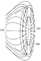
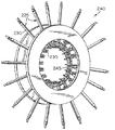
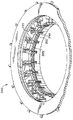
図5Aは二重走査ヘッド510を示す二重ブレスト(胸)スキャナーとしての本発明の実施の形態の前面図である。図5Bは調整可能なアイリス520、530、540を含む走査ヘッド510の1つの拡大側面図である。
Claims (44)
- 不均一表面形状を有する組織構造を光学断層写真式に作像するための装置において、
組織を通して少なくとも減衰伝送を行うことのできる波長を有する光を提供できる光源と;
上記光源から作像すべき組織へ光を伝送するためのファイバ束を含む第1のファイバアレイと;
組織により散乱された光を収集するファイバ束を含む第2のファイバアレイと;
各ファイバ束の一端を支持する少なくとも1つの調整可能な支持部材を含む調整可能な組立体であって、上記各ファイバ束が上記第1及び第2のファイバアレイからのものであって、多数の離間した地点で作像される組織の表面内へ光を伝送しかつ当該表面から出射する光を収集するように、上記各ファィバ束を作像されている組織の表面に適合すべく上記1つ以上の調整可能な各支持部材の部分に沿って分布させるよう構成された組立体と;
上記第2のファイバアレイのファイバにより収集された散乱された光を収集する検出アレイと;
を有することを特徴とする装置。 - 上記光源がダイオードレーザー、Ti:サフィアレーザー、ダイレーザー、多重波長レーザー、連続波(CW)レーザー及びパルスパワーレーザーからなるグループから選択されることを特徴とする請求項1に記載の装置。
- 上記光源が電磁スペクトルの近赤外部分内の光を発することを特徴とする請求項2に記載の装置。
- 上記第1のファイバアレイが上記光源に隣接する並進可能な受光端部を有することを特徴とする請求項1に記載の装置。
- 上記光源と上記第1のファイバアレイとの間に位置する合焦レンズを更に有することを特徴とする請求項1に記載の装置。
- 上記1つ以上の調整可能な支持部材が変形可能な材料から構成されることを特徴とする請求項1に記載の装置。
- 上記1つ以上の調整可能な支持部材が調整可能なアイリスを有することを特徴とする請求項1に記載の装置。
- 上記第2のファイバアレイと上記検出アレイ間に位置する減衰器を更に有することを特徴とする請求項1に記載の装置。
- 上記減衰器が、
上記第2のファイバアレイの上記ファイバ束の他端を収納する内側シリンダと;
光を上記検出アレイに導く複数の上記ファイバ束の一端を収納する外側シリンダと;
上記内側及び外側シリンダ間に位置する複数の減衰器と;
を有することを特徴とする請求項8に記載の装置。 - 上記複数の減衰器が対応する複数の密度値を有する中立密度フィルタを有することを特徴とする請求項9に記載の装置。
- 上記複数の減衰器が偏光子を有することを特徴とする請求項9に記載の装置。
- 上記第1のファイバアレイの部分から当該光源を監視するための参照検出器を更に有することを特徴とする請求項1に記載の装置。
- 上記調整可能な組立体を包む外側ハウジングを更に有し、上記外側ハウジングが周囲の光を排除することを特徴とする請求項1に記載の装置。
- 上記外側ハウジングが更に潜水性の屈折率整合流体を収容するためのラバーダムを有することを特徴とする請求項13に記載の装置。
- 上記検出アレイが電荷結合素子(CCD)検出アレイであることを特徴とする請求項1に記載の装置。
- 上記検出アレイと上記第2のファイバアレイの上記ファイバ束の端部との間に一定の物理的関係が存在することを特徴とする請求項1に記載の装置。
- 二重ヘッドスキャナーを形成する第2の調整可能な組立体を更に有することを特徴とする請求項1に記載の装置。
- 上記減衰器が更に波長選択性フィルタを有することを特徴とする請求項8に記載の装置。
- 上記ファイバ束が多数の小径ファイバを有し、上記各ファイバが200ミクロンよりも小さな直径を有し、当該各ファイバ束が少なくとも2mm2 の合計活性表面を有することを特徴とする請求項1に記載の装置。
- 最小で100個のファイバ束を設けたことを特徴とする請求項19に記載の装置。
- 三次元画像再現及び上記検出アレイから受け取ったデータの表示を行うための計算手段を更に有することを特徴とする請求項1に記載の装置。
- 不均一表面形状を有する組織構造を光学断層写真式に作像するための装置において、
組織を通して少なくとも減衰伝送を行うことのできる波長を有する光を提供できる光源と;
上記光源から作像すべき組織へ光を伝送するためのファイバ束を含む第1のファイバアレイと;
組織により散乱された光を収集するファイバ束を含む第2のファイバアレイと;
各ファイバ束の一端を支持する少なくとも1つの調整可能な支持部材を含む調整可能な組立体であって、上記各ファイバ束が上記第1及び第2のファイバアレイからのものであって、多数の離間した地点で作像される組織の表面内へ光を伝送しかつ当該表面から出射する光を収集するように、上記各ファィバ束を作像されている組織の表面に適合すべく上記1つ以上の調整可能な各支持部材の部分に沿って分布させるよう構成された組立体と;
上記第2のファイバアレイのファイバにより収集された主に定常の散乱光を受け取る検出アレイと;
を有することを特徴とする装置。 - 上記光源がダイオードレーザー、Ti:サフィアレーザー、ダイレーザー、多重波長レーザー、連続波(CW)レーザー及びパルスパワーレーザーからなるグループから選択されることを特徴とする請求項22に記載の装置。
- 上記光源が電磁スペクトルの近赤外部分内の光を発することを特徴とする請求項23に記載の装置。
- 上記第1のファイバアレイが上記光源に隣接する並進可能な受光端部を有することを特徴とする請求項22に記載の装置。
- 上記光源と上記第1のファイバアレイとの間に位置する合焦レンズを更に有することを特徴とする請求項22に記載の装置。
- 上記1つ以上の調整可能な支持部材が変形可能な材料からなることを特徴とする請求項22に記載の装置。
- 上記1つ以上の調整可能な支持部材が調整可能なアイリスを有することを特徴とする請求項22に記載の装置。
- 上記第2のファイバアレイのファイバ束と上記検出アレイ間に位置する減衰器を更に有することを特徴とする請求項22に記載の装置。
- 上記減衰器が、
上記第2のファイバアレイの上記ファイバ束の他端を収納する内側シリンダと;
光を上記検出アレイに導く複数の上記ファイバ束の一端を収納する外側シリンダと;
上記内側及び外側シリンダ間に位置する複数の減衰器と;
を有することを特徴とする請求項29に記載の装置。 - 上記複数の減衰器が対応する複数の密度値を有する中立密度フィルタを有することを特徴とする請求項30に記載の装置。
- 上記複数の減衰器が偏光子を有することを特徴とする請求項30に記載の装置。
- 上記第1のファイバアレイの部分から当該光源を監視するための参照検出器を更に有することを特徴とする請求項22に記載の装置。
- 上記調整可能な組立体を包む外側ハウジングを更に有し、上記外側ハウジングが周囲の光を排除することを特徴とする請求項22に記載の装置。
- 上記外側ハウジングが更に潜水性の屈折率整合流体を収容するためのラバーダムを有することを特徴とする請求項34に記載の装置。
- 上記検出アレイが電荷結合素子(CCD)検出アレイであることを特徴とする請求項22に記載の装置。
- 上記検出アレイと上記第2のファイバアレイの上記ファイバ束の端部との間に一定の物理的関係が存在することを特徴とする請求項22に記載の装置。
- 二重ヘッドスキャナーを形成する第2の調整可能な組立体を更に有することを特徴とする請求項22に記載の装置。
- 上記減衰器が更に波長選択性フィルタを有することを特徴とする請求項29に記載の装置。
- 上記ファイバ束が多数の小径ファイバを有し、上記各ファイバが200ミクロンよりも小さな直径を有し、当該各ファイバ束が少なくとも2mm 2 の合計活性表面を有することを特徴とする請求項22に記載の装置。
- 最小で100個のファイバ束を設けたことを特徴とする請求項40に記載の装置。
- 三次元画像再現及び上記検出アレイから受け取ったデータの表示を行うための計算手段を更に有することを特徴とする請求項22に記載の装置。
- 上記検出アレイが定常散乱光に対応し、かつ放射線搬送式の変数として使用し得る出力を生ずるように作用することを特徴とする請求項22に記載の装置。
- 不均一表面形状を有する組織構造を光学断層写真式に作像するための装置において、
組織を通して少なくとも減衰伝送を行うことのできる波長を有する光を提供できる光源と;
上記光源から作像すべき組織へ光を伝送するためのファイバ束を含む第1のファイバアレイと;
組織により散乱された光を収集するファイバ束を含む第2のファイバアレイと;
各ファイバ束の一端を支持する少なくとも1つの調整可能な支持部材を含む調整可能な組立体であって、上記各ファイバ束が上記第1及び第2のファイバアレイからのものであって、多数の離間した地点で作像される組織の表面内へ光を伝送しかつ当該表面から出射する光を収集するように、作像されている組織構造を一定の形状に適合する上記1つ以上の調整可能な各支持部材の部分に沿って分布させるよう構成された組立体と;
上記第2のファイバアレイのファイバにより収集された散乱された光を収集する検出アレイと;
を有することを特徴とする装置。
Applications Claiming Priority (2)
| Application Number | Priority Date | Filing Date | Title |
|---|---|---|---|
| US08/951,892 US6081322A (en) | 1997-10-16 | 1997-10-16 | NIR clinical opti-scan system |
| US08/951,892 | 1997-10-16 |
Related Parent Applications (1)
| Application Number | Title | Priority Date | Filing Date |
|---|---|---|---|
| JP2000517267A Division JP2001521147A (ja) | 1997-10-16 | 1998-10-05 | 近赤外診療光学走査装置 |
Publications (2)
| Publication Number | Publication Date |
|---|---|
| JP2010101902A true JP2010101902A (ja) | 2010-05-06 |
| JP5342992B2 JP5342992B2 (ja) | 2013-11-13 |
Family
ID=25492285
Family Applications (2)
| Application Number | Title | Priority Date | Filing Date |
|---|---|---|---|
| JP2000517267A Pending JP2001521147A (ja) | 1997-10-16 | 1998-10-05 | 近赤外診療光学走査装置 |
| JP2009288287A Expired - Fee Related JP5342992B2 (ja) | 1997-10-16 | 2009-12-18 | 近赤外診療光学走査装置 |
Family Applications Before (1)
| Application Number | Title | Priority Date | Filing Date |
|---|---|---|---|
| JP2000517267A Pending JP2001521147A (ja) | 1997-10-16 | 1998-10-05 | 近赤外診療光学走査装置 |
Country Status (8)
| Country | Link |
|---|---|
| US (2) | US6081322A (ja) |
| EP (1) | EP1023584B1 (ja) |
| JP (2) | JP2001521147A (ja) |
| CN (1) | CN1211650C (ja) |
| AT (1) | ATE258678T1 (ja) |
| CA (1) | CA2306922C (ja) |
| DE (1) | DE69821380T2 (ja) |
| WO (1) | WO1999020997A1 (ja) |
Families Citing this family (61)
| Publication number | Priority date | Publication date | Assignee | Title |
|---|---|---|---|---|
| US6274086B1 (en) * | 1996-12-16 | 2001-08-14 | The Trustees Of The University Of Pennsylvania | Apparatus for non-invasive imaging oxygen distribution in multi-dimensions |
| US6592847B1 (en) * | 1998-05-14 | 2003-07-15 | The General Hospital Corporation | Intramolecularly-quenched near infrared flourescent probes |
| EP1609410B1 (en) * | 1999-06-03 | 2014-05-28 | Hamamatsu Photonics K.K. | Optical CT apparatus |
| DE69943188D1 (de) * | 1999-06-03 | 2011-03-24 | Hamamatsu Photonics Kk | Optische ct-vorrichtung und verfahren zur wiederherstellung von bildern |
| US6795195B1 (en) * | 1999-09-14 | 2004-09-21 | Research Foundation Of State University Of New York | System and method for tomographic imaging of dynamic properties of a scattering medium |
| AU7579300A (en) * | 1999-09-14 | 2001-04-17 | Research Foundation of State University of New York, Technology Transfer Office, The | Method and system for imaging the dynamics of scattering medium |
| US20010032053A1 (en) * | 2000-01-24 | 2001-10-18 | Hielscher Andreas H. | Imaging of a scattering medium using the equation of radiative transfer |
| US7383076B2 (en) * | 2000-11-27 | 2008-06-03 | The General Hospital Corporation | Fluorescence-mediated molecular tomography |
| US6615063B1 (en) * | 2000-11-27 | 2003-09-02 | The General Hospital Corporation | Fluorescence-mediated molecular tomography |
| US20030044353A1 (en) * | 2001-01-05 | 2003-03-06 | Ralph Weissleder | Activatable imaging probes |
| US6944322B2 (en) * | 2001-03-28 | 2005-09-13 | Visiongate, Inc. | Optical tomography of small objects using parallel ray illumination and post-specimen optical magnification |
| US6965108B2 (en) * | 2001-07-30 | 2005-11-15 | Euro-Celtique, S.A. | Method and apparatus for three dimensional imaging using infrared radiation |
| US6662128B2 (en) * | 2002-01-18 | 2003-12-09 | The Research Foundation Of State University Of New York | Normalized-constraint algorithm for minimizing inter-parameter crosstalk in imaging of scattering media |
| CA2481518A1 (en) * | 2002-04-06 | 2003-10-23 | Randall L. Barbour | A system and method for quantifying the dynamic response of a target system |
| WO2003088133A1 (en) * | 2002-04-06 | 2003-10-23 | Barbour Randall L | Modification of the normalized difference method for real-time optical tomography |
| US7636413B2 (en) * | 2002-04-16 | 2009-12-22 | General Electric Company | Method and apparatus of multi-energy imaging |
| EP1514093B1 (en) | 2002-06-04 | 2021-04-28 | Visen Medical, Inc. | Imaging volumes with arbitrary geometries in non-contact tomography |
| US6784988B2 (en) * | 2002-07-17 | 2004-08-31 | Hamilton Associates, Inc. | Apparatus and process for analyzing a stream of fluid |
| US7647091B2 (en) * | 2003-02-05 | 2010-01-12 | The General Hospital Corporation | Method and system for free space optical tomography of diffuse media |
| US7359540B2 (en) * | 2003-06-27 | 2008-04-15 | Ge Medical Systems Global Technology Company, Llc | Systems and methods for correcting inhomogeneity in images |
| CN100403985C (zh) * | 2004-04-09 | 2008-07-23 | 清华大学 | 数字化近红外光漫射断层成像系统 |
| EP1765172B1 (en) | 2004-06-18 | 2013-04-24 | Elmaleh, David R. | Intravascular imaging device and uses thereof |
| US20060018525A1 (en) * | 2004-07-21 | 2006-01-26 | Barbour Randall L | Method and system for temporal spectral imaging |
| US20080218732A1 (en) * | 2005-07-27 | 2008-09-11 | University Of Massachusetts Lowell | Infrared Scanner for Biological Applications |
| FR2895896A1 (fr) * | 2006-01-12 | 2007-07-13 | Commissariat Energie Atomique | Procede de reconstruction d'image par lumiere retrodiffusee et dispositif pour la mise en oeuvre d'un tel procede |
| FR2895897B1 (fr) * | 2006-01-12 | 2008-10-17 | Commissariat Energie Atomique | Procede et dispositif de reconstruction d'image de tomographie optique de fluorescence par double mesure |
| US9889043B2 (en) * | 2006-01-20 | 2018-02-13 | Lensar, Inc. | System and apparatus for delivering a laser beam to the lens of an eye |
| US7796260B1 (en) * | 2006-04-25 | 2010-09-14 | J.A. Woollam Co., Inc. | System and method of controlling intensity of an electromagnetic beam |
| WO2008039988A2 (en) * | 2006-09-28 | 2008-04-03 | The Florida International University Board Of Trustees | Hand-held optical probe based imaging system with 3d tracking facilities |
| US8712504B2 (en) * | 2006-09-28 | 2014-04-29 | The Florida International University Board Of Trustees | Hand-held optical probe based imaging system with 3D tracking facilities |
| US8323694B2 (en) * | 2007-05-09 | 2012-12-04 | Nanoprobes, Inc. | Gold nanoparticles for selective IR heating |
| TW200906478A (en) * | 2007-08-09 | 2009-02-16 | Univ Nat Central | Method for preparing mesocrystals by organic and organometallic compounds |
| US8815214B2 (en) * | 2008-01-18 | 2014-08-26 | Visen Medical, Inc. | Fluorescent imaging agents |
| WO2009117124A1 (en) * | 2008-03-19 | 2009-09-24 | Nanospectra Biosciences, Inc. | Narrowband imaging using near-infrared absorbing nanoparticles |
| DE102009044303B4 (de) * | 2009-10-21 | 2012-04-19 | Karlsruher Institut für Technologie | Schnelle optische Tomographie |
| CN102883658B (zh) | 2009-11-19 | 2016-06-22 | 调节成像公司 | 用于使用结构化照明经由单元件检测来分析浑浊介质的方法和设备 |
| US20110123452A1 (en) * | 2009-11-25 | 2011-05-26 | Nanoprobes, Inc. | Metal oligomers and polymers and their use in biology and medicine |
| US9635349B2 (en) | 2010-06-11 | 2017-04-25 | The Florida International University Board Of Trustees | Second generation hand held optical imager |
| US8378302B2 (en) * | 2010-10-11 | 2013-02-19 | National Central University | Bidirectional optical scanner assisting in mammography |
| US10376150B2 (en) * | 2011-09-30 | 2019-08-13 | The Trustees Of Columbia University In The City Of New York | Interfacing systems, devices, and methods for optical imaging |
| US10674918B2 (en) | 2012-01-06 | 2020-06-09 | The Florida International University Board Of Trustees | Near-infrared (NIR) optical scanner |
| US20130237858A1 (en) * | 2012-03-12 | 2013-09-12 | Gary P. Warren | Apparatus and Method for Mitigating Noise Affecting a Transcutaneous Signal |
| US20130237849A1 (en) * | 2012-03-12 | 2013-09-12 | Ivwatch, Llc | Geometry of a Transcutaneous Sensor |
| BR112015010459B1 (pt) | 2012-11-07 | 2021-01-26 | Modulated Imaging, Inc. | método para a medição de uma amostra turva |
| TWI572331B (zh) * | 2012-12-28 | 2017-03-01 | 國立交通大學 | 旋轉式光學斷層掃描裝置 |
| EP3091897A4 (en) | 2014-01-06 | 2017-12-27 | The Florida International University Board of Trustees | Near infrared optical imaging system for hemodynamic imaging, pulse monitoring, and mapping spatio-temporal features |
| USD763938S1 (en) | 2014-04-02 | 2016-08-16 | Cephalogics, LLC | Optical sensor array |
| USD763939S1 (en) | 2014-04-02 | 2016-08-16 | Cephalogics, LLC | Optical sensor array liner with optical sensor array pad |
| WO2015174975A1 (en) * | 2014-05-15 | 2015-11-19 | Ohio State Innovation Foundation | Active control guards and rationometric calibration and reconstruction for use with electrical capacitance volume tomography |
| TWI577345B (zh) * | 2015-02-09 | 2017-04-11 | 國立中央大學 | A circular scanning device for optical tomography systems |
| CN105938101B (zh) * | 2016-04-14 | 2020-12-11 | 中国科学院力学研究所 | 一种基于化学发光的用于火焰三维重建的成像系统及方法 |
| ES3037621T3 (en) | 2018-07-19 | 2025-10-03 | Activ Surgical Inc | Systems and methods for multi-modal sensing of depth in vision systems for automated surgical robots |
| CN113853151A (zh) | 2018-12-28 | 2021-12-28 | 艾科缇弗外科公司 | 用于在手术期间定向远程相机的用户界面元件 |
| CN113993474A (zh) | 2018-12-28 | 2022-01-28 | 艾科缇弗外科公司 | 在微创手术中优化可达性、工作空间和灵巧性的系统和方法 |
| US12292564B2 (en) | 2019-04-08 | 2025-05-06 | Activ Surgical, Inc. | Systems and methods for medical imaging |
| CN113950279B (zh) | 2019-04-08 | 2023-04-14 | 艾科缇弗外科公司 | 用于医疗成像的系统和方法 |
| WO2020214821A1 (en) | 2019-04-19 | 2020-10-22 | Activ Surgical, Inc. | Systems and methods for trocar kinematics |
| US11304456B2 (en) | 2019-07-28 | 2022-04-19 | Holovisions LLC | Smart bra with optical sensors to detect abnormal breast tissue |
| US11950881B2 (en) | 2019-07-28 | 2024-04-09 | Holovsions LLC | Smart bra for optical scanning of breast tissue to detect abnormal tissue with selectively-expandable components to reduce air gaps |
| US12201402B2 (en) | 2019-07-28 | 2025-01-21 | Holovisions LLC | Wearable device (smart bra) with compressive chambers and optical sensors for analyzing breast tissue |
| CN114599263A (zh) | 2019-08-21 | 2022-06-07 | 艾科缇弗外科公司 | 用于医疗成像的系统和方法 |
Citations (13)
| Publication number | Priority date | Publication date | Assignee | Title |
|---|---|---|---|---|
| JPH03218443A (ja) * | 1989-11-21 | 1991-09-26 | Toshiba Corp | 光を用いた断層撮影方法及び装置 |
| JPH0454439A (ja) * | 1990-06-22 | 1992-02-21 | Hitachi Ltd | 生体計測方法および装置 |
| JPH04122248A (ja) * | 1990-09-13 | 1992-04-22 | Res Dev Corp Of Japan | 光断層像画像化装置 |
| JPH05184580A (ja) * | 1992-01-16 | 1993-07-27 | Toshiba Corp | 光ct装置 |
| JPH05223738A (ja) * | 1992-02-14 | 1993-08-31 | Res Dev Corp Of Japan | 蛍光断層像測定装置 |
| JPH06506127A (ja) * | 1991-01-24 | 1994-07-14 | ノン―インヴェイシヴ テクノロジイ,インク. | 低酸素症を測定する時間及び周波数領域分光装置 |
| JPH06221913A (ja) * | 1992-10-06 | 1994-08-12 | Hamamatsu Photonics Kk | 散乱吸収体の光学情報計測装置及び方法 |
| JPH0749304A (ja) * | 1993-06-02 | 1995-02-21 | Hamamatsu Photonics Kk | 散乱吸収体の内部情報計測方法及び装置 |
| JPH07151673A (ja) * | 1993-11-30 | 1995-06-16 | Hitachi Ltd | 光学的計算断層撮影装置及び撮影方法 |
| JPH0829329A (ja) * | 1994-07-14 | 1996-02-02 | Hitachi Ltd | 吸収物質濃度の空間分布画像化方法 |
| JPH08166341A (ja) * | 1994-12-14 | 1996-06-25 | Technol Res Assoc Of Medical & Welfare Apparatus | 光ct |
| JPH08334458A (ja) * | 1995-06-05 | 1996-12-17 | Technol Res Assoc Of Medical & Welfare Apparatus | 光測定装置の検出器ホルダー |
| US5625458A (en) * | 1994-11-10 | 1997-04-29 | Research Foundation Of City College Of New York | Method and system for imaging objects in turbid media using diffusive fermat photons |
Family Cites Families (5)
| Publication number | Priority date | Publication date | Assignee | Title |
|---|---|---|---|---|
| US5137355A (en) * | 1988-06-08 | 1992-08-11 | The Research Foundation Of State University Of New York | Method of imaging a random medium |
| US5353799A (en) * | 1991-01-22 | 1994-10-11 | Non Invasive Technology, Inc. | Examination of subjects using photon migration with high directionality techniques |
| US5213105A (en) * | 1990-12-04 | 1993-05-25 | Research Corporation Technologies, Inc. | Frequency domain optical imaging using diffusion of intensity modulated radiation |
| US5408093A (en) * | 1992-08-31 | 1995-04-18 | Hitachi, Ltd. | Optical computed tomography equipment having image inverting optical device |
| US5876339A (en) * | 1997-01-09 | 1999-03-02 | Lemire; Robert | Apparatus for optical breast imaging |
-
1997
- 1997-10-16 US US08/951,892 patent/US6081322A/en not_active Ceased
-
1998
- 1998-10-05 CA CA002306922A patent/CA2306922C/en not_active Expired - Fee Related
- 1998-10-05 CN CN98810247.1A patent/CN1211650C/zh not_active Expired - Fee Related
- 1998-10-05 EP EP98949750A patent/EP1023584B1/en not_active Expired - Lifetime
- 1998-10-05 DE DE69821380T patent/DE69821380T2/de not_active Expired - Lifetime
- 1998-10-05 JP JP2000517267A patent/JP2001521147A/ja active Pending
- 1998-10-05 AT AT98949750T patent/ATE258678T1/de not_active IP Right Cessation
- 1998-10-05 WO PCT/US1998/020810 patent/WO1999020997A1/en not_active Ceased
-
2002
- 2002-06-27 US US10/183,276 patent/USRE38800E1/en not_active Expired - Lifetime
-
2009
- 2009-12-18 JP JP2009288287A patent/JP5342992B2/ja not_active Expired - Fee Related
Patent Citations (13)
| Publication number | Priority date | Publication date | Assignee | Title |
|---|---|---|---|---|
| JPH03218443A (ja) * | 1989-11-21 | 1991-09-26 | Toshiba Corp | 光を用いた断層撮影方法及び装置 |
| JPH0454439A (ja) * | 1990-06-22 | 1992-02-21 | Hitachi Ltd | 生体計測方法および装置 |
| JPH04122248A (ja) * | 1990-09-13 | 1992-04-22 | Res Dev Corp Of Japan | 光断層像画像化装置 |
| JPH06506127A (ja) * | 1991-01-24 | 1994-07-14 | ノン―インヴェイシヴ テクノロジイ,インク. | 低酸素症を測定する時間及び周波数領域分光装置 |
| JPH05184580A (ja) * | 1992-01-16 | 1993-07-27 | Toshiba Corp | 光ct装置 |
| JPH05223738A (ja) * | 1992-02-14 | 1993-08-31 | Res Dev Corp Of Japan | 蛍光断層像測定装置 |
| JPH06221913A (ja) * | 1992-10-06 | 1994-08-12 | Hamamatsu Photonics Kk | 散乱吸収体の光学情報計測装置及び方法 |
| JPH0749304A (ja) * | 1993-06-02 | 1995-02-21 | Hamamatsu Photonics Kk | 散乱吸収体の内部情報計測方法及び装置 |
| JPH07151673A (ja) * | 1993-11-30 | 1995-06-16 | Hitachi Ltd | 光学的計算断層撮影装置及び撮影方法 |
| JPH0829329A (ja) * | 1994-07-14 | 1996-02-02 | Hitachi Ltd | 吸収物質濃度の空間分布画像化方法 |
| US5625458A (en) * | 1994-11-10 | 1997-04-29 | Research Foundation Of City College Of New York | Method and system for imaging objects in turbid media using diffusive fermat photons |
| JPH08166341A (ja) * | 1994-12-14 | 1996-06-25 | Technol Res Assoc Of Medical & Welfare Apparatus | 光ct |
| JPH08334458A (ja) * | 1995-06-05 | 1996-12-17 | Technol Res Assoc Of Medical & Welfare Apparatus | 光測定装置の検出器ホルダー |
Also Published As
| Publication number | Publication date |
|---|---|
| US6081322A (en) | 2000-06-27 |
| JP2001521147A (ja) | 2001-11-06 |
| USRE38800E1 (en) | 2005-09-20 |
| CA2306922C (en) | 2009-03-17 |
| DE69821380D1 (de) | 2004-03-04 |
| EP1023584A1 (en) | 2000-08-02 |
| JP5342992B2 (ja) | 2013-11-13 |
| DE69821380T2 (de) | 2004-12-09 |
| ATE258678T1 (de) | 2004-02-15 |
| WO1999020997A1 (en) | 1999-04-29 |
| EP1023584B1 (en) | 2004-01-28 |
| CA2306922A1 (en) | 1999-04-29 |
| CN1211650C (zh) | 2005-07-20 |
| CN1276869A (zh) | 2000-12-13 |
Similar Documents
| Publication | Publication Date | Title |
|---|---|---|
| JP5342992B2 (ja) | 近赤外診療光学走査装置 | |
| JP5808741B2 (ja) | 小動物の光音響イメージング用画像化装置及び画像化方法 | |
| JP6174658B2 (ja) | 対象体の断層光音響撮像用の手持ち式装置及び方法 | |
| CA2708675C (en) | Three-dimensional photoacoustic imager and methods for calibrating an imager | |
| JP7082383B2 (ja) | 触覚センサと光断層撮影を融合する検査装置と検査方法 | |
| US11020006B2 (en) | Transcranial photoacoustic/thermoacoustic tomography brain imaging informed by adjunct image data | |
| US20110306865A1 (en) | photoacoustic imaging device | |
| JP2013500091A5 (ja) | ||
| EP2527815B1 (en) | Thermoacoustic imaging with quantitative extraction of an absorption image | |
| WO2014073907A1 (ko) | 유방암 진단용 광음향 스캐닝 장치 | |
| BRPI0708897A2 (pt) | dispositivo e mÉtodo para gerar imagem de um meio turvo, e, produto de programa de computador | |
| Ai | Photoacoustic tomography for prostate imaging by transurethral illumination | |
| WO2025179125A1 (en) | Optoacoustic probe for imaging | |
| Haupt et al. | Non-Contact Optical Ultrasound Concept for Biomedical Imaging | |
| JPH04319345A (ja) | 光断層イメージング装置 | |
| KR20120074904A (ko) | 커플링 패드 및 광 음향 시스템 |
Legal Events
| Date | Code | Title | Description |
|---|---|---|---|
| A621 | Written request for application examination |
Free format text: JAPANESE INTERMEDIATE CODE: A621 Effective date: 20091218 |
|
| A521 | Request for written amendment filed |
Free format text: JAPANESE INTERMEDIATE CODE: A523 Effective date: 20100205 |
|
| A521 | Request for written amendment filed |
Free format text: JAPANESE INTERMEDIATE CODE: A523 Effective date: 20100311 |
|
| A131 | Notification of reasons for refusal |
Free format text: JAPANESE INTERMEDIATE CODE: A131 Effective date: 20120308 |
|
| A601 | Written request for extension of time |
Free format text: JAPANESE INTERMEDIATE CODE: A601 Effective date: 20120607 |
|
| A602 | Written permission of extension of time |
Free format text: JAPANESE INTERMEDIATE CODE: A602 Effective date: 20120612 |
|
| A521 | Request for written amendment filed |
Free format text: JAPANESE INTERMEDIATE CODE: A523 Effective date: 20120910 |
|
| A131 | Notification of reasons for refusal |
Free format text: JAPANESE INTERMEDIATE CODE: A131 Effective date: 20130307 |
|
| A601 | Written request for extension of time |
Free format text: JAPANESE INTERMEDIATE CODE: A601 Effective date: 20130605 |
|
| A602 | Written permission of extension of time |
Free format text: JAPANESE INTERMEDIATE CODE: A602 Effective date: 20130610 |
|
| A521 | Request for written amendment filed |
Free format text: JAPANESE INTERMEDIATE CODE: A523 Effective date: 20130704 |
|
| TRDD | Decision of grant or rejection written | ||
| A01 | Written decision to grant a patent or to grant a registration (utility model) |
Free format text: JAPANESE INTERMEDIATE CODE: A01 Effective date: 20130723 |
|
| A61 | First payment of annual fees (during grant procedure) |
Free format text: JAPANESE INTERMEDIATE CODE: A61 Effective date: 20130812 |
|
| R150 | Certificate of patent or registration of utility model |
Free format text: JAPANESE INTERMEDIATE CODE: R150 |
|
| LAPS | Cancellation because of no payment of annual fees |