WO2013176197A1 - Procédé de production de lymphocytes t spécifiques d'un antigène - Google Patents
Procédé de production de lymphocytes t spécifiques d'un antigène Download PDFInfo
- Publication number
- WO2013176197A1 WO2013176197A1 PCT/JP2013/064291 JP2013064291W WO2013176197A1 WO 2013176197 A1 WO2013176197 A1 WO 2013176197A1 JP 2013064291 W JP2013064291 W JP 2013064291W WO 2013176197 A1 WO2013176197 A1 WO 2013176197A1
- Authority
- WO
- WIPO (PCT)
- Prior art keywords
- cells
- cell
- human
- ips
- cd8sp
- Prior art date
- Legal status (The legal status is an assumption and is not a legal conclusion. Google has not performed a legal analysis and makes no representation as to the accuracy of the status listed.)
- Ceased
Links
Images















Classifications
-
- A—HUMAN NECESSITIES
- A61—MEDICAL OR VETERINARY SCIENCE; HYGIENE
- A61K—PREPARATIONS FOR MEDICAL, DENTAL OR TOILETRY PURPOSES
- A61K35/00—Medicinal preparations containing materials or reaction products thereof with undetermined constitution
- A61K35/12—Materials from mammals; Compositions comprising non-specified tissues or cells; Compositions comprising non-embryonic stem cells; Genetically modified cells
- A61K35/14—Blood; Artificial blood
-
- A—HUMAN NECESSITIES
- A61—MEDICAL OR VETERINARY SCIENCE; HYGIENE
- A61K—PREPARATIONS FOR MEDICAL, DENTAL OR TOILETRY PURPOSES
- A61K40/00—Cellular immunotherapy
- A61K40/10—Cellular immunotherapy characterised by the cell type used
- A61K40/11—T-cells, e.g. tumour infiltrating lymphocytes [TIL] or regulatory T [Treg] cells; Lymphokine-activated killer [LAK] cells
-
- A—HUMAN NECESSITIES
- A61—MEDICAL OR VETERINARY SCIENCE; HYGIENE
- A61K—PREPARATIONS FOR MEDICAL, DENTAL OR TOILETRY PURPOSES
- A61K40/00—Cellular immunotherapy
- A61K40/20—Cellular immunotherapy characterised by the effect or the function of the cells
- A61K40/22—Immunosuppressive or immunotolerising
-
- A—HUMAN NECESSITIES
- A61—MEDICAL OR VETERINARY SCIENCE; HYGIENE
- A61K—PREPARATIONS FOR MEDICAL, DENTAL OR TOILETRY PURPOSES
- A61K40/00—Cellular immunotherapy
- A61K40/40—Cellular immunotherapy characterised by antigens that are targeted or presented by cells of the immune system
- A61K40/41—Vertebrate antigens
- A61K40/416—Antigens related to auto-immune diseases; Preparations to induce self-tolerance
-
- A—HUMAN NECESSITIES
- A61—MEDICAL OR VETERINARY SCIENCE; HYGIENE
- A61K—PREPARATIONS FOR MEDICAL, DENTAL OR TOILETRY PURPOSES
- A61K40/00—Cellular immunotherapy
- A61K40/40—Cellular immunotherapy characterised by antigens that are targeted or presented by cells of the immune system
- A61K40/41—Vertebrate antigens
- A61K40/42—Cancer antigens
-
- A—HUMAN NECESSITIES
- A61—MEDICAL OR VETERINARY SCIENCE; HYGIENE
- A61K—PREPARATIONS FOR MEDICAL, DENTAL OR TOILETRY PURPOSES
- A61K40/00—Cellular immunotherapy
- A61K40/40—Cellular immunotherapy characterised by antigens that are targeted or presented by cells of the immune system
- A61K40/46—Viral antigens
-
- A—HUMAN NECESSITIES
- A61—MEDICAL OR VETERINARY SCIENCE; HYGIENE
- A61P—SPECIFIC THERAPEUTIC ACTIVITY OF CHEMICAL COMPOUNDS OR MEDICINAL PREPARATIONS
- A61P31/00—Antiinfectives, i.e. antibiotics, antiseptics, chemotherapeutics
-
- A—HUMAN NECESSITIES
- A61—MEDICAL OR VETERINARY SCIENCE; HYGIENE
- A61P—SPECIFIC THERAPEUTIC ACTIVITY OF CHEMICAL COMPOUNDS OR MEDICINAL PREPARATIONS
- A61P31/00—Antiinfectives, i.e. antibiotics, antiseptics, chemotherapeutics
- A61P31/12—Antivirals
-
- A—HUMAN NECESSITIES
- A61—MEDICAL OR VETERINARY SCIENCE; HYGIENE
- A61P—SPECIFIC THERAPEUTIC ACTIVITY OF CHEMICAL COMPOUNDS OR MEDICINAL PREPARATIONS
- A61P35/00—Antineoplastic agents
-
- A—HUMAN NECESSITIES
- A61—MEDICAL OR VETERINARY SCIENCE; HYGIENE
- A61P—SPECIFIC THERAPEUTIC ACTIVITY OF CHEMICAL COMPOUNDS OR MEDICINAL PREPARATIONS
- A61P37/00—Drugs for immunological or allergic disorders
- A61P37/02—Immunomodulators
-
- C—CHEMISTRY; METALLURGY
- C12—BIOCHEMISTRY; BEER; SPIRITS; WINE; VINEGAR; MICROBIOLOGY; ENZYMOLOGY; MUTATION OR GENETIC ENGINEERING
- C12N—MICROORGANISMS OR ENZYMES; COMPOSITIONS THEREOF; PROPAGATING, PRESERVING, OR MAINTAINING MICROORGANISMS; MUTATION OR GENETIC ENGINEERING; CULTURE MEDIA
- C12N5/00—Undifferentiated human, animal or plant cells, e.g. cell lines; Tissues; Cultivation or maintenance thereof; Culture media therefor
- C12N5/06—Animal cells or tissues; Human cells or tissues
- C12N5/0602—Vertebrate cells
- C12N5/0634—Cells from the blood or the immune system
- C12N5/0636—T lymphocytes
-
- C—CHEMISTRY; METALLURGY
- C12—BIOCHEMISTRY; BEER; SPIRITS; WINE; VINEGAR; MICROBIOLOGY; ENZYMOLOGY; MUTATION OR GENETIC ENGINEERING
- C12N—MICROORGANISMS OR ENZYMES; COMPOSITIONS THEREOF; PROPAGATING, PRESERVING, OR MAINTAINING MICROORGANISMS; MUTATION OR GENETIC ENGINEERING; CULTURE MEDIA
- C12N5/00—Undifferentiated human, animal or plant cells, e.g. cell lines; Tissues; Cultivation or maintenance thereof; Culture media therefor
- C12N5/06—Animal cells or tissues; Human cells or tissues
- C12N5/0602—Vertebrate cells
- C12N5/0696—Artificially induced pluripotent stem cells, e.g. iPS
-
- C—CHEMISTRY; METALLURGY
- C12—BIOCHEMISTRY; BEER; SPIRITS; WINE; VINEGAR; MICROBIOLOGY; ENZYMOLOGY; MUTATION OR GENETIC ENGINEERING
- C12N—MICROORGANISMS OR ENZYMES; COMPOSITIONS THEREOF; PROPAGATING, PRESERVING, OR MAINTAINING MICROORGANISMS; MUTATION OR GENETIC ENGINEERING; CULTURE MEDIA
- C12N2501/00—Active agents used in cell culture processes, e.g. differentation
- C12N2501/10—Growth factors
- C12N2501/125—Stem cell factor [SCF], c-kit ligand [KL]
-
- C—CHEMISTRY; METALLURGY
- C12—BIOCHEMISTRY; BEER; SPIRITS; WINE; VINEGAR; MICROBIOLOGY; ENZYMOLOGY; MUTATION OR GENETIC ENGINEERING
- C12N—MICROORGANISMS OR ENZYMES; COMPOSITIONS THEREOF; PROPAGATING, PRESERVING, OR MAINTAINING MICROORGANISMS; MUTATION OR GENETIC ENGINEERING; CULTURE MEDIA
- C12N2501/00—Active agents used in cell culture processes, e.g. differentation
- C12N2501/10—Growth factors
- C12N2501/165—Vascular endothelial growth factor [VEGF]
-
- C—CHEMISTRY; METALLURGY
- C12—BIOCHEMISTRY; BEER; SPIRITS; WINE; VINEGAR; MICROBIOLOGY; ENZYMOLOGY; MUTATION OR GENETIC ENGINEERING
- C12N—MICROORGANISMS OR ENZYMES; COMPOSITIONS THEREOF; PROPAGATING, PRESERVING, OR MAINTAINING MICROORGANISMS; MUTATION OR GENETIC ENGINEERING; CULTURE MEDIA
- C12N2502/00—Coculture with; Conditioned medium produced by
- C12N2502/13—Coculture with; Conditioned medium produced by connective tissue cells; generic mesenchyme cells, e.g. so-called "embryonic fibroblasts"
-
- C—CHEMISTRY; METALLURGY
- C12—BIOCHEMISTRY; BEER; SPIRITS; WINE; VINEGAR; MICROBIOLOGY; ENZYMOLOGY; MUTATION OR GENETIC ENGINEERING
- C12N—MICROORGANISMS OR ENZYMES; COMPOSITIONS THEREOF; PROPAGATING, PRESERVING, OR MAINTAINING MICROORGANISMS; MUTATION OR GENETIC ENGINEERING; CULTURE MEDIA
- C12N2506/00—Differentiation of animal cells from one lineage to another; Differentiation of pluripotent cells
- C12N2506/11—Differentiation of animal cells from one lineage to another; Differentiation of pluripotent cells from blood or immune system cells
-
- C—CHEMISTRY; METALLURGY
- C12—BIOCHEMISTRY; BEER; SPIRITS; WINE; VINEGAR; MICROBIOLOGY; ENZYMOLOGY; MUTATION OR GENETIC ENGINEERING
- C12N—MICROORGANISMS OR ENZYMES; COMPOSITIONS THEREOF; PROPAGATING, PRESERVING, OR MAINTAINING MICROORGANISMS; MUTATION OR GENETIC ENGINEERING; CULTURE MEDIA
- C12N2506/00—Differentiation of animal cells from one lineage to another; Differentiation of pluripotent cells
- C12N2506/45—Differentiation of animal cells from one lineage to another; Differentiation of pluripotent cells from artificially induced pluripotent stem cells
Definitions
- the present invention relates to a method for producing T cells having antigen specificity, and more specifically, a step of differentiating iPS cells derived from human T cells into CD4 / CD8 double negative cells, and the CD4 / CD8 double negative cells. And a step of differentiating the CD4 / CD8 double negative cells that have stimulated the T cell receptor into CD8 single positive cells and / or CD4 single positive cells.
- the present invention relates to a method for producing human CD8 single positive cells and / or CD4 single positive cells.
- the present invention also relates to a human CD8 single positive cell or CD4 single positive cell having antigen specificity produced by the method.
- T cells are cells that play a central role in the acquisition of acquired immunity and systemic immunity against pathogens.
- CTLs cytotoxic T cells
- MHC class 1, HLA class 1 major histocompatibility antigens
- TCR T cell receptors
- CTLs cytotoxic T cells
- MHC class 1, HLA class 1 major histocompatibility antigens
- TCR T cell receptors
- a part of the CTL becomes a long-lived memory T cell, is stored in the host while maintaining the cytotoxicity against the foreign substance, and can cope with the next exposure to the same foreign substance. . Therefore, in the immune system that fights against microorganisms, viral infections, and neoplasms (tumors), CTL functions as a major element (Non-Patent Documents 1 and 2).
- CD8SP cells are cells in which both CD8 and CD4 are expressed from immature cells (CD4 / CD8 double negative (DN) cells) that do not express TCR and have neither CD4 nor CD8 in the thymus. It is known to differentiate through (CD4 / CD8 double positive (DP) cells).
- T cells such as CTLs specifically recognize antigenic peptides via TCR under HLA restriction, their proliferation function and effector function begin to be exerted.
- T cell immunity is not only long-term immunity monitoring with long-lived memory T cells, but also specific recognition of target cells and eradication of the cells (non-patent literature). 3-5).
- T cell immunity is often hampered by insufficient recognition of antigens associated with potential viral infections or cancer / self-antigens. That is, persistent latent viral infection or exposure to cancer / self-antigens impairs T cell long-term viability, proliferative capacity and effector function, and severely depletes T cells. Eventually, the antigen-responsive T cell pool is lost (Non-patent Documents 6 and 7).
- Non-Patent Documents 9 and 10 development of an approach for imparting a desired antigen specificity to non-specific T cells for such diseases is also underway.
- the therapeutic effect by this approach should largely depend on the antigen specificity of induced and amplified T cells (Non-patent Documents 11 and 12).
- TCR is introduced exogenously in order to induce such hematopoietic stem cells or peripheral mature T cells into antigen-specific T cells, it has been revealed that a mismatched TCR usually occurs. .
- Non-patent Document 13 a technique for inducing antigen-specific juvenile T cells from pluripotent stem cells has been developed.
- Non-patent Document 13 a technique for inducing antigen-specific juvenile T cells from pluripotent stem cells has been developed.
- hematopoietic stem / progenitor cells a sufficiently functional differentiation method to human T cells has not been established.
- T-iPS cells induced pluripotent stem cells (inducible pluripotent stem cells, iPS cells) that allow unlimited self-replication while maintaining pluripotency
- Reconstitution of the TCR gene has been completed and iPS cells (hereinafter also referred to as “T-iPS cells”) have been successfully established from human T cells having antigen specificity.
- the T-iPS cells were also successfully induced to differentiate into T cells, and further, T cells having the same gene rearrangement pattern as the original T cells (ie, recognized by the original human T cells) by the method. It was also clarified that T cells exhibiting specificity for the antigen to be obtained (Patent Document 1).
- the present inventors have developed a method for establishing T-iPS cells from human T cells having antigen specificity and further inducing differentiation from the T-iPS cells to T cells. It has also been clarified that this method can yield T cells having the same gene rearrangement pattern as the original T cells (Patent Document 1).
- the frequency of T cells having the same gene rearrangement pattern as the original T cells in the obtained T cells was newly found to be about one quarter. .
- the present invention relates to a T cell obtained by redifferentiation from a human T cell having antigen specificity via a T-iPS cell, and having a TCR gene rearrangement pattern identical to that of the original human T cell.
- the object is to remarkably improve the frequency of appearance of cells (ie, T cells showing specificity for the antigen recognized by the original human T cells).
- TCR is usually not expressed in the most immature stage (CD4 / CD8DN stage) that does not express CD4 and CD8. Since it is known, the present inventors first verified the expression of TCR in the process of redifferentiating T-iPS cells into T cells.
- TCR is expressed even at the CD4 / CD8DN stage, unlike the conventional findings in the process of maturation of T cells in the thymus. (See Example 1 below).
- the TCR signal through the antigen peptide-MHC complex is known to stop the expression of the RAG gene and suppress further assembly of the TCR gene. Yes. Furthermore, it has also been clarified that a TCR signal-like signal generated by anti-CD3 antibody stimulation has the same effect.
- the present inventors next suppressed the further assembly of the TCR gene by stimulating the TCR expressed in CD4 / CD8DN cells redifferentiated from T-iPS cells, It was verified whether or not it was possible to improve the appearance frequency of T cells having the same gene rearrangement pattern as that of T cells. Furthermore, it was also verified whether or not CD4 / CD8DN cells subjected to such stimulation can be differentiated into CD8SP cells by allowing molecules that contribute to CD8 lineage selection to act ( See Example 1 below).
- the TCR expressed in CD4 / CD8DN cells derived from T-iPS cells was stimulated by anti-CD3 antibody or the like, and further CD4 / CD8DN cells subjected to such stimulation were selected for CD8 lineage selection. It was found that the frequency of appearance of CD8SP cells having the same TCR gene rearrangement pattern as that of the original human T cells can be remarkably improved by acting molecules such as IL-7 that contributes. Furthermore, the CD8SP cells produced in this way were also found to have a longer telomere length and higher proliferation ability (replication ability) than the original T cells.
- the TCR expressed in the CD4 / CD8DN cells derived from T-iPS cells is stimulated by an anti-CD3 antibody or the like, and further, the CD4 / CD8DN cells to which such stimulation is given such as IL-7.
- CD4SP cells could also be obtained by acting molecules.
- the present invention has been made based on the above findings, and more specifically, a method for producing human CD8 single positive cells having antigen specificity, wherein iPS cells derived from human T cells are treated with CD4 / CD8 double cells.
- the present invention also provides single positive T cells having antigen specificity, specifically CD4 single positive cells and human CD8 single positive cells produced by the above method.
- the present invention provides the following inventions.
- a method for producing human single positive T cells having antigen specificity Differentiating iPS cells derived from human T cells into CD4 / CD8 double negative cells; Stimulating the CD4 / CD8 double negative cell T cell receptor; Differentiating said CD4 / CD8 double negative cells stimulated with T cell receptors into single positive T cells.
- a human single positive T cell having antigen specificity produced by the method according to any one of (1) to (3) above.
- a pharmaceutical composition comprising the human single positive T cell according to (4) above.
- a T cell obtained by redifferentiation from a human T cell having antigen specificity via a T-iPS cell has a TCR gene rearrangement pattern identical to that of the original human T cell. It becomes possible to make the appearance frequency of a cell very high.
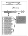
- V ⁇ primers were designed to amplify all components of the V ⁇ segment at the human TCRA locus spanning 1000 kb or more on chromosome 14q11.2.
- 12 J ⁇ primers can be amplified downstream of each sequence of J ⁇ 1, J ⁇ 6, J ⁇ 10, J ⁇ 17, J ⁇ 23, J ⁇ 29, J ⁇ 32, J ⁇ 36, J ⁇ 41, J ⁇ 48, J ⁇ 53 or J ⁇ 58 segments to amplify 3-7 different J ⁇ segments. Designed.
- FIG. 3 is a schematic diagram showing a process of producing T cell-derived iPS cells (T-iPS cells) from peripheral blood T cells using a retroviral vector encoding OCT3 / 4, SOX2 or KLF4.
- the tapered region (7 to 11 days from the start of T cell activation) indicates the period during which the medium was gradually replaced with human iPS medium.
- the scale bar indicates 200 ⁇ m
- b to d show the results of counterstaining nuclei with DAPI (points shining blue in the figure).
- Results of observing with a microscope the expression of AP activity and pluripotency markers (SSEA-4, Tra-1-60 and Tra-1-81) in iPS cells (TkT3V1-7) prepared from peripheral blood T cells. It is a photograph shown.
- a indicates that the scale bar is 500 ⁇ m.
- the scale bar is 200 ⁇ m, and the result of counterstaining the DAPI nuclei (shown in blue in the figure) is shown.
- Tg indicates expression of a gene introduced exogenously by a retrovirus
- endo indicates expression of an endogenously expressed gene
- total indicates retrovirus. It shows that it is the expression of the gene introduced exogenously and the gene expressed endogenously.
- PB CD3 + indicates the result of peripheral blood T cells from which TkT3V1-7 was derived
- KhES3 indicates the result of ES cells that are pluripotent stem cells (positive control)
- Water Shows the result of RT-PCR performed without adding template DNA (negative control). In this RT-PCR, GAPDH was used as an internal standard.
- FIG. 4 is a photograph showing the results of RT-PCR detection of residual SeV RNAs (four factors expressed by cistronic (tetracistronic factors: OCT4, SOX2, KLF4 and c-MYC) and T antigen).
- SeVp (KOSM302L)” and “SeV18 + SV40 / TS15 ⁇ F” show the results in SeV used to establish H254SeVT-3, respectively, and “Water” is RT ⁇ conducted without adding template DNA. Results of PCR (negative control) are shown. Further, “na” indicates “not evaluated”. In this RT-PCR, GAPDH was used as an internal standard.
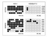
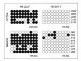
- white circles and black circles indicate unmethylated CpG dinucleotides and methylated CpG dinucleotides, respectively, and “% Me” indicates the methylation rate in each region.
- white circles and black circles indicate unmethylated CpG dinucleotides and methylated CpG dinucleotides, respectively, and “% Me” indicates the methylation rate in each region.
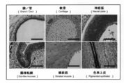
- the teratoma is a photomicrograph showing that it contains an anatomical structure derived from three germ layers.
- gland / duct and intestinal mucosa are observed as anatomical structure derived from endoderm
- cartilage and striated muscle are observed as anatomical structure derived from mesoderm
- anatomical derived from ectoderm Neural plate and pigment epithelium have been observed as structures.
- the scale bar indicates 100 ⁇ m.
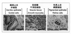
- H254SeVT-3 is an endoderm-derived cell lineage (goblet cells in the intestinal-like epithelium), mesoderm-derived cell lineage (smooth muscle cells in muscle tissue) and ectoderm-derived cell lineages (retinal cells in the pigment epithelium)
- the microscope picture which shows having differentiated into.
- the scale bar indicates 100 ⁇ m. It is a photograph which shows the result of having detected rearrangement of the TCRB gene in the TkT3V1-7 genome by multiplex PCR analysis.
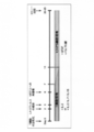
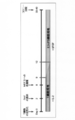
- Tubes A and B show V ⁇ - (D) J ⁇ assembly, and tube C shows D-J ⁇ assembly. It is a photograph showing the result of detecting TCRA gene rearrangement (V-J ⁇ assembly) in the TkT3V1-7 genome by multiplex PCR analysis. It is a photograph which shows the result of having detected rearrangement of the TCRB gene in the H254SeVT-3 genome by multiplex PCR analysis. Tubes A and B show V ⁇ - (D) J ⁇ assembly, and tube C shows D-J ⁇ assembly. It is a photograph showing the results of detecting TCRA gene rearrangement (V-J ⁇ assembly) in the H254SeVT-3 genome by multiplex PCR analysis. FIG.
- TkT3V1-7-derived CD34 + hematopoietic stem / progenitor cells was cultured with VEGF, SCF and FLT-3L on C3H10T1 / 2 feeder cells.
- VEGF vascular endothelial growth factor
- SCF vascular endothelial growth factor
- FLT-3L C3H10T1 / 2 feeder cells.
- the CD34 + hematopoietic stem / progenitor cells (T-iPS sac cells) derived from TkT3V1-7 thus generated were expressed as OP9 (OP9-DL1) feeder cells expressing delta-like molecule 1. Moved up.
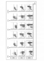
- FIG. 3 is a dot plot showing the results of flow cytometry analysis of developmental progress from H254SeVT-3-derived T-iPS sack cells to human T lineage cells.
- H254SeVT-3 was cultured with VEGF, SCF and FLT-3L on C3H10T1 / 2 feeder cells. Then, on the 14th day, the TkT3V1-7-derived T-iPS sack cells thus generated were transferred onto OP9-DL1 feeder cells. Then, the cells undergoing redifferentiation on OP9-DL1 cells were collected and subjected to redifferentiation induction for 15 to 36 days (“0 wks” to “3 wks” in the figure), weekly on the cell surface of H254SeVT-3-derived cells.
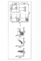
- FIG. 3 is a dot plot showing the results of flow cytometry analysis of the presence of T4PS-derived CD4 / 8DP cells established in vitro.
- a is the result of analyzing the expression of CD45, CD56, CD3, TCR ⁇ , CD4 and CD8 on the cell surface of TkT3V1-7-derived cells by flow cytometry during 35 to 42 days after induction of redifferentiation. Indicates.
- FIG. 4 is a photograph showing the results of RT-PCR amplification of TCRA and TCRB genes expressed in DN cell-derived cDNA library synthesized by SMART method or DP cell-derived cDNA library synthesized by SMART method.
- “Water” indicates the result of RT-PCR carried out without adding template DNA (negative control). GAPDH was used as an internal standard for each PCR.
- FIG. 2 is a photograph showing the results of RT-PCR analysis of RAG1 and RAG2 expression in DN cells and DP cells.
- “Water” indicates the result of RT-PCR carried out without adding template DNA (negative control).
- Samples used as PCR templates were aligned based on GAPDH expression.
- It is a dot plot diagram showing the results of collecting floating cells on OP9-DL1 cells on the 42nd day after the start of redifferentiation induction and analyzing them by flow cytometry.
- DN cells and DP cells are sorted from cells partitioned into CD45 + , CD56 ⁇ , CD3 + , TCR ⁇ + , CD2 + and CD5 + by pre-gating with CD1a ⁇ and CD1a +, respectively. did.
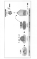
- FIG. 24 It is the schematic which shows the manufacturing method of the human CD8 single positive (SP) cell which has the antigen specificity of this invention. That is, it is a schematic diagram showing the process of redifferentiation from T-iPS cells to human CD8SP cells.
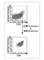
- the phenotype of cells obtained by redifferentiating T-iPS cells by the method of the present invention was determined by flow cytometry. It is a dot plot figure which shows the result of analysis. Shown in this figure are representative results among the results of experiments carried out independently at least three times.
- a and b show the results of analyzing the phenotype as T cells
- c and d Shows the result of analyzing the phenotype as a memory cell.
- b since uniform expression of HLA-A24 was observed, it also indicates that there was no contamination of CD3 + / CD8 + cells from PBMCs.
- Cells obtained by redifferentiating T-iPS cells according to the method of the present invention were designated as A24 / Nef-138-8.
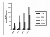
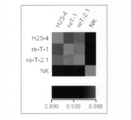
- GAPDH was used as an internal standard for each PCR. It is a graph which shows the result of having compared the expression level of the main cell surface molecule among CD4 + cell, CD8 + cell, reT-2.1 cell, and H25-4 cell using quantitative PCR. In each PCR reaction, the expression level of 18S rRNA was used as an internal standard. In the figure, the vertical axis indicates the expression ratio when the expression level of each gene in CD8 + cells is 1.0. On the horizontal axis, the results of CD4 + cells, CD8 + cells, reT-2.1 cells, and H25-4 cells are shown in order from the left.
- the expression level of 18S rRNA was used as an internal standard.
- the vertical axis indicates the expression ratio when the expression level of each gene in CD8 + cells is 1.0.
- the results of CD4 + cells, CD8 + cells, reT-2.1 cells, and H25-4 cells are shown in order from the left.
- FIG. 6 is a graph showing the relative telomere length (RTL) determined by flow-FISH. It is a histogram which shows the result of having analyzed the secretion of the cytolytic molecule in reT-2.1 when stimulated with ⁇ -CD3 / CD28 beads.
- the left panel is a histogram showing the intracellular production of granzyme B.
- the white histogram portion surrounded by a gray line indicates the reactivity between unstimulated cells and anti-granzyme B antibody
- the white histogram portion surrounded by a black line indicates the stimulated cells and anti-granzyme.
- the filled histogram portion showing the reactivity with the B antibody shows the reactivity between the stimulated cells and the antibody used as a negative control (an antibody of the same isotype as the anti-granzyme B antibody).
- the right panel is a histogram showing the results of CD107a expression assay (CD107a mobilization assay) in reT-2.1 when stimulated with ⁇ -CD3 / CD28 beads.
- the white histogram portion surrounded by a gray line indicates the reactivity between unstimulated cells and the anti-CD107a antibody
- the white histogram portion surrounded by a black line indicates the stimulated cells and the anti-CD107a antibody.
- the filled-in histogram portion shows the reactivity between the stimulated cells and the antibody used as a negative control (an antibody of the same isotype as the anti-CD107a antibody).
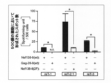
- FIG. 5 is a graph showing the results of 51 Cr release assays performed using epitope peptides such as Nef-138-8 (wt) at various concentrations.
- the 51 Cr release assay was performed by reacting effector cells with target cells incorporating 51 Cr at a ratio of 5: 1.
- the vertical axis represents the specific release rate (%) of 51 Cr.
- 1 is a schematic view of a method for producing human CD4SP cells having antigen specificity from T-iPS cells.
- the present invention comprises a step of differentiating iPS cells derived from human T cells (hereinafter also referred to as “T-iPS cells”) into CD4 / CD8 double negative (DN) cells, and the T cells of the CD4 / CD8DN cells.
- An antigen comprising a step of stimulating a receptor, and a step of differentiating the CD4 / CD8DN cell stimulated to a T cell receptor into a single positive T cell, specifically, a CD8SP cell and / or a CD4SP cell.
- a method for producing human CD8SP cells and / or CD4SP cells having specificity is provided.
- the “human” from which T cells are isolated in the present invention is not particularly limited, and even if it is a healthy person, a person with reduced immune function, an infectious disease such as a malignant tumor or chronic infection, or autoimmunity It may be a person suffering from a disease or the like.
- T cells obtained by the present invention are administered to humans from whom T cells are isolated from the viewpoint that rejection does not occur.
- the human and HLA types are preferably the same, and more preferably the same person as the human to whom the T cells obtained by the present invention are administered.
- T cell means a cell expressing an antigen receptor called T cell receptor (TCR) on its surface and its precursor cell (TCR (TCR ⁇ )). No pro-T cells, pre-T cells in which TCR ⁇ and pre-TCR ⁇ are associated, etc.).
- CD4 / CD8 double negative (DN) cell means a T cell that does not express both CD4 and CD8, and “CD4 / CD8 double positive (DP) cell” means both CD4 and CD8.
- DN CD4 / CD8 double negative
- DP CD4 / CD8 double positive
- SP single positive
- SP single positive
- the “CD4 single positive (SP) cell” means a T cell in which only CD4 is expressed among CD4 and CD8.
- human T cells induced into iPS cells are T cells expressing CD3 and CD8, and examples thereof include cytotoxic T cells that are CD8 positive cells.
- Human T cells induced into iPS cells are also T cells expressing CD3 and CD4, for example, T cells that are CD4 positive cells.
- Specific examples of such human T cells include helper / regulatory T cells that are CD4 positive cells, cytotoxic T cells (CTL) that are CD8 positive cells, naive T cells (CD45RA + CD62L + cells), and central memory T cells. (CD45RA-CD62L + cells), effector memory T cells (CD45RA-CD62L- cells), and terminal effector T cells (CD45RA + CD62L- cells).
- antigen specificity in T cells is provided by antigen-specific, rearranged TCR genes.
- antigen-specific CD8SP cells it is preferable to use antigen-specific CD8-positive T cells as human T cells induced into iPS cells.
- antigen-specific CD4SP cells it is preferable to use antigen-specific CD4-positive T cells as human T cells induced into iPS cells.
- the human T cell differentiated from an iPS cell has the same or substantially the same antigen specificity as the human T cell induced
- human T cells induced into iPS cells can be isolated from human tissues by a known technique.
- the human tissue is not particularly limited as long as it is a tissue containing the T cell, and examples thereof include peripheral blood, lymph nodes, bone marrow, thymus, spleen, umbilical cord blood, and lesioned tissue.
- peripheral blood and umbilical cord blood are preferable from the viewpoint of low invasiveness to humans and easy preparation.
- flow cytometry using an antibody against a cell surface marker such as CD4 or CD8 and a cell sorter as shown in Examples described later can be mentioned.
- desired T cells can be isolated using cytokine secretion or functional molecule expression as an index.
- T cells have different cytokines secreted depending on Th1 type or Th2 type. Therefore, T cells having a desired Th type can be isolated by selecting such cytokines as indicators. .
- cytotoxic T cells CTL can be isolated using secretion or production of granzyme or perforin as an index.
- a method of purification using an affinity column or the like on which a desired antigen is immobilized can be employed.
- T cells having desired antigen specificity can also be purified from human tissues.
- the “iPS cell” in the present invention is a cell also called an induced pluripotent stem cell or an inducible pluripotent stem cell, and is induced by introducing a cell reprogramming factor into the T cell. can do.
- the “cell reprogramming factor” is a factor that can impart differentiation pluripotency to a somatic cell by being introduced into the T cell alone or in cooperation with other differentiation pluripotency factors.
- Oct3 / 4 c-Myc, Sox2, Klf4, Klf5, LIN28, Nanog, ECAT1, ESG1, Fbx15, Eras, ECAT7, ECAT8, Gdf3, Sox15, ECAT15-1, ECAT15-2, It is preferably at least one protein selected from the group consisting of Fthl17, Sal14, Rex1, Utf1, Tcl1, Stella, ⁇ -catenin, Stat3 and Grb2. Furthermore, among these proteins, it is more preferable to introduce Oct3 / 4, c-Myc, Sox2 and Klf4 (4 factors) into the T cells from the viewpoint that iPS cells can be established efficiently with few factors.
- OCT4, SOX2, KLF4, C-MYC and NANOG are selected from the viewpoint of further enhancing the induction efficiency of human CD8-positive T cells or CD4 positive T cells into iPS cells. It is more preferable to introduce it into human CD8 positive T cells or CD4 positive T cells, and it is particularly preferable to introduce OCT4, SOX2, KLF4, C-MYC, NANOG and LIN28 into human CD8 positive T cells or CD4 positive T cells. Further, from the viewpoint of reducing the risk of canceration of the resulting pluripotent stem cells, it is more preferable to introduce Oct3 / 4, Sox2 and Klf4 (3 factors) excluding c-Myc into the T cells.
- a method for establishing iPS cells derived from human T cells is not particularly limited, and a known method may be appropriately selected and used. it can.
- a method using a protein introduction reagent a method using a protein introduction domain (PTD) fusion protein, an electroporation method, a microinjection method Is mentioned.
- PTD protein introduction domain
- the nucleic acid (eg, cDNA) encoding the cell reprogramming factor when introduced into the T cell in the form of a nucleic acid encoding the cell reprogramming factor, contains a promoter that functions in the T cell. And can be introduced into cells by infection, lipofection method, liposome method, electroporation method, calcium phosphate coprecipitation method, DEAE dextran method, microinjection method, electroporation method .
- expression vectors examples include lentiviruses, retroviruses, adenoviruses, adeno-associated viruses, herpes viruses, Sendai virus and other viral vectors, and animal cell expression plasmids. From the viewpoint of high efficiency and a large number of introduced gene copies, it is preferable to introduce a nucleic acid encoding the cell reprogramming factor into the T cells using Sendai virus.
- the promoter used in such an expression vector examples include SR ⁇ promoter, SV40 promoter, LTR promoter, CMV promoter, RSV promoter, HSV-TK promoter and the like.
- a promoter may be one that can control the expression of a gene inserted downstream of the promoter depending on the presence or absence of a drug (eg, tetracycline).
- the expression vector may further contain an enhancer, a poly A addition signal, a selection marker gene (for example, neomycin resistance gene), an SV40 replication origin, and the like.
- the T cells are interleukin-2 (IL-2) prior to introduction of the cell reprogramming factor.
- Is preferably stimulated with anti-CD3 antibody and anti-CD28 antibody in the presence of phytohemagglutinin (PHA), interleukin-2 (IL-2), alloantigen-expressing cells, anti-CD3 antibody, and anti-CD3 antibody.
- PHA phytohemagglutinin
- IL-2 interleukin-2
- alloantigen-expressing cells anti-CD3 antibody
- anti-CD3 antibody anti-CD3 antibody
- anti-CD3 antibody anti-CD3 antibody
- anti-CD3 antibody anti-CD3 antibody
- anti-CD3 antibody anti-CD3 antibody
- the anti-CD3 antibody and the anti-CD28 antibody may be ones to which magnetic beads or the like are bound, and instead of adding these antibodies to the medium, the anti-CD3 antibody and the anti-CD28 antibody are bound to the surface.
- Stimulation may be given by culturing the T cells for a certain period on a cultured dish.
- stimulation may be given by adding an antigen peptide recognized by the T cells (for example, human T cells) together with feeder cells to the medium.
- the concentration of PHA added to the medium is not particularly limited, but it is preferably 1 to 100 ⁇ g / ml.
- the concentration of IL-2 added to the medium is not particularly limited but is preferably 1 to 200 ng / ml.
- the concentration of the anti-CD3 antibody and anti-CD28 antibody added to the medium is not particularly limited, but is preferably 1 to 10 times the culture amount of the T cells.
- the concentration of the anti-CD3 antibody and the anti-CD28 antibody bound on the surface of the culture dish is not particularly limited, but the concentration at the time of coating is 0 for the anti-CD3 antibody. It is preferably 1 to 100 ⁇ g / ml, preferably 1 to 100 ⁇ g / ml, and 0.1 to 10 ⁇ g / ml for anti-CD28 antibody.
- the culture period for performing such stimulation is a period sufficient to give such stimulation to the T cells, and the T cells can be expanded to the number of cells necessary for introduction of the cell reprogramming factor.
- the period is not particularly limited, but is usually 1 to 14 days, but may be 1 to 6 days, and preferably 2 to 7 days, more preferably 2 to 4 days from the viewpoint of gene transfer efficiency. . Furthermore, from the viewpoint of increasing gene transfer efficiency, it is preferable to culture on a culture dish coated with retronectin.
- a medium for culturing the T cell and adding PHA, IL-2, anti-CD3 antibody and / or anti-CD28 antibody, etc. for example, a known medium suitable for culturing the T cell (more specifically, Roswell Park Memorial Institute (RPMI) 1640 medium, minimum essential medium ( ⁇ -MEM), Dulbecco's modified Eagle medium (DMEM), F12 medium, etc.) containing other cytokines and human serum can be used.
- RPMI Roswell Park Memorial Institute
- ⁇ -MEM minimum essential medium
- DMEM Dulbecco's modified Eagle medium
- F12 medium etc.
- the medium was supplemented with amino acids necessary for culture (eg L-glutamine) and antibiotics (eg streptomycin, penicillin). May be.
- IL-7 and IL-15 it is preferable to add IL-7 and IL-15 to the medium.
- concentration of IL-7 and IL-15 it is preferably 1 to 100 ng / ml.
- the T cell into which the cell reprogramming factor has been introduced is preferably cultured on a feeder cell layer.
- a feeder cell layer For example, a mouse embryo fibroblast (MEF), STO cell, and SNL cell which stopped cell division by irradiation of radiation or antibiotic treatment are mentioned.
- the cell reprogramming factor when introducing the cell reprogramming factor into the T cells with basic fibroblast growth factor (bFGF), or Thereafter, it is preferably added to the medium.
- bFGF basic fibroblast growth factor
- HDAC histone deacetylase
- VPA valproic acid
- trichostatin A sodium butyrate
- small molecule inhibitors such as MC1293, M344, siRNA for HDAC, etc.
- a G9a histone methyltransferase inhibitor a small molecule inhibitor such as BIX-01294, an siRNA against G9a
- a p53 inhibitor a small molecule inhibitor such as Pifthrin- ⁇ (PFT- ⁇ ), an siRNA against p53, etc.
- PFT- ⁇ Pifthrin- ⁇
- siRNA against p53 etc.
- the cell reprogramming factor is added to the medium at the time of introducing the cell reprogramming factor into the T cell or thereafter.
- a viral vector when introducing a cell reprogramming factor into T cells, it is preferable to add protamine sulfate to the medium from the viewpoint that the vector is likely to bind to the T cells. .
- a known medium suitable for culturing the T cells is gradually replaced with a medium suitable for culturing iPS cells. It is preferable to culture while continuing.
- a medium suitable for culturing such iPS cells a known medium can be appropriately selected and used.
- knockout serum substitute, L-glutamine, non-essential amino acid, 2-mercaptoethanol, b-FGF, etc. Dulbecco's modified Eagle's medium / F12 medium (human iPS cell medium) containing
- the feeder cells are used from the viewpoint of further increasing the efficiency of induction into iPS cells.
- it is preferably cultured under a low oxygen concentration condition (oxygen concentration: 5%, for example) in a medium further supplemented with 1 to 1000 ⁇ M ROCK inhibitor, and 1 to 1000 ⁇ M MEK inhibitor (for example, PD0325901) and 1-1000 ⁇ M GSK3 inhibitor (eg, CHIR99021) are preferably added to the medium until colony formation described below.
- T-iPS cells iPS cells derived from the T cells as described above can be performed by appropriately selecting a known technique.
- known methods include, for example, a method of observing and selecting the morphology of ES cells / iPS cell-like colonies as shown in the examples described later under a microscope, or knowing that they are specifically expressed in iPS cells.
- Recombinant T cells targeting drug resistance gene or reporter gene (GFP gene, etc.) at the gene locus of the gene are selected using drug resistance or reporter activity as an index.
- GFP gene drug resistance gene, etc.
- iPS cells iPS cells
- T-iPS cells iPS cells
- the confirmation that the cells selected in this way are derived from the T cells can be performed, for example, by detecting the state of TCR gene rearrangement by genomic PCR, as shown in Examples below. it can.
- the timing for selecting and recovering these cells can be appropriately determined while observing the growth state of the colonies, and is generally 10 to 40 days after introduction of the cell reprogramming factor into the T cells, preferably 14 to 28 days.
- the culture environment is preferably 5% CO 2 , 35 to 38 ° C., more preferably 37 ° C. unless otherwise specified.
- T-iPS cells In order to differentiate iPS cells derived from human T cells (ie, T-iPS cells) into CD4 / CD8DN cells, from the viewpoint of facilitating differentiation into mesodermal system, first, T-iPS cells May not contain cytokines on feeder cells (preferably stromal cells, more preferably human stromal cells), but cytokines, serum (eg, fetal bovine serum (FBS)), insulin, transferrin, It is preferable to culture in a medium containing sodium selenate, L-glutamine, ⁇ -monothioglycerol, ascorbic acid and the like.
- cytokines eg, fetal bovine serum (FBS)
- FBS fetal bovine serum
- the stromal cells to be used are preferably OP9 cells, 10T1 / 2 cells (C3H10T1 / 2 cells) subjected to treatment such as irradiation from the viewpoint of facilitating differentiation into a hematopoietic system.
- the cytokine added to the medium is preferably at least one cytokine selected from the VEGF, SCF, TPO, SCF, and FLT3L groups. SCF and TPO, or VEGF, SCF and FLT3L are more preferable.
- the medium examples include X-VIVO medium, Iskov modified Dulbecco medium (IMDM medium), ⁇ -MEM, and DMEM, and a bag-like structure containing “hematopoietic progenitor cells” (“T-iPS sac”). From the standpoint of high formation efficiency of "" or "ES sac"), IMDM medium is preferable.
- the T-iPS cell culture period is preferably a period until the formation of a T-iPS sac, preferably 8 to 14 days from the start of T-iPS cell culture, more preferably 10 to 10 days. 14 days.
- the culture environment is not particularly limited, but preferably 5% CO 2 , 35 to 38 ° C., more preferably 37 ° C.
- culture is performed under low oxygen concentration conditions (oxygen concentration: for example, 5 to 20%). Is more preferable.
- T-iPS cells In order to differentiate T-iPS cells into CD4 / CD8DN cells, cells (hematopoietic progenitor cells, CD34 positive cells, blood cells, etc.) contained in the T-iPS sack obtained above are then cytokines. It is preferable to culture on feeder cells (preferably stromal cells, more preferably human stromal cells) in a medium containing serum or serum (for example, FBS). Cells present inside the T-iPS sack can be separated by passing through physical means, eg, a sterilized sieve device (eg, a cell strainer).
- a sterilized sieve device eg, a cell strainer
- the stromal cells used for this culture are OP9-DL1 cells, OP9-DL4 cells, 10T1 / 2/10 treated with radiation and the like from the viewpoint of inducing differentiation into T lymphocytes via a notch signal.
- DL4 cells and 10T1 / 2 / DL1 cells are preferred.
- Examples of cytokines added to the medium include IL-7, FLT3L, VEGF, SCF, TPO, IL-2, and IL-15.
- any one or more of SCF, TPO, FLT-3L and IL-7, or all in combination can be added.
- IL-7 and FLT3L are preferable from the viewpoint of helping differentiation of early T cells.
- SCF is preferably not contained in the medium.
- the concentration of IL-7 added to the medium is preferably 0.1 to 9. from the viewpoint that CD3-positive CD56-negative T lineage cells are easily obtained and differentiation into CD8SP cells or CD4SP cells is easily induced. 5 ng / ml, more preferably 1 to 4 ng / ml.
- the concentration of FLT3L added to the medium is preferably 0.1 to 100 ng / ml from the viewpoint of enhancing blood cell proliferation. Examples of the medium include ⁇ -MEM medium, DMEM medium, and IMDM medium, and ⁇ -MEM medium is preferable from the viewpoint of easy maintenance of stromal cells.
- amino acids eg, L-glutamine
- antibiotics eg, streptomycin, penicillin
- the culture period of the cells contained in the T-iPS sack is a period until the T cell receptor (TCR) is expressed on the cell surface of the CD4 / CD8DN cells obtained by differentiation in this way. Preferably, it is preferably 14 to 28 days from the start of culturing the cells contained in the T-iPS sack.
- the culture environment is not particularly limited, but preferably 5% CO 2 , 35 to 38 ° C., more preferably 37 ° C.
- T cell receptor TCR
- Comparative Example 1 Whether or not the T cell receptor (TCR) is expressed on the cell surface of CD4 / CD8DN cells is determined as shown in Comparative Example 1 described below, such as anti-TCR ⁇ antibody, anti-CD3 antibody, anti-CD4 antibody and anti-CD8. It can be evaluated by flow cytometry using an antibody (see FIG. 20).
- the DP stage has a higher frequency of having a TCR gene rearrangement pattern different from the original human T cell, It became clear for the first time by the present inventors, and in the process of redifferentiating T-iPS cells into T cells, particularly in the process from the CD4 / CD8DN stage to the CD4 / CD8DP stage, further assembly (reception of the TCRA gene by RAG1 and RAG2). It was strongly suggested that (body correction) occurred.
- TCR is expressed even at the DN stage in T cells obtained by redifferentiating T-iPS cells.
- T cells do not express TCR at the DN stage. Therefore, it was surprising that TCR was expressed in DN cells obtained by differentiating T-iPS cells.
- the TCR signal via the peptide-MHC complex stops the expression of the RAG gene and suppresses further assembly of the TCR gene. It has also been revealed that a TCR signal-like signal by an anti-CD3 antibody has a similar effect (that is, an effect of suppressing further assembly of the TCR gene).
- the present inventors succeeded in suppressing further rearrangement of the TCR gene by stimulating the TCR expressed in DN cells derived from T-iPS cells, and thus the same as the original human T cells.
- the present inventors succeeded in improving the appearance frequency of T cells having a TCR gene rearrangement pattern.
- CD4 / CD8DN cells derived from T-iPS cells are stimulated via TCR expressed on the cell surface.
- further rearrangement of the TCRA gene can be suppressed.
- the same rearrangement pattern of the TCR gene as that of the original human T cell is obtained. It is possible to make the appearance frequency of T cells to be extremely high.
- PHA, anti-CD3 antibody, anti-CD28 antibody, and human T cells that are the origin of T-iPS cells are specifically bound.
- the method of contacting one substance with CD4 / CD8DN cells derived from T-iPS cells is preferred, and the method of contacting specific peptide / HLA complex-expressing cells is more preferred from the viewpoint of giving a physiological stimulus. Further, from the viewpoint of placing importance on the uniformity of stimulation, a method of contacting an antibody or a reagent is more preferable.
- the contact method can be performed, for example, by adding PHA or the like to the medium and culturing the T cells for a certain period, as shown in the examples described later.
- the anti-CD3 antibody and the anti-CD28 antibody may be ones to which magnetic beads or the like are bound, and instead of adding these antibodies to the medium, the anti-CD3 antibody and the anti-CD28 antibody are bound to the surface.
- Stimulation may be given by culturing the T cells for a certain period on a cultured dish.
- stimulation may be given by adding the antigen peptide together with feeder cells to the medium.
- the concentration of PHA added to the medium is not particularly limited, but is preferably 1 to 100 ⁇ g / ml.
- the concentration of the anti-CD3 antibody and anti-CD28 antibody added to the medium is not particularly limited, but is preferably 1 to 10 times the culture amount of the T cells.
- the concentration of anti-CD3 antibody and anti-CD28 antibody bound on the surface of the culture dish to stimulate TCR of CD4 / CD8DN cells is not particularly limited. It is preferably 0.1 to 100 ⁇ g / ml, and 0.1 to 10 ⁇ g / ml for the anti-CD28 antibody.
- the culture period of the cells contained in this T-iPS sack is the period per time T cell receptor (TCR) is expressed on the cell surface of CD4 / CD8DN cells obtained by differentiation in this way. Preferably, it is preferably 7 to 29 days after the start of culturing of the cells contained in the T-iPS sack.
- the culture environment is not particularly limited, but preferably 5% CO 2 , 35 to 38 ° C., more preferably 37 ° C.
- CD8 + memory stem cells stem cell-like memory T cells (TSCM )
- TSCM stem cell-like memory T cells
- an inhibitor against glycogen synthase kinase-3 ⁇ may be added to the medium.
- any inhibitor can be used as long as it can activate the Wnt signaling pathway by suppressing phosphorylation by ⁇ -catenin by GSK3 ⁇ .
- a 4,6-disubstituted pyrrolopyrimidine compound (TWS119) Is mentioned.
- T SCM is "Luca Gattinoni et al., Nature Medicine, 2011 years, Vol. 17, 1290-1298 page” See.
- For the relationship between inhibition of GSK3 ⁇ and enhanced production of T SCM see “Luca Gattinoni et al., Nature Medicine, 2009, Vol. 15, pp. 808-813”.
- CD4 / CD8DN cells into CD8SP cells or CD4SP cells-
- the CD4 / CD8DN cells derived from T-iPS cells are stimulated via TCR expressed on the cell surface, whereby the T-iPS cells are converted to DN. It has been found that further rearrangement (receptor correction) of the TCRA gene that can occur as it differentiates from the stage to the DP stage, and further to the SP stage (eg, CD8SP stage or CD4SP stage).
- the method for producing human CD8SP cells or CD4SP cells having antigen specificity of the present invention after stimulating CD4 / CD8DN cells derived from T-iPS cells via the TCR expressed on the cell surface, in the CD8SP cell or CD4SP cell obtained by redifferentiation by differentiating the cell into a single positive T cell, specifically, a CD8SP cell or a CD4SP cell, the same TCR gene as that of the original human T cell is regenerated.
- the frequency of appearance of T cells having a configuration pattern can be made extremely high.
- T-iPS cell-derived T lineage cells differentiate into not only CD4 / CD8DN cells but also CD4 / CD8DP cells. Is also present. Therefore, in this “step of differentiating CD4 / CD8DN cells stimulated by T cell receptor into single positive T cells”, CD4 / CD8DN cells stimulated by T cell receptor are changed to CD8SP cells or CD4SP cells. This includes not only the step of differentiating, but also differentiating CD4 / CD8DP cells in which TCR is stimulated in the DN stage into CD8SP cells or CD4SP cells.
- the CD4 / CD8DN cells are contained in a medium containing cytokine, serum (for example, human serum) and the like. It is preferable to culture in Any cytokine can be added to the medium as long as it can differentiate CD4 / CD8DN cells into CD8SP cells or CD4SP cells. Examples thereof include IL-7, IL-15, and IL-2. Among these, it is preferable to add IL-7 and IL-15 in combination from the viewpoint of selecting the CD8 lineage and easily generating memory-type CD8 + T cells in differentiation into CD8SP cells.
- the concentration of IL-7 and IL-15 added is preferably 1 to 20 ng / ml.
- the medium include RPMI-1640 medium, X-VIVO medium, DMEM medium, and ⁇ -MEM medium. From the viewpoint of being more suitable for the growth of blood cells, RPMI-1640 medium or X-VIVO medium is preferable. preferable.
- the medium includes amino acids necessary for culture (eg, L-glutamine), antibiotics (eg, streptomycin, penicillin), IL-7, IL-15, etc.
- the cytokine may be added.
- CD4 / CD8DN cells may be co-cultured with feeder cells.
- a feeder cell From a viewpoint of promoting the differentiation and proliferation to a CD8SP cell or a CD4SP cell through cell contact etc., it is preferable that it is a peripheral blood mononuclear cell (PBMC).
- PBMC peripheral blood mononuclear cell
- an auto (from the same person) relationship with CD4 / CD8DN cells from the viewpoint of supporting survival while preventing excessive TCR stimulation.
- peripheral blood mononuclear cells that present antigen peptides that specifically bind to human T cells that are the origin of CD4 / CD8DN cells, from the viewpoint of continuing to stimulate TCR and to suppress further reconstitution of TCR. More preferably, cells are used.
- the culture period for differentiating the CD4 / CD8DN cells into CD8SP cells or CD4SP cells is preferably 2 to 4 weeks.
- the culture environment is not particularly limited, but preferably 5% CO 2 , 35 to 38 ° C., more preferably 37 ° C.
- CD8SP cells or CD4SP cells induced to differentiate in this way are derived from T-iPS cells and derived from the T cells from which the T-iPS cells are derived is described in, for example, the Examples described later. As shown, it can be done by detecting the status of TCR gene rearrangement by genomic PCR.
- the CD8SP cells or CD4SP cells obtained in this way can be isolated by appropriately selecting a known technique.
- a known technique for example, flow cytometry using an antibody against a cell surface marker such as CD8 or CD4 and a cell sorter as shown in the Examples described later can be mentioned.
- a desired antigen for example, in the case of a CD8SP cell, an antigen recognized by the T cell that is the origin of the CD8SP cell, a CD4SP cell
- a method of purifying using an affinity column or the like on which an antigen (recognized by a T cell that is a source of CD4SP cells) is immobilized, an MHC multimer (for example, MHC tetramer) bound with the antigen is used.
- a method of purifying “T cells having a desired antigen specificity” can also be employed.
- CD8SP cells obtained by the present invention do not express PD-1, whereas CCR7 is expressed together with CD27 and CD28 representing the phenotype of central memory T cells, and telomeres are also originally derived. It is longer than the T cell that has become, and has a high self-renewal ability. Therefore, according to the present invention, T cells having the same TCR gene rearrangement pattern as the original T cells (eg, CD8SP cells) do not express PD-1, and express CD27, CD28 and CCR7. Cells can be produced. T cells collected from humans differ from the obtained T cells in that they express PD-1 and do not express CD27, CD28, and CCR7. CD4SP cells are considered to be similar.
- the cells may be stimulated every 1 to 2 weeks.
- stimulation for CD8SP cells, anti-CD3 antibody, anti-CD28 antibody, IL-2, IL-7, IL-15, an antigen recognized by the CD8SP cell, an MHC multimer bound with the antigen, Examples include contact with at least one substance selected from the group consisting of feeder cells in an allo relationship with the CD8SP cells and feeder cells in an auto relationship with the CD8SP cells.
- such stimulation includes anti-CD3 antibody, anti-CD28 antibody, IL-2, IL-7, IL-15, an antigen recognized by the CD4SP cell, and a large amount of MHC bound to the antigen.
- the human CD8SP cell or CD4SP cell produced by the method of the present invention has an antigen-specific immune function as shown in the Examples below. Furthermore, in particular, CD8SP cells do not express PD-1, which is one of the markers of exhausted T cells (that is, T cells whose long-term viability, self-renewal ability and / or effector function are significantly weakened). On the other hand, CCR7 is expressed together with CD27 and CD28 representing the phenotype of central memory T cells, and telomeres are longer than the original T cells and have high self-replicating ability. This is considered to be the same in CD4SP cells. Accordingly, human T cells produced by the method of the present invention are useful in the treatment or prevention of diseases such as tumors, infectious diseases (for example, chronic infectious diseases), and autoimmune disorders.
- diseases such as tumors, infectious diseases (for example, chronic infectious diseases), and autoimmune disorders.
- the present invention provides a human T cell produced by the method of the present invention, a pharmaceutical composition containing the human T cell, and a method of immune cell therapy (immunocell therapy or immunotherapy) using the human T cell.
- the present invention relates to human CD8SP cells and / or human CD4SP cells produced by the method of the present invention, pharmaceutical compositions comprising the human CD8SP cells and / or human CD4SP cells, and the human CD8SP cells and / or human CD4SP cells. Methods of immune cell therapy used are provided.
- the method of immune cell therapy of the present invention can be performed as follows, for example.
- T cells are collected from a human, preferably a human with the same HLA type, more preferably a subject to be treated.
- T-iPS cells are prepared from the T cells, and then the T-iPS cells are differentiated into T cells having the same TCR gene rearrangement pattern as the original T cells, preferably CD4SP cells or CD8SP cells.
- the obtained T cells express CCR7 together with CD27 and CD28, which represent the phenotype of central memory T cells, do not express PD-1, and are longer than the T cells from which telomeres are derived. It has a high self-replicating ability. Therefore, if desired, the obtained cells may confirm the expression of CD27, CD28, CCR7 and / or PD-1.
- the T cells obtained in this way can be administered to the treatment subject.
- the administration of T cells to a subject to be treated is not particularly limited, but preferably can be administered parenterally, for example, intravenously, intraperitoneally, subcutaneously or intramuscularly. More preferably, it can be administered intravenously. Alternatively, it can be administered locally to the affected area.
- the pharmaceutical composition of the present invention can be prepared by formulating human CD8SP cells or CD4SP cells produced by the method of the present invention by a known pharmaceutical method.
- it can be used mainly parenterally as capsules, liquids, film coating agents, suspensions, emulsions, injections (intravenous injections, infusions, etc.).
- a pharmacologically acceptable carrier or medium specifically, sterile water or physiological saline, vegetable oil, solvent, base, emulsifier, suspending agent, surfactant, stabilizer, vehicle, Combined with preservatives, binders, diluents, tonicity agents, soothing agents, extenders, disintegrating agents, buffering agents, coating agents, lubricants, colorants, solubilizers or other additives as appropriate.
- a pharmacologically acceptable carrier or medium specifically, sterile water or physiological saline, vegetable oil, solvent, base, emulsifier, suspending agent, surfactant, stabilizer, vehicle, Combined with preservatives, binders, diluents, tonicity agents, soothing agents, extenders, disintegrating agents, buffering agents, coating agents, lubricants, colorants, solubilizers or other additives as appropriate.
- the dosage is appropriately selected according to the age, weight, symptom, health condition of the subject, type of composition (pharmaceutical, food and drink, etc.) and the like.
- the product (pharmaceutical product) of the composition of the present invention or its instructions may be labeled with an indication that it is used to treat or prevent a decrease in immune function.
- labeled product or instructions means that the product body, container, packaging, etc. are marked, or instructions, package inserts, promotional materials, or other printed materials that disclose product information. It means that the display is attached to.
- the method of immune cell therapy of the present invention comprises a step of isolating T cells having a desired antigen specificity from a human, a step of inducing iPS cells from T cells having the desired antigen specificity, and the iPS cells. Is differentiated into CD4 / CD8 double negative cells, the step of stimulating the T cell receptor of the CD4 / CD8 double negative cell, and the CD4 / CD8 double negative cell stimulated of the T cell receptor is transformed into CD8. Differentiating into single positive cells and / or CD4 single positive cells, and administering the obtained CD8 single positive cells and / or CD4 single positive cells into the human body.
- the human from which T cells are isolated is the same as the human to which the CD8SP cells and / or CD4SP cells obtained by the present invention are administered.
- the HLA types are preferably matched, and more preferably the same person as the human to which the CD8SP cells and / or CD4SP cells obtained by the present invention are administered.
- human T cells produced by the method of the present invention may be administered as they are, and as described above, they are administered in the form of a formulated pharmaceutical composition. May be.
- NLA138-8 (wt) -specific CTL strain was obtained from PBMCs of HIV-1 infected patients who are A24 type HLA, as described in “Kawana-Tachikawa, A. et al., J Virol 2002, 76, 11982-11988”. Established according to the description.
- peripheral blood T cells were stimulated and activated by beads coated with ⁇ -CD3 / CD28 antibody (Miltenyi Biotec).
- CTL clones were stimulated and activated by PHA (manufactured by Sigma-Aldrich).
- PHA manufactured by Sigma-Aldrich
- the CTL clone was activated by stimulation with PBMCs (allogeneic antigen-expressing cells) derived from other people than the HIV-1-infected patients irradiated. It has been confirmed that there is no significant difference in the properties of the obtained T-iPS cells between the case of stimulation by PHA and the case of stimulation by alloantigen-expressing cells.
- a reprogramming factor was introduced into the activated cells via a retrovirus vector (pMXs retrovirus vector) or a Sendai virus vector (SeV vector), and 10 ng / ml (200 U) IL-2 in RH10 medium.
- the cells were cultured with 5 to 10 ng / ml IL-7 and 5 to 10 ng / ml IL-15 (manufactured by Peprotech). Then, it was gradually replaced with human iPS medium (manufactured by Wako) containing bFGF and the like.
- the viral vector was introduced into the cells by centrifugation on a retronectin-coated plate (Takara).
- the composition of the RH10 medium is as follows. RPMI-1640 supplemented with 10% human AB serum, 2 mM L-glutamine, 100 U / ml penicillin and 100 ng / ml streptomycin.
- the composition of the human iPS medium is as follows. DMEM / F12FAM supplemented with 20% KSR, 2 mM L-glutamine, 1% non-essential amino acid, 10 ⁇ M 2-mercaptoethanol and 5 ng / ml b-FGF.
- siRNA L527 (“Nishimura, K. et al., J Biol Chem, 2011, 286, 4760- 4760-) was established on an iPS clone established by Lipofectamine RNAi Max (Invitrogen). Page 4771 ").
- Immunocytochemical staining was performed using the following antibodies in accordance with the description in “Takayama, N. et al., J Exp Med, 2010, 207, 2817-2830”. Numerical values in parentheses indicate the dilution ratio of each antibody.
- SSEA-4 (1:50, FAB1435P, manufactured by R & D Systems)
- Tra-1-60 (1: 100, MAB4360, manufactured by Millipore)
- Tra-1-81 (1: 100, MAB4381, manufactured by Millipore
- HLA-A24 1: 100, BIH0964, manufactured by Veritas
- the micrograph was performed using an Axio Observer Z1 fluorescence microscope (Carl Zeiss).
- Genomic DNA was processed using a methyl easy exceed rapid DNA bisulfite conversion kit (Methyl Easy Xceed Rapid DNA Bisulfite Modification Kit, manufactured by Human Genetic Signatures) according to the instructions for use thereof.
- the promoter regions of human Oct3 / 4 and Nanog gene were amplified by PCR using EpiTaq HS (manufactured by Takara).
- the obtained PCR product was inserted into a pGEM-T-Easy vector (Promega), cloned, and subjected to sequencing.
- Table 1 shows the primer sequences (SEQ ID NOs: 5 to 8) used for PCR.
- RT-PCR was performed as described in “Takayama, N. et al., J Exp Med, 2010, 207, 2817-2830”. Table 2 shows the sequences of target genes and PCR primers (SEQ ID NOs: 9 to 43) used for the analysis.
- Quantitative PCR was performed using a TaqMan array human stem cell pluripotency card and a special card (manufactured by TaqMan Array Human Stem Cell Pluripotency Card and Customized Card, Applied Biosystems).
- Genomic DNA was extracted from about 5 ⁇ 10 6 cells using a QIAamp DNA kit (Qiagen) according to the instructions for use.
- Table 3 shows primers (SEQ ID NOs: 44 to 81) used for PCR for analyzing the reconstruction of the TCRB gene.
- PCR was performed using the primers (SEQ ID NOs: 82 to 127) and LATaq HS (manufactured by Takara) shown in FIG. 1 and Table 4. PCR is performed at 95 ° C. for 30 seconds, 68 ° C. for 45 seconds and 72 ° C. for 6 minutes for 3 cycles, 95 ° C. for 30 seconds, 62 ° C. for 45 seconds and 72 ° C. Amplification step consisting of 6 minutes was programmed and performed for 15 cycles and 12 cycles of amplification step consisting of 15 seconds at 95 ° C., 30 seconds at 62 ° C. and 6 minutes at 72 ° C.
- the main band within the expected molecular weight range was purified using a QIA quick gel extraction kit (Qiagen) and subjected to sequencing. Segment usage for V, D and J was identified using an online tool (IMGT / V-Quest) and comparing to the ImMunoGeneTics (IMGT) database (http://www.cines.fr/). Lefranc, MP, Leukemia, 2003, 17, 2003, pages 260-266 ”). The gene fragment (segment) was named according to the IMGT nomenclature.
- SMART method A method based on the switch mechanism at the 5′-end of the reverse transcript (SMART method, “Du, G., et al., J Immunol Methods, 2006, 308, 19-35). Double-stranded cDNA was synthesized using a super smart cDNA synthesis kit (Super SMART (TM) cDNA synthesis kit, manufactured by Clontech Laboratories) according to the instruction manual.
- Super SMART (TM) cDNA synthesis kit manufactured by Clontech Laboratories
- the synthesized double-stranded cDNA was amplified using Advantage 2 PCR kit (manufactured by BD Clontech) and TCRA-specific amplification or TCRB-specific amplification from the amplified cDNA using the primers (SEQ ID NOs: 128 to 130) shown in Table 5 Amplification was performed.
- the obtained PCR product was inserted into a pGEM-T-Easy vector (Promega), cloned, and subjected to sequencing.
- T cells were incubated with ⁇ -CD3 / 28 beads and 10 ⁇ g / ml brefeldin A (BFA, Invitrogen).
- the cells are recovered, fixed with a fixing / membrane permeabilization solution (Fixation / Permeabilization solution, BD Pharmingen), and using FITC-conjugated anti-granzyme B antibody (BD Bioscience), the instructions for use thereof. Intracellular staining was performed as follows.
- T cells were incubated with ⁇ -CD3 / 28 beads and cultured together with FITC-conjugated anti-CD107a antibody (manufactured by BioLegend).
- the cells thus prepared were collected using a FACS Aria II apparatus (BD Bioscience) and analyzed using Flowjo software (Treestar).
- Total RNA was extracted from human ES cells, T-iPS cells, redifferentiated T cells, peripheral blood T cells and peripheral blood NK cells using an RNeasy micro kit (Qiagen).
- telomere length by Flow-FISH The length of telomeres was measured using a DAKO telomere PNA kit / FITC (manufactured by DAKO) as described in “Neuber, K. et al., Immunology, 2003, 109, 24-31”.
- HLA type: A24 HIV chronically infected persons
- PBMCs derived from HIV chronically infected persons were isolated and antigenic peptides derived from HIV-1 Nef protein (Nef-138-8 (wt); RYPLTFGW, SEQ ID NO: 1, “Miyazaki, E Et al., AIDS, 2009, Vol. 23, pages 651 to 660)), a CD8 + CTL clone specific to that was established.
- One of the clones, named H25-4 was stimulated with 5 ⁇ g / ml PHA.
- the activated H25-4 encodes two Sendai virus (SeV) vectors: cistronically expressed four factors (OCT4, SOX2, KLF4 and c-MYC) and the miR-302 target sequence.
- SeV vector See SeVp [KOSM302L], “Nishimura, K. et al., J Biol Chem, 2011, 286, 4760-4771”) and SeV vector encoding SV40 large T antigen (SeV18 + SV40 / TS15 ⁇ F, “Fusaki” , N., et al., Proc Jpn Acad Ser B Phys Biol Sci, 2009, Vol. 85, pages 348-362 ”). Then, by introducing these two types of SeV vectors into H25-4 activated with PHA, it was possible to confirm the appearance of a sufficient number of human ES cell-like colonies in the subsequent culture for 40 days. (See Figures 3 and 4).
- CD3 + T cell-derived ES cell-like colonies and H25-4-derived ES cell-like colonies have alkaline phosphatase (AP) activity.
- the expression of pluripotent cell markers SSEA-4, Tra-1-60 and Tra-1-81) was observed (see FIGS. 4 and 5). It was also confirmed that human ES cell-related genes were expressed even after the expression of exogenous reprogramming factors from the integrated provirus (TkT3V1-7) or cytoplasmic SeVRNAs (H254SeVT-3) was terminated. (See FIGS. 6 and 7).
- ⁇ TCR gene rearrangement in T-iPS cells Reconstitution of the TCR ⁇ gene is known to be involved in the normal ⁇ T cell development process in the thymus. Therefore, next, it was confirmed retrospectively whether or not the T-iPS cells established above were derived from ⁇ T cells based on such reconstitution. That is, a multiplex PCR primer for analyzing the TCRB gene assembly was designed by the BIOMED-2 consortium (see “van Dongen, JJ, et al., Leukemia, 2003, Vol. 17, pages 2257 to 2317”). Moreover, the primer for detecting TCRA gene assembly was designed uniquely (refer FIG. 1). Then, PCR was performed using such primers to detect the rearrangement of the TCR ⁇ gene in the T-iPS cells. The obtained results are shown in FIGS.
- TCRB gene and TCRA gene assembly were identified as a single band in each of the TkT3V1-7 and H254SeVT-3 alleles.
- the structure of the antigen recognition site of TCR is composed of three complementarity determining regions (CDR1, CDR2 and CDR3). Furthermore, in these three regions, CDR3 has the most diverse sequence because it extends to the V (D) J binding region into which various random nucleotides (N-nucleotides or P-nucleotides) are inserted ( See “Alt, FW, et al., Proc Natl Acad Sci USA, 1982, 79, 4118-4122" and "Lafile, JJ, et al., Cell, 1989, 59, 859-870". ).
- the T-iPS cells are redifferentiated into types of cells derived from mesoderm, particularly hematopoietic stem / progenitor cells, according to a specific in vitro differentiation protocol.
- International Publication No. 2011-096482 “Vodyanik, MA, et al., Blood, 2005, 105, 617-626” and “Takayama, N., et al., Blood, 2008, 111, 5298- (See page 5306).
- the T-iPS cell nodules ( ⁇ 100 cells or less) were transferred onto irradiated C3H10T1 / 2 cells and in the presence of 20 ng / mL VEGF, 50 ng / mL SCF and 50 ng / mL FLT-3L (Peprotech). Co-cultured in EB medium.
- the composition of the EB medium is as follows.
- fetal bovine serum FBS
- 10 ⁇ g / mL human insulin 5.5 ⁇ g / mL human transferrin and 5 ng / mL sodium selenite
- 2 mM L-glutamine 0.45 mM ⁇ -monothioglycerol And IMDM supplemented with 50 ⁇ g / mL ascorbic acid.
- hematopoietic cells CD34 + hematopoietic stem / progenitor cells contained in the iPS sack were collected, and these cells were irradiated with OP9-DL1 cells.
- OP9-DL1 cells are cells provided by the RIKEN BioResource Center through the National Bioresource Project of the Ministry of Education, Culture, Sports, Science and Technology (Japan) (see “Watarai, H. et al., Blood, 2010, Vol. 115, pages 230-237”). .
- hematopoietic cells were differentiated into T lineage cells in OP9 medium in the presence of 10 ng / mL FLT-3L and 1 ng / mL IL-7 (“Ikawa, T. et al., Science, 2010, 329, See pages 93-96.)
- the composition of the OP9 medium is as follows. ⁇ MEM supplemented with 15% FBS, 2 mM L-glutamine, 100 U / ml penicillin and 100 ng / ml streptomycin.
- the CD4 / CD8DN stage or CD4 / CD8DP stage corresponds to the ⁇ chain assembly stage or the ⁇ chain assembly stage, respectively (“Von Boehmer, H. Advances in Immunology, 2004, 84 volumes”. , Pages 201-238 ").
- negative feedback control of gene assembly and deterrence against further rearrangement at the TCRB locus have been carried out very strictly (“Khor, B. et al., Current Opinion in Immunology, 2002, Vol. 14, 230-234). Page ").
- CD1a is derived from the CD45 + , CD3 + , TCR ⁇ + and CD5 + T lineage cells.
- - collected cells in DN and CD1a + DP stage, expression and sequences of TCRmRNA (SEQ ID NO: 131-149) were analyzed. The obtained results are shown in FIG. 21 and Tables 10 to 13.
- the base sequence of TCRB mRNA of T lineage cells and those of T-iPS cells were identical.
- TCRA mRNA contained the same sequence and a different sequence at the DN stage and DP stage.
- a different sequence is confirmed at a higher frequency in the DP stage than in the DN stage.
- redifferentiated DP cells derived from H254SeVT-3 have the same gene rearrangement pattern as the original T cells. The frequency of T cells was about a quarter.
- the TCRA gene of TkT3V1-7-derived T lineage cells has the same gene rearrangement as the original T cell as it progresses to the DN stage, DP stage, and CD8 + SP stage. It became clear that the frequency of T cells having a pattern decreased to 100%, 89%, and 75%.
- Example 1 Redifferentiation from T-iPS cells to CD8 single positive cells by the method of the present invention> As shown in Comparative Example 1, in the TCRA mRNA of T cells redifferentiated from T-iPS cells, the frequency at which the DP stage has a different TCR gene rearrangement pattern than the original human T cells than the DN stage Became clear. In addition, in T cells obtained by redifferentiating T-iPS cells, the expression of RAG1 and RAG2 was found to be stronger in the DP stage than in the DN stage.
- TCR TCR
- TCR ⁇ TCR (TCR ⁇ ) is present on the cell surface of T cells redifferentiated from T-iPS cells. It was also revealed for the first time that it was expressed.
- TCR ⁇ TCR
- TCR ⁇ TCR
- the nodule ( ⁇ 100 cells or less) of T-iPS cells (H254SeVT-3) obtained in Preparation Example 1 was transferred onto irradiated C3H10T1 / 2 cells, and 20 ng / Co-cultured in EB medium in the presence of mL VEGF, 50 ng / mL SCF and 50 ng / mL FLT-3L (manufactured by Peprotech).
- hematopoietic cells contained in the iPS sack were collected, transferred to irradiated OP9-DL1 cells, and in the presence of 10 ng / mL FLT-3L and 1 ng / mL IL-7, Hematopoietic cells were differentiated into T lineage cells in OP9 medium.
- the T lineage cells were collected and cultured in RH10 medium together with irradiated HLA-A24-PMBCs in the presence of 10 ng / mL IL-7 and 10 ng / mL IL-15.
- IL-7 signaling has been reported to contribute to CD8 lineage selection (“Chong, MM, et al., Immunity, 2003, 18, 475-487”, “Singer, A. et al., Nat Rev Immunol, 2008, 8, 788-801 ”and“ Park, JH, et al., Nat Immunol, 2010, 11, 257-264 ”), and IL- 7 and IL-15 have also been reported to be necessary for the generation of memory-type CD8 + T cells (“Becker, TC, et al., J Exp Med, 2002, 195, 1541-1548. "Tan, JT et al., J Exp Med, 2002, 195, 1523-1532", “Pric.” , M. et al., J Exp Med, 2002, 195, F49-52 ”and“ Kaneko, S. et al., Blood, 2009, 113, 1006-1015 ”).
- CD8SP cells appeared on the 60th day of culture, and these cells were confirmed to be derivatives of H254SeV-3 by the expression of HLA-A24.
- CD56 expressed on CD8 + T cells cultured in vitro was observed in these CD8SP cells (for CD56, see “Lu, PH et al., J Immunol, 1994. 153, pp. 1687-1696 ”).
- CD7 was also expressed in these CD8SP cells, and expression of CD2 was observed in some cells.
- most of these CD8SP cells did not express PD-1, which is one of the markers of exhausted T cells.
- FIG. 25, FIGS. 26 to 39, and Tables 8 and 9, which will be described later, are graphs showing the results when PHA stimulation was given to the T lineage cells as the first stimulation.
- FIGS. 26 to 39, and Tables 8 and 9, which will be described later are graphs showing the results when PHA stimulation was given to the T lineage cells as the first stimulation.
- Example 2 Antigen specificity of the CD8SP cell of the present invention>
- the redifferentiated CD8SP cells obtained in Example 1 have the same reconstitution pattern as the original H25-4 TCR gene, so that H25-4 can recognize an antigenic peptide showing specificity. It was. That is, all the redifferentiated T cells obtained in Example 1 were mixed with A24 / Nef-138-8 (wt) tetramer, and “Kawana-Tachikawa, A. et al., J Virol, 2002, Vol. 76, Flow cytometry analysis was performed as described on pages 11982-11988.
- the “A24 / Nef-138-8 (wt) tetramer” is a tetramerized antigen (Nef-138-8 (wt)) and HLA (A24) recognized by H25-4. The obtained results are shown in FIG.
- CD8SP cells derived from T-iPS cells were stained with A24 / Nef-138-8 (wt) tetramer. Further, the ratio of CD8SP positive cells (antigen-specific CD8SP cells) in the CD8SP cells derived from T-iPS cells (CD8SP tetramer positive cells and CD8SP tetramer negative cells) was 77%. However, although not shown in the figure, CD8SP cells derived from T-iPS cells were stained by HLA-A24 tetramer presenting a peptide derived from HIV-1 envelope (RYLRDQQLL, SEQ ID NO: 2), which was used as a control. There wasn't.
- CD8 + cells that respond to the A24 / Nef-138-8 (wt) tetramer were collected, expanded, and stimulated again by PHA (this stimulation by PHA was combined with the second stimulation). To do). Then, by performing such a redifferentiation experiment several times independently, finally, CD8SP cells (reT-1, reT-2.1, reT, which react with A24 / Nef-138-8 (wt) tetramer). -2.2 and reT-3) were obtained.
- the expression patterns of CD3, CD4 and CD8 were PB CD8 + T cells, redifferentiated CD8SP cells and the original T cell clone H25-4. (Hereinafter also referred to as “H25-4 original T cell clone”).
- genes characterizing cytotoxicity such as granzyme B (GZMB), perforin (PFR1), IFN- ⁇ (IFNG) and FAS ligand (FASLG) are expressed in PB CD8 + T cells.
- GZMB granzyme B
- PFR1 perforin
- IFN- ⁇ IFN- ⁇
- FAS ligand FAS ligand
- the expression patterns of several factors in transcription or signal transduction and cell surface molecules are PB CD8 + T cells, redifferentiated CD8SP cells and H25-4 original T cells. It was the same among clones.
- Example 3 ⁇ High proliferation of CD8SP cells of the present invention>
- the number of T lineage cells obtained from co-culture of T-iPS cells and OP9-DL1 on 6 cm dishes was less than 10 5 .
- the H25-4 original T cell clone was amplified about 20 times, whereas the CD8 + cells were amplified 100 to 1000 times.
- central memory T cell markers eg, CCR7, CD27, and CD28
- central memory T cell markers eg, CCR7, CD27, and CD28
- the telomere shortened in the H25-4 original T cell clone by passing through the state of iPS cells with extremely high telomerase activity It is assumed that high replication ability could be imparted to redifferentiated T cells by re-elongation (“Takahashi, K. et al., Cell, 2007, vol. 131, pages 861 to 872”, “Marion, R. et al. M. et al., Cell Stem Cell, 2009, Volume 4, pages 141-154, “Monteiro, J. et al., J Immunol, 1996, Volume 156, pages 3287-3590” and “Weng, N.P. et al. , Immunity, 1998, Vol. 9, pages 151-157 ”). Therefore, the telomere length of CD8SP cells obtained by the method of the present invention was measured. The obtained results are shown in FIG.
- the redifferentiated T cells had a longer telomere length than the original T cell clone.
- CD8SP cells cloned cytotoxic T cells obtained by the method of the present invention rejuvenate into central memory-like T cells exhibiting excellent proliferative ability and viability through the state of T-iPS cells. It became clear that it was possible.
- Example 4 ⁇ Antigen-specific function of CD8SP cell of the present invention>
- One of the main mechanisms of cytotoxicity of CTLs is the secretion of cytolytic molecules that occur with TCR signals. Therefore, also in CD8SP cells (reT-2.2) obtained by the method of the present invention, the TCR is stimulated by ⁇ -CD3 / CD28 beads three times the amount of CD8SP cells, so that the cytolytic molecule can be obtained. Whether it was secreted was examined by intracellular staining. The obtained result is shown in FIG.
- FIG. 36 it was revealed that the cytolytic molecule granzyme B was produced and accumulated in the granules of redifferentiated CD8 T cells after stimulation with ⁇ -CD3 / 28 beads (see the left panel of FIG. 36). .
- CD107a known as lysosomal membrane protein 1 (LAMP1), is a granulocyte membrane protein that is transiently expressed in conjunction with degranulation that secretes cytolytic molecules when CTLs are stimulated. It is known that after its secretion, it returns to the cytoplasm (Rubio, V. et al., Nat Med, 2003, Vol. 9, pages 1377 to 1382). Therefore, the redifferentiated CD8 T cells of the present invention were cultured with or without stimulation with ⁇ -CD3 / 28 beads, and the CD107a molecule on the cell surface was captured by an antibody bound with a fluorescent dye. The obtained result is shown in FIG.
- LAMP1 lysosomal membrane protein 1
- HLA-A24 positive B-LCL is used as an antigen presenting cell.
- Functional assays ELISPOT (enzyme-linked immunospot, Enzyme-Linked ImmunoSpot) and 51 Cr release assay) using cells (cells that were identical in H25-4 T cell clone and patient origin) were performed. The obtained results are shown in FIGS.
- Gag-28-9 (wt) (KYKLKHIVW, SEQ ID NO: 3) used in these evaluations is an antigenic peptide (amino acid residues: positions 28 to 36) of the HIV-1 Gag protein
- Nef-138- 8 (2F) (RFPLTFGW, SEQ ID NO: 4) is a 1-residue mutant of Nef-138-8 (wt) in which tyrosine is substituted with phenylalanine, and both are present on HLA-A24.
- the redifferentiated CD8 T cells of the present invention responded to stimulation with Nef-138-8 (wt) as a specific antigen. Was found to produce significantly.
- the redifferentiated CD8 T cells of the present invention incorporated 51 Cr only in the presence of Nef-138-8 (wt). B-LCL was found to dissolve.
- CD8SP cells obtained by the method of the present invention release functional molecules and specifically kill target cells expressing the antigen, so that they are functional antigen-specific T cells. It was revealed.
- Example 5 ⁇ Redifferentiation from T-iPS cells to CD4 single positive cells by the method of the present invention>
- mature CD4SP cells are obtained from T-iPS cell-derived T lineage cells using a technique for stimulating TCR of redifferentiated T lineage cells. Tried to manufacture.
- T-iPS cells H254SeVT-3
- EB medium containing mL VEGF EB medium containing mL VEGF.
- SCF 20 ng / mL a small mass ( ⁇ 100 cells or less) of T-iPS cells (H254SeVT-3) obtained in Preparation Example 1 was transferred onto irradiated C3H10T1 / 2 cells, and 20 ng / Co-cultured in EB medium containing mL VEGF.
- hematopoietic cells contained in the iPS sack were collected, transferred to irradiated OP9-DL1 cells, SCF 20 ng / mL, TPO 10 ng / mL, FLT-3L 10 ng / mL.
- hematopoietic cells were transferred onto irradiated OP9-DL1 cells, and hematopoietic cells were differentiated into T lineage cells in OP9 medium in the presence of 5 ng / mL FLT-3L and 1 ng / mL IL-7. Thereafter, on day 35 of culture, 3 times the amount of hematopoietic cells, ⁇ -CD3 / CD28 beads or 5 ⁇ g / ml PHA was stimulated by adding to OP9 medium, and cells directed to the T lineage were treated with OP9. -Continued culturing on DL1 (stimulation with ⁇ -CD3 / CD28 beads or PHA is the primary stimulus).
- the T lineage cells were recovered and cultured in RH10 medium together with irradiated HLA-A24-PMBCs in the presence of 10 ng / mL IL-7 and 10 ng / mL IL-15.
- a CD8SP cell obtained by redifferentiation from an antigen-specific human T cell via an iPS cell the same TCR gene reconstitution as the original human T cell is performed.
- the appearance frequency of T cells having a pattern can be made extremely high.
- the human CD8SP cells obtained in this manner have an antigen-specific immune function. This is believed to be true for human CD4SP cells.
- PD-1 which is one of the markers of exhausted T cells, is not expressed, whereas CCR7 is expressed together with CD27 and CD28, which represent the phenotype of central memory T cells, and the telomere is also the original. It is longer than the T cells that have become, and has a high self-renewal ability.
- the production method of the present invention and the CD8SP cells or CD4SP cells obtained by the method are useful in the treatment or prevention of diseases such as tumors, infectious diseases (chronic infections), and autoimmune disorders.
Landscapes
- Health & Medical Sciences (AREA)
- Life Sciences & Earth Sciences (AREA)
- General Health & Medical Sciences (AREA)
- Animal Behavior & Ethology (AREA)
- Public Health (AREA)
- Veterinary Medicine (AREA)
- Engineering & Computer Science (AREA)
- Chemical & Material Sciences (AREA)
- Epidemiology (AREA)
- Organic Chemistry (AREA)
- Biomedical Technology (AREA)
- Immunology (AREA)
- Biotechnology (AREA)
- Zoology (AREA)
- Bioinformatics & Cheminformatics (AREA)
- Genetics & Genomics (AREA)
- Wood Science & Technology (AREA)
- Pharmacology & Pharmacy (AREA)
- Medicinal Chemistry (AREA)
- Chemical Kinetics & Catalysis (AREA)
- Nuclear Medicine, Radiotherapy & Molecular Imaging (AREA)
- General Chemical & Material Sciences (AREA)
- Cell Biology (AREA)
- Biochemistry (AREA)
- Hematology (AREA)
- General Engineering & Computer Science (AREA)
- Microbiology (AREA)
- Oncology (AREA)
- Communicable Diseases (AREA)
- Developmental Biology & Embryology (AREA)
- Virology (AREA)
- Transplantation (AREA)
- Micro-Organisms Or Cultivation Processes Thereof (AREA)
- Medicines Containing Material From Animals Or Micro-Organisms (AREA)
- Peptides Or Proteins (AREA)
Priority Applications (2)
| Application Number | Priority Date | Filing Date | Title |
|---|---|---|---|
| EP13794060.7A EP2853590B1 (fr) | 2012-05-22 | 2013-05-22 | Procédé de production de lymphocytes t spécifiques d'un antigène |
| JP2014516839A JP6164746B2 (ja) | 2012-05-22 | 2013-05-22 | 抗原特異的t細胞の製造方法 |
Applications Claiming Priority (2)
| Application Number | Priority Date | Filing Date | Title |
|---|---|---|---|
| JP2012116639 | 2012-05-22 | ||
| JP2012-116639 | 2012-05-22 |
Publications (1)
| Publication Number | Publication Date |
|---|---|
| WO2013176197A1 true WO2013176197A1 (fr) | 2013-11-28 |
Family
ID=49623879
Family Applications (1)
| Application Number | Title | Priority Date | Filing Date |
|---|---|---|---|
| PCT/JP2013/064291 Ceased WO2013176197A1 (fr) | 2012-05-22 | 2013-05-22 | Procédé de production de lymphocytes t spécifiques d'un antigène |
Country Status (3)
| Country | Link |
|---|---|
| EP (1) | EP2853590B1 (fr) |
| JP (5) | JP6164746B2 (fr) |
| WO (1) | WO2013176197A1 (fr) |
Cited By (29)
| Publication number | Priority date | Publication date | Assignee | Title |
|---|---|---|---|---|
| WO2016010148A1 (fr) * | 2014-07-18 | 2016-01-21 | 国立大学法人京都大学 | Méthode d'induction de lymphocytes t pour l'immunocytothérapie à partir de cellules souches pluripotentes |
| WO2016076415A1 (fr) * | 2014-11-13 | 2016-05-19 | 国立大学法人京都大学 | Procédé d'induction de lymphocytes t à partir de cellules souches pluripotentes |
| WO2017065288A1 (fr) * | 2015-10-15 | 2017-04-20 | 国立大学法人京都大学 | Procédé de production de cellules t cd4 positives à partir de cellules souches pluripotentes |
| WO2017179720A1 (fr) * | 2016-04-15 | 2017-10-19 | 国立大学法人京都大学 | Procédé d'induction de lymphocytes t cd8+ |
| WO2018124119A1 (fr) * | 2016-12-27 | 2018-07-05 | 住友化学株式会社 | Procédé de détection d'anomalie chromosomique, et procédé destiné à évaluer des propriétés inductibles |
| EP3170895A4 (fr) * | 2014-07-18 | 2018-07-11 | Kawamoto, Hiroshi | Méthode de production de cellules souches pluripotentes possédant un gène codant pour le récepteur des cellules t spécifique d'un antigène |
| WO2018135646A1 (fr) | 2017-01-20 | 2018-07-26 | 国立大学法人京都大学 | PROCÉDÉ DE PRODUCTION DE LYMPHOCYTES T CD8α+β+ CYTOTOXIQUES |
| JP2018533363A (ja) * | 2015-11-04 | 2018-11-15 | フェイト セラピューティクス,インコーポレイテッド | 造血細胞分化を誘導するための方法および組成物 |
| JP2018186828A (ja) * | 2012-05-22 | 2018-11-29 | 国立大学法人 東京大学 | 抗原特異的t細胞の製造方法 |
| JP2019187454A (ja) * | 2014-03-04 | 2019-10-31 | フェイト セラピューティクス,インコーポレイテッド | 改善された再プログラム方法及び細胞培養基盤 |
| WO2020022512A1 (fr) | 2018-07-26 | 2020-01-30 | 国立大学法人京都大学 | Procédé de production d'une cellule introduite par un gène récepteur d'antigène étranger |
| JPWO2018168829A1 (ja) * | 2017-03-14 | 2020-02-06 | 国立大学法人京都大学 | 多能性幹細胞からヘルパーt細胞を製造する方法 |
| WO2020102715A1 (fr) * | 2018-11-16 | 2020-05-22 | Rapa Therapeutics, Llc | Méthode de dédifférenciation de lymphocytes t et cellules ainsi obtenues |
| US10787642B2 (en) | 2010-02-03 | 2020-09-29 | The University Of Tokyo | Method for reconstructing immune function using pluripotent stem cells |
| WO2020204086A1 (fr) * | 2019-04-02 | 2020-10-08 | 学校法人順天堂 | PROCÉDÉ THÉRAPEUTIQUE DE CELLULE iPS DE RECONNAISSANCE DE DEUX ANTIGÈNES DÉRIVÉE D'UNE CELLULE T EXPRIMANT UN RÉCEPTEUR ANTIGÉNIQUE CHIMÉRIQUE |
| WO2021085576A1 (fr) | 2019-11-01 | 2021-05-06 | 国立大学法人京都大学 | Procédé de production de lymphocyte t |
| WO2021106832A1 (fr) * | 2019-11-25 | 2021-06-03 | 国立大学法人京都大学 | Banque de cellules maîtresses de lymphocytes t |
| US11072781B2 (en) | 2015-11-04 | 2021-07-27 | Fate Therapeutics, Inc. | Genomic engineering of pluripotent cells |
| JPWO2021157685A1 (fr) * | 2020-02-07 | 2021-08-12 | ||
| WO2022059780A1 (fr) | 2020-09-18 | 2022-03-24 | サイアス株式会社 | Procédé de production de lymphocytes t régénérés par des cellules ips |
| WO2022145490A1 (fr) | 2021-01-04 | 2022-07-07 | サイアス株式会社 | Procédé de production de lymphocyte t régénéré par l'intermédiare de cellule ips |
| US11441126B2 (en) | 2015-10-16 | 2022-09-13 | Fate Therapeutics, Inc. | Platform for the induction and maintenance of ground state pluripotency |
| WO2022220146A1 (fr) | 2021-04-16 | 2022-10-20 | サイアス株式会社 | Banque cellulaire composée de cellules souches pluripotentes induites pour l'introduction d'un gène de récepteur de lymphocytes t |
| JP2023509512A (ja) * | 2020-01-07 | 2023-03-08 | アメリカ合衆国 | 人工多能性幹細胞を用いたt細胞集団の製造方法 |
| US11634688B2 (en) | 2015-01-26 | 2023-04-25 | Fate Therapeutics, Inc. | Methods and compositions for inducing hematopoietic cell differentiation |
| WO2023182328A1 (fr) | 2022-03-23 | 2023-09-28 | 国立大学法人京都大学 | Procédé de production de lymphocytes t régulateurs |
| WO2024071010A1 (fr) | 2022-09-26 | 2024-04-04 | 国立大学法人京都大学 | Procédé de production de lymphocytes t |
| US12239647B2 (en) | 2018-11-16 | 2025-03-04 | Rapa Therapeutics, Llc | ALS treatment using induced regulatory T (iTREG) cells |
| US12415989B2 (en) | 2010-12-22 | 2025-09-16 | Fate Therapeutics, Inc. | Cell culture platform for single cell sorting and enhanced reprogramming of iPSCs |
Families Citing this family (13)
| Publication number | Priority date | Publication date | Assignee | Title |
|---|---|---|---|---|
| US12270050B2 (en) | 2015-12-08 | 2025-04-08 | Regents Of The University Of Minnesota | Human T cell derived from T cell-derived induced pluripotent stem cell and methods of making and using |
| CN110546267B (zh) * | 2017-02-01 | 2024-09-13 | 哈佛学院董事及会员团体 | 提高细胞基因编辑效率的方法 |
| JP7092281B2 (ja) | 2017-02-07 | 2022-06-28 | エージェンシー フォー サイエンス,テクノロジー アンド リサーチ | 多能性幹細胞から模倣自然免疫細胞を生成する方法及びキット |
| US11806393B2 (en) | 2017-02-10 | 2023-11-07 | La Jolla Institute For Allergy And Immunology | Flavivirus peptide sequences, epitopes, and methods and uses thereof |
| WO2018148499A1 (fr) * | 2017-02-10 | 2018-08-16 | La Jolla Institute For Allergy And Immunology | Séquences peptidiques de flavivirus, épitopes, et procédés et utilisations de ceux-ci |
| JP6601850B2 (ja) * | 2017-08-21 | 2019-11-06 | 株式会社大一商会 | 遊技機 |
| JP6601848B2 (ja) * | 2017-08-21 | 2019-11-06 | 株式会社大一商会 | 遊技機 |
| KR102130599B1 (ko) * | 2017-12-06 | 2020-07-07 | 주식회사 이뮤노맥스 | 세포독성 t 세포 생성방법 및 이의 용도 |
| EP3831936A4 (fr) | 2018-07-31 | 2022-04-13 | Thyas Co. Ltd. | Procédé de production d'une population de lymphocytes t régénérés par l'intermédiaire de cellules ips |
| WO2020251046A1 (fr) | 2019-06-14 | 2020-12-17 | サイアス株式会社 | Composition médicinale |
| JP7407415B2 (ja) * | 2019-10-30 | 2024-01-04 | 学校法人順天堂 | Cd4陽性制御性t細胞の製造方法 |
| CN120787234A (zh) | 2023-03-16 | 2025-10-14 | 学校法人顺天堂 | Hpv特异性细胞毒性t细胞的t细胞受体或其功能性片段 |
| JPWO2024248157A1 (fr) | 2023-05-31 | 2024-12-05 |
Citations (2)
| Publication number | Priority date | Publication date | Assignee | Title |
|---|---|---|---|---|
| WO2010141801A2 (fr) * | 2009-06-05 | 2010-12-09 | Cellular Dynamics International, Inc. | Reprogrammation de lymphocytes t et de cellules hématopoïétiques |
| WO2011096482A1 (fr) | 2010-02-03 | 2011-08-11 | 国立大学法人東京大学 | Procédé de reconstruction du système immunitaire par utilisation de cellules souches pluripotentes |
Family Cites Families (3)
| Publication number | Priority date | Publication date | Assignee | Title |
|---|---|---|---|---|
| WO2002014481A1 (fr) * | 2000-08-16 | 2002-02-21 | Takara Bio Inc. | Procede de culture extensive de lymphocytes t cytotoxiques specifiques de l'antigene |
| JP6043934B2 (ja) * | 2008-10-31 | 2016-12-14 | 国立大学法人名古屋大学 | 抗原特異的細胞傷害性t細胞の調製キット |
| EP2853590B1 (fr) * | 2012-05-22 | 2018-11-07 | The University of Tokyo | Procédé de production de lymphocytes t spécifiques d'un antigène |
-
2013
- 2013-05-22 EP EP13794060.7A patent/EP2853590B1/fr active Active
- 2013-05-22 WO PCT/JP2013/064291 patent/WO2013176197A1/fr not_active Ceased
- 2013-05-22 JP JP2014516839A patent/JP6164746B2/ja active Active
-
2017
- 2017-04-06 JP JP2017075652A patent/JP2017158559A/ja active Pending
-
2018
- 2018-07-31 JP JP2018143504A patent/JP2018186828A/ja active Pending
-
2020
- 2020-09-09 JP JP2020151166A patent/JP7613712B2/ja active Active
-
2022
- 2022-08-10 JP JP2022128283A patent/JP2022163165A/ja active Pending
Patent Citations (2)
| Publication number | Priority date | Publication date | Assignee | Title |
|---|---|---|---|---|
| WO2010141801A2 (fr) * | 2009-06-05 | 2010-12-09 | Cellular Dynamics International, Inc. | Reprogrammation de lymphocytes t et de cellules hématopoïétiques |
| WO2011096482A1 (fr) | 2010-02-03 | 2011-08-11 | 国立大学法人東京大学 | Procédé de reconstruction du système immunitaire par utilisation de cellules souches pluripotentes |
Non-Patent Citations (70)
| Title |
|---|
| ALT, F. W. ET AL., PROC. NATL. ACAD. SCI. USA, vol. 79, 1982, pages 4118 - 4122 |
| ANTONIO DI STASI ET AL., N ENGL J MED, vol. 365, 2011, pages 1673 - 1683 |
| ARATA KANEKO: "Byogensei CD4 Helper T Saibo kara no Reprogramming to Seigyosei CD4T Saibo eno Saibunka Yudo o Mochiita, Atarashii Kogen Tokuiteki Men'eki Yokusei Ryoho no Kaihatsu", THE JAPANESE SOCIETY FOR REGENERATIVE MEDICINE ZASSHI, vol. 11, no. 4, November 2012 (2012-11-01), pages 63 - 66, XP008175578 * |
| BECKER, T. C. ET AL., J. EXP. MED., vol. 195, 2002, pages 1541 - 1548 |
| BENDLE,G.M. ET AL., NAT MED, vol. 16, 2010, pages 565 - 570 |
| BONDANZA ET AL., BLOOD, vol. 24, 2011, pages 6469 - 6478 |
| BRIVANLOU, A. H. ET AL., SCIENCE, vol. 300, 2003, pages 913 - 916 |
| BUTLER,N.S, CELL MICROBIOL, vol. 13, 2011, pages 925 - 933 |
| CHONG, M. M. ET AL., IMMUNITY, vol. 18, 2003, pages 475 - 487 |
| DONGEN, J. J. ET AL., LEUKEMIA, vol. 17, 2003, pages 2257 - 2317 |
| DU, G. ET AL., J. IMMUNOL. METHODS, vol. 308, 2006, pages 19 - 35 |
| FABIO CICERI ET AL., THE LANCET, vol. 10, 2009, pages 489 - 500 |
| FREBERG, C. T. ET AL., MOL. BIOL. CELL, vol. 18, 2007, pages 1543 - 1553 |
| FUSAKI, N. ET AL., PROC. JPN. ACAD. SER. B PHYS. BIOL. SCI., vol. 85, 2009, pages 348 - 362 |
| GREENBERG,P.D., ADV IMMUNOL, vol. 49, 1991, pages 281 - 355 |
| HUANG, C. ET AL., J. IMMUNOL., vol. 166, 2001, pages 2597 - 2601 |
| IKAWA, T. ET AL., SCIENCE, vol. 329, 2010, pages 93 - 96 |
| JAMESON,S.C.; MASOPUST,D., IMMUNITY, vol. 31, 2009, pages 859 - 871 |
| JUNE,C.H., J CLIN INVEST, vol. 117, 2007, pages 1466 - 1476 |
| KANEKO, S. ET AL., BLOOD, vol. 113, 2009, pages 1006 - 1015 |
| KANEKO, S., BLOOD, vol. 113, 2009, pages 1006 - 1015 |
| KAWANA-TACHIKAWA, A. ET AL., J. VIROL., vol. 76, 2002, pages 11982 - 11988 |
| KHOR, B. ET AL., CURRENT OPINION IN IMMUNOLOGY, vol. 14, 2002, pages 230 - 234 |
| KLEBANOFF, C.A. ET AL., IMMUNOL REV, vol. 211, 2006, pages 214 - 224 |
| KRANGEL, M. S., CURR. OPIN. IMMUNOL., vol. 21, 2009, pages 133 - 139 |
| LAFAILLE, J. J. ET AL., CELL, vol. 59, 1989, pages 859 - 870 |
| LAURENCE A. TURKA ET AL.: "Thymocyte expression of RAG-1 and RAG-2: termination by T cell receptor cross-linking.", vol. 253, 1991, pages 778 - 781, XP055177108 * |
| LEFRANC, M. P., LEUKEMIA, vol. 17, 2003, pages 260 - 266 |
| LU, P. H. ET AL., J. IMMUNOL., vol. 153, 1994, pages 1687 - 1696 |
| LUCA GATTINONI ET AL., NATURE MEDICINE, vol. 15, 2009, pages 808 - 813 |
| LUCA GATTINONI ET AL., NATURE MEDICINE, vol. 17, 2011, pages 1290 - 1298 |
| MACLEOD,M.K, IMMUNOLOGY, vol. 130, 2010, pages 10 - 15 |
| MARION, R. M. ET AL., CELL STEM CELL, vol. 4, 2009, pages 141 - 154 |
| MASAKI, H. ET AL., STEM CELL RES., vol. 1, 2007, pages 105 - 115 |
| MIYAZAKI, E. ET AL., AIDS, vol. 23, 2009, pages 651 - 660 |
| MONTEIRO, J. ET AL., J. IMMUNOL., vol. 156, 1996, pages 3587 - 3590 |
| MORGAN,R.A, SCIENCE, vol. 314, 2006, pages 126 - 129 |
| NEUBER, K ET AL., IMMUNOLOGY, vol. 109, 2003, pages 24 - 31 |
| NISHIMURA, K ET AL., J BIOL. CHEM., vol. 286, 2011, pages 4760 - 4771 |
| NISHIMURA, K. ET AL., J BIOL CHEM, vol. 286, 2011, pages 4760 - 4771 |
| PADOVAN, E ET AL., SCIENCE, vol. 262, 1993, pages 422 - 424 |
| PARK, J. H. ET AL., NAT. IMMUNOL., vol. 11, 2010, pages 257 - 264 |
| PETRIE, H. T. ET AL., J EXP. MED., vol. 178, 1993, pages 615 - 622 |
| PORTER,D.L., N ENGL J MED, vol. 365, 2011, pages 725 - 733 |
| PRLIC, M. ET AL., J. EXP. MED., vol. 195, 2002, pages F49 - 52 |
| ROMERO, P. ET AL., J. IMMUNOL., vol. 178, 2007, pages 4112 - 4119 |
| RUBIO, V. ET AL., NAT. MED., vol. 9, 2003, pages 1377 - 1382 |
| SEKI T ET AL.: "Generation of induced pluripotent stem cells from human terminally differentiated circulating T cell", CELL STEM CELL, vol. 7, no. 1, July 2010 (2010-07-01), pages 11 - 14, XP008162341 * |
| SERWOLD, T. ET AL., PROC NATL ACAD SCI USA, vol. 107, 2010, pages 18939 - 18943 |
| SINGER, A. ET AL., NAT. REV. IMMUNOL., vol. 8, 2008, pages 788 - 801 |
| TAKAHASHI, K. ET AL., CELL, vol. 131, 2007, pages 861 - 872 |
| TAKAYAMA, N. ET AL., BLOOD, vol. 111, 2008, pages 5298 - 5306 |
| TAKAYAMA, N. ET AL., J EXP MED, vol. 207, 2010, pages 2817 - 2830 |
| TAKAYAMA, N. ET AL., J. EXP. MED., vol. 207, 2010, pages 2817 - 2830 |
| TAN, J. T. ET AL., J. EXP. MED., vol. 195, 2002, pages 1523 - 1532 |
| TIMMERMANS, F. ET AL., J IMMUNOL, vol. 182, 2009, pages 6879 - 6888 |
| TOPALIAN,S.L, J CLIN ONCOL, vol. 29, 2011, pages 4828 - 4836 |
| TOSHINOBU NISHIMURA ET AL.: "Generation of Monoclonal TCR-Expressing Human T-Lineage Lymphocytes From Induced Pluripotent Stem Cells of Single Peripheral T-Lymphocyte Origin.", BLOOD, vol. 116, no. 21, 2010, pages ABSTR.490, XP055177505 * |
| TOSHINOBU NISHIMURA ET AL.: "In Vitro Generation of Mature T Lymphocytes From Human Ips Cells and Genetic Analysis of TCR Gene Rearrangements.", BLOOD, vol. 118, no. 21, 2011, pages ABSTR.2984, XP055177106 * |
| TOSHINOBU NISHIMURA ET AL.: "Rejuvenation of antigen-specific T cells through reprogramming and redifferentiation.", PROCEEDINGS OF THE JAPANESE SOCIETY FOR IMMUNOLOGY, vol. 41, November 2012 (2012-11-01), pages 174, XP008175765 * |
| TOSHINOBU NISHIMURA ET AL.: "T Saibo Yurai iPS Saibo to T Saibo no Bunka Yudo", REGENERATIVE MEDICINE, vol. 11, June 2012 (2012-06-01), pages 212, O-34-4, XP008175756 * |
| TSUNETSUGU-YOKOTA, Y. ET AL., J. VIROL., vol. 77, 2003, pages 10250 - 10259 |
| TURKA, L. A. ET AL., SCIENCE, vol. 253, 1991, pages 778 - 781 |
| VAN DONGEN, J. J. ET AL., LEUKEMIA, vol. 17, 2003, pages 2257 - 2317 |
| VODYANIK, M. A. ET AL., BLOOD, vol. 105, 2005, pages 617 - 626 |
| VON BOEHMER, H., ADVANCES IN IMMUNOLOGY, vol. 84, 2004, pages 201 - 238 |
| WATARAI, H. ET AL., BLOOD, vol. 115, 2010, pages 230 - 237 |
| WENG, N. P. ET AL., IMMUNITY, vol. 9, 1998, pages 151 - 157 |
| WHERRY, E.J., NAT IMMUNOL, vol. 12, 2011, pages 492 - 499 |
| ZHANG, N.; BEVAN, M.J., IMMUNITY, vol. 35, 2011, pages 161 - 168 |
Cited By (72)
| Publication number | Priority date | Publication date | Assignee | Title |
|---|---|---|---|---|
| US12024717B2 (en) | 2010-02-03 | 2024-07-02 | The University Of Tokyo | Method for reconstructing immune function using pluripotent stem cells |
| US10787642B2 (en) | 2010-02-03 | 2020-09-29 | The University Of Tokyo | Method for reconstructing immune function using pluripotent stem cells |
| US12415989B2 (en) | 2010-12-22 | 2025-09-16 | Fate Therapeutics, Inc. | Cell culture platform for single cell sorting and enhanced reprogramming of iPSCs |
| JP7613712B2 (ja) | 2012-05-22 | 2025-01-15 | 国立大学法人 東京大学 | 抗原特異的t細胞の製造方法 |
| JP2021000108A (ja) * | 2012-05-22 | 2021-01-07 | 国立大学法人 東京大学 | 抗原特異的t細胞の製造方法 |
| JP2018186828A (ja) * | 2012-05-22 | 2018-11-29 | 国立大学法人 東京大学 | 抗原特異的t細胞の製造方法 |
| JP7170600B2 (ja) | 2014-03-04 | 2022-11-14 | フェイト セラピューティクス,インコーポレイテッド | 改善された再プログラム方法及び細胞培養基盤 |
| JP2025114793A (ja) * | 2014-03-04 | 2025-08-05 | フェイト セラピューティクス,インコーポレイテッド | 改善された再プログラム方法及び細胞培養基盤 |
| US11268069B2 (en) | 2014-03-04 | 2022-03-08 | Fate Therapeutics, Inc. | Reprogramming methods and cell culture platforms |
| US12618049B2 (en) | 2014-03-04 | 2026-05-05 | Fate Therapeutics, Inc. | Reprogramming methods and cell culture platforms |
| JP2019187454A (ja) * | 2014-03-04 | 2019-10-31 | フェイト セラピューティクス,インコーポレイテッド | 改善された再プログラム方法及び細胞培養基盤 |
| CN114292817A (zh) * | 2014-07-18 | 2022-04-08 | 国立大学法人京都大学 | 从多能性干细胞诱导细胞免疫治疗用t细胞的方法 |
| EP3170895A4 (fr) * | 2014-07-18 | 2018-07-11 | Kawamoto, Hiroshi | Méthode de production de cellules souches pluripotentes possédant un gène codant pour le récepteur des cellules t spécifique d'un antigène |
| JP7072808B2 (ja) | 2014-07-18 | 2022-05-23 | 国立大学法人京都大学 | 多能性幹細胞から免疫細胞療法用t細胞を誘導する方法 |
| US12241089B2 (en) | 2014-07-18 | 2025-03-04 | Kyoto University | Method for inducing t cells for cell-based immunotherapy from pluripotent stem cells |
| CN107075484A (zh) * | 2014-07-18 | 2017-08-18 | 国立大学法人京都大学 | 从多能性干细胞诱导细胞免疫治疗用t细胞的方法 |
| JPWO2016010148A1 (ja) * | 2014-07-18 | 2017-06-15 | 国立大学法人京都大学 | 多能性幹細胞から免疫細胞療法用t細胞を誘導する方法 |
| JP2020141695A (ja) * | 2014-07-18 | 2020-09-10 | 国立大学法人京都大学 | 多能性幹細胞から免疫細胞療法用t細胞を誘導する方法 |
| WO2016010148A1 (fr) * | 2014-07-18 | 2016-01-21 | 国立大学法人京都大学 | Méthode d'induction de lymphocytes t pour l'immunocytothérapie à partir de cellules souches pluripotentes |
| WO2016076415A1 (fr) * | 2014-11-13 | 2016-05-19 | 国立大学法人京都大学 | Procédé d'induction de lymphocytes t à partir de cellules souches pluripotentes |
| US10660915B2 (en) | 2014-11-13 | 2020-05-26 | Kyoto University | Method for induction of T cells from pluripotent stem cells |
| JPWO2016076415A1 (ja) * | 2014-11-13 | 2017-08-24 | 国立大学法人京都大学 | 多能性幹細胞からt細胞への誘導方法 |
| US11634688B2 (en) | 2015-01-26 | 2023-04-25 | Fate Therapeutics, Inc. | Methods and compositions for inducing hematopoietic cell differentiation |
| US12385012B2 (en) | 2015-01-26 | 2025-08-12 | Fate Therapeutics, Inc. | Methods and compositions for inducing hematopoietic cell differentiation |
| US11987811B2 (en) | 2015-10-15 | 2024-05-21 | Kyoto University | Method for producing CD4-positive T cells from pluripotent stem cells |
| WO2017065288A1 (fr) * | 2015-10-15 | 2017-04-20 | 国立大学法人京都大学 | Procédé de production de cellules t cd4 positives à partir de cellules souches pluripotentes |
| JPWO2017065288A1 (ja) * | 2015-10-15 | 2018-08-09 | 国立大学法人京都大学 | 多能性幹細胞からcd4陽性t細胞を製造する方法 |
| JP7017008B2 (ja) | 2015-10-15 | 2022-02-08 | 国立大学法人京都大学 | 多能性幹細胞からcd4陽性t細胞を製造する方法 |
| US12351831B2 (en) | 2015-10-16 | 2025-07-08 | Fate Therapeutics, Inc. | Platform for the induction and maintenance of ground state pluripotency |
| US11441126B2 (en) | 2015-10-16 | 2022-09-13 | Fate Therapeutics, Inc. | Platform for the induction and maintenance of ground state pluripotency |
| JP2024054194A (ja) * | 2015-11-04 | 2024-04-16 | フェイト セラピューティクス,インコーポレイテッド | 造血細胞分化を誘導するための方法および組成物 |
| US11162076B2 (en) | 2015-11-04 | 2021-11-02 | Fate Therapeutics, Inc. | Methods and compositions for inducing hematopoietic cell differentiation |
| US11162075B2 (en) | 2015-11-04 | 2021-11-02 | Fate Therapeutics, Inc. | Methods and compositions for inducing hematopoietic cell differentiation |
| US11072781B2 (en) | 2015-11-04 | 2021-07-27 | Fate Therapeutics, Inc. | Genomic engineering of pluripotent cells |
| JP7534070B2 (ja) | 2015-11-04 | 2024-08-14 | フェイト セラピューティクス,インコーポレイテッド | 造血細胞分化を誘導するための方法および組成物 |
| US12410403B2 (en) | 2015-11-04 | 2025-09-09 | Fate Therapeutics, Inc. | Methods and compositions for inducing hematopoietic cell differentiation |
| US11352607B2 (en) | 2015-11-04 | 2022-06-07 | Fate Therapeutics, Inc. | Genomic engineering of pluripotent cells |
| US10947505B2 (en) | 2015-11-04 | 2021-03-16 | Fate Therapeutics, Inc. | Methods and compositions for inducing hematopoietic cell differentiation |
| JP2018533363A (ja) * | 2015-11-04 | 2018-11-15 | フェイト セラピューティクス,インコーポレイテッド | 造血細胞分化を誘導するための方法および組成物 |
| JPWO2017179720A1 (ja) * | 2016-04-15 | 2019-02-28 | 国立大学法人京都大学 | Cd8陽性t細胞を誘導する方法 |
| WO2017179720A1 (fr) * | 2016-04-15 | 2017-10-19 | 国立大学法人京都大学 | Procédé d'induction de lymphocytes t cd8+ |
| US12391921B2 (en) | 2016-04-15 | 2025-08-19 | Kyoto University | Method for inducing antigen specific CD8 positive T cells |
| WO2018124119A1 (fr) * | 2016-12-27 | 2018-07-05 | 住友化学株式会社 | Procédé de détection d'anomalie chromosomique, et procédé destiné à évaluer des propriétés inductibles |
| JPWO2018124119A1 (ja) * | 2016-12-27 | 2019-10-31 | 住友化学株式会社 | 染色体の異常の検出方法および誘発性を評価する方法 |
| JP7158682B2 (ja) | 2016-12-27 | 2022-10-24 | 住友化学株式会社 | 染色体の異常の検出方法および誘発性を評価する方法 |
| EP4053268A2 (fr) | 2017-01-20 | 2022-09-07 | Kyoto University | Procédé de production de lymphocytes t cd8alpha+beta+cytotoxiques |
| WO2018135646A1 (fr) | 2017-01-20 | 2018-07-26 | 国立大学法人京都大学 | PROCÉDÉ DE PRODUCTION DE LYMPHOCYTES T CD8α+β+ CYTOTOXIQUES |
| US12006512B2 (en) | 2017-01-20 | 2024-06-11 | Kyoto University | Method for producing CD8alpha+beta+ cytotoxic t cells |
| JP7171055B2 (ja) | 2017-03-14 | 2022-11-15 | 国立大学法人京都大学 | 多能性幹細胞からヘルパーt細胞を製造する方法 |
| US11559548B2 (en) | 2017-03-14 | 2023-01-24 | Kyoto University | Method for producing helper T cells from pluripotent stem cells |
| JPWO2018168829A1 (ja) * | 2017-03-14 | 2020-02-06 | 国立大学法人京都大学 | 多能性幹細胞からヘルパーt細胞を製造する方法 |
| WO2020022512A1 (fr) | 2018-07-26 | 2020-01-30 | 国立大学法人京都大学 | Procédé de production d'une cellule introduite par un gène récepteur d'antigène étranger |
| US12239647B2 (en) | 2018-11-16 | 2025-03-04 | Rapa Therapeutics, Llc | ALS treatment using induced regulatory T (iTREG) cells |
| WO2020102715A1 (fr) * | 2018-11-16 | 2020-05-22 | Rapa Therapeutics, Llc | Méthode de dédifférenciation de lymphocytes t et cellules ainsi obtenues |
| JPWO2020204086A1 (fr) * | 2019-04-02 | 2020-10-08 | ||
| WO2020204086A1 (fr) * | 2019-04-02 | 2020-10-08 | 学校法人順天堂 | PROCÉDÉ THÉRAPEUTIQUE DE CELLULE iPS DE RECONNAISSANCE DE DEUX ANTIGÈNES DÉRIVÉE D'UNE CELLULE T EXPRIMANT UN RÉCEPTEUR ANTIGÉNIQUE CHIMÉRIQUE |
| WO2021085576A1 (fr) | 2019-11-01 | 2021-05-06 | 国立大学法人京都大学 | Procédé de production de lymphocyte t |
| CN114729318A (zh) * | 2019-11-01 | 2022-07-08 | 国立大学法人京都大学 | T细胞的制备方法 |
| JPWO2021106832A1 (fr) * | 2019-11-25 | 2021-06-03 | ||
| WO2021106832A1 (fr) * | 2019-11-25 | 2021-06-03 | 国立大学法人京都大学 | Banque de cellules maîtresses de lymphocytes t |
| JP2023509512A (ja) * | 2020-01-07 | 2023-03-08 | アメリカ合衆国 | 人工多能性幹細胞を用いたt細胞集団の製造方法 |
| JP7793523B2 (ja) | 2020-01-07 | 2026-01-05 | アメリカ合衆国 | 人工多能性幹細胞を用いたt細胞集団の製造方法 |
| WO2021157685A1 (fr) * | 2020-02-07 | 2021-08-12 | 学校法人順天堂 | Lymphocytes t cytotoxiques dérivés de cellules ips t dérivés de lymphocytes t humains |
| JPWO2021157685A1 (fr) * | 2020-02-07 | 2021-08-12 | ||
| CN115087732A (zh) * | 2020-02-07 | 2022-09-20 | 学校法人顺天堂 | 来自人T细胞来源的iPS细胞的细胞毒性T细胞 |
| CN115087732B (zh) * | 2020-02-07 | 2024-07-30 | 学校法人顺天堂 | 来自人T细胞来源的iPS细胞的细胞毒性T细胞 |
| JP7743980B2 (ja) | 2020-02-07 | 2025-09-25 | 学校法人順天堂 | ヒトT細胞由来iPS細胞由来の細胞傷害性T細胞 |
| WO2022059780A1 (fr) | 2020-09-18 | 2022-03-24 | サイアス株式会社 | Procédé de production de lymphocytes t régénérés par des cellules ips |
| WO2022145490A1 (fr) | 2021-01-04 | 2022-07-07 | サイアス株式会社 | Procédé de production de lymphocyte t régénéré par l'intermédiare de cellule ips |
| WO2022220146A1 (fr) | 2021-04-16 | 2022-10-20 | サイアス株式会社 | Banque cellulaire composée de cellules souches pluripotentes induites pour l'introduction d'un gène de récepteur de lymphocytes t |
| WO2023182328A1 (fr) | 2022-03-23 | 2023-09-28 | 国立大学法人京都大学 | Procédé de production de lymphocytes t régulateurs |
| WO2024071010A1 (fr) | 2022-09-26 | 2024-04-04 | 国立大学法人京都大学 | Procédé de production de lymphocytes t |
Also Published As
| Publication number | Publication date |
|---|---|
| EP2853590B1 (fr) | 2018-11-07 |
| JPWO2013176197A1 (ja) | 2016-01-14 |
| JP6164746B2 (ja) | 2017-07-19 |
| JP2018186828A (ja) | 2018-11-29 |
| EP2853590A4 (fr) | 2015-11-25 |
| JP2022163165A (ja) | 2022-10-25 |
| EP2853590A1 (fr) | 2015-04-01 |
| JP2017158559A (ja) | 2017-09-14 |
| JP7613712B2 (ja) | 2025-01-15 |
| JP2021000108A (ja) | 2021-01-07 |
Similar Documents
| Publication | Publication Date | Title |
|---|---|---|
| JP7613712B2 (ja) | 抗原特異的t細胞の製造方法 | |
| US12024717B2 (en) | Method for reconstructing immune function using pluripotent stem cells | |
| JP7072808B2 (ja) | 多能性幹細胞から免疫細胞療法用t細胞を誘導する方法 | |
| JP7267601B2 (ja) | 多能性幹細胞を用いた免疫機能再建法 | |
| JP6948072B2 (ja) | Cd8陽性t細胞を誘導する方法 | |
| JP7171055B2 (ja) | 多能性幹細胞からヘルパーt細胞を製造する方法 | |
| AU2015290561A1 (en) | Method for generating pluripotent stem cells bearing antigen specific T cell receptor genes | |
| WO2017159087A1 (fr) | Procédé d'induction de cellule t spécifique de l'antigène ny-eso1 pour immunothérapie cellulaire | |
| JP7725014B2 (ja) | HTLV-1がコードするTax抗原特異的細胞傷害性Tリンパ球のT細胞受容体又はその機能的断片 | |
| JP2025090024A (ja) | HTLV-1がコードするTax抗原特異的細胞傷害性Tリンパ球のT細胞受容体又はその機能的断片 | |
| WO2026100580A1 (fr) | Cellules t spécifiques du virus d'epstein-barr (ebv) | |
| WO2024190905A1 (fr) | Récepteur de lymphocytes t pour lymphocyte t cytotoxique spécifique du hpv, ou fragment fonctionnel de celui-ci |
Legal Events
| Date | Code | Title | Description |
|---|---|---|---|
| 121 | Ep: the epo has been informed by wipo that ep was designated in this application |
Ref document number: 13794060 Country of ref document: EP Kind code of ref document: A1 |
|
| ENP | Entry into the national phase |
Ref document number: 2014516839 Country of ref document: JP Kind code of ref document: A |
|
| NENP | Non-entry into the national phase |
Ref country code: DE |
|
| WWE | Wipo information: entry into national phase |
Ref document number: 2013794060 Country of ref document: EP |