EP0338497A2 - Bispezifische hybride monoklonale Antikörper - Google Patents
Bispezifische hybride monoklonale Antikörper Download PDFInfo
- Publication number
- EP0338497A2 EP0338497A2 EP19890106899 EP89106899A EP0338497A2 EP 0338497 A2 EP0338497 A2 EP 0338497A2 EP 19890106899 EP19890106899 EP 19890106899 EP 89106899 A EP89106899 A EP 89106899A EP 0338497 A2 EP0338497 A2 EP 0338497A2
- Authority
- EP
- European Patent Office
- Prior art keywords
- human lymphotoxin
- antibody
- hlt
- monoclonal antibody
- human
- Prior art date
- Legal status (The legal status is an assumption and is not a legal conclusion. Google has not performed a legal analysis and makes no representation as to the accuracy of the status listed.)
- Withdrawn
Links
Images




Classifications
-
- C—CHEMISTRY; METALLURGY
- C07—ORGANIC CHEMISTRY
- C07K—PEPTIDES
- C07K16/00—Immunoglobulins [IG], e.g. monoclonal or polyclonal antibodies
- C07K16/46—Hybrid immunoglobulins
- C07K16/468—Immunoglobulins having two or more different antigen binding sites, e.g. multifunctional antibodies
-
- G—PHYSICS
- G01—MEASURING; TESTING
- G01N—INVESTIGATING OR ANALYSING MATERIALS BY DETERMINING THEIR CHEMICAL OR PHYSICAL PROPERTIES
- G01N33/00—Investigating or analysing materials by specific methods not covered by groups G01N1/00 - G01N31/00
- G01N33/48—Biological material, e.g. blood, urine; Haemocytometers
- G01N33/50—Chemical analysis of biological material, e.g. blood, urine; Testing involving biospecific ligand binding methods; Immunological testing
- G01N33/68—Chemical analysis of biological material, e.g. blood, urine; Testing involving biospecific ligand binding methods; Immunological testing involving proteins, peptides or amino acids
- G01N33/6863—Cytokines, i.e. immune system proteins modifying a biological response such as cell growth proliferation or differentiation, e.g. TNF, CNF, GM-CSF, lymphotoxin, MIF or their receptors
Definitions
- the present invention relates to a bispecific hybrid monoclonal antibody and a polydoma (hybrid hybridoma) for production thereof; more particularly to a polydoma for producing a hybrid monoclonal antibody specific for both human lymphotoxin (hereinafter occasionally referred to as hLT) and a substance which can permit detection of or analysis for hLT; as well as the monoclonal antibody (hereinafter occasionally referred to as MoAb) produced therefrom.
- hLT human lymphotoxin
- MoAb monoclonal antibody
- This invention is further directed to immunodiagnostic methods for determining hLT specifically, rapidly and easily by use of hybrid monoclonal antibodies having the above characteristics.
- Human lymphotoxin is a lymphokine consisting of 171 amino acid residues, which is believed to act as an antitumor agent in a manner similar to that of tumor necrosis factor (hereinafter occasionally referred to as TNF), due to its selective antitumor activity.
- TNF tumor necrosis factor
- the bioassay using L929 cells is most widely used as a method for detecting hLT [B. R. Spofford et al., J. Immunol ., 112 , 211 (1974)].
- This method is however complicated and time-consuming. Further, in this method, hLT can not be distinguished from similar proteins such as TNF or other cytotoxic substances. There has been therefore awaited a method for determining hLT more rapidly, easily and specifically.
- labeling procedures require a chemical binding method and are accompanied by a reduction of antigen-binding activity of the antibodies, aggregation of the macromobecules, and nonspecific adsorption to the solid phase used in the assay, and other problems.
- the labeling procedures also decrease the stability of the labeled antibody.
- hLT in a test solution is first linked to an anti-hLT antibody in a solid phase. After washing, a labeled antibody linked to a marker is added thereto, followed by washing again. Then, the determination of the marker is carried out. At least two steps of linking reactions and washing procedures are thus required. This method therefore requires so much manipulation that it cannot be said to be an easy method.
- the L-form is represented unless otherwise specified.
- DNA deoxyribonucleic acid
- A 2′-Deoxyadenylic acid residue
- C 2′-Deoxycytidylic acid residue
- G 2′-Deoxyguanylic acid residue
- T Thymidylic acid residue
- the direction from the left to the right in sequence indicates the direction from 5′-position to the 3′-position.
- the present invention makes it possible to simplify the assay manipulation and to shorten the manipulating time.
- the present invention provides a bispecific hybrid MoAb specific both for hLT and for a substance which can measure or permit detection of hLT, such as an enzyme, a fluorescent substance or a radionuclide.
- the present invention also provides polydomas for producing such bispecific hybrid MoAbs; as well as methods for making and using such polydomas and hybrid MoAbs.
- a bispecific hybrid MoAb which is produced by a trioma obtained by the fusion of an anti-hLT antibody-producing hybridoma with spleen cells from a mouse immunized with horseradish peroxidase (hereinafter occasionally referred to as HRP).
- the present invention further includes immunodiagnostic methods for determining hLT specifically, rapidly and easily by use of the above hybrid MoAb.
- hybridomas producing anti-hLT neutralizing antibodies are used for preparation of the bispecific hybrid MoAb-producing polydomas of the present invention.
- N-terminal portion-deleted LT may be used as an immunogen.
- the immunogen can be used in mice, for example, to develop the anti-hLT antibody-producing cells.
- the scope of the present invention is not intended to be limited to anti-hLT antibody-producing hybridomas derived from immunized mice, however, since such hybridomas may also be derived from a variety of immunized mammalian species, including rat, rabbit, human, and others.
- the immunogen can be prepared, by expression in, for example, bactrial cells (e.g. Escherichia coli ) in the following manner:
- a restriction enzyme NsiI recognition site is located on the human LT gene in the region coding for methionine-histidine situated in the 20th-21st positions from the N-terminus.
- a DNA fragment coding for hLT(20-171) can be obtained by digesting the LT gene with NsiI at that site.
- a DNA fragment coding for LT (20-171) is produced by linking an appropriate adapter containing the following sequence to the above fragment. The resulting DNA fragment is inserted into an appropriate vector:
- a chemically synthesized DNA fragment can be linked with the DNA fragment coding for N-terminal portion-deleted LT which has been cleaved with NsiI.
- a DNA fragment coding for a peptide chain corresponding to hLT (10-20) or a DNA fragment coding for a peptide chain in which one or more amino acids or peptide of a part of the hLT (10-20) peptide chain is eliminated or substituted with another amino acid or peptide can be used. These may be synthesized so as to maintain the reading frame correctly.
- a restriction enzyme PvuII recognition site is located on the human LT gene in the region coding for serine-alanine situated in the 9th-10th positions from the N-terminus.
- a DNA fragment coding for N-terminal portion-deleted LT can also be obtained by digesting the LT gene with PvuII.
- a DNA fragment coding for LT (10-171) is produced by linking an appropriate adapter containing the following sequence with the above fragment. The produced DNA fragment is inserted into an appropriate vector.
- an exonuclease such as nuclease BAL31
- the DNA coding for a peptide chain in which an amino acid or peptide of a part of the peptide chain from alanine situated in the 10th position to methionine situated in the 20th position from the N-terminus of LT is eliminated or substituted with another amino acid or peptide is prepared by a chemical synthesis method and used as a primer.
- This primer is mixed with the M13 phage DNA fragment previously prepared to form a double-stranded DNA by DNA polymerase I large fragment. Thereafter, the cyclization thereof can be achieved by T4 DNA ligase.
- This circular DNA is introduced into Escherichia coli JM103, and the released M13 phage DNAs are transferred onto a filter. Then, plaque hybridization [T.
- Plasmid DNA, cosmid DNA, phage DNA containing the whole or a part of the human LT gene can be used as a template DNA for the site-directed mutagenesis.
- M13 phage or ⁇ X174 phage is desirable as a template DNA, because a single-stranded DNA can be easily prepared therefrom.
- phage particles present in the broth are precipitated with polyethylene glycol, deproteinized by phenol treatment, and then precipitated with ethanol to obtain single-stranded DNA.
- the double-stranded DNA is subjected to heat treatment at 100°C for 1 to 10 minutes, preferably 3 to 5 minutes, and then cooled rapidly with ice water to be denatured to single-stranded DNA, prior to use.
- the primer for the site-directed mutagenesis may have any DNA sequence so long as it has a DNA sequence to be changed and functions as the primer on the DNA synthesis through hybridization with a template DNA.
- the primer may be produced by any method, but it is desirable to prepare a single-stranded DNA having an appropriate sequence by a chemical synthesis method. Such a primer is mixed with the single-stranded DNA previously prepared, and repaired to a double-stranded DNA by DNA polymerase I large fragment. Thereafter, the cyclization can be achieved by T4 DNA ligase. After the circular DNA thus obtained is introduced into Escherichia coli , plaque hybridization [T.
- a recombinant DNA capable of expressing the DNA coding for N-terminal portion-deleted LT can be constructed, by inserting the obtained DNA fragment coding for N-terminal portion-deleted LT at the 3′-terminus of the promoter region which functions in the host to be used.
- Hosts include Escherichia coli , Bacillus subtilis , yeast or animal cells. Any promotor region is available, if the region contains a site necessary for initiating the mRNA synthesis by linkage of RNA polymerase.
- the promoter used is not limited to the trp promoter (trp-p).
- the recA promoter Japanese Patent Unexamined Publication No. 59- 65099/1984
- the lac promoter the ⁇ P L promoter or the like.
- the recombinant DNA capable of expressing the DNA sequence coding for N-terminal portion-deleted LT can be constructed by inserting the DNA fragment coding for N-terminal portion-deleted LT at the 3′-terminus of a vector containing a promoter region capable of functioning in Escherichia coli .
- Escherichia coli plasmids such as pBR322, pBR325, ptrp781, pUC8, pUC9, pJB8 can be used as vectors, and the DNA fragment coding for N-terminal portion-deleted LT is inserted into the selected vector using T4 DNA ligase.
- Escherichia coli strains such as C600, MM294, DH1, W3110, PR1 or PR13 can be transformed by the vector containing the N-terminal portion-deleted hLT according to the method of S. N. Cohen et al., Proc. Natl. Acad. Sci. U.S.A. , 69 , 2110 (1972) or a similar method thereto.
- the transformant carrying the recombinant plasmid DNA containing the DNA fragment coding for N-terminal-deleted LT obtained can be selected by phenotype, for example, an ampicillin resistance type, a tetracycline resistance type or a resistance type to both of these drugs.
- the selected transformant is cultivated in a known medium, for example, L-broth, Penassay broth and M-9 medium containing glucose and Casamino Acids [J. Miller, Experiments in Molecular Genetics , p. 431-433 (Cold Spring Harbor Laboratory, New York, 1972)].
- a drug such as 3 ⁇ -indolylacrylic acid may be added thereto if necessary.
- Cultivation of the above transformant is generally carried out at 15 to 43°C, preferably 28 to 40°C, for 2 to 24 hours, preferably 4 to 16 hours, with aeration or agitation if necessary.
- the cells are collected by a known method.
- the cells are suspended in an appropriate buffer, for example, Tris-hydrochloric acid buffer (pH 7.5), and disrupted by ultrasonic treatment, lysozyme and/or freeze-thawing. Thereafter, the supernatant containing N-terminal portion-deleted LT is obtained by centrifugation.
- Tris-hydrochloric acid buffer pH 7.5
- the collected cells are suspended in a buffer solution, to which lysozyme is added, and incubated at 0 to 10°C for 10 minutes to 3 hours, and the resulting culture is treated with ultrasonication at 0 to 10°C for 30 seconds to 5 minutes and then centrifuged to obtain the supernatant.
- N-terminal portion-deleted LT from the extract can be carried out, for example, by gel filtration, hydroxyapatite column chromatography, ion- exchange column chromatography, ultracentrifugation or affinity chromatography using anti-hLT antibody.
- the N-terminal portion-deleted LT described above is inoculated in animals to induce anti-hLT antibodies.
- the animals for inoculation include, for example, rabbit, rat, mouse and guinea pig.
- MoAb is prepared, mouse is preferably used.
- the inoculation is performed according to a standard method. For example, 1 to 100 ⁇ g each time, preferably 10 to 25 ⁇ g each time of the N-terminal portion-deleted LT emulsified with an equal volume (0.1 ml) of physiological saline and Freund's complete adjuvant is inoculated into a mouse subcutaneously in the back or the abdomen or intraperitoneally, 3 to 6 times every 2 to 3 weeks.
- the fusing operation may be conducted according to a known method.
- Fusogens include polyethylene glycol (hereinafter referred to as PEG) and Sendai virus; PEG having a molecular weight of 1,000 to 6,000 is preferably used.
- the myeloma cells include NS-1, P3UI and Sp2/0. Particularly, P3UI is preferably used.
- the preferable ratio of the number of the spleen cells to that of the myeloma cells is 1:1 to 10:1.
- PEG is added to the cells in a concentration of 10 to 80%, and the resulting mixture is incubated at 20 to 37°C, preferably 30 to 37°C, for 3 to 10 minutes.
- Hybridoma cells having positive antibody activity which is selected an HAT (hypoxanthine, aminopterin, and thymidine) medium are subjected to screening which can occur by various methods.
- HAT hyperxanthine, aminopterin, and thymidine
- an ELISA can be performed by adding the hybridoma culture supernatant to a microtiter plate on which human LT is adsorbed, then adding the anti-mouse immunoglobulin antibody labeled with horseradish peroxidase thereto, and detecting the anti-human LT monoclonal antibody bound to the plate as the solid phase.
- the antibody titers of the hybridoma culture supernatants are determined by the limiting dilution method, and hybridoma cells which stably produce antibody having a high titer are selected and cloned. Thus, the desired monoclonal anti-hLT antibody-producing hybridoma cells can be obtained.
- the anti-hLT antibody-producing hybridoma cells of the present invention include mouse hybridoma LT3-11 and LT3-135.
- the above HAT-resistant anti-hLT antibody-producing hybridoma cells are conditioned stepwise in a culture solution containing 8-azaguanine (hereinafter occasionally referred to as AZG) to turn them HAT-sensitive, and then fused with antibody-producing cells, for example spleen cells, of mouse immunized with HRP.
- AZG 8-azaguanine
- the methods for preparing the hybrid antibody-producing polydoma cells can conceivably include fusion of antibody-producing cells derived from mouse immunized with hLT and anti-HRP antibody-producing hybridoma cells (preparation of trioma cells), the fusion between anti-hLT antibody-producing hybridoma cells and anti-HRP antibody-producing hybridoma cells (preparation of tetraoma cells) and the like, in addition to the method described above.
- fusogens such as Sendai virus, PEG, and electrical stimulation.
- PEG is used.
- One example thereof will hereinafter be described.
- the reaction time is about 0.5 to 30 minutes.
- PEG 6,000 is allowed to contact with cells in a concentration of about 35 to 55% at 37°C for about 4 to 10 minutes to perform the fusion efficiently.
- Hybrid hybridoma cells are selected and cloned which secrete a hybrid antibody capable of linking to both hLT and HRP.
- the polydoma cells are conditioned with drugs such as 8-AZG, 6-thioguanine (6-TG) and 5-bromodeoxyuridine to obtain the respective drug-resistant strains.
- drugs such as 8-AZG, 6-thioguanine (6-TG) and 5-bromodeoxyuridine to obtain the respective drug-resistant strains.
- various selection media are used for the introduction of a new marker into fused cells. Examples of such media include neomycin-added media and hygromycin B-added media [B. Sugden et al., Mol. Cell. Biol. , 5, 410 (1985)].
- the hybrid antibody-producing polydoma cells can be screened by various methods. For example, the following methods can be suitably used in combination:
- Polydoma cells having positive antibody activity are immediately subjected to cloning, which is usually conducted with ease by a limiting dilution method.
- the antibody titer of the cloned polydoma culture supernatant is determined by the above-mentioned method, and polydoma cells which stably produce an antibody having a high titer are selected.
- the desired monoclonal hybrid antibody-producing polydoma cells can be obtained.
- the hybrid antibody-producing polydomas prepared by the methods described above include the trioma PLT-109 of the present invention, the detailed preparation of which is shown in Example 1.
- the above-mentioned polydoma cells of the present invention can be cultivated in liquid media or in peritoneal cavities of animals (for example, in peritoneal cavities of mammals such as mouse) by methods known in the art.
- the antibodies in culture solutions or ascites fluid can be purified by using biochemical techniques known in the art in combination.
- the hybrid MoAb obtained as described above can be treated with a protease (for example, pepsin) to produce F(ab′)2 fragments having dual binding capacity for hLT and HRP. These F(ab′)2 fragments may be used for the same purposes as those of the intact MoAb of the present invention.
- a cell culture solution or ascites is centrifuged to obtain a supernatant.
- the supernatant is taken out and salted out, usually by use of ammonium sulfate or sodium sulfate.
- the resulting protein precipitate is dissolved in an appropriate solution.
- the solution is submitted to column chromatography such as ion-exchange column, gel filtration column, Protein A column or hydroxyapatite column chromatography.
- the desired antibody can be separated and purified.
- separation and purification procedures for example, about 1 to 5 mg of the hybrid MoAb having purity of more than 70% by a protein weight ratio can be obtained from 1 l(liter) of the culture supernatant. Further, 3 to 10 mg of the similar antibody can be obtained from 20 ml of ascites fluid.
- hLT of the present invention various assay systems are used. For example, there are (1) a method for determining hLT in a test solution by adding fixed amounts of the hybrid antibody of the present invention, hLT linked to a solid phase and an enzyme to a test solution containing an unknown amount of hLT, and measuring the activity of the enzyme linked to the solid phase, and (2) a method for determining hLT in a test solution by adding a hLT-containing test solution, fixed amounts of the hybrid antibody of the present invention and an enzyme to a solid phase to which an anti-hLT antibody is linked, wherein the antigen-recognition site of the above anti-hLT antibody and that of the hybrid antibody of the present invention do not overlap each other.
- the determination can be easily carried out in a short time, compared with previously known determination methods.
- the concentration of hLT can be determined within 1 to 2 hours. It is not necessary to allow the enzyme to link chemically to the antibody or the antigen in advance. Since, the free enzyme can be used. Moreover, a purified enzyme preparation is not necessarily required to be used in this case.
- test samples used in the assays of the present invention any of serum, plasma, urine and tissue extract can be measured.
- Any solid phase anti-hLT antibody may be used in the assay system of the method (2), so long as the antigen-recognition site thereof and that of the hybrid antibody of the present invention do not overlap each other.
- rabbit PoAbs prepared by using the C-terminal peptide of hLT as an immunogen are used.
- anti-hLT C-terminal peptide antibodies include LT-R1, LT-R2 and LT-R3.
- the following peptide is preferable as the C-terminal peptide used for the immunogen: H-R-Ser-Thr-Val-Phe-Phe-Gly-Ala-Phe-Ala-Leu-OH wherein R is a peptide chain represented by Leu-Thr-Gln-Gly-Asp-Gln-Leu-Ser-Thr-His-Thr-Asp-Gly-Ile-Pro- His-Leu-Val-Leu-Ser-Pro or a portion thereof.
- the above C-terminal peptide is prepared by known conventional peptide synthesis methods [Schroder and Lubke, The Peptides , vol. 1, Academic Press, New York, U.S.A. (1986) or Izumiya et al., Peptide Synthesis , Maruzen (1975)].
- the substances which can detect hLT in the present invention there are used enzymes, fluorescent substances, radionuclides or the like.
- enzymes are frequently selected for their sensitivity, safety and ease of handling.
- the enzymes include HRP, ⁇ -galactosidase and alkaline phosphatase.
- HRP is frequently used.
- the bispecific hybrid antibodies of the present invention can detect and measure hLT specifically, rapidly and easily, and hence are very useful for the clinical measurement of hLT having properties superior to those of known MoAbs.
- the transformants and the animal cells used in the present invention are deposited in the deposition institutes as shown in the following table: Microorganism, animal cell IFO (IFO No.) FRI (FERM No.) Escherichia coli DH1/pTB618 14542 BP-1587 Escherichia coli C600/pTB622 14544 BP-1589 Escherichia coli DH1/ptrp781 14546 BP-1591 Mouse-mouse hybridoma LT3-11 50167 BP-1813 Mouse-mouse hybridoma LT3-135 50164 BP-1814 Hybrid-hybridoma PLT1-109 50166 BP-1840 IFO: The Institute for Fermentation, Osaka, Japan FRI: Fermentation Research Institute, Agency of Industrial Science and Technology, Ministry of International Trade and Industry, Japan
- the cytotoxicity of LT was measured by a method similar to that described in J. Immunol. , 126 , 235 (1981) or J. Immunol. Methods , 70 , 257 (1984), using L929 cells. Namely, 50 ⁇ l of L929 cells suspended in a concentration of 4 X 10⁵ cells/ml in RPMI 1640 medium supplemented with 10% fetal calf serum (FCS) containing 4 ⁇ g/ml of mitomycin C were added to 50 ⁇ l of a sample serially two-fold diluted with RPMI 1640 medium supplemented with 10% FCS by using a microplate for tissue culture (Flow Laboratory) having 96 wells, and then cultivated in 5% carbon dioxide gas at 37°C for 48 hours.
- FCS fetal calf serum
- the live cells were dyed with dimethylthiazolyl diphenyltetrazolium bromide (MTT), and dissolved with 10% SDS-0.01N HCl. Thereafter, the absorbance at 590 nm was measured with Titertek Multiscan (Flow Laboratory). The absorbance obtained is proportional to the number of live cells.
- the amount of biological activity required for killing 50% of L929 cells was defined as 1 unit/ml, and the biological activity of the sample was represented by unit/ml.
- Each colony of Escherichia coli strains DH1, C600 and MM294 was cultivated in 10 ml of SOB medium [experimental textbook (1), p. 69], until the absorbance at 550 nm reached 0.3. After being cooled with ice, the culture was centrifuged. Then, the resulting cells were washed with 5 ml of 10 mM NaCl. The cells were resuspended in 5 ml of 50 mM CaCl2 and allowed to stand for 15 minutes under ice cooling. After centrifugation, the cells were suspended in 0.5 ml of 50 mM CaCl2 and used immediately thereafter.
- the lymphocytes prepared from human peripheral blood were cultivated in RPMI 1640 medium (supplemented with 10% FCS) containing TPA (15 ng/ml) and ConA (40 ⁇ g/ml) at 37°C to induce LT. After 24 hours, 1 X 10¹⁰ cells of the induced human lymphocytes were denatured by disruption with a Teflon homogenizer in a solution containing 5 M guanidine thiocyanate, 5% mercaptoethanol, 50 mM Tris-HCl (pH 7.6) and 10 mM EDTA.
- RNA thus obtained was adsorbed on an oligo(dT) cellulose column in a high salt solution [0.5 M NaCl, 10 mM Tris-HCl (pH 7.6), 1 mM EDTA and 0.3% SDS], and eluted with a low salt solution [10 mM Tris-HCl (pH 7.6), 1 mM EDTA and 0.3% SDS], to obtain 300 ⁇ g of mRNA containing poly(A).
- a high salt solution 0.5 M NaCl, 10 mM Tris-HCl (pH 7.6), 1 mM EDTA and 0.3% SDS
- a low salt solution 10 mM Tris-HCl (pH 7.6), 1 mM EDTA and 0.3% SDS
- the mRNA thus obtained was further precipitated with ethanol, and dissolved in 0.2 ml of a solution [10 mM Tris-HCl (pH 7.6), 2 mM EDTA and 0.3% SDS]. After treatment at 65°C for 2 minutes, fractionation by sucrose density gradient centrifugation (by a Beckmann SW28 rotor, at 25,000 rpm at 20°C for 21 hours) was conducted. For each of the fractions, a part of RNA was injected into oocyte cells of Xenopus laevis , and the LT activity in protein to be synthesized was measured. As a result, the activity of LT was detected in the fraction corresponding to a sedimentation constant of about 16S. The amount of LT mRNA in this fraction was about 25 ⁇ g.
- a cDNA library was prepared by using pcDVL vector and pL1 linker according to the method of Okayama and Berg [ Mol. Cell. Biol. , 2 , 161 (1982); ibid., 3 , 280 (1983)].
- Escherichia coli DH1 was infected with circular vector plasmids containing cDNA, and a cDNA library of about 5 X 10⁵ clones of which the host was Escherichia coli DH1 was obtained from 5 ⁇ g of poly(A) RNA.
- the above human cDNA library using Escherichia coli DH1 was inoculated to 10 nitrocellulose filters (HATF filter manufactured by Millipore) so as to reach 3 X 10⁴ clones/filter. Twenty (20) replica filters (each pair consisting of 2 filters) were prepared from the above 10 master filters. Plasmid DNA exposed for denaturation by lysing Escherichia coli on the replica filter with 0.5 N NaOH solution was dried for fixation on the filter [M. Grunstein and D. S. Hogness, Proc. Natl. Acad. Sci. U.S.A. , 72 , 3961 (1975)].
- the 5′-terminus of the oligonucleotide probe was labeled with 32P by using T4 polynucleotide kinase and [ ⁇ -32P]ATP.
- the labeled probe was hybridized with each of the replica filters on which DNA was fixed.
- the hybridization reaction was carried out at 40°C for 16 hours in 10 ml of a solution of 5 X SSC (0.15 M NaCl, 0.015 M sodium citrate), 5 X Denhardt's, 0.1% SDS and 100 ⁇ g/ml of denatured salmon sperm DNA containing 10 ⁇ Ci of the labeled probe.
- the filters were washed with a solution of 6 X SSC and 0.1% SDS at room temperature for 30 minutes 3 times and further at 43°C for 60 minutes twice [the experimental textbook (1), p. 309].
- Radioautograms were taken from the washed filters and the radioautograms of the replica filters in sets of two filters were put together in layers for detecting the cells reactive to the probe.
- 50 Escherichia coli DH1 strains reactive to the probe were obtained from about 3 X 10⁵ colonies.
- the plasmid DNA was extracted and purified from these cells by the alkaline method [C. H. Birnboim and J. Doly, Nucleic Acids Res. , 7 , 1513 (1979)].
- the DNA was cleaved by the restriction enzyme BamHI (Takara Syuzo) and fractionated by agarose gel electrophoresis. Thereafter, the DNA fragments were transferred on a nitrocellulose filter (BA85 manufactured by S & S) [Southern blotting method, the experimental textbook (1), p. 382].
- BamHI restriction enzyme
- a nitrocellulose filter BA85 manufactured by S & S
- Southern blotting method the experimental textbook (1), p. 382.
- the plasmid pTB618 prepared above was cleaved by restriction enzymes NsiI (Takara Syuzo) and BamHI, and a DNA fragment of 1.1 kilobase pair (hereinafter referred to as Kbp) containing the LT gene was separated.
- T4 DNA polymerase (PL Inc.) was reacted with the DNA fragment to change the termini of the product to flush ends.
- a 16-mer EcoRI linker including an ATG translation initiation codon (AACATGAATTCATGTT) was ligated therewith by T4 DNA ligase for adjusting the frame.
- T4 DNA ligase was inactivated by heat treatment at 65°C for 10 minutes, digestion was carried out with restriction enzyme EcoRI, and a DNA fragment of 0.6 Kbp containing the LT gene linked with the linker was separated by agarose electrophoresis.
- the plasmid ptrp781 described in T. Kurokawa et al., Nucl. Acids Res. 11 , 3077 (1983) was cleaved by restriction enzyme EcoRI, and the phosphate of the 5′-terminus was removed by alkaline phosphatase treatment.
- the thus obtained DNA fragment was mixed with the LT DNA fragment of 0.6 Kbp linked with the EcoRI linker including the ATG codon, and T4 DNA ligase was allowed to act on the mixture, whereby human LT expression vector pTB622 for Escherichia coli was constructed in which the LT gene was inserted downstream of the tryptophan promoter.
- Escherichia coli strain described in Reference Example 3 was transformed by using plasmid pTB 622 prepared as described above.
- the transformant thus obtained was cultivated in 2.5 l of M9-CA medium [the experimental textbook (1), p. 69] at 37°C for 4 hours. After 25 ⁇ g/ml of indolylacrylic acid was added thereto, cultivation was further continued for 4 hours. The cells were collected and suspended in 100 ml of Tris-hydrochloric acid buffer (pH 7.5) containing 0.01% lysozyme and 10% sucrose. The suspension was reacted at 5°C for 1 hour, and then treated with ultrasonication for 5 minutes under ice cooling.
- the extract was added to a DEAE-Sepharose CL-6B column (Pharmacia) equilibrated with 5 mM phosphate buffer (pH 8.0), and washed with the same buffer, followed by elution with the same buffer containing 0.1 M NaCl to provide a roughly purified solution having a specific activity of 7.8 X 10⁵ U/mg.
- the eluate was further subjected to gel filtration by a Sephacryl S-200 column equilibrated with 5 mM phosphate buffer (pH 7.3) to obtain a purified solution having a specific activity of 1.6 X 10⁷ U/mg.
- the 5 ⁇ g/ml hLT solution obtained in Reference Example 4-(4) was added to a microplate having 96 wells in an amount of 100 ⁇ l/well and allowed to stand at 4°C for a day and night. Further, 20 mM phosphate buffered saline (PBS, pH 7.3) containing 2% bovine serum albumin (BSA) was added thereto to prepare a sensitized plate.
- PBS phosphate buffered saline
- BSA bovine serum albumin
- the 5 ⁇ g/ml rabbit anti-mouse IgG antibody solution was added to a microplate having 96 wells in an amount of 100 ⁇ l/well and allowed to stand at 4°C for a day and night. Further, 20 mM PBS containing 2% BSA was added thereto to prepare a sensitized plate. When ELISA was carried out, the above solution was removed from the microplate and the microplate was washed with PBS. Then, a test hybridoma culture supernatant was added thereto and reacted at room temperature for 2 hours. After washing with PBS, HRP was added thereto and further reacted at room temperature for 2 hours. Thereafter, according to the method described in Reference Example 5, enzyme reaction was conducted and the antibody titer was determined.
- a test hybridoma culture supernatant was added to the hLT-sensitized plate prepared in Reference Example 5 and reacted at room temperature for 2 hours. After washing with PBS, HRP was added thereto and further reacted at room temperature for 2 hours. Thereafter, according to the method described in Reference Example 5, the enzyme reaction was conducted and the antibody titer was determined.
- the spleen was taken out of the mouse 3 days after the final immunization, and a spleen cell suspension containing about 108 cells was prepared by a conventional method. Then, 2 X 10⁷ mouse myeloma cells (P3UI) were added thereto, and cell fusion was conducted by using PEG 6000 according to the method of Köhler and Milstein [ Nature , 256 , 495 (1975)].
- P3UI mouse myeloma cells
- the cell mixture was suspended in HAT medium containing hypoxanthine, aminopterin and thymidine, and cultivated for 10 days. Then, immediately after selection of parent cells was completed, HAT medium was substituted for HT medium from which aminopterin was eliminated, and the cultivation was continued.
- the antibody titer of hybridoma culture supernatants was determined by the ELISA using a microplate in which purified human LT was adsorbed on a solid phase. After 10 to 20 days from the fusion, hybridomas and an antibody which could bind to human LT were observed. The hybridomas having particularly high binding activity were cloned by the limiting dilution method.
- the cloned hybridoma culture supernatants were screened by the ELISA, and the supernatants having high binding activity to human LT were selected.
- the neutralization ability against the human LT activity was measured by using the test of the cytotoxic activity against L929 cells described in Reference Example 1. Namely, to 100 units/ml of human LT, the same amount of the hybridoma culture supernatant was added and reacted for 1 hour, followed by the test of the cytotoxic activity against L929.
- MoAB-producing hybridomas LT3-11 and LT3-135 which were capable of linking with human LT and neutralizing the cytotoxic activity against them.
- Their immunoglobulin class and subclass were examined by the Ouchterlony test. They were found to belong to IgG 2b and IgG 2a , respectively.
- a 10 mg/5 ml aqueous solution of the human LT C-terminal peptide (152-171) prepared by using a peptide synthesizer (Model 430A manufactured by Applied Biosystems) according to a known solid phase synthesis method was added to a 40 mg/5 ml aqueous solution of bovine thyroglobulin (BTG). After slight ultrasonication, 1 ml of a 2% glutalaldehyde(GLA) solution was gently added dropwise thereto under ice cooling and reacted for 5 hours. The mixture was dialyzed 3 times against every 3 l of a physiological saline, and stored under a frozen condition for use as an immunogen.
- Booster immunizations were carried out by inoculating the immunogen to which the same volume of Freund's incomplete adjuvant was added, 5 times at 4-week intervals.
- the blood is collected from the ear veins to obtain the antiserum by centrifugation.
- Specific antibodies were prepared by subjecting the above-mentioned serum to salting out and column chromatography and further purifying the obtained antibody IgG fraction by affinity chromatography using an insolubilized human LT-cellulofine column, according to a known method. That is to say, the rabbit anti-human LT-IgG fraction was added to a human LT-linked column equilibrated with 0.02 M borate buffer (pH 8.0) containing 0.15 M NaCl, and thoroughly washed, followed by elution with 0.02 M glycine-hydrochloric acid buffer (pH 2.3). Thus, specific neutralizing antibodies LT-R1, LT-R2 and LT-R3 having high affinity for human LT were obtained.
- the hLT-neutralizing antibody-producing hybridoma LT3-11 obtained in Reference Example 8 was cultivated and conditioned with 8-AZG-added medium to turn it HAT- sensitive. Namely, cultivation was initiated with Iskove-Ham F12 mixture medium containing 1 ⁇ M 8-AZG and 10% FCS, and continued while increasing concentrations of 8-AZG two-to five-fold at a time successively from 1 ⁇ M. For strains which became sensitive to 100 ⁇ M of 8-AZG, the HAT sensitivity was examined, and thereby anti-hLT antibody-productive and HAT-sensitive strain LTAG-1 was obtained.
- the spleen cells of the HRP-immunized mice obtained in (1) were added to the above LTAG-1 in a ratio of 1 : 5 and fused with each other in such a manner as described in Reference Example 8-(2).
- the obtained trioma cells were selectively cultivated with HAT medium. As a result, the growth of the trioma cells was observed after 10 to 15 days from the cell fusion.
- the antibody titer of the culture supernatants was determined by the ELISAs described in Reference Examples 5, 6 and 7. Cloning was performed for 3 wells which was positive in all of the assay systems. As a result of the cloning by the limiting dilution method, trioma (hybrid hybridoma) PLT-109 strain was obtained which showed the maximum hybrid antibody activity in the ELISA described in Reference Example 7.
- mice Into 6 BALB/c mice to which 0.5 ml of mineral oil had been intraperitoneally administered, 5 X 10⁶ cells/mouse of trioma PLT1-109 was intraperitoneally injected. After 10 to 20 days, about 20 ml of pooled ascites was collected and salted out with 45-50% saturated ammonium sulfate to obtain an IgG fraction.
- the fraction was equilibrated with 20 mM PBS (pH 8.0) and subjected to a Protein A column to obtain a protein fraction which was eluted with 0.1 M acetate buffer, followed by application to affinity chromatography using a HRP-combined cellulofine column.
- An anti-HRP-specific antibody fraction which was eluted with 0.05 M glycine-hydrochloric acid buffer (pH 2.3) was collected, and then applied to high performance liquid chromatography using a hydroxyapatite column to isolate 4.2 mg of bispecific hybrid antibody PLT1-109 of the present invention.
- the hybrid antibody described above was subjected to the ELISAs described in Reference Examples 5 to 7. The results shown in Fig. 1 were obtained.
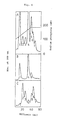
- FIGs. 6(A), 6(B) and 6(C) High performance liquid chromatograms obtained by use of the above hydroxyapatite column are shown in Figs. 6(A), 6(B) and 6(C).
- Figs. 6(A), 6(B) and 6(C) show the high performance liquid chromatograms of the IgG fraction obtained by salting out with ammonium sulfate, the fraction eluted at pH 2.3 on the HRP-combined column and a fraction passing at pH 8.0 on the HRP-combined column, respectively.
- Fig. 7 shows the results obtained in subjecting the peaks 1 to 7 [see Fig. 6(A)] and parent anti-hLT MoAb LT3-11 to sodium dodecyl sulfate-polyacrylamide electrophoresis (SDS-PAGE) according to the method of Laemmli [U. K. Laemmli, Nature , 227 , 680 (1974)].
- the anti-HRP antibody is represented by H1L1-H1L1
- the anti-hLT MoAb LT3-11 is represented by H2L2-H2L2L2
- hybrid antibodies are composed of two kinds of H chains (H1 and H2) and L chains (L1 and L2)(see Table 1).
- Table 1 shows the results obtained in subjecting the peaks 1 to 7 [see Fig. 6(A)] to the ELISAs described in Reference Examples 5 to 7 and immunoglobulin species included in the peaks. Each peak was identified on the basis of the chromatograms shown in Fig. 6 and the results of the SDS-PAGE shown in Fig. 7. The bispecific hybrid MoAB of the present invention is included in the peak 4.
- Peak Peak area(%) ELISA antibody titer Included Ig species Anti-HRP antibody Anti-hLT antibody Bispecific antibody Peak 1 22 512 ⁇ 100 ⁇ 100 H1L2-H1L2 H1L1-H1L2 H1L1-H1L1 Peak 2 8 ⁇ 100 256 ⁇ 100 H1L2-H2L2 Peak 3 7 ⁇ 100 ⁇ 100 ⁇ 100 H1L2-H2L1 Peak 4 34 1024 1024 256 H1L1-H2L2 Peak 5 16 256 256 ⁇ 100 H1L1-H2L1 H2L2-H2L2 Peak 6 8 ⁇ 100 256 ⁇ 100 H2L1-H2L2 Peak 7 5 ⁇ 100 100 ⁇ 100 H2L1-H2L1
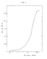
- Fig. 2 shows a standard curve of hLT.
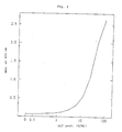
- Fig. 3 shows the results obtained in comparing the ELISA of the present invention with the bioassay of Reference Example 1 in which the lymphocyte culture supernatant obtained in Reference Example 2 and the Escherichia coli cell-extract obtained in Reference Example 4-(4) were added.
- the correlation coefficient (r) obtained with respect to 15 specimens was 0.986, which was satisfactory.
- the 25 ⁇ g/ml solution of the purified hLT C-terminal peptide-specific antibody prepared in Reference Example was added to a microplate having 96 wells in an amount of 100 ⁇ l/well and allowed to stand at 4°C for a day and night. Further, PBS containing 2% BSA was added thereto to prepare a sensitized plate. The plate was washed with PBS, and 100 ⁇ l of an hLT-containing test solution was added thereto, followed by reaction at room temperature for 1 hour. Then, the plate was washed again with PBS containing 0.1% Tween 20.
- Example 3 To the hLT-C-terminal peptide antibody-sensitized plate described in Example 3, 50 ⁇ l of an hLT-containing test solution, 25 ⁇ l of a hybrid antibody solution (PLT1-109, 500 ng/ml) and 25 ⁇ l of an HRP solution (50 ⁇ g/ml) were added, and the reaction was conducted at room temperature for 1 hour. The plate was thereafter washed with PBS containing 0.1% Tween 20. Then, the enzyme reaction was conducted according to the method described in Reference Example 5, followed by preparation of a standard curve of hLT.
- a hybrid antibody solution PHT1-109, 500 ng/ml
- HRP solution 50 ⁇ g/ml
- Fig. 8 shows the results obtained in comparing the ELISA of the present invention with the bioassay of Reference Example 1.
- the correlation coefficient (r) obtained with respect to 20 specimens was 0.96, which was satisfactory.
- the hLT-neutralizing activity of anti-hLT MoAb LT3-11 and bispecific hybrid MoAb PLT1-109 was determined, using the test for neutralizing the cytotoxic activity against L929 described in Reference Example 8-(3). The results are as shown in Fig. 9.
- the hLT-neutralizing activity of hybrid MoAb PLT1-109 of the present invention was 50 to 70% of that of the parent antibody, LT3-11, and the neutralizing activity per valence of the former was nearly equal to that of the latter.
Landscapes
- Health & Medical Sciences (AREA)
- Life Sciences & Earth Sciences (AREA)
- Immunology (AREA)
- Chemical & Material Sciences (AREA)
- Molecular Biology (AREA)
- Engineering & Computer Science (AREA)
- Proteomics, Peptides & Aminoacids (AREA)
- Biomedical Technology (AREA)
- Organic Chemistry (AREA)
- Hematology (AREA)
- General Health & Medical Sciences (AREA)
- Urology & Nephrology (AREA)
- Cell Biology (AREA)
- Biochemistry (AREA)
- Medicinal Chemistry (AREA)
- Analytical Chemistry (AREA)
- Physics & Mathematics (AREA)
- Food Science & Technology (AREA)
- Microbiology (AREA)
- General Physics & Mathematics (AREA)
- Pathology (AREA)
- Biophysics (AREA)
- Genetics & Genomics (AREA)
- Biotechnology (AREA)
- Preparation Of Compounds By Using Micro-Organisms (AREA)
- Micro-Organisms Or Cultivation Processes Thereof (AREA)
- Peptides Or Proteins (AREA)
Applications Claiming Priority (4)
| Application Number | Priority Date | Filing Date | Title |
|---|---|---|---|
| JP9827888 | 1988-04-22 | ||
| JP98278/88 | 1988-04-22 | ||
| JP148160/88 | 1988-06-17 | ||
| JP14816088 | 1988-06-17 |
Publications (2)
| Publication Number | Publication Date |
|---|---|
| EP0338497A2 true EP0338497A2 (de) | 1989-10-25 |
| EP0338497A3 EP0338497A3 (de) | 1990-05-16 |
Family
ID=26439470
Family Applications (1)
| Application Number | Title | Priority Date | Filing Date |
|---|---|---|---|
| EP89106899A Withdrawn EP0338497A3 (de) | 1988-04-22 | 1989-04-18 | Bispezifische hybride monoklonale Antikörper |
Country Status (1)
| Country | Link |
|---|---|
| EP (1) | EP0338497A3 (de) |
Cited By (1)
| Publication number | Priority date | Publication date | Assignee | Title |
|---|---|---|---|---|
| WO1992020746A1 (en) * | 1991-05-14 | 1992-11-26 | Hybritech, Incorporated | Polymeric compositions having bound antibodies |
Family Cites Families (2)
| Publication number | Priority date | Publication date | Assignee | Title |
|---|---|---|---|---|
| US4474893A (en) * | 1981-07-01 | 1984-10-02 | The University of Texas System Cancer Center | Recombinant monoclonal antibodies |
| NZ212207A (en) * | 1984-05-31 | 1991-07-26 | Genentech Inc | Recombinant lymphotoxin |
-
1989
- 1989-04-18 EP EP89106899A patent/EP0338497A3/de not_active Withdrawn
Cited By (1)
| Publication number | Priority date | Publication date | Assignee | Title |
|---|---|---|---|---|
| WO1992020746A1 (en) * | 1991-05-14 | 1992-11-26 | Hybritech, Incorporated | Polymeric compositions having bound antibodies |
Also Published As
| Publication number | Publication date |
|---|---|
| EP0338497A3 (de) | 1990-05-16 |
Similar Documents
| Publication | Publication Date | Title |
|---|---|---|
| Black et al. | Generation of biologically active interleukin-1 beta by proteolytic cleavage of the inactive precursor. | |
| Staehelin et al. | Purification and characterization of recombinant human leukocyte interferon (IFLrA) with monoclonal antibodies. | |
| EP0617706B1 (de) | Multivalente antigen-bindende proteine | |
| JP4001604B2 (ja) | 新規蛋白質及びこれを使用した薬剤の製法 | |
| EP0364778B1 (de) | Antikörper gegen Interleukin-1-beta | |
| JPH0730116B2 (ja) | ヒトインターロイキン―4の抗体アンタゴニスト | |
| JP3192651B2 (ja) | インターロイキン―1インヒビター | |
| JPH07507682A (ja) | Lプロテインに由来する免疫グロブリン結合性タンパク質およびその用途 | |
| JP2598666B2 (ja) | 抗ヒトbcdfモノクローナル抗体 | |
| JP2930713B2 (ja) | タンパク質ホルモン形成の抑制用の組成物およびその使用法 | |
| JP3223260B2 (ja) | T細胞増殖促進因子 | |
| US5142026A (en) | Recombinant human lymphotoxins that possess N-terminal extended linker peptides for antibody-binding sites | |
| EP0338497A2 (de) | Bispezifische hybride monoklonale Antikörper | |
| US5434247A (en) | Peptides for inducing monocyte cytotoxicity in diagnostics | |
| Seyer et al. | Primary structural similarities between types 5 and 24 M proteins of Streptococcuspyogenes | |
| EP0335263A2 (de) | Monoklonale Antikörper gegen menschliches Lymphotoxin, Hybridom für seine Erzeugung, Antikörper gegen menschliches Lymphotoxin-C-Terminal-Peptid und Methode zum Nachweis von menschlichem Lyphotoxin damit | |
| EP0347728B1 (de) | Monoklonaler Antikörper gegen menschliches Lymphotoxin und seine Verwendung | |
| US4751183A (en) | Monoclonal antibody that recognizes a structure common to human interleukin-2 (TCGF) and to the light lambda chain of human immunoglobulin and lines of hybridoma cells that produce these monoclonal antibodies | |
| JPH02227095A (ja) | モノクローナル抗体 | |
| EP0345811B1 (de) | Gegen menschliches Fibrinopepid A spezifische monoklonale Antikörper | |
| AU725682B2 (en) | Anti-human lect2 antibody, cells producing the same, and method and kit for assaying the same | |
| AU616195B2 (en) | Lectines fixing beta-d-galactoside | |
| JPH02219569A (ja) | 二重特異性を有するハイブリッドモノクローナル抗体 | |
| JPH0977799A (ja) | ヒト−マクロファージ遊走阻止因子(ヒト−mif)に対するモノクローナル抗体および該抗体を産生するハイブリドーマ | |
| JP2979321B2 (ja) | 新規組み換え型リンホトキシン |
Legal Events
| Date | Code | Title | Description |
|---|---|---|---|
| PUAI | Public reference made under article 153(3) epc to a published international application that has entered the european phase |
Free format text: ORIGINAL CODE: 0009012 |
|
| AK | Designated contracting states |
Kind code of ref document: A2 Designated state(s): AT BE CH DE ES FR GB GR IT LI LU NL SE |
|
| PUAL | Search report despatched |
Free format text: ORIGINAL CODE: 0009013 |
|
| AK | Designated contracting states |
Kind code of ref document: A3 Designated state(s): AT BE CH DE ES FR GB GR IT LI LU NL SE |
|
| 17P | Request for examination filed |
Effective date: 19901012 |
|
| RAP3 | Party data changed (applicant data changed or rights of an application transferred) |
Owner name: TAKEDA CHEMICAL INDUSTRIES, LTD. |
|
| 17Q | First examination report despatched |
Effective date: 19920903 |
|
| STAA | Information on the status of an ep patent application or granted ep patent |
Free format text: STATUS: THE APPLICATION HAS BEEN WITHDRAWN |
|
| 18W | Application withdrawn |
Withdrawal date: 19921229 |