EP0484401B1 - Production de facteur de croissance de cellules endotheliales vasculaires - Google Patents
Production de facteur de croissance de cellules endotheliales vasculaires Download PDFInfo
- Publication number
- EP0484401B1 EP0484401B1 EP90911525A EP90911525A EP0484401B1 EP 0484401 B1 EP0484401 B1 EP 0484401B1 EP 90911525 A EP90911525 A EP 90911525A EP 90911525 A EP90911525 A EP 90911525A EP 0484401 B1 EP0484401 B1 EP 0484401B1
- Authority
- EP
- European Patent Office
- Prior art keywords
- growth factor
- vascular endothelial
- endothelial cell
- cell growth
- sequence
- Prior art date
- Legal status (The legal status is an assumption and is not a legal conclusion. Google has not performed a legal analysis and makes no representation as to the accuracy of the status listed.)
- Expired - Lifetime
Links
Images


Classifications
-
- C—CHEMISTRY; METALLURGY
- C07—ORGANIC CHEMISTRY
- C07K—PEPTIDES
- C07K14/00—Peptides having more than 20 amino acids; Gastrins; Somatostatins; Melanotropins; Derivatives thereof
- C07K14/435—Peptides having more than 20 amino acids; Gastrins; Somatostatins; Melanotropins; Derivatives thereof from animals; from humans
- C07K14/52—Cytokines; Lymphokines; Interferons
-
- A—HUMAN NECESSITIES
- A61—MEDICAL OR VETERINARY SCIENCE; HYGIENE
- A61P—SPECIFIC THERAPEUTIC ACTIVITY OF CHEMICAL COMPOUNDS OR MEDICINAL PREPARATIONS
- A61P17/00—Drugs for dermatological disorders
- A61P17/02—Drugs for dermatological disorders for treating wounds, ulcers, burns, scars, keloids, or the like
-
- A—HUMAN NECESSITIES
- A61—MEDICAL OR VETERINARY SCIENCE; HYGIENE
- A61P—SPECIFIC THERAPEUTIC ACTIVITY OF CHEMICAL COMPOUNDS OR MEDICINAL PREPARATIONS
- A61P43/00—Drugs for specific purposes, not provided for in groups A61P1/00-A61P41/00
-
- C—CHEMISTRY; METALLURGY
- C07—ORGANIC CHEMISTRY
- C07K—PEPTIDES
- C07K14/00—Peptides having more than 20 amino acids; Gastrins; Somatostatins; Melanotropins; Derivatives thereof
- C07K14/435—Peptides having more than 20 amino acids; Gastrins; Somatostatins; Melanotropins; Derivatives thereof from animals; from humans
- C07K14/46—Peptides having more than 20 amino acids; Gastrins; Somatostatins; Melanotropins; Derivatives thereof from animals; from humans from vertebrates
- C07K14/47—Peptides having more than 20 amino acids; Gastrins; Somatostatins; Melanotropins; Derivatives thereof from animals; from humans from vertebrates from mammals
-
- C—CHEMISTRY; METALLURGY
- C07—ORGANIC CHEMISTRY
- C07K—PEPTIDES
- C07K14/00—Peptides having more than 20 amino acids; Gastrins; Somatostatins; Melanotropins; Derivatives thereof
- C07K14/435—Peptides having more than 20 amino acids; Gastrins; Somatostatins; Melanotropins; Derivatives thereof from animals; from humans
- C07K14/475—Growth factors; Growth regulators
- C07K14/49—Platelet-derived growth factor [PDGF]
-
- C—CHEMISTRY; METALLURGY
- C07—ORGANIC CHEMISTRY
- C07K—PEPTIDES
- C07K16/00—Immunoglobulins [IG], e.g. monoclonal or polyclonal antibodies
- C07K16/18—Immunoglobulins [IG], e.g. monoclonal or polyclonal antibodies against material from animals or humans
- C07K16/24—Immunoglobulins [IG], e.g. monoclonal or polyclonal antibodies against material from animals or humans against cytokines, lymphokines or interferons
-
- C—CHEMISTRY; METALLURGY
- C12—BIOCHEMISTRY; BEER; SPIRITS; WINE; VINEGAR; MICROBIOLOGY; ENZYMOLOGY; MUTATION OR GENETIC ENGINEERING
- C12N—MICROORGANISMS OR ENZYMES; COMPOSITIONS THEREOF; PROPAGATING, PRESERVING, OR MAINTAINING MICROORGANISMS; MUTATION OR GENETIC ENGINEERING; CULTURE MEDIA
- C12N15/00—Mutation or genetic engineering; DNA or RNA concerning genetic engineering, vectors, e.g. plasmids, or their isolation, preparation or purification; Use of hosts therefor
- C12N15/09—Recombinant DNA-technology
- C12N15/63—Introduction of foreign genetic material using vectors; Vectors; Use of hosts therefor; Regulation of expression
- C12N15/79—Vectors or expression systems specially adapted for eukaryotic hosts
- C12N15/85—Vectors or expression systems specially adapted for eukaryotic hosts for animal cells
-
- A—HUMAN NECESSITIES
- A61—MEDICAL OR VETERINARY SCIENCE; HYGIENE
- A61K—PREPARATIONS FOR MEDICAL, DENTAL OR TOILETRY PURPOSES
- A61K38/00—Medicinal preparations containing peptides
-
- C—CHEMISTRY; METALLURGY
- C07—ORGANIC CHEMISTRY
- C07K—PEPTIDES
- C07K2319/00—Fusion polypeptide
-
- C—CHEMISTRY; METALLURGY
- C07—ORGANIC CHEMISTRY
- C07K—PEPTIDES
- C07K2319/00—Fusion polypeptide
- C07K2319/01—Fusion polypeptide containing a localisation/targetting motif
- C07K2319/02—Fusion polypeptide containing a localisation/targetting motif containing a signal sequence
-
- C—CHEMISTRY; METALLURGY
- C07—ORGANIC CHEMISTRY
- C07K—PEPTIDES
- C07K2319/00—Fusion polypeptide
- C07K2319/70—Fusion polypeptide containing domain for protein-protein interaction
- C07K2319/74—Fusion polypeptide containing domain for protein-protein interaction containing a fusion for binding to a cell surface receptor
- C07K2319/75—Fusion polypeptide containing domain for protein-protein interaction containing a fusion for binding to a cell surface receptor containing a fusion for activation of a cell surface receptor, e.g. thrombopoeitin, NPY and other peptide hormones
-
- C—CHEMISTRY; METALLURGY
- C12—BIOCHEMISTRY; BEER; SPIRITS; WINE; VINEGAR; MICROBIOLOGY; ENZYMOLOGY; MUTATION OR GENETIC ENGINEERING
- C12N—MICROORGANISMS OR ENZYMES; COMPOSITIONS THEREOF; PROPAGATING, PRESERVING, OR MAINTAINING MICROORGANISMS; MUTATION OR GENETIC ENGINEERING; CULTURE MEDIA
- C12N2830/00—Vector systems having a special element relevant for transcription
- C12N2830/001—Vector systems having a special element relevant for transcription controllable enhancer/promoter combination
- C12N2830/002—Vector systems having a special element relevant for transcription controllable enhancer/promoter combination inducible enhancer/promoter combination, e.g. hypoxia, iron, transcription factor
-
- C—CHEMISTRY; METALLURGY
- C12—BIOCHEMISTRY; BEER; SPIRITS; WINE; VINEGAR; MICROBIOLOGY; ENZYMOLOGY; MUTATION OR GENETIC ENGINEERING
- C12N—MICROORGANISMS OR ENZYMES; COMPOSITIONS THEREOF; PROPAGATING, PRESERVING, OR MAINTAINING MICROORGANISMS; MUTATION OR GENETIC ENGINEERING; CULTURE MEDIA
- C12N2830/00—Vector systems having a special element relevant for transcription
- C12N2830/80—Vector systems having a special element relevant for transcription from vertebrates
- C12N2830/85—Vector systems having a special element relevant for transcription from vertebrates mammalian
Definitions
- the invention relates to the field of wound healing.
- the invention relates to the production of a wound healing agent that is mitogenic for vascular endothelial cells and consequently is useful in promoting neovascularization (angiogenesis) and re-endothelialization of inner vascular surfaces.
- the invention provides methods and means for producing vascular endothelial cell growth factor by means of recombinant DNA technology.
- Angiogenesis i.e. the growth of new capillary blood vessels, is a process which is crucial to the proper healing of many types of wounds. Consequently, factors that are capable of promoting angiogenesis are useful as wound healing agents.
- Angiogenesis is a multi-step process involving capillary endothelial cell proliferation, migration and tissue penetration.
- a number of known growth factors including basic and acidic fibroblast growth factor, transforming growth factor alpha and epidermal growth factor, are broadly mitogenic for a variety of cell types as well as being angiogenic and are, therefore, potentially useful in promoting tissue repair. Broad spectrum mitogenicity is desirable in many types of wound healing applications. There are, however, specific types of wound healing applications in which it would be desirable to have a more cell-specific mitogenic activity.
- a wound healing agent incorporating a mitogenic factor having mitogenic activity that is highly specific for vascular endothelial cells since proliferation of other cell types along with endothelial cells could cause blockage and/or reduced blood flow.
- mitogenic factor is widely available for this type of application.
- the present invention provides methods and means for obtaining commercial scale quantities of vascular endothelial cell growth factor for use as a wound healing agent.
- the present invention provides DNA sequences that encode amino acid sequences of bovine vascular endothelial cell growth factor having the amino acid sequence 1 to 114 followed by 159 to 164 shown in Figure 6 wherein amino acid 114 is Lysine and human vascular endothelial cell growth factor having the amino acid sequence 1 to 115 followed by 160 to 165 shown in Figure 7 wherein amino acid 115 is Lysine.
- These DNA sequences are inserted into expression vectors under the control of regulatory elements that direct the expression of the encoded amino acid sequences in a suitable expression host.
- the vascular endothelial cell growth factor expressed in this manner can be recovered and formulated into pharmaceutical compositions that are useful in a variety of wound healing applications in which angiogenesis and/or re-endothelialization play an important role.
- the invention relates to the isolated DNA sequences, encoding bVEGF 120 and hVEGF 121 replicable expression vectors capable of directing expression of such sequences in host cells, host cells transformed with such vectors and methods of producing the vascular endothelial cell growth factors which comprise culturing such cells.
- the invention further provides the vascular endothelial cell growth factors, bVEGF 120 and hVEGF 121 in isolated form and in pharmaceutical compositions.
- the invention also relates to proteins useful in inhibiting angiogenesis comprising a heterodimer of two different subunits, one of which is hVEGF 121 having the amino acid sequence 1 to 115 followed by 160 to 165 shown in Figure 7 wherein amino acid 115 is Lysine and the other of which is hVEGF 165 , having the amino acid sequence 1 to 165 shown in Figure 7, or hVPF 189 (human vascular permeability factor), and the use of such proteins in the manufacture of a medicament for use in the treatment of an individual by inhibiting angiogenesis, as well as pharmaceutical compositions containing them.
- the invention also provides chimeric proteins comprising an A-chain or B-chain subunit of platelet-derived growth factor and a bVEGF 120 or hVEGF 121 subunit.
- the invention yet further relates to neutralising antibodies specific to bVEGF 120 or hVEGF 121 , pharmaceutical compositions containing them and use of such antibodies in the manufacture of a medicament for use in the treatment of an individual by inhibiting angiogenesis.
- the invention also provides an analog of hVEGF 121 in which the cysteine residue at position 116 has been substituted by a different amino acid residue.
- the novel, lower molecular weight factor predicted from the coding region lacking the insert comprises a 120-amino acid sequence in the bovine case and 121-amino acid sequence in the human case.
- the lower molecular weight form differs not only in the length of the amino acid sequence, but also in the presence of a Lys residue at position 114 in the bovine protein (position 115 in the human protein) that is not present in the higher molecular weight form because of the differential message splicing which occurs within the corresponding codon at this position.
- these two forms of vascular endothelial cell growth factor will be referred to, respectively, as bVEGF 164 and bVEGF 120 for the bovine factor (hVEGF 165 and hVEGF 121 for the human factor).
- hVEGF 121 and bVEGF 120 differ in their properties from hVEGF 165 and bVEGF 164 in a manner which provides therapeutic advantages in the clinical use of the protein.
- hVEGF 121 and bVEGF 120 do not bind heparin, whereas the longer forms are characterized by strong heparin binding.
- the absence of heparin binding affinity leaves more of the protein free to bind to vascular endothelial cell growth factor receptor and increases the half-life and distribution of the protein in circulation.
- amino acid sequence deduced from N-terminal sequence analysis and an isolated DNA sequence indicates a significant level of sequence homology between bovine vascular endothelial cell growth factor and corresponding sequences from each of the A-chain and B-chain subunits of human platelet-derived growth factor (PDGF), with complete conservation of eight cysteine residues among the mature forms of all three sequences.
- PDGF platelet-derived growth factor
- hybrid dimeric proteins can be prepared comprising a first polypeptide chain and a second polypeptide chain, wherein one of the chains comprises at least a portion of the amino acid sequence of the A-chain or the B-chain subunit of platelet-derived growth factor and the other chain comprises at least a portion of the amino acid sequence of vascular endothelial cell growth factor.
- Preparation of the hybrid proteins allows one to "tailor" the properties of the molecule such that the hybrid exhibits a profile of mitogenic activity between that of vascular endothelial cell growth factor and platelet-derived growth factor.
- the PDGF B-B homodimer is mitogenic for vascular smooth muscle cells but not for vascular endothelial cells.
- the vascular endothelial cell growth factor of the present invention has the opposite specificity.
- a hybrid factor may stimulate both cell types and therefore be useful as a broader-spectrum mitogen in wound healing therapies.
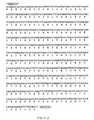
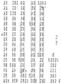
- Fig. 1 is a representation of five DNA sequences generated by a modification of the polymerase chain reaction process.
- One of the five (pET-19A; clone no. 5) encodes amino acids no. 15 to 38 of the mature, sequenced form of bovine vascular endothelial cell growth factor.
- the figure also includes a consensus DNA sequence derived from the five DNA sequences, as well as a translation of pET-19A.
- Each sequence includes DNA linkers at either end which represent an EcoRI restriction site and a HindIII restriction site.
- Fig. 2 is a schematic representation of the method by which the five DNAs of Fig. 1 were generated and amplified from bovine folliculo stellate cell mRNA.
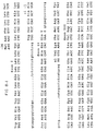
- Fig. 3a is a representation of a DNA sequence, as well as its deduced amino acid sequence, derived from a clone, designated 11B'.
- the illustrated sequence encodes amino acids no. 15 to 120 of bovine vascular endothelial cell growth factor (bVEGF 120 ).
- Fig. 3b is a representation of a synthetic DNA sequence, based on preferred codon usage in human cells, which encodes amino acids no. 1 to 19 of bVEGF 120 and which overlaps the 5' end of the DNA sequence of Fig. 3a.
- This synthetic DNA can be enzymatically joined to the isolated DNA sequence of Fig. 3a, after the DNA sequence in Fig. 3a has been digested with the restriction enzyme AccI, to produce a DNA sequence encoding the full length, mature bVEGF 120 protein.
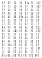
- Fig. 4 is a representation of isolated DNA sequences encoding the A-chain and B-chain subunits of human platelet-derived growth factor, and the amino acid sequences of the precursors of these two proteins as deduced from the DNA sequences.
- Fig. 5 is a photograph of an ethidium bromide stained polyacrylamide gel containing DNA produced by amplification of a portion of the mRNA encoding bovine vascular endothelial cell growth factor.
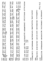
- Fig. 6 is a representation of the isolated cDNA sequences encoding bVEGF 120 and bVEGF 164 .
- the boxed DNA sequence beginning at base 342 represents the insert sequence that is present in the alternatively spliced cDNA which encodes bVEGF 164 .
- the amino acid sequence given immediately below the nucleotide sequence represents the deduced sequence for bVEGF 164 .
- the deduced amino acid sequence for bVEGF 120 is identical to that of bVEGF 164 through position 113 (Glu).
- the carboxyl-terminal sequence of bVEGF 120 beginning at position 111 (Arg) is given in italics below the bVEGF 164 sequence in Fig. 6.
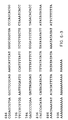
- Fig. 7 is a representation of the native human DNA sequences encoding the mature forms of human vascular endothelial cell growth factor, hVEGF 121 and hVEGF 165 .
- the DNA sequences shown represent composites of sequences obtained from human genomic and human cDNA clones.
- the bracketed amino acid (Gly) encoded by codon 7 represents an inserted amino acid relative to the sequences of bVEGF 120 and bVEGF 164 .
- Fig. 8 is a representation of portions of the DNA sequences of the overlapping genomic inserts in two bacteriophages, which together contain eight exons encoding the various forms of hVEGF, along with contiguous splice junctions
- Fig. 9 is a representation of two oligonucleotide primers used to amplify a full length cDNA sequence for hVEGF 121 from U937 cell mRNA.
- Fig. 10a is a schematic representation of pLEN, an expression vector used to express hVEGF 121 in Chinese hamster ovary cell culture.
- Fig. 10b is a schematic representation of pMTN, another expression vector used to express hVEGF 121 in Chinese hamster ovary cell culture.
- vascular endothelial cell growth factor refers to a mammalian protein that has mitogenic activity for vascular endothelial cells and that:
- amino acid sequence is considered to be “substantially homologous” herein if the level of amino acid sequence homology is at least 50% and, preferably at least 80%, compared with the protein in question.
- Standard hybridization conditions as used herein means the use of 40% Formamide Buffer (described below) as the prehybridization/hybridization buffer and washing in 1 x SSC, 0.1% SDS at 50°C.
- amino acid sequence numbering system used herein for vascular endothelial cell growth factor is based on the mature forms of the protein, i.e. the post-translationally processed forms. Accordingly, the residue numbered one in the bovine or human proteins is alanine, which is the first residue of the isolated, mature forms of these proteins.
- Mitogenic activity for vascular endothelial cells can be determined by an assay which uses, as target cells, adrenal cortex-derived capillary endothelial cells (ACE cells). This assay is carried out essentially as described in Gospodarowicz et al., J. Cell Physiol. (1986) 127 :121-136), the disclosure of which is incorporated herein by reference. Generally, stock cultures of ACE cells are maintained in the presence of Dulbecco's modified Eagle's medium (DMEM-21) supplemented with 10% calf serum.
- DMEM-21 Dulbecco's modified Eagle's medium
- the antibiotics penicillin (50 IU/ml), streptomycin (50 ⁇ g/ml), gentamycin (50 ⁇ g/ml), and Fungizone (0.25 ⁇ g/ml) and 2mM L-glutamine can also be added to the medium.
- Cells are passaged weekly on tissue culture dishes at a split ratio of between 1:40 and 1:200 (the preferred split ratio is that which gives 2.5 x 10 5 cells in 15 ml of medium in T75 flasks).
- cells are seeded in 12 well cluster plates at a density of 5 x 10 3 cells per well in 1 ml Dulbecco's modified Eagle's medium supplemented with 10% calf serum and antibiotics as described in Gospodarowicz et al., Europ. J. Cell. Biol. (1988) 46 :144-151.
- the ACE cells are plated in 35 mm dishes or 6 well cluster plates at a density of 5 - 10 x 10 3 cells per dish or well in 2 ml of medium as described in Gospodarowicz, et al., J. Cell Physiol. (1986) 127 :121-136.
- a factor is considered to have mitogenic activity for vascular endothelial cells if the cell density at the end of this assay is at least 1.5 times and preferably at least 3 times the cell density of control wells receiving no factor additions.
- DNA sequence illustrated in Fig. 3a was obtained from a bovine cell cDNA library, and therefore represents a sequence which encodes a bovine protein
- the DNA sequence provided allows for the retrieval of sequences encoding homologous proteins from other mammalian species. Accordingly, we have employed the illustrated bovine sequence as a probe to retrieve DNA sequences encoding the corresponding human proteins.
- vascular endothelial cell growth factor herein are biologically active fragments thereof, as well as N-terminally or C-terminally extended versions thereof or analogs thereof substituting and/or deleting or inserting one or more amino acid residues which retain qualitatively the biological activities of the protein described herein.
- Preferred analogs include those in which one or more cysteine residues not required for biological activity are substituted by a different amino acid residue, preferably serine. Substitution of one or more cysteine residues reduces the opportunity for intermolecular and intramolecular disulfide bond formation, thereby rendering the molecule more stable.
- cysteine residues there are nine cysteine residues that are present in hVEGF 121 , bVEGF 120 , hVEGF 165 and bVEGF 164 . Of these, eight are conserved with PDGF. Accordingly, the most preferred analog is one in which the ninth cysteine residue is substituted by serine. This cysteine residue is present at position 160 of hVEGF 165 and position 116 of hVEGF 121 and the corresponding positions in the bovine forms. Amino acid substitutions can be accomplished by site specific mutagenesis of the DNA sequences described herein using well known techniques (see, e.g., Zoller, M.J. and Smith, M., Nucleic Acids Research (1982) 10 :6487-6500).
- vascular endothelial cell growth factor While the native form of the bovine vascular endothelial cell growth factor described herein is apparently glycosylated, there is currently no evidence that glycosylation is essential for biological activity. Accordingly, biologically active non-glycosylated or partially glycosylated forms, which will be produced by prokaryotic or eukaryotic hosts using the expression sequences provided herein, are included within the scope of "vascular endothelial cell growth factor".
- hVEGF 121 vascular endothelial cell growth factor in which both subunits are either glycosylated or unglycosylated and dimers in which one of the subunits is glycosylated and the other is not glycosylated.
- Bovine vascular endothelial cell growth factor was obtained in homogeneous form from cell culture media conditioned by folliculo stellate cells, by a process which involved the steps of ammonium sulfate precipitation; heparin-Sepharose affinity chromatography; size-exclusion gel chromatography; cation exchange chromatography; and reverse phase high performance liquid chromatography.
- vascular endothelial cell growth factor for example, murine AtT20 cells.
- human fetal vascular smooth muscle cells are a good source of human vascular endothelial cell growth factor and mRNA encoding the factor.
- Isolated bovine vascular endothelial cell growth factor obtained as described above was sequenced using the Edman degradation technique on an automated gas-phase protein sequenator. A single major 24-amino acid N-terminal sequence was obtained, indicating that the protein is homodimeric.
- the bovine protein has the following 41-amino acid N-terminal sequence*: (*Using the standard single letter abbreviation code for amino acids)
- N-terminal amino acid sequence for bovine vascular endothelial cell growth factor described above, a number of unsuccessful efforts were made to retrieve a full or partial length cDNA encoding the protein by probing a folliculo stellate cell cDNA library using degenerate oligonucleotide probe mixtures encoding portions of the amino acid sequence.
- the DNA probe used to retrieve the cDNA of Fig. 3a was selected from the five homologous sequences shown in Fig. 1. These sequences were obtained by a procedure which is illustrated schematically in Fig. 2 and which is described in greater detail in the examples which follow.
- poly(A) + RNA from bovine folliculo stellate cells was precipitated with an anti-sense primer consisting of a 16-fold degenerate synthetic oligonucleotide mixture based on the amino acid sequence of amino acids no. 35 to 39 of bovine vascular endothelial cell growth factor.
- the 24-base oligonucleotide primer consisted of 14 bases reflecting the amino acid sequence, with a 10-base EcoRI linker on the 5' end.
- the oligonucleotide primer sequences in the mixture which hybridized to the poly(A) + RNA served to prime the synthesis of a DNA strand complementary to a section of the desired mRNA in the presence of deoxynucleotide triphosphates (dNTPs) and reverse transcriptase.
- dNTPs deoxynucleotide triphosphates
- a second DNA strand, complementary to the first synthesized strand was then prepared by hybridizing the first synthesized strand to a sense-strand primer consisting of an 8-fold degenerate synthetic 24-base oligonucleotide mixture based on the amino acid sequence of amino acids no. 15 to 19 of bovine vascular endothelial cell growth factor.
- the second strand oligonucleotide primer contained a 14-base region reflecting the amino acid sequence, joined to a 10-base HindIII linker on the 5' end.
- the oligonucleotide primer sequences in the mixture which hybridized to the first synthesized DNA strand served to prime the synthesis of a second DNA strand in the presence of dNTPs and DNA polymerase I, Klenow fragment. Since the 10 base linker sequence in the primer could not hybridize to the first strand DNA, second strand synthesis was carried out at a temperature, i.e. 28°C, at which the remaining 14 nucleotides could be expected to remain hybridized to the first strand DNA.
- DNA polymerase I Klenow fragment
- Thermus aquaticus ( Taq ) DNA polymerase which is normally used in the polymerase chain reaction, would not be effective to catalyze DNA synthesis at this temperature.
- Second strand synthesis produced a sense strand coding for that portion of bovine vascular endothelial cell growth factor extending from amino acid no. 15 to amino acid no. 38 (and including two-thirds of the codon for amino acid 39).
- the two synthesized DNA strands were then separated and the desired sequence was amplified by a repeated sequence of reactions in which the single stranded DNAs were used as templates for the synthesis of complementary strands in the presence of both the sense- and anti-sense oligonucleotide primer mixtures and Thermus aquaticus ( Taq ) DNA polymerase. After each synthesis of complementary strands, the reaction mixture was heated to separate the strands and the reaction was repeated.
- the DNA from the polymerase chain reaction mixture was subjected to electrophoresis on a 6% polyacrylamide gel.
- the DNA in the gel was stained with ethidium bromide and the band having the appropriate size for the coding sequence for amino acids 15 to 38 (and two-thirds of the codon for amino acid 39) together with the HindIII and EcoRI linkers from the priming oligonucleotides was cut from the gel.
- the ethidium bromide stained gel is shown in the photograph of Fig. 5, where the dominant band representing the desired amplified sequence can be clearly visualized.
- DNA was electroeluted from the excised gel fragment containing the dominant band, digested with HindIII and EcoRI and ligated into HindIII- and EcoRI-cut M13mp19 and M13mp18 phage vectors.
- DNA sequence analysis of white plaques isolated after transformation of the ligation mixtures into E. coli JM103 host cells demonstrated that a cDNA sequence encoding amino acids 15 to 38 (and containing two-thirds of the codon for amino acid 39) of vascular endothelial cell growth factor indeed had been obtained.
- the amplified DNA sequence contained in one of these recombinant phage was employed as a probe to retrieve a cloned cDNA sequence from a bovine folliculo stellate cell cDNA library.
- the isolated cloned sequence consisted of an 797-base pair insert coding for all but the 14 N-terminal amino acids of one of the mature forms of bovine vascular endothelial cell growth factor.
- the isolated cloned insert was ligated into the EcoRI site of pUC8 to create the plasmid designated pST800.
- the insert contained the nucleotide sequence shown in Fig. 3a (in Fig.
- the amino acid sequence predicted from the isolated DNA sequence shown in Fig. 3a contains one potential site for N-linked glycosylation at the asparagine residue at amino acid no. 74 (corresponding to amino acid no. 75 in the human form). Since an N-linked glycosylation of approximately 3 kD at this site would predict a total molecular weight of about 17 kD for the encoded protein, which is considerably smaller than the apparent molecular weight of 23 kD observed for the vascular endothelial cell growth factor subunits isolated by Gospodarowicz et al. and by Ferrara and Henzel, it is apparent that the isolated cDNA encodes a different form of vascular endothelial cell growth factor than that previously observed. A polymerase chain reaction experiment indicated that alternative forms of the vascular endothelial cell growth factor coding region exist.
- a polymerase chain reaction was primed from folliculo stellate poly(A) + RNA using a sense oligonucleotide corresponding to bases 70-126 in Fig. 6, and an antisense oligonucleotide corresponding to bases 513-572.
- Polyacrylamide gel analysis of the products after digestion with BstNI revealed two major species of approximately 300 and 450 bp. Both of these products were subcloned into M13 vectors and sequenced. The DNA sequence of the smaller product (311 bp) corresponded to that predicted if the PCR amplified the cDNA sequence carried in pST800.
- the sequence of the larger fragment was identical to that of the 311 bp product, except for an insert of 132 bp (boxed sequence in Fig. 6).
- Analysis of human vascular endothelial cell growth factor genomic clones, obtained as described below, has indicated that this insert occurs at an exon-intron junction, suggesting that the two forms of the coding region arise through alternative exon splicing.
- Fig. 1 or Fig. 3a are useful in the retrieval of DNA coding for full length bovine vascular endothelial cell growth factor, or for the corresponding vascular endothelial cell growth factor of other species, including the corresponding human protein, or for related proteins in the same gene family as vascular endothelial cell growth factor.
- Genomic libraries can be prepared by known techniques and are now widely commercially available.
- a suitable genomic DNA library from which genomic DNA sequences encoding human vascular endothelial cell growth factor can be isolated is a human fibroblast genomic library (Stratagene Inc., La Jolla, CA). This library, obtained from the W138 cell line, harbors >15 kb DNA inserts in the Lambda FIX TM vector.
- a genomic library can be prepared by the technique disclosed by Frischholz, A.M. in Methods in Enzymology , eds. Berger, S.L. and Kimmel, A.R., Vol. 152, pp. 190-199 (1987) Academic Press, N.Y. Methods for preparing cDNA libraries are also well known to those skilled in the art (see, e.g., Kimmel, A.R. and Berger, S.L., ibid ., pp. 307-316).
- the cDNA library is prepared from a cell line or tissue source which actively produces vascular endothelial cell growth factor.
- a cDNA library prepared from fetal human vascular smooth muscle cells.
- a DNA sequence encoding vascular endothelial cell growth factor which is obtained as described above, is inserted into a suitable expression vector under the control of regulatory sequences capable of directing expression of the DNA sequence in a desired host. If the DNA sequence retrieved is a genomic sequence containing introns, then it is desirable to insert the sequence into an expression vector that is compatible with a eukaryotic host.
- genomic DNA encoding vascular endothelial cell growth factor in a eukaryotic host is accompanied by correct splicing of the encoded RNA to remove intron sequences, thereby producing an mRNA template encoding the desired protein.
- a synthetic DNA sequence can be constructed from synthetic oligonucleotides that represents the coding sequence obtained after the intron sequences in the genomic clone have been removed. Expression vectors containing this synthetic sequence or the cDNA sequence encoding vascular endothelial cell growth factor can be used to express the protein in prokaryotic or eukaryotic hosts. Exemplary control sequence DNAs and hosts are described below under Standard Procedures .
- Biologically active vascular endothelial cell growth factor is produced in accordance with the teachings of this invention, as a homodimeric molecule.
- the term "homodimeric” refers to a dimer in which the two subunits have the same primary amino acid structure.
- one or both of the subunits may be modified by N-linked glycosylation or neither of the subunits modified.
- a fully active protein is produced by expression and/or recovery of the polypeptide sequence encoded by the DNA sequence of the invention under conditions which allow the formation of disulfide bonds in order to form a dimer.
- the present invention also provides for the production of chimeric, dimeric proteins in which a portion of the primary amino acid structure corresponds to a portion of either the A-chain subunit or the B-chain subunit of platelet-derived growth factor and a portion of the primary amino acid structure corresponds to a portion of vascular endothelial cell growth factor.
- a chimeric growth factor comprising a first polypeptide chain and a second polypeptide chain, said chains being disulfide linked, wherein the first polypeptide chain comprises at least a portion of the amino acid sequence of either the A-chain subunit or the B-chain subunit of platelet-derived growth factor and the second chain comprises at least a portion of the amino acid sequence of vascular endothelial cell growth factor.
- Platelet-derived growth factor is an approximately 30 kD dimer which has been isolated in both homodimeric and heterodimeric forms. Platelet-derived growth factor heterodimer contains two polypeptide chains, designated the A- and B-chains. The mature A- and B-chains exhibit approximately 40% amino acid sequence homology, with complete conservation of eight cysteine residues. Platelet-derived growth factor exists in vivo as either an A-A or a B-B homodimer or as an A-B heterodimer.
- A-chain cDNA human is disclosed in Betsholtz, C., et al., Nature (1986) 320 ; A-chain genomic DNA (human) in Bonthron, D.T., et al., PNAS (1988) 85:1496; B-chain cDNA (human) in Collins, T., et al., Nature (1985) 316 :748; and B-chain genomic DNA (human) in Chin, I.-M., et al., Cell (1984) 37 :123).
- FIG. 4 illustrates the isolated cDNA sequences and deduced precursor amino acid sequences for the A- and B-chain subunits of human platelet-derived growth factor with a BamHI linker joined to the 5' end and an EcoRV linker joined to the 3' end of the cDNAs in each case.
- a suitable expression vector under the control of appropriate regulatory elements and expressed in a suitable host, such as for example, E . coli or a eukaryotic host such as Chinese hamster ovary (CHO) cells or yeast.
- Examples of the chimeric growth factor proteins contemplated by the present invention include: a dimeric protein consisting of the full length A-chain subunit of platelet-derived growth factor linked by disulfide bonds to a full length vascular endothelial cell growth factor polypeptide chain; and a dimeric protein consisting of the full length B-chain subunit of platelet-derived growth factor linked by disulfide bonds to a full length vascular endothelial cell growth factor polypeptide chain.
- the dimeric protein can consist of two disulfide-linked polypeptide chains in which one or both of the chains consists of an N-terminal segment having an amino acid sequence corresponding to an N-terminal portion of either the A- or B-chain subunit of platelet-derived growth factor or vascular endothelial cell growth factor and a C-terminal segment corresponding to a C-terminal sequence selected from one of the other two chains.
- one of the polypeptide chains can be composed of an N-terminal portion of vascular endothelial cell growth factor linked to a C-terminal segment of the A- or B-chain subunit of platelet-derived growth factor and the other polypeptide chain can have the amino acid sequence of vascular endothelial cell growth factor.
- Numerous different hybrid combinations can be prepared, as will be readily apparent.
- a DNA sequence encoding each desired chain is inserted into a suitable expression vector, e.g. a plasmid, under the control of regulatory sequences capable of directing its expression in a host cell.
- Host cells are then transformed with the expression vectors.
- a single host may be cotransformed with expression vectors for each of the two chains.
- separate host cells can be transformed with the vectors encoding the two polypeptide chains.
- the polypeptide chains are then expressed and recovered in a conventional manner. If the polypeptide chains are expressed with secretion signal sequences such that they are secreted from host cells, they may naturally form the correct dimer structure during synthesis and secretion.
- the dimers may then be purified using the techniques described in Gospodarowicz et al., PNAS (1989) 86(19):7311-7316 and Ferrara and Henzel, BBRC (1989) 161 (2):851-858. If the correct dimer structure is not obtained by this route, or if the two chains of the chimera are synthesized in different hosts, then an example of one means of refolding and dimerizing the chains would be to treat the partially-purified or purified chains with guanidine-HCl, Na 2 SO 3 and Na 2 S 4 O 6 , as described in more detail in the examples below. The resulting S-sulfonated, denatured proteins are then refolded and dimerized together in the presence of 5 mM glutathione, 0.5 mM glutathione disulfide and urea before final purification.
- Vascular endothelial cell growth factor (bVEGF 120 or hVEGF 121 ) provided by the invention is useful as a wound healing agent, particularly in applications where it is desired to re-endothelialize vascular tissue, or where the growth of a new capillary bed (angiogenesis) is important.
- Vascular endothelial cell growth factor can, therefore, be used in the treatment of full-thickness wounds such as dermal ulcers, including the categories of pressure sores, venous ulcers and diabetic ulcers.
- vascular endothelial cell growth factor can be used in the treatment of full-thickness burns and injuries where angiogenesis is required to prepare the burn or injured site for a skin graft or flap.
- the vascular endothelial cell growth factor is either applied directly to the site or it is used to soak the skin or flap that is being transplanted prior to grafting.
- vascular endothelial cell growth factor can be used in plastic surgery when reconstruction is required following a burn, other trauma or for cosmetic purposes.
- Angiogenesis is also important in keeping wounds clean and non-infected.
- Vascular endothelial cell growth factor can, therefore, be used in association with general surgery and following the repair of cuts and lacerations. It is particularly useful in the treatment of abdominal wounds with a high risk of infection.
- Neovascularization is also key to fracture repair since blood vessels develop at the site of bone injury. Administration of vascular endothelial cell growth factor to the site of a fracture is, therefore, another utility.
- vascular endothelial cell growth factor In cases where vascular endothelial cell growth factor is being used for topical wound healing, as described above, it may be administered by any of the routes described below for the re-endothelialization of vascular tissue, or more preferably by topical means. In these cases, it will be administered as either a solution, spray, gel, cream, ointment or as a dry powder directly to the site of injury. Slow release devices directing vascular endothelial cell growth factor to the injured site will also be used. In topical applications, vascular endothelial cell growth factor will be applied at a concentration ranging from 50 to 1,000 ⁇ g/ml either in a single application, or in dosing regimens that are daily or every few days for a period of one to several weeks. Generally, the amount of topical formulation administered is that which is sufficient to apply from about 0.1 to 100 ⁇ g/cm 2 of vascular endothelial cell growth factor, based on the surface area of the wound.
- Vascular endothelial cell growth factor can be used as a post-operative wound healing agent in balloon angioplasty, a procedure in which vascular endothelial cells are removed or damaged, together with compression of atherosclerotic plaques.
- Vascular endothelial cell growth factor can be applied to inner vascular surfaces by systemic or local intravenous application either as intravenous bolus injection or infusions. If desired, the vascular endothelial cell growth factor can be administered over time using a micrometering pump.
- Suitable compositions for intravenous administration comprise vascular endothelial cell growth factor in an amount effective to promote endothelial cell growth and a parenteral carrier material.
- the vascular endothelial cell growth factor can be present in the composition over a wide range of concentration, for example, from about 50 ⁇ g/ml to about 1,000 ⁇ g/ml using injections of 3 to 10 ml per patient, administered once or in dosing regimens that allow for multiple applications.
- Any of the known parenteral carrier vehicles can be used, such as normal saline or 5-10% dextrose.
- Vascular endothelial cell growth factor can also be used to promote endothelialization in vascular graft surgery.
- vascular endothelial cell growth factor can be applied to the surfaces of the graft and/or at the junctions of the graft and the existing vasculature in order to promote the growth of vascular endothelial cells.
- the vascular endothelial cell growth factor can be applied intravenously as described above for balloon angioplasty or it can be applied directly to the surfaces of the graft and/or the existing vasculature either before or during surgery.
- vascular endothelial cell growth factor in a thickened carrier material so that it will adhere to the affected surface.
- Suitable carrier materials include, for example, 1-5% carbopol.
- the vascular endothelial cell growth factor can be present in the carrier over a wide range of concentrations, for example, from about 50 ⁇ g/mg to about 1,000 ⁇ g/mg.
- the vascular endothelial cell growth factor can be delivered to the site by a micrometering pump as a parenteral solution.
- Vascular endothelial cell growth factor can also be employed to repair vascular damage following myocardial infarction and to circumvent the need for coronary bypass surgery by stimulating the growth of a collateral circulation.
- the vascular endothelial cell growth factor is administered intravenously for this purpose, either in individual injections or by micrometering pump over a period of time as described above or by direct infusion or injection to the site of damaged cardiac muscle.
- Vascular endothelial cell growth factor can also be used as a growth factor for the in vitro culturing of endothelial cells.
- vascular endothelial cell growth factor can be added to the cell culture medium at a concentration from about 10 pg/ml to about 10 ng/ml.
- the hybrid growth factor molecules of the invention will be expected to exhibit mitogenic profiles falling between those of platelet-derived growth factor and vascular endothelial cell growth factor or, in some cases, may be employed as inhibitors of angiogenesis.
- the most pronounced distinction between the activities of the two factors is that platelet-derived growth factor exhibits substantial mitogenic activity on smooth muscle cells and fibroblasts, but not on endothelial cells, whereas vascular endothelial cell growth factor exhibits the opposite specificity.
- the mitogenic activity of PDGF A- and/or B-chain on smooth muscles cells and fibroblasts tends to impart tensile strength to a healing wound.
- the growth factor which is a hybrid between platelet-derived growth factor and vascular endothelial cell growth factor can be applied to a wound in order to induce neovascularization and impart tensile strength to the wound area during and after healing.
- the hybrid growth factors are applied in essentially the same manner and at the same dosages as described above for vascular endothelial cell growth factor.
- Elucidation of the DNA sequences encoding the various forms of vascular endothelial cell growth factor and their deduced amino acid sequences also provides the means for producing inhibitors of vascular endothelial cell growth factor activity. Inhibition of the angiogenic activity of vascular endothelial cell growth factor is useful, for example, in retarding or preventing the growth of tumors, since neovascularization is required to provide the necessary blood supply to a growing tumor.
- One may administer antibodies to vascular endothelial cell growth factor or one may administer fragments of vascular endothelial cell growth factor which are capable of binding vascular endothelial cell growth factor receptor but which do not exhibit the angiogenic activity of full-length vascular endothelial cell growth factor.
- Antibodies to the expressed and isolated vascular endothelial cell growth factor proteins can be produced by known techniques.
- the therapeutic antibodies may be polyclonal or monoclonal.
- Antibodies are prepared using standard immunization protocols in rabbits, mice or other suitable animal and recovering the antisera.
- antibody-secreting cells from the immunized animals can be immortalized using fusion techniques to produce hybridomas which can be screened for antibodies immunoreactive with the vascular endothelial cell growth factor (see e.g. "Antibodies: A Laboratory Manual", E. Harlow and D. Lane, Cold Spring Harbor Laboratory, Cold Spring Harbor, N.Y.).
- the determination of an appropriate treatment regimen i.e., dosage, frequency of administration, systemic vs.
- the antibodies will be formulated in a unit dosage injectable form (solution, suspension, emulsion, etc.) in association with a pharmaceutically acceptable parenteral vehicle.
- a pharmaceutically acceptable parenteral vehicle Such vehicles are usually nontoxic and nontherapeutic. Examples of such vehicles are water, saline, Ringer's solution, dextrose solution, and Hank's solution. Nonaqueous vehicles such as fixed oils and ethyl oleate may also be used.
- a preferred vehicle is 5% (w/w) human albumin in saline.
- the vehicle may contain minor amounts of additives, such as substances that enhance isotonicity and chemical stability, e.g., buffers and preservatives.
- the antibody is typically formulated in such vehicles at a concentration of about 20 ⁇ g/ml to 20 mg/ml.
- hVPF 189 refers to a 189-amino acid protein which has now been found to arise from differential message splicing of the transcribed genomic DNA sequence shown in Fig. 8.
- the homodimeric form of hVPF 189 is referred to as vascular permeability factor.
- hVPF 189 contains a sequence of 68 amino acids, beginning at position 115, which are not present in hVEGF 121 .
- the complete cDNA coding sequence and deduced amino acid sequence of hVPF 189 are disclosed in Keck, P. et al., Science (1989) 246 :1309-1312.
- dimeric PDGF-like growth factors exert their biological activity by a mechanism wherein each of the subunits of the dimer binds a separate receptor subunit on the cell surface, with binding to the two adjacent receptor subunits being necessary to trigger receptor activity.
- each of the subunits may bind a specific receptor subtype, thereby blocking the bound receptor from interacting effectively with endogenous, homodimeric vascular endothelial cell growth factor.
- the dimer since the other subunit of the administered homodimer is a different vascular endothelial cell growth factor subtype, the dimer will be incapable of binding a second receptor subunit of the same subtype to trigger the biological activity of the receptor.
- the heterodimeric protein which is employed in this manner to inhibit angiogenesis can be produced by expressing the desired amino acid sequences for hVEGF 121 , hVEGF 165 or hVPF 189 in separate hosts or by co-expression in the same host. Dimerization is then carried out by procedures described elsewhere herein.
- the desired heterodimer can be separated from homodimeric forms by any convenient method for size separation of proteins.
- the heterodimer is administered to a host in need of anti-angiogenic treatment, e.g. an individual suffering from a tumor, in a manner similar to that in which vascular endothelial cell growth factor is administered as an angiogenic agent.
- the precise dosing regimen and dosage is within the skill of those in the art to determine.
- An alternative means of inhibiting angiogenesis is the administration of a heterodimer in which one subunit is selected from hVEGF 121 , hVEGF 165 and hVPF 189 ; and the other subunit is a biologically inactive fragment or an analog of hVEGF 121 , hVEGF 165 or hVPF 189 in which one or more amino acids are substituted by different amino acids which render the subunits inactive.
- vascular endothelial cell growth factor is produced at elevated levels in tumors, one can employ anti-vascular endothelial cell growth factor antibodies in a conventional immunoassay to detect the presence of a tumor in an individual.
- Antibodies to vascular endothelial cell growth factor, preferably to human vascular endothelial cell growth factor, hVEGF 121 can be produced by known techniques.
- the antibodies can be monoclonal or polyclonal.
- Vascular endothelial cell growth factor levels are determined in a fluid sample, preferably a serum sample, of an individual suspected of having a tumor, using any of the conventional immunoassay techniques which employ antibodies to the substance being measured.
- Preferred assays are the sandwich type immunoassays such as a sandwich type enzymeimmunoassay or radioimmunoassay. Circulating levels of vascular endothelial cell growth factor can reliably be measured by these techniques to the 1-1000 pg/ml level. The measured level of vascular endothelial cell growth factor is compared with control levels taken from normal individuals, i.e. individuals who do not have tumors. Elevated levels of circulating vascular endothelial cell growth factor are considered diagnostic for tumors.
- prokaryotic hosts are, of course, the most convenient for cloning procedures. Prokaryotes most frequently are represented by various strains of E. coli ; however, other microbial strains may also be used. Plasmid vectors which contain replication sites, selectable markers and control sequences derived from a species compatible with the microbial host are used; for example, E. coli is typically transformed using derivatives of pBR322, a plasmid constructed from parts of three naturally-occurring plasmids, two obtained from species of Salmonella , and one isolated from E.
- prokaryotic control sequences which are defined herein to include promoters for transcription initiation, optionally with an operator, along with ribosome binding site sequences, include such commonly used promoters as the beta-lactamase (penicillinase) and lactose (lac) promoter systems (Chang, et al., Nature (1977) 198 :1056) and the tryptophan (trp) promoter system (Goeddel, et al., Nucleic Acids Res . (1980) 8 :4057 and the lambda derived P L promoter and N-gene ribosome binding site (Shimatake, et al., Nature (1981) 292 :
- eucaryotic microbes such as yeast may also be used as hosts.
- Laboratory strains of Saccharomyces cerevisiae Baker's yeast, are most used although a number of other stains or species are commonly available.
- Vectors employing, for example, the 2 ⁇ origin of replication of Broach, J.R., Meth. Enz. (1983) 101 :307, or other yeast compatible origins of replication (see, for example, Stinchcomb, et al., Nature (1979) 282:39, Tschumper, G., et al., Gene (1980) 10 :157 and Clarke, L. et al., Meth. Enz. (1983) 101 :300) may be used.
- Control sequences for yeast vectors include promoters for the synthesis of glycolytic enzymes (Hess, et al., J. Adv. Enzyme Req. (1968) 7 :149; Holland, et al., Biochemistry (1978) 17 :4900). Additional promoters known in the art include the promoter for 3-phosphoglycerate kinase (Hitzeman, et al., J. Biol. Chem. (1980) 255 :2073).
- promoters which have the additional advantage of transcription controlled by growth conditions and/or genetic 5 background are the promoter regions for alcohol dehydrogenase 2, isocytochrome C, acid phosphatase, degradative enzymes associated with nitrogen metabolism, the alpha factor system and enzymes responsible for maltose and galactose utilization. It is also believed terminator sequences are desirable at the 3' end of the coding sequences. Such terminators are found in the 3' untranslated region following the coding sequences in yeast-derived genes.
- genes encodinq polypeptides in eukaryotic host cell cultures derived from multicellular organisms. See, for example, Axel, et al. 4,399,216. These systems have the additional advantage of the ability to splice out introns and thus can be used directly to express genomic fragments. These systems can also provide post-translational modification mimicing those occurring in some natural proteins.
- Useful host cell lines include VERO and HeLa cells, and Chinese hamster ovary (CHO) cells.
- Expression vectors for such cells ordinarily include promoters and control sequences compatible with mammalian cells such as, for example, the commonly used early and late promoters from Simian Virus 40 (SV40) (Fiers, et al., Nature (1978) 273 :113), or other viral promoters such as those derived from polyoma, Adenovirus 2, bovine papilloma virus, or avian sarcoma viruses.
- the controllable promoter, hMT-II A Kerin, M., et al. Nature (1982) 299:797-802
- General aspects of mammalian cell host system transformations have been described by Axel (supra).
- promoter regions are also important in optimizing expression; these are, generally, sequences found upstream or downstream of the promoter region in non-coding DNA regions. Origins of replication may be obtained, if needed, from viral sources. However, integration into the chromosome is a common mechanism for DNA replication in eucaryotes.
- transformation is done using standard techniques appropriate to such cells.
- the calcium treatment employing calcium chloride, as described by Cohen, S.N. PNAS (1972) 69:2110, or the RbCl 2 method described in Maniatis, et al., Molecular Cloning: A Laboratory Manual (1982) Cold Spring Harbor Press, p. 254 and Hanahan, D., J. Mol. Biol. (1983) 166 :557-580 may be used for procaryotes or other cells which contain substantial cell wall barriers.
- Plasmids containing the desired coding and regulatory elements for expression of the DNA sequences provided herein employs standard ligation and restriction techniques which are well understood in the art. Isolated plasmids, DNA sequences, or synthesized oligonucleotides are cleaved, tailored, and religated in the form desired.
- DNA sequences which form the vectors are available from a number of sources.
- Backbone vectors and control systems are generally found on available "host" vectors which are used for the bulk of the sequences in the constructions.
- Typical sequences have been set forth in Hosts and Control Sequences above.
- initial construction may be, and usually is, a matter of retrieving the appropriate sequences from cDNA or genomic DNA libraries.
- sequence is disclosed it is possible to synthesize the entire gene sequence in vitro starting from the individual nucleotide derivatives.
- genes of sizeable length e.g., 500-1000 bp may be prepared by synthesizing individual overlapping complementary oligonucleotides and filling in single stranded nonoverlapping portions using DNA polymerase in the presence of the deoxyribonucleotide triphosphates.
- This approach has been used successfully in the construction of several genes of known sequence. See for example, Edge, M.D., Nature (1981) 292 :756; Nambair, K.P., et al., Science (1984) 223 :1299; Jay, Ernest, J. Biol. Chem. (1984) 259 :6311.
- Synthetic oligonucleotides are prepared by either the phosphotriester method as described by Edge, et al., Nature (supra) and Duckworth, et al., Nucleic Acids Res. (1981) 9:1691 or the phosphoramidite method as described by Beaucage, S.L., and Caruthers, M.H. Tet. Letts. (1981) 22:1859 and Matteucci, M.D., and Caruthers, M.H., J. Am. Chem. Soc. (1981) 103 :3185 and can be prepared using commercially available automated oligonucleotide synthesizers.
- kinasing of single strands prior to annealing or for labeling is achieved using an excess, e.g., approximately 10 units of polynucleotide kinase to 1 nmole substrate in the presence of 50 mM Tris, pH 7.6, 10 mM MgCl 2 , 5 mM dithiothreitol, 1-2 mM ATP, 1.7 pmoles ⁇ - 32 P-ATP (2.9 mCi/mmole), 0.1 mM spermidine, 0.1 mM EDTA.
- an excess e.g., approximately 10 units of polynucleotide kinase to 1 nmole substrate in the presence of 50 mM Tris, pH 7.6, 10 mM MgCl 2 , 5 mM dithiothreitol, 1-2 mM ATP, 1.7 pmoles ⁇ - 32 P-ATP (2.9 mCi/mmole), 0.1 mM spermidine
- the components of the desired vectors are thus available, they can be excised and ligated using standard restriction and ligation procedures.
- Site specific DNA cleavage is performed by treating with the suitable restriction enzyme (or enzymes) under conditions which are generally understood in the art, and the particulars of which are specified by the manufacturer of these commercially available restriction enzymes. See, e.g., New England Biolabs, Product Catalog.
- suitable restriction enzyme or enzymes
- about 1 ⁇ g of plasmid or DNA sequence is cleaved by one unit of enzyme in about 20 ⁇ l of buffer solution: in the examples herein, typically, an excess of restriction enzyme is used to insure complete digestion of the DNA substrate. Incubation times of about one hour to two hours at about 37°C are workable, although variations can be tolerated.
- protein is removed by extraction with phenol/chloroform, and may be followed by ether extraction, and the nucleic acid recovered from aqueous fractions by precipitation with ethanol.
- size separation of the cleaved fragments may be performed by polyacrylamide gel or agarose gel electrophoresis using standard techniques. A general description of size separations is found in Methods in Enzymology (1980) 65 :499-560.
- Restriction cleaved fragments may be blunt ended by treating with the large fragment of E. coli DNA polymerase I (Klenow) in the presence of the four deoxynucleotide triphosphates (dNTPs) using incubation times of about 15 to 25 minutes at 20 to 25°C in 50 mM Tris pH 7.6, 50 mM NaCl, 6 mM MgCl 2 , 6 mM DTT and 0.1-1.0 mM dNTPs.
- the Klenow fragment fills in at 5' single-stranded overhangs but chews back protruding 3' single strands, even though the four dNTPs are present.
- selective repair can be performed by supplying only one of the, or selected, dNTPs within the limitations dictated by the nature of the overhang. After treatment with Klenow, the mixture is extracted with phenol/chloroform and ethanol precipitated. Treatment under appropriate conditions with S1 nuclease or BAL-31 results in hydrolysis of any single-stranded portion.
- Ligations are performed in 15-50 ⁇ l volumes under the following standard conditions and temperatures: for example, 60 mM Tris-Cl pH 7.5, 16 mM MgCl 2 , 10 mM DTT, 33 ⁇ g/ml BSA, and either 40 ⁇ M ATP, 0.01-0.02 (Weiss) units T4 DNA ligase at 0°C (for "sticky end” ligation) or 1 mM ATP, 0.3-0.6 (Weiss) units T4 DNA ligase at 14°C (for "blunt end” ligation). Intermolecular "sticky end” ligations are usually performed at 33-100 ⁇ g/ml total DNA concentrations (5-100 nM total end concentration). Intermolecular blunt end ligations are performed at 1 ⁇ M total ends concentration.
- vector fragment In vector construction employing "vector fragments", the vector fragment is commonly treated with bacterial alkaline phosphatase (BAP) or calf intestinal alkaline phosphatase (CIP) in order to remove the 5' phosphate and prevent self-ligation of the vector.
- BAP bacterial alkaline phosphatase
- CIP calf intestinal alkaline phosphatase
- Digestions are conducted at pH 8 in approximately 10 mM Tris-HCl, 1 mM EDTA using about 1 unit of BAP per ⁇ g of vector at 60°C for about one hour, or in 50 mM Tris-HCl (pH 9.0), 1 mM MgCl 2 , 0.1 mM ZnCl 2 , 1 mM Spermidine, 1 unit CIP at 37°C for 60 minutes (for protruding 5' ends) or 15 minutes at 37°C and then 15 minutes at 56°C (for blunt ends or recessed 5' ends).
- the preparation is extracted with phenol/chloroform and ethanol precipitated.
- religation can be prevented in vectors which have been double digested by additional restriction enzyme digestion and separation of the unwanted fragments.
- site specific primer directed mutagenesis may be used (Zoller, M.J., and Smith, M. Nucleic Acids Res. (1982) 10 :6487-6500 and Adelman, J.P., et al., DNA (1983) 2 :183-193). This is conducted using a primer synthetic oligonucleotide, complementary to a single stranded phage DNA to be mutagenized, except for limited mismatching which represents the desired mutation.
- the synthetic oligonucleotide is used as a primer to direct synthesis of a strand complementary to the phage, and the resulting partially or fully double-stranded DNA is transformed into a phage-supporting host bacterium. Cultures of the transformed bacteria are plated in top agar, permitting plaque formation from single cells which harbor the phage.
- 50% of the new plaques will contain the phage having, as a single strand, the mutated form; 50% will have the original sequence.
- Plaque lifts of the resulting plaques onto nitrocellulose are washed after hybridization with kinased synthetic primer at a wash temperature which permits binding of an exact match, but at which the mismatches with the original strand are sufficient to prevent binding. Plaques which hybridize with the probe are then picked, cultured, and the DNA recovered.
- Plasmids from the transformants are then prepared according to the method of Clewell, D.B., et al., PNAS (1969) 62 :1159, optionally following chloramphenicol amplification (Clewell, D.B. J. Bacterial. (1972) 110:667).
- Several mini DNA preps are commonly used, e.g., Holmes, D.S. et al., Anal. Biochem. (1981) 114 :193-197 and Birnboim, H.C., et al., Nucleic Acids Res. (1979) 7 :1513-1523.
- the isolated DNA is analyzed by restriction and/or sequenced by the dideoxy nucleotide method of Sanger, F.
- Host strains used in cloning and prokaryotic expression herein are as follows:
- E. coli strains such as B, MC1061, DH1, RR1, C600hfl - , K803, HB101, JA221, JM101, and JM103 were used.
- the DNA encoding vascular endothelial cell growth factor is obtained initially by first obtaining a pivotal probe by means of amplification of the desired sequence in a preparation of folliculo stellate poly(A) + RNA. However, it would not be necessary to repeat the procedure, since the sequence of the pivotal probe is now known and could thus be constructed chemically in vitro .
- the oligonucleotide designated #4296, was a 24-mer having a 16-fold degeneracy. The degeneracy was confined to a region of 14 bases corresponding to the anti-sense strand of the coding region for amino acids 35 to 39. At the 5' end of the 14 bases there was added a 10-base linker containing an EcoRI restriction site.
- the sequence of the oligonucleotide primer was as follows: The mRNA and oligonucleotide were dissolved in 55 ⁇ l of 36 mM KCl, 9 mM MgCl 2 , 45 mM Tris pH 7.5, 12 units RNasin and 0.5 mM each of the four dNTPs.
- the sample was heated to 70°C for 2 minutes and then was brought to room temperature.
- 60 units of avian myeloblastosis virus reverse transcriptase was added, and the reaction was allowed to stay at room temperature for 2 minutes and then brought to 42°C for 45 minutes.
- the sample was then extracted with phenol and chloroform, precipitated with ethanol and dried.
- a second DNA strand was then synthesized using all of the first synthesized strand as template.
- the dried pellet was dissolved in 50 ⁇ l of 50 mM NaCl, 7 mM MgCl 2 , 7 mM Tris pH 7.5 and 1 mM each of the four dNTPs.
- 1 ⁇ g sense strand oligonucleotide primer which was based on the known amino acid sequence of bovine vascular endothelial cell growth factor at amino acid positions no. 15 to 19.
- the oligonucleotide, designated #4295 was a 24-mer with an 8-fold degeneracy.
- the degeneracy was confined to the 14-base region corresponding to the sense strand of the coding region for amino acids 15 to 19. At the 5' end of these 14 bases there was added a 10-base linker containing a HindIII restriction site.
- the sequence of the oligonucleotide primer was as follows: The sample was heated to 100°C for 2 minutes and then brought to 28°C. Second strand synthesis was carried out at 28°C for 10 minutes with the addition of 10 units of DNA polymerase I, Klenow fragment. Afterwards, the polymerase enzyme was inactivated by heating at 100°C for 2 minutes.
- the DNA sequence extending between the two primer hybridization sites was amplified by a repetitive series of enzymatically catalyzed polymerization reactions using an automated thermal cycler (Perkin Elmer Cetus DNA Thermal Cycler).
- an automated thermal cycler Perkin Elmer Cetus DNA Thermal Cycler
- 5 ⁇ l of the above reaction was brought to 100 ⁇ l in 1 x reaction mix by the addition of 10 ⁇ l 10x Taq buffer mix (supplied in a polymerase chain reaction kit from Cetus Corp.), 52 ⁇ l dH 2 O, and 16 ⁇ l containing all 4 dNTPs at 1.25 mM each.
- a portion of the DNA from the amplification reaction (20 ⁇ l) was loaded onto a 6% polyacrylamide gel and subjected to electrophoresis using HaeIII-digested pUC8 DNA to provide size markers.
- the gel was stained with ethidium bromide.
- the stained gel is shown in Fig. 5.
- a major band (marked with an arrow in Fig. 5) running between 80 and 100 base pairs corresponded to the appropriate length DNA (94 base pairs) to encode the two oligonucleotide primers as well as the amino acid coding region segment bracketed by the two primers.
- the DNA that was electroeluted from the gel as described in Example 1 was subcloned in bacteriophage M13mp18 and M13mp19.
- One-half of the DNA obtained from the gel was dissolved in 20 ⁇ l of water and digested with HindIII in standard HindIII digestion buffer for 90 minutes at 37°C.
- the concentration of Tris-HCl (pH 7.5) in the reaction was raised to 85 mM, EcoRI was added and the reaction was incubated a further 90 minutes at 37°C.
- the four plaques from each of the M13mp18 and M13mp19 infected plates that were picked were used to infect JM103 and replicative form (RF) DNA was prepared from the infected cultures using standard techniques.
- the RF DNA from each infection was then cut with HaeIII and loaded onto a polyacrylamide gel. Electrophoresis was conducted using HaeIII-cut pUC8 and HaeIII-cut M13mp18 RF as size markers. Upon visualization with ethidium bromide, the DNA in all eight sample lanes was shown to contain an insert of the correct size to encode the amino acid sequence lying in the region between and including the amino acids used to design the primers used in the polymerase chain reaction.
- the fifth sequenced clone contained a 5 single encoded amino acid difference, encoding His (CAC), rather than Pro (CCC), at the position corresponding to amino acid no. 27 of bovine vascular endothelial cell growth factor.
- CAC His
- CCC Pro
- sequence differences presumably were the result of either polymerase errors or polymorphisms in the vascular endothelial cell growth factor mRNA. While these sequences may not correspond precisely to the native DNA sequence, four of the five nonetheless encode the correct amino acid sequence (excluding the unreadable region in the M13mp18 clones) for vascular endothelial cell growth factor. Moreover, they can be used as probes to isolate full-length DNA sequences for bovine vascular endothelial cell growth factor or for isolating DNAs encoding the corresponding protein in non-bovine species.
- the picked phage in which the amplified DNA had been ligated into M13mp19 was renamed pET-19A; the inserted fragment in this phage was used as the probe to screen a folliculo stellate cell cDNA library, as described in Example 3 below.
- a bovine folliculo stellate cell cDNA library was prepared in ⁇ gt10 bacteriophage according to a modification of the procedure of Huynh, et al., in DNA Cloning, D.M. Glover ed., Vol. I, p. 49, IRL Press, Washington, D.C. (1985).
- the poly(A) + RNA used to make the cDNA for the library was obtained from folliculo stellate cells isolated and expanded from bovine pituitary by Dr. Denis Gospodarowicz according to published procedures (Ferrara, et al., PNAS (1987) 84 :5773-5777).
- the cDNA library (approximately 1.5 x 10 6 phage) in ⁇ gt10 was plated on C600hfl - cells (30 plates, 5 x 10 4 phage/plate). Two plaque lifts from each plate were made onto nitrocellulose filter papers. The filters were immersed in a denaturing solution (0.2 M NaOH, 1.5 M NaCl) for 3 minutes, followed by a neutralization solution (2 x SSC, 0.4 M Tris pH 7.5) for 3 minutes and a wash solution (2 x SSC) for 3 minutes. The filters were then air-dried and baked at 80°C for 2 hours in a vacuum oven. One set of the filters was prehybridized in 200 ml of 40% Formamide Buffer at 42°C.
- a single-stranded preparation was made of the M13mp19-derived phage pET-19A, isolated as described in Example 2 above, using standard methods (Messing, J., Methods Enzymol. (1983) 101 :20-78). This preparation was annealed with the "universal" primer (Messing, J., Methods Enzymol. (1983) 101 :20-78), and a complementary strand for pET-19A was synthesized by extending the primer using Klenow-fragment polymerase and ⁇ 32 P-dNTPs.
- plaque lifts was screened with this radiolabelled probe.
- the probe was heated to 100°C for 2 minutes to melt the double-stranded DNA and then set on ice.
- the probe (1 ml; 5 x 10 8 cpm) was then added to the 200 ml of 40% Formamide Buffer used for prehybridization and mixed thoroughly.
- the prehybridized filters were added and incubated overnight at 42°C in a rocking water bath.
- the filters were then washed in 1 x SSC (20 x SSC equals 3 M NaCl, 0.3 M Na citrate) containing 0.1% SDS for several hours at 50°C. After washing, the filters were exposed to X-ray film at -70 to -80°C overnight.
- the second set of plaque lifts was screened with a radiolabelled synthetic oligonucleotide probe designed on the basis of the sequence obtained for the amplified DNA insert in pET-19A, shown in Fig. 1.
- the filters were pre-hybridized in 100 ml of Long Oligo Prehybridization Buffer at 43°C for approximately 6 hours. The filters in the pre-hybridization buffer were then heated for 10 minutes in a 65°C water bath.
- the probe (5 x 10 8 cpm), radiolabelled using ⁇ - 32 P-ATP and polynucleotide kinase, was added to the 65°C buffer and the temperature was slowly cooled to room temperature by shutting off the heat to the water bath. The following day, the filters were removed from the hybridization buffer and washed for 2 hours [6 ⁇ in 3 x SSC, 0.1% SDS at 45°C with a change of wash buffer at 1 hr. The filters were dried and exposed to X-ray film overnight at -70 to -80°C.
- the picked clones 2c-11c were plated out for a second round of screening.
- plugs of each clone were removed from the appropriate areas of the agar plates by placing the open end of a sterile Falcon 12 x 75 mm tube over the desired area of the plate corresponding to the positive signal on the filter lifted from the plate, pushing the tube down through the agar, and picking up the plug with a sterile spatula.
- SM buffer 100 mM NaCl; 8 mM MgSO 4 ; 50 mM Tris pH 7.5; 0.01% gelatin
- SM buffer 100 mM NaCl; 8 mM MgSO 4 ; 50 mM Tris pH 7.5; 0.01% gelatin
- One ⁇ l of the resulting suspension for each picked clone was suspended in 1 ml of SM buffer (1:1000 dilution).
- the DNA on the filters was then denatured by treating the filters with NaOH in the same manner described above for the plaque lifts from the primary screen of the cDNA library. After baking, the filters were prehybridized by immersing them in plastic sealable bags (3 filters/bag) each containing 10 ml of 50% Formamide Buffer, in a rocking water bath for 2 hours at 42°C.
- a double-stranded (replicative form) preparation was made of the M13mp19-derived phage pET-19A. This preparation was digested with EcoRI and HindIII and the 82-base pair insert 5 fragment representing the amplified DNA segment was isolated by gel electrophoresis and then labelled by filling in single-stranded ends with Klenow-fragment polymerase and ⁇ 32 P-dNTPs. The probe was boiled for 2 minutes to melt the double-stranded DNA, cooled on ice and then added to the 0 prehybridization buffer in which the filters were immersed.
- the filters were incubated overnight in a rocking water bath at 42°C and washed in 0.1 x SSC, 0.1% SDS wash buffer at 50°C for 1-1/2 hours with two buffer changes.
- the filters were exposed to X-ray film overnight at -70 to -80°C.
- the DNA on the filters was denatured and baked using procedures similar to those described above.
- the first set of plaque lifts was screened with radiolabelled probe #4340 (previously described) and the second set of plaque lifts was screened with the previously described probe prepared from the 82 base pair insert of pET-19A.
- the first set of filters was prehybridized by immersion in plastic bags containing 7 ml of Short Oligo Prehybridization Buffer at room temperature. Radiolabelled probe #4340 was added to the prehybridization buffer containing these filters. The temperature was brought to 65°C by placing the bags for a few minutes in a shaking water bath set at 65°C. The heat for the water bath was then shut off, allowing the temperature to return slowly to room temperature. Incubation was allowed to proceed for approximately 2-1/2 days at room temperature.
- the second set of filters was prehybridized by immersion in plastic bags containing 10 ml of 50% Formamide Buffer, and incubation at 42°C.
- the probe prepared from the 82 base pair insert of pET-19A was boiled and added directly to the prehybridization buffer containing the second set of filters.
- the hybridization reaction was incubated at 42°C in a rocking water bath for approximately 2-1/2 days.
- the set of filters hybridized with probe #4340 was washed in 1 x SSC, 0.1% SDS at 55°C.
- the set of filters hybridized with the probe derived from pET-19A were washed at 55°C in 0.1 x SSC, 0.1% SDS. Both sets of filters were exposed to X-ray film for 3-1/2 hours.
- Several plaques on plates 11A, 11B and 11C hybridized strongly to both probe #4340 and the 82 base pair insert from pET-19A. No plaques on the plate representing a dilution of the pick from plate 7c of the second round screening hybridized to either probe.
- Two strongly hybridizing plaques from plate 11A were picked using the thin end of a Pasteur pipet. These clones, designated 11A' and 11B', were diluted in plating cells and replated, as previously described, for a fourth round of screening.
- Two plaque lifts were prepared on nitrocellulose filter paper from each of the plates prepared from positive clones 11A' and 11B'.
- the DNA on the filters was denatured and baked using procedures similar to those described above.
- Each set of filters was prehybridized by immersion in a plastic bag containing 10 ml of Short Oligo Prehybridization Buffer and incubating at room temperature.
- the first plaque lift from each of plates 11A' and 11B' was screened with radiolabelled probe #4340 (previously described). The probe was added to the plastic bag containing the filters and prehybridization buffer and incubated first at 65°C and then with slow cooling as described above.
- probe #4255 The second plaque lift from each of plates 11A' and 11B' was screened with a radiolabelled 48-fold degenerate mixed oligonucleotide probe, identified as probe #4255, which was based on the amino acid sequence derived from an internal tryptic fragment of bovine vascular endothelial cell growth factor and which has the sequence
- probe #4255 a radiolabelled 48-fold degenerate mixed oligonucleotide probe
- plaque lifts from plates 11A' and 11B' were washed with 1 x SSC, 0.1% SDS at 55°C (#4340) or 3 x SSC, 0.1% SDS at 30°C and then with 3 M tetramethylammonium chloride (TMACl), 0.05 M Tris-HCl, pH 8.0, 0.1% SDS, 0.002 M EDTA at 45°C (#4255) and exposed to film.
- TMACl tetramethylammonium chloride
- the plaques from both plates were found to hybridize to both probe #4340 and probe #4255, with all plaques on each filter hybridizing. It was concluded that clones 11A' and 11B' constitute single, pure clones.
- the DNA insert in clone 11B' was sequenced by first digesting the phage DNA with EcoRI, fractionating the digest on a 6% polyacrylamide gel, electroeluting the approximately 800 base pair insert fragment, and ligating the fragment into EcoRI-cut M13mp18. The insert was then sequenced by the dideoxynucleotide procedure, using standard methods. The nucleotide sequence, as well as the encoded amino acid sequence, is shown in Fig. 3a (only nucleotides 7 through 795 of the 797-nucleotide insert sequence are shown in the figure; the EcoRI linker sequences on each end have been omitted).
- the 797-nucleotide insert sequence encodes the known portion of the amino acid sequence of bovine vascular endothelial cell growth factor, beginning at amino acid no. 15 of the known protein sequence.
- the open reading frame extends to an in-frame translation stop codon at nucleotide 327.
- the insert sequence of clone 11B' was ligated into the EcoRI site of plasmid pUC8.
- the resulting plasmid, designated pST800 has been deposited in an E. coli JM83 host at the American Type Culture Collection, Rockville, MD with Accession No. 68060.
- a full length coding sequence for a mature form of bovine vascular endothelial cell growth factor is represented by the sequence of Fig. 3a, taken together with the sequence in Fig. 3b.
- Fig. 3b can be synthesized using known methods of oligonucleotide synthesis and enzymatically joined to a portion of the sequence shown in Fig. 3a, which can be conveniently obtained from the plasmid deposited at ATCC, in order to produce a full-length coding sequence for a mature form of bovine vascular endothelial cell growth factor.
- the sequence in Fig. 3a is excised from the plasmid pST800 using EcoRI, and the isolated insert is digested with NlaIV which cuts the insert five times, all within the 3' untranslated region.
- a linker encoding a convenient restriction site, e.g.
- HindIII is then joined at the 5'-most NlaIV site via blunt-end ligation.
- the resulting ligation mix is then digested with AccI and the linker enzyme (e.g. HindIII), to release a 325 base pair fragment (AccI-NlaIV) of the pST800 insert with a digested linker ligated at the 3' end (at the NlaIV site). This fragment is purified and ligated to the synthetic fragment shown in Fig. 3b.
- NcoI and the restriction enzyme that cleaves the linker e.g.
- HindIII if a HindIII linker is used), a fragment is produced with the desired coding sequence for a mature form of bovine vascular endothelial cell growth factor, flanked on the 5' side with a digested NcoI site, and on the 3' side by a digested restriction site useful for insertion of the fragment into an expression vector.
- the composite sequence is inserted into an appropriate expression vector under the control of regulatory elements capable of directing its expression in a prokaryotic or eukaryotic host.
- a convenient vector would be pKK233-2 (Amman and Brosius, Gene (1985) 40 :183-190), which is commercially available from Pharmacia, Inc. Insertion of the composite sequence between the NcoI and HindIII sites of this vector would place the coding sequence under the control of the trc promoter.
- the expression vector is then used to transform a suitable host, such as E. coli and the transformants are cultured under conditions in which the encoded DNA is expressed.
- the expressed protein is then recovered by means which are conventional in the art.
- sequences in Fig. 3a could be altered so that the 5' end represents an NdeI site, rather than NcoI.
- sequence in Fig. 3b could be extended in the 5' direction such that it encodes the amino-terminal sequence of bovine vascular endothelial cell growth factor operably joined to a secretion signal sequence, e.g. the signal sequence for human growth hormone.
- the coding sequence represented by Fig. 3a can be used as a probe, under standard conditions for DNA hybridization, to retrieve native, full-length DNA sequences encoding bovine vascular endothelial cell growth factor or the corresponding protein in other mammalian species, including man.
- the sequence of Fig. 3a can be used as a probe to retrieve the desired sequences from either cDNA or genomic DNA libraries.
- Clones extended toward the 5' end of the bovine vascular endothelial cell growth factor mRNA were generated by priming first-strand cDNA synthesis as described above in Example 1 using as a primer the antisense oligonucleotide 4338 (5'-GCCAAGCTTGCACCAGGGTCTCGATGGGACGGCAGAA-3') and then ligating onto the 5' end of the resulting duplex a partially double-stranded linker molecule consisting of the oligonucleotides no. 4537 and 4514 (5'-GATCGCGG-3' and 5'-CCGCGATCAAGCTTCCCGGGAATTCGGC-3', respectively).
- the products were amplified by polymerase chain reaction using as primers the oligonucleotides 4338 and 4315 (5'-GCCGAATTCCCGGGAAGCTTGATCGCGG; complementary to 4514).
- the resulting clones gave the 5' sequences shown in Fig. 6.
- bVEGF 164 Bovine Vascular Endothelial Cell Growth Factor
- first-strand cDNA synthesis was carried out using folliculo stellate poly(A) + RNA as a template and using as a primer the antisense oligonucleotide 4456 (5'-GTAGTTCTGTGTCAGTCTTTCCTGGTGAGACGTCTGGTTCCCGAAACCCTGAGGGAGGCT -3'). The resulting products were then amplified by 30 rounds of polymerase chain reaction, using as primers the antisense oligonucleotide 4456 and the sense oligonucleotide 4414 (5'-TTCTGCCGTCCCATCGAGACCCTGGTGGACATCTTCCAGGAGTACCCAGATGAGATT-3').
- Polyacrylamide gel analysis and DNA sequencing of the products revealed two species of cDNA encoding vascular endothelial cell growth factor, as shown in Fig. 6.
- the open reading frame which includes the 132 bp insert shown in the box in Fig. 6 encodes bVEGF 164 .
- Human genomic clones containing DNA encoding amino acid sequences of vascular endothelial cell growth factor were isolated from a commercially available human lung fibroblast genomic library (Stratagene Inc., La Jolla, CA). One ⁇ l of stock phage (approximately 3 x 10 10 phage/ml) was diluted into 1 ml SM Buffer and 20 ⁇ l of CHCl 3 were added. LE 392 cells (hsdR514 (r - ,m - ), supE44, supF58, lacY1 or ⁇ (lacIZY)6, galK2, galT22, metB1, trpR55, ⁇ -) were grown in NZYM medium to an O.D.
- the filters were probed with a radiolabelled probe which had been prepared by nick translation of gel-purified EcoRI insert fragment from the plasmid pST800, a plasmid made as described in Example 3 above.
- the 797 base pair EcoRI insert of pST800 contains a cDNA fragment encoding a portion of bovine vascular endothelial cell growth factor.
- the probe was boiled at 100°C for 2 minutes to melt the double-stranded DNA and then cooled on ice.
- the probe was added directly to the prehybridization buffer containing the filters at 10 6 cpm/ml and the filters were hybridized overnight at 37°C.
- the filters were washed in 1 x SSC, 0.1% SDS at 50°C with 3 changes of wash buffer, then blotted dry and exposed to X-ray film overnight at -70 to -80°C.
- the exposed films indicated approximately 200 positives per plate with 19 clones being characterized as strong positives.
- Positive plaques were picked representing each of the six positive clones in the second round screen.
- the six picked plaques were diluted as before and replated for a third round of screening. Additionally, the 7 strong positive clones from the first round which had not been rescreened were also picked and replated for a second round of screening.
- Two plaque lifts were prepared, as previously described, from each of the replated clones. The filters were prehybridized in 40% Formamide Buffer at 37°C for 5 hours.
- the EcoRI-HpaII fragment consisted of 331 base pairs at the 5' end of the 797-base pair insert, thereby eliminating the 3' end of the insert which was rich in A and T nucleotides and may have accounted for false positive hybridizations in earlier screening rounds.
- the probe was hybridized in the prehybridization buffer overnight at 37°C.
- the 4 second-round positives were picked and subjected to a third round of screening, with one set of plaque lifts being screened with the radiolabeled 797-base pair probe, and the other set being hybridized with the radiolabeled 331-base pair probe, as described above. All 4 were found to hybridize with both probes.
- Phage DNA preps were prepared according to standard methods. Fragments of the genomic DNA inserts in the phage were then transferred to M13mp18 and M13mp19 phage for sequencing, to confirm that they encoded human vascular endothelial cell growth factor.
- One of the bacteriophage containing a genomic clone encoding human vascular endothelial cell growth factor has been deposited at the American Type Culture Collection, with accession number ATCC 40636.
- a genomic library (the same one as before) was screened using oligonucleotide 5'-CTCTCTTGGGTACATTGGAGCCTTGCCTTGCTGCTCTACCTTCACCATGCCAAG.
- the oligonucleotide sequence was derived from a bovine cDNA which was generated via PCR. Approximately 1.2 x 10 6 phage were screened.
- Duplicate nitrocellulose filter plaque lifts were treated with NaOH, neutralizing, and 6 x SSC buffers, for 4 min each. After air drying, they were baked in a vacuum oven for 2 hours at 80°C.
- Prehybridization was carried out using short oligo prehybridization buffer at room temperature for 6 hours. 2 x 10 6 cpm/ml of the [ 32 p]-labeled oligonucleotide was added to the filters and hybridized in the same buffer overnight at room temperature. Filters were washed in 1 x SSC, 0.1% SDS at 50°C for 2 hours with two changes of buffer. Filters were exposed to X-ray film overnight. First round positives were subject to plaque purification and rescreening. One positive clone resulted.
- Fig. 8 The composite genomic sequence obtained from these two clones is represented in Fig. 8.
- the figure represents eight exons (indicated by roman numerals), which encode the native signal sequence and all of the forms of human vascular endothelial cell growth factor which can arise from differential message splicing.
- the complete intron sequences are not represented, but rather only the "junction" sequences contiguous with each of the exons are presented.
- mature hVEGF 121 is encoded by exons II-V and VIII
- hVEGF 165 is encoded by exons I-V, VII and VIII
- mature hVPF 189 is encoded by exons II-VIII.
- vascular smooth muscle cells produce high levels of mRNA encoding the vascular endothelial cell growth factor protein and are therefore a good source of mRNA for the preparation of a cDNA library enriched in vascular endothelial cell growth factor sequences.
- Fetal human vascular smooth muscle (fhVSM) cells are cultured in low glucose Dulbecco's Modified Eagle's Medium (DMEM-16, GIBCO) supplemented with 10% (v/v) Fetal Bovine Serum (HYCLONE), 2 mM L-glutamine, 100 U each of penicillin and streptomycin per ml, and recombinant human basic fibroblast growth factor (added at a concentration of 1 ng/ml every 48 hours). Cells are subcultivated at confluence, and are typically seeded at 25% confluence.
- DMEM-16 low glucose Dulbecco's Modified Eagle's Medium
- HYCLONE Fetal Bovine Serum
- 2 mM L-glutamine 100
- the cells are typically grown to confluence and then treated or not with phorbol myristate acetate (PMA) to additionally stimulate synthesis of vascular endothelial cell growth factor mRNA.
- PMA phorbol myristate acetate
- the cell monolayer is rinsed twice with 5 to 20 ml of Dulbecco's Phosphate Buffered Saline (D-PBS) to remove residual media before isolating RNA according to the methods of Chirgwin et al., Biochemistry (1979) 18 :5294-5299.
- D-PBS Dulbecco's Phosphate Buffered Saline
- RNA is directly precipitated by the addition of acetic acid and ethanol.
- the precipitated RNA is then resuspended in diethylpyrocarbonate (DEP)-treated deionized water (D-H 2 O, typically about 400 ⁇ l) and 2.6 ml of guanidine-HCl buffer (7.5 M guanidine hydrochloride, 25 mM sodium citrate, 5 mM dithiothreitol) is added and the RNA precipitated by the addition of acetic acid and ethanol.
- DEP diethylpyrocarbonate
- D-H 2 O diethylpyrocarbonate
- D-H 2 O diethylpyrocarbonate
- guanidine-HCl buffer 7.5 M guanidine hydrochloride, 25 mM sodium citrate, 5 mM dithiothreitol
- the precipitated RNA is again resuspended in about 400 ⁇ l of DEP-treated d-H 2 O and precipitated by the addition of sodium acetate and ethanol.
- Total cellular RNA isolated by the guanidine-thiocyanate procedure (above) is further fractionated by oligo d(T)-cellulose chromatography to isolate poly(A) + RNA following established procedures (Edmonds, M., et al., PNAS (1971) 68 :1336; Aviv, H. and Leder, P., PNAS (1972) 69 :1408).
- cDNA synthesis is performed according to the methods of Gubler and Hoffmann ( Gene , 25 :263-269) using a cDNA synthesis kit purchased from Boehringer-Mannheim Biochemicals. The method is briefly described as follows: first strand cDNA synthesis is primed using oligo d(T) 15 as a primer to begin synthesis by reverse transcriptase from the 3'-ends in 5-20 ⁇ g of fhVSM poly (A) + RNA. Limited digestion of the resulting RNA-DNA hybrid with RNase H provides 3'-OH primers for synthesis of the second DNA strand using E. coli DNA polymerase I. T 4 DNA polymerase is then used to remove any remaining overhanging 3'-ends, yielding a blunt-ended cDNA product.
- the blunt-ended cDNA is methylated (e.g. with EcoRI methylase) according to standard procedures to block cleavage of a particular subset of the restriction sites present in the cDNA (i.e., methylation with EcoRI methylase will block cleavage of the cDNA by EcoRI).
- the cDNA is then ligated to oligonucleotide linkers (e.g. EcoRI linkers, GGAATTCC), the linkers are cleaved with the appropriate restriction endonuclease (e.g.
- the cDNA is finally ligated into a suitable cloning site in the lambda vector (e.g. the EcoRI site of ⁇ ZAPII) after removal of excess linkers.
- a lambda packaging extract such as Gigapack II Gold (Stratagene, Inc., La Jolla, CA).
- the lambda phage are titered on the appropriate host strain (e.g. XL1-Blue, Stratagene, Inc., La Jolla, CA, for the ⁇ ZAPII vector), then plated on 150 mm plates of NZYM agar at a titer of between 10,000 and 50,000 pfu/plate. Following growth for 6-8 hours at 37°C, the plates are chilled to 4°C and plaque lifts onto nitrocellulose (BA85, SCHLEICHER AND SCHUELL) or Hybond-N (AMERSHAM) membranes are prepared according to standard procedures (Benton, W.D. and Davis, R.W., Science (1977) 196 :180).
- the appropriate host strain e.g. XL1-Blue, Stratagene, Inc., La Jolla, CA, for the ⁇ ZAPII vector
- Clones containing sequences homologous or partially homologous to vascular endothelial cell growth factor sequences are detected by hybridization to 32 P-labeled bovine vascular endothelial cell growth factor probes derived from the cDNA insert in pST800 (Example 3 above) or vascular endothelial cell growth factor sequence-specific oligonucleotides based on the sequence given in Fig. 3a.
- the hybridizations are carried out in standard hybridization buffers containing between 20% and 50% formamide and between 0 and 10% dextran sulfate, and are performed at between 37° and 42°C.
- Clones hybridizing to the vascular endothelial cell growth factor probes are subsequently single-plaque purified and the related sequences subcloned into bacteriophage M13 vectors such as M13mp18 and M13mp19 for DNA sequence analysis.
- the human cDNA sequence for vascular endothelial cell growth factor can be used to predict the specific amino acid sequence of the human vascular endothelial cell growth factor gene products.
- the cDNA can also be joined to transcriptional control elements in constructs designed to express the human vascular endothelial cell growth factor protein product in bacteria such as E. coli , or in yeast or mammalian cells.
- hVEGF 121 and hVEGF 165 Human Vascular Endothelial Cell Growth Factor
- cDNA clones encoding human vascular endothelial cell growth factor were prepared and isolated. Sequence analysis of several clones confirmed that alternative message splicing occurs analogously to the bovine case. Accordingly, there are expressed forms of the human protein which correspond to the bVEGF 120 and bVEGF 164 proteins (however, since the human proteins contain an additional amino acid at position 7 not found in the bovine forms of vascular endothelial cell growth factor, the human forms of vascular endothelial cell growth factor contain 121 and 165 residues, respectively).
- a cDNA clone containing a portion of the coding region for hVEGF 121 has been deposited with the American Type Culture Collection with accession number 40728.
- a cDNA clone containing a portion of the coding region for hVEGF 165 has also been deposited with accession number 40727. Further clones can be obtained in an analogous fashion encoding the entire primary translation products for hVEGF 121 and hVEGF 165 .
- the native DNA coding sequences for hVEGF 121 and hVEGF 165 were determined.
- the DNA sequences are shown in Fig. 7.
- the boxed sequence of 132 nucleotides comprises the DNA sequence corresponding to the alternatively spliced portion of the message.
- the encoded protein is hVEGF 165 , the amino acid sequence of which is given directly above the nucleotide sequence of Fig. 7.
- the encoded protein is hVEGF 121 .
- This form of the protein has the same amino acid sequence as hVEGF 165 through position 114.
- the carboxyl-terminal sequence of hVEGF 121 beginning at position 112, is shown in italics below the nucleotide sequence in Fig. 7.
- Contiguous cDNA sequences encoding hVEGF 121 and hVEGF 165 can be generated from synthetic oligonucleotides, or through the use of polymerase chain reactions from human fetal vascular smooth muscle poly(A) + RNA, using methods analogous to those described in Example 4.
- a cDNA clone containing the entire coding region for the primary translation product of human vascular endothelial cell growth factor (hVEGF 121 or hVEGF 165 ) is most conveniently used in complete or truncated (modified) form to produce the recombinant protein in a variety of hosts as set forth in Standard Procedures above.
- expression in mammalian systems is favored as the host is capable of post translational processing analogous to that experienced by the natively produced protein, and either cDNA or genomic sequences may be used, as the host is also capable of processing introns.
- a cDNA or genomic clone containing the entire coding region for either form of human vascular endothelial cell growth factor is prepared for insertion into a host vector, illustrated by, but not limited to, those described below.
- the cloned cDNA or genomic insert is excised from the cloning vector in which it was isolated.
- the insert is provided with NcoI, BamHI, EcoRI or other appropriate linkers if necessary, and then inserted into an appropriate host vector such as pHS1 or its derivatives as described below.
- in vitro mutagenesis may be used to introduce convenient restriction sites or additional coding sequences into the cloned insert, before excision of the insert and insertion into an appropriate vector.
- the plasmid pHSl is suitable for expression of inserted DNA in mammalian hosts. It contains approximately 840 base pair of the human metallothionein-II A (hMT-II A ) sequence from p84H (Karin, M., et al., Nature (1982) 299 :797-802) which spans from the HindIII site at position -765 of the hMT-II A gene to the BamHI cleavage site at base +70.
- hMT-II A human metallothionein-II A sequence from p84H (Karin, M., et al., Nature (1982) 299 :797-802) which spans from the HindIII site at position -765 of the hMT-II A gene to the BamHI cleavage site at base +70.
- plasmid p84H was digested to completion with BamHI, treated with exonuclease BAL-31 to remove terminal nucleo
- pHS1 contains the hMT-II A control sequences upstream of a polylinker containing convenient restriction sites (BamHI, SmaI, and EcoRI).
- the workable host plasmid pHSl can be further modified to contain additional control elements besides the metallothionein promoter.
- the enhancer elements of viral systems such as SV40, can be included, as well as termination signals associated with the 3' untranslated regions of other proteins such as human growth hormone (hGH).
- a pair of host expression vectors containing the SV40 enhancer in operable linkage to the MT-II A promoter was constructed by inserting an 1118 base pair SV40 DNA fragment into the HindIII site preceding the MT-II A promoter sequences of pHS1.
- the SV40 DNA fragment spans the SV40 origin of replication and includes nucleotide 5172 through nucleotide 5243 (at the origin), the duplicated 72 base pair repeat from nucleotide 107-250, and continues through nucleotide 1046 on the side of the origin containing the 5' end of late viral genes.
- This HindIII 1118 base pair fragment is obtained from a HindIII digest of SV40 DNA (Buchman, A.R., et al., DNA Tumor Viruses , 2nd ed (J. Tooze, ed.), Cold Spring Harbor Laboratory, New York (1981), pp. 799-841), and cloned into pBR322 for amplification.
- the pBR322 vector containing the SV40 fragment was cut with HindIII, and the 1118 base pair SV40 DNA fragment was isolated by gel electrophoresis and ligated into HindIII-digested, CIP-treated, pHS1.
- the resulting vectors designated pHSl-SV(9) and pHS1-SV(10), contain the SV40 fragment in opposite orientation preceding the MT-II A promoter.
- the enhancer is about 1600 base pair from the 5' mRNA start site of the MT-II A promoter, in the opposite orientation it is approximately 980 base pair from the 5' mRNA start site. Both orientations are operable, but the orientation wherein the enhancer sequences are proximal to the start site provides higher levels of expression. It is believed that deletions which place the enhancer 250-400 base pairs upstream of the transcription start are optimal.
- Additional vectors were constructed which place the SV40 enhancer 3' terminus 190 base pairs, 250 base pairs, and 360 base pairs respectively upstream from the 5' end of the MT-II A promoter TATA box.
- the constructions were based on the mapping of the upstream regulatory regions of the human MT-II A promoter described by Karin, M., et al., Nature (1984) 308 :513-519. All constructions retain the sequences containing the duplicated sites for regulation by heavy metals, but the constructions with the 190 base pair and 250 base pair separations do not retain the sequence for glucocorticoid regulation which is further upstream from these heavy metal regulatory sites.
- pHS'-SV190 pHS'-SV250
- pHS'-SV360 pHS'-SV360
- pHSl is digested with SacII, blunted, and ligated to KpnI linkers. The DNA is then digested with EcoRI and KpnI to liberate the appropriate portion of the MT-II A control sequences.
- pHS'-SV250 pHSl is digested with HgaI, blunted, ligated to KpnI linkers and digested with EcoRI and KpnI; for pHS'-SV360, DdeI is used in the initial digestion.
- An intermediate vector containing the SV40 enhancer is prepared by inserting the HindIII/KpnI fragment of SV40 (which extends from position 5172 to position 298 and which contains the enhancer element 50 base pairs from the KpnI site) into KpnI/HindIII digested pUC19 to obtain pUC-SV.
- pUC19 contains three convenient restriction sites in the polylinker region, in order, HindIII, KpnI, and EcoRI.
- the finished vectors are obtained by inserting the KpnI/EcoRI fragments prepared as described above into KpnI/EcoRI digested pUC-SV.
- DNA representing the coding sequence and 3' untranslated sequence of human growth hormone was ligated into pHSl.
- the intermediate vector can provide the hGH 3' untranslated sequence to coding sequences subsequently ligated into the vector in place of the hGH coding sequence.
- the genomic sequences encoding hGH were isolated from p2.6-3 (DeNoto, et al., Nucleic Acids Res. (1981) 19 :3719) by digestion with BamHI, which cuts at the 5' end of the first exon, and EcoRI, which cuts 3' of the functional gene, followed by polyacrylamide gel purification. The isolated fragment was ligated into BamHI/EcoRI digested pHSl and the ligation mixture transformed into E. coli MC1061 to Amp R . Successful transformants were screened by restriction analysis, and a strain containing the desired plasmid, pMT-hGHg, was further propagated to prepare quantities of plasmid DNA.
- pHS1-SV(9) or pHS1-SV(10) substituting for pHSl, pMT-hGHg, a pair of vectors containing the hGH gene under the control of the MT-II A promoter, and operably linked to the SV40 enhancer, and designated, respectively, phGHg-SV(9) and phGHg-SV(10), were obtained.
- the ligation mixtures were used to transform E. coli MC1061 to Amp R , and the correct constructions verified.
- phGHg-SV(10) is used as a host vector to accommodate human vascular endothelial cell growth factor.
- phGHg-SV(10) is digested with BamHI and SmaI, blunted with Klenow, and treated with CIP to excise the hGH coding sequence.
- This opened vector is ligated to the insert fragment derived from a cDNA or genomic clone encoding full-length vascular endothelial cell growth factor to obtain expression vector pVEGF-SV(10).
- the full primary translation product of the vascular endothelial cell growth factor gene contains a 26 amino acid secretion signal sequence which is capable of effecting secretion of mature vascular endothelial cell growth factor into mammalian cell culture media.
- synthetic oligonucleotides can be added to the coding sequence of mature vascular endothelial cell growth factor to operably join a heterologous secretion signal sequence (e.g. from hGH) to the vascular endothelial cell growth factor produced in the primary tanslation product. In either case, secretion of the product should result.
- host vectors may be used to obtain expression of the vascular endothelial cell growth factor gene or cDNA sequences, including pHS1 and pHSl modified to contain the various configurations of SV40 enhancer as above described.
- the host vectors may be further modified such that they encode not only vascular endothelial cell growth factor, but the neomycin resistance gene (obtained from pSV2:NEO) and/or the human metallothionein-II A protein as well (called pMT-VEGF-NEO or pMT-VEGF-NEO-MT).
- pMT-VEGF vectors that are generically designated pMT-VEGF for the purposes of the discussion below.
- CHO-K1 cells Chinese hamster ovary (CHO)-K1 cells are grown in medium composed of a 1:1 mixture of F12 medium and DME medium with 12% fetal calf serum.
- the competent cells are co-transformed with pMT-VEGF and pSV2:NEO (Southern, P., et al., J. Mol. Appl. Genet. (1982) 1 :327-341), pSV2:NEO contains a functional gene conferring resistance to the neomycin analog G418.
- 1 ⁇ g of pSV2:NEO and 10 ⁇ g of pMT-VEGF are applied to cells in a calcium phosphate-DNA co-precipitate according to the protocol of Wigler, M., et al., Cell (1979) 16 :777-785, with the inclusion of a two minute "shock" with 15% glycerol after four hours of exposure to the DNA.
- the cells can be transformed with 10 ⁇ g pMT-VEGF-NEO or pMT-VEGF-NEO-MT, using the same calcium phosphate protocol.
- the cells are subjected to 1 mg/ml G418 to provide a pool of G418-resistant colonies. After sufficient growth of the pool of resistant colonies, the pool is assayed for vascular endothelial cell growth factor production in either cell-associated or secreted form.
- Successful G418-resistant transformants also having a stable inheritance of pMT-VEGF, or other vascular endothelial cell growth factor expression plasmid, are plated at low density for purification of clonal isolates. Small amounts of these isolates are grown in multi-well plates after exposure to 2 x 10 -4 M zinc chloride for convenient assay of vascular endothelial cell growth factor production.
- Vascular endothelial cell growth factor determinations are made by mitogenic assays testing endothelial cell mitogenic activity present in the cells and/or conditioned medium, or by standard ELISA or radioimmunoassays against the antisera prepared against the appropriate vascular endothelial cell growth factor protein or peptides using standard methods.
- Clonal isolates which produce large amounts of the desired vascular endothelial cell growth factor, preferably in secreted form, are selected.
- the cells are seeded at 1/10 confluency in basal medium (1:1 mix of F12 medium and DME medium) supplemented with 10% fetal calf serum, incubated overnight, and then induced for vascular endothelial cell growth factor production by addition of zinc chloride in the concentration range of 1 x 10 -4 M to 3 x 10 -4 M.
- CHO cells are cotransformed with pMT-VEGF, pSV:NEO, and pHSl containing a human MT-II A insert (pHS1-MT) or with pMT-VEGF-NEO-MT.
- pHS1-MT human MT-II A insert
- pMT-VEGF-NEO-MT pHSl containing a human MT-II A insert
- pooled resistant colonies are selected for cadmium resistance (due to expression of MT-II A protein) by growing them in the presence of 10 ⁇ M CdCl 2 with 100 ⁇ M ZnCl 2 as inducer. Pools of resistant clones are then assayed, as described above, to measure vascular endothelial cell growth factor production levels.
- an operable secretion signal sequence for vascular endothelial cell growth factor such as the native vascular endothelial cell growth factor signal or the signal derived from hGH
- secretion using the normal constitutive pathways could be effected using CHO or other mammalian cell hosts. Effecting secretion has some advantages, of course, since the protein purification task becomes much simpler, and folding of the protein may be closer to the native configuration, eliminating the need for refolding steps.
- Purification of the secreted vascular endothelial cell growth factor can then be carried out according to the procedures set forth in Example 9, or by other standard methods known in the art, which may include such steps as ion-exchange chromatography, size-exclusion chromatography, separation by hydrophobicity, e.g. reverse-phase HPLC, and antibody-affinity chromatography using anti-vascular endothelial cell growth factor antibodies produced according to known techniques.
- hVEGF 165 When expressed in a mammalian expression system in such a way as to obtain secretion of the produced growth factor, hVEGF 165 can be purified by the procedure of Gospodarowicz, et al., PNAS (1989) 86(19):7311-7315.
- the medium conditioned by the host cells expressing the growth factor is centrifuged at 10000 g for 15 to 30 minutes.
- the supernatant solution is adjusted to pH 5 to 6 with 1 N HCl, and at least 500 g of ammonium sulfate is added per liter.
- the solution is stirred for 2 to 6 hours at 4°C and then centrifuged for 30 to 60 minutes at 10000 g. The supernatant is discarded, and the pellet is retained for further purification.
- the pellet is redissolved in 5 to 25 mM Tris, pH 6.5 to 8.0, containing 25 to 100 mM NaCl.
- the solution is dialysed overnight against the same buffer. Any precipitated material is centrifuged out of solution and discarded (10000 g, 30 to 60 minutes).
- the solution is loaded onto a column of heparin-Sepharose, which is equilibrated with the same buffer used for dialysis. After all of the protein solution is loaded, the column is washed with the equilibration buffer until the eluant absorbance returns to baseline levels.
- the protein is step-eluted from the column with equilibration buffer containing between 0.1 M and 2.5 M NaCl. Active fractions are combined and concentrated in an Amicon stirred cell with a 10,000 MW cutoff membrane.
- the concentrated biologically active material which is collected from the heparin-Sepharose column is applied to a column of Bio-Gel P-60 equilibrated in phosphate buffered saline.
- the column is eluted in the same buffer and the biologically active fractions are combined.
- This material is diluted three fold with 20 mM HEPES pH 8.3 and loaded onto a Mono-S column.
- the column is eluted with a gradient of 0.0 M to 1.0 M NaCl in the same buffer.
- the final purification is accomplished on a Vydac C4 reverse phase column (RP-HPLC) with a gradient of 10 to 60% acetonitrile in water containing 0.1% trifluoroacetic acid.
- the product is purified in a manner analogous to the method of Hoppe, et al., Biochemistry (1989) 28 :2956-2960.
- the cells are suspended in 5 to 25 mM Tris pH 6.5 to 8.0, 1 mM EDTA and are ruptured by passage through a microfluidizer.
- the solution is centrifuged at 10000 g for 15 to 30 minutes and the pellet is washed with 5 to 25 mM Tris pH 6.5 to 8.0, 1 mM EDTA and 1 to 2% Triton X-100.
- the pellet is resuspended in 20 mM Tris pH 7.5, 1 mM EDTA, 6 M guanidine-HCl, 0.1 M Na 2 SO 3 , and 0.01 mM Na 2 S 4 O 6 , and the solution is left at room temperature for 4 to 12 hours. This step converts the molecule to the monomeric form. Insoluble material is removed by centrifugation.
- the resulting S-sulfonated protein is chromatographed on Sephacryl S-200 equilibrated in 10 to 50 mM Tris pH 6.5 to 8.0, 1 mM EDTA, 3 to 6 M guanidine-HCl. Those fractions containing the protein are pooled and are dialysed against water. Final purification of the S-sulfonated protein is accomplished on RP-HPLC C4 chromatography. The protein is eluted with a linear gradient of 0% to 100% acetonitrile (chamber A for making the gradient is 0.1% trifluoroacetic acid in water and chamber B is 0.1% trifluoroacetic acid in acetonitrile).
- the protein is dissolved to a final concentration of 0.1 to 0.5 mg/ml in 50 mM Tris pH 8.0, 1 mM EDTA, 5 mM glutathione and 0.5 mM glutathione disulfide with enough urea to maintain solubility of the protein.
- the protein folds to form the native homodimeric structure of vascular endothelial cell growth factor.
- the protein is repurified on same RP-HPLC system as above, or by affinity chromatography steps such as heparin-Sepharose or Mono-S.
- hVEGF 121 (corresponding to bVEGF 120 ) lacks the heparin binding characteristics of hVEGF 165 and hVPF 189 , purification of hVEGF 121 and bVEGF 120 is carried out using steps other than heparin-Sepharose chromatography.
- a parenteral solution suitable for administration intravascularly via catheter to a wound site can be prepared by dissolving vascular endothelial cell growth factor ( bVEGF 120 , or hVEGF 121 ) in water for injection, together with a suitable amount of buffer to maintain stable pH in the range of 5.0 to 7.0 and a suitable amount of sodium chloride to attain isotonicity.
- vascular endothelial cell growth factor bVEGF 120 , or hVEGF 121
- a typical composition is as follows: mg/ml Vascular endothelial cell growth factor 0.05-1.0 Citric acid 0.2 Sodium chloride 8.5 0.01 N sodium hydroxide to adjust pH to 6.0 Water for injection sufficient to make 1.0 ml
- the solution described above can also be applied topically to a wound site with the assistance of a mechanical spray pump.
- An aqueous gel suitable for topical application to a wound site, can be prepared by dispersing the thickening agent hydroxyethylcellulose (250H grade) in an aqueous solution containing buffer, preservative and tonicity modifier. When the thickening agent is completely dissolved, a concentrated aqueous solution of vascular endothelial cell growth factor is added and mixed until the product is uniform.
- the following pharmaceutical composition is typical of such a gel: mg/ml Vascular endothelial cell growth factor 0.05-1.0 Hydroxyethylcellulose (250H) 20 Chlorhexidine gluconate 2.5 Citric acid 0.5 Glycerin 20 0.01 N sodium hydroxide to adjust pH to 6.0 Purified water sufficient to make 1 ml
- a dry powder suitable for dusting onto a wound site, can be prepared by lyophilizing vascular endothelial cell growth factor with a water soluble carrier and comminuting the lyophilized product to yield a powder of uniform particle size.
- the powder can be applied to the wound site directly or with the aid of an aerosol propellant.
- a typical powder composition is prepared as follows: mg/ml Vascular endothelial cell growth factor 0.05-1.0 Dextran (Mol. wt. 1000) 100 Purified water sufficient to make 1 ml
- the solution is freeze dried and the resulting dried substance is ground in a ball mill to a medium particle size of about 75 ⁇ m.
- the powder can be applied by a shaker. In case a large surface area needs to be covered, the powder can be delivered by an aerosol-driven canister containing fluorocarbon (Freon R ), hydrocarbon (isobutane) or compressed gas (carbon dioxide) as the propellant.
- fluorocarbon Fluorocarbon
- hydrocarbon isobutane
- carbon dioxide compressed gas
- a chimeric molecule containing one chain of vascular endothelial cell growth factor and one A chain of platelet-derived growth factor is prepared by first constructing vectors for the recombinant expression of these two molecules. Expression vectors to direct the synthesis of human vascular endothelial cell growth factor in mammalian cells are described in Example 8.
- the preferred vector is one that is constructed such that the synthesized vascular endothelial cell growth factor is secreted from the host cell (e.g., phGHg-SV(10), altered such that the coding region for the full primary translation product of vascular endothelial cell growth factor, including the native vascular endothelial cell growth factor secretion signal, is operably inserted between the BamHI and SmaI sites of the parental vector).
- a similar vector is constructed for the expression of the A chain of platelet-derived growth factor by taking a synthetic or partially-synthetic DNA fragment having the sequence shown in Fig. 4a (which encodes the full primary translation product of the A chain), digesting with BamHI and EcoRV, and inserting the resulting coding region fragment between the BamHI and SmaI sites of phGHg-SV(10).
- the expression plasmids are introduced into mammalian hosts cells, such as CHO cells, by the calcium phosphate precipitation method described in Example 8. Two different transformations of the CHO cells are carried out: in one transformation, the DNA introduced into the cells represents a co-transformation of the vascular endothelial cell growth factor expression plasmid and pSV2:NEO in a 10:1 weight ratio; in the other transformation, the co-transformation is with the platelet-derived growth factor A-chain expression vector and pSV2:NEO in a 10:1 weight ratio.
- G418-resistant pools of transformants are selected from each transformation, and individual growth factor-producing clones are screened for high-level growth factor production by assays of the conditioned medium for mitogenic activity on endothelial cells (in the case of vascular endothelial cell growth factor-producing cells) or on mouse NIH 3T3 cells obtainable from the ATCC (#ATCC CRL 1658) (in the case of platelet-derived growth factor A-chain-producing cells), or by ELISA or radio-immune assays for the two growth factor chains developed by methods standard in the art.
- a transformation is carried out in which three plasmids are co-precipitated with calcium phosphate onto the cells: the expression vector for vascular endothelial cell growth factor, the expression vector for platelet-derived growth factor A-chain, and 5 pSV2:NEO (in a weight ratio of 10:10:1).
- a G418-resistant pool of clones is selected from the transformation, and individual clones are then screened by ELISA or other antibody-based assays for the simultaneous secretion of both of the growth factor chains.
- vascular endothelial cell growth factor chains from conditioned medium is carried out as described in Example 9.
- Purification of the platelet-derived growth factor A-chains from conditioned medium is carried out following the protocols known in the art, e.g., the protocol of Heldin, et al., Nature (1986) 319 :511-514. In this latter protocol, the conditioned medium containing the secreted growth factor is fractionated by adsorption to Sulphadex beads, followed by elution of the platelet-derived growth factor A-chain material with 1.5 M NaCl in 0.01 M phosphate buffer, pH 7.4.
- the sample is resuspended in 1 M NaCl, 0.01 M phosphate buffer, pH 7.4, and dialyzed against this buffer.
- the sample is then fractionated over a Sephacryl S-200 column (elution with 1 M NaCl, 0.01 M phosphate buffer, pH 7.4), dialyzed against 1 M acetic acid, lyophilized, dissolved in 1 M acetic acid, and applied to a BioGel P-150 column (elution with 1 M acetic acid).
- the sample After lyophilization of the fractions containing the A-chain material, the sample is re-dissolved in 1 M acetic acid and fractionated by reverse-phase HPLC (elution with a gradient of 0 to 50% propanol in 1 M acetic acid, 2 M guanidine-HCl).
- a chimeric dimer containing one chain of vascular endothelial cell growth factor and one chain of platelet-derived growth factor A-chain is produced by mixing the two purified samples of these proteins prepared as described above. The mixture is then denatured and refolded as described in Example 9. Briefly, the mixture is first denatured and S-sulfonated by treatment with 20 mM Tris, pH 7.5, 1 mM EDTA, 6 M guanidine-HCl, 0.1 M Na 2 SO 3 , 0.01 mM Na 2 S 4 O 6 for 4 to 12 hours at room temperature.
- the S-sulfonated chains are lyophilized and then dissolved at a final concentration of 0.1 to 0.5 mg/ml in 50 mM Tris-HCl, pH 8.0, 1 mM EDTA, 5 mM glutathione, 0.5 mM glutathione disulfide with enough urea to maintain solubility of the protein chains.
- Monomers are separated from dimers by chromatography on S-Sepharose in 20 mM Tris-HCl, pH 7.5, using steps of increasing concentrations of NaCl in the Tris buffer for elution.
- the chimeric dimers are then separated from the homodimers by a combination of the steps used to purify the individual homodimers before the denaturation and refolding steps were carried out. Additionally, the chimeric molecule can be purified by passage over an anti-vascular endothelial cell growth factor antibody column, followed by passage over an anti-platelet-derived growth factor A-chain antibody column, following procedures known in the art.
- hVEGF 121 Human Vascular Endothelial Cell Growth Factor
- U937 promonocytic leukemia cells produce high levels of mRNA encoding the vascular endothelial cell growth factor protein and are therefore a good source of mRNA for the preparation of cDNA encoding human vascular endothelial cell growth factor.
- U937 cells obtained from the American Type Culture Collection were maintained in RPMI-1640 (GIBCO) supplemented with 10% fetal bovine serum (FBS), 1% L-glutamine and 100 U each of penicillin and streptomycin per ml. Cells were typically subcultivated every 3 to 4 days and were typically seeded at a density of 5 X 10 5 cells per ml of medium.
- the cells were grown to a density of about 5 X 10 6 cells per ml. Cells were pelleted from the medium by gentle centrifugation at 500 X g for 5 minutes. The cell pellet was washed once with 50 ml of ice-cold Dulbecco's Phosphate Buffered Saline (D-PBS) to remove residual media before isolating mRNA according to the methods of Chirgwin et al., Biochemistry (1979) 18:5294-5299.
- D-PBS Dulbecco's Phosphate Buffered Saline
- RNA was then resuspended in diethylpyrocarbonate (DEP)-treated deionized water (D-H 2 O, typically about 400 ⁇ l) and 2.6 ml of guanidine-HCl buffer (7.5 M guanidine hydrochloride, 25 mM sodium citrate, 5 mM dithiothreitol) was added and the RNA precipitated by the addition of acetic acid and ethanol.
- DEP diethylpyrocarbonate
- D-H 2 O diethylpyrocarbonate
- D-H 2 O diethylpyrocarbonate
- guanidine-HCl buffer 7.5 M guanidine hydrochloride, 25 mM sodium citrate, 5 mM dithiothreitol
- RNA isolated by the guanidine-thiocyanate procedure was further fractionated by oligo d(T)-cellulose chromatography to isolate poly(A) + RNA following established procedures (Edmonds, M., et al., PNAS (1971) 68 :1336; Aviv, H. and Leder, P., PNAS (1972) 69 :1408).
- vascular endothelial cell growth factor cDNA Synthesis and amplification of vascular endothelial cell growth factor cDNA by reverse transcription and polymerase chain reaction (PCR) amplification.
- PCR polymerase chain reaction
- oligonucleotide primers (Fig. 9) were used to prime a PCR from U937 poly(A) + RNA (see above), yielding a vascular endothelial cell growth factor fragment containing BamHI cloning sites at each end and termination codons in all reading frames at the 3' end.
- First-strand cDNA synthesis was performed by annealing 1 ⁇ g of the antisense oligonucleotide (primer 4738, Fig. 9) to 5 ⁇ g of U937 poly(A) + RNA followed by polymerization with AMV reverse transcriptase for 2 hours at 42°C using a cDNA synthesis kit (Boehringer-Mannheim).
- the products of the PCR reaction were then fractionated on a 5% polyacrylamide gel, and the band corresponding to the hVEGF 121 expression cassette was eluted, digested with BamHI, and ligated into M13mp18.
- the sequence was confirmed by the method of Sanger et al., J. Mol. Biol. (1980) 143 :161-178.
- the BamHI fragment containing the vascular endothelial cell growth factor expression cassette was ligated into the BamHI cloning site of the mammalian expression plasmid pLEN in both the sense, and the anti-sense orientations (Fig. 10a), as well as into the pMTN vector in the sense orientation (Fig. 10b).
- pLEN phGHg-SV(10), which is described above, was digested with SmaI, and BamHI linkers were ligated onto the SmaI site. The vector was then digested with BamHI (which removes the growth hormone gene) and religated, yielding pLEN.
- pLEN contains the SV40 enhancer, the MT-II promoter, and approximately 600 bp of the growth hormone 3' untranslated region containing the polyadenylation site. Construction of this vector and its use to express cDNA encoding human estrogen receptor is described in detail by Greene et al., Science (1986) 231 :1150.
- pMTN is a derivative of the pLEN plasmid (pMTNSV40 polyA Bam) which incorporates the neomycin resistance marker and the SV40 early promoter sequences from the plasmid pSV2neo.
- the neomycin resistance marker encodes aminoglycoside phosphotransferase, an enzyme which confers G418 resistance to cells carrying the marker.
- the neomycin resistance marker (neo R in Fig. 10b) was released from pSV2neo by digestion of pSV2neo with BamHI and HindIII.
- the SV40 early promoter sequences from pSV2neo were released by digestion of pSV2neo with PvuII and HindIII, and were ligated to the HindIII-BamHI fragment carrying the neo R marker.
- This cassette containing the SV40 early promoter linked to the neo R marker, was inserted into a HindIII site that lies between the SV40 enhancer and the pUC8 sequences of pLEN.
- the hVEGF 121 expression cassette was inserted into the BamHI site that lies between the human metallothionein promoter and the human growth hormone 3'-untranslated sequences in the plasmid vectors pLEN and pMTN.
- CHO-K1 cells were obtained from the ATCC (Rockville, MD) and were maintained in a 1:1 mixture (vol/vol) of Dulbecco's modified Eagle's medium (DMEM)-21:Coon's F-12 supplemented with 10% FBS, and 1% L-glutamine and 100 U of penicillin and streptomycin per ml.
- DMEM Dulbecco's modified Eagle's medium
- CHO-K1 cells were transfected with plasmid DNAs by the calcium phosphate precipitation method (Graham and van der Eb, Virology (1973) 52 :456; and Wigler et al., Cell (1979) 16 :777).
- the vascular endothelial cell growth factor-pLEN121 plasmid was co-transfected with two plasmids containing selectable markers; pUC9MT18, containing a complete metallothionein gene, and pSV2neo, which contained the primary selectable marker for neomycin resistance.
- the three plasmids were mixed at a weight ratio of 10:5:1, respectively.
- the pMTN plasmid contains a gene encoding aminoglycoside phosphotransferase (neomycin and G418 resistance marker); therefore the pMTN plasmid alone was transfected directly into CHO-K1 cells. 24 hours after transfection, cells expressing the G418 resistance marker carried by pSV2neo (in pLEN co-transfectants) or by pMTN were selected by growth in medium containing the neomycin analog Geneticin (G418) at a concentration of 600 ⁇ g/ml. Colonies of cells transfected with the pLEN constructs that survived G418 selection were sub-cultivated and subjected to further selection in medium containing 5 ⁇ M CdC12. Surviving colonies were expanded into pools of cells which were used for vascular endothelial cell growth factor protein expression and analysis.
- hVEGF 121 and secretion into the culture medium by CHO-K1 cells transfected with pLEN121 was confirmed by metabolically labeling cellular proteins, then immunoprecipitating vascular endothelial cell growth factor from cell lysates or conditioned medium.
- Cell lysate or conditioned medium samples were prepared from transfected CHO cells following induction of the metallothionein promoter by culture of the cells in serum-free DMEM-21/Coon's F12 medium containing 50 ⁇ M ZnSO 4 (induction medium) for 24 hours before the start of the labeling interval.
- [ 35 S]-L-methionine Prior to the addition of [ 35 S]-L-methionine, cells were washed and preincubated in DMEM-21/Coon's F12 medium lacking methionine for 30 min at 37°C. To begin the labeling interval, [ 35 S]L-methionine was added to confluent cultures in 6 cm tissue culture dishes to a final concentration of 100 ⁇ Ci/ml. For pulse-chase analysis, the "chase" interval was initiated after a 30 minute labeling interval by first removing the labeling medium, washing the cells once in serum-free DMEM-21/Coon's F12 containing L-methionine ("chase medium”), then re-feeding the cells with chase medium for the duration of the chase interval.
- chase medium serum-free DMEM-21/Coon's F12 containing L-methionine
- antiserum to vascular endothelial cell growth factor or preimmune rabbit serum was added to cell lysates or conditioned medium to a final concentration of 2% (vol/vol) and the samples were incubated overnight at 4°C prior to the addition of 50 ⁇ l of a slurry (100 mg/ml) of Protein A Sepharose CL-4B.
- hVEGF 121 synthesized by pLEN121-transfected CHO cells results of the immunoprecipitation and pulse-chase analysis of hVEGF 121 synthesized by pLEN121-transfected CHO cells demonstrated that hVEGF 121 synthesized by CHO cells is detected by immunoprecipitation in two size classes of approximately 15 kD and 20 kD after reduction of the protein with 2-mercaptoethanol.
- the size of unmodified reduced hVEGF 121 is predicted to be approximately 15 kD.
- the monomer protein is expected to migrate with a molecular mass greater than 18 kD.
- Pulse-chase analysis indicates that both forms are secreted with a half-time of secretion of less than 60 min.
- pLEN121-transfected CHO cells were grown in serum-free medium for 4 hours in the absence or presence of the antibiotic tunicamycin (typically 1 to 10 ⁇ g/ml). Following growth in medium containing tunicamycin for 4 hours, the cells were starved of methionine for 30 min in DMEM-21/Coon's F12 lacking methionine but containing tunicamycin at the same level as present in the culture for the previous 4 hours. The transfected CHO cells were then labeled with [ 35 S]L-methionine for 4 hours (as above) in labeling medium lacking or containing tunicamycin. Growth in the presence of tunicamycin blocked the synthesis of the 20 kD reduced form of hVEGF 121 indicating that the 20 kD monomer form of vascular endothelial cell growth factor is modified by N-linked glycosylation.
- the antibiotic tunicamycin typically 1 to 10 ⁇ g/ml
- immunoprecipitates were prepared from the medium conditioned by pLEN121-transfected CHO cells grown in the absence or presence of tunicamycin. Following immunoprecipitation, the samples were eluted from the Protein A Sepharose beads by boiling in SDS sample buffer either containing or lacking 100 mM ⁇ -mercaptoethanol.
- Samples prepared from cells grown in the absence of tunicamycin had the usual forms (15 kD and 20 kD) of vascular endothelial cell growth factor when fractionated by SDS-PAGE following reduction of disulfide bonds by boiling in the presence of ⁇ -mercaptoethanol, but when the samples were prepared in the absence of ⁇ -mercaptoethanol, 5 bands were detected on the SDS-polyacrylamide gel, including vascular endothelial cell growth factor species at approximately 15 kD, 20 kD, 28 kD, 32 kD, and 36 kD, indicating that hVEGF 121 is capable of forming disulfide-linked dimers.
- hVEGF 121 does not bind to heparin-Sepharose.
- hVEGF 121 for heparin-Sepharose
- a 1 ml (bed volume) column of heparin-Sepharose was prepared and equilibrated with 10 mM Tris-hydrochloride, pH 7.5, 50 mM NaCl (HS equilibration buffer) at a flow rate of 0.4 ml per min at 4°C.
- 200 ml of medium conditioned by pLEN121-transfected CHO cells was collected and concentrated 10-fold by precipitation with ammonium sulfate (80% w/v final concentration) and dialyzed extensively against HS equilibration buffer.
- hVEGF 121 In a comparison of hVEGF 121 , hVEGF 165 , and hVPF 189 , the 121 amino acid form of vascular endothelial cell growth factor was shown to be unique in its loss of heparin-binding ability.
- Zinc-Sepharose chromatography yields further purification of hVEGF 121 .
- a 2 ml (bed volume) column of metal chelating Sepharose (PHARMACIA) was prepared and washed with deionized water before "charging” with 24 ml of ZnCl 2 solution (1 mg/ml in water).
- the column was washed again with deionized water and "activated” with 10 ml of 50 mM NaH 2 PO 4 pH 7.0, 0.5 M NaCl, 10 mM imidazole.
- the column was then re-equilibrated with 50 mM NaH 2 PO 4 pH 7.0, 0.5 M NaCl, 0.5 mM imidazole (Zn equilibration buffer).
- hVEGF 121 the unbound material from heparin-Sepharose chromatography
- 10 mM Tris-hydrochloride pH 7.5, 50 mM NaCl was then loaded onto the zinc-Sepharose column.
- the column was then washed with Zn equilibration buffer, and fractions were eluted with Zn equilibration buffer supplemented with imidazole at 5 mM, 10 mM, 15 mM, 20 mM, 25 mM, 30 mM, 60 mM and 100 mM final concentrations.
- hVEGF 121 binds to zinc-Sepharose in Zn equilibration buffer and can be eluted from the column with Zn equilibration buffer supplemented with imidazole at a final concentration between 15 and 25 mM.
- hVEGF 121 purified by chromatography on zinc-Sepharose (above) was reduced in loading buffer containing 100 mM ⁇ -mercaptoethanol, fractionated by SDS-polyacrylamide gel electrophoresis, transferred to a polyvinylidene-difluoride (PVDF) membrane and subjected to N-terminal sequence analysis by successive Edman degradation in an automated gas-phase protein sequenator (Applied Biosystems). Analysis of both the 15 kD and 20 kD forms of vascular endothelial cell growth factor revealed that 90% of each form begins with the sequence APMAEGGGQNHHEV, whereas 10% of each band is of the des 1-3 form and begins AEGGGQNHHEV.
- PVDF polyvinylidene-difluoride
- hVEGF 121 eluting from zinc-Sepharose in buffer 5 containing between 15 and 25 mM imidazole was desalted and loaded on a Mono-Q (PHARMACIA) column in equilibration buffer (10 mM Tris-hydrochloride pH 7.5). Elution of bound proteins with a gradient (30 column volumes) ranging from 0 mM NaCl to 300 mM NaCl in equilibration buffer resulted in elution of vascular endothelial cell growth factor between approximately 80 mM and 140 mM NaCl.
Landscapes
- Health & Medical Sciences (AREA)
- Chemical & Material Sciences (AREA)
- Organic Chemistry (AREA)
- Life Sciences & Earth Sciences (AREA)
- Genetics & Genomics (AREA)
- General Health & Medical Sciences (AREA)
- Medicinal Chemistry (AREA)
- Biophysics (AREA)
- Molecular Biology (AREA)
- Zoology (AREA)
- Biochemistry (AREA)
- Proteomics, Peptides & Aminoacids (AREA)
- Gastroenterology & Hepatology (AREA)
- Engineering & Computer Science (AREA)
- Toxicology (AREA)
- Bioinformatics & Cheminformatics (AREA)
- Wood Science & Technology (AREA)
- General Engineering & Computer Science (AREA)
- Biomedical Technology (AREA)
- Biotechnology (AREA)
- Pharmacology & Pharmacy (AREA)
- Chemical Kinetics & Catalysis (AREA)
- Plant Pathology (AREA)
- Microbiology (AREA)
- Veterinary Medicine (AREA)
- Public Health (AREA)
- Animal Behavior & Ethology (AREA)
- Immunology (AREA)
- Nuclear Medicine, Radiotherapy & Molecular Imaging (AREA)
- Physics & Mathematics (AREA)
- General Chemical & Material Sciences (AREA)
- Dermatology (AREA)
- Micro-Organisms Or Cultivation Processes Thereof (AREA)
- Medicines That Contain Protein Lipid Enzymes And Other Medicines (AREA)
- Peptides Or Proteins (AREA)
- Preparation Of Compounds By Using Micro-Organisms (AREA)
- Medicines Containing Material From Animals Or Micro-Organisms (AREA)
- Medicines Containing Antibodies Or Antigens For Use As Internal Diagnostic Agents (AREA)
Claims (31)
- Procédé de production de facteur de croissance de cellules endothéliales vasculaires bVEGF120, ayant la séquence d'acides aminés 1 à 114 suivis par 159 à 164 représentés à la figure 6, dans laquelle l'acide aminé 114 est la lysine, ledit procédé comprenant :(a) la culture de cellules transformées par un vecteur d'expression contenant une séquence d'ADN codant la séquence d'acides aminés d'une chaîne polypeptidique de facteur de croissance de cellules endothéliales vasculaires bVEGF120, dans des conditions dans lesquelles le polypeptidique est exprimé;(b) la récupération du polypeptidique exprimé; et(c) la formation d'un dimère à liaison disulfure des chaînes polypeptidiques.
- Procédé de production de facteur de croissance de cellules endothéliales vasculaires hVEGF121, ayant la séquence d'acides aminés 1 à 115 suivis par 160 à 165 représentés à la figure 7, dans laquelle l'acide aminé 115 est la lysine, ledit procédé comprenant :(a) la culture de cellules transformées par un vecteur d'expression contenant une séquence d'ADN codant la séquence d'acides aminés d'une chaîne polypeptidique de facteur de croissance de cellules endothéliales vasculaires hVEGF121, dans des conditions dans lesquelles le polypeptidique est exprimé;(b) la récupération du polypeptidique exprimé; et(c) la formation d'un dimère à liaison disulfure des chaînes polypeptidiques.
- Procédé selon la revendication 1 ou 2, dans lequel les cellules hôtes sont des cellules hôtes de mammifère et le facteur de croissance de cellules endothéliales vasculaires résultant est glycosylé.
- Procédé selon l'une quelconque des revendications précédentes, dans lequel lesdites cellules hôtes sont des cellules de mammifère et ladite séquence d'ADN code la séquence d'acides aminés prédite codée par les exons I-V et VIII de la figure 8.
- Procédé selon la revendication 4, dans lequel ladite expression entraîne la sécrétion de hVEGF121 mature dans le milieu de culture.
- Procédé selon la revendication 1 ou 2, dans lequel le hVEGF121 contient une glycosylation liée à l'azote au résidu Asn en position 75 ou le bVEGF120 contient une glycosylation liée à l'azote au résidu Asn correspondant en position 74.
- Procédé selon la revendication 1, 2 ou 6, dans lequel environ 50% du hVEGF121 ou du bVEGF120 est glycosylé.
- Procédé selon la revendication 1 ou 2, dans lequel hVEGF121 ou bVEGF120 est un homodimère dans lequel les deux sous-unités ne sont pas glycosylées.
- Procédé selon la revendication 1 ou 2, dans lequel hVEGF121 ou bVEGF120 est un homodimère dans lequel les deux sous-unités sont glycosylées.
- Procédé selon la revendication 1 ou 2, dans lequel hVEGF121 ou bVEGF120 est un hétérodimère dans lequel l'une des deux sous-unités est glycosylée et l'autre n'est pas glycosylée.
- Procédé selon la revendication 2, dans lequel le polypeptidique exprimé est un analogue de hVEGF121 dans lequel le résidu cystéine en position 116 a été remplacé par un résidu acide aminé différent.
- Procédé de préparation d'un facteur de croissance chimère comprenant une première et une seconde chaînes polypeptidiques, lesdites chaînes étant liées par liaison disulfure, dans lequel la première chaîne polypeptidique comprend au moins une partie de la séquence d'acides aminés soit de la sous-unité chaîne A soit de la sous-unité chaîne B de facteur de croissance dérivé de plaquettes sanguines, et la seconde chaîne polypeptidique est bVEGF120 ou hVEGF121, ledit procédé comprenant :(a) l'insertion d'une séquence d'ADN codant chaque chaîne désirée dans un vecteur d'expression approprié;(b) la transformation d'une unique cellule hôte ou de deux cellules hôtes séparées par les vecteurs d'expression;(c) l'expression et la récupération des chaînes polypeptidiques; et(d) la formation d'un hétérodimère à liaison disulfure des chaînes polypeptidiques.
- Procédé de préparation d'une protéine servant à inhiber l'angiogénèse, comprenant un hétérodimère de deux sous-unités différentes, dont l'une est hVEGF121 et dont l'autre est hVEGF185, ayant la séquence d'acides aminés 1 à 165 représentés à la figure 7, ou hVPF189 (facteur de perméabilité vasculaire humain), lequel procédé comprend l'expression de hVEGF121 et de hVEGF165 ou hVPF189 dans des hôtes séparés ou dans le même hôte, et la réalisation de la dimérisation.
- Procédé de préparation d'une composition pharmaceutique comprenant, comme ingrédient actif, une protéine telle que définie dans l'une quelconque des revendications précédentes, par mélange de ladite protéine avec un support pharmaceutique.
- Procédé de préparation d'un vecteur d'expression réplicable qui comprend une séquence d'ADN codant le facteur de croissance de cellules endothéliales vasculaires bVEGF120, ayant la séquence d'acides aminés 1 à 114 suivis par 159 à 164 représentés à la figure 6, dans laquelle l'acide aminé 114 est la lysine, ou hVEGF121, ayant la séquence d'acides aminés 1 à 115 suivis par 160 à 165 représentés à la figure 7, dans laquelle l'acide aminé 115 est la lysine, ledit procédé comprenant la liaison de manière fonctionnelle de ladite séquence à une séquence régulatrice dans un vecteur capable de diriger l'expression de la séquence d'ADN dans une cellule hôte.
- Procédé selon la revendication 15, dans lequel la séquence codant l'ADN comprend la séquence de nucléotides 1 à 341 suivis par les nucléotides 474 à 492 comme représentés à la figure 6.
- Procédé selon la revendication 15, dans lequel la séquence codant l'ADN comprend la séquence de nucléotides 1 à 344 suivis par les nucléotides 477 à 495 comme représentés à la figure 7.
- Procédé de préparation d'une cellule hôte transformée, ledit procédé comprenant la transformation d'une cellule hôte par un vecteur tel que défini dans l'une quelconque des revendications 15 à 17.
- Procédé selon la revendication 18, dans lequel la cellule hôte est E. coli.
- Procédé selon la revendication 18, dans lequel la cellule hôte est une cellule eucaryote.
- Procédé selon la revendication 20, dans lequel la cellule hôte est une cellule (CHO).
Applications Claiming Priority (5)
| Application Number | Priority Date | Filing Date | Title |
|---|---|---|---|
| US38754589A | 1989-07-27 | 1989-07-27 | |
| US387545 | 1989-07-27 | ||
| US07/450,883 US5194596A (en) | 1989-07-27 | 1989-12-14 | Production of vascular endothelial cell growth factor |
| US450883 | 1989-12-14 | ||
| PCT/US1990/004227 WO1991002058A1 (fr) | 1989-07-27 | 1990-07-27 | Production de facteur de croissance de cellules endotheliales vasculaires |
Publications (3)
| Publication Number | Publication Date |
|---|---|
| EP0484401A1 EP0484401A1 (fr) | 1992-05-13 |
| EP0484401A4 EP0484401A4 (en) | 1992-08-19 |
| EP0484401B1 true EP0484401B1 (fr) | 1996-09-11 |
Family
ID=27011922
Family Applications (1)
| Application Number | Title | Priority Date | Filing Date |
|---|---|---|---|
| EP90911525A Expired - Lifetime EP0484401B1 (fr) | 1989-07-27 | 1990-07-27 | Production de facteur de croissance de cellules endotheliales vasculaires |
Country Status (10)
| Country | Link |
|---|---|
| US (1) | US5194596A (fr) |
| EP (1) | EP0484401B1 (fr) |
| JP (4) | JP3523645B2 (fr) |
| AT (1) | ATE142693T1 (fr) |
| AU (1) | AU6079890A (fr) |
| CA (1) | CA2063810C (fr) |
| DE (1) | DE69028535T2 (fr) |
| DK (1) | DK0484401T3 (fr) |
| ES (1) | ES2094159T3 (fr) |
| WO (1) | WO1991002058A1 (fr) |
Cited By (5)
| Publication number | Priority date | Publication date | Assignee | Title |
|---|---|---|---|---|
| US7056509B2 (en) | 1999-04-28 | 2006-06-06 | Board Of Regents The University Of Texas System | Antibody methods for selectively inhibiting VEGF |
| US7112317B2 (en) | 1992-03-05 | 2006-09-26 | Board Of Regents, The University Of Texas System | Combined methods and compositions for tumor vasculature targeting and tumor treatment |
| US7691380B2 (en) | 1992-03-05 | 2010-04-06 | Board Of Regents, The University Of Texas System | Combined methods for tumor coagulation and tumor treatment |
| US8906648B2 (en) | 2005-12-22 | 2014-12-09 | Genentech, Inc. | Recombinant production of vascular endothelial growth factor |
| US9200030B2 (en) | 2006-07-14 | 2015-12-01 | Genentech, Inc. | Refolding of recombinant proteins |
Families Citing this family (439)
| Publication number | Priority date | Publication date | Assignee | Title |
|---|---|---|---|---|
| US6048729A (en) * | 1987-05-01 | 2000-04-11 | Transkaryotic Therapies, Inc. | In vivo protein production and delivery system for gene therapy |
| US6899882B1 (en) | 1989-03-24 | 2005-05-31 | The Regents Of The University Of California | Endothelial cell growth factor methods of isolation and expression |
| ATE152626T1 (de) * | 1989-03-24 | 1997-05-15 | Univ California | Endothelialer zellwachsfaktor, isolierung und expression |
| US20030114374A1 (en) * | 1989-05-12 | 2003-06-19 | Genentech, Inc. | Production of vascular endothelial cell growth factor and DNA encoding same |
| US5332671A (en) * | 1989-05-12 | 1994-07-26 | Genetech, Inc. | Production of vascular endothelial cell growth factor and DNA encoding same |
| US7300791B2 (en) * | 1989-05-12 | 2007-11-27 | Genentech, Inc. | Production of vascular endothelial cell growth factor and DNA encoding same |
| DE69024261T2 (de) * | 1989-05-24 | 1996-07-18 | Merck & Co Inc | Reinigung und Charakterisierung eines von einem Glioma abstammenden Wachstumsfaktors |
| US5726152A (en) * | 1990-09-21 | 1998-03-10 | Merck & Co., Inc. | Vascular endothelial cell growth factor II |
| US6077823A (en) * | 1991-03-11 | 2000-06-20 | Creative Biomolecules, Inc. | Method for reducing tissue damage associated with ischemia-reperfusion or hypoxia injury |
| EP0506477B1 (fr) * | 1991-03-28 | 1999-06-23 | Merck & Co. Inc. | Sous-unité C du facteur de croissance de cellules vasculaires endotheliales |
| US6582959B2 (en) | 1991-03-29 | 2003-06-24 | Genentech, Inc. | Antibodies to vascular endothelial cell growth factor |
| US5514779A (en) * | 1991-06-07 | 1996-05-07 | Zeneca Limited | Biocidal proteins from plants |
| WO1993008210A1 (fr) * | 1991-10-18 | 1993-04-29 | Beth Israel Hospital Association | Composes cibles sur le facteur de permeabilite vasculaire |
| WO1993009229A1 (fr) * | 1991-11-04 | 1993-05-13 | Genetics Institute, Inc. | Proteines heterodimeres morphogenetiques d'os de recombinaison, compositions et procedes d'utilisation |
| US6054288A (en) * | 1991-11-05 | 2000-04-25 | Transkaryotic Therapies, Inc. | In vivo protein production and delivery system for gene therapy |
| US6692737B1 (en) | 1991-11-05 | 2004-02-17 | Transkaryotic Therapies, Inc. | In vivo protein production and delivery system for gene therapy |
| PT101031B (pt) * | 1991-11-05 | 2002-07-31 | Transkaryotic Therapies Inc | Processo para o fornecimento de proteinas por terapia genetica |
| US6063630A (en) * | 1991-11-05 | 2000-05-16 | Transkaryotic Therapies, Inc. | Targeted introduction of DNA into primary or secondary cells and their use for gene therapy |
| US6670178B1 (en) | 1992-07-10 | 2003-12-30 | Transkaryotic Therapies, Inc. | In Vivo production and delivery of insulinotropin for gene therapy |
| US6531124B1 (en) | 1992-07-10 | 2003-03-11 | Transkaryotic Therapies, Inc. | In vivo production and delivery of insulinotropin for gene therapy |
| ES2149823T3 (es) * | 1992-09-03 | 2000-11-16 | Univ California | Factor y composiciones que afectan al tejido dorsal. |
| AU687727B2 (en) * | 1992-10-28 | 1998-03-05 | Genentech Inc. | Vascular endothelial cell growth factor antagonists |
| EP0697883A1 (fr) * | 1993-04-29 | 1996-02-28 | Washington University | Utilisation du facteur de croissance derive des plaquettes pour l'amelioration de la circulation collaterale |
| CA2166278A1 (fr) * | 1993-06-29 | 1995-01-12 | Denis J. Gospodarowicz | Facteur de croissance tronque des keratinocytes, possedant une plus grande activite biologique |
| WO1995006131A1 (fr) * | 1993-08-23 | 1995-03-02 | Monash University | Methode de depistage, de prophylaxie et/ou de traitement de maladies affectant l'homme |
| US5464815A (en) * | 1993-09-08 | 1995-11-07 | Genentech, Inc. | Inhibition of heparin-binding |
| ES2177625T3 (es) * | 1993-12-24 | 2002-12-16 | Wolf-Georg Forssmann | Citoquina cc-1 humana circulante. |
| US5480975A (en) * | 1994-02-08 | 1996-01-02 | Brigham And Women's Hospital | Induction of vascular endothelial growth factor (VEGF) by transition metals |
| US5840301A (en) * | 1994-02-10 | 1998-11-24 | Imclone Systems Incorporated | Methods of use of chimerized, humanized, and single chain antibodies specific to VEGF receptors |
| US6811779B2 (en) | 1994-02-10 | 2004-11-02 | Imclone Systems Incorporated | Methods for reducing tumor growth with VEGF receptor antibody combined with radiation and chemotherapy |
| US5861499A (en) | 1994-02-10 | 1999-01-19 | Imclone Systems Incorporated | Nucleic acid molecules encoding the variable or hypervariable region of a monoclonal antibody that binds to an extracellular domain |
| US6448077B1 (en) | 1994-02-10 | 2002-09-10 | Imclone Systems, Inc. | Chimeric and humanized monoclonal antibodies specific to VEGF receptors |
| EP0751992B1 (fr) * | 1994-03-08 | 2005-11-09 | Human Genome Sciences, Inc. | Facteur 2 de croissance endotheliale vasculaire |
| US5932540A (en) | 1994-03-08 | 1999-08-03 | Human Genome Sciences, Inc. | Vascular endothelial growth factor 2 |
| US6040157A (en) * | 1994-03-08 | 2000-03-21 | Human Genome Sciences, Inc. | Vascular endothelial growth factor 2 |
| US7186688B1 (en) * | 1994-03-08 | 2007-03-06 | Human Genome Sciences, Inc. | Methods of stimulating angiogenesis in a patient by administering vascular endothelial growth factor 2 |
| US6734285B2 (en) | 1994-03-08 | 2004-05-11 | Human Genome Sciences, Inc. | Vascular endothelial growth factor 2 proteins and compositions |
| US6608182B1 (en) * | 1994-03-08 | 2003-08-19 | Human Genome Sciences, Inc. | Human vascular endothelial growth factor 2 |
| US7153827B1 (en) * | 1994-03-08 | 2006-12-26 | Human Genome Sciences, Inc. | Vascular endothelial growth factor 2 and methods of use |
| US7109308B1 (en) | 1994-03-08 | 2006-09-19 | Human Genome Sciences, Inc. | Antibodies to human vascular endothelial growth factor 2 |
| US6150092A (en) * | 1994-06-27 | 2000-11-21 | Taogosei Company, Ltd. | Antisense nucleic acid compound targeted to VEGF |
| US6152141A (en) * | 1994-07-28 | 2000-11-28 | Heartport, Inc. | Method for delivery of therapeutic agents to the heart |
| AU3374795A (en) * | 1994-08-29 | 1996-03-22 | Prizm Pharmaceuticals, Inc. | Conjugates of vascular endothelial growth factor with targeted agents |
| US6451523B1 (en) * | 1994-09-14 | 2002-09-17 | Interneuron Pharmaceuticals, Inc. | Detection of a leptin receptor variant and methods for regulating obesity |
| IL114615A0 (en) * | 1995-07-16 | 1995-11-27 | Yeda Res & Dev | Modulators of the function of fas receptors and other proteins |
| JP3911017B2 (ja) * | 1995-02-09 | 2007-05-09 | アメリカ合衆国 | 腫瘍遺伝子Int6のヌクレオチド配列及び推定アミノ酸配列 |
| US6693077B1 (en) | 1995-02-14 | 2004-02-17 | Human Genome Sciences, Inc. | Keratinocyte growth factor-2 |
| US7232667B2 (en) * | 1995-02-14 | 2007-06-19 | Human Genome Sciences, Inc. | Keratinocyte growth factor-2 polynucleotides |
| US6077692A (en) * | 1995-02-14 | 2000-06-20 | Human Genome Sciences, Inc. | Keratinocyte growth factor-2 |
| US5928939A (en) * | 1995-03-01 | 1999-07-27 | Ludwig Institute For Cancer Research | Vascular endothelial growth factor-b and dna coding therefor |
| EP0815221B1 (fr) * | 1995-03-02 | 2003-10-22 | AMRAD Operations Pty.,Ltd. | Nouveau facteur de croissance et sequence genetique codant ce facteur |
| US7160991B1 (en) * | 1995-03-02 | 2007-01-09 | Ludwig Institute For Cancer Research | Vascular endothelial growth factor polypeptides |
| IL117645A (en) * | 1995-03-30 | 2005-08-31 | Genentech Inc | Vascular endothelial cell growth factor antagonists for use as medicaments in the treatment of age-related macular degeneration |
| WO1996030406A1 (fr) * | 1995-03-30 | 1996-10-03 | Human Genome Sciences, Inc. | Recepteurs couples par une proteine-g humaine |
| US7429646B1 (en) * | 1995-06-05 | 2008-09-30 | Human Genome Sciences, Inc. | Antibodies to human tumor necrosis factor receptor-like 2 |
| US7427492B1 (en) * | 1995-06-05 | 2008-09-23 | Human Genome Sciences, Inc. | Polynucleotides encoding human tumor necrosis factor receptor-like2 |
| US20020150578A1 (en) * | 1995-06-05 | 2002-10-17 | Human Genome Sciences, Inc. | Human prostatic specific reductase |
| US20020026037A1 (en) * | 1995-06-06 | 2002-02-28 | Jing-Shan Hu | Vascular endothelial growth factor 3 antibodies |
| US6013483A (en) * | 1995-06-07 | 2000-01-11 | Human Genome Sciences, Inc. | DNA encoding endothelial monocyte activating polypeptide III |
| US7423125B2 (en) | 1995-08-01 | 2008-09-09 | Vegenics Limited | Antibodies to VEGF-C |
| CA2229479C (fr) * | 1995-08-15 | 2010-03-30 | Connective Therapeutics, Inc. | Procede de stimulation de l'angiogenese |
| ES2202469T5 (es) | 1995-09-08 | 2011-06-06 | Genentech, Inc. | Proteína relacionada con el vegf. |
| DE69638142D1 (de) * | 1995-09-29 | 2010-04-15 | Univ Siena | Regulierte Gene und ihre Verwendungen |
| US6329169B1 (en) * | 1995-09-29 | 2001-12-11 | Human Genome Sciences, Inc. | Nucleic acid molecules encoding cytostatin II |
| JP3840262B2 (ja) * | 1995-10-23 | 2006-11-01 | ザ チルドレンズ メディカル センター コーポレイション | 治療用の抗血管新生組成物および方法 |
| US7393943B1 (en) * | 1995-12-08 | 2008-07-01 | Human Genome Sciences, Inc. | Polynucleotides encoding a human chemotactic cytokine I |
| US7888466B2 (en) * | 1996-01-11 | 2011-02-15 | Human Genome Sciences, Inc. | Human G-protein chemokine receptor HSATU68 |
| US7063958B1 (en) * | 1996-01-16 | 2006-06-20 | The Rockefeller University | Nucleic acids db, the receptor for leptin |
| US7084252B1 (en) * | 1996-01-16 | 2006-08-01 | The Rockefeller University | DB, the receptor for leptin |
| US20030185792A1 (en) * | 1996-01-22 | 2003-10-02 | Curis, Inc. | Morphogen analogs of bone morphogenic proteins |
| WO1997026277A2 (fr) * | 1996-01-22 | 1997-07-24 | Creative Biomolecules, Inc. | Procedes et composition de production d'analogues morphogenes |
| DE69733815T2 (de) | 1996-02-15 | 2006-06-08 | Biosense Webster, Inc., Diamond Bar | Sonde zur exkavation |
| US6713061B1 (en) * | 1996-03-12 | 2004-03-30 | Human Genome Sciences, Inc. | Death domain containing receptors |
| US7357927B2 (en) * | 1996-03-12 | 2008-04-15 | Human Genome Sciences, Inc. | Death domain containing receptors |
| US6410268B1 (en) | 1996-03-18 | 2002-06-25 | Human Genome Sciences, Inc. | Polynucleotides encoding chemokine alpha-3 |
| US6443974B1 (en) | 1996-07-28 | 2002-09-03 | Biosense, Inc. | Electromagnetic cardiac biostimulation |
| EP1283268A3 (fr) * | 1996-08-23 | 2004-01-02 | Ludwig Institute For Cancer Research | Facteur recombinant de croissance de cellules vascularies endothéliales D (VEGF-D) |
| US20030129643A1 (en) * | 1996-08-23 | 2003-07-10 | Human Genome Sciences, Inc. | T1 receptor-like ligand I |
| US6541217B2 (en) * | 1996-08-27 | 2003-04-01 | Kyowa Hakko Kogyo Co., Ltd. | Hematopoietic stem cell growth factor (SCGF) |
| US6013780A (en) * | 1996-09-06 | 2000-01-11 | Technion Research & Development Co. Ltd. | VEGF145 expression vectors |
| ATE343400T1 (de) * | 1996-09-24 | 2006-11-15 | Merck & Co Inc | Verbindungen zur hemmung der angiogenese durch gentherapie |
| US20070299005A1 (en) * | 1996-09-24 | 2007-12-27 | Tanox, Inc. | Family of genes encoding apoptosis-related peptides, peptides encoded thereby and methods of use thereof |
| US6743422B1 (en) | 1996-10-15 | 2004-06-01 | Amgen, Inc. | Keratinocyte growth factor-2 products |
| US20030144202A1 (en) * | 1996-10-15 | 2003-07-31 | Amgen Inc. | Uses of keratinocyte growth factor-2 |
| US6998418B1 (en) | 1996-11-05 | 2006-02-14 | Gp Medical, Inc. | Acellular biological material chemically treated with genipin |
| US20060171907A1 (en) * | 1996-11-21 | 2006-08-03 | The Procter & Gamble Company | Oral care compositions providing enhanced whitening and stain prevention |
| DE69740107D1 (de) * | 1996-12-23 | 2011-03-10 | Immunex Corp | Rezeptor aktivator von nf-kappa b, rezeptor is mitglied der tnf rezeptor superfamilie |
| US20040013664A1 (en) * | 1997-01-14 | 2004-01-22 | Gentz Reiner L. | Tumor necrosis factor receptors 6 alpha & 6 beta |
| CA2277925A1 (fr) * | 1997-01-14 | 1998-07-16 | Human Genome Sciences, Inc. | Recepteurs 6.alpha. et 6.beta. du facteur de necrose tumorale |
| US7285267B2 (en) * | 1997-01-14 | 2007-10-23 | Human Genome Sciences, Inc. | Tumor necrosis factor receptors 6α & 6β |
| EP0972022A2 (fr) * | 1997-01-21 | 2000-01-19 | Human Genome Sciences, Inc. | Polynucleotides et polypeptides codant des recepteurs |
| US20030165467A1 (en) * | 1997-01-21 | 2003-09-04 | Technion Research & Development Co., Ltd. | Angiogenic factor and use thereof in treating cardiovascular disease |
| US20020155538A1 (en) * | 1997-01-23 | 2002-10-24 | Incyte Pharmaceuticals, Inc. | Novel endothelial growth factor |
| WO1998033918A1 (fr) * | 1997-02-05 | 1998-08-06 | The Regents Of The University Of California | Proteines morphogeniques |
| EP0973888A1 (fr) * | 1997-02-06 | 2000-01-26 | Human Genome Sciences, Inc. | Facteur de croissance derive de cellules dendritiques |
| US6485942B1 (en) | 1997-02-14 | 2002-11-26 | Genentech, Inc. | Variants of vascular endothelial cell growth factor having altered pharmacological properties, and recombinant methods of production |
| US6395707B1 (en) | 1997-02-14 | 2002-05-28 | Genentech, Inc. | Methods of treatment utilizing variants of vascular endothelial cell growth factor |
| US6486133B1 (en) | 1997-03-07 | 2002-11-26 | The Wistar Institute Of Anatomy And Biology | Methods for inducing localized vascular development and enhancing the repair of a wound in the mammamian dermis |
| US20060084082A1 (en) | 1997-03-07 | 2006-04-20 | Human Genome Sciences, Inc. | 186 human secreted proteins |
| WO2000075375A1 (fr) * | 1999-06-07 | 2000-12-14 | Human Genome Sciences, Inc. | 26 proteines humaines secretees |
| US7196164B2 (en) * | 1997-07-08 | 2007-03-27 | Human Genome Sciences, Inc. | Secreted protein HHTLF25 |
| US7968689B2 (en) * | 1997-03-07 | 2011-06-28 | Human Genome Sciences, Inc. | Antibodies to HSDEK49 polypeptides |
| WO2000029422A1 (fr) * | 1998-11-12 | 2000-05-25 | Human Genome Sciences, Inc. | 31 proteines humaines secretees |
| US6951924B2 (en) * | 1997-03-14 | 2005-10-04 | Human Genome Sciences, Inc. | Antibodies against secreted protein HTEBYII |
| US6541212B2 (en) * | 1997-03-10 | 2003-04-01 | The Regents Of The University Of California | Methods for detecting prostate stem cell antigen protein |
| US5935813A (en) * | 1997-03-20 | 1999-08-10 | Incyte Pharmaceuticals, Inc. | Human pancreatitis-associated protein |
| US20020137890A1 (en) * | 1997-03-31 | 2002-09-26 | Genentech, Inc. | Secreted and transmembrane polypeptides and nucleic acids encoding the same |
| EP1005355A4 (fr) * | 1997-04-15 | 2002-07-17 | Radix Organization Inc | Procede et compositions de traitement des ulceres gastro-intestinaux |
| US7205381B2 (en) * | 1997-04-21 | 2007-04-17 | Schering Corporation | Mammalian cytokines; related reagents and methods |
| US20030166065A1 (en) * | 1997-04-24 | 2003-09-04 | Human Genome Sciences, Inc. | Novel integrin ligand ITGL-TSP |
| US7220557B2 (en) * | 1997-04-24 | 2007-05-22 | Human Genome Sciences, Inc. | METH1 polynucleotides |
| EP0979282A1 (fr) * | 1997-04-30 | 2000-02-16 | F. Hoffmann-La Roche Ag | Chemokine st38.2 du rat |
| US6342369B1 (en) * | 1997-05-15 | 2002-01-29 | Genentech, Inc. | Apo-2-receptor |
| US6506569B1 (en) * | 1997-05-30 | 2003-01-14 | Human Genome Sciences, Inc. | Antibodies to human tumor necrosis factor receptor TR10 |
| US6031155A (en) * | 1997-06-05 | 2000-02-29 | Cameron-Mills; Verena | Arabinoxylan degradation |
| US20060223077A1 (en) * | 1997-06-06 | 2006-10-05 | Human Genome Sciences, Inc. | 207 human secreted proteins |
| US5891674A (en) * | 1997-06-19 | 1999-04-06 | Incyte Pharmaceuticals, Inc. | Insulin receptor tyrosine kinase substrate |
| EP0996725A1 (fr) * | 1997-06-27 | 2000-05-03 | Human Genome Sciences, Inc. | Gene-1 specifique de la prostate apparente au nk-3 humain |
| WO1999006445A1 (fr) * | 1997-07-31 | 1999-02-11 | The Johns Hopkins University School Of Medicine | Facteur de differenciation en cours de croissance 15 |
| US6635480B1 (en) * | 1997-07-31 | 2003-10-21 | The Johns Hopkins University School Of Medicine | Growth differentiation factor, lefty-2 |
| US6428966B1 (en) * | 1997-07-31 | 2002-08-06 | The Johns Hopkins University School Of Medicine | Growth differentiation factor, lefty-1 |
| US6416973B1 (en) * | 1997-08-01 | 2002-07-09 | Schering Corporation | Nucleic acids encoding mammalian cell membrane protein MDL-1 |
| US20020112251A1 (en) * | 1997-08-04 | 2002-08-15 | Mccarthy Sean A. | Novel genes encoding proteins having prognostic, diagnostic, preventive, therapeutic and other uses |
| FR2767135B1 (fr) * | 1997-08-06 | 2002-07-12 | Genset Sa | Recepteur complexe lsr, activite, clonage, et application au diagnostic, a la prevention et/ou au traitement de d'obesite et des risques ou complications associes |
| US20040213789A1 (en) * | 1997-09-02 | 2004-10-28 | Oron Yacoby-Zeevi | Heparanase activity neutralizing anti-heparanase monoclonal antibody and other anti-heparanase antibodies |
| US20020088019A1 (en) * | 1997-09-02 | 2002-07-04 | Oron Yacoby-Zeevi | Methods of and pharmaceutical compositions for improving implantation of embryos |
| US6177545B1 (en) * | 1997-09-02 | 2001-01-23 | Insight Strategy & Marketing Ltd. | Heparanase specific molecular probes and their use in research and medical applications |
| US6699672B1 (en) * | 1997-09-02 | 2004-03-02 | Insight Biopharmaceuticals Ltd. | Heparanase specific molecular probes and their use research and medical applications |
| US20030161823A1 (en) * | 1998-08-31 | 2003-08-28 | Neta Ilan | Therapeutic and cosmetic uses of heparanases |
| US20010006630A1 (en) * | 1997-09-02 | 2001-07-05 | Oron Yacoby-Zeevi | Introducing a biological material into a patient |
| ES2333385T3 (es) | 1997-09-17 | 2010-02-19 | Human Genome Sciences, Inc. | Proteina del tipo de interleuquina-17. |
| US20030092002A1 (en) * | 1997-09-17 | 2003-05-15 | Genentech, Inc. | Secreted and transmembrane polypeptides and nucleic acids encoding the same |
| US6849719B2 (en) * | 1997-09-17 | 2005-02-01 | Human Genome Sciences, Inc. | Antibody to an IL-17 receptor like protein |
| US6482923B1 (en) * | 1997-09-17 | 2002-11-19 | Human Genome Sciences, Inc. | Interleukin 17-like receptor protein |
| JP3956272B2 (ja) * | 1997-09-22 | 2007-08-08 | 敏男 宮田 | メグシンタンパク質 |
| US7321023B2 (en) * | 1997-11-07 | 2008-01-22 | Incyte Corporation | SP16 protein |
| US6392019B1 (en) | 1997-11-22 | 2002-05-21 | John Ford | Antibodies specific for EGF motif proteins |
| US6808890B2 (en) * | 1999-07-28 | 2004-10-26 | Nuvelo, Inc. | Method of detecting a cancerous cell expressing EGFL6, and EGF mutif protein |
| JP2001523453A (ja) * | 1997-11-13 | 2001-11-27 | ジェンセット | 分泌タンパク質の伸長cDNA |
| US20060286639A1 (en) * | 1997-11-13 | 2006-12-21 | Serono Genetics Institute S.A. | Complementary DNAs encoding proteins with signal peptides |
| US20060009633A9 (en) * | 1997-11-13 | 2006-01-12 | Genset, S.A. | Complementary DNAs encoding proteins with signal peptides |
| CA2309897A1 (fr) | 1997-11-22 | 1999-06-03 | Hyseq, Inc. | Nouvelle proteine a motifs structuraux du facteur de croissance epidermique obtenue dans une genotheque d'adnc de foie et de rate foetaux |
| US20020165143A1 (en) * | 1997-12-03 | 2002-11-07 | Genentech, Inc. | Secreted and transmembrane polypeptides and nucleic acids encoding the same |
| EP1037982A1 (fr) * | 1997-12-08 | 2000-09-27 | Ontogeny, Inc. | Genes et proteines ptc humains, et leurs utilisations |
| US6303770B1 (en) * | 1997-12-10 | 2001-10-16 | Zymogenetics, Inc. | Nucleic acids encoding mammalian alpha helical protein-1 |
| US6869927B1 (en) | 1997-12-22 | 2005-03-22 | Human Genome Sciences, Inc. | Keratinocyte growth factor-2 formulations |
| WO1999032135A1 (fr) | 1997-12-22 | 1999-07-01 | Human Genome Sciences, Inc. | Formulations du facteur-2 de croissance des keratinocytes |
| US6703485B2 (en) * | 1997-12-23 | 2004-03-09 | The Trustees Of Columbia University In The City Of New York | Brain and heart cyclic nucleotide gated ion channel compounds and uses thereof |
| CA2315977A1 (fr) * | 1997-12-24 | 1999-07-08 | Ludwig Institute For Cancer Research | Vecteurs d'expression et lignees cellulaires exprimant le facteur de croissance endothelial vasculaire d, et procede de traitement des melanomes |
| JPH11187884A (ja) * | 1997-12-26 | 1999-07-13 | Hsp Kenkyusho:Kk | 熱ショック転写因子結合蛋白質 |
| US6297367B1 (en) * | 1997-12-30 | 2001-10-02 | Chiron Corporation | Polynucleotide encoding TNFL1 |
| AU2451199A (en) * | 1998-01-07 | 1999-07-26 | Human Genome Sciences, Inc. | 36 human secreted proteins |
| US20030170213A1 (en) * | 1998-01-23 | 2003-09-11 | Marc F. Charette | Methods and compositions for enhancing cognitive function using morphogenic proteins |
| US7722671B1 (en) | 1998-01-27 | 2010-05-25 | St. Jude Medical, Inc. | Medical devices with associated growth factors |
| US20030113303A1 (en) * | 1998-02-05 | 2003-06-19 | Yitzhack Schwartz | Homing of embryonic stem cells to a target zone in tissue using active therapeutics or substances |
| US20030129750A1 (en) * | 1998-02-05 | 2003-07-10 | Yitzhack Schwartz | Homing of donor cells to a target zone in tissue using active therapeutics or substances |
| ES2255155T3 (es) | 1998-02-05 | 2006-06-16 | Biosense Webster, Inc. | Dispositivo para la administracion intracardiaca de farmacos. |
| SG117417A1 (en) | 1998-02-06 | 2005-12-29 | Collateral Therpeutics Inc | Variants of the angiogenic factor vascular endothelial cell growth factor: vegf |
| US6706869B1 (en) * | 1998-02-11 | 2004-03-16 | Wyeth | Map kinase phosphatases and polynucleotides encoding them |
| US6395890B1 (en) * | 1998-02-20 | 2002-05-28 | Zymogenetics, Inc. | Nucleic acids encoding connective tissue growth factor homologs |
| US6054276A (en) * | 1998-02-23 | 2000-04-25 | Macevicz; Stephen C. | DNA restriction site mapping |
| US7579160B2 (en) * | 1998-03-18 | 2009-08-25 | Corixa Corporation | Methods for the detection of cervical cancer |
| US20050026248A1 (en) * | 1998-03-26 | 2005-02-03 | Hsueh Aaron J.W. | Novel mammalian G-protein coupled receptors having extracellular leucine rich repeat regions |
| DE19813774A1 (de) * | 1998-03-27 | 1999-09-30 | Max Planck Gesellschaft | Parapockenvirus-kodierter vaskulärer Endothelzell Wachstumsfaktor (PPV-VEGF) |
| US6573095B1 (en) * | 1998-04-29 | 2003-06-03 | Genesis Research & Development Corporation Limited | Polynucleotides isolated from skin cells |
| WO1999058668A1 (fr) * | 1998-05-14 | 1999-11-18 | Ono Pharmaceutical Co., Ltd. | Nouveaux polypeptides, adn complementaires les codant et utilisation de ces polypeptides |
| US20030092063A1 (en) * | 1998-05-15 | 2003-05-15 | Genentech, Inc. | Secreted and transmembrane polypeptides and nucleic acids encoding the same |
| EP1082433A4 (fr) * | 1998-05-29 | 2003-01-02 | Human Genome Sciences Inc | Interleukine 21 et 22 |
| US20010023070A1 (en) * | 1998-05-29 | 2001-09-20 | Reinhard Ebner | Interleukins-21 and 22 |
| AU4411499A (en) * | 1998-06-05 | 1999-12-20 | Human Genome Sciences, Inc. | Connective tissue growth factor-4 |
| US20070020724A1 (en) | 1998-06-16 | 2007-01-25 | Ruben Steven M | 94 human secreted proteins |
| HK1045528B (zh) * | 1998-07-14 | 2006-10-27 | 詹森药业有限公司 | 神经营养生长因子 |
| US7067473B1 (en) * | 1998-07-14 | 2006-06-27 | Janssen Pharmaceutica N.V. | Neurotrophic growth factor |
| US20070014787A1 (en) | 1998-07-15 | 2007-01-18 | Human Genome Sciences, Inc. | 71 human secreted proteins |
| US20050048612A1 (en) * | 1998-07-28 | 2005-03-03 | Incyte Corporation | Human epidermal proteins HEP1-1 to HEP1-6 |
| US6706951B1 (en) * | 1998-08-17 | 2004-03-16 | Pioneer Hi-Bred International, Inc. | Maize nucleic acid encoding a GDP-mannose pyrophosphorylase |
| WO2000011199A1 (fr) * | 1998-08-20 | 2000-03-02 | Pioneer Hi-Bred International, Inc. | Compositions et procedes de modification d'un mecanisme d'action metabolique de l'acetyl-coa d'une plante |
| US20030217375A1 (en) * | 1998-08-31 | 2003-11-20 | Eyal Zcharia | Transgenic animals expressing heparanase and uses thereof |
| WO2000012707A1 (fr) * | 1998-09-02 | 2000-03-09 | Millennium Pharmaceuticals, Inc. | Recepteur 14926, nouveau recepteur couple a la proteine g |
| US6352975B1 (en) * | 1998-09-09 | 2002-03-05 | Scios Inc. | Methods of treating hypertension and compositions for use therein |
| US7030083B2 (en) * | 1998-09-09 | 2006-04-18 | University Of Washington | Treatment of eclampsia and preeclampsia |
| EP1417971A3 (fr) * | 1998-09-09 | 2004-06-30 | Scios Inc. | Utilisation d' un facteur angiogénique pour le traitement d'angiopathies microvasculaires |
| EP1114155A2 (fr) * | 1998-09-17 | 2001-07-11 | Incyte Pharmaceuticals, Inc. | Proteines gpcr humaines |
| US6680380B1 (en) * | 1998-09-18 | 2004-01-20 | Schering Corporation | Nucleic acids encoding mammalian interleukin-1ζ, related reagents and methods |
| WO2000018923A2 (fr) * | 1998-09-30 | 2000-04-06 | Millennium Pharmaceuticals, Inc. | Proteine et nouvelle molecule d'acide nucleique slgp et utilisations de celles-ci |
| US20040058355A1 (en) * | 1998-09-30 | 2004-03-25 | Millennium Pharmaceuticals, Inc. | Novel 21910, 56634, 55053, 2504, 15977, 14760, 25501, 17903, 3700, 21529, 26176, 26343, 56638, 18610, 33217, 21967, H1983, M1983, 38555 or 593 molecules and uses therefor |
| US20030166550A1 (en) * | 1999-08-27 | 2003-09-04 | Chiron Corporation | Angiogenically effective unit dose of FGF-2 and method of use |
| JP2002527401A (ja) * | 1998-10-13 | 2002-08-27 | カイロン コーポレイション | Fgfの脈管形成的に有効な単位用量および投与方法 |
| US20020072489A1 (en) * | 1998-10-13 | 2002-06-13 | Whitehouse Martha J. | Angiogenically effective unit dose of FGF-2 and method of use |
| US6440934B1 (en) | 1998-10-13 | 2002-08-27 | Chiron Corporation | Angiogenically effective unit dose of FGF-2 and method of use |
| US6375680B1 (en) | 1998-12-01 | 2002-04-23 | St. Jude Medical, Inc. | Substrates for forming synthetic tissues |
| AU3124000A (en) * | 1998-12-17 | 2000-07-03 | Human Genome Sciences, Inc. | 47 human secreted proteins |
| US6783953B1 (en) | 1998-12-22 | 2004-08-31 | Janssen Pharmaceutica N.V. | Vascular endothelial growth factor-X |
| JP2000189171A (ja) * | 1998-12-25 | 2000-07-11 | Banyu Pharmaceut Co Ltd | 新規なグアノシン三リン酸(gtp)結合タンパク質共役型のレセプタ―タンパク質 |
| US20030009013A1 (en) * | 1998-12-30 | 2003-01-09 | Genentech, Inc. | Secreted and transmembrane polypeptides and nucleic acids encoding the same |
| AU2856400A (en) * | 1999-01-25 | 2000-08-07 | University Of Florida | Materials and methods for producing geminivirus resistant plants |
| JP2002535997A (ja) * | 1999-02-02 | 2002-10-29 | ザ・トラスティーズ・オブ・コランビア・ユニバーシティー・イン・ザ・シティー・オブ・ニューヨーク | 細胞の老化および細胞の最終分化の際に向上した発現を示す遺伝子、およびその使用 |
| US7223724B1 (en) * | 1999-02-08 | 2007-05-29 | Human Genome Sciences, Inc. | Use of vascular endothelial growth factor to treat photoreceptor cells |
| AU2757500A (en) * | 1999-02-10 | 2000-08-29 | Human Genome Sciences, Inc. | 33 human secreted proteins |
| DE19906096A1 (de) * | 1999-02-13 | 2000-08-17 | Walter Sebald | Protein mit einem Heparin-bindenden Epitop |
| US6392126B1 (en) * | 1999-02-25 | 2002-05-21 | Pioneer Hi-Bred International, Inc. | Adenosine deaminase homologues and uses thereof |
| US6406907B1 (en) * | 1999-02-26 | 2002-06-18 | Iowa State University Research Foundation | Bovine tumor necrosis factor receptor-1 and methods of use |
| US7083793B2 (en) * | 1999-02-26 | 2006-08-01 | Millennium Pharmaceuticals, Inc. | Tango 243 polypeptides and uses thereof |
| AU2878600A (en) * | 1999-03-01 | 2000-09-21 | Hadasit Medical Research Services & Development Company Ltd | Polynucleotide encoding a polypeptide having heparanase activity and expression of same in genetically modified cells |
| IL128852A0 (en) * | 1999-03-05 | 2000-01-31 | Compugen Ltd | Novel nucleic acid and amino acid sequences |
| US6783954B2 (en) * | 1999-03-05 | 2004-08-31 | Compugen Ltd. | VEGF nucleic acid and amino acid sequences |
| US6689866B1 (en) * | 1999-03-09 | 2004-02-10 | Curagen Corporation | Polynucleotides and proteins encoded thereby |
| US8410248B2 (en) * | 1999-03-12 | 2013-04-02 | Human Genome Sciences Inc. | HWBAO62 polypeptides |
| US8133734B2 (en) * | 1999-03-16 | 2012-03-13 | Human Genome Sciences, Inc. | Kit comprising an antibody to interleukin 17 receptor-like protein |
| US6965012B1 (en) * | 1999-03-30 | 2005-11-15 | Eli Lilly And Company | Flint polypeptide analogs |
| CA2365913C (fr) * | 1999-04-12 | 2015-03-03 | Genentech, Inc. | Homologues recepteurs de facteur de necrose tumorale et acides nucleiques les codant |
| US7345019B1 (en) * | 1999-04-13 | 2008-03-18 | The Kenneth S. Warren Institute, Inc. | Modulation of excitable tissue function by peripherally administered erythropoietin |
| WO2000064261A1 (fr) * | 1999-04-26 | 2000-11-02 | Ludwig Institute For Cancer Research | Role du facteur-b (vegf-b) de croissance endotheliale dans le developpement osseux et utilisations correspondantes |
| CN1365389A (zh) * | 1999-04-30 | 2002-08-21 | 宫田敏男 | Meg-3蛋白 |
| AU4986700A (en) * | 1999-05-06 | 2000-11-21 | National Jewish Medical And Research Center | Tall-1 nucleic acid molecules, proteins, receptors and methods of use thereof |
| EP1183357A2 (fr) * | 1999-05-20 | 2002-03-06 | Scios Inc. | Dimeres, facteurs de croissance de l'endothelium vasculaire |
| AU5023300A (en) | 1999-05-20 | 2000-12-12 | Scios Inc. | Vascular endothelial growth factor variants |
| NZ516237A (en) * | 1999-05-25 | 2004-03-26 | Human Genome Sciences Inc | Meth1 and meth2 polynucleotides and polypeptides |
| US20030207793A1 (en) * | 1999-05-26 | 2003-11-06 | Conklin Darrell C. | Secreted alpha-helical protein - 32 |
| US6781028B1 (en) * | 1999-06-14 | 2004-08-24 | Exelixis, Inc. | Animal models and methods for analysis of lipid metabolism and screening of pharmaceutical and pesticidal agents that modulate lipid metabolism |
| US20030153050A1 (en) * | 1999-06-28 | 2003-08-14 | Conklin Darrell C. | Helical polypeptide zalpha29 |
| US20030096355A1 (en) * | 1999-07-09 | 2003-05-22 | Ke Zhang | Isolation, identification and characterization of ymkz5, a novel member of the TNF-receptor supergene family |
| US6951738B2 (en) * | 1999-07-16 | 2005-10-04 | Human Genome Sciences, Inc. | Human tumor necrosis factor receptors TR13 and TR14 |
| ES2401939T3 (es) * | 1999-07-23 | 2013-04-25 | Kenji Kangawa | Nuevos péptidos |
| WO2001010891A2 (fr) * | 1999-08-05 | 2001-02-15 | Research Corporation Technologies, Inc. | Antagonistes d'il-16 |
| US20030134788A1 (en) * | 1999-08-12 | 2003-07-17 | Baker Kevin P. | Human tumor necrosis factor receptor TR16 |
| US20040214783A1 (en) * | 2002-05-08 | 2004-10-28 | Terman David S. | Compositions and methods for treatment of neoplastic disease |
| KR100510795B1 (en) * | 1999-08-31 | 2005-08-30 | Compositions and Methods for the Treatment of Tumor | |
| US20030120037A1 (en) * | 1999-09-22 | 2003-06-26 | Ji Ming Wang | Utilization of FPRL1 as a functional receptor by serum amyloid a (SAA) |
| US6998239B1 (en) * | 1999-10-12 | 2006-02-14 | Chemocentryx, Inc. | Method for identifying a modulator of the binding of CCX CKR polypeptide to a chemokine |
| USRE39849E1 (en) | 1999-10-12 | 2007-09-18 | Chemocentryx, Inc. | Methods for identifying modulators of CCX CKR activity |
| ATE402953T1 (de) * | 1999-10-18 | 2008-08-15 | Lexicon Pharmaceuticals Inc | Menschliches chordin und polynukleotiden die dafür kodieren |
| US7459308B2 (en) * | 1999-10-21 | 2008-12-02 | Arbor Vita Corporation | Nucleic acid molecule encoding a CLASP-2 transmembrane protein |
| US6726718B1 (en) | 1999-12-13 | 2004-04-27 | St. Jude Medical, Inc. | Medical articles prepared for cell adhesion |
| US7029857B1 (en) * | 1999-12-17 | 2006-04-18 | H. Lundbeck A/S | Processes for obtaining compounds employing SNORF72 receptors |
| JP2001211886A (ja) * | 2000-02-07 | 2001-08-07 | Japan Science & Technology Corp | L−体及びd−体アミノ酸を輸送するナトリウム非依存性小型中性アミノ酸トランスポーター及びその遺伝子 |
| CA2897626C (fr) * | 2000-02-11 | 2020-03-24 | Biogen Ma Inc. | Polypeptide heterologue de la famille tnf |
| AU2001239884B2 (en) | 2000-02-25 | 2006-08-10 | Vegenics Limited | Materials and methods involving hybrid vascular endothelial growth factor DNAs and proteins and screening methods for modulators |
| US20030165891A1 (en) * | 2000-02-29 | 2003-09-04 | Millennium Pharmaceuticals, Inc. | Novel TWIK-6, TWIK-7, IC23927, TWIK-8, IC47611, IC47615, HNMDA-1, TWIK-9 alpha2delta-4, 54414, and 53763 molecules and uses therefor |
| CA2401979A1 (fr) * | 2000-03-03 | 2001-09-13 | University Of Utah Research Foundation | Le gene mass1, une cible pour le developpement de medicaments anticonvulsifs |
| US7514239B2 (en) * | 2000-03-28 | 2009-04-07 | Amgen Inc. | Nucleic acid molecules encoding beta-like glycoprotein hormone polypeptides and heterodimers thereof |
| US20030119739A1 (en) * | 2000-03-29 | 2003-06-26 | Beth Israel Deaconess Medical Center | Anti-angiogenic and anti-tumor properties of Vascostatin and other nidogen domains |
| US7070946B2 (en) * | 2000-05-23 | 2006-07-04 | Tanabe Seiyaku Co., Ltd. | Bone metabolism related protein and gene thereof |
| AU4171301A (en) * | 2000-05-31 | 2001-12-11 | Ortho Mcneil Pharm Inc | Dnas encoding mammalian histamine receptor of the h4 subtype |
| US7355019B2 (en) * | 2000-06-06 | 2008-04-08 | Sibtech, Inc. | Cysteine-containing peptide tag for site-specific conjugation of proteins |
| US20030059461A1 (en) * | 2000-06-06 | 2003-03-27 | Sibtech, Inc. | Molecular delivery vehicle for delivery of selected compounds to targets |
| CA2413454A1 (fr) * | 2000-06-21 | 2001-12-27 | Merck Patent Gesellschaft Mit Beschraenkter Haftung | Recepteur couple a la proteine g |
| JP2004505619A (ja) * | 2000-08-04 | 2004-02-26 | ヒューマン ジノーム サイエンシーズ, インコーポレイテッド | 血管内皮増殖因子2 |
| WO2002012334A2 (fr) * | 2000-08-07 | 2002-02-14 | Zymogenetics, Inc. | Zsnki, polypeptide de venin de serpent |
| CA2421588C (fr) * | 2000-09-05 | 2010-01-26 | Amgen Inc. | Molecules analogues au recepteur du tnf et ses utilisations |
| AU2002213259A1 (en) * | 2000-10-13 | 2002-04-22 | Hyseq, Inc. | Egf motif protein, egfl6 materials and methods |
| US6489456B1 (en) * | 2000-10-27 | 2002-12-03 | Pe Corporation (Ny) | Isolated human secreted proteins, nucleic acid molecules encoding human secreted proteins, and uses thereof |
| GB0028971D0 (en) * | 2000-11-28 | 2001-01-10 | Inpharmatica Ltd | Novel proteins |
| AU2002228841C1 (en) * | 2000-12-07 | 2006-11-23 | Sangamo Biosciences, Inc | Regulation of angiogenesis with zinc finger proteins |
| US7067317B2 (en) * | 2000-12-07 | 2006-06-27 | Sangamo Biosciences, Inc. | Regulation of angiogenesis with zinc finger proteins |
| US20040072997A1 (en) * | 2000-12-20 | 2004-04-15 | Alsobrook John P. | Therapeutic polypeptides, nucleic acids encoding same, and methods of use |
| US20030072737A1 (en) * | 2000-12-29 | 2003-04-17 | Michael Brines | Tissue protective cytokines for the protection, restoration, and enhancement of responsive cells, tissues and organs |
| US7767643B2 (en) | 2000-12-29 | 2010-08-03 | The Kenneth S. Warren Institute, Inc. | Protection, restoration, and enhancement of erythropoietin-responsive cells, tissues and organs |
| AU2002309473A1 (en) * | 2001-01-08 | 2002-10-08 | Human Genome Sciences, Inc. | Keratinocyte growth factor-2 |
| US20030180309A1 (en) * | 2001-01-08 | 2003-09-25 | Baum Peter R. | Human B7 polypeptides |
| US7442370B2 (en) * | 2001-02-01 | 2008-10-28 | Biogen Idec Ma Inc. | Polymer conjugates of mutated neublastin |
| US20030114371A1 (en) * | 2001-02-07 | 2003-06-19 | Feder John N. | Polynucleotide encoding a novel human potassium channel beta-subunit, K+betaM3 |
| TW200526779A (en) * | 2001-02-08 | 2005-08-16 | Wyeth Corp | Modified and stabilized GDF propeptides and uses thereof |
| US20020169122A1 (en) * | 2001-02-23 | 2002-11-14 | Wyeth | Chondrogenic potential of human bone marrow-derived CD105+ cells by BMP |
| US7276580B2 (en) * | 2001-03-12 | 2007-10-02 | Biogen Idec Ma Inc. | Neurotrophic factors |
| US7741049B2 (en) * | 2001-03-15 | 2010-06-22 | Sumitomo Chemical Company, Limited | Analysis of agonist-activity and antagonist-activity to cytokinin receptor |
| US6962971B2 (en) * | 2001-03-16 | 2005-11-08 | The Regents Of The University Of California | Chemokines and methods for inducing the differentiation of fibroblasts to myofibroblasts |
| AU2002257703A1 (en) * | 2001-03-23 | 2002-10-08 | Biopharm Gesellschaft Zur Biotechnologischen Entwicklung Von Pharmaka Mbh | Use of cytokines of the tgf-beta superfamily for the treatment and diagnosis of skin related disorders |
| US20030027751A1 (en) * | 2001-04-10 | 2003-02-06 | Genvec, Inc. | VEGF fusion proteins |
| PT1573022E (pt) * | 2001-04-10 | 2011-08-23 | Agensys Inc | Ácido nucleico e proteína correspondente intitulada 184p1e2, utilizadas para o tratamento e detecção de cancro |
| US7795218B2 (en) * | 2001-04-12 | 2010-09-14 | Bioaxone Therapeutique Inc. | ADP-ribosyl transferase fusion variant proteins |
| AU2002252631A1 (en) * | 2001-04-13 | 2002-10-28 | Human Genome Sciences, Inc. | Vascular endothelial growth factor 2 |
| US7402312B2 (en) * | 2001-04-13 | 2008-07-22 | Human Genome Sciences, Inc. | Antibodies to vascular endothelial growth factor 2 (VEGF-2) |
| US20050232921A1 (en) * | 2001-04-13 | 2005-10-20 | Rosen Craig A | Vascular endothelial growth factor 2 |
| JP2004536579A (ja) * | 2001-04-13 | 2004-12-09 | ヒューマン ジノーム サイエンシーズ, インコーポレイテッド | 血管内皮増殖因子2 |
| AU2002256172A1 (en) * | 2001-04-13 | 2002-10-28 | Human Genome Sciences, Inc. | Vascular endothelial growth factor 2 |
| US6878729B2 (en) * | 2001-05-04 | 2005-04-12 | The Procter & Gamble Company | Medicinal uses of dihydropyrazoles |
| US6660737B2 (en) | 2001-05-04 | 2003-12-09 | The Procter & Gamble Company | Medicinal uses of hydrazones |
| GB0119015D0 (en) * | 2001-08-03 | 2001-09-26 | Univ Cardiff | A fusion protein |
| EP1423426A1 (fr) * | 2001-08-15 | 2004-06-02 | Endocube SAS | Genes sut-2 et sut-3, proteines et dosages biologiques pour inhibiteurs d'adherence lymphocytaire |
| AU2002326742B2 (en) * | 2001-08-21 | 2007-05-31 | Novartis Vaccines And Diagnostics, Inc. | KGF polypeptide compositions |
| AU2002331669A1 (en) * | 2001-08-23 | 2003-03-10 | The Wistar Institute Of Anatomy And Biology | An organotypic intestinal culture and methods of use thereof |
| MXPA04001986A (es) * | 2001-08-29 | 2004-06-07 | Genentech Inc | Composiciones y metodos para el tratamiento de enfermedades relacionadas a la inmunidad. |
| US20050222070A1 (en) * | 2002-05-29 | 2005-10-06 | Develogen Aktiengesellschaft Fuer Entwicklungsbiologische Forschung | Pancreas-specific proteins |
| US20040214766A1 (en) * | 2001-10-01 | 2004-10-28 | Kari Alitalo | VEGF-C or VEGF-D materials and methods for treatment of neuropathologies |
| US20040031067A1 (en) * | 2001-10-11 | 2004-02-12 | Meenhard Herlyn | Regulation of human skin healing |
| JPWO2003033727A1 (ja) * | 2001-10-12 | 2005-02-03 | 山之内製薬株式会社 | 細胞死抑制剤スクリーニング法 |
| EP1578907A2 (fr) * | 2001-10-17 | 2005-09-28 | ZymoGenetics, Inc. | Proteine secretee, ztnf9 |
| US7371258B2 (en) * | 2001-10-26 | 2008-05-13 | St. Jude Medical, Inc. | Valved prosthesis with porous substrate |
| US6671189B2 (en) * | 2001-11-09 | 2003-12-30 | Minebea Co., Ltd. | Power converter having primary and secondary side switches |
| US20030093147A1 (en) * | 2001-11-13 | 2003-05-15 | Ogle Matthew F. | Medical devices that stimulate growth factor production |
| US20050137130A1 (en) * | 2001-11-14 | 2005-06-23 | Bodmer Mark W. | Medical treatment |
| US20030157525A1 (en) * | 2001-11-26 | 2003-08-21 | Mintier Gabriel A. | Novel human G-protein coupled receptor, HGPRBMY31, and variants and methods of use thereof |
| US20030104496A1 (en) * | 2001-11-30 | 2003-06-05 | Cytokinetics, Inc. | Novel motor protein of P. falciparum and methods for its use |
| US20030171556A1 (en) * | 2001-12-13 | 2003-09-11 | Chi-Bom Chae | Beta-amyloid binding factors and inhibitors thereof |
| US7795213B2 (en) * | 2001-12-13 | 2010-09-14 | Posco | Methods of contacting β amyloid protein with VEGF |
| AU2003205615B2 (en) * | 2002-01-10 | 2008-09-25 | Universiteit Gent | A novel splice variant of MyD88 and uses thereof |
| US20040162237A1 (en) * | 2002-01-15 | 2004-08-19 | Fox Niles Wayne | Use of lp82 to treat body weight disorders |
| KR20030062788A (ko) | 2002-01-19 | 2003-07-28 | 포휴먼텍(주) | 생체분자 전달 펩타이드 mph1-btm 및 이것을포함하는 생명공학제품 |
| KR20030062789A (ko) * | 2002-01-19 | 2003-07-28 | 포휴먼텍(주) | 생체분자 전달 펩타이드 sim2-btm 및 이것을포함하는 생명공학제품 |
| AU2003219679B2 (en) * | 2002-01-23 | 2009-02-26 | Texas Tech University | Mammalian migration inducting gene and methods for detection and inhibition of migrating tumor cells |
| EP1474515A2 (fr) * | 2002-02-01 | 2004-11-10 | Bayer HealthCare AG | Regulation de la proteine du type de recepteur hematopoietique humain |
| GB0202906D0 (en) | 2002-02-07 | 2002-03-27 | Univ London | Prevention of myocardial damage |
| AU2003214171A1 (en) * | 2002-03-12 | 2003-09-29 | Harold Brem | Method for treating diabetic ulcers |
| EP1530590A2 (fr) * | 2002-03-22 | 2005-05-18 | Bioxell S.p.a. | Nouveau recepteur trem (recepteur declencheur exprime sur les cellules myeloides) et utilisations correspondantes |
| ES2286450T3 (es) * | 2002-03-28 | 2007-12-01 | The Genetics Company Inc. | Proteinas de regulacion del crecimiento. |
| SI1499729T1 (sl) * | 2002-04-18 | 2011-11-30 | Yeda Res & Dev | Derivati encima ki inducira nf-kappab njihova priprava in uporaba |
| CA2483701A1 (fr) * | 2002-04-24 | 2003-11-06 | Banyu Pharmaceutical Co., Ltd. | Mutants actifs constitutifs du recepteur h3 de l'histamine et utilisations correspondantes |
| US20030055221A1 (en) * | 2002-05-07 | 2003-03-20 | Jirong Lu | Stability of flint through o-linked glycosylation |
| EP1382613A1 (fr) * | 2002-07-16 | 2004-01-21 | Sumitomo Chemical Company, Limited | Nouvelle protéine G, son utilisation et acides nucléiques la codant |
| US7335362B2 (en) * | 2002-07-19 | 2008-02-26 | Beth Israel Deaconess Medical Center | Methods of treating pre-eclampsia or eclampsia |
| US7435419B2 (en) * | 2002-07-19 | 2008-10-14 | Beth Israel Deaconess Medical Center | Methods of diagnosing and treating pre-eclampsia or eclampsia |
| KR101215701B1 (ko) * | 2002-07-19 | 2012-12-26 | 베스 이스라엘 데코니스 메디칼 센터 | 자간전증 또는 자간의 진단 및 치료 방법 |
| GB0219303D0 (en) * | 2002-08-19 | 2002-09-25 | Ares Trading Sa | Protein |
| AU2003267563A1 (en) * | 2002-09-10 | 2004-04-30 | Lorantis Limited | Pharmaceutical composition and medical treatments comprising notch ligand proteins |
| AU2003299919B2 (en) * | 2002-10-08 | 2009-08-06 | Ares Trading S.A. | The use of cytokine able to bind IL-18BP and of inhibiting the activity of a second cytokine |
| GB0223984D0 (en) * | 2002-10-15 | 2002-11-20 | Novartis Forschungsstiftung | Methods for detecting teneurin signalling and related screening methods |
| US20040223966A1 (en) * | 2002-10-25 | 2004-11-11 | Wolfman Neil M. | ActRIIB fusion polypeptides and uses therefor |
| JP4571776B2 (ja) * | 2002-11-05 | 2010-10-27 | Jx日鉱日石エネルギー株式会社 | 潤滑油組成物 |
| US7060684B1 (en) | 2002-12-16 | 2006-06-13 | Quijano Rodolfo C | Device for treating diabetes and methods thereof |
| US20040120950A1 (en) * | 2002-12-20 | 2004-06-24 | Kari Alitalo | Modulation of VEGF-C/VEGFR-3 interactions in the treatment of rheumatoid arthritis |
| US20040235747A1 (en) * | 2003-01-02 | 2004-11-25 | Ramot At Tel-Aviv University Ltd. | Methods of treating and/or preventing autoimmune diseases |
| WO2004062621A2 (fr) * | 2003-01-13 | 2004-07-29 | Georgia Tech Research Corporation | Agents antiinflammatoires et leurs procedes d'utilisation |
| CA2514986A1 (fr) * | 2003-02-05 | 2004-08-19 | Applied Research Systems Ars Holding N.V. | Nouveaux polypeptides de type mucine |
| EP1613348B1 (fr) * | 2003-03-12 | 2010-06-23 | The Arizona Board of Regents on Behalf of the University of Arizona | Procedes de modulation de l'angiogenese avec des compositions d'apeline |
| US7084246B2 (en) * | 2003-04-17 | 2006-08-01 | Molecular Logix, Inc. | Epidermal growth factor agonists |
| WO2004094592A2 (fr) | 2003-04-18 | 2004-11-04 | Biogen Idec Ma Inc. | Neublastine a conjugaison polymere glycosylee |
| TWI353991B (en) | 2003-05-06 | 2011-12-11 | Syntonix Pharmaceuticals Inc | Immunoglobulin chimeric monomer-dimer hybrids |
| US7553827B2 (en) * | 2003-08-13 | 2009-06-30 | Depuy Spine, Inc. | Transdiscal administration of cycline compounds |
| US7429378B2 (en) * | 2003-05-13 | 2008-09-30 | Depuy Spine, Inc. | Transdiscal administration of high affinity anti-MMP inhibitors |
| US8273347B2 (en) | 2003-05-13 | 2012-09-25 | Depuy Spine, Inc. | Autologous treatment of degenerated disc with cells |
| US7344716B2 (en) * | 2003-05-13 | 2008-03-18 | Depuy Spine, Inc. | Transdiscal administration of specific inhibitors of pro-inflammatory cytokines |
| US20040229878A1 (en) * | 2003-05-13 | 2004-11-18 | Depuy Spine, Inc. | Transdiscal administration of specific inhibitors of P38 kinase |
| WO2005110009A2 (fr) * | 2003-07-22 | 2005-11-24 | Immunex Corporation | Compositions et methodes relatives aux tsp-30a, b, c et d |
| KR100632985B1 (ko) * | 2003-07-26 | 2006-10-11 | 메덱스젠 주식회사 | 생리활성 조절 단백질의 효능 향상 방법 및 그 예시변이체들 |
| US8361467B2 (en) * | 2003-07-30 | 2013-01-29 | Depuy Spine, Inc. | Trans-capsular administration of high specificity cytokine inhibitors into orthopedic joints |
| NZ592039A (en) * | 2003-08-27 | 2013-03-28 | Ophthotech Corp | Combination therapy for the treatment of ocular neovascular disorders |
| ES2300817T3 (es) * | 2003-09-08 | 2008-06-16 | Laboratoires Serono Sa | Tratamiento de enfermedades fibroticas. |
| US7901874B2 (en) * | 2003-09-11 | 2011-03-08 | Hubit Genomix, Inc. | Methods for identifying agents for preventing or treating proliferative diseases, and for inhibiting extracellular matrix or α1 type IV collagen |
| WO2005032467A2 (fr) * | 2003-09-29 | 2005-04-14 | Warren Pharmaceuticals, Inc. | Cytokines de protection des tissus pour le traitement et la prevention des septicemies et de la formation d'adhesions |
| WO2005040194A2 (fr) * | 2003-10-24 | 2005-05-06 | Ludwig Institute For Cancer Research | Methodes et compositions d'activation et d'inhibition de pdgf-c |
| WO2005047327A2 (fr) | 2003-11-12 | 2005-05-26 | Biogen Idec Ma Inc. | Variants de polypeptide se liant au recepteur fc neonatal (fcrn), proteines de liaison fc dimeres et techniques associees |
| US20050255560A1 (en) * | 2003-11-21 | 2005-11-17 | Zeren Gao | Ztnf11, a tumor necrosis factor |
| US8895540B2 (en) | 2003-11-26 | 2014-11-25 | DePuy Synthes Products, LLC | Local intraosseous administration of bone forming agents and anti-resorptive agents, and devices therefor |
| JP2007535307A (ja) * | 2003-12-16 | 2007-12-06 | ザイモジェネティクス,インコーポレイティド | 腫瘍壊死因子Ztnf12 |
| ES2305886T3 (es) * | 2003-12-30 | 2008-11-01 | Merck Patent Gmbh | Proteinas de fusion de il-7 con porciones de anticuerpo, su preparacion y su empleo. |
| US20050176073A1 (en) * | 2004-02-06 | 2005-08-11 | Chemocentryx, Inc. | Cell proliferation associated with CCX CKR expression |
| US8273383B2 (en) * | 2004-05-04 | 2012-09-25 | Children's Medical Center Corporation | Methods and compositions for treatment of preeclampsia |
| DE602005005392T2 (de) * | 2004-06-30 | 2009-04-23 | Tyco Electronics Nederland B.V. | Verbinder für elektronische Bauteile |
| WO2006020580A2 (fr) * | 2004-08-09 | 2006-02-23 | Alios Biopharma Inc. | Variants de polypeptides synthetiques hyperglycosyles resistants a la protease, formulations orales et leurs procedes d'utilisation |
| US7597884B2 (en) * | 2004-08-09 | 2009-10-06 | Alios Biopharma, Inc. | Hyperglycosylated polypeptide variants and methods of use |
| SE0402025D0 (sv) * | 2004-08-13 | 2004-08-13 | Active Biotech Ab | Treatment of hyperproliferative disease with superantigens in combination with another anticancer agent |
| JP2008509928A (ja) * | 2004-08-13 | 2008-04-03 | アノーメド インコーポレイテッド | 前駆/幹細胞を動員するためのケモカインの組み合わせ |
| DK1786454T3 (da) * | 2004-08-19 | 2010-09-06 | Biogen Idec Inc | Neublastinvarianter |
| CA2577690C (fr) * | 2004-08-19 | 2013-08-06 | Biogen Idec Ma Inc. | Repliement des proteines de la famille du facteur de croissance transformant beta |
| AR050608A1 (es) * | 2004-08-19 | 2006-11-08 | Alza Corp | Aparato y metodo para administracion transdermica de factores de crecimiento endotelial vascular |
| US20070166281A1 (en) * | 2004-08-21 | 2007-07-19 | Kosak Kenneth M | Chloroquine coupled antibodies and other proteins with methods for their synthesis |
| US20080171033A1 (en) * | 2004-08-26 | 2008-07-17 | Chiwen Chang | Compositions and methods for treating diseases associated with angiogenesis and inflammation |
| EP2347765B1 (fr) | 2004-09-24 | 2014-01-22 | Beth Israel Deaconess Medical Center | Méthodes pour le diagnostic et le traitement de complications associées à une grossesse |
| US7740849B2 (en) * | 2004-09-24 | 2010-06-22 | Beth Israel Deaconess Medical Center | Use of compounds that bind soluble endoglin and SFLT-1 for the treatment of pregnancy related hypertensive disorders |
| US7939267B2 (en) * | 2004-11-04 | 2011-05-10 | Laboratory Corporation Of America Holdings | Detection of activation of endothelial cells as surrogate marker for angiogenesis |
| US20060090646A1 (en) * | 2004-11-04 | 2006-05-04 | Questair Technologies Inc. | Adsorbent material for selective adsorption of carbon monoxide and unsaturated hydrocarbons |
| CN101102791B (zh) | 2004-11-18 | 2011-12-14 | 英克隆有限责任公司 | 抗血管内皮生长因子受体-1的抗体 |
| US20080026410A1 (en) * | 2004-12-02 | 2008-01-31 | Antonia Vlahou | Biomarkers for Bladder Cancer |
| DE602005023216D1 (de) * | 2004-12-15 | 2010-10-07 | Beth Israel Hospital | Nukleinsäuren und polypeptide zur diagnose und behandlung von schwangerschaftskomplikationen |
| EP1838736B1 (fr) | 2005-01-05 | 2013-03-06 | Biogen Idec MA Inc. | Molécules à liaison cripto |
| ATE553122T1 (de) | 2005-02-28 | 2012-04-15 | Sangamo Biosciences Inc | Antiangiogene methoden und zusammensetzungen |
| WO2006097682A1 (fr) * | 2005-03-18 | 2006-09-21 | Ucl Business Plc | Peptides du facteur de mecano-croissance et leur utilisation |
| JP2008537777A (ja) * | 2005-03-23 | 2008-09-25 | ワイス | Gdf−8調節剤に対する免疫応答の検出 |
| US7767789B2 (en) * | 2005-06-02 | 2010-08-03 | University Hopitals of Cleveland | Truncated proteins as cancer markers |
| AU2006261641B2 (en) * | 2005-06-17 | 2011-09-29 | The Brigham And Women's Hospital, Inc. | Protein profile for osteoarthritis |
| AU2006259408A1 (en) * | 2005-06-17 | 2006-12-28 | Genentech, Inc. | Use of VEGF for wound healing |
| WO2007024752A2 (fr) * | 2005-08-26 | 2007-03-01 | Beth Israel Deaconess Medical Center | Methodes et compositions pour le traitement et le diagnostic de troubles des cellules endotheliales et de troubles angiogeniques |
| US7659250B2 (en) * | 2005-12-22 | 2010-02-09 | Centocor, Inc. | BMP-7 variant compositions, methods and uses |
| US20080064631A1 (en) * | 2006-01-13 | 2008-03-13 | Jeffrey Molldrem | T-cell receptors for use in diagnosis and therapy of cancers and autoimmune disease |
| TWI501774B (zh) * | 2006-02-27 | 2015-10-01 | Biogen Idec Inc | 神經性病症之治療 |
| ES2450065T3 (es) * | 2006-03-01 | 2014-03-21 | Biogen Idec Ma Inc. | Composiciones y métodos para la administración de proteínas de la familia de ligandos del GDNF |
| GB0604460D0 (en) * | 2006-03-06 | 2006-04-12 | Isis Innovation | Treatment |
| US9878003B2 (en) | 2006-03-06 | 2018-01-30 | The University Of Manchester | Method of treating bone disorders using TSG-6 |
| US8101564B2 (en) * | 2006-05-03 | 2012-01-24 | Trustees Of The University Of Pennsylvania | Methods for regulating osteoclast differentiation and bone resorption using LRRc17 |
| PL2018184T3 (pl) | 2006-05-17 | 2013-12-31 | The Ludwig Institute For Cancer Res | Przeciwciało Anty-VEGF-B do stosowania w leczeniu lub profilaktyce cukrzycy typu II lub zespołu metabolicznego |
| US20090286271A1 (en) * | 2006-05-31 | 2009-11-19 | Karumanchi Ananth S | Methods of Diagnosing and Treating Complications of Pregnancy |
| PL2046394T3 (pl) * | 2006-07-04 | 2015-05-29 | Combigene Ab | Zastosowanie sekwencji kwasów nukleinowych do leczenia chorób neurologicznych i psychiatrycznych, wektory i kompozycje |
| CN101500595A (zh) * | 2006-08-07 | 2009-08-05 | 健赞股份有限公司 | 组合治疗 |
| US8865169B2 (en) * | 2007-02-20 | 2014-10-21 | Tufts University | Methods and systems for multi-antibody therapies |
| US9023352B2 (en) | 2007-02-20 | 2015-05-05 | Tufts University | Methods, compositions and kits for treating a subject using a recombinant heteromultimeric neutralizing binding protein |
| KR100984483B1 (ko) * | 2007-03-19 | 2010-10-01 | (주)바이오니아 | 순환적 역전사 방법 |
| US20080234194A1 (en) * | 2007-03-20 | 2008-09-25 | Harold Brem | Growth factor mediated cosmeceuticals and use thereof to enhance skin quality |
| EP2139447A2 (fr) * | 2007-03-20 | 2010-01-06 | Harold Brem | Compositions gm-csf cosméceuticales et leurs procédés d'utilisation |
| US20080260820A1 (en) * | 2007-04-19 | 2008-10-23 | Gilles Borrelly | Oral dosage formulations of protease-resistant polypeptides |
| EP2142205B1 (fr) | 2007-05-01 | 2014-04-02 | Biogen Idec MA Inc. | Peptides de Neublastin pour augmenter la vascularisation dans les tissus avec de la perfusion réduite |
| WO2009020964A2 (fr) * | 2007-08-08 | 2009-02-12 | Biogen Idec Ma Inc. | Anticorps anti-neublastine et leurs utilisations |
| US20090162351A1 (en) * | 2007-12-21 | 2009-06-25 | Depuy Spine, Inc. | Transdiscal administration of inhibitors of p38 MAP kinase |
| US8986696B2 (en) * | 2007-12-21 | 2015-03-24 | Depuy Mitek, Inc. | Trans-capsular administration of p38 map kinase inhibitors into orthopedic joints |
| AU2009233718A1 (en) * | 2008-04-09 | 2009-10-15 | Ludwig Institute For Cancer Research | Regulation of fatty acid transporters |
| EP2310507A4 (fr) | 2008-07-08 | 2013-03-20 | David Gladstone Inst | Procédés et compositions de modulation de l'angiogenèse |
| WO2010085606A1 (fr) * | 2009-01-22 | 2010-07-29 | Case Western Reserve University | Biomarqueurs protéiniques et cibles thérapeutiques pour l'arthrose |
| JP5755635B2 (ja) * | 2009-03-30 | 2015-07-29 | アクセルロン ファーマ, インコーポレイテッド | Bmp−alk3アンタゴニストおよび骨成長促進のためのその使用 |
| WO2010127304A2 (fr) * | 2009-05-01 | 2010-11-04 | Illumina, Inc. | Procédés de séquençage |
| EP2445512B1 (fr) * | 2009-06-23 | 2018-08-29 | The Regents of The University of California | Amélioration de la rétention des protéines morphogénétiques osseuses (bmp) |
| US9409983B2 (en) * | 2009-07-23 | 2016-08-09 | The Board Of Trustess Of The University Of Illinois | Methods and compositions involving PBEF inhibitors for lung inflammation conditions and diseases |
| EP2536432B1 (fr) * | 2010-02-19 | 2018-08-08 | Cornell University | Procédé pour traiter des maladies démyélinisantes auto-immunes et d'autres maladies auto-immunes ou inflammatoires |
| JP5616973B2 (ja) * | 2010-02-26 | 2014-10-29 | オステムインプラント カンパニー リミテッド | 造骨細胞の分化能を高めるオリゴペプチド |
| US20110218141A1 (en) * | 2010-03-03 | 2011-09-08 | Hamrick Mark W | Leptin therapy to increase muscle mass and to treat muscle wasting conditions |
| EP2569253A4 (fr) | 2010-05-14 | 2013-10-02 | Beth Israel Hospital | Dispositifs extracorporels et procédés de traitement de complications de grossesse |
| CA2802994A1 (fr) * | 2010-06-17 | 2011-12-22 | The United States Of America As Represented By The Secretary, National I Nstitutes Of Health | Compositions et procedes pour traiter des affections inflammatoires |
| TW201217527A (en) | 2010-07-09 | 2012-05-01 | Biogen Idec Hemophilia Inc | Processable single chain molecules and polypeptides made using same |
| US20120171769A1 (en) * | 2010-12-30 | 2012-07-05 | Mcgonigle Joseph S | Cell attachment coatings and methods |
| WO2013012733A1 (fr) | 2011-07-15 | 2013-01-24 | Biogen Idec Ma Inc. | Régions fc hétérodimères, molécules de liaison les comprenant, et méthodes associées |
| US8507446B2 (en) * | 2011-08-09 | 2013-08-13 | Mackay Memorial Hospital | Use of PEDF-derived polypeptides for treating liver cirrhosis |
| US9676849B2 (en) | 2012-01-10 | 2017-06-13 | Biogen Ma Inc. | Enhancement of transport of therapeutic molecules across the blood brain barrier |
| US9920295B2 (en) * | 2012-02-21 | 2018-03-20 | The Trustees Of The University Of Pennsylvania | Bioreactor for isolation of rare cells and methods of use |
| CA2862516C (fr) | 2012-03-27 | 2023-02-14 | Ngm Biopharmaceuticals, Inc. | Compositions et methodes d'utilisation pour le traitement de troubles metaboliques |
| US9133440B2 (en) * | 2012-09-27 | 2015-09-15 | Claresa Levetan | Generation of new pancreatic beta cells |
| KR101993714B1 (ko) | 2013-01-30 | 2019-06-28 | 엔지엠 바이오파마슈티컬스, 아이엔씨. | 대사 장애를 치료하는데 이용하기 위한 조성물과 방법 |
| US9161966B2 (en) | 2013-01-30 | 2015-10-20 | Ngm Biopharmaceuticals, Inc. | GDF15 mutein polypeptides |
| WO2014144549A1 (fr) | 2013-03-15 | 2014-09-18 | Biogen Idec Ma Inc. | Préparations contenant un polypeptide du facteur ix |
| DK4374873T3 (da) | 2013-07-12 | 2025-11-03 | Astellas Us Llc | Middel til brug ved behandling eller forebyggelse af oftalmologiske tilstande |
| BR112016011930A2 (pt) * | 2013-12-23 | 2017-09-26 | Alk Abello As | combinações de peptídeos e seus usos em tratamento de alergia de ácaro de poeira |
| AU2015296640B2 (en) * | 2014-07-30 | 2020-05-21 | Ngm Biopharmaceuticals, Inc. | Compositions and methods of use for treating metabolic disorders |
| EP3701969A1 (fr) | 2014-10-31 | 2020-09-02 | NGM Biopharmaceuticals, Inc. | Compositions et leurs méthodes d'utilisation pour le traitement de troubles métaboliques |
| US11001625B2 (en) | 2014-12-10 | 2021-05-11 | Tufts University | VHH based binding antibodies for anthrax and botulinum toxins and methods of making and using therefor |
| US11077132B2 (en) | 2015-02-04 | 2021-08-03 | F. Hoffmann-La Roche Ag | Tau antisense oligomers and uses thereof |
| CN115181778A (zh) | 2015-02-04 | 2022-10-14 | 百时美施贵宝公司 | 选择治疗性分子的方法 |
| RS62672B1 (sr) | 2015-04-03 | 2021-12-31 | Univ Massachusetts | Oligonukleotidna jedinjenja za tretman preeklampsije i drugih angiogenskih poremećaja |
| US20160319278A1 (en) | 2015-04-03 | 2016-11-03 | University Of Massachusetts | Fully stabilized asymmetric sirna |
| WO2017030973A1 (fr) | 2015-08-14 | 2017-02-23 | University Of Massachusetts | Conjugués bioactifs pour l'administration d'oligonucléotides |
| US10478503B2 (en) | 2016-01-31 | 2019-11-19 | University Of Massachusetts | Branched oligonucleotides |
| MY194669A (en) | 2016-03-31 | 2022-12-12 | Ngm Biopharmaceuticals Inc | Binding Proteins and Methods of use Thereof |
| CA3033368A1 (fr) | 2016-08-12 | 2018-02-15 | University Of Massachusetts | Oligonucleotides conjugues |
| JP7406793B2 (ja) | 2017-06-23 | 2023-12-28 | ユニバーシティー オブ マサチューセッツ | 2テイル自己デリバリー型siRNAおよび関連方法 |
| WO2020033899A1 (fr) | 2018-08-10 | 2020-02-13 | University Of Massachusetts | Oligonucléotides modifiés ciblant des snp |
| US11279930B2 (en) | 2018-08-23 | 2022-03-22 | University Of Massachusetts | O-methyl rich fully stabilized oligonucleotides |
| CN113614232A (zh) | 2019-01-18 | 2021-11-05 | 马萨诸塞大学 | 动态药代动力学修饰锚 |
| KR20220047989A (ko) | 2019-08-09 | 2022-04-19 | 유니버시티 오브 매사추세츠 | Snp를 표적화하는 화학적으로 변형된 올리고뉴클레오타이드 |
| US12365894B2 (en) | 2019-09-16 | 2025-07-22 | University Of Massachusetts | Branched lipid conjugates of siRNA for specific tissue delivery |
| WO2021242883A1 (fr) | 2020-05-26 | 2021-12-02 | University Of Massachusetts | Oligonucléotides synthétiques ayant des régions de séquences et de modifications de groupe |
| US12077576B2 (en) | 2020-07-02 | 2024-09-03 | Trustees Of Tufts College | VHH polypeptides that bind to Clostridium difficile toxin b and methods of use thereof |
| CN117677699A (zh) | 2021-06-23 | 2024-03-08 | 马萨诸塞大学 | 用于治疗先兆子痫和其他血管生成病症的优化抗flt1寡核苷酸化合物 |
Family Cites Families (10)
| Publication number | Priority date | Publication date | Assignee | Title |
|---|---|---|---|---|
| JPS61272063A (ja) * | 1985-05-29 | 1986-12-02 | 東レ株式会社 | マスク |
| JPH0638860B2 (ja) * | 1986-06-30 | 1994-05-25 | 日本バイリ−ン株式会社 | 成形マスク |
| DE3735787A1 (de) * | 1987-09-22 | 1989-03-30 | Stiftung Inst Fuer Werkstoffte | Verfahren und vorrichtung zum zerstaeuben mindestens eines strahls eines fluessigen stoffs, vorzugsweise geschmolzenen metalls |
| US5240848A (en) * | 1988-11-21 | 1993-08-31 | Monsanto Company | Dna sequences encoding human vascular permeability factor having 189 amino acids |
| ATE152626T1 (de) * | 1989-03-24 | 1997-05-15 | Univ California | Endothelialer zellwachsfaktor, isolierung und expression |
| US5332671A (en) * | 1989-05-12 | 1994-07-26 | Genetech, Inc. | Production of vascular endothelial cell growth factor and DNA encoding same |
| JPH06339542A (ja) * | 1994-04-20 | 1994-12-13 | Asahi Chem Ind Co Ltd | 防塵マスク |
| JPH08107939A (ja) * | 1994-10-11 | 1996-04-30 | Takehiko Tatsumi | マスク |
| JP4251677B2 (ja) * | 1997-10-13 | 2009-04-08 | 大和紡績株式会社 | 使い捨てマスク |
| JP3578631B2 (ja) * | 1998-06-16 | 2004-10-20 | サンエム・パッケージ株式会社 | マスク |
-
1989
- 1989-12-14 US US07/450,883 patent/US5194596A/en not_active Expired - Lifetime
-
1990
- 1990-07-27 AT AT90911525T patent/ATE142693T1/de not_active IP Right Cessation
- 1990-07-27 CA CA002063810A patent/CA2063810C/fr not_active Expired - Lifetime
- 1990-07-27 ES ES90911525T patent/ES2094159T3/es not_active Expired - Lifetime
- 1990-07-27 JP JP51112390A patent/JP3523645B2/ja not_active Expired - Lifetime
- 1990-07-27 WO PCT/US1990/004227 patent/WO1991002058A1/fr not_active Ceased
- 1990-07-27 DK DK90911525.5T patent/DK0484401T3/da active
- 1990-07-27 DE DE69028535T patent/DE69028535T2/de not_active Expired - Lifetime
- 1990-07-27 EP EP90911525A patent/EP0484401B1/fr not_active Expired - Lifetime
- 1990-07-27 AU AU60798/90A patent/AU6079890A/en not_active Abandoned
-
1998
- 1998-04-21 JP JP10111201A patent/JPH10309191A/ja not_active Withdrawn
-
2003
- 2003-07-08 JP JP2003272098A patent/JP2004000255A/ja not_active Withdrawn
-
2006
- 2006-12-06 JP JP2006330035A patent/JP2007068545A/ja not_active Withdrawn
Cited By (7)
| Publication number | Priority date | Publication date | Assignee | Title |
|---|---|---|---|---|
| US7112317B2 (en) | 1992-03-05 | 2006-09-26 | Board Of Regents, The University Of Texas System | Combined methods and compositions for tumor vasculature targeting and tumor treatment |
| US7125541B2 (en) | 1992-03-05 | 2006-10-24 | The University Of Texas System Board Of Regents | Combined methods for tumor vasculature targeting and tumor treatment with radiotherapy |
| US7691380B2 (en) | 1992-03-05 | 2010-04-06 | Board Of Regents, The University Of Texas System | Combined methods for tumor coagulation and tumor treatment |
| US7056509B2 (en) | 1999-04-28 | 2006-06-06 | Board Of Regents The University Of Texas System | Antibody methods for selectively inhibiting VEGF |
| US8906648B2 (en) | 2005-12-22 | 2014-12-09 | Genentech, Inc. | Recombinant production of vascular endothelial growth factor |
| US9200030B2 (en) | 2006-07-14 | 2015-12-01 | Genentech, Inc. | Refolding of recombinant proteins |
| US9994612B2 (en) | 2006-07-14 | 2018-06-12 | Genentech, Inc. | Refolding of recombinant proteins |
Also Published As
| Publication number | Publication date |
|---|---|
| DE69028535T2 (de) | 1997-04-10 |
| WO1991002058A1 (fr) | 1991-02-21 |
| EP0484401A4 (en) | 1992-08-19 |
| DK0484401T3 (da) | 1996-09-30 |
| ATE142693T1 (de) | 1996-09-15 |
| CA2063810C (fr) | 2003-02-04 |
| JPH05501350A (ja) | 1993-03-18 |
| CA2063810A1 (fr) | 1991-01-28 |
| DE69028535D1 (de) | 1996-10-17 |
| JP3523645B2 (ja) | 2004-04-26 |
| JP2007068545A (ja) | 2007-03-22 |
| ES2094159T3 (es) | 1997-01-16 |
| AU6079890A (en) | 1991-03-11 |
| JP2004000255A (ja) | 2004-01-08 |
| US5194596A (en) | 1993-03-16 |
| EP0484401A1 (fr) | 1992-05-13 |
| JPH10309191A (ja) | 1998-11-24 |
Similar Documents
| Publication | Publication Date | Title |
|---|---|---|
| EP0484401B1 (fr) | Production de facteur de croissance de cellules endotheliales vasculaires | |
| US5219739A (en) | DNA sequences encoding bVEGF120 and hVEGF121 and methods for the production of bovine and human vascular endothelial cell growth factors, bVEGF120 and hVEGF121 | |
| US5128321A (en) | PDGF analogs and methods of use | |
| AU590006B2 (en) | Recombinant fibroblast growth factors | |
| US5604293A (en) | Recombinant human basic fibroblast growth factor | |
| JP2957974B2 (ja) | エリスロポエチン活性を有するタンパク質の製造方法 | |
| JP2953573B2 (ja) | 組換え繊維芽細胞成長因子 | |
| JP2553829B2 (ja) | 組換えコロニー刺激因子−1 | |
| DE3587022T2 (de) | Menschlicher transformationswachstumsfaktor und vorlaeufer oder fragment hiervon, zellen, dna, vektoren und verfahren zu ihrer herstellung, zusammensetzungen und produkte, die diese enthalten, sowie davon abgeleitete antikoerper und diagnostizierverfahren. | |
| NO301775B1 (no) | DNA-sekvens som koder for nye former av kolonistimulerende faktor-1, vektor, ekspresjonssystem og rekombinant vertscelle | |
| IE61146B1 (en) | Amphiregulin glycoprotein exhibiting bifunctional cell growth regulatory activity | |
| US5115096A (en) | Amphiregulin: a bifunctional growth modulating glycoprotein | |
| NO175312B (no) | Fremgangsmåte for rensing av et polypeptid med en del av den strukturelle oppbygning til blodplate-avledet vekstfaktor, samt monoklonalt antistoff som er spesifikt for en epitop derav | |
| AU3055789A (en) | Cleaved dimers of mullerian inhibiting substance-like polypeptides | |
| US5474982A (en) | PDGF analogs and methods of use | |
| DD296691A5 (de) | Verfahren zur herstellung von antikoerpern gegen ein epitop von amphiregulin |
Legal Events
| Date | Code | Title | Description |
|---|---|---|---|
| PUAI | Public reference made under article 153(3) epc to a published international application that has entered the european phase |
Free format text: ORIGINAL CODE: 0009012 |
|
| 17P | Request for examination filed |
Effective date: 19920129 |
|
| AK | Designated contracting states |
Kind code of ref document: A1 Designated state(s): AT BE CH DE DK ES FR GB IT LI LU NL SE |
|
| RIN1 | Information on inventor provided before grant (corrected) |
Inventor name: TISCHER, EDMUND, G. Inventor name: MITCHELL, RICHARD, L. Inventor name: FIDDES, JOHN, C. Inventor name: ABRAHAM, JUDITH, A. |
|
| A4 | Supplementary search report drawn up and despatched |
Effective date: 19920701 |
|
| AK | Designated contracting states |
Kind code of ref document: A4 Designated state(s): AT BE CH DE DK ES FR GB IT LI LU NL SE |
|
| 17Q | First examination report despatched |
Effective date: 19941103 |
|
| GRAH | Despatch of communication of intention to grant a patent |
Free format text: ORIGINAL CODE: EPIDOS IGRA |
|
| GRAH | Despatch of communication of intention to grant a patent |
Free format text: ORIGINAL CODE: EPIDOS IGRA |
|
| RAP1 | Party data changed (applicant data changed or rights of an application transferred) |
Owner name: SCIOS NOVA INC. |
|
| GRAA | (expected) grant |
Free format text: ORIGINAL CODE: 0009210 |
|
| AK | Designated contracting states |
Kind code of ref document: B1 Designated state(s): AT BE CH DE DK ES FR GB IT LI LU NL SE |
|
| REF | Corresponds to: |
Ref document number: 142693 Country of ref document: AT Date of ref document: 19960915 Kind code of ref document: T |
|
| REG | Reference to a national code |
Ref country code: CH Ref legal event code: NV Representative=s name: R. A. EGLI & CO. PATENTANWAELTE |
|
| REG | Reference to a national code |
Ref country code: DK Ref legal event code: T3 |
|
| REF | Corresponds to: |
Ref document number: 69028535 Country of ref document: DE Date of ref document: 19961017 |
|
| ITF | It: translation for a ep patent filed | ||
| ET | Fr: translation filed | ||
| REG | Reference to a national code |
Ref country code: ES Ref legal event code: FG2A Ref document number: 2094159 Country of ref document: ES Kind code of ref document: T3 |
|
| PLBE | No opposition filed within time limit |
Free format text: ORIGINAL CODE: 0009261 |
|
| STAA | Information on the status of an ep patent application or granted ep patent |
Free format text: STATUS: NO OPPOSITION FILED WITHIN TIME LIMIT |
|
| 26N | No opposition filed | ||
| REG | Reference to a national code |
Ref country code: GB Ref legal event code: IF02 |
|
| PGFP | Annual fee paid to national office [announced via postgrant information from national office to epo] |
Ref country code: FR Payment date: 20090710 Year of fee payment: 20 Ref country code: ES Payment date: 20090812 Year of fee payment: 20 Ref country code: DK Payment date: 20090714 Year of fee payment: 20 |
|
| PGFP | Annual fee paid to national office [announced via postgrant information from national office to epo] |
Ref country code: SE Payment date: 20090708 Year of fee payment: 20 Ref country code: CH Payment date: 20090721 Year of fee payment: 20 Ref country code: GB Payment date: 20090722 Year of fee payment: 20 Ref country code: DE Payment date: 20090723 Year of fee payment: 20 Ref country code: NL Payment date: 20090705 Year of fee payment: 20 Ref country code: AT Payment date: 20090715 Year of fee payment: 20 Ref country code: LU Payment date: 20090804 Year of fee payment: 20 |
|
| PGFP | Annual fee paid to national office [announced via postgrant information from national office to epo] |
Ref country code: BE Payment date: 20090805 Year of fee payment: 20 |
|
| PGFP | Annual fee paid to national office [announced via postgrant information from national office to epo] |
Ref country code: IT Payment date: 20090725 Year of fee payment: 20 |
|
| BE20 | Be: patent expired |
Owner name: *SCIOS NOVA INC. Effective date: 20100727 |
|
| REG | Reference to a national code |
Ref country code: NL Ref legal event code: V4 Effective date: 20100727 |
|
| REG | Reference to a national code |
Ref country code: CH Ref legal event code: PL |
|
| REG | Reference to a national code |
Ref country code: DK Ref legal event code: EUP |
|
| REG | Reference to a national code |
Ref country code: GB Ref legal event code: PE20 Expiry date: 20100726 |
|
| REG | Reference to a national code |
Ref country code: ES Ref legal event code: FD2A Effective date: 20100728 |
|
| EUG | Se: european patent has lapsed | ||
| PG25 | Lapsed in a contracting state [announced via postgrant information from national office to epo] |
Ref country code: ES Free format text: LAPSE BECAUSE OF EXPIRATION OF PROTECTION Effective date: 20100728 Ref country code: NL Free format text: LAPSE BECAUSE OF EXPIRATION OF PROTECTION Effective date: 20100727 |
|
| PG25 | Lapsed in a contracting state [announced via postgrant information from national office to epo] |
Ref country code: GB Free format text: LAPSE BECAUSE OF EXPIRATION OF PROTECTION Effective date: 20100726 |
|
| PG25 | Lapsed in a contracting state [announced via postgrant information from national office to epo] |
Ref country code: DE Free format text: LAPSE BECAUSE OF EXPIRATION OF PROTECTION Effective date: 20100727 |