EP0489837B2 - Inhibition de l'adhesion de lymphocytes sur l'endothelium vasculaire au moyen d'une nouvelle interaction entre le recepteur matriciel extra-cellulaire et son ligand - Google Patents
Inhibition de l'adhesion de lymphocytes sur l'endothelium vasculaire au moyen d'une nouvelle interaction entre le recepteur matriciel extra-cellulaire et son ligand Download PDFInfo
- Publication number
- EP0489837B2 EP0489837B2 EP90913598.0A EP90913598A EP0489837B2 EP 0489837 B2 EP0489837 B2 EP 0489837B2 EP 90913598 A EP90913598 A EP 90913598A EP 0489837 B2 EP0489837 B2 EP 0489837B2
- Authority
- EP
- European Patent Office
- Prior art keywords
- adhesion
- cells
- fibronectin
- cell
- receptor
- Prior art date
- Legal status (The legal status is an assumption and is not a legal conclusion. Google has not performed a legal analysis and makes no representation as to the accuracy of the status listed.)
- Expired - Lifetime
Links
Images




Classifications
-
- A—HUMAN NECESSITIES
- A61—MEDICAL OR VETERINARY SCIENCE; HYGIENE
- A61K—PREPARATIONS FOR MEDICAL, DENTAL OR TOILETRY PURPOSES
- A61K38/00—Medicinal preparations containing peptides
- A61K38/16—Peptides having more than 20 amino acids; Gastrins; Somatostatins; Melanotropins; Derivatives thereof
-
- A—HUMAN NECESSITIES
- A61—MEDICAL OR VETERINARY SCIENCE; HYGIENE
- A61K—PREPARATIONS FOR MEDICAL, DENTAL OR TOILETRY PURPOSES
- A61K38/00—Medicinal preparations containing peptides
- A61K38/16—Peptides having more than 20 amino acids; Gastrins; Somatostatins; Melanotropins; Derivatives thereof
- A61K38/17—Peptides having more than 20 amino acids; Gastrins; Somatostatins; Melanotropins; Derivatives thereof from animals; from humans
- A61K38/39—Connective tissue peptides, e.g. collagen, elastin, laminin, fibronectin, vitronectin, cold insoluble globulin [CIG]
-
- A—HUMAN NECESSITIES
- A61—MEDICAL OR VETERINARY SCIENCE; HYGIENE
- A61P—SPECIFIC THERAPEUTIC ACTIVITY OF CHEMICAL COMPOUNDS OR MEDICINAL PREPARATIONS
- A61P37/00—Drugs for immunological or allergic disorders
-
- A—HUMAN NECESSITIES
- A61—MEDICAL OR VETERINARY SCIENCE; HYGIENE
- A61P—SPECIFIC THERAPEUTIC ACTIVITY OF CHEMICAL COMPOUNDS OR MEDICINAL PREPARATIONS
- A61P37/00—Drugs for immunological or allergic disorders
- A61P37/02—Immunomodulators
- A61P37/06—Immunosuppressants, e.g. drugs for graft rejection
-
- A—HUMAN NECESSITIES
- A61—MEDICAL OR VETERINARY SCIENCE; HYGIENE
- A61P—SPECIFIC THERAPEUTIC ACTIVITY OF CHEMICAL COMPOUNDS OR MEDICINAL PREPARATIONS
- A61P9/00—Drugs for disorders of the cardiovascular system
-
- C—CHEMISTRY; METALLURGY
- C07—ORGANIC CHEMISTRY
- C07K—PEPTIDES
- C07K14/00—Peptides having more than 20 amino acids; Gastrins; Somatostatins; Melanotropins; Derivatives thereof
- C07K14/435—Peptides having more than 20 amino acids; Gastrins; Somatostatins; Melanotropins; Derivatives thereof from animals; from humans
- C07K14/705—Receptors; Cell surface antigens; Cell surface determinants
- C07K14/70546—Integrin superfamily
- C07K14/7055—Integrin beta1-subunit-containing molecules, e.g. CD29, CD49
-
- C—CHEMISTRY; METALLURGY
- C07—ORGANIC CHEMISTRY
- C07K—PEPTIDES
- C07K14/00—Peptides having more than 20 amino acids; Gastrins; Somatostatins; Melanotropins; Derivatives thereof
- C07K14/435—Peptides having more than 20 amino acids; Gastrins; Somatostatins; Melanotropins; Derivatives thereof from animals; from humans
- C07K14/78—Connective tissue peptides, e.g. collagen, elastin, laminin, fibronectin, vitronectin or cold insoluble globulin [CIG]
-
- C—CHEMISTRY; METALLURGY
- C07—ORGANIC CHEMISTRY
- C07K—PEPTIDES
- C07K16/00—Immunoglobulins [IG], e.g. monoclonal or polyclonal antibodies
- C07K16/18—Immunoglobulins [IG], e.g. monoclonal or polyclonal antibodies against material from animals or humans
- C07K16/28—Immunoglobulins [IG], e.g. monoclonal or polyclonal antibodies against material from animals or humans against receptors, cell surface antigens or cell surface determinants
- C07K16/2839—Immunoglobulins [IG], e.g. monoclonal or polyclonal antibodies against material from animals or humans against receptors, cell surface antigens or cell surface determinants against the integrin superfamily
- C07K16/2842—Immunoglobulins [IG], e.g. monoclonal or polyclonal antibodies against material from animals or humans against receptors, cell surface antigens or cell surface determinants against the integrin superfamily against integrin beta1-subunit-containing molecules, e.g. CD29, CD49
-
- A—HUMAN NECESSITIES
- A61—MEDICAL OR VETERINARY SCIENCE; HYGIENE
- A61K—PREPARATIONS FOR MEDICAL, DENTAL OR TOILETRY PURPOSES
- A61K39/00—Medicinal preparations containing antigens or antibodies
- A61K2039/505—Medicinal preparations containing antigens or antibodies comprising antibodies
Definitions
- the present invention relates to the use of certain antibodies binding to the ⁇ 4 ⁇ 1 receptor and inhibiting the adhesion of one cell to another for the preparation of a pharmaceutical composition for use in humans to suppress the immune response. It is based on the discovery that the ⁇ 4 ⁇ 1 extracellular matrix receptor promotes adhesion of lymphocytes to endothelial cells via attachment to a defined peptide sequence.
- monoclonal antibodies may be used to inhibit binding of lymphocytes to endothelial cells, thereby preventing lymphocyte entrance into tissue and suppressing the immune response.
- R specific cell surface receptors for extracellular matrix (ECM) components such as collagen, fibronectin and laminin have been described (reviewed by Hynes, 1987, Cell, 48:549-554 ; Hemler, 1988, Immunol. Today, 9:109 ).
- ECMRs have been identified using these techniques. Using monoclonal antibodies, Wayner and Carter (1987, J. Cell Biol. 105:1873-1884 ) identified two classes of cell surface receptors for native collagen in human fibrosarcoma cells; class I was involved in cell adhesion to collagen, fibronectin and laminin, whereas class II was involved in cell adhesion only to native collagen. Wayner et al. (1988, J. Cell Biol.
- a complex comprising at least three glycoproteins was isolated from chicken embryo fibroblasts, using monoclonal antibodies which block cell adhesion to fibronectin ( Knudsen et al., 1985, Exp. Cell Res. 157:218-226 ; Chen et al., 1985, J. Cell Biol. 100:1103-1114 ) whereas a complex of two glycoproteins was isolated from mammalian cells using vitronectin affinity chromatography ( Pytela et al., 1985, Proc. Natl. Acad. Sci. U.S.A. 82:5766-5770 ; Pytela et al., 1986, Science 231:1559-1562 ).
- Major platelet surface glycoproteins IIb and IIIa have been found to exist as a noncovalent 1:1 complex in the platelet membrane ( Jennings and Phillips, 1982, J. Biol. Chem. 257:10458-10463 ) and to serve as an ECMR for fibrinogen ( Bennett et al., 1983, Proc. Natl. Acad. Sci. U.S.A. 80:2417-2421 ; Marguerie et al., 1984, Eur. J. Biochem.
- the ECMRs are members of the integrin family of cell adhesion molecules and possess unique ⁇ subunits complexed to the integrin ⁇ 1 subunit ( Hynes, 1987, Cell 48:549-554 ; Wayner and Carter, 1987, J. Cell Biol. 105:1873-1884 ; Wayner et al., 1988, J. Cell Biol., 107:1881-1891 ). Additional members of the integrin receptor family include leukocyte adhesion proteins and the VLA antigens.
- the leukocyte adhesion proteins include LFA-1, Mac-1, and P150/95, and are dimeric glycoproteins composed of different a chains and a common, 95 kDa ⁇ chain, ( Kishiomoto et al., 1987, Cell 48:681-690 ).
- VLA antigens are named for their very late appearance on cultured T lymphocytes ( Hemler et al., 1983, J. Immunol. 131:334-340 ; Hemler et al., 1984, J. Immunol. 132:3011-3018 ; Hemler et al., 1985, Eur. J. Immunol. 15:502-508 ).
- VLA- ⁇ subunit Antisera to the VLA- ⁇ subunit were found to block cell adhesion to fibronectin or laminin ( Takada et al., 1987, Nature 326:607-610 ). Antibodies to VLA4 have been shown to inhibit CTL-mediated lysis of B cells ( Takada et al., 1989, EMBO J., 8:1361-1368 ).
- ECMR VI is identical to the prototype fibronectin receptor ( Pytela et al., 1985, Cell, 40:191-198 ), ⁇ 5 ⁇ 1, platelet glycoprotein (gp) Ic/IIa and VLA 5, ECMR II is identical to ⁇ 2 ⁇ 1, platelet glycoprotein Ia/IIa and VLA 2 ( Hemler et al., 1987, J. Biol. Chem., 262:11478-11485 ), and ECMR I is identical to ⁇ 3 ⁇ 1 and VLA 3 ( Kunicki et al., 1988, J. Biol.
- Table I lists some of the members of the integrin family described supra , and Table II lists a number of monoclonal antibodies that recognize various ECMRs.
- the ⁇ 1 integrins are differentially expressed in cultured cells and tissue, and demonstrate clear differences in activation dependent expression.
- expression of ⁇ 5 ⁇ 1 in hematopoietic cells is restricted to subpopulations of thymocytes and peripheral blood lymphocytes, monocytes, acute lymphocytic or myelogenous leukemias, activated T cells, migrating hemopoietic precursor cells, and some cultured T, B or erythroleukemia cell lines ( Bernardi et al., 1987, J. Cell Biol., 105:489-498 ; Cardarelli et al., 1988, J.
- Fibronectin is a protein found in the extracellular matrix as well as in plasma and on the surface of certain types of cells ( Akiyama and Yamada, 1987, Adv. Enzymol. 59:1-57 ). In plasma, fibronectin exists as a glycoprotein heterodimer consisting of two similar subunits (called A and B chains), each having a molecular weight of approximately 220 kDa ( Akiyama and Yamada, 1987, Adv. Enzymol. 59:1-57 ; Erickson et al., 1981, J. Cell Biol. 91:673-678 ). Multiple specialized intramolecular domains ( Ruoslahti et al., 1981, J. Biol. Chem.
- fibronectin molecule may be cleaved into fragments which, in turn, are capable of interacting with collagen, fibrin, heparin, and cell surfaces in a manner analogous to that of the intact molecule ( Hynes and Yamada, 1982, J. Cell Biol. 95:369-377 ).
- Cellular and plasma fibronectin heterodimers comprise similar but not identical polypeptides.
- the variability in the structure of fibronectin subunits derives from variations in fibronectin mRNA primary sequence due to alternative splicing in at least 2 regions of the pre-fibronectin mRNA (the ED and IIICS regions).
- Fibronectin is capable of promoting adhesion of a variety of cell types, such as fibroblasts ( Grinell et al., 1977, Exp. Cell Res. 110:175-210 ), macrophages ( Bevilacque et al., 1981, J. Exp. Med. 153:42-60 ), polymorphonuclear leukocytes ( Marino et al., 1985, J. Lab. Clin. Med. 105:725-730 ), platelets ( Koteliansky et al., 1981, Fed. Euro. Biochem. Soc. 123:59-62 ) and keratinocytes ( Clark et al., 1985, J. Invest. Dermatol.
- fibroblasts Grinell et al., 1977, Exp. Cell Res. 110:175-210
- macrophages Bevilacque et al., 1981, J. Exp. Med. 153:42-60
- polymorphonuclear leukocytes Marino et al
- lymphoid cells Brown and Juliano, 1986, J. Cell Biol. 103:1595-1603 ; platelets ( Pytela et al., 1986, Science 228:1559-1562 ; Gardner and Hynes, 1985, Cell 42:439-448 ), muscle cells ( Horowitz et al., 1985, J. Cell Biol. 101:2134-2144 ; Dambsky et al., 1985, J. Cell Biol. 100:1528-1539 ; Chapman, 1984, J. Cell Biochem. 259:109-121 ), and osteosarcoma cells ( Pytela et al., 1985, Cell 40:191-198 ).
- fibronectin binding may be competitively inhibited by fragments of fibronectin ( Akiyama et al., 1985, J. Biol. Chem. 260:13256-13260 ).
- RGDS tetrapeptide Arg-Gly-Asp-Ser
- Adhesive interactions between cells have been found to occur during many important biological events, inlcuding tissue differentiation, growth and development, and also appear to play a critical role in the pathogenesis of various diseases ( Humphries et al., 1986, J. Cell Biol. 103:2637-2647 ; Grinnell, 1984, J. Cell Biochem. 26:107-116 ; Hynes, 1986, Sci. Am. 254:42-51 ).
- lymphoid cells demonstrate a preference for the type of secondary lymphoid organ that they will enter. In trafficking through a secondary lymphoid organ, lymphocytes must first bind to the vascular endothelium in the apprpriate post-capillary venules, then open up the tight junctions between endothelial cells, and finally migrate into the underlying tissue.
- homing Migration of recirculating lymphocytes from blood into specific lymphoid tissues, called homing, has been associated with complementary adhesion molecules on the surface of the lymphocytes and on the endothelial cells of the high endothelial venules.
- antibodies to a murine lymphocyte receptor (LPAM-1) inhibited lymphocyte homing to Peyer's patch HEV ( Holzmann et al., 1989, Cell, 56:37-46 ).
- the polymorphonuclear leukocytes When stimulated by specific agonist substances, the polymorphonuclear leukocytes ( Tonnensen et al., 1984, J. Clin. Invest. 74:1581-1592 ), endothelial cells ( Zimmerman et al., 1985, J. Clin. Invest. 76:2235-2246 ; Bevilacque et al., J. Clin. Invest. 76:2003-2011 ), or both ( Gamble et al., 1985, Proc. Natl. Acad. Sci. U.S.A. 82:8667-8671 ) become adhesive; as a result, polymorphonuclear leukocytes accumulate on the endothelial cell surface.
- CD18 complex is identical to the ⁇ 2 integrin subfamily ( supra ).
- lymphocyte sub-populations localize in different anatomical sites; for example, immature T cells localize in the thymus.
- IgA-producing B cells are observed to localize in the intestinal mucosa ( Parrott, 1976, Clin. Gastroenterol. 5:211-228 ).
- IgG-producing B cells localize primarily in lymph nodes, from which IgG is secreted into the systemic circulation ( Parrott and deSousa, 1966, Nature 212:1316-1317 ).
- T cells appear to be more abundant in skin epidermis than in mucosal linings ( Cahill et al., 1977, J. Exp. Med. 145:420-428 ).
- LAD leukocyte adhesion deficiency
- Various studies have indicated that the molecular defect associated with LAD results in either lack of synthesis of the common ⁇ chain or normal rate of synthesis followed by rapid degradation ( Liowska-Grospierre et al., 1986, Eur. J. Immunol. 16:205 ; Diamanche et al., 1987, Eur. J. Immunol. 17:417 ).
- ECMRs have also been observed to be associated with functions outside of the immune system. Loss of the IIb/IIIa platelet surface glycoprotein complex appears to result in defective platelet function in a genetic disease known as Glanzmann's thrombasthenia, ( Hynes, 1987, Cell 48:549-554 ). Humphries et al. (1988, J. Cell Biol. 106:1289-1297 ) observed that neurons of the peripheral nervous system were able to extend neurites onto substrates bearing both the central cell-binding domain and the IIICS region of fibronectin. Furthermore, we have recently shown that neurite formation on laminin or fibronectin can be inhibited by antibodies to ECMRs.
- the present invention relates to the claim 1.
- the antibody or fragment or derivative thereof interferes with the interaction between the extracellular matrix receptor and its ligand.
- the invention is based upon the discovery that the ⁇ 4 ⁇ 1 extracellular matrix receptor promotes adhesion of lymphocytes to endothelial cells via attachment to a defined peptide sequence.
- the ligand of the a4b1 receptor had not been identified, nor had the function of the a4b1 receptor in'lymphocyte attachment been known.
- the present invention enables, for the first time, specific intervention in the migration of lymphocytes through the vascular endothelium and into tissues.
- the present invention therefore, has particular clinical utility in suppression of the immune response; in various specific embodiments of the invention, the adherence of lymphocytes to endothelium may be inhibited systemically, or may, alternatively, be localized to particular tissues or circumscribed areas. Accordingly, the present invention provides for treatment of diseases involving autoimmune responses as well as other chronic or relapsing activations of the immune system, including allergy, asthma, and chronic inflammatory skin conditions.
- T lymphocytes In experiments designed to examine the function of ⁇ 5 ⁇ 1 in lymphocytes, it was observed that resting peripheral blood and cultured T lymphocytes (Molt 4 or Jurkat) expressed an-affinity for fibronectin independent of the prototype fibronectin receptor, ⁇ 5 ⁇ 1. Although these cells attached to fibronectin-coated surfaces they expressed low or undetectable levels of ⁇ 5 ⁇ 1 recognized by the functionally defined monoclonal antibody, P1D6 ( Wayner et al., 1988, J. Cell Biol. 107:1881-1891 ). Furthermore, T lymphocyte adhesion to fibronectin could only be partially inhibited by P1D6 or RGD containing peptides suggesting the involvement of other receptors for fibronectin in the adhesion process.
- fibronectin such as malignant or transformed fibroblasts and activated T lyaphocytes (LAX cells) could be completely inhibited by P1D6. This suggested that resting peripheral blood T lymphocytes and cultured T cell leukemias express multiple independent and functional fibronectin receptors.
- an alternative fibronectin receptor was identified by preparing monoclonal antibodies that specifically inhibited the adhesion of T lymphocytes but not other cells to fibronectin. This receptor was identical to the integrin receptor, ⁇ 4 ⁇ 1, and mediated the attachment of peripheral blood lymphocytes, cultured T cell lines and RD cells to plasma fibronectin. Furthermore, T lymphocytes expressed a clear preference for a 38 kDa tryptic fragment of plasma fibronectin ( Garcia-Pardo et al., 1987, Biochem.
- T lymphocytes were found to attach only to CS-1 and monoclonal antibodies to ⁇ 4 ⁇ 1 (P3E3, P4C2, P4G9) completely inhibited T lymphocyte adhesion to the 38 kDa fragment and to CS-1.
- T lymphocytes were also found to attach (with much lower affinity) to a site present in the Heparin II domain and monoclonal antibodies to ⁇ 4 ⁇ 1 also inhibited this interaction.
- the functionally defined monoclonal antibodies to a4b1 did not inhibit T lymphocyte adhesion to an 80 kDa tryptic fragment of plasma fibronectin containing the RGD sequence, whereas antibodies to ⁇ 5 ⁇ 1 (the prototype fibronectin receptor) completely inhibited this interaction.
- the present invention relates to the discovery that the ⁇ 4 ⁇ 1 receptor mediates the interaction between lymphocytes and endothelial cells.
- antibodies can be used to block the adhesion of lymphocytes to endothelial cells.
- Preparation of antibodies to extracellular matrix receptors may be performed using any method for generating antibodies known in the art. Intact cells, or purified extracellular matrix receptor (ECMR) may be used as immunogen. Immunization of a host is preferably performed using immunogen obtained from a xenogeneic source. Antibodies may be polyclonal or monoclonal.
- adjuvants may be utilized to increase the immunological response, depending on the host species, and including but not limited to Freund's (complete and incomplete), mineral gels such as aluminum hydroxide, surface active substances such as lysolecithin, pluronic polyols, polyanions, peptides, oil emulsions, keyhole limpet hemocyanins, dinitrophenol, and potentially useful human adjuvants such as BCG (bacille Calmette-Guerin) and Corynebacterium parvum .
- BCG Bacille Calmette-Guerin
- a monoclonal antibody to an epitope of a ECMR can be prepared by using any technique which provides for the production of antibody molecules by continuous cell lines in culture. These include but are not limited to the hybridoma techniques originally described by Kohler and Milstein (1975, Nature 256:495-497 ) and Taggart and Samloff (1983, Science 219:1228-1230 ), and the more recent human B cell hybridoma technique ( Kozbor et al., 1983, Immunology Today 4:72 ) and EBV-hybridoma technique ( Cole et al., 1985, Monoclonal Antibodies and Cancer Therapy, Alan R. Liss, Inc., pp. 77-96 ).
- the monoclonal antibodies for therapeutic use may be human monoclonal antibodies or chimeric human-mouse (or other species) monoclonal antibodies.
- Human monoclonal antibodies may be made by any of numerous techniques known in the art (e.g. , Teng et al., 1983, Proc. Natl. Acad. Sci. U.S.A. 80:7308-7312 ; Kozbor et al., 1983, Immunology Today 4:72-79 ; Olsson et al., 1982, Meth. Enzymol. 92:3-16 ).
- Chimeric antibody molecules may be prepared containing a mouse antigen-binding domain with human constant regions ( Morrison et al., 1984, Proc. Natl. Acad. Sci. U.S.A. 81:6851 , Takeda et al., 1985, Nature 314:452 ).
- a molecular clone of an antibody to an ECMR epitope can be prepared by known techniques. Recombinant DNA methodology (see e.g., Maniatis et al., 1982, Molecular Cloning, A Laboratory Manual, Cold Spring Harbor Laboratory, Cold Spring Harbor, New York ) may be used to construct nucleic acid sequences which encode a monoclonal antibody molecule, or antigen binding region thereof.
- Antibody molecules may be purified by known techniques, e.g. , immunoabsorption or immunoaffinity chromatography, chromatographic methods such as HPLC (high performance liquid chromatography), or a combination thereof, etc.
- Antibody fragments which contain the idiotype of the molecule can be generated by known techniques.
- such fragments include but are not limited to: the F(ab') 2 fragment which can be produced by pepsin digestion of the antibody molecule; the Fab' fragments which can be generated by reducing the disulfide bridges of the F(ab') 2 fragment, and the 2 Fab or Fab fragments which can be generated by treating the antibody molecule with papain and a reducing agent.
- antibodies which are reactive with ECMRs produced by the above methods may be identified and selected by any technique known in the art. For example, antibodies may be shown to bind to and/or immunoprecipitate a known ECMR which has been purified or otherwise separated from other proteins, as in a polyacrylamide gel. Alternatively, antibodies to ECMRs may be identified by their ability to compete with previously known ECMR antibodies for binding to ECMRs. Antibodies Which bind to ECMRs may also be identified by their ability to block an ECMR/ligand interaction.
- cells bearing an ECMR receptor which binds to fibronectin may be shown to adhere to a substrate coated with fibronectin. If an antisera or hybridoma supernatant may be shown to inhibit the adherence of cells to the substrate, the antibodies contained in antisera or supernatant may recognize the ECMR receptor.
- antibodies which recognize the ⁇ 4 ⁇ 1 receptor may be prepared by the methods outlined supra.
- monoclonal antibodies directed toward ⁇ 4 ⁇ 1 may be produced as follows: mice obtained from RBF/DN mice may be immunized with about 100 ⁇ l of packed T lymphocytes; their spleens may subsequently be removed and fused with myeloma cells, for example, NS-1/FOX-NY myeloma cells, as described by Oi and Herzenberg (1980, in "Selected Methods In Cellular Immunology," Mishell and Shiigi, eds, Freeman and Co., San Francisco, pp.
- Viable heterokaryons may then be selected in RPMI 1640 media supplemented with adenine/aminopterin/thymidine.
- Hybridomas producing antibody directed toward lymphocyte ECMRs may be screened by adhesion to fibronectin-coated surfaces and cloned by limiting dilution.
- antibodies directed toward ⁇ 4 ⁇ 1 may be identified, for example, by the ability to block adherence of lymphocytes to substrate coated with CS-1 peptide or its derivatives, or to endothelial cells.
- Antibodies which recognize ⁇ 4 ⁇ 1 will not, however, inhibit the binding of cells bearing the ⁇ 5 ⁇ 1 receptor to RGD-peptide coated substrate.
- antibodies directed toward ⁇ 4 ⁇ 1 may be identified by their ability to i) competitively inhibit the binding of known anti- ⁇ 4 ⁇ 1 antibodies (such as P4C2 or P4C10), or ii) bind to the same protein as known anti- ⁇ 4 ⁇ 1, antibodies (e.g. in a protein gel, Western blot, or in sequential immunoprecipitation experiments).
- an extracellular matrix receptor and its ligand may be characterized, for example, and not by way of limitation, by the following methods:
- receptor distribution may be determined using any method known in the art.
- cell populations bearing the ECMR may be identified using monoclonal antibodies directed toward the ECMR of interest. Binding of antibody to the ECMR may be detected using immunohistochemical techniques such as immunofluorescence and immune peroxidase staining.
- populations of cells bearing the ECMR of interest may be collected using fluorescence-activated cell sorting techniques.
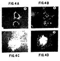
- one method for characterizing the functional interaction between a given receptor and a potential ligand involves determining whether the ECMR of interest distributes into the focal adhesions formed between cell and ligand substrate. For example, and not by way of limitation, ⁇ 4 ⁇ 1 may be shown to interact with fibronectin in a receptor/ligand relationship by the following method (see also section 6.2.3., infra ).
- Lymphocytes may be allowed to adhere to a fibronectin substrate, and the focal adhesions between cells and substrate may be visualized by interference reflexion microscopy ( Izzard et al., 1976, J. Cell Sci. 21:129-159 ).
- Antibodies which recognize ⁇ 4 ⁇ 1, such as P4G9 or P4C10, may be used to show, using standard immunohistochemical techniques, e.g., fluoro-iso thiocyanate, that in the absence of serum, ⁇ 4 ⁇ 1 redistributes into the focal adhesions.
- Interaction between ECMR and ligand may also be characterized by testing for the ability of the ECMR to adhere to a variety of different substrates.
- a cell type of interest or an ECMR of interst may be tested for the ability to bind to substrates consisting of purified components of the extracellular matrix, such as fibronectin, collagen, vitronectin or laminin.
- substrates consisting of purified components of the extracellular matrix, such as fibronectin, collagen, vitronectin or laminin.
- cells bearing the ⁇ 4 ⁇ 1 may be shown to adhere to fibronectin, but not to collagen or laminin substrates as a result of the ⁇ 4 ⁇ 1/fibronectin interaction.
- any ECMR of interest is shown to bind to a particular protein ligand
- substrates bearing subfragments of the protein ligand may be tested for the ability to bind to an ECMR on the surface of cells, thereby permitting the localization of the binding site between ECMR and ligand.
- the receptor is a4b1
- substrates bearing subfragments of fibronectin may be tested for their ability to bind a4bl-bearing cells, as exemplified in Section 6, infra.
- T lymphocytes attached to the 80 kDa cell binding domain of fibronectin bearing the ⁇ 4 ⁇ 1, receptor they demonstrated a clear preference for an non-RGD containing region located on a 38 kDa fragment.
- the 38 kDa fragment was three times more efficient than the 58 kDa fragment in mediating T lymphocyte adhesion.
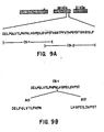
- the 38 kDa and 58 kDa fragments were derived from the A and B chains of plasma fibronectin, respectively. They therefore differ in the presence or absence of IIICS (Kornblihtt et al., 1985, supra ; Garcia-Pardo, 1987, supra ).
- the 38 kDa and 58 kDa fragments used here share a common low affinity T lymphocyte binding site, located in the Hop II domain, and that additional high affinity T lymphocyte adhesion sites are present in the IIICS region unique to the 38 kDa fragment.
- T lymphocytes appear to specifically recognize and bind to CS-1, which has been defined as a high affinity adhesion site for B16 melanoma cells and avian neural crest cells (Humphries et al., 1987, supra ; Humphries et al., 1988, J. Cell Biol., 106:1289-1297 ; Dufour et al., 1988, EMBO J., 7:2661-2671 ).
- CS-1 is a region of molecular heterogeneity (generated by alternative splicing) present in the type III CS domain on the A chain of plasma fibronectin.
- the ECMR/ligand relationship may be further characterized by identifying and evaluating agents which interfere with receptor/ligand binding.
- antibodies directed to an ECMR of interest may be used to inhibit ligand/receptor binding. Given the observation that a particular cell type adheres to a given ligand or cellular substrate, it may be of interest to identify the ECMR involved in the interaction. A panel of monoclonal antibodies, each directed toward a different ECMR, may be tested for the ability to block the adherence of cells to substrate. Inhibition of binding by a particular antibody would suggest that the ECMR recognized by that antibody is involved in the adhesive interaction. In a specific.
- lymphocyte adherence to endothelial cells in culture may be inhibited by antibodies directed toward ⁇ 4 ⁇ 1 but not by antibodies directed toward a variety of other ECMRs (see Section 7, below), indicating that ⁇ 4 ⁇ 1, is necessary for lymphocyte adhesion to endothelial cells. Additionally, monoclonal antibodies may be used to determine the relationship between ECMR and ligand substrate.
- T lymphocyte adhesion to the 38 and 58 kDa fragments could be completely inhibited by functionally defined monoclonal antibodies to a4bl.
- T lymphocyte adhesion to CS-1 (IgG conjugate) coated surfaces could also be completely inhibited by P4C2, P3E3 or P4G9.
- P4C2, P3E3 or P4G9 T lymphocyte adhesion to CS-1.
- these antibodies failed to inhibit adhesion of T cells to the 80 kDa fragment containing the prototype adhesion sequence arg-gly-asp (RGD).
- Adhesion of T cells to the 80 kDa fragment could be completely inhibited by a monoclonal antibody to ⁇ 5 ⁇ 1 (P1D6) or by RGDS.
- P1D6 and RGDS failed to inhibit T lymphocyte adhesion to the 38 and 58 kDa fragments or to CS-1.
- ⁇ 4 ⁇ 1 functions as the receptor for the carboxy terminal adhesion domain of plasma fibronectin receptor for alternative adhesion sequences in IIICS (CS-1) and possibly Hep II.
- the ECMR/ligand relationship may be characterized by determining the structure of the ligand.
- the ability of agents to compete with ligand in the ECMR/ligand interaction may be evaluated.
- the ligand is a protein
- various fragments of the protein may be tested for their ability to competitively inhibit receptor/ligand binding.
- peptide fragments of fibronectin may be tested for the ability to competitively inhibit the binding of lymphocytes to endothelial cell substrate.
- CS-1 peptide and, in particular, the peptide EILDVPST was able to competitively inhibit the binding of lynphocytes to fibronectin and to endothelial cells, thereby localizing the binding site on the ligand to a region identical or homologous to EILDVPST.
- adherence of one cell to another may be inhibited by intervening in the ECMR/ligand interaction.
- the binding of lymphocytes to endothelial cells may be inhibited by interfering with the binding of ⁇ 4 ⁇ 1 to its ligand. This may be accomplished by using antibodies directed toward the ECMR, or, alternatively, to its ligand (antibodies-may be generated toward ligand in a manner analogous to that described in Section 5.1).
- anti- ⁇ 4 ⁇ 1 antibody may be used to inhibit the binding of lymphocytes bearing ⁇ 4 ⁇ 1 receptors to vascular endothelial cells.
- the antibody is monoclonal antibody P4C2 ( ⁇ 4 ⁇ 1) or P4C10 ( ⁇ 1), or fragments or derivatives thereof, including chimeric antibodies with the same binding specificities.
- the adherence of one cell to another may be inhibited by interfering in the binding between the ECMR and its ligand.
- the adherence of lymphocytes to endothelial cells may be inhibited by interfering with the binding of ⁇ 4 ⁇ 1 on lymphocytes to its ligand on the endothelial cell surface.
- the interaction of additional ECMR with endothelial cell ligands, and the inhibition of adhesion of these cells to endothelium by interfering with the ECMR/endothelial cell interaction is envisioned.
- the adhesion of macrophages to the endothelium may also be inhibited by intervention in the macrophage ECMR/endothelial cell interaction.
- melanoma cells which also recognize the CS-1 peptide, may be inhibited from metastasizing and entering tissues using the antibodies of the invention.
- the present invention is for the use in suppressing the immune response in human patients in need of such treatment.
- the present invention is for the use in the treatment of diseases associated with chronic or relapsing activation of the immune system, including collagen vascular diseases and other autoimmune diseases (such as systemic lupus erythematosis and rheumatoid arthritis), multiple sclerosis, asthma, and allergy, to name but a few.
- the present invention also is for use in the treatment of relatively acute activations of the immune system in patients in need of such treatment, including, for example, and not by way of limitation, graft versus host disease, allograft rejection, or transfusion reaction.
- lymphocyte migration into tissues systemically or, alternatively, locally it may be desirable to inhibit lymphocyte migration into tissues systemically or, alternatively, locally.
- tissue systemically in diseases involving multiple organ systems, such as systemic lupus erythematosis, it may be desirable to inhibit lymphocyte adhesion systemically during a clinical exacerbation.
- lymphocyte adhesion in diseases involving multiple organ systems, such as systemic lupus erythematosis, it may be desirable to inhibit lymphocyte adhesion systemically during a clinical exacerbation.
- lymphocyte adhesion systemically during a clinical exacerbation.
- Control of systemic versus localized use of the methods of the present invention may be achieved by modifying the compositions of antibodies administered or by altering the structure of these agents or their pharmacologic compositions.
- the antibodies of the invention may be administered by any route, including subcutaneous, intramuscular, intravascular, intravenous, intraarterial, intranasal, oral, intraperitoneal, rectal, intratracheal, or intrathecal.
- Alternatively, to achieve systemic inhibition of lymphocyte adhesion it may be desirable to administer the antibodies intravenously.
- a pharmacologic carrier which facilities delivery of the antibodies of the invention.
- a pharmacologic carrier which aids in the penetration of the cuticle, epidermis, and dermis may be advantageous.
- Dissemination of the antibodies of the invention may also be controlled by altering the half-life of the antibody, or its effective half-life.
- the antibodies of the invention may be conjugated to antibodies or other ligands which might direct the antibodies to specific tissues.
- antibodies may be produced which mimic the ECMR, and thereby attach to endothelial cell ligands, blocking lymphocyte adhesion.
- Antibodies of the invention produced and defined as described supra are monoclonal antibodies and fragments and derivatives thereof, including the F(ab') 2 , Fab', and Fab fragments.
- Fluorescein-conjugated (goat) anti-mouse IgG and IgM (H and L chains) or rhodamine-conjugated (goat) anti-rabbit IgG and IgM (H and L chains) were obtained from Tago, Inc. (Burlingame, CA).
- R-phycoerythrin-conjugated strepavidin was from Biomeda (foster City, CA).
- Rabbit anti-mouse IgG (H+L) antiserum was obtained from Cappel (Cooper Bioaedical, Malvern, PA).
- 51 Cr-sodium chromate was from New England Nuclear.
- 125 I was from Amersham (Arlington Hts., IL).
- IL-2 Human recombinant interleukin-2 (IL-2) was a generous gift from Dr. D. Urdal (Immunex Corp., Seattle, WA). Laminin was purchased from Collaborative Research, Inc. Bedford, MA) and purified plasma fibronectin and collagen Types I and III were prepared as previously described (Wayner, E. A. and Carter, W. G., 1987, supra and Wayner et al., 1988, supra ).
- RD Human rhabdomyosarcoma
- HT1080 human fibrosarcoma
- PBMC Peripheral blood mononuclear cell
- platelet and granulocyte populations from normal human donors were prepared as described (Kunicki et al., 1988, supra ; Wayner et al., 1988, supra ).
- Peripheral blood cells from patients with acute lymphocytic, large granular lymphocyte (LGL) or myelogenous leukemia were obtained from Dr. I. Bernstein and Dr. T. Loughran (Fred Hutchinson Cancer Research Center).
- Human lymphokine 500 U/ml IL-2) activated killer (LAK) cells and the monoclonal HLA B7 specific human cytotoxic T lymphocyte (CTL) cell line, C1C4, were prepared according to standard protocols ( Grimm et al., 1982, J. Exp. Med., 155:1923-1941 ; Glasebrook, A. L. and Fitch, F. W., 1980, J. Exp. Med., 151:876-895 ; Brooks, 1983, Nature, 305:155-158 ; Wayner, E. A. and Brooks, C. G., 1984, J. Immuno1 132:2135-2142 ; Wayner, E. A. and Carter, W. G., 1987, J.
- the EBV transformed B lymphocyte cell line (BLCL), ST-1, was derived from the donor spleen used in the production of the C1C4 CTL line. All other cell lines and cell culture conditions were as previously described (Wayner, E. A. and Carter, W. G., 1987, supra ; Wayner et al., 1988, supra ).
- Monoclonal antibodies A1A5, against the common integrin (Hynes, R. O., 1987, supra ) ⁇ 1 subunit of the VIA family of receptors (Hemler, M. E., 1988, supra ) and B5-G10 to the VLA 4 a subunit (Hemler et al., 1987, supra ) were obtained from Dr. Martin Hemler of the Dana-Farber Cancer Inst., Boston, MA).
- P1B5 Monoclonal antibodie to the integrin receptors ⁇ 3 ⁇ 1 (P1B5), ⁇ 2 ⁇ 1 (P1H5) and ⁇ 5 ⁇ 1 (P1D6) have been described and were developed in this laboratory.
- P1H5 and P1D6 inhibit fibroblast and platelet adhesion to collagen and fibronectin-coated substrates, respectively (Wayner, E. A. and Carter, W. G., 1987, supra ; Kunicki et al., 1988, supra ; Wayner et al., 1988, supra ).
- Monoclonal antibodies to lymphocyte adhesion receptors were produced by the methods of Oi and Herzenberg ( Oi, V.T. and Herzenberg, L.A. 1980, Immunoglobulin producing hybrid cell lines. In: Selected Methods in Cellular Immunology. Ed. by B.B. Mishell and S.M. Shiigi, W.H. Freeman and Co., San Francisco, pp. 351-373 ) and Taggart and Samloff ( Taggart, R.T. and Samloff, I.M., 1983, Science, 219, 1228-1230 ) as described (Wayner and Carter, 1987; Wayner, et al., 1988).
- Spleens from RBF/DN mice immunized with 100 ⁇ l packed T lymphocytes were removed and fused with NS-1/FOX-NY myeloma cells.
- Viable heterokaryons were selected in RPMI 1640 supplemented with adenine/aminopterin/thymidine (Taggart and Samloff, 1983).
- Hybridomas producing antibody directed to lymphocyte adhesion receptors were screened by specific inhibition of lymphocyte adhesion to fibronectin-coated surfaces and cloned by limiting dilution.
- Antibodies that would alter cell adhesion to purified plasma fibronectin, tryptic fragments and CS peptides were identified as previously described (Wayner and Carter, 1987). Briefly, 48 well virgin styrene plates were coated with human plasma fibronectin (5 ⁇ l/ml). The plates were blocked with PBS supplemented with 10 mg/ml heat denatured BSA (HBSA).
- HBSA heat denatured BSA
- T lymphocyte or HT1080 cells were labeled with Na 2 51 CrO 4 (50 ⁇ lCi/ml for 2-4 hr), washed, and 5 X 10 4 HT1080 or cultured T cells or 5 X 10 10 PBL/well were incubated with hybridoma culture supernatants (1:2 dilution in PBS supplemented with 1 mg/ml heat denatured BSA) or control myeloma cell culture supernatant for 15 minutes at room temperature. The cells were allowed to adhere to the protein-coated surfaces in the presence of the hybridoma supernatants for 15-30 minutes (HT1080) or 2-4 hours (lymphocytes) at 37°C. Non-adherent cells were removed by washing with PBS, and the adherent cells were dissolved in SDS/NaOH and bound 51 Cr-cpm were quantitated in a gamma counter.
- Viable cells were surface labeled with 125-iodine as described (Wayner and Carter, 1987) followed by extraction with 1% v/v Triton® X-100 detergent or 0.3% CHAPS detergent in 50 mM phosphate buffered saline pH 7.2. In some cases 1mM CaCl 2 was added to the lysis buffer. 1mM phenylmethyl sulfonyl fluoride, 1mM N-ethylmaleimide, 1 ⁇ l/ml leupeptin and 1 ⁇ g/ml trypsin soybean inhibitor were used as protease inhibitors. Immune precipitation and sequential immune precipitations were performed exactly as previously described (Wayner and Carter, 1987, supra ).
- fibronectin Human plasma fibronectin was a generous gift from Drs. Horowitz and R. Schulman (New York Blood Center, NY). Fibronectin was digested with TPCK-trypsin for 90 min at 37°C, and the digest was fractionated by affinity and ion-exchange chromatography as previously described ( Garcia-Pardo et al., 1987, Biochem. J. 241:923-928 ; Garcia-Pardo et al., 1989, Exp. Cell Res. 181:426-431 ).
- CS-1 and CS-2 Two overlapping peptides spanning the initial 48 residues of the IIICS region of human fibronectin (CS-1 and CS-2) were synthesized and coupled to rabbit IgG as described (Humphries et al., 1986, and Humphries et al., 1987, J. Biol. Chem. 262:6886-6892 ).
- Adherent cells were trypsinized, washed in RPMI supplemented with 1 ⁇ g/ml BSA plus 100 ug/ml soybean trypsin inhibitor and allowed to adhere to acid washed and silanized glass cover slips coated with fibronectin, laminin or collagen (20 ⁇ g/ml) in the absence of serum for 1-4 hour as described (Carter and Wayner, in preparation). At the end of the incubation non-adherent cells were removed and adherent cells were fixed in 100 mM sodium cacodylate, 100 mM sucrose, 4.5 mM CaCl 2 , 2% formaldehyde for 20 min.
- Cryostat sections (6 ⁇ m) were prepared from human skin, tonsil, or tumor samples embedded in OCT medium after snap freezing in isopentane/liquid nitrogen. All sections were fixed in 4% paraformaldehyde in PBS prior to incubation with primary antibodies and secondary fluorescent antibodies as described ( Carter and Wayner, 1988, J. Biol. Chem. 263:4193-4201 ). In control experiments, no fluorescence of rhodamine was detected using the fluorescein filters or vice versa.
- adhesion of cells to fibronectin that expressed abundant ⁇ 5 ⁇ 1 could be effectively inhibited by P1D6.
- the synthetic peptide RGDS did not completely inhibit T lymphocyte adhesion to plasma fibronectin (50-70% for Molt 4 or Jurkat cells versus 80-90% for fibroblasts and 100% for K562-1 cells).
- T lymphocytes express fibronectin adhesion receptors other than ⁇ 5 ⁇ 1.
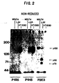
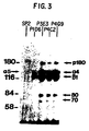
- p150 The biochemical characteristics of p150 suggested that it might be related to the VLA 4 antigen described by Hemler (Hemler et al., 1987). This was confirmed by sequential immune precipitation (not shown) with a VLA 4 specific monoclonal antibody, B5-G10. p150 was established as an a subunit of the integrin super family by its association with ⁇ 1 when immune precipitations were carried out after CHAPS detergent (0.3%) solubilization of 125 I surface labeled T lymphocytes in the presence of 1 mM Ca ++ ( Figure 3 ). Under these conditions ⁇ 4 was precipitated as a heterodimer with ⁇ 1. The identity of ⁇ 1 was confirmed by V8 protease peptide mapping (not shown).
- the ⁇ 4 ⁇ 1 heterodimer immune precipitated from T lymphocytes with the inhibitory monoclonal antibodies (P3E3, P4C2 and P4G9) was shown to be distinct from the prototype fibronectin receptor, ⁇ 5 ⁇ 1, immune precipitated with P1D6 by three criteria. 1) The relative quantities of ⁇ 4 ⁇ 1 and ⁇ 5 ⁇ 1 present in detergent extracts of T lymphocytes were distinct with higher levels of ⁇ 4 ⁇ 1 being present ( Figure 3 ). This was in agreement with the data we obtained using flow cytometry ( Figure 1 ). 2) In sequential immune precipitation experiments, monoclonal antibodies to ⁇ 4 ⁇ 1 did not preclear ⁇ 5 ⁇ 1 (now shown).
- V8 protease peptide maps derived from the ⁇ 4 and ⁇ 5 subunits precipitated with monoclonal antibodies P3E3 and P1D6 were clearly distinguishable (not shown). Furthermore, under the conditions (0.3% CHAPS and 1mM CaCl 2 ) used to solubilize the conjugate of ⁇ 4 ⁇ 1 from Jurkat cells ( Figure 3 ) another protein of higher molecular weight (p180) also reacted with the monoclonal antibodies or co-precipitated with ⁇ 4 ⁇ 1. p180 was absent from extracts prepared with P1D6 monoclonal antibody ( Figure 3 ), non-lymphoid cells or Triton® X-100 detergent extracts prepared in the absence of Ca ++ .
- peripheral blood monocytes freshly derived acute lymphocytic (T or B) leukemias, all large granular lymphocytic (LGL) and myelogenous leukemias and cultured T and B lymphocyte cell lines we examined expressed abundant ⁇ 4 ⁇ 1 (Table IV). Normal human blood platelets and granulocytes were negative for ⁇ 4 ⁇ 1 (Table IV). Normal human blood platelets and granulocytes were negative for ⁇ 4 ⁇ 1.
- T cells hematopoietic cell populations that expressed ⁇ 5 ⁇ 1 were activated T cells, platelets, monocytes and granulocytes, acute lymphocytic (T or B) and myelogenous leukemias and cultured K562, HL-60 and U937 cells.
- Some cultured T (Molt 4 or Jurkat) and B (ST-1) cell lines expressed low levels of ⁇ 5 ⁇ 1 as detected by P1D6 monoclonal antibody.
- P1D6 monoclonal antibody a subpopulation of PBL were positive for P1D6 fluorescence detected by flow cytometry. We are investigating the nature of this subpopulation of PBL which express ⁇ 5 ⁇ 1.
- TY cells a CD3 T cell lymphoma, were completely negative for P1D6 by flow cytometry.
- the major fibronectin receptor constitutively expressed by resting T lymphocytes is ⁇ 4 ⁇ 1 and as we have previously reported (Wayner et al., 1988) expression of ⁇ 5 ⁇ 1 in T lymphocytes is restricted to leukemic or activated cultured cells.
- most fibroblast cell lines expressed low levels of ⁇ 4 ⁇ 1 while large vessel endothelial cells (HUVEs) and cultured epithelial cells were negative for ⁇ 4 ⁇ 1 by flow cytometry.
- HUVEs large vessel endothelial cells
- ⁇ 4 ⁇ 1 was present in adult spleen, lymph node and tonsil and essentially absent from all other tissues we examined.
- the relative quantities of the fibronectin adhesion receptors expressed by cells in specific tissue domains varied dramatically.
- PBL and lymphocytes in tonsil and cortex and germinal center areas expressed large quantities of ⁇ 4 ⁇ 1 but virtually no ⁇ 5 ⁇ 1.
- ⁇ 4 ⁇ 1 was also found in epithelial regions in adult lymphatic tissue, but whether this was the result of lymphocyte infiltration of these areas or expression of ⁇ 4 ⁇ 1 by lymphatic epithelial cells was unclear.
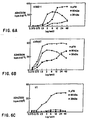
- hematopoietic cell lines such as K562 cells ( Figure 6 ) exhibited a clear preference for the 80 kDa fragment of plasma fibronectin while RD cells expressed promiscuous adhesion to all the gragments of plasma fibronectin tested except the N-terminal 29 kDa fragment.
- RDGS (1 mg/ml) partially inhibited (50%) Jurkat cell adhesion to intact fibronectin and completely (100%) inhibited their adhesion to the 80 kDa fragment.
- Jurkat cell adhesion to the 38 kDa fragment was unaffected by RGDS (up to 1 mg/ml).
- T lymphocytes to the 58 kDa fragment which also contains Hep II could be inhibited by P4C2.
- T lymphocyte cell lines which express both ⁇ 4 ⁇ 1 and ⁇ 5 ⁇ 1 (such as Jurkat cells) behave exactly as Molt 4 cells ( Figure 7 ).
- K562 cells express only ⁇ 5 ⁇ 1.
- Adhesion of K562 cells to the 38 ( Figure 6 ) and 58 kDa fragments was greatly reduced when compared to their adhesion to the 80 kDa fragment ( Figure 6 ).
- Adhesion of these cells to intact plasma fibronectin ( Figure 1 ) or the 80 kDa fragment could be completely inhibited by P1D6.
- YT cells which do not express ⁇ 5 ⁇ 1 adhere poorly to intact plasma fibronectin and the 80 kDa fragment ( Figure 6 ). These cells require 2-3 times longer to adhere to plasma fibronectin-coated surfaces than Jurkat or Molt 4 cells. YT cells, however, adhere efficiently and in a dose dependent manner to the 38 kDa fragment ( Figure 6 ) and adhesion of these cells to the 38 kDa fragment could be completely inhibited by P4C2.
- the IIICS region present on the A chain of plasma fibronectin contains at least two sites responsible for mediating cell adhesion to fibronectin ( Humphries et al., 1986, J. Cell Biol. 103:2637-2647 ; Humphries et al.; 1987, J. Biol. Chem. 262:6886-6892 ; Humphries et al.; 1988, J. Cell Biol. 106:1289-1297 ).
- CS-1 N-terminal peptides contained adhesion sequences recognized by mouse melanoma cells (Humphries et al., 1986, 1987).
- 38 kDa fragment contains a high affinity adhesion site recognized by human T lymphocytes and that ⁇ 4 ⁇ 1 is the receptor which mediates T lymphocyte adhesion to the 38 kDa fragment. This fragment does not contain the CS-5 site but it does contain the entire CS-1 region ( Garcia-Pardo et al., 1987, Biochem.
- T lymphocytes (Jurkat or Molt 4 cells) recognize and attach to CS-1 (rabbit IgG conjugate) -coated plastic surfaces (Table VI).
- T lymphocytes (Jurkat) do not attach to CS-2 (rabbit IgG conjugate) coated surfaces or to plastic surfaces coated with rabbit IgG alone.
- monoclonal antibodies to ⁇ 4 ⁇ 1 (P4C2) completely inhibited T lymphocyte adhesion to CS-1 while antibodies to ⁇ 5 ⁇ 1 (P1D6) had absolutely no effect (Table VI).
- the ligand in IIICS for ⁇ 4 ⁇ 1 was the CS-1 region previously defined as an adhesion site for melanoma cells.
- the functionally defined monoclonal antibodies to ⁇ 4 partially inhibited T lymphocyte adhesion to intact plasma fibronectin and had no effect on their attachment to an 80 kDa tryptic fragment containing the RGD adhesion sequence.
- Monoclonal antibodies (P1D6 and P1F8) to the previously described fibronectin receptor, ⁇ 5 ⁇ 1 completely inhibited T lymphocyte adhesion to the 80 kDa fragment but had no effect on their attachment to the 38 kDa fragment or to CS-1.
- Both ⁇ 4 ⁇ 1 and ⁇ 5 ⁇ 1 localized to focal adhesions when fibroblasts which express these receptors were grown on fibronectin-coated surfaces.
- the 38 kDa fragment does not contain CS-5 (Garcia-Pardo, 1987, supra ) the minimal amino acid sequence responsible for T lymphocyte adhesion to 38 kDa and therefore the ligand for ⁇ 4 ⁇ 1 in these cells is not arg-glu-asp-val or REDV (Humphries et al., 1986, supra ).
- LAK cells expressed abundant cell surface ⁇ 4 ⁇ 1 it did not appear to be a functional receptor; P1D6 completely inhibited LAK cell adhesion to fibronectin. The reason for this could be that LAK cells express a degraded form of ⁇ 4 (see Figure 2 ). In addition, because they are activated, LAK cells over-express ⁇ 5 ⁇ 1 when compared to resting peripheral blood or leukemic T cells (Table VIII). In other cells which express larger quantities of ⁇ 5 ⁇ 1 relative to ⁇ 4 ⁇ 1 (K562-1 and HT1080) adhesion to the 80 kDa RGD containing domain via ⁇ 5 ⁇ 1 is dominant (see K562-1 cells, Figure 6 ).
- T lymphocytes use two independent receptors during attachment to fibronectin and that i) ⁇ 5 ⁇ 1 is the receptor for the RGD containing cell adhesion domain, and ii) ⁇ 4 ⁇ 1 is the receptor for a carboxy terminal cell adhesion region containing the Heparin II and IIICS domains. Furthermore, these data show that T lymphocytes express a clear preference for a region of molecular heterogeneity in IIICS (CS-1) generated by alternative splicing of fibronectin pre-mRNA and that ⁇ 4 ⁇ 1 is the' receptor for this adhesion site.
- CS-1 molecular heterogeneity in IIICS
- cytokines associated with the inflammatory response including IL-1, tumor necrosis factor alpha (INF ⁇ ), and tumor necrosis factor beta (TNF ⁇ ).
- INF ⁇ tumor necrosis factor alpha
- TNF ⁇ tumor necrosis factor beta
- Jurkat Human T cell leukemia
- Dr. Paul Conlon Immunex. Corp., Seattle, WA
- Ramos Human B cell Leukemia
- the LAD (leukocyte adhesion deficient) and ST-1 B cell lines were prepared by Epsten-Barr virus tansfonnation of human B lymphoeytes.
- the LAD cell line was developed from the B cells of a patient with a deficiency in the ⁇ 2 integrin family of adhesion receptors and was obtained from Dr. John Harlan (Harborview Medical Center, Seattle, WA).
- Human umbilical vein endothelial cells (HUVEs) were purchased from Cell Systems, Seattle, WA.
- HUVEs were maintained in defined (serum-free) media also purchased from Cell systems (CS-100® media).
- HUVEs were incubated with IL-1 ⁇ (1 ng/ml) or in some experiments with TNF ⁇ (10 ng/ml) for 6-24 hours. At the end of this incubation the HUVE monolayers were washed and used in the adhesion assay.
- Peptides derived from the CS-1 region of plasma fibronectin were synthesized and HPLC purified according to standard protocols by Dr. James Blake at the Oncogen Corp., Seattle, WA.
- the CS-1 peptide was conjugated to rabbit serum albumin or KLH also according to standard protocols by Dr. James Blake.
- the RGDS control peptide was obtained from Peninsula Laboratories (Belmont, CA).
- P1H5 which recognizes the ⁇ 2 ⁇ 1 receptor ( Wayner et al., 1987, J. Cell Biol. 105:1873-11884 ; Wayner et al. 1988, J. Cell Biol. 107:1881-1891 ); P1B5, which recognizes the ⁇ 3 ⁇ 1 receptor ( supra ); P1D6, which recognizes the ⁇ 5 ⁇ 1 prototype fibronection receptor described by Pytela et al. 1985 (Cell 40:191-198 ); P4C10, which recognizes the ⁇ 1 subunit; and P4H9 which recognizes ⁇ 2 (CD18), using methods described fully in Wayner et al. (1987, J. Cell Biol. 105:1873-1884 ) and Wayner et al. 1988, J. Cell. Biol. 107:1881-1891 ) and described in Table II.
- Human umbilical vein endothelial cells were cultured in 48 well plates as described ( supra ). To measure adherence of lymphocytes to HUVE monolayer cultures, lyaphocytes were labeled with Na 2 51 CrO 4 (50 ⁇ Ci/ml) for 2-4 hours), washed, and then 10 5 lymphocytes were incubated with HUVE monolayers in the presence or absence of inhibitory antibodies or CS-1 derived peptides. The lymphocytes were allowed to adhere for 30 minutes at 37°C. Non-adherent cells were subsequently removed by washing with PBS, and the adherent cells were dissolved in SDS/NaOH. Bound 51 Cr-cpm were quantitated in a gamma counter.
- endothelial cells were activated prior to the adhesion assay by incubating with IL-1 ⁇ (1 ng/ml) or TNF- ⁇ (10 ng/ml) for 6-24 hours in defined CS-100® media (Cell Systems, Seattle, WA).
- lymphocytes use during extravasation we first determined the surface phenotype of normal and LAD lymphocytes with respect to the integrin receptors. These data are in Table VII and show clearly that the LAD cells possess a normal cell surface phenotype with respect to the ⁇ 1 containing integrins. Since, as expected, the B cells derived from the patient with LAD were negative for ⁇ 2, this strongly suggests that LAD lymphocytes use the ⁇ 1 containing integrins during their adhesion to and passage through the endothelium.
- Chromium-labeled lymphocytes from various cell lines were tested for their ability to adhere to either resting or activated endothelial cells (Table VIII). Although all the cell lines tested were found to adhere to some extent to resting endothelium, adhesion of T and B lymphocytes to endothelium activated by either IL-1 or TNF was observed to be much greater, by a factor of as much as ten-fold. Adhesion of lymphocytes from LAD patients to endothelium was not found to be significantly different from that observed for ST-1 cell lines derived from normal B cells (ST-1). There was no difference observed among cell lines between adhesion to IL-1 versus TNF activated endothelium.
- the amino acid sequence EILDVPST (see Table XII) might also be involved in lymphocyte diapedesis via binding of ⁇ 4 ⁇ 1 to this sequence present in a ligand expressed on the surface of the endothelium.
- the ability of synthetic CS-1 and derivative peptides to inhibit adherence of chromium-labeled lymphocytes to activated endothelial cells was evaluated using various peptides.
- the synthetic CS-1 peptide was a strong inhibitor of T or B lymphocyte adhesion to basal or activated endothelial cell monolayers (Tables X and XI).
- the EILDVPST sequence was the minimal peptide also required to inhibit lymphocyte adhesion to resting or activated HUVEs (Tables X and XI). In some cases, such as with the Ramos B cell line, adhesion of these cells to HUVEs could be completely abrogated with the EILDVPST peptide.
- LAD leukocyte adhesion deficiency
- LFA-1, Mac-1 or p150/95 ⁇ 2 containing receptors
- lymphocytes do undergo diapedesis to pass through the endothelium and can be found in tissues derived from patients with this disorder. This, therefore, implies that lymphocytes use a mechanism distinct from the ⁇ 2 containing integrins during their passage from the blood stream into the peripheral tissues.
- the following series of experiments comprises our attempts to fully understand the mechanisms utilized by peripheral blood lymphocytes during diapedesis.
- lymphocyte cell lines tested were shown to express ⁇ 4 ⁇ 1 and/or ⁇ 5 ⁇ 1 by fluorescence analysis, and were observed to adhere to cultured human umbilical vein endothelial cells. This adhesion was found to be blocked only by monoclonal antibodies directed toward ⁇ 4 ⁇ 1 ; antibodies directed toward other receptors were not found to have essentially any inhibitory effect, revealing the importance of the ⁇ 4 ⁇ 1 receptor in the adhesive interaction between lymphocytes and endothelium.
Landscapes
- Health & Medical Sciences (AREA)
- Life Sciences & Earth Sciences (AREA)
- Chemical & Material Sciences (AREA)
- Organic Chemistry (AREA)
- Immunology (AREA)
- General Health & Medical Sciences (AREA)
- Medicinal Chemistry (AREA)
- Engineering & Computer Science (AREA)
- Bioinformatics & Cheminformatics (AREA)
- Animal Behavior & Ethology (AREA)
- Pharmacology & Pharmacy (AREA)
- Proteomics, Peptides & Aminoacids (AREA)
- Public Health (AREA)
- Veterinary Medicine (AREA)
- Gastroenterology & Hepatology (AREA)
- Chemical Kinetics & Catalysis (AREA)
- Molecular Biology (AREA)
- Zoology (AREA)
- Biochemistry (AREA)
- Biophysics (AREA)
- Genetics & Genomics (AREA)
- General Chemical & Material Sciences (AREA)
- Nuclear Medicine, Radiotherapy & Molecular Imaging (AREA)
- Epidemiology (AREA)
- Toxicology (AREA)
- Heart & Thoracic Surgery (AREA)
- Cardiology (AREA)
- Biomedical Technology (AREA)
- Cell Biology (AREA)
- Transplantation (AREA)
- Peptides Or Proteins (AREA)
- Medicines Containing Antibodies Or Antigens For Use As Internal Diagnostic Agents (AREA)
- Preparation Of Compounds By Using Micro-Organisms (AREA)
- Pharmaceuticals Containing Other Organic And Inorganic Compounds (AREA)
- Medicines That Contain Protein Lipid Enzymes And Other Medicines (AREA)
- Medicines Containing Material From Animals Or Micro-Organisms (AREA)
- Plural Heterocyclic Compounds (AREA)
- Micro-Organisms Or Cultivation Processes Thereof (AREA)
- Acyclic And Carbocyclic Compounds In Medicinal Compositions (AREA)
- Polysaccharides And Polysaccharide Derivatives (AREA)
Claims (6)
- Utilisation d'un anticorps monoclonal ou d'un fragment de celui-ci qui se lie au récepteur α4β1 et inhibe l'adhésion des lymphocytes aux cellules endothéliales vasculaires en supprimant ainsi la réponse immunitaire, pour la préparation d'une composition pharmaceutique destinée à être utilisée chez un humain afin de supprimer la réponse immunitaire, dans laquelle l'anticorps ou fragment de celui-ci se lie à la sous-unité α4 du récepteur α4β1.
- Utilisation selon la revendication 1, dans laquelle l'anticorps monoclonal est P4C2, produit par l'hybridome déposé auprès de l'American Type Culture Collection sous le numéro d'accès HB 10215.
- Utilisation selon la revendication 1 ou 2, dans laquelle le fragment d'anticorps est un Fab, Fab' ou F(ab')2.
- Utilisation selon l'une quelconque des revendications précédentes, dans laquelle la suppression de la réponse immunitaire est un traitement d'une allergie, de l'asthme, ou d'une affection cutanée inflammatoire chronique.
- Utilisation selon l'une quelconque des revendications précédentes, dans laquelle la composition pharmaceutique est formulée pour une administration par voie sous-cutanée, intramusculaire, intravasculaire, intraveineuse, intra-artérielle, intranasale, orale, intrapéritonéale, intratrachéale, ou intrathécale.
- Procédé de préparation d'une composition pharmaceutique destinée à supprimer la réponse immunitaire chez un humain, caractérisé en ce que, en tant que constituant essentiel de ladite composition pharmaceutique, un anticorps ou un fragment de celui-ci est utilisé tel que défini dans l'une quelconque des revendications 1 à 5.
Priority Applications (2)
| Application Number | Priority Date | Filing Date | Title |
|---|---|---|---|
| EP20030017578 EP1366769A1 (fr) | 1989-09-01 | 1990-08-31 | Inhibition de l'adhésion de lymphocytes sur l'endothélium vasculaire au moyen d'une interaction entre un récepteur de la matrice extracellulaire et son ligand |
| DE69034116.4T DE69034116T3 (de) | 1989-09-01 | 1990-08-31 | Hemmung der anheftung von lymphozyten an das gefässendothelium mittels einer neuen extrazellulärer matrixrezeptor-ligand-wechselwirkung |
Applications Claiming Priority (3)
| Application Number | Priority Date | Filing Date | Title |
|---|---|---|---|
| US402389 | 1982-07-27 | ||
| US40238989A | 1989-09-01 | 1989-09-01 | |
| PCT/US1990/004978 WO1991003252A1 (fr) | 1989-09-01 | 1990-08-31 | Inhibition de l'adhesion de lymphocytes sur l'endothelium vasculaire au moyen d'une nouvelle interaction entre le recepteur matriciel extra-cellulaire et son ligand |
Related Child Applications (2)
| Application Number | Title | Priority Date | Filing Date |
|---|---|---|---|
| EP20030017578 Division EP1366769A1 (fr) | 1989-09-01 | 1990-08-31 | Inhibition de l'adhésion de lymphocytes sur l'endothélium vasculaire au moyen d'une interaction entre un récepteur de la matrice extracellulaire et son ligand |
| EP03017578.0 Division-Into | 2003-08-07 |
Publications (4)
| Publication Number | Publication Date |
|---|---|
| EP0489837A1 EP0489837A1 (fr) | 1992-06-17 |
| EP0489837A4 EP0489837A4 (en) | 1993-06-16 |
| EP0489837B1 EP0489837B1 (fr) | 2003-11-05 |
| EP0489837B2 true EP0489837B2 (fr) | 2013-11-13 |
Family
ID=23591680
Family Applications (2)
| Application Number | Title | Priority Date | Filing Date |
|---|---|---|---|
| EP90913598.0A Expired - Lifetime EP0489837B2 (fr) | 1989-09-01 | 1990-08-31 | Inhibition de l'adhesion de lymphocytes sur l'endothelium vasculaire au moyen d'une nouvelle interaction entre le recepteur matriciel extra-cellulaire et son ligand |
| EP20030017578 Withdrawn EP1366769A1 (fr) | 1989-09-01 | 1990-08-31 | Inhibition de l'adhésion de lymphocytes sur l'endothélium vasculaire au moyen d'une interaction entre un récepteur de la matrice extracellulaire et son ligand |
Family Applications After (1)
| Application Number | Title | Priority Date | Filing Date |
|---|---|---|---|
| EP20030017578 Withdrawn EP1366769A1 (fr) | 1989-09-01 | 1990-08-31 | Inhibition de l'adhésion de lymphocytes sur l'endothélium vasculaire au moyen d'une interaction entre un récepteur de la matrice extracellulaire et son ligand |
Country Status (19)
| Country | Link |
|---|---|
| EP (2) | EP0489837B2 (fr) |
| JP (1) | JP3357359B2 (fr) |
| KR (1) | KR100188459B1 (fr) |
| AT (1) | ATE253642T1 (fr) |
| AU (1) | AU654657B2 (fr) |
| CA (1) | CA2065292C (fr) |
| DD (1) | DD297562A5 (fr) |
| DE (3) | DE122006000044I2 (fr) |
| DK (1) | DK0489837T4 (fr) |
| ES (1) | ES2210225T5 (fr) |
| FI (2) | FI116793B (fr) |
| GR (2) | GR1001372B (fr) |
| IE (2) | IE20040028A1 (fr) |
| IL (3) | IL95501A (fr) |
| LU (1) | LU91273I2 (fr) |
| NL (1) | NL300240I2 (fr) |
| NZ (1) | NZ235131A (fr) |
| PT (1) | PT95180A (fr) |
| WO (1) | WO1991003252A1 (fr) |
Families Citing this family (29)
| Publication number | Priority date | Publication date | Assignee | Title |
|---|---|---|---|---|
| US5730978A (en) * | 1989-09-01 | 1998-03-24 | Fred Hutchinson Cancer Research Center | Inhibition of lymphocyte adherence with α4β1-specific antibodies |
| US7238668B1 (en) | 1989-09-01 | 2007-07-03 | Fred Hutchinson Cancer Research Center | Inhibition of lymphocyte adherence with CS-1-peptides and fragments thereof |
| US6033665A (en) * | 1989-09-27 | 2000-03-07 | Elan Pharmaceuticals, Inc. | Compositions and methods for modulating leukocyte adhesion to brain endothelial cells |
| US5610148A (en) * | 1991-01-18 | 1997-03-11 | University College London | Macroscopically oriented cell adhesion protein for wound treatment |
| US5629287A (en) * | 1991-01-18 | 1997-05-13 | University College London | Depot formulations |
| AU3420693A (en) * | 1991-12-24 | 1993-07-28 | Fred Hutchinson Cancer Research Center | Competitive inhibition of high-avidity alpha4-beta1 receptor using tripeptide ldv |
| WO1993013798A1 (fr) * | 1992-01-13 | 1993-07-22 | Biogen, Inc. | Traitement de l'asthme |
| US5871734A (en) * | 1992-01-13 | 1999-02-16 | Biogen, Inc. | Treatment for asthma with VLA-4 blocking agents |
| US5932214A (en) | 1994-08-11 | 1999-08-03 | Biogen, Inc. | Treatment for inflammatory bowel disease with VLA-4 blockers |
| IL102646A (en) * | 1992-07-26 | 1996-05-14 | Yeda Res & Dev | Non-peptidic surrogates of the ldv sequence and pharmaceutical compositions comprising them |
| EP0670735B1 (fr) | 1992-11-13 | 1997-04-02 | Board Of Regents Of The University Of Washington | Peripherisation de cellules souches hematopoietiques |
| JP3593343B2 (ja) † | 1993-02-09 | 2004-11-24 | バイオジェン インコーポレイテッド | インシュリン依存型糖尿病の治療 |
| EP0689609B2 (fr) † | 1993-03-18 | 2005-04-13 | N.V. Innogenetics S.A. | Procede de typage de hla-b au moyen d'amorces specifiques et de groupes de sondes |
| ZA947006B (en) * | 1993-09-15 | 1995-05-02 | Univ Emory | Method of inhibiting binding of reticulocytes to endothelium by interfering with vla-4/vcam-1 interactions |
| US5936065A (en) * | 1993-12-06 | 1999-08-10 | Cytel Corporation | CS-1 peptidomimetics, compositions and methods of using the same |
| US5821231A (en) * | 1993-12-06 | 1998-10-13 | Cytel Corporation | CS-1 peptidomimetics, compositions and methods of using same |
| US5770573A (en) * | 1993-12-06 | 1998-06-23 | Cytel Corporation | CS-1 peptidomimetics, compositions and methods of using the same |
| WO1995015973A1 (fr) * | 1993-12-06 | 1995-06-15 | Cytel Corporation | Peptidomimetiques cs-1, compositions et procedes pour les utiliser |
| US7435802B2 (en) | 1994-01-25 | 2008-10-14 | Elan Pharaceuticals, Inc. | Humanized anti-VLA4 immunoglobulins |
| US5840299A (en) * | 1994-01-25 | 1998-11-24 | Athena Neurosciences, Inc. | Humanized antibodies against leukocyte adhesion molecule VLA-4 |
| ES2424292T3 (es) * | 1994-01-25 | 2013-09-30 | Biogen Idec Ma Inc. | Anticuerpos humanizados contra la molécula de adhesión leucocitaria VLA-4 |
| FR2724393A1 (fr) * | 1994-09-12 | 1996-03-15 | Inst Nat Sante Rech Med | Obtention d'un anticorps monoclonal recombinant humanise a partir d'un anticorps monoclonal murin, sa production en cellules d'insecte, et ses utilisations |
| WO1997035557A1 (fr) * | 1996-03-22 | 1997-10-02 | Kao Corporation | Composition topique pour soigner la peau |
| US7147851B1 (en) | 1996-08-15 | 2006-12-12 | Millennium Pharmaceuticals, Inc. | Humanized immunoglobulin reactive with α4β7 integrin |
| SI1485127T1 (sl) | 2002-02-25 | 2011-09-30 | Elan Pharm Inc | Dajanje aktivne snovi za zdravljenje vnetja |
| US7125650B2 (en) | 2004-07-20 | 2006-10-24 | Roberts David H | Method for bump exposing relief image printing plates |
| TWI907924B (zh) | 2011-05-02 | 2025-12-11 | 日商武田藥品工業股份有限公司 | 抗-α4β7抗體之調配物 |
| UA116189C2 (uk) | 2011-05-02 | 2018-02-26 | Мілленніум Фармасьютікалз, Інк. | КОМПОЗИЦІЯ АНТИ-α4β7 АНТИТІЛА |
| IT202400002376A1 (it) * | 2024-02-05 | 2025-08-05 | Univ Degli Studi Di Brescia | Marcatori plasmatici di sindrome di ehlers-danlos ipermobile, disordini nello spettro dell’ipermobilità articolare, osteoartrite, artrite reumatoide e artrite psoriasica |
Family Cites Families (2)
| Publication number | Priority date | Publication date | Assignee | Title |
|---|---|---|---|---|
| US4578079A (en) * | 1982-08-04 | 1986-03-25 | La Jolla Cancer Research Foundation | Tetrapeptide |
| WO1990008833A1 (fr) * | 1989-02-02 | 1990-08-09 | Massachusetts Institute Of Technology | Expression de la fibronectine de recombinaison dans des cellules mises au point par genie genetique |
-
1990
- 1990-08-27 IL IL95501A patent/IL95501A/xx active IP Right Grant
- 1990-08-27 IL IL11326190A patent/IL113261A/en active IP Right Grant
- 1990-08-28 GR GR930100041A patent/GR1001372B/el not_active IP Right Cessation
- 1990-08-28 GR GR900100648A patent/GR1001161B/el not_active IP Right Cessation
- 1990-08-31 JP JP51276390A patent/JP3357359B2/ja not_active Expired - Lifetime
- 1990-08-31 PT PT95180A patent/PT95180A/pt not_active Application Discontinuation
- 1990-08-31 DE DE122006000044C patent/DE122006000044I2/de active Active
- 1990-08-31 WO PCT/US1990/004978 patent/WO1991003252A1/fr not_active Ceased
- 1990-08-31 AU AU63542/90A patent/AU654657B2/en not_active Expired
- 1990-08-31 NZ NZ235131A patent/NZ235131A/xx unknown
- 1990-08-31 KR KR1019920700621A patent/KR100188459B1/ko not_active Expired - Fee Related
- 1990-08-31 AT AT90913598T patent/ATE253642T1/de active
- 1990-08-31 EP EP90913598.0A patent/EP0489837B2/fr not_active Expired - Lifetime
- 1990-08-31 EP EP20030017578 patent/EP1366769A1/fr not_active Withdrawn
- 1990-08-31 ES ES90913598.0T patent/ES2210225T5/es not_active Expired - Lifetime
- 1990-08-31 IE IE20040028A patent/IE20040028A1/en not_active IP Right Cessation
- 1990-08-31 DE DE69034116.4T patent/DE69034116T3/de not_active Expired - Lifetime
- 1990-08-31 IE IE316990A patent/IE903169A1/en active IP Right Review Request
- 1990-08-31 DK DK90913598.0T patent/DK0489837T4/da active
- 1990-08-31 DE DE1990634116 patent/DE122006000044I1/de active Pending
- 1990-08-31 CA CA002065292A patent/CA2065292C/fr not_active Expired - Lifetime
- 1990-09-03 DD DD90343788A patent/DD297562A5/de unknown
-
1992
- 1992-02-28 FI FI920899A patent/FI116793B/fi active IP Right Grant
-
1995
- 1995-04-05 IL IL11326195A patent/IL113261A0/xx unknown
-
2005
- 2005-09-21 FI FI20050941A patent/FI118842B/fi active IP Right Grant
-
2006
- 2006-08-10 LU LU91273C patent/LU91273I2/fr unknown
- 2006-08-11 NL NL300240C patent/NL300240I2/nl unknown
Also Published As
Similar Documents
| Publication | Publication Date | Title |
|---|---|---|
| EP0489837B1 (fr) | Inhibition de l'adhesion de lymphocytes sur l'endothelium vasculaire au moyen d'une nouvelle interaction entre le recepteur matriciel extra-cellulaire et son ligand | |
| US5730978A (en) | Inhibition of lymphocyte adherence with α4β1-specific antibodies | |
| WO1993012809A1 (fr) | Inhibition competitive du recepteur alpha4-beta1 a avidite elevee a l'aide du tripeptide ldv | |
| IE83907B1 (en) | Inhibition of lymphocyte adherence to vascular endothelium utilizing a novel extracellular matrix receptor-ligand interaction | |
| Diamond et al. | A subpopulation of Mac-1 (CD11b/CD18) molecules mediates neutrophil adhesion to ICAM-1 and fibrinogen. | |
| Wayner et al. | Identification and characterization of the T lymphocyte adhesion receptor for an alternative cell attachment domain (CS-1) in plasma fibronectin. | |
| Wayner et al. | Epiligrin, a component of epithelial basement membranes, is an adhesive ligand for alpha 3 beta 1 positive T lymphocytes. | |
| Carter et al. | Epiligrin, a new cell adhesion ligand for integrin α3β1 in epithelial basement membranes | |
| Humphries et al. | Role of fibronectin in adhesion, migration, and metastasis | |
| Meerschaert et al. | The adhesion molecules used by monocytes for migration across endothelium include CD11a/CD18, CD11b/CD18, and VLA-4 on monocytes and ICAM-1, VCAM-1, and other ligands on endothelium. | |
| Pasqualini et al. | A study of the structure, function and distribution of β5 integrins using novel anti-β5 monoclonal antibodies | |
| Allavena et al. | Molecules and structures involved in the adhesion of natural killer cells to vascular endothelium. | |
| Matsuyama et al. | Activation of CD4 cells by fibronectin and anti-CD3 antibody. A synergistic effect mediated by the VLA-5 fibronectin receptor complex. | |
| Bernardi et al. | Lymphoid precursor cells adhere to two different sites on fibronectin. | |
| Bendall et al. | Human acute myeloid leukemia cells bind to bone marrow stroma via a combination of beta-1 and beta-2 integrin mechanisms | |
| EP0625912B1 (fr) | Traitement de l'inflammation intestinale | |
| HU216097B (hu) | Eljárás endotoxinsokk kezelésére antiadhéziós antitestek alkalmazásával | |
| US7238668B1 (en) | Inhibition of lymphocyte adherence with CS-1-peptides and fragments thereof | |
| Boyle et al. | Regulation of CS1 fibronectin expression and function by IL-1 in endothelial cells | |
| HK1062397A (en) | Inhibition of lymphocyte adherence to vascular endothelium utilizing extracellular matrix receptor-ligand interaction | |
| US6596276B1 (en) | Method for inhibiting tumor angiogenesis in a living subject | |
| Shock et al. | Cell adhesion in wound healing and pulmonary fibrosis | |
| Michael | A Subpopulation of Mac-1 (CDllb/CD18) Molecules Mediates Neutrophil Adhesion to ICAM-1 and Fibfinogen | |
| Stuiver et al. | Characterization of monoclonal antibodies against integrin αVβ5 | |
| HK1007683B (en) | Treatment for inflammatory bowel disease |
Legal Events
| Date | Code | Title | Description |
|---|---|---|---|
| PUAI | Public reference made under article 153(3) epc to a published international application that has entered the european phase |
Free format text: ORIGINAL CODE: 0009012 |
|
| 17P | Request for examination filed |
Effective date: 19920323 |
|
| AK | Designated contracting states |
Kind code of ref document: A1 Designated state(s): AT BE CH DE DK ES FR GB IT LI LU NL SE |
|
| A4 | Supplementary search report drawn up and despatched |
Effective date: 19930427 |
|
| AK | Designated contracting states |
Kind code of ref document: A4 Designated state(s): AT BE CH DE DK ES FR GB IT LI LU NL SE |
|
| 17Q | First examination report despatched |
Effective date: 19940610 |
|
| GRAG | Despatch of communication of intention to grant |
Free format text: ORIGINAL CODE: EPIDOS AGRA |
|
| RIC1 | Information provided on ipc code assigned before grant |
Free format text: 7C 12P 21/08 A, 7C 07K 7/06 B, 7A 61K 39/395 B, 7C 12N 5/16 - |
|
| RIC1 | Information provided on ipc code assigned before grant |
Free format text: 7C 12P 21/08 A, 7C 07K 7/06 B, 7A 61K 39/395 B, 7C 12N 5/16 - |
|
| GRAG | Despatch of communication of intention to grant |
Free format text: ORIGINAL CODE: EPIDOS AGRA |
|
| GRAG | Despatch of communication of intention to grant |
Free format text: ORIGINAL CODE: EPIDOS AGRA |
|
| GRAH | Despatch of communication of intention to grant a patent |
Free format text: ORIGINAL CODE: EPIDOS IGRA |
|
| GRAH | Despatch of communication of intention to grant a patent |
Free format text: ORIGINAL CODE: EPIDOS IGRA |
|
| GRAA | (expected) grant |
Free format text: ORIGINAL CODE: 0009210 |
|
| AK | Designated contracting states |
Kind code of ref document: B1 Designated state(s): AT BE CH DE DK ES FR GB IT LI LU NL SE |
|
| REG | Reference to a national code |
Ref country code: GB Ref legal event code: FG4D |
|
| RIN1 | Information on inventor provided before grant (corrected) |
Inventor name: WAYNER, ELIZABETH |
|
| REG | Reference to a national code |
Ref country code: CH Ref legal event code: EP |
|
| REF | Corresponds to: |
Ref document number: 69034116 Country of ref document: DE Date of ref document: 20031211 Kind code of ref document: P |
|
| REG | Reference to a national code |
Ref country code: SE Ref legal event code: TRGR |
|
| REG | Reference to a national code |
Ref country code: CH Ref legal event code: NV Representative=s name: E. BLUM & CO. PATENTANWAELTE |
|
| REG | Reference to a national code |
Ref country code: DK Ref legal event code: T3 |
|
| ET | Fr: translation filed | ||
| REG | Reference to a national code |
Ref country code: ES Ref legal event code: FG2A Ref document number: 2210225 Country of ref document: ES Kind code of ref document: T3 |
|
| REG | Reference to a national code |
Ref country code: AT Ref legal event code: UEP Ref document number: 253642 Country of ref document: AT Kind code of ref document: T |
|
| PLBI | Opposition filed |
Free format text: ORIGINAL CODE: 0009260 |
|
| PLBQ | Unpublished change to opponent data |
Free format text: ORIGINAL CODE: EPIDOS OPPO |
|
| PLAX | Notice of opposition and request to file observation + time limit sent |
Free format text: ORIGINAL CODE: EPIDOSNOBS2 |
|
| 26 | Opposition filed |
Opponent name: SERONO INTERNATIONAL S.A. Effective date: 20040805 |
|
| REG | Reference to a national code |
Ref country code: DE Ref legal event code: 8363 Ref document number: 69034116 Country of ref document: DE |
|
| NLR1 | Nl: opposition has been filed with the epo |
Opponent name: SERONO INTERNATIONAL S.A. |
|
| PLAX | Notice of opposition and request to file observation + time limit sent |
Free format text: ORIGINAL CODE: EPIDOSNOBS2 |
|
| PLAX | Notice of opposition and request to file observation + time limit sent |
Free format text: ORIGINAL CODE: EPIDOSNOBS2 |
|
| RAP2 | Party data changed (patent owner data changed or rights of a patent transferred) |
Owner name: FRED HUTCHINSON CANCER RESEARCH CENTER |
|
| NLT2 | Nl: modifications (of names), taken from the european patent patent bulletin |
Owner name: FRED HUTCHINSON CANCER RESEARCH CENTER Effective date: 20060118 |
|
| REG | Reference to a national code |
Ref country code: IT Ref legal event code: SPCF Ref document number: 502004901183049 Country of ref document: IT Free format text: AUTHORISATION NUMBER(S) AND DATE(S): EU/1/06/346/001, 20060627 Spc suppl protection certif: 132006901441326 Ref country code: DE Ref legal event code: V448 Ref document number: 69034116 Country of ref document: DE Free format text: PRODUCT NAME: NATALIZUMAB; REGISTRATION NO/DATE: EU/1/06/346/001, 20060627 Spc suppl protection certif: 12 2006 000 044 Filing date: 20060811 |
|
| REG | Reference to a national code |
Ref country code: FR Ref legal event code: CP Free format text: PRODUCT NAME: NATALIZUMAB; NAT. REGISTRATION NO/DATE: EU/1/06/346/001 20060627; FIRST REGISTRATION: FR - EU/1/06/346/001 20060627 Spc suppl protection certif: 06C0027 Filing date: 20060816 |
|
| REG | Reference to a national code |
Ref country code: GB Ref legal event code: CTFF Free format text: SPC/GB06/030: 20060814 Spc suppl protection certif: SPC/GB06/030 Filing date: 20060814 |
|
| REG | Reference to a national code |
Ref country code: AT Ref legal event code: ESZA Ref document number: 253642 Country of ref document: AT Kind code of ref document: T Free format text: PRODUCT NAME: NATALIZUMAB Spc suppl protection certif: SZ 27/2006 Filing date: 20060814 |
|
| REG | Reference to a national code |
Ref country code: SE Ref legal event code: SPCF Free format text: PRODUCT NAME: NATALIZUMAB OCH FARMACEUTISKT GODTAGBARA SALTER DAERAV; NAT REG. NO/DATE: EG EU/1/06/346 20060627; FIRST REG.: EG EU/1/06/346 20060627 Spc suppl protection certif: 06900252.L Ref country code: SE Ref legal event code: SPCF Free format text: 0690025-2, 20060627 Spc suppl protection certif: 0690025-2 Filing date: 20060627 |
|
| REG | Reference to a national code |
Ref country code: NL Ref legal event code: AC1 Spc suppl protection certif: 300240 Filing date: 20060811 |
|
| REG | Reference to a national code |
Ref country code: LU Ref legal event code: CCP Free format text: 91273, EXPIRES: 20150831 Spc suppl protection certif: 91273 Expiry date: 20150831 |
|
| REG | Reference to a national code |
Ref country code: FR Ref legal event code: CR Free format text: PRODUCT NAME: NATALIZUMAB OU L?UN DE SES SELS PHARMACEUTIQUEMENT ACCEPTABLES; REGISTRATION NO/DATE IN FRANCE: EU/1/06/346/001 DU 20060627; REGISTRATION NO/DATE AT EEC: EU/1/06/346/001 DU 20060627 Spc suppl protection certif: O6C0027 Filing date: 20060816 Ref country code: FR Ref legal event code: CR Free format text: PRODUCT NAME: NATALIZUMAB OU L?UN DE SES SELS PHARMACEUTIQUEMENT ACCEPTABLES; REGISTRATION NO/DATE IN FRANCE: EU/1/06/346/001 DU 20060627; REGISTRATION NO/DATE AT EEC: EU/1/06/346/001 DU 20060627 Spc suppl protection certif: 06C0027 Filing date: 20060816 |
|
| REG | Reference to a national code |
Ref country code: DK Ref legal event code: CTFF Spc suppl protection certif: CA 2006 00023 Filing date: 20060811 Expiry date: 20150831 |
|
| REG | Reference to a national code |
Ref country code: IT Ref legal event code: SPCG Ref document number: 502004901183049 Country of ref document: IT Free format text: AUTHORISATION NUMBER(S) AND DATE(S): EU/1/06/346/001, 20060627 Spc suppl protection certif: 132006901441326 Extension date: 20150831 |
|
| APBP | Date of receipt of notice of appeal recorded |
Free format text: ORIGINAL CODE: EPIDOSNNOA2O |
|
| APAH | Appeal reference modified |
Free format text: ORIGINAL CODE: EPIDOSCREFNO |
|
| REG | Reference to a national code |
Ref country code: FR Ref legal event code: CP |
|
| REG | Reference to a national code |
Ref country code: NL Ref legal event code: KC1 Free format text: 300240, 20100831, EXPIRES: 20150830 Spc suppl protection certif: 300240 Filing date: 20100831 Expiry date: 20150830 |
|
| APBQ | Date of receipt of statement of grounds of appeal recorded |
Free format text: ORIGINAL CODE: EPIDOSNNOA3O |
|
| REG | Reference to a national code |
Ref country code: CH Ref legal event code: PFA Owner name: FRED HUTCHINSON CANCER RESEARCH CENTER Free format text: FRED HUTCHINSON CANCER RESEARCH CENTER#1124 COLUMBIA STREET#SEATTLE WASHINGTON 98104 (US) -TRANSFER TO- FRED HUTCHINSON CANCER RESEARCH CENTER#1124 COLUMBIA STREET#SEATTLE WASHINGTON 98104 (US) |
|
| REG | Reference to a national code |
Ref country code: DK Ref legal event code: CTFG Free format text: PRODUCT NAME: NATALIZUMAB Spc suppl protection certif: CA 2006 00023 Filing date: 20060811 |
|
| REG | Reference to a national code |
Ref country code: SE Ref legal event code: SPCG Free format text: 0690025-2 Spc suppl protection certif: 0690025-2 Ref country code: SE Ref legal event code: SPCG Spc suppl protection certif: 06900252.L |
|
| REG | Reference to a national code |
Ref country code: AT Ref legal event code: EEZF Ref document number: 253642 Country of ref document: AT Kind code of ref document: T Free format text: PRODUCT NAME: NATALIZUMAB Spc suppl protection certif: SZ 27/2006 Filing date: 20060814 Extension date: 20150831 Effective date: 20080813 |
|
| REG | Reference to a national code |
Ref country code: HK Ref legal event code: WD Ref document number: 1017230 Country of ref document: HK |
|
| PGFP | Annual fee paid to national office [announced via postgrant information from national office to epo] |
Ref country code: FR Payment date: 20090806 Year of fee payment: 20 Ref country code: ES Payment date: 20090805 Year of fee payment: 20 Ref country code: DK Payment date: 20090724 Year of fee payment: 20 |
|
| REG | Reference to a national code |
Ref country code: FR Ref legal event code: AV Spc suppl protection certif: 06C0027 Ref country code: FR Ref legal event code: CT Free format text: PRODCUT NANE: NATALIZUMAB OU L'UN DE SES SELS PHARMACEUTIQUEMENT; REG. NO/DATE: EU/1/06/346/001 20060627 Spc suppl protection certif: 06C0027 Filing date: 20060816 Extension date: 20150830 |
|
| PGFP | Annual fee paid to national office [announced via postgrant information from national office to epo] |
Ref country code: AT Payment date: 20090723 Year of fee payment: 20 Ref country code: LU Payment date: 20090818 Year of fee payment: 20 Ref country code: SE Payment date: 20090807 Year of fee payment: 20 Ref country code: CH Payment date: 20090728 Year of fee payment: 20 Ref country code: NL Payment date: 20090814 Year of fee payment: 20 Ref country code: GB Payment date: 20090723 Year of fee payment: 20 Ref country code: DE Payment date: 20090831 Year of fee payment: 20 |
|
| PGFP | Annual fee paid to national office [announced via postgrant information from national office to epo] |
Ref country code: BE Payment date: 20090910 Year of fee payment: 20 |
|
| PLAB | Opposition data, opponent's data or that of the opponent's representative modified |
Free format text: ORIGINAL CODE: 0009299OPPO |
|
| PGFP | Annual fee paid to national office [announced via postgrant information from national office to epo] |
Ref country code: IT Payment date: 20090812 Year of fee payment: 20 |
|
| PLAB | Opposition data, opponent's data or that of the opponent's representative modified |
Free format text: ORIGINAL CODE: 0009299OPPO |
|
| R26 | Opposition filed (corrected) |
Opponent name: MERCK SERONO INTERNATIONAL S.A. Effective date: 20040805 |
|
| R26 | Opposition filed (corrected) |
Opponent name: MERCK SERONO S.A. Effective date: 20040805 |
|
| REG | Reference to a national code |
Ref country code: GB Ref legal event code: CTFG Free format text: PRODUCT NAME: NATALIZUMAB OR PHARMACEUTICALLY ACCEPTABLE SALTS THEREOF; REGISTERED: UK EU/1/06/346/001 20060627 Spc suppl protection certif: SPC/GB06/030 Filing date: 20060814 Extension date: 20150830 Effective date: 20100621 |
|
| BE20 | Be: patent expired |
Owner name: FRED *HUTCHINSON CANCER RESEARCH CENTER Effective date: 20100831 |
|
| REG | Reference to a national code |
Ref country code: CH Ref legal event code: PL |
|
| REG | Reference to a national code |
Ref country code: DK Ref legal event code: EUP |
|
| REG | Reference to a national code |
Ref country code: GB Ref legal event code: CTFE Free format text: PRODUCT NAME: NATALIZUMAB OR PHARMACEUTICALLY ACCEPTABLE SALTS THEREOF; REGISTERED: UK EU/1/06/346/001 20060627 Spc suppl protection certif: SPC/GB06/030 Filing date: 20060814 Expiry date: 20100829 Extension date: 20150830 Ref country code: GB Ref legal event code: PE20 Expiry date: 20100830 |
|
| REG | Reference to a national code |
Ref country code: DE Ref legal event code: V484 Ref document number: 69034116 Country of ref document: DE Free format text: PRODUCT NAME: NATALIZUMAB; REGISTRATION NO/DATE: EU/1/06/346/001, 20060627 Spc suppl protection certif: 12 2006 000 044 Filing date: 20060811 |
|
| REG | Reference to a national code |
Ref country code: DE Ref legal event code: V457 Ref document number: 69034116 Country of ref document: DE Free format text: PRODUCT NAME: NATALIZUMAB; REGISTRATION NO/DATE: EU/1/06/346/001, 20060627 Spc suppl protection certif: 12 2006 000 044 Filing date: 20060811 |
|
| EUG | Se: european patent has lapsed | ||
| REG | Reference to a national code |
Ref country code: AT Ref legal event code: EELA Ref document number: 253642 Country of ref document: AT Kind code of ref document: T |
|
| PG25 | Lapsed in a contracting state [announced via postgrant information from national office to epo] |
Ref country code: GB Free format text: LAPSE BECAUSE OF EXPIRATION OF PROTECTION Effective date: 20100830 |
|
| APBU | Appeal procedure closed |
Free format text: ORIGINAL CODE: EPIDOSNNOA9O |
|
| APAN | Information on closure of appeal procedure modified |
Free format text: ORIGINAL CODE: EPIDOSCNOA9O |
|
| PLAY | Examination report in opposition despatched + time limit |
Free format text: ORIGINAL CODE: EPIDOSNORE2 |
|
| PLBC | Reply to examination report in opposition received |
Free format text: ORIGINAL CODE: EPIDOSNORE3 |
|
| PLAY | Examination report in opposition despatched + time limit |
Free format text: ORIGINAL CODE: EPIDOSNORE2 |
|
| REG | Reference to a national code |
Ref country code: ES Ref legal event code: FD2A Effective date: 20120110 |
|
| PG25 | Lapsed in a contracting state [announced via postgrant information from national office to epo] |
Ref country code: ES Free format text: LAPSE BECAUSE OF EXPIRATION OF PROTECTION Effective date: 20100901 |
|
| REG | Reference to a national code |
Ref country code: NL Ref legal event code: V4 Effective date: 20100831 |
|
| PG25 | Lapsed in a contracting state [announced via postgrant information from national office to epo] |
Ref country code: DE Free format text: LAPSE BECAUSE OF EXPIRATION OF PROTECTION Effective date: 20100831 |
|
| PUAH | Patent maintained in amended form |
Free format text: ORIGINAL CODE: 0009272 |
|
| STAA | Information on the status of an ep patent application or granted ep patent |
Free format text: STATUS: PATENT MAINTAINED AS AMENDED |
|
| 27A | Patent maintained in amended form |
Effective date: 20131113 |
|
| AK | Designated contracting states |
Kind code of ref document: B2 Designated state(s): AT BE CH DE DK ES FR GB IT LI LU NL SE |
|
| REG | Reference to a national code |
Ref country code: DE Ref legal event code: R102 Ref document number: 69034116 Country of ref document: DE |
|
| REG | Reference to a national code |
Ref country code: DK Ref legal event code: T4 Effective date: 20131115 |
|
| REG | Reference to a national code |
Ref country code: SE Ref legal event code: RPEO |
|
| REG | Reference to a national code |
Ref country code: DE Ref legal event code: R102 Ref document number: 69034116 Country of ref document: DE Effective date: 20131113 |
|
| REG | Reference to a national code |
Ref country code: DE Ref legal event code: R082 Ref document number: 69034116 Country of ref document: DE Free format text: PRODUCT NAME: NATALIZUMAB; REGISTRATION NO/DATE: EU/1/06/346/001 20060627 Spc suppl protection certif: 122006000044 Filing date: 20060811 Expiry date: 20100901 Representative=s name: MEISSNER, BOLTE & PARTNER GBR, DE Extension date: 20150831 Ref country code: ES Ref legal event code: DC2A Ref document number: 2210225 Country of ref document: ES Kind code of ref document: T5 Effective date: 20140204 |
|
| REG | Reference to a national code |
Ref country code: DE Ref legal event code: R082 Ref document number: 69034116 Country of ref document: DE Representative=s name: MEISSNER, BOLTE & PARTNER GBR, DE |
|
| REG | Reference to a national code |
Ref country code: DE Ref legal event code: R071 Ref document number: 69034116 Country of ref document: DE Free format text: PRODUCT NAME: NATALIZUMAB; REGISTRATION NO/DATE: EU/1/06/346/001 20060627 Spc suppl protection certif: 122006000044 Filing date: 20060811 Expiry date: 20100901 Extension date: 20150831 |
|
| REG | Reference to a national code |
Ref country code: NL Ref legal event code: SPCX Free format text: PRODUCT: NATALIZUMAB, DESGEWENST IN DE VORM VAN EEN FARMACEUTISCH AANVAARDBAAR ZOUT; FIRST REGISTRATION, DATE: EU/1/06/346/001, 20060627 Spc suppl protection certif: 300240 Filing date: 20060811 Expiry date: 20100831 Extension date: 20150830 Effective date: 20150831 |
|
| REG | Reference to a national code |
Ref country code: GB Ref legal event code: SPCE Free format text: PRODUCT NAME: NATALIZUMAB OR PHARMACEUTICALLY ACCEPTABLE SALTS THEREOF; REGISTERED: UK EU/1/06/346/001 20060627 Spc suppl protection certif: SPC/GB06/030 Filing date: 20060814 Expiry date: 20100829 Extension date: 20150830 |
|
| REG | Reference to a national code |
Ref country code: AT Ref legal event code: SPCX Ref document number: 253642 Country of ref document: AT Kind code of ref document: T Free format text: PRODUCT NAME: NATALIZUMAB; REGISTRATION NO/DATE: EU/1/06/346/001 20060627 Spc suppl protection certif: 27/2006 Filing date: 20060814 Expiry date: 20100831 Extension date: 20150831 Effective date: 20150831 |