EP0637631A2 - Expression à très bon rendement d'un polypeptide contenant une région préS1 modifiée de l'antigène majeur du virus hépatitis B - Google Patents
Expression à très bon rendement d'un polypeptide contenant une région préS1 modifiée de l'antigène majeur du virus hépatitis B Download PDFInfo
- Publication number
- EP0637631A2 EP0637631A2 EP94111872A EP94111872A EP0637631A2 EP 0637631 A2 EP0637631 A2 EP 0637631A2 EP 94111872 A EP94111872 A EP 94111872A EP 94111872 A EP94111872 A EP 94111872A EP 0637631 A2 EP0637631 A2 EP 0637631A2
- Authority
- EP
- European Patent Office
- Prior art keywords
- antigen
- pres1
- cell
- virus
- promoter
- Prior art date
- Legal status (The legal status is an assumption and is not a legal conclusion. Google has not performed a legal analysis and makes no representation as to the accuracy of the status listed.)
- Withdrawn
Links
Images








Classifications
-
- C—CHEMISTRY; METALLURGY
- C07—ORGANIC CHEMISTRY
- C07K—PEPTIDES
- C07K14/00—Peptides having more than 20 amino acids; Gastrins; Somatostatins; Melanotropins; Derivatives thereof
- C07K14/005—Peptides having more than 20 amino acids; Gastrins; Somatostatins; Melanotropins; Derivatives thereof from viruses
-
- G—PHYSICS
- G01—MEASURING; TESTING
- G01N—INVESTIGATING OR ANALYSING MATERIALS BY DETERMINING THEIR CHEMICAL OR PHYSICAL PROPERTIES
- G01N33/00—Investigating or analysing materials by specific methods not covered by groups G01N1/00 - G01N31/00
- G01N33/48—Biological material, e.g. blood, urine; Haemocytometers
- G01N33/50—Chemical analysis of biological material, e.g. blood, urine; Testing involving biospecific ligand binding methods; Immunological testing
- G01N33/53—Immunoassay; Biospecific binding assay; Materials therefor
- G01N33/576—Immunoassay; Biospecific binding assay; Materials therefor for hepatitis
- G01N33/5761—Hepatitis B
- G01N33/5764—Hepatitis B surface antigen
-
- A—HUMAN NECESSITIES
- A61—MEDICAL OR VETERINARY SCIENCE; HYGIENE
- A61K—PREPARATIONS FOR MEDICAL, DENTAL OR TOILETRY PURPOSES
- A61K39/00—Medicinal preparations containing antigens or antibodies
- A61K2039/51—Medicinal preparations containing antigens or antibodies comprising whole cells, viruses or DNA/RNA
- A61K2039/525—Virus
-
- A—HUMAN NECESSITIES
- A61—MEDICAL OR VETERINARY SCIENCE; HYGIENE
- A61K—PREPARATIONS FOR MEDICAL, DENTAL OR TOILETRY PURPOSES
- A61K39/00—Medicinal preparations containing antigens or antibodies
-
- C—CHEMISTRY; METALLURGY
- C12—BIOCHEMISTRY; BEER; SPIRITS; WINE; VINEGAR; MICROBIOLOGY; ENZYMOLOGY; MUTATION OR GENETIC ENGINEERING
- C12N—MICROORGANISMS OR ENZYMES; COMPOSITIONS THEREOF; PROPAGATING, PRESERVING, OR MAINTAINING MICROORGANISMS; MUTATION OR GENETIC ENGINEERING; CULTURE MEDIA
- C12N2730/00—Reverse transcribing DNA viruses
- C12N2730/00011—Details
- C12N2730/10011—Hepadnaviridae
- C12N2730/10111—Orthohepadnavirus, e.g. hepatitis B virus
- C12N2730/10122—New viral proteins or individual genes, new structural or functional aspects of known viral proteins or genes
-
- Y—GENERAL TAGGING OF NEW TECHNOLOGICAL DEVELOPMENTS; GENERAL TAGGING OF CROSS-SECTIONAL TECHNOLOGIES SPANNING OVER SEVERAL SECTIONS OF THE IPC; TECHNICAL SUBJECTS COVERED BY FORMER USPC CROSS-REFERENCE ART COLLECTIONS [XRACs] AND DIGESTS
- Y10—TECHNICAL SUBJECTS COVERED BY FORMER USPC
- Y10S—TECHNICAL SUBJECTS COVERED BY FORMER USPC CROSS-REFERENCE ART COLLECTIONS [XRACs] AND DIGESTS
- Y10S435/00—Chemistry: molecular biology and microbiology
- Y10S435/975—Kit
-
- Y—GENERAL TAGGING OF NEW TECHNOLOGICAL DEVELOPMENTS; GENERAL TAGGING OF CROSS-SECTIONAL TECHNOLOGIES SPANNING OVER SEVERAL SECTIONS OF THE IPC; TECHNICAL SUBJECTS COVERED BY FORMER USPC CROSS-REFERENCE ART COLLECTIONS [XRACs] AND DIGESTS
- Y10—TECHNICAL SUBJECTS COVERED BY FORMER USPC
- Y10S—TECHNICAL SUBJECTS COVERED BY FORMER USPC CROSS-REFERENCE ART COLLECTIONS [XRACs] AND DIGESTS
- Y10S436/00—Chemistry: analytical and immunological testing
- Y10S436/82—Hepatitis associated antigens and antibodies
Definitions
- NP viruses non-passable viruses
- the diseases caused by NP viruses are often very serious, including cancer, immune system disorders and debilitation.
- HBV infection is endemic in many African and Asian countries. Large parts of these populations are carriers of the virus, and it is therefore extremely difficult to control the spread of the disease. In the more developed countries, blood transfusions are a major form of transmission.
- HBV contains three surface glycoproteins of different sizes that are encoded by a single open reading frame. Each uses a different ATG start codon, but all three use the same TAA stop signal; Ganem & Varmus, 1987; Robinson, 1990.
- the small surface antigen S-sAg main antigen or main protein
- the medium surface glycoprotein preS2-S-sAg (medium antigen or medium protein) has an additional 55 amino acids at the amino terminus of S-sAg.
- the large surface glycoprotein preS1-S2-S-sAg large antigen or large protein
- preS1-S2-S-sAg large antigen or large protein
- preS1 region below or "preS2" region refer to the segments with the additional 108 or 55 amino acids found for the large or medium antigen.
- the large protein is myristylated on its N-terminal glycine residue; Persing et al., 1987.
- the molecular weight of the individual antigens depends on the glycosylation and is 24 or 27 kD for the small protein, 30 or 33 kD for the medium protein and 39 or 42 kD for the large protein; Ganem & Varmus, 1987.
- preS regions bind to receptors on the surface of liver cells either via polymerized human serum albumin (preS2 region) or by direct binding of the preS1 region to the receptor molecule; Pontisso et al., 1989a; Neurath et al., 1990; Pontisso et al., 1989b; Petit et al., 1992.
- PreS1-S2-S-sAg remains associated with the endoplasmic reticulum (ER) and leaves the infected cell only as a component of an infectious virus particle (22 nm) or as a component of filamentous, non-infectious particles (42 nm) in the late infection phase; Hollinger, 1990.
- middle antigen and the major antigen are efficiently secreted and associate to non-infectious 22nm particles.
- Mechanisms that mediate translocation of the middle antigen and the major antigen through the membrane of the ER or prevent the secretion of the large antigen have been described; Ou & Rutter, 1987; Masuda et al., 1990; Prange et al., 1992.
- HBV surface proteins induce a B-cell and T-cell-specific immune response in infected individuals. In approximately 90% of cases, these immune responses lead to complete clearance of the infectious virus; Hollinger, 1990.
- discrete B-cell and T-cell-specific neutralizing epitopes were found both in the main antigen and in the preS regions; Neurath et al., 1985; Milich et al., 1985a; Milich et al., 1985b; Neurath et al., 1989.
- the hepatitis B virus antigen consists of a group-specific antigen a and at least two subtype determinants d / y and w / r (Le Bouvier, 1971; Bancroft et al., 1972), resulting in several different antigens leads genomic subtypes: adw, ayw, adr and ayr.
- genomic subtypes adw, ayw, adr and ayr.
- the geographical distribution of the subtypes is from Nishioka et al. (1975).
- a cross-subtype immune response is described by Szmuness et al. (1982).
- Some of the subtype-specific determinants are located in the pres regions (e.g. Milich et al., 1990a & 1990b).
- mice Vaccination studies with different strains of mice have shown that the response to certain S-sAg epitopes depends on the presence of defined molecules in the main histocompatibility complex. However, certain strains that do not respond to the major protein respond to additional epitopes in the preS regions of the large protein. The lack of response of mice to vaccines based on the main antigen alone can therefore be avoided by making the large protein a component of the vaccine; Milich et al., 1985a & 1985b.
- HBV antigens Despite intensive efforts in this regard, the antigenic structure and function of various HBV antigens remains largely unknown.
- One of the obstacles to obtaining information about HBV antigens is the difficulty in obtaining sufficient amounts of these polypeptides.
- Another problem is the isolation of individual HBV components for the development of vaccines and drugs with general effectiveness against a number of infectious HBV strains.
- antigenic components can be isolated from the serum or from tissues of infected people or other primates. However, this strategy leads to unacceptably small amounts. Antigens made from infected cells, blood, or purified viruses also pose a risk of contamination with the active virus. Therefore, isolation and purification of HBV's antigenic components in appropriate amounts appears to be acceptable only by recombinant techniques. It is expected that larger amounts and purer samples of antigenic components can be obtained by recombinant methods compared to those available from tissues of infected individuals. The production of vaccines and the development of therapies should therefore be supported by the improved production and purification options that the in vitro expression of recombinant HBV proteins offers.
- yeast (Dehoux et al., 1986; Imamura et al., 1987; Shiosaki et al., 1991; Kuroda et al., 1992), mammalian cells (Ou & Rutter, 1987; Lee et al., 1989; Youn et al., 1989; Korec et al., 1992) and viral expression systems (McLachlan et al, 1987; Yuasa et al., 1991; Shiraki et al., 1991; Belyaev et al., 1991 ).
- yeast expression systems express over-glycosylated large HBV antigens and non-glycosylated major proteins, none of which of these proteins is secreted.
- Prokaryotic systems do not glycosylate polypeptides and do not secrete recombinant proteins.
- the previously published system for the large-scale expression of HBV surface glycoproteins for the production of vaccines includes a yeast expression system which constitutively expresses these proteins.
- Transformed yeast cells form the small HBV surface glycoprotein with characteristics that correspond to the 22 nm particles found in the blood of naturally infected people.
- the main protein that is formed in yeast is not glycosylated (cf. EP 0 414 374 A2, page 16), while the large antigen is overglycosylated.
- large antigens with deleted glycosylation sites have been constructed (EP 0 414 374 A2, example F.3).
- modifications are likely to reduce the natural immunogenicity of the antigen.
- the large surface glycoprotein is translated by a preS1-specific mRNA, while the medium and small protein are translated by a preS2-specific mRNA (Robinson, 1990).
- the transcriptional promoter elements for the preS1-S2-S gene are upstream of this gene, while the preS2-S gene has its own promoter elements that are within the preS1 region.
- both the 2.4 kb preS1-S2-S mRNA and the 2.1 kb preS2-S mRNA are transcribed, and all three antigens are expressed.
- HBV surface glycoproteins expressed in eukaryotic cells are more efficiently secreted, but the yield of recombinant protein is lower than in prokaryotes or yeasts.
- continuous expression of recombinant HBV surface glycoprotein genes has only been shown in Chinese hamster ovary cells (CHO cells); Ou & Rutter, 1987; Lee et al., 1989; Youn et al., 1989; Korec et al., 1992.
- HBV surface glycoproteins were mainly expressed under the control of the weak vaccinia promoter P7.5. Since the expression yields are low using this promoter, the large antigen could only be detected using sensitive detection methods, such as radioactive labeling and autoradiography; Cheng et al., 1986; Cheng et al., 1987; Kutinivà et al., 1990; Nemeckovà et al., 1991. Expression of the small and medium antigen in the vaccinia virus system has been reported to result in effective secretion of both polypeptides; Cheng et al., 1987. However, the large antigen expressed with vaccinia virus is not secreted but remains associated with intracellular membranes; Cheng et al., 1986.
- Elroy-Stein et al. (1989) have developed yet another variation of the vaccinia virus expression system. Using the hybrid T7 vaccinia system already described, they additionally integrated the 5'-untranslated region (UTR) of the encephalomyocarditis virus in order to improve the translation of the mRNA which is transcribed by the T7 RNA polymerase obtain. The results showed an increased expression of the chloramphenicol transferase gene (cat gene) with protein concentrations that reached 10% of the total cell protein.
- UTR 5'-untranslated region
- Li et al. (U.S. Patent 5,077,213) that a recombinant vaccinia virus (Guang strain) carrying the preS1-S2-S gene from the HBV strain adr, efficiently secreted the small, medium and large antigen from TK- Osteosarcoma cells infected with this recombinant are allowed. It is not clear from Li et al. Which factors are responsible for the results described, especially since other reports show that (i) vaccinia does not secrete a large antigen and (ii) vaccinia is unable to produce the small and medium-sized antigen from the native HBV promoter.
- Li et al. neoplastic cells (osteosarcoma cells) used as the host.
- the unusual characteristics of these cells may account for the different results reported by Li et al. be reported to be responsible.
- the present invention includes a method for producing an HBV preS1-containing antigen using a vaccinia virus expression vector.
- the deletion of the myristylation site at the amino terminus of the large HBV antigen allows a significantly higher expression of HBV preS1-preS2-S-sAg polypeptides modified in this way.
- Vaccinia virus demyristylated antigen can be expressed (i) using a strong vaccinia promoter or (ii) using a Bacteriophage T7 promoter sequence in conjunction with the Bacteriophage T7 RNA polymerase. Demyristylation of the recombinant HBV large antigen results in expression yields that would not have been predicted in the prior art.
- the large HBV antigen can optionally be combined with an immunopotentiator.
- an immunopotentiator In the case of a polypeptide immunopotentiator, the large antigen can be replaced by a chemical linkage or by recombinant methods, ie ligating DNA sequences coding for the immunopotentiator and the large HBV antigen Hybrid gene to be coupled.
- the use of the vaccinia virus expression system allows cells to be used as hosts in which HBV replication normally occurs.
- the resulting availability of large amounts of purified large HBV antigen, which was processed in a "natural" manner enables the establishment of an area of application, for example diagnostic, therapeutic and vaccination applications, which was previously not possible.
- the myristylation site can be destroyed by targeted mutagenesis of the site where the myristylation takes place or by complete deletion of the site.
- the construct can then be cloned into the genome of a first chimeric vaccinia virus vector under the control of a regulatory vaccinia sequence. After infection of cells with the virus, the demyristylated protein is synthesized in large quantities.
- Another object of the present invention is to provide a chimeric vaccinia virus vector that expresses a demyristylated version of a preS-containing antigen under the control of a bacteriophage T7 promoter.
- a second chimeric vaccinia virus vector encodes the bacterial T7 phage RNA polymerase.
- the preS1-containing antigen is formed in large amounts by co-infection of cells with the first and the second virus.
- Another object of the present invention is to provide a preS1-containing antigen according to any of the above objectives, the antigen being synthesized by infection of cells derived from the liver or kidney. The antigen is thus processed in a manner that matches its natural antigenicity and immunogenicity.
- Another object of the present invention is to provide a vaccine containing preS1 according to the above objectives.
- the importance of such a product is demonstrated by the ability of the preS1 region to confer immunity to a certain percentage of people who do not respond to standard HBV vaccines.
- Another object of the present invention is to provide a preS1 containing antigen with more than one copy of the preS1 region.
- a chimeric vaccinia virus which comprises a promoter operably linked to a polynucleotide encoding a region of a large antigen of a hepatitis B virus which at least contains a preS1-B or T cell epitope, the region lacking the myristylation site of the preS1 region.
- a method of synthesizing a hepatitis B virus antigen comprising the steps of: (a) infecting a suitable eukaryotic cell with a first chimeric vaccinia virus which comprises a promoter which is functional with a Linked polynucleotide encoding a region of the preS1 protein region of a large hepatitis B virus antigen that contains at least one preS1 B or T cell epitope, the region lacking the myristylation site of the preS1 region; (b) culturing the coinfected cell; (c) lysis of the coinfected cell; and (d) obtaining the antigen.
- a hepatitis B virus antigen is prepared by a method comprising the following steps: (a) co-infecting a suitable eukaryotic cell with a first chimeric vaccinia virus comprising a promoter that is functional with a Linked polynucleotide encoding a region of the preS1 protein region of a large hepatitis B virus antigen that contains at least one preS1 B or T cell epitope, the region lacking the myristylation site of the preS1 region; (b) culturing the coinfected cell; (c) lysis of the coinfected cell; and (d) obtaining the antigen.
- the hepatitis B virus antigen is linked to an immunopotentiator.
- a vaccine which comprises an antigen of a hepatitis B virus, which was produced by the method described above, in a pharmaceutically acceptable carrier.
- an anti-HBV preS1 antibody is provided, which is produced by immunizing a mammal with an antigen of the hepatitis B virus, which was produced according to the method described above.
- Diagnostic reagent kits are also provided which comprise either a hepatitis B virus antigen or an anti-HBV preS1 antibody.
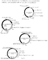
- Fig. 1 Structure of the plasmids pselP-gpt-L2 / preS1 and pselP-gpt-L2 / preS1dMyr.
- the plasmid structures of the gene for the large HBV protein or the demyristylated large HBV protein are given. Arrows show the direction of transcription from the selP and P7.5 promoters.
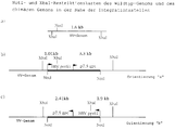
- FIG. 2 - XbaI restriction pattern of chimeric preS1 or preS1dMyr vaccinia viruses.
- the sizes of the XbaI fragments of the chimeric vaccinia viruses that hybridize with a 32 P-labeled preS1-specific probe are shown. Arrows show the direction of transcription from the selP and P7.5 promoters.
- FIG. 3 SacI restriction pattern of chimeric preS1 or preS1dMyr vaccinia viruses.
- the figure shows the size of the SacI fragments which hybridize with the 32 P-labeled 7.1 kb vaccinia virus SacI fragment "I" from chimeric preS1 and preS1dMyr vaccinia viruses. Arrows show the direction of transcription from the promoters selP and P7.5.
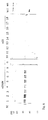
- Fig. 4 Pattern of hybridization with a 32 P-labeled, preS1-specific probe of XbaI cut genomic DNA from CV-1 cells infected with chimeric preS1 or preS1dMyr vaccinia virus clones.
- CV-1 cells were infected with chimeric preS1 or preS1dMyr vaccinia virus clones or wild-type vaccinia viruses.
- Lanes 1 to 10 XbaI cut genomic DNA from CV-1 cells infected with vS1 # 1-10.
- WT XbaI cut genomic DNA from CV-1 cells infected with the wild-type vaccinia virus.
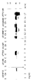
- FIG. 5 Pattern of hybridization with a 32 P-labeled vaccinia virus SacI fragment 'I' specific probe of SacI cut genomic DNA from CV-1 cells infected with chimeric vS1 or vS1dM viruses .
- CV-1 cells were infected with vS1, vS1dM or wild-type vaccinia virus.
- Lanes 1 to 10 SacI-cut genomic DNA from CV-1 cells infected with vS1 # 1-10.
- Lanes 11 to 18 SacI-cut genomic DNA from CV-1 cells infected with vS1dM # 1-6, 9 and 10.
- WT SacI cut genomic DNA from CV-1 cells infected with wild-type vaccinia virus.
- FIG. 7 Construction of plasmid pselP-elp4gpt-L2.
- the vaccinia virus promoter P7.5 (P7.5) or the synthetic early / late vaccinia promoter 4 (elp4) control the xanthine guanine phosphoribosyl transferase gene (gpt gene) from Escherichia coli. Arrows indicate the direction of the transcription.
- Fig. 8 Structure of the plasmids pTZ-L2 / preS1 and pTZ-L2 / preS1dMyr.
- the genes encoding the large HBV protein (preS1) or its demyristylated version (preS1dMyr) were under the control of the bacteriophage T7 promoter, which was matched with a DNA copy of the 5 'non-coding region of the encephalomyocarditis Virus (emc) was linked.
- emc encephalomyocarditis Virus
- Figure 9 Western blot of total proteins extracted from cells coinfected with the T7 polymerase-expressing virus vT7 / selP and the viruses vT7S1 or vT7S1dM.
- Lanes 1 to 4 lysates from CV-1 cells infected with vT7S1dM # 1-4.
- Lanes 5 and 6 lysates from CV-1 cells infected with vT7S1 # 1 and 2.
- Lane 7 lysate from CV-1 cells infected with wild-type vaccinia viruses.
- Lane 8 lysate from uninfected CV-1 cells.
- Lanes 9 to 12 Lysates from CV-1 cells infected with vT7S1 # 3-6. Arrows indicate the monomeric large antigen (lower arrow) and aggregated forms of the antigen (upper arrow).
- Figure 10 Western blots of total proteins from different cell lines infected with chimeric viruses.
- Lanes 1 to 3 Lysates from SK-Hep1 cells (s), Chang liver cells (Ch) or CV-1 cells (C), which were labeled with vS1 # 10 were infected.
- Lanes 4 to 6 Lysates from SK-Hep1 cells, Chang liver cells or CV-1 cells which were infected with vS1 # 9.
- Lane 7 lysate from CV-1 cells infected with vT7S1 # 1.
- Lanes 8 to 10 Lysates from Sk-Hep1 cells, Chang liver cells or CV-1 cells which were infected with wild-type vaccinia viruses.
- Lanes 11 to 13 Lysates from SK-Hep1 cells, Chang liver cells or CV-1 cells which were infected with vS1dM # 10.
- Lanes 14 to 16 Lysates from SK-Hep1 cells, Chang liver cells or CV-1 cells that were infected with vS1dM # 9.
- Lane 17 lysate from CV-1 cells infected with vT71dM # 1.
- FIG. 11 Western blots of total proteins from chimeric virus infected cells expressing myristylated and demyristylated antigens.
- 11A shows the signals of the immunostaining which are specific for preS1 in a Western blot with lysate dilutions of 1:10, 1:20, 1:30, 1:40 and 1:50. Different dilutions were required to determine the immunostaining signal that is suitable for evaluation in a densitometric scanning system.
- Coomassie blue staining at 1:30 dilution of the lysates from CV-1 cells infected with vS1 # 10 and vT7S1 dM is shown in Figure 11B.
- Figure 12 Results of densitometric scanning of Western blots of total proteins from chimeric virus-infected cells expressing myristylated and demyristylated large antigens.
- Computer generated graphics of the results of densitometric scanning of CV-1 cells using vT7S1dM and vS1 # 10 were infected. A 1:50 dilution was used for the T7 / modified construct, and a 1:10 dilution was used for the selP / wild type construct. The shaded area under the curve has been calculated and is shown under "total" at the bottom of each diagram.
- FIG. 13 Structure of plasmids pTZ-L2 / CRM228A, pTZ-L2 / preS1dMyr-CRM228A, pTZ-L2 / preS2-CRM228A and pTZ-L2 / sAg-CRM228A.
- the nucleotide sequence coding for fragment A of the Corynebacteriophage beta tox 228 gene is in the same reading frame as an NcoI / PstI fragment with the complete open reading frames of the HBV ayw-preS1-preS2-S-sAg gene, the preS2- S-sAg gene and the S-sAg gene fused.
- the fusion genes are arranged under the transcriptional control of a modified Bacteriophage T7 promoter. The direction of the transcription is indicated by arrows.
- FIG 14 Construction of plasmid pTZ-L2 / preS1dMyr (x2).
- the nucleotide sequence of the preS1 portion (nucleotides 1-324) of the preS1dMyr preS2 sAg gene was amplified using the plasmid pTZ-L2 / preS1dMyr as a template and specific PCR primer.
- the PCR product was cloned as an NcoI fragment into a pTZ-L2 / preS1dMyr vector cut with NcoI and treated with alkaline phosphatases.
- the preS1-preS1-preS2-sAg gene is arranged under the transcriptional control of a modified Bacteriophage T7 promoter. The position of the PCR primers and the direction of transcription are indicated by arrows.
- a recombinant nucleotide molecule is constructed that includes the sequence encoding the structure of the preS1 region of the HBV large antigen.
- Dane particle DNA is an readily available source from which components of the HBV genome can be isolated using standard methods known to those skilled in the art. In general, construction involves isolating Dane particle DNA, cutting DNA with enzymes, identifying DNA fragments containing the structural gene for the large antigen, ligating the isolated gene to a cloning vector, recombining a portion of the cloning vector including the preS1 region in a vaccinia virus vector and infection of a suitable host with the chimeric vaccinia vector.
- the chimeric vaccinia vector is capable of replication in the host and expression of both vaccinia genes and non-vaccinia genes. Infected cells express a protein containing preS1.
- Oligonucleotides can be obtained using a device such as that from Applied Biosystems, eg Model 380A, or by chemical techniques, such as the method of Matteucci et al., J. Am. Chem. Soc., Vol. 103 (1981), pp. 3185-3191.
- DNA sequences that code for HBV's proteins can be made from Dane particles using methods such as those described by Robinson, Am. J. Med. Sci., Vol. 270 (1975), pp. 151-159.
- the aminoterminal myristylation site of the large HBV antigen is believed to result in the binding of the protein to components of the cell, particularly the cell membrane. It now appears, unexpectedly, that the conversion of G to C on the 5th nucleotide of the region coding for preS1, with which the destruction of the myristylation site was intended, also leads to greater translational efficiency and thus to increased yields on the large antigen; see. Figures 11 and 12 and Example 4.
- the chimeric virus also contains a marker gene, and cells infected with such a virus express a selectable marker protein.
- a marker gene By adding a marker gene to the chimeric vaccinia virus together with further sequences of interest, vectors or cells which carry the sequences can be identified and separated from those cells or vectors without these sequences. This selection is made by culturing the cells in an environment that favors the growth of viruses that have the marker gene.
- the structural gene is placed under the control of a promoter.
- the vaccinia virus has unique transcriptional regulatory signals that are recognized by the viral RNA polymerase. Therefore defined vaccinia promoter sequences are usually ligated to coding sequences of interest.
- a strong vaccinia promoter, such as selP, is preferred in this context.
- the present invention also contemplates the use of non-vaccinia virus promoters.
- the promoter of choice comes from the bacteriophage T7.
- the vaccinia virus bacteriophage T7 hybrid expression system has been shown to significantly increase expression of several foreign genes; Elroy-Stein et al., 1989. Since the transcription machinery of the vaccinia virus does not act on this promoter, the present invention also provides a T7 RNA polymerase.
- the structural gene is thus functionally linked to a regulatory element from the phage, a phage polymerase acting on the regulatory element.
- the promoter / HBV large antigen gene and contain the T7 RNA polymerase gene in different chimeric vaccinia viruses and are contacted by co-infection of the same cell.
- a "cassette” is a covalently linked series of DNA codons that encode several non-overlapping proteins.
- a cassette comprises a gene containing preS1, and can also code for a marker gene.
- the cassette also contains a promoter sequence derived from the T7 phage.
- cassettes are flanked by singular restriction sites that allow clipping from a series of cloning vectors and recombination into a series of cloning vectors, in this case genetically modified vaccinia viruses.
- a cloning vector is used as an intermediate carrier of the isolated gene.
- the cloning vector contains a promoter and a marker gene that can be used to generate a cassette for direct transfer to a recipient, such as the vaccinia virus genome.
- Incorporation of the cassette into the vaccinia sequences requires cutting both the preS1-containing cloning vector and the viral genome, the latter generally being cut at a preferred location relative to the vaccinia virus genes.
- the next step is the ligation of the cut or linearized cassette with the vaccinia genome. This leads to the formation of a linear chimeric viral genome.
- the ligated DNA is then transfected into cells infected with a second vaccinia virus, which aids in packaging the chimeric DNA into an infectious viral particle.
- the formation of a preS1-containing protein comprises the steps of (i) antigenizing a cell culture and (ii) infecting the culture with one or more chimeric vaccinia viruses.
- the cells must be permissive for the replication of vaccinia and can be mammalian kidney cell lines such as Vero (ATCC # CCl81) and CV-1 (ATCC # CCl70), as well as liver cell lines such as SK-HEP-1 (ATCC # HTB52) and Chang ( ATCC # CCl13).
- An advantage of the vaccinia virus system is the wide range of host cells, including liver cells, kidney cells and many other mammalian cell lines. This property has not yet been used for the expression of recombinant HBV surface glycoproteins. Since post-translational processing and modification are important aspects of the immunological properties of an antigen, the cheapest expression system for this type of vaccine includes a mammalian cell, and particularly liver and kidney cells.
- the host cells are not only permissive, but also represent the types of cells in which HBV is normally replicated. They are therefore able to produce HBV antigens that closely resemble those produced during a natural infection.
- the cultured cells should be able to provide post-translational modifications that allow recombinant antigens to show natural, antigenic and immunogenic epitopes. Cells originating in the liver and kidney are most preferred in this regard.
- Cultures are incubated under conditions that optimize the expression of the recombinant proteins.
- Variables that can be optimized include, but are not limited to, temperature, pH, CO2 concentration, and the presence of serum proteins, growth factors, and nutrients.
- a selective factor that favors the growth of viruses that carry the recombinant genes is also included.
- Expression of a protein is usually optimal when a maximum amount of protein has been produced. Infected cells can be broken up mechanically, chemically or thermally to increase the yield of product.
- the recombinant product is further purified as required for a given application.
- Process for cleaning Polypeptides from cell cultures are known to the person skilled in the art. Techniques that can be used include salt extraction, gradient centrifugation, gel filtration, gel chromatography, ultrafiltration, electrophoresis, ion exchange and the like.
- immunopotentiators include oil-based vehicles (Freund's adjuvant), aluminum vehicles (Alum), toxins (Lipid A) and proteins (Interleukins).
- the immunopotentiators are toxins which can be chemically or, in the case of protein toxins, genetically linked to the immunogens.
- Possible toxins for use in such constructs include, but are not limited to, ricin, abrin, pertussis toxin, Shigella-like toxin, cholera toxin, Staphylococcus exotoxin A, heat-stable and heat-labile enterotoxin and Pseudomonas exotoxin A.
- diphtheria toxin is effective in immunopotentiating an immunogen to which it is linked.
- the use of diphtheria toxin in fusion constructs as an immunotoxin has been reported so far; Strom et al., Ann. NY Acad. Sci., Vol. 636 (1981), pp. 233-250; Shaw et al., J. Biol. Chem., Vol. 266 (1991), pp. 21118-21124; Williams et al., J. Biol. Chem., Vol. 265 (1990), pp. 11885-11889.
- immunotoxins differ from immunopotentiators in that the former is a toxin / antibody fusion molecule that selectively destroys target cells, while the latter is a toxin or a toxin / protein fusion molecule, the toxin portion enhancing the immunogenicity of the antigen portion.
- toxins other than diphtheria toxin, as immunopotentiators, have been reported; Cholera toxin: Hussain et al. (1989); Pertussis toxin: Halsov et al., 1991.
- nucleic acid sequences encoding a protein immunogen can be appended to nucleic acid sequences encoding the immunopotentiator in the same reading frame.
- Expression of the "hybrid gene” obtained results in a single polypeptide that contains both immunogen and immunopotentiator domains.
- Another method of increasing the immunogenicity of the preS1 protein sequences is to provide a larger effective dosage of the epitopes contained therein.
- One way in which this can be achieved is to double the preS1 sequences in the large HBV antigen.
- the expressed protein thereof has at least twice as many preS1-B and T-cell epitopes. This higher epitope density can help stimulate the anti-preS1 response.
- HBV-preS1-containing antigens consists in the production of a suitable material for an improved HBV vaccine.
- a significant proportion (7-15%) of the population does not respond to conventional HBV vaccines that do not contain preS1 regions.
- the ability to immunize individuals with antigens that contain this region that has been processed in a manner consistent with the natural infection allows a larger number of individuals to be protected from HBV infections.
- Antibodies directed against the recombinant protein containing preS1 also belong to the present invention.
- the present invention provides large amounts of preS1-containing antigens that are processed by cell types that are normally infected with HBV. Therefore, the possibility of producing both polyclonal and monoclonal antibodies, which are suitable for the detection, therapy and prevention of HBV-related diseases, is significantly improved.
- Immunotoxins are hybrid protein molecules that are generated by the fusion of an antibody and a toxin and can be used to selectively destroy certain cell populations.
- the potent cell-killing effect of a toxin is associated with the excellent specificity of monoclonal antibodies, which causes selective destruction of target cells while other cells are not affected.
- toxins include, but are not limited to, ricin, abrin, pertussis toxin, Shigella-like toxin, cholera toxin, Staphylococcus exotoxin A, heat-stable and heat-stable enterotoxin, Pseudomonas exotoxin A and diphtheria toxin. In some cases it is advantageous to reduce the toxicity of the toxin part by chemical or genetic measures.
- Toxins can be fused to antibodies chemically or, in the case of protein toxins, genetically.
- Protein toxin genes can be fused in frame with a single chain antibody gene (light chain / heavy chain hybrids; see Huston et al., Proc. Natl. Acad. Sci. USA, Vol. 85 (1988), p. 5879- p. 5879- 5883, and Bird et al., Science, Vol. 242 (1988), p. 423-426).
- the single chain antibody part can be replaced by other binding proteins, such as receptors or receptor ligands, for example II-2.
- a vaccine that includes at least the preS1 region of HBV that has been prepared by the recombinant technique described in the present application.
- the vaccine comprises an effective amount of the preS1-containing proteins which are produced by the recombinant expression system in cells infected with chimeric vaccinia viruses and which have a suitable immunogenicity.
- Appropriate immunogenicity means that the polypeptide is capable of eliciting an immune response that protects against natural HBV infection.
- a reagent kit that includes a container and (i) at least one antibody directed against a molecule containing preS1 or (ii) a molecule containing preS is contemplated.
- the antibodies according to the invention are involved in the detection of a molecule containing preS1, which was formed by a natural infection.
- the molecules according to the invention containing preS1 are involved in the detection of preS1-specific antibodies which have been formed by a host against a natural infection or an immunization. In both cases, an antibody that is provided with a chromatographic, fluorometric or other detectable label is used to detect either the preS1-containing protein or a preS1-specific antibody.
- the reagent set preferably also comprises a positive control for the binding of a molecule containing preS1 to a preS1-specific antibody or vice versa, and the detection reaction.
- the present invention also relates to a method for producing proteinaceous particles which show HBV antigenicity and / or HBV immunogenicity. Culturing infected cells leads to the formation of proteinaceous particles. The cells containing the particles can then be collected. According to preferred embodiments, the Particles are isolated from the cells using a number of methods known to those skilled in the art.
- the 1447 base pair fragment NsiI (position 1070 in the HBV ayw genome) / BstEII (position 2817 in the HBV ayw genome) was isolated from the plasmid pTHBV; Christman et al., 1982. Overhangs resulting from restriction endonuclease digestion were removed using T4 DNA polymerase. The blunt-ended fragment was cloned into the SmaI cut and dephosphorylated vector pTM3 (Moss et al., 1990), resulting in the intermediate plasmid pTM3 / preS1A, which carried a correctly oriented insert.
- the bacterial clone carrying the plasmid pTM3 / preS1A was infected with an M13 helper phage (Stratagene, Heidelberg, FRG) to obtain single-stranded (ss) plasmid DNA.
- This ss-DNA was used to generate an NcoI interface on the preS1-ATG start codon by means of targeted mutagenesis (mutagenesis oligonucleotide: 5'-GGA AAG ATT CTG CCC CAT GGT GTA GAT CTT GTT-3 ').
- the intermediate plasmid was named pTM3 / preS1B.
- pselP-gpt-L2 / preS1dMyr Single-stranded DNA of the plasmid pTM3 / preS1A was used to generate an NcoI site at the preS1-ATG start codon by targeted mutagenesis using a mutagenesis oligonucleotide 5'-GGA AAG ATT CTG CGC CAT GGT GTA GAT CTT GTT-3 ', which simultaneously introduced a cytocin at position 2 of the second codon, which changed a glycine at the N-terminus of the open reading frame for preS1-S2-S to an alanine (pTM3 / preS1dMyrA).
- the resulting 40 base pair NcoI fragment containing 5 'non-coding sequences was removed and the remaining one Vector was purified and religated over the NcoI sites to give plasmid pTM3 / preS1dMyrB.
- the 1418 bp NcoI / PstI fragment which contained the fully modified preS1-S2-S gene without 5 'non-coding sequences, was isolated from pTM3 / preS1dMyrB and with the pselP-gpt-L2 vaccinia cut with NcoI / PstI Virus cloning vector ligated; see. US serial no. 07/750 080.
- the plasmid obtained was designated pselP-gpt-L2 / preS1dMyr.
- the NcoI / PstI cassette from pTM-3 / preS1dMyrB was ligated with the pTZ-L2 vector cut with NcoI / PstI; see. US serial no. 07/750 080.
- telomere sequence was determined in a polymerase chain reaction (PCR) from pSV2gpt (ATCC # 37145) using gpt gene-specific oligonucleotides (5'-ATC ATA TGA GGA TCC ATG AGC GAA AAA TAC ATC GTC ACC-3 'and 5 '-ATC CCG GGA CGT CAT AAA AAG ATT AGC GAC CGG AGA TTG GCG G-3') amplified.
- PCR polymerase chain reaction
- NdeI and BamHI sites were introduced at the 5 'end and AatII and SmaI sites at the 3' end of the gpt gene.
- the PCR products were cut with NdeI / SmaI and ligated with a pFS51 vector (EP-A-91 114 300.6) cut with NdeI / SmaI.
- the elp4 sequence was designed as a synthetic NdeI / BamHI linker consisting of the hybridized oligonucleotides oelP4.1 (5'-TAT GTA AAA GTT GAA ATT ATT TTT TTA TGC TGT AAA TAA GTT AAC A-3 ') and oelP4.2 (5'-GAT CTG TTA ACT TAT TTA CTA GCA TAA AAA AAT AAT TTC AAC TTT TAC A-3 '), was introduced upstream of the open reading frame of the gpt gene to give the plasmid pelP4-gpt.
- oelP4.1 5'-TAT GTA AAA GTT GAA ATT ATT TTT TTA TGC TGT AAA TAA GTT A-3 '
- oelP4.2 5'-GAT CTG TTA ACT TAT TTA CTA GCA TAA AAA AAT AAT TTC AAC TTT TAC A-3 '
- the elp4-gpt gene cassette was excised from the plasmid pelP4-gpt with NdeI and AatII and ligated with NdeI / AatII cut and purified pselP-gpt-L2 vector DNA, resulting in the plasmid pselP-elp4gpt-L2.
- pselP-elp4gpt-L2 / T7 The 2.7 kb BamHI fragment with the gene for the bacteriophage T7-RNA polymerase was isolated from the vaccinia virus vTF7 / 3 (Fuerst et al., 1986) and with the BamHI-cut and dephosphorylated pselP-elp4gpt- L2 vector ligated.
- a plasmid clone which carried the T7 RNA polymerase gene in the correct orientation with respect to the selP promoter was isolated. This plasmid was named pselP-elp4gpt-L2 / T7.
- the 5 'non-coding sequences were completely removed from the preS1-S2-S gene.
- An NcoI interface (5'-CCATGG-3 ') was introduced around the ATG start codon by targeted mutagenesis. This manipulation allowed the ATG start codon of the corresponding gene to fuse directly with the early / late vaccinia virus promoter (selP) or the bacteriophage T7 promoter.
- the plasmids pselP-gpt-L2 / preS1 and pselP-gpt-L2 / preS1dMyr Fig.
- the double gene cassettes can be cut out of the plasmids with several restriction enzymes (e.g. NotI, SnaBI, SfiI) and ligated with the corresponding singular interfaces of a suitable vaccinia virus vector.
- the cassettes were cut out with NotI and ligated with vaccinia virus arms, which had also been cut with NotI.
- plasmids pTZ-L2 / preS1 and pTZ-L2 / preS1dMyr (Fig. 8) the large HBV antigen gene or its demyristylated version are under control of the bacteriophage T7 promoter.
- the viruses constructed with these plasmids were referred to as the vT7S1 series and vT7S1dM series.
- the nucleotide sequence encoding fragment A of the Corynebacteriophage beta tox 228 polypeptide (without signal peptide sequence) is in a polymerase chain reaction using the PCR primers 5 'CCT TCA GCC CAT GCC ATG GCT GAT GAT GTT 3' and 5 'A GCC ACC TAC TGC AGG CCT GAC ACG 3 'amplified.
- the 5 'primer introduces an NcoI site, while the 3' primer introduces a PstI site.
- the 606 bp PCR product is cut with NcoI and PstI and cloned into the pTZ-L2 vector cut with NcoI / PstI (cf. US Serial No. 07/914 738, which leads to the plasmid pTZ-L2 / CRM228A.
- pTZ-L2 / preS1dMyr-CRM228A An NcoI site is introduced at the C-terminus of the preS1dMyr open reading frame (ORF) using single-stranded DNA from pTZ-L2 / preS1dMyr as a template and the mutagenic oligonucleotide 5 'TTT TGT TAG GGT TTC CAT GGA TAC CCA AAG ACA 3'. This manipulation changes the last two amino acids of the open reading frame from preS1dMyr from Tyr-Ile to Ser-Met and introduces an additional Glu residue.
- the plasmid obtained is referred to as pTZ-L2 / preS1dMyrdTAA.
- This plasmid is linearized with PstI, partially digested with NcoI and ligated with the NcoI / PstI insert of the plasmid pTZ-L2 / CRM228A.
- a plasmid clone carrying the gene for the CRM228 A fragment is selected and designated pTZ-L2-preS1-CRM 22817.
- pTZ-L2 / preS2-CRM228A An NcoI site is introduced at the C-terminus of the preS2 open reading frame using single-stranded DNA from the plasmid pTZ-L2 / preS2 as a template and the mutagenic oligonucleotide 5 'TTT TGT TAG GGT TTC CAT GGA TAC CCA AAG ACA 3'. This manipulation changes the last two amino acids of the preS2 ORF from Tyr-Ile to Ser-Met and introduces an additional Glu residue.
- the plasmid obtained is referred to as pTZ-L2 / preS2dTAA.
- This plasmid is linearized with PstI, partially digested with NcoI and ligated with the NcoI / PstI insert of the plasmid pTZ-L2 / CRM228A.
- a plasmid clone carrying the gene for the CRM228 A fragment is selected and designated pTZ-L2 / preS2-CRM228 A.
- pTZ-L2 / sAg-CRM228A An NcoI site is introduced at the C-terminus of the HBV-sAg open reading frame using single-stranded DNA from the plasmid pTZ-L2 / sAg as a template and the mutagenic oligonucleotide 5 'TTT TGT TAG GGT TTC CAT GGA TAC CCA AAG ACA 3' .
- This manipulation changes the amino acid sequence of the ORF from HBV sAg from Tyr-Ile to Ser-Met and introduces an additional Glu residue.
- the plasmid obtained is referred to as pTZ-L2 / sAgdTAA.
- This plasmid is linearized with PstI, partially digested with NcoI and ligated with the NcoI / PstI insert of the plasmid pTZ-L2CRM228A.
- a plasmid clone bearing the gene for the CRM228 A fragment is selected and designated pTZ-L2 / sAg-CRM228A.
- pTZ-L2 / preS1dMyr (x2).
- the nucleotide sequence comprising amino acids 1-108 of the ORF from preS1dMyr is generated in a PCR reaction using pTZ-L2 / preS1dMyr as template and the oligonucleotides 5 'TTT TGG AAT ATA AAT AAG GC 3' and 5 'ATT CCA CTC CAT GGC CTG AG 3 'amplified as a PCR primer, the latter primer introducing a second NcoI interface into the PCR product.
- the 360 bp PCR product is cut with NcoI and ligated to the pTZ-L2 / preS1dMyr vector cut with NcoI and treated with alkaline phosphatase.
- a plasmid which carries the PCR product in the correct orientation relative to the old reading frame of the preS1 gene is selected and designated pTZ-L2 / preS1dMyr (x2).
- the NotI gene cassettes from the plasmids containing preS1, preS1dMyr or T7 polymerase (pTZ-L2 / preS1, pTZ-L2 / preS1dMyr, pselP-gpt-L2 / preS1, pselP-gpt-L2 / preS1dMyr, pselP-elp4gpt- L2 / T7) were isolated and ligated with the NotI cut genomic DNA from the vaccinia virus wild-type strain WR, as is essentially described in US serial no. 07/750 080.
- the ligation components consisted of 1 ⁇ g of genomic vaccinia virus DNA cut with NotI, 500 ng NotI cassette which had been isolated from the corresponding plasmids, 5 units of T4 DNA ligase and reaction buffer in a total volume of 50 ⁇ l. After incubation for 16 hours at 16 ° C., the ligation mixture was coprecipitated together with calcium phosphate and added to CV-1 cells that had grown subconfluently. This Cells had been infected with the fowlpox virus strain HP1-441 (Mayr & Malicki, 1996) two hours earlier with a multiplicity of infection, (moi; virus per cell) of 0.1 plaque-forming units (pfu) per cell. 5 to 7 days after the infection, the cell monolayer was completely infected with vaccinia viruses. The cells were harvested and the virus was released from the cell fragments by sonication.
- chimeric vaccinia viruses The selection for chimeric vaccinia viruses was carried out by titrating the virus progeny in the presence of 25 ug / ml mycophenolic acid, 15 ug / ml hypoxanthine and 125 ug / ml xanthine. Viruses grown under these conditions were further plaque-cleaned and used for the preparation of "small" and "large” virus stock solutions. Small virus stock solutions isolated from individual virus plaques were also grown under selective conditions. Infected cells from small stock solutions were divided into two cultures. One culture was used to make large virus stocks. DNA was isolated from the other, digested with various restriction endonucleases and transferred to nitrocellulose filters. These filters were hybridized with specific probes to confirm the orientation of the NotI cassette in the chimeric vaccinia virus genome and the absence of wild-type vaccinia virus.
- the XbaI fragments from genomic DNA of a chimeric preS1 vaccinia virus that hybridize with a 32 P-dCTP-labeled preS1-specific probe should have a size of 1.01 kb and 3.3 kb or 1.9 kb and 2, 41 kb, depending on the orientation of the NotI cassette (Fig. 2), while the SacI fragments from the same chimeric virus as the 32 P-dCTP-labeled 7.1 kb vaccinia virus SacI fragment Hybrid "I" should have a size of 5.35 kb and 4.47 kb or 5.15 kb and 4.68 kb, depending on the orientation (FIG. 3).
- FIG. 2a The singular NotI interface and the flanking XbaI interfaces of the wild-type vaccinia virus genome are shown in FIG. 2a.
- the integration of the NotI cassette into the singular NotI interface in the orientation "a" leads to the formation of two new XbaI fragments with a length of 1.01 kb and 3.3 kb (cf. FIG. 2b).
- the integration in the orientation "b” leads to two new XbaI fragments with a length of 2.41 kb and 1.9 kb (cf. FIG. 2c).
- Fig. 3a the singular NotI interface and the flanking SacI interfaces in the wild-type vaccinia virus genome are shown.
- FIG. 4 The hybridization patterns of the XbaI and SacI cut genomic DNA from 10 chimeric preS1 viruses, vS1 # 1-10, and 8 chimeric preS1dMyr viruses, vS1dM # 1-6, 9 and 10, either with the preS1 probe or the Vaccinia Virus SacI Probe-I are shown in Figures 4 and 5, respectively.
- genomic DNA was isolated from infected cell pellets and cut with XbaI. DNA fragments were separated on a 1% DNA agarose gel and transferred to nitrocellulose filters before probe hybridization. Hybridization of the XbaI fragments showed that vS1 # 1, 2, 4 and 5 (FIG.
- the viruses vS1dM # 1, 2, 5, 6 and 9 had inserted the NotI cassette in the orientation “b”, the virus isolate vS1dM # 10, FIG. 4, lane 18) represents the orientation "a”.
- the chimeric viruses vS1dM # 3 and 4 (Fig. 4, lanes 13 and 14) showed hybridization signals in the high molecular range. This type of signal also occurred in vS1dM # 2 and 5 (Fig. 4, lanes 12 and 15) and in vS1dM # 10 (Fig. 4, lane 18) and may indicate that multiple copies of the NotI cassette in the viral genome has been integrated. The exact structure of these viruses has yet to be determined.
- genomic DNA was isolated from infected cells and cut with SacI.
- the DNA fragments were separated on a 1% DNA agarose gel and transferred to nitrocellulose before probe hybridization.
- Analysis of the SacI fragments that hybridized with SacI Probe-I showed the expected patterns of orientation "a” (4.47 and 5.35 kb) and orientation "b" (4.68 kb and 5.15 kb).
- orientation "a" (4.47 and 5.35 kb)
- orientation "b" (4.68 kb and 5.15 kb
- the 7.1 kb wild-type vaccinia virus SacI fragment "I" (FIG. 5, lane WT) is not present in any of the chimeric viruses. The possibility of contamination of the chimeric viruses with wild-type vaccinia virus could therefore be excluded.
- a chimeric vaccinia virus carrying the T7 RNA polymerase gene, vT7 / selP was constructed by integrating the NotI cassette from the plasmid pselP-elp4gpt-L2 / T7 into the NotI interface of the vaccinia virus genomic DNA.
- This plasmid was constructed from the starting plasmid pselP-elp4gpt-L2, which contains an improved gpt gene cassette.
- the plasmid pselP-gpt-L2 was constructed as shown in FIG. 7.
- the gene which codes for the dominant selection marker is controlled by the vaccinia promoter P7.5, which is located on a 500 base pair vaccinia DNA fragment. This fragment can lead to undesirable recombination events with two endogenous vaccinia virus p7.5 promoter sequences; Venkatesan et al., 1981.
- the P7.5 gpt gene cassette (1.1 kb in size) was replaced by the elp4 gpt gene cassette (in size) of 0.5 kb) replaced.
- the new gene cassette was significantly reduced in size in addition to a weak, non-homologous synthetic early / late promoter.
- the resulting plasmid which had the optimized dominant marker and the strong synthetic early / late promoter followed by a multiple cloning site, was named pselP-elp4gpt-L2.
- CV-1 cells ATCC CCL 70
- SK-Hep1 cells ATCC HTB 52
- Chang liver cells ATCC CCL3
- a moi of 1 with chimeric preS1 vaccinia viruses chimeras preS1dMyr vaccinia viruses or chimeric vaccinia viruses infected with the preS1 or preS1dMyr genes under the control of the bacteriophage T7 promoter.
- Cells infected with the T7 preS1 chimeras were co-infected with a moi of 1 with a vaccinia helper virus chimera which carried the gene for the bacteriophage T7 RNA polymerase under the control of the selP promoter (vT7 / selP).
- vT7 / selP selP promoter
- cells were infected with vT7 / selP alone or after. The cells were harvested 3 days after infection. Infected cells were pelleted and resuspended in 300 ⁇ l sodium dodecyl sulfate sample buffer (SDS sample buffer). 10 to 20 ⁇ l of the lysate were sonicated, boiled and subjected to electrophoresis on a 12% SDS polyacrylamide gel.
- SDS sample buffer sodium dodecyl sulfate sample buffer
- FIG. 6 shows the expression yield in CV-1 cells which were infected with eight vS1dM isolations (FIG. 6, lanes 1 to 8) or 10 vS1 isolations (FIG. 6, lanes 9 to 18).
- vS1dM isolations FIGS. 4, and 5
- FIGS. 4 and 5 influence the expression efficiency of the vS1dM viruses (FIG. 6, lanes 1 to 8).
- the direct comparison of the expression yield of the cells infected with vS1dM viruses (FIG. 6, lanes 1 to 8) with vS1 viruses FIG. 6
- CV-1 cells were co-infected with vT7S1 or vT7S1dM viruses and with the second virus, vT7 / selP, which carried the gene for the bacteriophage T7 RNA polymerase. Lysates from CV-1 cells infected with four different isolates of vT7S1dM (Fig. 9, lanes 1 to 4) and 6 different vT7S1 viruses (Fig. 9, lanes 5 to 6 and 9 to 12) were analyzed . As a control, lysates from post-infected CV-1 cells or from CV-1 cells infected with vT7 / selP alone were also analyzed (FIG. 9, lanes 7 and 8, respectively).
- Example 4 Quantitative determination of the large antigen by Western blot analysis
- the expression yield was compared in a Western blot, which was essentially according to Towbin et al. (1979) with the following modifications.
- the first antibody was either a goat serum specific for hepatitis-associated antigens (HAA) (Axell, catalog number AXL 683), or monoclonal mouse antibodies specific for the preS1 region (Immunotech SA, catalog number 0866), diluted 1: 200 in a Western blot buffer (phosphate buffered saline containing 0.1% Tween 20).
- the second antibody was an alkaline phosphatase-coupled goat anti-mouse serum (Sigma Chemicals, Inc., Catalog No. A-4656) at a 1: 1000 dilution.
- the detection system (NBT and alkaline phosphatase) was from Promega, Inc.
- the quantitative determination of the large HBV antigen was carried out by comparing the Western blot bands.
- purified medium HBV ayw antigen was used in the Vero-cell / Vaccinia virus system was used.
- a series of dilutions was carried out with the purified medium antigen and with preparations of the large antigen.
- the dilutions were electrophoresed on a 12% SDS polyacrylamide gel, transferred to nitrocellulose and in a Western blot using anti-HAA serum as a first antibody and an alkaline phosphatase conjugated anti-goat IgG serum as a second Antibodies carried out.
- anti-HAA serum as a first antibody
- an alkaline phosphatase conjugated anti-goat IgG serum as a second Antibodies carried out.
- 1 to 10 ⁇ g large antigen / 106 cells were detected.
- CV-1 cells a monkey kidney cell line
- SK-Hep1 cells and Chang liver cells both human liver cell lines
- vS1 # 9 orientation: "a”
- vS1 # 10 orientation: "b”
- vS1dM # 9 orientation: "b”
- vS1dM # 10 orientation: "a"
- Figure 10 shows the results of the expression study.
- Lysates from SK-Hep1 cells (S), Chang liver cells (Ch) and CV-1 cells (C) with vS1 # 9 and vS1 # 10, wild-type vaccinia virus and vS1dM # 9 and vS1dM # 10 infected were compared.
- the increased levels of expression of the chimeras with the demyristylated preS1 gene (Fig. 10, lanes 11 to 16) were again clearly visible in comparison with those of the wild-type preS1 gene (lanes 1 to 6). This result shows that this effect is not limited to a single cell line, but could be observed repeatedly in different cell lines.
- the direct comparison of the expression yield starting from the bacteriophage T7 promoter against the vaccinia virus selP promoter was carried out in parallel.
- lysates from CV-1 cells infected with vT7S1 and vT7S1dM were also analyzed in the Western blot shown in FIG. 10.
- the difference in expression yield of the large antigen and its modified version was analyzed in a Western blot for comparison.
- the expression yield of the large antigen or its modified version in the T7 system was at least 5 times higher compared to the expression yields found in the selP promoter system (FIG. 10, lane 6 compared to 7, or lane 16, compared to 17).
- Cultures with 1 x 10⁷ CV-1 cells were infected with vLS1 # 10 with a moi of 2 pfu / cell or with vT7S1dM and the helper virus vT7 / selP with a moi of 1 pfu / cell for each chimeric virus.
- 48 hours after infection the infected cells were harvested and resuspended in 1 ml SDS sample buffer.
- the lysates were sonicated thoroughly and further diluted with SDS sample buffer. 20 ul of the diluted lysate were separated on a 12% SDS polyacrylamide gel. The gels were either used to transfer the proteins to a nitrocellulose filter (Western blot), or the proteins were stained with Coomassie blue.
- FIG. 11A shows the immunologically stained filters.
- the filters were analyzed with a densitometer (Hirschmann Elscript 400).
- the 1:30 dilutions of the lysates from each infection stained with Coomassie blue are shown in Figure 11B.
- the results of the densitometric analysis of the Western blot signals, a suitable dilution (1:10 for the vS1 # 10 lysate; 1:50 for the vT7S1dM lysate) are given in FIG. 12.
- the two peaks correspond to the two preS1 glycoforms.
- Example 5 Enhancement of protein immunogenicity: Construction of hybrid proteins consisting of the HBV surface glycoprotein and the diphtheria toxin fragment A cross-reacting material, and doubling of the HBV preS1-specific epitopes
- a new immunopotentiator namely diphtheria toxin cross-reacting material (CRM)
- CRMs diphtheria toxin cross-reacting material
- a strong immune response is elicited by Corynebacterium diphtheriae, which expresses CRM; "Pharmacology of Bacterial Toxins", in: International Encyclopedia of Pharmacology and Therapeutics, Dorner and Drews (Editor), pp.
- the diphtheria toxin gene or CRM gene is carried by a lysogenic ⁇ phage that is integrated or not integrated into the bacterial genome.
- the intact toxin consists of a fragment "A" with a molecular weight of 22 kD and a fragment "B” with a molecular weight of 40 kD. Fragment A ensures the toxic enzymatic properties, while fragment B is required for the translocation of fragment A across the cell membrane.
- CRM 228 is similar to the wild-type toxin but has no enzymatic activity due to changes in the amino acid sequence; Uchida et al., J. Biol. Chem., Vol. 248 (1973a), pp. 3838-3844; Streeck et al., Science, 221: 855-858.
- the phage carrying the CRM 228 gene is available from the Pasteur Institute, Paris, France.
- the linkage of the DNA coding for the glycoprotein with the nucleic acid coding for the CRM 228 fragment A is achieved by recombinant DNA methods; see. Fig. 13.
- the antigen and the immunopotentiator are therefore genetically linked and are expressed as a fusion protein. Fragment A was chosen because it is smaller compared to CRM 228 fragment B.
- the CRM 228 fragment B or the complete protein CRM 228 exceed the size of the preS1-S2-S-sAg, which may have a negative impact on the immune response directed against the preS1 part.
- the smaller size of the CRM 228 fragment A is advantageous in that it ensures the accessibility of the preS1 epitopes within the fusion protein.
- fragment A's strong immunogenicity is equivalent to that of the native molecule.
- An alternative method of linking HBV antigens and immunopotentiators is to chemically link the highly purified CRM 228 fragment A to purified preS1-preS2-S-sAg (demyristylated version), preS2-S-sAg or S-sAg.
- equimolar amounts of CRM 228 fragment A and antigen to be linked are crosslinked using standard chemical procedures; e.g. Widder and Green, Methods in Enzymology, vol. 112 (1985), p. 207.
- preS1 region of the large HBV antigen amino acids 1-108. This is accomplished by inserting a second copy of the preS1 coding region into a vector encoding all of the large HBV antigen; see. Fig. 14. The expression of this construct results in the new protein (preS1) 2-preS2-S-sAg, which contains two B and T cell-specific preS1 epitopes.
- the proteinaceous particles of the present invention are used in a pharmaceutically acceptable composition which, when administered in an effective amount, is capable of inducing protective immunity to HBV infection.
- the preparation of the vaccines which contain proteins as active ingredients is known to the person skilled in the art.
- such vaccines are manufactured as injectable vaccines, namely as liquid solutions or suspensions; solid forms suitable for dissolving or suspending in a liquid before injection can also be made.
- the preparation can also be emulsified.
- the active immunogenic component is often mixed with a carrier that is pharmaceutically acceptable and compatible with the active component. Suitable carriers are e.g. Water, saline, dextrose, glycerin, ethanol and the like, and combinations thereof.
- the vaccine can optionally contain small amounts of auxiliary substances, such as wetting agents or emulsifiers, pH-buffering agents, adjuvants or immunopotentiators, which increase the effectiveness of the vaccine.
- the vaccines are administered parenterally in a conventional manner by injection, e.g. subcutaneously or intramuscularly.
- Other formulations suitable for other modes of administration include suppositories and in some cases oral formulations.
- conventional binders and carriers e.g. include polyalkalene glucose or triglycerides; Suppositories can be formed from mixtures which contain the active ingredient in a concentration in the range from 0.5% to 10% and preferably in a concentration of 1.2%.
- Oral formulations include typical pharmaceutical grade carriers such as mannitol, lactose, starch, magnesium stearate, sodium saccharin, cellulose, magnesium carbonate and the like.
- the compositions take the form of solutions, suspensions, tablets, pills, capsules, sustained release formulations or powders and contain 10% to 95% and preferably 25 to 70% of active ingredient.
- the proteinaceous particles can be formulated into a vaccine as neutral forms or salt forms.
- Pharmaceutically acceptable salts include acid addition salts formed with the free amino groups of the peptide and inorganic acids such as hydrochloric acid or phosphoric acid, or organic acids such as acetic acid, oxalic acid, tartaric acid, mandelic acid or the like. Salts formed with the free carboxyl groups can also be from inorganic bases such as sodium hydroxide, potassium hydroxide, ammonium hydroxide, calcium hydroxide or iron (III) hydroxide, and organic bases such as isopropylamine, trimethylamine, 2-ethylaminoethanol, histidine, procaine and the like.
- unit dose refers to physically discrete units suitable for use in humans, each unit containing a predetermined amount of active material which has been calculated to provide the desired therapeutic effect in combination with the required diluent, ie the wearer, and a special treatment plan.
- the amount to be administered both in terms of the number of treatments and the amount, depends on the patient being treated, the ability of the patient's immune system to synthesize antibodies and the level of protection desired. Exact amounts of active ingredient that need to be administered depend on the practitioner's judgment and apply specifically to an individual.
- the suitable dosage range is, however, on the order of 1 to several 100 ⁇ g of active ingredient per individual. Appropriate schedules for initial administration and refreshments also vary, but typically an initial administration is followed by 1 or 2 week intervals with one or more subsequent injections or other administrations.
- mice 3 month old female BALB / c mice were vaccinated five times with purified HBV ayw preS1 expressed in Vero cells infected with the appropriate vaccinia virus chimera in a series of vaccinations over a period of 6 weeks ( approximately 1 to 10 ⁇ g protein per vaccination). 3 days after the last vaccination, blood samples were taken from the immunized mice and examined for preS-specific antibodies using an ELISA system with preS1 as the test antigen. Spleen cells from mice with a positive ELISA signal were isolated and plasmacytoma cell line P3X63Ag8 (ATCC TIB 9) using polyethylene glycol according to the method of Lane et al., J. Immunol. Meth. Vol. 81 (1985), pp.
- Hybridoma cell clones that secrete preS-specific monoclonal antibodies were selected in a dilution assay in RPMI 1640 medium (8041-2400, GIBCO). The specificity for the preS1 portion of the monoclonal antibodies was confirmed by synthetic preS1 peptides in a commercially available peptide mapping reagent set (Cambridge Research Biochemicals).
- a diagnostic system preferably in the form of a reagent set, which is suitable for the detection of the presence of preS1-containing antigens or antibodies against preS1-containing antigens in a body sample comprises (a) a protein according to the invention or (b) an antibody according to the invention and (c) a labeled specific binding agent to elicit a signal in the presence of the immune response of a protein containing preS1 and an anti-preS1 antibody.
- label in its various grammatical forms refers to individual atoms and molecules that are involved, either directly or indirectly, in triggering a detectable signal to indicate the presence of an immunoreactant. Any label can be attached to, incorporated into, or used separately from a specific agent, and these atoms or molecules can be used alone or in conjunction with other reagents. Such labels are known as such in the field of immunochemistry and form part of the present invention only insofar as they are used with proteins, methods and / or systems which are new in other respects.
- the diagnostic reagent sets of the invention are typically used in an "ELISA format" to detect the presence or amount of anti-preS1 antibodies or proteins containing preS1 in a body sample, such as serum or plasma.
- ELISA also refers to a solid phase enzyme immunoassay ("enzyme-linked immunosorbent assay") using an antibody or antigen bound to a solid phase and an enzyme-antigen or enzyme-antibody conjugate to determine the amount of antigen or antibody present in a sample to be proven and quantified.
- enzyme-linked immunosorbent assay enzyme-linked immunosorbent assay
- a description of the ELISA technique can be found in Chapter 22 of the 4th edition of Basic and Clinical Immunology, DP Sites et al., Lange Medical Publications, Los Altos, CA (1982) and in U.S. Patents 3,654,090, 3,850 752 and 4 016 043, all of which are made the subject of the present application by appropriate reference.
- the protein containing the preS1 according to the invention or the anti-preS1 antibody according to the invention are fixed to a solid matrix, a solid carrier being formed.
- the molecules are typically fixed to the solid matrix by adsorption from an aqueous medium, although several other types of adsorption and other types of fixation known to those skilled in the art can also be used. Examples of such types are the interaction of proteins with the carboxyl units, which are formed by reaction of cyanogen bromide with matrices containing glucose. These matrices include cross-linked dextrose or cellulose and linkage with glutaraldehyde, as discussed below in connection with latex particles and the like.
- Suitable solid matrices are known to the person skilled in the art. Such materials include the cross-linked dextran available under the trademark SEPHADEX from Pharmacia Fine Chemicals (Piscataway, NJ), agarose, polystyrene beads from about 1 ⁇ m to about 5 mm in diameter (Abbott Laboratories, North Chicago, IL), polyvinyl chloride , Polystyrene, cross-linked polyacrylamide, nitrocellulose or nylon-based fabrics such as layers, strips, blades and tubes.
- SEPHADEX cross-linked dextran available under the trademark SEPHADEX from Pharmacia Fine Chemicals (Piscataway, NJ)
- agarose polystyrene beads from about 1 ⁇ m to about 5 mm in diameter (Abbott Laboratories, North Chicago, IL)
- polyvinyl chloride Polystyrene
- cross-linked polyacrylamide nitrocellulose or nylon-based fabrics such as layers, strips, blades and tubes.
- a preferred matrix is in the form of a
- Hepatitis B virus envelope epitopes gene assembly and expression in Escherichia coli of an immunologically reactive novel multiple-epitope polypeptide 1 (MEP-1). Genes. 110, 137-144. Kuroda, S., Fujisawa, Y., Iino, S., Akahane, Y., Suzuki, H. 1992a. Induction of protection level of anti-pre-S2 antibodies in humans immunized with a novel hepaptitis B vaccine consisting of M (pre-S2 + S) protein particles (a third generation vaccine). Vaccine, 9, 163.
- Milich, DR, Mc Namara, MK, Mc Lachlan, A., Thornthon, GB, Chisari, FV 1985a Distinct H-2-linked regulation of T-cell responses to the pre-S and S regions of the same hepatitis B surface antigen polypeptide allows circumvention of nonresponsiveness to the S region. Proc. Natl. Acad. Sci. USA 82, 8168-8172. Milich, DR, Thornton, GB, Kent, SB, Michel, ML, Tiollais, P. 1985b. Enhanced Immunogenicity of the Pre-S Region of Hepatitis B Surface Antigen. Science, 228, 1195-1199. Milich et al., 1990a.
- Hepatitis B virus contains pre-S gene-encoded domains. Nature 315 ,, 154-156. Neurath, AR, Seto, B., Strick, N. 1989b. Antibodies to synthetic peptides from the preS1 region of the hepatitis B virus (HBV) envelope (env) protein are virus-neutralizing and protective. Vaccine 7, 234-236. Nishioka K., Levin, AG, Simons, MJ 1975. Hepatitis B. antigen, antigen subtypes, and hepatitis B antibody in normal subjects and patients with liver disease. Bulletin of the WHO 52, 293-300.
- Pontisso P., Petit, MA, Bankowski, MJ, Peeples, ME 1989a. Human Liver Plasma Membranes Contain Receptors for the Hepatitis B Virus Pre-S1 Region and, via Polymerized Human Serum Albumin, for the Pre-S2 Region. Journal of Virology, 63, 1981-1988. Pontisso, P., Ruvoletto, MG, Gerlich, WH, Heermann, KH, Bardini, R., Alberti, A. 1989b. Identification of an Attachment Site for Human Liver Plasma Membranes on Hepatitis B Virus Particles. Virology 173, 522-530. Prange, R., Nagel, R., Streek, R. 1992.
- EP 0 198 474 A1 Salstroem et al. Hepatitis B surface antigen formed by recombinant DNA techniques, vaccines, diagnostics cell lines and methods forming same.
- ENDOTRONICS INC. preS1-S2-S protein containing particles produced in mammalian cells (CHO, Vero, mouse L Cells)
- SMITHKLINE BIOLOGICALS. preS1-S2-S protein containing particles produced in Hansenula yeasts US Serial No. 07 / 750.080. Dorner et al. 1991.
- Patent application "Direct molecular cloning of a modified eukaryotic cytoplasmic DNA virus genome”. US Serial No. 07 / 750.080 CIP. Dorner et al. 1992. Continuation in Part of "Direct molecular cloning of a modified eukaryotic cytoplasmic DNA virus genome”. US 5,077,213. Li et al. Recombinant vaccinia virus. SHANGHAI INSTITUTE OF BIOCHEMISTRY. (preS1-S2-S protein containing particles produced in vaccinia) PCT WO 85/03876. Sobczak et al., 1985. Composition useful for. . . INSTITUT PASTEUR.
- preS2-S protein containing particles produced in CHO EP 0 174 444 B1. Valenzuela, PT 1984. Hepatitis surface antigen particle vaccine. CHIRON CORPORATION (preS2-S protein containing particles produced in yeast) EP A 0 312 159. Ellis et al., 1987. Method for producing hepatitis B proteins in yeast. MERCK & CO. (preS2-S protein containing particles produced in Saccharomyces) PCT WO 90/10058. Even-Chen, 1989. Method for production and purification of hepatitis B vaccine. BIO-TECHNOLOGY GENERAL CORP. (preS1-S2-S containing particles produced in CHO cells) US Serial # 07 / 882,768.
Landscapes
- Health & Medical Sciences (AREA)
- Life Sciences & Earth Sciences (AREA)
- Chemical & Material Sciences (AREA)
- Immunology (AREA)
- Engineering & Computer Science (AREA)
- Molecular Biology (AREA)
- General Health & Medical Sciences (AREA)
- Medicinal Chemistry (AREA)
- Hematology (AREA)
- Biochemistry (AREA)
- Biomedical Technology (AREA)
- Urology & Nephrology (AREA)
- Organic Chemistry (AREA)
- Cell Biology (AREA)
- Gastroenterology & Hepatology (AREA)
- Physics & Mathematics (AREA)
- Analytical Chemistry (AREA)
- Microbiology (AREA)
- Biotechnology (AREA)
- General Physics & Mathematics (AREA)
- Pathology (AREA)
- Virology (AREA)
- Food Science & Technology (AREA)
- Biophysics (AREA)
- Genetics & Genomics (AREA)
- Proteomics, Peptides & Aminoacids (AREA)
- Communicable Diseases (AREA)
- Preparation Of Compounds By Using Micro-Organisms (AREA)
- Micro-Organisms Or Cultivation Processes Thereof (AREA)
- Medicines Containing Antibodies Or Antigens For Use As Internal Diagnostic Agents (AREA)
- Peptides Or Proteins (AREA)
Applications Claiming Priority (2)
| Application Number | Priority Date | Filing Date | Title |
|---|---|---|---|
| US99351 | 1979-12-03 | ||
| US9935193A | 1993-07-30 | 1993-07-30 |
Publications (2)
| Publication Number | Publication Date |
|---|---|
| EP0637631A2 true EP0637631A2 (fr) | 1995-02-08 |
| EP0637631A3 EP0637631A3 (fr) | 1996-08-21 |
Family
ID=22274582
Family Applications (1)
| Application Number | Title | Priority Date | Filing Date |
|---|---|---|---|
| EP94111872A Withdrawn EP0637631A3 (fr) | 1993-07-30 | 1994-07-29 | Expression à très bon rendement d'un polypeptide contenant une région préS1 modifiée de l'antigène majeur du virus hépatitis B. |
Country Status (4)
| Country | Link |
|---|---|
| US (2) | US6077691A (fr) |
| EP (1) | EP0637631A3 (fr) |
| AT (1) | AT409863B (fr) |
| CA (1) | CA2129156A1 (fr) |
Cited By (1)
| Publication number | Priority date | Publication date | Assignee | Title |
|---|---|---|---|---|
| EP0753581A1 (fr) | 1995-07-10 | 1997-01-15 | Immuno Ag | Virus recombinantes eucaryotes cytoplasmiques améliorés, un procédé pour leur préparation et leur utilisation comme vaccins |
Families Citing this family (2)
| Publication number | Priority date | Publication date | Assignee | Title |
|---|---|---|---|---|
| TWI691598B (zh) * | 2015-10-08 | 2020-04-21 | 台灣生物製劑股份有限公司 | 以整體表現細胞作為抗原載體及其於製備疫苗或診斷試劑及篩選單株抗體之應用 |
| JP2020108355A (ja) * | 2019-01-07 | 2020-07-16 | 公益財団法人ヒューマンサイエンス振興財団 | B型肝炎ウイルス株 |
Family Cites Families (4)
| Publication number | Priority date | Publication date | Assignee | Title |
|---|---|---|---|---|
| US4847080A (en) * | 1984-03-07 | 1989-07-11 | New York Blood Center, Inc. | Pre-S gene coded peptide hepatitis B immunogens, vaccines, diagnostics, and synthetic lipide vesicle carriers |
| EP0304578B1 (fr) * | 1987-06-22 | 2001-10-24 | Medeva Holdings Bv | Peptide contenant l'antigène de surface de l'hépatite B |
| US5077213A (en) * | 1988-11-10 | 1991-12-31 | Shanghai Institute Of Biochemistry, Chinese Academy Of Sciences | Recombinant vaccinia virus |
| DK0414374T3 (da) * | 1989-07-25 | 1998-03-09 | Smithkline Beecham Biolog | Hidtil ukendte antigener og fremgangsmåder til fremstilling deraf |
-
1994
- 1994-07-29 EP EP94111872A patent/EP0637631A3/fr not_active Withdrawn
- 1994-07-29 CA CA002129156A patent/CA2129156A1/fr not_active Abandoned
- 1994-07-29 AT AT0150494A patent/AT409863B/de not_active IP Right Cessation
-
1997
- 1997-02-06 US US08/796,415 patent/US6077691A/en not_active Expired - Fee Related
- 1997-02-07 US US08/797,629 patent/US6004561A/en not_active Expired - Fee Related
Cited By (1)
| Publication number | Priority date | Publication date | Assignee | Title |
|---|---|---|---|---|
| EP0753581A1 (fr) | 1995-07-10 | 1997-01-15 | Immuno Ag | Virus recombinantes eucaryotes cytoplasmiques améliorés, un procédé pour leur préparation et leur utilisation comme vaccins |
Also Published As
| Publication number | Publication date |
|---|---|
| US6077691A (en) | 2000-06-20 |
| ATA150494A (de) | 2002-04-15 |
| CA2129156A1 (fr) | 1995-01-31 |
| US6004561A (en) | 1999-12-21 |
| AT409863B (de) | 2002-12-27 |
| EP0637631A3 (fr) | 1996-08-21 |
Similar Documents
| Publication | Publication Date | Title |
|---|---|---|
| DE3788902T3 (de) | Hepatitis-Delta-Diagnostika und Impfstoffe, ihre Herstellung und Verwendung. | |
| DE69031556T2 (de) | Antigene sowie Verfahren zu deren Herstellung | |
| DE3650536T2 (de) | Mittels rekombinanter DNS-Techniken gebildetes Hepatitis-B-Oberflächenantigen, Impfstoffe, Diagnostika, Zellinien und Verfahren zu deren Herstellung | |
| DE69433285T2 (de) | Hepatitis-b-virusmutanten, und verbindungen und verfahren für ihre bestimmung | |
| DE69111801T2 (de) | Vakzin gegen hepatitis b. | |
| DE69130071T3 (de) | Zusammensetzung verwendbar als therapeutisches mittel gegen chronische virale leberkrankheiten | |
| DE3072205T2 (de) | Dns-transfer-vektor, damit transformierter wirt, impfstoff und ihre herstellung. | |
| DE3382659T2 (de) | Verfahren zur herstellung von poxvirus-rekombinanten zur expression fremder gene. | |
| DE3587991T2 (de) | Rekombinanter herpes simplex gb-gd impfstoff. | |
| DD285994A5 (de) | Verfahren zur herstellung eines hbv-impfstoffes | |
| US6998252B1 (en) | Recombinant poxviruses having foreign DNA expressed under the control of poxvirus regulatory sequences | |
| DE69021002T2 (de) | Schimärische Hepadnavirus-Kernantigenproteine. | |
| DE3789399T2 (de) | Herstellung von Vakkzinen gegen Hepatitis B-Virus. | |
| DE69734585T2 (de) | Synthetische hiv-gene | |
| DE60122300T2 (de) | Hepatitis b kernprotein fusionsproteine | |
| DE68923862T2 (de) | Diagnostika für den infektiösen Katzen-Bauchfellentzündungsvirus. | |
| DE69131200T2 (de) | Herpes simplex virus vp16 impfstoffe | |
| JPH01503675A (ja) | 中空ウイルスキヤプシドのワクチン | |
| JPH02500409A (ja) | 弱毒化ヘルペスウイルス、アミノ酸配列コード化外来dna含有ヘルペスウイルス並びに同上含有ワクチン | |
| DE69332045T2 (de) | Hepatistherapeutikum | |
| EP0237546A1 (fr) | Mutants de deletion du virus pseudo-rabique et vaccins les contenant | |
| DE69627526T2 (de) | Rekombinantes pockenvirus der infektiösen peritonitis der katze, dessen zusammensetzungen und methoden zur herstellung und verwendung | |
| HU196625B (en) | Process for producing hepatitis b virus vaccine | |
| DE69535561T2 (de) | NUKLEOTID- UND AMINOSÄURESEQUENZEN gB, gC UND gD DES HERPESVIRUS DES HUNDES UND IHRE VERWENDUNGEN | |
| DE69524415T2 (de) | UNGESPLEISSTE VARIANTEN DES gp350/220 |
Legal Events
| Date | Code | Title | Description |
|---|---|---|---|
| PUAI | Public reference made under article 153(3) epc to a published international application that has entered the european phase |
Free format text: ORIGINAL CODE: 0009012 |
|
| AK | Designated contracting states |
Kind code of ref document: A2 Designated state(s): AT BE CH DE DK ES FR GB IT LI NL SE |
|
| RIN1 | Information on inventor provided before grant (corrected) |
Inventor name: FALKNER, FALKO-GUENTER DR., Inventor name: PFLEIDERER, MICHAEL, DR. Inventor name: DORNER, FRIEDRICH, PROF. |
|
| 17P | Request for examination filed |
Effective date: 19950308 |
|
| PUAL | Search report despatched |
Free format text: ORIGINAL CODE: 0009013 |
|
| AK | Designated contracting states |
Kind code of ref document: A3 Designated state(s): AT BE CH DE DK ES FR GB IT LI NL SE |
|
| RAP1 | Party data changed (applicant data changed or rights of an application transferred) |
Owner name: BAXTER AKTIENGESELLSCHAFT |
|
| 17Q | First examination report despatched |
Effective date: 20000417 |
|
| GRAP | Despatch of communication of intention to grant a patent |
Free format text: ORIGINAL CODE: EPIDOSNIGR1 |
|
| STAA | Information on the status of an ep patent application or granted ep patent |
Free format text: STATUS: THE APPLICATION IS DEEMED TO BE WITHDRAWN |
|
| 18D | Application deemed to be withdrawn |
Effective date: 20040309 |