EP0709466B1 - Zusammensetzungen und Verfahren für die gleichzeitige Detektion und Quantifizierung von einer Mehrheit spezifischer Nuklein Säure Sequenzen - Google Patents
Zusammensetzungen und Verfahren für die gleichzeitige Detektion und Quantifizierung von einer Mehrheit spezifischer Nuklein Säure Sequenzen Download PDFInfo
- Publication number
- EP0709466B1 EP0709466B1 EP95116199A EP95116199A EP0709466B1 EP 0709466 B1 EP0709466 B1 EP 0709466B1 EP 95116199 A EP95116199 A EP 95116199A EP 95116199 A EP95116199 A EP 95116199A EP 0709466 B1 EP0709466 B1 EP 0709466B1
- Authority
- EP
- European Patent Office
- Prior art keywords
- chemiluminescent
- coupled
- nucleic acid
- different
- probes
- Prior art date
- Legal status (The legal status is an assumption and is not a legal conclusion. Google has not performed a legal analysis and makes no representation as to the accuracy of the status listed.)
- Expired - Lifetime
Links
- 0 *[n+]1c(cccc2)c2c(C(Oc(c(Br)cc(CCC(*#*)=O)c2)c2Br)=O)c2c1cccc2 Chemical compound *[n+]1c(cccc2)c2c(C(Oc(c(Br)cc(CCC(*#*)=O)c2)c2Br)=O)c2c1cccc2 0.000 description 4
Images


Classifications
-
- C—CHEMISTRY; METALLURGY
- C12—BIOCHEMISTRY; BEER; SPIRITS; WINE; VINEGAR; MICROBIOLOGY; ENZYMOLOGY; MUTATION OR GENETIC ENGINEERING
- C12Q—MEASURING OR TESTING PROCESSES INVOLVING ENZYMES, NUCLEIC ACIDS OR MICROORGANISMS; COMPOSITIONS OR TEST PAPERS THEREFOR; PROCESSES OF PREPARING SUCH COMPOSITIONS; CONDITION-RESPONSIVE CONTROL IN MICROBIOLOGICAL OR ENZYMOLOGICAL PROCESSES
- C12Q1/00—Measuring or testing processes involving enzymes, nucleic acids or microorganisms; Compositions therefor; Processes of preparing such compositions
- C12Q1/68—Measuring or testing processes involving enzymes, nucleic acids or microorganisms; Compositions therefor; Processes of preparing such compositions involving nucleic acids
-
- C—CHEMISTRY; METALLURGY
- C12—BIOCHEMISTRY; BEER; SPIRITS; WINE; VINEGAR; MICROBIOLOGY; ENZYMOLOGY; MUTATION OR GENETIC ENGINEERING
- C12Q—MEASURING OR TESTING PROCESSES INVOLVING ENZYMES, NUCLEIC ACIDS OR MICROORGANISMS; COMPOSITIONS OR TEST PAPERS THEREFOR; PROCESSES OF PREPARING SUCH COMPOSITIONS; CONDITION-RESPONSIVE CONTROL IN MICROBIOLOGICAL OR ENZYMOLOGICAL PROCESSES
- C12Q1/00—Measuring or testing processes involving enzymes, nucleic acids or microorganisms; Compositions therefor; Processes of preparing such compositions
- C12Q1/68—Measuring or testing processes involving enzymes, nucleic acids or microorganisms; Compositions therefor; Processes of preparing such compositions involving nucleic acids
- C12Q1/6813—Hybridisation assays
-
- C—CHEMISTRY; METALLURGY
- C07—ORGANIC CHEMISTRY
- C07D—HETEROCYCLIC COMPOUNDS
- C07D217/00—Heterocyclic compounds containing isoquinoline or hydrogenated isoquinoline ring systems
-
- C—CHEMISTRY; METALLURGY
- C07—ORGANIC CHEMISTRY
- C07D—HETEROCYCLIC COMPOUNDS
- C07D219/00—Heterocyclic compounds containing acridine or hydrogenated acridine ring systems
- C07D219/04—Heterocyclic compounds containing acridine or hydrogenated acridine ring systems with hetero atoms or with carbon atoms having three bonds to hetero atoms with at the most one bond to halogen, e.g. ester or nitrile radicals, directly attached to carbon atoms of the ring system
-
- C—CHEMISTRY; METALLURGY
- C07—ORGANIC CHEMISTRY
- C07H—SUGARS; DERIVATIVES THEREOF; NUCLEOSIDES; NUCLEOTIDES; NUCLEIC ACIDS
- C07H21/00—Compounds containing two or more mononucleotide units having separate phosphate or polyphosphate groups linked by saccharide radicals of nucleoside groups, e.g. nucleic acids
-
- C—CHEMISTRY; METALLURGY
- C12—BIOCHEMISTRY; BEER; SPIRITS; WINE; VINEGAR; MICROBIOLOGY; ENZYMOLOGY; MUTATION OR GENETIC ENGINEERING
- C12Q—MEASURING OR TESTING PROCESSES INVOLVING ENZYMES, NUCLEIC ACIDS OR MICROORGANISMS; COMPOSITIONS OR TEST PAPERS THEREFOR; PROCESSES OF PREPARING SUCH COMPOSITIONS; CONDITION-RESPONSIVE CONTROL IN MICROBIOLOGICAL OR ENZYMOLOGICAL PROCESSES
- C12Q1/00—Measuring or testing processes involving enzymes, nucleic acids or microorganisms; Compositions therefor; Processes of preparing such compositions
- C12Q1/68—Measuring or testing processes involving enzymes, nucleic acids or microorganisms; Compositions therefor; Processes of preparing such compositions involving nucleic acids
- C12Q1/6813—Hybridisation assays
- C12Q1/6816—Hybridisation assays characterised by the detection means
-
- Y—GENERAL TAGGING OF NEW TECHNOLOGICAL DEVELOPMENTS; GENERAL TAGGING OF CROSS-SECTIONAL TECHNOLOGIES SPANNING OVER SEVERAL SECTIONS OF THE IPC; TECHNICAL SUBJECTS COVERED BY FORMER USPC CROSS-REFERENCE ART COLLECTIONS [XRACs] AND DIGESTS
- Y10—TECHNICAL SUBJECTS COVERED BY FORMER USPC
- Y10S—TECHNICAL SUBJECTS COVERED BY FORMER USPC CROSS-REFERENCE ART COLLECTIONS [XRACs] AND DIGESTS
- Y10S436/00—Chemistry: analytical and immunological testing
- Y10S436/805—Optical property
-
- Y—GENERAL TAGGING OF NEW TECHNOLOGICAL DEVELOPMENTS; GENERAL TAGGING OF CROSS-SECTIONAL TECHNOLOGIES SPANNING OVER SEVERAL SECTIONS OF THE IPC; TECHNICAL SUBJECTS COVERED BY FORMER USPC CROSS-REFERENCE ART COLLECTIONS [XRACs] AND DIGESTS
- Y10—TECHNICAL SUBJECTS COVERED BY FORMER USPC
- Y10S—TECHNICAL SUBJECTS COVERED BY FORMER USPC CROSS-REFERENCE ART COLLECTIONS [XRACs] AND DIGESTS
- Y10S436/00—Chemistry: analytical and immunological testing
- Y10S436/826—Additives, e.g. buffers, diluents, preservatives
-
- Y—GENERAL TAGGING OF NEW TECHNOLOGICAL DEVELOPMENTS; GENERAL TAGGING OF CROSS-SECTIONAL TECHNOLOGIES SPANNING OVER SEVERAL SECTIONS OF THE IPC; TECHNICAL SUBJECTS COVERED BY FORMER USPC CROSS-REFERENCE ART COLLECTIONS [XRACs] AND DIGESTS
- Y10—TECHNICAL SUBJECTS COVERED BY FORMER USPC
- Y10T—TECHNICAL SUBJECTS COVERED BY FORMER US CLASSIFICATION
- Y10T436/00—Chemistry: analytical and immunological testing
- Y10T436/14—Heterocyclic carbon compound [i.e., O, S, N, Se, Te, as only ring hetero atom]
- Y10T436/142222—Hetero-O [e.g., ascorbic acid, etc.]
- Y10T436/143333—Saccharide [e.g., DNA, etc.]
-
- Y—GENERAL TAGGING OF NEW TECHNOLOGICAL DEVELOPMENTS; GENERAL TAGGING OF CROSS-SECTIONAL TECHNOLOGIES SPANNING OVER SEVERAL SECTIONS OF THE IPC; TECHNICAL SUBJECTS COVERED BY FORMER USPC CROSS-REFERENCE ART COLLECTIONS [XRACs] AND DIGESTS
- Y10—TECHNICAL SUBJECTS COVERED BY FORMER USPC
- Y10T—TECHNICAL SUBJECTS COVERED BY FORMER US CLASSIFICATION
- Y10T436/00—Chemistry: analytical and immunological testing
- Y10T436/25—Chemistry: analytical and immunological testing including sample preparation
Definitions
- This invention concerns compositions and methods for simultaneously detecting and quantifying multiple nucleic acid analytes in a single sample.
- the present invention involves the use of two or more different chemiluminescent compounds coupled to single-stranded nucleic acid hybridization probes.
- the chemiluminescent compound or "label" coupled thereto may be distinguished from the label coupled to unhybridized probe and from a different label hybridized to a different target nucleic acid.
- the light emitted is an indication of the presence or amount of each hybridized probe, and thus of the presence or amount of each target nucleic acid.
- the present invention also discloses methods for separately detecting and/or measuring the light emitted by each chemiluminescent label in a single tube as an indication of the presence and/or quantity of two or more nucleic acid analytes.
- chemiluminescent reagents as labeling reagents in analyte-specific immunological assays. See e.g., W. Rudolf Seitz, Immunoassay Labels based on Chemiluminescence and Bioluminescence, Clin. Chem. 17:120-126 (1984).
- acridinium derivatives as specific labeling reagents in such assays has been described in Weeks et al., Acridinium Esters as High Specific Activity Labels in Immunoassays , Clin. Chem. 29:1474-1478 (1983).
- Assays employing chemiluminescent labels or "reporter groups” proceed according to a generalized mechanism.
- the light-emitting compound reacts with a second compound which causes the light-emitting compound to enter a transient high energy state.
- a photon is emitted.
- the reaction may or may not involve additional cofactors or catalysts to facilitate or accelerate the reaction.
- the emitted light can be measured in a light measuring device called a luminometer. The amount of measured light is proportional to the concentration of reacting luminescent compounds in the test sample.
- the amount of light generated is also proportional to the amount of analyte in the sample, so long as any excess or unassociated chemiluminescent reagent has been removed from the sample before reaction and measurement.
- the compound can be directly bonded to the analyte or can be linked or bonded with a compound which itself is capable of physically associating with the analyte.
- An example of the latter would be where the chemiluminescent reagent is bonded to an antibody specific for the analyte of interest or to a single-stranded nucleic acid complementary to a nucleic acid whose presence in the test sample is suspected.
- Various assay systems have been devised employing two or more different radioisotopes each incorporated in one of a binding pair, such as a member of an antibody-antigen pair, a receptor-substrate pair or one of two complementary nucleic acid strands.
- a binding pair such as a member of an antibody-antigen pair, a receptor-substrate pair or one of two complementary nucleic acid strands.
- By-using radionuclides emitting different kinds of energy (such as ⁇ radiation and ⁇ particle emission) or energies of different intensities it is possible to differentiate between the two radionuclides, and thus between the compounds into which they are incorporated.
- Scintillation and gamma counters are commercially available which can measure radioactive decay in more than one channel simultaneously.
- a multi-analyte competition radioimmunoassay two or more populations of analyte molecules are labeled with different radioisotopes at a known specific activity (mCi of radioisotope/mmole of analyte).
- mCi of radioisotope/mmole of analyte a known specific activity
- the amount of unlabeled analyte in the test sample is proportional to the decrease in signal as compared to the amount measured without addition of the test sample.
- Radioactive assays have obvious disadvantages.
- Non-radioactive methods for detecting and measuring an analyte in a test sample are known in the art.
- enzyme-linked immunoassays utilizing biotin and avidin, carbohydrates and lectins have been described, as have assay systems using fluorescent reporter groups such as fluorescein and rhodamine, as well as chemiluminescent reporter groups.
- fluorescent reporter groups such as fluorescein and rhodamine
- chemiluminescent reporter groups such as fluorescein and rhodamine
- Some of these systems also are inherently limited in the sensitivity with which they may detect the analyte of interest due to inherent sensitivity of the label, and/or by the spectral or kinetic characteristics of the particular fluorescent or chemiluminescent compound.
- Non-radioactive multiple labeling systems have been reported for the measurement of proteins; Vuori et al., Clin. Chem. 37:2087-2092 (1991), and nucleic acids; Iitia et al ., Mol. and Cell. Probes 6:505-512 (1992), in which chelates of fluorescent lanthanides (e.g., europium, samarium and terbium) are coupled to one of a specific binding pair.
- the unknown components are assayed either through a competition immunoassay or by nucleic acid hybridization, and the fluorescence is measured.
- the fluorescent lanthanides have narrow emission peaks and the components of the pairs Eu 3+ /Sm 3+ and Eu 3+ /Tb 3+ have emission maxima sufficiently far apart that they may be distinguished from each other. Moreover, the post-excitation fluorescent decay of Eu is relatively long lived, while that of Sm and Tb is extremely short, which provides another way of distinguishing the signals: by measuring the fluorescence of each chelate at different times.
- WO-A-89/02476 discloses labels whose stability detectably changes when the probe to which it is linked is hybridised providing therefore a homogeneous assay.
- the present invention features the simultaneous detection and quantification of more than one specific nucleic acid sequence in a sample.
- each of the labeling reagents of the present invention is linked to a specific oligonucleotide hybridization assay probe, the labeled probes are mixed and are allowed to hybridize to any nucleic acid contained in the test sample having a sequence sufficiently complementary to the probe sequence to allow hybridization under appropriately selective conditions.
- a reagent can then be added to the solution which will specifically alter the labeling reagent associated with unhybridized labeled probe while leaving the labeling reagent associated with the hybridized probes substantially unaltered.
- This allows each labeling compound to be differentially resistant to loss of chemiluminescent potential depending on whether the label is associated with a hybridized or unhybridized probe.
- the hybridized probe-associated label is so protected.
- the reaction of at least two chemiluminescent reagents is initiated simultaneously, and the resulting light emitted by each chemiluminescent reagent is detected and measured essentially simultaneously.
- the detection and measurement of one or more chemiluminescent reagent is a separate temporal event from the detection and measurement of one or more other chemiluminescent reagents.
- the emitted light may be measured differently depending on the multiple analyte detection mode desired.
- the light may be detected and measured: 1) at two or more different wavelengths, 2) during a predetermined time period, 3) over more than one set of reaction conditions (such as different concentrations of hydrogen ion), or 4) in a combination of these methods.
- the data obtained from this light measurement enables the separate detection and measurement of each chemiluminescent label in the test sample as an indication of the amount of each analyte present therein.
- An important feature of the present invention is therefore the design and selection of pairs or sets of chemiluminescent reagents that are capable of emitting signals sufficiently distinct from each other or under sufficiently different conditions to be separately detected and measured upon the occurrence of one or more reaction-triggering events.
- the members of each pair or set of reagents of the present invention are similarly susceptible to loss of their chemiluminescent reactivity and similarly resistant to said loss depending on whether coupled to hybridized or unhybridized probe.
- the labeling reagents of the present invention are particularly useful in, although not limited to, a homogeneous assay system in which the presence and quantification of the analytes of interest may be detected and measured without the need for the analyte-bound label to be physically separated from the unbound label prior to detection.
- compositions and methods of the present invention may be used in heterogeneous systems or in combinations of homogeneous and heterogenous assay systems as well.
- a system can involve performing a different hydrolysis of unhybridized probe preferentially binding the labeled hybrid (comprising a labeled single-stranded oligonucleotide probe and an unlabeled target nucleic acid) and not the unhybridized labeled oligonucleotide probe to a solid support such as a polycationic microsphere, separating hybridized, from unhybridized probe, and then measuring the chemiluminescence of the hybrid-associated label, either while still bound to the support or after eluting from the support.
- the method and compositions of the present invention make use of the combination of the two properties mentioned above: the ability of each member of a set of labeling compounds to emit a separately distinguishable signal (distinguishability), and the ability of each member of a set to be susceptible to loss of chemiluminescent activity or protected from such loss depending on whether the label is coupled to hybridized or unhybridized probe (selectivity). Both of these properties depend not only on the structure of the labeling compounds themselves but also on the molecular environment in which they are placed during the course of the assay.
- Additional factors can thus include, without limitation, the type and location of attachment of the label to the nucleic acid probe, the composition of the assay solution, the nature and reactivity of nearby chemical moieties, the steric properties of the labeling compound, and any changes in molecular configuration or conformation of the bound label relative to the nucleic acid probe upon hybridization of the probe to its target nucleic acid.
- the exemplary labeling reagents described herein are acridinium derivatives capable of emitting light when reacted with a triggering reagent, for example an alkaline solution of hydrogen peroxide.
- a triggering reagent for example an alkaline solution of hydrogen peroxide.
- other chemiluminescent labels or methods e.g. electrochemiluminescence
- triggering reagents may be used in the multiple analyte assay of the present disclosure, such compounds and methods being apparent to one of ordinary skill in the art in light of the disclosure of this application. Accordingly, the following examples are supplied to fully and clearly describe the best mode of the present invention known to the Applicant at this time and are not intended in any way to limit the scope of the invention.
- the wavelengths or reaction kinetics of light emission for each of the labeling compounds are sufficiently unique to allow the separate measurement of the amount of each labeling reagent in the test sample.
- a luminometer may measure emitted light over a range of wavelengths as a single event, or may independently measure each of several narrow wavelength ranges simultaneously. Examples of the latter are known to those of skill in the art. For example, use may be made of two or more photomultiplier tubes (PMT's), each measuring a different wavelength or range of wavelengths, to simultaneously measure the same sample, or of a diode array detector capable of measuring more than one wavelength of emitted light simultaneously.
- PMT's photomultiplier tubes
- the "light-off" characteristics of one member of a two member set may cause virtually all the chemiluminescence to be emitted quickly after the triggering reagent is mixed with the bound label.
- the other member of the set may have "light-off" characteristics which involve a relatively long period of light emission following addition of the triggering reagent.
- a feature of the preferred chemiluminescent reagents and assay method is that, when the oligonucleotide hybridization probe to which each such reagent is attached hybridizes to its target nucleic acid, each of the reagents of the present invention is similarly protected from degradation under conditions which will degrade that population of the reagents attached to unhybridized oligonucleotide probe.
- chemiluminescent reagents of the present invention are similarly susceptible to degradation when coupled to unhybridized probes.
- Yet another feature of the preferred method is that, although the labels are protected from degradation when associated with a double-stranded nucleic acid region, each such label is similarly susceptible to reaction with an appropriate triggering reagent causing initiation of a chemiluminescent reaction.
- the unbound labels are selectively hydrolyzed to destroy the chemiluminescent potential of the acridinium ester coupled thereto.
- the remaining acridinium ester, coupled to probe:analyte complexes, is then "lighted off" at a first pH, and the light emission characteristics of the resulting reaction are measured over time.
- the pH is adjusted to a different pH value, and the light emission characteristics again measured over time. This method is not limited to the use of two pH values: it will be readily apparent that three or more chemiluminescent compounds may be used that have different pH optima for the chemiluminescent reaction.
- this method may be used in combination with other discrimination methods described herein, such as by measuring the emitted light at different wavelengths or observation of the reaction kinetics, employing differences in wavelength or reaction kinetics to measure or detect the presence of more than one analyte at each pH increment.
- the present invention comprises compositions and methods for the specific detection of multiple different analytes, preferably nucleic acids, in a single test sample.
- the invention can be used to detect the presence of more than a single nucleic acid sequence in a test or clinical sample.
- nucleic acid sequence may indicate a particular disease state or infection.
- nucleic acid analyte at least one nucleic acid or nucleotide sequence region the presence and/or amount of which is sought to be detected with a single labeling reagent by the methods and compositions of the present invention when present in a sample.
- the analyte may be a single nucleic acid molecule having one or more distinct target regions, or more than one different molecule each one of which has one or more distinct target regions.
- an analyte may be a particular nucleotide sequence contained within a single nucleic acid; hence, a single nucleic acid may contain more than one nucleic acid analyte.
- the Applicant contemplates that it may sometimes be desirable that more than one target region, whether on the same or different nucleic acid molecules, or both, be detected using the same probe label. This would allow, for example, both chromosomal r-DNA and ribosomal RNA of a first organism to be targeted with one or more probes bearing one label, and the chromosomal r-DNA and ribosomal RNA of a second organism to be targeted by one or more probes bearing another label.
- the first analyte consists of all the targeted nucleotide sequence regions of the nucleic acid(s) of the first organism
- the second analyte consists of all the targeted nucleotide sequence regions of the nucleic acid(s) of the second organism.
- a given target region may, without limitation, be contained on the same nucleic acid molecule as another target region and overlap the other target region by one or more nucleotides, be overlapped by the other target region by one or more nucleotides, or may be contained completely within another target nucleotide sequence.
- probe an oligonucleotide having a nucleotide sequence sufficiently complementary to a target nucleotide sequence comprised in a nucleic acid analyte to permit said oligonucleotide to hybridize therewith under highly stringent hybridization conditions.
- probe it will be understood by those of skill in the art that the term applies to one or more oligonucleotide molecules, either identical or non-identical, which are designed, selected, and/or otherwise able to specifically hybridize to a target nucleic acid region.
- a probe as defined herein may comprise a collection of different oligonucleotide molecules targeted to one or more target regions of the same nucleic acid analyte.
- probe as used herein may mean either the singular or the plural, such meaning being made clear by the context of usage in the present specification. By definition, this term preferentially applies to oligonucleotides between 10 and 100 nucleotides in length.
- untargeted nucleic acids nucleic acids which are not sought to be detected in a given assay using the methods or compositions of the present invention.
- sample or “test sample” is meant any aqueous or water- miscible solution, suspension, or emulsion suspected of containing one or more nucleic acid analytes.
- a sample may include, without limitation, purified or unpurified nucleic acids, virus particles, and/or plant, animal, protozoan or bacterial cells, and may be derived, without limitation, from laboratory, environmental, agricultural, food, human, animal, excretory or secretory sources.
- a test sample may be produced as the result of a pre-treatment of a previous sample, such as, without limitation, by homogenizing, concentrating, suspending, extracting, solubilizing, digesting, lysing, diluting or grinding the previous sample to put the suspected nucleic acid analyte, if present, in a water-containing environment.
- telomere sequence region By “specifically hybridize” is meant that a single-stranded nucleic acid can form a stable hydrogen-bonded duplex with a targeted nucleic acid or nucleotide sequence region under hybridization conditions which do not favor the formation of stable double-stranded duplexes between the same single-stranded nucleic acid and non-targeted nucleic acids or nucleotide sequence regions.
- acridinium derivative is meant any of the family of chemiluminescent compounds based on the acridinium ring.
- acridinium ester derivative any of the family of chemiluminescent compounds based on the acridinium ring and having an ester linkage from the C-9 position.
- reaction kinetics of a reaction mixture containing a given chemiluminescent label can be plotted as amount of light emitted in a given time period versus time, and the curve thus obtained is reproducible and characteristic for a given chemiluminescent reactant under the same reaction conditions.
- the reagents used in the preferred embodiments of the present invention are acridinium derivatives, preferably acridinium phenyl ester derivatives.
- Figure 1 shows examples of representative acridinium phenyl ester derivatives. It will be understood that other suitable chemiluminescent reagents and acridinium ester derivatives including other acridinium derivatives may be found suitable for use in the present invention in light of the present disclosure by routine screening.
- Acridinium phenyl ester compounds are derivatives of acridine possessing a quaternary nitrogen center and derivatized at the 9 position to yield a phenyl ester moiety.
- Acridinium derivatives useful in the present invention share the property of reacting with hydrogen peroxide to form a transient dioxetan ring involving the C-9 carbon of the acridinium ring, followed by the formation of an excited acridone.
- the radiative relaxation of the excited acridone results in the production of light.
- the synthesis of acridinium esters, as well as a general description of their use as chemiluminescent labeling reagents, is described in Weeks et al., Acridinium Esters as High Specific Activity Labels in Immunoassays , Clin. Chem. 29:1474-1478 (1984), previously cited.
- the linker arm moiety to which the label will be attached is placed at a predetermined position within the oligonucleotide. It may be placed as an insertion between or as a substitution for one or more nucleotides comprising a nucleic acid sequence sufficiently complementary to at least a portion of a target nucleic acid to be able to hybridize thereto under stringent hybridization conditions.
- the solid-phase synthesis of oligonucleotides is well known in the art and is described in Brown & Brown, Modern Machine-Aided Methods of oligodeoxyribonucleotide Synthesis in oligonucleotides and Analogues-A Practiced Approach (1991).
- Acridinium ester derivatives may be joined to the linker arm:hybridization probe conjugate using techniques well known in the art.
- Applicants use the methods described in Nelson et al., Detection of Acridinium Esters by Chemiluminescence in Non-Isotopic Probe Techniques (Academic Press 1992), Arnold et al., Non-Nucleotide Linking Reagents for Nucleotide Probes, EPO Publication No. EPO 313219.
- an N-hydroxysuccinimide (NHS) ester of acridinium e.g., 4-(2-succinimidyloxycarbonyl ethyl) phenyl-10-methylacridinium 9-carboxylate fluorosulfonate
- acridinium e.g., 4-(2-succinimidyloxycarbonyl ethyl) phenyl-10-methylacridinium 9-carboxylate fluorosulfonate
- Reaction of the primary amine of the linker arm:hybridization probe conjugate with the selected NHS-acridinium ester is performed as follows.
- the oligonucleotide hybridization probe:linker arm conjugate synthesized as described above is vacuum-dried in a Savant Speed-vacTM drying apparatus, then dissolved in 8 ⁇ l of 0.125 M HEPES buffer (pH 8.0) in 50% (v/v) DMSO. To this solution is added 2 ⁇ l of 25 mM of the desired NHS-acridinium ester. The solution is mixed and incubated at 37°C for 20 minutes.
- the labeled oligonucleotide is recovered from solution by the addition of 30 ⁇ l 3 M sodium acetate buffer (pH 5.0), 245 ⁇ l water, and 5 ⁇ l of 40 mg/ml , glycogen. Six hundred forty microliters of chilled 100% ethanol is added to the tube, and the tube is held on dry ice for 5 to 10 minutes. The precipitated labeled nucleic acids are sedimented in a refrigerated microcentrifuge at 15,000 rpm using a standard rotor head. The supernatant is aspirated off, and the pellet is redissolved in 20 ⁇ l 0.1 M sodium acetate (pH 5.0) containing 0.1% (w/v) sodium dodecyl sulfate (SDS).
- SDS sodium dodecyl sulfate
- the labeled oligomer may then be purified as necessary and desired; methods for the purification of labeled oligonucleotides are well known in the art. In a preferred method described in Arnold, et al., Acridinium Ester Labeling and Purification of Nucleotide Probes, U.S. Patent No. 5,185,439 (which enjoys common ownership with the present application the oligomer is purified using reverse-phase high performance liquid chromatography (RP-HPLC).
- RP-HPLC reverse-phase high performance liquid chromatography
- the sample is applied to a Vydac C4 reverse-phase HPLC column and eluted with a linear gradient from 10% to 15% Buffer B in 25 minutes where Buffer A is 0.1% (w/v) triethylammonium acetate (pH 7.0) in HPLC grade water, and Buffer B is 100% acetonitrile.
- Buffer A is 0.1% (w/v) triethylammonium acetate (pH 7.0) in HPLC grade water
- Buffer B is 100% acetonitrile.
- the absorbance of the resulting effluent is monitored at 260 nm, and 0.5 ml fractions are collected. The fractions are then assayed for chemiluminescence, the fractions corresponding to the major active peak precipitated with ethanol, and the labeled probes resuspended in 0.1 M sodium acetate (pH 5.0) containing 0.1% SDS.
- compositions and methods of the present invention are preferably used in conjunction with the hybridization protection assay (HPA) described in Nelson et al., Detection of Acridinium Esters by Chemiluminescence in Non-Isotopic Probe Techniques (Academic Press 1992) and Arnold et al., U.S. Patent No. 5,283,174,.
- HPA hybridization protection assay
- the acridinium ester labeling reagents are susceptable to hydrolysis when bound to unhybridized probe but are protected from hydrolysis when bound to hybridized probe.
- the differential hydrolysis characteristics of this system allow for a homogeneous, single-phase assay wherein hybridization of probe to target, discrimination between hybridized and unhybridized probe, and detection and/or quantification of the labeled hybridized probe can be conducted in a single test tube.
- differential hydrolysis is not the only method whereby hybridized and unhybridized probe can be differentiated; other chemical modifications of the chemiluminescent label, such as adduct formation can or may be able to differentiate between chemiluminescent label coupled to hybridized versus unhybridized probe.
- the assay format described herein is also amenable to an assay format mixing elements of a homogeneous and a heterogeneous assay as well.
- N-hydroxysuccinimide (NHS) ester labeling reagents of acridinium ester (AE) derivatives were synthesized generally as described in Weeks et al., supra.
- hydroxyphenyl- or hydroxynaphthyl acids namely, 3-(4-hydroxyphenyl)propionic acid, 3-(4-hydroxy-3-methoxyphenyl)propionic acid, 4-hydroxy-3-methoxycinnamic acid, and 6-hydroxy-2-naphthoic acid
- benzyl (Bz) esters by treating their potassium salts with benzyl bromide in 95% ethanol (EtOH) solution under refluxing conditions for about 3 hours.
- EtOH 95% ethanol
- the benzyl ester protecting groups were hydrolyzed by treating the acridine esters with 30 wt.% hydrogen bromide (HBr) in acetic acid (HOAc) for about 4 hours at 60°C.
- the resulting acid was converted to the N-hydroxysuccinimide (NHS) ester reagent using dicyclohexylcarbodiimide (DCC) catalysis in anhydrous tetrahydrofuran (THF).
- transformation to the methyl acridinium labeling reagent was accomplished by methylation of the acridine by treatment with excess methyl trifluoromethanesulfonate (methyltriflate) in anhydrous methylene chloride for 5 to 24 hours at room temperature.
- the NHS-ester labeling reagents used for the standard-AE, naphthyl-AE, o -MeO-AE and o -MeO-(cinnamyl)-AE were prepared in this way.
- the NHS-ester labeling reagents used for 4,5-diMe-AE, 2,7-diMe-AE, and the mixture of 1- and 3-Me-AE, required synthesis of methyl and dimethyl substituted ACA's as described below.
- ACA 9-acridinecarboxylic acid
- 2,2'-dimethyldiphenylamine and 4,4'-dimethyldiphenylamine were prepared by reacting 2- or 4-methylformanilide with a slight excess of 2- or 4-bromotoluene, respectively, in the presence of anhydrous sodium carbonate and traces of copper in nitrobenzene at 200°C for 24 hours.
- Hydrolysis of the resulting N,N-diphenylformamides was accomplished by refluxing them in a 1:1 (v/v) mixture of concentrated HCl in acetic acid (HOAc) for 5 hours to provide the dimethyldiphenyl amines in good yields after purification over silica.
- propionitrile derivatives of several substituted phenols -- namely, 2-methylphenol, 2,6-dimethylphenol, 3,5-dimethylphenol, 2-fluorophenol, and 3,5-difluorophenol -- were prepared by cyanoethylation via aluminum chloride catalyzed condensation of acrylonitrile with the phenol and isolation of the corresponding substituted 4-hydroxyphenylpropionitrile.
- These hydroxyphenylpropionitrile derivatives were then reacted with the acridine acid chlorides and the resulting ester compounds were treated with hydrogen chloride to hydrolyze the nitriles to corresponding propionic acid derivatives which could be processed further as described above to afford the corresponding AE-NHS ester labeling reagents.
- Exemplary AE derivatives used to demonstrate the present invention were naphthyl-AE, o -diBr-AE, a mixture of 1- and 3-Me-AE, 4,5-diMe-AE, 2,7-diMe-AE, o -diMe-AE, o- Me-AE, m -diMe-AE, o -MeO(cinnamyl)-AE, o -MeO-AE, o -AE (an acridinium ester derivative having the nucleic acid-coupling linker arm attached to the phenyl ring at the ortho position), o -F-AE, a mixture of 1- and 3-Me- o -F-AE, standard AE, and a mixture of 1- and 3-Me- m -diF-AE (see Figure 1).
- oligonucleotides were synthesized to contain phosphodiester bonds using standard solid-phase phosphoramidite chemistry using a Biosearch 8750 or ABI 380A DNA synthesizer, and purified using polyacrylamide gel electrophoresis; oligonucleotide synthesis and gel purification techniques are well known in the art (see e.g., Sambrook et al. , supra).
- the particular luminometer described herein measures light between wavelengths 300 to 650 nm; it will be understood that a luminometer need not detect emitted light in this range of wavelengths in order to be useful in the methods and compositions of the present invention. In fact, in certain modes of the present method, for example, in a multiple wavelength mode, it may be useful or necessary for a luminometer to measure emitted light over a broader or narrower range of wavelengths than is herein described, or over more than one more narrow wavelength independently and simultaneously. Thus, the breadth of wavelengths monitored in the example described herein should be understood as being exemplary and is not a limitation on the scope of the present invention.
- the chemiluminescent reaction characteristics were determined by measuring the light emitted by the reacting acridinium ester derivatives.
- the emitted light was quantified by the luminometer using relative light units (RLU), a unit of measurement indicating the relative number of photons emitted by the sample at a given wavelength or band of wavelengths.
- RLU relative light units
- the light was detected at multiple time points during the 5 second measurement period. From these data the length of time required for each labeled oligonucleotide to reach peak light emission ("time-to-peak”), and the duration of the light emission, were determined.
- “Duration” was arbitrarily defined to mean the time required for the RLU to reach 10% of baseline after the peak emission had occurred.
- the pH required for each labeled probe to emit the maximum amount of light was also determined and defined as the "optimal pH".
- the reaction was initiated as described above except that the first reaction solution contained 0.1 N HCl rather than 0.4 N HCl, and rather than using NaOH, the second reaction solution was 0.24 M sodium borate buffer titrated to various pH values.
- the "time-to-peak" and duration were calculated for each labeled probe at the optimum pH.
- This example demonstrates a preferred method for screening individual chemiluminescent labels to determine their hydrolysis characteristics, such as the rate of hydrolysis, when coupled to hybridization assay probes.
- the method is useful for a preliminary determination of the suitability of one or more acridinium ester derivatives for use in a multiple analyte assay system.
- this method illustrates a preferred method of chemically distinguishing hybridized from unhybridized labeled oligonucleotide probes
- other methods of chemically or physically separating single-stranded from wholly or partially double-stranded nucleic acids such as hydroxyapatite adsorption, gel filtration, or reverse-phase chromatography are well known to those of skill in the art.
- each candidate acridinium ester is coupled to a single-stranded oligonucleotide hybridization assay probe and the probe:AE ester purified, as described above.
- Ten microliters of each acridinium ester-labeled probe dissolved in PSB (10 mM lithium succinate (pH 5.2), 0.1% lithium lauryl sulfate) were added to a 12 x 75 mm polystyrene test tube.
- Multiple replicate tubes are made for each labeled probe to be tested; Applicants usually use 13 replicate tubes for each labeled tube, three of which are used as "time zero" (-T 0 ) controls.
- the T 0 controls are placed in a test tube rack at room temperature.
- One hundred microliters of Hydrolysis Buffer are given to each of the 10 remaining replicates a test tube rack, and the rack is shaken to mix.
- the test tube rack is immediately placed in a circulating water bath at 60°C (or any other desired test temperature) and timing is initiated.
- 200 ⁇ l of a solution of 0.4 N HCl, 0.1% (v/v) H 2 O 2 are added to one tube from each set and the tube is immediately removed from the water bath to room temperature and mixed. The tube is allowed to stand at room temperature for at least 1 minute.
- the chemiluminescence of each sample is measured in a luminometer, by a single injection of a solution containing 1 N NaOH, and measurement of the chemiluminescence for 5 seconds.
- the average RLU's of the negative controls are subtracted from the experimental RLU's.
- the net RLU's for each sample can then be divided by the average T 0 RLU's and multiplied by 100; this yields the %T 0 values; the data can be plotted with log (%T 0 ) as the y-axis and time as the x-axis.
- the following is a generalized procedure for measuring the ratio of the hydrolysis of the chemiluminescent label coupled to a hybridized oligonucleotide probe as compared to the hydrolysis of the same label coupled in the same manner to the same probe unhybridized to its target nucleic acid.
- Hybridization of the labeled single-stranded oligonucleotide probe is accomplished as follows. The following reagents are combined in a 1.5 ml microcentrifuge tube for each acridinium ester labeled probe to be tested: 15 ⁇ l of a solution of PSB containing 0.05-0.1 pmol of the AE-labeled probe (a calculated total RLU potential of about 4-5 x 10 6 ), 0.5-1.0 pmol equivalents of the target nucleotide sequence (e.g., 0.25-0.5 pmol of a nucleic acid having two copies of the target nucleotide sequence), and 5-10 pmoles each of any desired helper probes to facilitate hybridization of the probe to the target nucleic acid.
- Helper probes also called helper oligonucleotides
- helper oligonucleotides are unlabeled oligonucleotides used to increase the rate to hybridization by disrupting the secondary structure of the target nucleic acid in the area of the target nucleotide sequence, (see Hogan et al., U.S. Patent No. 5,030,557, which enjoys common ownership with the present application).
- helper probes is not essential to the operation of the present invention.
- the microcentrifuge tube is also given 15 ⁇ l of 2 x Hybridization Buffer (200 mM lithium succinate (pH 5.2), 17% (w/v) lithium lauryl sulfate, 3 mM EDTA (ethylenediamine tetraacetic acid) and 3 mM EGTA ([ethylenebis (oxyethylenitrilo))-tetraacetic acid)).
- the tube is incubated at a temperature at least about 5°C below the Tm of the probe:target duplex for at least 30 minutes, then 270 ⁇ l of 1 x Hybridization Buffer is added.
- the chemiluminescence of the time point samples from each set of tubes is measured in a luminometer using an injection of 1 N NaOH. The emitted light is measured for 5 seconds.
- chemiluminescent signals of labeling reagents can be used to detect more than one nucleic acid analyte in a single sample tube according to the present invention.
- This and the following examples are illustrations of such modes. However, by those examples Applicants do not intend to limit the number or description of possible assay modes, or the composition or combination of labeling reagents for use in the present invention.
- the chemiluminescence was measured for a total of 2 seconds with readings at intervals of 0.04 seconds.
- the results show that the o-diBr-AE label has a sharp peak of light emission which occurs very quickly after the initiation of the chemiluminescence reaction.
- the peak of light emission under these conditions is at interval 4; about 0.16 seconds after initiation, and then the signal decays rapidly.
- the light emitted by the mixture of 1- and 3-Me-AE derivative peaks at about interval 8 (0.32 seconds after initiation) and decays slowly thereafter.
- the raw data obtained from this experiment was treated further using a reiterative data analysis method as follows. Samples containing only one labeled probe were used as standards for data analysis. For each standard the ratio between the sum of the RLU values obtained in intervals 1-10 and the sum of the RLU values obtained in intervals 41-50 was determined (E RLU 41-50/E RLU 1-10); for o-diBr-AE this ratio was 0.00267 and for 1-Me-AE the ratio was 0.645. The chemiluminescent signals measured in intervals 41-50 (in RLU) were added together and then divided by 0.645, the ratio obtained for the 1- and 3-Me standard.
- the resulting figure is the amount of RLU contributed in intervals 1-10 by 1- and 3-Me-AE-labeled probe.
- This amount subtracted from the total RLU in intervals 1-10, gives the amount of RLU contributed in these intervals by o -diBr-AE.
- the latter number when multiplied by 0.00267 (the ratio for o- diBr-AE), yields the RLU within the intervals 41-50 which were contributed by o -diBr-AE-labeled probe.
- a corrected value for the RLU contributed by 1-Me-AE in this interval is yielded.
- Two oligonucleotides each labeled with an AE derivative having a different optimum pH for the chemiluminescent reaction, were singly diluted into a solution containing 10 mM lithium succinate (pH 5.0) and 0.1% (w/v) lithium lauryl sulfate.
- the first oligonucleotide specific to Chlamydia trachomatis 16S rRNA, was coupled via a linker arm to standard AE.
- the second oligonucleotide specific to Chlamydia trachomatis 23S rRNA, was coupled via a linker arm to o-F-AE.
- each oligonucleotide was combined with 10 ⁇ l of the same dilution buffer in separate tubes; additionally, 10 ⁇ l of each labeled oligonucleotide were combined in a single tube.
- Each tube was given 40 ⁇ l of 0.4 N HCl, 60 ⁇ l water and 200 ⁇ l of a solution containing 1 mM HNO 3 and 0.1% H 2 O 2 .
- the tubes were placed into a LEADER® 50 luminometer (Gen-Probe Incorporated, San Diego, CA), and chemiluminescence was measured with the automatic injection of 200 ⁇ l of 0.24 M boric acid (adjusted to pH 12.9 with NaOH).
- the Ctr and Ngo-specific probes were those used in Gen-Probe's commercially available PACE® 2 assay (Gen-Probe Incorporated, San Diego, CA).
- the Ctr and Ngo probes are each labeled with standard AE (see Figure 1) and assayed separately.
- the commercially available Ctr probes were replaced with the identical probes labeled with 1- and 3-Me-AE, and the standard Ngo probes were replaced with the identical probes labeled with a mixture of 1- and 3-Me- m -diF-AE.
- the present invention does not depend on the particular nucleotide sequences of the nucleic acid analyte or the hybridization assay probe; thus the specific oligonucleotide probes used in these examples are not an essential feature of the present invention.
- the present methods and compositions can be used with any set of two or more nucleic acid analyte/hybridization probe pairs which form mutually exclusive stable double-stranded hybrids.
- Chemiluminescence of each sample was measured in a LEADER® 50 luminometer with the automatic injection of 200 ⁇ l of a solution containing 0.1% H 2 O 2 , 1 mM HNO 3 , followed after a 2 second delay by another injection of 1 N NaOH, 2% (w/v) Zwittergent® 3- 14 (N-tetradecyl-N,N-dimethyl-3-ammonio-1-propane-sulfonate; Zwittergent® is a registered trademark of CalBiochem, La Jolla, CA). The chemiluminescence of each sample was monitored for 2 seconds using the luminometer's kinetic mode and intervals of 0.04 seconds.
- Example 6 Simultaneous Detection of Chlamydia trachomatis and Neisseria gonorrhoeae Nucleic Acids in a Mixed Homogeneous/Heterogeneous Assay Format
- the method and reagents of the present invention were used to simultaneously detect the presence of Ctr and Ngo ribosomal RNA in a clinical specimen.
- the assay format was the same as used in Example 6 with the following differences.
- Hybridization was conducted as in Example 5, but was lengthened to 2.5 hours. Following hybridization, chemiluminescence was measured as described in Example 6, except for the following changes. A solution of 0.5 N NaOH and 0.5% Zwittergent® was substituted for 0.7 N NaOH and 0.5% Zwittergent® the Ngo and Ctr ribosomal RNA standards were 1.25 fmoles and 0.35 fmoles, respectively; and the intervals chosen for the time windows were intervals 1-5 and 41-50. Assay results are shown in Table 8 below.
- a region of the HIV genome containing both the gag and pol regions was first amplified as described below.
- the gag and pol nucleotide sequence regions were then simultaneously detected using the method and reagents of the present invention in a hybridization protection assay (HPA) format.
- HPA hybridization protection assay
- Probes complementary to the gag and pol regions of the HIV-1 genome were synthesized.
- the gag-specific probe (SEQ ID NO: 11) was labeled with a mixture of 1- and 3-Me-AE and the pol-specific probe (SEQ ID NO: 5) was labeled with o-diBr-AE, also as described above, using a non-nucleotide linker arm incorporated as part of the oligonucleotide during synthesis to join the label to the probe.
- a cloned HIV-1 DNA fragment containing both target sequence regions was amplified in a 100 ⁇ l volume as described in Kacian, et al., PCT Publication No. WO 91/01384 which enjoys common ownership with the present application.
- Hydrolysis of the acridinium derivatives on the unhybridized probe was accomplished by adding 300 ⁇ l of a solution of 0.13 M Na 2 B 4 O 7 (pH 9.3), 2% (v/v) TRITON® X-100, and 13 mM iodoacetic acid and incubating the mixture for 20 minutes at 60°C. At this pH iodoacetic acid is added to the reaction mixute to prevent formation of acridinium ester adducts which are unreactive in the chemiluminescent assay.
- gag and pol targets can be detected simultaneously when the target nucleic acid is amplified in the presence of a cell lysate from blood mononuclear cells.
- the multiple analyte assay is capable of detecting less than 2.5 copies of more than one different target nucleic acid bearing a given nucleic acid sequence.
- nucleic acid hybridization was carried out by mixing 20 ⁇ l of the PCR reaction mixture with 80 ⁇ l of water, and then adding 100 ⁇ l of the probe mixture described in Example 8. The probe and target nucleic acids were incubated together for 30 minutes at 60°C. Differential hydrolysis, measurement of the chemiluminescence, and calculation of the results were performed as described in Example 8. The assay results are shown in Table 11 below.
- Example 12 Simultaneous Detection of More Than Two Analytes in a Single Test Sample
- AE derivatives were individually coupled to separate oligonucleotide probes as disclosed above: diBr-AE; 2,7,-diMe-AE; o -MeO-(cinnamyl)-AE, o- Me-AE, and o -diMe-AE.
- Approximately 0.003 pmoles of each indicated coupled chemiluminescent label in a volume of 1.5 ⁇ l per label were added to a tube as shown in Table 12 below, then given 200 ⁇ l of a solution containing 0.4 N HCl, 0.1% H 2 O 2 .
- the light emitted by the chemiluminescent compounds contained in Tube 2 (o-diBr-AE, 2,7-diMe-AE and o -MeO(cinnamyl)-AE) reaches baseline at about interval 80 (8.0 seconds); the light emitted in intervals 69-100 can be assumed to be the sum of the light emitted by the chemiluminescent compounds contained in Tubes 3 and 4.
- the light emitted by the compounds in Tube 3 o-diBr-AE, 2,7-diMe-AE, o- MeO(cinnamyl)-AE and o -Me-AE
- Figure 5 shows a overlay plot of the separately assayed light emissions of 1) a combination of o -diBr-AE and a mixture of 1- and 3-Me-AE, 2) the same as 1), plus ortho-AE, 3) the same as 2), plus o -Me-AE, and 4) the same as 3), plus o -diMe-AE.
- the o -diBr-AE/1-and 3-Me-AE mixture reacts quickly and emits little light after approximately interval 40, at which time the other AE derivatives still emit light.
- the ortho-AE emits little light after about interval 80.
- this Figure does not show the baseline resolution of the o -Me-AE derivative, additional experimentation has confirmed that the light emission decay of this derivative consistantly proceeds more quickly than does the reaction of the remaining AE-derivative, o- diMe-AE. Extrapolation of the curves for these latter two compounds indicates that the kinetic profiles of these derivatives would be distinguishable in later time intervals than are shown in this Figure.
- the reaction kinetics of seven different chemiluminescent labels (o-diBr-AE, 2,7-diMe-AE, a mixture of 1- and 3-Me-AE, o -linker-AE, o -MeO(cinnamyl)-AE, o -Me-AE, and o -diMe-AE) were evaluated by separately measuring the light emitted by each compound following initiation of a chemiluminescent reaction. Each chemiluminescent label was coupled to a different oligonucleotide. The experimental conditions were the same as in Example 12 except as indicated herein. Emitted light was measured over a total time of seven seconds at 0.1 second intervals, and detected and measured using a luminometer.
- Figure 6 shows the resulting light emission characteristics of these compounds as a computer-generated single plot comprising the superimposed individual plots for each chemiluminescent compound.
- the decay of emitted light by each reacting compound is sufficiently different and distinct from that of each other chemiluminescent compound that each may be separately detected and measured in a single test sample when reaction is initiated simultaneously. It will be appreciated by those of skill in the art in light of the present disclosure that while this example presents data gathered separately for each member compound, the reaction kinetics and decay of emitted light would not differ substantially when these compounds are combined in a single sample.
- the present example provides a set of seven chemiluminescent reagents which may be used simultaneously in a single assay for the detection of seven nucleic acid analytes in accordance with the compositions and methods of the present invention.
- chemiluminescent reagents were evaluated for use in a four analyte, two-pH assay system: o -diBr-AE, o -F-AE, standard AE, and o- MeO(cinnamyl)-AE.
- each chemiluminescent reagent was coupled to a different oligonucleotide.
- Experimental conditions were the same as in Example 4 except the oligonucleotides were given 74 ⁇ l 0.4 N HCl + 26 ⁇ l H 2 O prior to addition of H 2 O 2 .
- Each chemiluminescent reagent was evaluated separately.
- Figure 7 shows the results of each experiment combined in a computer generated single plot wherein the data obtained for each chemiluminescent reaction is superimposed for greater clarity.
- the o -diBr-AE and o -F-AE participate in a chemiluminescent reaction at the first pH.
- these two reagents are clearly distinguishable from each other with the light emitted by the o-diBr-AE having decayed to baseline at approximately interval 25.
- the light emitted between intervals 25 and 75 represents the contribution of the o -F-AE reagent.
- standard AE and o -MeO(cinnamyl)-AE are relatively resistant to reaction at this pH, with only a small amount of light emitted by each compound between intervals 0 and approximately 85.
- the pH of the reaction mixtures was adjusted to 13 at a time corresponding to approximately interval 85. As can be seen, this pH shift allowed the largely unreacted standard AE and o -MeO(cinnamyl)-AE to emit light at a time when virtually all of the o-diBr and oF-AE derivatives had already reacted at the previous pH.
- the two reagents reacting at the new pH value can also be clearly distinguished on the basis of the time required for each compound to completely react; standard AE has almost completely reacted by interval 120, while o -MeO(cinnamyl)-AE is still emitting light between intervals 120 and approximately 175.
- Figure 8A shows a computer-generated superimposed plot of the light emitted by o -diBr-AE and o -F-AE which had been separately assayed.
- Figure 8B shows a computer-generated plot of the combined light emitted by both reagents; this plot is the sum of the individual plots of Figure 8A, and represents a prediction of the reaction characteristics of a single reaction mixture containing both reagents.
- Figure 8C shows the actual reaction characteristics of a mixture of these two compounds in a single tube.
- Figure 8D similarly shows a computer-generated superimposed plot of the light emitted by standard AE and o -MeO(cinnamyl)-AE which had been separately assayed.
- Figure 8E displays the computer-generated sum of these superimposed plots, and
- Figure 8F shows the actual light emitted by a mixture of these two compounds following initiation of a chemiluminescent reaction.
- a comparison between Figures E and F shows that the reaction characteristics of a mixture of standard AE and o -MeO(cinnamyl)-AE are accurately predicted by adding the curves obtained from the two individually assayed AE derivatives.
- Figure 8G shows superimposed plots of the light emitted by all four of these individually assayed acridinium ester derivatives.
- Figure 8H is a computer-generated sum of the plots of Figure 8G, and Figure 8I shows the light emission characteristics of a mixture of all four of these compounds over time.
- Figure 8H shows the predicted light emission characteristics of the four compounds and Figure 8I, the actual results. Again, there is close to an exact correlation between the "predicted" plot of Figure 8H and the "actual" plot of Figure 8I.
- Example 17 Mode Three: Multiple Wavelengths. Simultaneous Initiation
- the assay could be run essentially as in Example 6, with the following modifications.
- each tube is given 1 ml of a solution of 60 mM sodium tetraborate (pH 8.9), 6.89% (v/v) TRITON® X-102 and 0.1% (w/v) gelatin containing 50 ⁇ l of a 1.25% (w/v) suspension of BIOMAGTM 4100 magnetic particles in 0.02% (w/v) sodium azide and 1 mM EDTA.
- Incubation and wash steps are as in Example 6.
- a luminometer is equipped with 4 photomultiplier tubes (PMT's), one monitoring emitted light in the wavelength range from 300 nm to 700 nm, one having a 375 to 415 nm cut-off filter, one having a 400 nm to 435 nm cut-off filter, and one having a 500 nm to 575 nm cut-off filter.
- PMT's photomultiplier tubes
- Standards of each label are loaded into the luminometer, caused to emit light, and the emitted light monitored by each PMT.
- Ratios of the chemiluminescence in each wavelength window are determined for each label as illustrated in the calculation method of Example 3.
- Figures 9A and 9B show the chemiluminescent spectra of 2,7-diMe-AE and standard AE; the shaded portions of these spectra represent the wavelength windows referred to above.
- Figure 9C is a computer-generated overlay of the spectra of 9A and 9B. As can be seen, the maximum wavelength emission is different for each label, and each label may be distinguished in a mixture of the two.
- Figure 9D is a computer-generated sum of the two individual wavelength emission profiles.
- each experimental sample is loaded into the luminometer, a light emitting reaction is initiated, and the resulting emitted light is monitored in exactly the same way as for the standards. Results can then be determined using the reiterative calculation method of Example 3.
Landscapes
- Chemical & Material Sciences (AREA)
- Organic Chemistry (AREA)
- Life Sciences & Earth Sciences (AREA)
- Health & Medical Sciences (AREA)
- Engineering & Computer Science (AREA)
- Molecular Biology (AREA)
- Biochemistry (AREA)
- Proteomics, Peptides & Aminoacids (AREA)
- Wood Science & Technology (AREA)
- Zoology (AREA)
- Biotechnology (AREA)
- General Health & Medical Sciences (AREA)
- Genetics & Genomics (AREA)
- Biophysics (AREA)
- Microbiology (AREA)
- Immunology (AREA)
- Bioinformatics & Cheminformatics (AREA)
- General Engineering & Computer Science (AREA)
- Analytical Chemistry (AREA)
- Physics & Mathematics (AREA)
- Measuring Or Testing Involving Enzymes Or Micro-Organisms (AREA)
- Investigating Or Analysing Materials By The Use Of Chemical Reactions (AREA)
Claims (48)
- Eine Zusammensetzung für die Untersuchung einer Vielzahl von Nukleinsäureanalyten in einer einzelnen Probe umfassend:(a) eine Vielzahl von verschiedenen Oligonukleotid-Hybridisierungsassaysonden, wobei jede von besagten Sonden eine Nukleotidsequenz besitzt, die zu einer spezifischen unterschiedlichen Zielnukleotidsequenz von einem oder mehreren Nukleinsäureanalyt (en), von dem/denen vermutet wird, dass er/sie in besagter Testprobe vorhanden ist/sind, komplementär ist,(b) eine Vielzahl von verschiedenen Chemolumineszenzmarkern, wobei jeder von besagten Markern über einen Linker an eine oder mehrere von besagten Hybridisierungssonden gekoppelt ist, so dass zwei oder mehr Nukleinsäureanalyten jeweils von Hybridisierungsassaysonden, die an verschiedene von besagten Chemolumineszenzmarkern gekoppelt sind, angesteuert werden,wobei jede von besagten Hybridisierungssonden unter Bedingungen, die die Hybridisierung von besagten Sonden an nicht angesteuerte Nukleinsäuren nicht begünstigen, spezifisch an einen oder mehrere Analyt(e), wenn er/sie in besagter Probe vorhanden ist/sind, hybridisiert,
wobei besagte Chemolumineszenzmarker, wenn sie an eine Hybridisierungssonde gekoppelt sind, die an eine Zielnukleinsäure hybridisiert ist, gleichermaßen vor dem Verlust an Chemolumineszenzpotential geschützt sind, wobei die Verlustraten an Chemolumineszenzpotential von verschiedenen Chemolumineszenzmarkern, die an Oligonukleotid-Hybridisierungsassaysonden gekoppelt sind, abhängig davon, ob die Probe an eine Zielnukleinsäure oder -nukleotidsequenzregion hybridisiert ist, verringert werden und wobei unter denselben Bedingungen besagte Verlustraten innerhalb eines Faktors von bis zu 250 zueinander liegen; und
wobei besagte Chemolumineszenzmarker, wenn sie an eine nicht hybridisierte Sonde gekoppelt sind, gleichermaßen gegenüber einem Verlust ihres Chemolumineszenzpotentials empfänglich sind, wobei unter identischen Bedingungen die Verlustraten an Chemolumineszenzpotential von verschiedenen Chemolumineszenzmarkern, die an Oligonukleotid-Hybridisierungsassaysonden gekoppelt sind, durch die Aussetzung gegenüber einem destabilisierenden Mittel innerhalb eines Faktors von ungefähr 50 zueinander liegen; und
wobei nach dem Auslösen einer lichtemittierenden Reaktion und dem selektiven Nachweis markierter hybridisierter Sonden, jeder von besagten Chemolumineszenzmarkern Licht einer oder mehrerer Wellenlängen emittiert, die ausreichend unterschiedlich von einer Wellenlänge einer Lichtemission von jedem anderen von besagten Chemolumineszenzmarkern ist, so dass besagte Chemolumineszenzmarker unabhängig voneinander nachweisbar sind, wenn das emittierte Licht bei besagten Wellenlängen als Hinweis auf die Vorhandensein von jedem der Nukleinsäureanalyten in besagter Testprobe gleichzeitig nachgewiesen wird. - Eine Zusammensetzung für die Untersuchung einer Vielzahl von Nukleinsäureanalyten in einer einzelnen Probe umfassend:(a) eine Vielzahl von verschiedenen Oligonukleotid-Hybridisierungsassaysonden, wobei jede von besagten Sonden eine Nukleotidsequenz besitzt, die zu einer spezifischen unterschiedlichen Zielnukleotidsequenz von einem oder mehreren Nukleinsäureanalyt(en), von dem/denen vermutet wird, dass er/sie in besagter Testprobe vorhanden ist/sind, komplementär ist,(b) eine Vielzahl von verschiedenen Chemolumineszenzmarkern, wobei jeder von besagten Markern über einen Linker an eine oder mehrere von besagten Hybridisierungssonden gekoppelt ist, so dass zwei oder mehr Nukleinsäureanalyten jeweils von Hybridisierungsassaysonden, die an verschiedene von besagten Chemolumineszenzmarkern gekoppelt sind, angesteuert werden,wobei jede von besagten Hybridisierungssonden unter Bedingungen, die die Hybridisierung von besagten Sonden an nicht angesteuerte Nukleinsäuren nicht begünstigen, spezifisch an einen oder mehrere Analyt(e), wenn er/sie in besagter Probe vorhanden ist/sind, hybridisiert, wobei besagte Chemolumineszenzmarker gleichermaßen vor dem Verlust ihres Chemolumineszenzpotentials geschützt sind, wenn sie an eine Hybridisierungssonde gekoppelt sind, die an eine Zielnukleinsäure hybridisiert ist und besagte Chemolumineszenzmarker gleichermaßen gegenüber einem Verlust ihres Chemolumineszenzpotentials empfänglich sind, wenn sie an eine nicht hybridisierte Sonde gekoppelt sind und wobei bei der Auslösung einer auslösbaren lichtemittierenden Reaktion bei einem ersten pH-Wert mindestens einer von besagten gekoppelten Chemolumineszenzmarkern an besagter Reaktion bei besagtem pH-Wert teilnehmen wird und mindestens ein anderer von besagten gekoppelten Chemolumineszenzmarkern in einer lichtemittierenden Reaktion reagieren wird, wenn der pH der Reaktionsmischung auf einen oder mehrere andere(n) pH-Wert(e) verändert wird; wobei besagte Zusammensetzung dadurch die getrennte Identifizierung von zwei oder mehr Chemolumineszenzmarkern in einer einzelnen Testprobe als Hinweis auf die Vorhandensein von besagten Nukleinsäureanalyten in der Testprobe erlaubt.
- Die Zusammensetzung gemäß Anspruch 2, wobei die gekoppelten Chemolumineszenzmarker, die in besagter nachweisbaren lichtemittierenden Reaktion bei besagtem ersten pH-Wert reagieren, pH-Reaktionsoptima besitzen, die sich um mindestens ungefähr 0,5 pH-Einheiten von dem von besagten anderen gekoppelten Chemolumineszenzmarkern, die bei besagten einem oder mehreren anderen pH-Wert(en) reagieren, unterscheiden.
- Die Zusammensetzung gemäß Anspruch 2, wobei die gekoppelten Chemolumineszenzmarker, die in besagter nachweisbaren lichtemittierenden Reaktion bei besagtem ersten pH-Wert reagieren, pH-Reaktionsoptima besitzen, die sich um mindestens ungefähr 0,7 pH-Einheiten von dem von besagten anderen gekoppelten Chemolumineszenzmarkern, die bei besagten einem oder mehreren anderen pH-Wert(en) reagieren, unterscheiden.
- Die Zusammensetzung gemäß Anspruch 2, wobei die gekoppelten Chemolumineszenzmarker, die in besagter nachweisbaren lichtemittierenden Reaktion bei besagtem ersten pH-Wert reagieren, pH-Reaktionsoptima besitzen, die sich um mindestens ungefähr 1,0 pH-Einheiten von dem von besagten anderen gekoppelten Chemolumineszenzmarkern, die bei besagten einem oder mehreren anderen pH-Wert(en) reagieren, unterscheiden.
- Die Zusammensetzung gemäß Anspruch 2, wobei die gekoppelten Chemolumineszenzmarker, die in besagter nachweisbaren lichtemittierenden Reaktion bei besagtem ersten pH-Wert reagieren, pH-Reaktionsoptima besitzen, die sich um mindestens ungefähr 1,5 pH-Einheiten von dem von besagten anderen gekoppelten Chemolumineszenzmarkern, die bei besagten einem oder mehreren anderen pH-Wert(en) reagieren, unterscheiden.
- Die Zusammensetzung gemäß Anspruch 2, wobei die gekoppelten Chemolumineszenzmarker, die in besagter nachweisbaren lichtemittierenden Reaktion bei besagtem ersten pH-Wert reagieren, pH-Reaktionsoptima besitzen, die sich um mindestens ungefähr 2,0 pH-Einheiten von dem von besagten anderen gekoppelten Chemolumineszenzmarkern, die bei besagten einem oder mehreren anderen pH-Wert(en) reagieren, unterscheiden.
- Die Zusammensetzung gemäß Anspruch 2, in der mindestens zwei von besagten Chemolumineszenzmarkern Acridiniumester sind.
- Die Zusammensetzung gemäß Anspruch 8, in der einer von besagten Acridiniumestern ausgewählt wird aus der Gruppe umfassend Standard-AE, Naphthyl-AE, o-diBr-AE, 1-oder 3-Me-AE, 4,5-diMe-AE, 2,7-diMe-AE, o-Me-AE, o-MeO(Cinnamyl)-AE, o-MeO-AE, o-diMe-AE, m-diMe-AE, ortho-AE, o-F-AE, 1- und 3-Me-o-F-AE und 1- oder 3-Me-m-diF-AE, dargestellt durch die folgenden Formeln:


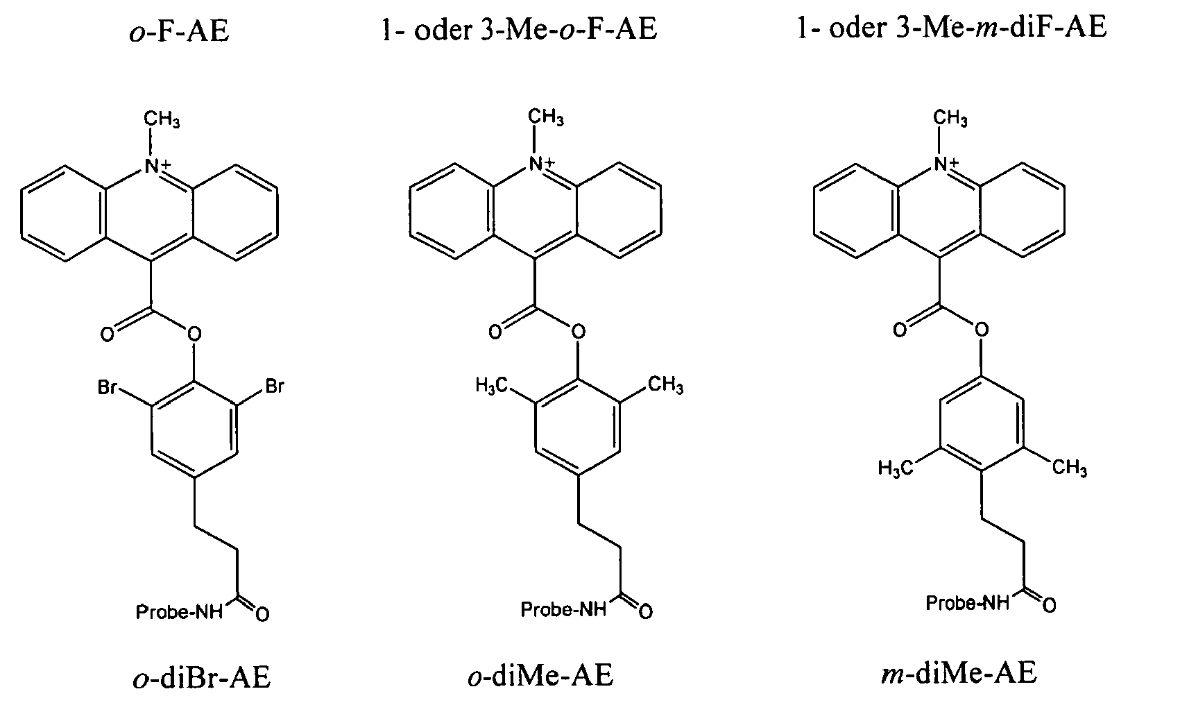
- Die Zusammensetzung gemäß Anspruch 9, wobei ein erster Acridiniumester ausgewählt wird aus der Gruppe bestehend aus o-diBr-AE und o-F-AE und ein zweiter Acridiniumester ausgewählt wird aus der Gruppe bestehend aus Standard-AE und o-MeO-AE.
- Die Zusammensetzung gemäß Anspruch 2, wobei mindestens einer der gekoppelten Chemolumineszenzmarker, die in besagter lichtemittierenden Reaktion bei einem ersten pH-Wert reagieren, o-F-AE umfasst und mindestens einer von besagten anderen gekoppelten Chemolumineszenzmarkern, die bei besagten einem oder mehreren anderen pH-Werten reagieren, ein Chemolumineszenzreagenz umfasst, das aus der Gruppe bestehend aus 1-Me-AE, 3-Me-AE, einer Mischung von 1- und 3-ME-AE, o-MeO (Cinnamyl)-AE und Standard-AE ausgewählt wird.
- Die Zusammensetzung gemäß Anspruch 2, wobei mindestens einer der gekoppelten Chemolumineszenzmarker, die in besagter lichtemittierenden Reaktion bei einem ersten pH-Wert reagieren, eine Mischung aus 1- und 3-Me-m-diF-AE umfasst und mindestens einer von besagten anderen gekoppelten Chemolumineszenzmarkern, die bei besagten einem oder mehreren anderen pH-Werten reagieren, ein Chemolumineszenzreagenz umfasst, das aus der Gruppe bestehend aus 1-Me-AE, 3-Me-AE, einer Mischung von 1- und 3-Me-AE und o-MeO(Cinnamyl)-AE ausgewählt wird.
- Die Zusammensetzung gemäß Anspruch 2, wobei mindestens einer der gekoppelten Chemolumineszenzmarker, die in besagter lichtemittierenden Reaktion bei einem ersten pH-Wert reagieren, 2,7-diMe-AE umfasst und mindestens einer von besagten anderen gekoppelten Chemolumineszenzmarkern, die bei besagten einem oder mehreren anderen pH-Werten reagieren, o-Me-AE umfasst.
- Die Zusammensetzung gemäß Anspruch 2, wobei mindestens einer der gekoppelten Chemolumineszenzmarker, die in besagter lichtemittierenden Reaktion bei einem ersten pH-Wert reagieren, Naphthyl-AE umfasst und mindestens einer von besagten anderen gekoppelten Chemolumineszenzmarkern, die bei besagten einem oder mehreren anderen pH-Werten reagieren, Standard-AE umfasst.
- Die Zusammensetzung gemäß Anspruch 2, wobei mindestens einer der gekoppelten Chemolumineszenzmarker, die in besagter lichtemittierenden Reaktion bei besagtem ersten pH-Wert reagieren, o-diBr-AE umfasst und mindestens einer von besagten anderen gekoppelten Chemolumineszenzmarkern, die bei besagten einem oder mehreren unterschiedlichen pH-Werten reagieren, ein Chemolumineszenzreagenz umfasst, das aus der Gruppe bestehend aus 1-Me-AE, 3-Me-AE, einer Mischung von 1- und 3-ME-AE und o-MeO (Cinnamyl)-AE ausgewählt wird.
- Die Zusammensetzung gemäß Anspruch 2, wobei mindestens einer der gekoppelten Chemolumineszenzmarker, die in besagter lichtemittierender Reaktion bei besagtem ersten pH-Wert reagieren, ein erstes Chemolumineszenzreagenz umfasst, das aus der Gruppe bestehend aus o-F-AE, 1-Me-m-diF-AE, 3-Me-m-diF-AE, einer Mischung aus 1-Me-m-diF-AE und 3-Me-m-diF-AE, 2,7-diMe-AE, Naphthyl-AE und o-diBr-AE ausgewählt wird und mindestens einer von besagten anderen gekoppelten Chemolumineszenzmarkern, die bei besagten einem oder mehreren unterschiedlichen pH-Werten reagieren, ein zweites Chemolumineszenzreagenz umfasst, das aus der Gruppe bestehend aus 1-Me-AE, 3-Me-AE, einer Mischung aus 1- und 3-Me-AE, o-MeO(Cinnamyl)-AE, Standard-AE und o-Me-AE ausgewählt wird.
- Die Zusammensetzung gemäß Anspruch 16, umfassend mindestens drei unterschiedliche gekoppelte Chemolumineszenzmarker.
- Die Zusammensetzung gemäß Anspruch 2, die mindestens drei Chemolumineszenzmarker umfasst, wobei mindestens einer der gekoppelten Chemolumineszenzmarker, die in besagter lichtemittierenden Reaktion bei einem ersten pH-Wert reagieren, ein erstes Chemolumineszenzreagenz umfasst, das aus der Gruppe bestehend aus o-F-AE, 1-Me-m-diF-AE, 3-Me-m-diF-AE, einer Mischung aus 1-Me-m-diF-AE und 3-Me-m-diF-AE, 2,7-diMe-AE, Naphthyl-AE und o-diBr-AE ausgewählt wird und mindestens einer von besagten anderen gekoppelten Chemolumineszenzmarkern, die bei besagtem einem oder mehreren unterschiedlichen pH-Werten reagieren, ein zweites Chemolumineszenzreagenz umfasst, das aus der Gruppe bestehend aus 1-Me-AE, 3-Me-AE, einer Mischung aus 1- und 3-Me-AE, o-MeO(Cinnamyl)-AE, Standard-AE und o-Me-AE ausgewählt wird.
- Eine Zusammensetzung für die Untersuchung einer Vielzahl von Nukleinsäureanalyten in einer einzelnen Probe umfassend:(a) eine Vielzahl von verschiedenen Oligonukleotid-Hybridisierungsassaysonden, wobei jede von besagten Sonden eine Nukleotidsequenz besitzt, die zu einer spezifischen unterschiedlichen Zielnukleotidsequenz von einem oder mehreren Nukleinsäureanalyt(en), von dem/denen vermutet wird, dass er/sie in besagter Testprobe vorhanden ist/sind, komplementär ist,(b) eine Vielzahl von verschiedenen Chemolumineszenzmarkern, wobei jeder von besagten Markern über einen Linker an eine oder mehrere von besagten Hybridisierungssonden gekoppelt ist, so dass zwei oder mehr Nukleinsäureanalyten jeweils von Hybridisierungsassaysonden, die an verschiedene von besagten Chemolumineszenzmarkern gekoppelt sind, angesteuert werden,wobei jede von besagten Hybridisierungssonden unter Bedingungen, die die Hybridisierung von besagten Sonden an nicht angesteuerte Nukleinsäuren nicht begünstigen, spezifisch an einen oder mehrere Nukleinsäureanalyt(e), wenn er/sie in besagter Probe vorhanden ist/sind, hybridisiert,
wobei besagte Chemolumineszenzmarker, wenn sie an eine Hybridisierungssonde gekoppelt sind, die an eine Zielnukleinsäure hybridisiert ist, gleichermaßen vor dem Verlust an Chemolumineszenzpotential geschützt sind, wobei die Verlustraten an Chemolumineszenzpotential von verschiedenen Chemolumineszenzmarkern, die an Oligonukleotid-Hybridisierungsassaysonden gekoppelt sind, abhängig davon, ob die Probe an eine Zielnukleinsäure oder -nukleotidsequenzregion hybridisiert ist, verringert werden und wobei unter denselben Bedingungen besagte Verlustraten innerhalb eines Faktors von bis zu 250 zueinander liegen; und
wobei besagte Chemolumineszenzmarker, wenn sie an eine nicht hybridisierte Sonde gekoppelt sind, gleichermaßen gegenüber einem Verlust ihres Chemolumineszenzpotentials empfänglich sind, wobei unter identischen Bedingungen die Verlustraten an Chemolumineszenzpotential von verschiedenen Chemolumineszenzmarkern, die an Oligonukleotid-Hybridisierungsassaysonden gekoppelt sind, durch die Aussetzung gegenüber einem destabilisierenden Mittel innerhalb eines Faktors von ungefähr 50 zueinander liegen; und
wobei bei der Auslösung einer lichtemittierenden Reaktion die Werte der Zeit bis zum Scheitelpunkt des Signals und/oder der Dauer der Reaktion von mindestens einem reagierenden Chemolumineszenzmarker ausreichend unterschiedlich von denen von mindestens einem anderen reagierenden Chemolumineszenzmarker sind, um, wenn die Lichtemission über vorher festgelegte Zeitintervalle nach besagtem Auslösungsereignis als Nachweis für jeden von besagten Analyten nachgewiesen oder gemessen wird, den getrennten Nachweis von besagten reagierenden Chemolumineszenzmarkern in derselben Testprobe zu erlauben. - Die Zusammensetzung gemäß Anspruch 19, in der mindestens einer von besagten Chemolumineszenzmarkern ein Acridiniumester ist.
- Die Zusammensetzung gemäß Anspruch 19, in der mindestens zwei von besagten Chemolumineszenzmarkern Acridiniumester sind.
- Die Zusammensetzung gemäß Anspruch 19, in der alle besagten Chemolumineszenzmarker Acridiniumester sind.
- Die Zusammensetzung gemäß Anspruch 20 oder Anspruch 21, in der besagter mindestens einer Acridiniumester ausgewählt wird aus der Gruppe bestehend aus Standard-AE, Naphthyl-AE, o-diBr-AE, 1- oder 3-Me-AE, 4,5-diMe-AE, 2,7-diMe-AE, o-Me-AE, o-MeO(Cinnamyl)-AE, o-MeO-AE, o-diMe-AE, m-diMe-AE, ortho-AE, o-F-AE, 1 oder 3-Me-o-F-AE und 1- oder 3-Me-m-diF-AE.
- Die Zusammensetzung gemäß Anspruch 20 oder Anspruch 21, in der ein erster Acridiniumester Standard-AE ist und ein zweiter Acridiniumester ausgewählt wird aus der Gruppe: Naphthyl-AE, o-diBr-AE, o-diMe-AE, o-Me-AE, o-MeO(Cinnamyl)-AE, o-MeO-AE, o-AE, o-F-AE, 1-Me-o-F-AE und 1-Me-m-diF-AE.
- Die Zusammensetzung gemäß Anspruch 20 oder Anspruch 21, in der ein erster Acridiniumester Naphthyl-AE ist und ein zweiter Acridiniumester ausgewählt wird aus der Gruppe: 1-Me-AE, 4,5-diMe-AE, 2,7-diMe-AE, o-Me-AE, o-MeO(Cinnamyl)-AE und o-AE.
- Die Zusammensetzung gemäß Anspruch 20 oder Anspruch 21, in der ein erster Acridiniumester o-diBr-AE ist und ein zweiter Acridiniumester ausgewählt wird aus der Gruppe: 1-Me- oder 3-Me-AE, 3-Me-AE, 4,5-diMe-AE, 2,7-diMe-AE, o-diMe-AE, o-Me-AE, o-MeO(Cinnamyl)-AE und o-AE.
- Die Zusammensetzung gemäß Anspruch 20 oder Anspruch 21, in der ein erster Acridiniumester 1-Me- oder 3-Me-AE ist und ein zweiter Acridiniumester ausgewählt wird aus der Gruppe: o-diMe-AE, o-Me-AE, o-MeO(Cinnamyl)-AE, o-MeO-AE, o-F-AE, 1-Me-o-F-AE und 1-Me-m-diF-AE.
- Die Zusammensetzung gemäß Anspruch 20 oder Anspruch 21, in der ein erster Acridiniumester 4,5-diMe-AE ist und ein zweiter Acridiniumester ausgewählt wird aus der Gruppe: o-diMe-AE, o-Me-AE, o-MeO(Cinnamyl)-AE, o-MeO-AE, o-AE, o-F-AE, 1-Me-o-F-AE und 1-Me-m-diF-AE.
- Die Zusammensetzung gemäß Anspruch 20 oder Anspruch 21, in der ein erster Acridiniumester 2,7-diMe-AE ist und ein zweiter Acridiniumester ausgewählt wird aus der Gruppe: o-diMe-AE, oMe-AE, o-MeO(Cinnamyl)-AE, o-MeO-AE, o-AE, o-F-AE, 1-Me-o-F-AE und 1-Me-m-diF-AE.
- Die Zusammensetzung gemäß Anspruch 20 oder Anspruch 21, in der ein erster Acridiniumester o-diMe-AE ist und ein zweiter Acridiniumester ausgewählt wird aus der Gruppe: o-Me-AE, o-MeO(Cinnamyl)-AE, o-MeO-AE, o-AE, o-F-AE, 1-Meo-F-AE und 1-Me-m-diF-AE.
- Die Zusammensetzung gemäß Anspruch 20 oder Anspruch 21, in der ein erster Acridiniumester o-Me-AE ist und ein zweiter Acridiniumester ausgewählt wird aus der Gruppe: o-MeO(Cinnamyl)-AE, o-MeO-AE, o-AE, o-F-AE, 1-Me-o-F-AE und 1-Me-m-diF-AE.
- Die Zusammensetzung gemäß Anspruch 20 oder Anspruch 21, in der ein erster Acridiniumester o-MeO(Cinnamyl)-AE ist und ein zweiter Acridiniumester ausgewählt wird aus der Gruppe: o-MeO-AE, o-F-AE, 1-Me-o-F-AE und 1-Me-m-diF-AE.
- Die Zusammensetzung gemäß Anspruch 20 oder Anspruch 21, in der ein erster Acridiniumester o-MeO-AE und ein zweiter Acridiniumester o-AE ist.
- Die Zusammensetzung gemäß Anspruch 20 oder Anspruch 21, in der ein erster Acridiniumester o-AE ist und ein zweiter Acridiniumester ausgewählt wird aus der Gruppe bestehend aus o-F-AE, 1-Me-o-F-AE und 1-Me-m-diF-AE.
- Eine Zusammensetzung für die Untersuchung einer Vielzahl von Nukleinsäureanalyten in einer einzelnen Testprobe umfassend mindestens zwei Chemolumineszenzmarker ausgewählt aus der Gruppe bestehend aus:(a) o-diBr-AE,(b) 2,7-diMe-AE,(c) einer Mischung aus 1- und 3-Me-AE,(d) o-AE,(e) o-MeO (Cinnamyl)-AE,(f) o-Me-AE und(g) o-diMe-AE,wobei jeder von besagten Chemolumineszenzmarkern an mindestens eine unterschiedliche Hybridisierungsassaysonde gekoppelt ist, wobei jede von besagten Sonden eine Nukleotidsequenz besitzt, die zu einer Zielregion, die in einem oder mehreren Nukleinsäureanalyten enthalten ist, ausreichend komplementär ist, um unter Hybridisierungsbedingungen, die die Hybridisierung zwischen besagter Probe und Nicht-Zielregionen nicht begünstigen, daran zu binden, und wobei nach der Auslösung einer auslösbaren lichtemittierenden Reaktion, die Reaktionskinetiken, die Wellenlänge der Lichtemission und/oder die optimalen pH-Eigenschaften von jedem von besagten Chemolumineszenzmarkern von denen von anderen besagten Markern ausreichend unterschiedlich ist, so dass jeder von besagten Markern einzeln als Hinweis auf das Vorhandensein von jedem von besagten Nukleinsäureanalyten in besagter Probe nachweisbar ist.
- Ein Verfahren für die Untersuchung einer Vielzahl von Nukleinsäureanalyten in einer einzelnen Probe umfassend die Schritte:(a) Bereitstellen in einem Medium(i) einer Vielzahl von verschiedenen Oligonukleotid-Hybridisierungsassaysonden, wobei jede von besagten Sonden eine Nukleotidsequenz besitzt, die zu einer Zielnukleotidsequenz eines Nukleinsäureanalyt, für den vermutet wird, dass er sich in besagter Probe befindet, komplementär ist,(ii) einer Vielzahl von verschiedenen Chemolumineszenzmarkern, wobei jeder von besagten Markern an eine oder mehrere von besagten Hybridisierungssonden gekoppelt ist, so dass zwei oder mehr Nukleinsäureanalyte jeweils von mindestens einer Hybridisierungsassaysonde, die an einen unterschiedlichen Chemolumineszenzmarker gekoppelt ist, angesteuert werden und(iii) besagter Probe und(b) Herstellen von Hybridisierungsbedingungen, unter denen die verschiedenen Hybridisierungsassaysonden vorzugsweise an besagte Nukleinsäureanalyte hybridisieren, um markierte Doppelstrangnukleinsäurehybride zu bilden, wobei besagte Bedingungen die Bildung von Doppelstrangnukleinsäurehybriden zwischen besagten markierten Sonden und nicht angesteuerten Nukleinsäuren nicht begünstigen,(c) selektives Zerstören oder Hemmen des Chemolumineszenzpotentials von besagten Chemolumineszenzmarkern, die an nicht hybridisierte Sonden gekoppelt sind, wobei besagte Chemolumineszenzmarker gleichermaßen vor dem Verlust an Chemolumineszenzpotential geschützt sind, wenn sie an eine Hybridisierungssonde gekoppelt sind, die an eine Zielnukleinsäure hybridisiert ist, wobei die Verlustraten an Chemolumineszenzpotential von verschiedenen Chemolumineszenzmarkern, die an Oligonukleotid-Hybridisierungsassaysonden gekoppelt sind, abhängig davon, ob die Probe an eine angesteuerte Nukleinsäure oder Nukleotidsequenzregion hybridisiert ist, abnehmen und wobei die besagten Verlustraten unter denselben Bedingungen innerhalb eines Faktors von bis zu 250 zueinander liegen; und wobei besagte Chemolumineszenzmarker gleichermaßen gegenüber einem Verlust ihres Chemolumineszenzpotentials empfänglich sind, wenn sie an eine nicht hybridisierte Sonde gekoppelt sind, wobei unter identischen Bedingungen die Verlustraten an Chemolumineszenzpotential von verschiedenen Chemolumineszenzmarkern, die an Oligonukleotid-Hybridisierungsassaysonden gekoppelt sind, durch die Aussetzung gegenüber einem destabilisierenden Mittel innerhalb eines Faktors von ungefähr 50 zueinander liegen;(d) Veranlassen der Chemolumineszenzmarker, die mit doppelsträngigen Nukleinsäurehybriden assoziiert sind, Licht zu emittieren und(e) Nachweis von jedem von besagten Nukleinsäureanalyten, wenn vorhanden, durch das Messen des Lichtes, das von dem gekoppelten Chemolumineszenzmarker, der daran hybridisiert ist, emittiert wird bei einer Lichtemissionswellenlänge, die von der Wellenlänge einer Lichtemission von jedem anderen von besagten Chemolumineszenzmarkern ausreichend unterschiedlich ist, um so zu ermöglichen, besagte Analyten unabhängig voneinander in einer einzelnen Testprobe nachzuweisen.
- Ein Verfahren für die Untersuchung einer Vielzahl von Nukleinsäureanalyten in einer Einzelprobe umfassend die Schritte:(a) Bereitstellen in einem Medium(i) einer Vielzahl von verschiedenen Oligonukleotid-Hybridisierungsassaysonden, wobei jede von besagten Sonden eine Nukleotidsequenz besitzt, die zu einer Zielnukleotidsequenz eines Nukleinsäureanalyt, für den vermutet wird, dass er sich in besagter Probe befindet, komplementär ist,(ii) einer Vielzahl von verschiedenen Chemolumineszenzmarkern, wobei jeder von besagten Markern an eine oder mehrere von besagten Hybridisierungssonden gekoppelt ist, so dass zwei oder mehr Nukleinsäureanalyte jeweils von mindestens einer Hybridisierungsassaysonde, die an einen unterschiedlichen Chemolumineszenzmarker gekoppelt ist, angesteuert werden und(iii) besagter Probe und(b) Herstellen von Hybridisierungsbedingungen, unter denen die verschiedenen Hybridisierungsassaysonden vorzugsweise an besagte Nukleinsäureanalyte, wenn vorhanden, hybridisieren, um markierte Doppelstrangnukleinsäurehybride zu bilden, wobei besagte Bedingungen die Bildung von Doppelstrangnukleinsäurehybriden zwischen besagten markierten Sonden und nicht angesteuerten Nukleinsäuren nicht begünstigen,(c) selektives Zerstören oder Hemmen des Chemolumineszenzpotentials der Chemolumineszenzmarker, die an nicht hybridisierte Sonden gekoppelt sind, wobei besagte Chemolumineszenzmarker gleichermaßen vor dem Verlust an Chemolumineszenzpotential geschützt sind, wenn sie an eine Hybridisierungssonde gekoppelt sind, die an eine Zielnukleinsäure hybridisiert ist, wobei die Verlustraten an Chemolumineszenzpotential von verschiedenen Chemolumineszenzmarkern, die an Oligonukleotid-Hybridisierungsassaysonden gekoppelt sind, abhängig davon, ob die Probe an eine angesteuerte Nukleinsäure oder Nukleotidsequenzregion hybridisiert ist, abnehmen und wobei die besagten Verlustraten unter denselben Bedingungen innerhalb eines Faktors von bis zu 250 zueinander liegen; und wobei besagte Chemolumineszenzmarker gleichermaßen gegenüber einem Verlust ihres Chemolumineszenzpotentials empfänglich sind, wenn sie an eine nicht hybridisierte Sonde gekoppelt sind, wobei unter identischen Bedingungen die Verlustraten an Chemolumineszenzpotential von verschiedenen Chemolumineszenzmarkern, die an Oligonukleotid-Hybridisierungsassaysonden gekoppelt sind, durch die Aussetzung gegenüber einem destabilisierenden Mittel innerhalb eines Faktors von ungefähr 50 zueinander liegen;(d) Veranlassen der Chemolumineszenzmarker, die mit doppelsträngigen Nukleinsäurehybriden assoziiert sind, Licht zu emittieren und(e) Nachweis von jedem hybridisierten Nukleinsäureanalyt bei Auslösung der lichtemittierenden Reaktion durch das Messen der Werte der Zeit bis zum Scheitelpunkt des Signals und/oder der Dauer der Reaktion der reagierenden Chemolumineszenzmarker über vorherbestimmte Zeitintervalle nach besagtem Auslösungsereignis.
- Ein Verfahren für die Untersuchung einer Vielzahl von Nukleinsäureanalyten in einer Einzelprobe umfassend die Schritte:(a) Bereitstellen in einem Medium(i) einer Vielzahl von verschiedenen Oligonukleotid-Hybridisierungsassaysonden, wobei jede von besagten Sonden eine Nukleotidsequenz besitzt, die zu einer Zielnukleotidsequenz eines Nukleinsäureanalyt, für den vermutet wird, dass er sich in besagter Probe befindet, komplementär ist,(ii) einer Vielzahl von verschiedenen Chemolumineszenzmarkern, wobei jeder von besagten Markern an eine oder mehrere von besagten Hybridisierungssonden gekoppelt ist, so dass zwei oder mehr Nukleinsäureanalyte jeweils von mindestens einer Hybridisierungsassaysonde, die an einen unterschiedlichen Chemolumineszenzmarker gekoppelt ist, angesteuert werden und(iii) besagter Probe und(b) Herstellen von Hybridisierungsbedingungen, unter denen die verschiedenen Hybridisierungsassaysonden vorzugsweise an besagte Nukleinsäureanalyte, wenn vorhanden, hybridisieren, um markierte Doppelstrangnukleinsäurehybride zu bilden, wobei besagte Bedingungen die Bildung von Doppelstrangnukleinsäurehybriden zwischen besagten markierten Sonden und nicht angesteuerten Nukleinsäuren nicht begünstigen,(c) selektives Zerstören oder Hemmen des Chemolumineszenzpotentials der Chemolumineszenzmarker, die an nicht hybridisierte Sonden gekoppelt sind, wobei besagte Chemolumineszenzmarker gleichermaßen gegenüber einem Verlust ihres Chemolumineszenzpotentials empfänglich sind, wenn sie an besagte nicht hybridisierte Sonde gekoppelt sind, und besagte Chemolumineszenzmarker gleichermaßen vor dem Verlust ihres Chemolumineszenzpotential geschützt sind, wenn sie an besagte hybridisierte Sonde gekoppelt sind,(d) Veranlassen von mindestens einem der gekoppelten Chemolumineszenzmarker, die mit besagten Doppelstrangnukleinsäurehybriden assoziiert sind, an einer lichtemittierenden Reaktion bei einem ersten pH-Wert beteiligt zu sein und Messen des Lichts, das dadurch von besagtem Marker emittiert wird,(e) Einstellen der Assaylösung auf mindestens einen anderen pH-Wert,(f) Veranlassen von mindestens einem anderen gekoppelten Chemolumineszenzmarker, der mit einem oder mehreren verschiedenen Doppelstrangnukleinsäurehybriden assoziiert ist, an einer lichtemittierenden Reaktion bei besagten anderen pH-Wert(en) beteiligt zu sein und(g) Messen des Lichts, das von besagten anderen Markern während eines vorherbestimmten Zeitraums emittiert wird, dadurch die getrennte Identifizierung von verschiedenen gekoppelten Chemolumineszenzmarkern als Messung von jedem von verschiedenen Nukleinsäureanalyten, die an markierte Hybridisierungsassaysonden in einer Einzeltestprobe hybridisiert sind, erlaubend.
- Eine Zusammensetzung für den Nachweis von einem oder mehreren Organismen in einer einzelnen Probe umfassend:(a) eine Vielzahl von verschiedenen Oligonukleotid-Hybridisierungsassaysonden, wobei jede von besagten Sonden eine Nukleotidsequenz besitzt, die zu der von einem oder mehreren Nukleinsäureanalyten, die für die Identifizierung von mindestens einem Zielorganismus, von dem vermutet wird, dass er sich in besagter Testprobe befindet, nützlich ist, komplementär ist,(b) eine Vielzahl von verschiedenen Chemolumineszenzmarkern, wobei jeder an eine oder mehrere von besagten Hybridisierungssonden gekoppelt ist, so dass zwei oder mehr Nukleinsäureanalyten von verschiedenen von besagten chemolumineszenz-markierten Hybridisierungsassaysonden angesteuert werden, wobei jede von besagten Hybridisierungssonden unter Bedingungen, die die Hybridisierung von besagten Sonden an Nicht-Zielnukleinsäureregionen nicht begünstigen, spezifisch an besagte Nukleinsäureanalyten, falls diese in besagter Probe vorhanden sind, hybridisiert,wobei besagte Chemolumineszenzmarker gleichermaßen vor dem Verlust an Chemolumineszenzpotential geschützt sind, wenn sie an eine Hybridisierungssonde gekoppelt sind, die an eine Zielnukleinsäure hybridisiert ist, wobei die Verlustraten an Chemolumineszenzpotential von verschiedenen Chemolumineszenzmarkern, die an Oligonukleotid-Hybridisierungsassaysonden gekoppelt sind, abhängig davon, ob die Probe an eine Zielnukleinsäure oder Nukleotidsequenzregion hybridisiert ist, abnehmen und wobei die Verhältnisse von besagtem Verlust unter denselben Bedingungen innerhalb eines Faktors von bis zu 250 zueinander liegen; und
wobei besagte Chemolumineszenzmarker gleichermaßen gegenüber einem Verlust ihres Chemolumineszenzpotentials empfänglich sind, wenn sie an eine nicht hybridisierte Sonde gekoppelt sind, wobei unter identischen Bedingungen die Verlustraten an Chemolumineszenzpotential von verschiedenen Chemolumineszenzmarkern, die an Oligonukleotid-Hybridisierungsassaysonden gekoppelt sind, bei Aussetzung gegenüber einem destabilisierenden Mittel innerhalb eines Faktors von ungefähr 50 zueinander liegen; und
wobei bei Auslösung einer lichtemittierenden Reaktion jeder von besagten Chemolumineszenzmarkern Licht einer oder mehrerer Wellenlängen, die von einer Wellenlänge der Lichtemission von jedem anderen von besagten Chemolumineszenzmarkern ausreichend unterschiedlich ist, emittiert, so dass besagte Chemolumineszenzmarker, wenn das emittierte Licht nachgewiesen wird, unabhängig voneinander nachweisbar sind und besagte Wellenlängen ein Hinweis auf das Vorhandensein von jedem von besagten Zielorganismen in besagter Testprobe sind. - Eine Zusammensetzung für den Nachweis von einem oder mehreren Organismen in einer einzelnen Probe umfassend:(a) eine Vielzahl von verschiedenen Oligonukleotid-Hybridisierungsassaysonden, wobei jede von besagten Sonden eine Nukleotidsequenz besitzt, die zu der von einem oder mehreren Nukleinsäureanalyten, die für die Identifizierung von mindestens einem Zielorganismus, von dem vermutet wird, dass er sich in besagter Testprobe befindet, nützlich ist, komplementär ist,(b) eine Vielzahl von verschiedenen Chemolumineszenzmarkern, wobei jeder an eine andere von besagten Hybridisierungssonden gekoppelt ist, so dass zwei oder mehr Nukleinsäureanalyten durch verschiedene von besagten chemolumineszenz-markierten Hybridisierungsassaysonden angesteuert werden, wobei jede von besagten Hybridisierungssonden unter Bedingungen, die die Hybridisierung von besagten Sonden an Nicht-Zielnukleinsäureregionen nicht begünstigen, spezifisch an besagte Nukleinsäureanalyten, falls diese in besagter Probe vorhanden sind, hybridisiert, wobei besagte Chemolumineszenzmarker gleichermaßen resistent gegenüber einem Verlust an Chemolumineszenzpotential sind, wenn sie an eine Hybridisierungssonde gekoppelt sind, die an eine Zielnukleinsäure hybridisiert ist, und besagte Chemolumineszenzmarker gleichermaßen empfänglich gegenüber einem Verlust an Chemolumineszenzpotential sind, wenn sie an eine nicht hybridisierte Sonde gekoppelt sind, und wobei nach der Auslösung einer auslösbaren lichtemittierenden Reaktion bei einem ersten pH-Wert mindestens einer von besagten Chemolumineszenzmarkern in einer lichtemittierenden Reaktion bei besagtem pH-Wert reagiert und mindestens ein anderer von besagten Chemolumineszenzmarkern nicht in einer lichtemittierenden Reaktion reagiert, bis die Lösung auf mindestens einen anderen pH-Wert eingestellt wird, wobei besagte Zusammensetzung dadurch die getrennte Identifizierung von jedem Chemolumineszenzmarker in einer einzelnen Testprobe als Hinweis auf die Vorhandensein von jedem von besagten Zielorganismen in der Probe erlaubt.
- Eine Zusammensetzung für den Nachweis von einem oder mehreren Organismen in einer Einzelprobe umfassend:(a) eine Vielzahl von verschiedenen Oligonukleotid-Hybridisierungsassaysonden, wobei jede von besagten Sonden eine Nukleotidsequenz besitzt, die zu der von einem oder mehreren Nukleinsäureanalyten, die für die Identifizierung von mindestens einem Zielorganismus, von dem vermutet wird, dass er sich in besagter Testprobe befindet, nützlich ist, komplementär ist,(b) eine Vielzahl von verschiedenen Chemolumineszenzmarkern, wobei jeder an eine andere von besagten Hybridisierungssonden gekoppelt ist, so dass zwei oder mehr Nukleinsäureanalyten durch verschiedene von besagten chemolumineszenz-markierten Hybridisierungsassaysonden angesteuert werden, wobei jede von besagten Hybridisierungssonden unter Bedingungen, die die Hybridisierung von besagten Sonden an Nicht-Zielnukleinsäureregionen nicht begünstigen, spezifisch an besagte Nukleinsäureanalyten, falls diese in besagter Probe vorhanden sind, hybridisiert,wobei besagte Chemolumineszenzmarker gleichermaßen vor dem Verlust an Chemolumineszenzpotential geschützt sind, wenn sie an eine Hybridisierungssonde gekoppelt sind, die an eine Zielnukleinsäure hybridisiert ist, wobei die Verlustraten an Chemolumineszenzpotential von verschiedenen Chemolumineszenzmarkern, die an Oligonukleotid-Hybridisierungsassaysonden gekoppelt sind, abhängig davon, ob die Probe an eine Zielnukleinsäure oder Nukleotidsequenzregion hybridisiert ist, abnehmen und wobei die Verhältnisse von besagtem Verlust unter denselben Bedingungen innerhalb eines Faktors von bis zu 250 zueinander liegen; und
wobei besagte Chemolumineszenzmarker gleichermaßen gegenüber einem Verlust ihres Chemolumineszenzpotentials empfänglich sind, wenn sie an eine nicht hybridisierte Sonde gekoppelt sind, wobei unter identischen Bedingungen die Verlustraten an Chemolumineszenzpotential von verschiedenen Chemolumineszenzmarkern, die an Oligonukleotid-Hybridisierungsassaysonden gekoppelt sind, bei Aussetzung gegenüber einem destabilisierenden Mittel innerhalb eines Faktors von ungefähr 50 zueinander liegen;
und wobei bei der Auslösung einer lichtemittierenden Reaktion die Werte der Zeit bis zum Gipfelpunkt des Signals und/oder der Dauer der Reaktion von jedem reagierenden Chemolumineszenzmarker ausreichend unterschiedlich von denen von jedem anderen von besagten reagierenden Chemolumineszenzmarkern sind, um, wenn besagte Lichtemission über einen vorher bestimmten Zeitraum nach dem Auslösungsereignis als Hinweis auf das Vorhandensein von jedem von besagten Zielorganismen in der Probe nachgewiesen oder gemessen wird, in der Gegenwart von anderen von besagten Markern den getrennten Nachweis von jedem besagten Chemolumineszenzmarker zu erlauben. - Ein Verfahren für den gleichzeitigen Nachweis des Vorhandenseins oder der Menge von Chlamydia trachomatis und Neisseria gonorrhoeae Nukleinsäuren in einer einzelnen Probe umfassend:(a) Inkontaktbringen besagter Probe mit mindestens einer Oligonukleotid-Hybridisierungsassaysonde, die vorzugsweise eher an Chlamydia trachomatis Nukleinsäuren als an nicht-Chlamydia trachomatis Nukleinsäuren hybridisiert, und mindestens eine Oligonukleotid-Hybridisierungsassaysonde, die vorzugsweise eher an Neisseria gonorrhoeae Nukleinsäuren als an nicht-Neisseria gonorrhoeae Nukleinsäuren hybridisiert, wobei besagte Chlamydia trachomatis-spezifische Hybridisierungsassaysonden an ein erstes chemolumineszenz-markiertes Reagenz gekoppelt sind und besagte Neisseria gonorrhoeae-spezifische Hybridisierungsassaysonden an ein zweites chemolumineszenz-markiertes Reagenz gekoppelt sind,(b) Herstellen von Hybridisierungsbedingungen, die ausreichend sind, um die Hybridisierung zwischen besagten Chlamydia trachomatis-spezifischen Hybridisierungsassaysonden und, falls vorhanden, Chlamydia trachomatis Nukleinsäuren und Neisseria gonorrhoeaespezifischen Hybridisierungsassaysonden und, falls vorhanden, Neisseria gonorrhoeae Nukleinsäuren zu erlauben, wobei besagte Hybridisierungsbedingungen die nichtspezifische Hybridisierung von besagten Hybridisierungsassaysonden an Nicht-Zielnukleinsäuren nicht fördern,(c) Selektives Zerstören oder Hemmen des Chemolumineszenzpotentials der Chemolumineszenzmarker, die an nicht-hybridisierte Sonden gekoppelt sind, wobei besagte Chemolumineszenzmarker gleichermaßen vor dem Verlust an Chemolumineszenzpotential geschützt sind, wenn sie an eine Hybridisierungssonde gekoppelt sind, die an eine Zielnukleinsäure hybridisiert ist, wobei die Verlustraten an Chemolumineszenzpotential von verschiedenen Chemolumineszenzmarkern, die an Oligonukleotid-Hybridisierungsassaysonden gekoppelt sind, abhängig davon, ob die Sonde an eine Zielnukleinsäure oder Nukleotidsequenzregion hybridisiert ist, abnehmen und wobei die Verlustraten unter denselben Bedingungen innerhalb eines Faktors von bis zu 250 zueinander liegen; und wobei besagte Chemolumineszenzmarker gleichermaßen gegenüber einem Verlust an ihrem Chemolumineszenzpotential empfänglich sind, wenn sie an eine nicht-hybridisierte Sonde gekoppelt sind, wobei unter identischen Bedingungen die Verlustraten an Chemolumineszenzpotential von verschiedenen Chemolumineszenzmarkern, die an Oligonukleotid-Hybridisierungsassaysonden gekoppelt sind, bei Aussetzung gegenüber einem destabilisierenden Mittel innerhalb eines Faktors von ungefähr 50 zueinander liegen;(d) Nachweis von besagtem ersten Chemolumineszenzreagenz, das an eine hybridisierte Chlamydia trachomatis-spezifische Sonden gekoppelt ist, als Hinweis auf das Vorhandensein von Chlamydia trachomatis Nukleinsäuren in besagter Probe und besagtem zweiten Chemolumineszenzreagenz, das an hybridisierte Neisseria gonorrhoeae-spezifische Sonden gekoppelt ist, als Hinweis des Vorhandenseins von Neisseria gonorrhoeae Nukleinsäuren in besagter Probe, wobei jeder von besagten Chemolumineszenzmarkern Licht an einer oder mehreren Wellenlängen, die von anderen von besagten Chemolumineszenzmarkern emittierten Wellenlängen der Lichtemission ausreichend unterschiedlich sind, emittiert, so dass, wenn das emittierte Licht bei besagten Wellenlängen als Hinweis auf das Vorhandensein von jedem der Nukleinsäureanalyten in besagter Testprobe gleichzeitig nachgewiesen wird, besagte Chemolumineszenzmarker unabhängig voneinander nachweisbar sind.
- Ein Verfahren für die Untersuchung einer Vielzahl von Analyten in einer Zielnukleinsäure in einer Probe umfassend:(a) Inkontaktbringen von besagter Probe mit einer Vielzahl von verschiedenen Oligonukleotid-Hybridisierungsassaysonden, wobei jede von besagten verschiedenen Sonden spezifisch an eine andere Zielanalytregion von besagter Nukleinsäure hybridisiert und wobei jede von besagten Sonden an einen anderen Chemolumineszenzmarker gekoppelt ist;(b) Herstellen von Hybridisierungsbedingungen in besagter Probe, die ausreichend sind, um die spezifische Hybridisierung zwischen jeder von besagten Hybridisierungsassaysonden und ihrem Analyten zu erlauben, wobei besagte Hybridisierungsbedingungen die Hybridisierung von besagten Hybridisierungsassaysonden an Nicht-Zielnukleotidsequenzregionen von besagter Nukleinsäure nicht fördern,(c) Selektives Zerstören oder Hemmen des Chemolumineszenzpotentials der Chemolumineszenzmarker, die an nicht-hybridisierte Sonden gekoppelt sind, wobei besagte Chemolumineszenzmarker gleichermaßen vor dem Verlust an Chemolumineszenzpotential geschützt sind, wenn sie an eine Hybridisierungssonde gekoppelt sind, die an eine Zielnukleinsäure hybridisiert ist, wobei die Verlustraten an Chemolumineszenzpotential von verschiedenen Chemolumineszenmarkern, die an Oligonukleotid-Hybridisierungsassaysonden gekoppelt sind, abhängig davon, ob die Probe an eine Zielnukleinsäure oder Nukleotidsequenzregion hybridisiert ist, abnehmen und wobei unter denselben Bedingungen die Verhältnisse von besagtem Verlust innerhalb eines Faktors von bis zu 250 zueinander liegen; und wobei besagte Chemolumineszenzmarker gleichermaßen gegenüber einem Verlust ihres Chemolumineszenzpotentials empfänglich sind, wenn sie an eine nicht hybridisierte Sonde gekoppelt sind, wobei unter identischen Bedingungen die Verlustraten an Chemolumineszenzpotential von verschiedenen Chemolumineszenzmarkern, die an Oligonukleotid-Hybridisierungsassaysonden gekoppelt sind, bei Aussetzung gegenüber einem destabilisierenden Agens innerhalb eines Faktors von ungefähr 50 zueinander liegen;(d) Nachweis von besagten Chemolumineszenzmarkern, die an besagte verschiedene Sonden gekoppelt sind, als Hinweis auf das Vorhandensein von besagten Nukleinsäureanalyten in besagter Probe, wobei jeder von besagten Chemolumineszenzmarkern Licht einer oder mehrerer Wellenlängen emittiert, das von einer Wellenlänger der Lichtemission von jedem anderen von besagten Chemolumineszenzmarkern ausreichend unterschiedlich ist, so dass, wenn das emittierte Licht bei besagten Wellenlängen als Hinweis auf das Vorhandensein von jedem der Nukleinsäureanalyten in besagter Testprobe gleichzeitig nachgewiesen wird, besagte Chemolumineszenzmarker unabhängig nachweisbar sind.
- Das Verfahren gemäß Anspruch 43, wobei besagte Nukleinsäure vom humanen Immunodefizienzvirus abgeleitet ist.
- Das Verfahren gemäß Anspruch 44, wobei besagte verschiedene Analyte Zielregionen umfassen, die in Regionen des Genoms des humanen Immunodefizienzvirus umfassend die gag und pol Regionen lokalisiert sind.
- Ein Kit, der eine der Zusammensetzungen gemäß Anspruch 1 bis 35, 39, 40 oder 41 enthält für den Nachweis von einem oder mehreren Nukleinsäureanalyten in einer Testprobe.
- Der Kit gemäß Anspruch 46, ferner umfassend ein Unterscheidungsreagenz, umfassend Mittel für die selektive Zerstörung oder Hemmung des Chemolumineszenzpotentials von besagten Chemolumineszenzmarkern, die an nicht-hybridisierte Sonden gekoppelt sind.
- Das Kit gemäß Anspruch 46 oder 47, ferner umfassend ein Nachweisreagenz, das Mittel umfasst, Chemolumineszenzreagenzien zu veranlassen, nachweisbares Licht zu emittieren.
Applications Claiming Priority (2)
| Application Number | Priority Date | Filing Date | Title |
|---|---|---|---|
| US33110794A | 1994-10-28 | 1994-10-28 | |
| US331107 | 1994-10-28 |
Publications (3)
| Publication Number | Publication Date |
|---|---|
| EP0709466A2 EP0709466A2 (de) | 1996-05-01 |
| EP0709466A3 EP0709466A3 (de) | 1996-06-12 |
| EP0709466B1 true EP0709466B1 (de) | 2006-09-27 |
Family
ID=23292651
Family Applications (1)
| Application Number | Title | Priority Date | Filing Date |
|---|---|---|---|
| EP95116199A Expired - Lifetime EP0709466B1 (de) | 1994-10-28 | 1995-10-13 | Zusammensetzungen und Verfahren für die gleichzeitige Detektion und Quantifizierung von einer Mehrheit spezifischer Nuklein Säure Sequenzen |
Country Status (10)
| Country | Link |
|---|---|
| US (4) | US5756709A (de) |
| EP (1) | EP0709466B1 (de) |
| JP (1) | JP3190348B2 (de) |
| KR (1) | KR100276679B1 (de) |
| AT (1) | ATE340866T1 (de) |
| AU (1) | AU710884B2 (de) |
| CA (1) | CA2201595C (de) |
| DE (1) | DE69535240T2 (de) |
| ES (1) | ES2271944T3 (de) |
| WO (1) | WO1996013612A2 (de) |
Families Citing this family (248)
| Publication number | Priority date | Publication date | Assignee | Title |
|---|---|---|---|---|
| US5714380A (en) | 1986-10-23 | 1998-02-03 | Amoco Corporation | Closed vessel for isolating target molecules and for performing amplification |
| US5728575A (en) * | 1990-02-01 | 1998-03-17 | Emory University | Method of resolution of 1,3-oxathiolane nucleoside enantiomers |
| DE69535240T2 (de) * | 1994-10-28 | 2007-06-06 | Gen-Probe Inc., San Diego | Zusammensetzungen und Verfahren für die gleichzeitige Detektion und Quantifizierung von einer Mehrheit spezifischer Nuklein Säure Sequenzen |
| US5731148A (en) * | 1995-06-07 | 1998-03-24 | Gen-Probe Incorporated | Adduct protection assay |
| US5879885A (en) * | 1995-06-07 | 1999-03-09 | Gen-Probe Incorporated | Compositions for detecting and quantifying biological samples |
| CA2223047C (en) * | 1995-06-07 | 2008-11-18 | Gen-Probe Incorporated | Micelle protection assay |
| DE19627290A1 (de) * | 1996-07-06 | 1998-01-08 | Boehringer Mannheim Gmbh | Bestimmung von Analyten unter Verwendung zweier Markierungen |
| US5858665A (en) * | 1996-07-25 | 1999-01-12 | Navix, Inc. | Homogeneous diagnostic assay method utilizing simultaneous target and signal amplification |
| DE69840669D1 (de) | 1997-04-10 | 2009-04-30 | Stichting Katholieke Univ | Pca3, pca3-gene und verfahren zu ihrer verwendung |
| US6534273B2 (en) | 1997-05-02 | 2003-03-18 | Gen-Probe Incorporated | Two-step hybridization and capture of a polynucleotide |
| EP0975807B1 (de) | 1997-05-02 | 2006-09-27 | Gen-Probe Incorporated | Zwei-schritt hybridisierung und einfang von einem polynukleotid |
| AU8177398A (en) * | 1997-06-30 | 1999-01-19 | Government of the United States of America, as represented by The Secretary of the Department of Health and Human Services and His Successors, The | Spectral cloning-a new technical approach to the cloning and characterization ofevery chromosomal aberration in cancer samples |
| US6180340B1 (en) * | 1997-10-31 | 2001-01-30 | Gen-Probe Incorporated | Extended dynamic range assays |
| US6087102A (en) * | 1998-01-07 | 2000-07-11 | Clontech Laboratories, Inc. | Polymeric arrays and methods for their use in binding assays |
| US6365346B1 (en) * | 1998-02-18 | 2002-04-02 | Dade Behring Inc. | Quantitative determination of nucleic acid amplification products |
| US6270973B1 (en) | 1998-03-13 | 2001-08-07 | Promega Corporation | Multiplex method for nucleic acid detection |
| US6312902B1 (en) | 1998-03-13 | 2001-11-06 | Promega Corporation | Nucleic acid detection |
| US7090975B2 (en) * | 1998-03-13 | 2006-08-15 | Promega Corporation | Pyrophosphorolysis and incorporation of nucleotide method for nucleic acid detection |
| US6391551B1 (en) | 1998-03-13 | 2002-05-21 | Promega Corporation | Detection of nucleic acid hybrids |
| US6235480B1 (en) * | 1998-03-13 | 2001-05-22 | Promega Corporation | Detection of nucleic acid hybrids |
| US6268146B1 (en) | 1998-03-13 | 2001-07-31 | Promega Corporation | Analytical methods and materials for nucleic acid detection |
| US6277578B1 (en) | 1998-03-13 | 2001-08-21 | Promega Corporation | Deploymerization method for nucleic acid detection of an amplified nucleic acid target |
| US6703211B1 (en) | 1998-03-13 | 2004-03-09 | Promega Corporation | Cellular detection by providing high energy phosphate donor other than ADP to produce ATP |
| US6270974B1 (en) | 1998-03-13 | 2001-08-07 | Promega Corporation | Exogenous nucleic acid detection |
| US7244570B2 (en) * | 1998-08-04 | 2007-07-17 | Luminex Molecular Diagnostics, Inc. | Methods and compositions for modulating “marginally indiscriminant” hybridizations |
| JP3562968B2 (ja) | 1998-08-14 | 2004-09-08 | 富士写真フイルム株式会社 | 試験片、その製造方法および装置並びにその読取方法および装置 |
| WO2000047777A1 (en) * | 1999-02-12 | 2000-08-17 | Gen-Probe Incorporated | Protection probes |
| JP2003530817A (ja) | 1999-06-01 | 2003-10-21 | ベイラー カレッジ オブ メディスン | 聴覚障害、変形性関節症および細胞の異常増殖に関するatonal関連配列の治療用途のための組成物および方法 |
| WO2000079009A2 (en) * | 1999-06-22 | 2000-12-28 | Invitrogen Corporation | Improved primers and methods for the detection and discrimination of nucleic acids |
| JP4824236B2 (ja) | 1999-07-09 | 2011-11-30 | ジェン−プローブ・インコーポレーテッド | 核酸増幅によるhiv−1の検出 |
| EP1222266B1 (de) * | 1999-09-29 | 2006-03-29 | Diagnocure Inc. | Pca3 mrna in gutartigen und bösartigen prostatageweben |
| GB2355791B (en) * | 1999-10-28 | 2004-10-20 | Molecular Light Tech Res Ltd | Detection of mRNA expression using chemiluminescent labelled probes |
| GB2359625B (en) | 1999-12-10 | 2004-10-20 | Molecular Light Tech Res Ltd | Monitoring oligonucleotide binding process using chemiluminescence quenching |
| FI20000333A0 (fi) * | 2000-02-16 | 2000-02-16 | Jussi Nurmi | Homogeeninen menetelmä polynukleotidin havaitsemiseksi |
| EP1305632B1 (de) * | 2000-05-04 | 2011-07-27 | Siemens Healthcare Diagnostics Products GmbH | Verfahren zum nachweis von mehranalyten |
| EP1158057A1 (de) * | 2000-05-18 | 2001-11-28 | Centre National De La Recherche Scientifique | Zusammenzetzungen und Verfahren geeignet für genetische Analyse |
| US6872523B1 (en) * | 2000-05-30 | 2005-03-29 | The Board Of Regents Of The University Of Nebraska | Materials and methods for molecular detection of clinically relevant pathogenic fungal species |
| US6582920B2 (en) | 2000-09-01 | 2003-06-24 | Gen-Probe Incorporated | Amplification of HIV-1 RT sequences for detection of sequences associated with drug-resistance mutations |
| CA2731495C (en) * | 2000-09-01 | 2015-02-03 | Gen-Probe Incorporated | Amplification of hiv-1 sequences for detection of sequences associated with drug-resistance mutations |
| EP1399585B1 (de) * | 2000-10-23 | 2008-02-20 | Gen-Probe Incorporated | Zusammensetzungen und methoden zur detektion von humanem immundefizienz virus 2 (hiv-2) |
| US20060073530A1 (en) * | 2001-08-15 | 2006-04-06 | Olaf Schneewind | Methods and compositions involving sortase B |
| US20030165859A1 (en) | 2001-10-23 | 2003-09-04 | Invitrogen Corporation | Primers and methods for the detection and discrimination of nucleic acids |
| ATE556714T1 (de) | 2002-02-01 | 2012-05-15 | Life Technologies Corp | Doppelsträngige oligonukleotide |
| US20060009409A1 (en) | 2002-02-01 | 2006-01-12 | Woolf Tod M | Double-stranded oligonucleotides |
| EP1572902B1 (de) | 2002-02-01 | 2014-06-11 | Life Technologies Corporation | HOCHWIRKSAME siRNAS ZUR REDUZIERUNG DER EXPRESSION VON ZIELGENEN |
| US7560239B2 (en) * | 2002-06-04 | 2009-07-14 | Lin-Zhi International Inc. | Homogeneous enzyme immunoassay for simultaneous detection of multiple analytes |
| ATE519861T1 (de) | 2002-06-14 | 2011-08-15 | Gen Probe Inc | Zusammensetzungen zum nachweis von hepatitis-b- virus |
| KR20120002613A (ko) | 2002-08-12 | 2012-01-06 | 제네렉스, 인코포레이티드 | 폭스바이러스 및 암과 관련된 방법 및 조성물 |
| US6924154B2 (en) | 2002-08-20 | 2005-08-02 | Quest Diagnostics Investments Incorporated | Hydrophilic chemilumescent acridinium labeling reagents |
| ATE553089T1 (de) * | 2002-08-20 | 2012-04-15 | Quest Diagnostics Invest Inc | Hydrophile, chemilumineszente acridiniummarkierungsreagenzien |
| US7115374B2 (en) | 2002-10-16 | 2006-10-03 | Gen-Probe Incorporated | Compositions and methods for detecting West Nile virus |
| EP1592809B1 (de) | 2003-02-07 | 2013-04-10 | Diagnocure Inc. | Verfahren zum nachweis von prostatakrebs in einer probe |
| US20040265870A1 (en) * | 2003-04-09 | 2004-12-30 | Invitrogen Corporation | Methods of synthesizing and labeling nucleic acid molecules |
| US20040241661A1 (en) * | 2003-05-29 | 2004-12-02 | Arindam Bhattacharjee | Pseudo single color method for array assays |
| EP2251442B9 (de) | 2003-12-19 | 2012-04-25 | Gen-Probe Incorporated | Zusammensetzungen, Verfahren und Kits zum Nachweis der Nukleinsäuren von HIV-1 und HIV-2 |
| AU2005218507A1 (en) * | 2004-02-28 | 2005-09-15 | Chang-Ning J. Wang | Nucleic acid complexes |
| US7466908B1 (en) | 2004-04-16 | 2008-12-16 | Spartan Bioscience Inc. | System for rapid nucleic acid amplification and detection |
| WO2005111242A2 (en) * | 2004-05-10 | 2005-11-24 | Parallele Bioscience, Inc. | Digital profiling of polynucleotide populations |
| JP4753943B2 (ja) * | 2004-07-13 | 2011-08-24 | ジェン−プローブ・インコーポレーテッド | A型肝炎ウイルス核酸の検出のための組成物及び方法 |
| CA2581086C (en) | 2004-09-14 | 2023-11-07 | The Regents Of The University Of Colorado, A Body Corporate | Method for treatment with bucindolol based on genetic targeting |
| DK2975139T3 (da) | 2004-09-30 | 2019-12-09 | Gen Probe Inc | Assay til at detektere og kvantificere hiv-1 |
| WO2006137939A2 (en) * | 2004-11-09 | 2006-12-28 | Gen-Probe Incorporated | Compositions and methods for detecting group a streptococci |
| CA2491067A1 (en) | 2004-12-24 | 2006-06-24 | Stichting Katholieke Universiteit | Mrna rations in urinary sediments and/or urine as a prognostic marker for prostate cancer |
| ATE490342T1 (de) | 2005-02-07 | 2010-12-15 | Gen Probe Inc | Zusammensetzungen und verfahren zum nachweis von streptokokken der gruppe b |
| JP4819064B2 (ja) | 2005-02-18 | 2011-11-16 | ジェン−プロウブ インコーポレイテッド | アルカリショックを取り入れたサンプル調製法 |
| EP1930730B1 (de) | 2005-03-10 | 2019-08-14 | Gen-Probe Incorporated | Systeme und Verfahren zur Durchführung von Tests zum Nachweis oder zur Quantifizierung von Analyten |
| US7964413B2 (en) | 2005-03-10 | 2011-06-21 | Gen-Probe Incorporated | Method for continuous mode processing of multiple reaction receptacles in a real-time amplification assay |
| ATE551433T1 (de) | 2005-05-06 | 2012-04-15 | Gen Probe Inc | Verfahren und produkte zum erfassen von nukleinsäurezielen |
| EP2471805A3 (de) | 2005-05-06 | 2013-01-16 | Gen-Probe Incorporated | Zusammensetzungen und Verfahren zur spezifischen Detektion von Nukleinsäuren der Viren Influenza A oder B |
| WO2006122401A1 (en) * | 2005-05-16 | 2006-11-23 | Institut De Recherches Cliniques De Montreal/I.R.C.M. | Negative regulation of nk cell functions by eat-2, a sap-related adaptor expressed in innate immune cells |
| KR101772375B1 (ko) * | 2005-09-07 | 2017-08-29 | 신라젠(주) | Gm-csf를 발현하는 폭스바이러스를 사용한 전이성 및/또는 전신 파종성 암의 전신 치료법 |
| US8980246B2 (en) | 2005-09-07 | 2015-03-17 | Sillajen Biotherapeutics, Inc. | Oncolytic vaccinia virus cancer therapy |
| WO2007047912A2 (en) | 2005-10-17 | 2007-04-26 | Gen-Probe Incorporated | Compositions and methods to detect legionella pneumophila nucleic acid |
| WO2007087312A2 (en) * | 2006-01-23 | 2007-08-02 | Population Genetics Technologies Ltd. | Molecular counting |
| CA2651946A1 (en) * | 2006-05-12 | 2007-11-22 | Gen-Probe Incorporated | Compositions and methods to detect enterococci nucleic acid |
| US9051601B2 (en) | 2006-08-01 | 2015-06-09 | Gen-Probe Incorporated | Methods of nonspecific target capture of nucleic acids |
| WO2009016433A2 (en) | 2006-09-15 | 2009-02-05 | Ottawa Health Research Institute | Oncolytic rhabdovirus |
| ES2392900T3 (es) * | 2006-10-13 | 2012-12-14 | Siemens Healthcare Diagnostics Inc. | Ésteres de acridinio estables con emisión de luz rápida |
| US8305579B2 (en) | 2006-11-16 | 2012-11-06 | Thomas Pirrie Treynor | Sequential analysis of biological samples |
| US7629125B2 (en) * | 2006-11-16 | 2009-12-08 | General Electric Company | Sequential analysis of biological samples |
| US9201063B2 (en) * | 2006-11-16 | 2015-12-01 | General Electric Company | Sequential analysis of biological samples |
| EP3095873B1 (de) | 2006-12-21 | 2018-04-18 | Gen-Probe Incorporated | Verfahren und zusammensetzungen zur amplifizierung von nukleinsäuren |
| GB0701253D0 (en) | 2007-01-23 | 2007-02-28 | Diagnostics For The Real World | Nucleic acid amplification and testing |
| EP2126114B1 (de) * | 2007-02-01 | 2016-09-28 | Abacus Diagnostica OY | Verfahren zum nachweis des vorliegens von zielpolynukleotid in proben |
| EP1978111B1 (de) | 2007-04-02 | 2013-03-27 | Gen-Probe Incorporated | Zusammensetzungen, Kits und zugehörige Verfahren zur Erkennung und/oder Überwachung von Pseudomonas aeruginosa |
| US9458451B2 (en) | 2007-06-21 | 2016-10-04 | Gen-Probe Incorporated | Multi-channel optical measurement instrument |
| JP5498954B2 (ja) | 2007-12-21 | 2014-05-21 | ジェン−プロウブ インコーポレイテッド | 抗生剤耐性微生物の検出 |
| US7595164B2 (en) * | 2007-12-26 | 2009-09-29 | Gen-Probe Incorporated | Compositions and methods to detect Candida albicans nucleic acid |
| EP3957754B1 (de) | 2008-04-21 | 2026-01-28 | Gen-Probe Incorporated | Verfahren zur erkennung des chikungunya-virus |
| AU2009262870B2 (en) | 2008-05-30 | 2014-03-20 | Gen-Probe Incorporated | Compositions, kits and related methods for the detection and/or monitoring of Salmonella |
| US8097412B2 (en) * | 2008-07-12 | 2012-01-17 | Biodiagnostics, Inc. | DNA-based test for detection of annual and intermediate ryegrass |
| CA2638458A1 (en) * | 2008-07-31 | 2010-01-31 | Spartan Bioscience Inc. | Thermal recycling by positioning relative to fixed-temperature source |
| EP2910647A1 (de) | 2008-12-30 | 2015-08-26 | Gen-Probe Incorporated | Zusammensetzungen, Kits und damit verbundene Verfahren zum Nachweis und/oder Überwachen von Listerien |
| EP2391452B1 (de) | 2009-01-30 | 2015-06-17 | Gen-Probe Incorporated | Systeme und verfahren zur erfassung eines signals und beaufschlagung des signalübertragungselements mit wärmeenergie |
| EP3257859B1 (de) | 2009-02-26 | 2020-09-23 | Gen-Probe Incorporated | Assay zum nachweisen menschlicher parvoviren-nukleinsäure |
| US9587270B2 (en) | 2009-06-29 | 2017-03-07 | Luminex Corporation | Chimeric primers with hairpin conformations and methods of using same |
| US8512955B2 (en) | 2009-07-01 | 2013-08-20 | Gen-Probe Incorporated | Methods and compositions for nucleic acid amplification |
| AU2010276236B2 (en) | 2009-07-21 | 2014-03-20 | Gen-Probe Incorporated | Methods and compositions for quantitative detection of nucleic acid sequences over an extended dynamic range |
| US9409983B2 (en) | 2009-07-23 | 2016-08-09 | The Board Of Trustess Of The University Of Illinois | Methods and compositions involving PBEF inhibitors for lung inflammation conditions and diseases |
| US9512481B2 (en) | 2009-09-11 | 2016-12-06 | The Regents Of The University Of Colorado, A Body Corporate | Polymorphisms in the PDE3A gene |
| ES2848650T3 (es) | 2009-09-14 | 2021-08-11 | Sillajen Biotherapeutics Inc | Terapia combinada contra el cáncer con virus vaccinia oncolítico |
| US9677125B2 (en) * | 2009-10-21 | 2017-06-13 | General Electric Company | Detection of plurality of targets in biological samples |
| WO2011070440A2 (en) | 2009-12-10 | 2011-06-16 | Ottawa Hospital Research Institute | Oncolytic rhabdovirus |
| US9315857B2 (en) | 2009-12-15 | 2016-04-19 | Cellular Research, Inc. | Digital counting of individual molecules by stochastic attachment of diverse label-tags |
| US8835358B2 (en) | 2009-12-15 | 2014-09-16 | Cellular Research, Inc. | Digital counting of individual molecules by stochastic attachment of diverse labels |
| ES2587191T3 (es) | 2009-12-23 | 2016-10-21 | Arca Biopharma, Inc. | Métodos y composiciones para enfermedades y afecciones cardiovasculares |
| EP3040425B1 (de) | 2010-02-17 | 2019-08-28 | Gen-Probe Incorporated | Zusammensetzungen und verfahren für den nachweis von atopobium vaginae-nukleinsäure |
| EP2561094B1 (de) | 2010-04-21 | 2017-03-29 | Gen-Probe Incorporated | Zusammensetzungen, verfahren und kits für den nachweis von herpes-simplex-virusnukleinsäure |
| WO2012003289A1 (en) | 2010-06-30 | 2012-01-05 | Gen-Probe Incorporated | Method and apparatus for identifying analyte-containing samples using single-read determination of analyte and process control signals |
| EP2593569B1 (de) | 2010-07-12 | 2018-01-03 | Gen-Probe Incorporated | Zusammensetzungen und tests für den nachweis von nukleinsäuren des saisonalen h1 influenza a virus |
| WO2012030856A2 (en) | 2010-08-30 | 2012-03-08 | Gen-Probe Incorporated | Compositions, methods and reaction mixtures for the detection of xenotropic murine leukemia virus-related virus |
| EP2616555B1 (de) | 2010-09-16 | 2017-11-08 | Gen-Probe Incorporated | Über l-nukleotidenden immobilisierbare fängersonden |
| ES2523140T3 (es) | 2010-09-21 | 2014-11-21 | Population Genetics Technologies Ltd. | Aumento de la confianza en las identificaciones de alelos con el recuento molecular |
| EP2625297B1 (de) | 2010-10-04 | 2018-10-10 | Gen-Probe Prodesse, Inc. | Zusammensetzungen, verfahren und kits für den nachweis von adenovirus-nukleinsäuren |
| AU2012204467B2 (en) | 2011-01-04 | 2016-08-18 | Sillajen, Inc. | Generation of antibodies to tumor antigens and generation of tumor specific complement dependent cytotoxicity by administration of oncolytic vaccinia virus |
| EP2681566A2 (de) | 2011-02-28 | 2014-01-08 | University of Iowa Research Foundation | Anti-müller-hormon-veränderungen in der schwangerschaft und vorhersage von schwangerschaftsnebenwirkungen und des geschlechts |
| AU2012226530B2 (en) | 2011-03-08 | 2016-12-01 | King Abdullah University Of Science And Technology | Molecular biomarker set for early detection of ovarian cancer |
| EP2683833B1 (de) | 2011-03-10 | 2018-09-26 | Gen-Probe Incorporated | Verfahren zur auswahl und optimierung von oligonukleotid-tagsequenzen |
| WO2012149034A1 (en) | 2011-04-25 | 2012-11-01 | Gen-Probe Incorporated | Compositions and methods for detecting bv-associated bacterial nucleic acid |
| EP2718427B1 (de) | 2011-06-08 | 2017-01-11 | Children's Hospital of Eastern Ontario Research Institute Inc. | Zusammensetzungen zur behandlung von glioblastomen |
| EP3009522B1 (de) | 2011-07-15 | 2019-09-04 | Gen-Probe Incorporated | Verfahren zum nachweis von hepatitis-a-virus-nukleinsäuren bei single-plex- oder multiplextests |
| WO2013036928A1 (en) | 2011-09-08 | 2013-03-14 | Gen-Probe Incorporated | Compositions and methods for detecting bv-associated bacterial nucleic acid |
| WO2013036799A2 (en) | 2011-09-09 | 2013-03-14 | Fred Hutchinson Cancer Research Center | Methods and compositions involving nkg2d inhibitors and cancer |
| US9175337B2 (en) | 2011-09-21 | 2015-11-03 | Gen-Probe Incorporated | Methods for amplifying nucleic acid using tag-mediated displacement |
| DE102011120550B4 (de) | 2011-12-05 | 2013-11-07 | Gen-Probe Prodesse, Inc. | Zusammensetzungen, Verfahren und Kits zur Detektion von Adenovirusnukleinsäuren |
| CA2865281C (en) | 2012-02-24 | 2021-11-23 | Gen-Probe Prodesse, Inc. | Detection of shiga toxin genes in bacteria |
| EP2820174B1 (de) | 2012-02-27 | 2019-12-25 | The University of North Carolina at Chapel Hill | Verfahren und verwendung für molekulare tags |
| CA2865575C (en) | 2012-02-27 | 2024-01-16 | Cellular Research, Inc. | Compositions and kits for molecular counting |
| WO2013128281A1 (en) | 2012-02-28 | 2013-09-06 | Population Genetics Technologies Ltd | Method for attaching a counter sequence to a nucleic acid sample |
| GB2561425B (en) | 2012-03-16 | 2019-01-16 | Cambridge Entpr Ltd | Apparatus for obtaining liquid from a solid phase |
| AU2013205110B2 (en) | 2012-04-24 | 2016-10-13 | Gen-Probe Incorporated | Compositions, Methods and Kits to Detect Herpes Simplex Virus Nucleic Acids |
| AU2013205064B2 (en) | 2012-06-04 | 2015-07-30 | Gen-Probe Incorporated | Compositions and Methods for Amplifying and Characterizing HCV Nucleic Acid |
| EP2890814B1 (de) | 2012-08-30 | 2019-11-13 | Gen-Probe Incorporated | Mehrphasige nukleinsäureamplifikation |
| US10876152B2 (en) | 2012-09-04 | 2020-12-29 | Guardant Health, Inc. | Systems and methods to detect rare mutations and copy number variation |
| US11913065B2 (en) | 2012-09-04 | 2024-02-27 | Guardent Health, Inc. | Systems and methods to detect rare mutations and copy number variation |
| CN104781421B (zh) | 2012-09-04 | 2020-06-05 | 夸登特健康公司 | 检测稀有突变和拷贝数变异的系统和方法 |
| US20160040229A1 (en) | 2013-08-16 | 2016-02-11 | Guardant Health, Inc. | Systems and methods to detect rare mutations and copy number variation |
| AU2013205122B2 (en) | 2012-10-11 | 2016-11-10 | Gen-Probe Incorporated | Compositions and Methods for Detecting Human Papillomavirus Nucleic Acid |
| AU2013205090B2 (en) | 2012-12-07 | 2016-07-28 | Gen-Probe Incorporated | Compositions and Methods for Detecting Gastrointestinal Pathogen Nucleic Acid |
| US10125373B2 (en) | 2013-01-22 | 2018-11-13 | Arizona Board Of Regents On Behalf Of Arizona State University | Geminiviral vector for expression of rituximab |
| JP2016513115A (ja) | 2013-02-21 | 2016-05-12 | チルドレンズ ホスピタル オブ イースタン オンタリオ リサーチ インスティチュート インコーポレイテッド | ワクチン組成物 |
| EP4299767A3 (de) | 2013-08-14 | 2024-03-27 | Gen-Probe Incorporated | Zusammensetzungen und verfahren zur detektion von hev-nukleinsäure |
| KR102758333B1 (ko) | 2013-08-28 | 2025-01-23 | 벡톤 디킨슨 앤드 컴퍼니 | 대량의 동시 단일 세포 분석 |
| MX2016004283A (es) | 2013-10-03 | 2016-10-12 | Oklahoma Med Res Found | Biomarcadores para la actividad, e intesidad y arrebato de la enfermedad lupus eritematoso sistemico. |
| US9582877B2 (en) | 2013-10-07 | 2017-02-28 | Cellular Research, Inc. | Methods and systems for digitally counting features on arrays |
| ES2660989T3 (es) | 2013-12-28 | 2018-03-27 | Guardant Health, Inc. | Métodos y sistemas para detectar variantes genéticas |
| US10093989B2 (en) | 2014-10-20 | 2018-10-09 | Gen-Probe Incorporated | Red blood cell lysis solution |
| CA2969145A1 (en) | 2014-11-26 | 2016-06-02 | The Regents Of The University Of California | Therapeutic compositions comprising transcription factors and methods of making and using the same |
| EP3242956B1 (de) | 2015-01-09 | 2020-06-17 | Gen-Probe Incorporated | Verfahren und zusammensetzungen zur diagnose von bakterieller vaginose |
| EP3259371B1 (de) | 2015-02-19 | 2020-09-02 | Becton, Dickinson and Company | Hochdurchsatzeinzelzellenanalyse mit kombination von proteomischen und genomischen informationen |
| WO2016138496A1 (en) | 2015-02-27 | 2016-09-01 | Cellular Research, Inc. | Spatially addressable molecular barcoding |
| US10550438B2 (en) | 2015-03-16 | 2020-02-04 | Gen-Probe Incorporated | Methods and compositions for detecting bacterial nucleic acid |
| WO2016160844A2 (en) | 2015-03-30 | 2016-10-06 | Cellular Research, Inc. | Methods and compositions for combinatorial barcoding |
| EP4582556A3 (de) | 2015-04-23 | 2025-10-08 | Becton, Dickinson and Company | Verfahren und zusammensetzungen zur gesamttranskriptomamplifikation |
| WO2016196229A1 (en) | 2015-06-01 | 2016-12-08 | Cellular Research, Inc. | Methods for rna quantification |
| JP6940484B2 (ja) | 2015-09-11 | 2021-09-29 | セルラー リサーチ, インコーポレイテッド | ライブラリー正規化のための方法および組成物 |
| CN108350407B (zh) | 2015-12-11 | 2022-07-08 | 杰诺玛迪克斯公司 | 用于核酸扩增的管密封系统和方法 |
| SG11201805119QA (en) | 2015-12-17 | 2018-07-30 | Guardant Health Inc | Methods to determine tumor gene copy number by analysis of cell-free dna |
| US11111549B2 (en) | 2016-01-04 | 2021-09-07 | Gen-Probe Incorporated | Methods and compositions for detecting Candida species |
| EP3408349B1 (de) * | 2016-01-26 | 2021-03-31 | Ramot at Tel-Aviv University Ltd. | Chemiluminescente fluorophor-gekoppelte adamantylene-dioxetane sonden als diagnostika oder in-vivo bildgebende sensoren |
| KR102738971B1 (ko) | 2016-02-05 | 2024-12-06 | 젠-프로브 인코포레이티드 | 건조된 증폭 조성물 |
| EP3448998B1 (de) | 2016-04-27 | 2020-06-03 | Gen-Probe Incorporated | Reagens für blutzelllyse |
| JP7129343B2 (ja) | 2016-05-02 | 2022-09-01 | ベクトン・ディキンソン・アンド・カンパニー | 正確な分子バーコーディング |
| US10301677B2 (en) | 2016-05-25 | 2019-05-28 | Cellular Research, Inc. | Normalization of nucleic acid libraries |
| ES2979395T3 (es) | 2016-05-26 | 2024-09-25 | Becton Dickinson Co | Métodos de ajuste del recuento de etiquetas moleculares |
| US10640763B2 (en) | 2016-05-31 | 2020-05-05 | Cellular Research, Inc. | Molecular indexing of internal sequences |
| US10202641B2 (en) | 2016-05-31 | 2019-02-12 | Cellular Research, Inc. | Error correction in amplification of samples |
| JP6803930B2 (ja) | 2016-06-10 | 2021-01-06 | ジェン−プローブ・インコーポレーテッド | Zikaウイルス核酸を検出するための組成物および方法 |
| CN109791157B (zh) | 2016-09-26 | 2022-06-07 | 贝克顿迪金森公司 | 使用具有条形码化的寡核苷酸序列的试剂测量蛋白质表达 |
| WO2018075633A2 (en) | 2016-10-19 | 2018-04-26 | Gen-Probe Incorporated | Compositions and methods for detecting or quantifying hepatitis c virus |
| US11164659B2 (en) | 2016-11-08 | 2021-11-02 | Becton, Dickinson And Company | Methods for expression profile classification |
| JP7228510B2 (ja) | 2016-11-08 | 2023-02-24 | ベクトン・ディキンソン・アンド・カンパニー | 細胞標識分類の方法 |
| US10508313B2 (en) | 2016-11-21 | 2019-12-17 | Gen-Probe Incorporated | Compositions and methods for detecting or quantifying Hepatitis B virus |
| ES2961580T3 (es) | 2017-01-13 | 2024-03-12 | Cellular Res Inc | Revestimiento hidrófilo de canales de fluidos |
| AU2018213315A1 (en) | 2017-01-26 | 2019-07-25 | Oklahoma Medical Research Foundation | Biomarkers for systemic lupus erythematosus disease activity, and intensity and flare |
| CN110382708A (zh) | 2017-02-01 | 2019-10-25 | 赛卢拉研究公司 | 使用阻断性寡核苷酸进行选择性扩增 |
| US10844425B2 (en) | 2017-03-24 | 2020-11-24 | Gen-Probe Incorporated | Compositions and methods for detecting or quantifying parainfluenza virus |
| AU2018237331B2 (en) | 2017-03-24 | 2022-02-03 | Gen-Probe Incorporated | Compositions and methods for detection of viral pathogens in samples |
| JP6956800B2 (ja) | 2017-03-25 | 2021-11-02 | ジェン−プローブ・インコーポレーテッド | アデノウイルス、メタニューモウイルス、および/またはライノウイルス核酸を検出するための組成物、方法、およびキット |
| CN110612354B (zh) | 2017-05-11 | 2025-02-11 | 简·探针公司 | 用于分离靶核酸的组合物和方法 |
| CN110719959B (zh) | 2017-06-05 | 2021-08-06 | 贝克顿迪金森公司 | 针对单细胞的样品索引 |
| EP4276201A3 (de) | 2017-06-07 | 2024-01-17 | Gen-Probe Incorporated | Nachweis von nukleinsäure der babesia-spezies in einer probe |
| US12140592B2 (en) | 2017-06-16 | 2024-11-12 | Oklahoma Medical Research Foundation | Biomarkers for assessing risk of transitioning to systemic lupus erythematosus classification and disease pathogenesis |
| JP6997287B2 (ja) | 2017-07-10 | 2022-02-03 | ジェン-プローブ・インコーポレーテッド | 試料割り当てパラメータを使用した核酸増幅のための分析システムおよび方法 |
| US11859257B2 (en) | 2017-08-11 | 2024-01-02 | Gen-Probe Incorporated | Compositions and methods for detecting Staphylococcus aureus |
| CA3082909C (en) | 2017-11-17 | 2023-07-25 | Gen-Probe Incorporated | Compositions and methods for detecting c1orf43 nucleic acid |
| EP3724354A1 (de) | 2017-12-15 | 2020-10-21 | Gen-Probe Incorporated | Zusammensetzungen und verfahren zum nachweis von toxigenem clostridium difficile |
| EP3728636B1 (de) | 2017-12-19 | 2024-09-11 | Becton, Dickinson and Company | Mit oligonukleotiden assoziierte teilchen |
| EP3746225B1 (de) | 2018-01-29 | 2024-10-30 | Gen-Probe Incorporated | Analytische systeme und verfahren |
| CN112272710A (zh) | 2018-05-03 | 2021-01-26 | 贝克顿迪金森公司 | 高通量多组学样品分析 |
| JP7358388B2 (ja) | 2018-05-03 | 2023-10-10 | ベクトン・ディキンソン・アンド・カンパニー | 反対側の転写物末端における分子バーコーディング |
| AU2019286648B2 (en) | 2018-06-13 | 2023-11-23 | Gen-Probe Incorporated | Compositions and methods for detecting group B Streptococcus nucleic acid |
| JP7470095B2 (ja) | 2018-07-10 | 2024-04-17 | ジェン-プローブ・インコーポレーテッド | 核酸を検出および定量するための方法およびシステム |
| WO2020028631A1 (en) | 2018-08-01 | 2020-02-06 | Gen-Probe Incorporated | Compositions and methods for detecting nucleic acids of epstein-barr virus |
| US12460254B2 (en) | 2018-08-08 | 2025-11-04 | Gen-Probe Incorporated | Compositions, methods and kits for detecting Mycoplasma genitalium |
| AU2019326462C1 (en) | 2018-08-21 | 2026-02-12 | Gen-Probe Incorporated | Compositions and methods for amplifying, detecting or quantifying human cytomegalovirus |
| JP7389111B2 (ja) | 2018-08-24 | 2023-11-29 | ジェン-プローブ・インコーポレーテッド | 細菌の核酸を検出し、細菌性膣炎を診断するための組成物および方法 |
| CA3112651A1 (en) | 2018-09-27 | 2020-04-02 | Gen-Probe Incorporated | Compositions and methods for detecting bordetella pertussis and bordetella parapertussis nucleic acid |
| ES2992135T3 (es) | 2018-10-01 | 2024-12-09 | Becton Dickinson Co | Determinar secuencias de transcripción 5 |
| JP7432610B2 (ja) | 2018-10-01 | 2024-02-16 | ジェン-プローブ・インコーポレーテッド | 水痘帯状疱疹ウイルスを増幅または検出するための組成物および方法 |
| US20210349088A1 (en) | 2018-10-18 | 2021-11-11 | Oklahoma Medical Research Foundation | Biomarkers For A Systemic Lupus Erythematosus (SLE) Disease Activity Immune Index That Characterizes Disease Activity |
| US12510539B2 (en) | 2018-10-18 | 2025-12-30 | Progentec Diagnostics, Inc. | Biomarkers for a systemic lupus erythematosus (SLE) disease activity immune index that characterizes disease activity |
| CA3116895A1 (en) | 2018-10-22 | 2020-04-30 | Gen-Probe Incorporated | Compositions and methods for amplifying, detecting or quantifying human polyomavirus bk virus |
| CN113614535B (zh) | 2018-10-31 | 2024-08-16 | 亚利桑那大学评议会 | 用于辐射诱导的肺损伤的生物标志物和使用方法 |
| US11932849B2 (en) | 2018-11-08 | 2024-03-19 | Becton, Dickinson And Company | Whole transcriptome analysis of single cells using random priming |
| US11492660B2 (en) | 2018-12-13 | 2022-11-08 | Becton, Dickinson And Company | Selective extension in single cell whole transcriptome analysis |
| TW202030333A (zh) | 2018-12-20 | 2020-08-16 | 美商簡 探針公司 | 用於檢測瘧原蟲物種核酸之組成物及方法 |
| US11371076B2 (en) | 2019-01-16 | 2022-06-28 | Becton, Dickinson And Company | Polymerase chain reaction normalization through primer titration |
| ES2945227T3 (es) | 2019-01-23 | 2023-06-29 | Becton Dickinson Co | Oligonucleótidos asociados con anticuerpos |
| WO2020167920A1 (en) | 2019-02-14 | 2020-08-20 | Cellular Research, Inc. | Hybrid targeted and whole transcriptome amplification |
| EP3942078A1 (de) | 2019-03-22 | 2022-01-26 | Gen-Probe Incorporated | Zusammensetzungen und verfahren zur detektion von gruppe-a-streptococcus |
| US11965208B2 (en) | 2019-04-19 | 2024-04-23 | Becton, Dickinson And Company | Methods of associating phenotypical data and single cell sequencing data |
| WO2020226969A2 (en) | 2019-05-03 | 2020-11-12 | Gen-Probe Incorporated | Receptacle transport system for an analytical system |
| WO2021003331A1 (en) | 2019-07-03 | 2021-01-07 | Gen-Probe Incorporated | Oligonucleotides for use in determining the presence of trichomonas vaginalis in a sample |
| CN120099137A (zh) | 2019-07-22 | 2025-06-06 | 贝克顿迪金森公司 | 单细胞染色质免疫沉淀测序测定 |
| CN114286867B (zh) | 2019-08-20 | 2024-04-16 | 青岛华大智造科技有限责任公司 | 一种基于发光标记物光信号动力学及二次发光信号对多核苷酸进行测序的方法 |
| AU2020335996A1 (en) | 2019-08-23 | 2022-03-31 | Gen-Probe Incorporated | Compositions, methods and kits for detecting Treponema pallidum |
| EP4025715A1 (de) | 2019-09-05 | 2022-07-13 | Gen-Probe Incorporated | Nachweis von chlamydia-trachomatis-nukleinsäurevarianten |
| US20220296622A1 (en) | 2019-09-16 | 2022-09-22 | The Children's Hospital Of Philadelphia | Compositions and methods for the treatment of swi-snf mutant tumors |
| JP7522189B2 (ja) | 2019-11-08 | 2024-07-24 | ベクトン・ディキンソン・アンド・カンパニー | 免疫レパートリーシーケンシングのための完全長v(d)j情報を得るためのランダムプライミングの使用 |
| CN116287111A (zh) | 2019-11-14 | 2023-06-23 | 简·探针公司 | 用于捕获靶核酸的组合物和方法 |
| US11649497B2 (en) | 2020-01-13 | 2023-05-16 | Becton, Dickinson And Company | Methods and compositions for quantitation of proteins and RNA |
| WO2021146219A1 (en) | 2020-01-13 | 2021-07-22 | Becton, Dickinson And Company | Cell capture using du-containing oligonucleotides |
| WO2021155057A1 (en) | 2020-01-29 | 2021-08-05 | Becton, Dickinson And Company | Barcoded wells for spatial mapping of single cells through sequencing |
| WO2021173719A1 (en) | 2020-02-25 | 2021-09-02 | Becton, Dickinson And Company | Bi-specific probes to enable the use of single-cell samples as single color compensation control |
| US20230117369A1 (en) | 2020-03-04 | 2023-04-20 | Gen-Probe Incorporated | Compositions and methods for detecting sars-cov-2 nucleaic acid |
| WO2021224873A1 (en) | 2020-05-07 | 2021-11-11 | Grifols Diagnostic Solutions Inc. | Methods and compositions for detecting sars-cov-2 nucleic acid |
| US11661625B2 (en) | 2020-05-14 | 2023-05-30 | Becton, Dickinson And Company | Primers for immune repertoire profiling |
| US12157913B2 (en) | 2020-06-02 | 2024-12-03 | Becton, Dickinson And Company | Oligonucleotides and beads for 5 prime gene expression assay |
| US11932901B2 (en) | 2020-07-13 | 2024-03-19 | Becton, Dickinson And Company | Target enrichment using nucleic acid probes for scRNAseq |
| US12391940B2 (en) | 2020-07-31 | 2025-08-19 | Becton, Dickinson And Company | Single cell assay for transposase-accessible chromatin |
| CA3177256A1 (en) | 2020-10-21 | 2022-04-28 | Gen-Probe Incorporated | Fluid container management system |
| EP4247967A1 (de) | 2020-11-20 | 2023-09-27 | Becton, Dickinson and Company | Profilierung von stark exprimierten und schwach exprimierten proteinen |
| CN116685850A (zh) | 2020-12-15 | 2023-09-01 | 贝克顿迪金森公司 | 单细胞分泌组分析 |
| CA3226812A1 (en) | 2021-07-27 | 2023-02-02 | Gen-Probe Incorporated | Compositions and methods for detecting gastrointestinal pathogens |
| WO2023175434A1 (en) | 2022-03-15 | 2023-09-21 | Diagenode S.A. | Detection of methylation status of a dna sample |
| WO2024054924A1 (en) | 2022-09-08 | 2024-03-14 | Gen-Probe Incorporated | Method of detecting nucleic acid analytes using dual-specificity primers |
| WO2024137379A2 (en) | 2022-12-19 | 2024-06-27 | Gen-Probe Incorporated | Compositions and methods for detecting gastrointestinal pathogens |
| EP4389911A1 (de) | 2022-12-21 | 2024-06-26 | Mobidiag Oy | Verfahren und systeme zur isolierung eines analyten |
| EP4389887A1 (de) | 2022-12-21 | 2024-06-26 | Mobidiag Oy | Isolierung und lyse von zellen |
| WO2024161179A1 (en) | 2023-01-31 | 2024-08-08 | Mobidiag Oy | Compositions and methods for detecting stx nucleic acids |
| WO2024192338A1 (en) | 2023-03-16 | 2024-09-19 | Gen-Probe Incorporated | Compositions and methods for detecting gastrointestinal parasites |
| EP4455303A1 (de) | 2023-04-28 | 2024-10-30 | Mobidiag Oy | Kontrollen für nukleinsäureamplifikationsverfahren |
| EP4454758A1 (de) | 2023-04-28 | 2024-10-30 | Mobidiag Oy | Kontrollen für nukleinsäureamplifikationsverfahren |
| EP4455304A1 (de) | 2023-04-28 | 2024-10-30 | Mobidiag Oy | Kontrollen für nukleinsäureamplifikationsverfahren |
| FI20235652A1 (en) | 2023-06-12 | 2024-12-13 | Mobidiag Oy | Methods for isolating microbial analyte |
| KR20250074810A (ko) | 2023-11-20 | 2025-05-28 | 성균관대학교산학협력단 | 박테리아 dna 및 인간 미토콘드리아 dna의 동시 검출 또는 동시 정량을 위한 키트 |
| WO2026050332A1 (en) | 2024-08-29 | 2026-03-05 | Gen-Probe Incorporated | Methods and compositions for amplifying and/or detecting rotavirus nucleic acids |
Family Cites Families (29)
| Publication number | Priority date | Publication date | Assignee | Title |
|---|---|---|---|---|
| NL6515469A (de) * | 1965-01-22 | 1966-07-25 | ||
| US4238195A (en) * | 1979-01-18 | 1980-12-09 | Miles Laboratories, Inc. | Fluorescer-labeled specific binding assays |
| DE3279029D1 (en) * | 1981-12-11 | 1988-10-20 | Welsh Nat School Med | Luminescent labelling materials and procedures |
| US4672028A (en) * | 1984-05-23 | 1987-06-09 | Icn Micromedic Systems, Inc. | Compositions and method for simultaneous multiple array of analytes using radioisotope chelate labels |
| US4918000A (en) * | 1985-09-27 | 1990-04-17 | Walter Schubert | Multi-color labeling of different antigens in a biological system |
| GB8607101D0 (en) * | 1986-03-21 | 1986-04-30 | Serono Diagnostics Ltd | Immunoassay |
| US4806463A (en) * | 1986-05-23 | 1989-02-21 | Worcester Foundation For Experimental Biology | Inhibition of HTLV-III by exogenous oligonucleotides |
| US4931223A (en) * | 1986-07-24 | 1990-06-05 | Tropix, Inc. | Methods of using chemiluminescent 1,2-dioxetanes |
| WO1988003957A1 (en) * | 1986-11-24 | 1988-06-02 | Gen-Probe Incorporated | Nucleic acid probes for detection and/or quantitation of non-viral organisms |
| EP0281390B1 (de) * | 1987-03-02 | 1994-06-22 | Gen-Probe Incorporated | Polykationische Träger zur Reinigung, Trennung und Hybridisierung von Nukleinsäure |
| US4927769A (en) * | 1987-07-08 | 1990-05-22 | Ciba Corning Diagnostics Corp. | Method for enhancement of chemiluminescence |
| DE3854029T2 (de) * | 1987-09-21 | 1995-10-26 | Gen Probe Inc | Homogener Abschirmungstest. |
| US5283174A (en) * | 1987-09-21 | 1994-02-01 | Gen-Probe, Incorporated | Homogenous protection assay |
| JP3012244B2 (ja) * | 1987-09-21 | 2000-02-21 | ジェン―プローブ インコーポレイテッド | ヌクレオチドプローブ用非ヌクレオチド連結試薬 |
| US5185439A (en) * | 1987-10-05 | 1993-02-09 | Gen-Probe Incorporated | Acridinium ester labelling and purification of nucleotide probes |
| US5030557A (en) * | 1987-11-24 | 1991-07-09 | Ml Technology Venture | Means and method for enhancing nucleic acid hybridization |
| US5094939A (en) * | 1988-07-19 | 1992-03-10 | Fujirebio, Inc. | Chemiluminescence assays using stabilized dioxetane derivatives |
| GB2233450B (en) * | 1989-06-24 | 1993-06-30 | Univ Wales Medicine | Detecting or quantifing multiple analytes with luminescent reagents |
| WO1991001384A1 (en) * | 1989-07-11 | 1991-02-07 | Gen-Probe, Incorporated | Nucleic acid sequence amplification methods utilizing a transcription complex |
| JPH03247300A (ja) * | 1990-02-23 | 1991-11-05 | Chugai Pharmaceut Co Ltd | ヒト白血球抗原の型判定法 |
| US5401847A (en) * | 1990-03-14 | 1995-03-28 | Regents Of The University Of California | DNA complexes with dyes designed for energy transfer as fluorescent markers |
| JPH06504374A (ja) * | 1990-12-28 | 1994-05-19 | アボット・ラボラトリーズ | 時間分解不均一化学ルミネッセンスアッセイを用いた複数の分析対象物の同時決定 |
| US5206179A (en) * | 1991-03-25 | 1993-04-27 | Abbott Laboratories | Fluorescence polarization immunoassays for multiple analytes using sequential addition of tracers |
| US5216143A (en) * | 1991-06-28 | 1993-06-01 | Gen-Probe Products Company | Nucleic acid probes to mycobacterium gordonae |
| US5424413A (en) * | 1992-01-22 | 1995-06-13 | Gen-Probe Incorporated | Branched nucleic acid probes |
| JPH08501208A (ja) * | 1992-06-12 | 1996-02-13 | ジェン−プローブ・インコーポレイテッド | 血液から核酸の製造 |
| ATE217087T1 (de) * | 1993-03-19 | 2002-05-15 | Novartis Ag | Chemilumineszierende derivate mit langer emissionswellenlänge und ihre verwendung in assays |
| US5415503A (en) * | 1994-05-02 | 1995-05-16 | Jancy Engineering Company | Portable drilling machine with internal motor control cord |
| DE69535240T2 (de) * | 1994-10-28 | 2007-06-06 | Gen-Probe Inc., San Diego | Zusammensetzungen und Verfahren für die gleichzeitige Detektion und Quantifizierung von einer Mehrheit spezifischer Nuklein Säure Sequenzen |
-
1995
- 1995-10-13 DE DE69535240T patent/DE69535240T2/de not_active Expired - Lifetime
- 1995-10-13 AT AT95116199T patent/ATE340866T1/de not_active IP Right Cessation
- 1995-10-13 EP EP95116199A patent/EP0709466B1/de not_active Expired - Lifetime
- 1995-10-13 ES ES95116199T patent/ES2271944T3/es not_active Expired - Lifetime
- 1995-10-25 AU AU41356/96A patent/AU710884B2/en not_active Expired
- 1995-10-25 CA CA002201595A patent/CA2201595C/en not_active Expired - Lifetime
- 1995-10-25 WO PCT/US1995/013847 patent/WO1996013612A2/en not_active Ceased
- 1995-10-25 JP JP51277996A patent/JP3190348B2/ja not_active Expired - Fee Related
- 1995-10-25 KR KR1019970702802A patent/KR100276679B1/ko not_active Expired - Fee Related
-
1996
- 1996-07-16 US US08/683,122 patent/US5756709A/en not_active Expired - Lifetime
- 1996-07-16 US US08/683,124 patent/US5827656A/en not_active Expired - Lifetime
- 1996-08-06 US US08/692,610 patent/US5658737A/en not_active Expired - Lifetime
-
1997
- 1997-05-19 US US08/858,803 patent/US5840873A/en not_active Expired - Lifetime
Also Published As
| Publication number | Publication date |
|---|---|
| US5827656A (en) | 1998-10-27 |
| CA2201595C (en) | 2003-08-19 |
| AU4135696A (en) | 1996-05-23 |
| ATE340866T1 (de) | 2006-10-15 |
| KR100276679B1 (ko) | 2001-01-15 |
| JPH10507351A (ja) | 1998-07-21 |
| EP0709466A3 (de) | 1996-06-12 |
| WO1996013612A3 (en) | 1996-07-11 |
| JP3190348B2 (ja) | 2001-07-23 |
| ES2271944T3 (es) | 2007-04-16 |
| WO1996013612A2 (en) | 1996-05-09 |
| DE69535240T2 (de) | 2007-06-06 |
| KR970707299A (ko) | 1997-12-01 |
| DE69535240D1 (de) | 2006-11-09 |
| US5840873A (en) | 1998-11-24 |
| US5658737A (en) | 1997-08-19 |
| US5756709A (en) | 1998-05-26 |
| CA2201595A1 (en) | 1996-05-09 |
| AU710884B2 (en) | 1999-09-30 |
| EP0709466A2 (de) | 1996-05-01 |
Similar Documents
| Publication | Publication Date | Title |
|---|---|---|
| EP0709466B1 (de) | Zusammensetzungen und Verfahren für die gleichzeitige Detektion und Quantifizierung von einer Mehrheit spezifischer Nuklein Säure Sequenzen | |
| US5827653A (en) | Nucleic acid detection with energy transfer | |
| EP0242527B1 (de) | Analyte-Bestimmung mittels Energietransfer | |
| EP0330433B1 (de) | Geschützte chemilumineszierende Markierungen | |
| EP0232967B1 (de) | Kompetitiver homogener Test | |
| US5283174A (en) | Homogenous protection assay | |
| JP3587762B2 (ja) | 均一保護検定方法 | |
| US5948899A (en) | Compositions for homogenous protection assay | |
| US6903206B1 (en) | Kits for amplifying target nucleic acid sequences using modified oligonucleotides | |
| US5998135A (en) | Energy transfer hybridization assay using intercalators and lanthanide metals | |
| JPH0698039B2 (ja) | 核酸の交雑検定 | |
| EP0747706A1 (de) | Adduktabschirmungstest | |
| JPH11313685A (ja) | 標的核酸の検出における標識プライマー | |
| JP2002508935A (ja) | 酵素特異的切断可能ポリヌクレオチド基質およびアッセイ方法 | |
| EP0504278B1 (de) | Neues verfahren zum nachweis einer spezifischen nukleinsäuresequenz in einer zellprobe | |
| Oser et al. | Sensitive non-radioactive dot-blot hybridization using DNA probes labelled with chelate group substituted psoralen and quantitative detection by europium ion fluorescence | |
| WO1991018116A1 (en) | Fluorescent quenching methods and compositions | |
| EP1035214B1 (de) | Eine am 3'-Ende modifizierte Nukleinsäuresonde, geeignet zur Hybridisierung und Detektion über Fluoreszenz | |
| JPS60179657A (ja) | 標識プロ−ブ及び抗−ハイブリツドを用いるハイブリツド形成分析法 | |
| Alamri | Development of a series of modified acridinium esters for use in clinical diagnostics | |
| Coates | A novel homogeneous assay for DNA using europium as the signalling agent | |
| STURROCK et al. | Chemiluminescence: Nucleic Acid Detection |
Legal Events
| Date | Code | Title | Description |
|---|---|---|---|
| PUAI | Public reference made under article 153(3) epc to a published international application that has entered the european phase |
Free format text: ORIGINAL CODE: 0009012 |
|
| PUAL | Search report despatched |
Free format text: ORIGINAL CODE: 0009013 |
|
| AK | Designated contracting states |
Kind code of ref document: A2 Designated state(s): AT BE CH DE DK ES FR GB IT LI LU NL SE |
|
| AK | Designated contracting states |
Kind code of ref document: A3 Designated state(s): AT BE CH DE DK ES FR GB IT LI LU NL SE |
|
| 17P | Request for examination filed |
Effective date: 19961212 |
|
| 17Q | First examination report despatched |
Effective date: 19970203 |
|
| RAP1 | Party data changed (applicant data changed or rights of an application transferred) |
Owner name: GEN-PROBE INCORPORATED |
|
| GRAP | Despatch of communication of intention to grant a patent |
Free format text: ORIGINAL CODE: EPIDOSNIGR1 |
|
| GRAS | Grant fee paid |
Free format text: ORIGINAL CODE: EPIDOSNIGR3 |
|
| GRAA | (expected) grant |
Free format text: ORIGINAL CODE: 0009210 |
|
| AK | Designated contracting states |
Kind code of ref document: B1 Designated state(s): AT BE CH DE DK ES FR GB IT LI LU NL SE |
|
| PG25 | Lapsed in a contracting state [announced via postgrant information from national office to epo] |
Ref country code: IT Free format text: LAPSE BECAUSE OF FAILURE TO SUBMIT A TRANSLATION OF THE DESCRIPTION OR TO PAY THE FEE WITHIN THE PRESCRIBED TIME-LIMIT;WARNING: LAPSES OF ITALIAN PATENTS WITH EFFECTIVE DATE BEFORE 2007 MAY HAVE OCCURRED AT ANY TIME BEFORE 2007. THE CORRECT EFFECTIVE DATE MAY BE DIFFERENT FROM THE ONE RECORDED. Effective date: 20060927 |
|
| REG | Reference to a national code |
Ref country code: GB Ref legal event code: FG4D |
|
| REG | Reference to a national code |
Ref country code: CH Ref legal event code: EP |
|
| REF | Corresponds to: |
Ref document number: 69535240 Country of ref document: DE Date of ref document: 20061109 Kind code of ref document: P |
|
| PG25 | Lapsed in a contracting state [announced via postgrant information from national office to epo] |
Ref country code: SE Free format text: LAPSE BECAUSE OF FAILURE TO SUBMIT A TRANSLATION OF THE DESCRIPTION OR TO PAY THE FEE WITHIN THE PRESCRIBED TIME-LIMIT Effective date: 20061227 Ref country code: DK Free format text: LAPSE BECAUSE OF FAILURE TO SUBMIT A TRANSLATION OF THE DESCRIPTION OR TO PAY THE FEE WITHIN THE PRESCRIBED TIME-LIMIT Effective date: 20061227 |
|
| NLR4 | Nl: receipt of corrected translation in the netherlands language at the initiative of the proprietor of the patent | ||
| REG | Reference to a national code |
Ref country code: ES Ref legal event code: FG2A Ref document number: 2271944 Country of ref document: ES Kind code of ref document: T3 |
|
| ET | Fr: translation filed | ||
| PLBE | No opposition filed within time limit |
Free format text: ORIGINAL CODE: 0009261 |
|
| STAA | Information on the status of an ep patent application or granted ep patent |
Free format text: STATUS: NO OPPOSITION FILED WITHIN TIME LIMIT |
|
| 26N | No opposition filed |
Effective date: 20070628 |
|
| PGFP | Annual fee paid to national office [announced via postgrant information from national office to epo] |
Ref country code: AT Payment date: 20080915 Year of fee payment: 14 |
|
| PG25 | Lapsed in a contracting state [announced via postgrant information from national office to epo] |
Ref country code: AT Free format text: LAPSE BECAUSE OF NON-PAYMENT OF DUE FEES Effective date: 20091013 |
|
| PGFP | Annual fee paid to national office [announced via postgrant information from national office to epo] |
Ref country code: NL Payment date: 20111020 Year of fee payment: 17 Ref country code: LU Payment date: 20111020 Year of fee payment: 17 |
|
| PGFP | Annual fee paid to national office [announced via postgrant information from national office to epo] |
Ref country code: BE Payment date: 20111108 Year of fee payment: 17 |
|
| BERE | Be: lapsed |
Owner name: GEN-PROBE INC. Effective date: 20121031 |
|
| REG | Reference to a national code |
Ref country code: NL Ref legal event code: V1 Effective date: 20130501 |
|
| PG25 | Lapsed in a contracting state [announced via postgrant information from national office to epo] |
Ref country code: BE Free format text: LAPSE BECAUSE OF NON-PAYMENT OF DUE FEES Effective date: 20121031 |
|
| PG25 | Lapsed in a contracting state [announced via postgrant information from national office to epo] |
Ref country code: NL Free format text: LAPSE BECAUSE OF NON-PAYMENT OF DUE FEES Effective date: 20130501 |
|
| PGFP | Annual fee paid to national office [announced via postgrant information from national office to epo] |
Ref country code: GB Payment date: 20131028 Year of fee payment: 19 |
|
| PGFP | Annual fee paid to national office [announced via postgrant information from national office to epo] |
Ref country code: IT Payment date: 20131024 Year of fee payment: 19 Ref country code: ES Payment date: 20131028 Year of fee payment: 19 |
|
| PG25 | Lapsed in a contracting state [announced via postgrant information from national office to epo] |
Ref country code: LU Free format text: LAPSE BECAUSE OF NON-PAYMENT OF DUE FEES Effective date: 20121013 |
|
| PGFP | Annual fee paid to national office [announced via postgrant information from national office to epo] |
Ref country code: CH Payment date: 20141027 Year of fee payment: 20 Ref country code: DE Payment date: 20141029 Year of fee payment: 20 Ref country code: FR Payment date: 20141017 Year of fee payment: 20 |
|
| GBPC | Gb: european patent ceased through non-payment of renewal fee |
Effective date: 20141013 |
|
| PG25 | Lapsed in a contracting state [announced via postgrant information from national office to epo] |
Ref country code: GB Free format text: LAPSE BECAUSE OF NON-PAYMENT OF DUE FEES Effective date: 20141013 |
|
| PG25 | Lapsed in a contracting state [announced via postgrant information from national office to epo] |
Ref country code: IT Free format text: LAPSE BECAUSE OF NON-PAYMENT OF DUE FEES Effective date: 20141013 |
|
| REG | Reference to a national code |
Ref country code: DE Ref legal event code: R071 Ref document number: 69535240 Country of ref document: DE |
|
| REG | Reference to a national code |
Ref country code: CH Ref legal event code: PL |
|
| REG | Reference to a national code |
Ref country code: ES Ref legal event code: FD2A Effective date: 20151126 |
|
| PG25 | Lapsed in a contracting state [announced via postgrant information from national office to epo] |
Ref country code: ES Free format text: LAPSE BECAUSE OF NON-PAYMENT OF DUE FEES Effective date: 20141014 |