EP1020726A1 - Immunologische testverfahren und testsatz - Google Patents
Immunologische testverfahren und testsatz Download PDFInfo
- Publication number
- EP1020726A1 EP1020726A1 EP99926868A EP99926868A EP1020726A1 EP 1020726 A1 EP1020726 A1 EP 1020726A1 EP 99926868 A EP99926868 A EP 99926868A EP 99926868 A EP99926868 A EP 99926868A EP 1020726 A1 EP1020726 A1 EP 1020726A1
- Authority
- EP
- European Patent Office
- Prior art keywords
- component
- labeled
- immunochemical
- analyte
- labeling substance
- Prior art date
- Legal status (The legal status is an assumption and is not a legal conclusion. Google has not performed a legal analysis and makes no representation as to the accuracy of the status listed.)
- Withdrawn
Links
Images





Classifications
-
- G—PHYSICS
- G01—MEASURING; TESTING
- G01N—INVESTIGATING OR ANALYSING MATERIALS BY DETERMINING THEIR CHEMICAL OR PHYSICAL PROPERTIES
- G01N33/00—Investigating or analysing materials by specific methods not covered by groups G01N1/00 - G01N31/00
- G01N33/48—Biological material, e.g. blood, urine; Haemocytometers
- G01N33/50—Chemical analysis of biological material, e.g. blood, urine; Testing involving biospecific ligand binding methods; Immunological testing
- G01N33/53—Immunoassay; Biospecific binding assay; Materials therefor
- G01N33/543—Immunoassay; Biospecific binding assay; Materials therefor with an insoluble carrier for immobilising immunochemicals
- G01N33/54366—Apparatus specially adapted for solid-phase testing
- G01N33/54386—Analytical elements
- G01N33/54387—Immunochromatographic test strips
- G01N33/54388—Immunochromatographic test strips based on lateral flow
-
- G—PHYSICS
- G01—MEASURING; TESTING
- G01N—INVESTIGATING OR ANALYSING MATERIALS BY DETERMINING THEIR CHEMICAL OR PHYSICAL PROPERTIES
- G01N33/00—Investigating or analysing materials by specific methods not covered by groups G01N1/00 - G01N31/00
- G01N33/48—Biological material, e.g. blood, urine; Haemocytometers
- G01N33/50—Chemical analysis of biological material, e.g. blood, urine; Testing involving biospecific ligand binding methods; Immunological testing
- G01N33/53—Immunoassay; Biospecific binding assay; Materials therefor
- G01N33/543—Immunoassay; Biospecific binding assay; Materials therefor with an insoluble carrier for immobilising immunochemicals
-
- G—PHYSICS
- G01—MEASURING; TESTING
- G01N—INVESTIGATING OR ANALYSING MATERIALS BY DETERMINING THEIR CHEMICAL OR PHYSICAL PROPERTIES
- G01N33/00—Investigating or analysing materials by specific methods not covered by groups G01N1/00 - G01N31/00
- G01N33/48—Biological material, e.g. blood, urine; Haemocytometers
- G01N33/50—Chemical analysis of biological material, e.g. blood, urine; Testing involving biospecific ligand binding methods; Immunological testing
- G01N33/53—Immunoassay; Biospecific binding assay; Materials therefor
- G01N33/543—Immunoassay; Biospecific binding assay; Materials therefor with an insoluble carrier for immobilising immunochemicals
- G01N33/54306—Solid-phase reaction mechanisms
-
- Y—GENERAL TAGGING OF NEW TECHNOLOGICAL DEVELOPMENTS; GENERAL TAGGING OF CROSS-SECTIONAL TECHNOLOGIES SPANNING OVER SEVERAL SECTIONS OF THE IPC; TECHNICAL SUBJECTS COVERED BY FORMER USPC CROSS-REFERENCE ART COLLECTIONS [XRACs] AND DIGESTS
- Y10—TECHNICAL SUBJECTS COVERED BY FORMER USPC
- Y10S—TECHNICAL SUBJECTS COVERED BY FORMER USPC CROSS-REFERENCE ART COLLECTIONS [XRACs] AND DIGESTS
- Y10S436/00—Chemistry: analytical and immunological testing
- Y10S436/807—Apparatus included in process claim, e.g. physical support structures
- Y10S436/808—Automated or kit
Definitions
- the present invention relates to an immunological detection method capable of performing detection of an analyte in a test sample and a kit used therefor. More particularly, the present invention relates to immunochromatography comprising the step of amplifying a detection signal.
- a method for carrying out its detection quickly and simply includes immunochromatography.
- This method generally comprises the steps as described below. Concretely, when a mixture comprising a liquid of a test sample and a labeled antibody capable of specifically binding to an analyte is absorbed and developed from one end of a test strip comprising a water-absorbent substrate having a capture region immobilized with an antibody capable of specifically binding to the analyte, a labeled antibody-analyte complex formed in the mixture is bound to the immobilized antibody, thereby capturing the complex on the capture region. Therefore, the analyte in the liquid of a test sample can be determined by assaying the labeled antibody bound to the capture region.
- a method using two kinds of labeled antibodies is disclosed in Japanese Patent Laid-Open No. Hei 10-062419.
- the construction is such that a first labeled antibody and a second labeled antibody are respectively positioned (absorbed) as reagent regions between a capture region (immobilized with another antibody capable of specifically binding to the analyte) and a site at which a test sample is absorbed in the test strip, wherein the first labeled antibody is obtained by labeling an antibody capable of specifically binding to an analyte, and the second labeled antibody is obtained by labeling a secondary antibody capable of specifically binding to the antibody.
- the analyte in the sample forms a complex with the first labeled antibody, and thereafter the second labeled antibody is further bound to the first labeled antibody, thereby forming a complex of [analyte-first labeled antibody-second labeled antibody].
- the immunological complex is captured by an antibody immobilized on the capture region. Therefore, a signal amplified by the second labeled antibody is detected on the capture region.
- an object of the present invention is to provide an immunological detection method relating to immunochromatography, the method capable of detecting an analyte more quickly and with high sensitivity, and a kit used therefor.
- the present invention provides the following immunological detection methods and kits used therefor:
- Embodiment A is an immunological detection method for detecting an analyte by using a water-absorbent substrate in which a capture region immobilized with a first immunochemical component capable of specifically binding to the analyte is positioned in a given region on a surface thereof, wherein the immunological detection method is characterized by the use of:
- the analyte which can be detected by the method of this Embodiment A is not particularly limited, as long as it is those capable of forming a sandwiched immunological complex by binding to the first immunochemical component and the second immunochemical component via immunochemical reaction (i.e. antigen-antibody reaction).
- the analyte includes, for instance, bacteria and constituents thereof; bacteria-bearing toxins; proteins (for instance, microbial constitutive proteins such as surface antigens); antigenic peptides such as tumor marker antigens in biological samples; viral antigens and antibodies; mycoplasma; actinomycetes; yeasts; and molds.
- the bacteria and constituents thereof include, for instance, Escherichia coli O157, Salmonella , Staphylococcus (including, for instance, drug-resistant bacteria such as methicillin resistant Staphylococcus aureus ), hemolytic streptococcus, Campylobacer , Clostridium perfringens , Vibrio parahaemolyticus, Helicobacter pylori , Chlamydia trachomatis , constituents thereof, and the like.
- the bacteria-bearing toxin includes, for instance, Vera toxin, streptolysin O, and the like.
- the protein includes, for instance, human transferrin, human albumin, human immunoglobulin, microglobulin and C-reactive proteins, and the like.
- the viral antigens and antibodies include HBc, HBe and HBs antigens and antibodies from hepatitis B virus; antigens and antibodies from hepatitis C virus; human immunodeficient viral antigens and antibodies; rotavirus antigens and antibodies; adenovirus antigens and antibodies, and the like.
- each of the first immunochemical component immobilized to a capture region, and the second immunochemical component used as a first labeled immunochemical component is not particularly limited, as long as it is a substance capable of specifically binding to an analyte via antigen-antibody reaction.
- the analyte is an antigen (for instance, a protein, a peptide, a haptene, or the like)
- the first and second immunochemical components are antibodies each capable of specifically binding to the antigen.
- the antibody may be a monoclonal antibody or a polyclonal antibody.
- the antibody in the present invention is intended to encompass a fragment of an antibody possessing specific affinity to an analyte, including, for instance, H chain, L chain, Fab, F(ab') 2 , V H , V L , and the like.
- analyte is an antibody
- each of the first and second immunochemical components is an antigen capable of specifically binding to each of the antibody, or secondary antibody capable of specifically binding to the antibody as an antigen.
- the first immunochemical component and the second immunochemical component those by themselves of known ones used in the sandwich method may be appropriately selected depending upon the analyte.
- each of the immunochemical components is an antibody
- the components can also be prepared with the isolated analyte as a sensitized antigen by using a known antibody preparation technique.
- the first, second and third immunochemical components are antibodies, although the first antibody and the second antibody differ depending upon the kinds of the antibody used and the analyte, there can be used two kinds of antibodies recognizing an identical antigenic determinant, or two kinds of antibodies recognizing different antigenic determinants. More preferably, those recognizing different antigenic determinants can be used.
- the third immunochemical component used as a second labeled immunochemical component is an immunochemical component specifically binding to the second immunochemical component in the first labeled immunochemical component wherein the immunochemical component is incapable of binding to the immobilized first immunochemical component.
- the third immunochemical component those conventionally known to be used as a secondary antibody by indirect immunoassay can be appropriately selected. In addition, it can be prepared by using a known antibody preparation technique with the second immunochemical component as a sensitized antigen.
- all of the first, second and third immunochemical components are antibodies and an anti-IgG antibody is used as a third antibody, it is preferable that the origins of the animals from which a first antibody and a second antibody are derived are different from each other.
- the water-absorbent substrate usable for the present invention is not particularly limited, as long as it can absorb a test sample comprising an analyte, including, for instance, a liquid sample such as a solution extracted from foods, culture supernatant thereof, feces suspension (solution), plasma, sera, blood, urine or saliva, or a dilution prepared by diluting these liquid samples with an appropriate buffer, and solutions each comprising a first labeled immunochemical component and a second labeled immunochemical component.
- a liquid sample such as a solution extracted from foods, culture supernatant thereof, feces suspension (solution), plasma, sera, blood, urine or saliva, or a dilution prepared by diluting these liquid samples with an appropriate buffer, and solutions each comprising a first labeled immunochemical component and a second labeled immunochemical component.
- the water-absorbent substrate which can secure a time period for sufficiently carrying out a reaction of the analyte in a test sample with labeled immunochemical components or with a first immunochemical component immobilized to a capture region.
- a preferable extent of the water absorbency of the water-absorbent substrate in the present invention is such that a water absorption distance after 1 minute from immersing one end of the water-absorbent substrate cut in rectangles of 5 mm width is about 0.5 to about 5 cm.
- Preferable concrete examples of the water-absorbent substrate of the present invention include nonwoven fabrics, filter paper, glass fiber fabrics, glass filters, nitrocellulose filters, porous materials, and the like. These substrates have the advantages that these substrates have an appropriate water absorbing ratio, and that if the labeling substance is colored particles, when the labeling substance is bound to the colored particles and thereby color is developed, these substrates have excellent visual confirmability.
- the surface of the substrate is coated or impregnated with a hydrophilic polymer or a surfactant.
- a substrate made of the homogeneous material may be used, or a continuous substrate obtained by bonding those made of heterogeneous materials by a given bonding means can be used.
- the shape of the water-absorbent substrate is not particularly limited, as long as the shape is a shape capable of developing a test sample.
- the shape is a shape capable of developing a test sample.
- those of rectangular sheet-like (strip-like) or rod-like forms are preferable.
- a capture region means a region in which a first immunochemical component capable of binding to an analyte is immobilized on a water-absorbent substrate.
- a method for immobilizing a first immunochemical component on a water-absorbent substrate is not particularly limited, and methods for immobilizing by conventionally known physical adsorption method and covalent bonding method are preferable. In particular, from the viewpoint that the immunochemical components are less likely to be released from the substrate, a method for immobilizing by covalent bonding method is preferable.
- a substrate is prepared by, for instance, using a polymer having an appropriate functional group, and thereafter the components are attached to the water-absorbent substrate to an extent so as not to inhibit the water-absorbency of the water-absorbent substrate.
- a capture region can be also prepared by applying to a water-absorbent substrate a solution comprising a first immunochemical component and a hydrophilic polymer, and thereafter immersing in a solidifying agent for solidifying the above hydrophilic polymer.
- hydrophilic polymer there can be used hydroxypropyl methyl cellulose, a polyvinyl alcohol, hydroxyethyl cellulose, or the like.
- solidifying agent there can be used acetone, ethanol, methanol, an ether, or the like.
- the distance between the above capture region and the site at which absorption of a mixture and a solution comprising a second labeled immunochemical component is initiated (hereinafter referred to as solution-absorbing site) , the mixture comprising a liquid of a test sample and a solution comprising a first labeled immunochemical component
- the distance is preferably from 1 to 6 cm, more preferably from 3 to 4 cm or so.
- the distance is too far, there are undesirably likely to cause such problems that the test sample does not reach to the capture region, that the sensitivity of the detection signal becomes too strong, or that a long period of time is necessary for assaying.
- the distance is too close, there are undesirably like to cause such problems that the coloring in the capture region is not homogeneous, but becomes uneven, or that the sensitivity of the detection signal becomes too low.
- the solution-absorbing site is not particularly limited, as long as it does not prevent of a solution comprising a test sample or each of labeled immunochemical components from migrating onto the water-absorbent substrate, and it may also serve as a substrate, or it may be those prepared by newly gluing a nonwoven fabric, a woven fabric, or the like to the water-absorbent substrate.
- the water-absorbent substrate comprising a capture region and a solution-absorbing site, wherein a first immunochemical component capable of specifically binding to an analyte is immobilized to the capture region, may be hereinafter referred to as "an immunological test strip of the present invention," or simply "test strip” in some cases.
- the first labeled immunochemical component in the method of the present invention comprises an immunochemical component (second immunochemical component) capable of specifically binding to an analyte, and a labeling substance, wherein the labeling substance is bound to the second immunochemical component.
- the second labeled immunochemical component comprises an immunochemical component (third immunochemical component) capable of specifically binding to the second immunochemical component, and a labeled substance, wherein the labeling substance is bound to the third immunochemical component.
- the labeling substance used herein may be any labeling substances conventionally used in immunochemical assay. Examples thereof include colored particles; enzymes, such as alkali phosphatases and peroxidases; fluorescent substances, such as FITC and rhodamine, and the like.
- the labeling substances used in the first and second labeled immunochemical components are identical.
- the colored particles are preferably used as a labeling substance.
- the colored particles are not particularly limited, as long as they can be visually detected.
- colloidal particles comprising metals such as gold, silver and copper; colored latex prepared by coloring latex with pigments and dyes represented by Sudan Blue or Sudan Red IV, Sudan III, Oil Orange, Quinizaline Green, or the like.
- gold colloid or colored latex colored in blue, red, green or orange it is preferable to use gold colloid or colored latex colored in blue, red, green or orange.
- the particle size of the colored particles is not particularly limited, as long as the colored particles have excellent coloring during detection and have mobility in the substrate to an extent that the water absorbency of the water-absorbent substrate is not lowered. From the aspects of the storage stability and the ease in preparation, the particle size is exemplified in ranges of preferably from 0.01 to 5 ⁇ m, more preferably from 0.01 to 3 ⁇ m, more preferably from 0.05 to 3 ⁇ m, particularly preferably from 0.05 to 0.5 ⁇ m.
- the particle size is too small, the degree of coloring for one particle is small, so that the degree of coloring even when bound to a capture region is poor, thereby having poor visual confirmability.
- the particle size is too large, the clogging to the water-absorbent substrate takes place by slightly agglomerating the colored particles and non-specific coloring is likely to be caused.
- the method for labeling immunochemical components with the colored particles described above there can be used conventionally known methods, including, for instance, covalent bonding method, physical adsorption method, ionic bonding method, and the like. From the aspect that the colored particles are not released from the immunochemical components and thus being stable, the covalent bonding method is more preferably used.
- a corresponding plurality of immunochemical components in order to detect a plurality of analytes in a test sample, can be labeled with separate colored particles, and the colored particles usable herein may have an identical color or different colors.
- the colored particles having an identical color it is desired that the colored particles are positioned in a distance apart to an extent that a capture region to which each of immunochemical components capable of specifically binding to each analyte is immobilized can be distinguished.
- the labeling substance is an enzyme or a fluorescent substance
- conventionally used detection means by EIA or fluorescent antibody method (FIA) can be appropriately selected.
- the solution comprising a first labeled immunochemical component and the solution comprising a second labeled immunochemical component can be prepared by dispersing (dissolving) each labeled immunochemical component in an appropriate dispersant (solvent).
- the dispersant for dispersing the labeled immunochemical components is not particularly limited, as long as it does not inhibit an antigen-antibody reaction between an analyte and a first labeled immunochemical component, and that between the first labeled immunochemical component and a second labeled immunochemical component.
- a buffer including, for instance, phosphate buffer, acetate buffer, borate buffer, Tris-HCl buffer or the like can be appropriately selected to be used, the buffer having appropriate pH and salt concentration for an antigen-antibody reaction.
- the concentration of each labeled immunochemical component during the signal detection is in the range of from 0.005 to 5%, preferably from 0.01 to 0.5%.
- concentration is too low, the number of particles bound to the capture region is small, so that the detection sensitivity becomes poor.
- concentration is too high, it is not only economically disadvantageous but also there arise problems that excessive labeling substances remain on parts other than the capture region, thereby making the signal in the capture region unclear.
- the solution comprising a labeled immunochemical component is simply referred to as a labeled immunochemical component solution.
- the kit for immunological detection of the present invention usable for Embodiment A can be preferably used in the immunological detection method of the present invention.
- the kit at least comprises the following ingredients:
- the preferred embodiments of the water-absorbent substrate and the first and second labeled immunochemical components are those preferably usable for the immunological detection method of the present invention as described above.
- the kit of the present invention may further comprise additional ingredients which can be preferably used in the immunological detection method of the present invention in addition to the ingredients described above.
- additional ingredients which can be preferably used in the immunological detection method of the present invention in addition to the ingredients described above.
- additional ingredients which can be preferably used in the immunological detection method of the present invention in addition to the ingredients described above.
- buffers preferably usable for dispersing the first and second labeled immunochemical components.
- Embodiment A-1 since in the immunochromatography using the kit of the present invention, first an analyte and a first labeled immunochemical component are previously bound to a capture region, and thereafter a second labeled immunochemical component is bound thereto, problems in detection can be avoided which can take place in conventional methods that the binding efficiency of the analyte with the first immunochemical component immobilized to a capture region, namely the capturing ratio of the analyte in the capture region, is lowered owing to steric hindrance of two kinds of the labeled immunochemical components, or that clogging of the immunological complex comprising the two kinds of the labeled immunochemical components takes place on the water-absorbent substrate.
- Embodiment A-2 since in the immunochromatography using the kit of the present invention, a liquid of a test sample is previously mixed with a first labeled immunochemical component and a second labeled immunochemical component, and thereafter developed on a test strip, a sufficient period of time is secured for the formation of a double-labeled immunological complex of [analyte-first labeled immunochemical component-second labeled immunochemical component]. Therefore, problems in detection can be avoided which can take place in conventional methods that amplification of the detection signal is insufficient because an analyte is captured on a capture region without being doubly labeled.
- a test sample is previously absorbed or applied on a test strip, and a first labeled immunochemical component solution is firstly developed thereon, thereby capturing a formed immunological complex comprising an analyte and a first labeled immunochemical component by an immunochemical component specific to the analyte, the immunochemical component being immobilized to a capture region; and thereafter, a second labeled immunochemical component solution is further developed on a test strip, thereby binding the immunological complex captured on the capture region to the second labeled immunochemical component.
- Embodiment A-4 since in the immunochromatography using the kit of the present invention, the first labeled immunochemical component and the second labeled immunochemical component are previously mixed, and thereafter developed on a test strip, a sufficient period of time is secured for the formation of a double-labeled immunological complex of [analyte-first labeled immunochemical component-second labeled immunochemical component]. Therefore, problems in detection can be avoided which can take place in conventional methods that amplification of the detection signal is insufficient because an analyte is captured on a capture region without being doubly labeled, and consequently, the analyte can be detected at a higher sensitivity.
- a time period required for detection can be shortened than a conventional method, so that more quick detection of the analyte can be made.
- Embodiment B is an immunological detection method comprising forming on a capture region an immunological complex in which an analyte in a test sample is sandwiched with a first immunochemical component capable of specifically binding to the analyte, and a labeled component (first labeled component), the first labeled component comprising a second immunochemical component capable of specifically binding to the analyte, a third immunochemical component incapable of binding to the analyte and a labeling substance, wherein the labeling substance is bound to the second and third immunochemical components, wherein the first immunochemical component is immobilized on the capture region positioned in a given region on a surface of a water-absorbent substrate; and determining a signal of the labeling substance on the capture region, characterized in that the method comprises forming an immunological complex in which a labeled component (second labeled component) is bound to the third immunochemical component present in a sandwiched immunological complex via a mediating substance, the second labeled component comprising
- An immunological detection method characterized in that the method comprises the following steps:
- An immunological detection method characterized in that the method comprises the following steps:
- Another embodiment of the method of the present invention includes a method comprising, instead of adding dropwise a test sample from a solution-absorbing site in the above method, adding dropwise or applying the test sample between the solution-absorbing site and the capture region, and thereafter adding dropwise a mixture comprising:
- An immunological detection method characterized in that the method comprises the following steps:
- An immunological detection method characterized in that the method comprises the following steps:
- another preferred embodiment includes a method comprising, instead of adding dropwise a test sample from a solution-absorbing site in the above method, adding dropwise or applying the test sample between the solution-absorbing site and the capture region, and thereafter adding dropwise and developing a solution comprising a first labeled component to the solution-absorbing site.
- a solution comprising a first labeled component reaches the region in which a test sample is added dropwise or applied, an analyte in the test sample is bound to a second immunochemical component in the first labeled component, thereby forming an immunological complex.
- the immunological complex migrates on the water-absorbent substrate along with the migration of the solution, and is bound to the first immunochemical component immobilized on the capture region, thereby capturing the immunological complex.
- An immunological detection method characterized in that the method comprises the following steps:
- An immunological detection method characterized in that the method comprises the following steps:
- another one embodiment includes a method comprising, instead of adding dropwise a test sample from a solution-absorbing site in the above method, adding dropwise or applying the test sample between the solution-absorbing site and the capture region, and thereafter adding dropwise a mixture comprising:
- another embodiment includes a method comprising, instead of adding dropwise a test sample from a solution-absorbing site in the above method, adding dropwise or applying the test sample between the solution-absorbing site and the capture region, and thereafter adding dropwise a mixture comprising:
- An immunological detection method characterized in that the method comprises the following steps:
- another embodiment includes a method comprising, instead of adding dropwise a test sample from a solution-absorbing site in the above method, adding dropwise or applying the test sample between the solution-absorbing site and the capture region, and thereafter adding dropwise a solution comprising a mediating substance to the solution-absorbing site, and then developing the mixture.
- a test sample when the mixture reaches the region in which a test sample is added dropwise or applied, an analyte in the test sample migrates on the water-absorbent substrate along with the mediating substance.
- the solution reaches the reagent region, the first and second labeled components maintained in the reagent region are released by contact with water from the reagent region.
- the second immunochemical component in the first labeled component is bound to the analyte in the test sample, and the third immunochemical component is bound to the fourth immunochemical component in the second labeled component via the mediating substance, respectively, thereby forming a complex, and the complex further migrates on the water-absorbent substrate. Thereafter, the formed immunological complex is bound to the first immunochemical component immobilized to a capture region, thereby capturing the immunological complex on the capture region.
- a separation region capable of separating an analyte from other substances contained in a test sample may be further positioned in a given region between a capture region and said one end of a water-absorbent substrate, with the proviso that in a case where the test sample is provided to a given region between the capture region and said one end of a water-absorbent substrate, the separation region is positioned from the above region, inclusive, to the capture region.
- a water-absorbent substrate may be a substrate in which a separation region capable of separating an analyte from other substances contained in a test sample is further positioned in a given region between a capture region and one end closer to a reagent region than the capture region, with the proviso that in a case where the test sample is provided to a given region between the capture region and said one end of a water-absorbent substrate, the separation region is positioned from the above region, inclusive, to the capture region.
- the separation region is positioned, for instance, in a given region between one end of the water-absorbent substrate and the reagent region.
- the analyte which can be detected by the method of this Embodiment B may be the same as those of Embodiment A.
- the first immunochemical component and the second immunochemical component may be the same as those of Embodiment A.
- a first antibody and a second antibody may be identical ones or different ones.
- the third immunochemical component used as the first labeled component and the fourth immunochemical component used as the second labeled component are antibodies (which may be a monoclonal antibody or a polyclonal antibody, or alternatively may be fragmented antibodies such as H chain, L chain, Fab, F(ab') 2 , V H and V L ) or antigens (for instance, proteins, peptides, haptenes and the like), the antibodies and antigens of which are capable of specifically binding to each other via a mediating substance, wherein the third and fourth immunochemical components do not possess specific affinity with an analyte and an immobilized first immunochemical component.
- these immunochemical components are components which do not further have specific affinity to the second immunochemical component.
- the mediating substance in the present invention for mediating binding of the third and fourth immunochemical components is not particularly limited, as long as the mediating substance is a substance capable of forming a complex of [third immunochemical. component-mediating substance-fourth immunochemical component] by binding simultaneously to the third and fourth immunochemical components.
- the mediating substance is an immunochemical component capable of specifically binding to the third and fourth immunochemical components via an antigen-antibody reaction.
- the third immunochemical component is an antibody
- the mediating substance is an antigen recognized by the antibody, or a secondary antibody capable of specifically binding to the antibody
- the fourth immunochemical component is an antibody capable of specifically binding to the antigen, or an antigen recognized by the secondary antibody.
- both of the third and fourth immunochemical components are antibodies
- these may be identical antibodies or different ones.
- the mediating substance is an antibody capable of specifically binding to the antigen
- the fourth immunochemical component is an antigen recognized by the antibody or a secondary antibody capable of specifically binding to the antibody.
- these may be identical antigens, or different antigens having cross-reactivities with an antibody, which is a mediating substance.
- the third and fourth immunochemical components and the mediating substance there can be appropriately selected those of known ones used in sandwich method or the like to be used.
- the water-absorbent substrate and the capture region usable for the method of this Embodiment B may be the same as those of Embodiment A.
- the site at which absorption of the solution comprising a test sample, the solution comprising a first labeled component, the solution comprising a second labeled component, the solution comprising a mediating substance, and the like is initiated namely, a solution-absorbing site
- the solution-absorbing site is not particularly limited, as long as it does not prevent solutions each comprising a test sample, labeled components and a mediating substance from migrating to on a water-absorbent substrate.
- the first labeled component in the method of this Embodiment B is a component which comprises an immunochemical component (second immunochemical component) capable of specifically binding to an analyte, a third immunochemical component incapable of binding to the analyte, and a labeling substance, wherein the labeling substance is bound to the second immunochemical component and the third immunochemical component.
- the second labeled component is a component which comprises an immunochemical component (fourth immunochemical component) capable of specifically binding to the third immunochemical component via a mediating substance, and a labeling substance, wherein the labeling substance is bound to the fourth immunochemical component.
- the labeling substances usable herein may be the same as those of Embodiment A, and it is preferable that the labeling substance usable for the first and second labeled components is identical.
- the method for labeling both of the second and third immunochemical components and the fourth immunochemical component by colored particles may also be the same as those of Embodiment A.
- the solution comprising a first labeled component, the solution comprising a second labeled component, and the solution comprising a mediating substance are prepared by dispersing (dissolving) each of the labeled components or the mediating substance in an appropriate dispersion medium (solvent).
- the dispersion medium for dispersing each of the labeled components or the mediating substance is not particularly limited, as long as it does not inhibit specific binding reactions between an analyte and a first labeled component, between the first labeled component and a mediating substance, and between the mediating substance and a second labeled component.
- a buffer having appropriate pH and salt concentration for the antigen-antibody reaction including, for instance, phosphate buffer, acetate buffer, borate buffer, Tris-HCl buffer or the like can be appropriately selected to be used.
- concentration of each labeled component during the signal detection is in the range of from 0.005 to 5%, preferably from 0.01 to 0.5%.
- concentration is too low, the number of particles bound to the capture region is small, so that the detection sensitivity becomes poor.
- concentration is too high, it is not only economically disadvantageous but also there arise problems that excessive labeling substances remain on parts other than the capture region, thereby making the signal in the capture region unclear.
- the reagent region means a region positioned between the solution-absorbing site and the capture region of the water-absorbent substrate, wherein the region is maintained in a form capable of releasing by contact with water the first labeled component and/or the second labeled component.
- a first reagent region maintaining a first labeled component and a second reagent region maintaining a second labeled component may be prepared separately, or the first and second labeled components are mixed to be maintained in one reagent region.
- the method for preparing a reagent region is not particularly limited, and there can be included, for instance, a method comprising applying a solution comprising a labeled component to a given region between a solution-absorbing site and a capture region of the water-absorbent substrate, and drying under appropriate conditions (for instance, lyophilization).
- a labeled component is dispersed in a water-soluble polymer or a saccharose solution, and the dispersed solution is applied onto a water-absorbent substrate, and dried in the same manner.
- the water-soluble polymer or saccharose is easily dissolved in water, and the labeled component is quickly released from the substrate, so that the labeled component is capable of reacting with the other labeled component via an analyte in a test sample and/or a mediating substance, and at the same time an appropriate viscosity for maintaining a labeled component in a given region of the water-absorbent substrate can be obtained by adjusting the concentration of a water-soluble polymer or saccharose, thereby making it further advantageous in the aspects that the agglomeration and denaturation of the labeled component can be prevented during drying, and that the labeled component after drying is less likely to be released from the water-absorbent substrate.
- water-soluble polymer there are preferably used, for instance, polyvinyl pyrrolidones, polyvinyl alcohols, polyethylene glycols, cellulose esters (for instance, methyl cellulose, ethyl cellulose, carboxymethyl cellulose, carboxyethyl cellulose, hydroxyethyl cellulose, cyan ethyl cellulose, and the like), gelatins, and the like.
- polyvinyl pyrrolidones for instance, methyl cellulose, ethyl cellulose, carboxymethyl cellulose, carboxyethyl cellulose, hydroxyethyl cellulose, cyan ethyl cellulose, and the like
- gelatins for instance, methyl cellulose, ethyl cellulose, carboxymethyl cellulose, carboxyethyl cellulose, hydroxyethyl cellulose, cyan ethyl cellulose, and the like.
- the kit for immunochemical detection of the present invention usable for Embodiment B can be preferably used in the immunological detection method of the present invention.
- the kit comprises a water-absorbent substrate in which a capture region immobilized with a first immunochemical component capable of specifically binding to an analyte is positioned in a given region on a surface thereof; a labeled component (first labeled component), the first labeled component comprising a second immunochemical component capable of specifically binding to the analyte, a third immunochemical component incapable of binding to the analyte, and a labeling substance, wherein the labeling substance is bound to the second and third immunochemical components; a labeled component (second labeled component), the second labeled component comprising a fourth immunochemical component capable of specifically binding to the third immunochemical component via a mediating substance, and a labeling substance, wherein the labeling substance is bound to the fourth immunochemical component; and a mediating substance for mediating binding of the third and fourth immunochemical components.
- the preferred embodiments for the water-absorbent substrate, the first and second labeled components and the mediating substance are those preferably used in the method of the present invention as described above.
- a reagent region maintained in a form capable of releasing by contact with water at least one of first and second labeled components may be further positioned between a capture region and said one end of a water-absorbent substrate.
- one preferred embodiment of the water-absorbent substrate usable for the kit of the present invention includes a substance in which a separation region capable of separating an analyte from other substances contained in a test sample is further positioned in a given region between a capture region and said one end of a water-absorbent substrate, with the proviso that in a case where the test sample is added dropwise or applied to a given region between the capture region and said one end of a water-absorbent substrate, the separation region is positioned from the above region, inclusive, to the capture region.
- the water-absorbent substrate usable for the kit of the present invention includes a substrate in which a separation region capable of separating an analyte from other substances contained in a test sample is further positioned in a given region between a capture region and one end closer to a reagent region than the capture region, with the proviso that in a case where the test sample is provided to a given region between the capture region and said one end of a water-absorbent substrate, the separation region is positioned from the above region, inclusive, to the capture region. In this case, the separation region is positioned in a given region between the reagent region and one end of the water-absorbent substrate.
- the separation region has a pore size in a direction to be separated larger than the sizes of an analyte, each labeled component and a mediating substance, and smaller than other substances in a test sample to be separated and removed.
- the direction of separation may be a direction of developing a labeled component on a water-absorbent substrate, or a direction perpendicular to the above direction. Further, after the analyte is separated from other substances in the test sample, the separation region may be removed and then the subsequent assay may be carried out.
- the materials for the separation region include, for instance, nonwoven fabrics such as rayon and polyesters, filter paper, glass fiber cloth, glass filter, nitrocellulose filter, polysulfone filter, porous materials, and the like.
- the kit of the present invention may comprise additional ingredients which can be preferably used in the immunological detection method of the present invention in addition to the ingredients described above.
- additional ingredients which can be preferably used in the immunological detection method of the present invention in addition to the ingredients described above.
- the above buffers preferably usable for dispersing the first and second labeled components and the mediating substance.
- Embodiment C is a sandwiched-type immunological detection method wherein at a capture region immobilized with a first immunochemical component capable of binding to an analyte, the analyte is sandwiched by the first immunochemical component and a labeled component comprising a second immunochemical component capable of binding to the analyte and a labeling substance, wherein the labeling substance is bound to the second immunochemical component, characterized in that the immunological detection method comprises forming a complex via binding between a biotin and an avidin, and detecting the analyte.
- the immunological detection method characterized in that the method comprises forming a complex of the labeling substance via an avidin capable of binding to the biotin, wherein the labeling substance is further bound to a biotin [for instance, water-absorbent substrate-first immunochemical component-analyte-second immunochemical component-labeling substance-biotin-avidin-biotin-labeling substance-second immunochemical substance], thereby detecting the analyte by the labeling substance in the complex.
- a biotin for instance, water-absorbent substrate-first immunochemical component-analyte-second immunochemical component-labeling substance-biotin-avidin-biotin-labeling substance-second immunochemical substance
- an avidin which is maintained in a form capable of releasing by contact with water in a given region between the capture region and one end of the water-absorbent substrate.
- the immunological detection method characterized in that the method comprises using as the labeled component a conjugate comprising the second immunochemical component, a first labeling substance and an avidin, and then reacting the conjugate together with a conjugate comprising a biotin and a second labeling substance, thereby forming a complex via binding between the avidin and the biotin [for instance, water-absorbent substrate-first immunochemical component-analyte-second immunochemical component-first labeling substance-avidin-biotin-second labeling substance]; and detecting the analyte by the first and second labeling substances in the complex.
- a conjugate comprising the second immunochemical component, a first labeling substance and an avidin
- the conjugate comprising a biotin and the second labeling substance which is maintained in a form capable of releasing by contact with water in a given region between the capture region and one end of the water-absorbent substrate.
- the immunological detection method characterized in that the method comprises using as the labeled component a conjugate comprising the second immunochemical component, a first labeling substance and a biotin, and then reacting the conjugate with a conjugate comprising an avidin and a second labeling substance, thereby forming a complex via binding between the biotin and the avidin [for instance, water-absorbent substrate-first immunochemical component-analyte-second immunochemical component-first labeling substance-biotin-avidin-second labeling substance]; and detecting the analyte by the first and second labeling substances in the complex.
- a conjugate comprising the second immunochemical component, a first labeling substance and a biotin
- an avidin for instance, water-absorbent substrate-first immunochemical component-analyte-second immunochemical component-first labeling substance-biotin-avidin-second labeling substance
- the conjugate comprising an avidin and the second labeling substance which is maintained in a form capable of releasing by contact with water in a given region between the capture region and one end of the water-absorbent substrate.
- the analyte referred to in this embodiment is the same as the analyte described in Embodiment A.
- the first immunochemical component and the second immunochemical component each capable of binding to the analyte mentioned above are the same ones as the first immunochemical component and the second immunochemical component as described in Embodiment A, each of which is capable of specifically binding to an analyte to be detected, which include antibodies or antigens (here, antigens include proteins, peptides, haptene, and the like), wherein those known to be usable for sandwich method or the like, depending upon analytes to be detected are appropriately selected.
- analyte is an antigen
- a corresponding antibody can be used as an immunochemical component.
- the first immunochemical component immobilized to a capture region and the second immunochemical component used as a constituent for a labeled component there can be used a polyclonal antibody or a monoclonal antibody.
- the other immunochemical component is one recognizing an antigenic determinant different from that recognized by the monoclonal antibody.
- an analyte is an antibody
- a corresponding antigen can be used as an immunochemical component.
- the first immunochemical component and the second immunochemical component there can be used a corresponding antigen and an anti-antibody (anti-immunoglobulin antibody) against an antibody, which is the analyte, respectively.
- the water-absorbent substrate in the present invention is the same one as in the water-absorbent substrate described in Embodiment A, and it is not particularly limited, as long as it can absorb a test sample comprising an analyte including, for instance, serum, blood, urea, feces, saliva, or the like, or it can absorb a dilution prepared by diluting the test sample mentioned above with a buffer, and it can absorb a solution comprising a labeled component.
- the buffer used herein is not particularly limited, and includes borate buffer, phosphate buffer, Tris-HCl buffer, and the like.
- the capture region in the present invention refers to a region in which the first immunochemical component mentioned above is immobilized on the water-absorbent substrate mentioned above.
- a method for immobilizing a first immunochemical component on a water-absorbent substrate is not particularly limited, and those immobilized by conventionally known physical adsorption method and covalent bonding method are preferable.
- the amount of the first immunochemical component immobilized differs depending upon the immunochemical components used and properties thereof, and the amount is usually about 0.001 to about 10 mg/cm 2 .
- the water-absorbent substrate after formation of the capture region is blocked with a surfactant, or with a protein or with a water-soluble polymer in order to prevent non-specific adsorption of an analyte or a labeled component
- the surfactant includes polyoxyethylene(20) sorbitan monolaurate (TweenTM 20), polyoxyethylene(20) sorbitan monooleate (TweenTM 80), polyoxyethylene(10) octylphenyl ether (TritonTM X-100), sodium dodecylbenzensulfonate, and the like
- the protein includes bovine serum albumin, skim milk, casein, and the like
- the water-soluble polymer includes polyethylene glycols, polyvinyl alcohols, polyvinyl pyrrolidones, and the like.
- the labeled component in the present invention as referred to in Embodiment C-1 is a conjugate comprising a labeling substance, the second immunochemical component, and further a biotin, wherein the labeling substance is bound to the second immunochemical component, wherein the substance is further bound to a biotin;
- the labeled component as referred to in Embodiment C-2 is a conjugate comprising the second immunochemical component, a first labeling substance and an avidin;
- the labeled component as referred to in Embodiment C-3 is a conjugate comprising the second immunochemical component, a first labeling substance and a biotin.
- the labeling substance or the first labeling substance used herein is not particularly limited, and the colored particles are preferable from the viewpoint of simplicity in detection.
- the colored particles are not limited, as long as their coloring is visually detectable, and are the same as the colored particles described in Embodiment A.
- the biotin is not particularly limited, as long as it is capable of specifically binding to the avidin, which may be biotin or derivatives thereof.
- the biotin derivatives include, for instance, methyl ester of biotin, biotinol, biotinyl ⁇ -bromide, biocytin, desthiobiotin, biotin L-sulfoxide, and the like. In particular, biotin and biocytin are preferable.
- the avidin is not particularly limited, as long as it is capable of specifically binding to the biotin.
- the avidin may be those isolated from egg white, or it may be streptoavidin isolated from Streptomyces avidinii . In particular, avidin is preferable.
- the second immunochemical component and a biotin to the colored particles or a method for binding the second immunochemical component and an avidin to the colored particles there can be employed conventionally well known methods, including covalent bonding method, physical adsorption method, ionic bonding method, and the like. From the aspect that the second immunochemical component and a biotin after binding to the colored particles, or the second immunochemical component and an avidin after binding to the colored particles, are not released therefrom and thus being stable, it is preferable to employ the covalent bonding method.
- the color of the colored particles to which the second immunochemical component and a biotin are bound, or to which the second immunochemical component and an avidin are bound may be different, it is preferable that the colored particles are identical to each other.
- the labeled components obtained in the manner described above can be used by dispersing in a buffer.
- the buffer used herein includes borate buffer, phosphate buffer, Tris-HCl buffer, and the like, and there is appropriately used a buffer having pH and a salt concentration so as not to inhibit an antigen-antibody reaction.
- the amount of the labeled component used can be appropriately set in each of the embodiments of the present invention.
- the concentration of the labeled component in a buffer containing the labeled component is in ranges of from 0.005 to 5% by weight, preferably from 0.01 to 0.5% by weight. When the concentration is too low, the number of the colored particles bound to a captured region is small, thereby making the coloring poor.
- a buffer comprising labeled components is also referred to as a labeled component solution.
- the conjugate comprising a biotin and the second labeling substance usable for the method (Embodiment C-2) of the present invention is a conjugate comprising a labeling substance and a biotin, wherein a biotin is bound to the labeling substance, in which the labeling substance may be the same ones as those in the first labeling substance.
- the colors of the colored particles preferably usable for the first labeling substance and the second labeling substance may be different, it is preferable that the colored particles are identical to each other.
- the conjugate comprising an avidin and the second labeling substance usable for the method (Embodiment C-3) of the present invention is a conjugate comprising a labeling substance and an avidin, wherein an avidin is bound to the labeling substance, in which the labeling substance may be the same ones as those in the first labeling substance.
- the colors of the colored particles preferably usable for the first labeling substance and the second labeling substance may be different, it is preferable that the colored particles are identical to each other.
- a method for binding a biotin or an avidin to the colored particles there can be employed conventionally well known methods, including covalent bonding method, physical adsorption method, ionic bonding method, and the like. From the aspect that a biotin or an avidin after binding to the colored particles is not released therefrom and thus being stable, it is preferable to employ the covalent bonding method.
- an avidin in Embodiment C-1 As a method of applying an avidin in Embodiment C-1, a method of applying a conjugate comprising a biotin and a second labeled substance in Embodiment C-2, or a method of applying a conjugate comprising an avidin and a second labeled substance in Embodiment, it may be maintained in a form capable of releasing by contact with water in a given region between one end of the water-absorbent substrate and the capture region in which the first immunochemical component is immobilized, or it may be used by mixing with a labeled component solution. In addition, it may be used by mixing with a buffer.
- the buffer used herein includes the same ones usable for dispersing the labeled component.
- Embodiment C-1 an avidin is capable of being released from the water-absorbent substrate by contact with water such as a test sample, a buffer, or the like
- Embodiment C-2 a conjugate comprising a biotin and a second labeling substance is capable of being released from the water-absorbent substrate by contact with water such as a test sample, a buffer, or the like
- Embodiment C-3 a conjugate comprising an avidin and a second labeling substance is capable of being released from the water-absorbent substrate by contact with water such as a test sample, a buffer, or the like.
- An application method includes, for instance, a process comprising applying on a water-absorbent substrate a solution of an avidin in Embodiment C-1, a solution of a conjugate comprising a biotin and a second labeling substance in Embodiment C-2, or a solution of a conjugate comprising an avidin and a second labeling substance in Embodiment C-3; and thereafter drying under appropriate conditions.
- drying lyophilization can be employed.
- a labeling substance is dispersed in a water-soluble polymer or a saccharose solution, and the dispersion is applied on a water-absorbent substrate, and the resulting substrate is similarly dried.
- the water-soluble polymer or saccharose is readily solubilized, so that the labeling substance is quickly released from the substance to, thereby allowing to react with the other labeling substance via an analyte in a test sample and/or a mediating substance.
- concentration of the water-soluble polymer or saccharose there are such an advantage that a viscosity appropriate for maintaining the labeling substance in a given region of the water-absorbent substrate can be obtained, that agglomeration and denaturation of the labeling substance are prevented during drying, or that the labeling substance is less likely to be released from the water-absorbent substrate after drying.
- the sandwiched-type immunoassay of the present invention has a high affinity between the avidin and the biotin, such that a plurality of biotins are capable of binding to one molecule of an avidin, in Embodiment C-1, a further higher order complex is formed by binding a large number of biotin-bound labeled components via an avidin to a complex of an analyte with a first immunochemical component in a capture region. Consequently, there is exhibited an effect that a binding signal of the analyte and the first immunochemical component is amplified, so that the detection of the colored labeled component is made easy.
- Embodiments C-2 and C-3 when a complex of an analyte with a first immunochemical complex in a capture region is bound to a labeled component, a further higher order complex is formed via a conjugate comprising a biotin and a second labeling substance or a conjugate comprising an avidin and a second labeling substance. Consequently, there is exhibited an effect that a binding signal of an analyte with a first immunochemical component is amplified, so that a visual detection is made easy by the colored labeled components and the second labeling substance.
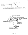
- the complex in the present invention for Embodiment C-1 can be expressed in the simplest binding form as water-absorbent substrate-first immunochemical component-analyte-second immunochemical component-labeling substance-biotin-avidin-biotin-labeling substance-second immunochemical component. Since a plural number of the -biotin-labeling substance-second immunochemical component bound to an avidin can be bound to the same avidin molecule, it can be expressed as water-absorbent substrate-first immunochemical component-analyte-second immunochemical component-labeling substance-biotin-avidin- (biotin-labeling substance-second immunochemical component)n.
- an analyte is further bindable to a second immunochemical component positioned at the end of the complex, the analyte is further bound to the second immunochemical component, whereby subsequently there can be also bound the -second immunochemical component-labeling substance-biotin-avidin- (biotin-labeling substance-second immunochemical component)n.
- Embodiment C-2 it can be expressed in the simplest binding form as water-absorbent substrate-first immunochemical component-analyte-second immunochemical component-first labeling substance-avidin-biotin-second labeling substance. Since a plural number of the -biotin-second labeling substance bound to an avidin can be bound to the same avidin molecule, it can be expressed, for instance, as water-absorbent substrate-first immunochemical component-analyte-second immunochemical component-first labeling substance-avidin-(biotin-second labeling substance)n.
- the second labeling substance positioned at the end of the complex mentioned above is bound to a large number of biotins, and each of the biotins is further bound to an avidin bound to a different first labeling substance, thereby forming various complicated complexes via biotin-avidin binding.
- Embodiment C-3 it can be expressed in the simplest binding form as water-absorbent substrate-first immunochemical component-analyte-second immunochemical component-first labeling substance-biotin-avidin-second labeling substance.
- a plural number of the biotin-first labeling substance-second immunochemical component bound to an avidin can be bound to the same avidin molecule, and the first labeling substance is further bound to a large number of biotins, and each of biotins is further bound to an avidin bound to a different second labeling substance, thereby forming various complicated complexes via biotin-avidin binding.
- the distance between the above capture region and the site at which absorption of solutions of a test sample, a labeled component solution, or a mixture comprising a test sample and a labeled component solution; a buffer, or the like is initiated is from 1 to 6 cm, preferably from 2 to 4 cm or so.
- dropping site is from 1 to 6 cm, preferably from 2 to 4 cm or so.
- Embodiment C-1 there are exemplified the following two embodiments.
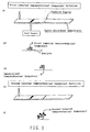
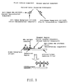
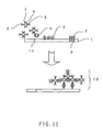
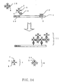
- a test sample is mixed with a buffer comprising a labeled component 4, and an avidin 8, and the resulting mixture is developed from a dropping site 11 on a water-absorbent substrate 1, thereby allowing the mixture to migrate to a capture region 3 to which a first immunochemical component 2 is immobilized.
- a binding of a second immunochemical component 6 in the labeled component 4 with an analyte 9 and a binding of a biotin 7 and an avidin 8 in the labeled component 4 take place.
- the first immunochemical component 2 immobilized on the water-absorbent substrate 1 is bound to the analyte 9, thereby forming a complex 10 [water-absorbent substrate-first immunochemical component-analyte-second immunochemical component-labeling substance-biotin-avidin-labeling substance].
- a test sample is developed along with a buffer comprising a labeled component 4, thereby allowing the mixture to migrate to a capture region 3 to which a first immunochemical component 2 is immobilized.
- the mixture reaches a region to which an avidin 8 is applied (avidin region), and the avidin 8 is released by contact with water.
- the first immunochemical component 2 immobilized on the water-absorbent substrate is bound to the analyte 9, thereby forming a complex 10 [water-absorbent substrate-first immunochemical component-analyte-second immunochemical component-labeling substance-biotin-avidin-labeling substrate].
- the complex 10 formed as described above can be visually confirmed, since the colored particles as a labeling substance 5 constituting an immunolabeled component are assembled by higher order binding via avidin-biotin, so that the coloring is clear.
- Embodiment C-2 there are exemplified the following two embodiments.
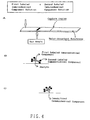
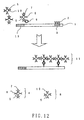
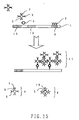
- a test sample is mixed with a buffer containing a labeled component 4, and a conjugate 8 comprising a biotin and a second labeling substance, and the resulting mixture is developed from a dropping site 13 on a water-absorbent substrate 1, thereby allowing the mixture to migrate to a capture region 3 to which a first immunochemical component 2 is immobilized.
- a test sample is developed along with a buffer containing a labeled component 4, thereby allowing the mixture to migrate to a capture region 3 to which a first immunochemical component 2 is immobilized.
- the mixture reaches a reagent region 12 to which the conjugate 8 comprising a biotin and a second labeling substance is applied, and the conjugate 8 comprising a biotin and the second labeling substance is released by contact with water.
- the first immunochemical component 2 immobilized on the water-absorbent substrate is bound to the analyte 9, thereby forming a complex 11 [water-absorbent substrate-first immunochemical component-analyte-second immunochemical component-first labeling substance-avidin-biotin-second labeling substance].
- the complex 11 formed as described above can be visually confirmed, since the colored particles as a first labeling substance 5 and a second labeling substance 5 are assembled by higher order binding via avidin-biotin, so that the coloring is clear.
- Embodiment C-3 there are exemplified the following two embodiments.
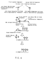
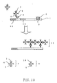
- a test sample is mixed with a buffer comprising a labeled component 4, and a conjugate 8 comprising an avidin and a second labeling substance, and the resulting mixture is developed from a dropping site 13 on a water-absorbent substrate 1, thereby allowing the mixture to migrate to a capture region 3 to which a first immunochemical component 2 is immobilized.
- a test sample is developed along with a buffer comprising a labeled component 4, thereby allowing the mixture to migrate to a capture region 3 to which a first immunochemical component 2 is immobilized.
- the mixture reaches a reagent region 12 to which the conjugate 8 comprising an avidin and a second labeling substance is applied, and the conjugate 8 comprising an avidin and the second labeling substance is released by contact with water.
- the first immunochemical component 2 immobilized on the water-absorbent substrate is bound to the analyte 9, thereby forming a complex 11 [water-absorbent substrate-first immunochemical component-analyte-second immunochemical component-first labeling substance-biotin-avidin-second labeling substance].
- the complex 11 formed as described above can be visually confirmed, since the colored particles as a first labeling substance 5 and a second labeling substance 5 are assembled by higher order binding via avidin-biotin, so that the coloring is clear.
- the water-absorbent substrate comprising a capture region in which a first immunochemical component is immobilized is also referred to as the immunological test strip of the present invention.
- the present invention provides in Embodiment C-1 the immunological test strip mentioned above and a kit for immunological detection method comprising the labeled components mentioned above and an avidin; in Embodiment C-2 the immunological test strip mentioned above and a kit for immunological detection method comprising the labeled components mentioned above and a conjugate comprising the biotin and a second labeling substance; and in Embodiment C-3 the immunological test strip mentioned above and a kit for immunological detection method comprising the labeled components mentioned above and a conjugate comprising the avidin and a second labeling substance.
- kits of the present invention can be suitably used for the immunological detection methods of the present invention.
- the immunological test strip and the labeled components contained in the kit of the present invention are as defined in the immunological detection method of the present invention mentioned above.
- the avidin in Embodiment C-1, the conjugate comprising a biotin and the second labeling substance in Embodiment C-2, and the conjugate comprising an avidin and the second labeling substance, each contained in the kit of the present invention are as defined in the immunological detection method of the present invention mentioned above, and each is preferably maintained in a form capable of being released by contact with water in a given region between the capture region and one end of the water-absorbent substrate of the immunological test strip.
- the resulting mixture was allowed to react at 10°C for 3 hours, and thereafter, the reaction mixture was washed by centrifugation using borate buffer (pH 8) as a washing solution, to prepare a blue-colored latex particle-labeled anti- Escherichia coli O157:H7 antibody.
- the resulting latex particle labeled antibody was suspended in 0.01 M-borate buffer (pH 8) so as to have a concentration of 2% by weight on a solid basis.
- the resulting mixture was allowed to react at 10°C for 3 hours, and thereafter, the reaction mixture was washed by centrifugation using borate buffer (pH 8) as a washing solution, to prepare a blue-colored latex particle-labeled anti-goat IgG antibody.
- the resulting latex-labeled antibody was suspended in 0.01 M-borate buffer (pH 8) so as to have a concentration of 2% by weight on a solid basis.
- a dispenser 0.5 ⁇ l of a 1 mg/ml rabbit IgG anti- Escherichia coli O157:H7 antibody [manufactured by Capricorn, in 0.1 M phosphate buffer (pH 7.4)] was applied in a linear form at a site of 30 mm from one end of a nitrocellulose membrane (pore size: 8 ⁇ m, 6 mm x 60 mm).
- This membrane was immersed into an aqueous solution comprising 1% by weight of bovine serum albumin and 0.1% by weight of polyoxyethylene(10) octyl phenyl ether (manufactured by Wako Pure Chemical Industries, Ltd.) for 10 minutes, and thereafter dried at 40°C for 2 hours.
- Example A-1 There was prepared a liquid of a test sample obtained from dispersion of Escherichia coli O157:H7 strain in 0.1 M phosphate buffer (pH 7.4) containing 0.9% by weight NaCl at each concentration shown in Table A-1.
- the resulting liquid of a test sample was mixed with the first labeled antibody solution prepared in item (1) of Example A-1 so as to have a concentration of 0.02% by weight on a solid basis, and the mixture was stirred. Thereafter, 60 ⁇ l of a mixture was added dropwise to a polyester nonwoven fabric portion of the test strip prepared in item (3) of Example A-1.
- the mixture was developed on the test strip, and thereafter 60 ⁇ l of a diluted solution was added dropwise to the above polyester nonwoven fabric portion, the diluted solution being prepared by diluting the second labeled antibody solution prepared in item (2) of Example A-1 with 0.1 M phosphate buffer (pH 7.4) containing 0.9% by weight NaCl so as to have a concentration of 0.02% by weight on a solid basis.
- the presence or absence of coloring on a capture region after 20 minutes was visually observed.
- the results are shown in Table A-1.
- the assay results of the case where only the first labeled antibody was used without using a second labeled antibody are also given side by side.
- Example A-1 There was prepared a liquid of a test sample obtained from dispersion of Escherichia coli O157:H7 strain in 0.1 M phosphate buffer (pH 7.4) containing 0.9% by weight NaCl at each concentration shown in Table A-1.
- the resulting liquid of a test sample was mixed with the first labeled antibody solution prepared in item (1) of Example A-1 and the second labeled antibody solution prepared in item (2) of Example A-1 so as to have a concentration of 0.02% by weight each on a solid basis, and the mixture was stirred. Thereafter, 60 ⁇ l of the mixture was added dropwise to a polyester nonwoven fabric portion of the immunological test strip prepared in item (3) of Example A-1. The presence or absence of coloring on a capture region after 20 minutes was visually observed. The results are shown in Table A-1. As a comparison, the assay results of the case where only the first labeled antibody was used without using a second labeled antibody are also given side by side.
- Example A-1 There was prepared a test sample obtained from dispersion of Escherichia coli O157:H7 strain in 0.1 M phosphate buffer (pH 7.4) containing 0.9% by weight NaCl at each concentration shown in Table A-1. Two microliters of this test sample was allowed to absorb on the front side of the immunological test strip prepared in item (3) of Example A-1 at a site 12 to 20 mm from the opposite side to the antibody-applied site. Subsequently, the first labeled antibody solution prepared in item (1) of Example A-1 was diluted with 0.1 M phosphate buffer (pH 7.4) containing 0.9% by weight NaCl so as to have a concentration of 0.02% by weight on a solid basis.
- test sample obtained from dispersion of Escherichia coli O157:H7 strain in 0.1 M phosphate buffer (pH 7.4) containing 0.9% by weight NaCl at each concentration shown in Table A-1. Two microliters of this test sample was allowed to absorb on the front side of the immunological test strip prepared in item (3) of Example A-1 at a site 12 to 20 mm from the opposite side to the antibody-applied site.
- Example A-1 the first labeled antibody solution prepared in item (1) of Example A-1 and the second labeled antibody solution prepared in item (2) of Example A-1 were mixed, and the resulting mixture was diluted with 0.1 M phosphate buffer (pH 7.4) containing 0.9% by weight NaCl so as to have a concentration of 0.02% by weight each on a solid basis. Thereafter, 60 ⁇ l of the mixed dilution was added dropwise to a polyester nonwoven fabric portion of the immunological test strip prepared in item (3) of Example A-1. The presence or absence of coloring on a capture region after 20 minutes was visually observed. The results are shown in Table A-1.
- the polymeric particle dispersion was washed by centrifugation sequentially with an alkali, an acid and distilled water, and thereafter adjusted so as to have a concentration of 10% by weight on a solid basis (carrier particle dispersion).
- carrier particle dispersion In 20 ml of toluene was dissolved 0.2 g of Sudan blue, and to the resulting solution were added 0.2 g of sodium dodecyl sulfate and 100 ml of distilled water, and the mixture was emulsified by an ultrasonic dispersion machine. To the mixture was added 30 ml of the above carrier particle dispersion (concentration of 10% by weight on a solid basis), and the resulting mixture was stirred at room temperature for 24 hours.
- the resulting product was washed by centrifugation with 0.01 M borate buffer (pH 7.5) and adjusted so as to have a concentration of 5% by weight on a solid basis.
- 0.01 M borate buffer pH 7.5
- To 50 ml of this mixture were added 5 ml of an aqueous solution of 1-ethyl-3-(3-dimethylaminopropyl)carbodiimide hydrochloride (10 mg/ml) and 50 ml of an aqueous solution of 0.03 M m-xylenediamine, and the resulting mixture was allowed to react at room temperature for 5 hours.
- This resulting mixture was heat-treated at 75°C for 5 hours, and thereafter washed by centrifugation with the same buffer as above, and adjusted so as to have a concentration of 1% by weight on a solid basis (Sudan blue-stained xylenediamine-spacer particle dispersion).
- the resulting mixture was washed by centrifugation with the same buffer as above, and re-dispersed so as to have a concentration of 1% by weight on a solid basis, to give a solution comprising Sudan blue-stained particle-labeled anti-human hemoglobin antibody-anti-human HBs antibody (first labeled component), in which the antibodies are bound by covalent bonding.
- the resulting mixture was washed by centrifugation with the, same buffer as above, and re-dispersed so as to have a concentration of 1% by weight on a solid basis, to give a solution comprising Sudan blue-stained particle-labeled anti-human hemoglobin antibody (second labeled component), in which the antibodies are bound by covalent bonding.
- second labeled component Sudan blue-stained particle-labeled anti-human hemoglobin antibody
- Anti-human HBs antibodies as a first immunochemical component were diluted with 0.1 M phosphate buffer (pH 7.4), to adjust to an aqueous solution having a final concentration of 1 mg/ml.
- Ten microliters of this aqueous solution was applied to a site 50 mm from one end of a nitrocellulose membrane filter (Toyo Filter Paper, 5 x 100 mm), and thereafter the resulting filter was immediately allowed to stand at 37°C for 1 hour. Thereafter, the nitrocellulose membrane filter was taken out, and immersed into an aqueous solution of 0.1% bovine serum albumin and 0.1% Tween 20 for 1 hour.
- nitrocellulose membrane filter was taken out, and allowed to stand at room temperature for 3 hours, to give a nitrocellulose membrane filter carrying anti-human HBs antibodies.
- a separation region for hematocyte Toyo Filter Paper No.2, 5 x 10 mm was positioned near one end (solution-absorbing site) of the test strip of the above anti-human HBs antibody-immobilized nitrocellulose membrane filter, to give a test strip comprising a nitrocellulose membrane filter comprising a capture region and a separation region for hematocyte.
- Example B-1 To the separation region for hematocyte of the test strip prepared in item (5) of Example B-1 was added dropwise 10 ⁇ l of a liquid of a test sample prepared by dissolving human HBs antigens in a physiological saline. Immediately thereafter, 100 ⁇ l of a mixture of a solution comprising hemoglobin antigens (protein concentration: 200 ng/ml) and the solution comprising the second labeled component (concentration on a solid basis: 0.2% by weight) prepared in item (3) of Example B-1 was added dropwise to the solution-absorbing site, and then developed. The coloring on a capture region after 10 minutes was visually observed (Figure 5). The results are shown in Table B-1. Concentration of HBs Antigen ng/ml Evaluation Results 0 1 5 10 50 100 1,000 10,000 100,000
- Example B-1 To the separation region for hematocyte of the test strip prepared in item (4) of Example B-1 was added dropwise 10 ⁇ l of a liquid of a test sample prepared by dissolving human HBs antigens in a physiological saline. Immediately thereafter, 50 ⁇ l of the solution comprising the first labeled component prepared in item (2) of Example B-1 (concentration on a solid basis: 0.2% by weight) was added dropwise to the solution-absorbing site, and then developed. After 10 minutes, 100 ⁇ l of a solution comprising hemoglobin antigens (protein concentration: 200 ng/ml) was added dropwise to the solution-absorbing site, and then developed.
- a solution comprising hemoglobin antigens protein concentration: 200 ng/ml
- Example B-1 To the separation region for hematocyte of the test strip prepared in item (4) of Example B-1 was added dropwise 10 ⁇ l of a liquid of a test sample prepared by dissolving human HBs antigens in a physiological saline. Immediately thereafter, 50 ⁇ l of the solution comprising the first labeled component prepared in item (2) of Example B-1 (concentration on a solid basis: 0.2% by weight) was added dropwise to the solution-absorbing site, and then developed. After 10 minutes, 100 ⁇ l of a solution comprising hemoglobin antigens (protein concentration: 200 ng/ml) was added dropwise to the solution-absorbing site, and then developed.
- a solution comprising hemoglobin antigens protein concentration: 200 ng/ml
- Example B-1 To the separation region for hematocyte of the test strip prepared in item (4) of Example B-1 was added dropwise 10 ⁇ l of a liquid of a test sample prepared by dissolving human HBs antigens in a physiological saline. Immediately thereafter, 50 ⁇ l of the solution comprising the first labeled component prepared in item (2) of Example B-1 (concentration on a solid basis: 0.2% by weight) was added dropwise to the solution-absorbing site, and then developed. The coloring on a capture region after 10 minutes was visually observed. The results are shown in Table B-4. Concentration of HBs Antigen ng/ml Evaluation Results After Addition of First Labeled Component 0 1 5 10 50 100 1,000 10,000 100,000
- Example B-1 To the separation region for hematocyte of the test strip prepared in item (4) of Example B-1 was added dropwise 10 ⁇ l of a liquid of a test sample prepared by dissolving human HBs antigens in a physiological saline. Immediately thereafter, 100 ⁇ l of a mixture of the solution comprising the first labeled component prepared in item (2) of Example B-1 (concentration on a solid basis: 0.2% by weight), the solution comprising hemoglobin antigens (protein concentration: 200 ng/ml) and the solution comprising the second labeled component prepared in item (3) of Example B-1 (concentration on a solid basis: 0.2% by weight) was added dropwise to the solution-absorbing site, and then developed. The coloring on a capture region after 10 minutes was visually observed (Figure 7). The results are shown in Table B-5. Concentration of HBs Antigen ng/ml Evaluation Results 0 1 5 10 50 100 1,000 10,000 100,000
- Example B-1 To the separation region for hematocyte of the test strip prepared in item (4) of Example B-1 was added dropwise 10 ⁇ l of a liquid of a test sample prepared by dissolving human HBs antigens in a physiological saline. Immediately thereafter, 50 ⁇ l of the solution comprising the first labeled component prepared in item (2) of Example B-1 (concentration on a solid basis: 0.2% by weight) was added dropwise to the solution-absorbing site, and then developed. After 10 minutes, 100 ⁇ l of a solution comprising hemoglobin antigens (protein concentration: 200 ng/ml) was added dropwise to the solution-absorbing site, and then developed.
- a solution comprising hemoglobin antigens protein concentration: 200 ng/ml
- Example B-5 To the separation region for hematocyte of the test strip prepared in Example B-5 was added dropwise 10 ⁇ l of a liquid of a test sample prepared by dissolving human HBs antigens in a physiological saline. Immediately thereafter, 100 ⁇ l of a mixture of the solution comprising hemoglobin antigens (protein concentration: 200 ng/ml) and the solution comprising the first labeled component prepared in item (2) of Example B-1 (concentration on a solid basis: 0.2% by weight) was added dropwise to the solution-absorbing site, and then developed. The coloring on a capture region after 10 minutes was visually observed (Figure 8). The results are shown in Table B-7. Concentration of HBs Antigen ng/ml Evaluation Results After Addition of First Labeled Component After Addition of Hb Antigen After Addition of Second Labeled Component 0 1 5 10 50 100 1,000 10,000 100,000
- Example B-6 An assay for human Hbs antigen was carried out in the same manner as in Example B-6 except for using only the first labeled component prepared in item (2) of Example B-1 without using second labeled component or hemoglobin antigens which were a mediating substance.
- To the separation region for hematocyte of the test strip prepared in Example B-5 was added dropwise 10 ⁇ l of a liquid of a test sample prepared by dissolving human HBs antigens in a physiological saline.
- 50 ⁇ l of the solution comprising the first labeled component (concentration on a solid basis: 0.2% by weight) was added dropwise to the solution-absorbing site, and then developed. The coloring on a capture region after 10 minutes was visually observed.
- the results are shown in Table B-8. Concentration of HBs Antigen ng/ml After Addition of First Labeled Component 0 1 5 10 50 100 1,000 10,000 100,000
- Example B-7 To the separation region for hematocyte of the test strip prepared in Example B-7 was added dropwise 10 ⁇ l of a liquid of a test sample prepared by dissolving human HBs antigens in a physiological saline. Immediately thereafter, 100 ⁇ l of the solution comprising hemoglobin antigens (protein concentration: 200 ng/ml) was added dropwise to the solution-absorbing site, and then developed. The coloring on a capture region after 10 minutes was visually observed ( Figure 9). The results are shown in Table B-9. Concentration of HBs Antigen ng/ml Evaluation Results 0 1 5 10 50 100 1,000 10,000 100,000
- Example B-7 To the separation region for hematocyte of the test strip prepared in Example B-7 was added dropwise 10 ⁇ l of a liquid of a test sample prepared by dissolving human HBs antigens in a physiological saline. Immediately thereafter, 50 ⁇ l of the solution comprising the first labeled component (concentration on a solid basis; 0.2% by weight) prepared in item (2) of Example B-1 was added dropwise to the solution-absorbing site, and then developed. After 10 minutes, 100 ⁇ l of a solution comprising hemoglobin antigens (protein concentration: 200 ng/ml) was added dropwise to the solution-absorbing site, and then developed.
- a solution comprising hemoglobin antigens protein concentration: 200 ng/ml
- the resulting polymeric particle dispersion was washed by centrifugation sequentially with an alkali, an acid and distilled water, and thereafter adjusted so as to have a concentration of 10% by weight on a solid basis (carrier particle dispersion).
- a 0.2 g Sudan blue was dissolved in 20 ml of toluene, and 0.2 g of sodium dodecyl sulfate and 100 ml of distilled water were added thereto. Thereafter, the resulting mixture was emulsified by an ultrasonic dispersion machine.
- the resulting mixture was washed by centrifugation with the same buffer as above, and re-dispersed so as to have a concentration of 1% by weight on a solid basis, to give a Sudan blue-stained particle-labeled biocytin-anti-human HBs antibody (labeled component), in which the antibodies are bound by covalent bonding.
- Anti-human HBs antibodies (rabbit IgG) were diluted with 0.1 M phosphate buffer (pH 7.4), to prepare an aqueous solution having a final concentration of 1 mg/ml. Ten microliters of this aqueous solution was applied to a site of 2 cm from one end of a nitrocellulose membrane filter (Toyo Filter Paper, 5 x 100 mm), and immediately thereafter the resulting filter was allowed to stand at 37°C for 1 hour. Thereafter, the nitrocellulose membrane filter was taken out, and immersed into an aqueous solution of 0.1% bovine serum albumin and 0.1% Tween 20 for 1 hour.
- a nitrocellulose membrane filter Toyo Filter Paper, 5 x 100 mm
- nitrocellulose membrane filter (immunological test strip) having a capture region in which the anti-human HBs antibodies were immobilized and at one end of the filter a receiver of nonwoven fabric (5 mm x 10 mm).
- test sample prepared by dissolving human HBs antigens in a physiological saline, the labeled components, and a mixture comprising avidin (concentration on a solid basis: 0.2% by weight) were mixed, and 100 ⁇ l of this mixture was added dropwise from one end of the above immunological test strip (receiver), and then developed. The coloring on a capture region after 20 minutes was observed.
- Table C-1 The results are shown in Table C-1.
- a Sudan blue-stained particle-labeled biocytin-anti-human HBs antibody (labeled component) was obtained in the same manner as in Example C-1.
- Anti-human HBs antibodies (rabbit IgG) were diluted with 0.1 M phosphate buffer (pH 7.4), to prepare an aqueous solution having a final concentration of 1 mg/ml. Ten microliters of this aqueous solution was applied to a site of 2 cm from one end of a nitrocellulose membrane filter (Toyo Filter Paper, 5 x 100 mm), and immediately thereafter the resulting filter was allowed to stand at 37°C for 1 hour. Thereafter, the nitrocellulose membrane filter was taken out, and immersed into an aqueous solution of 0.1% bovine serum albumin and 0.1% Tween 20 for 1 hour.
- a nitrocellulose membrane filter Toyo Filter Paper, 5 x 100 mm
- nitrocellulose membrane filter (immunological test strip) having a capture region in which the anti-human HBs antibodies were immobilized and at one end of the filter a receiver of nonwoven fabric (5 mm x 10 mm).
- 10 ⁇ l of a solution prepared by dissolving avidin (1 mg/ml) in 0.1 M phosphate buffer (pH 7.4) was applied to a part between the capture region and receiver of the above immunological test strip, and immediately thereafter, the resulting test strip was allowed to stand at 37°C for 1 hour, to give an immunological test strip comprising an avidin region.
- the detection sensitivity for human HBs antigen according to the immunological method of the present invention was about 0.25 ng/ml.
- Example C-1 In the same manner as in Example C-1, there were obtained a nitrocellulose membrane filter in which the anti-human HBs antibodies were immobilized, and a Sudan blue-stained particle-labeled anti-human HBs antibody (labeled component).
- the detection sensitivity for human HBs antigen according to the immunological method of the comparative example was about 10 ng/ml. Therefore, it is found that the sensitivity of the immunological method of the present invention increases about 40 times compared to a conventional method.
- Example C-1 One milliliter of an aqueous solution of glutaraldehyde (0.1 mg/ml) was added to 10 ml of a Sudan blue-stained xylenediamine-spacer particle dispersion obtained in the same manner as in Example C-1, and the resulting mixture was allowed to react at room temperature for 2 hours. Thereafter, the resulting reaction mixture was washed by centrifugation with the same buffer as in Example C-1, and adjusted to a dispersion having a concentration of 1% by weight on a solid basis.
- Example C-1 One milliliter of an aqueous solution of glutaraldehyde (0.1 mg/ml) was added to 10 ml of a Sudan blue-stained xylenediamine-spacer particle dispersion obtained in the same manner as in Example C-1, and the resulting mixture was allowed to react at room temperature for 2 hours. Thereafter, the resulting reaction mixture was washed by centrifugation with the same buffer as in Example C-1, and adjusted to a dispersion having a concentration of 1% by weight on a solid basis. To 10 ml of this dispersion was added 1 mg of biotin, and the resulting mixture was stirred at 10°C for 24 hours.
- the resulting mixture was washed by centrifugation with the same buffer as above, and re-dispersed so as to have a concentration of 1% by weight on a solid basis, to give a Sudan blue-stained particle-labeled biotin (conjugate comprising biotin and the second labeling substance), in which biotin is bound by covalent bonding.
- Example C-1 In the same manner as in Example C-1, there was obtained a nitrocellulose membrane filter (immunological test strip) having a capture region in which the anti-human HBs antibodies were immobilized and at one end a receiver of nonwoven fabric (5 x 10 mm).
- test sample prepared by dissolving human HBs antigens in a physiological saline, the labeled components (concentration on a solid basis: 0.2% by weight), and the conjugate comprising biotin and a second labeling substance (concentration on a solid basis: 0.2% by weight) were mixed, and 100 ⁇ l of this mixture was added dropwise from one end of the above immunological test strip (receiver), and then developed. The coloring on a capture region after 20 minutes was observed. The results are shown in Table C-3.
- a Sudan blue-stained particle-labeled avidin-anti-human HBs antibody (labeled component) was obtained in the same manner as in Example C-3. Also, a Sudan blue-stained particle-labeled biotin (conjugate comprising biotin and the second labeling substance) was obtained in the same manner as in Example C-3.
- Anti-human HBs antibodies (rabbit IgG) were diluted with 0.1 M phosphate buffer (pH 7.4), to prepare an aqueous solution having a final concentration of 1 mg/ml. Ten microliters of this aqueous solution was applied to a site of 2 cm from one end of a nitrocellulose membrane filter (Toyo Filter Paper, 5 x 100 mm), and immediately thereafter the resulting filter was allowed to stand at 37°C for 1 hour. Thereafter, the nitrocellulose membrane filter was taken out, and immersed into an aqueous solution of 0.1% bovine serum albumin and 0.1% TweenTM 20 for 1 hour.
- a nitrocellulose membrane filter Toyo Filter Paper, 5 x 100 mm
- nitrocellulose membrane filter (immunological test strip) having a capture region in which the anti-human HBs antibodies were immobilized and at one end of the filter a receiver of nonwoven fabric (5 x 10 mm).
- the detection sensitivity for human HBs antigen according to the immunological method of the present invention was about 0.25 ng/ml. Therefore, it is found that the sensitivity of the immunological method of the present invention increases about 40 times compared to the method of Comparative Example C-1.
- Example C-1 One milliliter of an aqueous solution of glutaraldehyde (0.1 mg/ml) was added to 10 ml of a Sudan blue-stained xylenediamine-spacer particle dispersion obtained in the same manner as in Example C-1, and the resulting mixture was allowed to react at room temperature for 2 hours. Thereafter, the resulting reaction mixture was washed by centrifugation with the same buffer as in Example C-1, and adjusted to a dispersion having a concentration of 1% by weight on a solid basis.
- Example C-1 One milliliter of an aqueous solution of glutaraldehyde (0.1 mg/ml) was added to 10 ml of a Sudan blue-stained xylenediamine-spacer particle dispersion obtained in the same manner as in Example C-1, and the resulting mixture was allowed to react at room temperature for 2 hours. Thereafter, the resulting reaction mixture was washed by centrifugation with the same buffer as in Example C-1, and adjusted to a dispersion having a concentration of 1% by weight on a solid basis. To 10 ml of this dispersion was added 1 mg of avidin, and the resulting mixture was stirred at 10°C for 24 hours.
- the resulting mixture was washed by centrifugation with the same buffer as above, and re-dispersed so as to have a concentration of 1% by weight on a solid basis, to give a Sudan blue-stained particle-labeled avidin (conjugate comprising avidin and the second labeling substance), in which the avidin is bound by covalent bonding.
- Example C-1 In the same manner as in Example C-1, there was obtained a nitrocellulose membrane filter (immunological test strip) having a capture region in which the anti-human HBs antibodies were immobilized and at one end of the filter a receiver of nonwoven fabric (5 x 10 mm).
- test sample prepared by dissolving human HBs antigens in a physiological saline, the labeled components (concentration on a solid basis: 0.2% by weight), and the conjugate comprising avidin and the second labeling substance (concentration on a solid basis: 0.2% by weight) were mixed. From one end of the above immunological test strip (receiver), 100 ⁇ l of this mixture was added dropwise, and then developed. The coloring on a capture region after 20 minutes was observed. The results are shown in Table C-4.
- a Sudan blue-stained particle-labeled biotin-anti-human HBs antibody (labeled component) was obtained in the same manner as in Example C-5. Also, a Sudan blue-stained particle-labeled avidin (conjugate comprising avidin and the second labeling substance) was obtained in the same manner as in Example C-5.
- Anti-human HBs antibodies (rabbit IgG) were diluted with 0.1 M phosphate buffer (pH 7.4), to prepare an aqueous solution having a final concentration of 1 mg/ml. Ten microliters of this aqueous solution was applied to a site of 2 cm from one end of a nitrocellulose membrane filter (Toyo Filter Paper, 5 x 100 mm), and immediately thereafter the resulting filter was allowed to stand at 37°C for 1 hour. Thereafter, the nitrocellulose membrane filter was taken out, and immersed into an aqueous solution of 0.1% bovine serum albumin and 0.1% TweenTM 20 for 1 hour.
- a nitrocellulose membrane filter Toyo Filter Paper, 5 x 100 mm
- nitrocellulose membrane filter (immunological test strip) having a capture region in which the anti-human HBs antibodies were immobilized and at one end of the filter a receiver of nonwoven fabric (5 x 10 mm).
- the detection sensitivity for human HBs antigen according to the immunological method of the present invention was about 0.25 ng/ml. Therefore, it is found that the sensitivity of the immunological method of the present invention increases about 40 times compared to the method of Comparative Example C-1.
Landscapes
- Health & Medical Sciences (AREA)
- Immunology (AREA)
- Life Sciences & Earth Sciences (AREA)
- Engineering & Computer Science (AREA)
- Chemical & Material Sciences (AREA)
- Molecular Biology (AREA)
- Biomedical Technology (AREA)
- Hematology (AREA)
- Urology & Nephrology (AREA)
- Biotechnology (AREA)
- Biochemistry (AREA)
- Cell Biology (AREA)
- Food Science & Technology (AREA)
- Medicinal Chemistry (AREA)
- Physics & Mathematics (AREA)
- Analytical Chemistry (AREA)
- Microbiology (AREA)
- General Health & Medical Sciences (AREA)
- General Physics & Mathematics (AREA)
- Pathology (AREA)
- Chemical Kinetics & Catalysis (AREA)
- Peptides Or Proteins (AREA)
- Investigating Or Analysing Biological Materials (AREA)
Applications Claiming Priority (25)
| Application Number | Priority Date | Filing Date | Title |
|---|---|---|---|
| JP18662898 | 1998-07-01 | ||
| JP18662498 | 1998-07-01 | ||
| JP18663698 | 1998-07-01 | ||
| JP18662898A JP3930969B2 (ja) | 1998-07-01 | 1998-07-01 | 免疫学的検査方法および免疫学的検査用キット |
| JP18664098 | 1998-07-01 | ||
| JP18664098A JP3930970B2 (ja) | 1998-07-01 | 1998-07-01 | 免疫学的検査方法および免疫学的検査用キット |
| JP18662498A JP3841555B2 (ja) | 1998-07-01 | 1998-07-01 | 免疫学的検査方法および免疫学的検査用キット |
| JP18663698A JP3841556B2 (ja) | 1998-07-01 | 1998-07-01 | 免疫学的検査方法および免疫学的検査用キット |
| JP19198198 | 1998-07-07 | ||
| JP19215198A JP3841559B2 (ja) | 1998-07-07 | 1998-07-07 | 免疫学的検査方法および免疫学的検査キット |
| JP19211498 | 1998-07-07 | ||
| JP19215098 | 1998-07-07 | ||
| JP10191981A JP2000028612A (ja) | 1998-07-07 | 1998-07-07 | 免疫学的検査方法および免疫学的検査キット |
| JP10192114A JP2000028611A (ja) | 1998-07-07 | 1998-07-07 | 免疫学的検査方法および免疫学的検査キット |
| JP19215198 | 1998-07-07 | ||
| JP19205298 | 1998-07-07 | ||
| JP10192052A JP2000028613A (ja) | 1998-07-07 | 1998-07-07 | 免疫学的検査方法および免疫学的検査キット |
| JP19215098A JP3841558B2 (ja) | 1998-07-07 | 1998-07-07 | 免疫学的検査方法および免疫学的検査キット |
| JP806499 | 1999-01-14 | ||
| JP11008064A JP2000206114A (ja) | 1999-01-14 | 1999-01-14 | 免疫学的測定方法 |
| JP11024285A JP2000221193A (ja) | 1999-02-01 | 1999-02-01 | 免疫学的測定方法 |
| JP2428599 | 1999-02-01 | ||
| JP11024286A JP2000221194A (ja) | 1999-02-01 | 1999-02-01 | 免疫学的測定方法 |
| JP2428699 | 1999-02-01 | ||
| PCT/JP1999/003539 WO2000002049A1 (en) | 1998-07-01 | 1999-06-30 | Immunologic test method and immunologic test kit |
Publications (2)
| Publication Number | Publication Date |
|---|---|
| EP1020726A1 true EP1020726A1 (de) | 2000-07-19 |
| EP1020726A4 EP1020726A4 (de) | 2007-11-14 |
Family
ID=27583143
Family Applications (1)
| Application Number | Title | Priority Date | Filing Date |
|---|---|---|---|
| EP99926868A Withdrawn EP1020726A4 (de) | 1998-07-01 | 1999-06-30 | Immunologische testverfahren und testsatz |
Country Status (3)
| Country | Link |
|---|---|
| US (1) | US6753190B1 (de) |
| EP (1) | EP1020726A4 (de) |
| WO (1) | WO2000002049A1 (de) |
Cited By (5)
| Publication number | Priority date | Publication date | Assignee | Title |
|---|---|---|---|---|
| WO2002053768A3 (en) * | 2001-01-05 | 2003-04-03 | Diagnosticart Llc | Dna rapid immune detection methods and apparatus |
| FR2853077A1 (fr) * | 2003-03-28 | 2004-10-01 | Vedalab | Procedes immunochromatographiques en phase solide |
| EP2128617A1 (de) * | 2008-05-27 | 2009-12-02 | Koninklijke Philips Electronics N.V. | Vorrichtung und Verfahren zur Feststellung von Analyten in der Spucke |
| CN102227631A (zh) * | 2008-11-28 | 2011-10-26 | 英佛皮亚有限公司 | 免疫层析测试中信号放大的方法以及使用该方法的免疫层析试剂盒 |
| EP2776150A4 (de) * | 2011-11-10 | 2015-04-22 | Ceres Nanosciences Inc | Saugfähige getrocknete biofluidaufnahmesubstrate |
Families Citing this family (14)
| Publication number | Priority date | Publication date | Assignee | Title |
|---|---|---|---|---|
| US20010018192A1 (en) * | 1998-02-12 | 2001-08-30 | Terstappen Leon W.M.M. | Labeled cells for use as an internal functional control in rare cell detection assays |
| FI118061B (fi) * | 2001-09-24 | 2007-06-15 | Beanor Oy | Menetelmä ja bioanturi analyysiä varten |
| US7939283B2 (en) * | 2001-11-01 | 2011-05-10 | Fisher Scientific Company L.L.C. | Analyte binding turbidity assay |
| FI115166B (fi) * | 2001-12-31 | 2005-03-15 | Biofons Oy | Diagnostisia menetelmiä |
| EP1758448A4 (de) * | 2004-06-02 | 2009-11-04 | Relia Diagnostic Systems Llc | Quantitatives querstromsystem und quantitative bestimmung |
| CN101031798B (zh) | 2004-07-29 | 2012-06-27 | 瑞莱诊断体系有限公司 | 侧向流系统和测定 |
| US7189522B2 (en) | 2005-03-11 | 2007-03-13 | Chembio Diagnostic Systems, Inc. | Dual path immunoassay device |
| WO2006098804A2 (en) | 2005-03-11 | 2006-09-21 | Chembio Diagnostic Systems, Inc. | Dual path immunoassay device |
| US20100022916A1 (en) | 2008-07-24 | 2010-01-28 | Javanbakhsh Esfandiari | Method and Apparatus for Collecting and Preparing Biological Samples for Testing |
| US8603835B2 (en) | 2011-02-10 | 2013-12-10 | Chembio Diagnostic Systems, Inc. | Reduced step dual path immunoassay device and method |
| US9804161B1 (en) * | 2012-05-14 | 2017-10-31 | Lawrence Livermore National Security, Llc | Detector and related, devices, methods and systems |
| CA2944488C (en) | 2014-04-02 | 2023-03-21 | Chembio Diagnostic Systems, Inc. | Immunoassay utilizing trapping conjugate |
| US20160116466A1 (en) | 2014-10-27 | 2016-04-28 | Chembio Diagnostic Systems, Inc. | Rapid Screening Assay for Qualitative Detection of Multiple Febrile Illnesses |
| US9885147B2 (en) * | 2015-04-24 | 2018-02-06 | University Of South Carolina | Reproducible sample preparation method for quantitative stain detection |
Family Cites Families (13)
| Publication number | Priority date | Publication date | Assignee | Title |
|---|---|---|---|---|
| US4632901A (en) * | 1984-05-11 | 1986-12-30 | Hybritech Incorporated | Method and apparatus for immunoassays |
| US5160701A (en) * | 1986-02-18 | 1992-11-03 | Abbott Laboratories | Solid-phase analytical device and method for using same |
| DE3922960A1 (de) * | 1989-07-12 | 1991-01-17 | Boehringer Mannheim Gmbh | Verfahren zur bestimmung eines analyten |
| US5141850A (en) * | 1990-02-07 | 1992-08-25 | Hygeia Sciences, Inc. | Porous strip form assay device method |
| JPH0466871A (ja) * | 1990-07-07 | 1992-03-03 | Eiji Ishikawa | 高感度な免疫測定法 |
| US5567591A (en) * | 1991-06-24 | 1996-10-22 | Becton Dickinson And Company | Amplified assay for analyte |
| JPH0782019B2 (ja) * | 1991-09-05 | 1995-09-06 | 三洋化成工業株式会社 | 免疫測定法及び免疫測定試薬 |
| JPH0829422A (ja) * | 1994-07-12 | 1996-02-02 | Aisin Seiki Co Ltd | 蛍光集合体を用いた検出方法 |
| JPH0829442A (ja) * | 1994-07-18 | 1996-02-02 | Fuji Xerox Co Ltd | ファン動作検出装置 |
| US20010051350A1 (en) * | 1995-05-02 | 2001-12-13 | Albert Nazareth | Diagnostic detection device and method |
| JP3490560B2 (ja) * | 1995-11-16 | 2004-01-26 | 日東電工株式会社 | 糞便中のヒトヘモグロビンの検出方法 |
| US5876944A (en) * | 1996-06-10 | 1999-03-02 | Bayer Corporation | Method for amplification of the response signal in a sandwich immunoassay |
| US6194221B1 (en) * | 1996-11-19 | 2001-02-27 | Wyntek Diagnostics, Inc. | Hybrid one-step immunochromatographic device and method of use |
-
1999
- 1999-06-30 WO PCT/JP1999/003539 patent/WO2000002049A1/ja not_active Ceased
- 1999-06-30 US US09/486,640 patent/US6753190B1/en not_active Expired - Fee Related
- 1999-06-30 EP EP99926868A patent/EP1020726A4/de not_active Withdrawn
Cited By (11)
| Publication number | Priority date | Publication date | Assignee | Title |
|---|---|---|---|---|
| WO2002053768A3 (en) * | 2001-01-05 | 2003-04-03 | Diagnosticart Llc | Dna rapid immune detection methods and apparatus |
| FR2853077A1 (fr) * | 2003-03-28 | 2004-10-01 | Vedalab | Procedes immunochromatographiques en phase solide |
| WO2004088320A1 (fr) * | 2003-03-28 | 2004-10-14 | Vedalab | Procedes immunochromatographiques en phase solide |
| EP2128617A1 (de) * | 2008-05-27 | 2009-12-02 | Koninklijke Philips Electronics N.V. | Vorrichtung und Verfahren zur Feststellung von Analyten in der Spucke |
| WO2009144660A1 (en) * | 2008-05-27 | 2009-12-03 | Koninklijke Philips Electronics N.V. | Device and methods for detecting analytes in saliva |
| US9103843B2 (en) | 2008-05-27 | 2015-08-11 | Koninklijke Philips N.V. | Device and methods for detecting analytes in saliva |
| US9575081B2 (en) | 2008-05-27 | 2017-02-21 | Koninklijke Philips N.V. | Device and methods for detecting analytes in saliva |
| CN102227631A (zh) * | 2008-11-28 | 2011-10-26 | 英佛皮亚有限公司 | 免疫层析测试中信号放大的方法以及使用该方法的免疫层析试剂盒 |
| EP2376906A4 (de) * | 2008-11-28 | 2012-07-04 | Infopia Co Ltd | Verfahren zur signalverstärkung bei einem immunchromatographietest sowie immunchromatographie-kit mit verwendung des verfahrens |
| CN102227631B (zh) * | 2008-11-28 | 2014-04-23 | 英佛皮亚有限公司 | 免疫层析测试中信号放大的方法以及使用该方法的免疫层析试剂盒 |
| EP2776150A4 (de) * | 2011-11-10 | 2015-04-22 | Ceres Nanosciences Inc | Saugfähige getrocknete biofluidaufnahmesubstrate |
Also Published As
| Publication number | Publication date |
|---|---|
| US6753190B1 (en) | 2004-06-22 |
| WO2000002049A1 (en) | 2000-01-13 |
| EP1020726A4 (de) | 2007-11-14 |
Similar Documents
| Publication | Publication Date | Title |
|---|---|---|
| US6753190B1 (en) | Immunologic test method and immunologic test kit | |
| US20080138842A1 (en) | Indirect lateral flow sandwich assay | |
| CA1289875C (en) | Immunoassay test strip | |
| US20100317126A1 (en) | Agglutination assay method in porous medium layer | |
| JP2002507725A (ja) | イムノアッセイデバイスおよび方法 | |
| US6391652B2 (en) | Immunoassay method and immunoassay kit | |
| US5266460A (en) | Method of preparing immunological analytical element | |
| JPH10177028A (ja) | 乳頭分泌液中の特定物質の測定方法 | |
| EP1061368B1 (de) | Agglutinations-Bestimmungsmethode in einem Bindermedium | |
| JP3841559B2 (ja) | 免疫学的検査方法および免疫学的検査キット | |
| JP3930970B2 (ja) | 免疫学的検査方法および免疫学的検査用キット | |
| JPH1068730A (ja) | イムノクロマト法用試験用具 | |
| JPH09184840A (ja) | イムノクロマト法用試験用具 | |
| JP3841558B2 (ja) | 免疫学的検査方法および免疫学的検査キット | |
| JP3930969B2 (ja) | 免疫学的検査方法および免疫学的検査用キット | |
| JP2000028612A (ja) | 免疫学的検査方法および免疫学的検査キット | |
| JP3841555B2 (ja) | 免疫学的検査方法および免疫学的検査用キット | |
| JP2001133455A (ja) | 免疫クロマトグラフィー用試験片 | |
| JP3841556B2 (ja) | 免疫学的検査方法および免疫学的検査用キット | |
| JP2001013146A (ja) | 免疫クロマトグラフィー用試験片 | |
| JP2000221194A (ja) | 免疫学的測定方法 | |
| USH1664H (en) | Analytical element for immunoassay and method for its preparation | |
| JP2004184295A (ja) | イムノクロマトグラフ媒体およびイムノクロマトグラフ法 | |
| JP2000028613A (ja) | 免疫学的検査方法および免疫学的検査キット | |
| JPH10206429A (ja) | 免疫学的検査片 |
Legal Events
| Date | Code | Title | Description |
|---|---|---|---|
| PUAI | Public reference made under article 153(3) epc to a published international application that has entered the european phase |
Free format text: ORIGINAL CODE: 0009012 |
|
| 17P | Request for examination filed |
Effective date: 20000331 |
|
| AK | Designated contracting states |
Kind code of ref document: A1 Designated state(s): DE FR GB |
|
| A4 | Supplementary search report drawn up and despatched |
Effective date: 20071012 |
|
| RIC1 | Information provided on ipc code assigned before grant |
Ipc: G01N 33/558 20060101ALI20071009BHEP Ipc: G01N 33/543 20060101AFI20000117BHEP |
|
| STAA | Information on the status of an ep patent application or granted ep patent |
Free format text: STATUS: THE APPLICATION HAS BEEN WITHDRAWN |
|
| 18W | Application withdrawn |
Effective date: 20080411 |