EP2042870A1 - Procédé de dosage immunologique de haute sensibilité - Google Patents
Procédé de dosage immunologique de haute sensibilité Download PDFInfo
- Publication number
- EP2042870A1 EP2042870A1 EP08016700A EP08016700A EP2042870A1 EP 2042870 A1 EP2042870 A1 EP 2042870A1 EP 08016700 A EP08016700 A EP 08016700A EP 08016700 A EP08016700 A EP 08016700A EP 2042870 A1 EP2042870 A1 EP 2042870A1
- Authority
- EP
- European Patent Office
- Prior art keywords
- antibody
- antigen
- antibodies
- carrier
- latex
- Prior art date
- Legal status (The legal status is an assumption and is not a legal conclusion. Google has not performed a legal analysis and makes no representation as to the accuracy of the status listed.)
- Withdrawn
Links
- 238000000034 method Methods 0.000 title claims abstract description 66
- 238000003018 immunoassay Methods 0.000 title claims abstract description 11
- 239000000427 antigen Substances 0.000 claims abstract description 35
- 102000036639 antigens Human genes 0.000 claims abstract description 35
- 108091007433 antigens Proteins 0.000 claims abstract description 35
- 230000004520 agglutination Effects 0.000 claims abstract description 33
- 206010070834 Sensitisation Diseases 0.000 claims abstract description 8
- 230000008313 sensitization Effects 0.000 claims abstract description 8
- 239000003153 chemical reaction reagent Substances 0.000 claims description 37
- 108010074051 C-Reactive Protein Proteins 0.000 claims description 16
- 102100032752 C-reactive protein Human genes 0.000 claims description 16
- 102000011923 Thyrotropin Human genes 0.000 claims description 16
- 108010061174 Thyrotropin Proteins 0.000 claims description 16
- 239000000203 mixture Substances 0.000 claims description 15
- 238000002156 mixing Methods 0.000 claims description 10
- 238000006243 chemical reaction Methods 0.000 abstract description 53
- 239000000126 substance Substances 0.000 abstract description 7
- 238000011002 quantification Methods 0.000 abstract description 4
- 239000004816 latex Substances 0.000 description 55
- 229920000126 latex Polymers 0.000 description 55
- 238000003556 assay Methods 0.000 description 34
- 239000000243 solution Substances 0.000 description 23
- 239000012491 analyte Substances 0.000 description 20
- 239000002245 particle Substances 0.000 description 20
- DHMQDGOQFOQNFH-UHFFFAOYSA-N Glycine Chemical compound NCC(O)=O DHMQDGOQFOQNFH-UHFFFAOYSA-N 0.000 description 18
- 238000002372 labelling Methods 0.000 description 18
- 239000000872 buffer Substances 0.000 description 16
- 238000002835 absorbance Methods 0.000 description 11
- 239000000969 carrier Substances 0.000 description 11
- 239000004471 Glycine Substances 0.000 description 9
- 239000007787 solid Substances 0.000 description 9
- 238000002360 preparation method Methods 0.000 description 8
- 230000035945 sensitivity Effects 0.000 description 8
- 238000005119 centrifugation Methods 0.000 description 7
- 239000006185 dispersion Substances 0.000 description 7
- 239000000463 material Substances 0.000 description 7
- 108091003079 Bovine Serum Albumin Proteins 0.000 description 6
- 229940098773 bovine serum albumin Drugs 0.000 description 6
- 239000002244 precipitate Substances 0.000 description 6
- 230000004044 response Effects 0.000 description 6
- 239000003381 stabilizer Substances 0.000 description 6
- 239000006228 supernatant Substances 0.000 description 6
- 239000004094 surface-active agent Substances 0.000 description 6
- 239000002202 Polyethylene glycol Substances 0.000 description 5
- 239000002981 blocking agent Substances 0.000 description 5
- 210000004027 cell Anatomy 0.000 description 5
- 238000001514 detection method Methods 0.000 description 5
- 229920001223 polyethylene glycol Polymers 0.000 description 5
- 102000004190 Enzymes Human genes 0.000 description 4
- 108090000790 Enzymes Proteins 0.000 description 4
- 239000004793 Polystyrene Substances 0.000 description 4
- 210000004369 blood Anatomy 0.000 description 4
- 239000008280 blood Substances 0.000 description 4
- 229920001577 copolymer Polymers 0.000 description 4
- 230000000694 effects Effects 0.000 description 4
- 238000012123 point-of-care testing Methods 0.000 description 4
- 229920002223 polystyrene Polymers 0.000 description 4
- 235000018102 proteins Nutrition 0.000 description 4
- 102000004169 proteins and genes Human genes 0.000 description 4
- 108090000623 proteins and genes Proteins 0.000 description 4
- 229920001059 synthetic polymer Polymers 0.000 description 4
- 230000008901 benefit Effects 0.000 description 3
- 150000001720 carbohydrates Chemical class 0.000 description 3
- 238000007796 conventional method Methods 0.000 description 3
- 239000012530 fluid Substances 0.000 description 3
- 230000006872 improvement Effects 0.000 description 3
- 230000000977 initiatory effect Effects 0.000 description 3
- 229920005615 natural polymer Polymers 0.000 description 3
- 239000000843 powder Substances 0.000 description 3
- 238000003127 radioimmunoassay Methods 0.000 description 3
- 238000000926 separation method Methods 0.000 description 3
- 210000002966 serum Anatomy 0.000 description 3
- 229920002307 Dextran Polymers 0.000 description 2
- 239000006173 Good's buffer Substances 0.000 description 2
- 241001465754 Metazoa Species 0.000 description 2
- PXHVJJICTQNCMI-UHFFFAOYSA-N Nickel Chemical compound [Ni] PXHVJJICTQNCMI-UHFFFAOYSA-N 0.000 description 2
- VYPSYNLAJGMNEJ-UHFFFAOYSA-N Silicium dioxide Chemical compound O=[Si]=O VYPSYNLAJGMNEJ-UHFFFAOYSA-N 0.000 description 2
- 239000013504 Triton X-100 Substances 0.000 description 2
- 229920004890 Triton X-100 Polymers 0.000 description 2
- 230000000035 biogenic effect Effects 0.000 description 2
- 210000001124 body fluid Anatomy 0.000 description 2
- 239000010839 body fluid Substances 0.000 description 2
- 230000003139 buffering effect Effects 0.000 description 2
- 239000005018 casein Substances 0.000 description 2
- BECPQYXYKAMYBN-UHFFFAOYSA-N casein, tech. Chemical compound NCCCCC(C(O)=O)N=C(O)C(CC(O)=O)N=C(O)C(CCC(O)=N)N=C(O)C(CC(C)C)N=C(O)C(CCC(O)=O)N=C(O)C(CC(O)=O)N=C(O)C(CCC(O)=O)N=C(O)C(C(C)O)N=C(O)C(CCC(O)=N)N=C(O)C(CCC(O)=N)N=C(O)C(CCC(O)=N)N=C(O)C(CCC(O)=O)N=C(O)C(CCC(O)=O)N=C(O)C(COP(O)(O)=O)N=C(O)C(CCC(O)=N)N=C(O)C(N)CC1=CC=CC=C1 BECPQYXYKAMYBN-UHFFFAOYSA-N 0.000 description 2
- 235000021240 caseins Nutrition 0.000 description 2
- 229960002086 dextran Drugs 0.000 description 2
- 235000014113 dietary fatty acids Nutrition 0.000 description 2
- UKMSUNONTOPOIO-UHFFFAOYSA-N docosanoic acid Chemical compound CCCCCCCCCCCCCCCCCCCCCC(O)=O UKMSUNONTOPOIO-UHFFFAOYSA-N 0.000 description 2
- POULHZVOKOAJMA-UHFFFAOYSA-N dodecanoic acid Chemical compound CCCCCCCCCCCC(O)=O POULHZVOKOAJMA-UHFFFAOYSA-N 0.000 description 2
- 230000002708 enhancing effect Effects 0.000 description 2
- 239000000194 fatty acid Substances 0.000 description 2
- 229930195729 fatty acid Natural products 0.000 description 2
- 150000004665 fatty acids Chemical class 0.000 description 2
- 150000004676 glycans Chemical class 0.000 description 2
- IPCSVZSSVZVIGE-UHFFFAOYSA-N hexadecanoic acid Chemical compound CCCCCCCCCCCCCCCC(O)=O IPCSVZSSVZVIGE-UHFFFAOYSA-N 0.000 description 2
- 230000001900 immune effect Effects 0.000 description 2
- 230000001965 increasing effect Effects 0.000 description 2
- 210000002381 plasma Anatomy 0.000 description 2
- 229920001282 polysaccharide Polymers 0.000 description 2
- 239000005017 polysaccharide Substances 0.000 description 2
- 229920000036 polyvinylpyrrolidone Polymers 0.000 description 2
- 239000001267 polyvinylpyrrolidone Substances 0.000 description 2
- 235000013855 polyvinylpyrrolidone Nutrition 0.000 description 2
- 230000035484 reaction time Effects 0.000 description 2
- 235000020183 skimmed milk Nutrition 0.000 description 2
- 210000004989 spleen cell Anatomy 0.000 description 2
- 230000000087 stabilizing effect Effects 0.000 description 2
- 210000002700 urine Anatomy 0.000 description 2
- OYHQOLUKZRVURQ-NTGFUMLPSA-N (9Z,12Z)-9,10,12,13-tetratritiooctadeca-9,12-dienoic acid Chemical compound C(CCCCCCC\C(=C(/C\C(=C(/CCCCC)\[3H])\[3H])\[3H])\[3H])(=O)O OYHQOLUKZRVURQ-NTGFUMLPSA-N 0.000 description 1
- WRIDQFICGBMAFQ-UHFFFAOYSA-N (E)-8-Octadecenoic acid Natural products CCCCCCCCCC=CCCCCCCC(O)=O WRIDQFICGBMAFQ-UHFFFAOYSA-N 0.000 description 1
- JKMHFZQWWAIEOD-UHFFFAOYSA-N 2-[4-(2-hydroxyethyl)piperazin-1-yl]ethanesulfonic acid Chemical compound OCC[NH+]1CCN(CCS([O-])(=O)=O)CC1 JKMHFZQWWAIEOD-UHFFFAOYSA-N 0.000 description 1
- JQXYBDVZAUEPDL-UHFFFAOYSA-N 2-methylidene-5-phenylpent-4-enoic acid Chemical compound OC(=O)C(=C)CC=CC1=CC=CC=C1 JQXYBDVZAUEPDL-UHFFFAOYSA-N 0.000 description 1
- LQJBNNIYVWPHFW-UHFFFAOYSA-N 20:1omega9c fatty acid Natural products CCCCCCCCCCC=CCCCCCCCC(O)=O LQJBNNIYVWPHFW-UHFFFAOYSA-N 0.000 description 1
- QSBYPNXLFMSGKH-UHFFFAOYSA-N 9-Heptadecensaeure Natural products CCCCCCCC=CCCCCCCCC(O)=O QSBYPNXLFMSGKH-UHFFFAOYSA-N 0.000 description 1
- 102000002260 Alkaline Phosphatase Human genes 0.000 description 1
- 108020004774 Alkaline Phosphatase Proteins 0.000 description 1
- 206010003445 Ascites Diseases 0.000 description 1
- 235000021357 Behenic acid Nutrition 0.000 description 1
- 241000283707 Capra Species 0.000 description 1
- 229920002134 Carboxymethyl cellulose Polymers 0.000 description 1
- 238000002965 ELISA Methods 0.000 description 1
- 239000007995 HEPES buffer Substances 0.000 description 1
- 241000282414 Homo sapiens Species 0.000 description 1
- 108060003951 Immunoglobulin Proteins 0.000 description 1
- 239000005639 Lauric acid Substances 0.000 description 1
- 239000007987 MES buffer Substances 0.000 description 1
- CERQOIWHTDAKMF-UHFFFAOYSA-M Methacrylate Chemical compound CC(=C)C([O-])=O CERQOIWHTDAKMF-UHFFFAOYSA-M 0.000 description 1
- CERQOIWHTDAKMF-UHFFFAOYSA-N Methacrylic acid Chemical compound CC(=C)C(O)=O CERQOIWHTDAKMF-UHFFFAOYSA-N 0.000 description 1
- 241000699666 Mus <mouse, genus> Species 0.000 description 1
- 206010029719 Nonspecific reaction Diseases 0.000 description 1
- 239000005642 Oleic acid Substances 0.000 description 1
- ZQPPMHVWECSIRJ-UHFFFAOYSA-N Oleic acid Natural products CCCCCCCCC=CCCCCCCCC(O)=O ZQPPMHVWECSIRJ-UHFFFAOYSA-N 0.000 description 1
- 241000283973 Oryctolagus cuniculus Species 0.000 description 1
- 235000021314 Palmitic acid Nutrition 0.000 description 1
- 241001494479 Pecora Species 0.000 description 1
- 206010035226 Plasma cell myeloma Diseases 0.000 description 1
- 239000004373 Pullulan Substances 0.000 description 1
- 229920001218 Pullulan Polymers 0.000 description 1
- 241000700159 Rattus Species 0.000 description 1
- 229920002125 Sokalan® Polymers 0.000 description 1
- 235000021355 Stearic acid Nutrition 0.000 description 1
- RTAQQCXQSZGOHL-UHFFFAOYSA-N Titanium Chemical compound [Ti] RTAQQCXQSZGOHL-UHFFFAOYSA-N 0.000 description 1
- 239000007983 Tris buffer Substances 0.000 description 1
- 239000002253 acid Substances 0.000 description 1
- XECAHXYUAAWDEL-UHFFFAOYSA-N acrylonitrile butadiene styrene Chemical compound C=CC=C.C=CC#N.C=CC1=CC=CC=C1 XECAHXYUAAWDEL-UHFFFAOYSA-N 0.000 description 1
- 229920000122 acrylonitrile butadiene styrene Polymers 0.000 description 1
- 239000004676 acrylonitrile butadiene styrene Substances 0.000 description 1
- 239000003513 alkali Substances 0.000 description 1
- DTOSIQBPPRVQHS-PDBXOOCHSA-N alpha-linolenic acid Chemical compound CC\C=C/C\C=C/C\C=C/CCCCCCCC(O)=O DTOSIQBPPRVQHS-PDBXOOCHSA-N 0.000 description 1
- 235000020661 alpha-linolenic acid Nutrition 0.000 description 1
- PNEYBMLMFCGWSK-UHFFFAOYSA-N aluminium oxide Inorganic materials [O-2].[O-2].[O-2].[Al+3].[Al+3] PNEYBMLMFCGWSK-UHFFFAOYSA-N 0.000 description 1
- 150000001413 amino acids Chemical class 0.000 description 1
- 230000003321 amplification Effects 0.000 description 1
- 125000000129 anionic group Chemical group 0.000 description 1
- 239000003945 anionic surfactant Substances 0.000 description 1
- 229940116226 behenic acid Drugs 0.000 description 1
- 102000005936 beta-Galactosidase Human genes 0.000 description 1
- 108010005774 beta-Galactosidase Proteins 0.000 description 1
- 230000029918 bioluminescence Effects 0.000 description 1
- 238000005415 bioluminescence Methods 0.000 description 1
- 210000000601 blood cell Anatomy 0.000 description 1
- 239000001768 carboxy methyl cellulose Substances 0.000 description 1
- 235000010948 carboxy methyl cellulose Nutrition 0.000 description 1
- 239000008112 carboxymethyl-cellulose Substances 0.000 description 1
- 229940105329 carboxymethylcellulose Drugs 0.000 description 1
- 239000003093 cationic surfactant Substances 0.000 description 1
- 210000000170 cell membrane Anatomy 0.000 description 1
- 239000007795 chemical reaction product Substances 0.000 description 1
- 239000003638 chemical reducing agent Substances 0.000 description 1
- SQNNHEYXAJPPKH-UHFFFAOYSA-N chloroethene;prop-2-enoic acid Chemical compound ClC=C.OC(=O)C=C SQNNHEYXAJPPKH-UHFFFAOYSA-N 0.000 description 1
- 239000007979 citrate buffer Substances 0.000 description 1
- 238000003759 clinical diagnosis Methods 0.000 description 1
- 239000012228 culture supernatant Substances 0.000 description 1
- 238000012258 culturing Methods 0.000 description 1
- 238000004925 denaturation Methods 0.000 description 1
- 230000036425 denaturation Effects 0.000 description 1
- 230000006866 deterioration Effects 0.000 description 1
- 229960000633 dextran sulfate Drugs 0.000 description 1
- 238000007865 diluting Methods 0.000 description 1
- 201000010099 disease Diseases 0.000 description 1
- 208000037265 diseases, disorders, signs and symptoms Diseases 0.000 description 1
- 239000003814 drug Substances 0.000 description 1
- 229940079593 drug Drugs 0.000 description 1
- 238000006911 enzymatic reaction Methods 0.000 description 1
- 238000002474 experimental method Methods 0.000 description 1
- 210000003608 fece Anatomy 0.000 description 1
- 230000006870 function Effects 0.000 description 1
- PCHJSUWPFVWCPO-UHFFFAOYSA-N gold Chemical compound [Au] PCHJSUWPFVWCPO-UHFFFAOYSA-N 0.000 description 1
- 239000010931 gold Substances 0.000 description 1
- 229910052737 gold Inorganic materials 0.000 description 1
- 230000035931 haemagglutination Effects 0.000 description 1
- 230000003054 hormonal effect Effects 0.000 description 1
- 239000005556 hormone Substances 0.000 description 1
- 229940088597 hormone Drugs 0.000 description 1
- 210000004408 hybridoma Anatomy 0.000 description 1
- 230000002209 hydrophobic effect Effects 0.000 description 1
- VKOBVWXKNCXXDE-UHFFFAOYSA-N icosanoic acid Chemical compound CCCCCCCCCCCCCCCCCCCC(O)=O VKOBVWXKNCXXDE-UHFFFAOYSA-N 0.000 description 1
- 230000000951 immunodiffusion Effects 0.000 description 1
- 102000018358 immunoglobulin Human genes 0.000 description 1
- QXJSBBXBKPUZAA-UHFFFAOYSA-N isooleic acid Natural products CCCCCCCC=CCCCCCCCCC(O)=O QXJSBBXBKPUZAA-UHFFFAOYSA-N 0.000 description 1
- 229960004488 linolenic acid Drugs 0.000 description 1
- KQQKGWQCNNTQJW-UHFFFAOYSA-N linolenic acid Natural products CC=CCCC=CCC=CCCCCCCCC(O)=O KQQKGWQCNNTQJW-UHFFFAOYSA-N 0.000 description 1
- 150000002632 lipids Chemical class 0.000 description 1
- 210000004880 lymph fluid Anatomy 0.000 description 1
- 238000005259 measurement Methods 0.000 description 1
- 230000007246 mechanism Effects 0.000 description 1
- 229910052751 metal Inorganic materials 0.000 description 1
- 239000002184 metal Substances 0.000 description 1
- 229920003145 methacrylic acid copolymer Polymers 0.000 description 1
- 229940117841 methacrylic acid copolymer Drugs 0.000 description 1
- 244000005700 microbiome Species 0.000 description 1
- 201000000050 myeloid neoplasm Diseases 0.000 description 1
- WQEPLUUGTLDZJY-UHFFFAOYSA-N n-Pentadecanoic acid Natural products CCCCCCCCCCCCCCC(O)=O WQEPLUUGTLDZJY-UHFFFAOYSA-N 0.000 description 1
- 229910052759 nickel Inorganic materials 0.000 description 1
- 239000002736 nonionic surfactant Substances 0.000 description 1
- 238000003199 nucleic acid amplification method Methods 0.000 description 1
- QIQXTHQIDYTFRH-UHFFFAOYSA-N octadecanoic acid Chemical compound CCCCCCCCCCCCCCCCCC(O)=O QIQXTHQIDYTFRH-UHFFFAOYSA-N 0.000 description 1
- OQCDKBAXFALNLD-UHFFFAOYSA-N octadecanoic acid Natural products CCCCCCCC(C)CCCCCCCCC(O)=O OQCDKBAXFALNLD-UHFFFAOYSA-N 0.000 description 1
- ZQPPMHVWECSIRJ-KTKRTIGZSA-N oleic acid Chemical compound CCCCCCCC\C=C/CCCCCCCC(O)=O ZQPPMHVWECSIRJ-KTKRTIGZSA-N 0.000 description 1
- 229960002969 oleic acid Drugs 0.000 description 1
- 235000021313 oleic acid Nutrition 0.000 description 1
- 229920000620 organic polymer Polymers 0.000 description 1
- 102000013415 peroxidase activity proteins Human genes 0.000 description 1
- 108040007629 peroxidase activity proteins Proteins 0.000 description 1
- 239000008363 phosphate buffer Substances 0.000 description 1
- 238000005375 photometry Methods 0.000 description 1
- 229920003023 plastic Polymers 0.000 description 1
- 239000004033 plastic Substances 0.000 description 1
- 210000004910 pleural fluid Anatomy 0.000 description 1
- 229920000642 polymer Polymers 0.000 description 1
- 238000001556 precipitation Methods 0.000 description 1
- 230000008569 process Effects 0.000 description 1
- 108090000765 processed proteins & peptides Proteins 0.000 description 1
- 235000019423 pullulan Nutrition 0.000 description 1
- 239000012070 reactive reagent Substances 0.000 description 1
- 238000005070 sampling Methods 0.000 description 1
- 229920006395 saturated elastomer Polymers 0.000 description 1
- 230000001235 sensitizing effect Effects 0.000 description 1
- 239000000377 silicon dioxide Substances 0.000 description 1
- 238000001179 sorption measurement Methods 0.000 description 1
- 238000007811 spectroscopic assay Methods 0.000 description 1
- 239000008117 stearic acid Substances 0.000 description 1
- 239000000725 suspension Substances 0.000 description 1
- 210000004243 sweat Anatomy 0.000 description 1
- 238000012360 testing method Methods 0.000 description 1
- TUNFSRHWOTWDNC-HKGQFRNVSA-N tetradecanoic acid Chemical compound CCCCCCCCCCCCC[14C](O)=O TUNFSRHWOTWDNC-HKGQFRNVSA-N 0.000 description 1
- 239000010936 titanium Substances 0.000 description 1
- 229910052719 titanium Inorganic materials 0.000 description 1
- LENZDBCJOHFCAS-UHFFFAOYSA-N tris Chemical compound OCC(N)(CO)CO LENZDBCJOHFCAS-UHFFFAOYSA-N 0.000 description 1
- 229920002554 vinyl polymer Polymers 0.000 description 1
Images
Classifications
-
- G—PHYSICS
- G01—MEASURING; TESTING
- G01N—INVESTIGATING OR ANALYSING MATERIALS BY DETERMINING THEIR CHEMICAL OR PHYSICAL PROPERTIES
- G01N33/00—Investigating or analysing materials by specific methods not covered by groups G01N1/00 - G01N31/00
- G01N33/48—Biological material, e.g. blood, urine; Haemocytometers
- G01N33/50—Chemical analysis of biological material, e.g. blood, urine; Testing involving biospecific ligand binding methods; Immunological testing
- G01N33/53—Immunoassay; Biospecific binding assay; Materials therefor
- G01N33/543—Immunoassay; Biospecific binding assay; Materials therefor with an insoluble carrier for immobilising immunochemicals
- G01N33/54313—Immunoassay; Biospecific binding assay; Materials therefor with an insoluble carrier for immobilising immunochemicals the carrier being characterised by its particulate form
-
- G—PHYSICS
- G01—MEASURING; TESTING
- G01N—INVESTIGATING OR ANALYSING MATERIALS BY DETERMINING THEIR CHEMICAL OR PHYSICAL PROPERTIES
- G01N2333/00—Assays involving biological materials from specific organisms or of a specific nature
- G01N2333/435—Assays involving biological materials from specific organisms or of a specific nature from animals; from humans
- G01N2333/46—Assays involving biological materials from specific organisms or of a specific nature from animals; from humans from vertebrates
- G01N2333/47—Assays involving proteins of known structure or function as defined in the subgroups
- G01N2333/4701—Details
- G01N2333/4737—C-reactive protein
-
- G—PHYSICS
- G01—MEASURING; TESTING
- G01N—INVESTIGATING OR ANALYSING MATERIALS BY DETERMINING THEIR CHEMICAL OR PHYSICAL PROPERTIES
- G01N2333/00—Assays involving biological materials from specific organisms or of a specific nature
- G01N2333/435—Assays involving biological materials from specific organisms or of a specific nature from animals; from humans
- G01N2333/575—Hormones
- G01N2333/59—Follicle-stimulating hormone [FSH]; Chorionic gonadotropins, e.g. HCG; Luteinising hormone [LH]; Thyroid-stimulating hormone [TSH]
Definitions
- the present invention relates to a method of immunoassay comprising performing an agglutination reaction using an immunological material, such as an antigen or antibody, and a carrier, such as latex, to thereby assay an antigen. More particularly, the present invention relates to a method of high sensitive immunoassay using an antibody-supported carrier that is obtained by simultaneously sensitizing a plurality of types of monoclonal antibodies reacting with different epitopes to the carrier.
- a disease has been identified through clinical diagnosis by sampling a body fluid component, such as blood (e.g., whole blood, blood plasma, or blood serum) or urine, using such body fluid component as an analyte, quantifying a concentration of a given component in the analyte, and employing the concentration of such given component as an indicator.
- a body fluid component such as blood (e.g., whole blood, blood plasma, or blood serum) or urine
- an immunodiagnostic reagent has been used as a means for quantifying the component with the utilization of properties of an antibody to strictly distinguish and bind to an antigen.
- non-labeling methods such as the precipitation reaction-based immunodiffusion, immunoturbidimetry, and immunonephelometry, the agglutination reaction-based hemagglutination or latex method and the like may be employed.
- EIA enzyme immunoassay
- RIA radioimmunoassay
- FIA fluorescence immunoassay
- CLIA chemiluminescence immunoassay
- BLIA bioluminescence immunoassay
- an enzyme reaction is allowed to proceed for a long period of time to accumulate (i.e., amplify) the reaction product, and a signal emitted from enzyme is then detected.
- a very sensitive immune-reactive reagent can be prepared.
- Such technique requires labeling of an antibody or antigen bound to a target antigen or antibody as an assay target component with an enzyme (e.g., peroxidase, ⁇ -galactosidase, or alkaline phosphatase). This complicates preparation of a reagent.
- Labeling techniques including the EIA method, are generally classified into homogeneous techniques that do not involve B/F separation and heterogeneous techniques that involve B/F separation.
- the heterogeneous technique is particularly excellent in terms of sensitivity and safety because of B/F separation; however, the heterogeneous technique is inferior to the homogeneous technique in terms of promptness and convenience.
- labeling techniques such as the EIA or RIA method
- EIA or RIA method necessitate complicated assay operations and use of a special assay apparatus, equipment, facilities, and the like.
- Manufacturers of labeling-based immunodiagnostic reagents have overcome such drawbacks by supplying relatively large automated analyzers, which are developed exclusively for their own reagents. This consequently makes extensive-spreading of immunodiagnostic reagents and systems difficult in the POCT field.
- labeling techniques are advantageous in terms of sensitivity and safety; however, such techniques are disadvantageous in terms of promptness and convenience. Further, labeling techniques necessitate complicated assay operations and use of a special assay apparatus, equipment, facilities, and the like. Accordingly, labeling techniques can be relatively easily employed at moderate- to large-scale hospitals equipped with a central laboratory where clinical technologists are always present. At small-scale medical facilities, such as small-scale hospitals, practitioners, clinics, or veterinary hospitals, or in the POCT field, however, employment of such techniques still remains difficult.
- non-labeling techniques typified by the latex method
- a process of labeling is unnecessary.
- photometry at a given wavelength in the ultraviolet, visible, and near-infrared regions enables quantification of assay target components.
- provision of general and small spectroscopic apparatus is sufficient. Accordingly, it is easily deduced that employment of non-labeling techniques in the small-scale medical facilities or in the POCT field is easy.
- non-labeling techniques are advantageous in terms of promptness and convenience, the aforementioned amplification effects of the labeling techniques cannot be attained in principle.
- non-labeling techniques are disadvantageous in terms of sensitivity and safety. From a different point of view, this indicates that improvement in sensitivity of a non-labeling technique, such as the latex method, enables easy introduction of immunodiagnostic reagents and systems into small-scale medical facilities or the POCT field.
- an agglutination reaction via the latex method is excellent in terms of promptness, convenience, and general versatility in the following respects. That is, a method for preparing a reagent is relatively easy. A user is only required to add an analyte to the latex reagent to initiate an assay, assay may be carried out at a given wavelength in the ultraviolet, visible, and near-infrared regions for a given period of time, and assay is generally completed within several minutes to several tens of minutes.
- agglutination promoter such as polyvinyl pyrrolidone, polyethylene glycol, dextran, or dextran sulfate, disclosed in, for example, JP Patent Publication (kokai) No. H05-80051 A (1993 ), H05-297002 A (1993 ), or H06-58935 A (1994 ) is available.
- agglutination promoter such as polyvinyl pyrrolidone, polyethylene glycol, dextran, or dextran sulfate
- a method of defining diameters of latex particles to be used in a given range, a method of allowing the presence of latex particles having different diameters, and a method of adding a surfactant disclosed in JP Patent Publication (kokai) Nos. S60-53846 A (1985 ), H01-118770 A (1989 ), and H02-257063 A (1990 ) are disadvantageous in the following respect.
- a latex particle diameter becomes larger sensitivity is increased, but the range of quantification becomes narrower.
- the effects of an individual particle diameter are limited.
- a surfactant is to be added, the effects of sensitivity improvement are insufficient.
- JP Patent Publication (kokai) No. H10-90268 A (1998 ) discloses an immunological particle agglutination reaction using both a polyclonal antibody and a monoclonal antibody as antibodies reacting with an assay target component.
- this technique is not intended for supersensitization, i.e., improvement in a response of an assay target component at a low concentration region.
- This technique is intended to inhibit the prozone phenomenon at a high concentration region and to enable assay in an extensive dynamic range even with the use of an apparatus without a mechanism of diluting an analyte.
- a polyclonal antibody may be used in combination with a monoclonal antibody.
- such antibodies may be immobilized independently on an insoluble carrier particle, and then the resultants may be mixed.
- a mixture of antibodies may be immobilized on an insoluble carrier particle.
- An object of the present invention is to provide a method for specifically and selectively enhancing the agglutination reaction of analyte components without deterioration of the advantage of a latex agglutination method (i.e., convenience of preparation of a reagent), which could not be achieved by a conventional method for supersensitizing an immunodiagnostic reagent.
- the present inventors have conducted concentrated studies in order to attain the above object. As a result, they succeeded in specifically and selectively enhancing the agglutination reaction of target analyte components while maintaining convenience of preparation of a reagent by preparing an antibody solution comprising the previously mixed plurality of types of antibodies and using the resulting antibody solution to allow the antibody to be supported on latex, instead of individual supporting of antibodies separately on latex and mixing them at the time of the agglutination reaction, when preparing a latex reagent using a plurality of types of monoclonal antibodies that recognize different epitopes of antigens. This has led to the completion of the present invention.
- the present invention provides a method for assaying an antigen comprising assaying agglutination which is generated by contacting an antigen with a carrier on which antibodies are supported, wherein the carrier on which antibodies are supported comprises a plurality of types of monoclonal antibodies that recognize different epitopes of an antigen supported thereon via simultaneous sensitization to the carrier.
- the carrier on which antibodies are supported comprises a mixture of a plurality of monoclonal antibodies that recognize different epitopes of an antigen at an arbitrary mixing ratio via sensitization to the carrier.
- the antigen is CRP (C-reactive protein) or TSH (thyroid stimulating hormone).
- Another aspect of the present invention provides a reagent for immunoassay used for the method of the present invention, which comprises a carrier which is prepared via simultaneous sensitization and supporting of a plurality of monoclonal antibodies that recognize different epitopes of an antigen.
- the present invention provides a method for assaying an antigen that specifically and selectively enhances the agglutination reaction of the target analyte components while maintaining convenience of preparation of a reagent.
- the present invention also provides a reagent for immunoassay used for the above method.
- the method for assaying an antigen of the present invention comprises assaying agglutination which is generated by contacting an antigen with a carrier on which antibodies are supported, wherein the carrier on which antibodies are supported comprises a plurality of types of monoclonal antibodies that recognize different epitopes of an antigen supported thereon via simultaneous sensitization to the carrier.
- the term "assay of an antigen” used herein should be understood in the broadest possible way, including detection of the presence or absence of an antigen and assay of the antigen amount (i.e., quantification).
- the term "assay of agglutination” used herein is not limited to assay of the degree of agglutination. It should be understood in a broad way, including, for example, observation of agglutination.
- An antibody that can be used in the present invention may be any monoclonal antibody without particular limitation, provided that a plurality of types of antibodies that recognize different epitopes of the target analyte components are included.
- animal species of the antibody are not particularly limited.
- a monoclonal antibody derived from a mouse, rat, rabbit, goat, sheep, or human can be used.
- a modified antibody such as a chimeric antibody, a commercialized antibody, or an antibody prepared from the animal blood serum or culture supernatant in accordance with a conventional technique can be used.
- a mixture comprising a plurality of types of monoclonal antibodies that recognize different epitopes of the antigen at any mixing ratio may be sensitized to a carrier, so that the antibody can be supported on the carrier.
- a plurality of types of antibodies such plurality of types of antibodies can be mixed at any mixing ratio without particular limitation.
- agglutination of target analyte components can be selectively and specifically enhanced by allowing a solution of previously mixed antibodies to be supported on latex, instead of individual supporting of antibodies separately on latex and mixing them at the time of the agglutination reaction.
- known carriers can be used without particular limitation, provided that such carriers can support an antibody thereon, and the antibody-supporting carrier can cause the agglutination reaction resulting from an antigen-antibody reaction in the presence of an antigen.
- the amounts of antibodies and carriers used for preparing carriers on which antibodies are supported are not particularly limited, and any amounts can be employed.
- polystyrene particles can be used as carrier.
- the material of such particle is not particularly limited, provided that such material is a known material for a carrier used in the field of immunoagglutination reaction.
- examples of such materials include synthetic polymer particles, such as polystyrene, a styrene-methacrylic acid copolymer, a styrene-glycidyl (meth)acrylate copolymer, a styrene-styrene sulfonate copolymer, a methacrylic acid polymer, an acrylic acid polymer, an acrylonitrile-butadiene-styrene copolymer, a vinyl chloride-acrylic acid ester copolymer, and a polyvinyl acetate-acrylate copolymer.
- a latex suspension comprising such particles homogeneously suspended therein is preferable.
- Further examples include other organic polymer powders, inorganic powders, microorganisms, blood cells or pieces of cell membrane, and a plastic microtiter plate.
- inorganic powders include metal pieces of gold, titanium, and nickel, silica, and alumina.
- the diameters of such carrier particles vary depending on the material of a carrier, the concentration range at which an assay target component is quantified, the assay equipment, the method, or the like. In general, a diameter is 0.01 to 1.0 ⁇ m, and more preferably 0.05 to 0.7 ⁇ m.
- any known method for preparing a reagent for immunoagglutination as disclosed in, for example, JP Patent Publication (kokai) No. 2000-206115 A can be used.
- An antibody can be supported on a carrier via physical adsorption or chemical binding via covalent binding.
- a surface of a carrier that is not covered by an antibody can be coated with a known blocking agent, such as bovine serum albumin (BSA), Block Ace, skimmed milk, or casein.
- BSA bovine serum albumin
- Block Ace Block Ace
- skimmed milk or casein.
- Such blocking agent may be subjected to pretreatment such as partial denaturation with heat, acid, alkali, or the like, according to need.
- Carriers are mixed with antibodies to bind the antibodies to the carriers, and part of the carrier surface to which no antibodies have been bound is then preferably protected with a component that is not associated with an antigen-antibody reaction, preferably a protein, and more preferably a blocking agent, such as bovine serum albumin (BSA), Block Ace, skimmed milk, or casein, for the purpose of preventing a non-specific reaction at the time of assay.
- a component that is not associated with an antigen-antibody reaction preferably a protein, and more preferably a blocking agent, such as bovine serum albumin (BSA), Block Ace, skimmed milk, or casein, for the purpose of preventing a non-specific reaction at the time of assay.
- BSA bovine serum albumin
- Block Ace Block Ace
- skimmed milk or casein
- Target analytes may be any fluid containing a biogenic component.
- a biogenic component examples thereof that can be used include blood (whole blood, blood plasma, or blood serum), pleural fluid, ascites, lymph fluid, urine, feces, sweat, and spinal fluid.
- a biogenic component (antigen) as an analyte target may be any substance, provided that an antigen-antibody reaction can be carried out. Examples thereof include immunoglobulin, another protein or peptide, a polysaccharide, a saccharide, lipid, hormone, hormone-like substance, and drug.
- a dispersion of an antibody-supporting carrier is brought into contact with an analyte containing an assay target component to initiate the reaction, and generation of aggregates resulting from the antigen-antibody reaction is quantified by assay at a given wavelength in the ultraviolet, visible, or near-infrared region for a given period of time.
- assay is completed within several minutes to several hours.
- the reaction may be initiated with the use of two solutions, i.e., a carrier dispersion and an analyte solution.
- an additional solution containing a stabilizer or agglutination promoter may be prepared, and the reaction may proceed with the use of three solutions.
- Such stabilizer or agglutination promoter may be contained in a carrier dispersion or analyte solution, and such substance may be added according to need.
- a stabilizer is not particularly limited, provided that it would stabilize the reagents and components used for the reaction or the progress of reaction.
- examples thereof include a buffer, such as Good's buffer, a synthetic or natural polymer, such as polyethylene glycol or a polysaccharide, and a surfactant. This applies to a case in which such substance functions as a promoter of an agglutination reaction, as well as a stabilizer.
- the reagents and components used for the reaction refer to, for example, an antibody that has been bound to a carrier or an assay target component. As described in JP Patent Publication (kokai) No.
- H08-187095 A (1996 ) for example, addition of a saccharide for the purpose of stabilizing a protein such as an antibody or enzyme is known. Accordingly, copresence of a stabilizer, such as a saccharide or amino acid, for the purpose of stabilizing an antibody in the reagent, is easy to deduce.
- a stabilizer such as a saccharide or amino acid
- an assay target component is an unstable substance such as a protein
- a stabilizer may be allowed to be present for the purpose of maintaining the assay target stably during the assay.
- an agglutination promoter examples include a synthetic or natural polymer, such as polyethylene glycol, polyglycosyl methacrylate, polyvinyl pyrrolidone, carboxy methyl cellulose, dextran, and pullulan.
- a surfactant having similar effects is a polyethylene glycol fatty acid monoester derivative disclosed in JP Patent Publication (kokai) No. H05-297002 A (1993 ).
- a fatty acid constituting a hydrophobic portion may be saturated or unsaturated, and specific examples thereof include lauric acid, myristic acid, palmitic acid, stearic acid, arachic acid, behenic acid, oleic acid, linoleic acid, and linolenic acid.
- a buffer that can be used may have a buffering capacity at a pH level at which a general antigen-antibody reaction is carried out. Such pH level is 5 to 11, and more preferably 6 to 10.
- Any buffer that can be used in general biochemical experiment such as buffer capable of buffering at the required pH levels, can be used. Examples thereof include phosphate buffer, Tris buffer, MES buffer, HEPES buffer, glycine buffer, citrate buffer, and Good's buffer.
- fluid associated with the agglutination reaction such as a dispersion of antibody-supporting carriers or an analyte comprising an assay target component, can comprise a surfactant, a synthetic or natural polymer, an organic or inorganic reagent, or the like, according to need.
- any known surfactant for an immunoagglutination reaction such as a nonionic, anionic, or cationic surfactant, can be used. The same applies to other reagents.
- Spectroscopic assay may be carried out in accordance with a known technique with the use of a solution comprising a dispersion of antibody-supporting carriers in contact with an analyte containing an assay target component. For example, diameters of carrier particles, concentration, or changes (increase or decrease) in the intensity of scattered light, absorbance, or transmitted beam with the elapse of a reaction time may be assayed.
- mice-derived anti-CRP monoclonal antibody A and mouse-derived anti-CRP monoclonal antibody B reacting with different epitopes
- a method for preparing monoclonal antibodies is described in, for example, Handbook of Experimental Immunology, (NAKASHIMA, Izumi (ed.), the University of Nagoya Press ). Specifically, CRP was used as an antigen to immunize a mouse, the spleen cells were removed therefrom several weeks later, and the spleen cells were fused with myeloma cells using polyethylene glycol.
- Glycine buffer pH 9.6 was added to the precipitate for redispersion, centrifugation was carried out again at 11,000 rpm for 15 minutes, and the supernatant was removed. Glycine buffer (pH 9.6) was added to the precipitate for redispersion to a final solid latex concentration of 3.0% therein, and the resultant was designated as an anti-CRP antibody-bound latex reagent.
- anti-CRP monoclonal antibody A and anti-CRP monoclonal antibody B were bound to latex individually, and the latexes were mixed at given ratios (100:0, 75:25, 50:50, 25:75, 0:100) as shown in Table 1.
- CRP solution A standard CRP solution was diluted with buffer to bring the CRP concentration to 0 to 24 ⁇ g/dl, and the resultant was designated as the CRP solution (analyte).
- the anti-CRP antibody-bound latex shown in Table 1 was allowed to react with an analyte at 37°C, and changes in absorbance at given time points were assayed using a spectrophotometer (Hitachi, Ltd.). At the outset, 400 ⁇ l of the CRP solution diluted to a given concentration was added to the reaction cell, followed by preheating at 37°C for 5 minutes.
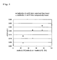
- Fig. 1 The horizontal axis indicates the ratios of antibodies A to antibodies B, and the vertical axis indicates changes in OD values at 0.25 minutes and 1.93 minutes after the initiation of the reaction in terms of the changes per minute, ⁇ OD/min.
- a higher ⁇ OD/min. value indicates a stronger agglutination reaction.
- an agglutination reaction was stronger when antibodies were mixed in advance and then bound to latex, as compared with when antibodies were independently bound to latex and the latexes were mixed at the time of reaction. This demonstrates that a higher sensitivity can be realized when antibodies are mixed in advance.
- using an anti-CRP polyclonal antibody-bound latex reagent which was prepared for the purpose of comparison the same procedure was performed. As a result, weaker agglutination reaction was observed as compared with the case of the latex reagent which was prepared by mixing monoclonal antibodies at a given ratio. Therefore, it was shown that a mixture of several types of monoclonal antibodies which recognize each specific epitome is more superior than a polyclonal antibody which is also a mixture.
- TSH thyroid stimulating hormone
- anti-TSH monoclonal antibody Four types of the commercially available anti-TSH monoclonal antibody were purchased.
- the purchased anti-TSH monoclonal antibodies are anti-TSH monoclonal antibodies which are manufactured by Medix (Finland), and four types of Clone Numbers 5401, 5405, 5407 and 5409 were used.
- the clone numbers 5405 and 5407 recognize the same epitope of TSH
- 5401, 5405/5407 and 5409 recognize the different epitope of TSH respectively.
- anti-TSH antibodies were bound to a latex particle using the same method as that of Example 1.
- polystyrene latex particles particles (particle diameter: 0.5 ⁇ m, JSR) were diluted with glycine buffer (pH 9.6) to bring a solid concentration to 3%.
- glycine buffer pH 9.6
- the anti-TSH antibodies 5401, 5405, 5407 and 5409 were adjusted at a concentration of approximately 2 mg/ml. The equivalent amounts of the mixture were added to the latex dispersion, the resulting mixture was agitated, and the reaction was allowed to proceed at room temperature for 1 hour.
- Centrifugation was carried out at 11,000 rpm for 15 minutes, the supernatant was removed, and 0.2% BSA in glycine buffer (pH 9.6, containing 0.001% of Triton X-100) was added to the precipitate as a blocking agent to bring the solid latex concentration to 1.5%. After the resultant was agitated at room temperature for 1 hour, centrifugation was carried out at 11,000 rpm for 15 minutes, and the supernatant was removed. Glycine buffer (pH 9.6) was added to the precipitate for redispersion, centrifugation was carried out again at 11,000 rpm for 15 minutes, and the supernatant was removed.
- glycine buffer pH 9.6
- Glycine buffer pH 9.6 was added to the precipitate for redispersion to a final solid latex concentration of 3.0% therein, and the resultant was designated as an anti-TSH antibody-bound latex reagent.
- three types of anti-TSH monoclonal antibodies were bound to latex individually, and the latexes were mixed at an equal ratio (1:1:1), and separately three types of anti-TSH monoclonal antibodies were mixed in advance at an equal ratio (1:1:1) and the resulting mixture was allowed to bind to latex.
- TSH solution A standard TSH solution was diluted with buffer to bring the TSH concentration to 5 ⁇ g/dl, and the resultant was designated as a TSH solution (analyte).
- the anti-TSH antibody-bound latex was allowed to react with an analyte at 37°C, and changes in absorbance at given time points were assayed using a spectrophotometer (Hitachi, Ltd.).
- a spectrophotometer Haitachi, Ltd.
Landscapes
- Health & Medical Sciences (AREA)
- Immunology (AREA)
- Life Sciences & Earth Sciences (AREA)
- Engineering & Computer Science (AREA)
- Molecular Biology (AREA)
- Biomedical Technology (AREA)
- Chemical & Material Sciences (AREA)
- Hematology (AREA)
- Urology & Nephrology (AREA)
- Biotechnology (AREA)
- Microbiology (AREA)
- Cell Biology (AREA)
- Food Science & Technology (AREA)
- Medicinal Chemistry (AREA)
- Physics & Mathematics (AREA)
- Analytical Chemistry (AREA)
- Biochemistry (AREA)
- General Health & Medical Sciences (AREA)
- General Physics & Mathematics (AREA)
- Pathology (AREA)
- Peptides Or Proteins (AREA)
Applications Claiming Priority (1)
| Application Number | Priority Date | Filing Date | Title |
|---|---|---|---|
| JP2007254179 | 2007-09-28 |
Publications (1)
| Publication Number | Publication Date |
|---|---|
| EP2042870A1 true EP2042870A1 (fr) | 2009-04-01 |
Family
ID=39916749
Family Applications (1)
| Application Number | Title | Priority Date | Filing Date |
|---|---|---|---|
| EP08016700A Withdrawn EP2042870A1 (fr) | 2007-09-28 | 2008-09-23 | Procédé de dosage immunologique de haute sensibilité |
Country Status (3)
| Country | Link |
|---|---|
| US (1) | US20090088334A1 (fr) |
| EP (1) | EP2042870A1 (fr) |
| JP (1) | JP2009098138A (fr) |
Cited By (4)
| Publication number | Priority date | Publication date | Assignee | Title |
|---|---|---|---|---|
| CN106680505A (zh) * | 2017-01-04 | 2017-05-17 | 南京鼓楼医院 | 一种基于多株相互配对单克隆抗体的胶乳增强免疫比浊检测方法 |
| CN107533053A (zh) * | 2015-02-25 | 2018-01-02 | 积水医疗株式会社 | L‑fabp的免疫测定方法以及用于所述方法的测定试剂 |
| CN108008130A (zh) * | 2017-11-24 | 2018-05-08 | 海格德生物科技(深圳)有限公司 | 基于胶乳增强免疫比浊法测定Lp-PLA2的试剂盒及其制备方法 |
| CN114206367A (zh) * | 2019-08-09 | 2022-03-18 | 美国西门子医学诊断股份有限公司 | 抗猪tcn1单克隆抗体及其生产和使用方法 |
Families Citing this family (11)
| Publication number | Priority date | Publication date | Assignee | Title |
|---|---|---|---|---|
| CN102628867B (zh) * | 2011-12-30 | 2016-03-23 | 北京九强生物技术股份有限公司 | 双抗体胶乳增强视黄醇结合蛋白检测试剂盒 |
| US10175174B2 (en) | 2012-12-11 | 2019-01-08 | Ge Healthcare Bio-Sciences Corp. | Photoactivated chemical bleaching of dyes |
| US20180113127A1 (en) * | 2015-02-25 | 2018-04-26 | Sekisui Medical Co., Ltd. | Immunoassay method and assay reagent used in said method |
| WO2018141768A1 (fr) * | 2017-02-02 | 2018-08-09 | Roche Diagnostics Gmbh | Dosage immunologique utilisant au moins deux agents de liaison spécifiques à un analyte pegylés |
| JP2018173334A (ja) * | 2017-03-31 | 2018-11-08 | 株式会社シマ研究所 | 免疫学的測定方法及びそれに用いる試薬キット |
| EP3990923A4 (fr) * | 2019-06-28 | 2022-08-31 | Siemens Healthcare Diagnostics, Inc. | Réactifs pour dosages immunologiques en sandwich utilisant une détection d'agglutination améliorée par des particules et leurs procédés de production et d'utilisation |
| JP2023550688A (ja) * | 2020-10-19 | 2023-12-05 | ベクトン・ディキンソン・アンド・カンパニー | SARS-CoV-2抗原側方流動アッセイ検出デバイス及びそれを使用するための方法 |
| JP7858322B2 (ja) * | 2021-02-26 | 2026-05-14 | キヤノン株式会社 | ラテックス凝集反応増感剤、ラテックス凝集試薬、及びそれを用いたラテックス凝集法による検体中の標的物質の測定方法 |
| US20220276232A1 (en) * | 2021-02-26 | 2022-09-01 | Canon Kabushiki Kaisha | Latex agglutination reaction sensitizer, latex agglutination reagent, and method for measuring target substance in specimen by latex agglutination method using the same |
| JP7639420B2 (ja) * | 2021-03-16 | 2025-03-05 | 東ソー株式会社 | パラチロイドホルモンの測定方法 |
| CN113092790B (zh) * | 2021-04-02 | 2023-12-15 | 三诺生物传感股份有限公司 | 一种降钙素原的检测试剂盒及其制备方法 |
Citations (10)
| Publication number | Priority date | Publication date | Assignee | Title |
|---|---|---|---|---|
| JPS6053846A (ja) | 1983-07-30 | 1985-03-27 | ベ−リンガ−・マンハイム・ゲゼルシヤフト・ミツト・ベシユレンクテル・ハフツング | 免疫反応の成分を測定する方法及び試薬 |
| JPH01118770A (ja) | 1987-05-23 | 1989-05-11 | Behringwerke Ag | タンパクの測定方法 |
| JPH02257063A (ja) | 1989-03-29 | 1990-10-17 | Sekisui Chem Co Ltd | 免疫測定試薬および免疫測定法 |
| JPH0580051A (ja) | 1991-03-15 | 1993-03-30 | F Hoffmann La Roche Ag | 凝集イムノアツセイ |
| JPH05297002A (ja) | 1992-04-23 | 1993-11-12 | Iatron Lab Inc | 免疫凝集反応の増感方法 |
| JPH0658935A (ja) | 1992-08-08 | 1994-03-04 | Denka Seiken Co Ltd | 免疫学的測定方法及びその試薬 |
| US5500345A (en) * | 1989-04-25 | 1996-03-19 | Iatron Laboratories, Inc. | Hybridomas producing monoclonal antibodies specific for C-reactive protein and methods for detection of C-reactive protein |
| JPH08187095A (ja) | 1995-01-10 | 1996-07-23 | Toyobo Co Ltd | コレステロールオキシダーゼの安定化法 |
| JPH1090268A (ja) | 1996-09-18 | 1998-04-10 | Eiken Chem Co Ltd | 免疫学的粒子凝集反応方法 |
| JP2000206115A (ja) | 1999-01-11 | 2000-07-28 | Eiken Chem Co Ltd | 抗体断片を用いた免疫凝集反応試薬 |
Family Cites Families (7)
| Publication number | Priority date | Publication date | Assignee | Title |
|---|---|---|---|---|
| NL185309C (nl) * | 1980-07-28 | 1990-03-01 | Akzo Nv | Methode voor het bepalen van antigenen met behulp van twee of meer monoklonale antilichamen alsmede immuunreagens. |
| US4447545A (en) * | 1980-11-03 | 1984-05-08 | New England Deaconess Hospital | Bladder cancer detection |
| US5026653A (en) * | 1985-04-02 | 1991-06-25 | Leeco Diagnostic, Inc. | Scavenger antibody mixture and its use for immunometric assay |
| US5100805A (en) * | 1989-01-26 | 1992-03-31 | Seradyn, Inc. | Quantitative immunoassay system and method for agglutination assays |
| US5888749A (en) * | 1990-12-20 | 1999-03-30 | Iatron Laboratories, Inc. | Anti-human plasmin-α2 -plasmin inhibitor complex antibodies, hybridomas, and immunological determination method |
| US20090047685A1 (en) * | 2005-11-30 | 2009-02-19 | Nihon University | Ultrahighly sensitive determination reagent for c-reactive protein and determination method |
| CA2645125A1 (fr) * | 2006-03-02 | 2007-09-13 | Oncotherapy Science, Inc. | Procedes de diagnostic du cancer du pancreas utilisant la proteine reg4 |
-
2008
- 2008-09-23 EP EP08016700A patent/EP2042870A1/fr not_active Withdrawn
- 2008-09-25 US US12/238,075 patent/US20090088334A1/en not_active Abandoned
- 2008-09-26 JP JP2008248023A patent/JP2009098138A/ja not_active Abandoned
Patent Citations (10)
| Publication number | Priority date | Publication date | Assignee | Title |
|---|---|---|---|---|
| JPS6053846A (ja) | 1983-07-30 | 1985-03-27 | ベ−リンガ−・マンハイム・ゲゼルシヤフト・ミツト・ベシユレンクテル・ハフツング | 免疫反応の成分を測定する方法及び試薬 |
| JPH01118770A (ja) | 1987-05-23 | 1989-05-11 | Behringwerke Ag | タンパクの測定方法 |
| JPH02257063A (ja) | 1989-03-29 | 1990-10-17 | Sekisui Chem Co Ltd | 免疫測定試薬および免疫測定法 |
| US5500345A (en) * | 1989-04-25 | 1996-03-19 | Iatron Laboratories, Inc. | Hybridomas producing monoclonal antibodies specific for C-reactive protein and methods for detection of C-reactive protein |
| JPH0580051A (ja) | 1991-03-15 | 1993-03-30 | F Hoffmann La Roche Ag | 凝集イムノアツセイ |
| JPH05297002A (ja) | 1992-04-23 | 1993-11-12 | Iatron Lab Inc | 免疫凝集反応の増感方法 |
| JPH0658935A (ja) | 1992-08-08 | 1994-03-04 | Denka Seiken Co Ltd | 免疫学的測定方法及びその試薬 |
| JPH08187095A (ja) | 1995-01-10 | 1996-07-23 | Toyobo Co Ltd | コレステロールオキシダーゼの安定化法 |
| JPH1090268A (ja) | 1996-09-18 | 1998-04-10 | Eiken Chem Co Ltd | 免疫学的粒子凝集反応方法 |
| JP2000206115A (ja) | 1999-01-11 | 2000-07-28 | Eiken Chem Co Ltd | 抗体断片を用いた免疫凝集反応試薬 |
Non-Patent Citations (4)
| Title |
|---|
| "Handbook of Experimental Immunology", UNIVERSITY OF NAGOYA PRESS |
| EHRLICH P H ET AL: "MIXING 2 MONO CLONAL ANTIBODIES YIELDS ENHANCED AFFINITY FOR ANTIGEN", JOURNAL OF IMMUNOLOGY, vol. 128, no. 6, 1982, pages 2709 - 2713, XP002504627, ISSN: 0022-1767 * |
| SHAMIM M ET AL: "Production of monoclonal antibodies against Nosema bombycis and their utility for detection of pebrine infection in Bombyx mori L", JOURNAL OF IMMUNOASSAY, vol. 18, no. 4, 1997, pages 357 - 370, XP009108726, ISSN: 0197-1522 * |
| ZENKE G ET AL: "A COCKTAIL OF THREE MONOCLONAL ANTIBODIES SIGNIFICANTLY INCREASES THE SENSITIVITY OF AN ENZYME IMMUNOASSAY FOR HUMAN GRANULOCYTE-MACROPHAGE COLONY-STIMULATING FACTOR", JOURNAL OF IMMUNOASSAY, vol. 12, no. 2, 1991, pages 185 - 206, XP009108801, ISSN: 0197-1522 * |
Cited By (6)
| Publication number | Priority date | Publication date | Assignee | Title |
|---|---|---|---|---|
| CN107533053A (zh) * | 2015-02-25 | 2018-01-02 | 积水医疗株式会社 | L‑fabp的免疫测定方法以及用于所述方法的测定试剂 |
| CN113671193A (zh) * | 2015-02-25 | 2021-11-19 | 积水医疗株式会社 | L-fabp的免疫测定方法以及用于所述方法的测定试剂 |
| CN106680505A (zh) * | 2017-01-04 | 2017-05-17 | 南京鼓楼医院 | 一种基于多株相互配对单克隆抗体的胶乳增强免疫比浊检测方法 |
| CN108008130A (zh) * | 2017-11-24 | 2018-05-08 | 海格德生物科技(深圳)有限公司 | 基于胶乳增强免疫比浊法测定Lp-PLA2的试剂盒及其制备方法 |
| CN114206367A (zh) * | 2019-08-09 | 2022-03-18 | 美国西门子医学诊断股份有限公司 | 抗猪tcn1单克隆抗体及其生产和使用方法 |
| US12509506B2 (en) | 2019-08-09 | 2025-12-30 | Siemens Healthcare Diagnostics Inc. | Anti-Hog TCN1 monoclonal antibodies and methods of production and use thereof |
Also Published As
| Publication number | Publication date |
|---|---|
| JP2009098138A (ja) | 2009-05-07 |
| US20090088334A1 (en) | 2009-04-02 |
Similar Documents
| Publication | Publication Date | Title |
|---|---|---|
| US20090088334A1 (en) | Method of high sensitive immunoassay | |
| CA2244326C (fr) | Essai d'agglutination par diffusion de la lumiere, ameliore par des microparticules; reactifs microparticulaires utiles a cette fin | |
| EP0163312A2 (fr) | Immunoessai | |
| JP2009085753A (ja) | サンドイッチイムノアッセイ法 | |
| JP5137620B2 (ja) | アルカリホスファターゼ標識検出用試薬キット、免疫測定用試薬キット及び免疫測定方法 | |
| EP1978363B1 (fr) | Procédé de dosage immunologique, kit de réactif pour détecter la phosphatase alcaline, et kit réactif pour dosage immunologique | |
| JP3284016B2 (ja) | 非特異的反応抑制剤 | |
| TWI679421B (zh) | 肌酸激酶mb型同功酶的測定方法、肌酸激酶bb型同功酶影響的規避方法以及肌酸激酶mb型同功酶測定用套組 | |
| EP1801590B1 (fr) | Procédé d'analyse d'antigène et réactif correspondant | |
| KR101974230B1 (ko) | Pivka-ii 측정 시약에서의 비특이 반응의 억제 방법 | |
| WO2016136863A1 (fr) | Procédé de dosage immunologique de l-fabp et réactif de dosage utilisé dans ledit procédé | |
| JP2006038823A (ja) | 非特異的反応抑制剤、非特異的反応の抑制方法、免疫学的測定方法及び免疫学的測定試薬 | |
| EP2660603B1 (fr) | Procédé d'essai immunologique | |
| US20090087923A1 (en) | Method of high sensitive immunoassay | |
| EP1347301A1 (fr) | Procede de mesure ultrarapide et ultrasensible | |
| JPH0712818A (ja) | 免疫学的検出方法 | |
| JPH026023B2 (fr) | ||
| JP2003107089A (ja) | 超迅速超高感度測定法 | |
| EP2230516B1 (fr) | Procédé d'immunodosage d'échantillon à l'aide d'une réaction d'agrégation de microparticules et coffret de dosage | |
| JP3328053B2 (ja) | 免疫凝集反応による抗体又は抗原の濃度定量法 | |
| JP4261889B2 (ja) | Hmg−1、hmg−2の測定方法及び測定試薬 | |
| JP4318092B2 (ja) | 測定対象物質の測定試薬及び測定対象物質の測定試薬の安定化方法 | |
| JP2009085702A (ja) | 免疫反応試薬 | |
| CN1178580A (zh) | 均相溶胶-溶胶测定 | |
| JP2012078161A (ja) | 試料中の測定対象物質の測定方法、測定試薬及び測定値差の改善方法 |
Legal Events
| Date | Code | Title | Description |
|---|---|---|---|
| PUAI | Public reference made under article 153(3) epc to a published international application that has entered the european phase |
Free format text: ORIGINAL CODE: 0009012 |
|
| AK | Designated contracting states |
Kind code of ref document: A1 Designated state(s): AT BE BG CH CY CZ DE DK EE ES FI FR GB GR HR HU IE IS IT LI LT LU LV MC MT NL NO PL PT RO SE SI SK TR |
|
| AX | Request for extension of the european patent |
Extension state: AL BA MK RS |
|
| AKX | Designation fees paid | ||
| STAA | Information on the status of an ep patent application or granted ep patent |
Free format text: STATUS: THE APPLICATION IS DEEMED TO BE WITHDRAWN |
|
| 18D | Application deemed to be withdrawn |
Effective date: 20091002 |
|
| REG | Reference to a national code |
Ref country code: DE Ref legal event code: 8566 |