EP3643777A1 - Nouvelle cellule souche musculosquelettique et support pour induire la différenciation de cellules souches musculosquelettiques - Google Patents
Nouvelle cellule souche musculosquelettique et support pour induire la différenciation de cellules souches musculosquelettiques Download PDFInfo
- Publication number
- EP3643777A1 EP3643777A1 EP19170901.3A EP19170901A EP3643777A1 EP 3643777 A1 EP3643777 A1 EP 3643777A1 EP 19170901 A EP19170901 A EP 19170901A EP 3643777 A1 EP3643777 A1 EP 3643777A1
- Authority
- EP
- European Patent Office
- Prior art keywords
- msscs
- cells
- hmsscs
- marker
- positive
- Prior art date
- Legal status (The legal status is an assumption and is not a legal conclusion. Google has not performed a legal analysis and makes no representation as to the accuracy of the status listed.)
- Granted
Links
- 0 CCC(*)N(C)* Chemical compound CCC(*)N(C)* 0.000 description 1
Images


Classifications
-
- A—HUMAN NECESSITIES
- A61—MEDICAL OR VETERINARY SCIENCE; HYGIENE
- A61K—PREPARATIONS FOR MEDICAL, DENTAL OR TOILETRY PURPOSES
- A61K35/00—Medicinal preparations containing materials or reaction products thereof with undetermined constitution
- A61K35/12—Materials from mammals; Compositions comprising non-specified tissues or cells; Compositions comprising non-embryonic stem cells; Genetically modified cells
- A61K35/34—Muscles; Smooth muscle cells; Heart; Cardiac stem cells; Myoblasts; Myocytes; Cardiomyocytes
-
- A—HUMAN NECESSITIES
- A61—MEDICAL OR VETERINARY SCIENCE; HYGIENE
- A61K—PREPARATIONS FOR MEDICAL, DENTAL OR TOILETRY PURPOSES
- A61K35/00—Medicinal preparations containing materials or reaction products thereof with undetermined constitution
- A61K35/12—Materials from mammals; Compositions comprising non-specified tissues or cells; Compositions comprising non-embryonic stem cells; Genetically modified cells
- A61K35/32—Bones; Osteocytes; Osteoblasts; Tendons; Tenocytes; Teeth; Odontoblasts; Cartilage; Chondrocytes; Synovial membrane
-
- A—HUMAN NECESSITIES
- A61—MEDICAL OR VETERINARY SCIENCE; HYGIENE
- A61P—SPECIFIC THERAPEUTIC ACTIVITY OF CHEMICAL COMPOUNDS OR MEDICINAL PREPARATIONS
- A61P19/00—Drugs for skeletal disorders
-
- A—HUMAN NECESSITIES
- A61—MEDICAL OR VETERINARY SCIENCE; HYGIENE
- A61P—SPECIFIC THERAPEUTIC ACTIVITY OF CHEMICAL COMPOUNDS OR MEDICINAL PREPARATIONS
- A61P21/00—Drugs for disorders of the muscular or neuromuscular system
-
- C—CHEMISTRY; METALLURGY
- C12—BIOCHEMISTRY; BEER; SPIRITS; WINE; VINEGAR; MICROBIOLOGY; ENZYMOLOGY; MUTATION OR GENETIC ENGINEERING
- C12N—MICROORGANISMS OR ENZYMES; COMPOSITIONS THEREOF; PROPAGATING, PRESERVING, OR MAINTAINING MICROORGANISMS; MUTATION OR GENETIC ENGINEERING; CULTURE MEDIA
- C12N5/00—Undifferentiated human, animal or plant cells, e.g. cell lines; Tissues; Cultivation or maintenance thereof; Culture media therefor
- C12N5/0018—Culture media for cell or tissue culture
-
- C—CHEMISTRY; METALLURGY
- C12—BIOCHEMISTRY; BEER; SPIRITS; WINE; VINEGAR; MICROBIOLOGY; ENZYMOLOGY; MUTATION OR GENETIC ENGINEERING
- C12N—MICROORGANISMS OR ENZYMES; COMPOSITIONS THEREOF; PROPAGATING, PRESERVING, OR MAINTAINING MICROORGANISMS; MUTATION OR GENETIC ENGINEERING; CULTURE MEDIA
- C12N5/00—Undifferentiated human, animal or plant cells, e.g. cell lines; Tissues; Cultivation or maintenance thereof; Culture media therefor
- C12N5/06—Animal cells or tissues; Human cells or tissues
- C12N5/0602—Vertebrate cells
- C12N5/0603—Embryonic cells ; Embryoid bodies
- C12N5/0606—Pluripotent embryonic cells, e.g. embryonic stem cells [ES]
-
- C—CHEMISTRY; METALLURGY
- C12—BIOCHEMISTRY; BEER; SPIRITS; WINE; VINEGAR; MICROBIOLOGY; ENZYMOLOGY; MUTATION OR GENETIC ENGINEERING
- C12N—MICROORGANISMS OR ENZYMES; COMPOSITIONS THEREOF; PROPAGATING, PRESERVING, OR MAINTAINING MICROORGANISMS; MUTATION OR GENETIC ENGINEERING; CULTURE MEDIA
- C12N5/00—Undifferentiated human, animal or plant cells, e.g. cell lines; Tissues; Cultivation or maintenance thereof; Culture media therefor
- C12N5/06—Animal cells or tissues; Human cells or tissues
- C12N5/0602—Vertebrate cells
- C12N5/0652—Cells of skeletal and connective tissues; Mesenchyme
- C12N5/0653—Adipocytes; Adipose tissue
-
- C—CHEMISTRY; METALLURGY
- C12—BIOCHEMISTRY; BEER; SPIRITS; WINE; VINEGAR; MICROBIOLOGY; ENZYMOLOGY; MUTATION OR GENETIC ENGINEERING
- C12N—MICROORGANISMS OR ENZYMES; COMPOSITIONS THEREOF; PROPAGATING, PRESERVING, OR MAINTAINING MICROORGANISMS; MUTATION OR GENETIC ENGINEERING; CULTURE MEDIA
- C12N5/00—Undifferentiated human, animal or plant cells, e.g. cell lines; Tissues; Cultivation or maintenance thereof; Culture media therefor
- C12N5/06—Animal cells or tissues; Human cells or tissues
- C12N5/0602—Vertebrate cells
- C12N5/0652—Cells of skeletal and connective tissues; Mesenchyme
- C12N5/0654—Osteocytes, Osteoblasts, Odontocytes; Bones, Teeth
-
- C—CHEMISTRY; METALLURGY
- C12—BIOCHEMISTRY; BEER; SPIRITS; WINE; VINEGAR; MICROBIOLOGY; ENZYMOLOGY; MUTATION OR GENETIC ENGINEERING
- C12N—MICROORGANISMS OR ENZYMES; COMPOSITIONS THEREOF; PROPAGATING, PRESERVING, OR MAINTAINING MICROORGANISMS; MUTATION OR GENETIC ENGINEERING; CULTURE MEDIA
- C12N5/00—Undifferentiated human, animal or plant cells, e.g. cell lines; Tissues; Cultivation or maintenance thereof; Culture media therefor
- C12N5/06—Animal cells or tissues; Human cells or tissues
- C12N5/0602—Vertebrate cells
- C12N5/0652—Cells of skeletal and connective tissues; Mesenchyme
- C12N5/0655—Chondrocytes; Cartilage
-
- C—CHEMISTRY; METALLURGY
- C12—BIOCHEMISTRY; BEER; SPIRITS; WINE; VINEGAR; MICROBIOLOGY; ENZYMOLOGY; MUTATION OR GENETIC ENGINEERING
- C12N—MICROORGANISMS OR ENZYMES; COMPOSITIONS THEREOF; PROPAGATING, PRESERVING, OR MAINTAINING MICROORGANISMS; MUTATION OR GENETIC ENGINEERING; CULTURE MEDIA
- C12N5/00—Undifferentiated human, animal or plant cells, e.g. cell lines; Tissues; Cultivation or maintenance thereof; Culture media therefor
- C12N5/06—Animal cells or tissues; Human cells or tissues
- C12N5/0602—Vertebrate cells
- C12N5/0652—Cells of skeletal and connective tissues; Mesenchyme
- C12N5/0658—Skeletal muscle cells, e.g. myocytes, myotubes, myoblasts
-
- C—CHEMISTRY; METALLURGY
- C12—BIOCHEMISTRY; BEER; SPIRITS; WINE; VINEGAR; MICROBIOLOGY; ENZYMOLOGY; MUTATION OR GENETIC ENGINEERING
- C12N—MICROORGANISMS OR ENZYMES; COMPOSITIONS THEREOF; PROPAGATING, PRESERVING, OR MAINTAINING MICROORGANISMS; MUTATION OR GENETIC ENGINEERING; CULTURE MEDIA
- C12N5/00—Undifferentiated human, animal or plant cells, e.g. cell lines; Tissues; Cultivation or maintenance thereof; Culture media therefor
- C12N5/06—Animal cells or tissues; Human cells or tissues
- C12N5/0602—Vertebrate cells
- C12N5/0652—Cells of skeletal and connective tissues; Mesenchyme
- C12N5/066—Tenocytes; Tendons, Ligaments
-
- C—CHEMISTRY; METALLURGY
- C12—BIOCHEMISTRY; BEER; SPIRITS; WINE; VINEGAR; MICROBIOLOGY; ENZYMOLOGY; MUTATION OR GENETIC ENGINEERING
- C12N—MICROORGANISMS OR ENZYMES; COMPOSITIONS THEREOF; PROPAGATING, PRESERVING, OR MAINTAINING MICROORGANISMS; MUTATION OR GENETIC ENGINEERING; CULTURE MEDIA
- C12N5/00—Undifferentiated human, animal or plant cells, e.g. cell lines; Tissues; Cultivation or maintenance thereof; Culture media therefor
- C12N5/06—Animal cells or tissues; Human cells or tissues
- C12N5/0602—Vertebrate cells
- C12N5/0652—Cells of skeletal and connective tissues; Mesenchyme
- C12N5/0662—Stem cells
-
- C—CHEMISTRY; METALLURGY
- C12—BIOCHEMISTRY; BEER; SPIRITS; WINE; VINEGAR; MICROBIOLOGY; ENZYMOLOGY; MUTATION OR GENETIC ENGINEERING
- C12N—MICROORGANISMS OR ENZYMES; COMPOSITIONS THEREOF; PROPAGATING, PRESERVING, OR MAINTAINING MICROORGANISMS; MUTATION OR GENETIC ENGINEERING; CULTURE MEDIA
- C12N5/00—Undifferentiated human, animal or plant cells, e.g. cell lines; Tissues; Cultivation or maintenance thereof; Culture media therefor
- C12N5/06—Animal cells or tissues; Human cells or tissues
- C12N5/0602—Vertebrate cells
- C12N5/0696—Artificially induced pluripotent stem cells, e.g. iPS
-
- G—PHYSICS
- G01—MEASURING; TESTING
- G01N—INVESTIGATING OR ANALYSING MATERIALS BY DETERMINING THEIR CHEMICAL OR PHYSICAL PROPERTIES
- G01N33/00—Investigating or analysing materials by specific methods not covered by groups G01N1/00 - G01N31/00
- G01N33/48—Biological material, e.g. blood, urine; Haemocytometers
- G01N33/50—Chemical analysis of biological material, e.g. blood, urine; Testing involving biospecific ligand binding methods; Immunological testing
- G01N33/53—Immunoassay; Biospecific binding assay; Materials therefor
- G01N33/569—Immunoassay; Biospecific binding assay; Materials therefor for microorganisms, e.g. protozoa, bacteria, viruses
- G01N33/56966—Animal cells
-
- C—CHEMISTRY; METALLURGY
- C12—BIOCHEMISTRY; BEER; SPIRITS; WINE; VINEGAR; MICROBIOLOGY; ENZYMOLOGY; MUTATION OR GENETIC ENGINEERING
- C12N—MICROORGANISMS OR ENZYMES; COMPOSITIONS THEREOF; PROPAGATING, PRESERVING, OR MAINTAINING MICROORGANISMS; MUTATION OR GENETIC ENGINEERING; CULTURE MEDIA
- C12N2501/00—Active agents used in cell culture processes, e.g. differentation
- C12N2501/10—Growth factors
- C12N2501/115—Basic fibroblast growth factor (bFGF, FGF-2)
-
- C—CHEMISTRY; METALLURGY
- C12—BIOCHEMISTRY; BEER; SPIRITS; WINE; VINEGAR; MICROBIOLOGY; ENZYMOLOGY; MUTATION OR GENETIC ENGINEERING
- C12N—MICROORGANISMS OR ENZYMES; COMPOSITIONS THEREOF; PROPAGATING, PRESERVING, OR MAINTAINING MICROORGANISMS; MUTATION OR GENETIC ENGINEERING; CULTURE MEDIA
- C12N2501/00—Active agents used in cell culture processes, e.g. differentation
- C12N2501/10—Growth factors
- C12N2501/15—Transforming growth factor beta (TGF-β)
-
- C—CHEMISTRY; METALLURGY
- C12—BIOCHEMISTRY; BEER; SPIRITS; WINE; VINEGAR; MICROBIOLOGY; ENZYMOLOGY; MUTATION OR GENETIC ENGINEERING
- C12N—MICROORGANISMS OR ENZYMES; COMPOSITIONS THEREOF; PROPAGATING, PRESERVING, OR MAINTAINING MICROORGANISMS; MUTATION OR GENETIC ENGINEERING; CULTURE MEDIA
- C12N2501/00—Active agents used in cell culture processes, e.g. differentation
- C12N2501/10—Growth factors
- C12N2501/155—Bone morphogenic proteins [BMP]; Osteogenins; Osteogenic factor; Bone inducing factor
-
- C—CHEMISTRY; METALLURGY
- C12—BIOCHEMISTRY; BEER; SPIRITS; WINE; VINEGAR; MICROBIOLOGY; ENZYMOLOGY; MUTATION OR GENETIC ENGINEERING
- C12N—MICROORGANISMS OR ENZYMES; COMPOSITIONS THEREOF; PROPAGATING, PRESERVING, OR MAINTAINING MICROORGANISMS; MUTATION OR GENETIC ENGINEERING; CULTURE MEDIA
- C12N2501/00—Active agents used in cell culture processes, e.g. differentation
- C12N2501/10—Growth factors
- C12N2501/16—Activin; Inhibin; Mullerian inhibiting substance
-
- C—CHEMISTRY; METALLURGY
- C12—BIOCHEMISTRY; BEER; SPIRITS; WINE; VINEGAR; MICROBIOLOGY; ENZYMOLOGY; MUTATION OR GENETIC ENGINEERING
- C12N—MICROORGANISMS OR ENZYMES; COMPOSITIONS THEREOF; PROPAGATING, PRESERVING, OR MAINTAINING MICROORGANISMS; MUTATION OR GENETIC ENGINEERING; CULTURE MEDIA
- C12N2501/00—Active agents used in cell culture processes, e.g. differentation
- C12N2501/20—Cytokines; Chemokines
- C12N2501/23—Interleukins [IL]
- C12N2501/235—Leukemia inhibitory factor [LIF]
-
- C—CHEMISTRY; METALLURGY
- C12—BIOCHEMISTRY; BEER; SPIRITS; WINE; VINEGAR; MICROBIOLOGY; ENZYMOLOGY; MUTATION OR GENETIC ENGINEERING
- C12N—MICROORGANISMS OR ENZYMES; COMPOSITIONS THEREOF; PROPAGATING, PRESERVING, OR MAINTAINING MICROORGANISMS; MUTATION OR GENETIC ENGINEERING; CULTURE MEDIA
- C12N2501/00—Active agents used in cell culture processes, e.g. differentation
- C12N2501/40—Regulators of development
- C12N2501/415—Wnt; Frizzeled
-
- C—CHEMISTRY; METALLURGY
- C12—BIOCHEMISTRY; BEER; SPIRITS; WINE; VINEGAR; MICROBIOLOGY; ENZYMOLOGY; MUTATION OR GENETIC ENGINEERING
- C12N—MICROORGANISMS OR ENZYMES; COMPOSITIONS THEREOF; PROPAGATING, PRESERVING, OR MAINTAINING MICROORGANISMS; MUTATION OR GENETIC ENGINEERING; CULTURE MEDIA
- C12N2501/00—Active agents used in cell culture processes, e.g. differentation
- C12N2501/70—Enzymes
- C12N2501/72—Transferases [EC 2.]
- C12N2501/727—Kinases (EC 2.7.)
-
- C—CHEMISTRY; METALLURGY
- C12—BIOCHEMISTRY; BEER; SPIRITS; WINE; VINEGAR; MICROBIOLOGY; ENZYMOLOGY; MUTATION OR GENETIC ENGINEERING
- C12N—MICROORGANISMS OR ENZYMES; COMPOSITIONS THEREOF; PROPAGATING, PRESERVING, OR MAINTAINING MICROORGANISMS; MUTATION OR GENETIC ENGINEERING; CULTURE MEDIA
- C12N2501/00—Active agents used in cell culture processes, e.g. differentation
- C12N2501/998—Proteins not provided for elsewhere
-
- C—CHEMISTRY; METALLURGY
- C12—BIOCHEMISTRY; BEER; SPIRITS; WINE; VINEGAR; MICROBIOLOGY; ENZYMOLOGY; MUTATION OR GENETIC ENGINEERING
- C12N—MICROORGANISMS OR ENZYMES; COMPOSITIONS THEREOF; PROPAGATING, PRESERVING, OR MAINTAINING MICROORGANISMS; MUTATION OR GENETIC ENGINEERING; CULTURE MEDIA
- C12N2501/00—Active agents used in cell culture processes, e.g. differentation
- C12N2501/999—Small molecules not provided for elsewhere
-
- C—CHEMISTRY; METALLURGY
- C12—BIOCHEMISTRY; BEER; SPIRITS; WINE; VINEGAR; MICROBIOLOGY; ENZYMOLOGY; MUTATION OR GENETIC ENGINEERING
- C12N—MICROORGANISMS OR ENZYMES; COMPOSITIONS THEREOF; PROPAGATING, PRESERVING, OR MAINTAINING MICROORGANISMS; MUTATION OR GENETIC ENGINEERING; CULTURE MEDIA
- C12N2506/00—Differentiation of animal cells from one lineage to another; Differentiation of pluripotent cells
- C12N2506/02—Differentiation of animal cells from one lineage to another; Differentiation of pluripotent cells from embryonic cells
-
- C—CHEMISTRY; METALLURGY
- C12—BIOCHEMISTRY; BEER; SPIRITS; WINE; VINEGAR; MICROBIOLOGY; ENZYMOLOGY; MUTATION OR GENETIC ENGINEERING
- C12N—MICROORGANISMS OR ENZYMES; COMPOSITIONS THEREOF; PROPAGATING, PRESERVING, OR MAINTAINING MICROORGANISMS; MUTATION OR GENETIC ENGINEERING; CULTURE MEDIA
- C12N2506/00—Differentiation of animal cells from one lineage to another; Differentiation of pluripotent cells
- C12N2506/13—Differentiation of animal cells from one lineage to another; Differentiation of pluripotent cells from connective tissue cells, from mesenchymal cells
-
- C—CHEMISTRY; METALLURGY
- C12—BIOCHEMISTRY; BEER; SPIRITS; WINE; VINEGAR; MICROBIOLOGY; ENZYMOLOGY; MUTATION OR GENETIC ENGINEERING
- C12N—MICROORGANISMS OR ENZYMES; COMPOSITIONS THEREOF; PROPAGATING, PRESERVING, OR MAINTAINING MICROORGANISMS; MUTATION OR GENETIC ENGINEERING; CULTURE MEDIA
- C12N2506/00—Differentiation of animal cells from one lineage to another; Differentiation of pluripotent cells
- C12N2506/45—Differentiation of animal cells from one lineage to another; Differentiation of pluripotent cells from artificially induced pluripotent stem cells
-
- C—CHEMISTRY; METALLURGY
- C12—BIOCHEMISTRY; BEER; SPIRITS; WINE; VINEGAR; MICROBIOLOGY; ENZYMOLOGY; MUTATION OR GENETIC ENGINEERING
- C12N—MICROORGANISMS OR ENZYMES; COMPOSITIONS THEREOF; PROPAGATING, PRESERVING, OR MAINTAINING MICROORGANISMS; MUTATION OR GENETIC ENGINEERING; CULTURE MEDIA
- C12N2533/00—Supports or coatings for cell culture, characterised by material
- C12N2533/50—Proteins
- C12N2533/52—Fibronectin; Laminin
-
- C—CHEMISTRY; METALLURGY
- C12—BIOCHEMISTRY; BEER; SPIRITS; WINE; VINEGAR; MICROBIOLOGY; ENZYMOLOGY; MUTATION OR GENETIC ENGINEERING
- C12N—MICROORGANISMS OR ENZYMES; COMPOSITIONS THEREOF; PROPAGATING, PRESERVING, OR MAINTAINING MICROORGANISMS; MUTATION OR GENETIC ENGINEERING; CULTURE MEDIA
- C12N2533/00—Supports or coatings for cell culture, characterised by material
- C12N2533/50—Proteins
- C12N2533/54—Collagen; Gelatin
Definitions
- the present disclosure relates to stem cells and more specifically relates to musculoskeletal stem cells capable of differentiating into musculoskeletal tissues.
- the disease of the musculoskeletal system made up of muscles, bones, joints, etc. causes severe activity limitation, body pain, etc.
- the degeneration of the functions of muscles, bones and joints with aging is an unavoidable consequence.
- the diseases occurring frequently as a result of the degeneration of the function of the musculoskeletal system include degenerative arthritis, tendinitis, bone fracture, sprain, sarcopenia, etc.
- As life expectancy increases recently due to improvement in health care the number of patients suffering from musculoskeletal diseases is also increasing. However, quality of life is aggravated because healthy aging with healthy musculoskeletal system is not achieved.
- Ossification is the process of bone formation. There are two processes of bone formation: intramembranous ossification and endochondral ossification. Intramembranous ossification is the direct conversion of mesenchymal tissue into bone and occurs inside the skull, while endochondral ossification involves the formation of cartilage tissue from aggregated mesenchymal cells followed by conversion of the cartilage tissue into bone. This ossification process is essential mostly in the bone formation of vertebrates.
- hESCs Human embryonic stem cells
- hESCs Human embryonic stem cells
- hESCs Human embryonic stem cells
- hESCs are pluripotent cells that can grow without limitation and can differentiate into any cell type.
- hESCs are useful tools for the study of embryonic development in cellular level and for the cell replacement therapy.
- hESCs can differentiate into specific tissues including skeletal tissues such as bone and cartilage and, therefore, may be used for the restoration of skeletal tissues.
- hiPSCs Human-induced pluripotent stem cells
- hiPSCs are known as pluripotent stem cells that can differentiate into any type of cells.
- hiPSCs are useful for the study of embryonic development in cellular level and are drawing attentions as cell therapeutic agents. Because these cells can be differentiated into skeletal tissue, e.g., bone or cartilage, through transplantation, they may be usefully used for the restoration and treatment of damaged skeletal tissue.
- MSCs Mesenchymal stem cells
- mesenchymal stem cells indirectly facilitate the regeneration of damaged tissues by stimulating intrinsic stem cells by secreting various biofactors, rather than participating directly in the differentiation into mesenchymal tissues including the musculoskeletal tissues ( Stem Cells Transl Med. 6(6):1445-1451, 2017 ).
- compositions comprising noggin, LIF (leukemia inhibitory factor), bFGF (basic fibroblast growth factor), Wnt signaling activator, ERK (extracellular signal-regulated kinase) signaling inhibitor and TGF- ⁇ /activin/nodal signaling inhibitor.
- the composition may further comprise musculoskeletal stem cells (MSSCs).
- composition may further comprise at least one of embryonic stem cells (ESCs) and induced pluripotent stem cells (iPSCs).
- composition may further comprise musculoskeletal stem cells (MSSCs).
- ESCs embryonic stem cells
- iPSCs induced pluripotent stem cells
- MSSCs musculoskeletal stem cells
- the Wnt signaling activator may comprise at least one selected from the group consisting of SB216763 (3-(2,4-dichlorophenyl)-4-(1-methyl-1H-indol-3-yl)-1H-pyrrole-2,5-dione), SB415286 (3-[(3-chloro-4-hydroxyphenyl)amino]-4-(2-nitrophenyl)-1H-pyrrole-2,5-dione), kenpaullone (9-bromo-7,12-dihydro-indolo[3,2-d]-[1]benzazepin-6(5H)-one), CHIR99021 (9-bromo-7,12-dihydro-pyrido[3',2':2,3]azepino[4,5-b]indol-6(5H)-one), CP21R7 (3-(3-amino-phenyl)-4-(1-methyl-1H-indol-3, SB415286 (3
- the ERK signaling inhibitor may comprise at least one selected from the group consisting of AS703026 (N-[(2S)-2,3-dihydroxypropyl]-3-[(2-fluoro-4-iodophenyl)amino]-isonicotinamide), AZD6244 (6-(4-bromo-2-chloroanilino)-7-fluoro-N-(2-hydroxyethoxy)-3-methylbenzimidazole-5-carbo xamide), PD0325901 (N-[(2R)-2,3-dihydroxypropoxy]-3,4-difluoro-2-[(2-fluoro-4-iodophenyl)amino]-benzamide), ARRY-438162 (5-[(4-bromo-2-fluorophenyl)amino]-4-fluoro-N-(2-hydroxyethoxy)-1-methyl-1H-benzimida zole-6-carboxamide),
- the TGF- ⁇ /activin/nodal signaling inhibitor may comprise at least one selected from the group consisting of E-616452 (2-[3-(6-methyl-2-pyridinyl)-1H-pyrazol-4-yl]-1,5-naphthyridine), A-83-01 (3-(6-methyl-2-pyridinyl)-N-phenyl-4-(4-quinolinyl)-1H-pyrazole-1-carbothioamide), and SB431542 (4-[4-(1,3-benzodioxol-5-yl)-5-(2-pyridinyl)-1H-imidazol-2-yl]benzamide).
- MSSCs musculoskeletal stem cells
- ESC epidermal stem cell
- iPS induced pluripotent stem cell
- culturing may be performed for at least 5 passes without changing ingredients of the composition.
- culturing may be performed for at least 5 passes while maintaining ingredients of noggin, LIF (leukemia inhibitory factor), bFGF (basic fibroblast growth factor), Wnt signaling activator, ERK (extracellular signal-regulated kinase) signaling inhibitor and TGF- ⁇ /activin/nodal signaling inhibitor of the composition.
- culturing may be performed for at least 5 passes while maintaining concentrations of the ingredients.
- MSSCs musculoskeletal stem cells having the following characteristics: positive for the ectodermal marker nestin (NES); positive for the myogenic satellite marker Pax7; positive for the mesodermal marker ⁇ -SMA; negative for the pluripotency marker LIN28; and negative for the mesenchymal stem cell marker CD90.
- the MSSCs may further have a characteristic of positive for CD146.
- the cell composition may further comprise a pharmaceutically acceptable carrier.
- at least part of the MSSCs may be suspended in the pharmaceutically acceptable carrier.
- the cell composition may further comprise a culture medium for culturing mesenchymal stem cells.
- the MSSCs may further have at least one of the following characteristics: positive for the pluripotency marker DPPA4; negative for the mesodermal markers T and nodal; positive for the neuroectodermal marker Pax6; positive for the intestinal stem cell marker LGR5; negative for the chondrocyte marker SOX9; negative for the myoblast marker MyoD; positive for CD10; positive for CD44; positive for CD105; positive for CD146; and positive for CD166.
- the pluripotency marker DPPA4 negative for the mesodermal markers T and nodal
- positive for the neuroectodermal marker Pax6 positive for the intestinal stem cell marker LGR5
- negative for the chondrocyte marker SOX9 negative for the myoblast marker MyoD
- positive for CD10 positive for CD44; positive for CD105; positive for CD146; and positive for CD166.
- the MSSCs may further have a characteristic of negative for CD271.
- the MSSCs may be configured to differentiate into mesoderm but not into ectoderm or endoderm.
- the MSSCs may be configured to differentiate into muscle, bone, cartilage, tendon or ligament.
- the MSSCs may not be configured to differentiate into a nerve cell.
- the MSSCs may not be configured to differentiate into an endothelial cell.
- the MSSCs may comprise a cell deposited under the accession number KCLRF-BP-00460.
- the cell composition may further comprise cells differentiated from at least part of the MSSCs, where the differentiated cells comprise cells of muscle, bone, cartilage, tendon or ligament.
- Another aspect of the invention provides a method of in vitro differentiation of MSSCs.
- the method comprises: in vitro culturing, in a culture medium, MSSCs of the cell of one of the foregoing cell compositions; and collecting cells differentiated from at least part of the MSSCs.
- the culture medium may comprise an osteogenic differentiation medium, wherein the collected cells comprise osteoblasts.
- the culture medium may comprise an adipogenic differentiation medium, wherein the collected cells comprise adipocytes.
- the culture medium may comprise a chondrogenic differentiation medium, wherein the collected cells comprise cartilage cells.
- Another aspect of the invention provides a method of preparing musculoskeletal cells.
- the method comprises: in vitro culturing, in a culture medium, MSSCs of the cell of one of the foregoing cell compositions; and transplanting at least part of the MSSCs into a mass of tissues such that the at least part of the MSSCs differentiate into musculoskeletal cells in the mass of tissues.
- the musculoskeletal may comprise at least one of muscle, bone, cartilage, tendon or ligament.
- Still another aspect of the invention provides a method of treating a musculoskeletal disease.
- the method comprises: administering, to a subject in need of such treatment, an effective amount of one of the cell compositions, wherein the musculoskeletal disease may be selected from the group consisting of osteoporosis, osteomalacia, osteogenesis imperfecta, osteopetrosis, osteosclerosis, Paget's disease, bone cancer, arthritis, rickets, fracture, periodontal disease, segmental bone defect, osteolytic bone disease, primary and secondary hyperparathyroidism, hyperostosis, degenerative arthritis, degenerative knee joint disease, degenerative hip joint disease, degenerative foot joint disease, degenerative hand joint disease, degenerative shoulder joint disease, degenerative elbow joint disease, chondromalacia patellae, simple knee arthritis, osteochondritis dissecans, lateral epicondylitis, medial epicondylitis, Heberden's nodes, Bouchard's nodes, degenerative thumb CM arthrosis, meniscal injury,
- Still another aspect of the invention provides a method for screening musculoskeletal stem cells (MSSCs).
- the method comprises: providing cells for screening; and subjecting the cells to a plurality of predetermined tests to determine whether the cells have one or more of the following characteristics: positive for the ectodermal marker nestin (NES), positive for the myogenic satellite marker Pax7, positive for the mesodermal marker ⁇ -SMA, negative for the pluripotency marker LIN28, and negative for the mesenchymal stem cell marker CD90.
- the cells are determined to be MSSCs when the cells have all of the foregoing characteristics listed in this paragraph.
- the method for screening may further comprise determining that the cells are MSSCs when the cells have all of the characteristics.
- the plurality of predetermined tests are to determine whether the cells have one or more of the following characteristics: positive for the pluripotency marker DPPA4; negative for the mesodermal markers T and nodal; positive for the neuroectodermal marker Pax6; positive for the intestinal stem cell marker LGR5; negative for the chondrocyte marker SOX9; negative for the myoblast marker MyoD; positive for CD10; positive for CD44; positive for CD105; positive for CD146; and positive for CD166.
- the method may further comprise determining that the cells are MSSCs when the cells have all of the foregoing characteristics listed in this paragraph.
- the method may further comprise determining that the cells are MSSCs when the cells have all of the foregoing characteristics listed in this and immediately preceding paragraphs.
- stem cell refers to an undifferentiated cell capable of differentiating into various body tissues.
- the stem cells may be classified into totipotent stem cells, pluripotent stem cells, multipotent stem cells, etc.
- the term stem cell may be used interchangeably with the terms precursor cell, progenitor cell, etc.
- the stem cell may be an embryonic stem cell (ESC), an induced pluripotent stem cell (iPSC) or a mesenchymal stem cell (MSC).
- ESC embryonic stem cell
- iPSC induced pluripotent stem cell
- MSC mesenchymal stem cell
- the embryonic stem cell is a pluripotent cell derived from all the three germ layers, having capacity for unlimited proliferation without transformation and self-renewal, although not being limited thereto.
- the embryonic stem cell or induced pluripotent stem cell used in the present disclosure is derived from human, cow, horse, goat, sheep, dog, cat, mouse, rat, bird, etc., specifically from human.
- musculoskeletal stem cell refers to a cell that can differentiate into tissues of bone, cartilage, tendon, ligament or muscle without limitation.
- differentiation refers to the process wherein the structure or function of a cell is specialized while the cell grows through division and proliferation, i.e., the process wherein the form or function changes to perform the task assigned to the cell, tissue, etc. of an organism. In general, it refers to a phenomenon where a relatively simple system is split into two or more qualitatively different subsystems. That is to say, the differentiation refers to the variation of the parts of an initially substantially homogenous biological system or the division to qualitatively different parts or subsystems as a result thereof, for example, the division of head, body, etc. from an initially homogeneous egg during ontogeny.
- a musculoskeletal stem cell is differentiated from an embryonic stem cell (ESC) or an induced pluripotent stem cell (iPSC).
- the musculoskeletal stem cell can further differentiate into bone through endochondral ossification as well as into musculoskeletal tissues such as cartilage, tendon, muscle, ligament, bone, etc.
- the present disclosure provides a medium that induces an embryonic stem cell (ESC) to differentiate into a musculoskeletal stem cell (MSSC).
- the medium induces a human embryonic stem cell to differentiate into a musculoskeletal stem cell (MSSC).
- the medium contains noggin, leukemia inhibitory factor (LIF), basic fibroblast growth factor (bFGF), Wnt signaling activator, extracellular signal-regulated kinase (ERK) signaling inhibitor and TGF- ⁇ /activin/nodal signaling inhibitor.
- the medium contains all six (6) ingredients of noggin, LIF, bFGF, Wnt signaling activator, ERK signaling inhibitor and TGF- ⁇ /activin/nodal signaling inhibitor. In other examples, at least one of the six ingredients is absent in the medium.
- the present disclosure provides a medium that induces an induced pluripotent stem to differentiate into a musculoskeletal stem cell (MSSC).
- the medium induces a human-derived pluripotent stem cell to differentiate into a musculoskeletal stem cell (MSSC).
- the medium contains noggin, leukemia inhibitory factor (LIF), basic fibroblast growth factor (bFGF), Wnt signaling activator, extracellular signal-regulated kinase (ERK) signaling inhibitor and TGF- ⁇ /activin/nodal signaling inhibitor.
- the medium contains the six (6) ingredients of noggin, LIF, bFGF, Wnt signaling activator, ERK signaling inhibitor and TGF- ⁇ /activin/nodal signaling inhibitor. In other examples, at least one of the six ingredients is absent in the medium.
- the medium may contain noggin in an amount of about 25, 50, 75, 100, 125, 150, 175, 200, 225, 250, 275, 300, 325, 350, 375, 400, 425, 450, 475, 500, 525, 550, 575, 600, 625, 650, 675, 700, 725, 750, 775, 800, 825, 850, 875, 900, 925, 950, 975, 1000, 1025, 1050, 1075, 1100, 1150 1200, 1250, 1300, 1350, 1400, 1450, 1500, 1550, 1600, 1650, 1700, 1750, 1800, 1850, 1900, 1950, 2000, 2050, 2100, 2150, 2200, 2250, 2300, 2350, 2400, 2450 or 2500 ng/ml.
- the amount of noggin may be within a range formed by selecting any two numbers (two concentration values) listed in the immediately previous sentence, e.g., between about 150 and about 300
- the medium may contain noggin in an amount of about 25, 50, 75, 100, 125, 150, 175, 200, 225, 250, 275, 300, 325, 350, 375, 400, 425, 450, 475, 500, 525, 550, 575, 600, 625, 650, 675, 700, 725, 750, 775, 800, 825, 850, 875, 900, 925, 950, 975, 1000, 1025, 1050, 1075, 1100, 1150 1200, 1250, 1300, 1350, 1400, 1450, 1500, 1550, 1600, 1650, 1700, 1750, 1800, 1850, 1900, 1950, 2000, 2050, 2100, 2150, 2200, 2250, 2300, 2350, 2400, 2450 or 2500 ng/ml, when the medium contains 20 ng/ml of LIF.
- the amount of noggin may be within a range formed by selecting any two numbers (two concentration values) listed in the immediately previous sentence, e.g., between about 150 and about 300 ng/ml, between about 50 and about 500 ng/ml, etc.
- LIF Leukemia Inhibitory Factor
- the medium may contain LIF in an amount of about 1, 1.2, 1.4, 1.6, 1.8, 2, 2.2, 2.4, 2.6, 2.8, 3, 3.2, 3.4, 3.6, 3.8, 4, 4.2, 4.4, 4.6, 4.8, 5, 5.2, 5.4, 5.6, 5.8, 6, 6.2, 6.4, 6.6, 6.8, 7, 7.2, 7.4, 7.6, 7.8, 8, 8.2, 8.4, 8.6, 8.8, 9, 9.2, 9.4, 9.6, 9.8, 10, 10.2, 10.4, 10.6, 10.8, 11, 12, 13, 14, 15, 16, 17, 18, 19, 20, 21, 22, 23, 24, 25, 26, 27, 28, 29, 30, 31, 32, 33, 34, 35, 36, 37, 38, 39, 40, 42, 44, 46, 48, 50, 52, 54, 56, 58, 60, 62, 64, 66, 68, 70, 72, 74, 76, 78, 80, 82, 84, 86, 88, 90, 92, 94, 96, 98, 100, 110, 120, 130, 140, 150, 160, 170, 180
- the amount of LIF may be within a range formed by selecting any two numbers (two concentration values) listed in the immediately previous sentence, e.g., between about 5 and about 20 ng/ml, between about 10 and about 50 ng/ml, etc.
- bFGF Basic Fibroblast Growth Factor
- the medium may contain bFGF in an amount of about 1, 1.2, 1.4, 1.6, 1.8, 2, 2.2, 2.4, 2.6, 2.8, 3, 3.2, 3.4, 3.6, 3.8, 4, 4.2, 4.4, 4.6, 4.8, 5, 5.2, 5.4, 5.6, 5.8, 6, 6.2, 6.4, 6.6, 6.8, 7, 7.2, 7.4, 7.6, 7.8, 8, 8.2, 8.4, 8.6, 8.8, 9, 9.2, 9.4, 9.6, 9.8, 10, 10.2, 10.4, 10.6, 10.8, 11, 12, 13, 14, 15, 16, 17, 18, 19, 20, 21, 22, 23, 24, 25, 26, 27, 28, 29, 30, 31, 32, 33, 34, 35, 36, 37, 38, 39, 40, 42, 44, 46, 48, 50, 52, 54, 56, 58, 60, 62, 64, 66, 68, 70, 72, 74, 76, 78, 80, 82, 84, 86, 88, 90, 92, 94, 96, 98, 100, 105, 110, 115, 120, 125, 130
- the amount of bFGF may be within a range formed by selecting any two numbers (two concentration values) listed in the immediately previous sentence, e.g., between about 10 and about 30 ng/ml, between about 20 and about 100 ng/ml, etc.
- the Wnt signaling activator may be at least one of SB216763 (3-(2,4-dichlorophenyl)-4-(1-methyl-1H-indol-3-yl)-1H-pyrrole-2,5-dione), SB415286 (3-[(3-chloro-4-hydroxyphenyl)amino]-4-(2-nitrophenyl)-1H-pyrrole-2,5-dione), kenpaullone (9-bromo-7,12-dihydro-indolo[3,2-d]-[1]benzazepin-6(5H)-one), CHIR99021 (9-bromo-7,12-dihydro-pyrido[3',2':2,3]azepino [4,5-b]indol-6(5H)-one), CP21R7 (3-(3-amino-phenyl)-4-(1-methyl-1H-indol-3-yl)-pyrrole-2,5-
- the medium may contain the Wnt signaling activator in an amount of about 100, 120, 140, 160, 180, 200, 220, 240, 260, 280, 300, 320, 340, 360, 380, 400, 420, 440, 460, 480, 500, 600, 700, 800, 900, 1000, 1100, 1200, 1300, 1400, 1500, 1600, 1700, 1800, 1900, 2000, 2200, 2400, 2600, 2800, 3000, 3200, 3400, 3600, 3800, 4000, 4200, 4400, 4600, 4800, 5000, 5200, 5400, 5600, 5800, 6000, 6200, 6400, 6600, 6800, 7000, 7500, 8000, 8500, 9000, 9500, or 10,000 ng/ml, when the medium contains 250 ng/ml of noggin.
- the amount of Wnt signaling activator may be within a range formed by selecting any two numbers (two concentration values) listed in the immediately previous sentence, e.g., between about 200 and about 5000 ng/ml, between about 500 and about 2000 ng/ml, etc.
- the ERK signaling inhibitor may be at least one of AS703026 (N-[(2S)-2,3-dihydroxypropyl]-3-[(2-fluoro-4-iodophenyl)amino]-isonicotinamide), AZD6244 (6-(4-bromo-2-chloroanilino)-7-fluoro-N-(2-hydroxyethoxy)-3-methylbenzimidazole-5-carbo xamide), PD0325901 (N-[(2R)-2,3-dihydroxypropoxy]-3,4-difluoro-2-[(2-fluoro-4-iodophenyl)amino]-benzamide), ARRY-438162 (5-[(4-bromo-2-fluorophenyl)amino]-4-fluoro-N-(2-hydroxyethoxy)-1-methyl-1H-benzimida zole-6-carboxamide), RDEA119 ((S)-N-(3,
- the medium may contain the ERK signaling inhibitor in an amount of about 40, 50, 60, 70, 80, 90, 100, 120, 140, 160, 180, 200, 220, 240, 260, 280, 300, 320, 340, 360, 380, 400, 420, 440, 460, 480, 500, 600, 700, 800, 900, 1000, 1100, 1200, 1300, 1400, 1500, 1600, 1700, 1800, 1900, 2000, 2200, 2400, 2600, 2800, 3000, 3200, 3400, 3600, 3800, 4000 4200, 4400, 4600, 4800 or 5000 ng/ml, when the medium contains 250 ng/ml of noggin.
- the amount of ERK signaling inhibitor may be within a range formed by selecting any two numbers (two concentration values) listed in the immediately previous sentence, e.g., between about 80 and about 2500 ng/ml, between about 500 and about 1400 ng/ml, etc.
- the TGF- ⁇ /activin/nodal signaling inhibitor may be at least one of E-616452 (2-[3-(6-methyl-2-pyridinyl)-1H-pyrazol-4-yl]-1,5-naphthyridine), A-83-01 (3-(6-methyl-2-pyridinyl)-N-phenyl-4-(4-quinolinyl)-1H-pyrazole-1-carbothioamide) and SB431542 (4-[4-(1,3-benzodioxol-5-yl)-5-(2-pyridinyl)-1H-imidazol-2-yl]benzamide), although not being limited thereto.
- the medium may contain TGF- ⁇ /activin/nodal signaling inhibitor in an amount of about 100, 120, 140, 160, 180, 200, 220, 240, 260, 280, 300, 320, 340, 360, 380, 400, 420, 440, 460, 480, 500, 520, 540, 560, 580, 600, 700, 800, 900, 1000, 1100, 1200, 1300, 1400, 1500, 1600, 1700, 1800, 1900, 2000, 2200, 2400, 2600, 2800, 3000, 3200, 3400, 3600, 3800, 4000, 4200, 4400, 4600, 4800, 5000, 6000, 7000, 8000, 9000, 10000, 11000, 12000, 13000, 14000, 15000, 16000, 17000, 18000, 19000, 20000, 22000, 24000, 26000, 28000, 30000, 34000, 36000, 38000 or 40000 ng/ml, when the medium contains 250 ng
- the amount of TGF- ⁇ /activin/nodal signaling inhibitor may be within a range formed by selecting any two numbers (two concentration values) listed in the immediately previous sentence, e.g., between about 300 and about 10000 ng/ml, between about 2000 and about 6000 ng/ml, etc.
- the culture medium may contain all of noggin, LIF, bFGF, Wnt signaling activator, ERK signaling inhibitor and TGF- ⁇ /activin/nodal signaling inhibitor to provide MSSC that would further differentiate into cartilage (Alcian blue) or bone (ALP and Alizarin red S). See Figure 7 and Table 3.
- a conditioned medium a culture supernatant obtained after culturing CF1 mouse embryonic fibroblasts with a medium obtained by replacing DMEM/F12 in a complete medium with knockout DMEM (supplemented with 20% knockout serum replacement (Invitrogen, USA), 1 mM glutamine, 1% nonessential amino acids (Invitrogen, USA), 0.1 mM ⁇ -mercaptoethanol, 0.1% penicillin-streptomycin and 5 mg/mL bovine serum albumin)) was added in place of noggin and the differentiation capacity was compared, it was confirmed that the medium composition using noggin increased the tendency for osteogenic differentiation 10-fold or higher and increased the differentiation speed by 1-2 weeks (Tables 1 and 2).
- the present disclosure provides a method of preparing a musculoskeletal stem cell (MSSC).
- MSSCs embryonic stem cells
- iPSCs induced pluripotent stem cells
- Culturing of the stem cells may be performed for at least 5 passes, e.g., 5, 6, 7, 8, 9, 10, 11, 12, 13, 14, 15, 16, 17, 18, 19, 20, 21, 22, 23, 24, 25, 26, 27, 28, 29, 30 or more passages, with or without change of the composition of culture medium.
- the number of culturing passages may be within a range formed by selecting any two numbers (two concentration values) listed in the immediately previous sentence, e.g., 5-25 passages, 7-18 passages, etc. with or without change of the composition of culture medium.
- the composition of culture medium may be maintained for multiple passages.
- the composition of culture medium may change but within the concentration ranges of each component disclosed in this application.
- musculoskeletal stem cells are differentiated by culturing ESCs or iPSCs for 7 passages or longer in the medium for inducing differentiation into a musculoskeletal stem cell.
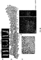
- the musculoskeletal stem cells obtained from 7 passages or more were stably identical. In some examples, , they grew with similar morphologies for 10 passages or more, from passage 7 to passage 17, and showed a positive response to staining with the aging marker ⁇ -galactosidase since passage 19, suggesting that aging was progressed.
- Figure 1A shows cell morphology of hESCs cultured with a medium for inducing differentiation into MSSCs from passage 7 to passage 19.
- the resulting musculoskeletal stem cells have at least one of the characteristics listed below.
- the resulting MSSCs have the characteristics of (d)-(g).
- the resulting MSSCs have the characteristics of (d)-(g) and (r).
- the resulting MSSCs have the characteristics of (a)-(b).
- the resulting MSSCs have the characteristics of (a), (b) and (r).
- the resulting MSSCs have the characteristics of (a)-(c).
- the resulting MSSCs have the characteristics of (a)-(c) and (r).
- the resulting MSSCs have the characteristics of (a)-(d).
- the resulting MSSCs have the characteristics of (a)-(d) and (r).
- the resulting MSSCs have the characteristics of (a)-(e).
- the resulting MSSCs have the characteristics of (a)-(e) and (r).
- the resulting MSSCs have the characteristics of (a)-(f).
- the resulting MSSCs have the characteristics of (a)-(g).
- the resulting MSSCs have the characteristics of (a)-(f) and (h).
- the resulting MSSCs have the characteristics of (a)-(f) and (i).
- the resulting MSSCs have the characteristics of (a)-(f) and (j).
- the resulting MSSCs have the characteristics of (a)-(f) and (k).
- the resulting MSSCs have the characteristics of (a)-(f) and (1).
- the resulting MSSCs have the characteristics of (a)-(f) and (m).
- the resulting MSSCs have the characteristics of (a)-(f) and (n).
- the resulting MSSCs have the characteristics of (a)-(f) and (o).
- the resulting MSSCs have the characteristics of (a)-(f) and (p).
- the resulting MSSCs have the characteristics of (a)-(f) and (q).
- the resulting MSSCs have the characteristics of (a)-(f) and (r).
- the resulting MSSCs have the characteristics of (a)-(f) and (s).
- the resulting MSSCs have the characteristics of (a)-(h).
- the resulting MSSCs have the characteristics of (a)-(g) and (i). In one example, the resulting MSSCs have the characteristics of (a)-(g) and (j). In one example, the resulting MSSCs have the characteristics of (a)-(g) and (k). In one example, the resulting MSSCs have the characteristics of (a)-(g) and (1). In one example, the resulting MSSCs have the characteristics of (a)-(g) and (m). In one example, the resulting MSSCs have the characteristics of (a)-(g) and (n). In one example, the resulting MSSCs have the characteristics of (a)-(g) and (o).
- the resulting MSSCs have the characteristics of (a)-(g) and (p). In one example, the resulting MSSCs have the characteristics of (a)-(g) and (q). In one example, the resulting MSSCs have the characteristics of (a)-(g) and (r). In one example, the resulting MSSCs have the characteristics of (a)-(g) and (s). In one example, the resulting MSSCs have the characteristics of (a)-(i).
- the resulting MSSCs have the characteristics of (a)-(h) and (j). In one example, the resulting MSSCs have the characteristics of (a)-(h) and (k). In one example, the resulting MSSCs have the characteristics of (a)-(h) and (1). In one example, the resulting MSSCs have the characteristics of (a)-(h) and (m). In one example, the resulting MSSCs have the characteristics of (a)-(h) and (n). In one example, the resulting MSSCs have the characteristics of (a)-(h) and (o). In one example, the resulting MSSCs have the characteristics of (a)-(h) and (p).
- the resulting MSSCs have the characteristics of (a)-(h) and (q). In one example, the resulting MSSCs have the characteristics of (a)-(h) and (r). In one example, the resulting MSSCs have the characteristics of (a)-(h) and (s). In one example, the resulting MSSCs have the characteristics of (a)-(j).
- the resulting MSSCs have the characteristics of (a)-(i) and (k). In one example, the resulting MSSCs have the characteristics of (a)-(i) and (l). In one example, the resulting MSSCs have the characteristics of (a)-(i) and (m). In one example, the resulting MSSCs have the characteristics of (a)-(i) and (n). In one example, the resulting MSSCs have the characteristics of (a)-(i) and (o). In one example, the resulting MSSCs have the characteristics of (a)-(i) and (p).
- the resulting MSSCs have the characteristics of (a)-(i) and (q).
- the resulting MSSCs have the characteristics of (a)-(i) and (r).
- the resulting MSSCs have the characteristics of (a)-(i) and (s).
- the resulting MSSCs have the characteristics of (a)-(k).
- the resulting MSSCs have the characteristics of (a)-(j) and (1).
- the resulting MSSCs have the characteristics of (a)-(j) and (m).
- the resulting MSSCs have the characteristics of (a)-(j) and (n).
- the resulting MSSCs have the characteristics of (a)-(j) and (o).
- the resulting MSSCs have the characteristics of (a)-(j) and (p).
- the resulting MSSCs have the characteristics of (a)-(j) and (q).
- the resulting MSSCs have the characteristics of (a)-(j) and (r).
- the resulting MSSCs have the characteristics of (a)-(j) and (s).
- the resulting MSSCs have the characteristics of (a)-(l).
- the resulting MSSCs have the characteristics of (a)-(k) and (m).
- the resulting MSSCs have the characteristics of (a)-(k) and (n).
- the resulting MSSCs have the characteristics of (a)-(k) and (o).
- the resulting MSSCs have the characteristics of (a)-(k) and (p).
- the resulting MSSCs have the characteristics of (a)-(k) and (q).
- the resulting MSSCs have the characteristics of (a)-(k) and (r).
- the resulting MSSCs have the characteristics of (a)-(k) and (s).
- the resulting MSSCs have the characteristics of (a)-(m).
- the resulting MSSCs have the characteristics of (a)-(1) and (n).
- the resulting MSSCs have the characteristics of (a)-(1) and (o).
- the resulting MSSCs have the characteristics of (a)-(1) and (p).
- the resulting MSSCs have the characteristics of (a)-(1) and (q).
- the resulting MSSCs have the characteristics of (a)-(1) and (r).
- the resulting MSSCs have the characteristics of (a)-(1) and (s).
- the resulting MSSCs have the characteristics of (a)-(n).
- the resulting MSSCs have the characteristics of (a)-(m) and (o).
- the resulting MSSCs have the characteristics of (a)-(m) and (p).
- the resulting MSSCs have the characteristics of (a)-(m) and (q).
- the resulting MSSCs have the characteristics of (a)-(m) and (r).
- the resulting MSSCs have the characteristics of (a)-(m) and (s).
- the resulting MSSCs have the characteristics of (a)-(o).
- the resulting MSSCs have the characteristics of (a)-(n) and (p).
- the resulting MSSCs have the characteristics of (a)-(n) and (q).
- the resulting MSSCs have the characteristics of (a)-(n) and (r).
- the resulting MSSCs have the characteristics of (a)-(n) and (s).
- the resulting MSSCs have the characteristics of (a)-(p).
- the resulting MSSCs have the characteristics of (a)-(o) and (q).
- the resulting MSSCs have the characteristics of (a)-(o) and (r).
- the resulting MSSCs have the characteristics of (a)-(o) and (s).
- the resulting MSSCs have the characteristics of (a)-(q).
- the resulting MSSCs have the characteristics of (a)-(p) and (r).
- the resulting MSSCs have the characteristics of (a)-(p) and (s).
- the resulting MSSCs have the characteristics of (a)-(r).
- the resulting MSSCs have the characteristics of (a)-(q) and (s).
- the resulting MSSCs have the characteristics of (a)-(s).
- the expression of most pluripotency markers was not observed in the musculoskeletal stem cells of the present disclosure but the expression of DPPA4 was observed and the cells were positive for the ectodermal marker NES. In addition, they were positive for most mesodermal markers except DES and the early mesodermal markers T and nodal and negative for most endodermal markers. See Figure 1C .
- mesenchymal stem cell-specific cell surface antigens were investigated for hMSSC, among the mesenchymal stem cell markers, CD44, CD51, CD73, CD105, CD146 and CD166 were expressed in the hMSSCs but CD90 and CD271 were not expressed in the hMSSC.
- the vascular cell surface markers CD2, CD3, CD7, CD8, CD11b, CD14, CD19, CD20, CD31, CD34 and CD56 were not expressed, the pre-B cell marker CD10 was expressed. See Figure 1D .
- a-SMA mesodermal marker alpha smooth muscle actin
- the neuroectodermal marker Pax6, the myogenic satellite marker Pax7, and the intestinal stem cell marker LGR5 were expressed, but the chondrocyte marker SOX9 and the myoblast marker MyoD were not expressed. See Figure 1E .
- the musculoskeletal stem cells (MSSCs) differentiated from ESCs or iPSCs differ from intrinsic stem cells. It is known that intrinsic stem cells require mesenchymal stem cells' signaling or stimulation to differentiate into musculoskeletal tissues. Chan, C. K., et al. (2016). Identification of the Human Skeletal Stem Cell, Cell, 175, 43-56 . However, MSSCs differentiated from ESCs or iPSCs do not need such signaling or stimulation by mesenchymal stem cells for the differentiation into musculoskeletal tissues. Further, it is also known that the intrinsic stem cells show negative for CD146; however, the MSSCs differentiated from ESCs or iPSCs show positive for CD 146.
- the musculoskeletal stem cells differentiated from ESCs or iPSCs are capable of being differentiated into the mesoderm but not into the ectoderm or endoderm.
- the musculoskeletal stem cells differentiated from ESCs or iPSCs are also capable of being differentiated into musculoskeletal cells in vitro and in vivo.
- the musculoskeletal stem cells may be differentiated into cells of muscle, bone, cartilage, tendon or ligament in vivo and in vitro.
- Figure 2A shows a result of comparing the in-vitro bone, cartilage and fat differentiation capacity of hMSCs and hMSSCs.
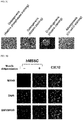
- Figure 2B shows a result of confirming that hMSSCs has the potential to differentiate into skeletal muscle by immunocytochemistry for the skeletal muscle cell-specific marker MYH9.
- C2C12 was used as a positive control group for the skeletal muscle cell.
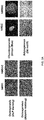
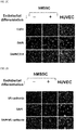
- the MSSCs differentiated from ESCs or iPSCs do not have the potential to differentiate into endothelial cells even if placed in a medium for inducing differentiation into an EC (endothelial growth medium).
- Figures 2C and 2D confirm that hMSSCs did not differentiate into endothelial cells by immunocytochemistry for the endothelial cell-specific markers CD31 and VE-cadherin.
- the MSSCs differentiated from ESCs or iPSCs do not have the potential to differentiate into nerve cells even if placed in a medium for inducing differentiation into nerve cells.
- Figure 2E confirms that hMSSCs did not differentiate into nerve cells by immunocytochemistry for the nerve cell-specific marker MAP2.
- neural stem cells differentiated from H9 hESCs were used.
- the MSSCs differentiated from ESCs or iPSCs may be further cultured in a medium.
- the culture medium may include any medium for culturing mesenchymal stem cells, including MSCGM, MSCGM-CD, etc.
- the MSSCs are transplanted into a mass of certain tissues or an organ to the formation of musculoskeletal cells in the body of tissues or the organ.
- the MSSCs are transplanted into kidney capsule or hypoderm for the formation of muscle, fat, tendon, bone and cartilage cells.
- the MSSCs are cultured in a medium for culturing mesenchymal stem cells prior to transplanting.
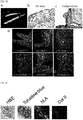
- FIG 3A shows that muscle, fat and tendon cells were formed by transplanting of hMSSCs into kidney as confirmed by H&E staining.
- (b) shows the differentiation of hMSSCs in kidney into muscle, fat and tendon cells by immunohistochemistry for the muscle-specific marker pMLC, the fat-specific marker PPARgamma (PPAr) and ligament-specific marker Scx.
- hLA is a human cell-specific marker, and the staining result shows that the cell is derived from human.
- the differentiated muscle cells were skeletal muscle cells and not smooth muscle cells. It was also confirmed in the in-vivo experiment that they could be differentiated into fat, although the differentiation into fat was not observed in the in-vitro experiment.
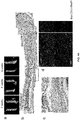
- FIG 3B (a) is a micro-CT scanning result confirming that bone was formed by transplanting hMSSCs into the kidney.
- (b) and (c) show the formation of bone by H&E and pentachrome immunohistochemical staining.
- (d) shows the expression of the human cell marker hLA (human leukocyte antigen), the bone markers Osx (osterix), Runx2, DMP1 and OCN (osteocalin) and the vascular marker vWF in the cells of the osteoblastic tissue by immunohistochemistry.
- hLA human leukocyte antigen
- Osx osteosterix
- Runx2 osteocalin
- Figure 3C shows that cartilage cells were formed by transplanting hMSSCs into the hypoderm by H&E and toluidine blue immunohistochemical staining.
- the expression of the cartilage marker ColII (collagen II) was also confirmed by immunohistochemistry.
- the MSSCs differentiated from ESCs or iPSCs may not have the potential for differentiating into nerve cells even if placed in a medium for inducing differentiation into nerve cells.
- Figure 2E confirms that hMSSCs did not differentiate into nerve cells by immunocytochemistry for the nerve cell-specific marker MAP2.
- neural stem cells differentiated from H9 hESCs were used.
- the MSSCs differentiated from ESCs or iPSCs may not have the potential for differentiating into endothelial cells even if placed in a medium for inducing differentiation into an EC (endothelial growth medium).
- Figures 2C and 2D confirm that hMSSCs did not differentiate into endothelial cells by immunocytochemistry for the endothelial cell-specific markers CD31 and VE-cadherin.
- the musculoskeletal stem cell was deposited in the Korean Cell Line Bank on October 10, 2018 and was given the accession number KCLRF-BP-00460.
- the MSSCs differentiated from ESCs or iPSCs may be used to provide a pharmaceutical composition or therapeutic agent for preventing or treating a musculoskeletal disease.
- the pharmaceutical composition contains an effective amount of the MSSCs differentiated from ESCs or iPSCs.
- the pharmaceutical composition may be applicable for treatment or prevention of one or more diseases selected from a group consisting of osteoporosis, osteomalacia, osteogenesis imperfecta, osteopetrosis, osteosclerosis, Paget's disease, bone cancer, arthritis, rickets, fracture, periodontal disease, segmental bone defect, osteolytic bone disease, primary and secondary hyperparathyroidism, hyperostosis, degenerative arthritis, degenerative knee joint disease, degenerative hip joint disease, degenerative foot joint disease, degenerative hand joint disease, degenerative shoulder joint disease, degenerative elbow joint disease, chondromalacia patellae, simple knee arthritis, osteochondritis dissecans, lateral epicondylitis, medial epicondylitis, Heberden's nodes, Bouchard's nodes, degenerative thumb CM arthrosis, meniscal injury, degenerative disc disease, cruciate ligament injury, biceps brachii muscle injury, ligament injury, tendon injury, frozen shoulder, rotator cuff tear, calcific tendinit
- the pharmaceutical composition may contain one or more pharmaceutically acceptable carriers.
- the one or more pharmaceutically acceptable carriers are selected from the group consisting of lactose, dextrose, sucrose, sorbitol, mannitol, starch, acacia gum, calcium phosphate, alginate, gelatin, calcium silicate, microcrystalline cellulose, polyvinylpyrrolidone, cellulose, water, syrup, methyl cellulose, methyl hydroxybenzoate, propyl hydroxybenzoate, talc, magnesium stearate, mineral oil, etc., although not being limited thereto.
- the pharmaceutical composition may further contain at least one of a lubricant, a humectant, a sweetener, a flavorant, an emulsifier, a suspending agent, a preservative, etc., in addition to the MSSCs and pharmaceutically acceptable carrier.
- the pharmaceutical composition containing the MSSCs may be administered for treatment or prevention of a musculoskeletal disease.
- the pharmaceutical composition of the present disclosure may be administered orally or parenterally.
- parenteral administration it may be administered via intravenous injection, subcutaneous injection, intramuscular injection, intraarticular injection, intraosseous infusion, intraperitoneal injection, endothelial administration, topical administration, intranasal administration, intrapulmonary administration, intrarectal administration, etc.
- the composition may be administered by any device capable of delivering the active ingredient to a target cell.
- the pharmaceutical composition containing the MSSCs is administered in an amount effective of the MSSCs for treatment or prevention of a musculoskeletal disease.
- the effective amount or an appropriate administration dosage may be determined in consideration of various factors such as formulation method, administration mode, the age, body weight and sex of a patient, pathological condition, diet, administration time, administration route, excretion rate and response sensitivity.
- the administration dosage may be about 10 2 , 10 3 , 10 4 , 10 5 , 10 6 , 10 7 , 10 8 , 10 9 , 10 10 , 10 11 or 10 12 cells/kg for an adult.
- the dosage may be within a range formed by selecting any two numbers (two concentration values) listed in the immediately previous sentence, e.g., about 10 2 to about 10 10 , about 10 4 to about 10 7 cells/kg for an adult.
- the pharmaceutical composition containing the MSSCs may be prepared into a single-dose unit or multiple-dose unit formulation using a pharmaceutically acceptable carrier and/or excipient according to a method that can be easily carried out by those skilled in the art.
- the formulation may be in the form of a solution in an oily or aqueous medium, a suspension, a syrup, an emulsion, an extract, a powder, a granule, a tablet or a capsule and may further contain a dispersant or a stabilizer.
- the composition may be administered either independently or in combination with other therapeutic agent(s) and they may be administered either sequentially or simultaneously. Also, it may be administered as a single dose or repeatedly as desired.
- the term "cell therapeutic agent” refers to medication used for therapeutic, diagnostic and preventive purposes, which contains a cell or tissue isolated from human and cultured and prepared through special operation (as provided by the USFDA). It is a medication used for therapeutic, diagnostic and preventive purposes through a series of actions of in-vitro multiplication and screening of living autologous, allogenic and xenogenic cells or changing of the biological characteristics of cells by other means for recovering the functions of cells or tissues.
- prevention refers to any action of inhibiting a musculoskeletal disease or delaying the progression thereof by administering the composition or cell therapeutic agent of the present disclosure.
- treatment refers to any action of improving or favorably changing a musculoskeletal disease by administering the composition or cell therapeutic agent of the present disclosure.
- the pharmaceutical composition or cell therapeutic agent of the present disclosure may be used for human or an animal.
- composition or cell therapeutic agent of the present disclosure may be used either alone or in combination with surgery, radiotherapy, hormone therapy, chemotherapy, a biological response modifier, implantation, insertion of an artificial joint, artificial cartilage, etc., regeneration therapy, etc., for prevention and treatment of a musculoskeletal disease.
- the present disclosure provides a method of screening or identifying a musculoskeletal stem cell based on characteristics of the MSSCs differentiated from ESCs and/or iPSCs.
- cells having at least one of the following characteristics listed below may be determined as MSSCs:
- Cells having the characteristics of (d)-(g) may be identified as MSSCs.
- Cells having the characteristics of (d)-(g) and (r) may be identified as MSSCs.
- Cells having the characteristics of (a)-(b) may be identified as MSSCs.
- Cells having the characteristics of (a), (b) and (r) may be identified as MSSCs.
- Cells having the characteristics of (a)-(c) may be identified as MSSCs.
- Cells having the characteristics of (a)-(c) and (r) may be identified as MSSCs.
- Cells having the characteristics of (a)-(d) may be identified as MSSCs.
- Cells having the characteristics of (a)-(d) and (r) may be identified as MSSCs.
- Cells having the characteristics of (a)-(e) may be identified as MSSCs.
- Cells having the characteristics of (a)-(e) and (r) may be identified as MSSCs.
- Cells having the characteristics of (a)-(f) may be identified as MSSCs.
- Cells having the characteristics of (a)-(g) may be identified as MSSCs.
- Cells having the characteristics of (a)-(f) and (h) may be identified as MSSCs.
- Cells having the characteristics of (a)-(f) and (i) may be identified as MSSCs.
- Cells having the characteristics of (a)-(f) and (j) may be identified as MSSCs.
- Cells having the characteristics of (a)-(f) and (k) may be identified as MSSCs.
- Cells having the characteristics of (a)-(f) and (l) may be identified as MSSCs.
- Cells having the characteristics of (a)-(f) and (m) may be identified as MSSCs.
- Cells having the characteristics of (a)-(f) and (n) may be identified as MSSCs.
- Cells having the characteristics of (a)-(f) and (o) may be identified as MSSCs.
- Cells having the characteristics of (a)-(f) and (p) may be identified as MSSCs.
- Cells having the characteristics of (a)-(f) and (q) may be identified as MSSCs.
- Cells having the characteristics of (a)-(f) and (r) may be identified as MSSCs.
- Cells having the characteristics of (a)-(f) and (s) may be identified as MSSCs.
- Cells having the characteristics of (a)-(h) may be identified as MSSCs.
- Cells having the characteristics of (a)-(g) and (i) may be identified as MSSCs.
- Cells having the characteristics of (a)-(g) and (j) may be identified as MSSCs.
- Cells having the characteristics of (a)-(g) and (k) may be identified as MSSCs.
- Cells having the characteristics of (a)-(g) and (l) may be identified as MSSCs.
- Cells having the characteristics of (a)-(g) and (m) may be identified as MSSCs.
- Cells having the characteristics of (a)-(g) and (n) may be identified as MSSCs.
- Cells having the characteristics of (a)-(g) and (o) may be identified as MSSCs.
- Cells having the characteristics of (a)-(g) and (p) may be identified as MSSCs.
- Cells having the characteristics of (a)-(g) and (q) may be identified as MSSCs.
- Cells having the characteristics of (a)-(g) and (r) may be identified as MSSCs.
- Cells having the characteristics of (a)-(g) and (s) may be identified as MSSCs.
- Cells having the characteristics of (a)-(i) may be identified as MSSCs.
- Cells having the characteristics of (a)-(h) and (j) may be identified as MSSCs.
- Cells having the characteristics of (a)-(h) and (k) may be identified as MSSCs.
- Cells having the characteristics of (a)-(h) and (l) may be identified as MSSCs.
- Cells having the characteristics of (a)-(h) and (m) may be identified as MSSCs.
- Cells having the characteristics of (a)-(h) and (n) may be identified as MSSCs.
- Cells having the characteristics of (a)-(h) and (o) may be identified as MSSCs.
- Cells having the characteristics of (a)-(h) and (p) may be identified as MSSCs.
- Cells having the characteristics of (a)-(h) and (q) may be identified as MSSCs.
- Cells having the characteristics of (a)-(h) and (r) may be identified as MSSCs.
- Cells having the characteristics of (a)-(h) and (s) may be identified as MSSCs.
- Cells having the characteristics of (a)-(j) may be identified as MSSCs.
- Cells having the characteristics of (a)-(i) and (k) may be identified as MSSCs.
- Cells having the characteristics of (a)-(i) and (l) may be identified as MSSCs.
- Cells having the characteristics of (a)-(i) and (m) may be identified as MSSCs.
- Cells having the characteristics of (a)-(i) and (n) may be identified as MSSCs.
- Cells having the characteristics of (a)-(i) and (o) may be identified as MSSCs.
- Cells having the characteristics of (a)-(i) and (p) may be identified as MSSCs.
- Cells having the characteristics of (a)-(i) and (q) may be identified as MSSCs.
- Cells having the characteristics of (a)-(i) and (r) may be identified as MSSCs.
- Cells having the characteristics of (a)-(i) and (s) may be identified as MSSCs.
- Cells having the characteristics of (a)-(k) may be identified as MSSCs.
- Cells having the characteristics of (a)-(j) and (l) may be identified as MSSCs.
- Cells having the characteristics of (a)-(j) and (m) may be identified as MSSCs.
- Cells having the characteristics of (a)-(j) and (n) may be identified as MSSCs.
- Cells having the characteristics of (a)-(j) and (o) may be identified as MSSCs.
- Cells having the characteristics of (a)-(j) and (p) may be identified as MSSCs.
- Cells having the characteristics of (a)-(j) and (q) may be identified as MSSCs.
- Cells having the characteristics of (a)-(j) and (r) may be identified as MSSCs.
- Cells having the characteristics of (a)-(j) and (s) may be identified as MSSCs.
- Cells having the characteristics of (a)-(1) may be identified as MSSCs.
- Cells having the characteristics of (a)-(k) and (m) may be identified as MSSCs.
- Cells having the characteristics of (a)-(k) and (n) may be identified as MSSCs.
- Cells having the characteristics of (a)-(k) and (o) may be identified as MSSCs.
- Cells having the characteristics of (a)-(k) and (p) may be identified as MSSCs.
- Cells having the characteristics of (a)-(k) and (q) may be identified as MSSCs.
- Cells having the characteristics of (a)-(k) and (r) may be identified as MSSCs.
- Cells having the characteristics of (a)-(k) and (s) may be identified as MSSCs.
- Cells having the characteristics of (a)-(m) may be identified as MSSCs.
- Cells having the characteristics of (a)-(l) and (n) may be identified as MSSCs.
- Cells having the characteristics of (a)-(l) and (o) may be identified as MSSCs.
- Cells having the characteristics of (a)-(l) and (p) may be identified as MSSCs.
- Cells having the characteristics of (a)-(l) and (q) may be identified as MSSCs.
- Cells having the characteristics of (a)-(l) and (r) may be identified as MSSCs.
- Cells having the characteristics of (a)-(l) and (s) may be identified as MSSCs.
- Cells having the characteristics of (a)-(n) may be identified as MSSCs.
- Cells having the characteristics of (a)-(m) and (o) may be identified as MSSCs.
- Cells having the characteristics of (a)-(m) and (p) may be identified as MSSCs.
- Cells having the characteristics of (a)-(m) and (q) may be identified as MSSCs.
- Cells having the characteristics of (a)-(m) and (r) may be identified as MSSCs.
- Cells having the characteristics of (a)-(m) and (s) may be identified as MSSCs.
- Cells having the characteristics of (a)-(o) may be identified as MSSCs.
- Cells having the characteristics of (a)-(n) and (p) may be identified as MSSCs.
- Cells having the characteristics of (a)-(n) and (q) may be identified as MSSCs.
- Cells having the characteristics of (a)-(n) and (r) may be identified as MSSCs.
- Cells having the characteristics of (a)-(n) and (s) may be identified as MSSCs.
- Cells having the characteristics of (a)-(p) may be identified as MSSCs.
- Cells having the characteristics of (a)-(o) and (q) may be identified as MSSCs.
- Cells having the characteristics of (a)-(o) and (r) may be identified as MSSCs.
- Cells having the characteristics of (a)-(o) and (s) may be identified as MSSCs.
- Cells having the characteristics of (a)-(q) may be identified as MSSCs.
- Cells having the characteristics of (a)-(p) and (r) may be identified as MSSCs.
- Cells having the characteristics of (a)-(p) and (s) may be identified as MSSCs.
- Cells having the characteristics of (a)-(r) may be identified as MSSCs.
- Cells having the characteristics of (a)-(q) and (s) may be identified as MSSCs.
- Cells having the characteristics of (a)-(s) may be identified as MSSCs.
- MSSCs a cell having one or more additional ones of the following characteristics may be determined as MSSCs:
- mice 7- to 10-week-old Balb/c-nude background mice (weighing 20-24 g) were purchased from Orient Bio (Seongnam, Korea). All animal experiments were performed according to the guidelines of the Chonbuk University Animal Care and Use Committee. The animals were accommodated under controlled-temperature (21-24 °C) and 12:12-hr light-darky cycle environments and were given free access to water and feed.
- H9 hESCs human embryonic stem cells
- WiCell WiCell (Madison, MI, USA).
- a hESC culture medium was prepared as DMEM/F12 (Invitrogen, USA) supplemented with 20% knockout serum replacement (KSR; Invitrogen, USA), 1 mM glutamine (Invitrogen, USA), 1% nonessential amino acids (Invitrogen, USA), 0.1 mM ⁇ -mercaptoethanol (Invitrogen, USA), 0.1% penicillin/streptomycin (Invitrogen, USA) and 15 ng/mL bFGF (R&D Systems, USA).
- KSR knockout serum replacement
- Invitrogen, USA 1 mM glutamine
- 1% nonessential amino acids Invitrogen, USA
- 0.1 mM ⁇ -mercaptoethanol Invitrogen, USA
- penicillin/streptomycin Invitrogen, USA
- 15 ng/mL bFGF R&D Systems, USA
- Example 2.1.2 Induction of differentiation from hESCs into hMSSCs
- MSSC medium A medium for inducing differentiation into MSSCs (hereinafter referred to as “MSSC medium”) was prepared with the following composition:
- the medium further contained 10% knockout serum replacement (Invitrogen, USA), 1% N2 supplement (Gibco, USA), 2% B27 supplement (Gibco, USA), 1% nonessential amino acids (Gibco, USA), 43% DMEM/F12 (Gibco, USA), 43% Neurobasal (Gibco, USA), 1 mM glutamine, 0.1 mM ⁇ -mercaptoethanol, 0.1% penicillin-streptomycin and 5 mg/mL bovine serum albumin (Gibco, USA).
- the hESCs from Example 2.1.1 were treated with ROCK (Rho-associated coiled-coil kinase) inhibitor (Y-27632, 10 ⁇ M, Calbiochem, Germany) and PKC (protein kinase C) inhibitor (Go6983, 2.5 ⁇ M, Sigma, USA) for 24 hours in order to enhance survivability. Then, hESCs were trypsinized by treating with TrypLE (Life Technologies, USA). Subsequently, the trypsinized hESCs were induced to differentiate into hMSSCs by culturing with the MSSC medium on a culture dish coated with vitronectin and gelatin (1 ng/mL, Sigma, USA) until passage 7. The differentiated MSSC cells were identified to be stably identical from passage 5 and the cells cultured for 10 passages were deposited in the Korean Cell Line Bank on October 10, 2018 and were given the accession number KCLRF-BP-00460.
- ROCK Rho-associated coiled-coil kinase
- Example 2.1.3 Induction of differentiation from hESCs into hMSSCs
- Example 2.1.2 The experiment of Example 2.1.2 is repeated except that the MSSC medium contains 1 ng/mL human LIF instead of 20 ng/ml.
- Example 2.1.4 Induction of differentiation from hESCs into hMSSCs
- Example 2.1.2 The experiment of Example 2.1.2 is repeated except that the MSSC medium contains 5 ng/mL human LIF instead of 20 ng/ml.
- Example 2.1.5 Induction of differentiation from hESCs into hMSSCs
- Example 2.1.2 The experiment of Example 2.1.2 is repeated except that the MSSC medium contains 50 ng/mL human LIF instead of 20 ng/ml.
- Example 2.1.6 Induction of differentiation from hESCs into hMSSCs
- Example 2.1.2 The experiment of Example 2.1.2 is repeated except that the MSSC medium contains 100 ng/mL human LIF instead of 20 ng/ml.
- Example 2.1.7 Induction of differentiation from hESCs into hMSSCs
- Example 2.1.2 The experiment of Example 2.1.2 is repeated except that the MSSC medium contains 1 ng/mL bFGF instead of 15 ng/ml.
- Example 2.1.8 Induction of differentiation from hESCs into hMSSCs
- Example 2.1.2 The experiment of Example 2.1.2 is repeated except that the MSSC medium contains 500 ng/mL bFGF instead of 15 ng/ml.
- Example 2.1.9 Induction of differentiation from hESCs into hMSSCs
- Example 2.1.2 The experiment of Example 2.1.2 is repeated except that the MSSC medium contains 100 ng/mL bFGF instead of 15 ng/ml.
- Example 2.1.2 The experiment of Example 2.1.2 is repeated except that the MSSC medium contains 200 ng/mL bFGF instead of 15 ng/ml.
- Example 2.1.11 Induction of differentiation from hESCs into hMSSCs
- Example 2.1.2 The experiment of Example 2.1.2 is repeated except that the MSSC medium contains 100 ng/mL Wnt signaling activator instead of 1,396 ng/ml.
- Example 2.1.2 The experiment of Example 2.1.2 is repeated except that the MSSC medium contains 200 ng/mL Wnt signaling activator instead of 1,396 ng/ml.
- Example 2.1.2 The experiment of Example 2.1.2 is repeated except that the MSSC medium contains 5,000 ng/mL Wnt signaling activator instead of 1,396 ng/ml.
- Example 2.1.2 The experiment of Example 2.1.2 is repeated except that the MSSC medium contains 10,000 ng/mL Wnt signaling activator instead of 1,396 ng/ml.
- Example 2.1.15 Induction of differentiation from hESCs into hMSSCs
- Example 2.1.2 The experiment of Example 2.1.2 is repeated except that the MSSC medium contains 40 ng/mL ERK signaling inhibitor instead of 482.19 ng/ml.
- Example 2.1.2 The experiment of Example 2.1.2 is repeated except that the MSSC medium contains 80 ng/mL ERK signaling inhibitor instead of 482.19 ng/ml.
- Example 2.1.2 The experiment of Example 2.1.2 is repeated except that the MSSC medium contains 2,500 ng/mL ERK signaling inhibitor instead of 482.19 ng/ml.
- Example 2.1.2 The experiment of Example 2.1.2 is repeated except that the MSSC medium contains 5,000 ng/mL ERK signaling inhibitor instead of 482.19 ng/ml.
- Example 2.1.2 The experiment of Example 2.1.2 is repeated except that the MSSC medium contains 100 ng/mL TGF- ⁇ /activin/nodal signaling inhibitor instead of 3,843.9 ng/ml.
- Example 2.1.20 Induction of differentiation from hESCs into hMSSCs
- Example 2.1.2 The experiment of Example 2.1.2 is repeated except that the MSSC medium contains 300 ng/mL TGF- ⁇ /activin/nodal signaling inhibitor instead of 3,843.9 ng/ml.
- Example 2.1.2 The experiment of Example 2.1.2 is repeated except that the MSSC medium contains 10,000 ng/mL TGF- ⁇ /activin/nodal signaling inhibitor instead of 3,843.9 ng/ml.
- Example 2.1.2 The experiment of Example 2.1.2 is repeated except that the MSSC medium contains 40,000 ng/mL TGF- ⁇ /activin/nodal signaling inhibitor instead of 3,843.9 ng/ml.
- Example 2.1.2 through Example 2.1.22 are repeated except that the MSSC medium contains 25 ng/mL human noggin instead of 250 ng/ml.
- Example 2.1.2 through Example 2.1.22 are repeated except that the MSSC medium contains 50 ng/mL human noggin instead of 250 ng/ml.
- Example 2.1.2 through Example 2.1.22 are repeated except that the MSSC medium contains 500 ng/mL human noggin instead of 250 ng/ml.
- Example 2.1.2 through Example 2.1.22 are repeated except that the MSSC medium contains 2,500 ng/mL human noggin instead of 250 ng/ml.
- hiPSCs human induced pluripotent stem cells
- OCT4, KLF4, SOX2 and cMYC genes were obtained by introducing the OCT4, KLF4, SOX2 and cMYC genes to BJ fibroblasts (ATCC®CRL2522TM) using Sendai virus according to the method developed by Hasegawa et al. (Fusaki et al., 2009, PNAS 85, 348-362 ).
- a hiPSC culture medium was prepared as DMEM/F12 (Invitrogen, USA) supplemented with 20% knockout serum replacement (KSR; Invitrogen, USA), 1 mM glutamine (Invitrogen, USA), 1% nonessential amino acids (Invitrogen, USA), 0.1 mM ⁇ -mercaptoethanol (Invitrogen, USA), 0.1% penicillin/streptomycin (Invitrogen, USA) and 15 ng/mL bFGF (R&D Systems, USA).
- KSR knockout serum replacement
- Invitrogen, USA 1 mM glutamine
- 1% nonessential amino acids Invitrogen, USA
- 0.1 mM ⁇ -mercaptoethanol Invitrogen, USA
- penicillin/streptomycin Invitrogen, USA
- 15 ng/mL bFGF R&D Systems, USA.
- the hiPSCs were cultured with the prepared hiPSC culture medium on CF1 mouse embryonic fibroblast (MEF) feeder
- Example 2.2.2 Induction of differentiation from hiPSCs into hMSSCs
- MSSC medium A medium for inducing differentiation into MSSCs (hereinafter, referred to as “MSSC medium”) was prepared with the following composition:
- the hiPSCs from Example 2.2.1 were treated with ROCK (Rho-associated coiled-coil kinase) inhibitor (Y-27632, 10 ⁇ M, Calbiochem, Germany) and PKC (protein kinase C) inhibitor (Go6983, 2.5 ⁇ M, Sigma, USA) for 24 hours in order to enhance survivability. Then, the hiPSCs were trypsinized by treating with TrypLE (Life Technologies, USA). Subsequently, the trypsinized hiPSCs were induced to differentiate into hMSSCs by culturing with the MSSC medium on a culture dish coated with vitronectin and gelatin (1 ng/mL, Sigma, USA) until passage 7. The differentiated MSSC cells were identified to be stably identical from passage 5.
- ROCK Rho-associated coiled-coil kinase
- PKC protein kinase C inhibitor
- Example 2.2.3 Induction of differentiation from hiPSCs into hMSSCs
- Example 2.2.2 The experiment of Example 2.2.2 is repeated except that the MSSC medium contains 1 ng/mL human LIF instead of 20 ng/ml.
- Example 2.2.4 Induction of differentiation from hiPSCs into hMSSCs
- Example 2.2.2 The experiment of Example 2.2.2 is repeated except that the MSSC medium contains 5 ng/mL human LIF instead of 20 ng/ml.
- Example 2.2.5 Induction of differentiation from hiPSCs into hMSSCs
- Example 2.2.2 The experiment of Example 2.2.2 is repeated except that the MSSC medium contains 50 ng/mL human LIF instead of 20 ng/ml.
- Example 2.2.6 Induction of differentiation from hiPSCs into hMSSCs
- Example 2.2.2 The experiment of Example 2.2.2 is repeated except that the MSSC medium contains 100 ng/mL human LIF instead of 20 ng/ml.
- Example 2.2.2 The experiment of Example 2.2.2 is repeated except that the MSSC medium contains 1 ng/mL bFGF instead of 15 ng/ml.
- Example 2.2.2 The experiment of Example 2.2.2 is repeated except that the MSSC medium contains 500 ng/mL bFGF instead of 15 ng/ml.
- Example 2.2.9 Induction of differentiation from hiPSCs into hMSSCs
- Example 2.2.2 The experiment of Example 2.2.2 is repeated except that the MSSC medium contains 100 ng/mL bFGF instead of 15 ng/ml.
- Example 2.2.10 Induction of differentiation from hiPSCs into hMSSCs
- Example 2.2.2 The experiment of Example 2.2.2 is repeated except that the MSSC medium contains 200 ng/mL bFGF instead of 15 ng/ml.
- Example 2.2.11 Induction of differentiation from hiPSCs into hMSSCs
- Example 2.2.2 The experiment of Example 2.2.2 is repeated except that the MSSC medium contains 100 ng/mL Wnt signaling activator instead of 1,396 ng/ml.
- Example 2.2.12. Induction of differentiation from hiPSCs into hMSSCs
- Example 2.2.2 The experiment of Example 2.2.2 is repeated except that the MSSC medium contains 200 ng/mL Wnt signaling activator instead of 1,396 ng/ml.
- Example 2.2.2 The experiment of Example 2.2.2 is repeated except that the MSSC medium contains 5,000 ng/mL Wnt signaling activator instead of 1,396 ng/ml.
- Example 2.2.2 The experiment of Example 2.2.2 is repeated except that the MSSC medium contains 10,000 ng/mL Wnt signaling activator instead of 1,396 ng/ml.
- Example 2.2.15 Induction of differentiation from hiPSCs into hMSSCs
- Example 2.2.2 The experiment of Example 2.2.2 is repeated except that the MSSC medium contains 40 ng/mL ERK signaling inhibitor instead of 482.19 ng/ml.
- Example 2.2.2 The experiment of Example 2.2.2 is repeated except that the MSSC medium contains 80 ng/mL ERK signaling inhibitor instead of 482.19 ng/ml.
- Example 2.2.2 The experiment of Example 2.2.2 is repeated except that the MSSC medium contains 2,500 ng/mL ERK signaling inhibitor instead of 482.19 ng/ml.
- Example 2.2.2 The experiment of Example 2.2.2 is repeated except that the MSSC medium contains 5,000 ng/mL ERK signaling inhibitor instead of 482.19 ng/ml.
- Example 2.2.2 The experiment of Example 2.2.2 is repeated except that the MSSC medium contains 100 ng/mL TGF- ⁇ /activin/nodal signaling inhibitor instead of 3,843.9 ng/ml.
- Example 2.2.20 Induction of differentiation from hiPSCs into hMSSCs
- Example 2.2.2 The experiment of Example 2.2.2 is repeated except that the MSSC medium contains 300 ng/mL TGF- ⁇ /activin/nodal signaling inhibitor instead of 3,843.9 ng/ml.
- Example 2.2.2 The experiment of Example 2.2.2 is repeated except that the MSSC medium contains 10,000 ng/mL TGF- ⁇ /activin/nodal signaling inhibitor instead of 3,843.9 ng/ml.
- Example 2.2.2 The experiment of Example 2.2.2 is repeated except that the MSSC medium contains 40,000 ng/mL TGF- ⁇ /activin/nodal signaling inhibitor instead of 3,843.9 ng/ml.
- Example 2.2.22 are repeated except that the MSSC medium contains 25 ng/mL human noggin instead of 250 ng/ml.
- Example 2.2.22 are repeated except that the MSSC medium contains 50 ng/mL human noggin instead of 250 ng/ml.
- Example 2.2.22 are repeated except that the MSSC medium contains 500 ng/mL human noggin instead of 250 ng/ml.
- Example 2.2.22 are repeated except that the MSSC medium contains 2,500 ng/mL human noggin instead of 250 ng/ml.
- Example 3.1 is repeated for the hMSSCs differentiated in Examples 2.1.3-2.1.26 and Examples 2.2.3-2.2.26.
- RNAs were extracted from H9 hESCs, human mesenchymal stem cells (hMSCs; Lonza, Switzerland) and the hMSSCs of Example 2.1.2 using Trizol reagent (Invitrogen, USA). The RNA quality was evaluated with the Agilent 2100 bioanalyzer and the RNA 6000 Nano Chip (Agilent Technologies, USA) and quantification was performed using the ND-2000 spectrophotometer (Thermo Inc., USA). An RNA library for RNA sequencing was established using the SENSE 3' mRNA-Seq Library Prep Kit (Lexogen Inc., Australia). RNA sequencing was conducted using NextSeq 500 (Illumina Inc., USA).
- the SENSE 3' mRNA-Seq reads were aligned using Bowtie2 version 2.1.0. The difference in gene expression was determined using Bioconductor R version 3.2.2 with EdgeR. The read count data were processed with Genowiz version 4.0.5.6 (Ocium Biosolutions, USA).
- Example 4.1 is repeated for the hMSSCs differentiated in Examples 2.1.3-2.1.26 and Examples 2.2.3-2.2.26.
- Immunocytochemistry was performed. For immunofluorescence staining, the cells were fixed in 4% paraformaldehyde, made permeable with 0.5% Triton X-100 and then blocked with 10% normal goat, normal rabbit or fetal bovine serum in phosphate-buffered saline (PBS).
- PBS phosphate-buffered saline
- the sample was stained overnight at 4°C with primary antibodies against Tuj1 (Covance, USA), ⁇ -smooth muscle (a-SMA, Sigma, USA), Nanog (Santa Cruz, USA), Oct3/4 (Santa Cruz, USA), Sox2 (Santa Cruz, USA), CD31 (DAKO, Japan), vascular endothelial-cadherin (R&D, USA), MYH9 (Santa Cruz, USA), HNK-1 (Santa Cruz, USA) and MAP-2 (Santa Cruz, USA).
- the cells were stained with the secondary antibodies Alexa Fluor 488-goat anti-mouse IgG, Alexa Fluor 594-donkey anti-rabbit IgG, Alexa Fluor 488-donkey anti-rabbit IgG and Alexa Fluor 594-donkey anti-mouse IgG (Invitrogen, USA). Then, the cell nuclei were stained with DAPI (4,6-diamidino-2-phenylindole). Then, images were obtained using the Olympus IX71 optical microscope and the MetaMorph software (Molecular Devices, USA).
- the primary antibodies treated on the cut surface are as follows: mouse monoclonal antibody against HLA class I (Abcam, United Kingdom), goat polyclonal antibody against collagen type II (Santacruz, USA), rabbit polyclonal antibody against osteocalcin (Santacruz, USA), osterix (Abcam, USA), phospho-myosin light chain (pMLC) (Abcam, USA), scleraxis (Antibodies Online, USA), PPARgamma (PPAr) (Santacruz, USA) Runx2 (Novus, USA), DMP1 (Santacruz, USA), vWF (Santacruz, USA) and sclerostin (Santacruz, USA).
- the used secondary antibodies were Alexa 555 (Invitrogen, USA) and Alexa 488 (Invitrogen, USA) IgG.
- the immunostained cut surface was counterstained with TO-PRO3 (Invitrogen, USA) to visualize the nuclei.
- the fluorescence-labeled cut surface was imaged with the Leica DM 5000 microscope (Leica Microsystems, Germany) or a confocal microscope (LSM510; Carl Zeiss, Germany) and analyzed with the Zen software.
- Example 5.1 is repeated for the hMSSCs differentiated in Examples 2.1.3-2.1.26 and Examples 2.2.3-2.2.26.
- the cells were contacted with monoclonal antibodies against Sca, CD2, CD3, CD4, CD7, CD8, CD10, CD11b, CD14, CD19, CD20, CD31, CD34, CD44, CD45, CD51, CD56, CD73, CD90, CD105, CD146, CD166, CD235a and CD271 (BD Biosciences, USA) in a buffer solution [1xPBS, 1% BSA and 0.01% sodium azide] and then washed.
- monoclonal antibodies against Sca, CD2, CD3, CD4, CD7, CD8, CD10, CD11b, CD14, CD19, CD20, CD31, CD34, CD44, CD45, CD51, CD56, CD73, CD90, CD105, CD146, CD166, CD235a and CD271 (BD Biosciences, USA) in a buffer solution [1xPBS, 1% BSA and 0.01% sodium azide] washed.
- the cells were contacted with Alexa Fluor 488 secondary mouse-IgGs (Invitrogen, USA), washed and then analyzed using a flow cytometer (FACStar Plus Flowcytometer, BD Biosciences, USA). Normal mouse IgGs (BD Biosciences, USA) were used as negative control group.
- Example 6.1 is repeated for the hMSSCs differentiated in Examples 2.1.3-2.1.26 and Examples 2.2.3-2.2.26.
- Example 7.1.1 Differentiation of human mesenchymal stem cells (hMSCs) and hMSSCs into Osteoblast in vitro
- hMSSCs of Examples 2.1.2 and 2.2.2 into osteoblasts were cultured in an osteogenic differentiation medium (StemPro?osteogenic differentiation kit, Life Technologies, USA) under the condition of 37 °C and 5% CO 2 for 14 days. Alkaline phosphatase (Roche, Switzerland) staining and alizarin red S (Sigma, USA) staining were conducted to observe osteogenesis. The differentiation of hMSCs (Lonza, Switzerland) into osteoblasts was also compared in the same manner.
- Example 7.1.1 is repeated for the hMSSCs differentiated in Examples 2.1.3-2.1.26 and Examples 2.2.3-2.2.26.
- Example 7.2.1 Differentiation of human mesenchymal stem cells (hMSCs) and hMSSCs into Adipocyte in vitro
- hMSSCs of Examples 2.1.2 and 2.2.2 into adipocytes were cultured in an adipogenic differentiation medium (StemPro?adipogenic differentiation kit, Life Technologies, USA) under the condition of 37°C and 5% CO 2 for 14 days. Oil red O (Sigma, USA) staining was conducted to observe adipogenesis. The differentiation of hMSCs (Lonza, Switzerland) into adipocytes was also compared in the same manner.
- Example 7.2.1 is repeated for the hMSSCs differentiated in Examples 2.1.3-2.1.26 and Examples 2.2.3-2.2.26.
- Example 7.3.1 Differentiation of human mesenchymal stem cell (hMSCs) and hMSSCs into Cartilage Cell in vitro
- the cells were resuspended in a chondrogenic differentiation medium (StemPro? chondrogenic differentiation kit, Life Technologies, USA) and then centrifuged.
- a chondrogenic differentiation medium StemPro? chondrogenic differentiation kit, Life Technologies, USA
- the formed pellets were resuspended in a differentiation medium to 1x10 5 / ⁇ L and then 5 ⁇ L of the cell solution was dropped at the center of a 96-well plate.
- incubation was performed in an incubator under the condition of 5% CO 2 and 37°C.
- the culture medium was re-feeded with 3- to 4-day intervals. 14 days later, the chondrogenic pellets were stained with Alcian blue.
- the differentiation of hMSCs (Lonza, Switzerland) into cartilage cells was also compared in the same manner.
- Example 7.3.1 is repeated for the hMSSCs differentiated in Examples 2.1.3-2.1.26 and Examples 2.2.3-2.2.26.
- Example 8.1.1 Differentiation capacity of hMSSCs into endothelial cell in vitro
- hMSSCs of Example 2.1.2 were differentiated into endothelial cells (ECs).
- the hMSSCs were differentiated by culturing with a medium for inducing differentiation into an EC (endothelial growth medium (EGM)-2 (Lonza, Walkersville, MD, USA) supplemented with 50 ng/mL VEGF (vascular endothelial growth factor: ProSpec, Rehovot, Israel) and 10 ng/mL bFGF (basic fibroblast growth factor; ProSpec, Rehovot, Israel) for 6 days. The differentiation was confirmed by immunocytochemistry.
- EMM endothelial growth medium
- VEGF vascular endothelial growth factor
- bFGF basic fibroblast growth factor
- Example 8.1.1 is repeated for the hMSSCs differentiated in Examples 2.1.3-2.1.26 and Examples 2.2.3-2.2.26.
- Example 8.2.1 Differentiation capacity of hMSSCs into skeletal muscle cell in vitro
- hMSSCs of Examples 2.1.2 and 2.2.2 were differentiated into skeletal muscle cells.
- the hMSSCs were differentiated by culturing with a skeletal muscle differentiation medium (DMEM supplemented with 2% B27) for 2 weeks on a Matrigel-coated cover slip. The differentiation was confirmed by immunocytochemistry.
- Example 8.2.2 Differentiation capacity of hMSSCs into skeletal muscle cell in vitro
- Example 8.2.1 is repeated for the hMSSCs differentiated in Examples 2.1.3-2.1.26 and Examples 2.2.3-2.2.26.
- Example 9.1 Induction of differentiation from hMSSCs to nerve cell in vitro
- the hMSSCs of Example 2.1.2 were plated on a polyornithine- and laminin-coated culture dish. 2 days later, the culture medium was exchanged with a medium for inducing differentiation into a nerve (Neurobasal medium containing 2% B27, 2 mM GlutaMAX and antibiotics). From day 7, 0.5 mM dibutyl cAMP (Sigma, USA) was added every day for 3 days.
- human neural stem cells differentiated from H9 hESCs (Gibco, USA) were differentiated into nerve cells in the same manner. The differentiation was confirmed by immunocytochemistry.
- Example 9.2 Induction of differentiation from hMSSCs to nerve cell in vitro
- Example 9.1 is repeated for the hMSSCs differentiated in Examples 2.1.3-2.1.26 and Examples 2.2.3-2.2.26.
- Example 10.1.1 Differentiation capacity of hMSSCs in mouse kidney
- the hMSSCs were cultured with a MSCGM-CD (Lonza, Switzerland) medium for 2-5 passages and the hMSSCs (2x10 5 cells) were cultured in an agarose gel well with DMEM + 20% FBS for 2 days to form cell aggregates, which were transplanted into the kidney capsule of Balb/c nude mouse. Immunohistochemistry and immunohistochemical staining were performed 4 weeks after the transplantation.
- Example 10.1.1 is repeated for the hMSSCs differentiated in Examples 2.1.3-2.1.26 and Examples 2.2.3-2.2.26.
- the hMSSCs were cultured with a MSCGM-CD (Lonza, Switzerland) medium for 2-5 passages and the hMSSCs (2x10 5 cells) were loaded in fibrin glue (Greenplast? Green Cross, Korea) to which 1 ⁇ g/mL hyaluronic acid (Sigma, USA) was added and then transplanted into the hypoderm of Balb/c nude mouse. Immunohistochemistry and immunohistochemical staining were performed 4 weeks after the transplantation.
- Example 10.2.1 is repeated for the hMSSCs differentiated in Examples 2.1.3-2.1.26 and Examples 2.2.3-2.2.26.
- hMSCs (Lonza, Switzerland) were cultured with a MSCGM-CD (Lonza, Switzerland) medium for 7 passages and then absorbed into a collagen membrane (SK Bioland, Korea) cut to a size of 1 mm x 1 mm.
- MSCGM-CD Longza, Switzerland
- SK Bioland, Korea collagen membrane
- the hMSCs absorbed in the collagen membrane were inserted into the fracture site of the mouse. Every two weeks, the mouse was anesthetized and micro-CT (Skyscan 1076, Antwerp, Belgium) images were obtained for the fracture site. Immunohistochemistry and immunohistochemical staining were performed 6 weeks later.
- the hMSSCs of Example 2.1.2 were cultured with a MSCGM-CD (Lonza, Switzerland) medium for 2-5 passages and then absorbed into a collagen membrane (SK Bioland, Korea) cut to a size of 1 mm x 1 mm. After perforating one tibia of a 6-week-old Balb/c nude mouse about 1 mm using a drill (Bosch Professional, Germany), the hMSSCs absorbed in the collagen membrane were inserted into the fracture site of the mouse. Every two weeks, the mouse was anesthetized and micro-CT (Skyscan 1076, Antwerp, Belgium) images were obtained for the fracture site. Immunohistochemistry and immunohistochemical staining were performed 6 weeks later.
- Example 11.2.1 is repeated for the hMSSCs differentiated in Examples 2.1.3-2.1.26 and Examples 2.2.3-2.2.26.
- Example 10.1 The bone formed in the kidney into which the hMSSCs were transplanted in Example 10.1 was scanned by micro-CT (Skyscan 1076, Antwerp, Belgium) to obtain 3D CT (computed tomography) images. Then, the data were digitalized with a frame grabber and the resulting images were transmitted to a computer using the Comprehensive TeX Archive Network (CTAN) topographic reconstruction software.
- CTAN Comprehensive TeX Archive Network
- RNAs were extracted from the transplant of the hMSSCs of Example 2.1.2 in the kidney using 500 ⁇ L of Trizol (Life Technologies, USA) according to the manufacturer's protocol. After treating the transplant of the hMSSCs in the kidney with DNAse (RQ1 DNase, Promega, USA), 500 ng of RNAs were reversely transcribed to cDNAs using oligo-d(T) and random hexamers according to the Superscript III RT (Life Technologies, USA) first-strand cDNA synthesis protocol. qRT-PCR was conducted on the StepOne Plus PCR cycler (Applied Biosystems) using SYBR green (Applied Biosystems, Foster City, CA).
- RNA expression data were analyzed using the ⁇ CT method and normalized with glyceraldehyde-3-phosphate dehydrogenase (GAPDH) for gene detection.
- GPDH glyceraldehyde-3-phosphate dehydrogenase
- the primers necessary for the qRT-PCR were purchased from Qiagen (USA).
- As a control group RNAs were extracted from hMSSCs and qRT-PCR was conducted in the same manner.
- FIG. 1A The differentiation from hESCs to hMSSCs was induced as described in Example 2 and the morphological change of the induced hMSSCs was observed. The result is shown in FIG. 1A .
- Figure 1A it was confirmed that the undifferentiated single H9 hESCs were differentiated into cells with fibroblast morphology within 7 passages. They grew with similar morphologies for 10 passages or longer, from passage 7 to passage 17, and showed a positive response to staining with the aging marker ⁇ -galactosidase since passage 19, suggesting that aging was progressed.
- the expression of pluripotency, ectodermal, mesodermal and endodermal markers in hESCs, hMSCs and hMSSCs at passages 7 and 17 was investigated through RNA sequencing. The result is shown in Figure 1C .
- the expression of the mRNAs of the pluripotency markers TDGF, NANOG, POU5F1, SOX2, DPPA4, LEFTY1, GDF3, etc. was confirmed in the H9 hESCs (hESC-1, hESC-2).
- the expression of the pluripotency marker DPPA4 was observed but the expression of the pluripotency markers TDGF, NANOG, POU5F1, LEFTY1 and GDF3 was not observed.
- the expression level of DPPA4 was comparable to that in the H9 hESCs.
- DPPA4 The expression of DPPA4 was not observed in the human mesenchymal stem cells.
- the hMSSCs were positive for the ectodermal marker NES, were positive for most mesodermal markers except for DES and the early mesodermal markers T and nodal and were negative for most endodermal markers.
- NES was not expressed in the mesenchymal stem cells.
- the expression of antigens on the surface of hMSSCs was measured as seen from Figure 1D .
- the expression of mesenchymal stem cell-specific cell surface antigens was investigated, the expression of the mesenchymal stem cell markers CD44, CD51, CD73, CD105, CD146 and CD166 was observed in the hMSSCs but the expression of the mesenchymal stem cell markers CD90 and CD271 was not observed.
- the expression of the vascular cell surface markers CD2, CD3, CD7, CD8, CD11b, CD14, CD19, CD20, CD31, CD34 and CD56 was not observed but the expression of the pre-B cell marker CD10 was observed.
- DAPI represents stained nuclei and the blue triangle indicates a ⁇ -galactosidase positive cell.
- the mesodermal marker alpha smooth muscle actin (a-SMA), the neuroectodermal marker Pax6, the myogenic satellite marker Pax7, and the intestinal stem cell marker LGR5 were expressed, whereas the chondrocyte marker SOX9 and the myoblast marker MyoD were not expressed. This suggests that the hMSSCs are progenitor cells prior to differentiation into cartilage cells and muscle cells.
- Example 7.1.1 In-vitro osteogenesis, chondrogenesis and adipogenesis were tested in Example 7.1.1, Example 7.2.1 and Example 7.3.1 for hMSCs and the hMSSCs from Example 2.1.2 and the result is shown in Figure 2A . From Figure 2A , it was confirmed that the hMSCs could be differentiated into bone, cartilage and fat in vitro. Meanwhile, the hMSSCs were differentiated into bone and cartilage but were hardly differentiated into fat under the same conditions in vitro. That is to say, the cells were found to be functionally different from the mesenchymal stem cells.
- hMSSCs of Example 14 It was investigated whether the hMSSCs of Example 14 has the potential to be differentiate into skeletal muscle.
- the hMSSCs were cultured for 2 weeks in a medium for inducing differentiation into skeletal muscle (DMEM containing 2% B27) on a Matrigel-coated cover slip and then immunofluorescence assay was performed for the skeletal muscle marker MYH9.
- DMEM containing 2% B27 fetal bovine serum
- FIG. 2B C2C12 cells were used as a control group.
- the skeletal muscle-specific marker MYH9 was expressed when the hMSSCs were cultured in the skeletal muscle differentiation medium, suggesting that the hMSSCs have the potential to differentiate into skeletal muscle.
- hMSSCs of Example 14 has the potential to differentiate into endothelial cells.
- the hMSSCs were cultured for 6 days in a medium for inducing differentiation into an EC (endothelial growth medium (EGM)-2 (Lonza, Walkersville, MD)) supplemented with 50 ng/mL VEGF (vascular endothelial growth factor: ProSpec, Rehovot, Israel) and 10 ng/mL bFGF (basic fibroblast growth factor; ProSpec) and then immunofluorescence assay was performed for the endothelial cell markers CD31 and VE-cadherin. The result is shown in Figures 2C and 2D .
- EMM endothelial growth medium
- VEGF vascular endothelial growth factor
- bFGF basic fibroblast growth factor
- HUVECs were used as a positive control group for endothelial cell differentiation. As seen from Figure 2C and Figure 2D , the expression of CD31 and VE-cadherin was not observed in the hMSSCs, suggesting that the hMSSCs lack the potential to differentiate into endothelial cells. In contrast, expression of the markers were observed in the control group HUVECs.
- the hMSSCs were incubated for 7 days in a medium for inducing differentiation into a nerve (Neurobasal medium containing 2% B27, 2 mM GlutaMAX and antibiotics) and then cultured for 3 days while adding 0.5 mM dibutyl cAMP (Sigma) every day. Then, immunofluorescence assay was performed for the nerve cell differentiation marker MAP2.
- the result is shown in Figure 2E .
- NSCs neurovascular stem cells
- the cell morphology of the NSCs was changed to that of nerve cells and the expression of the nerve cell-specific marker MAP2 was observed, suggesting that the cells were differentiated into nerve cells.
- the hMSSCs showed no change in cell morphology and the expression of MAP2 was not observed, suggesting that they lack the potential to be differentiate into nerve cells.
- the hMSSCs were positive for the ectodermal marker NES as confirmed in Example 14, they were not differentiated into nerve cells. It was confirmed that the hMSSCs can be differentiated into the mesoderm, more particularly to bone, cartilage and muscle.
- Example 16 Confirmation of differentiation of hMSSCs into bone, cartilage, muscle, fat and tendon in vivo
- the hMSSCs were transplanted into the kidney (Example 10.1) and hypoderm (Example 10.2) of an immune-deficient mouse. After transplanting the hMSSCs into mouse kidney and staining tissues with H&E 3-4 weeks later, immunofluorescence staining was performed for bone-, muscle-, fat- and tendon-specific markers and the cell nuclei were counterstained with TO-PRO3. The result is shown in Figure 3A and Figure 3B .
- Figure 3A shows images obtained 4 weeks after culturing the hMSSCs in a MSCGM-CD (Lonza, Switzerland) medium for 2-5 passages and transplanting them into the kidney.
- the H&E staining result shows that muscle, fat and tendon were formed well in the kidney. See (a) of Figure 3A ).
- the differentiated muscle tissues were analyzed, the differentiation into skeletal muscle was observed but the differentiation into smooth muscle was not observed.
- human MSCs were transplanted under the same condition, muscle, fat, tendon, etc. were not formed at all (data not shown).
- FIG. 3B shows a result of micro-CT scanning showing that hard tissue, or bone, was formed at the site where the hMSSCs were transplanted into the kidney.
- FIG. 3B shows the immunohistochemical assay at the transplanted site. It was confirmed that the cells in the tissue were positive for the human cell marker hLA (human leukocyte antigen), the bone markers Osx (osterix), Runx2, DMP1, OCN (osteocalin), etc. and the vascular marker vWF, suggesting that bone was confirmed. Therefore, it can be seen that the transplanted hMSSCs were differentiated into bone.
- hLA human leukocyte antigen
- Osx osteosterix
- Runx2 osteocalin
- vWF vascular marker
- FIG. 3B (a) 3C shows that the hMSSCs transplanted into mouse hypoderm by loading in fibrin glue to which hyaluronic acid was added were differentiated into cartilage cells. The cartilage formation was confirmed by H&E and toluidine blue staining.
- the hMSSCs of the present disclosure can be differentiated into cartilage, muscle, tendon and bone at the transplanted site and have superior differentiation capacity.
- Figure 4A shows that, when the hMSCs were transplanted into a fracture site in a thighbone fracture model, bone was formed about 6 weeks later at the fracture site. However, because the osteogenic site was positive for the bone marker Runx2 but negative for the human cell marker hLA, it was estimated that the osteogenesis was not by the transplanted hMSCs but by the mouse cells.
- FIG. 4A shows micro-CT images obtained 2, 4 and 6 weeks after the transplantation of hMSCs into the fracture site; (b) shows a result of H&E immunohistochemistry of the thighbone containing the fracture site into which hMSCs were transplanted; (c) shows a result of magnifying the red square portion of (b); and (d) shows a result of confirming that the transplanted hMSCs were not differentiated into bone cells by immunohistochemistry for the bone cell marker Runx2 and the human cell marker hLA.
- FIG. 4B shows micro-CT images obtained 2, 4 and 6 weeks after the transplantation of hMSSCs into the fracture site; (b) shows a result of H&E immunohistochemistry of the thighbone containing the fracture site into which hMSSCs were transplanted; (c) shows a result of magnifying the red square portion of (b); and (d) shows a result of confirming that the transplanted hMSSCs were differentiated into bone cells by immunohistochemistry for the bone cell marker Runx2 and the human cell marker hLA. This suggests that the bone formation was owing to the differentiation of the hMSSCs.
- hiPSCs human induced pluripotent stem cells
- iPS-hMSSCs were obtained by inducing hMSSCs from hiPSCs in the same manner as in Example 2.
- the expression level of the pluripotency markers Oct4, Nanog, Sox2 and Lin28 in the iPS-hMSSCs was investigated by immunofluorescence assay and RT-PCR. The result is shown in Figure 5A .
- iPS cells were positive for OCT4, NANOG, SOX2 and LIN28, suggesting that the cells have pluripotency.
- the iPS-hMSSCs were negative for the pluripotency markers OCT4, NANOG, SOX2 and LIN28.
- Figure 5B shows a result of measuring the expression of surface antigens for the iPS-hMSSCs.
- mesenchymal stem cell-specific cell surface antigens When the expression of mesenchymal stem cell-specific cell surface antigens was investigated, it was confirmed that, among the mesenchymal stem cell markers, CD44, CD51, CD73, CD105, CD146 and CD166 were expressed in the iPS-hMSSCs, but CD90 and CD271 were not expressed in the iPS-hMSSCs.
- the pre-B cell marker CD10 was expressed whereas the vascular cell surface markers CD2, CD3, CD7, CD8, CD14, CD20 and CD56 were not expressed.
- the iPS-hMSSCs were cultured for 2 weeks in a medium for inducing differentiation into skeletal muscle (DMEM containing 2% B27) on a Matrigel-coated cover slip and then immunofluorescence assay was performed for the skeletal muscle marker MYH9.
- DMEM containing 2% B27 a medium for inducing differentiation into skeletal muscle
- MYH9 a Matrigel-coated cover slip
- C2C12 cells were used as a control group.
- the hMSSCs induced from the hiPSCs have the potential to differentiate into skeletal muscle.
- hMSSCs induced from the hiPSCs have the same characteristics as the hMSSCs induced from the hECSs, suggesting that hMSSCs can be obtained using hiPSCs instead of hECSs.
- the immunohistochemical assay result for the transplanted site was positive for the muscle marker phospho-myosin light chain (pMLC), the adipose marker PPARgamma (PPAr), the tendon marker sleraxis (Scx), etc. and also positive for the human cell marker hLA (human leukocyte antigen). Also, the result was positive for the bone markers Osx (osterix), Runx2, DMP1, OCN (osteocalin), etc. Through this, it was confirmed that the hMSSCs induced from the iPSCs can be differentiated into muscle, fat, tendon and bone.
- pMLC muscle marker phospho-myosin light chain
- PPAr adipose marker PPARgamma
- Scx tendon marker sleraxis
- hLA human leukocyte antigen
- Example 18 When the hMSSCs of Example 18 were transplanted into mouse hypoderm by loading in fibrin glue to which hyaluronic acid was added, it was confirmed through H&E and toluidine blue staining that the hMSSCs can be differentiated into cartilage.
- Example 20 Comparison of differentiation capacity of noggin-containing MSSC medium and conditioned medium-containing CM medium
- CM medium a medium obtained by replacing the human noggin (Life Technologies), i.e., the constitutional ingredient 1) of the seven constitutional ingredients of the MSSC medium of Example 2, with a conditioned medium (a culture supernatant obtained after culturing CF1 cells with a medium obtained by replacing DMEM/F12 in a complete medium with knockout DMEM (supplemented with 20% knockout serum replacement (Invitrogen, USA), 1 mM glutamine, 1% nonessential amino acids (Invitrogen, USA), 0.1 mM ⁇ -mercaptoethanol, 0.1% penicillin-streptomycin and 5 mg/mL bovine serum albumin)) (the remaining constitutional ingredients 2)-7) are identical) was compared with that of the MSSC medium.
- a conditioned medium a culture supernatant obtained after culturing CF1 cells with a medium obtained by replacing DMEM/F12 in a complete medium with knockout DMEM (supplemented with 20% knockout serum replacement (Invitrogen, USA), 1
- Noggin is generally used to maintain the characteristics of hESCs during culturing ( Chaturvedi G, Simone PD, Ain R, Soares MJ, Wolfe MW. Noggin maintains pluripotency of human embryonic stem cells grown on Matrigel. Cell Prolif. 2009 Aug; 42(4): 425-33 ). Contrarily to the previously known mechanism, it significantly increased the tendency toward the mesoderm. As can be seen from Table 1, the tendency for osteogenic differentiation was increased 10 times or greater when noggin was contained, as compared when the CM medium was used. Table 1: Differentiation tendency of MSSC medium vs. CM medium (number of observations out of 20 differentiations) Medium Bone Muscle Tendon Fat CM medium 1/20 20/20 2/20 2/20 Noggin-containing medium 15/20 20/20 10/20 12/20
- the expression level of CD44 was compared for the two media. After inducing differentiation using the CM-containing medium (CM medium) and the noggin-containing medium (MSSC medium), the expression level of CD44 was measured in the same manner as in Example 6. As a result, it was confirmed that the expression level of CD44 was increased remarkably when the noggin-containing MSSC medium was used as compared to when the CM medium was used. See Figure 6 .
- CM medium (increased mRNA level as compared to before transplantation of hMSSCs) mRNA level Week 1 Week 2 Week 3 Week 4 MYH9 CM 1.3 ?0.1 2.1 ?0.1 5.1 ?0.3 12.5 ?3.1 MSSC 2.2 ?0.3 4.4 ?0.4 20.1 ?3.1 23.1 ?3.4 Runx2 CM 1.2 ?0.3 1.8 ?0.3 3.6 ?0.3 6.5 ?.1 MSSC 2.1 ?0.2 4.3 ?0.3 7.1 ?0.3 13.3 ?3.1 Scx CM 1.3 ?0.2 2.3 ?1.2 5.2 ?1.3 10.7 ?2.2 MSSC 2.1 ?0.2 4.7 ?1.5 12.1 ?0.3 16.5 ?2.9
- the differentiation capacity of the MSSC medium of Example 2.1.2 not containing one of the constitutional ingredients 1)-6) was compared with that of the MSSC medium. As a result, it was confirmed that differentiation into cartilage (Alcian blue) or bone (ALP and Alizarin red S) was not achieved well when one of the constitutional ingredients 1)-6) was absent. See Figure 7 and Table 3. Table 3: Comparison of differentiation capacity of MSSC medium vs.
Landscapes
- Health & Medical Sciences (AREA)
- Life Sciences & Earth Sciences (AREA)
- Engineering & Computer Science (AREA)
- Biomedical Technology (AREA)
- Chemical & Material Sciences (AREA)
- Biotechnology (AREA)
- Zoology (AREA)
- Bioinformatics & Cheminformatics (AREA)
- Organic Chemistry (AREA)
- Genetics & Genomics (AREA)
- Wood Science & Technology (AREA)
- General Health & Medical Sciences (AREA)
- Cell Biology (AREA)
- Rheumatology (AREA)
- Developmental Biology & Embryology (AREA)
- Microbiology (AREA)
- Biochemistry (AREA)
- General Engineering & Computer Science (AREA)
- Medicinal Chemistry (AREA)
- Pharmacology & Pharmacy (AREA)
- Public Health (AREA)
- Veterinary Medicine (AREA)
- Animal Behavior & Ethology (AREA)
- Orthopedic Medicine & Surgery (AREA)
- Immunology (AREA)
- Virology (AREA)
- Epidemiology (AREA)
- Chemical Kinetics & Catalysis (AREA)
- Nuclear Medicine, Radiotherapy & Molecular Imaging (AREA)
- General Chemical & Material Sciences (AREA)
- Physical Education & Sports Medicine (AREA)
- Reproductive Health (AREA)
- Gynecology & Obstetrics (AREA)
- Neurology (AREA)
- Vascular Medicine (AREA)
- Cardiology (AREA)
- Urology & Nephrology (AREA)
- Hematology (AREA)
- Molecular Biology (AREA)
- Rehabilitation Therapy (AREA)
Applications Claiming Priority (2)
| Application Number | Priority Date | Filing Date | Title |
|---|---|---|---|
| KR20170139143 | 2017-10-25 | ||
| PCT/KR2018/012664 WO2019083281A2 (fr) | 2017-10-25 | 2018-10-24 | Nouvelle cellule souche musculo-squelettique |
Publications (2)
| Publication Number | Publication Date |
|---|---|
| EP3643777A1 true EP3643777A1 (fr) | 2020-04-29 |
| EP3643777B1 EP3643777B1 (fr) | 2021-09-08 |
Family
ID=66247326
Family Applications (2)
| Application Number | Title | Priority Date | Filing Date |
|---|---|---|---|
| EP18871193.1A Active EP3702445B8 (fr) | 2017-10-25 | 2018-10-24 | Nouvelle cellule souche musculo-squelettique |
| EP19170901.3A Active EP3643777B1 (fr) | 2017-10-25 | 2019-04-24 | Nouvelle cellule souche musculosquelettique et support pour induire la différenciation de cellules souches musculosquelettiques |
Family Applications Before (1)
| Application Number | Title | Priority Date | Filing Date |
|---|---|---|---|
| EP18871193.1A Active EP3702445B8 (fr) | 2017-10-25 | 2018-10-24 | Nouvelle cellule souche musculo-squelettique |
Country Status (10)
| Country | Link |
|---|---|
| US (3) | US11135250B2 (fr) |
| EP (2) | EP3702445B8 (fr) |
| JP (2) | JP6786145B2 (fr) |
| KR (1) | KR102118522B1 (fr) |
| CN (2) | CN112980782A (fr) |
| DK (2) | DK3702445T3 (fr) |
| ES (2) | ES2934808T3 (fr) |
| HU (1) | HUE056870T2 (fr) |
| PT (1) | PT3643777T (fr) |
| WO (1) | WO2019083281A2 (fr) |
Cited By (2)
| Publication number | Priority date | Publication date | Assignee | Title |
|---|---|---|---|---|
| EP3960848A4 (fr) * | 2019-04-23 | 2023-07-26 | Cellatoz Therapeutics, Inc. | Procédé de régulation de différenciation sélective de cellules souches musculo-squelettiques |
| USRE50313E1 (en) | 2017-10-25 | 2025-02-25 | Cellatoz Therapeutics, Inc. | Musculoskeletal stem cell and medium for inducing differentiation of musculoskeletal stem cell |
Families Citing this family (7)
| Publication number | Priority date | Publication date | Assignee | Title |
|---|---|---|---|---|
| GB202008119D0 (en) * | 2020-05-29 | 2020-07-15 | Babraham Inst | Cells |
| US20230256024A1 (en) * | 2020-07-13 | 2023-08-17 | Kyoto University | Skeletal muscle precursor cells and method for purifying same, composition for treating myogenic diseases, and method for producing cell group containing skeletal muscle precursor cells |
| CN111944752B (zh) * | 2020-08-28 | 2021-09-03 | 广州同康生物科技有限公司 | 骨骼肌干细胞无血清培养基及其制备方法 |
| WO2022251537A1 (fr) * | 2021-05-26 | 2022-12-01 | University Of Southern California | Procédés de production de chondrocytes humains hautement viables et greffables à partir de cellules souches |
| CN115089610A (zh) * | 2022-06-28 | 2022-09-23 | 中国人民解放军军事科学院军事医学研究院 | 骨骼干细胞在制备用于治疗骨关节炎的产品中的应用 |
| CN116536255B (zh) * | 2023-07-05 | 2023-09-12 | 夏同生物科技(苏州)有限公司 | 一种高效诱导肌肉干细胞的培养基及方法 |
| WO2025168108A1 (fr) * | 2024-02-09 | 2025-08-14 | 中国科学院分子细胞科学卓越创新中心 | Utilisation de cellules souches squelettiques/tendineuses itm2a-positives dans le traitement de lésions squelettiques/tendineuses |
Citations (3)
| Publication number | Priority date | Publication date | Assignee | Title |
|---|---|---|---|---|
| WO2016055519A1 (fr) * | 2014-10-07 | 2016-04-14 | Max-Delbrück-Centrum für Molekulare Medizin | Transcription endogène de rétrovirus comme marqueur pour des cellules souches pluripotentes naïves de primate |
| WO2017026878A1 (fr) * | 2015-08-13 | 2017-02-16 | 전북대학교 산학협력단 | Composition de milieu de culture pour induire des cellules progénitrices musculo-squelettiques et composition pharmaceutique comprenant des cellules progénitrices musculo-squelettiques pour la prévention ou le traitement de maladies musculo-squelettiques |
| WO2019083281A2 (fr) * | 2017-10-25 | 2019-05-02 | (주)셀라토즈테라퓨틱스 | Nouvelle cellule souche musculo-squelettique |
Family Cites Families (33)
| Publication number | Priority date | Publication date | Assignee | Title |
|---|---|---|---|---|
| WO2001098463A1 (fr) | 2000-06-20 | 2001-12-27 | Es Cell International Pte Ltd | Procede permettant de commander la differenciation de cellules souches embryonnaires (cellules es) par culture de cellules es en presence d'antagonistes d'une voie de bmp-2 |
| CA2461121A1 (fr) | 2001-09-20 | 2003-04-03 | Kyowa Hakko Kogyo Co., Ltd. | Cellules souches multipotentes dans les tissus interstiels des muscles squelettiques |
| ES2198216B1 (es) | 2002-07-02 | 2005-04-16 | Juan Carlos Instituto Cientifico Y Tecnologico De Navarra, S.A.(67%). | Medio de cultivo de celulas madre-progenitoras autologas humanas y sus aplicaciones. |
| KR100821128B1 (ko) | 2003-11-04 | 2008-04-14 | 가부시키가이샤 바이오마스타 | 지방 조직으로부터 줄기세포를 생성하는 방법 및 시스템 |
| KR20070029745A (ko) | 2004-05-21 | 2007-03-14 | 위셀 리서치 인스티튜트, 인크. | 배아 줄기 세포의 영양 세포 독립적 장기간 배양 |
| WO2006001473A1 (fr) | 2004-06-29 | 2006-01-05 | Japan Science And Technology Agency | Procede de culture selective et procede de separation pour de petits hepatocytes au moyen de l'acide hyaluronique |
| US20080213231A1 (en) | 2005-07-15 | 2008-09-04 | Kyoto University | Pluripotent Stem Cell Cloned From Single Cell Derived From Skeletal Muscle Tissue |
| JP4385076B2 (ja) | 2006-01-13 | 2009-12-16 | 独立行政法人科学技術振興機構 | 動物細胞を無血清培養するための培地用添加剤、キット及びこれらの利用 |
| ITTO20060365A1 (it) | 2006-05-19 | 2007-11-20 | Univ Degli Studi Torino | Uso di sostanze parasimpaticolitiche per potenziare ed accelerare il differenziamento di cellule staminali, relativo procedimento e relativi prodotti |
| EP2079831A2 (fr) * | 2006-11-07 | 2009-07-22 | Keck Graduate Institute | Populations enrichies en cellules souches et cellules progénitrices, et procédé de production et d'utilisation de telles populations |
| CN103627671A (zh) * | 2007-01-30 | 2014-03-12 | 佐治亚大学研究基金会 | 产生中内胚层细胞及多能游走细胞的方法与细胞群及用途 |
| WO2008128031A1 (fr) * | 2007-04-12 | 2008-10-23 | Regents Of The University Of Colorado | Compositions et procédés pour greffer des cellules souches musculaires adultes et prolonger leur survie |
| US20100291042A1 (en) * | 2007-05-03 | 2010-11-18 | The Brigham And Women's Hospital, Inc. | Multipotent stem cells and uses thereof |
| KR20090001151A (ko) | 2007-06-29 | 2009-01-08 | 주만식 | 영상 녹화재생 장치 및 그 방법 |
| KR100973324B1 (ko) | 2007-11-23 | 2010-07-30 | 재단법인서울대학교산학협력재단 | 사람 중간엽 줄기세포에서 운동 신경세포로의 분화 유도방법 |
| GB201111244D0 (en) * | 2011-06-30 | 2011-08-17 | Konink Nl Akademie Van Wetenschappen Knaw | Culture media for stem cells |
| US9752124B2 (en) * | 2009-02-03 | 2017-09-05 | Koninklijke Nederlandse Akademie Van Wetenschappen | Culture medium for epithelial stem cells and organoids comprising the stem cells |
| WO2011004911A1 (fr) * | 2009-07-09 | 2011-01-13 | Kyoto University | Procédé consistant à induire la différenciation de cellules souches pluripotentes en cellules progénitrices des muscles du squelette |
| KR101135636B1 (ko) * | 2009-10-27 | 2012-04-17 | 서울대학교산학협력단 | 인간 만능줄기세포로부터 중배엽 줄기세포를 생산하는 방법 및 이에 의해 생성된 중배엽 줄기세포 |
| EP2524034A1 (fr) | 2010-01-14 | 2012-11-21 | Organogenesis, Inc. | Constructions tissulaires obtenues par bio-ingénierie et leurs procédés de fabrication et d'utilisation |
| KR101716771B1 (ko) * | 2010-01-21 | 2017-03-28 | 전북대학교 산학협력단 | Sirt1활성 억제를 통한 전분화능 줄기세포의 염색체 이상을 억제하는 방법 |
| WO2011132799A1 (fr) * | 2010-04-22 | 2011-10-27 | Kyoto University | Procédé pour induire une différenciation de cellules souches pluripotentes dans des muscles du squelette ou dans des cellules progénitrices des muscles du squelette |
| US8883502B2 (en) * | 2010-09-09 | 2014-11-11 | The Regents Of The University Of California | Expandable cell source of neuronal stem cell populations and methods for obtaining and using them |
| ITRM20100529A1 (it) | 2010-10-08 | 2012-04-08 | Biomatica S R L | Dispositivo biomedico per testare la staminalità delle cellule |
| US8945536B2 (en) | 2011-01-26 | 2015-02-03 | The Chinese University Of Hong Kong | Stem cell sheet for tissue repair |
| WO2013010045A1 (fr) | 2011-07-12 | 2013-01-17 | Biotime Inc. | Nouveaux procédés et formulations pour thérapie cellulaire orthopédique |
| TWI571513B (zh) * | 2011-09-28 | 2017-02-21 | 幹細胞生物科技股份有限公司 | 體幹細胞及其製備方法 |
| WO2013108039A1 (fr) * | 2012-01-19 | 2013-07-25 | Ucl Business Plc | Procédé pour obtenir des cellules du type mésoangioblaste et utilisations de celles-ci |
| KR20140049772A (ko) | 2012-10-18 | 2014-04-28 | 건국대학교 산학협력단 | 근골격계 질환의 예방 또는 치료용 조성물 |
| JP2015535424A (ja) | 2012-11-15 | 2015-12-14 | インターナショナル ステム セル コーポレイション | ヒト線維芽細胞の分化 |
| EP3896153A1 (fr) | 2013-04-05 | 2021-10-20 | University Health Network | Procédés et compositions permettant de générer des cellules de la lignée des chondrocytes et/ou un tissu de type cartilage |
| US20140370515A1 (en) | 2014-09-03 | 2014-12-18 | Jafar Ai | Method of co-culturing human endometrial stem cells and rat embryonic tooth bud cells to obtain ameloblast cells |
| KR101957034B1 (ko) | 2017-08-24 | 2019-03-11 | 서울대학교산학협력단 | 인간유래 유도만능줄기세포에서 치계상피줄기세포로의 분화방법 및 이의 용도 |
-
2018
- 2018-10-24 US US16/630,841 patent/US11135250B2/en active Active
- 2018-10-24 DK DK18871193.1T patent/DK3702445T3/da active
- 2018-10-24 EP EP18871193.1A patent/EP3702445B8/fr active Active
- 2018-10-24 JP JP2020513478A patent/JP6786145B2/ja active Active
- 2018-10-24 WO PCT/KR2018/012664 patent/WO2019083281A2/fr not_active Ceased
- 2018-10-24 CN CN202110158586.4A patent/CN112980782A/zh active Pending
- 2018-10-24 ES ES18871193T patent/ES2934808T3/es active Active
- 2018-10-24 KR KR1020180127501A patent/KR102118522B1/ko active Active
- 2018-10-24 CN CN201880033012.0A patent/CN111225974B/zh active Active
-
2019
- 2019-04-23 US US16/392,524 patent/US10576106B2/en not_active Ceased
- 2019-04-24 EP EP19170901.3A patent/EP3643777B1/fr active Active
- 2019-04-24 PT PT191709013T patent/PT3643777T/pt unknown
- 2019-04-24 DK DK19170901.3T patent/DK3643777T3/da active
- 2019-04-24 HU HUE19170901A patent/HUE056870T2/hu unknown
- 2019-04-24 ES ES19170901T patent/ES2891367T3/es active Active
-
2020
- 2020-10-21 JP JP2020176686A patent/JP7013045B2/ja active Active
-
2022
- 2022-03-02 US US17/685,202 patent/USRE50313E1/en active Active
Patent Citations (3)
| Publication number | Priority date | Publication date | Assignee | Title |
|---|---|---|---|---|
| WO2016055519A1 (fr) * | 2014-10-07 | 2016-04-14 | Max-Delbrück-Centrum für Molekulare Medizin | Transcription endogène de rétrovirus comme marqueur pour des cellules souches pluripotentes naïves de primate |
| WO2017026878A1 (fr) * | 2015-08-13 | 2017-02-16 | 전북대학교 산학협력단 | Composition de milieu de culture pour induire des cellules progénitrices musculo-squelettiques et composition pharmaceutique comprenant des cellules progénitrices musculo-squelettiques pour la prévention ou le traitement de maladies musculo-squelettiques |
| WO2019083281A2 (fr) * | 2017-10-25 | 2019-05-02 | (주)셀라토즈테라퓨틱스 | Nouvelle cellule souche musculo-squelettique |
Non-Patent Citations (6)
| Title |
|---|
| CHAN CHARLES K F ET AL: "Identification of the Human Skeletal Stem Cell", CELL, ELSEVIER, AMSTERDAM, NL, vol. 175, no. 1, 20 September 2018 (2018-09-20), pages 43, XP085481129, ISSN: 0092-8674, DOI: 10.1016/J.CELL.2018.07.029 * |
| CHAN, C. K. ET AL.: "Identification of the Human Skeletal Stem Cell", CELL, vol. 175, 2018, pages 43 - 56 |
| CHATURVEDI G; SIMONE PD; AIN R; SOARES MJ; WOLFE MW: "Noggin maintains pluripotency of human embryonic stem cells grown on Matrigel", CELL PROLIF., vol. 42, no. 4, August 2009 (2009-08-01), pages 425 - 33 |
| FUSAKI ET AL., PNAS, vol. 85, 2009, pages 348 - 362 |
| STEM CELLS TRANSL MED., vol. 6, no. 6, 2017, pages 1445 - 1451 |
| WU SC; CHEN CH; CHANG JK; FU YC; WANG CK; ESWARAMOORTHY R; LIN YS; WANG YH; LIN SY; WANG GJ: "Hyaluronan initiates chondrogenesis mainly via cd44 in human adipose-derived stem cells", J APPL PHYSIOL, vol. 114, 1985, pages 1610 - 1618 |
Cited By (2)
| Publication number | Priority date | Publication date | Assignee | Title |
|---|---|---|---|---|
| USRE50313E1 (en) | 2017-10-25 | 2025-02-25 | Cellatoz Therapeutics, Inc. | Musculoskeletal stem cell and medium for inducing differentiation of musculoskeletal stem cell |
| EP3960848A4 (fr) * | 2019-04-23 | 2023-07-26 | Cellatoz Therapeutics, Inc. | Procédé de régulation de différenciation sélective de cellules souches musculo-squelettiques |
Also Published As
| Publication number | Publication date |
|---|---|
| WO2019083281A3 (fr) | 2019-06-27 |
| ES2891367T3 (es) | 2022-01-27 |
| DK3643777T3 (da) | 2021-10-04 |
| EP3702445B1 (fr) | 2022-12-07 |
| JP6786145B2 (ja) | 2020-11-18 |
| HUE056870T2 (hu) | 2022-04-28 |
| WO2019083281A2 (fr) | 2019-05-02 |
| US10576106B2 (en) | 2020-03-03 |
| EP3702445A4 (fr) | 2021-06-02 |
| USRE50313E1 (en) | 2025-02-25 |
| EP3702445B8 (fr) | 2023-01-18 |
| DK3702445T3 (da) | 2023-01-09 |
| PT3643777T (pt) | 2021-09-28 |
| JP7013045B2 (ja) | 2022-02-15 |
| EP3643777B1 (fr) | 2021-09-08 |
| EP3702445A2 (fr) | 2020-09-02 |
| CN111225974A (zh) | 2020-06-02 |
| JP2020519311A (ja) | 2020-07-02 |
| US20190247441A1 (en) | 2019-08-15 |
| ES2934808T3 (es) | 2023-02-27 |
| JP2021007413A (ja) | 2021-01-28 |
| CN112980782A (zh) | 2021-06-18 |
| US20210009958A1 (en) | 2021-01-14 |
| US11135250B2 (en) | 2021-10-05 |
| KR102118522B1 (ko) | 2020-06-03 |
| CN111225974B (zh) | 2021-09-17 |
| KR20190046677A (ko) | 2019-05-07 |
Similar Documents
| Publication | Publication Date | Title |
|---|---|---|
| USRE50313E1 (en) | Musculoskeletal stem cell and medium for inducing differentiation of musculoskeletal stem cell | |
| JP7236114B2 (ja) | 体細胞を製造する方法、体細胞、及び組成物 | |
| US12304900B2 (en) | Islet cell manufacturing compositions and methods of use | |
| Cancedda et al. | Transit Amplifying Cells (TACs): a still not fully understood cell population | |
| KR20110095218A (ko) | PI3K/AKT/GSK3 경로를 통해 성체줄기세포의 증식, 다분화능 및 재프로그래밍을 촉진하는 CD49f | |
| US20250032551A1 (en) | Method for producing neural crest cells specialized for differentiation into mesenchymal lineage | |
| JP2017104091A (ja) | 間葉系細胞の製造方法 | |
| AU2020261541B2 (en) | Method for regulation of selective differentiation of musculoskeletal stem cells | |
| US20220257664A1 (en) | Method for regulation of selective differentiation of musculoskeletal stem cells | |
| HK40068243A (en) | Method for regulation of selective differentiation of musculoskeletal stem cells | |
| Henning | Identification and characterisation of a novel, multi-potent, skeletal muscle-derived stem cell with broad developmental plasticity |
Legal Events
| Date | Code | Title | Description |
|---|---|---|---|
| PUAI | Public reference made under article 153(3) epc to a published international application that has entered the european phase |
Free format text: ORIGINAL CODE: 0009012 |
|
| STAA | Information on the status of an ep patent application or granted ep patent |
Free format text: STATUS: THE APPLICATION HAS BEEN PUBLISHED |
|
| AK | Designated contracting states |
Kind code of ref document: A1 Designated state(s): AL AT BE BG CH CY CZ DE DK EE ES FI FR GB GR HR HU IE IS IT LI LT LU LV MC MK MT NL NO PL PT RO RS SE SI SK SM TR |
|
| AX | Request for extension of the european patent |
Extension state: BA ME |
|
| STAA | Information on the status of an ep patent application or granted ep patent |
Free format text: STATUS: REQUEST FOR EXAMINATION WAS MADE |
|
| STAA | Information on the status of an ep patent application or granted ep patent |
Free format text: STATUS: EXAMINATION IS IN PROGRESS |
|
| 17P | Request for examination filed |
Effective date: 20201028 |
|
| RBV | Designated contracting states (corrected) |
Designated state(s): AL AT BE BG CH CY CZ DE DK EE ES FI FR GB GR HR HU IE IS IT LI LT LU LV MC MK MT NL NO PL PT RO RS SE SI SK SM TR |
|
| 17Q | First examination report despatched |
Effective date: 20201125 |
|
| REG | Reference to a national code |
Ref country code: DE Ref legal event code: R079 Ref document number: 602019007450 Country of ref document: DE Free format text: PREVIOUS MAIN CLASS: C12N0005077500 Ipc: A61P0021000000 |
|
| GRAP | Despatch of communication of intention to grant a patent |
Free format text: ORIGINAL CODE: EPIDOSNIGR1 |
|
| STAA | Information on the status of an ep patent application or granted ep patent |
Free format text: STATUS: GRANT OF PATENT IS INTENDED |
|
| RIC1 | Information provided on ipc code assigned before grant |
Ipc: A61P 21/00 20060101AFI20210301BHEP Ipc: A61K 35/34 20150101ALI20210301BHEP Ipc: C12N 5/0775 20100101ALI20210301BHEP |
|
| INTG | Intention to grant announced |
Effective date: 20210406 |
|
| GRAS | Grant fee paid |
Free format text: ORIGINAL CODE: EPIDOSNIGR3 |
|
| GRAA | (expected) grant |
Free format text: ORIGINAL CODE: 0009210 |
|
| STAA | Information on the status of an ep patent application or granted ep patent |
Free format text: STATUS: THE PATENT HAS BEEN GRANTED |
|
| AK | Designated contracting states |
Kind code of ref document: B1 Designated state(s): AL AT BE BG CH CY CZ DE DK EE ES FI FR GB GR HR HU IE IS IT LI LT LU LV MC MK MT NL NO PL PT RO RS SE SI SK SM TR |
|
| REG | Reference to a national code |
Ref country code: GB Ref legal event code: FG4D |
|
| REG | Reference to a national code |
Ref country code: CH Ref legal event code: EP Ref country code: AT Ref legal event code: REF Ref document number: 1428028 Country of ref document: AT Kind code of ref document: T Effective date: 20210915 |
|
| REG | Reference to a national code |
Ref country code: PT Ref legal event code: SC4A Ref document number: 3643777 Country of ref document: PT Date of ref document: 20210928 Kind code of ref document: T Free format text: AVAILABILITY OF NATIONAL TRANSLATION Effective date: 20210921 |
|
| REG | Reference to a national code |
Ref country code: IE Ref legal event code: FG4D |
|
| REG | Reference to a national code |
Ref country code: DE Ref legal event code: R096 Ref document number: 602019007450 Country of ref document: DE |
|
| REG | Reference to a national code |
Ref country code: DK Ref legal event code: T3 Effective date: 20210928 |
|
| REG | Reference to a national code |
Ref country code: SE Ref legal event code: TRGR |
|
| REG | Reference to a national code |
Ref country code: NL Ref legal event code: FP |
|
| REG | Reference to a national code |
Ref country code: LT Ref legal event code: MG9D |
|
| REG | Reference to a national code |
Ref country code: ES Ref legal event code: FG2A Ref document number: 2891367 Country of ref document: ES Kind code of ref document: T3 Effective date: 20220127 |
|
| PG25 | Lapsed in a contracting state [announced via postgrant information from national office to epo] |
Ref country code: RS Free format text: LAPSE BECAUSE OF FAILURE TO SUBMIT A TRANSLATION OF THE DESCRIPTION OR TO PAY THE FEE WITHIN THE PRESCRIBED TIME-LIMIT Effective date: 20210908 Ref country code: FI Free format text: LAPSE BECAUSE OF FAILURE TO SUBMIT A TRANSLATION OF THE DESCRIPTION OR TO PAY THE FEE WITHIN THE PRESCRIBED TIME-LIMIT Effective date: 20210908 Ref country code: NO Free format text: LAPSE BECAUSE OF FAILURE TO SUBMIT A TRANSLATION OF THE DESCRIPTION OR TO PAY THE FEE WITHIN THE PRESCRIBED TIME-LIMIT Effective date: 20211208 Ref country code: HR Free format text: LAPSE BECAUSE OF FAILURE TO SUBMIT A TRANSLATION OF THE DESCRIPTION OR TO PAY THE FEE WITHIN THE PRESCRIBED TIME-LIMIT Effective date: 20210908 Ref country code: LT Free format text: LAPSE BECAUSE OF FAILURE TO SUBMIT A TRANSLATION OF THE DESCRIPTION OR TO PAY THE FEE WITHIN THE PRESCRIBED TIME-LIMIT Effective date: 20210908 Ref country code: BG Free format text: LAPSE BECAUSE OF FAILURE TO SUBMIT A TRANSLATION OF THE DESCRIPTION OR TO PAY THE FEE WITHIN THE PRESCRIBED TIME-LIMIT Effective date: 20211208 |
|
| PG25 | Lapsed in a contracting state [announced via postgrant information from national office to epo] |
Ref country code: LV Free format text: LAPSE BECAUSE OF FAILURE TO SUBMIT A TRANSLATION OF THE DESCRIPTION OR TO PAY THE FEE WITHIN THE PRESCRIBED TIME-LIMIT Effective date: 20210908 Ref country code: GR Free format text: LAPSE BECAUSE OF FAILURE TO SUBMIT A TRANSLATION OF THE DESCRIPTION OR TO PAY THE FEE WITHIN THE PRESCRIBED TIME-LIMIT Effective date: 20211209 |
|
| REG | Reference to a national code |
Ref country code: HU Ref legal event code: AG4A Ref document number: E056870 Country of ref document: HU |
|
| PG25 | Lapsed in a contracting state [announced via postgrant information from national office to epo] |
Ref country code: IS Free format text: LAPSE BECAUSE OF FAILURE TO SUBMIT A TRANSLATION OF THE DESCRIPTION OR TO PAY THE FEE WITHIN THE PRESCRIBED TIME-LIMIT Effective date: 20220108 Ref country code: SM Free format text: LAPSE BECAUSE OF FAILURE TO SUBMIT A TRANSLATION OF THE DESCRIPTION OR TO PAY THE FEE WITHIN THE PRESCRIBED TIME-LIMIT Effective date: 20210908 Ref country code: SK Free format text: LAPSE BECAUSE OF FAILURE TO SUBMIT A TRANSLATION OF THE DESCRIPTION OR TO PAY THE FEE WITHIN THE PRESCRIBED TIME-LIMIT Effective date: 20210908 Ref country code: RO Free format text: LAPSE BECAUSE OF FAILURE TO SUBMIT A TRANSLATION OF THE DESCRIPTION OR TO PAY THE FEE WITHIN THE PRESCRIBED TIME-LIMIT Effective date: 20210908 Ref country code: PL Free format text: LAPSE BECAUSE OF FAILURE TO SUBMIT A TRANSLATION OF THE DESCRIPTION OR TO PAY THE FEE WITHIN THE PRESCRIBED TIME-LIMIT Effective date: 20210908 Ref country code: EE Free format text: LAPSE BECAUSE OF FAILURE TO SUBMIT A TRANSLATION OF THE DESCRIPTION OR TO PAY THE FEE WITHIN THE PRESCRIBED TIME-LIMIT Effective date: 20210908 Ref country code: CZ Free format text: LAPSE BECAUSE OF FAILURE TO SUBMIT A TRANSLATION OF THE DESCRIPTION OR TO PAY THE FEE WITHIN THE PRESCRIBED TIME-LIMIT Effective date: 20210908 Ref country code: AL Free format text: LAPSE BECAUSE OF FAILURE TO SUBMIT A TRANSLATION OF THE DESCRIPTION OR TO PAY THE FEE WITHIN THE PRESCRIBED TIME-LIMIT Effective date: 20210908 |
|
| REG | Reference to a national code |
Ref country code: DE Ref legal event code: R097 Ref document number: 602019007450 Country of ref document: DE |
|
| PLBE | No opposition filed within time limit |
Free format text: ORIGINAL CODE: 0009261 |
|
| STAA | Information on the status of an ep patent application or granted ep patent |
Free format text: STATUS: NO OPPOSITION FILED WITHIN TIME LIMIT |
|
| 26N | No opposition filed |
Effective date: 20220609 |
|
| PG25 | Lapsed in a contracting state [announced via postgrant information from national office to epo] |
Ref country code: SI Free format text: LAPSE BECAUSE OF FAILURE TO SUBMIT A TRANSLATION OF THE DESCRIPTION OR TO PAY THE FEE WITHIN THE PRESCRIBED TIME-LIMIT Effective date: 20210908 |
|
| PG25 | Lapsed in a contracting state [announced via postgrant information from national office to epo] |
Ref country code: MC Free format text: LAPSE BECAUSE OF FAILURE TO SUBMIT A TRANSLATION OF THE DESCRIPTION OR TO PAY THE FEE WITHIN THE PRESCRIBED TIME-LIMIT Effective date: 20210908 |
|
| REG | Reference to a national code |
Ref country code: AT Ref legal event code: UEP Ref document number: 1428028 Country of ref document: AT Kind code of ref document: T Effective date: 20210908 |
|
| P01 | Opt-out of the competence of the unified patent court (upc) registered |
Effective date: 20230522 |
|
| PG25 | Lapsed in a contracting state [announced via postgrant information from national office to epo] |
Ref country code: MK Free format text: LAPSE BECAUSE OF FAILURE TO SUBMIT A TRANSLATION OF THE DESCRIPTION OR TO PAY THE FEE WITHIN THE PRESCRIBED TIME-LIMIT Effective date: 20210908 Ref country code: CY Free format text: LAPSE BECAUSE OF FAILURE TO SUBMIT A TRANSLATION OF THE DESCRIPTION OR TO PAY THE FEE WITHIN THE PRESCRIBED TIME-LIMIT Effective date: 20210908 |
|
| PG25 | Lapsed in a contracting state [announced via postgrant information from national office to epo] |
Ref country code: TR Free format text: LAPSE BECAUSE OF FAILURE TO SUBMIT A TRANSLATION OF THE DESCRIPTION OR TO PAY THE FEE WITHIN THE PRESCRIBED TIME-LIMIT Effective date: 20210908 |
|
| PG25 | Lapsed in a contracting state [announced via postgrant information from national office to epo] |
Ref country code: MT Free format text: LAPSE BECAUSE OF FAILURE TO SUBMIT A TRANSLATION OF THE DESCRIPTION OR TO PAY THE FEE WITHIN THE PRESCRIBED TIME-LIMIT Effective date: 20210908 |
|
| PGFP | Annual fee paid to national office [announced via postgrant information from national office to epo] |
Ref country code: NL Payment date: 20250424 Year of fee payment: 7 |
|
| PGFP | Annual fee paid to national office [announced via postgrant information from national office to epo] |
Ref country code: LU Payment date: 20250429 Year of fee payment: 7 |
|
| PGFP | Annual fee paid to national office [announced via postgrant information from national office to epo] |
Ref country code: DE Payment date: 20250429 Year of fee payment: 7 |
|
| PGFP | Annual fee paid to national office [announced via postgrant information from national office to epo] |
Ref country code: GB Payment date: 20250429 Year of fee payment: 7 Ref country code: ES Payment date: 20250530 Year of fee payment: 7 Ref country code: DK Payment date: 20250429 Year of fee payment: 7 |
|
| PGFP | Annual fee paid to national office [announced via postgrant information from national office to epo] |
Ref country code: HU Payment date: 20250523 Year of fee payment: 7 |
|
| PGFP | Annual fee paid to national office [announced via postgrant information from national office to epo] |
Ref country code: IT Payment date: 20250429 Year of fee payment: 7 Ref country code: BE Payment date: 20250429 Year of fee payment: 7 |
|
| PGFP | Annual fee paid to national office [announced via postgrant information from national office to epo] |
Ref country code: PT Payment date: 20250424 Year of fee payment: 7 |
|
| PGFP | Annual fee paid to national office [announced via postgrant information from national office to epo] |
Ref country code: FR Payment date: 20250429 Year of fee payment: 7 |
|
| PGFP | Annual fee paid to national office [announced via postgrant information from national office to epo] |
Ref country code: CH Payment date: 20250508 Year of fee payment: 7 |
|
| PGFP | Annual fee paid to national office [announced via postgrant information from national office to epo] |
Ref country code: AT Payment date: 20250430 Year of fee payment: 7 |
|
| PGFP | Annual fee paid to national office [announced via postgrant information from national office to epo] |
Ref country code: IE Payment date: 20250429 Year of fee payment: 7 |
|
| PGFP | Annual fee paid to national office [announced via postgrant information from national office to epo] |
Ref country code: SE Payment date: 20250429 Year of fee payment: 7 |