EP3673075B1 - Procédé de détection spécifique de micro-organismes - Google Patents
Procédé de détection spécifique de micro-organismes Download PDFInfo
- Publication number
- EP3673075B1 EP3673075B1 EP18752516.7A EP18752516A EP3673075B1 EP 3673075 B1 EP3673075 B1 EP 3673075B1 EP 18752516 A EP18752516 A EP 18752516A EP 3673075 B1 EP3673075 B1 EP 3673075B1
- Authority
- EP
- European Patent Office
- Prior art keywords
- nucleic acid
- acid probe
- cells
- microorganism
- tagged nucleic
- Prior art date
- Legal status (The legal status is an assumption and is not a legal conclusion. Google has not performed a legal analysis and makes no representation as to the accuracy of the status listed.)
- Active
Links
Images



Classifications
-
- C—CHEMISTRY; METALLURGY
- C12—BIOCHEMISTRY; BEER; SPIRITS; WINE; VINEGAR; MICROBIOLOGY; ENZYMOLOGY; MUTATION OR GENETIC ENGINEERING
- C12Q—MEASURING OR TESTING PROCESSES INVOLVING ENZYMES, NUCLEIC ACIDS OR MICROORGANISMS; COMPOSITIONS OR TEST PAPERS THEREFOR; PROCESSES OF PREPARING SUCH COMPOSITIONS; CONDITION-RESPONSIVE CONTROL IN MICROBIOLOGICAL OR ENZYMOLOGICAL PROCESSES
- C12Q1/00—Measuring or testing processes involving enzymes, nucleic acids or microorganisms; Compositions therefor; Processes of preparing such compositions
- C12Q1/68—Measuring or testing processes involving enzymes, nucleic acids or microorganisms; Compositions therefor; Processes of preparing such compositions involving nucleic acids
- C12Q1/6813—Hybridisation assays
- C12Q1/6841—In situ hybridisation
-
- C—CHEMISTRY; METALLURGY
- C12—BIOCHEMISTRY; BEER; SPIRITS; WINE; VINEGAR; MICROBIOLOGY; ENZYMOLOGY; MUTATION OR GENETIC ENGINEERING
- C12Q—MEASURING OR TESTING PROCESSES INVOLVING ENZYMES, NUCLEIC ACIDS OR MICROORGANISMS; COMPOSITIONS OR TEST PAPERS THEREFOR; PROCESSES OF PREPARING SUCH COMPOSITIONS; CONDITION-RESPONSIVE CONTROL IN MICROBIOLOGICAL OR ENZYMOLOGICAL PROCESSES
- C12Q1/00—Measuring or testing processes involving enzymes, nucleic acids or microorganisms; Compositions therefor; Processes of preparing such compositions
- C12Q1/02—Measuring or testing processes involving enzymes, nucleic acids or microorganisms; Compositions therefor; Processes of preparing such compositions involving viable microorganisms
- C12Q1/04—Determining presence or kind of microorganism; Use of selective media for testing antibiotics or bacteriocides; Compositions containing a chemical indicator therefor
-
- C—CHEMISTRY; METALLURGY
- C12—BIOCHEMISTRY; BEER; SPIRITS; WINE; VINEGAR; MICROBIOLOGY; ENZYMOLOGY; MUTATION OR GENETIC ENGINEERING
- C12Q—MEASURING OR TESTING PROCESSES INVOLVING ENZYMES, NUCLEIC ACIDS OR MICROORGANISMS; COMPOSITIONS OR TEST PAPERS THEREFOR; PROCESSES OF PREPARING SUCH COMPOSITIONS; CONDITION-RESPONSIVE CONTROL IN MICROBIOLOGICAL OR ENZYMOLOGICAL PROCESSES
- C12Q1/00—Measuring or testing processes involving enzymes, nucleic acids or microorganisms; Compositions therefor; Processes of preparing such compositions
- C12Q1/68—Measuring or testing processes involving enzymes, nucleic acids or microorganisms; Compositions therefor; Processes of preparing such compositions involving nucleic acids
- C12Q1/6813—Hybridisation assays
- C12Q1/6816—Hybridisation assays characterised by the detection means
- C12Q1/6818—Hybridisation assays characterised by the detection means involving interaction of two or more labels, e.g. resonant energy transfer
-
- G—PHYSICS
- G01—MEASURING; TESTING
- G01N—INVESTIGATING OR ANALYSING MATERIALS BY DETERMINING THEIR CHEMICAL OR PHYSICAL PROPERTIES
- G01N15/00—Investigating characteristics of particles; Investigating permeability, pore-volume or surface-area of porous materials
- G01N15/10—Investigating individual particles
- G01N15/14—Optical investigation techniques, e.g. flow cytometry
-
- C—CHEMISTRY; METALLURGY
- C12—BIOCHEMISTRY; BEER; SPIRITS; WINE; VINEGAR; MICROBIOLOGY; ENZYMOLOGY; MUTATION OR GENETIC ENGINEERING
- C12Q—MEASURING OR TESTING PROCESSES INVOLVING ENZYMES, NUCLEIC ACIDS OR MICROORGANISMS; COMPOSITIONS OR TEST PAPERS THEREFOR; PROCESSES OF PREPARING SUCH COMPOSITIONS; CONDITION-RESPONSIVE CONTROL IN MICROBIOLOGICAL OR ENZYMOLOGICAL PROCESSES
- C12Q2563/00—Nucleic acid detection characterized by the use of physical, structural and functional properties
- C12Q2563/107—Nucleic acid detection characterized by the use of physical, structural and functional properties fluorescence
-
- Y—GENERAL TAGGING OF NEW TECHNOLOGICAL DEVELOPMENTS; GENERAL TAGGING OF CROSS-SECTIONAL TECHNOLOGIES SPANNING OVER SEVERAL SECTIONS OF THE IPC; TECHNICAL SUBJECTS COVERED BY FORMER USPC CROSS-REFERENCE ART COLLECTIONS [XRACs] AND DIGESTS
- Y02—TECHNOLOGIES OR APPLICATIONS FOR MITIGATION OR ADAPTATION AGAINST CLIMATE CHANGE
- Y02A—TECHNOLOGIES FOR ADAPTATION TO CLIMATE CHANGE
- Y02A50/00—TECHNOLOGIES FOR ADAPTATION TO CLIMATE CHANGE in human health protection, e.g. against extreme weather
- Y02A50/30—Against vector-borne diseases, e.g. mosquito-borne, fly-borne, tick-borne or waterborne diseases whose impact is exacerbated by climate change
Definitions
- the present invention relates to a method for the specific detection of a microorganism or a group of microorganisms by in situ hybridization by means of flow cytometry, as well as the use of a flow cytometer in this method.
- FISH fluorescence in situ hybridization
- washing steps required in whole-cell hybridization to increase the signal-to-noise ratio in routine analysis are very labor-intensive and also require well-trained laboratory staff to handle them.
- steps necessary to remove the aqueous supernatants during centrifugation represent a further process parameter that must be taken into account when assessing the observed result and the required standardization of the process.
- DE 10 2010 012 421 A1 discloses a method for the specific detection of microorganisms, which can be carried out quickly and does not require the washing steps required in the classic FISH technique.
- the method comprises carrying out the hybridization reaction in a microtiter plate and the subsequent determination of the results using a microtiter plate reader, the fluorescence signal read out corresponding to the sum of the fluorescence-labeled nucleic acid probes specifically bound to the microorganisms.
- a decisive prerequisite for this semiquantitative bacterial determination consists in the specific fluorescence extinction of unbound fluorescence-labeled nucleic acid probe molecules within the reaction space of the microtiter plate in order to enable a discrimination between specific and unspecific signals.
- the method has the disadvantage that a precise quantification of the number of cells is not given, since the number is specific bound fluorescence-labeled nucleic acid probe molecules per cell (and thus the contribution of a cell to the measured fluorescence signal intensity) cannot be clearly determined.
- a single cell can usually bind between 5000 to 15000 fluorescence-labeled oligonucleotide probes.
- the method comprises fixing cells from Yersinia pestis or Yersinia tuberculosis with formaldehyde, permeabilizing the fixed cells with 70% ethanol, contacting and incubating the permeabilized cells with a hybridization buffer which contains a multiplicity of Yersinia-specific, fluorescence-labeled nucleic acid probes which Capturing unbound fluorescence-labeled nucleic acid probes by contacting and incubating the reaction mixture with a solution of a plurality of complementary, quenching-labeled nucleic acid probes, and detecting the fluorescence of a sample obtained in this way using a spectrophotometer.
- the present invention was therefore based on the object of providing a method for the detection of microorganisms in general and a microorganism in particular which overcomes the above disadvantages of the prior art.
- the method should be able to be carried out as simply as possible (i.e. with the least possible technical effort and also in the absence of trained laboratory personnel) and offer the possibility of analyzing a large number of samples within a short time and at the same time ensuring a high specificity for sample-relevant microorganisms.
- the results should be determined and assessed in an objective and standardized manner.
- the invention is defined by the claims.
- the invention thus relates to a method as defined in claim 1.
- the method according to the invention allows the rapid and specific detection of microorganisms without the need for a washing step as in the classic FISH technique.
- the method according to the invention suppresses the unspecific autofluorescence which occurs in the FISH technique, and in particular the classical FISH technique and which is not unproblematic in the evaluation.
- the method according to the invention compared to that in DE 10 2010 012 421 A1 microtiter plate method described a greatly simplified implementation, in that the conventional process steps are shortened to the addition of 2 reaction solutions to the sample preparation and the reaction can take place in a single reaction vessel.
- the use of a flow cytometer enables direct quantification of the detected microorganisms as part of the detection.
- the present method can be automated at least and also with regard to the implementation and evaluation.
- the method according to the invention is therefore also particularly suitable for analysis with a high sample throughput (high-throughput).
- the present invention relates to the specific detection of a single or multiple (ie at least two different) microorganisms in a naturally occurring or artificially composed sample. It goes without saying that when such a microorganism is detected within the scope of the method according to the invention, more than just a single cell is detected. As a rule, one or more cells are detected, the detection being based on the detection of the fluorescence emanating from an individual cell.
- microorganism as used in the context of the present application includes both naturally occurring and artificially generated microorganisms which can be of a pathogenic or non-pathogenic nature and include bacteria, fungi, microalgae and protozoa, among others.
- the at least one microorganism to be detected is preferably a bacterium, a fungus or a unicellular higher organism (protozoon), wherein the bacterium, the fungus and / or the unicellular higher organism can come from any taxonomic unit and the term "taxonomic unit "includes domains / kingdoms, divisions / phyla, classes, subclasses, orders, suborders, families, subfamilies, genera, subgenera, species, subspecies, tribes and sub-tribes.
- the detection of bacteria, Yeasts and molds are particularly preferred in this context.
- Bacteria relevant to the sample are in particular bacteria of the genera Acetobacter, Achromobacter, Acinetobacter, Aerococcus, Aeromonas, Agrobacterium, Alcaligenes, Alicyclobacillus, Aneurinibacillus, Anoxybacillus, Aquabacterium, Arcobacter, Arthrobia, CaldaBacillus, Brochothria, Brochothrcardius, Brochothrcardius , Campylobacter, Carnobacterium, Cellulomonas, Chloroflexi, Chryseobacterium, Chryseobacterium, Citrobacter, Cloacibacterium, Clostridium, Colwellia, Corynebacterium, Cronobacter, Delftia, Desulfotomaculum, Dickeya, Enterobacteria, Enterobacteriaceae, Geobacteriaceae, Fackacobacteria, Flinia, Flinia, Fackobacteria, Flinobacteriacea
- Sample-relevant fungi which can be detected by means of the method of the present invention include in particular molds and yeasts of the genera Aspergillus, Candida, Debaromyces, Dekkera, Geotrichum, Hanseniaspora, Hyphopichia, Ukraine, Kloeckera, Kluyveromyces, Lodderomyces, Penicillomyium, Pichia, Rhodotorcesula , Saccharomycopsis, Schizosaccharomyces, Torulaspora, Wickerhamomyces, Yarrowia and Zygosaccharomyces.
- Sample-relevant unicellular higher organisms which can be detected by means of the method of the present invention include in particular Giardia, Cryptosporidium, Amoeba, Trichomonas, Toxoplasma, Balantidium and Blastocystis.
- the sample as used in the context of the method according to the invention is preferably a liquid sample. It is sufficient if at least some of the individual cells of the microorganism to be detected are in the liquid phase. To this extent, according to the invention, an only partially liquid sample or a suspension or a dispersion can also be used, a solution being preferred. A liquid sample is particularly preferred aqueous sample is used, such as a water sample or a beverage sample, which is fed to an analysis as such (ie without adding further liquid).
- the sample to be analyzed in the context of carrying out the method according to the invention can be any primary sample from which a secondary sample is generated, which is then used as a liquid sample in the method according to the invention.
- a primary sample can be a solid, pasty, liquid or gaseous sample.
- a representative mixture of microorganisms is obtained from the primary sample, which is then transferred into the liquid sample as used in the context of the method according to the invention.
- the cells of the microorganism contained in the sample or the microorganisms contained in the sample are fixed within the scope of the method according to the invention using a fixing agent.
- fixing as used in the context of the present application is well known in the field of cell biology and generally designates a preservation of biological samples for subsequent investigations.
- a method for fixing cells, which can be used in the context of FISH techniques, is for example in Amann et al. (Nature Reviews Microbiology 6 (2008), 339-350 ) described.
- the reaction conditions for the fixation are preferably adapted to the microorganism to be detected in each case.
- the fixing agent is preferably an alcohol, in particular a short-chain alcohol such as methanol, ethanol or isopropanol, or an aldehyde, in particular a short-chain aldehyde such as formaldehyde or para-formaldehyde, is used.
- the precise reaction conditions of a fixing process using such fixing agents can be determined by a person skilled in the art of microbial diagnostics by means of simple standard tests and routine experiments.
- the cells contained in the sample are fixed before the cells are brought into contact with the detection reagent specific for the respective microorganism.
- the detection reagent specific for the respective microorganism.
- the step of fixing the cells can also include an enzymatic treatment of the cells or the cell envelope system, which can be of interest in particular in the case of gram-positive bacteria, yeasts or molds.
- the peptidoglycan envelope can be modified using lysozyme, or in the case of yeasts and molds, the protein cell wall can be modified using proteases, for example.
- Further treatments can include, for example, brief treatment of the cells using solvents or hydrochloric acid to remove wax layers or the like.
- the step of fixing the cells leads to the fixed cells no longer being viable and still being intact, preferably morphologically intact.
- the fixed cells After the cells have been fixed with a suitable fixing agent and, if necessary, additionally treated with an enzyme or the like, the fixed cells are separated from the sample.
- the sample is expediently centrifuged, whereupon the sedimented cells are passed on for further processing and the supernatant is discarded.
- Other methods for separating the fixed cells from the sample are well known to a person skilled in the art of microbial diagnostics and can be used in a suitable manner depending on the particular circumstances.
- a chemical homogenizing agent is added to the fixed cells before the cells are brought into contact with the detection reagent specific for the microorganism to be detected and then dried.
- the term "chemical homogenizing agent” as used herein denotes a chemical reagent which prevents the aggregation of fixed cells during the subsequent drying, for example by the formation of covalent bonds, ionic interactions, or the like between the cell walls of two or more fixed cells Cells is prevented.
- the chemical homogenizing agent contains (a) a monosaccharide or a disaccharide, (b) a polyol, and (c) water.
- That Monosaccharide or disaccharide can be of natural or synthetic origin, the monosaccharide in particular being a tetrose, pentose or hexose, such as erythrose, threose, ribose, arabinose, lyxose, xylose, allose, altrose, Galactose, glucose, gulose, idose, mannose, talose and fructose, can be used.
- a compound selected from the group consisting of gentiobiose, isomaltose, lactose, lactulose, maltose, maltulose, raffinose, sucrose and trehalose can be used as the disaccharide, although the invention is not limited to the use of these substances. More preferably, within the scope of the invention, a monosaccharide or disaccharide selected from the group consisting of fructose, galactose, glucose and sucrose is used, and most preferably glucose.
- a further component of the chemical homogenizing agent used according to the invention is a polyol, the term "polyol" as used in the present application denoting a low molecular weight organic substance with at least two alcoholic hydroxyl groups and excluding the monosaccharides and disaccharides defined above.
- the polyol can be of natural or synthetic origin and, in the case of the presence of one or more stereocenters, can be in both the D-form and the L-form.
- Examples of polyols to be used according to the invention include ethylene glycol, glycerol, inositol, isomalt, mannitol, pentaerythritol, sorbitol and xylitol, but are not limited to these. More preferably, within the scope of the invention, a polyol selected from the group consisting of ethylene glycol, glycerin, mannitol and sorbitol is used, with glycerin being most preferred.
- the third component of the chemical homogenizing agent is water.
- This is the amount of Water usually adjusted so that the amount of monosaccharide or disaccharide in the chemical homogenizing agent is approximately in the range of 10 to 70% by weight, preferably approximately 20 to 60% by weight, more preferably approximately 30 to 50% by weight, and most preferably about 35 to 45 weight percent based on the total weight of the chemical homogenizer.
- the amount of polyol in the chemical homogenizer is usually in the range of about 5 to 50% by weight, preferably about 10 to 40% by weight, more preferably about 10 to 30% by weight, and most preferably about 15 to 40% by weight 25% by weight based on the total weight of the chemical homogenizing agent.
- the cells are dried in a suitable manner, for example in an oven or drying cabinet at a temperature in the range from 40 to 90 ° C, preferably at a temperature in the range from 60 to 80 ° C .
- the dried cells obtained in this way are then brought into contact with a detection reagent that is specific for the respective microorganism to be detected.
- the dried cells are typically placed in a suitable reaction vessel and then initially mixed with a solution of at least one fluorescently labeled nucleic acid probe specific for the microorganism, i.e. a nucleic acid probe labeled with a fluorescent dye, in order to provide a first reaction mixture.
- This first reaction mixture is then incubated under suitable conditions in order to bring about binding of the at least one fluorescence-labeled nucleic acid probe to the corresponding target nucleic acid sequence in the cells of the microorganism to be detected.
- the first reaction mixture with a solution of at least one quench-labeled nucleic acid probe ie one with a the fluorescence of the fluorescent dye of the fluorescence-labeled nucleic acid probe, at least partially quenching quencher labeled nucleic acid probe, brought into contact to provide a second reaction mixture, the quencher-labeled nucleic acid probe having a nucleic acid sequence which is essentially complementary and preferably reverse complementary to the nucleic acid sequence of the fluorescence-labeled nucleic acid probe.
- the second reaction mixture is then again incubated under suitable conditions in order to bind the molecules of the fluorescently labeled nucleic acid probe that are not bound to the target nucleic acid sequence to the quench-labeled nucleic acid probe and thus at least partially quench the fluorescence of any free fluorescently labeled nucleic acid probe.
- the solution of the at least one quench-labeled nucleic acid probe is added directly to the solution of the at least one fluorescence-labeled nucleic acid probe, whereby a previous separation of any excess fluorescence-labeled nucleic acid probe by means of a washing step can be avoided. Furthermore, only those fluorescence-labeled nucleic acid probes which have bound to the target nucleic acid sequence (i.e. in the cells of the microorganism to be specifically detected) contribute to the fluorescence signal.
- the signal of the free fluorescence-labeled nucleic acid probes is extinguished as far as possible by hybridization with the quenching-labeled nucleic acid probe. This reaction mechanism enables a one-step test system.
- the quantitative ratio of the two nucleic acid probes ie the fluorescence-labeled nucleic acid probe and the quench-labeled nucleic acid probe
- this depends on the conditions of the specific embodiment of the method according to the invention and can easily be determined by a person skilled in the art by means of routine examinations.
- the required extinction of the fluorescence of the excess, ie fluorescence-labeled nucleic acid probe not bound to the target nucleic acid sequence by the quench-labeled nucleic acid probe implies that the quantitative ratio of quench-labeled nucleic acid probe to fluorescence-labeled nucleic acid probe is at least 1: 1, and preferably an excess of quenched acid-labeled probe is at least 1: 1.
- the incubation time and the incubation temperature can be determined by a person skilled in the art of microbial analysis based on the length and the GC content of the nucleic acid sequences and checked using simple optimization methods.
- the incubation temperature for the first reaction mixture obtained after adding the solution of the fluorescence-labeled nucleic acid probe to the dried cells is preferably about 60 to 120 min, and particularly preferably about 80 to 100 min, while the incubation temperature is preferably about 30 ° C to 50 ° C, and is particularly preferably about 40 ° C.
- the incubation temperature for the second reaction mixture obtained after adding the solution of the quencher-labeled nucleic acid probe to the first reaction mixture is preferably about 5 to 30 min, and particularly preferably about 10 to 20 min, while the incubation temperature is again preferably about 30 ° C to 50 ° C, and is particularly preferably about 40 ° C.
- Fig. 1 schematically illustrates the reaction process on which the method according to the invention is based, with both the fluorescence-labeled nucleic acid probe and the quench-labeled nucleic acid probe being present as a single-stranded DNA molecule in the specific example.
- the fluorescently labeled one diffuses Nucleic acid probe after its addition to the suitably fixed and dried cells to its target sequence and binds to it.
- An rRNA acts here as the target molecule in the cells of the microorganism to be detected, the sequence of the rRNA targeted by the fluorescence-labeled nucleic acid probe being specific for the microorganism to be detected.
- Unbound (ie excess) fluorescence-labeled nucleic acid probe in contrast to the classic FISH technique, in which it has to be removed as part of a stringent washing step, is intercepted by adding the quenching-labeled nucleic acid probe, a nucleic acid hybrid that is no longer fluorescing from fluorescence-labeled nucleic acid probe and quenched nucleic acid probe . If the fluorescence-labeled nucleic acid probe has previously bound to a suitable target nucleic acid sequence, then this fluorescence-labeled nucleic acid probe no longer forms a hybrid with the quench-labeled nucleic acid probe.
- the fluorescence of a particle detected in a flow cytometer is a direct qualitative and quantifiable signal for the microorganism for which the fluorescently labeled nucleic acid probe is specific.
- nucleic acid probes which consist of deoxyribonucleotides and can therefore also be referred to as DNA probes. It goes without saying, however, that, in principle, other nucleic acid probes can also be used, provided that these nucleic acid probes show the behavior described above with regard to the formation of hybrids in the microorganism to be detected.
- the fluorescence-labeled nucleic acid probe and / or the quenching-labeled nucleic acid probe is preferably a single-stranded nucleic acid probe.
- the said nucleic acid probes it is also possible for the said nucleic acid probes to be designed individually and independently of one another to be double-stranded. In cases in which at least one of the nucleic acid probes is double-stranded, however, it is preferred that only part of the sequence of the fluorescence-labeled nucleic acid probe or part of the sequence of the quenching-labeled nucleic acid probe is double-stranded. The extent to which a double strand is formed in the respective probes depends on the requirements of the hybridization with the target nucleic acid sequence and in particular the hybridization with the respective complementary probe.
- Each of the nucleic acid probes to be used within the scope of the present invention, and in particular the fluorescence-labeled nucleic acid probe, is preferably designed as a DNA probe, RNA probe, PNS probe or LNS probe, or as a combination of two or more thereof.
- the design or selection of the nucleotides forming the respective nucleic acid probes is within the capabilities of the person skilled in the art in the field of the invention and can be carried out by routine methods and considerations in the light of the disclosure given herein and in particular that of the present Invention underlying presumed mechanism take place.
- the fluorescence-labeled nucleic acid probe is specific for the microorganism to be detected.
- the generation of corresponding nucleic acid probes is known to a person skilled in the art of microbial analysis and is, inter alia, in DE 10 2010 012 421 A1 described in more detail.
- the specificity is preferably determined via the degree of homology between the nucleic acid probe and its target nucleic acid sequence.
- the degree of homology is at least 70%, more preferably at least 80%, even more preferably at least 95%, and most preferably at least 96%, 97%, 98%, 99% or 100%.
- the nucleic acid probe is therefore essentially identical to the target nucleic acid sequence within the predetermined homology values.
- the nucleotide sequence of the fluorescently labeled nucleic acid probe can be essentially reverse complementary to the target nucleic acid sequence.
- the homology values mentioned above apply as the extent of the identity or complementarity of the nucleotide sequence of the nucleic acid probe with or to the target nucleic acid sequence.
- the degree of homology can also be defined by the conditions under which hybridization of the nucleic acid probe and the target sequence still takes place.
- the fluorescently labeled nucleic acid probe is preferably reverse complementary to the target sequence, in particular if it is hybridized with the target nucleic acid sequence under moderate or stringent conditions. Corresponding conditions are for example in WO 00/68421 A1 described.
- the fluorescence-labeled nucleic acid probe essentially also applies to the quench-labeled one Nucleic acid probe, it being obvious to the person skilled in the art that the quench-labeled nucleic acid probe is essentially reverse complementary to the fluorescence-labeled nucleic acid probe and the extent of complementarity between the quench-labeled nucleic acid probe and the fluorescence-labeled nucleic acid probe can be defined in the same way as the extent of complementarity between the target sequence nucleic acid in the microorganism to be detected and the fluorescence-labeled nucleic acid probe.
- Target nucleic acid sequences which allow the specific detection of a microorganism are known in the technical field, reference being made to the publications by way of example Clementino et al. (J. Appl. Microbiol. 103: 141-151 (2007) ), Ni et al. (FEMS Microbiol. Lett. 270 (2007), 58-66 ), Leaw et al. (J. Clin. Microbiol. 45: 2220-2229 (2007) ) and Bhardwaj et al. (J. Med. Microbiol. 56: 185-189 (2007) ) is referenced.
- Preferred target nucleic acid sequences in this context are in particular 16S rRNA, 23S rRNA, 18S rRNA, tRNA, EF-Tu, mRNA 16S-23S rRNA spacer and 23S-5S rRNA spacer, 16S rRNA and 23S rRNA being particularly preferred.
- the length of the fluorescence-labeled nucleic acid probe and the quench-labeled nucleic acid probe is independently about 15 to 31 nucleotides, preferably about 17 to 25 nucleotides, more preferably about 17 to 23 nucleotides, and most preferably 17 or 18 nucleotides.
- the fluorescence-labeled nucleic acid probe and the quenching-labeled nucleic acid probe are essentially of the same length.
- Both nucleic acid probes can comprise further nucleotides than is necessary for the formation of the abovementioned lengths, but these further nucleotides preferably not contribute to the formation of a double-stranded structure or participate in this if the fluorescence-labeled nucleic acid probe base-pairs with the quench-labeled nucleic acid probe, the base pairing preferably being a Watson-Crick base pairing and a hybridized complex being formed.
- the length of the two nucleic acid probes and the mutually complementary region are of the same length.
- a fluorescent dye also referred to herein as a fluorophore
- a fluorophore is a molecule which absorbs light at a characteristic wavelength, ideally at its absorption maximum, or is energetically excited by it. This light (photon) is emitted again after a certain time, e.g. in the form of fluorescence or as vibration energy (heat). The fluorophore returns to the energetically more favorable, non-excited initial state.
- a quencher is a molecule that absorbs energy from an excited fluorophore (donor or donor) and thus extinguishes its fluorescence emission.
- fluorescence quenching the attenuation or extinction of a fluorescence signal is referred to as fluorescence quenching.
- fluorescence quenching For an optimal quenching efficiency, a precise coordination of the fluorescent dye and the corresponding quencher is crucial ( Marras et al., Methods in Molecular Biology (2006) 335: 3-16 ).
- a distinction can be made as to whether the observed extinction is based on static or dynamic quenching. The positioning of the fluorescent dye and the quencher can take place depending on whether the quenching is static or dynamic.
- FRET quenching Fluorescence Resonance Energy Transfer
- the excited fluorophore transfers its energy to the quencher and then returns to the ground state without radiation.
- Donor and acceptor are at a spatial distance of about 40 to 100 ⁇ (which corresponds to about 3 to 30 nucleotides within a double-stranded DNA).
- FRET quenching is another prerequisite for FRET quenching.
- the fluorescent dye of the fluorescence-labeled nucleic acid probe is arranged at the 3 'end or near the 3' end of the fluorescence-labeled nucleic acid probe and the quencher of the quenching-labeled nucleic acid probe is arranged at the 5 'end or near the 5th 'End of the quencher-labeled nucleic acid probe is arranged.
- the fluorescent dye of the fluorescence-labeled nucleic acid probe can, however, also be arranged at the 5 'end or near the 5' end of the fluorescence-labeled nucleic acid probe, while the quencher of the quencher-labeled nucleic acid probe is arranged at the 3 'end or near the 3' end of the quenching-labeled nucleic acid probe .
- the quencher of the quencher-labeled nucleic acid probe is arranged at the 3 'end or near the 3' end of the quenching-labeled nucleic acid probe.
- the quenching of the fluorescence emitted by the fluorescent dye of the fluorescence-labeled nucleic acid probe by the quencher of the quencher-labeled nucleic acid probe does not necessarily have to be complete. Rather, it is sufficient for the purposes of the present invention if there is a significant signal difference that can be detected by the detector system (i.e. the flow cytometer) between a cell of the microorganism to be detected that is labeled with the fluorescence-labeled nucleic acid probe and the hybridization complex of fluorescence-labeled nucleic acid probe and quench-labeled nucleic acid probe.
- the extent of the extinction is usually about 10 to 90%, and preferably at least 50%.
- the fluorescent dyes used to produce the fluorescence-labeled nucleic acid probes are in particular those that are also used in the context of classic FISH technology.
- the fluorescent dye can, for example, from the group consisting of FAM, TAMRA, CY3, Alexa 350, Pacific Blue, Coumarin, Cy2, Alexa 488, TET, Alexa 532, HEX, Alexa 546, TMR, Cy3.5, Alexa 568, Texas red, Alexa 594, Alexa 633, Cy5, Cy5.5 Alexa 660, Alexa 680, ATTO 490LS, Rox and Vic must be selected. It goes without saying that there is basically no restriction with regard to the suitability of fluorescent dyes, with the exception that there is at the same time a quencher which at least partially quenches the fluorescence of the fluorescent dye.
- Fluorescent dye Corresponding quencher FAM Dabcyl, BHQ-1, TAMRA TAMRA BHQ-2 CY3 BHQ-2
- competitor probes can also be added to the reaction in addition to the fluorescence-labeled nucleic acid probe and the quench-labeled nucleic acid probe.
- competitive probe as used in the context of the present application, is understood to mean in particular oligonucleotides which cover any unwanted bindings that may occur, ie in particular binding sites, of the nucleic acid probes, in particular the fluorescence-labeled nucleic acid probe, and which do not have a higher sequence similarity to one microorganism to be detected than to the microorganism (s) to be detected.
- the use of competitor probes can prevent the fluorescence-labeled nucleic acid probe from becoming binds the nucleic acid sequence of a microorganism that cannot be detected and thus leads to false-positive signals.
- the competitor probe is typically unlabelled and is preferably used before adding the fluorescence-labeled nucleic acid probe and the quench-labeled nucleic acid probe.
- the competitor probe should essentially be complementary to a target nucleic acid sequence of one or more undetectable microorganisms.
- the competitor probe can be a DNA or RNA sequence which as a rule comprises between 12 and 100 nucleotides, preferably between 15 and 50 nucleotides, and particularly preferably between 17 and 25 nucleotides.
- Complementarity to the nucleic acid sequence to be blocked should be given for a competitor probe of 15 nucleotides over 100% of the sequence.
- competitor probes with more than 15 nucleotides one to several mismatch points are permitted, depending on the length.
- Such competitor probes are for example in the international patent application WO 2005/031004 A2 , the disclosure of which is hereby incorporated by reference.
- At least one further pair consisting of a fluorescence-labeled nucleic acid probe and a respectively matched quenching-labeled probe Nucleic acid probe (second pair of nucleic acid probes, third pair of nucleic acid probes, etc.) can be used.
- Each further pair of nucleic acid probes is designed in accordance with the first pair of nucleic acid probes, the fluorescence-labeled nucleic acid probe of the second, third, etc. nucleic acid probe pair each addressing a different target nucleic acid sequence in the microorganism to be detected than the fluorescence-labeled nucleic acid probe of the first nucleic acid probe pair.
- step (d) initially in step (d) several fluorescence-labeled nucleic acid probes specific for the microorganism to be detected with different nucleic acid sequences in the form of a single solution or several solutions are added to the fixed and dried cells in order to produce several to hybridize fluorescence-labeled nucleic acid probes to target nucleic acid sequences of the microorganism to be detected, and then in step (f) a number of different quench-labeled nucleic acid probes corresponding to the number of fluorescence-labeled nucleic acid probes is added in the form of a single solution or multiple solutions in order to intercept the different fluorescence-labeled nucleic acid probes and thus their fluorescence at least partially to delete.
- the sample to be analyzed contains more than one microorganism, ie at least two different microorganisms which are to be detected in parallel.
- the fluorescently labeled nucleic acid probe of the second pair of nucleic acid probes is specific for the second microorganism to be detected
- the fluorescently labeled nucleic acid probe of the third pair of nucleic acid probes is specific for the third microorganism to be detected, etc.
- the second reaction mixture for detecting the fluorescence which is emitted by the cells of the microorganism to be detected containing the fluorescence-labeled nucleic acid probe (s) is introduced into a commercially available flow cytometer and in this analyzed. Since the second reaction mixture as such (i.e. without further processing and washing steps) can be introduced into the flow cytometer, the workload is reduced and possible sample losses can be prevented.
- the invention relates to the use of a flow cytometer in the method of the first aspect, as defined in claim 14.
- the fluorescence emitted by the cells can be used directly to quantify the number of cells, in particular whole cells, of the microorganism to be detected.
- the measured values obtained during this measurement are then visualized in the form of histograms or dot plots on the computer and allow reliable statements to be made about the type and quantity of the microorganisms contained in the sample.
- the method according to the invention thus allows direct detection and quantification of a microorganism or a plurality of microorganisms as whole cells in the context of whole cell hybridization.
- Essential advantages of the method according to the invention are therefore the very easy handling as well as the speed, reproducibility, reliability and objectivity with which the specific detection of Microorganisms in a sample is possible.
- Another advantage is that for the first time the advantageous method of in situ hybridization can be carried out in solution in such a way that any washing steps can be dispensed with. This simplifies the handling of the method and thus shortens the working time required for the preparation steps. Since, in addition, the entire procedure of the process can take place in just one reaction vessel, losses when transferring to new reaction vessels are excluded, so that a more precise quantification is possible.
- a sample of an orange juice concentrate to be examined was cultivated in a suitable manner for at least 48 hours. Then 0.9 ml of the culture was transferred to a suitable reaction vessel and 0.9 ml of a fixative which contained 80% ethanol was added.
- the fixed cells were sedimented by centrifugation (4000 ⁇ g, 5 min, room temperature), then mixed with 20 ⁇ l of a homogenizing agent which had been obtained by mixing a 50% by weight aqueous glucose solution with glycerol in a weight ratio of 8: 2, and finally for Dried in the oven for 20 minutes at 80 ° C.
- the dried and homogenized cells were then placed in a reaction vessel with 35 ⁇ l of a Hybridization solution added, in which 20 ng of a for Alicyclobacillus spec. specific nucleic acid probe labeled with FAM as fluorescent dye were dissolved in an aqueous buffer (solution of 0.9 M NaCl and 0.02 M Tris-HCl (pH 8.0) in a mixture of 65% by weight of water and 35% by weight of formamide).
- the fluorescence-labeled nucleic acid probe was 20 nucleotides in length and had a degree of homology to the target nucleic acid sequence of 100%.
- a quencher solution were added to the reaction mixture, in which 20 ng of a nucleic acid probe marked with BHQ1 as a quencher in an aqueous buffer (solution of 0.14 M NaCl and 0.04 M Tris-HCl (pH 8.0) in water) were dissolved.
- the quench-labeled nucleic acid probe had a length of 20 nucleotides and a degree of homology to the nucleic acid sequence of the fluorescence-labeled nucleic acid probe of 100%.
- reaction mixture obtained in this way was incubated for a further 15 minutes at 40 ° C. and then introduced into a flow cytometer (model CyFlow® Cube6 from Sysmex GmbH) without further processing and analyzed and evaluated in this at a flow rate of 0.5 ⁇ l / sec .
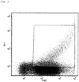
- Fig. 2 shows the result of the measurement.
- the fluorescence signal of the cells is plotted in the dot plot diagram in the Y direction against the scatter, which is a measure of the size of the particle, in the X direction.
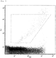
- Example 1 described above was repeated, except that before the fixed cells were dried no homogenizing agent was added. The results are in Fig. 3 shown.
- cluster formation results from the strongly divergent distribution of the detected particles in relation to the size (X-axis of the dot plot) and the intensity of the detected fluorescence signal (Y-axis of the dot plot), as a cluster, which consists of a larger number is formed by individual cells, is perceived by the detector as particles with a higher intensity of the fluorescence signal than a cluster consisting of a smaller number of individual cells. Accordingly, the distribution of the detected particles in the dot plot diverges very strongly, which greatly limits a differentiated determination of the organisms.
- Example 1 described above was essentially repeated, except that the in WO 03/083131 A1 described method was applied and no quencher-labeled nucleic acid probe was used. The result is in Fig. 4 shown.
- Example 1 described above was repeated, with the exception that, instead of the homogenizing agent used in Example 1, 70% strength ethanol was used as the homogenizing agent. The results are in Fig. 5 shown.
Landscapes
- Chemical & Material Sciences (AREA)
- Life Sciences & Earth Sciences (AREA)
- Organic Chemistry (AREA)
- Health & Medical Sciences (AREA)
- Wood Science & Technology (AREA)
- Proteomics, Peptides & Aminoacids (AREA)
- Zoology (AREA)
- Engineering & Computer Science (AREA)
- Immunology (AREA)
- Physics & Mathematics (AREA)
- General Health & Medical Sciences (AREA)
- Biochemistry (AREA)
- Analytical Chemistry (AREA)
- General Engineering & Computer Science (AREA)
- Biotechnology (AREA)
- Molecular Biology (AREA)
- Bioinformatics & Cheminformatics (AREA)
- Biophysics (AREA)
- Microbiology (AREA)
- Genetics & Genomics (AREA)
- Dispersion Chemistry (AREA)
- General Physics & Mathematics (AREA)
- Pathology (AREA)
- Toxicology (AREA)
- Measuring Or Testing Involving Enzymes Or Micro-Organisms (AREA)
Claims (14)
- Procédé pour la mise en évidence d'un micro-organisme dans un échantillon, comprenant les étapes :(a) de fourniture de l'échantillon ;(b) de fixation des cellules contenues dans l'échantillon avec un agent fixateur et de séparation des cellules fixées obtenues à cette occasion de l'échantillon, afin de fournir des cellules fixées ;c) d'amenée en contact des cellules fixées avec un agent d'homogénéisation chimique et de séchage des cellules homogénéisées obtenues à cette occasion, afin de fournir des cellules séchées, dans lequel l'agent d'homogénéisation chimique comprend (i) un monosaccharide ou disaccharide, (ii) un polyol, et (iii) de l'eau ;(d) d'amenée en contact des cellules séchées avec une solution d'une sonde d'acide nucléique marquée par fluorescence, spécifique du micro-organisme à mettre en évidence, afin de fournir un premier mélange réactionnel ;(e) d'incubation du premier mélange réactionnel, afin de provoquer une liaison de la sonde d'acide nucléique marquée par fluorescence à la séquence d'acide nucléique cible correspondante dans les cellules du micro-organisme à mettre en évidence ;(f) d'amenée en contact du premier mélange réactionnel à la suite de l'étape (e) avec une solution d'une sonde d'acide nucléique marquée par un quencher, afin de fournir un deuxième mélange réactionnel, dans lequel la sonde d'acide nucléique marquée par un quencher comprend un quencher éteignant au moins en partie la fluorescence de la sonde d'acide nucléique marquée par fluorescence et présente une séquence d'acide nucléique, laquelle est sensiblement complémentaire de la séquence d'acide nucléique de la sonde d'acide nucléique marquée par fluorescence ;(g) d'incubation du deuxième mélange réactionnel, afin de provoquer une liaison de la molécule de la sonde d'acide nucléique marquée par fluorescence non liée à la séquence d'acide nucléique cible dans les cellules du micro-organisme à mettre en évidence à la sonde d'acide nucléique marquée par un quencher ; et(h) d'introduction du deuxième mélange réactionnel à la suite de l'étape (g) dans un cytomètre en flux, et de détection de la fluorescence, laquelle est émise par les cellules du micro-organisme à mettre en évidence comportant la sonde d'acide nucléique marquée par fluorescence.
- Procédé selon la revendication 1, dans lequel il s'agit pour la sonde d'une sonde liquide.
- Procédé selon la revendication 1 ou 2, dans lequel il s'agit pour le micro-organisme d'une bactérie, d'un champignon, ou d'un organisme supérieur unicellulaire (protozoaire).
- Procédé selon la revendication 3, dans lequel il s'agit pour la bactérie d'une bactérie du genre Acinetobacter, Alicyclobacillus, Aquabacterium, Arcobacter, Bacillus, Campylobacter, Enterobacteriaceae, Escherichia, Lactobacillus, Lactococcus, Legionella, Listeria, Microthrix, Nitrobacter, Nitrosococcus, Nitrosomonas, Nitrospira, Nitrotoga, Propionibacterium, Salmonella, Shigella ou Streptococcus.
- Procédé selon la revendication 3, dans lequel il s'agit pour le champignon d'un champignon du genre Aspergillus, Candida, Debaromyces, Dekkera, Penicillium, Pichia ou Saccharomyces.
- Procédé selon l'une quelconque des revendications 1 à 5, dans lequel il s'agit pour le monosaccharide ou disaccharide d'une substance sélectionnée dans le groupe constitué du fructose, galactose, glucose et saccharose.
- Procédé selon l'une quelconque des revendications 1 à 6, dans lequel il s'agit pour le polyol d'une substance sélectionnée dans le groupe constitué d'éthylène glycol, de glycérine, de mannitol et de sorbitol.
- Procédé selon l'une quelconque des revendications 1 à 7, dans lequel la séquence d'acide nucléique cible dans les cellules du micro-organisme à mettre en évidence est sélectionnée dans le groupe constitué de ARNr 16S, ARNr 23S, ARNr 18S, ARNt, EF-Tu, ARNm espaceur ARNr 16S-23S et espaceur ARNr 23S-5S.
- Procédé selon l'une quelconque des revendications 1 à 8, dans lequel la sonde d'acide nucléique marquée par fluorescence est (i) sensiblement identique ou (ii) sensiblement inversement complémentaire de la séquence d'acide nucléique cible dans les cellules du micro-organisme à mettre en évidence.
- Procédé selon l'une quelconque des revendications 1 à 9, dans lequel la sonde d'acide nucléique marquée par fluorescence est sélectionnée parmi une sonde ADN, sonde ARN, sonde PNS et sonde LNS marquée par fluorescence.
- Procédé selon l'une quelconque des revendications 1 à 10, dans lequel (i) le fluorochrome de la sonde d'acide nucléique marquée par fluorescence est disposé à l'extrémité 3' ou près de l'extrémité 3' de la sonde d'acide nucléique marquée par fluorescence et le quencher de la sonde d'acide nucléique marquée par un quencher est disposé à l'extrémité 5' ou près de l'extrémité 5' de la sonde d'acide nucléique marquée par un quencher, ou (ii) le fluorochrome de la sonde d'acide nucléique marquée par fluorescence est disposé à l'extrémité 5' ou près de l'extrémité 5' de la sonde d'acide nucléique marquée par fluorescence et le quencher de la sonde d'acide nucléique marquée par un quencher est disposé à l'extrémité 3' ou près de l'extrémité 3' de la sonde d'acide nucléique marquée par un quencher.
- Procédé selon l'une quelconque des revendications 1 à 11, dans lequel à l'étape (d) plusieurs sondes d'acide nucléique marquées par fluorescence respectivement spécifiques du micro-organisme à mettre en évidence sont ajoutées à des séquences d'acide nucléique différentes, et à l'étape (f) un nombre de sondes d'acide nucléique différentes marquées par un quencher correspondant au nombre de sondes d'acide nucléique marquées par fluorescence est ajouté.
- Procédé selon l'une quelconque des revendications 1 à 12, dans lequel la sonde contient plus d'un micro-organisme et plusieurs micro-organismes différents sont mis en évidence simultanément.
- Utilisation d'un cytomètre en flux dans un procédé selon l'une quelconque des revendications 1 à 13.
Applications Claiming Priority (2)
| Application Number | Priority Date | Filing Date | Title |
|---|---|---|---|
| EP17187339.1A EP3447146A1 (fr) | 2017-08-22 | 2017-08-22 | Procédé de détection spécifique de micro-organismes |
| PCT/EP2018/072240 WO2019038181A1 (fr) | 2017-08-22 | 2018-08-16 | Procédé de mise en évidence spécifique de micro-organismes |
Publications (2)
| Publication Number | Publication Date |
|---|---|
| EP3673075A1 EP3673075A1 (fr) | 2020-07-01 |
| EP3673075B1 true EP3673075B1 (fr) | 2021-10-27 |
Family
ID=59738162
Family Applications (2)
| Application Number | Title | Priority Date | Filing Date |
|---|---|---|---|
| EP17187339.1A Withdrawn EP3447146A1 (fr) | 2017-08-22 | 2017-08-22 | Procédé de détection spécifique de micro-organismes |
| EP18752516.7A Active EP3673075B1 (fr) | 2017-08-22 | 2018-08-16 | Procédé de détection spécifique de micro-organismes |
Family Applications Before (1)
| Application Number | Title | Priority Date | Filing Date |
|---|---|---|---|
| EP17187339.1A Withdrawn EP3447146A1 (fr) | 2017-08-22 | 2017-08-22 | Procédé de détection spécifique de micro-organismes |
Country Status (7)
| Country | Link |
|---|---|
| US (1) | US11680287B2 (fr) |
| EP (2) | EP3447146A1 (fr) |
| JP (1) | JP7333311B2 (fr) |
| AU (1) | AU2018321102B2 (fr) |
| ES (1) | ES2899383T3 (fr) |
| PT (1) | PT3673075T (fr) |
| WO (1) | WO2019038181A1 (fr) |
Cited By (1)
| Publication number | Priority date | Publication date | Assignee | Title |
|---|---|---|---|---|
| DE102024105858B3 (de) | 2024-02-29 | 2025-02-20 | VERMICON Aktiengesellschaft | Verfahren zum spezifischen nachweis von mikroorganismen und insbesondere von gram-positiven bakterien |
Families Citing this family (1)
| Publication number | Priority date | Publication date | Assignee | Title |
|---|---|---|---|---|
| CN113502320A (zh) * | 2021-08-06 | 2021-10-15 | 海南微氪生物科技股份有限公司 | 一种具有稳定荧光特性的智能生物探针微生物检测方法 |
Family Cites Families (9)
| Publication number | Priority date | Publication date | Assignee | Title |
|---|---|---|---|---|
| JPH0746101B2 (ja) * | 1986-12-30 | 1995-05-17 | 素秀 高浜 | 粘液溶解性を具備した細胞固定・保存液 |
| DE50014456D1 (de) | 1999-05-07 | 2007-08-16 | Vermicon Ag | Verfahren zum nachweisen von mikroorganismen in einer probe |
| DE10129410A1 (de) | 2001-06-19 | 2003-01-02 | Vermicon Ag | Verfahren zum spezifischen Schnellnachweis bierschädlicher Bakterien |
| DE10160666A1 (de) | 2001-06-19 | 2003-01-09 | Vermicon Ag | Verfahren zum spezifischen Schnellnachweis von Trinkwasser relevanten Bakterien |
| DE10214153A1 (de) | 2002-03-28 | 2003-11-13 | Vermicon Ag | Verfahren zum Nachweis von Mikroorganismen mittels in situ-Hybridisierung und Durchflusszytometrie |
| US20050136446A1 (en) * | 2002-03-28 | 2005-06-23 | Jiri Snaidr | Method for the identification of microorganisms by means of in situ hybridization and flow cytometry |
| DE10344057B3 (de) | 2003-09-23 | 2005-06-09 | Vermicon Ag | Verfahren zum spezifischen Schnellnachweis getränkeschädlicher Mikroorganismen |
| DE102010012421B4 (de) | 2009-03-23 | 2023-04-20 | Vermicon Ag | Verfahren zum spezifischen Nachweis von Mikroorganismen |
| WO2016178953A1 (fr) * | 2015-05-01 | 2016-11-10 | The General Hospital Corporation | Analyse multiplexe de l'expression génique dans des cellules vivantes individuelles |
-
2017
- 2017-08-22 EP EP17187339.1A patent/EP3447146A1/fr not_active Withdrawn
-
2018
- 2018-08-16 US US16/641,308 patent/US11680287B2/en active Active
- 2018-08-16 PT PT187525167T patent/PT3673075T/pt unknown
- 2018-08-16 WO PCT/EP2018/072240 patent/WO2019038181A1/fr not_active Ceased
- 2018-08-16 ES ES18752516T patent/ES2899383T3/es active Active
- 2018-08-16 JP JP2020511289A patent/JP7333311B2/ja active Active
- 2018-08-16 EP EP18752516.7A patent/EP3673075B1/fr active Active
- 2018-08-16 AU AU2018321102A patent/AU2018321102B2/en active Active
Cited By (2)
| Publication number | Priority date | Publication date | Assignee | Title |
|---|---|---|---|---|
| DE102024105858B3 (de) | 2024-02-29 | 2025-02-20 | VERMICON Aktiengesellschaft | Verfahren zum spezifischen nachweis von mikroorganismen und insbesondere von gram-positiven bakterien |
| WO2025180575A1 (fr) | 2024-02-29 | 2025-09-04 | Vermicon Ag | Procédé pour détecter spécifiquement des micro-organismes et, plus particulièrement, des bactéries à gram positif |
Also Published As
| Publication number | Publication date |
|---|---|
| EP3447146A1 (fr) | 2019-02-27 |
| US20200385793A1 (en) | 2020-12-10 |
| US11680287B2 (en) | 2023-06-20 |
| PT3673075T (pt) | 2021-12-09 |
| WO2019038181A1 (fr) | 2019-02-28 |
| AU2018321102B2 (en) | 2024-06-13 |
| ES2899383T3 (es) | 2022-03-11 |
| JP2020531024A (ja) | 2020-11-05 |
| AU2018321102A1 (en) | 2020-04-16 |
| EP3673075A1 (fr) | 2020-07-01 |
| JP7333311B2 (ja) | 2023-08-24 |
Similar Documents
| Publication | Publication Date | Title |
|---|---|---|
| DE69434255T2 (de) | Nukleinsäuresonde zum nachweis von lactobacillus | |
| DE19732086C2 (de) | Verfahren zur quantitativen Bestimmung von Eubakterien | |
| EP1198597A1 (fr) | Procede permettant de detecter des organismes de fa on specifique a l'espece | |
| DE19945916A1 (de) | Nukleinsäuremoleküle zum Nachweis von Bakterien und phylogenetischen Einheiten von Bakterien | |
| EP1177319B1 (fr) | Procede de detection de micro-organismes dans un echantillon | |
| EP3673075B1 (fr) | Procédé de détection spécifique de micro-organismes | |
| EP2162553A2 (fr) | Procédé et test rapide pour identifier des séquences d'acide nucléique spécifiques | |
| EP1664351B1 (fr) | Procede de detection specifique rapide de micro-organismes nocifs pour les boissons | |
| DE102015012691A1 (de) | Verfahren zum quantitativen Nachweis von Vibrio parahaemolyticus, Vibrio vulnificus und Vibrio cholerae | |
| EP1522595A2 (fr) | Oligonucléotides, méthodes et système pour détecter par real-time PCR des gènes bactériens de résistance aux antibiotiques | |
| DE102024105858B3 (de) | Verfahren zum spezifischen nachweis von mikroorganismen und insbesondere von gram-positiven bakterien | |
| DE10100493A1 (de) | Nachweis von pathogenen Bakterien | |
| DE102010012421B4 (de) | Verfahren zum spezifischen Nachweis von Mikroorganismen | |
| EP1490507B1 (fr) | Procede de detection de micro-organismes par une hybridation in situ et une cytometrie de flux | |
| EP1397519A2 (fr) | Procede pour la mise en evidence rapide specifique de bacteries a effet nefaste sur la biere | |
| DE102020103957B4 (de) | Verfahren zum Nachweis von Mikroorganismen und ein fluidisches Kanalsystem | |
| DE102010052524A1 (de) | Verfahren zum qualitativen und quantitativen Nachweis von spezifischen Nukleinsäuresequenzen in Echtzeit | |
| WO2011124684A1 (fr) | Procédé de détection de séquences d'acide nucléique spécifiques | |
| EP2877595B1 (fr) | Procédé de différenciation entre cellules vivantes et mortes | |
| EP3865588A2 (fr) | Procédé de détection des microorganismes et système de canal fluidique | |
| BE1032965B1 (de) | Primerpaar und sonde für recombinase-polymerase-amplifikation zum schnellen nachweis von rhizoctonia solani | |
| WO2006108205A2 (fr) | Procede de detection de fragments d'acide nucleique | |
| EP4705505A1 (fr) | Substrat enzymatique pour la détection de la toxine shiga | |
| DE102008029999A1 (de) | Neue PCR-gestützte Nachweismethode zur Unterscheidung lebender von toten Zellen | |
| DE112013002589T5 (de) | Extraktionskontrolle für DNA |
Legal Events
| Date | Code | Title | Description |
|---|---|---|---|
| STAA | Information on the status of an ep patent application or granted ep patent |
Free format text: STATUS: UNKNOWN |
|
| STAA | Information on the status of an ep patent application or granted ep patent |
Free format text: STATUS: THE INTERNATIONAL PUBLICATION HAS BEEN MADE |
|
| PUAI | Public reference made under article 153(3) epc to a published international application that has entered the european phase |
Free format text: ORIGINAL CODE: 0009012 |
|
| STAA | Information on the status of an ep patent application or granted ep patent |
Free format text: STATUS: REQUEST FOR EXAMINATION WAS MADE |
|
| 17P | Request for examination filed |
Effective date: 20200316 |
|
| AK | Designated contracting states |
Kind code of ref document: A1 Designated state(s): AL AT BE BG CH CY CZ DE DK EE ES FI FR GB GR HR HU IE IS IT LI LT LU LV MC MK MT NL NO PL PT RO RS SE SI SK SM TR |
|
| AX | Request for extension of the european patent |
Extension state: BA ME |
|
| DAV | Request for validation of the european patent (deleted) | ||
| DAX | Request for extension of the european patent (deleted) | ||
| STAA | Information on the status of an ep patent application or granted ep patent |
Free format text: STATUS: EXAMINATION IS IN PROGRESS |
|
| 17Q | First examination report despatched |
Effective date: 20210315 |
|
| GRAP | Despatch of communication of intention to grant a patent |
Free format text: ORIGINAL CODE: EPIDOSNIGR1 |
|
| STAA | Information on the status of an ep patent application or granted ep patent |
Free format text: STATUS: GRANT OF PATENT IS INTENDED |
|
| INTG | Intention to grant announced |
Effective date: 20210805 |
|
| GRAS | Grant fee paid |
Free format text: ORIGINAL CODE: EPIDOSNIGR3 |
|
| GRAA | (expected) grant |
Free format text: ORIGINAL CODE: 0009210 |
|
| STAA | Information on the status of an ep patent application or granted ep patent |
Free format text: STATUS: THE PATENT HAS BEEN GRANTED |
|
| AK | Designated contracting states |
Kind code of ref document: B1 Designated state(s): AL AT BE BG CH CY CZ DE DK EE ES FI FR GB GR HR HU IE IS IT LI LT LU LV MC MK MT NL NO PL PT RO RS SE SI SK SM TR |
|
| REG | Reference to a national code |
Ref country code: GB Ref legal event code: FG4D Free format text: NOT ENGLISH |
|
| REG | Reference to a national code |
Ref country code: CH Ref legal event code: EP |
|
| REG | Reference to a national code |
Ref country code: DE Ref legal event code: R096 Ref document number: 502018007605 Country of ref document: DE |
|
| REG | Reference to a national code |
Ref country code: AT Ref legal event code: REF Ref document number: 1441845 Country of ref document: AT Kind code of ref document: T Effective date: 20211115 |
|
| REG | Reference to a national code |
Ref country code: IE Ref legal event code: FG4D Free format text: LANGUAGE OF EP DOCUMENT: GERMAN |
|
| REG | Reference to a national code |
Ref country code: PT Ref legal event code: SC4A Ref document number: 3673075 Country of ref document: PT Date of ref document: 20211209 Kind code of ref document: T Free format text: AVAILABILITY OF NATIONAL TRANSLATION Effective date: 20211202 |
|
| REG | Reference to a national code |
Ref country code: LT Ref legal event code: MG9D |
|
| REG | Reference to a national code |
Ref country code: NL Ref legal event code: MP Effective date: 20211027 |
|
| REG | Reference to a national code |
Ref country code: ES Ref legal event code: FG2A Ref document number: 2899383 Country of ref document: ES Kind code of ref document: T3 Effective date: 20220311 |
|
| PG25 | Lapsed in a contracting state [announced via postgrant information from national office to epo] |
Ref country code: RS Free format text: LAPSE BECAUSE OF FAILURE TO SUBMIT A TRANSLATION OF THE DESCRIPTION OR TO PAY THE FEE WITHIN THE PRESCRIBED TIME-LIMIT Effective date: 20211027 Ref country code: LT Free format text: LAPSE BECAUSE OF FAILURE TO SUBMIT A TRANSLATION OF THE DESCRIPTION OR TO PAY THE FEE WITHIN THE PRESCRIBED TIME-LIMIT Effective date: 20211027 Ref country code: FI Free format text: LAPSE BECAUSE OF FAILURE TO SUBMIT A TRANSLATION OF THE DESCRIPTION OR TO PAY THE FEE WITHIN THE PRESCRIBED TIME-LIMIT Effective date: 20211027 Ref country code: BG Free format text: LAPSE BECAUSE OF FAILURE TO SUBMIT A TRANSLATION OF THE DESCRIPTION OR TO PAY THE FEE WITHIN THE PRESCRIBED TIME-LIMIT Effective date: 20220127 |
|
| PG25 | Lapsed in a contracting state [announced via postgrant information from national office to epo] |
Ref country code: IS Free format text: LAPSE BECAUSE OF FAILURE TO SUBMIT A TRANSLATION OF THE DESCRIPTION OR TO PAY THE FEE WITHIN THE PRESCRIBED TIME-LIMIT Effective date: 20220227 Ref country code: SE Free format text: LAPSE BECAUSE OF FAILURE TO SUBMIT A TRANSLATION OF THE DESCRIPTION OR TO PAY THE FEE WITHIN THE PRESCRIBED TIME-LIMIT Effective date: 20211027 Ref country code: PL Free format text: LAPSE BECAUSE OF FAILURE TO SUBMIT A TRANSLATION OF THE DESCRIPTION OR TO PAY THE FEE WITHIN THE PRESCRIBED TIME-LIMIT Effective date: 20211027 Ref country code: NO Free format text: LAPSE BECAUSE OF FAILURE TO SUBMIT A TRANSLATION OF THE DESCRIPTION OR TO PAY THE FEE WITHIN THE PRESCRIBED TIME-LIMIT Effective date: 20220127 Ref country code: NL Free format text: LAPSE BECAUSE OF FAILURE TO SUBMIT A TRANSLATION OF THE DESCRIPTION OR TO PAY THE FEE WITHIN THE PRESCRIBED TIME-LIMIT Effective date: 20211027 Ref country code: LV Free format text: LAPSE BECAUSE OF FAILURE TO SUBMIT A TRANSLATION OF THE DESCRIPTION OR TO PAY THE FEE WITHIN THE PRESCRIBED TIME-LIMIT Effective date: 20211027 Ref country code: HR Free format text: LAPSE BECAUSE OF FAILURE TO SUBMIT A TRANSLATION OF THE DESCRIPTION OR TO PAY THE FEE WITHIN THE PRESCRIBED TIME-LIMIT Effective date: 20211027 Ref country code: GR Free format text: LAPSE BECAUSE OF FAILURE TO SUBMIT A TRANSLATION OF THE DESCRIPTION OR TO PAY THE FEE WITHIN THE PRESCRIBED TIME-LIMIT Effective date: 20220128 |
|
| REG | Reference to a national code |
Ref country code: DE Ref legal event code: R097 Ref document number: 502018007605 Country of ref document: DE |
|
| PG25 | Lapsed in a contracting state [announced via postgrant information from national office to epo] |
Ref country code: SM Free format text: LAPSE BECAUSE OF FAILURE TO SUBMIT A TRANSLATION OF THE DESCRIPTION OR TO PAY THE FEE WITHIN THE PRESCRIBED TIME-LIMIT Effective date: 20211027 Ref country code: SK Free format text: LAPSE BECAUSE OF FAILURE TO SUBMIT A TRANSLATION OF THE DESCRIPTION OR TO PAY THE FEE WITHIN THE PRESCRIBED TIME-LIMIT Effective date: 20211027 Ref country code: RO Free format text: LAPSE BECAUSE OF FAILURE TO SUBMIT A TRANSLATION OF THE DESCRIPTION OR TO PAY THE FEE WITHIN THE PRESCRIBED TIME-LIMIT Effective date: 20211027 Ref country code: EE Free format text: LAPSE BECAUSE OF FAILURE TO SUBMIT A TRANSLATION OF THE DESCRIPTION OR TO PAY THE FEE WITHIN THE PRESCRIBED TIME-LIMIT Effective date: 20211027 Ref country code: DK Free format text: LAPSE BECAUSE OF FAILURE TO SUBMIT A TRANSLATION OF THE DESCRIPTION OR TO PAY THE FEE WITHIN THE PRESCRIBED TIME-LIMIT Effective date: 20211027 Ref country code: CZ Free format text: LAPSE BECAUSE OF FAILURE TO SUBMIT A TRANSLATION OF THE DESCRIPTION OR TO PAY THE FEE WITHIN THE PRESCRIBED TIME-LIMIT Effective date: 20211027 |
|
| PLBE | No opposition filed within time limit |
Free format text: ORIGINAL CODE: 0009261 |
|
| STAA | Information on the status of an ep patent application or granted ep patent |
Free format text: STATUS: NO OPPOSITION FILED WITHIN TIME LIMIT |
|
| 26N | No opposition filed |
Effective date: 20220728 |
|
| PG25 | Lapsed in a contracting state [announced via postgrant information from national office to epo] |
Ref country code: AL Free format text: LAPSE BECAUSE OF FAILURE TO SUBMIT A TRANSLATION OF THE DESCRIPTION OR TO PAY THE FEE WITHIN THE PRESCRIBED TIME-LIMIT Effective date: 20211027 |
|
| PG25 | Lapsed in a contracting state [announced via postgrant information from national office to epo] |
Ref country code: SI Free format text: LAPSE BECAUSE OF FAILURE TO SUBMIT A TRANSLATION OF THE DESCRIPTION OR TO PAY THE FEE WITHIN THE PRESCRIBED TIME-LIMIT Effective date: 20211027 |
|
| PG25 | Lapsed in a contracting state [announced via postgrant information from national office to epo] |
Ref country code: MC Free format text: LAPSE BECAUSE OF FAILURE TO SUBMIT A TRANSLATION OF THE DESCRIPTION OR TO PAY THE FEE WITHIN THE PRESCRIBED TIME-LIMIT Effective date: 20211027 |
|
| REG | Reference to a national code |
Ref country code: CH Ref legal event code: PL |
|
| PG25 | Lapsed in a contracting state [announced via postgrant information from national office to epo] |
Ref country code: LU Free format text: LAPSE BECAUSE OF NON-PAYMENT OF DUE FEES Effective date: 20220816 Ref country code: LI Free format text: LAPSE BECAUSE OF NON-PAYMENT OF DUE FEES Effective date: 20220831 Ref country code: CH Free format text: LAPSE BECAUSE OF NON-PAYMENT OF DUE FEES Effective date: 20220831 |
|
| REG | Reference to a national code |
Ref country code: BE Ref legal event code: MM Effective date: 20220831 |
|
| PG25 | Lapsed in a contracting state [announced via postgrant information from national office to epo] |
Ref country code: IE Free format text: LAPSE BECAUSE OF NON-PAYMENT OF DUE FEES Effective date: 20220816 |
|
| PG25 | Lapsed in a contracting state [announced via postgrant information from national office to epo] |
Ref country code: BE Free format text: LAPSE BECAUSE OF NON-PAYMENT OF DUE FEES Effective date: 20220831 |
|
| PG25 | Lapsed in a contracting state [announced via postgrant information from national office to epo] |
Ref country code: CY Free format text: LAPSE BECAUSE OF FAILURE TO SUBMIT A TRANSLATION OF THE DESCRIPTION OR TO PAY THE FEE WITHIN THE PRESCRIBED TIME-LIMIT Effective date: 20211027 |
|
| PG25 | Lapsed in a contracting state [announced via postgrant information from national office to epo] |
Ref country code: MK Free format text: LAPSE BECAUSE OF FAILURE TO SUBMIT A TRANSLATION OF THE DESCRIPTION OR TO PAY THE FEE WITHIN THE PRESCRIBED TIME-LIMIT Effective date: 20211027 Ref country code: HU Free format text: LAPSE BECAUSE OF FAILURE TO SUBMIT A TRANSLATION OF THE DESCRIPTION OR TO PAY THE FEE WITHIN THE PRESCRIBED TIME-LIMIT; INVALID AB INITIO Effective date: 20180816 |
|
| PG25 | Lapsed in a contracting state [announced via postgrant information from national office to epo] |
Ref country code: MT Free format text: LAPSE BECAUSE OF FAILURE TO SUBMIT A TRANSLATION OF THE DESCRIPTION OR TO PAY THE FEE WITHIN THE PRESCRIBED TIME-LIMIT Effective date: 20211027 |
|
| PGFP | Annual fee paid to national office [announced via postgrant information from national office to epo] |
Ref country code: PT Payment date: 20250801 Year of fee payment: 8 Ref country code: ES Payment date: 20250917 Year of fee payment: 8 |
|
| PGFP | Annual fee paid to national office [announced via postgrant information from national office to epo] |
Ref country code: DE Payment date: 20250819 Year of fee payment: 8 |
|
| PGFP | Annual fee paid to national office [announced via postgrant information from national office to epo] |
Ref country code: IT Payment date: 20250829 Year of fee payment: 8 |
|
| PGFP | Annual fee paid to national office [announced via postgrant information from national office to epo] |
Ref country code: GB Payment date: 20250818 Year of fee payment: 8 |
|
| PGFP | Annual fee paid to national office [announced via postgrant information from national office to epo] |
Ref country code: AT Payment date: 20250819 Year of fee payment: 8 Ref country code: FR Payment date: 20250827 Year of fee payment: 8 |
|
| PG25 | Lapsed in a contracting state [announced via postgrant information from national office to epo] |
Ref country code: TR Free format text: LAPSE BECAUSE OF FAILURE TO SUBMIT A TRANSLATION OF THE DESCRIPTION OR TO PAY THE FEE WITHIN THE PRESCRIBED TIME-LIMIT Effective date: 20211027 |