EP0785430B1 - Trennung von Plasma oder Serum von Vollblut - Google Patents
Trennung von Plasma oder Serum von Vollblut Download PDFInfo
- Publication number
- EP0785430B1 EP0785430B1 EP97100695A EP97100695A EP0785430B1 EP 0785430 B1 EP0785430 B1 EP 0785430B1 EP 97100695 A EP97100695 A EP 97100695A EP 97100695 A EP97100695 A EP 97100695A EP 0785430 B1 EP0785430 B1 EP 0785430B1
- Authority
- EP
- European Patent Office
- Prior art keywords
- plasma
- whole blood
- blood
- membrane
- agent
- Prior art date
- Legal status (The legal status is an assumption and is not a legal conclusion. Google has not performed a legal analysis and makes no representation as to the accuracy of the status listed.)
- Expired - Lifetime
Links
- 210000004369 blood Anatomy 0.000 title claims description 107
- 239000008280 blood Substances 0.000 title claims description 103
- 210000002966 serum Anatomy 0.000 title claims description 14
- 238000000926 separation method Methods 0.000 title description 14
- 210000002381 plasma Anatomy 0.000 claims description 98
- 239000012528 membrane Substances 0.000 claims description 52
- 238000001914 filtration Methods 0.000 claims description 45
- 239000003365 glass fiber Substances 0.000 claims description 41
- 210000000601 blood cell Anatomy 0.000 claims description 35
- 239000012982 microporous membrane Substances 0.000 claims description 28
- INHCSSUBVCNVSK-UHFFFAOYSA-L lithium sulfate Chemical compound [Li+].[Li+].[O-]S([O-])(=O)=O INHCSSUBVCNVSK-UHFFFAOYSA-L 0.000 claims description 25
- 238000000034 method Methods 0.000 claims description 25
- YCKRFDGAMUMZLT-UHFFFAOYSA-N Fluorine atom Chemical compound [F] YCKRFDGAMUMZLT-UHFFFAOYSA-N 0.000 claims description 18
- 239000011737 fluorine Substances 0.000 claims description 18
- 229910052731 fluorine Inorganic materials 0.000 claims description 18
- 239000000463 material Substances 0.000 claims description 18
- FAPWRFPIFSIZLT-UHFFFAOYSA-M Sodium chloride Chemical compound [Na+].[Cl-] FAPWRFPIFSIZLT-UHFFFAOYSA-M 0.000 claims description 14
- 150000001413 amino acids Chemical class 0.000 claims description 12
- FLJPGEWQYJVDPF-UHFFFAOYSA-L caesium sulfate Chemical compound [Cs+].[Cs+].[O-]S([O-])(=O)=O FLJPGEWQYJVDPF-UHFFFAOYSA-L 0.000 claims description 12
- 229910017053 inorganic salt Inorganic materials 0.000 claims description 12
- 229920002492 poly(sulfone) Polymers 0.000 claims description 12
- 150000003839 salts Chemical class 0.000 claims description 11
- 238000002156 mixing Methods 0.000 claims description 9
- 239000011734 sodium Substances 0.000 claims description 9
- 239000011780 sodium chloride Substances 0.000 claims description 9
- 239000007864 aqueous solution Substances 0.000 claims description 7
- 229910052784 alkaline earth metal Inorganic materials 0.000 claims description 4
- 150000001342 alkaline earth metals Chemical class 0.000 claims description 4
- 229910052736 halogen Inorganic materials 0.000 claims description 4
- 150000002367 halogens Chemical class 0.000 claims description 4
- IIPYXGDZVMZOAP-UHFFFAOYSA-N lithium nitrate Chemical compound [Li+].[O-][N+]([O-])=O IIPYXGDZVMZOAP-UHFFFAOYSA-N 0.000 claims description 4
- 229920005597 polymer membrane Polymers 0.000 claims description 4
- UXVMQQNJUSDDNG-UHFFFAOYSA-L Calcium chloride Chemical compound [Cl-].[Cl-].[Ca+2] UXVMQQNJUSDDNG-UHFFFAOYSA-L 0.000 claims description 3
- 229910052783 alkali metal Inorganic materials 0.000 claims description 3
- 150000001340 alkali metals Chemical class 0.000 claims description 3
- 239000001110 calcium chloride Substances 0.000 claims description 3
- 229910001628 calcium chloride Inorganic materials 0.000 claims description 3
- 229910000344 rubidium sulfate Inorganic materials 0.000 claims description 3
- 239000003795 chemical substances by application Substances 0.000 description 60
- 238000005534 hematocrit Methods 0.000 description 34
- 206010018910 Haemolysis Diseases 0.000 description 22
- 239000000306 component Substances 0.000 description 22
- 230000008588 hemolysis Effects 0.000 description 22
- 210000003743 erythrocyte Anatomy 0.000 description 21
- 229920000642 polymer Polymers 0.000 description 19
- 239000000523 sample Substances 0.000 description 16
- 239000000243 solution Substances 0.000 description 15
- 238000005259 measurement Methods 0.000 description 13
- 235000001014 amino acid Nutrition 0.000 description 11
- 229940024606 amino acid Drugs 0.000 description 11
- -1 amino acid salt Chemical class 0.000 description 10
- 238000003556 assay Methods 0.000 description 10
- 102000001554 Hemoglobins Human genes 0.000 description 8
- 108010054147 Hemoglobins Proteins 0.000 description 8
- 229920002678 cellulose Polymers 0.000 description 8
- 239000011148 porous material Substances 0.000 description 8
- 239000004094 surface-active agent Substances 0.000 description 8
- HTTJABKRGRZYRN-UHFFFAOYSA-N Heparin Chemical compound OC1C(NC(=O)C)C(O)OC(COS(O)(=O)=O)C1OC1C(OS(O)(=O)=O)C(O)C(OC2C(C(OS(O)(=O)=O)C(OC3C(C(O)C(O)C(O3)C(O)=O)OS(O)(=O)=O)C(CO)O2)NS(O)(=O)=O)C(C(O)=O)O1 HTTJABKRGRZYRN-UHFFFAOYSA-N 0.000 description 7
- 235000010724 Wisteria floribunda Nutrition 0.000 description 7
- 239000001913 cellulose Substances 0.000 description 7
- 230000007423 decrease Effects 0.000 description 7
- 230000000694 effects Effects 0.000 description 7
- 229960002897 heparin Drugs 0.000 description 7
- 229920000669 heparin Polymers 0.000 description 7
- 230000003204 osmotic effect Effects 0.000 description 7
- 239000011800 void material Substances 0.000 description 7
- XLYOFNOQVPJJNP-UHFFFAOYSA-N water Substances O XLYOFNOQVPJJNP-UHFFFAOYSA-N 0.000 description 7
- 150000002500 ions Chemical class 0.000 description 6
- 239000010410 layer Substances 0.000 description 6
- 239000007788 liquid Substances 0.000 description 6
- IMROMDMJAWUWLK-UHFFFAOYSA-N Ethenol Chemical group OC=C IMROMDMJAWUWLK-UHFFFAOYSA-N 0.000 description 5
- 239000011575 calcium Substances 0.000 description 5
- 238000010790 dilution Methods 0.000 description 5
- 239000012895 dilution Substances 0.000 description 5
- 238000001035 drying Methods 0.000 description 5
- 239000003792 electrolyte Substances 0.000 description 5
- 239000002202 Polyethylene glycol Substances 0.000 description 4
- 229910006130 SO4 Inorganic materials 0.000 description 4
- 230000009471 action Effects 0.000 description 4
- 239000000853 adhesive Substances 0.000 description 4
- 230000001070 adhesive effect Effects 0.000 description 4
- 239000012491 analyte Substances 0.000 description 4
- 238000004458 analytical method Methods 0.000 description 4
- 230000000052 comparative effect Effects 0.000 description 4
- 150000001875 compounds Chemical class 0.000 description 4
- 239000004744 fabric Substances 0.000 description 4
- 239000002736 nonionic surfactant Substances 0.000 description 4
- 229920001223 polyethylene glycol Polymers 0.000 description 4
- 238000002360 preparation method Methods 0.000 description 4
- 238000011084 recovery Methods 0.000 description 4
- LFQSCWFLJHTTHZ-UHFFFAOYSA-N Ethanol Chemical compound CCO LFQSCWFLJHTTHZ-UHFFFAOYSA-N 0.000 description 3
- LYCAIKOWRPUZTN-UHFFFAOYSA-N Ethylene glycol Chemical compound OCCO LYCAIKOWRPUZTN-UHFFFAOYSA-N 0.000 description 3
- ZMXDDKWLCZADIW-UHFFFAOYSA-N N,N-Dimethylformamide Chemical compound CN(C)C=O ZMXDDKWLCZADIW-UHFFFAOYSA-N 0.000 description 3
- 229910002651 NO3 Inorganic materials 0.000 description 3
- 229920002301 cellulose acetate Polymers 0.000 description 3
- 239000003153 chemical reaction reagent Substances 0.000 description 3
- 230000006378 damage Effects 0.000 description 3
- 238000002474 experimental method Methods 0.000 description 3
- 239000000835 fiber Substances 0.000 description 3
- 229910052708 sodium Inorganic materials 0.000 description 3
- 239000002759 woven fabric Substances 0.000 description 3
- OYPRJOBELJOOCE-UHFFFAOYSA-N Calcium Chemical compound [Ca] OYPRJOBELJOOCE-UHFFFAOYSA-N 0.000 description 2
- 102000004190 Enzymes Human genes 0.000 description 2
- 108090000790 Enzymes Proteins 0.000 description 2
- PEDCQBHIVMGVHV-UHFFFAOYSA-N Glycerine Chemical compound OCC(O)CO PEDCQBHIVMGVHV-UHFFFAOYSA-N 0.000 description 2
- XEEYBQQBJWHFJM-UHFFFAOYSA-N Iron Chemical compound [Fe] XEEYBQQBJWHFJM-UHFFFAOYSA-N 0.000 description 2
- 102000004856 Lectins Human genes 0.000 description 2
- 108090001090 Lectins Proteins 0.000 description 2
- 239000004677 Nylon Substances 0.000 description 2
- UIIMBOGNXHQVGW-UHFFFAOYSA-M Sodium bicarbonate Chemical compound [Na+].OC([O-])=O UIIMBOGNXHQVGW-UHFFFAOYSA-M 0.000 description 2
- WYURNTSHIVDZCO-UHFFFAOYSA-N Tetrahydrofuran Chemical compound C1CCOC1 WYURNTSHIVDZCO-UHFFFAOYSA-N 0.000 description 2
- 239000002390 adhesive tape Substances 0.000 description 2
- 239000000427 antigen Substances 0.000 description 2
- 102000036639 antigens Human genes 0.000 description 2
- 108091007433 antigens Proteins 0.000 description 2
- 238000009835 boiling Methods 0.000 description 2
- 229910052791 calcium Inorganic materials 0.000 description 2
- 239000013068 control sample Substances 0.000 description 2
- 238000012937 correction Methods 0.000 description 2
- 229920001477 hydrophilic polymer Polymers 0.000 description 2
- 230000001939 inductive effect Effects 0.000 description 2
- 239000002523 lectin Substances 0.000 description 2
- 238000004519 manufacturing process Methods 0.000 description 2
- 239000000203 mixture Substances 0.000 description 2
- 229920001778 nylon Polymers 0.000 description 2
- 239000002245 particle Substances 0.000 description 2
- 230000002093 peripheral effect Effects 0.000 description 2
- 239000012466 permeate Substances 0.000 description 2
- 238000005191 phase separation Methods 0.000 description 2
- 229920001343 polytetrafluoroethylene Polymers 0.000 description 2
- 239000004810 polytetrafluoroethylene Substances 0.000 description 2
- 229910052700 potassium Inorganic materials 0.000 description 2
- KMUONIBRACKNSN-UHFFFAOYSA-N potassium dichromate Chemical compound [K+].[K+].[O-][Cr](=O)(=O)O[Cr]([O-])(=O)=O KMUONIBRACKNSN-UHFFFAOYSA-N 0.000 description 2
- 230000009257 reactivity Effects 0.000 description 2
- 238000009877 rendering Methods 0.000 description 2
- 230000001846 repelling effect Effects 0.000 description 2
- 230000035945 sensitivity Effects 0.000 description 2
- 229920003169 water-soluble polymer Polymers 0.000 description 2
- LNAZSHAWQACDHT-XIYTZBAFSA-N (2r,3r,4s,5r,6s)-4,5-dimethoxy-2-(methoxymethyl)-3-[(2s,3r,4s,5r,6r)-3,4,5-trimethoxy-6-(methoxymethyl)oxan-2-yl]oxy-6-[(2r,3r,4s,5r,6r)-4,5,6-trimethoxy-2-(methoxymethyl)oxan-3-yl]oxyoxane Chemical compound CO[C@@H]1[C@@H](OC)[C@H](OC)[C@@H](COC)O[C@H]1O[C@H]1[C@H](OC)[C@@H](OC)[C@H](O[C@H]2[C@@H]([C@@H](OC)[C@H](OC)O[C@@H]2COC)OC)O[C@@H]1COC LNAZSHAWQACDHT-XIYTZBAFSA-N 0.000 description 1
- RYHBNJHYFVUHQT-UHFFFAOYSA-N 1,4-Dioxane Chemical compound C1COCCO1 RYHBNJHYFVUHQT-UHFFFAOYSA-N 0.000 description 1
- INGWEZCOABYORO-UHFFFAOYSA-N 2-(furan-2-yl)-7-methyl-1h-1,8-naphthyridin-4-one Chemical compound N=1C2=NC(C)=CC=C2C(O)=CC=1C1=CC=CO1 INGWEZCOABYORO-UHFFFAOYSA-N 0.000 description 1
- 101710186708 Agglutinin Proteins 0.000 description 1
- DCXYFEDJOCDNAF-UHFFFAOYSA-N Asparagine Natural products OC(=O)C(N)CC(N)=O DCXYFEDJOCDNAF-UHFFFAOYSA-N 0.000 description 1
- FERIUCNNQQJTOY-UHFFFAOYSA-M Butyrate Chemical compound CCCC([O-])=O FERIUCNNQQJTOY-UHFFFAOYSA-M 0.000 description 1
- FERIUCNNQQJTOY-UHFFFAOYSA-N Butyric acid Natural products CCCC(O)=O FERIUCNNQQJTOY-UHFFFAOYSA-N 0.000 description 1
- VEXZGXHMUGYJMC-UHFFFAOYSA-M Chloride anion Chemical compound [Cl-] VEXZGXHMUGYJMC-UHFFFAOYSA-M 0.000 description 1
- RYGMFSIKBFXOCR-UHFFFAOYSA-N Copper Chemical compound [Cu] RYGMFSIKBFXOCR-UHFFFAOYSA-N 0.000 description 1
- 208000005156 Dehydration Diseases 0.000 description 1
- 239000004375 Dextrin Substances 0.000 description 1
- 229920001353 Dextrin Polymers 0.000 description 1
- KCXVZYZYPLLWCC-UHFFFAOYSA-N EDTA Chemical compound OC(=O)CN(CC(O)=O)CCN(CC(O)=O)CC(O)=O KCXVZYZYPLLWCC-UHFFFAOYSA-N 0.000 description 1
- 239000001856 Ethyl cellulose Substances 0.000 description 1
- ZZSNKZQZMQGXPY-UHFFFAOYSA-N Ethyl cellulose Chemical compound CCOCC1OC(OC)C(OCC)C(OCC)C1OC1C(O)C(O)C(OC)C(CO)O1 ZZSNKZQZMQGXPY-UHFFFAOYSA-N 0.000 description 1
- 229920000544 Gore-Tex Polymers 0.000 description 1
- 101710146024 Horcolin Proteins 0.000 description 1
- 229920001479 Hydroxyethyl methyl cellulose Polymers 0.000 description 1
- 229920002153 Hydroxypropyl cellulose Polymers 0.000 description 1
- DGAQECJNVWCQMB-PUAWFVPOSA-M Ilexoside XXIX Chemical compound C[C@@H]1CC[C@@]2(CC[C@@]3(C(=CC[C@H]4[C@]3(CC[C@@H]5[C@@]4(CC[C@@H](C5(C)C)OS(=O)(=O)[O-])C)C)[C@@H]2[C@]1(C)O)C)C(=O)O[C@H]6[C@@H]([C@H]([C@@H]([C@H](O6)CO)O)O)O.[Na+] DGAQECJNVWCQMB-PUAWFVPOSA-M 0.000 description 1
- 239000005909 Kieselgur Substances 0.000 description 1
- DCXYFEDJOCDNAF-REOHCLBHSA-N L-asparagine Chemical compound OC(=O)[C@@H](N)CC(N)=O DCXYFEDJOCDNAF-REOHCLBHSA-N 0.000 description 1
- 101710189395 Lectin Proteins 0.000 description 1
- FYYHWMGAXLPEAU-UHFFFAOYSA-N Magnesium Chemical compound [Mg] FYYHWMGAXLPEAU-UHFFFAOYSA-N 0.000 description 1
- 101710179758 Mannose-specific lectin Proteins 0.000 description 1
- 101710150763 Mannose-specific lectin 1 Proteins 0.000 description 1
- 101710150745 Mannose-specific lectin 2 Proteins 0.000 description 1
- FXHOOIRPVKKKFG-UHFFFAOYSA-N N,N-Dimethylacetamide Chemical compound CN(C)C(C)=O FXHOOIRPVKKKFG-UHFFFAOYSA-N 0.000 description 1
- SECXISVLQFMRJM-UHFFFAOYSA-N N-Methylpyrrolidone Chemical compound CN1CCCC1=O SECXISVLQFMRJM-UHFFFAOYSA-N 0.000 description 1
- NHNBFGGVMKEFGY-UHFFFAOYSA-N Nitrate Chemical compound [O-][N+]([O-])=O NHNBFGGVMKEFGY-UHFFFAOYSA-N 0.000 description 1
- 239000000020 Nitrocellulose Substances 0.000 description 1
- 229910019142 PO4 Inorganic materials 0.000 description 1
- 239000002033 PVDF binder Substances 0.000 description 1
- OAICVXFJPJFONN-UHFFFAOYSA-N Phosphorus Chemical compound [P] OAICVXFJPJFONN-UHFFFAOYSA-N 0.000 description 1
- 229920003171 Poly (ethylene oxide) Polymers 0.000 description 1
- 239000004952 Polyamide Substances 0.000 description 1
- 239000004698 Polyethylene Substances 0.000 description 1
- 239000004743 Polypropylene Substances 0.000 description 1
- 239000004793 Polystyrene Substances 0.000 description 1
- 239000004372 Polyvinyl alcohol Substances 0.000 description 1
- VYPSYNLAJGMNEJ-UHFFFAOYSA-N Silicium dioxide Chemical compound O=[Si]=O VYPSYNLAJGMNEJ-UHFFFAOYSA-N 0.000 description 1
- 229920002125 Sokalan® Polymers 0.000 description 1
- 239000013504 Triton X-100 Substances 0.000 description 1
- 229920004890 Triton X-100 Polymers 0.000 description 1
- 229920004897 Triton X-45 Polymers 0.000 description 1
- HCHKCACWOHOZIP-UHFFFAOYSA-N Zinc Chemical compound [Zn] HCHKCACWOHOZIP-UHFFFAOYSA-N 0.000 description 1
- FJWGYAHXMCUOOM-QHOUIDNNSA-N [(2s,3r,4s,5r,6r)-2-[(2r,3r,4s,5r,6s)-4,5-dinitrooxy-2-(nitrooxymethyl)-6-[(2r,3r,4s,5r,6s)-4,5,6-trinitrooxy-2-(nitrooxymethyl)oxan-3-yl]oxyoxan-3-yl]oxy-3,5-dinitrooxy-6-(nitrooxymethyl)oxan-4-yl] nitrate Chemical compound O([C@@H]1O[C@@H]([C@H]([C@H](O[N+]([O-])=O)[C@H]1O[N+]([O-])=O)O[C@H]1[C@@H]([C@@H](O[N+]([O-])=O)[C@H](O[N+]([O-])=O)[C@@H](CO[N+]([O-])=O)O1)O[N+]([O-])=O)CO[N+](=O)[O-])[C@@H]1[C@@H](CO[N+]([O-])=O)O[C@@H](O[N+]([O-])=O)[C@H](O[N+]([O-])=O)[C@H]1O[N+]([O-])=O FJWGYAHXMCUOOM-QHOUIDNNSA-N 0.000 description 1
- DHKHKXVYLBGOIT-UHFFFAOYSA-N acetaldehyde Diethyl Acetal Natural products CCOC(C)OCC DHKHKXVYLBGOIT-UHFFFAOYSA-N 0.000 description 1
- 150000001241 acetals Chemical class 0.000 description 1
- 239000002253 acid Substances 0.000 description 1
- 230000002378 acidificating effect Effects 0.000 description 1
- 230000003213 activating effect Effects 0.000 description 1
- 230000004913 activation Effects 0.000 description 1
- 239000000910 agglutinin Substances 0.000 description 1
- 150000001298 alcohols Chemical class 0.000 description 1
- 150000001408 amides Chemical class 0.000 description 1
- 125000000129 anionic group Chemical group 0.000 description 1
- 239000003146 anticoagulant agent Substances 0.000 description 1
- 229940127219 anticoagulant drug Drugs 0.000 description 1
- 229960001230 asparagine Drugs 0.000 description 1
- 235000009582 asparagine Nutrition 0.000 description 1
- QVGXLLKOCUKJST-UHFFFAOYSA-N atomic oxygen Chemical compound [O] QVGXLLKOCUKJST-UHFFFAOYSA-N 0.000 description 1
- 229910052788 barium Inorganic materials 0.000 description 1
- DSAJWYNOEDNPEQ-UHFFFAOYSA-N barium atom Chemical compound [Ba] DSAJWYNOEDNPEQ-UHFFFAOYSA-N 0.000 description 1
- 239000012503 blood component Substances 0.000 description 1
- 230000036770 blood supply Effects 0.000 description 1
- 238000007664 blowing Methods 0.000 description 1
- 239000000872 buffer Substances 0.000 description 1
- 238000011088 calibration curve Methods 0.000 description 1
- 238000005266 casting Methods 0.000 description 1
- 230000015556 catabolic process Effects 0.000 description 1
- 125000002091 cationic group Chemical group 0.000 description 1
- 229920001727 cellulose butyrate Polymers 0.000 description 1
- 238000006243 chemical reaction Methods 0.000 description 1
- 229910052801 chlorine Inorganic materials 0.000 description 1
- 238000003759 clinical diagnosis Methods 0.000 description 1
- 230000001112 coagulating effect Effects 0.000 description 1
- 238000004040 coloring Methods 0.000 description 1
- 230000008602 contraction Effects 0.000 description 1
- 229910052802 copper Inorganic materials 0.000 description 1
- 239000010949 copper Substances 0.000 description 1
- 238000004132 cross linking Methods 0.000 description 1
- 238000006731 degradation reaction Methods 0.000 description 1
- 230000018044 dehydration Effects 0.000 description 1
- 238000006297 dehydration reaction Methods 0.000 description 1
- 238000011161 development Methods 0.000 description 1
- 235000019425 dextrin Nutrition 0.000 description 1
- 150000005690 diesters Chemical class 0.000 description 1
- 235000014113 dietary fatty acids Nutrition 0.000 description 1
- RKGLUDFWIKNKMX-UHFFFAOYSA-L dilithium;sulfate;hydrate Chemical compound [Li+].[Li+].O.[O-]S([O-])(=O)=O RKGLUDFWIKNKMX-UHFFFAOYSA-L 0.000 description 1
- 238000006073 displacement reaction Methods 0.000 description 1
- 239000012153 distilled water Substances 0.000 description 1
- 238000009826 distribution Methods 0.000 description 1
- 230000005611 electricity Effects 0.000 description 1
- 210000003617 erythrocyte membrane Anatomy 0.000 description 1
- 230000032050 esterification Effects 0.000 description 1
- 238000005886 esterification reaction Methods 0.000 description 1
- 150000002148 esters Chemical class 0.000 description 1
- 150000002170 ethers Chemical class 0.000 description 1
- 235000019325 ethyl cellulose Nutrition 0.000 description 1
- 230000001815 facial effect Effects 0.000 description 1
- 239000000194 fatty acid Substances 0.000 description 1
- 229930195729 fatty acid Natural products 0.000 description 1
- 150000004665 fatty acids Chemical class 0.000 description 1
- 238000005194 fractionation Methods 0.000 description 1
- 239000011521 glass Substances 0.000 description 1
- 235000011187 glycerol Nutrition 0.000 description 1
- BEBCJVAWIBVWNZ-UHFFFAOYSA-N glycinamide Chemical compound NCC(N)=O BEBCJVAWIBVWNZ-UHFFFAOYSA-N 0.000 description 1
- 230000034659 glycolysis Effects 0.000 description 1
- 238000010438 heat treatment Methods 0.000 description 1
- 230000002949 hemolytic effect Effects 0.000 description 1
- 238000006897 homolysis reaction Methods 0.000 description 1
- 229930195733 hydrocarbon Natural products 0.000 description 1
- 150000002430 hydrocarbons Chemical class 0.000 description 1
- 229910052739 hydrogen Inorganic materials 0.000 description 1
- 239000001257 hydrogen Substances 0.000 description 1
- 239000001863 hydroxypropyl cellulose Substances 0.000 description 1
- 235000010977 hydroxypropyl cellulose Nutrition 0.000 description 1
- 239000003112 inhibitor Substances 0.000 description 1
- 238000007689 inspection Methods 0.000 description 1
- JDNTWHVOXJZDSN-UHFFFAOYSA-N iodoacetic acid Chemical compound OC(=O)CI JDNTWHVOXJZDSN-UHFFFAOYSA-N 0.000 description 1
- 229910052742 iron Inorganic materials 0.000 description 1
- 238000005304 joining Methods 0.000 description 1
- 150000002576 ketones Chemical class 0.000 description 1
- 150000002632 lipids Chemical class 0.000 description 1
- 229910052749 magnesium Inorganic materials 0.000 description 1
- 239000011777 magnesium Substances 0.000 description 1
- 239000011159 matrix material Substances 0.000 description 1
- 239000002207 metabolite Substances 0.000 description 1
- 229910052751 metal Inorganic materials 0.000 description 1
- 239000002184 metal Substances 0.000 description 1
- 229920000609 methyl cellulose Polymers 0.000 description 1
- 239000001923 methylcellulose Substances 0.000 description 1
- 235000010981 methylcellulose Nutrition 0.000 description 1
- 229920003087 methylethyl cellulose Polymers 0.000 description 1
- 239000012046 mixed solvent Substances 0.000 description 1
- 229920001220 nitrocellulos Polymers 0.000 description 1
- QJGQUHMNIGDVPM-UHFFFAOYSA-N nitrogen group Chemical group [N] QJGQUHMNIGDVPM-UHFFFAOYSA-N 0.000 description 1
- 239000004745 nonwoven fabric Substances 0.000 description 1
- 230000003287 optical effect Effects 0.000 description 1
- 239000003960 organic solvent Substances 0.000 description 1
- 230000008520 organization Effects 0.000 description 1
- 229910052760 oxygen Inorganic materials 0.000 description 1
- 239000001301 oxygen Substances 0.000 description 1
- 238000010979 pH adjustment Methods 0.000 description 1
- 239000000123 paper Substances 0.000 description 1
- 238000003359 percent control normalization Methods 0.000 description 1
- NBIIXXVUZAFLBC-UHFFFAOYSA-K phosphate Chemical compound [O-]P([O-])([O-])=O NBIIXXVUZAFLBC-UHFFFAOYSA-K 0.000 description 1
- 239000010452 phosphate Substances 0.000 description 1
- AVFBYUADVDVJQL-UHFFFAOYSA-N phosphoric acid;trioxotungsten;hydrate Chemical compound O.O=[W](=O)=O.O=[W](=O)=O.O=[W](=O)=O.O=[W](=O)=O.O=[W](=O)=O.O=[W](=O)=O.O=[W](=O)=O.O=[W](=O)=O.O=[W](=O)=O.O=[W](=O)=O.O=[W](=O)=O.O=[W](=O)=O.OP(O)(O)=O AVFBYUADVDVJQL-UHFFFAOYSA-N 0.000 description 1
- 229910052698 phosphorus Inorganic materials 0.000 description 1
- 239000011574 phosphorus Substances 0.000 description 1
- 229920000172 poly(styrenesulfonic acid) Polymers 0.000 description 1
- 229920002401 polyacrylamide Polymers 0.000 description 1
- 239000004584 polyacrylic acid Substances 0.000 description 1
- 229920002647 polyamide Polymers 0.000 description 1
- 229920000573 polyethylene Polymers 0.000 description 1
- 229920005862 polyol Polymers 0.000 description 1
- 150000003077 polyols Chemical class 0.000 description 1
- 229920001155 polypropylene Polymers 0.000 description 1
- 229940005642 polystyrene sulfonic acid Drugs 0.000 description 1
- 229920002451 polyvinyl alcohol Polymers 0.000 description 1
- 229920002981 polyvinylidene fluoride Polymers 0.000 description 1
- 229920000036 polyvinylpyrrolidone Polymers 0.000 description 1
- 239000001267 polyvinylpyrrolidone Substances 0.000 description 1
- 235000013855 polyvinylpyrrolidone Nutrition 0.000 description 1
- 230000002265 prevention Effects 0.000 description 1
- 230000008569 process Effects 0.000 description 1
- 235000018102 proteins Nutrition 0.000 description 1
- 102000004169 proteins and genes Human genes 0.000 description 1
- 108090000623 proteins and genes Proteins 0.000 description 1
- 238000004445 quantitative analysis Methods 0.000 description 1
- 230000005855 radiation Effects 0.000 description 1
- 230000009467 reduction Effects 0.000 description 1
- 230000001105 regulatory effect Effects 0.000 description 1
- 239000002356 single layer Substances 0.000 description 1
- 229910000030 sodium bicarbonate Inorganic materials 0.000 description 1
- PUZPDOWCWNUUKD-UHFFFAOYSA-M sodium fluoride Chemical compound [F-].[Na+] PUZPDOWCWNUUKD-UHFFFAOYSA-M 0.000 description 1
- 239000007787 solid Substances 0.000 description 1
- 239000000126 substance Substances 0.000 description 1
- RBTVSNLYYIMMKS-UHFFFAOYSA-N tert-butyl 3-aminoazetidine-1-carboxylate;hydrochloride Chemical compound Cl.CC(C)(C)OC(=O)N1CC(N)C1 RBTVSNLYYIMMKS-UHFFFAOYSA-N 0.000 description 1
- YLQBMQCUIZJEEH-UHFFFAOYSA-N tetrahydrofuran Natural products C=1C=COC=1 YLQBMQCUIZJEEH-UHFFFAOYSA-N 0.000 description 1
- 238000011282 treatment Methods 0.000 description 1
- GPRLSGONYQIRFK-MNYXATJNSA-N triton Chemical compound [3H+] GPRLSGONYQIRFK-MNYXATJNSA-N 0.000 description 1
- 125000000391 vinyl group Chemical group [H]C([*])=C([H])[H] 0.000 description 1
- 229920002554 vinyl polymer Polymers 0.000 description 1
- 238000005406 washing Methods 0.000 description 1
- 238000005303 weighing Methods 0.000 description 1
- 229910052725 zinc Inorganic materials 0.000 description 1
- 239000011701 zinc Substances 0.000 description 1
Images

Classifications
-
- G—PHYSICS
- G01—MEASURING; TESTING
- G01N—INVESTIGATING OR ANALYSING MATERIALS BY DETERMINING THEIR CHEMICAL OR PHYSICAL PROPERTIES
- G01N33/00—Investigating or analysing materials by specific methods not covered by groups G01N1/00 - G01N31/00
- G01N33/48—Biological material, e.g. blood, urine; Haemocytometers
- G01N33/483—Physical analysis of biological material
- G01N33/487—Physical analysis of biological material of liquid biological material
- G01N33/49—Blood
- G01N33/491—Blood by separating the blood components
-
- G—PHYSICS
- G01—MEASURING; TESTING
- G01N—INVESTIGATING OR ANALYSING MATERIALS BY DETERMINING THEIR CHEMICAL OR PHYSICAL PROPERTIES
- G01N33/00—Investigating or analysing materials by specific methods not covered by groups G01N1/00 - G01N31/00
- G01N33/48—Biological material, e.g. blood, urine; Haemocytometers
- G01N33/50—Chemical analysis of biological material, e.g. blood, urine; Testing involving biospecific ligand binding methods; Immunological testing
- G01N33/5002—Partitioning blood components
-
- Y—GENERAL TAGGING OF NEW TECHNOLOGICAL DEVELOPMENTS; GENERAL TAGGING OF CROSS-SECTIONAL TECHNOLOGIES SPANNING OVER SEVERAL SECTIONS OF THE IPC; TECHNICAL SUBJECTS COVERED BY FORMER USPC CROSS-REFERENCE ART COLLECTIONS [XRACs] AND DIGESTS
- Y10—TECHNICAL SUBJECTS COVERED BY FORMER USPC
- Y10T—TECHNICAL SUBJECTS COVERED BY FORMER US CLASSIFICATION
- Y10T436/00—Chemistry: analytical and immunological testing
- Y10T436/25—Chemistry: analytical and immunological testing including sample preparation
- Y10T436/25375—Liberation or purification of sample or separation of material from a sample [e.g., filtering, centrifuging, etc.]
-
- Y—GENERAL TAGGING OF NEW TECHNOLOGICAL DEVELOPMENTS; GENERAL TAGGING OF CROSS-SECTIONAL TECHNOLOGIES SPANNING OVER SEVERAL SECTIONS OF THE IPC; TECHNICAL SUBJECTS COVERED BY FORMER USPC CROSS-REFERENCE ART COLLECTIONS [XRACs] AND DIGESTS
- Y10—TECHNICAL SUBJECTS COVERED BY FORMER USPC
- Y10T—TECHNICAL SUBJECTS COVERED BY FORMER US CLASSIFICATION
- Y10T436/00—Chemistry: analytical and immunological testing
- Y10T436/25—Chemistry: analytical and immunological testing including sample preparation
- Y10T436/25375—Liberation or purification of sample or separation of material from a sample [e.g., filtering, centrifuging, etc.]
- Y10T436/255—Liberation or purification of sample or separation of material from a sample [e.g., filtering, centrifuging, etc.] including use of a solid sorbent, semipermeable membrane, or liquid extraction
Definitions
- This invention relates to a method of preparing a plasma or serum sample from whole blood, especially to a method capable of obtaining a plasma or serum sample from whole blood having a high hematocrit value in a high separation rate without destroying blood cells.
- Type or concentration of blood components such as metabolites, proteins, lipids, electrolytes, enzymes, antigens, and antibodies, is measured, in general, using a plasma or serum sample obtained by centrifuging whole blood.
- centrifuging takes labor and time.
- centrifuging is unsuitable for an urgent case of measuring a small number of samples promptly and in site inspection, because of requiring a centrifuge and electricity.
- it has been investigated to separate serum from whole blood by filtration.
- JP-A-57/53661 discloses plasma filtration technique by glass fiber filter made of a particular material, but it states that plasma amount capable of taking out shall be limited to 1/2 or less of the volume of glass fiber filter.
- hematocrit value is 50 %
- space of glass fiber filter is filled out by erythrocytes, and the quantity of plasma obtained by filtering up to the filled out state is set 50 % of the volume of glass fiber filter at maximum.
- whole blood samples having a hemotocrit value of 55 to 60 % are not rare among whole blood samples daily dealt in clinical assay organization.
- An object of the invention is to provide a sure means capable of separating plasma or serum by filtration even from high hematocrit value whole blood samples having a hematocrit value of 60 to 70 % which are a great obstacle upon separating plasmas from whole bloods, actually recorded among newborns and patients suffering from dehydration.
- Another object of the invention is to provide a sure means capable of separating plasma or scrum in an amount of 100 ⁇ l or more, which is necessary for the measurement of plural items by a usual automatic analyzer for clinical assay, from a sample having any hematocrit value without limiting the recovering amount of plasma to 1/2 of glass fiber filter volume.
- Still another object of the invention is to provide a means capable of obtaining plasma with good quality without destruction of blood cell components which is suitable for the measurement of most assay items among clinical chemical assay and immunoserum assay, which is difficult to realize by conventional filtration using a column.
- the inventors investigated in order to solve the above problems and to develop a means capable of separating plasma in a high separation rate without hemolysis even from a high hematocrit value whole blood, and have achieved the objects by adding an agent capable of lowering hematocrit value to whole blood in a particular concentration, and then separating plasma.
- the present invention provides a method of preparing a plasma or serum sample from whole blood which comprises mixing an aqueous solution having a pH of 5 to 8 of an inorganic salt or an amino acid or salt thereof having a concentration of 0.1 to 5 mol/l with the whole blood in an amount of 20% or less of the whole blood volume so that the amount of said inorganic salt or said amino acid or salt thereof becomes 10 to 200 ⁇ mol/ml whole blood, and then, filtering the whole blood to remove blood cell components, wherein said inorganic salt is a combination of monovalent or divalent alkali metal or alkaline earth metal and a member selected from the group consisting of halogen, NO 3 , SO 4 and CO 3 except sodium chloride.
- Figure 1 is a sectional view of a filter unit used in Examples.
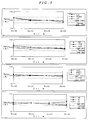
- Figure 2 shows graphs indicating variation of various analytical values of plasmas obtained from whole bloods different in hematocrit value.
- the whole blood sample to which the method of the invention is applicable may contain various anticoagulant or glycolysis inhibitor, such as heparin, NaF, EDTA or monoiodoacetate, or not.
- the method of the invention is effective especially against high hematocrit bloods, e.g. having a hematocrit (Hct) value of 50 to 70 %, particularly 55 to 70 %.
- Hct hematocrit
- the inorganic salt and amino acid and salt thereof are an agent (HL agent) having an action to accelerate separation between blood cells and plasma to decrease hematocrit value.
- the HL agent which is the inorganic salt, amino acid or salt thereof is water - soluble and has a solubility of 100 mmol/l or more, preferably 1 mol/l or more, particularly 3 mol/l or more.
- Exemplary inorganic salts are combinations of monovalent or divalent alkals metal or alkaline earth metal and halogen, NO 3 , SO 4 or CO 3 , such as CsCl 2 , Li 2 SO 4 , CaCl 2 , Rb 2 SO 4 and Cs 2 SO 4 .
- Exemplary amino acids are natural amino acids, such as Gly, Ala, Asp, Glu and glycinamide asparagine.
- the inorganic salt, amino acid and salt thereof are made so that pH of their aqueous solution is 5 to 8, preferably 6 to 7.5, and therefore, the inorganic salt and amino acid salt may be a hydrogen salt, such as NaHCO 3 .
- Acidic amino acids, such as Asp and Glu may be made into a salt of alkali metal or alkaline earth metal or the like, and basic amino acids, such as Lys and Arg, may be made into a salt of halogen, NO 3 , SO 4 , CO 3 or the like.
- the HL agent to be used is selected by considering subject matter to be measured, i.e. a component to be measured. For example, when sodium or chloride in blood is measured, NaCl is unsuitable as the HL agent. Phosphate is unsuitable as the HL agnet for measuring inorganic phosphorus in blood. Moreover, HL agent which influences reactivity of reagents used for measurement cannot be used.
- HL agent containing magnesium, iron, copper, barium, zinc, or the like cannot be used for measuring calcium, because they interfere with calcium measurement.
- the HL agent is used as an aqueous solution.
- a suitable concentration of the HL agent is 0.1 to 5 mol/l. preferably 0.5 to 3 mol/l, more preferably 1 to 2.5 mol/l.
- glycerine ethylene glycol, polyethylene glycol or the like may be added for the purpose of prevention against drying.
- a buffer may be added for the purpose of pH adjustment.
- various compounds may be added in order to facilitate separation of analyte in filtered plasma.
- exemplary of the compounds are selective binding reagents for LDL choresterol, such as dextrin and tungstophosphoric acid, which are fractionation agent for the measurement of HDL choresterol.
- Other exemplary compounds are having reactivity with a particular component in plasma, such as various antigens and antibodies including modified ones.
- HL agent By adding the HL agent, flexibility of erythrocyte membrane decreases, and erythrocytes become resistant to deformation. It is known that erythrocyte passes through a considerably smaller space, e.g. a capillary 1-2 ⁇ m in diameter, than apparent size of erythrocyte during filtering whole blood. However, by adding the HL agent, since deformation of erythrocytes is restrained, filtration can be conducted very smoothly.
- Reduction of hematocrit value by the HL agent is in proportion to molar concentration of the HL agent in plasma volume of whole blood to which the HL agent is added.
- a suitable amount of the HL agent added to whole blood is 10 to 200 ⁇ mol, preferably 10 to 100 ⁇ mol, more preferably 20 to 60 ⁇ mol, per 1 ml whole blood.
- Volume of aqueous HL agent solution to be added is not especially limited.
- a suitable volume of the aqueous HL agent solution to be added is 20 % or less, preferably 10 % or less, more preferably 5 % or less of whole blood by volume ratio.
- the lower limit of the volume of the aqueous HL agent solution depends on the water-solubility of HL agent, etc., and in general, 1 % or more.
- filtering ability is improved by adding only a small amount of HL agent, influence on measuring sensitivity and accuracy is small.
- HL agent When HL agent is added to whole blood, the HL agent is dissolved into plasma to elevate osmotic pressure of plasma to generate osmotic pressure difference between plasma and the inside of blood cells. Then, force acts so as to loosen the difference of osmotic pressure. That is, water in blood cell is discharged into plasma to elevate osmotic pressure (i.e. concentration of solute) in blood cells and to lower osmotic pressure of plasma. As a result, volume of blood cell decreases and volume of plasma increases, resulting in the decrease of hematocrit value.
- volume of erythrocyte decreases and flexibility of blood cell membrane decreases, and accordingly, filtration using a filtering material, such as porous membrane is facilitated. Moreover, since quantity of free plasma increases, recovery rate of plasma by filtration is improved.
- blood assay is based on the measurement of concentration of plasma component.
- erythrocytes When erythrocytes are destroyed or blood cell membranes are purctured, components in erythrocytes are leaked into plasma portion. Since erythrocyte contains Hb (hemoglobin) in a high concentration, when leakage occurs, plasma becomes red (called hemolysis). Even if the destruction of erythrocytes is very small, coloring of plasma caused by the leakage of Hb cannot be ignored, and in some analytical items, the leakage strongly influences analytical value. For example, in a blood assay of healthy persons, when 0.1 % of total blood cells are destroyed, measured values of most enzymes, such as CPK and ALP, are not accurate.
- Some components exist in erythrocyte in a higher concentration than plasma, and the measurement of those components is directly influenced by hemolysis. Accordingly, it is necessary to conduct separation, recovery of plasma under conditions where hemolysis occurs as small as possible.
- Heglobin Hb
- Influence by leaked hemoglobin is different according to measuring method and measuring item.
- GOT, GPT, LDH, K and the like are liable to be influenced by hemolysis, and particularly, in the case of LDH or K, even when hemoglobin concentration is 15 mg/dl (corresponding to hemolysis of 0.1% of total erythrocytes), measured values are influenced significantly in view of clinical diagnosis.
- hemolysis must be less than 0.1 %.
- Hematocrit value of healthy persons is 40 to 45 %, but whole blood samples having a hematocrit value of 55 to 60 % are dealt at a frequency of several percents in daily clinical assay.
- Hb content is 20 to 25 g/dl. If allowance of measurement against Hb is 15 mg/dl, hemolysis of high Hct samples must be restrained to less than 0.07 %.
- inorganic salt amino acid or salt thereof or agglutinin such as lectin is dissolved directly in blood from a dry state or from impregnated into filter paper or porous material followed by drying, blood contacts the dry matter. Blood is liable to hemolyze locally and temporarily in a initial stage of dissolving the dry matter in plasma. Accordingly, plasma obtained through the above process frequently contains hemoglobin which is not so much.
- the plasma has no problem for measuring blood sugar or choresterol, but has a problem for measuring GOT, GPT, LDH, K and the like. The tendency is more remarkable in high HcT samples.
- An amount of blood to be used is about 0.3 to 3ml, usually about 0.5 to 1.5 ml, and mixing of aqueous HL agent solution with whole blood may be carried out only by several times shaking. It is supposed that the action of HL agent is in a moment, and the mixture reaches equilibrium within several seconds. Accordingly, it is not necessary to adjust temperature or time upon mixing.
- the whole blond sample is filtered.
- various known filtering material capable of separating blood cells can be used.
- the filtering materials are glass fiber filter, microporous membranes having blood cell-separating ability such as made of fluorine-containing polymer, of which the surface has been made hydrophilic, or polysulfone, laminates of glass fiber filter and microporous membrane, laminates of glass fiber filter and cellulose filter, laminates of glass fiber filter, cellulose filter and microporous membrane, laminates of fibrous porous membrane and non-fibrous porous membrane disclosed in Japanese Patent KOKAI Nos. 62-138756-8, 2-105043, 3-16651, etc.
- Preferred ones are containing glass fiber filter, and laminates of glass fiber filter and microporous membrane, laminates of glass fiber filter and cellulose filter, and laminates of glass fiber filter, cellulose filter and microporous membrane are particularly preferred.
- Filter materials containing polysulfone microporous membrane are also preferable.
- the most preferable ones are laminates of glass fiber filter, cellulose filter and microporous membrane, especially using polysulfone membrane as the microporous membrane.
- Preferable glass fiber filter has a density of about 0.02 to 0.3 g/cm 3 preferably about 0.05 to 0.2 g/cm 3 , more preferably about 0.07 to 0.15 g/cm 3 , a retainable particle size of about 0.8 to 9 ⁇ m, preferably 1 to 5 ⁇ m.
- Microporous membranes having blood cell-separating ability of which the surface has been made hydrophilic separate whole blood specifically into blood cells and plasma without hemolysis to the degree of substantially influencing analytical values.
- a suitable pore size of the microporous membrane is smaller than the retainable particle size of glass fiber filter, and is 0.2 ⁇ m or more, preferably about 0.3 to 5 ⁇ m, more preferably about 0.5 to 4.5 ⁇ m, particularly preferably about 1 to 3 ⁇ m.
- the void content of the microporous membrane is preferably higher, and a suitable void content is about 40 to 95 %, preferably about 50 to 95 %, more preferably about 70 to 95 %.
- microporous membranes are polysulfone membrane, fluorine-containing polymer membrane, cellulose acetate membrane, nitrocellalose membrane, etc.
- the surface of the membrane may be hydrolyzed or may be rendered hydrophilic by a hydrophilic polymer or an activating agent.
- microporous matrix membrane composed of polytetrafluoroethylene fibrils (fines) disclosed in WO 87/02267, Gore-Tex (W. L. Gore and Associates), Zitex (Norton), Poreflon (Sumitomo Denki), etc.
- Other fluorine-containing polymer sheets usable as the microporous layer include polytetrafluoroethylene microporous membranes disclosed in U.S.P. 3,368,872 (Examples 3 and 4), U.S.P 3,260,413 (Examples 3 and 4), U.S. P.
- microporous membrane of fluorine-containing polymer may be prepared by using a single fluorine-containing polymer or blending two or more kinds of fluorine-containing polymers of further blending one or more polymers not containing fluorine or fibers therewith.
- As the structure there are unstretched one, uniaxially stretched one, biaxially stretched one, nonlaminated single layer type, laminated double layer type, such as a membrane laminated to another membrane structure such as a fiber membrane.
- microporous membrane having a great void content and a short filtering pass can be prepared by stretching.
- clogging rarely occurs by solid components (mainly red blood cells) in blood and the separation time of blood cells and plasma is short.
- accuracy in quantitative analysis is improved.
- the adhesive strength of adhesive used for the partial adhesion to the adjacent microporous membrane can be strengthened by providing the physical activation (preferably glow discharge or corona discharge) disclosed in U.S.P. 4,783,315 on at least one side of the microporous membrane of fluorine-containing polymer to render hydrophilic.
- fluorine-containing polymer microporous membranes as it is have a low surface tension.
- aqueous liquid samples are repelled and do not diffuse nor permeate over the surface or into the inside.
- the above repelling problem has been resolved by incorporating a sufficient amount of surfactant for rendering the outer surface and the inner space surface of the fluorine-containing polymer microporous membrane substantially hydrophilic thereinto.
- a surfactant in an amount of about 0.01 to 10 %, preferably about 0.1 to 5 %, more preferably about 0.1 to 1 % of the void volume of the membrane.
- a preferred amount of surfactant to be impregnated is usually in the range of 0.05 to 2.5 g/m 2 .
- a common method cmoprises immersing the fluorine-containng microporous membrane in the surfactant solution dissolved in a low boiling point (a preferable boiling point is in the range of about 50 °C to about 120 °C ) organic solvent (e.g. alcohols, esters, ketones) to permeate into the inner spaces of the membrane substantially sufficiently, taking the membrane out of the solution slowly, and then drying by blowing air (preferably warm air).
- a low boiling point e.g. alcohols, esters, ketones
- the surfacctant may be nonionic, anionic, cationic or ampholytic.
- nonionic surfactants are advantageous for the multilayer analytical elements for analyzing whole blood samples, because nonionic surfactants have a relatively low hemolytic activity among the above surfactants.
- Suitable nonionic surfactants include alkylphenoxypolyethoxyethanol, alkylpolyether alcohol, polyethyleneglycol monoester, polyethyleneglycol diester, higher alcohol-ethylene oxide adduct (condensate), polyol ester-ethylene oxide adduct (condensate), higher fatty acid alkanol amide, etc.
- nonionic surfactant examples include as follows: As the alkylphenoxypolyethoxyethanol, there are isooctylphenoxypol y-ethoxyethanols(Triton X-100; containing 9-10 hydroxyethylene units on average, Triton X-45; containing 5 hydroxyethylene units on average) and nonylphenoxypolyethoxyethanols (IGEPAL CO-680; containing 9 hydroxyethylene units on average, IGEPAL CO-710; containing 10-11 hydroxyethylene units on average, LENEX 698; containing 9 hydroxyethylene units on average). As the alkylpolyether alcohol, there are higher alcohol polyoxyethylenc ethers (Triton X-67; CA Registry No. 59030-15-8), etc.
- the fluorine-containing polymer microporuus membrane may be rendered hydrophilic by providing one or more water-insolubilized water coluble polymers in its porous spaces.
- the water-soluble polymers include oxygen-containing hydrocarbons, such as polyacrylamide, polyvinylpyrrolidone, polyvinylamine and polyethyleneiminc, negative charge-containing ones such as polyvinyl alcohol, polyethylene oxide, polyethylene glycol, methyl cellulose, ethyl cellulose, hydroxyethyl cellulose and hydroxypropyl cellulose, nitrogen-containing ones, such as polyacrylic acid, polymetacrylic acid and polystyrene sulfonic acid, and the like.
- the water-insolubilization may be conducted by heat treatment, acetal-inducing treatment, esterification, chemical reaction by potassium dichromate, crosslinking by ionizable radiation, or the like. Details are disclosed in Japanese Patent KOKOKU Nos. 56-2094 and 56-16187.
- the polysulfone microporous membrane can be prepared by dissolving polysulfone into dioxane, tetrahydrofuran, dimethylformamide, dimethylacetamide, N-methyl-2-pyrolidone or a mixed solvent thereof to obtaine a raw liquid for forming film, casting into film by flowing directly into a coagulating solution, washing, and then drying. Details are disclosed in Japanese Patent KOKAI No. 62-27006. In addition, polysulfone microporous membranes are also disclosed in Japanese Patent KOKAI Nos. 56-12640, 56-86941, 56-154051, etc., and they are applicable to the invention.
- the polysulfone microporous membrane can be rendered hydrophilic, similar to the fluorine-containing polymer, by incorporating surfactant or providing water-insolubilized water-soluble polymer.
- nonfibrous porous membranes in the laminate of a fibrous porous membrane and a nonfibrous porous membrane are blushed polymer membranes composed of a cellulose ester, such as cellulose acetate, cellulose acetate/butyrate or cellulose nitrate, disclosed in US 3 992 158 or US 1 421 341.
- the nonfibrous porous membrane may be a microporous membrane of polyamide, such as 6-nylon or 6,6-nylon, or polyethylene, polypropylene, or the like.
- nonfibrous porous membranes usable for tho blood cell-separating filter include continuous microspace-containing porous membranes where polymer particulates, glass particulates, diatomaceous earth or the like are joined by a hydrophilic or non-water-adsorptive polymer, such as disclosed in US 3 992 158, and US 4 258 001.
- Preferable effective pore size of the nonfibrous porous membrane is 0.2 to 10 ⁇ m, preferably 0.3 to 5 ⁇ m, particularly preferably 0.5 to 5 ⁇ m.
- the effective pore size of the nonfibrous porous membrane in the invention is the pore size measured by the bubble point method according to ASTM F316-70.
- the liquid passages in the thickness direction are, in general, the narrowest at the free surface (glossy face) in the manufacturing process of the membrane, and the pore size in section of each liquid passage stipulated a circle is the smallest near the free surface.
- the minimum pore size of passages in the thickness direction per unit area has a distribution in facial direction of the membrane filter, and the maximum value determines filtration performance. In general, it is determined by the limit bubble point method.
- liquid passages in the thickness direction become the narrowest at the free surface (glossy face) in the manufacturing process of the membrane.
- the material composing the fibrous porous membrane may be filter paper, nonwoven fabric, woven fabric such as plain weave fabric, knitted fabric such as tricot fabric, etc. Among them, woven fabric and knitted fabric are preferred.
- the woven fabric or the like may be treated by blow discharge as disclosed in Japanese Patent KOKAI No. 57-66359.
- the void volume (per unit area) of the fibrous porous membrane may be equal to or different from the nonfibrous porous layer.
- the void volume correlation between them may be adjusted by changing either or both of void content and thickness.
- the filtering material used in the invention is a laminate
- respective layers may be integrated by joining each other using partially disposed (e.g. spots) adhesive, according to disclosures in Japanese Patent KOKAI Nos. 62-138756-8, 2-105043, 3-16651, etc.
- the filter material does not trap blood cells only by the surface, but catches to remove blood cells gradually by entangling at first large blood cell components and then smaller blood cell components in the space structure with permeating in the thickness direction in total of the filtering material called the volumetric filtration.
- density, thickness, area, etc. of the filtering material may be arranged according to the supplied, i.e. to be separated, volume of blood.
- a volume of plasma or serum to be recovered becomes so as to be 10 % or more, preferably 20 to 100 %, particularly preferably 30 to 70 % of the volume of the glass fiber filter.
- it is preferably to use a glass fiber filter having a volume about twice as much as the volume of plasma to be recovered.
- a supply volume of whole blood to be filtered is not restricted. Although the volume of whole blood is smaller than the volume of glass fiber filter, plasma can be separated. However, the ratio (recovery) of recovered volume of plasma to the volume of whole blood is small. In the case that the volume of whole blood is greater than the volume of glass fiber filter, space of the glass fiber filter is filled out by blood cells, and then, leakage of blood cells occurs from glass fiber filter. If a microporous membrane is provided, leaked blood cells are caught by the microporous membrane. As a result, volume of recovered plasma increases compared with using glass fiber filter alone. In general, a suitable volume ratio of whole blood:glass fiber filter is 0.5 to 10:1, preferably 0.7 to 5:1, more preferably 0.8 to 3:1.
- a preferable volume (e.g. sectional area of priston ⁇ stroke in syringe) for inducing the pressurizing or sucking conditions is twice to 5 times as large as the volume of filtering material.
- the pressurizing or sucking period is not strictly restricted. However, in order to prevent homolysis, the period is within 60 seconds wherein filtration is preferably accomplished rapidly.
- a suitable pressurizing or sucking period is 1 to 60 seconds, preferably 2 to 30 seconds, more preferably 5 to 20 seconds, and thereafter, pressure is returned to atmospheric pressure promptly.
- plasma volume to be recovered is previously set, and glass fiber filter volume corresponding thereto was set. Then, a sufficient volume of blood is supplied, and pressurized or sucked. When a prescribed volume of plasma is obtained or a prescribed period is over, pressure is returned to atmospheric pressure. Plasma or serum thus obtained is subjected to analysis according to a conventional manner.
- the method of the invention is effective for the analysis of plural items using dry analytical elements.
- plasma separation from whole blood can be achieved efficiently, and recovered volume of plasma can be increased.
- good plasma which has no deviation in component viewpoint, and which is possible to measure in the same level accuracy as a plasma obtained by centrifuging, can be obtained through a simple filtration operation.
- Example 2 Instead of 2 mol/l aqueous Li 2 SO 4 solution in Example 1, saline was add to the whole blood, and lowering degree of hematocrit (Hct) was examined, to 1.5 ml of the whole blood with heparin, saline was added, and after mixing, hematocrit value was measured. Two levels of the whole blood of healthy person (Hct 44 %) and a high hematocrit whole blood (Hct 54 %) were examined.
- Blood was drawn from healthy women to obtain 20 ml whole blood with heparin. Hematocrit value of the blood was measured and found to be 41 %. A part of the blood was centrifuged to separate plasma to prepare high hematocrit whole blood samples. The plasma was added to the original whole blood to prepare a low hematocrit whole blood. Thus, 5 types (No.1-No.5) of whole blood samples different in hematocrit value were prepared.
- Example 2 In an experiment similar to Example 1, 15 ⁇ l aqueous 2 mol/l Li 2 SO 4 solution was added to 1.5 ml whole blood, and the blood was filtered similar to Example 1. As a result, 228 ⁇ l of plasma was recovered without hemolysis. The Hct value was 48 % before adding HL agent and 42 % after adding it.
- the whole blood was also filtered without HL agent, and in this case, plasma without hemolysis was not recovered.
- a whole blood was separated into blood cells and plasma, and then both were mixed at a different ratio to prepare whole blood samples having a hematocrit value of 20 %, 40 %, 48 % and 60 %.
- the samples were treated similar to Example 3, and measured by using an analyzer "Hitachi 7150" and an electrolyte analyzer "Fuji Drichem 800".
- Cs 2 SO 4 was used as the HL agent instead of Li 3 SO 4 .
- 60 ⁇ l of 2 mol/l Cs 2 SO 4 aqueous solution was added to 1.5 ml whole blood with heparin.
- the Hct value was 38 % before adding the HL agent and 31 % after the addition thereof.
- the whole blood containing the HL agent was filtered with suction similar to Example 1, and 320 ⁇ l plasma without hemolysis was obtained.
- Li 2 SO 4 and Cs 2 SO 4 were especially excellent as HL agent.
- HL agents containing the same ion species as the target analyte are unsuitable.
- the HL agents which are indicated by X can be used by substituting an ion species suitable for the measurement for the same or analogous ion species in each HL agent All of the HL agents in Table 5 are usable as the HL agent, unless measuring electrolyte.
Landscapes
- Health & Medical Sciences (AREA)
- Life Sciences & Earth Sciences (AREA)
- Engineering & Computer Science (AREA)
- Biomedical Technology (AREA)
- Hematology (AREA)
- Chemical & Material Sciences (AREA)
- Urology & Nephrology (AREA)
- Physics & Mathematics (AREA)
- Immunology (AREA)
- Molecular Biology (AREA)
- Food Science & Technology (AREA)
- General Health & Medical Sciences (AREA)
- Pathology (AREA)
- Medicinal Chemistry (AREA)
- General Physics & Mathematics (AREA)
- Analytical Chemistry (AREA)
- Biochemistry (AREA)
- Biotechnology (AREA)
- Microbiology (AREA)
- Cell Biology (AREA)
- Ecology (AREA)
- Biophysics (AREA)
- Investigating Or Analysing Biological Materials (AREA)
- Sampling And Sample Adjustment (AREA)
Claims (6)
- Verfahren zur Herstellung einer Plasma- oder Serumprobe aus Vollblut, wobei man eine wäßrige Lösung mit einem pH-Wert von 5 bis 8 eines anorganischen Salzes oder einer Aminosäure oder eines Salzes hiervon mit einer Konzentration von 0,1 bis 5 mol/l mit dem Vollblut in einer Menge von 20% oder weniger des Vollblutvolumens vermischt, so daß das anorganische Salz oder die Aminosäure oder das Salz hiervon in einer Menge von 10 bis 200 µmol/ml Vollblut vorliegt, und wobei man dann das Vollblut filtriert, um Blutzellbestandteile zu entfernen, wobei das anorganische Salz eine Kombination aus einwertigem oder zweiwertigem Alkalimetall oder Erdalkalimetall und einem Mitglied, ausgewählt aus der Gruppe bestehend aus Halogen, NO3, SO4 und CO3, ist, außer Natriumchlorid.
- Verfahren nach Anspruch 1, wobei das anorganische Salz ausgewählt ist aus der Gruppe bestehend aus Asp·Na, CaCl2, NaCl, LiNO3, Si2SO4, Rb2SO4, Cs2SO4.
- Verfahren nach Anspruch 1, wobei das bei dem Filtrieren verwendete Filtermaterial Glasfaserfilter umfaßt.
- Verfahren nach Anspruch 3, wobei der Glasfaserfilter eine Dichte von 0,02 bis 0,2 g/cm3 aufweist.
- Verfahren nach Anspruch 3, wobei das Filtermaterial weiterhin eine mikroporöse Membran umfaßt.
- Verfahren nach Anspruch 5, wobei die mikroporöse Membran eine Fluour enthaltende Polymermembran oder Polysulfonmembran ist.
Applications Claiming Priority (3)
| Application Number | Priority Date | Filing Date | Title |
|---|---|---|---|
| JP769196 | 1996-01-19 | ||
| JP7691/96 | 1996-01-19 | ||
| JP00769196A JP3664328B2 (ja) | 1996-01-19 | 1996-01-19 | 血漿または血清試料の調製方法 |
Publications (2)
| Publication Number | Publication Date |
|---|---|
| EP0785430A1 EP0785430A1 (de) | 1997-07-23 |
| EP0785430B1 true EP0785430B1 (de) | 2000-08-30 |
Family
ID=11672811
Family Applications (1)
| Application Number | Title | Priority Date | Filing Date |
|---|---|---|---|
| EP97100695A Expired - Lifetime EP0785430B1 (de) | 1996-01-19 | 1997-01-17 | Trennung von Plasma oder Serum von Vollblut |
Country Status (4)
| Country | Link |
|---|---|
| US (1) | US5876605A (de) |
| EP (1) | EP0785430B1 (de) |
| JP (1) | JP3664328B2 (de) |
| DE (1) | DE69702934T2 (de) |
Families Citing this family (39)
| Publication number | Priority date | Publication date | Assignee | Title |
|---|---|---|---|---|
| JP3903098B2 (ja) * | 1997-07-18 | 2007-04-11 | 富士フイルム株式会社 | 血液濾過方法 |
| JPH11237378A (ja) | 1998-02-19 | 1999-08-31 | Fuji Photo Film Co Ltd | 全血から血清を分離する方法 |
| US6471868B1 (en) * | 1998-04-10 | 2002-10-29 | Fuji Photo Film Co., Ltd. | Method of preparing glass fiber filter |
| US6908770B1 (en) * | 1998-07-16 | 2005-06-21 | Board Of Regents, The University Of Texas System | Fluid based analysis of multiple analytes by a sensor array |
| JP3990505B2 (ja) * | 1999-02-02 | 2007-10-17 | 富士フイルム株式会社 | 血液成分の分析方法 |
| US7022517B1 (en) | 1999-07-16 | 2006-04-04 | Board Of Regents, The University Of Texas System | Method and apparatus for the delivery of samples to a chemical sensor array |
| AU1325201A (en) | 1999-07-16 | 2001-02-05 | Board Of Regents, The University Of Texas System | Detection system based on an analyte reactive particle |
| US7651474B2 (en) * | 1999-10-01 | 2010-01-26 | Caridianbct, Inc. | Method and apparatus for leukoreduction of red blood cells |
| ATE403145T1 (de) * | 2000-01-31 | 2008-08-15 | Univ Texas | Tragbare vorrichtung mit einer sensor-array- anordnung |
| KR100513213B1 (ko) | 2000-06-01 | 2005-09-08 | 마츠시타 덴끼 산교 가부시키가이샤 | 바이오센서 및 혈액 성분 분석 방법 |
| DE60142394D1 (de) | 2000-06-28 | 2010-07-29 | Panasonic Corp | Biozensor |
| US20020197622A1 (en) * | 2001-01-31 | 2002-12-26 | Mcdevitt John T. | Method and apparatus for the confinement of materials in a micromachined chemical sensor array |
| WO2003005039A1 (fr) * | 2001-07-04 | 2003-01-16 | Kyowa Medex Co., Ltd. | Procede de preparation d'une solution de quantification, procede de quantification utilisant la solution de quantification, instrument de preparation de solutions de quantification et procede d'utilisation correspondant |
| US7087397B2 (en) | 2001-12-21 | 2006-08-08 | Polymer Technology Systems, Inc, | Method for determining HDL concentration from whole blood or plasma |
| US8257967B2 (en) | 2002-04-26 | 2012-09-04 | Board Of Regents, The University Of Texas System | Method and system for the detection of cardiac risk factors |
| KR101005970B1 (ko) * | 2002-11-19 | 2011-01-05 | 가부시키가이샤 아이 디자인 | 혈장 또는 혈청 분리막, 및 혈장 또는 혈청 분리막을이용한 필터 장치 |
| JP2004294388A (ja) * | 2003-03-10 | 2004-10-21 | Arkray Inc | 成分分析装置 |
| US8101431B2 (en) * | 2004-02-27 | 2012-01-24 | Board Of Regents, The University Of Texas System | Integration of fluids and reagents into self-contained cartridges containing sensor elements and reagent delivery systems |
| US8105849B2 (en) | 2004-02-27 | 2012-01-31 | Board Of Regents, The University Of Texas System | Integration of fluids and reagents into self-contained cartridges containing sensor elements |
| US20080003141A1 (en) * | 2004-04-30 | 2008-01-03 | Kazuya Iketani | Sample Analyzing Tool |
| JP4637533B2 (ja) * | 2004-08-31 | 2011-02-23 | 信和化工株式会社 | 固相抽出用分離剤 |
| EP1882524A4 (de) * | 2005-05-17 | 2010-09-08 | Universal Bio Research Co Ltd | Filterverarbeitungsverfahren, filterverkapselungschip und filterverarbeitungsvorrichtung |
| WO2007053186A2 (en) | 2005-05-31 | 2007-05-10 | Labnow, Inc. | Methods and compositions related to determination and use of white blood cell counts |
| EP1899450A4 (de) * | 2005-06-24 | 2010-03-24 | Univ Texas | Systeme und verfahren einschliesslich in sich geschlossener kassetten mit nachweissystemen und flüssigkeitszufuhrsystemen |
| WO2007005666A2 (en) * | 2005-07-01 | 2007-01-11 | Board Of Regents, The University Of Texas System | System and method of analyte detection using differential receptors |
| US20080017577A1 (en) * | 2006-07-21 | 2008-01-24 | Becton, Dickinson And Company | Membrane-based Double-layer Tube for Sample Collections |
| US7727764B2 (en) * | 2006-12-28 | 2010-06-01 | Puthalath Koroth Raghuprasad | Non-isopycnic cell purification using percoll |
| US20080300798A1 (en) * | 2007-04-16 | 2008-12-04 | Mcdevitt John T | Cardibioindex/cardibioscore and utility of salivary proteome in cardiovascular diagnostics |
| EP2264453B1 (de) * | 2009-06-17 | 2013-04-03 | Leukocare Ag | Verfahren zum Filtern von Blut |
| ITFI20110078A1 (it) * | 2011-04-20 | 2012-10-21 | Gianfranco Liguri | Metodo per la determinazione di analiti nel plasma diluito ottenuto da sangue intero, e dispositivo atto a realizzarlo |
| US9427707B2 (en) | 2012-08-10 | 2016-08-30 | Jean I. Montagu | Filtering blood |
| EP2890980B1 (de) | 2012-08-30 | 2018-12-19 | Qiagen GmbH | Verfahren zum erhalt von blutplasma aus einer vollblutprobe |
| CN103528876A (zh) * | 2013-10-29 | 2014-01-22 | 江苏德威兰医疗器械有限公司 | 血清快速分离器 |
| WO2016019113A1 (en) * | 2014-08-01 | 2016-02-04 | Siemens Healthcare Diagnostics Inc. | Vacuum-assisted plasma separation |
| WO2016158139A1 (ja) * | 2015-03-31 | 2016-10-06 | ソニー株式会社 | 電気的特性測定装置、電気的特性測定方法、血液状態解析システム、及び該方法をコンピューターに実現させるための電気的特性測定用プログラム |
| MX2021006613A (es) | 2018-12-07 | 2021-07-07 | Siemens Healthcare Diagnostics Inc | Dispositivo con una funcion de evaluacion de componentes fluidos. |
| CA3147512C (en) | 2019-07-19 | 2024-04-02 | Siemens Healthcare Diagnostics Inc. | Tangent flow hemolysis detection blood testing device |
| US20220059194A1 (en) * | 2020-08-24 | 2022-02-24 | FluoroMe, Inc., | 3d pharmacophore model for the rapid computational screening of sars-cov-2 modulators and compositions and methods thereof |
| JP7768638B1 (ja) * | 2024-03-22 | 2025-11-12 | 積水メディカル株式会社 | 血液採取容器、血漿の分離方法、細胞外遊離核酸の分離方法及び細胞外小胞の分離方法 |
Family Cites Families (28)
| Publication number | Priority date | Publication date | Assignee | Title |
|---|---|---|---|---|
| DE329117C (de) | 1916-08-21 | 1920-11-15 | Richard Zsigmondy Dr | Membranfilter und Verfahren zu deren Herstellung |
| US3260413A (en) | 1964-08-31 | 1966-07-12 | Scientific Industries | Automatic chemical analyzer |
| US3368872A (en) | 1964-08-31 | 1968-02-13 | Scientific Industries | Automatic chemical analyzer |
| US3552925A (en) * | 1967-07-13 | 1971-01-05 | Miles Lab | Whole blood separation method and test using same |
| US3552928A (en) * | 1967-07-19 | 1971-01-05 | Miles Lab | Whole blood separation means and test system using same |
| US3649505A (en) | 1969-03-03 | 1972-03-14 | Beckman Instruments Inc | Ammonia sensor |
| US3992158A (en) | 1973-08-16 | 1976-11-16 | Eastman Kodak Company | Integral analytical element |
| JPS548669A (en) | 1977-06-21 | 1979-01-23 | Sumitomo Electric Ind Ltd | Hydrophilic porous composite structure and its manufacture |
| JPS5392195A (en) | 1977-01-25 | 1978-08-12 | Dojindo Lab | Simple rapid determination method and apparatus for volatile matter in aqueous solution |
| JPS5417978A (en) | 1977-07-11 | 1979-02-09 | Sumitomo Electric Ind Ltd | Hydrophilic and porous composite structure and its production |
| US4258001A (en) | 1978-12-27 | 1981-03-24 | Eastman Kodak Company | Element, structure and method for the analysis or transport of liquids |
| JPS5612640A (en) | 1979-07-11 | 1981-02-07 | Nissha Printing Co Ltd | Simultaneously regenerating method for used developer and used bleaching bath |
| JPS5686941A (en) | 1979-12-17 | 1981-07-15 | Asahi Chem Ind Co Ltd | Porous membrane of polysulfone resin |
| CA1202837A (en) | 1980-03-14 | 1986-04-08 | Wolfgang J. Wrasidlo | Asymmetric membranes and process therefor |
| JPS5766359A (en) | 1980-10-09 | 1982-04-22 | Fuji Photo Film Co Ltd | Sheet-like material for analysis |
| US4678566A (en) * | 1984-07-20 | 1987-07-07 | Terumo Kabushiki Kaisha | Apparatus for separating proteins from blood plasma |
| JPS6138608A (ja) | 1984-07-31 | 1986-02-24 | Fuji Photo Film Co Ltd | 全血からの血漿の分離器具および分離方法 |
| JPS6227006A (ja) | 1985-07-27 | 1987-02-05 | Fuji Photo Film Co Ltd | 微孔性膜 |
| US4839296A (en) | 1985-10-18 | 1989-06-13 | Chem-Elec, Inc. | Blood plasma test method |
| JPS62138756A (ja) | 1985-12-12 | 1987-06-22 | Fuji Photo Film Co Ltd | 一体型多層分析要素 |
| US5423989A (en) * | 1988-05-19 | 1995-06-13 | Chemtrack, Inc. | Plasma forming device |
| JPH02105043A (ja) | 1988-10-14 | 1990-04-17 | Fuji Photo Film Co Ltd | 一体型多層分析要素の製造方法 |
| JP2864035B2 (ja) | 1989-02-09 | 1999-03-03 | 富士写真フイルム株式会社 | 全血分析要素 |
| JPH02208565A (ja) | 1989-02-09 | 1990-08-20 | Fuji Photo Film Co Ltd | 血液から血漿を分離する方法、器具、およびこれを用いる分析要素 |
| US5118428A (en) * | 1990-11-13 | 1992-06-02 | Quidel | Method to remove red blood cells from whole blood samples |
| JPH04208565A (ja) * | 1990-11-30 | 1992-07-30 | Olympus Optical Co Ltd | 強誘電体メモリ |
| JPH04208856A (ja) * | 1990-12-03 | 1992-07-30 | Sanwa Kagaku Kenkyusho Co Ltd | 全血から血清又は血漿を分離する方法および器具 |
| US5208142A (en) * | 1991-11-19 | 1993-05-04 | Miles Inc. | Method for separating erythrocytes from whole blood |
-
1996
- 1996-01-19 JP JP00769196A patent/JP3664328B2/ja not_active Expired - Fee Related
-
1997
- 1997-01-17 DE DE69702934T patent/DE69702934T2/de not_active Expired - Lifetime
- 1997-01-17 EP EP97100695A patent/EP0785430B1/de not_active Expired - Lifetime
- 1997-01-17 US US08/784,480 patent/US5876605A/en not_active Expired - Fee Related
Also Published As
| Publication number | Publication date |
|---|---|
| EP0785430A1 (de) | 1997-07-23 |
| JP3664328B2 (ja) | 2005-06-22 |
| US5876605A (en) | 1999-03-02 |
| JPH09196908A (ja) | 1997-07-31 |
| DE69702934D1 (de) | 2000-10-05 |
| DE69702934T2 (de) | 2000-12-28 |
Similar Documents
| Publication | Publication Date | Title |
|---|---|---|
| EP0785430B1 (de) | Trennung von Plasma oder Serum von Vollblut | |
| EP1291062B1 (de) | Blutfiltereinheit | |
| EP0893130B1 (de) | Methode zur Filterung von Blut | |
| US5996811A (en) | Plasma collecting device | |
| CA1255196A (en) | Test device and a method for the detection of a component of a liquid sample | |
| CN1168988C (zh) | 用于血液中葡萄糖测定的试剂测试条 | |
| EP2221621B1 (de) | Verfahren um HDL-Cholesterin im Vollblut zu bestimmen | |
| CA1322335C (en) | Process and device for the separation of a body fluid from particulate materials | |
| AU621236B2 (en) | Analysis method for determining substances from biological fluids | |
| US4783315A (en) | Analysis material sheet | |
| JP2940990B2 (ja) | 全血からの血漿または血清の分離装置および方法 | |
| US5652148A (en) | Method and apparatus for red blood cell separation | |
| US20090145840A1 (en) | Blood cell separation membrane and blood retention tool using the same | |
| EP0937981B1 (de) | Verfahren zur Abtrennung von Serum aus Vollblut | |
| CN116430036A (zh) | 一种滤血膜检测装置及制备方法和应用 | |
| EP0298473A2 (de) | Analytisches Element zur Vollblutuntersuchung | |
| JPH0843294A (ja) | 全血試料の分析方法および分析要素 | |
| CA2197386A1 (en) | Graphite nonwovens as functional layers in diagnostic test kits | |
| JPH11194126A (ja) | 血液濾過ユニット | |
| JPH0526875A (ja) | 一体型多層分析要素 | |
| JPH07191020A (ja) | 全血分析要素を用いた全血試料の分析方法 | |
| Tanishita et al. | Facilitated diffusion of carbon dioxide in whole blood and hemoglobin solutions | |
| Knight | The role of physical and chemical properties on gas transfer and blood compatibility of urethan membranes | |
| JPH0682444A (ja) | 電源を必要としない可搬型インキュベータ | |
| JPH03180762A (ja) | 一体型多層分析要素 |
Legal Events
| Date | Code | Title | Description |
|---|---|---|---|
| PUAI | Public reference made under article 153(3) epc to a published international application that has entered the european phase |
Free format text: ORIGINAL CODE: 0009012 |
|
| AK | Designated contracting states |
Kind code of ref document: A1 Designated state(s): DE GB |
|
| 17P | Request for examination filed |
Effective date: 19971021 |
|
| 17Q | First examination report despatched |
Effective date: 19971218 |
|
| GRAG | Despatch of communication of intention to grant |
Free format text: ORIGINAL CODE: EPIDOS AGRA |
|
| GRAG | Despatch of communication of intention to grant |
Free format text: ORIGINAL CODE: EPIDOS AGRA |
|
| GRAH | Despatch of communication of intention to grant a patent |
Free format text: ORIGINAL CODE: EPIDOS IGRA |
|
| GRAH | Despatch of communication of intention to grant a patent |
Free format text: ORIGINAL CODE: EPIDOS IGRA |
|
| GRAA | (expected) grant |
Free format text: ORIGINAL CODE: 0009210 |
|
| AK | Designated contracting states |
Kind code of ref document: B1 Designated state(s): DE GB |
|
| REF | Corresponds to: |
Ref document number: 69702934 Country of ref document: DE Date of ref document: 20001005 |
|
| EN | Fr: translation not filed | ||
| PLBE | No opposition filed within time limit |
Free format text: ORIGINAL CODE: 0009261 |
|
| STAA | Information on the status of an ep patent application or granted ep patent |
Free format text: STATUS: NO OPPOSITION FILED WITHIN TIME LIMIT |
|
| 26N | No opposition filed | ||
| REG | Reference to a national code |
Ref country code: GB Ref legal event code: IF02 |
|
| REG | Reference to a national code |
Ref country code: GB Ref legal event code: 732E |
|
| PGFP | Annual fee paid to national office [announced via postgrant information from national office to epo] |
Ref country code: GB Payment date: 20100113 Year of fee payment: 14 Ref country code: DE Payment date: 20091224 Year of fee payment: 14 |
|
| GBPC | Gb: european patent ceased through non-payment of renewal fee |
Effective date: 20110117 |
|
| PG25 | Lapsed in a contracting state [announced via postgrant information from national office to epo] |
Ref country code: GB Free format text: LAPSE BECAUSE OF NON-PAYMENT OF DUE FEES Effective date: 20110117 |
|
| REG | Reference to a national code |
Ref country code: DE Ref legal event code: R119 Ref document number: 69702934 Country of ref document: DE Effective date: 20110802 |
|
| PG25 | Lapsed in a contracting state [announced via postgrant information from national office to epo] |
Ref country code: DE Free format text: LAPSE BECAUSE OF NON-PAYMENT OF DUE FEES Effective date: 20110802 |